Employment Effectiveness of China's Economic Stimulus Package
Stimulus-specific adaptation in auditory thalamus of young and agedawake rats
Ben D. Richardson,1 Kenneth E. Hancock,2,3 and Donald M. Caspary1
1Department of Pharmacology, Southern Illinois University School of Medicine, Springfield, Illinois; 2Eaton-PeabodyLaboratories, Massachusetts Eye and Ear Infirmary, Boston, Massachusetts; and 3Department of Otology and Laryngology,Harvard Medical School, Boston, Massachusetts
Submitted 3 June 2013; accepted in final form 25 July 2013
Richardson BD, Hancock KE, Caspary DM. Stimulus-specificadaptation in auditory thalamus of young and aged awake rats. JNeurophysiol 110: 1892–1902, 2013. First published July 31, 2013;doi:10.1152/jn.00403.2013.—Novel stimulus detection by single neu-rons in the auditory system, known as stimulus-specific adaptation(SSA), appears to function as a real-time filtering/gating mechanismin processing acoustic information. Particular stimulus paradigmsallowing for quantification of a neuron’s ability to detect novel ordeviant stimuli have been used to examine SSA in the inferiorcolliculus, medial geniculate body (MGB), and auditory cortex ofanesthetized rodents. However, the study of SSA in awake animals islimited to auditory cortex. The present study used individually ad-vanceable tetrodes to record single-unit responses from auditorythalamus (MGB) of awake young adult and aged Fischer BrownNorway (FBN) rats to 1) examine the presence of SSA in the MGB ofawake rats and 2) determine whether SSA is altered by aging in MGB.MGB single units in awake FBN rats displayed SSA in response totwo stimulus paradigms: the oddball paradigm and a random blocked/interleaved presentation of a set of frequencies. SSA levels weremodestly, but nonsignificantly, increased in the nonlemniscal regionsof the MGB and at lower stimulus intensities, where 27 of 57 (47%)young adult MGB units displayed SSA. The present findings providethe initial description of SSA in the MGB of awake rats and supportSSA as being qualitatively independent of arousal level or anesthe-tized state. Finally, contrary to previous studies in auditory cortex ofanesthetized rats, MGB units in aged rats showed SSA levels indis-tinguishable from SSA levels in young adult rats, suggesting that SSAin MGB was not impacted by aging in an awake preparation.
stimulus-specific adaptation; medial geniculate body; auditory thala-mus; aging; awake animals
A SUBSET OF SENSORY NEURONS possesses the ability to selectivelysuppress responses to repetitive stimuli while maintaining thecapacity to robustly respond to novel stimuli, a property knownas stimulus-specific adaptation (SSA). This reduction in theimpact of repetitive, and presumably less relevant, stimuli oncentral sensory processing and simultaneous preservation ofthe salience of novel, and likely more relevant, stimuli isthought to function as a sensory filter at the single-neuronlevel.
Since the formal description of SSA as a property of units inmammalian auditory cortex (AC) by Ulanovsky et al. (2003),much has been learned regarding SSA in thalamic, midbrain,and brain stem auditory structures. In fact, early accounts ofmedial geniculate body (MGB) unit responses indicated that
SSA-like responses might be a property of auditory thalamus(Aitkin and Dunlop 1968; Bibikov 1977; Calford and Aitkin1983; Gross and Thurlow 1951). Recently, more direct exam-inations of SSA indicated that not only do units in AC displaySSA (Farley et al. 2010; Szymanski et al. 2009; Ulanovsky etal. 2003, 2004; von der Behrens et al. 2009) but SSA is presentin inferior colliculus (IC) (Ayala et al. 2012; Ayala andMalmierca 2012; Duque et al. 2012; Lumani and Zhang 2010;Malmierca et al. 2009; Pérez-González et al. 2012; Pérez-González and Malmierca 2012; Zhao et al. 2011) and MGB(Anderson et al. 2009; Antunes et al. 2010; Antunes andMalmierca 2011; Bäuerle et al. 2011; Kraus et al. 1994).
In a putative real-world corollary of SSA, auditory process-ing becomes increasingly more difficult in acoustically chal-lenging conditions (i.e., social settings, the “cocktail partyeffect”; Du et al. 2011), especially for older individuals withpresbycusis. Empirically, age-related deficits are more appar-ent in measures of novel stimulus detection and auditoryprocessing in noisy conditions or during performance of atten-tion-demanding tasks, functions likely related to gating mech-anisms that occur, in part, at the level of the thalamus (Alainand Woods 1999; Bertoli et al. 2002, 2005; Gaeta et al. 1998;Gifford et al. 2007; Gordon-Salant and Fitzgibbons 1995a,1995b; Grimault et al. 2001; Harris et al. 2010; Rajan andCainer 2008). Behavioral measures suggest that older animalshave difficulty identifying novel stimuli, which is well corre-lated with compromised single-unit novelty detection in theAC (de Villers-Sidani et al. 2010). Since SSA may function asa real-time sensory filter at the single-unit level, age-relateddeclines in signal-in-noise detection and in auditory processingduring periods of high attentional demand may reflect compro-mised SSA in the MGB or other central auditory structures.The hypothetical underpinning of the present study is based onthe substantial age-related loss of GABAA receptor-mediatedtonic inhibition in the aged auditory thalamus (Richardson etal. 2013) and evidence suggesting that GABAergic feedbackprojection to MGB from the thalamic reticular nucleus (TRN)is a key factor mediating auditory thalamic novelty detection.This study addressed the hypothesis that SSA may be altered inaged animals.
The ability of the central auditory system to selectivelyrespond to novel stimuli has typically been assessed by usingthe oddball paradigm to evoke the preattentive mismatch neg-ativity (MMN) response (review by Naatanen et al. 2007).Similar to MMN, auditory units respond to an oddball stimulusset (see Fig. 2A) with a decreased firing rate (and/or increase inonset latency) to repeated presentations of the same stimulus
Address for reprint requests and other correspondence: D. M. Caspary, Dept.of Pharmacology, PO Box 19629, Springfield, IL 62794-9629 (e-mail:[email protected]).. . .
J Neurophysiol 110: 1892–1902, 2013.First published July 31, 2013; doi:10.1152/jn.00403.2013.
1892 0022-3077/13 Copyright © 2013 the American Physiological Society www.jn.org

(standard) and increased firing rate (and/or decrease in onsetlatency) to a different, low-probability or novel stimulus (de-viant). A second, more efficient paradigm (see Fig. 2B) can beused to assess SSA when a series of tone bursts at differentfrequencies are presented in randomized identical blocks(“blocked”) or in random order (“interleaved”). Similar to theoddball paradigm, neurons displaying SSA respond withhigher average firing rates in response to randomly interleavedfrequency tone bursts than to the nonrandom-ordered presen-tation of tone bursts at different frequencies (Lumani andZhang 2010).
With the exception of an AC study by von der Behrens et al.(2009), SSA studies in IC, MGB, and AC have been performedunder a range of anesthetic conditions including urethane (adrug with a poorly defined mechanism of action) (Hara andHarris 2002), a combination of ketamine (NMDA receptorantagonist) and medetomidine/xylazine (�2-adrenergic recep-tor agonists), or halothane (another drug known to act atmultiple sites) (Anderson et al. 2009; Antunes et al. 2010;Antunes and Malmierca 2011; Ayala et al. 2012; Ayala andMalmierca 2012; Bäuerle et al. 2011; Farley et al. 2010;Malmierca et al. 2009; Pérez-González et al. 2005, 2012;Ulanovsky et al. 2003, 2004; Zhao et al. 2011). These anes-thetic agents are known to alter the response properties ofauditory neurons. For example, a recent preliminary SSA studyin the gerbil IC found little SSA until barbiturate anesthesiawas administered, which revealed prominent SSA (Jones et al.2012). Similarly, temporal coding variability was decreasedand adaptation rates were increased in the central auditorysystem of the gerbil upon the application of anesthesia, but theanesthesia effect was greater for higher-level primary auditorycortex (AI) units relative to IC units (Ter-Mikaelian et al.2007). Since use of anesthetic agents could alter SSA unitresponses, the present study examined SSA in the MGB of theunanesthetized rat.
The present study sought to determine the presence of SSAin MGB units recorded with implanted individually advance-able tetrode arrays in response to 1) the oddball paradigm and
2) the random/nonrandom paradigms in awake Fischer BrownNorway (FBN) rats. The impact of aging on SSA in the MGBof awake young adult and aged rats was examined to determinewhether SSA may be altered in aged animals.
MATERIALS AND METHODS
FBN rats were individually housed on a reversed 12:12-h light-darkcycle with ad libitum access to food and water. Procedures were inaccordance with guidelines of and protocols approved by the SouthernIllinois University School of Medicine Lab Animal Care and UseCommittee.
Acoustic brain stem responses. Acoustic brain stem response(ABR) threshold testing was completed on all aged FBN rats to ensurethat aged animals displayed hearing levels consistent with thoseobserved in previous studies (10- to 20-dB shift) (Wang et al. 2009).ABRs and single-unit recording experiments were completed in adouble-wall soundproof booth (Industrial Acoustic, Bronx, NY).Young adult (4–5 mo old) and aged (27–29 mo old) male FBN ratswere anesthetized with an intramuscular injection of a 3:1 mixture ofketamine and xylazine at a dose of 7 mg/kg xylazine and 105 mg/kgketamine (dose for aged rats was reduced by �20%). ABRs werecollected as previously described (Wang et al. 2009): 3-ms durationwith 1-ms rise/decay clicks and pure tones at 4, 8, 12, 16, 24, and 32kHz presented 512 times at 20/s. ABR signal gain totaled 200,000�and was filtered between 0.3 and 3 kHz. Absolute thresholds weredetermined for the P4-P5 ABR wave complex for each rat at eachfrequency by an experimenter blinded to the age of the subject. Ratswere allowed to recover for 3 days after ABR testing before beginningacclimatization to the recording chamber.
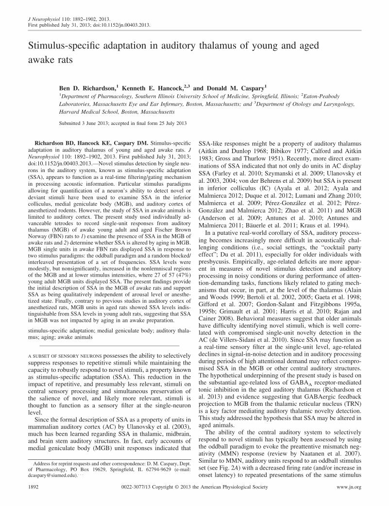
Tetrode microdrives and implantation surgery. VersaDrive4 te-trode drives (Neuralynx, Bozeman, MT) were assembled and loadedwith four tetrodes (Fig. 1A) cut to a total length of �8 mm, allowingeach tetrode to be advanced completely through the MGB; 2–2.5 mmof tetrode extended out of the supporting cannula. Each tetrode wirewas gold-electroplated to an impedance between 0.75 and 1.5 M�sampled at 1 kHz (nanoZ, Neuralynx). Prior to implantation, sterileultrawhite petroleum jelly (Tyco Healthcare Group, Mansfield, MA)was applied to the bottom of the drive at the exit point to protectagainst debris or fluid contaminating the drive mechanism. Driveswere sterilized with ethylene oxide before implantation.
Fig. 1. Implantation of individually advanceable tetrode drives and single-unit recording. A: assembled VersaDrive4 with tetrode tips positioned as they wouldbe in the medial geniculate body (MGB). B: tetrodes are advanceable with the turn of a drive screw easily accessed when the animal is awake. C: implantedFischer Brown Norway (FBN) rat in the modified restraining chamber with free access to water (front), the headstage attached to the VersaDrive4 and the Fostextweeter positioned at right adjacent to the wire mesh (not visible). D and E: waveforms (D) of 3 distinct single units and corresponding principal componentclusters (E). F: expanded individual raw waveforms corresponding with sorted waveforms identified in Plexon’s Sort Client (D and E). F1, F2, and F3 correspondwith units 1, 2, and 3 labeled in H. G: raw data trace from the same tetrode channel/wire represented in D and E. H: expanded underlined region in G. Scalebar in H applies to F–H: 1.5 ms (F), 500 ms (G), 30 ms (H).
1893SSA IN AWAKE AUDITORY THALAMUS
J Neurophysiol • doi:10.1152/jn.00403.2013 • www.jn.org

One day prior to surgery, acetaminophen (4.5 mg/ml) was providedvia the rat’s drinking water. On the day of implantation surgery, 10young adult and 6 aged FBN rats were weighed and anesthetized withintramuscular ketamine and xylazine (7 mg/kg xylazine, 105 mg/kgketamine). The aged dose was reduced by �20%. The head wasshaved, and the rat was placed in a Kopf stereotaxic fitted with a nosecone for gas anesthesia delivery. Lidocaine jelly (2%) was applied,and the dorsal surface of the head was scrubbed with alternatingNolvasan/70% alcohol. Rats were given sterile saline (2–3 ml sc) andplaced on a thermostatically controlled heating pad (Harvard Appa-ratus, Holliston, MA), O2 blood saturation level and heart rate weremonitored (PulseSense Vet, Nonin Medical, Minneapolis, MN), andophthalmic ointment was applied to each eye. Oxygen was adminis-tered continuously to maintain 95–100% blood saturation, and isoflu-rane (1–2.5%) was administered with a gaseous anesthesia system(VetEquip, Pleasanton, CA) when the animal showed a reduction inthe level of anesthesia (presence of pedal withdrawal or elevated heartrate).
Under sterile conditions, the skull surface was exposed and 4–5anchor screws were set in place (2 in the right frontal, 1 in theposterior left frontal, 1 in the parietal and 1 in the posterior nasalbones). A 2.3-mm-diameter hole was drilled over the left occipito-parietal cortex, dorsal to the MGB (5.5–5.7 mm bregma and 3.5–3.8mm of midline), and the dura was removed. A ground wire wasattached to a reference screw placed in the anterior right frontal bonethat made contact with the dura, and the tetrode drive was slowlyadvanced to a depth of 4.5–5 mm, placing the four tetrode tips justdorsal to MGB. Dental acrylate cement was added around the anchorscrews and drive, encapsulating the entire drive with the exception ofthe drive screws and pins (Fig. 1B). This method of mounting thetetrode drive did not appear to alter basic animal behavior anddemeanor, and postmortem examination of the brain indicated thatvery little damage to the surface of the brain occurred.
After surgery, triple antibiotic ointment was applied to the edge ofthe headcap and wound and an additional 2–3 ml of sterile saline wasadministered subcutaneously. The animal was exposed to 100% ox-ygen and kept on a heating pad throughout recovery until ambulatory.Acetaminophen (4.5 mg/ml) remained in the drinking water for atleast 1 full day after the surgery, with a recovery time of 7–10 daysprior to recording.
Single-unit recording procedures. Prior to surgery, rats were accli-matized to a modified Experimental Conditioning Unit (ECU; Brain-tree Scientific, Braintree, MA) with free access to water using a foodreward (1/4 to 1/2 Froot Loop) until they remained quiet and still forup to 3 h. The recording chamber had a 2 � 4-cm hole on the rightside covered with a wire mesh and a second 3 � 6-cm hole on the topof the chamber over the position of the rat’s head to allow access tothe tetrode drive (Fig. 1C). Significant effort was made to ensure thatdata were collected when each animal was in an awake and alert state.Most importantly, the animal husbandry facility room in which theserats were housed was always maintained on a reversed light-darkcycle. As a result, when recordings were obtained during the daytimehours, this was during the rats’ active period. All recordings tookplace in the dark. In addition, regularly occurring and typicallylow-frequency artifacts from movement, bruxing, chewing, and/ordrinking served as cues to each rat’s level of alertness and activity,despite being unable to reorient themselves away from stimulus.Sudden changes or decreases in single-unit responses and the presenceof bursting occurred rarely but indicated a decrease in each rat’sarousal. When this occurred, trials suspected to be affected by achange in arousal level were not included in analysis. Finally, eachanimal was regularly visually inspected throughout each recordingsession for alertness and correct orientation in the chamber, and theywere regularly found to be awake, alert, and appropriately oriented.
The tetrode drive was coupled to an 18-pin (16 single wires, 2ground) VersaDrive4-to-Omnetics adaptor (Neuralynx) and con-nected to a unity gain 18-channel headstage tethered to a preamplifier
[2� gain; 0.15 kHz (high pass), 8 kHz (low pass); Plexon, Dallas, TX](Fig. 1C). Sixteen channels of raw data were digitized with a Multi-channel Acquisition Processor (MAP) and visualized with Sort Client(Plexon). Tetrodes were advanced by turning a drive screw (1 fullturn � 250 �m) coupled to each tetrode and were advanced in1/4-turn (62.5 �m) increments with the distance recorded to aid inlocalization of units (Fig. 1B). A brief waiting period was included toallow the tips to “settle” before resuming the recording session. Toavoid unit resampling, after all units on a tetrode were studied thattetrode was advanced at least 125 �m. When auditory responsiveunits/field potentials were no longer present, tetrodes were left inposition for marking.
Spikes were sorted with standard methods (amplitude threshold andprincipal component analysis) and saved as timestamps. Reliability ofsingle-unit discrimination was evaluated by creating a cross-correlo-gram of all events for each putative single unit and subsequentlydetermining the percentage of spikes occurring within the absoluterefractory period (1 ms); units were typically discarded if �1% ofevents occurred within the defined refractory period. Timestampswere relayed to a system running a custom program for stimulusgeneration and real-time analysis of unit response [Auditory Neuro-physiology Experiment Control Software (ANECS), Ken Hancock,Blue Hills Scientific, Boston, MA].
When recordings were complete, each rat was anesthetized withketamine and xylazine as described above and current pulses (5 �Afor 5 s) were passed through the tip of each tetrode wire, producing asmall lesion. Rats were cardiac perfused with phosphate-bufferedsaline (0.1 M, pH 7.4) followed by paraformaldehyde (4%). Brainswere removed, placed in paraformaldehyde (1–2 h), and then trans-ferred to sucrose (20%). Frozen coronal sections (30–35 �m thick)were stained with fast thionin. Electrode tracks were localized to thedifferent subregions of the MGB (Paxinos and Watson 1998) and usedto determine the position of each recording site relative to the finallocation of the tetrode tip.
Acoustic stimuli generation and paradigms. Acoustic signals weregenerated by a 16-bit D/A converter [TDT RX6, Tucker DavisTechnologies (TDT) System III, Alachua, FL], amplified (YamahaP2500S, Buena Park, CA), and transduced by a Fostex tweeter (modelFT17H, Agoura Hills, CA) placed 2 cm from the 2 � 4-cm wiremesh-covered opening (Fig. 1C). The output was calibrated off-line asdescribed previously (Caspary et al. 2005) with a 1/4-in. microphone(model 4938; Brüel & Kjær, Naerum, Denmark) placed in the record-ing chamber at the approximate location of the rat’s pinna. Software-generated calibration tables in dB sound pressure level (SPL re: 20�Pa) were used to set programmable attenuators (TDT PA5) toachieve pure-tone intensities accurate to within 2 dB SPL for frequen-cies up to 45 kHz.
Response maps (RMs) were used to determine best frequency (BF)and threshold. Pseudorandom tone-burst stimuli (50-ms duration,5-ms rise/fall time) were presented with 250-ms onset-onset inter-stimulus interval (ISI) in 0.15- to 0.1-octave frequency steps (1–42kHz) and 5- to 10-dB intensity steps (0–80 dB). When multiplelow-threshold minima were present in the RM, BF was assigned as thefrequency at which the trough was present at the lowest intensity.When trough minima were similar, the highest frequency was re-corded as BF. Novelty detection was assessed with two paradigmsexamining changes in evoked firing rate to low- and high-probabilitystimuli: 1) oddball and 2) random blocked/interleaved frequencypresentation. Both paradigms were presented at 75 dB SPL and 35 �5 dB SPL above BF threshold to normalize for age-related thresholdvariation (Caspary et al. 2005). All tone bursts were 100 ms long(4-ms rise-fall) and were presented with a 250-ms onset-onset ISI.
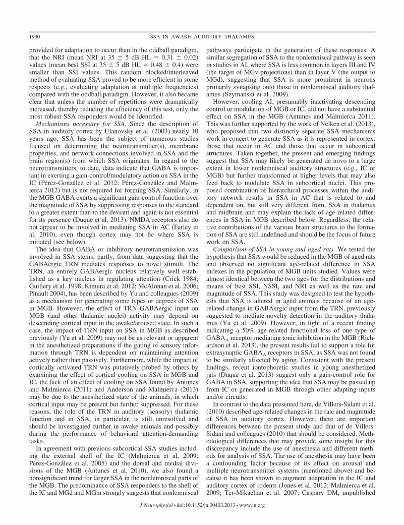
The oddball paradigm involved presentation of two tones withinthe excitatory response area of an MGB unit (S1 and S2) which were�0.16 octaves relative to BF (Fig. 2A). Similar to previous studies, ablock of 300 stimuli containing both frequencies was presented in aprobabilistic manner, with S1 as the standard (90%) interspersed with
1894 SSA IN AWAKE AUDITORY THALAMUS
J Neurophysiol • doi:10.1152/jn.00403.2013 • www.jn.org

random presentations of S2 as the deviant (10%) (Antunes et al. 2010;Antunes and Malmierca 2011; Malmierca et al. 2009; Pérez-Gonzálezet al. 2005; Ulanovsky et al. 2003, 2004). The probabilities of S1 andS2 were then reversed for comparison (Fig. 2A).
The random blocked and interleaved isointensity paradigm allowedobservation of SSA across a range of stimulus frequencies (Fig. 2B)(Lumani and Zhang 2010). Frequency was stepped linearly over agiven range (at least �0.5 octaves re BF, minimum of 6 frequencies).Figure 2B displays the random blocked presentation of the stimuli inlike blocks (i.e., S1, S1, S1, . . . S2, S2, S2, . . .) in the first series,whereas the frequency of each tone was randomly interleaved in thesecond paired series (i.e., S2, S4, S3, S7, S1, . . .). This allowed directcomparison of the adapted response (blocked) with the nonadaptedresponse (interleaved) over the range of frequencies tested.
To quantitatively assess SSA, first in MATLAB (The MathWorks,Natick, MA) the response of each unit was windowed with a variablewindow duration that ranged from 10 to 100 ms based on the responsetype (e.g., onset, sustained, etc.) apparent in the paired rasters andpoststimulus time histograms (PSTHs; bin � 3 ms). To determine themean normalized firing rate used in determining stimulus-specificindex (SSI) and neuronal stimulus-specific index (NSSI) values (seebelow), the numbers of spikes generated in response to each tonewithin the defined window were divided by the window duration andnormalized to the number of presentations. For SSI and NSSI calcu-lations, the mean normalized firing rates in response to the stimulus S1
when it is the deviant and standard, respectively, are defined as d(S1)and s(S1), and likewise for S2. These rate values were subsequentlyused in the two indexes developed by Ulanovsky and colleagues(2003) used to quantify SSA for a given unit: the SSI, SSI (Si) �[d(Si) � s(Si)]/[d(Si) � s(Si)], and the NSSI, NSSI � [d(S1) � d(S2) �s(S1) � s(S2)]/[d(S1) � d(S2) � s(S1) � s(S2)].
To determine the time course of adaptation, spikes within thedefined window (see above) for each trial were counted to determinethe change in firing rate over time with each consecutive trial presen-tation as described previously (de Villers-Sidani et al. 2010; seeUlanovsky et al. 2004). The spike count for each trial was firstnormalized to the first response (that was not a failure) then plotted(y-axis) against their trial number (x-axis). This trial-by-trial timeseries of firing rate was then fit with an exponential function (aleast-squares method) with IGOR Pro 6 (WaveMetrics, Lake Oswego,OR). The exponential fit provided the rate of adaptation in the decay(�) of the fitted curve and the magnitude of adaptation in the asymp-tote of the fitted curve for the response of each unit to a givenfrequency presented as the standard and deviant (see Fig. 5G).
For random blocked and interleaved stimulus pairs, novelty re-sponse index (NRI) values (NRI � [AInter � ABlock]/[AInter �ABlock]) were based on area under the curve values (AUC) for the
resulting blocked (ABlock) or interleaved (AInter) isointensity functions(Lumani and Zhang 2010). Isointensity functions were calculatedfrom the firing rate in response to each frequency for the blocked andinterleaved conditions. AUC was calculated with x-y pair area analysisin IGOR Pro 6.
All index values (SSI, NSSI, NRI) were used to assess the degreeof SSA for each neuron and ranged from �1 to 1. Negative valuesindicated a suppressed response to deviant stimuli. A value of 0indicated no preference for standard/blocked or deviant/interleaved. Avalue of 1 indicated an enhanced response to deviant/interleavedstimuli relative to standard/blocked.
Statistical analyses. Independent-samples t-tests were adequate formost statistical comparisons; they are noted in the text. A Kolmogo-rov-Smirnov (KS) test was used to determine differences betweendistributions. An � level of 0.05 was used for all statistical tests. Allvalues are expressed as means � SE.
RESULTS
Basic properties of MGB unit responses. Sixty young adultand 55 aged MGB single units responding to acoustic stimuliwere recorded from 10 young adult and 6 aged awake FBNrats. The ranges of BFs were similar between young adult andaged rats, but there was an age-related mean threshold elevation atBF of �10 dB (P 0.001; Fig. 3). Temporal response patternsfor all units are shown in Table 1 and suggest that the populationof units studied in rats from each age group may be similar basedon the similarity of the distribution of response types. Responsepatterns were classified as “on” (1–5 action potentials with regularshort ISIs) or “onset” (multiple action potentials with increasingISIs following stimulus onset) when a transient response waselicited after stimulus onset followed by a period of suppres-sion before the stimulus offset. Units responding only with an“off” response (a transient response after the stimulus offset)were rare, but on-off or onset-off responders were common,especially at higher intensities. Some unit responses wereclassified as “sustained” responders if they fired regularlythroughout the entire duration of the stimulus. Units show-ing more than one type of response pattern are includedmore than once. Most units displayed at least one clearminimum in the RM, indicating some degree of frequencyselectivity. Similarly, of all MGB units studied, only two
Fig. 3. Young adult and aged MGB single-unit best frequency (BF) andthreshold. A: composite scatterplot of each unit’s identified threshold at BFagainst BF for that MGB single unit from young adult (black circles) and aged(gray squares) FBN rats. B: average threshold at BF for MGB single units fromyoung adult (white) and aged (gray) rats. ***P 0.0001.
Fig. 2. Stimulus-specific adaptation (SSA) stimuli paradigms. Boxes representthe presentation of a stimulus across time from left to right. A: the oddballparadigm used to assess SSA. In the 90%/10% case S1 (black) is the standardwhile S2 (gray) is the deviant. Probabilities are reversed for S1 and S2 in the10%/90% case. B: random blocked and interleaved stimuli sets for SSAanalysis through comparison of isointensity functions collected for a broadrange of frequencies. In the first condition, all stimuli selected are presented inlike blocks to “induce” adaptation. In the second condition, the same stimuliare presented randomly interleaved with one another. The probability ofoccurrence for each stimulus at any time is the sole independent variable, andthe evoked firing rate is the dependent variable used for all comparisons.
1895SSA IN AWAKE AUDITORY THALAMUS
J Neurophysiol • doi:10.1152/jn.00403.2013 • www.jn.org

young and three aged neurons responded poorly or not at allto tones, while 10 young adult and 13 aged single unitsresponded poorly or not at all to broadband noise.
Stimulus-specific adaptation occurs in MGB of awake rats.SSA in the MGB of the awake rat was probed with a paired-tone oddball paradigm similar to previous studies (Antunes etal. 2010; Antunes and Malmierca 2011; Malmierca et al. 2009;Ulanovsky et al. 2003) (Fig. 2). To keep as many parametersconstant for comparison of SSA in young adult and aged rats,the two frequencies chosen were to be �0.16 octaves from BFwith a 0.22-octave separation. This frequency separation wasbetween the reported optimum separation for maximum SSAof �0.37 octaves and a more challenging separation of 0.1octaves (Antunes et al. 2010). To account for age-relatedthreshold shifts, novelty detection paradigms were alwayspresented at two intensities: 75 dB SPL (an absolute intensity)and 35 � 5 dB above threshold. A unit was considered todisplay SSA if it responded with a lower firing rate to a tone ofa given frequency when that frequency was the standard(presented with a high probability) compared with when thatsame frequency was the deviant (presented with a low proba-bility) (Fig. 4).
In Fig. 4, the example unit showing SSA adapts to 13.4 kHzas the standard (Fig. 4B) but responds vigorously when 13.4kHz is the deviant (Fig. 4C). The dot rasters (Fig. 4, B and C)show how the discharge rate at stimulus onset decreased andlatency increased with each subsequent trial when 13.4 kHz(standard) was paired with 16.7 kHz (deviant). In subsequentanalysis, the pair of rasters were converted to PSTHs like thoseshown in Fig. 4D. SSI and NSSI (see MATERIALS AND METHODS)were used to quantify differences in SSA magnitude. In somecases, only one frequency elicited SSA because of either areduced response to the second frequency or less adaptation tothe second frequency. As a result, there were units for whichSSI values for the two frequencies used in the oddball para-digm were quite disparate (e.g., 0.32 and �0.18), resulting inthe NSSI (an index taking into account SSA to both frequen-cies) inaccurately representing the maximum observed SSAcapability. For young adult rats 17 units at the 75 db SPL and19 units in the 35 dB HL conditions and for aged rats 19 unitsat the 75 dB SPL and 15 units in the 35 dB HL conditionsdisplayed SSA and had SSI values that were separated by�0.25. To provide a best descriptor of a unit’s observednovelty-detecting ability or level of SSA, a best SSI (maximumSSI of the response to S1 and S2 in each oddball pair) valuewas used as a measure along with the NSSI.
Young adult MGB units displayed varying SSA levels (Fig.4, E–J) as defined by the SSI and NSSI (Fig. 5, A–D). SSAvalues for MGB units ranged from units showing essentially nodifference/selectivity for deviant vs. standard (Fig. 4, E and F)to units displaying moderate levels (Fig. 4, G and H) or highlevels of SSA, in which a response was elicited by a particularfrequency only when it was the deviant (Fig. 4, I and J). SSAlevels tended to be higher at lower intensities (Fig. 4, G vs. H).Group data comparing unit SSI scatterplots at 75 dB SPL withthose recorded at 35 dB HL are shown in Fig. 5, A and B. SSIvalues for some units displaying SSA (SSI �0.25, see below)were greater at the lower intensity (Fig. 5, A–D), as was thenumber of units that displayed SSA at 35 � 5 dB HL (29 units)compared with the same oddball paradigm presented at 75 dBSPL (25 units) (Fig. 5, E and F).
As has been reported by others (Antunes et al. 2010), forunits that could be reliably localized SSA was greater innonlemniscal divisions, especially the medial MGB (MGm),but not significantly [best SSI 75 dB SPL condition: ventralMGB (MGv) � 0.20 � 0.05 (n � 12), dorsal MGB (MGd) �0.22 � 0.05 (n � 30), MGm � 0.30 � 0.08 (n � 12) 35 � 5dB HL condition: MGv � 0.24 � 0.07 (n � 12), MGd �0.27 � 0.06 (n � 27), MGm � 0.40 � 0.07 (n � 11); P � 0.4for all comparisons, ANOVA with Bonferroni post hoc]. Be-cause of the lack of significant divisional SSA differences,young adult and aged MGB comparisons were carried outirrespective of division.
Responses to oddball paradigm indicate no age-relatedchange in SSA for rodent MGB units. SSI values for youngadult and aged MGB units were compared in a test of thehypothesis that aging negatively impacts novelty detection inMGB neurons of awake FBN rats. Scatterplots show thedistribution of SSI for young and aged units at 75 dB SPL and35 � 5 dB HL (Fig. 5, A and B). The distributions were notsignificantly different at 75 dB SPL (n � 114 young adult and108 aged; P � 0.65, D � 0.097, C � 0.20; KS test) or for the35 � 5 dB HL condition (n � 106 young adult and 106 aged;P � 0.59, D � 0.10, C � 0.20; KS test). Figure 5, C and D,show the distribution of all best SSI values for each neuron at75 dB SPL and 35 � 5 dB HL, respectively. The distributionsof best SSI values for each condition were not significantlydifferent for the population of MGB units studied, either (75dB SPL: n � 57 young adult and 54 aged; P � 0.39, D � 0.17,C � 0.27; 35 � 5 dB HL: n � 53 young adult and 53 aged;P � 0.71, D � 0.13, C � 0.28; KS test).
Since not all units display SSA, only best SSI values forunits displaying positive SSI values were compared. A distri-
Table 1. Response types of MGB neurons in awake rats
Type
BBN BF
70 dB SPL 30 dB HL 70 dB SPL 30 dB HL
Young Aged Young Aged Young Aged Young Aged
On/onset 37 (61.7%) 31 (56.3%) 39 (65.0%) 31 (56.4%) 54 (90.0%) 40 (72.7%) 56 (93.3%) 42 (76.4%)Sustained 7 (11.7%) 9 (16.3%) 10 (16.7%) 10 (18.2%) 6 (10.0%) 8 (15.5%) 7 (11.7%) 9 (16.3%)Off 21 (35.0%) 20 (36.4%) 17 (28.3%) 17 (30.9%) 30 (50.0%) 23 (41.8%) 25 (41.7%) 21 (38.2%)Off only 5 (8.3%) 5 (9.1%) 3 (5.0%) 4 (7.3%) 7 (11.7%) 5 (9.1%) 3 (5.0%) 5 (9.1%)Poor 10 (16.7%) 13 (23.6%) 10 (16.7%) 13 (23.6%) 0 (0.0%) 3 (5.5%) 0 (0.0%) 2 (3.6%)Total 60 55
MGB, medial geniculate body; BBN, broadband noise; BF, best frequency.
1896 SSA IN AWAKE AUDITORY THALAMUS
J Neurophysiol • doi:10.1152/jn.00403.2013 • www.jn.org

bution (0.05 bins) of all SSI values (regardless of age groupand intensity condition) was created and fit with a Gaussiandistribution centered at “zero.” The standard deviation of thisdistribution was 0.25; units with SSI values 0.25 weredetermined to be indicative of no SSA, whereas SSI values�0.25 were indicative of units displaying SSA (Fig. 5, C andD). This criterion was similar to that used previously (cutoff:0.18, Antunes and Malmierca 2011). Average best SSI valuesfor neurons displaying SSA were not significantly differentbetween ages for the population of units studied for either the75 dB SPL (n � 25 young adult and 32 aged; P � 0.81) or the35 � 5 dB HL (n � 29 young adult and 27 aged; P � 0.96,independent-samples t-test) condition (Fig. 5, E and F).
Results were similar when the NSSI was used as a measure,which took into account the level of SSA in response to bothfrequencies in the oddball pair. The distributions of NSSIvalues for each intensity (data not shown) were not signifi-cantly different for the MGB units studied (75 dB SPL: n � 57young adult and 54 aged; P � 0.29, D � 0.17, C � 0.27;35 � 5 dB HL: n � 53 young adult and 53 aged; P � 0.45,D � 0.15, C � 0.28; KS test). The same cutoff of 0.25 wasapplied to the NSSI value distribution to determine which unitsdisplayed SSA. Average NSSI values for units displaying SSAwere not significantly different between ages at either 75 dB
SPL (n � 13 young adult and 14 aged; P � 0.49) or 35 � 5dBHL (n � 14 young adult and 15 aged; P � 0.60; independent-samples t-test).
Time course of adaptation to oddball paradigm indicates noage-related change in rate of SSA for rodent MGB units. Therate and magnitude of adaptation over the course of eachstandard and deviant trial were compared between young andaged MGB units (de Villers-Sidani et al. 2010; Ulanovsky et al.2004). As SSA was greatest for the 35 � 5 dB HL condition,time course plots of the normalized response (see MATERIALS
AND METHODS) were compared for each unit-frequency (S1and/or S2) combination where SSI � 0.25 (Fig. 5G; n � 33standard/deviant pairs from 29 young adult and n � 32 stan-dard/deviant pairs from 27 aged MGB units). No age-relateddifferences in the group mean asymptote/magnitude (P � 0.5)or the rate/� (P � 0.5) of adaptation of the normalized re-sponses plotted for each consecutive trial (x-axis) through thefirst 200 trials were observed (Fig. 5G). The time courses of theresponses to standard and deviant stimuli for young adult andaged MGB single units were similar.
Responses to random blocked/interleaved novelty detectionparadigm indicate no age-related change in SSA for rodentMGB units. The second paradigm used to examine noveltydetection by MGB units assessed rapid SSA across frequen-
Fig. 4. MGB single-unit SSA in response to oddball paradigm. A: response map for a single unit localized to the medial MGB (MGm). The higher frequencyof the 2 peaks (6.5 kHz at 45 dB, 14.9 kHz at 40 dB) was used as the BF for this unit. Frequency values �0.16 octaves from 14.9 kHz (13.37 kHz and 16.67kHz, 2 vertical lines) were used as the 2 frequencies presented in the oddball paradigm at high (standard, 90%) and low (deviant, 10%) probabilities. The thresholdfor the unit was 40 dB; therefore the oddball paradigm was presented at 75 dB, 35 dB above threshold (horizontal line). B and C: raster of single-unit responseto 100-ms tones presented randomly in the oddball paradigm with 10/90% probabilities. Each dot represents the occurrence of an action potential in responseto a tone (frequency of tone at each trial corresponds to the color in the key). Trial number is on the y-axis; poststimulus time is on the x-axis. This unit respondspoorly/adapts rapidly to 16.67 kHz but responds reliably to 13.37 kHz. B: 13.37 kHz (90%) is the standard (blue) and induces noticeable adaptation within thefirst 10–20 presentations. C: 13.37 kHz (10%) is the deviant (red) and elicits a robust response with each presentation. D: averaged poststimulus time histogram(PSTH; 3-ms bins) for the unit response to 13.37 kHz presented as the standard (blue; B) and as the deviant (red; C). E–J: paired PSTHs for single units [alllocalized to dorsal MGB (MGd)] displayed a range of SSA from no (E and F) to moderate (G and H) and high (I and J) SSA. Frequencies used (F) and thestimulus-specific index (SSI) value for each pair are noted in each histogram. G–J: note that SSI values for some units were smaller for some units at higherintensities (75 dB SPL; G and I) than at lower intensities (35 � 5 dB HL; H and J) and the latency of the response is longer for neurons with greater SSI valuescompared with the latency of the response for the nonadapting neuron (E and F).
1897SSA IN AWAKE AUDITORY THALAMUS
J Neurophysiol • doi:10.1152/jn.00403.2013 • www.jn.org

cies. A range of frequencies (�0.5 octaves above and belowBF with at least 6 frequency steps; Fig. 2B, Fig. 6, B–D) werepresented at 75 dB SPL and at 35 � 5 dB HL with twodifferent sequences (Fig. 2B). First, the sequence of tones waspresented in blocks of 20 identical repeated frequencies inorder to induce adaptation. The frequency blocks were alwaysrandomized. Second, the same sequence of frequencies (20repetitions per frequency) was randomly interleaved acrosspresentations to prevent adaptation to a single frequency (Fig.2B). The firing rates in response to the blocked and interleavedconditions were then compared (Fig. 6B).
Typically, a prominent peak in the resulting isointensitycurve was centered at or near BF for both stimulus sequences.The AUC for the peak of each function (for the blocked andinterleaved conditions) was calculated (IGOR Pro 6) to gener-ate the NRI (Lumani and Zhang 2010). An NRI value cutoffdetermined to be the standard deviation of the distribution(0.05 bins) of all NRI values (regardless of age group andintensity condition) was created and fit with a Gaussian distri-bution centered at “zero” (standard deviation � 0.11). NRIvalues 0.11 were determined to be from a unit not displayingSSA, whereas SSI values �0.11 were indicative of a unitdisplaying SSA (Fig. 6, E and G).
Similar to the results with the oddball paradigm, the distri-butions of NRI values (Fig. 6, E and G) for each condition werenot significantly different between young adult and aged units
in the population of MGB units studied [75 dB SPL (n � 56young adult and 49 aged): P � 0.17, D � 0.21, C � 0.28;35 � 5 dB HL (n � 56 young adult and 47 aged): P � 0.68,D � 0.13, C � 0.28; KS test]. Average NRI values for neuronsdisplaying SSA (NRI � 0.11) were not significantly differentfor the population of units studied for either the 75 dB SPL(n � 21 young adult and 23 aged; P � 0.63) or the 35 � 5 dBHL (n � 28 young adult and 23 aged; P � 0.79; independent-samples t-test) condition (Fig. 6, F and H). These data, from asecond SSA paradigm, suggest that SSA does occur in theMGB of awake rats but is not altered by age.
DISCUSSION
With implanted tetrode arrays to record single-unit re-sponses in the auditory thalamus of unanesthetized FBN rats,MGB neurons displayed qualitatively and quantitatively simi-lar, but not necessarily identical, SSA levels relative to theanesthetized preparation. Conclusions regarding the impact ofarousal and age were based on MGB unit responses with twodifferent stimulus paradigms to assess SSA, the standard odd-ball paradigm and a random blocked/interleaved frequencyparadigm. With SSI, NSSI, and NRI indexes, SSA responses inunanesthetized MGB were found to be present and qualita-tively comparable to those reported for MGB units in anesthe-tized rats, mice, and gerbils (Anderson et al. 2009; Antunes et
Fig. 5. MGB units display multiple levels of SSA but no age-related difference in SSA. SSI value pairs for S1 (low frequency) and S2 (high frequency) usedin the oddball paradigm are plotted against one another for the 75 dB SPL (A) and 35 � 5 dB HL (B) conditions. The number of single units used to composeeach scatterplot is the same for each age, as indicated in parentheses in the corresponding distribution below (C and D). Note the density of SSI values near 0for the 75 dB SPL condition (A). SSI values for these same neurons and the mean SSI values for each division shift toward 1, up and/or to the right, in thelower-intensity condition (B). C and D: distribution of all best SSI values in the 75 dB SPL (C) and 35 � 5 dB HL (D) conditions for young adult (white) andaged (gray) MGB single units. Dotted line represents cutoff (0.25) for single units that did not display SSA (SSI 0.25) and those that did display SSA(SSI � 0.25). E and F: average best SSI only for single units that displayed SSA (i.e., SSI values � 0.25) when the oddball paradigm was presented at 75 dBSPL (E) and 35 � 5 dB HL (F). G: group average plot of number of spikes at each trial (1–200) normalized to the first response to the standard (solid line) anddeviant (dashed line) for all young adult (black) and aged (gray) single units displaying SSA in the 35 � 5 dB HL condition. NS, not significant.
1898 SSA IN AWAKE AUDITORY THALAMUS
J Neurophysiol • doi:10.1152/jn.00403.2013 • www.jn.org

al. 2010; Antunes and Malmierca 2011; Bäuerle et al. 2011; Yuet al. 2009). These findings were consistent with descriptionsof SSA in auditory cortex of the awake rat as well (von derBehrens et al. 2009). Second, in addressing whether dimin-ished novelty detection in the elderly may be due to single-neuron deficiencies in novelty detection/SSA, we found noage-related changes in the magnitude or time course of SSA atthe level of the auditory thalamus. Notably, these findingscontrast with the age-related decrease in SSA observed in AI ofthe anesthetized rat (de Villers-Sidani et al. 2010).
Of the MGB units studied, 29 of 60 young adult units fromall three MGB divisions displayed SSA, suggesting that SSAdoes not require the suppressed level of arousal produced byanesthesia. Compared with previous SSA studies, the magni-tude of SSA was not as large as previously reported for theanesthetized rat MGB. For example, Antunes et al. (2010)found that 46% of MGB neurons, particularly in the MGm, hadindex values � 0.6 for both frequencies in an oddball pair withparameters similar to those of the present study. Here, only13% of MGB single units in young adult rats display a best SSIvalue above 0.6. However, it should be pointed out that thisdiscrepancy may reflect differences in age or strain or depend,in part, on the anesthetized state of the animal (see introductionfor discussion). Another major difference between the present
study and many previous studies was the use of a pair offrequencies at identical fixed distances relative to BF (0.16octaves) in order to compare young and aged unit responses toSSA stimulus sets that were as similar as possible. This is incontrast to previous studies, which often used a frequency pairchosen on a unit-by-unit basis to maximize SSI values inresponse to the oddball paradigm (Anderson et al. 2009;Antunes et al. 2010; Antunes and Malmierca 2011; de Villers-Sidani et al. 2010; Malmierca et al. 2009; Pérez-González et al.2005, 2012; Ulanovsky et al. 2003, 2004).
In the present study, a modestly greater number of unitsdisplayed SSA at lower stimulus intensities, an effect seen inthe IC of anesthetized rats as well (Duque et al. 2012). Thetime course and magnitude (young adult asymptote standard �0.22, deviant � 0.38) of adaptation to standard and deviantstimuli were found to be comparable to those described forauditory cortex in anesthetized rats (de Villers-Sidani et al.2010).
The second stimulus paradigm used to assess SSA in MGBemployed a random blocked/interleaved presentation of arange of frequencies, similar to that used by Lumani and Zhang(2010) in IC, in which rapid adaptation must occur within 20repetitions for SSA to be revealed. It is because of thisincreased stringency in testing SSA, in which much less time is
Fig. 6. Random blocked/interleaved paradigm evaluation of SSA. A: response map for a single unit in MGm with a BF at 19.7 kHz. B: average firing rate forthe same single unit as in A in response to each frequency when presented blocked (blue) and interleaved (red). The area under the curve (AUC; AUC differenceis shaded) for each curve was used to calculate the novelty response index (NRI; noted at bottom left for this unit). C and D: pair of rasters showing the responsesof this unit to a range of frequencies, 14–28 kHz (BF � 0.5 octaves), with 2-kHz intervals. Tones [100 ms; gray shaded region; 250-ms interstimulus interval(ISI), onset-onset] are presented at 75 dB SPL (35 � 5 dB HL not shown) nonrandomly in identical frequency blocks (C) and randomly (D). Each dot representsthe occurrence of an action potential (frequency of tone at each trial is marked on y-axis; poststimulus onset time is on x-axis). C: time-locked responses at theonset of the stimulus presented nonrandomly occur occasionally, especially in the first and second presentations of 16–20 kHz. D: when frequencies are presentedinterspersed randomly with other frequencies, time-locked responses to the onset occur with few failures. E and G: distribution of all NRI values for young adult(white) and aged (gray) MGB single units at 75 dB SPL (E) and 35 � 5 dB HL (G). Number of units in the distribution is noted in parentheses. Dotted linerepresents cutoff (0.11) for single units that did not display SSA (NRI 0.11) and those that did display SSA (NRI � 0.11). F and H: average NRI only forsingle units that displayed SSA (NRI � 0.11) when stimuli were presented at 75 dB SPL (F) and 35 � 5dB HL (H).
1899SSA IN AWAKE AUDITORY THALAMUS
J Neurophysiol • doi:10.1152/jn.00403.2013 • www.jn.org
provided for adaptation to occur than in the oddball paradigm,that the NRI (mean NRI at 35 � 5 dB HL � 0.31 � 0.02)values (mean best SSI at 35 � 5 dB HL � 0.48 � 0.4) weresmaller than SSI values. This random blocked/interleavedmethod of evaluating SSA proved to be more efficient in somerespects (e.g., evaluating adaptation at multiple frequencies)compared with the oddball paradigm. However, it also becameclear that unless the number of repetitions were dramaticallyincreased, thereby reducing the efficiency of this test, only themost robust SSA responders would be identified.
Mechanisms necessary for SSA. Since the description ofSSA in auditory cortex by Ulanovsky et al. (2003) nearly 10years ago, SSA has been the subject of numerous studiesfocused on determining the neurotransmitter(s), membraneproperties, and network connections involved in SSA and thebrain region(s) from which SSA originates. In regard to theneurotransmitters, to date, data indicate that GABA is impor-tant in exerting a gain-control/modulatory action on SSA in theIC (Pérez-González et al. 2012; Pérez-González and Malm-ierca 2012) but is not required for forming SSA. Similarly, inthe MGB GABA exerts a significant gain-control function overthe magnitude of SSA by suppressing responses to the standardto a greater extent than to the deviant and again is not essentialfor its presence (Duque et al. 2013). NMDA receptors also donot appear to be involved in mediating SSA in AC (Farley etal. 2010), even though cortex may not be where SSA isinitiated (see below).
The idea that GABA or inhibitory neurotransmission wasinvolved in SSA stems, partly, from data suggesting that theGABAergic TRN mediates responses to novel stimuli. TheTRN, an entirely GABAergic nucleus relatively well estab-lished as a key nucleus in regulating attention (Crick 1984;Guillery et al. 1998; Kimura et al. 2012; McAlonan et al. 2006;Pinault 2004), has been described by Yu and colleagues (2009)as a mechanism for generating some types or degrees of SSAin MGB. However, the effect of TRN GABAergic input onMGB (and other thalamic nuclei) activity may depend ondescending cortical input in the awake/aroused state. In such acase, the impact of TRN input on SSA in MGB as describedpreviously (Yu et al. 2009) may not be as relevant or apparentin the anesthetized preparations if the gating of sensory infor-mation through TRN is dependent on maintaining attentionactively rather than passively. Furthermore, while the impact ofcortically activated TRN was putatively probed by others byexamining the effect of cortical cooling on SSA in MGB andIC, the lack of an effect of cooling on SSA found by Antunesand Malmierca (2011) and Anderson and Malmierca (2013)may be due to the anesthetized state of the animals, in whichcortical input may be present but further suppressed. For thesereasons, the role of the TRN in auditory (sensory) thalamicfunction and in SSA, in particular, is still unresolved andshould be investigated further in awake animals and possiblyduring the performance of behavioral attention-demandingtasks.
In agreement with previous subcortical SSA studies includ-ing the external shell of the IC (Malmierca et al. 2009;Pérez-González et al. 2005) and the dorsal and medial divi-sions of the MGB (Antunes et al. 2010), we also found anonsignificant trend for larger SSA in the nonlemniscal parts ofthe MGB. The predominance of SSA responders to the shell ofthe IC and MGd and MGm strongly suggests that nonlemniscal
pathways participate in the generation of these responses. Asimilar segregation of SSA to the nonlemniscal pathway is seenin studies in AI, where SSA is less common in layers III and IV(the target of MGv projections) than in layer V (the output toMGd), suggesting that SSA is more prominent in neuronsprimarily synapsing onto those in nonlemniscal auditory thal-amus (Szymanski et al. 2009).
However, cooling AI, presumably inactivating descendingcontrol or modulation of MGB or IC, did not have a substantialeffect on SSA in the MGB (Antunes and Malmierca 2011).This was further supported by the work of Nelken et al. (2013),who proposed that two distinctly separate SSA mechanismswork in concert to generate SSA as it is represented in cortex:those that occur in AC and those that occur in subcorticalstructures. Taken together, the present and emerging findingssuggest that SSA may likely be generated de novo to a largeextent in lower nonlemniscal auditory structures (e.g., IC orMGB) but further transformed at higher levels that may alsofeed back to modulate SSA in subcortical nuclei. This pro-posed combination of hierarchical processes within the audi-tory network results in SSA in AC that is related to anddependent on, but still very different from, SSA in thalamusand midbrain and may explain the lack of age-related differ-ences in SSA in MGB described below. Regardless, the rela-tive contributions of the various brain structures to the forma-tion of SSA are still undefined and should be the focus of futurework on SSA.
Comparison of SSA in young and aged rats. We tested thehypothesis that SSA would be reduced in the MGB of aged ratsand observed no significant age-related difference in SSAindexes in the population of MGB units studied. Values werealmost identical between the two ages for the distributions andmeans of best SSI, NSSI, and NRI as well as the rate andmagnitude of SSA. This study was designed to test the hypoth-esis that SSA is altered in aged animals because of an age-related change in GABAergic input from the TRN, previouslysuggested to mediate novelty detection in the auditory thala-mus (Yu et al. 2009). However, in light of a recent findingindicating a 50% age-related functional loss of one type ofGABAA receptor mediating tonic inhibition in the MGB (Rich-ardson et al. 2013), the present results fail to support a role forextrasynaptic GABAA receptors in SSA, as SSA was not foundto be similarly affected by aging. Consistent with the presentfindings, recent iontophoretic studies in young anesthetizedrats (Duque et al. 2013) suggest only a gain-control role forGABA in SSA, supporting the idea that SSA may be passed upfrom IC or generated in MGB through other adapting inputsand/or circuits.
In contrast to the data presented here, de Villers-Sidani et al.(2010) described age-related changes in the rate and magnitudeof SSA in auditory cortex. However, there are importantdifferences between the present study and that of de Villers-Sidani and colleagues (2010) that should be considered. Meth-odological differences that may provide some insight for thisdiscrepancy include the use of anesthesia and different meth-ods for analysis of SSA. The use of anesthesia may have beena confounding factor because of its effect on arousal andmultiple neurotransmitter systems (mentioned above) and be-cause it has been shown to augment adaptation in the IC andauditory cortex of rodents (Jones et al. 2012; Malmierca et al.2009; Ter-Mikaelian et al. 2007; Caspary DM, unpublished
1900 SSA IN AWAKE AUDITORY THALAMUS
J Neurophysiol • doi:10.1152/jn.00403.2013 • www.jn.org

observation). For these reasons, additional studies comparingSSA in anesthetized and unanesthetized preparations areneeded. Methods of analysis also differed in that, in contrast tothe present study, not only units that displayed SSA but allunits were included in determining changes in adaptationmagnitude and rate in de Villers-Sidani et al. (2010). It isinteresting to consider why SSA may be maintained in agedMGB but less so in AI. This may be due to a selective impactof aging on different SSA sites of origin in AC relative to theascending subcortical auditory structures (IC and MGB) and issupported by the preliminary findings of Nelken et al. (2013) aswell as studies indicating a bottom-up origin of SSA (Andersonand Malmierca 2013; Antunes and Malmierca 2011). In addi-tion, age-related failure of SSA in AI could reflect cumulativeage-related neurotransmitter and coding changes occurringspecifically at the level of auditory cortex (Caspary et al. 2008;Hughes et al. 2010; Juarez-Salinas et al. 2010; Ling et al. 2005;Yang et al. 2008, 2009).
Our interest in performing the present study was stimulated,in part, by the de Villers-Sidani et al. (2010) study and theknown age-related changes that occur with the human far-fieldpotential recorded in response to the oddball paradigm, knownas mismatch negativity (MMN; 100- to 200-ms latency) (Alainand Woods 1999; Bertoli et al. 2002; Gaeta et al. 1998).However, recent studies concluded that SSA at the level ofauditory cortex and MMN may not be linked, suggesting thatSSA is a mechanism of early auditory processing not neces-sarily involved in gating or detecting novel stimuli as definedby those longer-latency processes mediating the later MMNresponse (Farley et al. 2010; von der Behrens et al. 2009).While the original hypothesis was that SSA, in the MGB inparticular, could be a single-unit correlate of MMN (Kraus etal. 1994; Ulanovsky et al. 2003), a more appropriate far-fieldrepresentative of SSA timing was recently proposed (Grimm etal. 2011; Slabu et al. 2010, 2012). Slabu et al. (2010) found thatthe middle-latency response (MLR), a shorter-latency (�50ms) precortical event-related potential not directly related toMMN, also displayed novelty or deviance preference. Thesestudies of novelty detection in the MLR, which is temporally inline with SSA in the IC and MGB (20- to 50-ms latency),imply that detection of novel stimuli may indeed occur lowerin the auditory system (Grimm et al. 2011; Slabu et al. 2012).Therefore, the MLR may provide a more accurate means bywhich to evaluate novelty detection deficits that may be relatedto single-unit SSA in humans.
ACKNOWLEDGMENTS
The authors thank Lynne L. Ling for her assistance with experiments, Dr.Kevin J. Otto for his advice and expertise in establishing the single-unitrecording in the awake animal preparation, and Dr. Manuel Malmierca andDaniel Duque for their assistance in data analysis.
GRANTS
These studies were supported by National Institute on Deafness and OtherCommunication Disorders Grant DC-00151.
DISCLOSURES
No conflicts of interest, financial or otherwise, are declared by the au-thor(s).. . .
AUTHOR CONTRIBUTIONS
Author contributions: B.D.R., K.E.H., and D.M.C. conception and design ofresearch; B.D.R. performed experiments; B.D.R., K.E.H., and D.M.C. ana-lyzed data; B.D.R., K.E.H., and D.M.C. interpreted results of experiments;B.D.R. prepared figures; B.D.R. drafted manuscript; B.D.R., K.E.H., andD.M.C. edited and revised manuscript; B.D.R., K.E.H., and D.M.C. approvedfinal version of manuscript.
REFERENCES
Aitkin LM, Dunlop CW. Interplay of excitation and inhibition in the catmedial geniculate body. J Neurophysiol 31: 44–61, 1968.
Alain C, Woods DL. Age-related changes in processing auditory stimuliduring visual attention: evidence for deficits in inhibitory control andsensory memory. Psychol Aging 14: 507–519, 1999.
Anderson LA, Christianson GB, Linden JF. Stimulus-specific adaptationoccurs in the auditory thalamus. J Neurosci 29: 7359–7363, 2009.
Anderson LA, Malmierca MS. The effect of auditory cortex deactivation onstimulus-specific adaptation in the inferior colliculus of the rat. Eur JNeurosci 37: 52–62, 2013.
Antunes FM, Malmierca MS. Effect of auditory cortex deactivation onstimulus-specific adaptation in the medial geniculate body. J Neurosci 31:17306–17316, 2011.
Antunes FM, Nelken I, Covey E, Malmierca MS. Stimulus-specific adap-tation in the auditory thalamus of the anesthetized rat. PLoS One 5: e14071,2010.
Ayala YA, Malmierca MS. Stimulus-specific adaptation and deviance detec-tion in the inferior colliculus. Front Neural Circuits 6: 89, 2012.
Ayala YA, Pérez-González D, Duque D, Nelken I, Malmierca MS. Fre-quency discrimination and stimulus deviance in the inferior colliculus andcochlear nucleus. Front Neural Circuits 6: 119, 2012.
Bäuerle P, von der Behrens W, Kössl M, Gaese BH. Stimulus-specificadaptation in the gerbil primary auditory thalamus is the result of a fastfrequency-specific habituation and is regulated by the corticofugal system. JNeurosci 31: 9708–9722, 2011.
Bertoli S, Smurzynski J, Probst R. Effects of age, age-related hearing loss,and contralateral cafeteria noise on the discrimination of small frequencychanges: psychoacoustic and electrophysiological measures. J Assoc ResOtolaryngol 6: 207–222, 2005.
Bertoli S, Smurzynski J, Probst R. Temporal resolution in young and elderlysubjects as measured by mismatch negativity and a psychoacoustic gapdetection task. Clin Neurophysiol 113: 396–406, 2002.
Bibikov NG. [“Novelty” neurons in the frog auditory system]. Zh Vyssh NervDeiat Im I P Pavlova 27: 1075–1082, 1977.
Calford MB, Aitkin LM. Ascending projections to the medial geniculatebody of the cat: evidence for multiple, parallel auditory pathways throughthalamus. J Neurosci 3: 2365–2380, 1983.
Caspary DM, Ling L, Turner JG, Hughes LF. Inhibitory neurotransmission,plasticity and aging in the mammalian central auditory system. J Exp Biol211: 1781–1791, 2008.
Caspary DM, Schatteman TA, Hughes LF. Age-related changes in theinhibitory response properties of dorsal cochlear nucleus output neurons:role of inhibitory inputs. J Neurosci 25: 10952–10959, 2005.
Crick F. Function of the thalamic reticular complex: the searchlight hypoth-esis. Proc Natl Acad Sci USA 81: 4586–4590, 1984.
de Villers-Sidani E, Alzghoul L, Zhou X, Simpson KL, Lin RC, MerzenichMM. Recovery of functional and structural age-related changes in the ratprimary auditory cortex with operant training. Proc Natl Acad Sci USA 107:13900–13905, 2010.
Du Y, Kong L, Wang Q, Wu X, Li L. Auditory frequency-followingresponse: a neurophysiological measure for studying the “cocktail-partyproblem.” Neurosci Biobehav Rev 35: 2046–2057, 2011.
Duque D, Malmierca MS, Caspary DM. Modulation of stimulus-specificadaptation by the GABAergic system in the medial geniculate body of theanesthetized rat. J Physiol, 2013. . . .
Duque D, Pérez-González D, Ayala YA, Palmer AR, Malmierca MS.Topographic distribution, frequency, and intensity dependence of stimulus-specific adaptation in the inferior colliculus of the rat. J Neurosci 32:17762–17774, 2012.
Farley BJ, Quirk MC, Doherty JJ, Christian EP. Stimulus-specific adap-tation in auditory cortex is an NMDA-independent process distinct from thesensory novelty encoded by the mismatch negativity. J Neurosci 30: 16475–16484, 2010.
1901SSA IN AWAKE AUDITORY THALAMUS
J Neurophysiol • doi:10.1152/jn.00403.2013 • www.jn.org

Gaeta H, Friedman D, Ritter W, Cheng J. An event-related potential studyof age-related changes in sensitivity to stimulus deviance. Neurobiol Aging19: 447–459, 1998.
Gifford RH, Bacon SP, Williams EJ. An examination of speech recognitionin a modulated background and of forward masking in younger and olderlisteners. J Speech Lang Hear Res 50: 857–864, 2007.
Gordon-Salant S, Fitzgibbons PJ. Comparing recognition of distorted speechusing an equivalent signal-to-noise ratio index. J Speech Hear Res 38:706–713, 1995a.
Gordon-Salant S, Fitzgibbons PJ. Recognition of multiply degraded speechby young and elderly listeners. J Speech Hear Res 38: 1150–1156, 1995b.
Grimault N, Micheyl C, Carlyon RP, Arthaud P, Collet L. Perceptualauditory stream segregation of sequences of complex sounds in subjectswith normal and impaired hearing. Br J Audiol 35: 173–182, 2001.
Grimm S, Escera C, Slabu L, Costa-Faidella J. Electrophysiological evi-dence for the hierarchical organization of auditory change detection in thehuman brain. Psychophysiology 48: 377–384, 2011.
Gross NB, Thurlow WR. Microelectrode studies of neural auditory activity ofcat. II. Medial geniculate body. J Neurophysiol 14: 409–422, 1951.
Guillery RW, Feig SL, Lozsadi DA. Paying attention to the thalamic reticularnucleus. Trends Neurosci 21: 28–32, 1998.
Hara K, Harris RA. The anesthetic mechanism of urethane: the effects onneurotransmitter-gated ion channels. Anesth Analg 94: 313–318, 2002.
Harris KC, Eckert MA, Ahlstrom JB, Dubno JR. Age-related differences ingap detection: effects of task difficulty and cognitive ability. Hear Res 264:21–29, 2010.
Hughes LF, Turner JG, Parrish JL, Caspary DM. Processing of broadbandstimuli across A1 layers in young and aged rats. Hear Res 264: 79–85,2010.
Jones S, Mill R, Denham S, Klump G. Stimulus-specific adaptation in theinferior colliculus of the awake gerbil in response to Markov chain tonesequences (Abstract). In: Association for Research in Otolaryngology (ARO)Midwinter Meeting, San Diego, CA, 795, 2012.
Juarez-Salinas DL, Engle JR, Navarro XO, Recanzone GH. Hierarchicaland serial processing in the spatial auditory cortical pathway is degraded bynatural aging. J Neurosci 30: 14795–14804, 2010.
Kimura A, Yokoi I, Imbe H, Donishi T, Kaneoke Y. Auditory thalamicreticular nucleus of the rat: anatomical nodes for modulation of auditory andcross-modal sensory processing in the loop connectivity between the cortexand thalamus. J Comp Neurol 520: 1457–1480, 2012.
Kraus N, McGee T, Littman T, Nicol T, King C. Nonprimary auditorythalamic representation of acoustic change. J Neurophysiol 72: 1270–1277,1994.
Ling LL, Hughes LF, Caspary DM. Age-related loss of the GABA syntheticenzyme glutamic acid decarboxylase in rat primary auditory cortex. Neuro-science 132: 1103–1113, 2005.
Lumani A, Zhang H. Responses of neurons in the rat’s dorsal cortex of theinferior colliculus to monaural tone bursts. Brain Res 1351: 115–129, 2010.
Malmierca MS, Cristaudo S, Perez-Gonzalez D, Covey E. Stimulus-specificadaptation in the inferior colliculus of the anesthetized rat. J Neurosci 29:5483–5493, 2009.
McAlonan K, Cavanaugh J, Wurtz RH. Attentional modulation of thalamicreticular neurons. J Neurosci 26: 4444–4450, 2006.
Naatanen R, Paavilainen P, Rinne T, Alho K. The mismatch negativity(MMN) in basic research of central auditory processing: a review. ClinNeurophysiol 118: 2544–2590, 2007.
Nelken I, Antunes F, Hershenhoren I, Khouri L, Yarden T, Yaron A.Stimulus-specific adaptation in real and model neurons (Abstract). In:Association for Research in Otolaryngology (ARO) Midwinter Meeting,Baltimore, MD, 366, 2013.
Paxinos W, Watson C. The Rat Brain in Stereotaxic Coordinates. San Diego,CA: Academic, 1998.
Pérez-González D, Hernández O, Covey E, Malmierca MS. GABAA-mediated inhibition modulates stimulus-specific adaptation in the inferiorcolliculus. PLoS One 7: e34297, 2012.
Pérez-González D, Malmierca MS. Variability of the time course of stimu-lus-specific adaptation in the inferior colliculus. Front Neural Circuits 6:107, 2012.
Pérez-González D, Malmierca MS, Covey E. Novelty detector neurons in themammalian auditory midbrain. Eur J Neurosci 22: 2879–2885, 2005.
Pinault D. The thalamic reticular nucleus: structure, function and concept.Brain Res Rev 46: 1–31, 2004.
Rajan R, Cainer KE. Ageing without hearing loss or cognitive impairmentcauses a decrease in speech intelligibility only in informational maskers.Neuroscience 154: 784–795, 2008.
Richardson BD, Ling LL, Uteshev VV, Caspary DM. Reduced GABAA
receptor-mediated tonic inhibition in aged rat auditory thalamus. J Neurosci33: 1218–1227, 2013.
Slabu L, Escera C, Grimm S, Costa-Faidella J. Early change detection inhumans as revealed by auditory brainstem and middle-latency evokedpotentials. Eur J Neurosci 32: 859–865, 2010.
Slabu L, Grimm S, Escera C. Novelty detection in the human auditorybrainstem. J Neurosci 32: 1447–1452, 2012.
Szymanski FD, Garcia-Lazaro JA, Schnupp JW. Current source densityprofiles of stimulus-specific adaptation in rat auditory cortex. J Neurophysiol102: 1483–1490, 2009.
Ter-Mikaelian M, Sanes DH, Semple MN. Transformation of temporalproperties between auditory midbrain and cortex in the awake Mongoliangerbil. J Neurosci 27: 6091–6102, 2007.
Ulanovsky N, Las L, Farkas D, Nelken I. Multiple time scales of adaptationin auditory cortex neurons. J Neurosci 24: 10440–10453, 2004.
Ulanovsky N, Las L, Nelken I. Processing of low-probability sounds bycortical neurons. Nat Neurosci 6: 391–398, 2003.
von der Behrens W, Bauerle P, Kossl M, Gaese BH. Correlating stimulus-specific adaptation of cortical neurons and local field potentials in the awakerat. J Neurosci 29: 13837–13849, 2009.
Wang H, Turner JG, Ling L, Parrish JL, Hughes LF, Caspary DM.Age-related changes in glycine receptor subunit composition and binding indorsal cochlear nucleus. Neuroscience 160: 227–239, 2009.
Yang Y, Liang Z, Li G, Wang Y, Zhou Y, Leventhal AG. Aging affectscontrast response functions and adaptation of middle temporal visual areaneurons in rhesus monkeys. Neuroscience 156: 748–757, 2008.
Yang Y, Zhang J, Liang Z, Li G, Wang Y, Ma Y, Zhou Y, Leventhal AG.Aging affects the neural representation of speed in macaque area MT. CerebCortex 19: 1957–1967, 2009.
Yu XJ, Xu XX, He S, He J. Change detection by thalamic reticular neurons.Nat Neurosci 12: 1165–1170, 2009.
Zhao L, Liu Y, Shen L, Feng L, Hong B. Stimulus-specific adaptation andits dynamics in the inferior colliculus of rat. Neuroscience 181: 163–174,2011.
1902 SSA IN AWAKE AUDITORY THALAMUS
J Neurophysiol • doi:10.1152/jn.00403.2013 • www.jn.org
Copyright © 2022 FDOKUMEN