Stem cell therapy in animal models of central nervous system ...
11
Maiti et al./ J. Adv. Vet. Anim. Res., 1(3): 75-85, September 2014 75 Stem cell therapy in animal models of central nervous system (CNS) diseases: therapeutic role, challenges and perspectives Swapan Kumar Maiti 1 , A.R. Ninu 1* , V. Remya 1 , T.B. Sivanarayanan 1 , Susan Cherian 2 , Deepak Kumar 3 and Amarpal 1 1 Division of Surgery, Indian Veterinary Research Institute, Izatnagar–243122, Uttar Pradesh, India; 2 Division of Pathology, Indian Veterinary Research Institute, Izatnagar–243122, Uttar Pradesh, India; 3 Department of Veterinary Public Health, College of Veterinary Sciences & Animal Husbandry, R.K. Nagar– 799008, Tripura, India. * Corresponding author’s e–mail: [email protected] ABSTRACT Many human diseases relating to central nervous system (CNS) are mimicked in animal models to evaluate the efficacy of stem cell therapy. The therapeutic role of stem cells in animal models of CNS diseases include replacement of diseased or degenerated neuron, oligodendrocytes or astrocytes with healthy ones, secretion of neurotrophic factors and delivery of therapeutics/genes. Scaffolds can be utilized for delivering stem cells in brain. Sustained delivery of stem cells, lineage specific differentiation, and enhanced neuronal network integration are the hallmarks of scaffold mediated stem cell delivery in CNS diseases. This review discusses the therapeutic role, challenges and future perspectives of stem cell therapy in animal models of CNS diseases. Keywords Animal models, Cell replacement, Challenges, CNS diseases, Scaffold, Stem cell therapy Received 02 May 2014, Revised: 20 May 2014, Accepted : 21 May 2014, Published online: 03 June 2014. INTRODUCTION Treatment of central nervous system (CNS) injuries had been a challenge for medical and veterinary clinicians. It was in the naïve stage until early 20 th century (Schmidt and Leach, 2003) as the recovery in CNS is limited by the insufficient self–repair and regeneration abilities of the brain tissues (Bjorklund and Lindvall, 2000). Main challenges in the treatment of brain diseases include blood brain barrier (bbb) with tight intercellular junctions, and absence of fenestrations (Brightman and Reese, 1969). These prevent the uptake of majority of therapeutics (Pardridge, 2003), active drug efflux pumps in the bbb (Golden and Pollack, 2003), which pumps out the drugs from the brain, high intercellular fluid pressure due to space occupying lesions that limits diffusion (Ali et al., 2006; Navalitloha et al., 2006), and the sensitivity of brain tissue that emphasizes the need for appropriate and precise dosing of chemotherapeutic agents (Roger et al., 2011). Unrelenting reports on therapeutic uses of stem cells for CNS diseases have led to their wider acceptance and importance in the present scenario. According to the early school of thoughts, neurons of adult CNS of mammals have limited regeneration capacity, but later studies have confirmed that subgranular zone and dentate gyrus of hippocampus and lateral ventricles of forebrain are regions of potential neurogenesis in adult mammalian brain (Kempermann and Gage, 1999; Gross, 2000; Lie et al., 2004). This endogenous regeneration potential of CNS could be stimulated to aid the repair of damaged brain tissue (Nakatomi et al., 2002). Even from the areas of adult CNS where neurogenesis is not apparent, stem cells and their J. Adv. Vet. Anim. Res., 1(3): 75-85. Available at- http://bdvets.org/JAVAR REVIEW ARTICLE OPEN ACCESS DOI: 10.5455/javar.2014.a17 eISSN 2311-7710 Volume 1 Issue 3 (September 2014)
-
Upload
khangminh22 -
Category
Documents
-
view
2 -
download
0
Transcript of Stem cell therapy in animal models of central nervous system ...
Maiti et al./ J. Adv. Vet. Anim. Res., 1(3): 75-85, September 2014 75
Stem cell therapy in animal models of central nervous system (CNS) diseases: therapeutic role, challenges and perspectives
Swapan Kumar Maiti1, A.R. Ninu1*, V. Remya1, T.B. Sivanarayanan1, Susan Cherian2, Deepak Kumar3 and Amarpal1 1Division of Surgery, Indian Veterinary Research Institute, Izatnagar–243122, Uttar Pradesh, India; 2Division of Pathology, Indian Veterinary Research Institute, Izatnagar–243122, Uttar Pradesh, India; 3Department of Veterinary Public Health, College of Veterinary Sciences & Animal Husbandry, R.K. Nagar– 799008, Tripura, India. *Corresponding author’s e–mail: [email protected]
ABSTRACT
Many human diseases relating to central nervous system (CNS) are mimicked in animal models to evaluate the efficacy of stem cell therapy. The therapeutic role of stem cells in animal models of CNS diseases include replacement of diseased or degenerated neuron, oligodendrocytes or astrocytes with healthy ones, secretion of neurotrophic factors and delivery of therapeutics/genes. Scaffolds can be utilized for delivering stem cells in brain. Sustained delivery of stem cells, lineage specific differentiation, and enhanced neuronal network integration are the hallmarks of scaffold mediated stem cell delivery in CNS diseases. This review discusses the therapeutic role, challenges and future perspectives of stem cell therapy in animal models of CNS diseases.
Keywords Animal models, Cell replacement, Challenges, CNS diseases, Scaffold, Stem cell therapy
Received 02 May 2014, Revised: 20 May 2014, Accepted : 21 May 2014, Published online: 03 June 2014.
INTRODUCTION
Treatment of central nervous system (CNS) injuries had been a challenge for medical and veterinary clinicians. It was in the naïve stage until early 20th century
(Schmidt and Leach, 2003) as the recovery in CNS is limited by the insufficient self–repair and regeneration abilities of the brain tissues (Bjorklund and Lindvall, 2000). Main challenges in the treatment of brain diseases include blood brain barrier (bbb) with tight intercellular junctions, and absence of fenestrations (Brightman and Reese, 1969). These prevent the uptake of majority of therapeutics (Pardridge, 2003), active drug efflux pumps in the bbb (Golden and Pollack, 2003), which pumps out the drugs from the brain, high intercellular fluid pressure due to space occupying lesions that limits diffusion (Ali et al., 2006; Navalitloha et al., 2006), and the sensitivity of brain tissue that emphasizes the need for appropriate and precise dosing of chemotherapeutic agents (Roger et al., 2011).
Unrelenting reports on therapeutic uses of stem cells for CNS diseases have led to their wider acceptance and importance in the present scenario. According to the early school of thoughts, neurons of adult CNS of mammals have limited regeneration capacity, but later studies have confirmed that subgranular zone and dentate gyrus of hippocampus and lateral ventricles of forebrain are regions of potential neurogenesis in adult mammalian brain (Kempermann and Gage, 1999; Gross, 2000; Lie et al., 2004). This endogenous regeneration potential of CNS could be stimulated to aid the repair of damaged brain tissue (Nakatomi et al., 2002). Even from the areas of adult CNS where neurogenesis is not apparent, stem cells and their
J. Adv. Vet. Anim. Res., 1(3): 75-85. Available at- http://bdvets.org/JAVAR
REVIEW ARTICLE
OPEN ACCESS
DOI: 10.5455/javar.2014.a17
eISSN 2311-7710
Volume 1 Issue 3 (September 2014)
Maiti et al./ J. Adv. Vet. Anim. Res., 1(3): 75-85, September 2014 76
progenitors can be extracted, expanded and differentiated into neurons and glia in vitro (Aboody et al., 2011), which could be later implanted in vivo. Innumerable CNS diseases including stroke, brain tumors, epilepsy, Parkinson’s Disease (PD), Huntington’s disease (HD), Alzheimer's disease (AD), multiple sclerosis (MS), amyotrophic lateral sclerosis (ALS) and spinal cord injuries could benefit from stem cell therapy (Chu et al., 2004; Carri et al., 2006; Ziv et al., 2006; James and Cavenne, 2009; Jiang et al., 2011; Cohen, 2013; Kim et al., 2013; Roper and Steindler, 2013; van Gorp et al., 2013; Mochizuki et al., 2014). The term stem cell is a broader concept. The stem cell types which are used for treating CNS diseases include adult neural stem/neural progenitor cells, bone marrow derived mesenchymal stem cells, adipose derived mesenchymal stem cells, umbilical cord blood mesenchymal stem cells, embryonic/fetal mesen-chymal stem cells, and induced pleuripotent stem cells (Chu et al., 2004; Ziv et al., 2006; Chen et al., 2010; Jiang et al., 2011; Gu, 2013; Razavi et al., 2013; Yang et al., 2013). Among the various types of stem cells, induced pleuripotent stem lines are derived from reprogram-ming the adult somatic cells to an embryonic stem cell state. They have proved themselves to be a potential autologous source of stem cells (Hu et al., 2010), and could be differentiated even to neurons (Kuo and Lin, 2013). The therapeutic role of stem cells in animal models of CNS diseases include cell replacement, as vehicle for delivery of genetically engineered genes and drugs (Roger et al., 2011), release of neurotrophic factors and vasoactive factors like anti–inflammatory cytokines which provide neuroprotection (Martino and Pluchino, 2006). This is only a bird’s eye view of the topic. Here, the role of animal models seems to be noteworthy in that the therapeutic efficiency of stem cells for human CNS diseases is primarily evaluated by conducting laboratory animal trials.
STEM CELLS FOR CELL REPLACEMENT
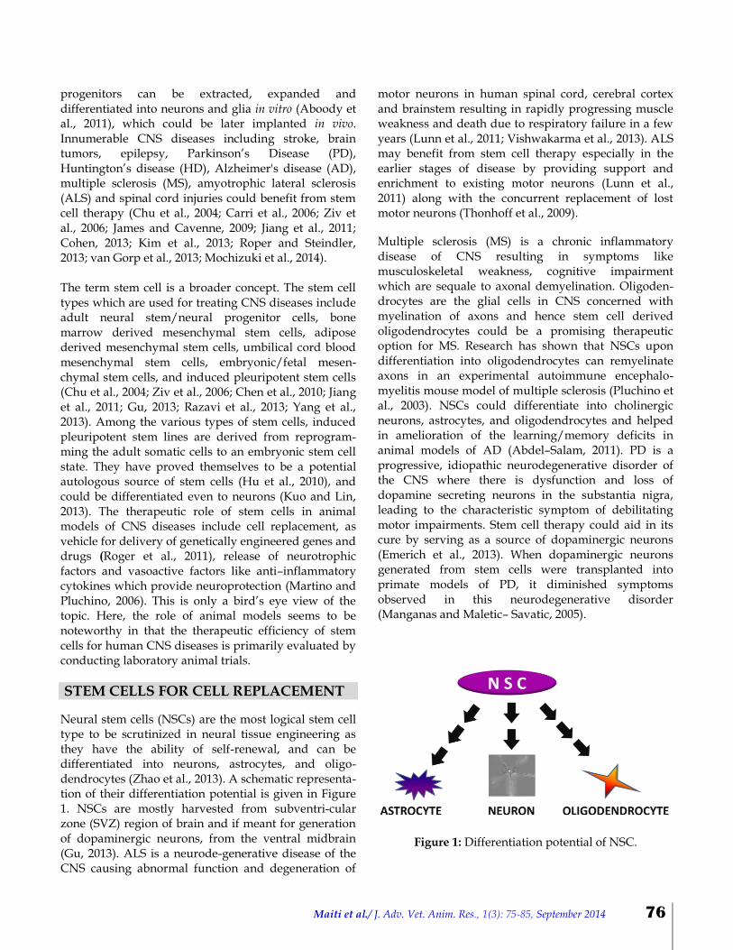
Neural stem cells (NSCs) are the most logical stem cell type to be scrutinized in neural tissue engineering as they have the ability of self-renewal, and can be differentiated into neurons, astrocytes, and oligo-dendrocytes (Zhao et al., 2013). A schematic representa-tion of their differentiation potential is given in Figure 1. NSCs are mostly harvested from subventri-cular zone (SVZ) region of brain and if meant for generation of dopaminergic neurons, from the ventral midbrain (Gu, 2013). ALS is a neurode-generative disease of the CNS causing abnormal function and degeneration of
motor neurons in human spinal cord, cerebral cortex and brainstem resulting in rapidly progressing muscle weakness and death due to respiratory failure in a few years (Lunn et al., 2011; Vishwakarma et al., 2013). ALS may benefit from stem cell therapy especially in the earlier stages of disease by providing support and enrichment to existing motor neurons (Lunn et al., 2011) along with the concurrent replacement of lost motor neurons (Thonhoff et al., 2009).
Multiple sclerosis (MS) is a chronic inflammatory disease of CNS resulting in symptoms like musculoskeletal weakness, cognitive impairment which are sequale to axonal demyelination. Oligoden- drocytes are the glial cells in CNS concerned with myelination of axons and hence stem cell derived oligodendrocytes could be a promising therapeutic option for MS. Research has shown that NSCs upon differentiation into oligodendrocytes can remyelinate axons in an experimental autoimmune encephalo-myelitis mouse model of multiple sclerosis (Pluchino et al., 2003). NSCs could differentiate into cholinergic neurons, astrocytes, and oligodendrocytes and helped in amelioration of the learning/memory deficits in animal models of AD (Abdel–Salam, 2011). PD is a progressive, idiopathic neurodegenerative disorder of the CNS where there is dysfunction and loss of dopamine secreting neurons in the substantia nigra, leading to the characteristic symptom of debilitating motor impairments. Stem cell therapy could aid in its cure by serving as a source of dopaminergic neurons (Emerich et al., 2013). When dopaminergic neurons generated from stem cells were transplanted into primate models of PD, it diminished symptoms observed in this neurodegenerative disorder (Manganas and Maletic– Savatic, 2005).
Figure 1: Differentiation potential of NSC.
Maiti et al./ J. Adv. Vet. Anim. Res., 1(3): 75-85, September 2014 77
Stem cells have found a role in successful treatment of stroke as there are reports on migration of neural progenitor cells (NPC) towards the lesion with formation of new neurons (Kelly et al., 2004), and reestablishment of neural connections with functional recovery (Hayashi et al., 2006). When human embryonic stem cell derived oligodendrocyte progeny-tors and motor neuron progenitors were transplanted into the transected spinal cord of adult rats immedia-tely after the injury, they could differentiate into oligodendrocytes, astrocytes and neurons. In addition to this, there was improvement in locomotor functions without teratoma formation (Erceg et al., 2010). This in turn corroborates cell replacement role of stem cells in spinal cord injury. Though the bone marrow stem cells could give rise to only a lesser proportion of neuron like cells in comparison to brain derived neural stem cells, they could be an assuring therapy for CNS injury and neurodegenerative diseases (Song et al., 2007).
STEM CELL MEDIATED GENE THERAPY
Gene therapy is the concept and procedure for transfer of therapeutic genetic material into the cells to cure diseases (Moirano and Emburg, 2006). Using stem cells, an ex vivo gene therapy is performed, which means that the genetic material is transferred into the cultured cells prior to transplantation (Loscher et al., 2008). In ex vivo gene therapy, mostly embryonic stem cells or neural stem cells are used owing to their expanding capabilities and differentiation potential to various types of neural cells. However, the chances of genetic incorporation into the brain to generate the desired neural phenotype is limited (Van Dycke et al., 2011). Genetically engineered stem cells have proven as useful in animal models of Parkinsonism (Anton et al., 1994); brain ischemia, spinal cord injury (Park et al., 2006; Kusano et al., 2010); gliomas (Aboody et al., 2000), ALS (Suzuki et al., 2007), and HD (Olson et al., 2012). Genetic modification of NSCs with neutrotrophin-3 (NT–3) has been reported to promote myelination and to reduce astroglial scarring after transplantation in rodents with either injury of spinal cord or ischemic brain injury (Park et al., 2006; Kusano et al., 2010). Transplantation of genetically modified embryonic stem cell derived cells overexpressing neuroprotective factors results in functional recovery in animal models of ischemia (Shinozuka et al., 2013). Genetically engineered stem cells expressing cytokines have reported promising results in glioma models following intracranial administration (Ehtesham et al., 2002; Yang et al., 2004; Yuan et al., 2006). GDNF (glial cell derived neurotrophic factor) – over expressing neural stem/
precursor cells delayed the degeneration of motor neurons in the spinal cord of rat model of ALS (Suzuki et al., 2007); whereas, they increased the survival of neuronal cells for up to 3 months post–transplantation in the striatum of presymptomatic transgenic mouse model of Huntinton’s disease (Ebert et al., 2010). Huntington’s disease is caused by mutation of gene coding for protein mHTT (mutant huntingtin protein) resulting in cellular toxicity. Research in several HD animal models had shown that neuronal survival could be prolonged by enhancing the degradation/clearance of this protein from affected neurons. Patient derived induced pleuripotent stem cells (iPSC) were used for studying gene manipulation strategies for achieving this. Genome editing approaches directly targeting DNA for reducing mHTT protein has shown success in patient iPSC– derived neuronal models. But this has to be validated further in in vivo models (Yu et al., 2014). Small interfering RNAs can reduce mHTT and studies regarding safety and efficacy of siRNA delivery system using human MSCs are underway (Olson et al., 2012).
RELEASE OF TROPHIC FACTORS AND OTHER PARACRINE EFFECTS OF STEM CELLS MSC and NPC secrete immune modulatory or neurotrophic paracrine factors which may have therapeutic benefits in treating experimentally induced CNS diseases in animal models (Drago et al., 2013; Lavoie and Rosu–Myles, 2013). In experimental studies of PD, NPC transplants secreting GDNF, and vascular–endothelial growth factor (VEGF) have shown positive outcome and are being assessed in pre–clinical trials for the treatment of the disease (Akerud et al., 2001). Upregulation of stromal cell-derived factor-1 (SDF–1), VEGF, and transforming growth factor beta (TGF β) were noticed in MSC transplanted spinal cord injury models of beagle dogs (Jung et al., 2009). Many recent studies focus on utilizing paracrine effects of stem cells in the therapy of CNS disease. Here, instead of going for implanting stem cells, the biologics secreted by stem cells termed as ‘secretome’ are used for repairing injured brain (Drago et al., 2013). Human umbilical cord blood–derived mesenchymal stem cells delivered intracranially, in a mouse model of AD, improved spatial learning and memory decline possibly by neuroprotective effect (Lee et al., 2012). Yang et al. (2013) reported that single intracerebral injection of neuron–like cells, differentiated from human umbilical cord derived mesenchymal stem cells

Maiti et al./ J. Adv. Vet. Anim. Res., 1(3): 75-85, September 2014 78
(hUMSC–NC) could ameliorate memory deficits in mouse model of AD by alternative activation of microglia cells. These “alternatively activated” microglia (M2–like microglia) played protective roles in AD by phagocytizing Aβ (amyloid β–peptide – the key pathogenic factor of AD and reducing neuroinflamma-tion. Yang et al. (2013) here emphasizes the paracrine effects of transplantation of hUMSC–NC. The paracrine effect has resulted from increased expression of an antiinflammatory cytokine namely, IL–4, which in turn led to M2–like microglial activation. As already mentioned, elevated amyloid β–peptide deposition is the key pathogenic factor for neuronal loss in AD. In another study utilizing human umbilical cord derived mesenchymal stem cells (hUMSC) in beta–amyloidosis mouse model of AD, amyloid plaques were reduced by secretion of a soluble intercellular adhesion molecule–1 (sICAM–1). This molecule exerted its effect by inducing Aβ degrading enzyme. This again outlines the paracrine mode of action of hUMSC (Kim et al., 2012).
STEM CELLS AS VECTOR FOR DRUG DELIVERY
Stem cell therapy combined with nanotechnology could be a promising tool to efficiently deliver drugs to brain tumors (Roger et al., 2011). Glioblastoma, is a lethal malignant tumor where even the standard protocols like surgical resection followed by concomitant chemotherapy and fractionated radiotherapy (Stupp et al., 2005; Stupp et al., 2009) could only prolong the life span by near about one year. Advances in the field of nanotechnology have led to the development of nanoparticles loaded with chemotherapeutics. The therapeutic agent is entrapped in, adsorbed or chemically coupled onto the nanoparticle surface. By this technique the therapeutic agents are protected from enzymatic and chemical degradation, thereby ensuring it’s sustained and controlled release (Roger et al., 2011).
Stem cells can be used to carry these drug bound nanoparticles (Figure 2) to the lesion site. Neural stem cells, owing to their tropism towards glioma cells and ability to cross bbb, are excellent carriers for cytokines, viral particles and prodrugs (Aboody et al., 2000). Mesenchymal stem cells also have homing properties around glioma which could be utilized for glioma therapy. This homing is due to mechanisms mediated by several factors like epidermal growth factor (Sato et al., 2005), SDF–1 (Wynn et al., 2004), platelet–derived growth factor (Fiedler et al., 2002), matrix metalloproteinase–1 (Ho et al., 2009), and macrophage chemoattractant protein–1 (Xu et al., 2010). First step in
stem cell mediated delivery of drug loaded nanoparticles is the incorporation of chemotherapeutic loaded nanoparticles into the stem cells in–vitro either spontaneously or via passive/active endocytosis. Secondly, these stem cells are injected intracranially into the tumor mass. The nanoparticle loaded stem cells will localize in the border between tumor cells and normal brain parenchyma and slowly release the chemotherapy drugs as depicted in Figure 2. This concept is already demonstrated by Roger et al. (2011), using MIAMI (Marrow–Isolated Adult Multilineage Inducible) cells, a subpopulation of human MSCs. NSC mediated delivery of secreted soluble variant of TRAIL, [Tumor Necrosis Factor –related apoptosis–inducing ligand (TRAIL) can selectively induce apoptosis in glioma cells] in combination with therapeutics like proteasome inhibitor, bortezomib (Balyasnikova et al., 2011) and kinase inhibitor, PI–103, in mice models of glioma (Bagci–Onder et al., 2011) increased survivability of mouse by inhibition of tumor growth and proliferation. This is yet another example of the role of stem cell as vector.
Figure 2: Stem cells acting as vector to release drug bound nanoparticles to the site of brain lesion.
Stem cells also have a role in treatment of epilepsy by delivery of adenosine (Van Dycke et al., 2010), which is a purine ribonucleoside with neuromodulator and neurotransmitter functions (Sachdeva and Gupta, 2013). Antiseizure and neuroprotective potentials of adenosine are known for long (Lee et al., 1984; Fredholm, 1997). The role of adenosine in seizure (Van Dycke et al., 2011) results from its binding to the presynaptic A1 receptors which inhibits the release of excitatory neurotransmitters like glutamate. Systemic use of adenosine has severe side effects like decreased blood pressure and heart rate which emphasizes the
Maiti et al./ J. Adv. Vet. Anim. Res., 1(3): 75-85, September 2014 79
need for its local delivery into the brain via stem cells (Boison, 2005). According to Van Dycke et al. (2010), astrocytes derived from NPC and undifferentiated NPC of adenosine kinase deficient mice released thera-peutically relevant amounts of adenosine under in vitro conditions. In case of brain tumors therapeutic delivery may be needed for short time duration only whereas epilepsy, which is a chronic disorder, needs lifelong local delivery of therapeutics. In the latter context, cell or gene therapy sounds theoretically a successful strategy, as long term release can be ensured without replacement or refilling (Van Dycke et al., 2011).
SCAFFOLDS FOR STEM CELL DELIVERY IN CNS DISEASES
Scaffolds aid cell proliferation and differentiation, by allowing diffusion of nutrients and exerting mechanical and biological influences on the cell. Delivery and duration of action of stem cells could also be prolonged by use of suitable scaffolds. In addition to these, they can be used for sustained release of lineage–specific inductive factors or small interfering ribonucleic acids (siRNAs) which can act as molecular mediators of neuronal differentiation. The efficacy of transplantation of NPC can be improved in CNS injury by the co–administration of biomaterial scaffolds (Potter et al., 2008). Biomaterials made of nanofibers, nanotubes and nanoparticles have been widely used in manipulating the fate of stem cells (Zhao et al., 2013). Carbon nano-tubes can provide support, direct the differentiation of stem cells to neural lineages and promote signal transmission among neurons (Pastorin, 2011).
A nano–biohybrid system created by NSC progeny and graphene showed that graphene films can not only support neural network without affecting its structure and function but also could amplify the network activity and efficacy of neural signals (Tang et al., 2013). In ICC construct, i.e., inverted colloidal crystal scaffold comprising chitin, chitosan and gelatin, mouse iPSC can remain viable. Also it can accelerate differentiation of iPSC to neurons (Kuo and Lin, 2013). The knowledge of down regulation of RE 1 silencing transcription (REST) factor upon differentiation to neurons in NPC can be utilized to enhance in vitro neuronal differentiation of stem cells. Low et al. (2013), investigated the possibility of scaffold mediated gene silencing by delivering small interfering RNA/ transfection agent complexes via mussel– inspired polydopamine modified electrospun polycaprolactone nanofibre scaffolds and concluded that it could be a future promise for therapy as enhanced neuronal
commitment of primary mouse neural progenitor cells and decreased glial cell differentiation was seen.
CHALLENGES AND PERSPECTIVES The main limitations of stem cell therapy are with regard to the higher cost of commercialization, and the difficulties in approval of clinical trials. Another major problem related to NSC mediated regeneration in CNS trauma is due to the upregulation of inhibitory immune factors around the site of lesion resulting from the inflammatory process that ensues (Dooley et al., 2014). A recent study by Kyritsis et al. (2014) suggests that in mammals, acute inflammation is followed by chronic inflammation, which prevents functional recovery of brain tissue. Survival of the implanted differentiated neurons is to be ensured after transplan-tation for successful outcome. In ALS, though it had been shown that motor neurons derived from stem cells can be grafted safely without any rejection, the microenvironment remains hostile for their survival because of neuroinflammation, oxidative stress and glutamate excitotoxicity (Thonhoff et al., 2009). As dysfunctional astrocytes also have a role in the survival of dying motor neurons in ALS, new studies are aimed at transplanting stem cell derived astrocytes for protecting the diseased motor neurons (Lindvall et al., 2012). The beneficial effects of NSC transplantation are limited by the unfriendly microenvironment at the site of CNS injury/degeneration (Lu et al., 2011). Hence stem cell transplantation together with enrichment of microenvironment with trophic factors is under investigation. In vivo tumorigenic potential of mesen-chymal stem cell exosome (Zhu et al., 2012), NSC (Shinozuka et al., 2013), embryonic stem cells (Thonhoff et al., 2009; Mothe and Tator, 2012) and induced pluripotent stem cells is another impeding factor in progress. However, many studies with the above stem cell sources have shown positive therapeutic outcomes without tumor formation. Further research on tumorigenic potential of stem cells revealed that the presence of undifferentiated pluripotent cells as contaminants in neural committed transplants is the cause for teratomas. This tumor transformation can be halted by incorporating suicidal gene into these stem cells. It is important to rule out any existing tumors by functional imaging modalities before stem cell infusion into brain or spinal cord (Olson et al., 2012). The homing tendency of MSC to hypoxic regions around tumor margins and its potent revascularization potency
Maiti et al./ J. Adv. Vet. Anim. Res., 1(3): 75-85, September 2014 80
has the flaw of supporting the survival of existing tumors, if given alone. A dark side to the use of human embryonic stem cells is the activation of immune responses. A way out has evolved by short term immune dampening treatment of these cells which could prevent post–transplantation rejection (Mochizuki et al., 2014). Undifferentiated iPSC are also immunogenic but are seldom used in transplantation studies without differentiating them into desired cell lines (Yamanaka, 2012). Presently preclinical cell therapy trials are being carried out in animal models of CNS diseases. Though the predictive clinical outcome of such therapies seems to be encouraging, controlled clinical trials in large animal models are to be undertaken before evaluating the feasibility of stem cell therapy in human beings. Even in those CNS diseases where the clinical phase trials have started or culminated the diagnostic techniques for accurately predicting the cell integration and survival in live patients is underway. A solution to this has evolved from the finding that NSCs may be labelled with superparamagnetic iron oxide before administration and their distribution in the body can be noninvasively monitored by MRI. This is awaiting FDA approval for implementation in clinical trials (Aboody et al., 2011). Certain other issues need to be addressed in the future and one of them is how the survivability of transplanted stem cells can be augmented. Studies have shown that the survival, growth and function of the neurons can be enhanced by trophic protein factors and hence combining differentiated neurons with these factors could be a concept for future research (Emerich et al., 2013). The optimal dose of stem cells to be used, route of administration and sex of the donor/recipient vary with the stem cell type being used (Shinozuka et al., 2013) and future trials should be aimed at standardizing these factors with regard to different stem cell types for various CNS diseases. When to give stem cell therapy after disease onset is yet another issue. Most of the CNS diseases that gained benefit from stem cell therapy had shown that an early intervention is necessary for successful outcome. Though there are reports of improvement of locomotor function following immediate stem cell therapy, optimal time period for stem cell transplantation following spinal cord injury is 1–2 weeks after injury. The immediate post–traumatic microenviroment does not support the survival and differentiation of neural stem cell/progenitor cells. In chronic stage, there is
glial scar formation at the site of injury which inhibits axonal regeneration (Nakamura and Okano, 2013). A recent progress in the field of cell therapy is the invention of technology for cell reprogramming and development of iPSC. Induced pluripotent stem cell lines derived from patients suffering from PD, AD, Autism, Rett syndrome (a paediatric neural development disease) and Schizophrenia are now used as cell models for studying pathogenesis, and to develop assays for drug discovery (Yuan and Shaner, 2013). They can serve as autologous stem cell sources with more controlled methods of reprogramming. This new technology provides scope for a diversion from the use of animal models as a primary step for lab evaluation of new treatment strategies.
CONCLUSIONS
The therapeutic effects of stem cells in animal models of CNS diseases are discussed in this review. Majority of the research outputs from different laboratories are encouraging and these results are extrapolated to predict the possible outcome in human beings. In this context, one main lacuna is that there is absence of complete recapitulation of the human CNS disease in these lab animal models. Here, we conclude this review by stating that research in animal models of CNS disease is currently in progress for developing new treatment options of which stem cell seems to be promising. The broad arena of stem cell biology in neuroscience provides an ample scope for future research in pet animal diseases like CNS related paralysis/seizures.
ACKNOWLEDGEMENT The authors are thankful to the Director, Indian Veterinary Research Institute for providing necessary facilities for compiling this review article.
REFERENCES Abdel–Salam OM (2011). Stem cell therapy for
Alzheimer's disease. CNS & Neurological Disorders–Drug Targets, 10:459–485.
Aboody KS, Brown A, Rainov NG, Bower KA, Liu S, Yang W, Small JE, Herrlinger U, Ourednik V, Black PM, Breakefield XO, Snyder EY (2000). Neural stem cells display extensive tropism for pathology in adult brain: evidence from intracranial gliomas. Proceedings of the National
Maiti et al./ J. Adv. Vet. Anim. Res., 1(3): 75-85, September 2014 81
Academy of Sciences of the United States of America, 97:12846–12851.
Aboody K, Capela A, Niazi N, Stern JH, Temple S (2011). Translating stem cell studies to the clinic for CNS repair: current state of the art and the need for a rosetta stone. Neuron, 70:597–613.
Akerud P, Canals JM, Snyder EY, Arenas E (2001). Neuroprotection through delivery of glial cell line – derived neurotrophic factor by neural stem cells in a mouse model of Parkinson’s disease. Journal of Neuroscience, 21:8108–8118.
Ali MJ, Navalitloha Y, Vavra MW, Kang EW, Itskovich AC, Molnar P, Levy RM, Groothuis DR (2006). Isolation of drug delivery from drug effect: problems of optimizing drug delivery parameters. Neuro–Oncology, 8:109–118.
Anton R, Kordower JH, Maidment NT, Manaster JS, Kane DJ, Rabizadeh S, Schueller SB, Yang J, Rabizadeh S, Edwards RH, Markham CH, Bredesen DE (1994). Neural–targeted gene therapy for rodent and primate hemiparkinsonism. Experimental Neurology, 127:207–218.
Bagci–Onder T, Wakimoto H, Anderegg M, Cameron C, Shah K (2011). A dual PI3K/mTOR inhibitor, PI–103, cooperates with stem cell–delivered TRAIL in experimental glioma models. Cancer Research, 71:154–163.
Balyasnikova IV, Ferguson SD, Han Y, Liu F, Lesniak MS (2011). Therapeutic effect of neural stem cells expressing TRAIL and bortezomib in mice with glioma xenografts. Cancer Letters, 310:148–159.
Bjorklund A, Lindvall O (2000). Cell replacement therapies for central nervous system disorders. Nature Neuroscience, 3:537–544.
Boison D (2005). Adenosine and epilepsy: from therapeutic rationale to new therapeutic strategies. Neuroscientist, 11:25–36.
Brightman MW, Reese TS (1969). Junctions between intimately apposed cell membranes in the vertebrate brain. Journal of Cell Biology, 40:648–677.
Carri MT, Giuliano GG, Bendotti C (2006). Targets in ALS: designing multidrug therapies. Trends Pharmacological Sciences, 27:267–273.
Chen SJ, Chang CM, Tsai SK, Chang YL, Chou SJ, Huang SS, Tai LK, Chen YC, Ku HH, Li HY, Chiou SH (2010). Functional improvement of focal cerebral ischemia injury by subdural
transplantation of induced pluripotent stem cells with fibrin glue. Stem Cells and Development, 19:1757–1767.
Chu K, Kim M, Park KI, Jeong SW, Park HK, Jung KH, Lee ST, Kang L, Lee K, Park DK, Kim SU, Roh JK (2004). Human neural stem cells improve sensorimotor deficits in the adult rat brain with experimental focal ischemia. Brain Research, 1016:145–153.
Cohen JA (2013). Mesenchymal stem cell transplantation in multiple sclerosis. Journal of the Neurological Sciences, 333:43–49.
Dooley D, Vidal P, Hendrix S (2014). Immunopharmacological intervention for successful neural stem cell therapy: New perspectives in CNS neurogenesis and repair. Pharmacology and Therapeutics, 141: 21–31.
Drago D, Cossetti C, Iraci N, Gaude E, Musco G, Bachi A, Pluchino S (2013). The stem cell secretome and its role in brain repair. Biochimie, 95:2271–2285.
Ebert AD, Barber AE, Heins BM, Svendsen CN (2010). Ex vivo delivery of GDNF maintains motor function and prevents neuronal loss in a transgenic mouse model of Huntington's disease. Experimental Neurology, 224:155–162.
Ehtesham M, Kabos P, Kabosova A, Neuman T, Black KL, Yu JS (2002). The use of interleukin 12–secreting neural stem cells for the treatment of intracranial glioma. Cancer Research, 62:5657–5663.
Emerich DF, Orive M, Thanos C, Tornoe J, Wahlberg LU (2013). Encapsulated cell therapy for neurodegenerative diseases: from promise to product. Advanced Drug Delivery Reviews, (Article in Press), doi: 10.1016/j.addr.2013.07.008.
Erceg S, Ronaghi M, Oria M, Rosello MG, Arago MAP, Lopez MG, Radojevic I, Moreno–Manzano V, Rodrigeuz–Jimenez F, Bhattacharya SS, Cordoba J, Stojkovic M (2010). Transplanted oligodendrocytes and motoneuron progenitors generated from human embryonic stem cells promote locomotor recovery after spinal cord transection. Stem Cells, 28:1541–1549.
Fiedler J, Roderer G, Gunther KP, Brenner RE (2002). BMP–2, BMP–4, and PDGF–bb stimulate chemotactic migration of primary human mesenchymal progenitor cells. Journal of Cellular Biochemistry, 87:305–312.
Fredholm BB (1997). Adenosine and neuroprotection. International Review of Neurobiology, 40: 259–280.
Maiti et al./ J. Adv. Vet. Anim. Res., 1(3): 75-85, September 2014 82
Golden PL, Pollack GM (2003). Blood–brain barrier efflux transport. Journal of Pharmaceutical Sciences, 9:1739–1753.
Gross CG (2000). Neurogenesis in the adult brain: death of a dogma. Nature Reviews Neuroscience, 1:67–73.
Gu H (2013). Stem cell – derived neurons for the treatment of neurodegenerative diseases. Clinical Pharmacology & Biopharmaceutics, 2:111.
Hayashi J, Takagi Y, Fukuda H, Imazato T, Nishimura M, Fujimoto M, Takahashi J, Hashimoto N, Nozaki K (2006). Primate embryonic stem cell–derived neuronal progenitors transplanted into ischemic brain. Journal of Cerebral Blood Flow and Metabolism, 26:906–914.
Ho IA, Chan KY, Ng WH, Guo CM, Hui KM, Cheang P, Lam PY (2009). Matrix metalloproteinase 1 is necessary for the migration of human bone marrowderived mesenchymal stem cells toward human glioma. Stem Cells, 27:1366–1375.
Hu BY, Weick JP, Yu J, Ma LX, Zhang XQ, Thomson JA, Zhang SC (2010). Neural differentiation of human induced pluripotent stem cells follows developmental principles but with variable potency. Proceedings of the National Academy of Sciences of the United States of America, 107:4335–4340.
James CD, Cavenee WK (2009). Stem cells for treating glioblastoma: how close to reality? Neuro–Oncology, 11:101.
Jiang M, Lv L, Ji H, Yang X, Zhu W, Cai L, Gu X, Chai C, Huang S, Sun J, Dong Q (2011). Induction of pluripotent stem cells transplantation therapy for ischemic stroke. Molecular and Cellular Biochemistry, 354:67–75.
Jung DI, Ha J, Kang BT, Kim JW, Quan FS, Lee JH, Woo EJ, Park HM (2009). A comparison of autologous and allogenic bone marrow – derived mesenchymal stem cell transplantation in canine spinal cord injury. Journal of the Neurological Sciences, 285:67–77.
Kelly S, Bliss TM, Shah AK, Sun GH, Ma M, Foo WC, Masel J, Yenari MA, Weissman IL, Uchida N, Palmer T, Steinberg GK (2004). Transplanted human fetal neural stem cells survive, migrate, and differentiate in ischemic rat cerebral cortex. Proceedings of the National Academy of Sciences of the United States of America, 101:11839–11844.
Kempermann G, Gage FH (1999). New nerve cells for the adult brain. Scientific American, 280: 48–53.
Kim JY, Kim DH, Kim JH, Yang YS, Oh W, Lee EH, Chang JW (2012). Umbilical cord blood mesenchymal stem cells protect amyloid –β42 neurotoxicity via paracrine. World Journal of Stem Cells, 4:110–116.
Kim KS, Kim HS, Park JM, Kim HW, Park M, Lee HS, Lim DS, Lee HT, Chopp M, Moon, J (2013). Long–term immunomodulatory effect of amniotic stem cells in an Alzheimers’s disease model. Neurobiology of Aging, 34:2408–2420.
Kuo YC, Lin CC (2013). Accelerated nerve regeneration using induced pluripotent stem cells in chitin–chitosan–gelatin scaffolds with inverted colloidal crystal geometry. Colloids and Surfaces B: Biointerfaces, 103:595–600.
Kusano K, Enomoto M, Hirai T, Tsoulfas P, Sotome S, Shinomiya K, Okawa A (2010). Transplanted neural progenitor cells expressing mutant NT3 promote myelination and partial hindlimb recovery in the chronic phase after spinal cord injury. Biochemical and Biophysical Research Communications, 393:812–817.
Kyritsis N, Kizil C, Brand M (2014). Neuroinflammation and central nervous system regeneration in vertebrates. Trends in Cell Biology, 24:128–135.
Lavoie JR, Rosu–Myles M (2013). Uncovering the secretes of mesenchymal stem cells. Biochimie, 95:2212–2221.
Lee HJ, Lee JK, Lee H, Carter JE, Chang JW, Oh W, Yang YS, Suh JG, Lee BH, Jin HK, Bae J (2012). Human umbilical cord blood–derived mesenchymal stem cells improve neuropathology and cognitive impairment in an Alzheimer’s disease mouse model through modulation of neuroinflammation. Neurobiology of Ageing, 33:588–602.
Lee KS, Schubert P, Heinemann U (1984). The anticonvulsive action of adenosine: a postsynaptic, dendritic action by a possible endogenous anticonvulsant. Brain Research, 321:160–164.
Lie DC, Song H, Colamarino SA, Ming G, Gage FH (2004). Neurogenesis in the adult brain: new strategies for central nervous system diseases. Annual Review of Pharmacology and Toxicology, 44:399–421.
Maiti et al./ J. Adv. Vet. Anim. Res., 1(3): 75-85, September 2014 83
Lindvall O, Barker RA, Brustle O, Isacson O, Svendsen CN (2012). Clinical translation of stem cells in neurodegenerative disorders. Cell Stem Cell, 10:151–155.
Loscher W, Gernert M, Heinemann, U (2008). Cell and gene therapies in epilepsy – promising avenues or blind alleys? Trends in Neurosciences, 31:62–73.
Low WC, Rujitanaroj P, Lee D, Messersmith PB, Stanton LW, Goh E, Chew SW (2013). Nanofibrous scaffold–mediated REST knockdown to enhance neuronal differentiation of stem cells. Biomaterials, 34:3581–3590.
Lu H, Hao Z, Jiao Q, Xie W, Zhang J, Lu Y, Cai M, Wang Y, Yang Z, Parker T, Liu Y (2011). Neurotrophin–3 gene transduction of mouse neural stem cells promotes proliferation and neuronal differentiation in organotypic hippocampal slice cultures. Medical Science Monitor, 17:BR305–BR311.
Lunn JS, Sakowski SA, Hur J, Feldman EL (2011). Stem cell technology for neurodegenerative diseases. Annals of Neurology, 70:353–361.
Manganas LN, Maletic – Savatic (2005). Stem cell therapy for CNS demyelinating disease. Current Neurology and Neuroscience Report, 5:225–231.
Martino G, Pluchino S (2006). The therapeutic potential of neural stem cells. Nature Reviews Neuroscience, 7:395–406.
Mochizuki H, Choong C, Yasuda T (2014). The promises of stem cells: stem cell therapy for movement disorders. Parkinsonism and Related Disorders, 20S1:S128–S131.
Moirano J, Emborg ME (2006). Nonhuman primate models for testing gene therapy for neurodegenerative disorders. In: Gene therapy in the central nervous system: from bench to bedside (Kaplitt MG, During MJ, Edn.). Amsterdam: Elsevier; pp 109–119.
Mothe AJ, Tator CH (2012). Advances in stem cell therapy for spinal cord injury. The Journal of Clinical Investigation, 122: 3824– 3834.
Nakamura M, Okano H (2013). Cell transplantation therapies for spinal cord injury focusing on induced pluripotent stem cells. Cell Research, 23:70–80.
Nakatomi H, Kuriu T, Okabe S, Yamamoto S, Hatano O, Kawahara N, Tamura A, Kirino T, Nakafuku M (2002). Regeneration of hippocampal pyramidal neurons after ischemic brain injury by recruitment
of endogenous neural progenitors. Cell, 110:429–441.
Navalitloha Y, Schwartz ES, Groothuis EN, Allen CV, Levy RM, Groothuis DR (2006). Therapeutic implications of tumor interstitial fluid pressure in subcutaneous RG–2 tumors. Neuro–Oncology, 8:227–233.
Olson SD, Pollock K, Kambal A, Cary W, Mitchell GM, Tempkin J, Stewart H, McGee J, Bauer G, Kim HS, Tempkin T, Wheelock V, Annett G, Dunnbar G, Nolta JA (2012). Genetically engineered mesenchymal stem cells as a proposed therapeutic for Huntinton’s disease. Molecular Neurobiology, 45:87–98.
Pardridge WM (2003). Molecular biology of the blood–brain barrier. Methods in Molecular Medicine, 89:385–399.
Park KI, Himes BT, Stieg PE, Tessler A, Fischer I, Snyder EY (2006). Neural stem cells may be uniquely suited for combined gene therapy and cell replacement: evidence from engraftment of neurotrophin–3 expressing stem cells in hypoxic–ischemic brain injury. Experimental Neurology, 199:179–190.
Pastorin G (2011). Carbon nanotubes: from bench chemistry to promising biomedical applications, 2nd Edn., Singapore: Pan Stanford Publishing Pvt. Ltd.
Pluchino S, Quattrini A, Brambilla E, Gritti A, Salani G, Dina G, Galli R, Carro DU, Amadio S, Bergami A, Furlan R, Comi G, Vescovi AL, Martino G (2003). Injection of adult neurospheres induces recovery in a chronic model of multiple sclerosis. Nature, 422:688–694.
Potter W, Kalil RE, Kao WJ (2008). Biomimetic material systems for neural progenitor cell–based therapy. Frontiers in Bioscience, 13:806–821.
Razavi S, Razavi MR, Esfahani HZ, Kazemi M, Mostafavi FS (2013). Comparing brain – derived neurotrophic factor and ciliary neurotrophic factor secretion of induced neurotrophic factor secreting cells from human adipose and bone marrow–derived stem cells. Development, Growth & Differentiation, 55:648–655.
Roger M, Clavreul A, Venier–Julienne MC, Passirani C, Montero–Menei C, Menei P (2011). The potential of combinations of drug–loaded nanoparticle systems and adult stem cells for glioma therapy. Biomaterials, 32:2106–2116.
Maiti et al./ J. Adv. Vet. Anim. Res., 1(3): 75-85, September 2014 84
Roper SN, Steindler DA (2013). Stem cells as a potential therapy for epilepsy. Experimental Neurology, 244: 59–66.
Sachdeva S, Gupta M (2013). Adenosine and its receptors as therapeutic targets: an overview. Saudi Pharmaceutical Journal, 21:245–253.
Sato H, Kuwashima N, Sakaida T, Hatano M, Dusak JE, Fellows–Mayle W K, Papworth GD, Watkins SC, Gambotto A, Pollack IF, Okada H (2005). Epidermal growth factor receptor–transfected bone marrow stromal cells exhibit enhanced migratory response and therapeutic potential against murine brain tumors. Cancer Gene Therapy, 12:757–768.
Schmidt CE, Leach JB (2003). Neural tissue engineering: strategies for repair and regeneration. Annual Reviews in Biomedical Engineering, 5:293–347.
Shinozuka K, Dailey T, Tajiri N, Ishikawa H, Kaneko Y, Borlongan CV (2013). Stem cell transplantation for neuroprotection in stroke. Brain Sciences, 3:239–261.
Song S, Song S, Zhang H, Cuevas J, Sanchez–Ramos J (2007). Comparison of neuron–like cells derived from bone marrow stem cells to those differentiated from adult brain neural stem cells. Stem Cells and Development, 16:747–756.
Stupp R, Hegi ME, Mason WP, van den Bent MJ, Taphoorn MJB, Janzer RC, Ludwin S K, Allgeier A, Fisher B, Belanger K, Hau P, Brandes AA, Gijtenbeek J, Marosi C, Vecht CJ, Mokhtari K, Wesseling P, Villa S, Eisenhauer E, Gorlia T, Weller M, Lacombe D, Cairncross JG, Mirimanoff R (2009). Effects of radiotherapy with concomitant and adjuvant temozolomide versus radiotherapy alone on survival in glioblastoma in a randomised phase III study: 5–year analysis of the EORTC–NCIC trial. The Lancet Oncology, 10:459–466.
Stupp R, Mason WP, van den Bent MJ, Weller M, Fisher B, Taphoorn MJB, Belanger K, Brandes AA, Marosi C, Bogdahn U, Curschmann J, Janzer RC, Ludwin SK, Gorlia T, Allgeier A, Lacombe D, Cairncross JG, Eisenhauer E, Mirimanoff, R (2005). Radiotherapy plus concomitant and adjuvant temozolomide for glioblastoma. New England Journal of Medicine, 352:987–996.
Suzuki M, McHugh J, Tork C, Shelley B, Klein SM, Aebischer P, Svendsen CN (2007). GDNF secreting human neural progenitor cells protect dying motor neurons, but not their projection to muscle, in a rat model of familial ALS. PLoS ONE, 2:e689.
Tang M, Song Q, Li N, Jiang Z, Huang R, Cheng G (2013). Enhancement of electrical signaling in neural networks on graphene films. Biomaterials, 34:6402–6411.
Thonhoff JR, Ojeda L, Wu P (2009). Stem cell–derived motor neurons: applications and challenges in Amyotrophic Lateral Sclerosis. Current Stem Cell Research & Therapy, 4:178–199.
Van Dycke A, Raedt R, Vonck K, Boon P (2011). Local delivery strategies in epilepsy; a focus on adenosine. Seizure, 20:376–382.
Van Dycke A, Raedt R, Verstraete A, Theofilas P, Wadmana W, Voncka K, Boison D, Boon P (2010). Astrocytes derived from fetal neural progenitor cells as a novel source for therapeutic adenosine delivery. Seizure, 19:390–396.
van Gorp S, Leerink M, Kakinohana O, Platoshyn O, Santucci C, Galik J, Joosten EA, Hruska–Plochan M, Goldberg D, Marsala S, Johe K, Ciacci JD, Marsala M (2013). Amelioration of motor/sensory dysfunction and spasticity in a rat model of acute lumbar spinal cord injury by human neural stem cell transplantation. Stem Cell Research & Therapy, 4: 57.
Vishwakarma SK, Bardia A, Tiwari SK, Paspala SAB, Khan AA (2013). Current concept in neural regeneration research: NSCs isolation, characterization and transplantation in various neurodegenerative diseases and stroke. Journal of Advanced Research, http://dx.doi.org/10.1016/ j.jare.2013.04.005
Wynn RF, Hart CA, Corradi–Perini, C, O’Neill L, Evans CA, Wraith EJ, Fairbairn LJ, Bellantuono I (2004). A small proportion of mesenchymal stem cells strongly expresses functionally active CXCR4 receptor capable of promoting migration to bone marrow. Blood, 104:2643–2645.
Xu J, Futakuchi M, Iigo M, Fukamachi K, Alexander DB, Shimizu H, Sakai Y, Tamano S, Furukawa F, Uchino T, Tokunaga H, Nishimura T, Hirose A, Kanno J, Tsuda H (2010). Involvement of macrophage inflammatory protein 1alpha (MIP1alpha) in promotion of rat lung and mammary carcinogenic activity of nanoscale titanium dioxide particles administered by intra–pulmonary spraying. Carcinogenesis, 31: 927–935.
Yamanaka S (2012). Induced pluripotent stem cells: past, present and future. Cell Stem Cell, 10:678–684.
Yang SY, Liu H, Zhang JN (2004). Gene therapy of rat malignant gliomas using neural stem cells

Maiti et al./ J. Adv. Vet. Anim. Res., 1(3): 75-85, September 2014 85
expressing IL–12. DNA and Cell Biology, 23:381–389.
Yang H, Xie Z, Wei L, Yang H, Yang S, Zhu Z, Wang P, Zhao C, Bi J (2013). Human umbilical cord mesenchymal stem cell–derived neuron–like cells rescue memory deficits and reduce amyloid –beta deposition in an AβPP/PS1 transgenic mouse model. Stem Cell Research & Therapy, 4:76.
Yu S, Liang Y, Palacino J, Difiglia M, Lu B (2014). Drugging unconventional targets: insights from Huntington’s disease. Trends in Pharmacological Sciences, 35: 53–62.
Yuan SH, Shaner M (2013). Bioengineered stem cells in neural development and neurodegeneration research. Ageing Research Reviews, 12:739–748.
Yuan X, Hu J, Belladonna ML, Black KL, Yu JS (2006). Interleukin–23–expressing bone marrow–derived neural stem–like cells exhibit antitumor activity
against intracranial glioma. Cancer Research, 66:2630–2638.
Zhao C, Tan A, Pastorin G, Ho HK (2013). Nanomaterial scaffolds for stem cell proliferation and differentiation in tissue engineering. Biotechnology Advances, 31:654–668.
Zhu W, Huang L, Li Y, Zhang X, Gu J, Yan Y, Xu X, Wang M, Qian H, Xu, W (2012). Exosomes derived from human bone marrow mesenchymal stem cells promote tumor growth in vivo. Cancer Letters, 315:28–37.
Ziv Y, Avidan H, Pluchino S, Martino G, Schwartz M (2006). Synergy between immune cells and adult neural stem/progenitor cells promotes functional recovery from spinal cord injury. Proceedings of the National Academy of Sciences of the United States of America, 103:13174–13179.