Strength in Numbers: Effects of Acceptor Abundance on FRET Efficiency
Upload
independentCategory
view
1download
0
Steady-State Acceptor Fluorescence Anisotropy Imagingunder Evanescent Excitation for Visualisation of FRET atthe Plasma MembraneViviane Devauges1, Daniel R. Matthews1¤, Justin Aluko2, Jakub Nedbal1, James A. Levitt1,
Simon P. Poland1, Oana Coban1, Gregory Weitsman1, James Monypenny1, Tony Ng1,3,
Simon M. Ameer-Beg1*
1 Richard Dimbleby Cancer Research Laboratory, Division of Cancer Studies and Randall Division of Cell and Molecular Biophysics, King’s College London, London, United
Kingdom, 2 Department of Physics, King’s College London, London, United Kingdom, 3 UCL Cancer Institute, University College London, London, United Kingdom
Abstract
We present a novel imaging system combining total internal reflection fluorescence (TIRF) microscopy with measurement ofsteady-state acceptor fluorescence anisotropy in order to perform live cell Forster Resonance Energy Transfer (FRET)imaging at the plasma membrane. We compare directly the imaging performance of fluorescence anisotropy resolved TIRFwith epifluorescence illumination. The use of high numerical aperture objective for TIRF required correction for induceddepolarization factors. This arrangement enabled visualisation of conformational changes of a Raichu-Cdc42 FRET biosensorby measurement of intramolecular FRET between eGFP and mRFP1. Higher activity of the probe was found at the cellplasma membrane compared to intracellularly. Imaging fluorescence anisotropy in TIRF allowed clear differentiation of theRaichu-Cdc42 biosensor from negative control mutants. Finally, inhibition of Cdc42 was imaged dynamically in live cells,where we show temporal changes of the activity of the Raichu-Cdc42 biosensor.
Citation: Devauges V, Matthews DR, Aluko J, Nedbal J, Levitt JA, et al. (2014) Steady-State Acceptor Fluorescence Anisotropy Imaging under EvanescentExcitation for Visualisation of FRET at the Plasma Membrane. PLoS ONE 9(10): e110695. doi:10.1371/journal.pone.0110695
Editor: Kurt I. Anderson, The Beatson Institute for Cancer Research, United Kingdom
Received June 25, 2014; Accepted September 15, 2014; Published October 31, 2014
Copyright: � 2014 Devauges et al. This is an open-access article distributed under the terms of the Creative Commons Attribution License, which permitsunrestricted use, distribution, and reproduction in any medium, provided the original author and source are credited.
Data Availability: The authors confirm that all data underlying the findings are fully available without restriction. All relevant data are within the paper and itsSupporting Information files.
Funding: This work was supported by The Biotechnology and Biological Sciences Research Council UK, http://www.bbsrc.ac.uk, BB/G007160/1 (TN, SMA) and BB/I022074/1 (SMA, DRM, TN), Cancer Research UK, Engineering and Physical Sciences Research Council (UK), Medical Research Council (UK) and Department ofHealth (UK), KCL/UCL Comprehensive Cancer Imaging Centre, and by The Dimbleby Cancer Care endowment fund to King’s College London. The funders had norole in study design, data collection and analysis, decision to publish, or preparation of the manuscript.
Competing Interests: The authors have declared that no competing interests exist.
* Email: [email protected]
¤ Current address: Queensland Brain Institute, the University of Queensland, St Lucia, Australia
Introduction
Many biological processes occur at the cell plasma membrane
which constitutes a signalling platform for the cell. For cells in
culture, Total internal reflection fluorescence (TIRF) microscopy is
the method of choice to image events occurring at the plasma
membrane since this technique enables excitation of only those
fluorophores that are located very close to the glass coverslip
(typically within 100 nm) [1], by generating an evanescent wave at
an interface between substrates with refractive index mismatch.
This confined excitation provided by the evanescent wave restricts
the fluorescence coming from intracellular regions and minimizes
the background fluorescence. It thus results in an increase in
signal-to-background noise and thus contrast [2], as well as a
reduced photodamage in comparison to epifluorescence illumina-
tion. It allows visualisation of cellular processes from ensemble
measurements down to the single molecule level [3] for a wide
range of biological processes such as cytoskeleton dynamics [4],
vesicle trafficking [5] and has been successfully combined with
super-resolution localization microscopies like PALM [6] or
STORM [7] or super-resolution microscopy like TIRF-SIM [8–
11].
Given its wide-field configuration, which enables fast acquisition
rates, TIRF imaging enables observation of dynamic protein
interactions at the plasma membrane [12]. TIRF microscopy has
already been applied to measurement of protein-protein interac-
tions using Forster resonance energy transfer (FRET) [13,14], by
fluorescence lifetime imaging microscopy (FLIM) [12,15–17] and
by acceptor photobleaching [18–21]. Fluorescence polarisation
constitutes another method of contrast for FRET imaging by
measuring the steady-state or time-resolved fluorescence anisotro-
py with wide field or laser scanning imaging techniques [22].
Monitoring fluorescence anisotropy is the method of choice to
follow interactions between identical proteins via homo-FRET,
and has been used to investigate protein oligomerisation [23–25].
In addition, fluorescence anisotropy can also probe hetero-FRET
(i.e. energy transfer between two distinct fluorescent molecules or
proteins) [26]. In the presence of an acceptor molecule and where
FRET is favourable, donor anisotropy can be observed to increase
due to a reduction in the fluorescence lifetime following the Perrin
equation [27]. However, the dynamic range of donor polarisation
PLOS ONE | www.plosone.org 1 October 2014 | Volume 9 | Issue 10 | e110695

as a result of FRET is limited. Conversely, a low value of
anisotropy of acceptor molecules is observed. The highly polarised
nature of the donor and the unconstrained orientation of the
acceptor molecules results in a highly depolarised acceptor
population. Acceptor anisotropy FRET (aaFRET) provides good
dynamic range in a system where the fundamental anisotropy (i.e.
in the absence of FRET) is high. Fluorescent proteins prove to be
particularly good candidates as fluorophores for this technique,
since they have rotational correlation times that far exceed their
fluorescence lifetime [25,28]. As the donors undergo minimal
rotational diffusion on the timescale of their excited state lifetimes,
their emission is predominantly polarised parallel to the excitation
light. As such, aaFRET enables the detection of FRET false
positives linked to the acceptor direct excitation (common in
spectral and intensity based analyses), since every direct excitation
of the acceptor is highly polarised and results thus in a
measurement of its unperturbed fluorescence anisotropy [26].
This technique is thus an efficient method for measurement of
hetero-FRET given its wide dynamic range, fast acquisition rates
(,200 ms), and simple and affordable implementation as shown
previously [29–31]. However, aaFRET requires knowledge or
control of the stoichiometry of the FRET pairs and high initial
fluorescence anisotropies for both donor and acceptor are required
[26,29]. The constraint on the stoichiometry makes aaFRET an
ideal technique to probe intramolecular FRET by monitoring
changes of protein conformation in a biological system with a well-
known donor: acceptor stoichiometry, intrinsic in FRET biosensor
systems.
In this paper, we perform steady-state acceptor fluorescence
anisotropy under evanescent excitation to monitor the activity of
Cdc42 at the cell plasma membrane using a Raichu-Cdc42 FRET
biosensor probe. We show that by measuring aaFRET in a TIRF
configuration we could follow dynamically differences in Cdc42
activity by imaging Raichu-Cdc42 FRET biosensors [32], which
consist of eGFP as donor and mRFP1 as acceptor [33]. We studied
the activity of Cdc42, a member of the Rho GTPase subfamily,
which regulates cellular functions such as cytoskeletal reorganiza-
tion, cell division [34], signal transduction or vesicle trafficking
[35]. Cdc42 plays a key role in cell motility as well as invasion and
migration [36,37], which are targeted processes involved in cancer
disease [38–40]. Furthermore, Cdc42 over-activity has been
associated to cancer [41–43], immune diseases [44] and neuronal
disorders [45]. FRET biosensors have been developed and allow
monitoring of the activity of signalling molecules [46,47] and have
helped further our understanding of the signalling pathways
involved in oncogenic processes. These single molecule probes
permit us to follow the differences in activity of guanine nucleotide
exchange factors and GTPases-activating proteins for Cdc42 at
the plasma membrane [48]. As a member of the family G proteins,
Cdc42 can switch between an active GTP bound state and an
inactive GDP bound state. This bipolar behaviour of Cdc42
activity is regulated by three classes of proteins: GEFs (guanine
nucleotide exchange factor), GAPs (GTPase activating protein),
and GDIs (Guanine nucleotide Dissociation Inhibitor). GEFs
promote the exchange of GDP into GTP and thus the conversion
of Cdc42 to its active state, which results in the interaction of
Cdc42 with its effector proteins and the induction of signalling
pathways [49] [50]. GAPs then catalyze the hydrolysis of bound
GTP and Cdc42 consequently returns to its inactive state [47].
Conversely, GDIs keeps Cdc42 in its inactive GDP-bound
conformation. In order to monitor the exchange between GEFs
and GAPs, different FRET probes have been developed [32] and
have already helped understanding the signalling pathways that
are affecting Cdc42 activity [30,33,51–55]. When Cdc42 is bound
to GDP, the biosensor is in its open form and no FRET can occur
between the donor and acceptor. Cdc42 binding to GTP induces
intramolecular binding of Cdc42 to p21-activated Kinase (PAK1)
which results in the occurrence of FRET [48].
In order to perform live cell FRET imaging of changes of
conformation of Raichu-Cdc42 FRET biosensor at the cell plasma
membrane, we performed aaFRET with a ‘‘through the objective’’
TIRF configuration [1], which allowed access to the specimen.
Evanescent wave excitation and time resolved fluorescence
anisotropy measurements were previously employed to study
orientation of fluorophores at an interface [56–58]. TIRF
excitation was achieved using a prism enabling an easy control
of the incidence angle, a good spatial separation of the excitation
signal and the fluorescence, increasing the signal-to-noise ratio but
preventing access to the specimen [59]. Recently, this technique
was coupled with donor FRET and permitted probing distances of
fluorescently labelled oligonucleotides sequences absorbed on a
silica surface, through related conformational changes [60].
Compared to a prism conformation, this ‘‘through the objective’’
TIRF imaging required correction for additional depolarization
factors induced by the high numerical aperture [1,58].
With our TIRF-aaFRET microscope, monitoring conforma-
tional changes of the Raichu-Cdc42 biosensor in epifluorescence
illumination compared to TIRF excitation elucidated differences
in the behaviour of the probe adjacent to and far from the plasma
membrane. We compared Raichu-Cdc42 biosensor activity at the
plasma membrane to the activity of constitutively inactive mutants
and show that TIRF-aaFRET provides superior fidelity of
interaction compared to epifluorescence illumination. A dominant
negative mutant T17N and an effector mutant Y40C of Raichu-
Cdc42 biosensor were thus imaged. T17N mutant is trapped in a
GDP-bound state (ie inactive), linked to the replacement of Thr17
with Asn, which is known to reduce the affinity of G proteins for
guanine nucleotide [61]. For Y40C mutant, the substitution of Cys
by Thyr in the effector domain of Cdc42 essential for the binding
of Cdc42 to PAK1, inhibits intramolecular binding and conse-
quently reduces the probability of FRET occuring [48].
The Raichu-Cdc42 activity at the cell plasma membrane was
finally monitored dynamically upon Cdc42 inhibition with a
selective guanine nucleotide binding inhibitor (ML141) and we
observed the time-scale of decrease of FRET interaction that is an
indirect read-out of Cdc42 activity. These datasets provide a
compelling argument for measurement of aaFRET by TIRF
illumination due to improvements in the dynamic range of
interaction observed on stimulation.
Theoretical Background: Steady-State AcceptorFluorescence Anisotropy
Fluorescence anisotropy is a measurement of fluorescence
depolarization, and provides information regarding molecular
rotation and translational mobility as well as microscopic
interactions with other molecules or structures. Depolarization
can occur due to fast rotational movement of the fluorophore or
non-radiative energy transfer to another fluorophore (via FRET),
non-parallel absorption and emission dipole moments of fluor-
ophores. Consequently, fluorescence anisotropy is an accurate tool
to monitor protein interactions by the analysis of fluorescence
depolarisation. Under continuous linearly polarised illumination,
steady-state fluorescence anisotropy r, is given by the relation:
r~I=={GI\
I==z2GI\ð1Þ
FRET at the Plasma Membrane
PLOS ONE | www.plosone.org 2 October 2014 | Volume 9 | Issue 10 | e110695

where I==and I\are the fluorescence intensities for polarisations
detected parallel and perpendicular to the excitation light
polarisation direction (after background subtraction measured).
The correction factor, G, accounts for differences in the
transmission efficiencies of the optics for the two polarizations
and is given directly by the ratio of the intensities:
G~I==
I\ð2Þ
The fluorescence anisotropy is consequently the ratio between the
differences of intensities for the two detected polarizations and
assuming G<1, the total intensity is directly given by the sum of
the parallel and the two perpendicular components relative to the
polarisation of the excitation light. When using objective lenses
with high numerical aperture (NA) [1,58], an additional depolar-
isation factor, related to the ability of high NA objective to resolve
oriented fluorophores from a continuous range of angles, is
generated [62–68]. When collimated beams are focused by high
NA objectives, the parallelism of the beams is obviously lost.
Consequently, the initial linear polarisation of the beam becomes
elliptical linked to the addition of new components. In the case of
wide field excitation, depolarisation is only induced at the
collection and the objective behaves as an ‘‘integrating sphere’’
resulting in additional projections of the emission dipoles on each
of the other two axes relative to the incident excitation [69,70].
The total intensity must therefore be corrected by a factor xNA and
the total fluorescence anisotropy is calculated accordingly:
r~I=={GI\
I==zxNAGI\ð3Þ
For objectives with NA!0.3, no additional depolarisation is
induced and xNA = 2. In the case of objectives with NA.0.3, xNA
decreases [69,70] and can be determined experimentally [71]. An
additional constraint appears when calculating fluorescence
anisotropy under evanescent wave excitation in order to determine
the initial fluorophore photoselection. At the interface between the
sample and the coverslip, for an angle larger than the critical angle
for total internal reflection, an evanescent wave is generated. The
intensity of this non propagating field decreases exponentially at a
rate that depends on the illuminating wavelength, the refractive
indexes of the media and the angle of illumination [1]. The
penetration depth (,100 nm) allows sub-wavelength confinement
and hence localization precision associated with low photobleach-
ing, by ensuring that only fluorophores located very close to the
glass-media interface, or in the case of a cell the plasma
membrane, are excited. For an evanescent field, in the case of s-polarized excitation (perpendicular to the plane of incidence), the
polarisation of the evanescent wave generated is perpendicular to
the incident plane and thus linearly polarised whereas p-polarised
excitation induces an elliptically polarized evanescent wave
[1,58,72]. These characteristics can be used to measure the
orientation of fluorophores near optical interfaces by time-resolved
fluorescence anisotropy generated for both excitation light
polarisations [57,72]. Excitation light has thus to be s-polarised
to achieve adequate photoselection [72].
Materials and Methods
Plasmids and ReagentsmRFP1-eGFP FRET standards were constructed by the ligation
of mRFP1 into the pEGFP-N1 vector, generating random amino
acid linkers. The linker for the 7 amino acids (7aa) sequence is
RDPPVAT and for the 32aa sequence is KLRILDITSGLE-
TRDASGQSTVPRARDPPVAT. These proteins are termed
eGFP-7aa-mRFP1 and eGFP-32aa-mRFP1 respectively and
published previously [25].
Raichu FRET biosensors were first developed with CFP/YFP
pairs [32] and were obtained from Professor M. Matsuda (Osaka
University, Japan). The mRFP1-Raichu-eGFP sensor was synthe-
sized by the excision of the sensor module between YFP and CFP
and insertion between the mRFP1 and enhanced GFP (eGFP) in
the pEGFP-N1 vector (Clontech) which had been modified by the
addition of mRFP1 and the alteration of the multiple cloning sites.
The membrane targeting CaaX sequence was cloned after the
eGFP, with the resulting construct maintaining the same linkers as
the original. The resultant eGFP and mRFP1-tagged Raichu
constructs retain the original design of the CFP/YFP- tagged
version, i.e., mRFP1-PBD-GTPase-eGFP-CaaX [52]. Upon GTP
binding, Cdc42 exhibits a higher affinity toward the Cdc42-
interactive binding (CRIB) domain of PAK1 (p21-activated
kinase), bringing the two different fluorescent proteins of the
biosensor into close proximity and enabling FRET to occur.
Additional negative control constructs derived from the Cdc42-
Raichu probe were cloned. In particular, a Thr17-to-Asn (T17N)
mutation, which is known to have a much-reduced affinity for
GTP [73] and a Cys was substituted for Tyr40 (Y40C) in the
effector domain of Cdc42, which is essential for the binding to
PAK1 [74]. Studies using these sensor controls allow us, in
principle, to determine the dynamic range of the mRFP1 -Raichu
-eGFP.
Cell lines and sample preparationThe different FRET rulers were expressed in MCF7 breast
cancer cells [75] primarily grown in DMEM medium supple-
mented with 10% foetal bovine serum and 1% penicillin G (100
U/ml)/streptomycin (100 mg/ml) +1% L-glutamine in an atmo-
sphere containing 5% CO2/95% air (v/v).
Cells were plated with 70% confluence into multiwell chambers
(Imaging Chambers CG, PAA Laboratories) coated with poly-
lysine. 24 hours later, cells were transiently transfected with
plasmids using a DNA/Fugene6 mixture (Promega, Madison, WI,
USA) and were maintained at 37uC in a humidified 5% CO2
atmosphere. 24 hours after the transfection, MCF7 cells were
fixed with 4% (w/v) paraformaldehyde at room temperature for
15 min. Cells were sodium borohydride treated with 1 mg/mL
NaBH4 in phosphate buffered saline (PBS) for 5 min and rinsed
with PBS before being imaged immersed in PBS.
The different Raichu constructs were transiently expressed in
MDA-MB 231 human breast cancer cells [76] primarily grown in
DMEM medium supplemented with 10% foetal bovine serum and
1% penicillin G (100 U/ml)/streptomycin (100 mg/ml) +1% L-
glutamine in an atmosphere containing 5% CO2/95% air (v/v).
Cells were plated into multi-well chambers with 70% confluence in
the supplemented DMEM medium described above but without
the antibiotics. 24 hours later, cells were transiently transfected
with plasmids using a DNA/Lipofectamine 2000 mixture (Invitro-
gen, Carlsbad, CA, USA) and were maintained at 37uC in a
humidified 5% CO2 atmosphere. 24–48 hours after transfection,
cells were imaged live in Optimem supplemented with 25 mM
HEPES.
FRET at the Plasma Membrane
PLOS ONE | www.plosone.org 3 October 2014 | Volume 9 | Issue 10 | e110695

Cdc42 protein inhibition assay was done in HCC1954 human
breast carcinoma cell lines [77], primarily grown in RPMI
medium supplemented with 10% foetal bovine serum and 1%
penicillin G (100 U/ml)/streptomycin (100 mg/ml) +1% L-
glutamine in an atmosphere containing 5% CO2/95% air (v/v).
Cells were plated into multiwell chambers with 70% confluence in
supplemented RPMI medium. 24 hours later, cells were tran-
siently transfected with plasmids using a DNA/FugeneHD
mixture (Promega, Madison, WI, USA) and were maintained at
37uC in a humidified 5% CO2 atmosphere. 24 h after transfec-
tion, cells were serum starved for additional 24 hours. Inhibition
was performed by using 30 mM of Cdc42 GTPase inhibitor
(ML141, Merck Millipore, Merck KGaA, Darmstadt, Germany)
in OPTIMEM supplemented with 25 mM HEPES and imaging
live cells for up to one hour. Inhibitor stock solutions were made in
DMSO at 50 mM.
Total Internal Reflection Fluorescence Microscopecombined with Steady-State Acceptor FluorescenceAnisotropy
Intramolecular FRET in biosensors occurring at, or just below,
the plasma membrane was monitored by steady state acceptor
fluorescence anisotropy performed on a TIRF microscope. A
491 nm continuous-wave diode-pumped solid-state laser (Cobolt
Calypso, Sweden) was used as the excitation source. This laser
emitted linearly polarized light (linear, vertical, .100:1), and a
half-wave plate was used to control the orientation of the
polarization at the objective back aperture (Fig. 1).
Steady state acceptor fluorescence anisotropy measurements
were undertaken for both TIRF and epifluorescence excitation.
To perform TIRF, a ‘‘through the objective configuration’’ was
chosen in order to have access to the specimen and perform live
cell imaging [12,71]. Experiments were carried out with a Nikon
606 TIRF 1.49 NA objective. To quantify the depolarisation
induced by the high NA objective, comparative measurements
were undertaken with a low NA objective (Nikon Plan Fluor
460.13 NA objective). The lateral position of the focused laser
beam in the back focal plane of the TIRF objective was translated
with a motorized stage (PT1/M-Z8, Thorlabs UK Ltd), enabling
switching from epifluorescence excitation to TIRF with penetra-
tion depths ranging between 0 and 300 nm, as previously
described [71]. A custom built incubator was implemented on
the set-up in order to be able to image at 37uC.
The fluorescence was imaged via a polarization-resolved image
splitter (QuadView, Photometrics, Tucson, AZ, USA) and relayed
on an electron multiplying charge coupled device (EMCCD)
(QuantEM 512, Photometrics, Tucson, AZ, USA). The captured
fluorescence image was thereby separated into two images with
polarization parallel and perpendicular respectively, to the
excitation light polarization, for the same region of interest for
two different emission wavelength bands of 515615 nm and
630635 nm (filters obtained from Chroma Technologies, Rock-
ingham, VT, USA), as described previously [31].
Images containing both spectral and polarization information
were acquired using software developed in LabVIEW (National
Instruments Ltd, Newbury, UK) with typically 100–200 ms
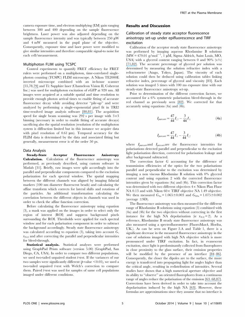
Figure 1. Schematic of the steady-state acceptor fluorescence anisotropy Total Internal Reflection Fluorescence Microscope. OD:Optical density to control the laser power in pupil plane of the objective. l/2: Half-waveplate in order to change the orientation of the laser outputpolarisation. L1&L2: form a telescope to enlarge the beam by a factor of 3. L3&L4: form a telescope to enlarge the beam by a factor of 5. Beamexpansion by a factor of 15 in order to illuminate the entire field of view. TM: Mirror mounted on a translation stage, which enables to switch from anepifluorescence excitation to a TIRF excitation. L5: Lens to focus in the back focal plane of the objective (60x, NA = 1.49). BS: Beam splitter. NF: Notchfilter to avoid any laser contamination. EMCCD: Electron Multiplying Charge Coupled Device.doi:10.1371/journal.pone.0110695.g001
FRET at the Plasma Membrane
PLOS ONE | www.plosone.org 4 October 2014 | Volume 9 | Issue 10 | e110695
camera exposure time, and electron multiplying (EM) gain ranging
between 300 and 400 depending on the sample fluorescence
brightness. Laser power was also adjusted depending on the
sample fluorescence intensity and was typically between 250 mW
and 4 mW measured in the pupil plane of the objective.
Consequently, exposure time and laser power were modified to
give similar intensities and therefore comparable signal-to noise for
each cell/measurement.
Multiphoton FLIM using TCSPCControl experiments to quantify FRET efficiency for FRET
rulers were performed on a multiphoton, time-correlated single-
photon counting (TCSPC) FLIM microscope. A Nikon TE2000E
inverted microscope combined with an in-house scanner
[55,78,79] and Ti: Sapphire laser (Chameleon Vision II, Coherent
Inc.) was used for multiphoton excitation of eGFP at 920 nm. All
images were acquired at a suitable spatial and time resolution to
provide enough photon arrival times to enable fitting of accurate
fluorescence decay while avoiding detector ‘‘pile-up’’ and were
analyzed by performing a single-exponential pixel fit in TRI2
time-resolved image analysis software [80,81]. The acquisition
speed for single beam scanning was 292 s per image with 363
binning (necessary in order to enable fitting of accurate decays)
sacrificing also the spatial resolution (resolution of the multiphoton
system is diffraction limited but in this instance we acquire data
with pixel resolution of 0.61 mm). Temporal accuracy for the
FLIM data is determined by the data and associated fitting but
generally, measurement error is of the order 50 ps.
Data AnalysisSteady-State Acceptor Fluorescence Anisotropy
Calculation. Calculation of the fluorescence anisotropy was
performed, as previously described, using custom software in
Matlab [31]. Briefly, raw images were split accordingly into the
parallel and perpendicular components compared to the excitation
polarization for each spectral window. The spatial mapping
between the different channels was aligned by imaging fiducial
markers (100 nm diameter fluorescent beads) and calculating the
affine transform which corrects for lateral shifts and rotations of
the particles. An additional transformation calculating the
correlation between the different objects in channels was used in
order to check the affine function correction.
Before calculating the fluorescence anisotropy using equation
(3), a mask was applied on the images in order to select only the
region of interest (ROI) and suppress background pixels
surrounding the ROI. Thresholds were applied for each spectral
window and for each polarisation component in order to subtract
the background accordingly. Steady state fluorescence anisotropy
was calculated according to equation (3), taking into account G,
xNA and after correcting the parallel and perpendicular intensities
for bleed-through.
Statistical analysis. Statistical analyses were performed
using GraphPad Prism software (version 5.00) (GraphPad, San
Diego, CA, USA). In order to compare two different populations,
we used two-tailed unpaired student t-test. If the variances of our
two samples were significantly different (p-value ,0.05), we used a
two-tailed unpaired t-test with Welch’s correction to compare
them. Paired t-test was used for samples of same cell populations
imaged under different conditions.
Results and Discussion
Calibration of steady state acceptor fluorescenceanisotropy set-up under epifluorescence and TIRFexcitation
Calibration of the acceptor steady state fluorescence anisotropy
was performed by imaging aqueous Rhodamine B solutions
(MW = 479.01 g/mol21, 1 mM, Sigma Aldrich, Saint Louis, MO,
USA) with a glycerol content ranging between 0 and 90% (v/v)
[71,82]. The accurate percentage of glycerol per solution was
determined by measuring the solution refractive index with a
refractometer (Atago, Tokyo, Japan). The viscosity of each
solution could then be deduced using calibration tables linking
refractive index, percentage of glycerol and viscosity [83]. Each
solution was imaged 5 times with 100 ms exposure time with our
steady-state fluorescence anisotropy set-up.
Prior to determination of the different correction factors, we
accounted for a 6% symmetric polarisation bleed-through in the
red channel as previously seen [82]. We corrected for that
accurately using equations (4a) and (4b).
Iparcorr~47
44I=={
3
44I\ ð4aÞ
Ipercorr~47
44I\{
3
44I== ð4bÞ
where Iparcorrand Ipercorrare the fluorescence intensities for
polarisations detected parallel and perpendicular to the excitation
light polarisation direction, corrected for polarisation leakage and
after background subtracted.
The correction factor G accounting for the difference of
transmission efficiencies of the optics for the two polarisations
parallel and perpendicular to the excitation was determined by
imaging a non viscous Rhodamine B solution with 0% glycerol
content and using equation 2 with the corrected fluorescence
intensities given by equations (4a) and (4b). This correction factor
was determined with two different objectives 46Nikon Plan Fluor
NA 0.13 and with Nikon 606TIRF objective NA 1.49 objective.
We then measured G4x = 1.06560.003 and G60x = 1.07560.002
(average 6SD).
The fluorescence anisotropy was then measured for the different
range of Rhodamine B solutions using equation (3) combined with
(4a) and (4b) for the two objectives without correcting in the first
instance for the high NA depolarisation (ie xNA = 2). As a
reference, Rhodamine B steady state fluorescence anisotropy was
also measured using a spectrofluorometer (FluoroMax4, Horiba,
UK.). As can be seen on Figure 2.A and Table 1, there is a
significant decrease in the measured fluorescence anisotropy in the
case of solutions imaged with high NA objective which is more
pronounced under TIRF excitation. In fact, in evanescent
excitation, since light is predominantly collected from fluorophores
in close proximity to the glass surface, their emission properties
will be modified by the presence of an interface [84–86].
Consequently, the closer the dipoles are to the surface, the more
energy is transferred into propagating light for angles higher than
the critical angle, resulting in redistribution of intensities. Several
studies have shown that a high numerical aperture objective and
its ability to ‘‘observe’’ an oriented fluorophores from a continuous
range of angles reduce the polarisation of the emission [63–68,87].
Corrections have been derived in order to take into account the
depolarisation induced by the high NA [63]. However, these
formulas are approximations since they assume that no interface is
FRET at the Plasma Membrane
PLOS ONE | www.plosone.org 5 October 2014 | Volume 9 | Issue 10 | e110695

present and it is known to affect the emission polarization
behaviour of a fluorophore. A more detailed calculation was thus
undertaken which includes near field coupling effect, taking into
account that for dipoles close to a surface, some of the near field
energy that doesn’t propagate is captured by the surface and
converted into propagating energy [86,87]. Thus, the presence of
a dielectric surface perturbs the emission of fluorescence in close
proximity and is dependent on fluorophore orientation and
proximity to surface. In fact, a theoretical study by Hellen et al.[87] looked at how a bare glass-water interface affects the angle-
dependent intensity, collected power, polarization, and lifetime of
the fluorescence emission as a function of the orientation and
distance of the fluorophore with respect to the interface, looking at
the emission properties of fluorophores considered as fixed power
dipole. Another theoretical study done by Burghardt et al. [88] by
deriving integral expressions for the electric field emitted by an
oscillating electric dipole when the dipole is near a dielectric
interface, has showed that an interface alters the effective aperture
of an objective by reflecting and refracting incident plane waves
and it also perturbs the emitted evanescent waves such that some
of them become transverse propagating waves with a strong
dependence on the distance of the dipole to the interface. More
recent works [89] showed that a large part of the radiation of a
fluorescing molecule in air or water located in close proximity to a
glass surface will be emitted as supercritical angle (SAF) into the
glass above the TIR angle. The dependency of the angular
radiation pattern of a fluorescing molecule with its distance to the
surface can thus be used in order to improve confinement at the
detection [90,91]. Consequently the proximity of the surface is
going to affect the emission properties of the dipole and effects are
qualitatively similar for both excitation polarization [86]. The
presence of the interface and the near field coupling induced
under evanescent wave excitation results thus in an increased
depolarization effect in TIRF compared to in epifluorescence
fluorescence excitation as shown in Figure 2.B. The high NA
depolarisation correction factor xNA was determined experimen-
tally as previously described [71], by considering the measure-
ments made with the low NA objective as reference and extracting
the correct xNA in order to match them with the measurements
made with the 606 objective using the following equation:
xNA~1
G60xI60xpercorr
I60xparcorr{G60xI60x
percorr
r4x
{I60xparcorr
!ð5Þ
In epifluorescence excitation, the xNA correction factor was
determined by correcting measurements made on solutions with
glycerol content between 58% and 93% (Table 1) using equation
(5). We then measured an averaged xNA = 1.2460.03 (average
6SD, N = 20).
In TIRF excitation, xNA was averaged by correcting measure-
ments made on solutions between 76% to 93% glycerol using
values in Table 1 and equation (5) and xNATIRF = 0.4560.04
(average 6SD, N = 15). The difference in the xNA correction
factors between epifluorescence fluorescence and TIRF excitation
thus accounts for the additional depolarisation linked to near field
interaction with the surface [85,86].
Calibration of aaFRET using FRET rulers inepifluorescence fluorescence excitation
Fixed MCF7 cells expressing eGFP-32aa-mRFP1 and eGFP-
7aa-mRFP1 for the FRET rulers and Cdc42-mRFP1 as a
reference value for acceptor anisotropy were used to calibrate
the aaFRET measurements under epifluorescence fluorescence
excitation. The steady-state acceptor fluorescence anisotropy was
calculated using equations 3 with equation 4 a&b and total
intensity and anisotropy maps were deduced as shown in Figure 3.
A & B.
The direct excitation of the acceptor in the case of cells
expressing Cdc42-mRFP1 resulted in highly polarized emission
(i.e. high anisotropy) (Figure 3.A, left) and a reduction of the
fluorescence anisotropy could be seen in the presence of FRET
between linked eGFP and mRFP1 (Figure 3.A, middle and right
panels). Additionally, the corresponding histograms of the
acceptor fluorescence anisotropy (Fig.3.C) underlined a clear
decrease of the acceptor fluorescence anisotropy in the case of the
different FRET rulers compared to cells expressing only the
acceptor (i.e. Cdc42-mRFP1).
Multiphoton, time-correlated single-photon counting (TCSPC)
FLIM experiments were also performed to quantify FRET
efficiency of the FRET standards using home-build set-up as
previously described [79,92]. FRET efficiencies were calculated
using the average fluorescence lifetimes from the donor in absence
or in the presence of an acceptor according to equation below [13]
and data in Table 2:
E~1{tdonor=acceptor
tdonor
ð6Þ
The FRET efficiencies measured were of E32aa = 0.16260.006
and E7aa = 0.2460.01 (average 6SEM, N = 9 for two independent
experiments) for eGFP-32aa-mRFP1 and eGFP-7aa-mRFP1,
respectively.
The different FRET rulers with respectively 7aa and 32aa
linkers could be clearly distinguished (p,0.0001,***) using steady
state acceptor anisotropy (Table 2 and Fig. 3.D) as well as with
FLIM experiments but with a much faster acquisition rate in the
case of aaFRET (100 ms compared to 5 minutes acquisitions for
FLIM/TCSPC experiments). Given that the fluorescence anisot-
ropy is typically measured on a microscope with an accuracy of
0.01, the different FRET rulers provided a dynamic range from 13
to 20 from the longest to the shortest linkers, whereas FLIM
measurements which are typically measured with an accuracy of
0.1 ns offered a dynamic range below 10 (Table 2). Furthermore,
it has been shown that even if sensitivity and dynamic range of
anisotropy measurements are dependent on signal-to-noise ratio
[93], variations in signal-to-noise and level of signal above
background can be tolerated, while maintaining the determination
of a robust value for the fluorescence anisotropy [30]. In fact, as
previously shown [94], large changes in anisotropy can occur for
the sensitized emission for modest values of FRET efficiency,
which makes it a sensitive technique with high dynamic range to
measure interactions between fluorophores. Even so, optimization
of imaging conditions (camera exposure time, excitation wave-
length and power) ensure the best signal-to-noise ratio is achieved,
while minimizing photobleaching and phototoxicity, essential in
live cells experiments.
No conversion of the fluorescence anisotropy data was done to
FRET efficiency [13] since as shown before [31], it would produce
an artificially broad histogram. Rather, we show that our
microscope arrangement can be used to distinguish different
FRET pairs with a good dynamic range as demonstrated
previously [26,29], in a significantly faster and less expensive
manner in comparison to TCSPC-FLIM and without loss in
signal-to-noise compared to time-gated system with Rapid
Lifetime Determination (RLD) [95–97].
FRET at the Plasma Membrane
PLOS ONE | www.plosone.org 6 October 2014 | Volume 9 | Issue 10 | e110695

Highlighting differences of activity of Raichu-Cdc42biosensor at the plasma membrane compared to cytosol
We monitored Raichu-Cdc42 biosensor activity using aaFRET
intracellularly as well as at the plasma membrane of cells by
switching from epifluorescence to TIRF excitation. Cells express-
ing the Raichu-Cdc42 FRET biosensor were imaged live at 20uCand 37uC. First experiments carried out at 20uC showed clear
differences in the intensity distribution across the image of the
Raichu-Cdc42 construct, linked to the confinement of the
excitation in TIRF to the plasma membrane. As can be seen in
Figure 4.A in the corresponding intensity images, in epifluores-
cence excitation, out of focus light is also detected which
deteriorates the signal-to-noise and contaminates the signal of
interest, whereas in TIRF, the labelling of the whole plasma
membrane can be efficiently visualized. In addition to this
improvement of the signal visualisation, the average steady-state
acceptor fluorescence anisotropy differs with the excitation type. A
decrease of the acceptor fluorescence anisotropy was highlighted
in TIRF compared to epifluorescence illumination as can be seen
in the fluorescence anisotropy maps in Figure 4.A and the
corresponding histograms of Raichu-Cdc42 fluorescence anisot-
ropy of the same cell imaged with both excitation types (Fig.4.B).
This can be related to a higher activity of the Raichu-Cdc42
biosensor occurring at the plasma membrane compared to
intracellularly which correlates with the active GTPase being
primarily localized on membranes and Cdc42 involvement with
cytoskeleton and actin polymerisation [36,37]. Additionally, TIRF
imaging provides good spatial confinement of the excitation and as
a result a better signal-to-noise ratio compared to epifluorescence
excitation, where fluorescence signal from planes located above or
below the imaging plane are also collected as in Figure 4.A in
epifluorescence illumination. This increase in signal-to-noise ratio
results in a more symmetric distribution of the anisotropy values in
TIRF compared to in epifluorescence excitation (Fig. 4.B).
Having observed significant differences of activity of the
Raichu-Cdc42 biosensor depending on excitation condition, we
imaged T17N and Y40C mutant biosensors, which constitute
negative controls [48]. Cells expressing Cdc42-mRFP1 provided a
baseline value for the acceptor fluorescence anisotropy in the
absence of donor (i.e. no FRET could occur). Determination of the
acceptor fluorescence anisotropy of several cells under epifluores-
cence illumination for the different constructs and statistical
analysis resulted in only a statistically significant difference
between Raichu-Cdc42 biosensor and its Y40C mutant
(p = 0.015,*, Fig. 4.C). In contrast, under TIRF excitation
(Fig.4.D), the four different constructs were clearly distinguished
Figure 2. Steady state fluorescence anisotropy of Rhodamine B solutions of different viscosities for objectives with different NA. (A)Variations of the steady state fluorescence anisotropy of solutions of Rhodamine B with different viscosities for objectives with different NA, Objective46 (NA = 0.13), Objective 606 (NA = 1.49) in epifluorescence and in TIRF excitation without correcting for the depolarisation induced by the high NAobjective (xNA = 2). Fluorescence anisotropy variations with viscosity are shown in (A) and for the different objectives and excitations (B). Eachanisotropy value is the average of 5 measurements made on each solution. The average and SD are represented.doi:10.1371/journal.pone.0110695.g002
Table 1. Steady-state fluorescence anisotropy of Rhodamine B solutions of different viscosities.
Glycerol (%) (v/v) Viscosity (cP) rspectro r4x r60xEPI r60x
TIRF
0 1.005 0.04 0.00060.003 0.00060.003 0.08960.008
58 9.586 0.15 0.10260.003 0.0785360.0002 0.03260.004
76 40.19 0.25 0.21260.002 0.163860.0005 0.11460.003
82 77.9 0.295 0.26160.005 0.21060.002 0.15060.006
93 356.2 0.35 0.30060.002 0.243660.001 0.17560.001
rspectro: fluorescence anisotropy measured using a spectrofluorometer. r4x, r60xEPI, r60x
TIRF: fluorescence anisotropy (average 6SD, N = 5), measured with objective 46(NA = 0.13), objective 606 (NA = 1.49) in epifluorescence and in TIRF excitation respectively, without correcting for high NA depolarisation (ie xNA = 2). SD: Standarddeviation.doi:10.1371/journal.pone.0110695.t001
FRET at the Plasma Membrane
PLOS ONE | www.plosone.org 7 October 2014 | Volume 9 | Issue 10 | e110695

Figure 3. Acceptor fluorescence anisotropy of fixed MCF7 cells transiently expressing eGFP/mRFP1 FRET rulers. Fluorescence intensity(A) and acceptor fluorescence anisotropy maps (B) of fixed MCF7 cells transiently expressing Cdc42-mRFP1 (left side) as a reference, eGFP-32aa-mRFP1 (middle) and eGFP-7aa-mRFP1 (right). Representative histograms of the acceptor fluorescence anisotropy of the corresponding cells (C). Meanvalues obtained from these histograms for the different cells imaged are then compared using unpaired t-test with Welch’s correction with 95%confidence intervals (***p,0.001) (D). The scale bar represents 5 mm.doi:10.1371/journal.pone.0110695.g003
Table 2. Average steady-state fluorescence acceptor anisotropy and fluorescence lifetime of MCF7 cells transiently expressingdifferent eGFP/mRFP1 FRET rulers.
Measurement Cdc42-mRFP1 eGFP-CaaX eGFP-32aa-mRFP1 eGFP-7aa-mRFP1
Acceptor anisotropy (mean±SEM) 0.28460.008 (N = 10) - 0.14660.005 (N = 9) 0.09360.002 (N = 7)
Fluorescence lifetime (mean±SEM) (ns) - 2.5860.04 (N = 9) 2.1660.01 (N = 9) 1.9560.02 (N = 9)
SEM: Standard Error of the Mean.doi:10.1371/journal.pone.0110695.t002
FRET at the Plasma Membrane
PLOS ONE | www.plosone.org 8 October 2014 | Volume 9 | Issue 10 | e110695

with high significance in the case of Raichu-Cdc42 and its Y40C
mutant (p = 0.0004, ***) and good significance for Raichu-Cdc42
and its T17N mutant (p = 0.008,**) as well as Cdc42-mRFP1 and
the Y40C mutant (p = 0.03,*). Due to the additional spatial
information provided by TIRF excitation combined with aa-
FRET, we were able to clearly distinguish Raichu-Cdc42 and its
different mutants and thus to show differences in activity for the
different constructs (Table 3). Spatial differences of the fluores-
cence anisotropy of the different constructs can be seen in the
anisotropy maps (Fig.5.A). This result is further supported by the
respective distinct histograms of the acceptor anisotropy (Fig.5.B)
which showed a significant increase of the acceptor fluorescence
anisotropy in the case of theY40C mutant compared to Raichu-
Cdc42 biosensor. This confirmed that both Y40C and T17N
mutants are negative controls [48], which could not be resolved in
epifluorescence excitation linked to both low signal-to-noise ratio
(resulting predominantly from fluorescence contamination from
out of focus light) and lower observed activity of Raichu-Cdc42
biosensor in the cellular milieu in comparison to at the plasma
membrane.
To monitor Cdc42 activity with changes in temperature, we
reproduced the previous experiments at 37uC for the different
Cdc42 constructs. Figure 6.A gathers all the data obtained on the
different mutants at 37uC and compared the effect of the
illumination. It shows that there was a significant decrease of the
acceptor fluorescence anisotropy for both Raichu-Cdc42 and its
T17N mutant in TIRF excitation compared to epifluorescence
excitation which was not the case for the Y40C mutant or the
Cdc42-mRFP1 constructs. This is perhaps surprising since T17N
mutant is inactive given its GDP-bound state. The Cdc42
Figure 4. Acceptor fluorescence anisotropy of MDA-MB231 cells expressing Raichu-Cdc42 biosensors imaged in epifluorescence orTIRF excitation. Fluorescence intensity (A left side) and acceptor fluorescence anisotropy maps (A right side) of live MDA-MB 231 cells transientlyexpressing Raichu-Cdc42 biosensor imaged at 20uC. Representative histograms of the fluorescence acceptor fluorescence anisotropy of thecorresponding cells for both excitations (B). Mean values obtained from these histograms on cells expressing different constructs Raichu-Cdc42biosensor, Y40C mutant, T17N mutant or Cdc42 imaged respectively in epifluorescence excitation (C) and in TIRF excitation (D). These measurementswere made on two independent experiments and are compared with a two-tailed unpaired t-test with 95% confidence intervals (***p,0.001,**p,
0.01, *p,0.05, ns non significant). Transfected MDA-MB 231 cells were typically imaged with 200 ms exposure time for the EMCCD and excitationpower between 250 mW to 4 mW depending of the expression level of the construct of interest. The scale bar represents 5 mm.doi:10.1371/journal.pone.0110695.g004
FRET at the Plasma Membrane
PLOS ONE | www.plosone.org 9 October 2014 | Volume 9 | Issue 10 | e110695

Figure 5. Acceptor fluorescence anisotropy of live MDA-MB 231 cells transiently expressing Raichu-Cdc42 or its different mutants.Fluorescence intensity (A. top) and acceptor fluorescence anisotropy maps (A, bottom) of live MDA-MB 231 cells transiently expressing Raichu-Cdc42biosensor, T17N mutant, Y40C, mutant or Cdc42 respectively imaged at 20uC. Representative histograms of the fluorescence acceptor fluorescenceanisotropy of the corresponding cells (B). The scale bar represents 5 mm.doi:10.1371/journal.pone.0110695.g005
Table 3. Average steady-state fluorescence acceptor anisotropy of MDA-MB 231 cells transfected with respectfully Raichu-Cdc42biosensor, T17N mutant, Y40C mutant and Cdc42-mRFP1 and imaged live at 20uC or 37uC in epifluorescence and in TIRFillumination.
Imaging conditions 206C 376C
Excitation Epifluorescence TIRF Epifluorescence TIRF
aaFRET Raichu-Cdc42 (mean±SEM) 0.24660.007 (N = 14) 0.1460.01 (N = 14) 0.17260.009 (N = 37) 0.13560.003 (N = 37)
aaFRET T17N (mean±SEM) 0.2460.01 (N = 15) 0.1860.01 (N = 15) 0.2360.02 (N = 11) 0.16960.007 (N = 11)
aaFRET Y40C (mean±SEM) 0.27460.008 (N = 14) 0.2260.01 (N = 14) 0.22760.006 (N = 32) 0.23760.007 (N = 32)
aaFRET Cdc42 (mean±SEM) 0.2960.01 (N = 7) 0.2760.02 (N = 7) 0.27660.008 (N = 5) 0.29160.007 (N = 5)
doi:10.1371/journal.pone.0110695.t003
FRET at the Plasma Membrane
PLOS ONE | www.plosone.org 10 October 2014 | Volume 9 | Issue 10 | e110695

component of the sensor has, therefore, a reduced affinity to the
PAK1-CRIB binding domain, but not abrogation of binding. As
such we must consider the potential energy landscape is sufficiently
modified at 37uC to allow significant FRET to occur. Comparing
results obtained respectively in epifluorescence illumination
(Fig.6.B) and in TIRF (Fig.6.C), we observed higher activity of
both Raichu-Cdc42 and T17N at the plasma membrane of the
cells compared to intracellularly. Furthermore, in epifluorescence
excitation, at 37uC, all constructs were more active since the
average acceptor fluorescence anisotropy values were lower
compared to those obtained at 20uC (Table 3). Given that no
effect of temperature was seen on Cdc42 for both illumination
modes (p = 0.46 insignificant in epifluorescence, p = 0.33 insignif-
icant for TIRF excitation), variations in activity of Cdc42, which
must occur due to imaging at physiological temperature, are the
most probable cause, although possible phase changes at the
plasma membrane [98] and increased probe/effector mobility
could also contribute to this effect. This increased activity of the
biosensors at 37uC already enabled us to distinguish the different
constructs in epifluorescence excitation (although the two negative
mutants T17N and Y40C could not be differentiated) (Fig.6.B) and
in TIRF excitation, all constructs could be clearly differentiated
(Fig. 6.A & C). Use of TIRF excitation for Raichu-Cdc42
constructs and T17N mutant resulted in anisotropy values with
a narrow distribution, which is predominantly due to the increase
in signal-to-noise ratio in TIRF excitation compared to epifluor-
escence illumination. The effect of temperature was noticeable on
Raichu-Cdc42 and on Y40C in epifluorescence excitation,
whereas on the other constructs, no effect of the temperature
could be detected for both excitation types. These results reflect
the dynamic nature of protein orientation in the biosensor. For the
Y40C construct, the binding of Cdc42 to the Cdc42 -interactive
binding motif (CRIB) of PAK1 is modified whereas the T17N
mutant is trapped in a GDP-bound state (inactive) and the affinity
of binding of Cdc42 to PAK1 in presence of guanine nucleotide is
reduced but binding of the sensor domain is unaffected. In
contrast for Y40C, the binding domain of Cdc42 to PAK1 has
been modified preventing binding of Cdc42 and thus the biosensor
will always be in an open conformation so FRET is unlikely. Thus,
the true negative control upon which we determine the dynamic
range of the biosensor must be the Y40C.
Dynamic inhibition of Raichu-Cdc42 biosensor at the cellplasma membrane
Since Cdc42 activation has been highlighted in various cancer
diseases [99,100], monitoring of its activity could help to clarify the
signalling pathways modulated by Cdc42 and design better
strategies to target those pathways. Recently a Cdc42 selective
guanine nucleotide binding inhibitor (ML141) has been found by
high throughput screening [101] and has provided selective,
reversible and non-competitive inhibition of Cdc42 [102]. In vitro
Figure 6. Ensemble steady state acceptor fluorescence anisotropy values of MDA-MB 231 cells expressing different Cdc42constructs. MDA-MB 231 cells were expressing different Cdc42 constructs and imaged live at 37uC, excited respectively in epifluorescence and inTIRF excitation. Measurements were compared using either two-tailed paired t-test (A) or two-tailed unpaired t-test with 95% confidence intervals (B,C) (***p,0.001,**p,0.01, *p,0.05, ns non significant).doi:10.1371/journal.pone.0110695.g006
FRET at the Plasma Membrane
PLOS ONE | www.plosone.org 11 October 2014 | Volume 9 | Issue 10 | e110695

and live cell experiments have shown that ML141 does not
prevent guanine nucleotide binding but its association induces the
dissociation of the guanine nucleotide resulting in Cdc42 being
locked in an inactive conformation. In comparison to other Cdc42
inhibitors like Clostridium difficile Toxin B, the association of
Cdc42 with membranes should not be inhibited allowing imaging
in TIRF excitation.
Cdc42 inhibition was achieved on Raichu-Cdc42 biosensor
expressed in HCC1954 cells and imaged live at 37uC, before and
after Cdc42 GTPase inhibitor addition (Fig.7.A&B). As can be
seen on the fluorescence anisotropy maps pre- and post-addition of
Figure 7. Live inhibition of Cdc42 in HCC1954 cells transiently expressing Raichu-Cdc42 biosensor. Fluorescence intensity (A, left) andacceptor fluorescence anisotropy maps (A, right) of live HCC1954 cells transiently expressing Raichu-Cdc42 biosensor before (A. top) and after (A.bottom) addition of Cdc42 inhibitor. The scale bar represents 5 mm. (B) Corresponding representative histograms of the fluorescence acceptoranisotropy before (A top right) and after inhibition (A bottom right) are shown. (C) Time lapse of acceptor fluorescence anisotropy (6SD) after 30 mMaddition of Cdc42 inhibitor for cell expressing respectively Raichu-Cdc42 biosensor (pink), Cdc42 (blue) or after addition of 30 mM DMSO for cellexpressing Raichu-Cdc42 biosensor (green). Images were taken every 5 minutes (200 ms exposure for EMCCD) for 50 minutes. (D) Ensemble steadystate acceptor fluorescence anisotropy values obtained on HCC1954 cells expressing Raichu-Cdc42biosensor before (pre) and after (post) addition ofCdc42 inhibitor, or before and after addition of DMSO, or for cells expressing Cdc42 before and after addition of Cdc42 inhibitor. Cells were imagedlive at 37uC, excited in TIRF excitation.doi:10.1371/journal.pone.0110695.g007
Table 4. Average steady-state acceptor fluorescence anisotropy before and after Cdc42 inhibition of HCC1954 cells transientlyexpressing different Raichu-Cdc42 biosensors.
Average acceptor anisotropy Raichu +DMSO Raichu +Inhibitor Cdc42 +Inhibitor
Before inhibition 0.14560.005 (N = 10) 0.14460.008 (N = 9) 0.24760.003 (N = 11)
After inhibition 0.16260.009 (N = 10) 0.18460.007 (N = 9) 0.23960.005 (N = 9)
p-value 0.124 ns Unpaired t-test 0.004 Paired t-test 0.220 ns Unpaired t-test
doi:10.1371/journal.pone.0110695.t004
FRET at the Plasma Membrane
PLOS ONE | www.plosone.org 12 October 2014 | Volume 9 | Issue 10 | e110695

the inhibitor (Fig. 7.A) and the corresponding histograms (Fig.7.B),
a clear increase of the acceptor fluorescence anisotropy of the
Raichu-Cdc42 biosensor was measured 50 minutes after addition
of the inhibitor. Furthermore, time-lapse experiments acquiring
images every 5 minutes to dynamically monitor Cdc42 inhibition
at the cell plasma membrane were undertaken. From these time
lapse data, both total intensity and the corresponding acceptor
fluorescence anisotropy maps were extracted (Movies S1&S2). The
average fluorescence anisotropy extracted from this time lapse
highlights a clear increase of acceptor fluorescence anisotropy after
Cdc42 inhibition (Fig.7.C). These measurements were repeated in
10 cells and the anisotropy measured before and after inhibition
showed a significant increase of the fluorescence anisotropy after
inhibition (Table 4 and Fig.7.D, p = 0.004, **). In order to confirm
that the increase in fluorescence anisotropy was directly related to
Cdc42 inhibition, control measurements were undertaken by
imaging Raichu-Cdc42 biosensor in 30 mM DMSO in order to
ensure that the effect on the fluorescence anisotropy was linked to
the inhibitor and not to the carrier. Measurements of the
fluorescence anisotropy before and after addition of the inhibitor
for several cells (Fig.7.D), and time-lapse experiments (Fig. 7.C)
indicate a stable and high activity of the biosensor over the
imaging period, without phototoxicity/photodamage induced by
the illumination or effect of the carrier. The reference value for the
open conformation (i.e. non interacting form) was given by
imaging cells expressing Cdc42 –mRFP1 in presence of the
inhibitor. In that case too, no effect of the inhibitor was observed
either on time lapse (Fig.7.C) or on ensemble measurements
(Table 4 and Fig.7.D). As seen in Figure 7.C, after 50 minutes of
treatment with the inhibitor, the fluorescence anisotropy of the
Raichu-Cdc42 biosensor went from low anisotropy values
(comparable to when imaged in DMSO) to reach the fluorescence
anisotropy of cells expressing Cdc42-mRFP1 (i.e. baseline value
for absence of FRET). Thus, the increase in the acceptor
fluorescence anisotropy is directly related to a decrease in the
FRET efficiency and consequently to a change of conformation of
the Raichu-Cdc42 biosensor from an active (GTP-bound)
conformation to an inactive (GDP-bound) conformation. We
noticed that the response to inhibition was cell specific in terms of
efficiency and time to response after inhibitor addition. On
average, the fluorescence anisotropy was increased by 25% after
50 minutes treatment (Table 4). This imaging time window
seemed to be optimal in order to have an effective inhibition of
Cdc42 activity without inducing phototoxicity/photodamage. In
addition to a clear decrease of the intramolecular FRET of the
Raichu-Cdc42 biosensor (Fig.7.C), intensity images also showed
morphological changes of the cell shape, which might be linked to
the induced inhibition of Cdc42 related filopodia formation and
cell migration (Movie S1).
ConclusionsWe combined TIRF Microscopy with steady-state acceptor
fluorescence anisotropy imaging (aaFRET) and observed intra-
molecular FRET under evanescent wave excitation. Correction
factors accounting for the additional depolarization factors
induced when imaging in TIRF with a high NA objective were
determined by calibration measurements made on Rhodamine B
solutions of different viscosities with those made with a low NA
objective. This technique enabled us to distinguish different
eGFP/mRFP1 FRET rulers with significant gain of speed and cost
compared to fluorescence lifetime imaging microscopy.
We then highlighted differences of activity of the FRET Raichu-
Cdc42 biosensor with a higher activity measured at the cell plasma
membrane compared to intracellularly. Furthermore, imaging in
TIRF provided better conditions in terms of signal-to-noise ratio
which helped differentiate Raichu-Cdc42 biosensor from its
different negative mutants T17N and Y40C. Live inhibition of
Cdc42 in the Raichu-Cdc42 biosensor was demonstrated by
following the decrease in the related intramolecular FRET
indicating changes of conformations of the probe at the cell
plasma membrane. We consequently demonstrated intramolecular
FRET imaging in wide-field configuration in TIRF excitation with
a good contrast and dynamic range as well as fast acquisition rates.
The additional spatial information provided insights about Cdc42
activation and underlined its clear inhibition. Furthermore, since
Cdc42 has been associated with a migratory behaviour which
seems to be dependent on the cell lines as recent studies have
shown [40], this new technique could, given its fast acquisition
rates, provide quantitative method to test the activation of Cdc42
and to associate it to a migratory or anti-migratory role depending
on the cancer cell line.
Whilst FRET biosensors constitute ideal systems to use with
aaFRET given their one-to-one stoichiometry, this technique
could be extended to imaging protein-protein interactions as
shown before [31]. An extra calibration is then required prior to
imaging in order to determine the different concentrations of the
free donor, free acceptor and of the FRET pairs [31,103].
Supporting Information
Movie S1 Fluorescence intensity time lapse video ofHCC1954 cells transiently expressing Raichu-Cdc42biosensor and under Cdc42 inhibition. Cells were imaged
live before (time 0) and after addition of Cdc42 inhibitor, every
5 minutes (200 ms exposure for EMCCD) for 60 minutes.
(AVI)
Movie S2 Fluorescence anisotropy time lapse video ofHCC1954 cells transiently expressing Raichu-Cdc42biosensor and under Cdc42 inhibition. Cells were imaged
live before (time 0) and after addition of Cdc42 inhibitor, every
5 minutes (200 ms exposure for EMCCD) for 60 minutes.
(AVI)
Author Contributions
Conceived and designed the experiments: VD GW JM TN SMA.
Performed the experiments: VD JA JAL. Analyzed the data: VD DRM
JA. Contributed reagents/materials/analysis tools: DRM JN SPP OC GW
JM TN SMA. Wrote the paper: VD DRM SMA.
References
1. Axelrod D (2001) Total internal reflection fluorescence microscopy in cell
biology. Traffic: 764–774. Available: http://www.ncbi.nlm.nih.gov/pubmed/
12624904.
2. Axelrod D (1989) Total Internal Reflection Fluorescence Microscopy.
Methods.Cell.Biol. pp.245–270. Available: http://books.google.com/
books?hl=fr&lr=&id=Xs1jBDr2WQYC&pgis=1. Accessed 2014 Jan 10.
3. Mattheyses AL, Simon SM, Rappoport JZ (2010) Imaging with total internal
reflection fluorescence microscopy for the cell biologist. J Cell Sci 123:
3621–3628. Available: http://www.pubmedcentral.nih.gov/articlerender.
fcgi?artid=2964103&tool=pmcentrez&rendertype=abstract. Accessed 2013
Nov 8.
4. Nayal A, Webb DJ, Brown CM, Schaefer EM, Vicente-Manzanares M, et al.
(2006) Paxillin phosphorylation at Ser273 localizes a GIT1-PIX-PAK complex
and regulates adhesion and protrusion dynamics. J Cell Biol 173: 587–589.
Available: http://jcb.rupress.org/content/173/4/587.full. Accessed 2014 May
7.
FRET at the Plasma Membrane
PLOS ONE | www.plosone.org 13 October 2014 | Volume 9 | Issue 10 | e110695

5. Cocucci E, Aguet F, Boulant S, Kirchhausen T (2012) The first five seconds in
the life of a clathrin-coated pit. Cell 150: 495–507. Available: http://www.
sciencedirect.com/science/article/pii/S0092867412007854. Accessed 2014Apr 28.
6. Betzig E, Patterson GH, Sougrat R, Lindwasser OW, Olenych S, et al. (2006)
Imaging intracellular fluorescent proteins at nanometer resolution. Science
313: 1642–1645. Available: http://www.sciencemag.org/content/313/5793/1642.abstract. Accessed 2011 Jul 16.
7. Huang B, Wang W, Bates M, Zhuang X (2008) Three-dimensional super-
resolution imaging by stochastic optical reconstruction microscopy. Science319: 810–813. Available: http://www.pubmedcentral.nih.gov/articlerender.
fcgi?artid=2633023&tool=pmcentrez&rendertype=abstract. Accessed 2012
Nov 2.
8. Fiolka R, Beck M, Stemmer A (2008) Structured illumination in total internalreflection fluorescence microscopy using a spatial light modulator. Opt Lett 33:
1629. Available: http://ol.osa.org/abstract.cfm?URI=ol-33-14-1629. Accessed2014 Aug 6.
9. Gliko O, Reddy DG, Brownell WE, Saggau P (2008) Development of fast two-
dimensional standing wave microscopy using acousto-optic deflectors. In:
Conchello J-A, Cogswell CJ, Wilson T, Brown TG, editors. Biomedical Optics(BiOS) 2008. International Society for Optics and Photonics. p. 68610B–
68610B–8. Available: http://proceedings.spiedigitallibrary.org/proceeding.aspx?articleid=819539. Accessed 2014 Aug 6.
10. Brunstein M, Wicker K, Herault K, Heintzmann R, Oheim M (2013) Full-field
dual-color 100-nm super-resolution imaging reveals organization and dynamics
of mitochondrial and ER networks. Opt Express 21: 26162–26173. Available:http://www.opticsinfobase.org/viewmedia.cfm?uri=oe-21-22-26162&seq=0&
html=true. Accessed 2014 Aug 6.
11. Kner P, Chhun BB, Griffis ER, Winoto L, Gustafsson MGL (2009) Super-resolution video microscopy of live cells by structured illumination. Nat
Methods 6: 339–342. Available: http://dx.doi.org/10.1038/nmeth.1324.
Accessed 2014 Jul 24.
12. Marquer C, Devauges V, Cossec J-C, Liot G, Lecart S, et al. (2011) Localcholesterol increase triggers amyloid precursor protein-Bace1 clustering in lipid
rafts and rapid endocytosis. FASEB J: 1–11. Available: http://www.ncbi.nlm.nih.gov/pubmed/21257714. Accessed 2011 Jan 26.
13. Jares-Erijman EA, Jovin TM (2003) FRET imaging. Nat Biotechnol 21: 1387–1395. Available: http://www.ncbi.nlm.nih.gov/pubmed/14595367. Accessed
2012 Oct 29.
14. Forster T (1946) Energiewanderung und Fluoreszenz. Naturwissenschaften 33:166–175. Available: http://www.springerlink.com/content/q3468p13200
52434/. Accessed 2011 Jul 28.
15. Ems-McClung SC, Hainline SG, Devare J, Zong H, Cai S, et al. (2013) Aurora
B inhibits MCAK activity through a phosphoconformational switch thatreduces microtubule association. Curr Biol 23: 2491–2499. Available: http://
www.sciencedirect.com/science/article/pii/S0960982213013298. Accessed2014 Mar 24.
16. Owen DM, Oddos S, Kumar S, Davis DM, Neil MAA, et al. (2010) High
plasma membrane lipid order imaged at the immunological synapse periphery
in live T cells. Mol Membr Biol 27: 178–189. Available: http://informahealthcare.com/doi/abs/10.3109/09687688.2010.495353. Accessed
2014 Mar 24.
17. Valdembri D, Caswell PT, Anderson KI, Schwarz JP, Konig I, et al. (2009)
Neuropilin-1/GIPC1 signaling regulates alpha5beta1 integrin traffic andfunction in endothelial cells. PLoS Biol 7: e25. Available: http://www.
plosbiology.org/article/info: doi/10.1371/journal.pbio.1000025#pbio-1000025-g011. Accessed 2014 Jul 10.
18. Bal M, Zaika O, Martin P, Shapiro MS (2008) Calmodulin binding to M-type
K+ channels assayed by TIRF/FRET in living cells. J Physiol 586: 2307–2320.
Available: http://www.pubmedcentral.nih.gov/articlerender.fcgi?artid=2479561&tool=pmcentrez&rendertype=abstract. Accessed 2013 Dec 5.
19. Bal M, Zhang J, Zaika O, Hernandez CC, Shapiro MS (2008) Homomeric and
heteromeric assembly of KCNQ (Kv7) K+ channels assayed by total internalreflection fluorescence/fluorescence resonance energy transfer and patch clamp
analysis. J Biol Chem 283: 30668–30676. Available: http://www.jbc.org/
content/283/45/30668.full. Accessed 2013 Dec 10.
20. Suzuki Y, Yamamura H, Ohya S, Imaizumi Y (2013) Direct molecularinteraction of caveolin-3 with KCa1.1 channel in living HEK293 cell
expression system. Available: http://www.sciencedirect.com/science/article/pii/S0006291X12023431. Accessed 2013 Dec 10.
21. Riven I, Iwanir S, Reuveny E (2006) GIRK Channel Activation Involves a
Local Rearrangement of a Preformed G Protein Channel Complex. Neuron
51: 561–573. Available: http://www.sciencedirect.com/science/article/pii/S0896627306006398. Accessed 2013 Dec 10.
22. Levitt JA, Matthews DR, Ameer-Beg SM, Suhling K (2009) Fluorescence
lifetime and polarization-resolved imaging in cell biology. Curr OpinBiotechnol 20: 28–36. Available: http://www.ncbi.nlm.nih.gov/pubmed/
19268568. Accessed 2011 May 19.
23. Bader AN, Hoetzl S, Hofman EG, Voortman J, van Bergen en Henegouwen
PMP, et al. (2011) Homo-FRET imaging as a tool to quantify protein and lipidclustering. Chemphyschem 12: 475–483. Available: http://www.ncbi.nlm.nih.
gov/pubmed/21344588. Accessed 2011 Jul 16.
24. Tramier M, Coppey-Moisan M (2008) Fluorescence anisotropy imaging
microscopy for homo-FRET in living cells. Methods Cell Biol 85: 395–414.
Available: http://www.ncbi.nlm.nih.gov/entrez/query.fcgi?cmd=Retrieve&
db=PubMed&dopt=Citation&list_uids=18155472.
25. Gautier I, Tramier M, Durieux C, Coppey J, Pansu RB, et al. (2001) Homo-
FRET microscopy in living cells to measure monomer-dimer transition of GFP-
tagged proteins. Biophys J 80: 3000–3008. Available: http://www.
pubmedcentral.nih.gov/articlerender.fcgi?artid=1301483&tool=pmcentrez&ren
dertype=abstract. Accessed 2011 Feb 22.
26. Rizzo MA, Piston DW (2005) High-contrast imaging of fluorescent protein
FRET by fluorescence polarization microscopy. Biophys J 88: L14–16.
Available: http://www.pubmedcentral.nih.gov/articlerender.fcgi?artid=1305
173&tool=pmcentrez&rendertype=abstract. Accessed 2011 Jun 22.
27. Perrin F (1926) Polarisation de la Lumiere de Fluorescence. Vie Moyenne des
Molecules Fluorescentes. JPhysique 7: 390–401.
28. Suhling K, Davis DM, Phillips D (2002) The Influence of Solvent Viscosity on
the Fluorescence Decay and Time-Resolved Anisotropy of Green Fluorescent
Protein. J Fluoresc 12: 91–95–95. Available: http://www.springerlink.com/
content/dme515hdka81cwb8/. Accessed 2011 Jul 29.
29. Rizzo MA, Springer G, Segawa K, Zipfel WR, Piston DW (2006) Optimization
of pairings and detection conditions for measurement of FRET between cyan
and yellow fluorescent proteins. Microsc Microanal 12: 238–254. Available:
http://www.ncbi.nlm.nih.gov/pubmed/17481360. Accessed 2011 Sep 28.
30. Matthews DR, Carlin LM, Ofo E, Barber PR, Vojnovic B, et al. (2010) Time-
lapse FRET microscopy using fluorescence anisotropy. J Microsc 237: 51–62.
Available: http://www.ncbi.nlm.nih.gov/pubmed/20055918.
31. Matthews DR, Fruhwirth GO, Weitsman G, Carlin LM, Ofo E, et al. (2012) A
multi-functional imaging approach to high-content protein interaction
screening. PLoS One 7: e33231. Available: http://www.pubmedcentral.nih.
gov/articlerender.fcgi?artid=3323588&tool=pmcentrez&rendertype=abstract.
32. Mochizuki N, Yamashita S, Kurokawa K, Ohba Y, Nagai T, et al. (2001)
Spatio-temporal images of growth-factor-induced activation of Ras and Rap1.
Nature 411: 1065–1068. Available: http://dx.doi.org/10.1038/35082594.
Accessed 2014 May 21.
33. Makrogianneli K, Carlin LM, Keppler MD, Matthews DR, Ofo E, et al. (2009)
Integrating receptor signal inputs that influence small Rho GTPase activation
dynamics at the immunological synapse. Mol Cell Biol 29: 2997–3006.
Available: http://mcb.asm.org/content/29/11/2997.short. Accessed 2013 Jan
9.
34. Bender A, Pringle JR (1989) Multicopy suppression of the cdc24 budding defect
in yeast by CDC42 and three newly identified genes including the ras-related
gene RSR1. Proc Natl Acad Sci 86: 9976–9980. Available: http://www.pnas.
org/content/86/24/9976.short. Accessed 2013 Dec 31.
35. Harris KP, Tepass U (2010) Cdc42 and vesicle trafficking in polarized cells.
Traffic 11: 1272–1279. Available: http://www.ncbi.nlm.nih.gov/pubmed/
20633244. Accessed 2013 Dec 31.
36. Ridley MAM and AJ, Ridley AJ (2006) Rho GTPases and actin dynamics in
membrane protrusions and vesicle trafficking. Trends Cell Biol 16: 522–529.
Available: http://www.sciencedirect.com/science/article/pii/S0962892406
002236. Accessed 2013 Dec 31.
37. Ridley AJ (2011) Life at the leading edge. Cell 145: 1012–1022. Available:
http://www.ncbi.nlm.nih.gov/pubmed/21703446. Accessed 2013 Dec 11.
38. Johnson E, Seachrist DD, DeLeon-Rodriguez CM, Lozada KL, Miedler J, et
al. (2010) HER2/ErbB2-induced breast cancer cell migration and invasion
require p120 catenin activation of Rac1 and Cdc42. J Biol Chem 285: 29491–
29501. Available: http://www.jbc.org/content/285/38/29491.short. Accessed
2014 Jan 3.
39. Reymond N, Im JH, Garg R, Vega FM, Borda d’Agua B, et al. (2012) Cdc42
promotes transendothelial migration of cancer cells through b1 integrin. J Cell
Biol 199: 653–668. Available: http://jcb.rupress.org/content/199/4/653.
short. Accessed 2013 Dec 13.
40. Zuo Y, Wu Y, Chakraborty C (2012) Cdc42 negatively regulates intrinsic
migration of highly aggressive breast cancer cells. J Cell Physiol 227: 1399–
1407. Available: http://www.ncbi.nlm.nih.gov/pubmed/21618528. Accessed
2013 Dec 17.
41. Fritz G, Just I, Kaina B (1999) Rho GTPases are over-expressed in human
tumors. Int J Cancer 81: 682–687. Available: http://www.ncbi.nlm.nih.gov/
pubmed/10328216. Accessed 2013 Dec 31.
42. Chen Q-Y, Jiao D-M, Yao Q-H, Yan J, Song J, et al. (2012) Expression analysis
of Cdc42 in lung cancer and modulation of its expression by curcumin in lung
cancer cell lines. Int J Oncol 40: 1561–1568. Available: http://www.
spandidos-publications.com/ijo/40/5/1561/abstract. Accessed 2013 Dec 31.
43. Gomez del Pulgar T, Valdes-Mora F, Bandres E, Perez-Palacios R, Espina C,
et al. (2008) Cdc42 is highly expressed in colorectal adenocarcinoma and
downregulates ID4 through an epigenetic mechanism. Int J Oncol 33: 185–
193. Available: http://www.spandidos-publications.com/ijo/33/1/185/
abstract. Accessed 2013 Dec 31.
44. Guo F, Zhang S, Tripathi P, Mattner J, Phelan J, et al. (2011) Distinct roles of
Cdc42 in thymopoiesis and effector and memory T cell differentiation. PLoS
One 6: e18002. Available: http://dx.plos.org/10.1371/journal.pone.0018002.
Accessed 2013 Dec 31.
45. Zhu X, Raina AK, Boux H, Simmons ZL, Takeda A, et al. (2000) Activation of
oncogenic pathways in degenerating neurons in Alzheimer disease. Int J Dev
Neurosci 18: 433–437. Available: http://www.sciencedirect.com/science/
article/pii/S0736574800000101. Accessed 2013 Dec 31.
FRET at the Plasma Membrane
PLOS ONE | www.plosone.org 14 October 2014 | Volume 9 | Issue 10 | e110695

46. Aoki K, Komatsu N, Hirata E, Kamioka Y, Matsuda M (2012) Stable
expression of FRET biosensors: a new light in cancer research. Cancer Sci 103:614–619. Available: http://www.ncbi.nlm.nih.gov/pubmed/22188216. Ac-
cessed 2013 Nov 8.
47. Miyawaki A (2003) Visualization of the Spatial and Temporal Dynamics ofIntracellular Signaling. Dev Cell 4: 295–305. Available: http://www.
sciencedirect.com/science/article/pii/S1534580703000601. Accessed 2013Dec 31.
48. Itoh RE, Kurokawa K, Ohba Y, Yoshizaki H, Mochizuki N, et al. (2002)
Activation of Rac and Cdc42 Video Imaged by Fluorescent Resonance EnergyTransfer-Based Single-Molecule Probes in the Membrane of Living Cells. Mol
Cell Biol 22: 6582–6591. doi: 10.1128/MCB.22.18.6582.
49. Takai Y, Sasaki T, Matozaki T (2001) Small GTP-binding proteins. PhysiolRev 81: 153–208. Available: http://www.ncbi.nlm.nih.gov/pubmed/
11152757.
50. Bishop A, Hall A (2000) Rho GTPases and their effector proteins. Biochem J
348: 241–255. Available: http://www.biochemj.org/bj/348/bj3480241.htm.
Accessed 2 January 2014.
51. Hirata E, Yukinaga H, Kamioka Y, Arakawa Y, Miyamoto S, et al. (2012) In
vivo fluorescence resonance energy transfer imaging reveals differentialactivation of Rho-family GTPases in glioblastoma cell invasion. J Cell Sci
125: 858–868. Available: http://jcs.biologists.org/content/125/4/858.short.
Accessed 2014 Mar 25.
52. Nakamura T, Aoki K, Matsuda M (2005) Monitoring spatio-temporal
regulation of Ras and Rho GTPase with GFP-based FRET probes. Methods37: 146–153. Available: http://www.sciencedirect.com/science/article/pii/
S1046202305001295. Accessed 2014 Mar 25.
53. Beutler M, Makrogianneli K, Vermeij RJ, Keppler M, Ng T, et al. (2008)satFRET: estimation of Forster resonance energy transfer by acceptor
saturation. Eur Biophys J 38: 69–82. Available: http://www.ncbi.nlm.nih.gov/pubmed/18769914. Accessed 2014 Mar 25.
54. Nalbant P, Hodgson L, Kraynov V, Toutchkine A, Hahn KM (2004)
Activation of endogenous Cdc42 visualized in living cells. Science 305: 1615–1619. Available: http://www.sciencemag.org/content/305/5690/1615.
abstract. Accessed 2014 Mar 25.
55. Carlin LM, Evans R, Milewicz H, Fernandes L, Matthews DR, et al. (2011) A
targeted siRNA screen identifies regulators of Cdc42 activity at the natural
killer cell immunological synapse. Sci Signal 4: ra81. Available: http://www.ncbi.nlm.nih.gov/pubmed/22126964. Accessed 2013 Nov 25.
56. Forkey JN, Quinlan ME, Goldman YE (2005) Measurement of singlemacromolecule orientation by total internal reflection fluorescence polarization
microscopy. Biophys J 89: 1261–1271. Available: http://www.sciencedirect.
com/science/article/pii/S0006349505727739. Accessed 2014 Mar 26.
57. Gee ML, Lensun L, Smith T a, Scholes C a (2004) Time-resolved evanescent
wave-induced fluorescence anisotropy for the determination of molecularconformational changes of proteins at an interface. Eur Biophys J 33: 130–139.
Available: http://www.ncbi.nlm.nih.gov/pubmed/14586518. Accessed 2011
Jun 22.
58. Axelrod D (2013) Evanescent excitation and emission in fluorescence
microscopy. Biophys J 104: 1401–1409. Available: http://www.ncbi.nlm.nih.gov/pubmed/23561516. Accessed 2013 Nov 10.
59. Axelrod D, Burghardt TP, Thompson NL (1984) Total internal reflection
fluorescence. Annu Rev Biophys Bioeng 13: 247–268. Available: http://www.ncbi.nlm.nih.gov/pubmed/6378070.
60. Scholes Ca, Millar DP, Gee ML, Smith Ta (2011) Resonance energy-transfer
studies of the conformational change on the adsorption of oligonucleotides to asilica interface. J Phys Chem B 115: 6329–6339. Available: http://www.ncbi.
nlm.nih.gov/pubmed/21500794.
61. Miki T (1995) Interaction of ect2 and Dbl with Rho-related GTPases. Methods
Enzymol 256: 90–98. Available: http://www.ncbi.nlm.nih.gov/pubmed/
7476459. Accessed 19 August 2014.
62. Axelrod D (1989) Fluorescence polarization microscopy. Methods in Cell
Biology. Fluorescence Microscopy of Living Cells in Culture Part B.Quantitative Fluorescence Microscopy—Imaging and Spectroscopy. Methods
in Cell Biology. Elsevier, Vol. 30. pp.333–352. Available: http://dx.doi.org/
10.1016/S0091-679X(08)60985-1. Accessed 29 July 2011.
63. Axelrod D (1979) Carbocyanine dye orientation in red cell membrane studied
by microscopic fluorescence polarization. Biophys J 26: 557–573. Available:http://www.pubmedcentral.nih.gov/articlerender.fcgi?artid=1328570&tool=pm
centrez&rendertype=abstract. Accessed 2011 Mar 7.
64. Shribak M (2002) Polarization aberrations caused by differential transmissionand phase shift in high-numerical-aperture lenses: theory, measurement, and
rectification. Opt Eng 41: 943. Available: http://opticalengineering.spiedigitallibrary.org/article.aspx?articleid=1098859. Accessed 2014 Aug 6.
65. Bahlmann K, Hell SW (2000) Depolarization by high aperture focusing. Appl
Phys Lett 77: 612. Available: http://scitation.aip.org/content/aip/journal/apl/77/5/10.1063/1.127061. Accessed 2014 Aug 6.
66. Torok P, Higdon PD, Wilson T (1998) On the general properties of polarised
light conventional and confocal microscopes. Opt Commun 148: 300–315.Available: http://www.sciencedirect.com/science/article/pii/S00304018970
05762. Accessed 2014 Aug 6.
67. Morishita H, Hoshino Y, Higuchi S, Kaneko F, Tashiro K, et al. (2000) Partial
depolarization effect of high-numerical-aperture objectives on polarized
microfocus Raman spectra of orthorhombic poly(oxymethylene) single crystal.
J Raman Spectrosc 31: 455–459. Available: http://doi.wiley.com/10.1002/
1097-4555(200006)31:6,455::AID-JRS551.3.0.CO;2-Q. Accessed 2014 Aug
6.
68. Bremard C, Dhamelincourt P, Laureyns J, Turrell G (1985) The Effect of
High-Numerical-Aperture Objectives on Polarization Measurements in Micro-
Raman Spectrometry. Appl Spectrosc 39: 1036–1039. Available: http://as.osa.
org/abstract.cfm?URI=as-39-6-1036. Accessed 2014 Aug 6.
69. Fisz JJ (2007) Fluorescence polarization spectroscopy at combined high-
aperture excitation and detection: application to one-photon-excitation
fluorescence microscopy. J Phys Chem A 111: 8606–8621. Available: http://
www.ncbi.nlm.nih.gov/pubmed/17691715.
70. Fisz JJ, Buczkowski M, Zuchowski P (2005) High-aperture-excitation and -
detection fluorescence spectroscopy with polarized light. Chem Phys Lett 412:
263–268. Available: http://linkinghub.elsevier.com/retrieve/pii/
S0009261405009838. Accessed 2014 Apr 9.
71. Devauges V, Marquer C, Lecart S, Cossec J-C, Potier M-C, et al. (2012)
Homodimerization of amyloid precursor protein at the plasma membrane: a
homoFRET study by time-resolved fluorescence anisotropy imaging. PLoS
One 7: e44434. Available: http://www.pubmedcentral.nih.gov/articlerender.
fcgi?artid=3433432&tool=pmcentrez&rendertype=abstract. Accessed 2012
Dec 14.
72. Smith TA, Gee ML, Scholes CA, Geddes CD, Lakowicz JR (2005) Time-
resolved evanescent wave-induced fluorescence anisotropy measurements. In:
Geddes CD, Lakowicz JR, editors. Reviews in Fluorescence. Reviews in
Fluorescence.Boston, MA: Springer US, Vol. 2005. pp.245–270. Available:
http://www.springerlink.com/content/w531t230m2515656/. Accessed 2011
Nov 3.
73. Feig LA, Cooper GM (1988) Inhibition of NIH 3T3 cell proliferation by a
mutant ras protein with preferential affinity for GDP. Mol Cell Biol 8: 3235–
3243. Available: http://mcb.asm.org/content/8/8/3235.short. Accessed 2014
Mar 25.
74. Lamarche N, Tapon N, Stowers L, Burbelo PD, Aspenstrom P, et al. (1996)
Rac and Cdc42 Induce Actin Polymerization and G1 Cell Cycle Progression
Independently of p65PAK and the JNK/SAPK MAP Kinase Cascade. Cell 87:
519–529. Available: http://www.sciencedirect.com/science/article/pii/
S0092867400813719. Accessed 2014 Mar 25.
75. Soule HD, Vazquez J, Long A, Albert S, Brennan M (1973) A Human Cell
Line From a Pleural Effusion Derived From a Breast Carcinoma. J Natl
Cancer Inst 51: 1409–1416.
76. Cailleau R, Young R, Olive M, Reeves WJ J (1974) Breast Tumor Cell Lines
From Pleural Effusions. J Natl Cancer Inst 53: 661–674. Available: http://jnci.
oxfordjournals.org/content/53/3/661.short. Accessed 2014 Jun 3.
77. Gazdar a F, Kurvari V, Virmani a, Gollahon L, Sakaguchi M, et al. (1998)
Characterization of paired tumor and non-tumor cell lines established from
patients with breast cancer. Int J Cancer 78: 766–774. Available: http://www.
ncbi.nlm.nih.gov/pubmed/9833771.
78. Fruhwirth GO, Fernandes LP, Weitsman G, Patel G, Kelleher M, et al. (2011)
How Forster resonance energy transfer imaging improves the understanding of
protein interaction networks in cancer biology. Chemphyschem 12: 442–461.
Available: http://www.ncbi.nlm.nih.gov/pubmed/21328516. Accessed 2014
Apr 28.
79. Peter M, Ameer-Beg SM, Hughes MKY, Keppler MD, Prag S, et al. (2005)
Multiphoton-FLIM quantification of the EGFP-mRFP1 FRET pair for
localization of membrane receptor-kinase interactions. Biophys J 88: 1224–
1237. Available: http://dx.doi.org/10.1529/biophysj.104.050153. Accessed
2012 Nov 7.
80. Barber PR, Ameer-Beg SM, Gilbey JD, Edens RJ, Ezike I, et al. (2005) Global
and pixel kinetic data analysis for FRET detection by multi-photon time-
domain FLIM. In: Periasamy A, So PTC, editors. Proc. SPIE. International
Society for Optics and Photonics.. pp.171–181. Available: http://proceedings.
spiedigitallibrary.org/proceeding.aspx?articleid=859117. Accessed 2014 May
21.
81. Barber P., Ameer-Beg S., Gilbey J, Carlin L., Keppler M, et al. (2009)
Multiphoton time-domain fluorescence lifetime imaging microscopy: practical
application to protein-protein interactions using global analysis. J R Soc
Interface 6: S93–S105. Available: http://rsif.royalsocietypublishing.org/
content/6/Suppl_1/S93.short. Accessed 2012 Oct 31.
82. Siegel J, Suhling K, Leveque-Fort S, Webb SED, Davis DM, et al. (2003)
Wide-field time-resolved fluorescence anisotropy imaging (TR-FAIM): Imaging
the rotational mobility of a fluorophore. Rev Sci Instrum 74: 182. Available:
http://link.aip.org/link/RSINAK/v74/i1/p182/s1&Agg=doi.
83. Godfrey TM, Kern JC, Hanke ME, Segur JB, Werkman CH (1953) Glycerol.
Miner Carl S, Dalton N., editors New York: New York Publishing
Corporation.
84. Sund SE, Swanson J a, Axelrod D (1999) Cell membrane orientation visualized
by polarized total internal reflection fluorescence. Biophys J 77: 2266–2283.
Available: http://www.pubmedcentral.nih.gov/articlerender.fcgi?artid=130
0506&tool=pmcentrez&rendertype=abstract.
85. Hellen EH, Axelrod D (1987) Fluorescence emission at dielectric and metal-
film interfaces. J Opt Soc Am B 4: 337. Available: http://www.opticsinfobase.
org/abstract.cfm?URI=josab-4-3-337.
86. Axelrod D (2012) Fluorescence excitation and imaging of single molecules
near dielectric-coated and bare surfaces: a theoretical study. J Microsc 247:
FRET at the Plasma Membrane
PLOS ONE | www.plosone.org 15 October 2014 | Volume 9 | Issue 10 | e110695

147–160. Available: http://www.ncbi.nlm.nih.gov/pubmed/22612666. Ac-
cessed 2014 May 9.87. Hellen EH, Axelrod D, Arbor A (1987) Fluorescence emission at dielectric and
metal-film interfaces. J Opt Soc Am B 4: 337–350.
88. Burghardt TP, Thompson NL (1984) Effect of planar dielectric interfaces onfluorescence emission and detection. Evanescent excitation with high-aperture
collection. Biophys J 46: 729–737. Available: http://www.sciencedirect.com/science/article/pii/S0006349584840710. Accessed 2014 Aug 5.
89. Enderlein J, Gregor I, Ruckstuhl T (2011) Imaging properties of supercritical
angle fluorescence optics. Opt Express 19: 8011–8018. Available: http://www.ncbi.nlm.nih.gov/pubmed/21643050.
90. Brunstein M, Herault K, Oheim M (2014) Eliminating unwanted far-fieldexcitation in objective-type TIRF. Part II. combined evanescent-wave
excitation and supercritical-angle fluorescence detection improves opticalsectioning. Biophys J 106: 1044–1056. Available: http://www.ncbi.nlm.nih.
gov/pubmed/24606929. Accessed 2014 Aug 22.
91. Barroca T, Balaa K, Leveque-Fort S, Fort E (2012) Full-Field Near-FieldOptical Microscope for Cell Imaging. Phys Rev Lett 108: 218101. Available:
http://link.aps.org/doi/10.1103/PhysRevLett.108.218101. Accessed 2012Dec 14.
92. Fruhwirth GO, Ameer-Beg SM, Cook R, Watson T, Ng T, et al. (2010)
Fluorescence lifetime endoscopy using TCSPC for the measurement of FRETin live cells. Opt Express 18: 11148–11158. Available: http://www.
researchgate.net/publication/44852938_Fluorescence_lifetime_endoscopy_using_TCSPC_for_the_measurement_of_FRET_in_live_cells/file/
d922b4f7ad6b4c6d41.pdf. Accessed 2013 Jan 9.93. Lidke K a, Rieger B, Lidke DS, Jovin TM (2005) The role of photon statistics in
fluorescence anisotropy imaging. IEEE Trans image Process 14: 1237–1245.
Available: http://www.ncbi.nlm.nih.gov/pubmed/16190460.94. Berberan-Santos MN, Valeur B (1991) Fluorescence depolarization by
electronic energy transfer in donor–acceptor pairs of like and unlikechromophores. J Chem Phys 95: 8048. Available: http://scitation.aip.org/
content/aip/journal/jcp/95/11/10.1063/1.461285. Accessed 2014 Aug 7.
95. Ballew RM, Demas JN (1989) An error analysis of the rapid lifetimedetermination method for the evaluation of single exponential decays. Anal
Chem 61: 30–33. Available: http://dx.doi.org/10.1021/ac00176a007. Ac-cessed 2014 Mar 26.
96. Elson DS, Munro I, Requejo-Isidro J, McGinty J, Dunsby C, et al. (2004) Real-
time time-domain fluorescence lifetime imaging including single-shot acquisi-
tion with a segmented optical image intensifier. New J Phys 6: 180–180.
Available: http://iopscience.iop.org/1367-2630/6/1/180/fulltext/. Accessed
2014 Mar 26.
97. Grant DM, McGinty J, McGhee EJ, Bunney TD, Owen DM, et al. (2007)
High speed optically sectioned fluorescence lifetime imaging permits study of
live cell signaling events. Opt Express 15: 15656–15673. Available: http://
www.ncbi.nlm.nih.gov/pubmed/19550853.
98. Owen DM, Lanigan PMP, Dunsby C, Munro I, Grant D, et al. (2006)
Fluorescence lifetime imaging provides enhanced contrast when imaging the
phase-sensitive dye di-4-ANEPPDHQ in model membranes and live cells.
Biophys J 90: L80–2. Available: http://www.pubmedcentral.nih.gov/
articlerender.fcgi?artid=1459501&tool=pmcentrez&rendertype=abstract. Ac-
cessed 2011 Aug 8.
99. Kikuchi K, Li X, Zheng Y, Takano Y (2011) Invasion of breast cancer cells into
collagen matrix requires TGF-a and Cdc42 signaling. FEBS Lett 585: 286–
290. Available: http://www.sciencedirect.com/science/article/pii/
S0014579310009981. Accessed 2014 Mar 25.
100. Ip CKM, Cheung ANY, Ngan HYS, Wong AST (2011) p70 S6 kinase in the
control of actin cytoskeleton dynamics and directed migration of ovarian
cancer cells. Oncogene 30: 2420–2432. Available: http://dx.doi.org/10.1038/
onc.2010.615. Accessed 2014 Mar 25.
101. Surviladze Z, Waller A, Wu Y, Romero E, Edwards BS, et al. (2010)
Identification of a small GTPase inhibitor using a high-throughput flow
cytometry bead-based multiplex assay. J Biomol Screen 15: 10–20. Available:
http://jbx.sagepub.com/content/15/1/10.short. Accessed 2014 Mar 19.
102. Surviladze Z, Young SM, Sklar LA (2012) High-throughput flow cytometry
bead-based multiplex assay for identification of Rho GTPase inhibitors.
Methods Mol Biol 827: 253–270. Available: http://www.ncbi.nlm.nih.gov/
pubmed/22144280. Accessed 2014 Mar 19.
103. Mattheyses AL, Hoppe AD, Axelrod D (2004) Polarized Fluorescence
Resonance Energy Transfer Microscopy. Biophys J 87: 2787–2797. Available:
http://www.sciencedirect.com/science/article/pii/S0006349504737492. Ac-
cessed 2014 Jan 7.
FRET at the Plasma Membrane
PLOS ONE | www.plosone.org 16 October 2014 | Volume 9 | Issue 10 | e110695
Copyright © 2022 FDOKUMEN