
Српски архив за целокупно лекарство је часопис...
134
ГОДИШТЕ / VOLUME 146 • СЕПТЕМБАР–ОКТОБАР / SEPTEMBER–OCTOBER 2018 • СВЕСКА / ISSUE 9–10 2018; 146: 9–10
-
Upload
khangminh22 -
Category
Documents
-
view
5 -
download
0
Transcript of Српски архив за целокупно лекарство је часопис...

ГОДИ
ШТЕ
/ VO
LUM
E 14
6 •
СЕПТ
ЕМБА
Р–ОК
ТОБА
Р / S
EPTE
MBE
R–O
CTOB
ER 20
18 •
СВЕ
СКА
/ ISS
UE 9–
10
2018; 146: 9–10

Српски архив за целокупно лекарство је часопис Српског лекарског друштва основан 1872. године, у којем се објављују радови чланова Српског лекарског друштва, претплатника часописа и чланова других
друштава медицинских и сродних струка. Часопис објављује: оригиналне радове, саопштења, приказе болесника, прегледе литературе, актуелне теме, радове из историје медицине, радове за праксу, радове који се односе на језик медицине, радове из медицинске етике (клиничка етика, етика публиковања, регулаторни стандарди у медицини), извештаје с конгреса и стручних саста-нака, стручне вести, приказе књига и дописе за рубрике Сећање, In memoriam и Promemoria, као и коментаре и писма Уредништву.
Сви рукописи који се разматрају за штампање у „Српском архиву за це-локупно лекарство“ не могу да се поднесу или да буду разматрани за публи-ковање на другим местима. Радови не смеју да буду претходно штампани на другим местима (делимично или у потпуности).
Приспели рукопис Уређивачки одбор шаље рецензентима ради стручне процене. Уколико рецензенти предложе измене или допуне, копија рецензије се доставља аутору с молбом да унесе тражене измене у текст рада или да аргументовано образложи своје неслагање с примедбама рецензента. Коначну одлуку о прихватању рада за штампу доноси главни и одговорни уредник.
За објављене радове се не исплаћује хонорар, а ауторска права се преносе на издавача. Рукописи и прилози се не враћају. За репродукцију или поновно објављивање неког сегмента рада публикованог у „Српском архиву“ неопходна је сагласност издавача.
Радови се штампају на енглеском језику са кратким садржајем на енглеском и српском језику, односно на српском језику, ћирилицом, са кратким садр-жајем на српском и енглеском језику.
Аутори прихватају потпуну одговорност за тачност целокупног садржаја рукописа. Материјал публикације представља мишљење аутора и није нужно одраз мишљења Српског лекарског друштва. С обзиром на брз напредак ме-дицинске научне области, корисници треба да независно процењују информа-цију пре него што је користе или се на њу ослањају. Српско лекарско друштво, уредник или Уређивачки одбор „Српског архива за целокупно лекарство“ не прихватају било какву одговорност за наводе у радовима. Рекламни материјал треба да буде у складу с етичким (медицинским) и правним стандардима. Ре-кламни материјал укључен у овај часопис не гарантује квалитет или вредност оглашеног производа, односно тврдње произвођача.
Поднесени рукопис подразумева да је његово публиковање одобрио одго-ворни ауторитет установе у којој је истраживање обављено. Издавач се неће сматрати правно одговорним у случају подношења било каквог захтева за компензацију. Треба да се наведу сви извори финансирања рада.
Serbian Archives of Medicine is the Journal of the Serbian Medical Society, founded in 1872, which publishes articles by the members of the Serbian Medi-cal Society, subscribers, as well as members of other associations of medical
and related fields. The Journal publishes: original articles, communications, case reports, review articles, current topics, articles of history of medicine, articles for practitioners, articles related to the language of medicine, articles on medical ethics (clinical ethics, publication ethics, regulatory standards in medicine), congress and scientific meeting reports, professional news, book reviews, texts for “In memory of...”, i.e. In memoriam and Promemoria columns, as well as comments and letters to the Editorial Board.
All manuscripts under consideration in the Serbian Archives of Medicine may not be offered or be under consideration for publication elsewhere. Articles must not have been published elsewhere (in part or in full).
The submitted manuscripts are forwarded by the Editorial Board to reviewers for editing and evaluation. If the reviewers find that the manuscript needs to be modified or amended, the copy of the report is sent to the author(s), requiring of them to make necessary modifications or amendments of the text or to provide argumentative explanation of their disagreement with the suggested reviewer's re-marks. The final decision on acceptance of the article for publication is made by the Editor-in-Chief.
The authors shall not be remunerated for the published articles, and they are required to assign copyright of their papers to the publisher. Manuscripts and enclo-sures shall not be returned to the authors. Reproduction or repeated publication of any section of the manuscript already published in the “Serbian Archives” requires the publisher's approval.
The articles are printed in the English language with an abstract both in Eng-lish and Serbian, or in the Serbian language, Cyrillic alphabet, with an abstract in Serbian and English.
Authors accept full responsibility for the accuracy of all content within the manuscript. Material in the publication represents the opinions of the authors and does not necessarily reflect opinions of the Serbian Medical Society. Because of rapid advances in the medical sciences, users should independently evaluate information before using or relying on it. Serbian Medical Society, the Editor or Editorial Board of the Serbian Archives of Medicine does not accept any responsibility for the state-ments in the articles. Advertising material is expected to conform to ethical (medi-cal) and legal standards. Inclusion of advertising material in this publication does not guarantee the quality or value of such product or claims made by its manufacturer.
Submission of the manuscript implies that its publication has been approved by the responsible authorities at the institution where the work has been carried out. The publisher will not be held legally responsible should be any claims for compensa-tion. Details of all funding sources for the work should be given.
Прва страна првог броја часописа на српском језику
The tle page of the fi rst journal volume in La n
Корице/CoverГлавни уредник
Јован Данић (1896–1924)
Editor-in-ChiefJovan Danić (1896–1926)
ГОДИШТЕ 146 СЕПТЕМБАР–ОКТОБАР 2018. СВЕСКА 9–10
Srp Arh Celok LekISSN 0370-8179UDC 61(497.11)COBISS.SR-ID 3378434Српски архив за целокупно лекарство
Званичан часопис Српског лекарског друштваИзлази шест пута годишње
ОСНИВАЧ, ВЛАСНИК И ИЗДАВАЧ
Српско лекарско друштвоЏорџа Вашингтона 19, 11000 Београд, СрбијаПредседникАкадемик Радоје ЧоловићИнтернет страна: http://www.sld.org.rs
ИЗДАВАЧКИ САВЕТ
Проф. др Павле Миленковић, председникПроф. др Владимир Бумбаширевић, САНУПроф. др Љиљана Вучковић-ДекићПроф. др Љубица ЂукановићПроф. др Небојша Лалић, САНУПроф. др Милица Чоловић
АДРЕСА УРЕДНИШТВА
Српски архивКраљице Наталије 1, 11000 Београд, СрбијаТелефон: +381 (0)11 409 27 76 +381 (0)11 409 44 79Е-пошта: [email protected]Интернет страна: www.srpskiarhiv.rs
ПРЕТПЛАТА И ЕКСПЕДИЦИЈА
Српско лекарско друштвоЏорџа Вашингтона 19, 11000 Београд, СрбијаТелефон: +381(0)11 3245-149Текући рачуни: 205-8041-21 и 355-1009094-22
Чланци у целости доступни су на интернет
страници: www.srpskiarhiv.rs
Цена претплате за календарску годину је 3.000,00 динара за појединце, 6.000,00 динара за установе и 100 евра за читаоце ван Србије. Цена појединачног примерка из текуће године је 600,00 динара, а свеске из претходних година 300,00 динара.
Штампање „Српског архива за целокупно
лекарство“ током 2018. године помогло је
Министарство просвете, науке и технолош-
ког развоја Републике Србије
ISSN 0370-8179; ISSN Suppl 0354-2793Copyright © 2018 Српско лекарско друштво
eISSN 2406-0895Отворен приступ(CC BY-NC)
Штампано у Србији
Часопис „Српски архив за целокупно лекарство“ је индексиран у базама: Science Citation Index Expanded, Journal Citation Reports/Science Edition, Index Medicus (Medline, PubMed),
Web of Science, Scopus, EBSCO, Directory of Open Access Journals, DOI Serbia.
ГЛАВНИ И ОДГОВОРНИ УРЕДНИК
Проф. др Гордана Теофиловски-Парапид
ЗАМЕНИК ГЛАВНОГ И ОДГОВОРНОГ УРЕДНИКА
Проф. др Павле Миленковић
ПОМОЋНИЦИ ГЛАВНОГ
И ОДГОВОРНОГ УРЕДНИКА
Проф. др Татјана Илле Проф. др Недељко Радловић Проф. др Зоран Радовановић Проф. др Драгослав Стаменковић
УРЕЂИВАЧКИ ОДБОР
Проф. др Бранко БелеслинПроф. др Бранислава БелићПроф. др Горан БелојевићПроф. др Горан БрајушковићПроф. др Марко Бумбаширевић, дописни члан САНУПроф. др Драгана ВујићПроф. др Мирјана ГотићПроф. др Нада ДимковићДоц. др Весна ЈакшићПроф. др Ђорђе ЈевтовићПроф. др Тања ЈовановићПроф. др Рајко ЈовићАкадемик Владимир КостићПроф. др Гордана КоцићПроф. др Зоран Кривокапић, дописни члан САНУ Академик Душица Лечић-ТошевскиПроф. др Милорад Митковић, дописни члан САНУ Проф. др Марјан МицевПроф. др Слободан НиколићДр Соња Павловић, научни саветникПроф. др Татјана СимићПроф. др Миодраг Стојковић
Проф. др Едита СтокићПроф. др Дино ТарабарПроф. др Милан ТерзићПроф. др Љубомир ТодоровићПроф. др Владимир ТрајковићПроф. др Владимир ЋукПроф. др Снежана Церовић
МЕЂУНАРОДНИ УРЕЂИВАЧКИ ОДБОР
Prof. dr Achilles Anagnostopoulos (Грчка)Prof. dr Athanassios Athanassiou (Грчка)Prof. dr Henry Dushan Edward Atkinson (Велика Британија)Prof. dr Sheryl Avery (Велика Британија)Prof. dr Alastair Forbes (Велика Британија)Prof. dr Mila Goldner-Vukov (Аустралија)Prof. dr Nagy Habib (Велика Британија)Prof. dr Richard John (Bill) Heald (Велика Британија)Prof. dr Rajko Igić (САД)Prof. dr Dorothy Keefe (Аустралија)Prof. dr Stanislaw Klek (Пољска)Prof. dr Bernhard Maisch (Немачка)Prof. dr Masatoshi Makuchi (Јапан)Prof. dr Gordana Matijašević-Cavrić (Боцвана)Prof. dr Veselin Mitrović (Немачка)Prof. dr Akimasa Nakao, MD, PhD, FACS (Јапан)Prof. dr Ljupčo T. Nikolovski (Македонија)Prof. dr Philip B. Paty (САД)Prof. dr Dan V. Poenaru (Румунија)Prof. dr Igor Vladimirovich Reshetov (Русија)Prof. dr Manuel Sobrinho Simões (Португал)Prof. dr Tatjana Stanković-Taylor (Велика Британија)Prof. dr Vladan Starčević (Аустралија)Prof. dr Igor Švab (Словенија)Prof. dr A. Malcolm R. Taylor (Велика Британија)Prof. dr Gaetano Thiene (Италија)Prof. dr Peter H. Wiernik (САД)
РЕДАКЦИЈА
Технички уредник: Јасмина ЖивковићЛектор за српски језик: Дивна Продановић
Лектор за енглески језик: Мирко РајићКорице и лого: Златко Т. Урошевић
Штампа: ЈП „Службени гласник“, Београд
Тираж: 700 примерака

VOLUME 146 SEPTEMBER–OCTOBER 2018 ISSUE 9–10
Srp Arh Celok LekISSN 0370-8179UDC 61(497.11)COBISS.SR-ID 3378434Serbian Archives of Medicine
Official Journal of the Serbian Medical SocietyPublished six times per year
FOUNDER, OWNER & PUBLISHER
Serbian Medical SocietyPresidentRadoje Čolović, academician
PUBLISHER’S ADVISORY BOARD
Prof. Pavle Milenković, MD, PhD Prof. Vladimir Bumbaširević, MD, PhD, MSASA Prof. Ljiljana Vučković-Dekić, MD, PhD Prof. Ljubica Đukanović, MD, PhD Prof. Nebojša Lalić, MD, PhD, MSASA Prof. Milica Čolović, MD, PhD
EDITORIAL OFFICE
Serbian Archives of MedicineKraljice Natalije 1, 11000 Belgrade, SerbiaPhone: +381 (0)11 409 27 76 +381 (0)11 409 44 79Е-mail: [email protected]: www.srpskiarhiv.rs
SUBSCRIPTION AND DISTRIBUTION
Serbian Medical SocietyDžordža Vašingtona 19, 11000 Belgrade SerbiaPhone: +381(0)11 3245-149Bank accounts: 205-8041-21 and 355-1009094-22
Full-text articles are available at website:
www.srpskiarhiv.rs
Calendar year subscription prices are as fol-lows: 3,000 dinars for individuals, 6,000 di-nars for institutions, and 100 euros for read-ers outside Serbia. The price of a current year issue is 600 dinars, and of issues from previ-ous years 300 dinars.
The publishing of the Serbian Archives of
Medicine during 2018 is supported by the
Ministry of Education, Science and Tech-
nological Development of the Republic of
Serbia.
ISSN 0370-8179; ISSN Suppl 0354-2793Copyright © 2018 Serbian Medical Society
eISSN 2406-0895Open Access(CC BY-NC)
Printed in Serbia
The journal “Srpski arhiv za celokupno lekarstvo” (Serbian Archives of Medicine) is indexed in: Science Citation Index Expanded, Journal Citation Reports/Science Edition, Index Medicus (Medline, PubMed), Web of Science, Scopus, EBSCO, Directory of Open Access Journals, DOI Serbia.
EDITOR-IN-CHIEF
Prof. Gordana Teofilovski-Parapid, MD, PhD
DEPUTY EDITOR-IN-CHIEF
Prof. Pavle Milenković, MD, PhD
ASSOCIATE EDITORS
Prof. Tatjana Ille, MD, PhD Prof. Nedeljko Radlović, MD, PhD Prof. Zoran Radovanović, MD, PhD Prof. Dragoslav Stamenković, DDM, PhD
EDITORIAL BOARD
Prof. Branko Beleslin, MD, PhD Prof. Branislava Belić, MD, PhD Prof. Goran Belojević, MD, PhD Prof. Goran Brajušković, MD, PhD Prof. Marko Bumbaširević, MD, PhD, SASAProf. Snežana Cerović, MD, PhDProf. Vladimir Ćuk, MD, PhDProf. Mirjana Gotić, MD, PhDProf. Nada Dimković, MD, PhD Asst. Prof. Vesna Jakšić, MD, PhD Prof. Đorđe Jevtović, MD, PhD Prof. Tanja Jovanović, MD, PhD Prof. Rajko Jović, MD, PhDProf. Gordana Kocić, MD, PhDProf. Vladimir Kostić, MD, PhD, SASAProf. Zoran Krivokapić, MD, PhD, FRCS (Eng), SASA Prof. Dušica Lečić-Toševski, MD, PhD, SASAProf. Marjan Micev, MD, PhDProf. Milorad Mitković, MD, PhD, SASA Prof. Slobodan Nikolić, MD, PhD Res. Prof. Sonja Pavlović, MD, PhD Prof. Tatjana Simić, MD, PhD Prof. Miodrag Stojković, VMD, PhD Prof. Edita Stokić, MD, PhD
Prof. Dino Tarabar, MD, PhD Prof. Milan Terzić, MD, PhD Prof. Ljubomir Todorović, DDM, PhD Prof. Vladimir Trajković, MD, PhD Prof. Dragana Vujić, MD, PhD
INTERNATIONAL EDITORIAL BOARD
Prof. Achilles Anagnostopoulos, MD, PhD (Greece)Prof. Athanassios Athanassiou, MD, PhD (Greece)Prof. Henry Dushan Edward Atkinson, MD, PhD (UK)Prof. Sheryl Avery, MD, PhD (UK)Prof. Alastair Forbes, MD, PhD (UK)Prof. Mila Goldner-Vukov, MD, PhD (Australia)Prof. Nagy Habib, MD, PhD (UK)Prof. Richard John (Bill) Heald, OBE, MChir, FRCS (Eng), FRCS (Ed) (UK)Prof. Rajko Igić, MD, PhD (USA)Prof. Dorothy Keefe, MD, PhD (Australia)Prof. Stanislaw Klek, MD, PhD (Poland)Prof. Bernhard Maisch, MD, PhD (Germany)Prof. Masatoshi Makuchi, MD, PhD (Japan)Prof. Gordana Matijašević-Cavrić, MD, PhD (Botswana)Prof. Veselin Mitrović, MD, PhD (Germany)Prof. Akimasa Nakao, MD, PhD, FACS (Japan)Prof. Ljupčo T. Nikolovski, MD, PhD (Macedonia)Prof. Philip B. Paty, MD, PhD (USA)Prof. Dan V. Poenaru, MD, PhD (Romania)Prof. Igor Vladimirovich Reshetov, MD, PhD (Russia)Prof. Manuel Sobrinho Simões, MD, PhD (Portugal)Prof. Tatjana Stanković-Taylor, MD, PhD (UK)Prof. Vladan Starčević, MD, PhD (Australia)Prof. Igor Švab, MD, PhD (Slovenia)Prof. A. Malcolm R. Taylor, MD, PhD (UK)Prof. Gaetano Thiene, MD, PhD (Italy)Prof. Peter H. Wiernik, MD, PhD (USA)
EDITORIAL OFFICE
Technical editor: Jasmina ŽivkovićSerbian language editor: Divna Prodanović
English language editor: Mirko RajićCover & Logo: Zlatko T. Urošević
Printed by: JP "Službeni glasnik", Belgrade
Circulation: 700 copies
ГОДИШТЕ 146 СЕПТЕМБАР–ОКТОБАР 2018 СВЕСКА 9–10
САДРЖАЈ • CONTENTS
ORIGINAL ARTICLES • ОРИГИНАЛНИ РАДОВИ
Ljiljana Bjelović, Jelena Krunić, Nikola Stojanović, Jelena Erić, Tatjana KanjevacEvaluation of permeability of root dentin after different irrigation protocols . . . . . 492–497
Љиљана Бјеловић, Јелена Крунић, Никола Стојановић, Јелена Ерић, Татјана Кањевац ИСПИТИВАЊЕ ПЕРМЕАБИЛНОСТИ КОРЕНСКОГ ДЕНТИНА ПОСЛЕ ИСПИРАЊА РАЗЛИЧИТИМ ИРИГАНСИМА
Ivan Tanasić, Ljiljana Tihaček-Šojić, Aleksandra Milić-LemićStrain visualization of supporting tissues rehabilitated using two different types of removable partial dentures . . . . . . . . . . . . . . . . . . . . . . . . . . . . . . . . . 498–505
Иван Танасић, Љиљана Тихачек-Шојић, Александра Милић-ЛемићВИЗУЕЛИЗАЦИЈА ДЕФОРМАЦИЈА У ПОТПОРНИМ ТКИВИМА РЕХАБИЛИТОВАНИМ СА ДВА РАЗЛИЧИТА ТИПА ПАРЦИЈАЛНИХ СКЕЛЕТИРАНИХ ПРОТЕЗА
Vesna Damnjanović , Radmila Janičić, Vesna JovanovićFactors affecting patient satisfaction in the health care sector in Serbia . . . . . . . . . . . 506–511
Весна Дамњановић, Радмила Јаничић, Весна ЈовановићФАКТОРИ КОЈИ УТИЧУ НА ЗАДОВОЉСТВО БОЛЕСНИКА У ЗДРАВСТВЕНОМ СЕКТОРУ У СРБИЈИ
Tatjana Smiljković, Svetlana Kostić-Dedić, Vesna Nikolić, Anđela Gavrilović, Sanja Jevđić Restless legs syndrome in patients with distal diabetic polyneuropathy . . . . . . . . . . . . . 512–515
Татјана Смиљковиц, Светлана Костић-Дедић, Весна Николић, Анђела Гавриловић, Сања Јевђић СИНДРОМ НЕМИРНИХ НОГУ КОД ОБОЛЕЛИХ ОД ДИСТАЛНЕ ДИЈАБЕТИЧНЕ ПОЛИНЕУРОПАТИЈЕ
Vesna Begović-Kuprešanin, Milomir Milanović, Dragan Mikić, Svetlana Popović, Dejan Hristović, Radmila Rajić-Dimitrijević, Tatjana Takić-Radovanović
The influence of early antibiotic therapy on the clinical manifestations in patients with early Lyme disease. . . . . . . . . . . . . . . . . . . . . . . . . . . . . . . . . . 516–523
Весна Беговић-Купрешанин, Миломир Милановић, Драган Микић, Светлана Поповић, Дејан Христовић, Радмила Рајић-Димитријевић, Татјана Такић-Радовановић
УТИЦАЈ РАНЕ ПРИМЕНЕ АНТИБИОТИКА НА КЛИНИЧКЕ МАНИФЕСТАЦИЈЕ КОД ОБОЛЕЛИХ У РАНОЈ ФАЗИ ЛАЈМСКЕ БОЛЕСТИ
Danijela Batinić-Škipina, Radmil Marić, Ljiljana Tadić-Latinović, Dražan Erić, Nenad LalovićImmunohistochemical evaluation of insulin-like growth factor receptor 1 in breast cancer . . . . . . . . . . . . . . . . . . . . . . . . . . . . . . . . . . . . . . . . . . . . . . . . . . . . . . . . . . . . . . . . . . 524–529
Данијела Батинић-Шкипина, Радмил Марић, Љиљана Тадић-Латиновић, Дражан Ерић, Ненад ЛаловићПРОЦЕНА ИМУНОХИСТOХЕМИЈСКЕ ЕКСПРЕСИЈЕ РЕЦЕПТОРА ИНСУЛИНУ-CЛИЧНОГ ФАКТОРA РАСТА 1 У КАРЦИНОМУ ДОЈКЕ
Anna A. Peresypkina, Victoria O. Gubareva, Elena A. Levkova, Anna S. Shabelnikova, Mikhail V. PokrovskiiPharmacological correction of retinal ischemia/reperfusion by minoxidil . . . . . . . . . . 530–533
Ана А. Пересипкина, Викторија О. Губарева, Јелена А. Левкова, Ана С. Шабељникова, Михајил В. ПокровскијФАРМАКОЛОШКА КОРЕКЦИЈА РЕТИНАЛНЕ ИСХЕМИЈЕ/РЕПЕРФУЗИЈЕ МИНОКСИДАЛОМ
Zorica Tončić, Nataša Jovović, Nada Šakotić, Veselinka Milović, Katarina Janićijević, Mirjana Petrović-Janićijević, Svetlana Jovanović
Reading performance of low vision children after using low vision aids . . . . . . . . . . . . . 534–537Зорица Тончић, Наташа Јововић, Нада Шакотић, Веселинка Миловић, Катарина Јанићијевић, Мирјана Петровић-Јанићијевић, Светлана Јовановић
БРЗИНА ЧИТАЊА КОД СЛАБОВИДЕ ДЕЦЕ ПОСЛЕ КОРИШЋЕЊА ПОМАГАЛА ЗА СЛАБОВИДЕ
Ivo Kehayov, Aleksandar Kostić, Borislav Kitov, Vesna Nikolov, Hristo Zhelyazkov, Atanas DavarskiThe impact of age, gender, acuteness, and etiology on short-term clinical outcome in patients with subdural hematomas – international dual-center study . . . . . . . 538–542
Иво Кехајов, Александар Костић, Борислав Китов, Весна Николов, Христо Жељазков, Атанас ДаварскиУТИЦАЈ СТАРОСТИ, ПОЛА, ДИНАМИКЕ НАСТАНКА И ЕТИЛОГИЈЕ НА КРАТКОРОЧНИ КЛИНИЧКИ ИСХОД КОД БОЛЕСНИКА СА СУБДУРАЛНИМ ХЕМАТОМИМА – МЕЂУНАРОДНА ДВОЦЕНТРИЧНА СТУДИЈА
Milan M. Mitković, Saša S. Milenković, Ivan D. Micić, Igor M. Kostić, Predrag M. Stojiljković, Milorad B. MitkovićOperation time and intraoperative fluoroscopy time in different internal fixation methods for subtrochanteric fractures treatment . . . . . . . . . . . . . . . . . . . 543–548
Милан М. Митковић, Саша С. Миленковић, Иван Д. Мицић, Игор М. Костић, Предраг М. Стојиљковић, Милорад Б. МитковићВРЕМЕ ОПЕРАЦИЈЕ И ИНТРАОПЕРАТИВНЕ ФЛУОРОСКОПИЈЕ КОД РАЗЛИЧИТИХ МЕТОДА УНУТРАШЊЕ ФИКСАЦИЈЕ СУПТРОХАНТЕРНИХ ПРЕЛОМА
Goran Aranđelović, Fedra Gottardo, Ivan IgnjatovićLow-intensity extracorporeal shock wave therapy of vasculogenic erectile dysfunction – three-week treatment in a cohort of North Italian patients . . . . . 549–553
Горан Аранђеловић, Федра Готардо, Иван ИгњатовићЕКСТРАКОРПОРАЛНИ УДАРНИ ТАЛАСИ НИСКОГ ИНТЕНЗИТЕТА ЗА ТРЕТМАН ВАСКУЛОГЕНЕ ЕРЕКТИЛНЕ ДИСФУНКЦИЈЕ – ТРОНЕДЕЉНА ТЕРАПИЈА НА ГРУПИ БОЛЕСНИКА ИЗ СЕВЕРОИСТОЧНЕ ИТАЛИЈЕ

VOLUME 146 SEPTEMBER–OCTOBER 2018 ISSUE 9–10
Elvis Mahmutović, Radoslava Doder, Zana Dolićanin, Ksenija BoškovićThe quality of life of patients after lumbar microdiscectomy . . . . . . . . . . . . . . . . . . . . . . . . . . 554–560
Елвис Махмутовић, Радослава Додер, Зана Долићанин, Ксенија БошковићКВАЛИТЕТ ЖИВОТА БОЛЕСНИКА ПОСЛЕ ЛУМБАЛНЕ МИКРОДИСЦЕКТОМИЈЕ
Milica Lazović, Mirjana Kocić, Marija Hrković, Dejan Nikolić, Ivana Petronić, Olivera Ilić-Stojanović, Tamara Filipović, Ivan Soldatović
Effectiveness of combined ultrasound and exercise therapy in the treatment of carpal tunnel syndrome – randomized, placebo-controlled investigation . . . . . . . . . . . . 561–566
Милица Лазовић, Мирјана Коцић, Марија Хрковић, Дејан Николић, Ивана Петронић, Оливера Илић-Стојановић, Тамара Филиповић, Иван Солдатовић
ЕФЕКТИ КОМБИНОВАНОГ УЛТРАЗВУКА И КИНЕЗИТЕРАПИЈЕ У ТЕРАПИЈИ СИНДРОМА КАРПАЛНОГ ТУНЕЛА – РАНДОМИЗОВАНО, ПЛАЦЕБО-КОНТРОЛИСАНО ИСПИТИВАЊЕ
CASE REPORTS • ПРИКАЗИ БОЛЕСНИКА
Vladimir S. Todorović, Marija S. Milić, Miroslav Vasović, Živorad NikolićOral rehabilitation of a patient with systemic lupus erythematosus using implant-supported fixed dentures – a case report with review of important considerations . . . . . . . . . . . . . . . . . . . . . . . . . . . . . . . . . . . . . . . . . . . . . . . . . . . . . . 567–571
Владимир С. Тодоровић, Марија С. Милић, Мирослав Васовић, Живорад НиколићОРАЛНА РЕХАБИЛИТАЦИЈА БОЛЕСНИКА СА СИСТЕМСКИМ ЕРИТЕМАТОЗНИМ ЛУПУСОМ ФИКСНИМ ЗУБНИМ НАДОКНАДАМА НА ЗУБНИМ ИМПЛАНТИМА – ПРИКАЗ СЛУЧАЈА И ПРЕГЛЕД ЗНАЧАЈНИХ САЗНАЊА
Slađana AnđelićDiagnostic dilemmas of Rasmussen’s encephalitis in adults . . . . . . . . . . . . . . . . . . . . . . . . . . . . 572–576
Слађана АнђелићДИЈАГНОСТИЧКЕ ДИЛЕМЕ КОД РАСМУСЕНОВОГ ЕНЦЕФАЛИТИСА ОДРАСЛИХ
Srđa Janković, Goran Đuričić, Aleksandra Radosavljević, Dragana JanićTNFRSF1A gene variant identified in a boy with recurrent episodes of fever . . . . . . . . . 571–580
Срђа Јанковић, Горан Ђуричић, Александра Радосављевић, Драгана ЈанићВАРИЈАНТА ГЕНА TNFRSF1A КОД ДЕЧАКА СА РЕКУРЕНТНОМ ФЕБРИЛНОШЋУ
Stojka Fuštik, Tatjana Jakovska, Dijana Plašeska-KaranfilskaHyponatremic dehydration and metabolic alkalosis as dominant manifestation in cystic fibrosis infants with mild phenotype – a case series . . . . . . . . . . 581–583
Стојка Фуштик, Татјана Јаковска, Дијана Плашеска-КаранфилскаХИПОНАТРЕМИЧНА ДЕХИДРАТАЦИЈА И МЕТАБОЛИЧКА АЛКАЛОЗА КАО ДОМИНАНТНА МАНИФЕСТАЦИЈА КОД ОДОЈЧАДИ СА ЦИСТИЧНОМ ФИБРОЗОМ И БЛАГИМ ФЕНОТИПОМ – СЕРИЈА СЛУЧАЈЕВА
Emilija Nestorović, Duško Terzić, Svetozar Putnik, Arsen Ristić, Miljko RistićHeartMate 3 fully magnetically levitated left ventricular assist device for advanced heart failure – initial Serbian experience . . . . . . . . . . . . . . . . 584–587
Емилија Несторовић, Душко Терзић, Светозар Путник, Арсен Ристић, Миљко РистићHEARTMATE 3 СИСТЕМ ЗА АСИСТИРАНУ ПОТПОРУ ЛЕВОЈ КОМОРИ СА ПОТПУНО МАГНЕТНИМ ЛЕВИТИРАЈУЋИМ МОТОРОМ ЗА УЗНАПРЕДОВАЛУ СРЧАНУ СЛАБОСТ – ПРВА ИСКУСТВА У СРБИЈИ
Aleksandra Petrić, Radomir Živadinović, Dejan Mitić, Predrag Vukomanović, Milan TrenkićUnruptured tubal pregnancy in early second trimester . . . . . . . . . . . . . . . . . . . . . . . . . . . . 588–592
Александра Петрић, Радомир Живадиновић, Дејан Митић, Предраг Вукомановић, Милан ТренкићНЕРУПТУРИРАНА ТУБАРНА ТРУДНОЋА У РАНОМ ДРУГОМ ТРИМЕСТРУ
REWIEV ARTICLE • ПРЕГЛЕД ЛИТЕРАТУРЕ
Aleksandar Pavlović, Nevena Kalezić, Slađana Trpković, Ana Sekulić, Olivera MarinkovićCardiac arrest and cardiopulmonary resuscitation in the operating room. . . . . . . . . . . 593–598
Александар Павловић, Невена Калезић, Слађана Трпковић, Ана Секулић, Оливера МаринковићАКУТНИ ЗАСТОЈ СРЦА И КАРДИОПУЛМОНАЛНА РЕАНИМАЦИЈА У ОПЕРАЦИОНОЈ САЛИ
ИСТОРИЈА МЕДИЦИНЕ • HISTORY OF MEDICINEСлавица Поповић-Филиповић
СРБИ НА КОРЗИЦИ У ВЕЛИКОМ РАТУ – 2. ДЕО . . . . . . . . . . . . . . . . . . . . . . . . . . . . . . . . . . . . . . . . . . . . . . . . . 599–606Slavica Popović-Filipović
Serbs on Corsica in the Great War – Part 2
ПРИКАЗ КЊИГЕ • BOOK REVIEW
Викторија ЦуцићСПОМЕНИЦА ПРОФЕСОРУ ВОЈИСЛАВУ ШУВАКОВИЋУ . . . . . . . . . . . . . . . . . . . . . . . . . . . . . . . . . . . . . . . . . . . 607–609
Viktorija CucićA tribute to Professor Vojislav Šuvaković
492
Correspondence to:
Ljiljana BJELOVIĆ
Department of Dental Pathology,
Faculty of Medicine, University of
East Sarajevo, Studentska 5
73 300 Foča, Republic of Srpska,
Bosnia and Herzegovina
DOI: https://doi.org/10.2298/SARH170731193B
UDC: 616.314-085
ORIGINAL ARTICLE / ОРИГИНАЛНИ РАД
Evaluation of permeability of root dentin after different irrigation protocols Ljiljana Bjelović1, Jelena Krunić1, Nikola Stojanović1, Jelena Erić2, Tatjana Kanjevac3
1University of East Sarajevo, Faculty of Medicine, Department of Dental Pathology, Foča, Bosnia and Herzegovina;2University of East Sarajevo, Faculty of Medicine, Department of Oral Rehabilitation, Foča, Republic of Srpska, Bosnia and Herzegovina;3University of Kragujevac, Faculty of Medical Sciences, Department of Preventive and Pediatric Dentistry, Kragujevac, Serbia
Received • Примљено: July 31, 2017
Revised • Ревизија: October 25, 2017
Accepted • Прихваћено: October 26, 2017
Online fi rst: October 31, 2017
INTRODUCTION
Irrigation is essential for successful debride-ment of the root canals with mechanical proce-dures [1]. Sodium hypochlorite (NaOCl) is the most commonly used irrigation solution due to its antimicrobial action and tissue-dissolving potential [2, 3]. However, NaOCl is not suffi-cient for total cleaning of the root canal system from microorganisms, debris, tissue remnants, and the smear layer. For optimal irrigation, a combination of irrigation solutions has to be used. Therefore, NaOCl has been used in combination with demineralizing agents such ethylenediamine tetraacetic acid (EDTA), for effective removal of the smear layer. Chlorhexi-dine (CHX), a chemical substance with consid-erable antimicrobial properties has, been stud-ied as a final irrigation solution after NaOCl and EDTA [2]. Recently, QMiX and MTAD, new combination products, have been aimed at removing the inorganic smear layer and disin-fecting the root canal system following NaOCl irrigation [4, 5, 6]. QMiX and MTAD contain an antibacterial agent with known prolonged antimicrobial action (substantivity) (CHX and doxycycline, respectively), a demineralizing
agent (EDTA and citric acid, respectively), and a detergent [7, 8].
Although a combination of irrigants may enhance its antimicrobial and cleaning effec-tiveness, a possible chemical reaction between them has to be considered. This is especially evident when CHX is combined with NaOCl. The chemical interaction between these two solutions results in the color change of mix-ture to brown and formation of precipitate [9]. When associated with EDTA, CHX produces white precipitate [10, 11]. In QMiX, this inter-action is avoided by its chemical design [5]. The combination of QMiX with NaOCl produced inconsistent results. While some authors found orange-brown precipitation, others found visu-ally detectable color change but without precipi-tate formation in the interaction of these two solutions [12, 13]. When MTAD was added to NaOCl, yellow precipitate formed [14].
The clinical significance of the precipitate formed in interaction with NaOCl and CHX is that it may contain substance harmful to the general health [15]. Concerns have been raised that color change could compromise esthetics [16]. Furthermore, it can act as a chemical layer occluding dentinal tubules and altering
SUMMARYIntroduction/Objective This study was aimed at evaluating dentin permeability after irrigation with sodium hypochlorite (NaOCl) and final rinse with chlorhexidine (CHX), ethylenediamine tetraacetic acid (EDTA) + CHX, and new combination products: QMiX or MTAD. Methods Roots of 60 maxillary incisors were randomly divided into five groups (n = 12) before instru-mentation and irrigation with NaOCl according to the final irrigation regimen: CHX (2% CHX), EDTA + CHX (17% EDTA + 2% CHX), QMiX, MTAD, and control group (distilled water). After final irrigation, ten roots of each group were horizontally sectioned and dye penetration was evaluated in the coronal, middle, and apical thirds. Remaining samples were subjected to scanning electron microscopy. Data were analyzed with ANOVA/Tukey’s test.Results Less dye penetration was found in CHX group compared with control as well as with QMiX and MTAD group in all thirds (p < 0.05). A significant difference between the control and EDTA + CHX, QMiX or MTAD group was observed only in the apical root third (p < 0.05).Conclusion Dentin permeability was significantly reduced after final irrigation with CHX, but not after use of other final irrigation solutions, except in the apical third of the root canal.Keywords: dentinal tubule cleansing; intra-canal disinfectants, irrigants; chlorhexidine; EDTA; MTAD; sodium hypochlorite; QMiX
493
Srp Arh Celok Lek. 2018 Sep-Oct;146(9-10):492-497 www.srpskiarhiv.rs
dentin permeability [17, 18]. Subsequently, diffusion of intracanal medicaments and sealing of root canal could be compromised [19]. The penetration of the precipitate into dentinal tubules formed in interaction between either NaOCl or CHX and other irrigation solutions has not yet been clarified.
Therefore, this study was aimed at evaluating dentin permeability after irrigation with NaOCl and final rinse with CHX, EDTA + CHX, QMiX or MTAD. The null hy-pothesis was that there would be no differences in per-meability of root dentin between different final irrigation solutions.
METHODS
Sample selection and treatment
This study was conducted after approval form the Institu-tional Ethics Committee (No. 01-3-88/2015). Sixty intact human maxillary incisors with single straight and mature roots, and single canals extracted from 18–30-year-old subjects were included in the study. Teeth with caries, restorations, calcifications, intraradicular resorption or complicated root canal anatomy were excluded. Root canal anatomy was verified with radiographs. The root surface was cleaned with a scalpel, ultrasonic instruments, and brushes. The teeth were then stored in 0.9% saline with a 0.2% thymol solution at 4°C until use.
The crown of each tooth was cut to standardize the root lengths to 14 mm. Before chemomechanical prepara-tion, the root canals were divided into five groups (n = 12) according to the final irrigant solution used: CHX (2% CHX solution, Consepsis, Dentsply Tulsa Dental, Tulsa, OK, USA), EDTA + CHX (17% EDTA, ENDO-SOLution, Cerkamed, PPH Cerkamed, Stalowa Wola, Poland, and 2% CHX solution, Consepsis, Dentsply Tulsa Dental), MTAD (Dentsply Tulsa Dental), QMiX (Dentsply Tulsa Dental) and distilled water (control group). The working length was established 1 mm short of the apical foramen by #15 K-file (Dentsply Maillefer, Ballaigues, Switzerland). After that, apical foramen of each root was sealed with wax. Root canal preparation was carried out with Pro-Taper rotary instruments (Dentsply Maillefer, Ballaigues, Switzerland) up to F4 file (40/0.06) as the master apical file. The ca-nals were irrigated with 1 mL 5.25% NaOCl, after each instrument, except in the MTAD group, where canals were irrigated with 1 mL 1.3% NaOCl (recommended manu-facturer’s protocol). Irrigation was performed with 27 gauge stainless steel needles (Endo-Eze, Ultradent, South Jordan, UT, USA), whose tip was placed 1 mm from the working length and was then moved up and down during irrigation. At the end of preparation, 5 mL of 17% EDTA was used in the canals for five minutes for smear layer removal, followed by distilled water, to remove traces of EDTA. Then, 5 mL of 5.25% (CHX, EDTA + CHX, and QMiX groups) or 1.3% NaOCl (MTAD group) was deliv-ered into the canals for two minutes, followed by 10 mL distilled water for two minutes to minimize the potential
reaction between NaOCl and final irrigant solutions. Final rinse was performed with 5 mL of 2% CHX, 17% EDTA, followed by 2% CHX, QMiX, or MTAD for two minutes. In the EDTA + CHX group, canals received an interme-diate flush between two solutions with 10 mL of distilled water for two minutes to prevent interaction. Finally, all canals were dried with paper points.
Dentin permeability analysis
Ten roots of each group were externally coated with fast polymerizing epoxy resin (Brascola Ltda, SP Santa Cata-rina, Brazil) leaving the root canals free and immersed in 0.2% Rhodamine B solution. After 24 hours, specimens were rinsed continuously under tap water over the next 24 hours. A sharp blade was used to remove resin coat-ings, and the teeth were embedded in polyester resin. Each root was horizontally sectioned using a slow-speed water-cooled cut machine (Extec Labcut 1010, Enfield, CT, USA) to obtain 1-mm-thick slices. All slices were polished with silicon carbide papers to obtain a flat surface. A slice from each third was randomly chosen, mostly from each third’s middle portion, and scanned (Epson Perfection 1240U scanner; Epson Corp, Tokyo, Japan) with a resolu-tion of 400 dpi, and analyzed with the software ImageLab 4.1 (Bio Red, Tokyo, Japan) to assess dye penetration. Dye penetration in dentin was expressed as percentage of the dye penetrated area in relation to the total root-third area.
Scanning electron microscopy analysis
Two roots of each group were prepared for scanning with an electron microscopy (SEM) analysis. The roots were transversely sectioned at 3 mm, 6 mm, and 9 mm from the apex using a diamond disc at slow-speed. The specimens were dehydrated using ascending grades of ethanol (25%, 50%, 75%, and 100%), mounted on an aluminum holder, sputter-coated with gold, and then examined with SEM (JEOL-JSM-6610LV, Tokyo, Japan). Specimens were ex-amined at a magnification between 3,700× and 6,500× and 20 kV to detect precipitate formation on the root dentin surfaces and inside the dentin tubules.
Statistical analysis
The statistical analyses were performed using SPSS soft-ware, version 20.0 (IBM Corp., Armonk, NY, USA). The results obtained for dye penetration (Kolmogorov–Smirnov test p > 0.05) were submitted to the one-way analysis of variance (ANOVA) and Tukey’s post hoc. The significance levels were set at 5%.
RESULTS
The results of percentage of dye penetration are shown in Table 1 and Figures 1, 2, and 3. The MTAD, QMiX and con-trol group showed significantly higher dye penetration than the CHX group in the coronal third (p < 0.05). In the middle
Evaluation of permeability of root dentin after diff erent irrigation protocols

494
Srp Arh Celok Lek. 2018 Sep-Oct;146(9-10):492-497DOI: https://doi.org/10.2298/SARH170731193B
third, all groups showed more dye penetration compared to the CHX group (p < 0.05). Finally, in the apical third, the control group showed significantly more dye penetration than other groups (p < 0.05), while both MTAD and QMiX groups showed significantly greater dye penetration than the CHX group (p < 0.05). The highest dye penetration was recorded in the coronal thirds of all groups with significant differences between the thirds (p < 0.05) (Table 1).
Representative SEM images of samples irrigated with different final irrigation protocols are shown in Figure 4. Precipitate was found in the samples irrigated with CHX, EDTA + CHX, QMiX, and MTAD, while the control group revealed root canals without precipitate formation.
DISCUSS ION
The present study evaluated the interaction between Na-OCl and different final irrigants (CHX, EDTA + CHX, QMiX, and MTAD) and its effect on dentin permeability. Root canal irrigation with CHX significantly decreased dentin permeability, while other final irrigant solutions exert no significant effect, except in the apical third.
A closed-end canal model was used in the current study to mimic a clinical setting. Distilled water was used be-tween NaOCl and final irrigation solution (CHX, EDTA + CHX, QMiX, and MTAD) as well as between EDTA and CHX in order to prevent precipitation, as it has been recommended in clinical conditions [4]. Moreover, the operational sequence used was aimed to exclude effect of smear layer on dentin permeability.
In the present study, irrigation with CHX after NaOCl significantly reduced dentin permeability in all thirds of root canals compared to the control group. This result in-dicates that the product formed in the interaction between NaOCl and CHX, characterized as brown precipitate, is present in dentinal tubules, as has been shown previously [9, 13]. This can be explained by the ability of both solu-tions to diffuse into tubules up to 500 μm, according to the results of studies that have used dyes or measured their antibacterial penetration [18, 20, 21, 22]. Akisue et al. [18], employing the same methodology for specimen analysis as in the present study, found that precipitate formed be-tween 1% NaOCl and 2% CHX caused reduction of dentin permeability only in the apical third, when compared to no irrigation control group and group irrigated with 15% citric acid followed by 2% CHX. Discrepancy in the re-sults between our study and the mentioned one could be
Table 1. The mean ± SD of dye penetration (%) in dentinal tubule at the coronal, middle and apical third of root dentin; mean values represented with the same superscript uppercase (row) or lowercase (column) letters are not significantly different (p > 0.05)
Root level CHX EDTA + CHX QMiX MTAD Distilled waterCoronal 61.61 ± 14.56Aa 76.72 ± 10.71Aba 81.33 ± 6.74Ba 79.28 ± 7.81Ba 83.77 ± 17.65Ba
Middle 25.80 ± 9.03Ab 52.95 ± 15.28Bb 62.24 ± 15.37Bb 58.52 ± 13.30Bb 65.50 ± 18.28Bb
Apical 14.43 ± 3.85Ac 16.80 ± 4.91ABc 24.57 ± 5.23Bc 25.87 ± 6.56Bc 36.01 ± 12.19Cc
CHX – chlorhexidine; EDTA – ethylenediamine tetraacetic acid
Figure 1. Box plots of dye penetration (%) in the coronal third; hori-zontal bars – medians; box-boundaries – the 25th and 75th percen-tiles; whiskers – maximum and minimum observed values; 0 – outliers
Figure 2. Box plots of dye penetration (%) in the middle third; horizon-tal bars – medians; box-boundaries – the 25th and 75th percentiles; whiskers – maximum and minimum observed values; 0 – outliers;
*extreme values
Figure 3. Box plots of dye penetration (%) in apical third; horizontal bars – medians; box-boundaries – the 25th and 75th percentiles; whis-kers – maximum and minimum observed values
Bjelović Lj. et al.

495
Srp Arh Celok Lek. 2018 Sep-Oct;146(9-10):492-497 www.srpskiarhiv.rs
attributed to the concentration of NaOCl used (5.25 vs. 1%). Namely, it has been shown that precipitation is con-centration-dependent [9]. The precipitate was formed in dentinal tubules, although an attempt was made to prevent its formation by introducing intermediate flush with dis-tilled water in the main canal. This raised some concerns because this precipitate may act as a reservoir of a toxic and carcinogenic substance, known as para-chloranilin (PCA), even after removal of precipitate form main canal [23, 24]. In addition, this precipitate acting as a chemical smear layer may limit the effective disinfection of dentinal tubules by preventing intracanal medicaments from pen-etrating the dentinal tubules [17, 18]. Namely, in infected root canals, viable bacteria have been found deep within dentinal tubules (up to 375 μm) and their persistence after chemomechanical procedures may be responsible for root canal reinfection and treatment failure [25]. The precipi-tate may compromise adaptability of the root filling ma-terials to the root canal walls and may reduce the sealer penetration into dentinal tubules as well [19]. The sealer penetration into dentinal tubules increases the surface contact between dentin walls and filling materials, which may improve retention of the filling material by mechani-cal locking and may exert antibacterial effect on bacteria remaining in dentinal tubules after canal preparation by isolating them from essential nutrient sources [26]. More-over, the precipitate may provide a path through which leakage could take place between the root canal filling and the dentinal walls. Vivacqua-Gomes et al. [19] found that a combination of 1% NaOCl with 2% CHX favors coronal microleakage of root-filled teeth. Staining potential of this insoluble dark-brown precipitate is also of relevance [16].
In root canals irrigated with EDTA + CHX, QMiX, or MTAD after NaOCl, dentin permeability was reduced but did not significantly differ from the control group in the
coronal and middle third. However, in the apical third, these groups showed significant less dye penetration than the control group.. Also, QMiX and MTAD exhibited more dye penetration than CHX in all root thirds. These re-sults indicate that precipitation probably occurs in dentinal tubules, but not in the amount that could affect dentin permeability in coronal and middle thirds, in contrast to interaction between NaOCl and CHX. Stereomicroscope study showed that QMiX had significantly lower scores of precipitate associated with 2.5% NaOCl than 2% CHX in root canals, probably due to lower concentration of CHX in QMiX [12]. On the other hand, Kolosowski et al. [13] found no precipitation neither on dentin surfaces nor in dentinal tubules after immersion of dentin discs in 2.5% NaOCl followed by saline and QMiX, measured by time-of-flight secondary ion mass spectrometry (TOF-SIMS). Although direct comparisons could not be made due to differences in methodology, it can be argued that the intermediate flush with distilled water in our study pre-vented the interaction between QMiX and 5.25% NaOCl in dentine tubules, as did saline in the mentioned study [13]. Moreover, a lack of significant differences in den-tin permeability of EDTA + CHX and MTAD group with control specimens also suggest that distilled water has a significant impact on precipitate prevention in dentinal tubules, except in the apical sections. Limitation of irri-gation modality used and impaired delivery of irrigants into the apical third, including distilled water, constitute possible reasons that could explain lower apical dentin permeability in EDTA + CHX, QMiX, and MTAD group. In addition, the influence of anatomical factors on dentin permeability should also be considered. Namely, tubular sclerosis that starts in the third decade of life in the apical region interferes with the penetration of root canal irrig-ants [27]. Moreover, dye penetration into dentinal tubules
Figure 4. Scanning electron microscope images (magnification between 3,700× and 6,500×) of the sectioned specimens showing precipitate formation on the root dentin surfaces and inside the dentin tubules after final irrigation with CHX (a), EDTA + CHX (b), QMiX (c), and MTAD (d); in the control group (distilled water), precipitate was not found (e)
a) CHX b) EDTA + CHX c) QMiX
d) MTAD group e) Distilled water
Evaluation of permeability of root dentin after diff erent irrigation protocols
496
Srp Arh Celok Lek. 2018 Sep-Oct;146(9-10):492-497
at the apical region is strongly dependent on the group of teeth [28]. In order to standardize dentin pattern among the specimens in the present study, only maxillary incisors of subjects under the age of 30 were included.
In agreement with the previous studies we found the highest dye penetration in the coronal third of the root canal and the lowest in the apical third in all the groups, including controls with significant differences between the thirds [18, 27, 29]. This may be due to the irregular-ity and lower size and density of dentinal tubules in the apical area [27]. Namely, the number of dentinal tubules decreases from 40,000 mm-2 from corona near the pulp to 14,400 mm-2 in the apex [30]. Moreover, lower efficacy of the irrigants in these portions of the root canal cleared out the dentinal tubules less thoroughly.
CONCLUSION
Final irrigation with CHX after initial NaOCl rinse sig-nificantly reduced dentin permeability at all root levels. Interactions between NaOCl and EDTA + CHX, QMiX or MTAD exert no significant effect on dentin permeability, except in the apical section of the root canal. Based on the current results, final irrigation with CHX after NaOCl should be avoided in order to prevent precipitate forma-tion, which reduces dentin permeability, subsequently compromising sealing of the root canal system. On the other hand, EDTA + CHX, QMiX, or MTAD might be recommended as reasonable solutions for final irrigation. Further studies are necessary to better clarify the influence of different final irrigants on the dentin permeability.
REFERENCES
1. Haapasalo M, Shen Y, Wang Z, Gao Y. Irrigation in endodontics. Br Dent J. 2014; 216(6):299–303.
2. Zehnder M. Root canal irrigants. J Endod. 2006; 32(5):389–98. 3. Nogo-Živanović D, Bjelović Lj, Ivanović V, Kanjevac T, Tanacković I.
Consideration of therapeutic possibilities of irrigants in endodontic therapy. Ser J Exp Clin Res. 2017; 19(2):103–12.
4. Basrani B, Haapasalo M. Update on endodontic irrigating solutions. Endod Topics. 2012; 27(9):74–102.
5. Stojicic S, Shen Y, Qian W, Johnson B, Haapasalo M. Antibacterial and smear layer removal ability of a novel irrigant, QMiX. Int Endod J. 2012; 45(4):363–71.
6. Jardine AP, Rosa RA, Santini MF, Wagner M, Só MV, Kuga MC, et al. The effect of final irrigation on the penetrability of an epoxy resin-based sealer into dentinal tubules: a confocal microscopy study. Clin Oral Investig. 2016; 20(1):117–23.
7. Rasimick BJ, Wan J, Musikant BL, Deutsch AS. Stability of doxycycline and chlorhexidine absorbed on root canal dentin. J Endod. 2010; 36(3):489–92.
8. Souza MA, Montagner A, Lana DL, Vidal CM, Farina AP, Cecchin D. Comparative evaluation of the retaining of QMix and chlorhexidine formulations on human dentin: a chemical analysis. Clin Oral Investig. 2016; 21(3):873–8.
9. Basrani BR, Manek S, Sodhi RN, Fillery E, Manzur A. Interaction between sodium hypochlorite and chlorhexidine gluconate. J Endod. 2007; 33(8):966–9.
10. Rasimick BJ, Nekich M, Hladek MM, Musikant BL, Deutsch AS. Interaction between chlorhexidine digluconate and EDTA. J Endod. 2008; 34(12):1521–3.
11. Prado M, Santos Júnior HM, Rezende CM, Pinto AC, Faria RB, Simão RA, et al. Interactions between irrigants commonly used in endodontic practice: a chemical analysis. J Endod. 2013; 39(4):505–10.
12. Arslan H, Uygun AD, Keskin A, Karatas E, Seçkin F, Yıldırım A. Evaluation of orange-brown precipitate formed in root canals after irrigation with chlorhexidine and QMix and spectroscopic analysis of precipitates produced by a mixture of chlorhexidine/NaOCl and QMix/NaOCl. Int Endod J. 2015; 48(12):1199–203.
13. Kolosowski KP, Sodhi RN, Kishen A, Basrani BR. Qualitative analysis of precipitate formation on the surface and in the tubules of dentin irrigated with sodium hypochlorite and a final rinse of chlorhexidine or QMiX. J Endod. 2014; 40(12):2036–40.
14. Tay FR, Pashley DH, Loushine RJ, Doyle MD, Gillespie WT, Weller RN, et al. Ultrastructure of smear layer-covered intraradicular dentin after irrigation with BioPure MTAD. J Endod. 2006; 32(3):218–21.
15. Rossi-Fedele G, Doğramacı EJ, Guastalli AR, Steier L, de Figueiredo JA. Antagonistic interactions between sodium hypochlorite, chlorhexidine, EDTA, and citric acid. J Endod. 2012; 38(4):426–31.
16. Souza M, Cecchin D, Barbizam JV, Almeida JF, Zaia AA, Gomes BP, et al. Evaluation of the colour change in enamel and dentine
promoted by the interaction between 2% chlorhexidine and auxiliary chemical solutions. Aust Endod J. 2013; 39(3):107–11.
17. Bui TB, Baumgartner JC, Mitchell JC. Evaluation of the interaction between sodium hypochlorite and chlorhexidine gluconate and its effect on root dentin. J Endod. 2008; 34(2):181–5.
18. Akisue E, Tomita VS, Gavini G, Poli de Figueiredo JA. Effect of the combination of sodium hypochlorite and chlorhexidine on dentinal permeability and scanning electron microscopy precipitate observation. J Endod. 2010; 36(5):847–50.
19. Vivacqua-Gomes N, Ferraz CC, Gomes BP, Zaia AA, Teixeira FB, Souza-Filho FJ. Influence of irrigants on the coronal microleakage of laterally condensed gutta-percha root fillings. Int Endod J. 2002; 35(9):791–5.
20. Kuga MC, Gouveia-Jorge É, Tanomaru-Filho M, Guerreiro-Tanomaru JM, Bonetti-Filho I, Faria G. Penetration into dentin of sodium hypochlorite associated with acid solutions. Oral Surg Oral Med Oral Pathol Oral Radiol Endod. 2011; 112(6):e155–9.
21. Ma J, Wang Z, Shen Y, Haapasalo M. A new noninvasive model to study the effectiveness of dentin disinfection by using confocal laser scanning microscopy. J Endod. 2011; 37(10):1380–5.
22. Wang Z, Shen Y, Haapasalo M. Effect of smear layer against disinfection protocols on Enterococcus faecalis-infected dentin. J Endod. 2013; 39(11):1395–400.
23. Boehncke A, Kielhorn J, Koennecker G, Pohlenz-Michel C, Mangelsdorf I. Concise International Chemical Assessment Document 48: 4-Chloroaniline. Geneva, Switzerland: World Health Organization; 2003.
24. Chhabra RS, Huff JE, Haseman JK, Elwell MR, Peters AC. Carcinogenicity of p-chloroaniline in rats and mice. Food Chem Toxicol. 1991; 29(2):119–24.
25. Peters LB, Wesselink PR, Buijs JF, van Winkelhoff AJ. Viable bacteria in root dentinal tubules of teeth with apical periodontitis. J Endod. 2001; 27(2):76–81.
26. Mamootil K, Messer HH. Penetration of dentinal tubules by endodontic sealer cements in extracted teeth and in vivo. Int Endod J. 2007; 40(11):873–81.
27. Paqué F, Luder HU, Sener B, Zehnder M. Tubular sclerosis rather than the smear layer impedes dye penetration into the dentine of endodontically instrumented root canals. Int Endod J. 2006; 39(1):18–25.
28. Ribeiro RG, Marchesan MA, Silva RG, Sousa-Neto MD, Pécora JD. Dentin permeability of the apical third in different groups of teeth. Braz Dent J. 2010; 21(3):216–9.
29. Thaler A, Ebert J, Petschelt A, Pelka M. Influence of tooth age and root section on root dentine dye penetration. Int Endod J. 2008; 41(12):1115–22.
30. Mjör IA, Smith MR, Ferrari M, Mannocci F. The structure of dentine in the apical region of human teeth. Int Endod J. 2001; 34(5):346–53.
Bjelović Lj. et al.
DOI: https://doi.org/10.2298/SARH170731193B

497
Srp Arh Celok Lek. 2018 Sep-Oct;146(9-10):492-497 www.srpskiarhiv.rs
САЖЕТАКУвод/Циљ Циљ овог истраживања је био да се испита пер-меабилност коренског дентина после иригације натријум-хипохлоритом (NaOCl) и финалне иригације хлорхексидином (CHX), етилен-диамино-сирћетном киселином (EDTA) + CHX и нових комбинација: QMiX или MTAD.Методе Корени 60 горњих централних секутића су, пре инструментације и иригације NaOCl, методом случајног узорка подељени у пет група (n = 12) на основу финалног протокола иригације: CHX (2% CHX), EDTA + CHX (17% EDTA + 2% CHX), QMiX, MTAD и контролна група (дестилована вода). После финалне иригације, десет коренова из сваке групе су хоризонтално пресечени и пенетрација боје је одређена у круничној, средњој и апексној трећини. Преостали узорци
су испитивани методом електронске микроскопије. Подаци су анализирани применом ANOVA/Tukey’s теста.Резултати Пенетрација боје у CHX групи је била мања у свим трећинама у односу на контролну, као и у односу на QMiX и MTAD групу (p < 0,05). Значајна разлика између контрол-не и група EDTA + CHX, QMiX и MTAD је забележена само у апексној трећини корена (p < 0,05).Закључак Пермеабилност дентина је значајно смањена по-сле финалне иригације CHX, али не и после иригације другим растворима, осим у апексној трећини.
Кључне речи: чишћење зубних тубула; интраканални де-зинфицијенси; хлорхексидин; EDTA; MTAD; натријум-хипо-хлорит; QMiX
Испитивање пермеабилности коренског дентина после испирања различитим иригансимаЉиљана Бјеловић1, Јелена Крунић1, Никола Стојановић1, Јелена Ерић2, Татјана Кањевац3 1Универзитет у Источном Сарајеву, Медицински факултет, Катедра за денталну патологију, Фоча, Босна и Херцеговина;2Универзитет у Источном Сарајеву, Медицински факултет, Катедра за оралну рехабилитацију, Фоча, Босна и Херцеговина;3Универзитет у Крагујевцу, Медицински факултет, Катeдра за превентивну и дечију стоматологију, Крагујевац, Србија
Evaluation of permeability of root dentin after diff erent irrigation protocols

498
DOI: https://doi.org/10.2298/SARH170725181T
UDC: 616.314-085
Received • Примљено: July 25, 2017
Ревизија • Revised: September 25, 2017
Accepted • Прихваћено: September 27, 2017
Online fi rst: October 3, 2017
Correspondence to:
Ivan TANASIĆ
Obrenovac Medical Health Center
11000 Belgrade, Serbia
ORIGINAL ARTICLE / ОРИГИНАЛНИ РАД
Strain visualization of supporting tissues rehabilitated using two different types of removable partial denturesIvan Tanasić1, Ljiljana Tihaček-Šojić2, Aleksandra Milić-Lemić2
1Obrenovac Medical Health Center, Belgrade, Serbia;2University of Belgrade, School of Dental Medicine, Department of Prosthodontics, Belgrade, Serbia
SUMMARYIntroduction/Objective Current biomechanical analyses can provide full view of the strain induced by loading of various replacements to be used for prosthetic rehabilitation. The aim of this study was to analyze strain distribution of supporting tissues beneath two different types of removable partial dentures, commonly indicated in the conventional rehabilitation of partially edentulous patients. Methods This in vitro study included two groups of experimental models composed of the mandibles (Kenedy Class 1) and two types of removable partial dentures. These models were exposed to occlusal loading and the digital image correlation method was used for strain visualization and strain measure-ment. Results The highest strain was measured beneath the removable partial dentures, on the surfaces of bone adjacent to distal abutments and in the anatomical structure called the retromolar area. Strain values in the experimental models with clasp removable partial dentures ranged 0–10%. Strain values in the experimental models with attachment – removable partial dentures ranged 0–2.3%. Conclusion The findings showed that the attachment retaining removable partial dentures induced lower strain in the residual alveolar ridges. However, higher strain was detected in the marginal bone next to the abutment teeth. Keywords: partially edentulous mandible; digital image correlation method; removable partial denture; bone strain
INTRODUCTION
The success or failure of the prosthetic treat-ment of patients rehabilitated with a remov-able partial denture (RPD) depends on the oral health state, the preparation designs on the available tooth structure, and the long-term prognosis of the remaining teeth [1]. Addi-tionally, the RPD-framework design, the clasp morphology, and the extension of the RPD sad-dles, as well as adequately established guiding planes, properly prepared rest seats and per-fectly designed milled crowns have a significant effect on ensuring a predictable and favorable prognosis for the treatment with RPDs [2, 3, 4]. Important factors like careful planning, design-ing, and preparation of remaining teeth are es-sential, since adequately prepared rest seats and precisely fitting rests will provide mutual as-sistance between teeth and the RPD in order to support each other [3, 4]. The design require-ments must be especially considered in order to achieve proper and uniform occlusal load distribution. Properly balanced and transferred occlusal loads improve the longevity of the re-maining teeth, bone, and prosthesis made to replace the missing oral structures. Therefore, a sophisticated RPD design manufactured in correlation with properly prepared abutments
fulfils the functional, prophylactic, and aes-thetic demands placed upon it.
Although significant explanations of bio-mechanical behavior of RPDs were proposed in the last few decades, our understanding of the ideal design is still lacking [2–6]. Some numerical and photoelastic models and in vivo analyses estimated and showed the RPD displacement under occlusal loading [3, 5–8]. Practical methods for biomechanical investiga-tion of biomaterials and the jawbone are based on either contact or non-contact mechanisms for strain/displacement measurements [9–18].
The aim of the following study was to de-termine and evaluate biomechanical behaviour as the function of strain in the supporting tis-sues beneath two different types of RPDs most commonly used in the conventional rehabilita-tion of partially edentulous patients. The study employed the digital image correlation (DIC) technique for the strain determination. Follow-ing the aim of this study, the role of this study was to explain the effects of the strain produced by vertically loaded RPD replacements on sup-porting dental tissue. A region of interest was considered a surface that surrounded RPDs and distal retainers/abutments. In order to facilitate the interpretation of the results, we divided the region of interest into two locations

499
Srp Arh Celok Lek. 2018 Sep-Oct;146(9-10):498-505 www.srpskiarhiv.rs
(segments): the anterior segment (AS), corresponding the supporting bone tissue-adjacent abutment, and the pos-terior segment (PS), corresponds to the retromolar area.
Three sets of null hypotheses were established prior to statistical analysis:
1. Mean strain values are the same for all models;2. Mean strain values are the same for both segments
(AS, PS);3. There is no interaction in effect between prostheses
and segments of interest.
METHODS
Six dried, partially edentulous mandibles (two groups of three models) with bilaterally shortened dental arch-es (Kennedy Class 1) with first premolars remaining (8 ≤ n ≤ 10; n = number of the remaining teeth) were used in the experiment: three mandibles were restored with conventional clasp-retained removable partial den-tures (cRPDs) and another three mandibles were restored with attachment-retained removable partial dentures (aR-PDs). The mandibles were borrowed from the Laboratory for Anthropology of the Institute of Anatomy, Faculty of Medicine in Belgrade, Serbia. The donors were men, in their late sixties. The mandibles were checked to exclude any damage. The chosen mandibles were immersed in the 0.9% NaCl for eight hours to reach the volume and elas-ticity considered in in vitro experiments [12]. Following the drying procedure (27°C), the remaining teeth were prepared to receive metal ceramic restorations. Coarse and fine diamond burs were used during the preparation of the remaining teeth. The tooth preparation was done by grinding up to 2 mm of enamel, for all the axial walls and incisal and occlusal planes. The preparation procedure was followed by two impression procedures with elastomers in standard trays for obtaining two experimental models.
For the experimental models with cRPDs, the teeth were prepared to receive metal ceramic crowns and splint-ed in full arch reconstruction. The parallel guiding planes on proximal and lingual tooth surfaces on the crowned abutment retainers were established. The experimental model with the attachment-retained removable partial dentures (aRPDs) included units with full arch metal-ceramic crowns with ball attachments (bredent medical GmbH & Co.KG, Senden, Germany) positioned on distal surfaces of the abutment retainers. When the fixed res-torations were finished, they were fitted to the models, verified, and impressions were taken for the definite RPD casts. The experimental models were restored with the fol-lowing prosthetic restorations used for strain distribution evaluation: conventional RPDs with Roach clasp as the type of extra-coronal retainer that originates from the den-ture framework going over the buccal periodontium and reaches the tooth undercut area from a gingival direction (T-bar design) and full coverage metal-ceramic crowns on the remaining teeth and lingual rest positioned on distally milled retainers; complex RPDs with Bredent attachments
(ball) positioned in the distal surfaces of the milled retain-ers with consideration that all the remaining teeth were splinted, as previously in cRPD models.
One peculiarity of the design of the RPDs employed in the experiment implied cutting of the buccal wings as parts of the denture-saddles in order to visualize strain during the simulated occlusal loading. The experimental models were then sprayed to enable the DIC method to perform surface-strain analysis. The distances between sprayed points were changed under vertical loading. This phenomenon was registered by cameras.
The experimental models were placed in the standard tensile testing machine (Tinius Olsen TMC, Horsham, PA, USA). The applied occlusal force was 300 N, in accordance with literature data about maximal willing force in humans and consideration that the mastication force intensity de-creased by reducing the number of teeth [19]. The loading measurement was performed using the horizontal exten-sion of the gnathodynamometer (Siemens, Munich, Ger-many). Occlusal (vertical) load was eccentric and it was directed at the cusps of artificial (acrylic) lower molars of the experimental models. The reason for performing only two-teeth loading was strictly experimental and was one of the inclusion criteria of the study. The acrylic teeth were loaded to visualize the strain below the partial dentures. The study included only the posterior mandible viewed from lateral aspect excluding the anterior mandible. The mandible was supported by two metallic plates within a tensile testing machine.
Strain measurement was conducted using the DIC method and the Aramis software (GOM-Optical Mea-suring Techniques, Braunschweig, Germany), in which stereophotogrammetric principles were used for analyz-ing model mobility. Generally, the system is based on two digital cameras (50 mm lenses with a 25 mm distance ring; Schneider Kreuznach, Bad Kreuznach, Germany), trigger box, PC, and the Aramis (software version 6.2.0, Braun-schweig, Germany), and immediately after the calibration process, the photographing procedure was performed in accordance with the basic principles of the stereophoto-grammetric measurements [15, 16]. The Aramis software used in this experiment detected three-dimensional (3D) changes on the surface of loaded objects and measured the strain automatically [12, 13].
This was experimental compressive static loading. Of the total number (n = 6) of the experimental models, four representative figures (virtual models) were selected fol-lowing software-data processing and used to present the behavior of models under the load of 300 N.
Interpretation of the results was done using the follow-ing two statistical analyses for the six models (three in each group):
• Two-way ANOVA was used in order to examine the differences in effectiveness of the type of model, specific segments of interest (AS and PS) and their mutual interac-tion on the strain values in models. The strains in models with different kind of prostheses and strains within the specific segments of interest were compared using the two-way ANOVA. Significance level (α) was set to 0.05.
Strain visualization of supporting tissues rehabilitated using two diff erent types of removable partial dentures
500
Srp Arh Celok Lek. 2018 Sep-Oct;146(9-10):498-505DOI: https://doi.org/10.2298/SARH170725181T
(p < 0.05). All comparisons and calculations were made in package “stats” (Software R, Vienna, Austria).
• The post hoc t-test with Bonferroni correction; this test can compare only two values of strain at the time, and results for segments of interest and prostheses were obtained.
RESULTS
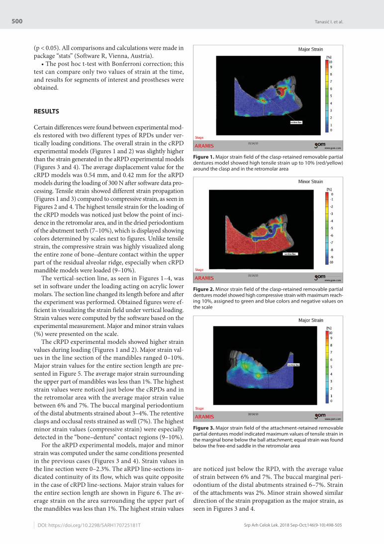
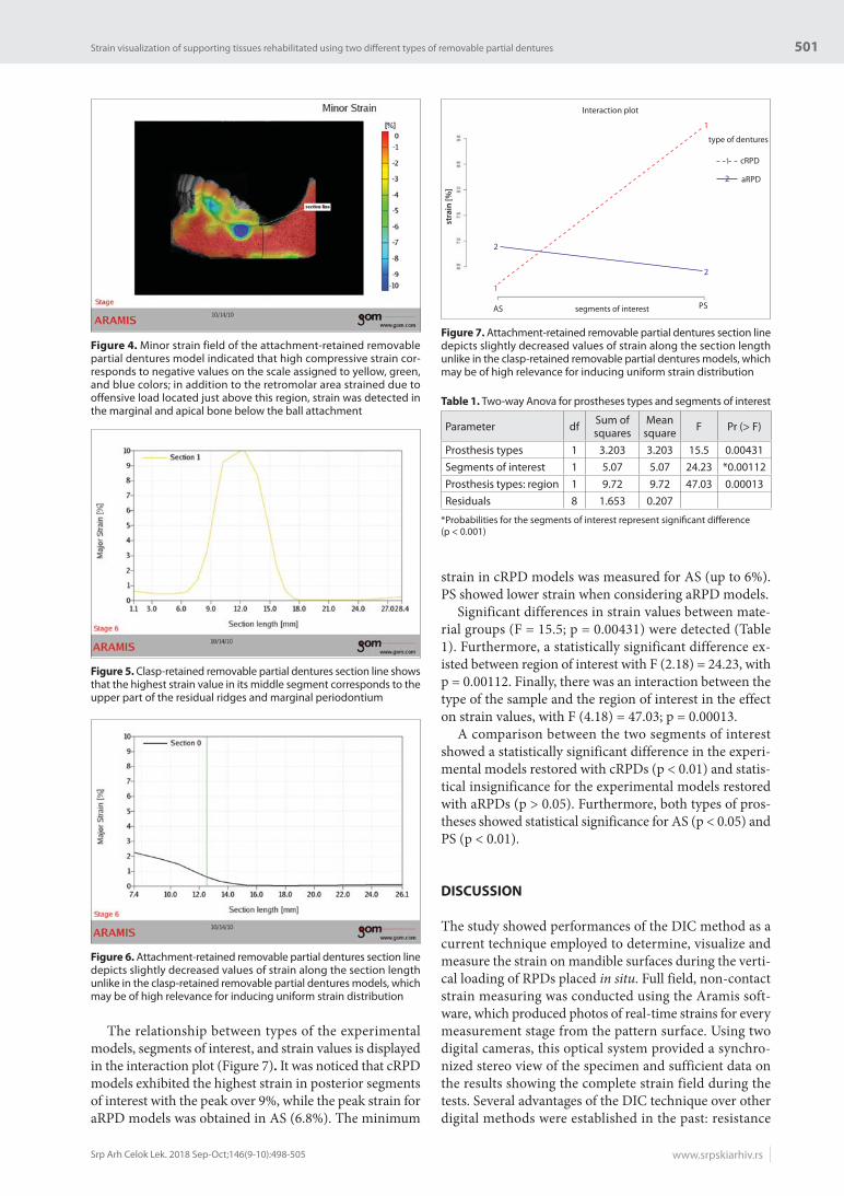
Certain differences were found between experimental mod-els restored with two different types of RPDs under ver-tically loading conditions. The overall strain in the cRPD experimental models (Figures 1 and 2) was slightly higher than the strain generated in the aRPD experimental models (Figures 3 and 4). The average displacement value for the cRPD models was 0.54 mm, and 0.42 mm for the aRPD models during the loading of 300 N after software data pro-cessing. Tensile strain showed different strain propagation (Figures 1 and 3) compared to compressive strain, as seen in Figures 2 and 4. The highest tensile strain for the loading of the cRPD models was noticed just below the point of inci-dence in the retromolar area, and in the dried periodontium of the abutment teeth (7–10%), which is displayed showing colors determined by scales next to figures. Unlike tensile strain, the compressive strain was highly visualized along the entire zone of bone–denture contact within the upper part of the residual alveolar ridge, especially when cRPD mandible models were loaded (9–10%).
The vertical-section line, as seen in Figures 1–4, was set in software under the loading acting on acrylic lower molars. The section line changed its length before and after the experiment was performed. Obtained figures were ef-ficient in visualizing the strain field under vertical loading. Strain values were computed by the software based on the experimental measurement. Major and minor strain values (%) were presented on the scale.
The cRPD experimental models showed higher strain values during loading (Figures 1 and 2). Major strain val-ues in the line section of the mandibles ranged 0–10%. Major strain values for the entire section length are pre-sented in Figure 5. The average major strain surrounding the upper part of mandibles was less than 1%. The highest strain values were noticed just below the cRPDs and in the retromolar area with the average major strain value between 6% and 7%. The buccal marginal periodontium of the distal abutments strained about 3–4%. The retentive clasps and occlusal rests strained as well (7%). The highest minor strain values (compressive strain) were especially detected in the “bone–denture” contact regions (9–10%).
For the aRPD experimental models, major and minor strain was computed under the same conditions presented in the previous cases (Figures 3 and 4). Strain values in the line section were 0–2.3%. The aRPD line-sections in-dicated continuity of its flow, which was quite opposite in the case of cRPD line-sections. Major strain values for the entire section length are shown in Figure 6. The av-erage strain on the area surrounding the upper part of the mandibles was less than 1%. The highest strain values
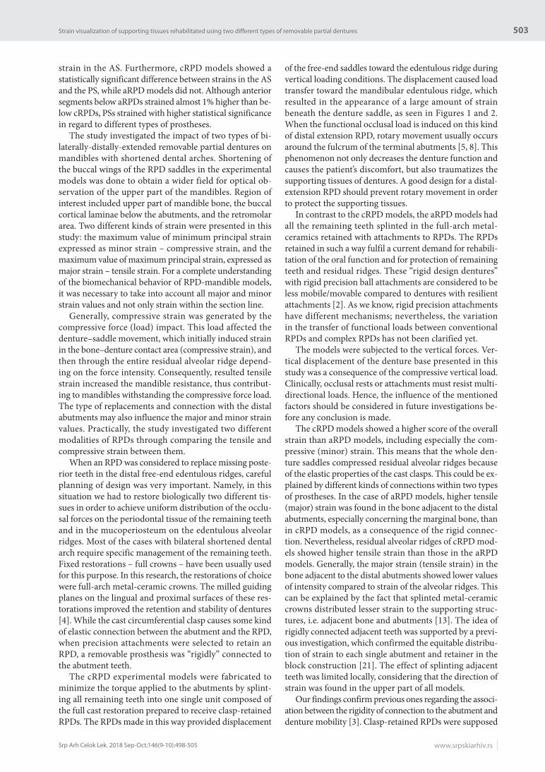
are noticed just below the RPD, with the average value of strain between 6% and 7%. The buccal marginal peri-odontium of the distal abutments strained 6–7%. Strain of the attachments was 2%. Minor strain showed similar direction of the strain propagation as the major strain, as seen in Figures 3 and 4.
Figure 1. Major strain field of the clasp-retained removable partial dentures model showed high tensile strain up to 10% (red/yellow) around the clasp and in the retromolar area
Figure 2. Minor strain field of the clasp-retained removable partial dentures model showed high compressive strain with maximum reach-ing 10%, assigned to green and blue colors and negative values on the scale
Figure 3. Major strain field of the attachment-retained removable partial dentures model indicated maximum values of tensile strain in the marginal bone below the ball attachment; equal strain was found below the free-end saddle in the retromolar area
Tanasić I. et al.
501
Srp Arh Celok Lek. 2018 Sep-Oct;146(9-10):498-505 www.srpskiarhiv.rs
The relationship between types of the experimental models, segments of interest, and strain values is displayed in the interaction plot (Figure 7). It was noticed that cRPD models exhibited the highest strain in posterior segments of interest with the peak over 9%, while the peak strain for aRPD models was obtained in AS (6.8%). The minimum
strain in cRPD models was measured for AS (up to 6%). PS showed lower strain when considering aRPD models.
Significant differences in strain values between mate-rial groups (F = 15.5; p = 0.00431) were detected (Table 1). Furthermore, a statistically significant difference ex-isted between region of interest with F (2.18) = 24.23, with p = 0.00112. Finally, there was an interaction between the type of the sample and the region of interest in the effect on strain values, with F (4.18) = 47.03; p = 0.00013.
A comparison between the two segments of interest showed a statistically significant difference in the experi-mental models restored with cRPDs (p < 0.01) and statis-tical insignificance for the experimental models restored with aRPDs (p > 0.05). Furthermore, both types of pros-theses showed statistical significance for AS (p < 0.05) and PS (p < 0.01).
DISCUSSIO N
The study showed performances of the DIC method as a current technique employed to determine, visualize and measure the strain on mandible surfaces during the verti-cal loading of RPDs placed in situ. Full field, non-contact strain measuring was conducted using the Aramis soft-ware, which produced photos of real-time strains for every measurement stage from the pattern surface. Using two digital cameras, this optical system provided a synchro-nized stereo view of the specimen and sufficient data on the results showing the complete strain field during the tests. Several advantages of the DIC technique over other digital methods were established in the past: resistance
Figure 4. Minor strain field of the attachment-retained removable partial dentures model indicated that high compressive strain cor-responds to negative values on the scale assigned to yellow, green, and blue colors; in addition to the retromolar area strained due to offensive load located just above this region, strain was detected in the marginal and apical bone below the ball attachment
Figure 5. Clasp-retained removable partial dentures section line shows that the highest strain value in its middle segment corresponds to the upper part of the residual ridges and marginal periodontium
Figure 6. Attachment-retained removable partial dentures section line depicts slightly decreased values of strain along the section length unlike in the clasp-retained removable partial dentures models, which may be of high relevance for inducing uniform strain distribution
Figure 7. Attachment-retained removable partial dentures section line depicts slightly decreased values of strain along the section length unlike in the clasp-retained removable partial dentures models, which may be of high relevance for inducing uniform strain distribution
2
2
1
1
type of dentures
cRPD
aRPD
1
2
Interaction plot
segments of interestAS PS
stra
in [
%]
Table 1. Two-way Anova for prostheses types and segments of interest
Parameter df Sum of squares
Mean square F Pr (> F)
Prosthesis types 1 3.203 3.203 15.5 0.00431Segments of interest 1 5.07 5.07 24.23 *0.00112Prosthesis types: region 1 9.72 9.72 47.03 0.00013Residuals 8 1.653 0.207
*Probabilities for the segments of interest represent signifi cant diff erence (p < 0.001)
Strain visualization of supporting tissues rehabilitated using two diff erent types of removable partial dentures

502
Srp Arh Celok Lek. 2018 Sep-Oct;146(9-10):498-505
on the displacement of the observed model during the measurement process and full field of strain measurement, low sensibility to ambient vibrations, ability to register rigid body motion and to measure 3D displacements in a high dynamic range (microns to millimeters) of measur-ing capacity, and high reproducibility of the DIC measur-ing [10–14]. In dental biomechanics, DIC is often utilized for in vitro setups [11, 12, 13]. Whether it concerns the biomechanical behavior of the human jaw under static or dynamic load, biomechanical testing of biomaterials, or photogrammetric measurements of initial tooth displace-ment under tensile force, the DIC has been confirmed as a method especially suitable for 3D-strain measurements of dental materials and structures with complex geometry due to the ability to catch non-linear surface strain in the tested specimens [9, 11–18].
The study was conducted as a static, non-impact, in vitro loading of the experimental models with different designs of dentures positioned in situ. Two types of replacements were compared and the better one was determined with respect to biomechanics. Knowing of the biomechanical behaviour of hard tissues (bone and teeth) and their inter-action with replacements is important for the investigation of biomaterials and designs of replacements, so this type of research can improve prognosis and treatment planning in partially edentulous subjects. The researchers used ca-daveric mandibles without soft tissue coverage. This fact distinguishes the donor-related variability of the examined bone features as the key factor when performing the DIC experimental analysis. The absence of the elevator muscles and soft tissue as supportive structures, and thus the fixa-tion of mandibles in contrast to the real (physiological) conditions, was another exclusion criteria addressed to the disadvantages of this study [20]. Nevertheless, this study investigated the upper part of the mandibles adjacent to prostheses; therefore, from the biomechanical standpoint, the results are adequate for arguing about the biomechan-ical behaviour of usually indicated RPDs. The study de-scribes preparing all remaining teeth and restoring them with splinted porcelain fused to metal restorations. This was expensive, technically difficult, and required radical amounts of tooth structure removal. Nevertheless, we were guided by the fact that high percentage of partially edentu-lous subjects indicates signs and symptoms of periodontal disease and tooth wear of the tooth structure; thus, restora-tion of such teeth was considered an imperative. Addition-ally, treatment of the remaining teeth was done to achieve similar loading conditions of the supporting dental struc-ture, for both types of RPD-restored experimental models, as much as possible. Following this criterion, experimental models restored with aRPDs included ball rather than slide attachment. Although both types of attachments, whether ball or slide, are indicated for rehabilitation of the Ken-nedy Class 1 partial edentulism, dimensions of the clinical crowns and length of the residual ridges/free-end saddles were the critical factors to opt for the ball attachments as more preferable.
In this experiment, results acquired from the Aramis system were sorted into two groups of experimental models
and two groups of interest locations (segments). Dentures, as a part of the experimental models and locations of in-terest within the tested models, presented two factors that caused different values of strains of the loaded models. Their mutual effect on experimental models was presented in the interaction plot where the connection between ex-perimental results was visualized.
Strains for different types of experimental models and different segments of interest were compared using two-way ANOVA. Two-way ANOVA was employed to deter-mine whether there were statistically significant differ-ences between the tested experimental groups. Prosthesis type and location of interest represented factors of influ-ence. The strain was considered the dependent variable. Both factors such as prosthesis type and location of interest showed significant influence. Significant differences in the strain values existed between two groups of prostheses for both segments of interest (p < 0.05, p < 0.01; Table 2), as well as in two different locations of measured surface, but only for cRPD models (p < 0.001; Table 3). Although ANOVA revealed statistically significant differences be-tween the type of the strained models, location of interest, and interaction of these two factors, this analysis could not determine between which groups of models and loca-tions of interest these differences actually existed. Thus, additional post hoc t-test was introduced to reveal statis-tical significance between observed variables and to find out where these differences actually occurred. In order to provide a more valid comparison and to reduce type I error, the conservative Bonferroni correction was applied. Therefore, all three null hypotheses were rejected, and al-ternative ones were adopted, which state that strain was dependent from the prostheses used and from the loca-tions within the region of interest. In addition, there was an interaction between prostheses and segments of interest in their effect on the strain values.
Although strain varied significantly between locations of interest, dentures’ effect was also noticed. Namely, mod-els with cRPDs showed highest strains for posterior loca-tions of interest (PS) while loaded models restored with aRPDs induced the highest strain in the anterior locations of interest (AS). The cRPD models displayed the lowest
Table 2. Comparison between prostheses types for different segments of interests (post hoc)
Segments cRPD aRPD p-value BonferroniAS 6.13 (0.21) 6.9 (0.4) < 0.05 0.042PS 9.23 (0.7) 6.4 (0.36) < 0.01 0.0034
AS – anterior segment; PS – posterior segment; cRPD – clasp-retained removable partial dentures; aRPD – attachment-retained removable partial dentures
Table 3. Comparison between segments of interest for different prostheses types (post hoc)
Prostheses AS PS p-value BonferronicRPD 6.13 (0.21) 9.23 (0.7) < 0.01 0.0018aRPD 6.9 (0.4) 6.4 (0.36) > 0.05 0.18
AS – anterior segment; PS – posterior segment; cRPD – clasp-retained removable partial dentures; aRPD – attachment-retained removable partial dentures
Tanasić I. et al.
DOI: https://doi.org/10.2298/SARH170725181T
503
Srp Arh Celok Lek. 2018 Sep-Oct;146(9-10):498-505 www.srpskiarhiv.rs
strain in the AS. Furthermore, cRPD models showed a statistically significant difference between strains in the AS and the PS, while aRPD models did not. Although anterior segments below aRPDs strained almost 1% higher than be-low cRPDs, PSs strained with higher statistical significance in regard to different types of prostheses.
The study investigated the impact of two types of bi-laterally-distally-extended removable partial dentures on mandibles with shortened dental arches. Shortening of the buccal wings of the RPD saddles in the experimental models was done to obtain a wider field for optical ob-servation of the upper part of the mandibles. Region of interest included upper part of mandible bone, the buccal cortical laminae below the abutments, and the retromolar area. Two different kinds of strain were presented in this study: the maximum value of minimum principal strain expressed as minor strain – compressive strain, and the maximum value of maximum principal strain, expressed as major strain – tensile strain. For a complete understanding of the biomechanical behavior of RPD-mandible models, it was necessary to take into account all major and minor strain values and not only strain within the section line.
Generally, compressive strain was generated by the compressive force (load) impact. This load affected the denture–saddle movement, which initially induced strain in the bone–denture contact area (compressive strain), and then through the entire residual alveolar ridge depend-ing on the force intensity. Consequently, resulted tensile strain increased the mandible resistance, thus contribut-ing to mandibles withstanding the compressive force load. The type of replacements and connection with the distal abutments may also influence the major and minor strain values. Practically, the study investigated two different modalities of RPDs through comparing the tensile and compressive strain between them.
When an RPD was considered to replace missing poste-rior teeth in the distal free-end edentulous ridges, careful planning of design was very important. Namely, in this situation we had to restore biologically two different tis-sues in order to achieve uniform distribution of the occlu-sal forces on the periodontal tissue of the remaining teeth and in the mucoperiosteum on the edentulous alveolar ridges. Most of the cases with bilateral shortened dental arch require specific management of the remaining teeth. Fixed restorations – full crowns – have been usually used for this purpose. In this research, the restorations of choice were full-arch metal-ceramic crowns. The milled guiding planes on the lingual and proximal surfaces of these res-torations improved the retention and stability of dentures [4]. While the cast circumferential clasp causes some kind of elastic connection between the abutment and the RPD, when precision attachments were selected to retain an RPD, a removable prosthesis was “rigidly” connected to the abutment teeth.
The cRPD experimental models were fabricated to minimize the torque applied to the abutments by splint-ing all remaining teeth into one single unit composed of the full cast restoration prepared to receive clasp-retained RPDs. The RPDs made in this way provided displacement
of the free-end saddles toward the edentulous ridge during vertical loading conditions. The displacement caused load transfer toward the mandibular edentulous ridge, which resulted in the appearance of a large amount of strain beneath the denture saddle, as seen in Figures 1 and 2. When the functional occlusal load is induced on this kind of distal extension RPD, rotary movement usually occurs around the fulcrum of the terminal abutments [5, 8]. This phenomenon not only decreases the denture function and causes the patient’s discomfort, but also traumatizes the supporting tissues of dentures. A good design for a distal-extension RPD should prevent rotary movement in order to protect the supporting tissues.
In contrast to the cRPD models, the aRPD models had all the remaining teeth splinted in the full-arch metal-ceramics retained with attachments to RPDs. The RPDs retained in such a way fulfil a current demand for rehabili-tation of the oral function and for protection of remaining teeth and residual ridges. These “rigid design dentures” with rigid precision ball attachments are considered to be less mobile/movable compared to dentures with resilient attachments [2]. As we know, rigid precision attachments have different mechanisms; nevertheless, the variation in the transfer of functional loads between conventional RPDs and complex RPDs has not been clarified yet.
The models were subjected to the vertical forces. Ver-tical displacement of the denture base presented in this study was a consequence of the compressive vertical load. Clinically, occlusal rests or attachments must resist multi-directional loads. Hence, the influence of the mentioned factors should be considered in future investigations be-fore any conclusion is made.
The cRPD models showed a higher score of the overall strain than aRPD models, including especially the com-pressive (minor) strain. This means that the whole den-ture saddles compressed residual alveolar ridges because of the elastic properties of the cast clasps. This could be ex-plained by different kinds of connections within two types of prostheses. In the case of aRPD models, higher tensile (major) strain was found in the bone adjacent to the distal abutments, especially concerning the marginal bone, than in cRPD models, as a consequence of the rigid connec-tion. Nevertheless, residual alveolar ridges of cRPD mod-els showed higher tensile strain than those in the aRPD models. Generally, the major strain (tensile strain) in the bone adjacent to the distal abutments showed lower values of intensity compared to strain of the alveolar ridges. This can be explained by the fact that splinted metal-ceramic crowns distributed lesser strain to the supporting struc-tures, i.e. adjacent bone and abutments [13]. The idea of rigidly connected adjacent teeth was supported by a previ-ous investigation, which confirmed the equitable distribu-tion of strain to each single abutment and retainer in the block construction [21]. The effect of splinting adjacent teeth was limited locally, considering that the direction of strain was found in the upper part of all models.
Our findings confirm previous ones regarding the associ-ation between the rigidity of connection to the abutment and denture mobility [3]. Clasp-retained RPDs were supposed
Strain visualization of supporting tissues rehabilitated using two diff erent types of removable partial dentures
504
Srp Arh Celok Lek. 2018 Sep-Oct;146(9-10):498-505
to be more elastic than attachment RPDs, and therefore higher mobility of cRPDs was observed. Thus, higher rate of strain can be expected beneath cRPDs. In contrast, the flexibility of the attachment was lower and needed less amount of bone tissue support under the denture base.
Attachment RPDs may not be suitable therapy solutions in cases of periodontally weakened abutment teeth due to instability and therapeutic failure. These situations request splinting of periodontally compromised teeth into single unit followed by adequately designed and adjusted RPDs with consideration of the denture extension and the level of periodontal damage [12, 21].
CONCLUSION
Visualizing the biomechanical behaviour of RPDs placed in situ on supporting dental tissues can improve the design of RPDs and preserve abutment teeth and bone. This will avoid possible failures in current dental practice. Within limitations and based on the results of this study, it can be said that higher strain was observed below the clasp RPDs, particularly if we consider movement of the distal portion of the free-end saddles caused by the teeth and dentures’ vertical displacement. The findings proved that attachment RPDs generated less strain in the residual alve-olar ridges, and thus, from the biomechanical standpoint, can be considered a better choice for the rehabilitation
of the Kennedy Class 1 partial edentulism compared to clasp RPDs. However, high strain was found in the bone adjacent to distal abutments. In accordance with the tasks provided by null hypothesis, the following conclusions were derived:
1. The mean strain was significantly different for all models, when its distribution and values are consid-ered. This fact could be the reason for differences that exist between two types of RPDs with different types of connections with the adjacent teeth.
2. The mean strain values showed significant differenc-es between mandibular AS and PS of cRPD models. However, the mean strain in AS and PS was similar in aRPD models probably due to the fact that aRPDs generated uniform strain distribution in mandibles compared to cRPDs.
3. The findings provide a noticeable difference in the ef-fect induced by interactions between prostheses and segments of interest due to incremental movements of two types of RPDs toward the residual ridges.
ACKNOWLEDGMENT
The authors would like to thank the Faculty of Mechanical Engineering of the University of Belgrade, Serbia, and the Institute for Anthropology, Faculty of Medicine, University of Belgrade, Serbia.
REFERENCES
1. Wright EW. Prosthetic management of the periodontally compromised dentition. J Calif Dent Assoc. 1989; 17:56–60.
2. Saito M, Miura Y, Notani K, Kawasaki T. Stress distribution of abutments and base displacement with precision attachment- and telescopic crown-retained removable partial dentures. J Oral Rehabil. 2003; 30(5):482–7.
3. Igarashi Y, Ogata A, Kuroiwa A, Wang CH. Stress distribution and abutment tooth mobility of distal-extension removable partial dentures with different retainers: an in vivo study. J Oral Rehabil. 1999; 26(2):111–6.
4. Chaiyabutr Y, Brudvik JS. Removable partial denture design using milled abutment surfaces and minimal soft tissue coverage for periodontally compromised teeth: A clinical report. J Prosthet Dent. 2008; 99(4):263–6.
5. Jin X, Sato M, Nishiyama A, Ohyama T. Influence of loading position of mandibular Unilateral distal extension, Removable Partial Dentures on movements of abutments tooth and denture base. J Med Dent Sci. 2004; 51(3):155–63.
6. Kibi M, Ono T, Dong J, Mitta K, Gonda T, Maeda Y. Development of an RPD CAD system with finite element stress analysis. J Oral Rehabil. 2009; 36(6):442–50.
7. Eto M, Wakabayashi N, Ohyama T. Finite Element analysis of deflections in major connectors for maxillary RPDs. Int J Prosthodont. 2002; 15(5):433–8.
8. Aras K, Hasanreisog˘lu U, Shinogaya T. Masticatory performance, maximum occlusal force, and occlusal contact area in patients with bilaterally missing molars and distal extension removable partial dentures. Int J Prosthodont. 2009; 22(2):204–9.
9. Tanasić I, Tihaček-Šojić Lj, Milić-Lemić A. Optical metrology analysis of the lower jaw deformations. Vojnosanitet pregl. 2011; 68(4):336–40.
10. Akca K, Cehreli MC. A photoelastic and strain-gauge analysis of interface force transmission of internal-cone implants. Int J Periodont Res Dent. 2008; 28(4):391–9.
11. Tanasić I, Milic-Lemic A, Tihacek-Sojic Lj, Stancic I, Mitrovic N. Analysis of the compressive strain below the removable and fixed prosthesis
in the posterior mandible using a digital image correlation method. Biomech Model Mechanobiol. 2012; 11(6):751–8.
12. Tihacek Sojic Lj, Milic Lemic A, Tanasic I, Mitrovic N, Milosevic M. Compressive strains and displacement in a partially dentate lower jaw rehabilitated with two different treatment modalities. Gerodontology. 2012; 29(2):e851–7.
13. Tiossi R, Lin L, Rodrigues RCS, Heo YC, Conrad HJ, de Mattos Mda G, et al. Digital image correlation analysis of the load transfer by implant-supported restorations. J Biomech. 2011; 44(6):1008–13.
14. Schmidt T, Tyson J, Galanulis K. Full-field dynamic displacement and strain measurement using advanced 3D image correlation photogrammetry: Part 1. Experimental Technic. 2003; 27:47–50.
15. Göllner M, Holst A, Berthold C, Schmitt J, Wichmann M, Holst S. Noncontact intraoral measurement of force-related tooth mobility. Clin Oral Investig. 2010; 14(5):551–7.
16. Sztefek P, Vanleene M, Olsson R, Collinson R, Pitsillides AA, Shefelbine S. Using digital image correlation to determine bone surface strains during loading and after adaptation of the mouse tibia. J Biomech. 2010; 43(4):599–605.
17. Tanasić I, Tihaček-Šojić Lj, Mitrović M, Milić-Lemić A, Vukadinović M, Marković A. An attempt to create a standardized (reference) model for experimental investigations on implant’s sample. Measurement. 2015; 72:37–42.
18. DuChesne A, Unnewehr M, Schmidt PF, Sotonyi P, Brinkmann B, Piffko J, et al. Deformation characteristics of the human mandible in low impact experiments. Int J Leg Med. 2003; 117(5):257–62.
19. Muller F, Heath MR, Ott R. Maximum bite force after the replacement of complete dentures. Gerodontology. 2001; 18(1):58–62.
20. Vollmer D, Meyer U, Joos U, Vegh A, Piffko J. Experimental and finite element study of a human mandible. J Cranio-Maxillofac Surg. 2000; 28:91–6.
21. Yamashita J, Wang Q, Dechow PC. Biomechanical effects of fixed partial denture therapy on strain patterns of the mandible. J Prosthet Dent. 2006; 95(1):55–62.
Tanasić I. et al.
DOI: https://doi.org/10.2298/SARH170725181T
505
Srp Arh Celok Lek. 2018 Sep-Oct;146(9-10):498-505 www.srpskiarhiv.rs
САЖЕТАКУвод/Циљ Савремене биомеханичке анализе омогућавају комплетну визуелизацију деформација од оптерећивања различитих зубних надокнада у протетској рехабилитацији. Циљ ове студије је био да анализа дистрибуције дефор-мација унутар потпорних ткива испод два различита типа најчешће коришћених парцијалних скелетираних протеза.Методе In vitro студија је обухватила две групе експеримен-талних модела доњих вилица (Кенеди 1 класа крезубости) и два типа парцијалних скелетираних протеза. Модели су били изложени оклузалним силама, а за приказ и мерење дефор-мација је коришћена метода дигиталне корелације слика. Резултати Највећа деформација је измерена испод пар-цијалних протеза, на површинама кости која окружује дис-
талне зубе носаче и у ретромоларној регији. Вредности деформација у експерименталним моделима са протезама ретинираним ливеним кукицама су биле 0–10%. Вредности деформација у експерименталним моделима са протезама ретинираним атечменима су биле 0–2,3%. Закључак Парцијалне скелетиране протезе са атечменима индуковале су мање деформације резидуалног алвеоларног гребена. Веће деформације су уочене у маргиналној кости која је у непосредном контакту са зубима носачима.
Кључне речи: крезубе доње вилице; метода дигиталне ко-релације слика; парцијална скелетирана протеза; дефор-мација кости
Визуелизација деформација у потпорним ткивима рехабилитованим са два различита типа парцијалних скелетираних протезаИван Танасић1, Љиљана Тихачек-Шојић2, Александра Милић-Лемић2
1Дом здравља Обреновац, Београд, Србија;2Универзитет у Београду, Стоматолошки факултет, Катедра за стоматолошку протетику, Београд, Србија
Strain visualization of supporting tissues rehabilitated using two diff erent types of removable partial dentures
506
DOI: https://doi.org/10.2298/SARH170718195D
UDC: 614.2(497.11)
Received • Примљено: July 18, 2017
Ревизија • Revised: October 27, 2017
Accepted • Прихваћено: October 31, 2017
Online fi rst: November 10, 2017
Correspondence to:
Vesna DAMNJANOVIĆ
University of Belgrade
Faculty of Organizational Sciences
Jove Ilića 154
11000 Belgrade, Serbia
ORIGINAL ARTICLE / ОРИГИНАЛНИ РАД
Factors affecting patient satisfaction in the health care sector in SerbiaVesna Damnjanović1, Radmila Janičić1, Vesna Jovanović2
1University of Belgrade, Faculty of Organizational Sciences, Belgrade, Serbia;2Banjica Institute of Orthopaedic Surgery, Belgrade, Serbia
SUMMARYIntroduction/Objective The aim of this paper was to highlight and understand factors that influence the quality of healthcare services in Serbia in private and public health institutions.Methods The data was collected during May of 2017 and June of 2017 through an on-field questionnaire. Out of 500 questionnaires in total, 406 were completed and returned, resulting in a response rate of 81.2%.Results The following four most influential factors for patient satisfaction in Serbia’s healthcare sector were identified: admission process, doctor care, staff care, and technology tools. Conclusion The model describes that 66.2 variance for the doctor care variable is based on three con-structs: admission process, technology tools, and staff care. The hypothesis that technology tools will have a positive effect on staff care was not confirmed.Keywords: patient satisfaction, factors; health care; Serbia
INTRODUCTION
Patient satisfaction is a way to measure the over-all quality of delivered healthcare services. Un-derstanding patients as clients and taking care of their needs is crucial for improvement of the healthcare sector in Serbia. A research study by Savić and Jakovljević [1] also confirms that a pa-tient perspective is important for clinical deci-sion making in Serbia. The main responsibility for defining and executing the patient satisfac-tion strategy is borne by health care managers. They constantly receive patient feedback about all aspects of health care, which affects customer retention and are in the position to adapt to dy-namic market conditions. The healthcare sec-tor in Serbia is comprised of the public and the private system for treating patients. The health system of Serbia employs some 130,000 work-ers. The largest number is employed in health institutions, primarily in the 70 state hospitals. There are approximately 1,200 private medical entities in Serbia, out of which 60 are hospitals. They employ over 3,700 doctors, accounting for about 10% of the total number of doctors in the health sector in Serbia [2]. In the past 30 years, the health system in Serbia has changed substantially. After the breakup of Yugoslavia in the 90s, all the weaknesses and strengths of the healthcare system of that time have become more visible. Knowledge of factors that influ-ence patient satisfaction is of great importance for the system. The country has entered the pe-riod of transition, and the creators of healthcare policies have been forced to start reforming the healthcare system by addressing structural, hu-man resources, financing, and organizational
issues [3]. In a recent research study, health pol-icy, socioeconomic transition, trends in health-care resources, and outcomes were observed among three historical health-policy legacies in Southeastern Europe. Significant differences exist between Serbia as the representative of for-mer Yugoslav countries, post-Semashko coun-tries, and free market SEE economies [4].
In order to improve the healthcare system in Serbia, it is necessary to understand the opin-ion of patients as clients of healthcare services. However, previous research in this field has identified different factors for patient dissat-isfaction. Authors from Brazil highlighted the main weaknesses as follows: lack of qualified professionals for exercising management ac-tivities, delay in implementing new informa-tion technologies and management and work organizational process deficiencies. The main reasons for dissatisfaction with the healthcare system in Serbia are similar to Brazil – unequal delivery of quality in different health services, waiting time for certain medical procedures and interventions, inefficient use of health technologies, and dissatisfaction of the health-care system staff [5, 6]. Scholars point out that knowledge-based resource allocation still has to make roots in health policy traditions of BRICS and other emerging nations [7].
A recent study from Balkan countries in-cluding Macedonia, Bulgaria, and Serbia identified the top three indicators of patient satisfaction: trust, attention of doctors, and perceived outcome of the treatment. Long wait-ing time, huge administrative procedures and patient privacy protection are also issues for concern in all three countries [8].

507
Srp Arh Celok Lek. 2018 Sep-Oct;146(9-10):506-511 www.srpskiarhiv.rs
Patient quality perceptions have been shown to account for 17–27% of variation in a hospital’s financial measures such as earnings, net revenue, and asset returns [9]. It is one of the most effective key performance indicators if healthcare institutions want to evaluate business success on the market. Today it is important to recognize the role of patient-centered care. According to Irwin and Rich-ardson [10], patient-focused care can be thought of as a merging of patient education, self-care, and evidence-based models of medical practice: communication with patients, partnerships, health promotion, and physical care (medications and treatments). The main goal of each health care institution is to recognize the factors that will improve patient-centered care.
Various research studies, which investigated factors that affect patient satisfaction, were analyzed.
The first literature stream relevant for this study is general literature on patient satisfaction. Naidu [11] con-ducted a systematic review of factors determining health care quality and patient satisfaction with 24 articles from international journals. A comprehensive model was made to better understand healthcare services. Healthcare ser-vices are difficult to evaluate: some authors feel patient perceptions are valuable healthcare quality indicators, oth-ers contend that health service quality should be evaluated by experts. The SERVQUAL instrument is used in many patient satisfaction studies. Dimensions that determine patient satisfaction have been identified, including: reli-ability – the ability to perform the promised service, de-pendably and accurately; assurance – employee knowledge and courtesy and their ability to convey trust and con-fidence; responsiveness – willingness to help customers and provide prompt services; empathy – caring, individu-alized attention the firm provides to its customers; and tangibles – physical facilities, equipment and appearance of the personnel. Another study written by Batbaatar et al. [12] reviewed studies of patient satisfaction between 1980 and 2014. Socio-demographic and personal-related characteristics were analyzed in the review.
The second type of study investigated the use of tech-nology in health care and its effect on patient satisfaction. A study from Bangladesh implies medical treatment of the hospital, service of the hospital staff, hospital facilities, and technology are factors that affect patient satisfaction [13]. The population aging trends in the Next-11 nations have led to increased health care expenditures [14]. On the other hand, the millennial generation for health provid-ers is crucial for understanding what content needs to be available at patient portals on mobile devices.
However, a recent study concerning health service in Serbia suggested the most common determinants of citi-zen satisfaction with health care are age, health condition, income, type of service (state or private sector), communi-cation, politeness of staff, and the overall hospital environ-ment [15]. Student satisfaction with the quality of service provided by student polyclinics showed that personal re-lationships had the most tangible impact on student sat-isfaction while promptness of service was also important [16]. Additionally, health managers from Serbia focus their
efforts on ensuring the competence of employees while managers from health care organizations from Slovenia are more external-oriented [17]. The analysis of previous literature revealed the need to develop a model for iden-tifying the most important factors for patient satisfaction in Serbia and their relationships.
The aim of this study is to determine factors that influ-ence patient satisfaction, as it is an indicator of quality health care in Serbia [18]. The findings should encour-age a shift in the attitude and relations of hospital staff with patients towards a more client/consumer-oriented healthcare service. The main reason is that better customer satisfaction leads to better customer loyalty for healthcare institutions.
METHODS
The research framework
The research framework of this study was based on the study by Otani et al. [19], which includes five elements for defining the initial conceptual model: admission pro-cess, doctor care, staff care, food, and room in the hospital. This study’s scope did not include food and room as the research aimed to evaluate patient perceptions before they have stayed in healthcare institutions. A research study of various large hospitals in the USA investigated the rela-tionship of doctor care and doctor environment to overall patient satisfaction. The results showed that all attributes were statistically significant and positively related to over-all satisfaction [20].
Several studies have already proved that courtesy and efficiency of admission processes in health provider insti-tutions are significant for patient satisfaction and waiting times [21, 22, 23]. The admission processes in this study consist of three elements: promptness or efficiency of the admission or registration, courtesy and helpfulness of the admission or registration, and waiting time for medical treatment.
Staff care is another important factor for determina-tion of patient satisfaction. It should be evaluated from two sides. Firstly, staff care is about the willingness to help patients if they have questions or concerns. Secondly, staff care is about providing clear and complete explanations about how patients should practice self-care at home. Again, the emphasis is about clear communication with patients.
In the health service sector, it is crucial to ensure avail-ability of doctors when patients need them [24]. Com-munication with a doctor usually develops trust with the patient and promotes patients’ desire to understand health treatment.
There are also some differences regarding patient sat-isfaction with healthcare providers based on age. Taking a look at the health habits of the millennial, baby-boomer, and X generations, health institutions can better under-stand how to provide personal relationships and integrate health IT tools into the care process to create the best
Factors aff ecting patient satisfaction in the health care sector in Serbia

508
Srp Arh Celok Lek. 2018 Sep-Oct;146(9-10):506-511DOI: https://doi.org/10.2298/SARH170718195D
patient outcomes. A large portion of this study’s sample were millennial patients (51%) and the main results from previous studies refer that they prefer a strong doctor con-nection, adequate time for discussion, and verbal commu-nication of recommendations. Younger generations abroad have unique preferences when they discuss health tech-nology (tele-health, mobile health applications). This is supported by a previous research study from Bangladesh, according to which technology in health care is one of the most important factors that affects patient satisfaction. Three items can describe construct technology including a connection with admission processes (healthcare insti-tution’s ability to provide online admission processes), a connection with doctors (healthcare institution’s ability to provide online doctor advice), and tracking health con-ditions (the possibility to track health conditions using mobile applications), see Table 1.
The set of relationships is given in a form of hypotheses that our model is testing (Figure 1):
H1: The quality of the admission process will have a positive effect on doctor care;
H2: The technology tools will have a positive effect on the admission process;
H3: The staff care will have a positive effect on doctor care;
H4: The technology tools will have a positive effect on doctor care;
H5: The technology tools will have a positive effect on staff care.
Data collection and sample size
Data were collected with a structured on-field ques-tionnaire from different hospitals located in Serbia. We covered all regions in Serbia including Vojvodina (102), Belgrade (126), Šumadija and Western Serbia (94), and Southern and Eastern Serbia (84). The data was collected during May of 2017 and June of 2017. Out of 500 ques-tionnaires in total, 406 were completed and returned, re-sulting in a response rate of 81.2%. Distribution of the background characteristics of the patients are regarding gender – 45.32% (184) were males, while 54.67% (222) were females. The majority of patients were young. The
age groups of 18–29 years and 30–39 years comprised 51.23% (208) and 31.53% (128) of the total sample sur-veyed, respectively. Those 50 years old and above consti-tuted only 17.24% (70) of the total sample. The responses for the patient satisfaction indicators were presented over the five-point Likert scale, ranging from highly unsatisfied to highly satisfied. After expiration of the surveying period and acquisition of the satisfactory number of completed surveys, the results were coded and entered into the IBM SPSS Statistics Version 22.0 (IBM Corp., Armonk, NY, USA). Following good practice, prior to data analysis, er-ror screening and data cleaning were undertaken. After ensuring that there are no missing values or values that fall outside of defined ranges, data analysis proceeded. The following statistical tools were used: descriptive statistics (means, frequencies) for capturing average values on the examined issues, and factor analysis for analyzing patient perception of health care factors in choosing a healthcare institution.
Data analysis
The model was constructed and analyzed using the Smart-PLS 3 (SmartPLS GmbH, Bönningstedt, Germany). Smart-PLS 3 supports work with covariance-based structural equation models, and is particularly useful when work-ing with small samples such as the sample evident in this study (n = 406). PLS analysis is a two-stage process [25].
Table 1. Factors of the initial conceptual model
Construct Construct type Items (given on a 1 to 5 Likert scale) Variable name
Admission process ReflectivePromptness of the efficiency of the admission or registration AP1Courtesy and helpfulness of the admission or registration AP2Waiting time for medical treatment is short AP3
Doctor (physician) care Reflective
Availability of your doctor when needed DC1Doctor ability to communicate with you DC2Doctor ability to provide adequate instructions or explanation of your treatment or test DC3
Staff care ReflectiveStaff willingness to help if you have question or concern SC1Clear and complete explanation provided by the staff on how to care about yourself at home SC2
Technology tools ReflectiveHealthcare institution ability to provide on line admission process TT1Health care institution ability to provide online doctor advice TT2Possibility to track health condition using mobile application TT3
Figure 1. Initial theoretical research model
Damnjanović D. et al.
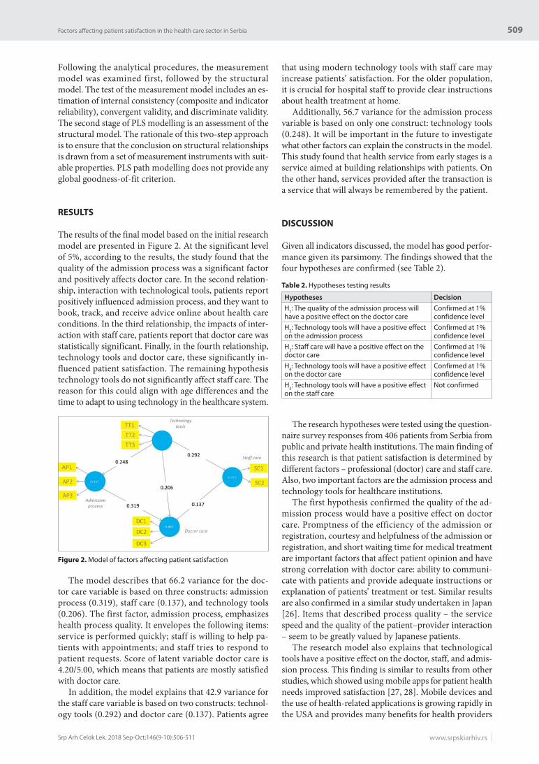
509
Srp Arh Celok Lek. 2018 Sep-Oct;146(9-10):506-511 www.srpskiarhiv.rs
Following the analytical procedures, the measurement model was examined first, followed by the structural model. The test of the measurement model includes an es-timation of internal consistency (composite and indicator reliability), convergent validity, and discriminate validity. The second stage of PLS modelling is an assessment of the structural model. The rationale of this two-step approach is to ensure that the conclusion on structural relationships is drawn from a set of measurement instruments with suit-able properties. PLS path modelling does not provide any global goodness-of-fit criterion.
RESULTS
The results of the final model based on the initial research model are presented in Figure 2. At the significant level of 5%, according to the results, the study found that the quality of the admission process was a significant factor and positively affects doctor care. In the second relation-ship, interaction with technological tools, patients report positively influenced admission process, and they want to book, track, and receive advice online about health care conditions. In the third relationship, the impacts of inter-action with staff care, patients report that doctor care was statistically significant. Finally, in the fourth relationship, technology tools and doctor care, these significantly in-fluenced patient satisfaction. The remaining hypothesis technology tools do not significantly affect staff care. The reason for this could align with age differences and the time to adapt to using technology in the healthcare system.
The model describes that 66.2 variance for the doc-tor care variable is based on three constructs: admission process (0.319), staff care (0.137), and technology tools (0.206). The first factor, admission process, emphasizes health process quality. It envelopes the following items: service is performed quickly; staff is willing to help pa-tients with appointments; and staff tries to respond to patient requests. Score of latent variable doctor care is 4.20/5.00, which means that patients are mostly satisfied with doctor care.
In addition, the model explains that 42.9 variance for the staff care variable is based on two constructs: technol-ogy tools (0.292) and doctor care (0.137). Patients agree
that using modern technology tools with staff care may increase patients’ satisfaction. For the older population, it is crucial for hospital staff to provide clear instructions about health treatment at home.
Additionally, 56.7 variance for the admission process variable is based on only one construct: technology tools (0.248). It will be important in the future to investigate what other factors can explain the constructs in the model. This study found that health service from early stages is a service aimed at building relationships with patients. On the other hand, services provided after the transaction is a service that will always be remembered by the patient.
DISCUSSION
Given all indicators discussed, the model has good perfor-mance given its parsimony. The findings showed that the four hypotheses are confirmed (see Table 2).
The research hypotheses were tested using the question-naire survey responses from 406 patients from Serbia from public and private health institutions. The main finding of this research is that patient satisfaction is determined by different factors – professional (doctor) care and staff care. Also, two important factors are the admission process and technology tools for healthcare institutions.
The first hypothesis confirmed the quality of the ad-mission process would have a positive effect on doctor care. Promptness of the efficiency of the admission or registration, courtesy and helpfulness of the admission or registration, and short waiting time for medical treatment are important factors that affect patient opinion and have strong correlation with doctor care: ability to communi-cate with patients and provide adequate instructions or explanation of patients’ treatment or test. Similar results are also confirmed in a similar study undertaken in Japan [26]. Items that described process quality – the service speed and the quality of the patient–provider interaction – seem to be greatly valued by Japanese patients.
The research model also explains that technological tools have a positive effect on the doctor, staff, and admis-sion process. This finding is similar to results from other studies, which showed using mobile apps for patient health needs improved satisfaction [27, 28]. Mobile devices and the use of health-related applications is growing rapidly in the USA and provides many benefits for health providers
Figure 2. Model of factors affecting patient satisfaction
Table 2. Hypotheses testing results
Hypotheses Decision
H1: The quality of the admission process will have a positive effect on the doctor care
Confirmed at 1% confidence level
H2: Technology tools will have a positive effect on the admission process
Confirmed at 1% confidence level
H3: Staff care will have a positive effect on the doctor care
Confirmed at 1% confidence level
H4: Technology tools will have a positive effect on the doctor care
Confirmed at 1% confidence level
H5: Technology tools will have a positive effect on the staff care
Not confirmed
Factors aff ecting patient satisfaction in the health care sector in Serbia
510
Srp Arh Celok Lek. 2018 Sep-Oct;146(9-10):506-511
– increased access to point-of-care tools, which has been shown to support better clinical decision-making and im-proved patient outcomes [29]. Diagnosaurus is an example of a popular low-cost mobile differential diagnosis app for patients, presented on iPhone, iPad, and iTouch [30]. The influence of technology on a doctor’s role is also an important relationship for future investigation. Regard-less of doctors’ technical competence, their ability to deal with patients and influence their behaviour will depend more on their personality and attitude [31]. Leadership skills are playing an important role in doctor care develop-ment. Our results highlight the importance of developing more technology tools in the healthcare sector in Serbia and providing education for patients, doctors, and staff for using these tools. Furthermore, resource constraints are influencing the quality of medical care in the Eastern Eu-ropean region and the Balkans. It is necessary to develop better healthcare planning practice for a more systematic policy approach in the future [32].
The third hypothesis that staff care would have a posi-tive effect on doctor care is confirmed. It is important for the staff to be willing to help with patient questions and concerns such as doctor availability. Additionally, a profes-sional approach is important for developing patient trust.
CONCLUSION
Public and priv ate healthcare providers need well-planned marketing strategies to strengthen health service quality that improves patient perceptions. Findings suggest that healthcare providers in Serbia should encourage their doctors to assign more time to their patients if they wish to improve overall satisfaction of their patients with the delivered services. Another important remark is that a marketing strategy should be adapted to different target populations. For the older population in Serbia, doctors should focus more on developing a personal relationship while for the younger generation they should use techno-logical tools along with personal relationships.
This study has certain limitations. Further research on Serbian health system satisfaction would require extend-ing the research population, composed of health service specialists, so that it would be representative of the whole country, by consulting healthcare managers and including additional variables in the research. It would seem, there-fore, that further empirical research is needed in order to determine an adequate marketing strategy for the millenni-al generation in order to find the right balance between the use of technology and personal relationship development.
1. Savic D, Jakovljevic M. Estimate of clinical outcomes from the patient perspective is important for clinical decision making. Rational treatment. 2012; 4(1):47–9.
2. Lončar D. Indicators of development of the health system of Serbia and the effectiveness of the current economic model in health care. Ekonomika preduzeća. 2016; 64(1–2):157–73.
3. Jakovljevic M. Resource allocation strategies in Southeastern European health policy. Eur J Health Econ. 2013; 14(2):153–9.
4. Jakovljevic M, Arsenijevic J, Pavlova M, Verhaeghe N, Laaser U, Groot W. Within the triangle of health care legacies – comparing the performance of South-Eastern European health systems. J Med Econ. 2017; 20(5):483–92.
5. Lorenzetti J, Marcellino G, de MeloLanzoni, Cardoso LF, Pires DE, dePires FR, et al. Health management in Brazil: dialogue with public and private managers. Nursing Florianópolis. 2014; 23(2):417–25.
6. Gavrilović A, Trmčić S. Health insurance system in Serbia – quality, reform, financial sustainability. MEST Journal. 2013; 1(2):114–26.
7. Jakovljevic MB. BRIC’s growing share of global health spending and their diverging pathways. Front Public Health. 2015; 3:135.
8. Lazarevik V, Kasapinov B. Predictors of Patients’ Satisfaction with Health Care Services in Three Balkan Countries (Macedonia, Bulgaria and Serbia): a Cross Country Survey. Acta Inform Med. 2015; 23(1):53–6.
9. Ndubisi NO, Malhotra NK, Miller GL. Customer Reactions to Conflict Management: A Review and Empirical Evidence from Two Service Industries. Review of Marketing Research. 1992; 10:63–96.
10. Irwin RS, Richardson ND. Patient-Focused Care: Using the Right Tools. Chest. 2006; 130(1 suppl):73S–82S.
11. Naidu A. Factors affecting patient satisfaction and healthcare quality. Int J Health Care Qual Assur. 2009; 22(4):366–81.
12. Batbaatar E, Dorjdagva J, Luvsannyam A, Savino MM, Amenta P. Determinants of patient satisfaction: a systematic review. Perspect Public Health. 2017; 137(2):89–101.
13. Ahmed S, Tarique KM, Arif I. Service quality, patient satisfaction and loyalty in the Bangladesh healthcare sector. Int J Health Care Qual Assur. 2017; 30(5):477–88.
14. Rancic N, Jakovljevic M. Long Term Health Spending Alongside Population Aging in N-11 Emerging Nations. Eastern European Business and Economics Journal. 2016; 2(1):2–26.
15. Mihailovic N, Kocic S, Trajkovic G, Jakovljevic M. Satisfaction with Health Services -among the Citizens of Serbia. Front Pharmacol. 2017; 8:50.
16. Senić V, Marinković V. Patient care, satisfaction and service quality in health care. Int J Consum Stud. 2013; 37:312–9.
17. Damnjanovic V, Filipovic V, Kostic SC, Novcic B, Janicic R. Managers' orientation of health care organization-comparison study of Serbia, Macedonia and Slovenia. Health Med. 2011; 5(6):2206–13.
18. Vuković M, Gvozdenović BS, Gajić T, Stamatović Gajić B, Jakovljević M, McCormick BP. Validation of a patient satisfaction questionnaire in primary health care. Public Health. 2012; 126(8):710–8.
19. Otani K, Waterman B, Faulkner KM, Boslaugh S, Burroughs TE, Dunagan WC. Patient Satisfaction: Focusing on “Excellent”. Int J Healthc Manag. 2009; 54(2):93–103.
20. Otani K, Herrmann PA, Kurz RS. Improving patient satisfaction in hospital care settings. Health Serv Manage Res. 2011; 24(4):163–9.
21. Mahakalkar C, Kaple M, Jain N, Jaipuria P, Wagh D, Laddha P. Measuring patient satisfaction: a need of the day for tertiary care services. Int J Med Sci Public Health. 2015; 4(5):658–63.
22. Al-Abri R, Al-Balushi A. Patient Satisfaction Survey as a Tool Towards Quality Improvement. Oman Med J. 2014; 29(1):3–7.
23. Stempniak M. The push is on to eliminate hospital wait times. Hospitals and Health Networks. (Epub 2013 Nov 1) Available from: from http://www.hhnmag.com/ articles/6417-the-push-is-on-to-eliminate-hospital-wait-times
24. Andrabi SA, Hamid S, Rohul J, Anjum F. Measuring patient satisfaction: A cross sectional study to improve quality of care at a tertiary care hospital. Health line. 2012; 3(1):59–62.
25. Vinzi EV, Chin WW, Henseler J, Wang H. Handbook of Partial Least Squares: Concepts, Methods and Applications. Berlin Heidelberg: Springer-Verlag; 2010.
26. Elleuch, A. Patient satisfaction in Japan. Int J Health Care Qual Assur. 2008; 21(7):692–705.
27. Mira JJ, Navarro I, Botella F, Borrás F, Nuño-Solinís R, Orozco D. A Spanish pillbox app for elderly patients taking multiple medications: randomized controlled trial. J Medical Internet Res. 2014; 16(4):e99.
28. Haze KA, Lynaugh J. Building patient relationships: A smartphone application supporting communication between teenagers with
REFERENCES
Damnjanović D. et al.
DOI: https://doi.org/10.2298/SARH170718195D
511
Srp Arh Celok Lek. 2018 Sep-Oct;146(9-10):506-511 www.srpskiarhiv.rs
asthma and the RN care coordinator. Comput Inform Nurs. 2013; 31(6):266–71.
29. Ventola CL. Mobile Devices and Apps for Health Care Professionals: Uses and Benefits. P T. 2014; 39(5):356–64.
30. Ozdalga E, Ozdalga A, Ahuja N. The smartphone in medicine: a review of current and potential use among physicians and students. J Med Internet Res. 2012; 14(5):e128.
31. Chamorro-Premuzic T. Social Skills And Leadership In Healthcare: The Case For Boosting Doctors' EQ, 2014, Forbes. (Epub
2014 Oct 24). Available from: https://www.forbes.com/sites/tomaspremuzic/#4e025af2759a,
32. Jakovljevic M, Vukovic M, Chen C-C, Antunovic M, Dragojevic-Simic V, Velickovic-Radovanovic R, et al. Do Health Reforms Impact Cost Consciousness of Health Care Professionals? Results from a Nation-Wide Survey in the Balkans. Balkan Med J. 2016; 33(1):8–17.
САЖЕТАКУвод/Циљ Циљ овог рада је да истакне факторе који ути-чу на квалитет здравствених услуга у приватним и јавним здравственим установама у Србији.Методе Подаци су прикупљени маја и јуна 2017. године пу-тем анкетног упитника. Укупно је примљено 406 попуњених упитника од 500, што је 81,2% одговора.Резултати Идентификована су четири важна фактора која највише утичу на задовољство болесника у здравственом
сектору у Србији: процес пријема, лекар, особље и техно-лошки алати. Закључак Модел описује да je варијабла брига лекара од 66,2 базирана на процесу пријема, технолошким алатима и нези особља. Хипотеза да ће технолошки алати имати пози-тиван ефекат на бригу о особљу није потврђена.
Кључне речи: задовољство болесника, фактори; здравстве-на заштита; Србија
Фактори који утичу на задовољство болесника у здравственом сектору у СрбијиВесна Дамњановић1, Радмила Јаничић1, Весна Јовановић2
1Универзитет у Београду, Факултет организационих наука, Београд, Србија; 2Институт за ортопедско-хируршке болести „Бањица“, Београд, Србија
Factors aff ecting patient satisfaction in the health care sector in Serbia
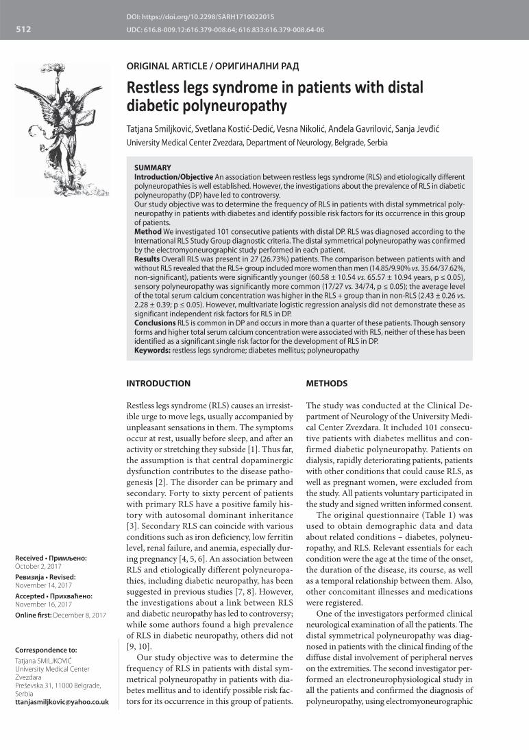
512
DOI: https://doi.org/10.2298/SARH171002201S
UDC: 616.8-009.12:616.379-008.64; 616.833:616.379-008.64-06
Received • Примљено: October 2, 2017
Ревизија • Revised: November 14, 2017
Accepted • Прихваћено: November 16, 2017
Online fi rst: December 8, 2017
Correspondence to:
Tatjana SMILJKOVIĆ
University Medical Center
Zvezdara
Preševska 31, 11000 Belgrade,
Serbia
ORIGINAL ARTICLE / ОРИГИНАЛНИ РАД
Restless legs syndrome in patients with distal diabetic polyneuropathyTatjana Smiljković, Svetlana Kostić-Dedić, Vesna Nikolić, Anđela Gavrilović, Sanja Jevđić University Medical Center Zvezdara, Department of Neurology, Belgrade, Serbia
SUMMARYIntroduction/Objective An association between restless legs syndrome (RLS) and etiologically different polyneuropathies is well established. However, the investigations about the prevalence of RLS in diabetic polyneuropathy (DP) have led to controversy.Our study objective was to determine the frequency of RLS in patients with distal symmetrical poly-neuropathy in patients with diabetes and identify possible risk factors for its occurrence in this group of patients.Method We investigated 101 consecutive patients with distal DP. RLS was diagnosed according to the International RLS Study Group diagnostic criteria. The distal symmetrical polyneuropathy was confirmed by the electromyoneurographic study performed in each patient. Results Overall RLS was present in 27 (26.73%) patients. The comparison between patients with and without RLS revealed that the RLS+ group included more women than men (14.85/9.90% vs. 35.64/37.62%, non-significant), patients were significantly younger (60.58 ± 10.54 vs. 65.57 ± 10.94 years, p ≤ 0.05), sensory polyneuropathy was significantly more common (17/27 vs. 34/74, p ≤ 0.05); the average level of the total serum calcium concentration was higher in the RLS + group than in non-RLS (2.43 ± 0.26 vs. 2.28 ± 0.39; p ≤ 0.05). However, multivariate logistic regression analysis did not demonstrate these as significant independent risk factors for RLS in DP.Conclusions RLS is common in DP and occurs in more than a quarter of these patients. Though sensory forms and higher total serum calcium concentration were associated with RLS, neither of these has been identified as a significant single risk factor for the development of RLS in DP.Keywords: restless legs syndrome; diabetes mellitus; polyneuropathy
INTRODUCTION
Restless legs syndrome (RLS) causes an irresist-ible urge to move legs, usually accompanied by unpleasant sensations in them. The symptoms occur at rest, usually before sleep, and after an activity or stretching they subside [1]. Thus far, the assumption is that central dopaminergic dysfunction contributes to the disease patho-genesis [2]. The disorder can be primary and secondary. Forty to sixty percent of patients with primary RLS have a positive family his-tory with autosomal dominant inheritance [3]. Secondary RLS can coincide with various conditions such as iron deficiency, low ferritin level, renal failure, and anemia, especially dur-ing pregnancy [4, 5, 6]. An association between RLS and etiologically different polyneuropa-thies, including diabetic neuropathy, has been suggested in previous studies [7, 8]. However, the investigations about a link between RLS and diabetic neuropathy has led to controversy; while some authors found a high prevalence of RLS in diabetic neuropathy, others did not [9, 10].
Our study objective was to determine the frequency of RLS in patients with distal sym-metrical polyneuropathy in patients with dia-betes mellitus and to identify possible risk fac-tors for its occurrence in this group of patients.
METHODS
The study was conducted at the Clinical De-partment of Neurology of the University Medi-cal Center Zvezdara. It included 101 consecu-tive patients with diabetes mellitus and con-firmed diabetic polyneuropathy. Patients on dialysis, rapidly deteriorating patients, patients with other conditions that could cause RLS, as well as pregnant women, were excluded from the study. All patients voluntary participated in the study and signed written informed consent.
The original questionnaire (Table 1) was used to obtain demographic data and data about related conditions – diabetes, polyneu-ropathy, and RLS. Relevant essentials for each condition were the age at the time of the onset, the duration of the disease, its course, as well as a temporal relationship between them. Also, other concomitant illnesses and medications were registered.
One of the investigators performed clinical neurological examination of all the patients. The distal symmetrical polyneuropathy was diag-nosed in patients with the clinical finding of the diffuse distal involvement of peripheral nerves on the extremities. The second investigator per-formed an electroneurophysiological study in all the patients and confirmed the diagnosis of polyneuropathy, using electromyoneurographic

513
Srp Arh Celok Lek. 2018 Sep-Oct;146(9-10):512-515 www.srpskiarhiv.rs
criteria [11]. Based on the symptoms, according to Wolf ’s criteria, polyneuropathy was classified as sensory, motor, or sensorimotor [12].
The third neurologist, blinded for the clinical and neurophysiological evaluation, established the diagnosis of RLS. The patients were diagnosed as RLS only if all four diagnostic criteria were present, defined by the In-ternational RLS Study Group: 1. an urge to move the legs, usually accompanied or caused by unpleasant and un-comfortable sensations in the legs; 2. the urge to move or unpleasant sensation begins or worsens during periods of
rest or inactivity, such as lying down or sitting; 3. the urge to move or an unpleasant sensation are partially or totally relieved by movement, such as walking or stretching, at least as long as the activity continues; 4. the urge to move or unpleasant sensations are stronger in the evening or at night than during the day, or only occur in the evening or at night [1]. At the onset of the investigation, new criteria have not been defined [13].
A blood sample was taken from every patient and com-plete blood count, hematocrit, concentration of hemoglobin, ferritin, iron, electrolytes, blood urea nitrogen, cholesterol, cholesterol fractions, and triglycerides were determined.
Statistical analysis included methods of descriptive statis-tics, Student’s t-test, and χ2 test. Logistic regression analysis assessed the importance of the risk factors, and odds ratio measured the effect with a 95% confidence interval. A p-value of 0.05 or less was considered statistically significant.
RESULTS
The study involved 52 (52.5%) men and 49 (47.5%) wom-en, with a mean age of 64.13 ± 11.02 years. One third of the patients had diabetes mellitus type I (34/101) and two thirds had diabetes mellitus type II (67/101), which per-sisted 12.64 ± 8.1 years on average. RLS was present in 27 (26.73%) patients, of whom 13 patients had diabetes mel-litus type I and 14 patients had diabetes mellitus type II.
The comparison between patients with and without RLS was performed (RLS+ and RLS- group, respectively). There were more women than men in the RLS+ group (14.85/9.90% vs. 35.64/37.62%, non-significant), and this group of patients was significantly younger than patients without RLS (60.58 ± 10.54 vs. 65.57 ± 10.94 years, p ≤ 0.05).
No difference in the type of diabetes, duration of dia-betes, or duration of diabetic polyneuropathy was found between the groups (Table 2). However, the type of distal
Table 2. Comparison of patients with and without restless legs syndrome in distal diabetic polyneuropathy
Parameter RLS+ (n = 27) RLS- (n = 74) pSex M/F 10/15 38/36 nsCurrent age (mean ± SD, years) 60.58 ± 10.54 65.57 ± 10.94 p ≤ 0.05DM type I/II 13/14 21/53 nsDuration DM (mean ± SD, years) 5.56 ± 5.74 12.84 ± 8.72 nsDuration of polyneuropathy (mean ± SD, years) 4.47 ± 3.22 6.05 ± 5.64 nsMCV (mean ± SD, cm) 36.38 ± 9.07 36.26 ± 8.14 nsLatency (mean ± SD, cm) 5.16 ± 3.89 5.56 ± 4.72 nsSCV (mean ± SD, cm) 29.62 ± 10.01 29.75 ± 9.90 nsEr 4.21 ± 1.21 4.41 ± 0.74 nsHct 39.27 ± 4.09 39.14 ± 4.42 nsFerritin 173.93 ± 189.20 175.87 ± 143.66 nsNa+ 140.82 ± 3.49 138.10 ± 8.38 nsK+ 5.16 ± 4.14 5.36 ± 4.16 nsCa2+ 2.43 ± 0.26 2.28 ± 0.39 p ≤ 0.05
Mg 0.94 ± 0.46 0.79 ± 0.081 nsCholesterol 5.94 ± 1.50 5.82 ± 1.164 nsTriglyceride 2.57 ± 1.87 2.39 ± 1.65 ns
RLS – restless legs syndrome; DM – diabetes mellitus; SD – standard deviation; ns – non-signifi cant; MCV – motor nerve conduction velocities; SCV – sensitive nerve conduction velocities; Hct – hematocrit; Er – erythrocytes
Restless legs syndrome in patients with distal diabetic polyneuropathy
Table 1. Questionnaire
Name and surnameGender M FAge at observation (years)Diabetes mellitus (DM)
Age at onset of DM (years)Disease duration of DM (years)DM type: I IIDM therapyDuration of therapy for DM (years)Age at onset of polyneuropathy (years)Duration of polyneuropathy (years)Concomitant illness Blood donor: YES NO No. of donations in the past 3 yearsBlood transfusions: YES NO No. of transfusions in the past 3 yearsHabits
Coffee intake: YES NO Duration of habit (years) Cups of coffee No./daySmoking: YES NO Duration of habit (years) Cigarettes No./dayAlcohol: YES NO Duration of habits (years) Restless legs syndrome (RLS): YES NODuration of symptoms (years)Age at the onset of RLS (years)Family history: YES NO

514
Srp Arh Celok Lek. 2018 Sep-Oct;146(9-10):512-515DOI: https://doi.org/10.2298/SARH171002201S
peripheral neuropathy was different: sensory polyneuropa-thy was significantly more common in the RLS+ group (17/27 vs. 34/74, p ≤ 0.05), while sensorimotor polyneu-ropathy was significantly more common in the RLS- pa-tients (10/27 vs. 40/74, p ≤ 0.05).
Comparing laboratory parameters, the only statisti-cally significant difference was found in the average level of total serum calcium concentration: it was higher in the RLS+ group than in the non-RLS patients (2.43 ± 0.26 vs. 2.28 ± 0.39, p ≤ 0.05). The complete blood count, iron and ferritin levels, electrolytes, and serum lipids concentrations were not different between the groups (Table 2).
Univariate logistic regression analysis revealed that to-tal serum calcium concentrations (p = 0.025) and nerve conduction latency (p = 0.048) were associated with RLS. However, multivariate logistic regression analysis did not demonstrate these as significant independent risk factors for RLS in diabetic polyneuropathy.
DISCUSSION
Our results confirmed that RLS was common in patients with diabetic polyneuropathy: almost a quarter of them had RLS (26.73%), which was significantly more than in the general population (7–10%) [14]. According to other studies, the number of patients with polyneuropathy and RLS varies from 5.2% up to 36%, and this difference was the aftermath of diverse study designs [15, 16]. However, most studies have shown an alliance between RLS and neu-ropathy, despite whether they analyzed RLS frequency in patients with neuropathy or the frequency of neuropathy in patients with RLS [7, 16–19]. Our results were consistent with the results of Lopes et al. [19], who found RLS in 27 patients from the group of 100 patients with diabetes, and 25 of those 27 patients had neuropathy as well. Gemignani et al. [18] reported a somewhat higher frequency of RLS in patients with diabetic neuropathy (33.3%), especially in patients with distal diabetic polyneuropathy. Slightly lower prevalence of RLS in our group, compared with the results of Gemignani et al. [18], could be explained by the deci-sion to include only patients with electrophysiologically confirmed neuropathy; consequently, the patients with small-fiber neuropathy could not qualify. Other authors, however, did not corroborate the frequency difference of RLS in diabetics and controls [10]. Since patients inform about similar symptoms in neuropathy and RLS, symp-toms overlap and RLS could be overlooked in patients with neuropathy. Some authors suggested that in every patient with a suspected neuropathy, an interview focused on RLS criteria should be performed [20].
Results of this study showed that RLS is more com-mon in patients with sensory polyneuropathy, the same as previously found by other authors [9], who particularly emphasized the association of RLS and sensory small-fiber neuropathy [18]. Further assumption was that abnormal inputs from the periphery activate spinal generators so that RLS is not exclusively associated with central dopaminergic
dysfunction but possibly starts on a different level of the nervous system, either central or peripheral [18].
Our RLS patients were younger than those without RLS. The observation was interesting because, although chil-dren may have RLS, epidemiological studies have shown that it occurs most frequently in the middle-aged and that the incidence rises with age [4]. The results in the literature differ: while some authors concluded that patients with RLS are older than controls, others did not find this dif-ference [8, 10, 16, 17, 18, 21].
In our RLS + group, women were slightly more represent-ed, which was determined in the majority of trials, although there are papers where there is no difference between sexes [7, 16, 21]. Other female patients (without neuropathy) also have RLS more often, but the connection between RLS and anemia or pregnancy was established [4, 5].
We did not find the difference in serum iron and ferritin levels between patients with and without RLS. Numerous studies have suggested the association of iron metabolism and low serum ferritin with RLS, and several studies have shown that the severity of RLS correlates with the level of serum ferritin [4]. However, it appears that the RLS+ dia-betic population is independent of serum iron and ferritin levels [8, 10, 21]. We recorded a significantly higher level of total serum calcium in those with RLS. The same was noted in hemodialysis patients and investigators suggested that high serum calcium was possibly connected to the pathophysiology of RLS [22]. The exact significance of this result in diabetic polyneuropathy is not clear and further tests are required to confirm and establish this result.
Though univariate logistic regression analysis has shown an association between serum calcium concentra-tions and nerve conduction latency with RLS, multivari-ate logistic regression analysis did not isolate any of the investigated factors as a significant single risk factor for the development of RLS in distal diabetic polyneuropathy. Investigation of possible risk factors for the occurrence of RLS in diabetics in some studies disclosed peripheral neuropathy to be the only risk factor for the occurrence of RLS, while others revealed none [10, 19, 21].
Our study had several limitations, including a relatively small sample and exclusion of patients with sensory small-fiber neuropathy. Nevertheless, we believe the research is important because only a few in our country deal with this problem.
CONCLUSION
The RLS is common in diabetic polyneuropathy and oc-curs in more than a quarter of these patients. Though sensory polyneuropathy and higher total serum calsium concentration have been associated with RLS, neither of these has been identified as a significant single risk factor for the development of RLS in diabetic polyneuropathy. Further studies are needed to clarify the real association between serum calsium concentration and RLS in diabetic polyneuropathy.
Smiljković T. et al.

515
Srp Arh Celok Lek. 2018 Sep-Oct;146(9-10):512-515 www.srpskiarhiv.rs
REFERENCES
1. Allen RP, Picchietti D, Hening WA, Trenkwalder C, Walters AS, Montplasir J. Restless legs syndrome: diagnostic criteria, special considerations, and epidemiology: a report from resless legs syndrome diagnosis and epidemiology workshop at the National Institute of Health. Sleep Med. 2003; 4(2):101–19.
2. Trenkwalder C, Paulus W, Walters AS. The restless legs syndrome. Lancet Neurol. 2005; 4(8):465–75.
3. Bonati MT, Ferini-Strambi L, Aridon P, Oldani A, Zucconi M, Casari G. Autosomal dominant restless legs syndrome maps on chromosome 14q. Brain. 2003; 126(Pt 6):1485–92.
4. Zucconi M, Ferini-Strambi L. Epidemiology and clinical findings of restless legs syndrome. Sleep Med. 2004; 5(3):293–9.
5. Srivanitchapoom P, Pandey S, Hallet M. Restless legs syndrome and pregnancy: a review. Parkinsonism Relat Disord. 2014; 20(7):716–22.
6. Gigli GL, Adorati M, Dolso P, Piani A,Velnete M, Brotini S, et al. Restless legs syndrome in end-stage renal disease. Sleep med. 2004; 5(3):309–15.
7. Gemignani F, Brindani F, Negrotti A, Vitetta F, Alfieri S, Marbini A. Restless Legs Syndrome and Polyneuropathy. Mov Disord. 2006; 21(8):1254–7.
8. Luigetti M, Del Grande A, Testani E, Bisogni G, Losurdo A, Giannantoni NM, et al. Restless leg syndrome in different types of demyeliniating neuropathies: a single-center pilot study. J Clin Sleep Med. 2013; 9(9):945–9.
9. Gemignani F, Brindani F, Negrotti A, Vitetta F, Marbini A, Calzetti S. Restless legs syndrome in diabetic neuropathy: a frequent manifestation of small fiber neuropathy. J Peripher Nerv Syst. 2007; 12(1):50–3.
10. Scomro RP, Ludwig S, Salamon E, Kryger MH. Complaints and restless legs syndrome in adult type 2 diabetics. Sleep Med. 2001; 2(5):417–22.
11. Cvijanović M, Ilin M, Slankamenac P, Banić Horvat S, Jovin Z. The sensitivity of electromyoneurography in the diagnosis of diabetic polyneuropathy. Med Pregl. 2011; 64(1–2):11–4.
12. Wolfe GI, Baker NS, Amato AA, Jacskon CE, Nations SP, Saperstein DS, et al. Chronic cryptogenic sensory polyneuropathy. Arch Neurol. 1999; 56(5):540–7.
13. Allen RP, Picchietti D, Garcia-Borreguero D, Ondo WG, Walters AS, Winkelman JW, et al. Restless Legs Syndrome Study Group. Restless legs syndrome /Willis-Ekbom disease diagnostic criteria: update International Restless Legs Syndrome Study Group (IRLSSG) consensus criteria-history, rationale, description and significance. Sleep Med. 2014; 15(8):860–73.
14. Hogl B, Kiechl S, Willeit J, Saletu M, Frauscher B, Seppi K, et al. Restless legs syndrome. A community-based study prevalence, severity and risk factors. Neurology. 2005; 64(11):1820–924.
15. Rutke SB, Matheson JK, Logigian EL. Restless legs syndrome in patients with polyneuropathy. Muscle Nerve. 1996; 19:670–2.
16. Cho YW, Na GY, Lim JG, Kim SH, Kim SH, Early CJ, et al. Prevalence and clinical characteristics of restless legs syndrome in diabetic peripheral neuropathy: comparison with chronic osteoarthritis. Sleep Med. 2013; 14(12):1387–92.
17. Bastia JK, Bhoi SK, Kalita J, Misra EK. Neuropathy in a cohort of restless leg syndrome patients. J Clin Neurosci. 2015; 22(8):1314–8.
18. Gemignani F, Vitetta F, Brindani F, Contini M, Negrotti A. Painful polyneuropathy associated with restless legs syndrome. Sleep Med. 2013; 14(1):79–84.
19. Lopes L, Linc C de MM, Adeodato V, Quental D, De Bruin PFC, Montenegro RM, et al. Restless legs syndrome and quality of sleep in type 2 diabetes. Diabetes Care. 2005; 11(28):2633–6.
20. Nineb A, Rosso C, Dumurgier J, Nordine T, Lefaucheur J-P, Creange A. Restless legs syndrome is frequently overlooked in patients being evaluated for polyneuropathies. Eur J of Neurol. 2007; 14(7):788–92.
21. Merlino G, Fratticci L, Valente M, Del Guidice A, Noacco C, Dolso P, et al. Association of restless legs syndrome in type 2 diabetes: a case control study. Sleep. 2007; 30(7):866–71.
22. Kawauchi A, Inoue Y, Hashimoto T, Tachibana N, Shirakawa S, Mizutani Y, et al. Restless legs syndrome in hemodyalisis patients: health-related quality of life and laboratory data analysis. Clin Nephrol. 2006; 66(6):440–6.
САЖЕТАКУвод/Циљ Повезаност синдрома немирних ногу (СНН) са неуропатијама различите етиологије јасно је утврђена. Међутим, резултати итраживања о учесталости СНН код дијабетичне полинеуропатије (ДПН) су контроверзни. Циљ нашег рада је био да се утврди учесталост СНН код бо-лесника са дисталном дијабетичном полинеуропатијом, као и да се установе могући фактори ризика за његову појаву у овој групи оболелих.Метод Испитивање је обухватило 101 консекутивног обо-лелог с дисталном ДПН. Дијагноза СНН је постављена на основу критеријума Интернационалне групе за испитивање синдрома немирних ногу. Сваком болеснику урађено је електромионеурографско испитивање којим је потврђена дијагноза дисталне ДПН.Резултати СНН је био присутан код 27 (26,73%) болесника у односу на целу групу. Поређење оболелих са СНН+ и без
СНН- показало је да је у групи СНН+ било нешто више жена него мушкараца (14,85/9,90% тј. 35,64/37,62%), оболели су били значајно млађи (60,58 ± 10,54 тј. 65,57 ± 0,94 година; р ≤ 0,05); значајно чешћа је била сензитивна полинеуропатија (17/27 тј. 34/74, р ≤ 0,05) и имали су виши ниво Ca у крви у односу на оболеле без СНН- (2,43 ± 0,26 према 2,28 ± 0,39; р ≤ 0,05). Међутим, мултиваријантна регресиона анализа није показала да је иједан од њих значајан фактор ризика за појаву СНН код дисталне ДПН.Закључак СНН је чест код ДПН и јавља се код више од четвртине оболелих. Иако су сензорна форма и повишен ниво укупног Ca у серуму били удружени са СНН, ниједан од њих се није издвојио као значајан појединачни фактор ризика за настанак СНН код дисталне ДПН.
Кључне речи: синдром немирних ногу; дијабетес; поли-неуропатија
Синдром немирних ногу код оболелих од дисталне дијабетичне полинеуропатијеТатјана Смиљковиц, Светлана Костић-Дедић, Весна Николић, Анђела Гавриловић, Сања Јевђић Клиничко-болнички центар „Звездара“, Клиничко одељење за неурологију, Београд, Србија
Restless legs syndrome in patients with distal diabetic polyneuropathy
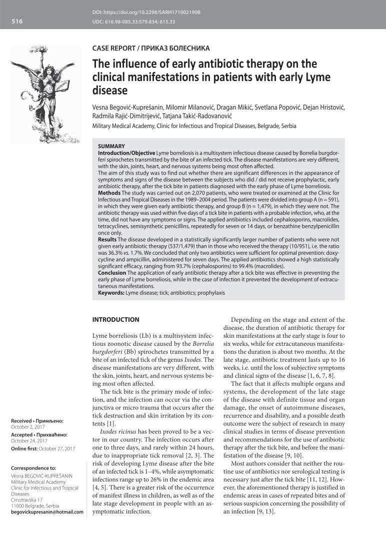
516
DOI: https://doi.org/10.2298/SARH171002190B
UDC: 616.98-085.33:579.834; 615.33
Received • Примљено: October 2, 2017
Accepted • Прихваћено: October 24, 2017
Online fi rst: October 27, 2017
Correspondence to:
Vesna BEGOVIĆ-KUPREŠANIN
Military Medical Academy
Clinic for Infectious and Tropical
Diseases
Crnotravska 17
11000 Belgrade, Serbia
SUMMARYIntroduction/Objective Lyme borreliosis is a multisystem infectious disease caused by Borrelia burgdor-feri spirochetes transmitted by the bite of an infected tick. The disease manifestations are very different, with the skin, joints, heart, and nervous systems being most often affected.The aim of this study was to find out whether there are significant differences in the appearance of symptoms and signs of the disease between the subjects who did / did not receive prophylactic, early antibiotic therapy, after the tick bite in patients diagnosed with the early phase of Lyme borreliosis. Methods The study was carried out on 2,070 patients, who were treated or examined at the Clinic for Infectious and Tropical Diseases in the 1989–2004 period. The patients were divided into group А (n = 591), in which they were given early antibiotic therapy, and group B (n = 1,479), in which they were not. The antibiotic therapy was used within five days of a tick bite in patients with a probable infection, who, at the time, did not have any symptoms or signs. The applied antibiotics included cephalosporins, macrolides, tetracyclines, semisynthetic penicillins, repeatedly for seven or 14 days, or benzathine benzylpenicillin once only. Results The disease developed in a statistically significantly larger number of patients who were not given early antibiotic therapy (537/1,479) than in those who received the therapy (10/951), i.e. the ratio was 36.3% vs. 1.7%. We concluded that only two antibiotics were sufficient for optimal prevention: doxy-cycline and ampicillin, administered for seven days. The applied antibiotics showed a high statistically significant efficacy, ranging from 93.7% (cephalosporins) to 99.4% (macrolides).Conclusion The application of early antibiotic therapy after a tick bite was effective in preventing the early phase of Lyme borreliosis, while in the case of infection it prevented the development of extracu-taneous manifestations.Keywords: Lyme disease; tick; antibiotics; prophylaxis
CASE REPORT / ПРИКАЗ БОЛЕСНИКА
The influence of early antibiotic therapy on the clinical manifestations in patients with early Lyme diseaseVesna Begović-Kuprešanin, Milomir Milanović, Dragan Mikić, Svetlana Popović, Dejan Hristović, Radmila Rajić-Dimitrijević, Tatjana Takić-RadovanovićMilitary Medical Academy, Clinic for Infectious and Tropical Diseases, Belgrade, Serbia
INTRODUCTION
Lyme borreliosis (Lb) is a multisystem infec-tious zoonotic disease caused by the Borrelia burgdorferi (Bb) spirochetes transmitted by a bite of an infected tick of the genus Ixodes. The disease manifestations are very different, with the skin, joints, heart, and nervous systems be-ing most often affected.
The tick bite is the primary mode of infec-tion, and the infection can occur via the con-junctiva or micro trauma that occurs after the tick destruction and skin irritation by its con-tents [1].
Ixodes ricinus has been proved to be a vec-tor in our country. The infection occurs after one to three days, and rarely within 24 hours, due to inappropriate tick removal [2, 3]. The risk of developing Lyme disease after the bite of an infected tick is 1–4%, while asymptomatic infections range up to 26% in the endemic area [4, 5]. There is a greater risk of the occurrence of manifest illness in children, as well as of the late stage development in people with an as-ymptomatic infection.
Depending on the stage and extent of the disease, the duration of antibiotic therapy for skin manifestations at the early stage is four to six weeks, while for extracutaneous manifesta-tions the duration is about two months. At the late stage, antibiotic treatment lasts up to 16 weeks, i.e. until the loss of subjective symptoms and clinical signs of the disease [1, 6, 7, 8].
The fact that it affects multiple organs and systems, the development of the late stage of the disease with definite tissue and organ damage, the onset of autoimmune diseases, recurrence and disability, and a possible death outcome were the subject of research in many clinical studies in terms of disease prevention and recommendations for the use of antibiotic therapy after the tick bite, and before the mani-festation of the disease [9, 10].
Most authors consider that neither the rou-tine use of antibiotics nor serological testing is necessary just after the tick bite [11, 12]. How-ever, the aforementioned therapy is justified in endemic areas in cases of repeated bites and of serious suspicion concerning the possibility of an infection [9, 13].
517
Srp Arh Celok Lek. 2018 Sep-Oct;146(9-10):516-523 www.srpskiarhiv.rs
One study involving 600 patients showed that the use of antibiotic therapy was unnecessary due to a small number of infected persons (1.4%) in patients who did not receive an an-tibiotic after the tick bite, but the same authors recommended carrying out additional studies concerning this subject [14].
Due to the early dissemination of the cause of the dis-ease, the difficulty in diagnosis, and major problems in the treatment of infected patients, particularly in the late stage of the disease, there are opinions that, if there is a suspicion of an infection after the bite, it is justified to apply antibiotics for up to two weeks [6, 8].
The aim of this paper is to determine the effect of pro-phylactic, i.e. early application of antibiotic therapy on the clinical manifestations in the early phase of Lb. For the purpose of this study, prophylactic antibiotic therapy was defined as the use of antibiotics up to five days after the tick bite in individuals with suspected Bb infection who did not show clinical symptoms and signs of the disease at the time of the application of antibiotics.
METHODS
The study was designed as a retrospective-prospective cohort study. It initially included 2,470 patients who were treated at the Clinic for Infectious and Tropical Diseases of the Military Medical Academy (MMA), Belgrade, Serbia, or examined at the outpatient clinic in the period between January 1, 1989 and December 31, 2004; out of 400 exclud-ed patients 207 did not show up for follow-up monitoring, and 193 received early antibiotic therapy. The study did not include patients who had a positive serological reac-tion to the Bb antigens at the first examination, i.e. those who had previously developed an infection.
All the patients were classified into two categories. The first category consisted of people who came to the Clinic because of a tick bite. The presence of probable Bb infec-tion was based on the existence of at least one of the fol-lowing criteria: the presence of Bb in the removed tick; the tick’s presence in the skin for more than 48 hours; data on inappropriate tick removal; and the presence of the anti-bodies to Bb antigens in the serum after four to six weeks. In some patients, based on the clinician’s assessment, early antibiotic therapy was applied, while in others it was not.
The second category included patients who came with some of the symptoms and were diagnosed with the early phase of Lyme disease. The diagnosis was based on a char-acteristic change in the skin, i.e. erythema migrans (EM), clinical picture and the course of the disease, an increase in antibody titer against Bb antigens in two consecutive serum samples within two to four weeks (IIF) or on the positive result after four to six weeks (ELISA), response to antibiotic therapy, exclusion of other related diseases, data concerning the tick bite, and stay in the endemic area.
A total of 2,070 patients were divided into group A, in which early antibiotic therapy was applied, and group B, in which it was not.
The disease symptoms included headaches, myalgia, ar-thralgia, loss of concentration, sleep and mood disorders,
paresthesia, palpitations, and pruritus. The observed signs of the disease included high body temperature, EM and multiple EM (MEM), heart rhythm disorders, myocardi-tis, pericarditis, encephalopathy, meningitis, encephalitis, cranial neuritis, radiculoneuritis, arthritis, myositis, and lymphadenopathy.
Based on the type of antibiotic and duration of its appli-cation, all patients who received antibiotic therapy were di-vided into three groups: those who were given benzathine benzylpenicillin once parenterally, those who were given antibiotics for seven days orally and those who were given antibiotics for 14 days orally (macrolides, tetracyclines, cephalosporins, semisynthetic penicillins). In each of these groups, those who suffered and those who did not suffer from the early stage of Lyme disease were singled out.
Data sources and examinations included biographi-cal data from medical and health records, tick bite data, physical, radiological, and electrophysiological examina-tions, serological tests concerning Bb antigens (reaction of indirect immunofluorescence IIF or ELISA tests done at the Institute of Microbiology of the MMA) and specialist examinations according to indications.
The tick was removed either appropriately by a surgeon or an epidemiologist at the MMA, or inappropriately by the patients themselves and/or other adults when a child had been bitten. Tick gut contents were tested for the presence of Bb by an epidemiologist at the MMA Epidemiology In-stitute, in a dark field, using phase-contrast microscopy. All the patients were monitored clinically, laboratorically, and serologically within the period of one to 15 years.
Antibiotics registered for administration in our country and recommended for the treatment of Lyme disease were applied in the early antibiotic therapy.
Continuous variables were summarized as means (M) and standard deviations (SD); the significance between the groups was determined by the Student’s t-test for the validity of data distribution. All variables were presented as frequency of certain categories, while statistical signifi-cance of differences was tested by the χ2 test. Statistical significance was accepted at a minimum level of p < 0.05.
Principles of ICH Good Clinical Practice were strictly followed and ethical approval from the Ethics Committee was obtained on September 15, 2017.
RESULTS
In total, there were 2,070 patients. Group A consisted of 591 patients who received early antibiotic therapy, while Group B comprised 1,479 patients who did not receive any antibiotic therapy. Comparing the number of patients in the groups, a statistically significant difference was found due to the fact that Group B was significantly larger (Table 1) than Group A since early antibiotic therapy was applied only to those with a probable infection, according to the assessment of the responsible physician.
In both groups, there were more male patients, a total of 1,572 (75.94%), while there were only 498 females (24.06%), and there was no statistically significant difference
The infl uence of early antibiotic therapy on the clinical manifestations in patients with early Lyme disease

518
Srp Arh Celok Lek. 2018 Sep-Oct;146(9-10):516-523DOI: https://doi.org/10.2298/SARH171002190B
in the distribution of sexes in the groups. The average age of patients in Group A was 35.2 ± 19.4 years, while in Group B it was 39.4 ± 20.7. The patients in Group B were statistically significantly older than those in Group A. The average age of women was 46.47 ± 20.26 years, while that of men was 33.89 ± 19.54 years, with women being statisti-cally significantly older (p = 0.001).
When the presence of a tick during the examination is concerned, it was found that the distribution of the sub-jects was practically identical, as there was no statistically significant difference between these groups concerning the presence or absence of ticks, or the use of antibiot-ics (p = 0.99). The analysis of a large number of patients (2,041) with appropriately and inappropriately removed ticks showed a uniform ratio of these categories: 994:1047 (48.7%:51.3%) (Table 1).
The applied treatment was analyzed concerning the method of removing ticks. It was found that in Group B, which had not received early antibiotic therapy, the tick was removed appropriately in 652 patients (45%), inap-propriately in 798 patients (55%), while 29 patients did not know about the tick bite and they came to the doctor’s in the early phase of Lb. In Group A, the tick was removed appropriately in 342 patients (57.9%), and inappropriately
in 249 patients (42.1%). There were statistically signifi-cantly more patients in the group that had received the antibiotic and in which the tick was previously appropri-ately removed (p < 0.001).
The duration of tick presence ranged from one hour to more than 48 hours. By analyzing the duration of tick pres-ence in the skin we can conclude that there was no statisti-cally significant difference between the groups (Table 1).
After the tick bite in infected patients, the signs of Lb were most frequently present on the skin (Table 2).
The results show that the clinical signs on the skin were found in 31.51% of patients who did not receive early antibiotic therapy, as opposed to those who did (1.52%) (p < 0.001).
The total number of signs of the disease was 679, and the number of patients was 537, which means that in some patients there were several affected systems. Table 2 shows the frequency of signs on a particular organ system in rela-tion to the total number of recorded signs in Group B (679). We observed that the nervous system was affected in 5.34%, other organs were affected in 3.49%, locomotor system in 2.84%, and cardiovascular system in 2.77% of the patients.
The frequency of changes on the skin, in relation to the total number of patients (547), was 86.84%, the changes
Table 1. Basic demographic and relevant clinical data
ParametersGroups
ProbabilityEarly antibiotic treatment A Without antibiotic treatment Bn % n %
Age (years), x ± SD 35.2 ± 19.4 39.4 ± 20.7 t = 4.24; p < 0.001Patient distribution (n = 2070) 591 24.06 1,479 75.94 χ2 = 112.39; p < 0.001
Sexmale 457 77.3 1,115 75.4
χ2 = 0.76; p = 0.38female 134 22.7 364 24.6Tick presenceyes 260 44.0 652 44.1
χ2 = 0.00; p = 0.99no 331 56.0 827 55.9Tick removalappropriately 342 57.9 652 45.0
χ2 = 27.34; p < 0.001inappropriately 249 42.1 798 55.0Duration of tick presence (hours)*1–12 74 12.52 146 9.87
χ2 = 2.31; p = 0.6913–23 228 38.58 562 38.0024–48 111 18.78 245 16.57> 48 63 10.66 157 10.62unknown 115 19.79 369 24.95
*Skin, based on anamnestic data
Table 2. Clinical signs due to Borrelia burgdorferi exposure
SystemsGroups
ProbabilityEarly antibiotic treatment A Without antibiotic treatment Bn % n %
Skin 9 1.52 466 31.51 χ2 = 213.03; p < 0.001Nervous 0 0.00 79 5.34
Not shown due to extremely low frequencies in early antibiotic treatment group
Other 1 0.17 51 3.49Joints 0 0.00 42 2.84Heart 0 0.00 41 2.77Total 10 1.69 679 45.91
Begović-Kuprešanin V. et al.

519
Srp Arh Celok Lek. 2018 Sep-Oct;146(9-10):516-523 www.srpskiarhiv.rs
in the nervous system were registered in 14.44%, on other organs in 9.50%, on the locomotor system in 7.88%, and in the cardiovascular system in 7.49% of the patients.
At the same time, we observed that in patients who re-ceived early antibiotic therapy, there were no signs of disease in the nervous, cardiovascular, and bone-muscular systems.
After applying early antibiotic therapy in 591 patients, 581 (98.3%) of them did not develop the early phase of Lyme disease. Macrolides (99.4%) showed the highest efficacy, followed by semi-synthetic penicillins (98.4%), benzathine benzylpenicillin (98.2%), and tetracyclines (97%). Cephalosporins (93.7%) showed the lowest effi-cacy, which was statistically significantly lower in relation to other groups of antibiotics (Table 3).
In Group A, which had received early antibiotic treat-ment, only 10 (1.7%) out of 591 patients were infected, while 581 of them were not infected (98.3%). In contrast, in Group B, which had not received early antibiotic ther-apy, 537 (36.3%) out of 1,479 patients were infected, while 942 (63.7%) were not (Table 4). The difference found be-tween these two groups of patients was highly statistically significant in favor of the group that had received early antibiotic therapy (p < 0.001).
DISCUSSION
Today, Lb is one of the greatest imitators in medicine, be-cause it is a multisystem disorder involving many organs and systems, with the occurrence of a chronic disease form, the development of disabilities, autoimmune dis-eases, and sometimes lethal outcome [15].
The disease occurs in all parts of the world. In the United States, the incidence is 12–39 patients per 100,000, since the probable cases have also been reported since 2012 [16, 17].
In Europe, the incidence ranges from 35/100,000 patients in Germany to 206/100,000 patients in Slovenia [18].
In Serbia, the number of the infected is decreasing (6.83/100,000), since only 487 cases were reported in 2015 [19]. In the period from 1986 to 2000, Lb was represented in the zoonotic group with 16.34% in Belgrade, while 4,768 peo-ple developed the disease in Serbia from 1991 to 2000 [20].
In our study, there were significantly more males (75%), aged 11–30 years, as most of them were members of the army who had been outdoors in the field (younger ages were members of their families). In Europe, about 60% of women with a tick bite have been registered, which is similar to the findings in Serbia (57%), aged 20–60 years and over [19].
In the early 1990s, the infected ticks were frequent on the territory of Belgrade, ranging 20–67%, with a tendency to decrease. In 2007, this number amounted to 30% [2, 21].
The situation was similar everywhere. In Europe, the in-fection of Ixodes ricinus with Bb ranged 15–40%, in endemic areas of America up to 50%, while Ixodes pacificus was in-fected with Bb in 1–2% of cases [22]. The risk of the occur-rence of manifest Lb after a bite by an infected tick in Europe ranges 1–4%, in hyperendemic areas 4.7–5%, but there are also data on a significantly higher risk (up to 27%) [4, 23, 24].
At the time of examination, 44% of our patients in both groups had a tick in the skin, while others came with a re-moved tick or not knowing about the bite. With the largest number of subjects, 48.31%, the tick stayed in the skin for up to 24 hours before the first examination.
In our study, there were 21 early-phase Lb patients with the tick staying in the skin for only 10 hours or up to one day who developed changes in the central and peripheral nervous systems, which is in agreement with some authors, but also in contradiction with some others [2, 3, 25, 26].
Given the large number of subjects in this study, i.e. 994 who had the tick removed appropriately (48.7%) and 1,047 who had it removed inappropriately (51.3%), it was possible to assess the significance of these two methods of tick removal for the occurrence of the disease. In the case of appropriate tick removal, the disease was reported in 22% of patients, while it was reported in 78% in the case of inappropriate removal, which was statistically significantly different. The ratio of patients with the tick inappropri-ately removed who were given early antibiotic therapy was 1.01% vs. 28.7% of patients in the group without antibiotic therapy. There was a significantly higher number of sub-jects in the group receiving the antibiotic and in whom the tick was previously appropriately removed (p < 0.001).
In patients with manifestations in the nervous and car-diovascular systems in our study, the tick was inappro-priately removed in most cases, which is in line with the data given by Southern [27]. According to other authors’ data concerning appropriate tick removal, the number of patients was statistically significantly lower in relation to the group where it was inappropriately removed (6% vs. 46%), but this study included a small number of subjects (52) [28]. According to the results of our authors, the early phase of Lb occurred in 0.61% of patients with appropri-ately removed tick, and in 2.5% of patients with inappro-priately removed tick, while others claim that it occurred
Table 3. Efficacy of the early-applied antibiotic therapy
Antibiotics class
Lyme disease absent
Lyme disease present
Total
n % n % n %Macrolides 182 99.4 1 0.6 183 100Semisynthetic penicillins 121 98.4 2 1.6 123 100Benzathine benzylpenicillin 165 98.2 3 1.8 168 100Tetracyclines 98 97 3 3 101 100Cephalosporins 15 93.7* 1 6.3 16 100Total 581 98.3 10 1.7 591 100
*signifi cantly diff erent (p < 0.05) from macrolides
Table 4. The frequency of the early phase of Lyme disease in patients regarding the type of treatment
Treatment
Lyme disease present
Lyme disease absent
Total
n % n % n %Early antibiotic treatment 10 1.7 581 98.3 591 100Without antibiotic treatment 537 36.3 942 63.7 1479 100
Total 547 1523 2070 100Probability χ2 = 258.40; p < 0.001
The infl uence of early antibiotic therapy on the clinical manifestations in patients with early Lyme disease
520
Srp Arh Celok Lek. 2018 Sep-Oct;146(9-10):516-523
in 0.2% of patients with appropriately removed tick and in 1.44% of patients with inappropriately removed tick [21, 29]. The aforementioned numerical indicators are lower in relation to the data from our research due to the longer monitoring of our patients and the timely started antibiotic therapy. The results of other studies suggest that 96% of patients with an appropriately removed tick did not suf-fer from Lb in a highly endemic region, thus emphasizing the importance of appropriate forceps tick removal even without the antibiotic prophylaxis [30, 31].
In our study, we observed the frequency of symptoms and signs of Lb and registered 12 different symptoms and 24 signs, which we grouped according to the affected or-gan systems. The disease was manifested on the skin in the form of EM and other skin changes in 86.84% of patients, in the nervous system in 14.44%, in other organs (lymph-adenopathy, liver damage, high body temperature, hypo-thyroidism, multiple-systemic infectious disease syndrome – MSIDS) in 9.5%, on the locomotor system in 7.88%, and in the cardiovascular system in 7.49% of patients com-pared to the total number of patients (547). In patients who received early antibiotic therapy, there were no signs of disease in the nervous, cardiovascular, and musculosk-eletal systems. In one patient (0.17%) who received early antibiotic therapy, we registered high body temperature without other signs of the early phase of Lb, as opposed to those who did not receive antibiotic therapy, in whom we registered the following: 3.49% MSIDS (in 29 patients), lymphadenopathy, liver, damage and hypothyroidism.
In patients who received early antibiotic therapy after a tick bite, Russian authors observed the infected (1.1%) with-out EM and with MSIDS, and in those without antibiotic therapy (12.3%) they noted those with MSIDS, sleep disorder, EM and MEM, lymphadenopathy, and cardiac disorders [32]. Other authors paid less attention to this category of patients and did not include them in the analysis when they evaluated the efficacy of prophylactic post-bite therapy [9, 14, 30, 33]. Our results show that in the group with antibiotic therapy only EM and MSIDS were observed, while all other signs were observed in the group without early antibiotic therapy. The difference between these two groups in relation to the number of signs was also statistically highly significant.
EM and MEM were significantly more frequently pres-ent (31.51%) in the group without early antibiotic therapy compared to patients with the treatment (1.52%). MEM was observed in 2.3% of patients without early antibiotic therapy, which was found in the United States in a higher percentage (20%) due to the early dissemination of Bb in that area [33]. Today, this can also be accounted for by new strains with high spirochetemia [34]. Compared to the to-tal number of patients (547), the incidence of skin changes was 86.84%. Similar results were also reported worldwide, with 70–90% in the United States, and 48% in Slovenia [35]. Some patients with Lb without early antibiotic therapy had several organic systems affected, while 5.34% had changes in the nervous system, 2.47% in the locomotor system, and 2.77% in the cardiovascular system. Other studies state the same signs of disease with approximately equal frequency, which we also observed in our patients [31].
Unlike in our study, other authors followed their pa-tients for a shorter period of time and observed fewer symptoms and signs. Among our patients, there were more neurological manifestations observed because we paid special attention to the damage of the peripheral nervous system due to Lb [36]. The incidence of cardiac mani-festations in Lb was lower in relation to the data of other authors where they stated a frequency of up to 10% [37].
Opinions regarding the type and duration of antibiotic therapy in the treatment of Lb have not yet been harmo-nized. The general recommendation is that doxycycline, amoxicillin, and cefuroxime should be administered at an early phase for two to three weeks, while intravenous ceftri-axone, cefotaxime, or benzylpenicillin for two to four weeks in the case of extracutaneous manifestations [38, 39, 40]. The previous recommendation for treating the late phase was two to four weeks’ therapy and even longer, until the disappearance of the symptoms [7]. New studies have not justified the extended, 12-week antibiotic treatment in late-stage patients with prolonged Lb symptoms [41]. Based on our experience, a longer four to six weeks’ treatment pre-vents the dissemination of Bb and its affecting other organs and systems [1, 6, 8]. On the other hand, other authors con-sider that 10 days of doxycycline and amoxicillin therapy is efficient in the treatment of the early phase of Lb [42].
There is a significant difference in the recommenda-tions for the treatment of the early phase of Lb, symptoms associated with Lb, and prophylaxis between the Infectious Diseases Society of America (IDSA) and the International Lyme and Associated Diseases Society, which advocates extended antibiotic treatment in all categories [8]. IDSA, along with several other medical associations, is in the process of developing new guidelines for the prevention, diagnosis, and treatment of Lb [43].
There are two opposing standpoints in the world regard-ing prophylaxis after a tick bite. The authors who are against the early application of antibiotic therapy base their opinion on a small proportion of instances of infection after a tick bite (2–3%), costs, and possible side effects of antibiotics [11, 12, 31]. Other authors, including us, advocate the use of early antibiotic therapy, regardless of the small number of infected persons after a tick bite. Among the infected, there are often patients with a severe clinical picture with extra-cutaneous manifestations and a possible fatal outcome [15].
In the prophylaxis o f Lyme disease, the most commonly used drugs were cefotaxime, ampicillin, doxycycline, and penicillin [22, 33]. The results of a multicenter study in Ger-many and Austria show that the local application of 10% azithromycin at the site of a bite of an infected tick reduces the occurrence of EM [44]. We consider that the recom-mendation for local therapy is not justified because EM does not occur in 20–30% of infected patients, and patients later develop extracutaneous manifestations. It was previously described in experimental studies that the damage to the hematoencephalic barrier occurs as soon as 12 hours after spirochete inoculation, which does not support the use of local antibiotic therapy at the tick bite site [45].
In our study, we administered four groups of antibiotics orally and benzathine benzylpenicillin once parenterally in
Begović-Kuprešanin V. et al.
DOI: https://doi.org/10.2298/SARH171002190B

521
Srp Arh Celok Lek. 2018 Sep-Oct;146(9-10):516-523 www.srpskiarhiv.rs
order to prevent the disease after the tick bite. The results of our study show that early antibiotic therapy after the tick bite was very effective in the prevention of Lb. This was documented by a statistically significant decrease in the number of patients who received early antibiotic ther-apy (1.7%) compared to the group that did not receive this treatment (36.3%). Moreover, the symptoms and signs of the disease were also significantly less pronounced in the group with early antibiotic therapy compared to the group without it. Extracutaneous manifestations as an indicator of the severity of the clinical picture, in the group that received early antibiotic therapy, were not present at all. In contrast, in the group that did not receive early antibiotic therapy, symptoms and signs were observed in cardiovas-cular, nervous, and musculoskeletal systems.
The high incidence of early phase of Lb during the con-ducted trial at the MMA Clinic for Infectious and Tropical Diseases correlates with the highest number of Lb cases reported in Serbia in the period from 1991 to 2000, which totaled 4,768, and a high percentage of infected ticks [10, 20]. The aforementioned findings coincided with the great migration of the population due to sanctions and wars on the territory of former Yugoslavia. In addition, in our patients we observed a greater number of symptoms and signs than had been observed in other studies, as we fol-lowed the patients for more than one year [10].
A study by Russian authors showed that the use of doxycycline reduces the occurrence of the early phase of Lb in patients with the infected tick bite, as 11 times more patients were found in the group without prophylactic an-tibiotic therapy [32].
Several studies showed, according to the results of the meta analysis, that the disease did not develop in people who received prophylaxis 72 hours after the tick bite, and that the risk of developing the infection was 11 times low-er in the group that received antibiotics (doxycycline or amoxicillin), compared to the placebo group (0.2%:2.22%) [14, 30].
The administration of a single dose of 200 mg doxycy-cline after a tick bite showed that early antibiotic therapy was very effective in reducing morbidity of the early phase of Lb, as the disease developed in eight times fewer pa-tients compared to those who did not receive antibiotics (0.4%:3.2%) [9]. In this study, subjects were monitored for six weeks and only EM was described, which is insufficient to evaluate the efficacy of the drug, because symptoms and signs of the disease can be subsequently exhibited up to one year after the bite. EM does not occur in 20–40% of patients with early phase of Lb. Following the publica-tion of the results of this study, a single dose of 200 mg doxycycline after the tick bite was recommended in certain individuals within the given treatment criteria [33].
Contradictory opinions concerning the justification of the application of antibiotics have existed since the discov-ery of Lb and its treatment up to the present [11, 12, 31]. According to some authors, a small number of patients after a tick bite (on average 1.1–3.4%) do not require the use of prophylactic therapy [11, 12, 31, 39]. In hyperen-demic areas in the United States, Lb was developed in 3%
of patients with a tick bite without antibiotic therapy, so the use of prophylaxis in less endemic areas would have an even smaller effect [31]. In Europe, the tick infection rate is lower than in the United States, the risk of developing Lb is low (up to 4%), so prophylaxis is not recommended after the bite [4, 31].
This study showed that the early antibiotic therapy was extremely effective in preventing the disease compared to the control group (1.7%:36.3%). Our study followed the patients for 1–15 years, and therefore the percentage of patients who were infected at the early stage of Lb without early antibiotic therapy was greater [10]. Secondly, all five antibiotic groups were found to be clinically highly effec-tive. The best results were achieved with roxithromycin (99.4%), followed by half-synthetic penicillins (98.4%), benzathine benzylpenicillin (98.2%), and doxycycline (97%), while cephalosporins (93.7%) showed the lowest statistically significant efficacy. As far as the efficacy of antibiotics and the duration of their use are concerned, (single-dose parenterally, seven or 14 days orally), there were no statistically significant differences.
Previously, the efficacy of short-term administration of amoxicillin as prophylaxis was not known and, therefore, was not recommended [33]. After a meta-analysis of four studies, it was concluded that the 10-day administration of amoxicillin may be as effective as 200 mg of doxycycline [30]. Our results showed that amoxicillin was very effec-tive, even when given for only seven days. In addition, benzathine benzylpenicillin, not having been considered so far, also showed good efficacy.
Our study showed that, out of the 13 antibiotics we used, roxithromycin was the most effective one, but at the same time it was, by far, the most expensive drug. In previ-ous studies, it was the second-line treatment for the early phase of Lb [33]. According to the principles of the World Health Organization that in the case of equal efficacy and good tolerance of more drugs, low-cost drugs should be used for treatment, in the past years we have decided to use ampicillin and doxycycline for a seven-day period. In case of need, i.e. in allergic patients (whom we did not register in our study), clarithromycin, azithromycin, (as suggested by British National Formulary [38]) or roxithro-mycin could be used.
In our study, we did not detect side effects of the ap-plied drugs. There was no evidence of photosensitivity or gastrointestinal disturbances in soldiers who were exposed to the sun as part of their regular activities and diets that were not fully aligned with their usual family eating habits.
CONCLUSION
Taking into account our extensive experience in this field and results of the application of prophylactic therapies, we join the authors who are in favor of early antibiotic therapy after a tick bite. It is clinically justified, prevents the development of extracutaneous manifestations of Lb, is well tolerated, and the proposed antibiotics (doxycycline and ampicillin) are inexpensive.
The infl uence of early antibiotic therapy on the clinical manifestations in patients with early Lyme disease

522
Srp Arh Celok Lek. 2018 Sep-Oct;146(9-10):516-523
1. Bojić I. Evropska borelioza (Lajmska bolest) i druge bolesti čiji se uzročnici prenose ubodom krpelja. Loznica: Naš dom; 2000. р. 29.
2. Drndarević D, Lako B, Stojanović R, Stajković N, Obradović M, Živanović B, et al. Ixodes ricinus dokazan vektor Lajm borelioze i u Jugoslaviji. Vojnosanit Pregl. 1992; 49(1):8–11.
3. Bağcı IS, Ruzicka T. Ticks, Borrelia Burgdorferi and Lyme Disease. Turk J Dermatol. 2016; 10:116–21.
4. Guy N. Lyme disease: Basis for treatment strategy, primary preventive care and secondary preventive care. Med Mal Infect. 2007; 37:381–93. (In French)
5. Fahrer H, van der Linden MS, Sauvain JM, Gern L, Zhioua E, Aeschilmann A. The prevalence and incidence of clinical and asymptomatic Lyme borreliosis in a population at risk. J Infect Dis. 1991; 163:305–10.
6. Bojić I. Principi lečenja inficiranih Borreliom burgdorferi i obolelih od lajmske bolesti. Vojnosanit Pregl. 1998; 55(1):37–40.
7. Burascano JJ. Advanced Topics in Lyme Disease. Diagnostic hints and treatment guidelines for Lyme and other tick borne illnesses. International Lyme and associated disease Society. 2008, 16th Edition. (Accessed: September 2017) Available from: http://www.lymenet.org/BurrGuide200810.pdf
8. Cameron DJ, Johnson LB, Maloney EL. Evidence assessments and guideline recommendations in Lyme disease: the clinical management of known tick bites, erythema migrans rashes and persistent disease. Expert Rev Anti Infect Ther. 2014; 12(9):1103–35.
9. Nadelman BR, Nowakowski J, Fisch D, Falco CR, Freeman K, McKenna D, et al. Prophylaxis with Single – Dose Doxycyline for the Prevention of Lyme Disease after an Ixodes scapularis Tick Bite. N Engl J Med. 2001; 345:79–84.
10. Begović Kuprešanin V. Rana primena antibiotika i njen uticaj na kliničke manifestacije u ranoj fazi Lajmske bolesti. (Dissertation). Kragujevac: Medicinski fakultet, 2011.
11. Costello CM, Steere AC, Pinkerton RE, Feder HM Jr. A prospective study of tick bites in and endemic area for Lyme disease. Conn Med. 1989; 53:338–40.
12. Shapiro ED, Gerber MA, Holabird NB, Berg AT, Feder HM Jr, Bell GL, et al. A controlled trial of antimicrobial prophylaxis for Lyme disease after deer-tick bites. N Engl J Med. 1992; 327:1769–73.
13. Fix AD, Stricland GT, Grant J. Tick bites and Lyme disease in an endemic setting: problematic use of serologic testing and prophylactic antibiotic therapy. JAMA. 1998; 279(3):206–10.
14. Warshafsky S, Nowakowski J, Nadelman RB, Komer RS, Peterson SJ, Wormser GP. Efficacy of antibiotic prophylaxis for prevention of Lyme disease: a meta- analysis. J Gen Intern Med. 1996; 11:329–33.
15. Kugeler KJ, Griffith KS, Gould LH, Kochanek K, Delorey MJ, Biggerstaff BJ, et al. A review of death certificates listing Lyme disease as a cause of death in the United States. Clin Infect Dis. 2011; 52(3):364–7.
16. Adams D, Fullerton K, Jajosky R, Sharp P, Onweh D, Schley A, et al. Summary of Notifiable Infectious Diseases and Conditions - United States, 2013. MMWR Morb Mortal Wkly Rep. 2015; 62(53):1–122.
17. Centers for Disease Control and Prevention. Lyme Disease Data. CDC. [Accessed: March 14, 2016.] Available at: http://www.cdc.gov/lyme/stats/index.html?s_cid=cs_281
18. Wilking H, Stark K. Trends in surveillance data of human Lyme borreliosis from six federal states in eastern Germany, 2009-2012. Ticks Tick Borne Dis. 2014; 5:219–24.
19. Izveštaj o zaraznim bolestima u Republici Srbiji za 2014 godinu: Instititut za javno zdravlje Srbije “Dr Milan Jovanović Batut“; 2015.
20. Čekanac R, Stajković N, Krstić G. Epidemiološke karakteristike lajmske bolesti u Jugoslaviji. Acta Medica Semendrica. 2002; 45–48, 202.
21. Mladenović J. Epidemiološki aspekt lajmske bolesti na teritoriji grada Beograda. (Dissertatio) Niš: Medicinski fakultet; 2014.
22. Edlow AJ. Tick – Borne Diseases, Lyme: Treatment & Medication. eMedicine. Available from: http://www.emedicine.com/derm/topic536.htm.Retrieved 2007-08-21.
23. Nahimana I, Gern L, Blanc DS, Praz G, Francioli P, Peter O. Risk of Borrelia burgdorferi infection in western Switzerland following a tick bite. Eur J Clin Microbiol Infect Dis. 2004; 23(8):603–8.
24. Maiwald M, Oehme R, March O, Petney TN, Kimmig P, Naser K, et al. Transmission risk of Borrelia burgdorferi sensu lato from Ixodes ricinus ticks to humans in southwest Germany. Epidemiol Infect. 1998; 121(1):103–8.
25. Stanek G. Tick-transmitted diseases in central Europe. Wien Clin Wach. 2002; 114 (13–14):471–2.
26. Meiners T, Hammer B, Gobel UB, Kahl O. Determining the tick scutal index allows assessment of tick feeding duration and estimation of infection risk with Borrelia burgdorferi sensu lato in a person bitten by an Ixodes ricinus nymph. Int J Med Microbiol. 2006; 296(suppl 40):103–7.
27. Southern R. Tick Bite Dangers and Symptoms. 2009. Available from: http://www.edubook.com/tick- bite-dangers-and-symptoms/17304/
28. Oteo JA, Martinez de Artola, Gomez-Cadinanos R, Casas JM, Blanco JR, Rosel L. Evaluation of methods of tick removal in human ixodidiasis. Rev Clin Esp. 1996; 196(9):584–7.
29. Mladenović J, Čekanac R, Stajković N, Krstić M. Rizik od pojave lajmske bolesti nakon uboda krpelja. Vojnosanit Pregl. 2010; 67(5):369–75.
30. Warshafsky S, Lee HD, Francois KL, Nowakowski J, NadelmanBR, Wormser PG. Efficacy of antibiotic prophylaxis for the prevention of Lyme disease: an updated systematic review and meta-analysis. J Antimicrob Chemother. 2010; 65:1137–44.
31. Stanek G, Wormser GP, Gray J, Strle F. Lyme borreliosis. Lancet. 2012; 379(9814):461–73.
32. Vorobjova NN, Korenberg JI, Volegova GM, Moskvitina IG, Rasinkaja TK. Klinicheskie aspekti rezultatov antibiotikoprofilaktiki iksodovih kleshtevih boreliozov. Med Parazitol (Mosk). 1996; 2:8–11.
33. Wormser GP, Dattwyler RJ, Shapiro ED, Halperin JJ, Steere AC, Klempner MS, et al. The clinical assessment, treatment, and prevention of lyme disease, human granulocytic anaplasmosis, and babesiosis: clinical practice guidelines by the Infectious Diseases Society of America. Clin Infect Dis. 2006; 43(9):1089–134.
34. Pritt BS, Mead PS, Johnson DKH, Neitzel DF, Respicio-Kingry LB, Davis JP, et al. Identification of a novel pathogenic Borrelia species causing Lyme borreliosis with unusually high spirochaetaemia: a descriptive study. Lancet Infect Dis. 2016; 16(5):556–64.
35. Dandache P, Nadelman RB. Erythema migrans. Infect Dis Clin North Am. 2008; 22(2):235–60.
36. Martić V, Begović Kuprešanin V. Neuropatije u sklopu lajmske bolesti. In: Infekcije perifernog nervnog sistema. Beograd: Medija centra „Odbrana“; p. 37–70.
37. Arroja JD, Fassa AA, Zimmermann M. Lyme carditis. Rev Med Suisse. 2015; 11(464):563–6, 568.
38. BNF Lyme disease. In: Martin J, Ed. British National Formulary. London: BMJ Group and Pharmaceutical Press; 2010; 60:328.
39. Shapiro ED. Lyme disease. N Engl J Med. 2014; 370:1724–31. 40. Jen C, DoradoV, Lu B, Nguyen S. Lyme Disease: The Pharmacist’s
Role in Treatment and Prevention. US Pharm. 2016; 41(4):22–6.41. Berende A, ter Hofstede HJ, Vos FJ, van Middendorp H, Vogelaar
ML, Tromp M, et al. Randomized Trial of Longer-Term Therapy for Symptoms Attributed to Lyme Disease. N Engl J Med. 2016; 374(13):1209–20.
42. Kowalski TJ, Tata S, Berth W, Mathiason MA, Agger WA. Antibiotic treatment duration and long-term outcomes of patients with early Lyme disease from a lyme disease-hyperendemic area. Clin Infect Dis. 2010; 50(4):512–20.
43. Infectious Diseases Society of America. Project Plan: Guidelines for the Prevention, Diagnosis, and Treatment of Lyme Disease by the Infectious Disease s Society of America, the American Academy of Neurology, and the American College of Rheumatology. (Accessed: March 14, 2016) Available at http://www.idsociety.org/uploadedFiles/IDSA/Guidelines-Patient_Care/PDF_Library/ LD%20Project%20Plan%20March%202015%282%29.pdf
44. Shapiro DE, Wormser PG. Prophylaxis with topical azithromycin against Lyme borreliosis. Lancet. 2017; 246–8.
45. Garcia-Monco JC, Villar BF, Alen JC, Benach JL. Borrelia burgdorferi in the central nervous system: Experimental and clinical evidence for early invasion. J Infect Dis. 1990; 161(6):1187–93.
REFERENCES
Begović-Kuprešanin V. et al.
DOI: https://doi.org/10.2298/SARH171002190B
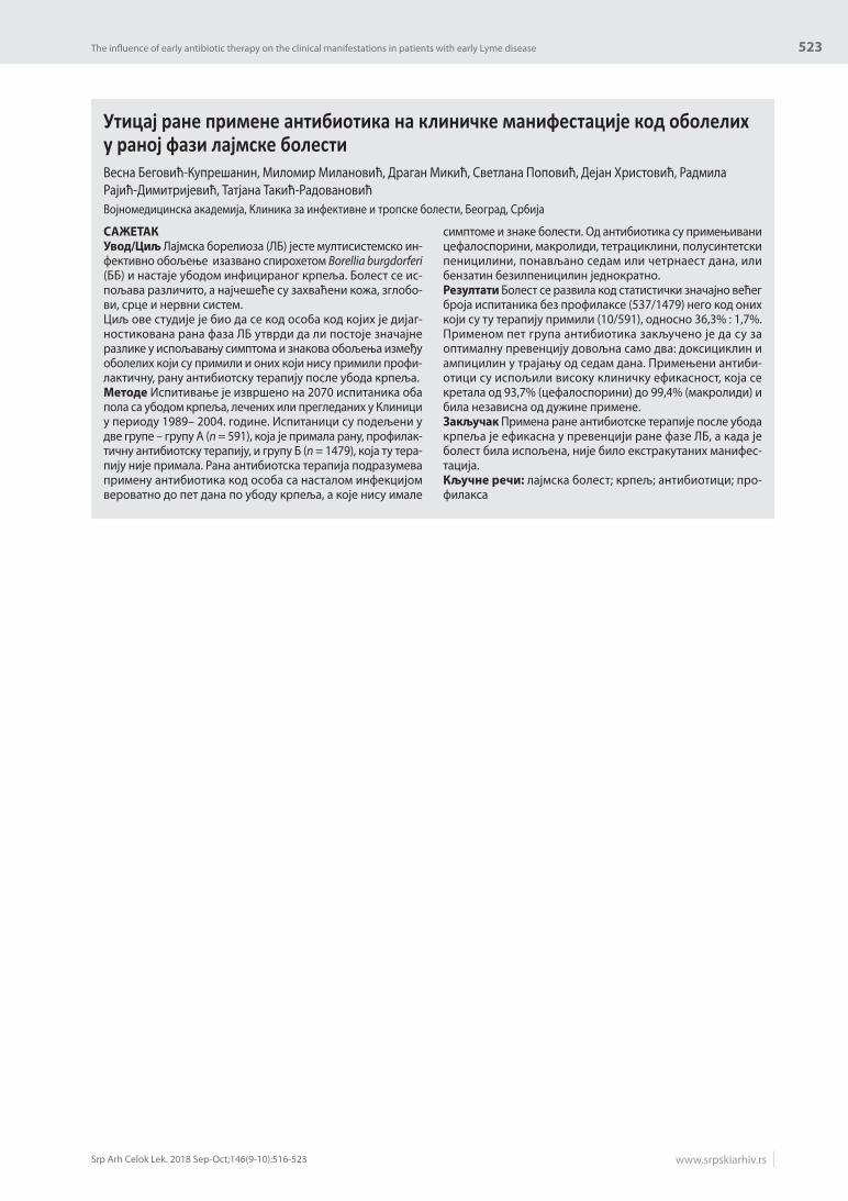
523
Srp Arh Celok Lek. 2018 Sep-Oct;146(9-10):516-523 www.srpskiarhiv.rs
САЖЕТАК Увод/Циљ Лајмска борелиоза (ЛБ) јесте мултисистемско ин-фективно обољење изазвано спирохетом Borellia burgdorferi (ББ) и настаје убодом инфицираног крпеља. Болест се ис-пољава различито, а најчешеће су захваћени кожа, зглобо-ви, срце и нервни систем. Циљ ове студије је био да се код особа код којих је дијаг-ностикована рана фаза ЛБ утврди да ли постоје значајне разлике у испољавању симптома и знакова обољења између оболелих који су примили и оних који нису примили профи-лактичну, рану антибиотску терапију после убода крпеља. Методе Испитивање је извршено на 2070 испитаника оба пола са убодом крпеља, лечених или прегледаних у Клиници у периоду 1989– 2004. године. Испитаници су подељени у две групе – групу А (n = 591), која је примала рану, профилак-тичну антибиотску терапију, и групу Б (n = 1479), која ту тера-пију није примала. Рана антибиотска терапија подразумева примену антибиотика код особа са насталом инфекцијом вероватно до пет дана по убоду крпеља, а које нису имале
симптоме и знаке болести. Од антибиотика су примењивани цефалоспорини, макролиди, тетрациклини, полусинтетски пеницилини, понављано седам или четрнаест дана, или бензатин безилпеницилин једнократно. Резултати Болест се развила код статистички значајно већег броја испитаника без профилаксе (537/1479) него код оних који су ту терапију примили (10/591), односно 36,3% : 1,7%. Применом пет група антибиотика закључено је да су за оптималну превенцију довољна само два: доксициклин и ампицилин у трајању од седам дана. Примењени антиби-отици су испољили високу клиничку ефикасност, која се кретала од 93,7% (цефалоспорини) до 99,4% (макролиди) и била независна од дужине примене. Закључак Примена ране антибиотске терапије после убода крпеља је ефикасна у превенцији ране фазе ЛБ, а када је болест била испољена, није билo екстракутаних манифес-тација.Кључне речи: лајмска болест; крпељ; антибиотици; про-филакса
Утицај ране примене антибиотика на клиничке манифестације код оболелих у раној фази лајмске болестиВесна Беговић-Купрешанин, Миломир Милановић, Драган Микић, Светлана Поповић, Дејан Христовић, Радмила Рајић-Димитријевић, Татјана Такић-РадовановићВојномедицинскa академијa, Клиника за инфективне и тропске болести, Београд, Србија
The infl uence of early antibiotic therapy on the clinical manifestations in patients with early Lyme disease
524
Correspondence to:
Danijela BATINIĆ-ŠKIPINA
Sokolska 15
73300 Foča
Republic of Srpska
Bosnia and Herzegovina
DOI: https://doi.org/10.2298/SARH170707177B
UDC: 618.19-006.6-076:577.1
SUMMARYIntroduction/Objective Activation of insulin-like growth factor receptor (IGF-1R) results in cell transi-tion from growth phase to synthesis phase of cell cycle. Breast cancer is categorized into prognostic and therapeutic subtypes based upon hormone receptor, estrogen receptor (ER), and progesterone receptor (PR) expression and human epidermal growth factor receptor 2 (HER-2) expression.The objective of this study was to examine the expression of IGF-1R in а specific subtype invasive breast cancer and its correlation with basic histopathological and immunohistochemical prognostic parameters. Methods Formalin-fixed paraffin-embedded tumor samples were obtained from 129 female patients with invasive breast cancer (I–III disease stage) with the follow-up ranging 36–108 months (average 48 months). For immunohistochemical staining, we used monoclonal antibodies for ER, PR, IGF-1R, and polyclonal antibody for HER-2.Results IGF-1R inversely correlated with tumor stage (p = 0.017), tumor grade (p = 0.001), HER-2 (p = 0.003), whereas significant positive correlation was found with multifocality/multicentricity of breast cancer (p = 0.036), ER (p = 0.001) and PR (p = 0.0001) expression. Cox-regression analysis for relapse-free survival (RFS) showed that disease stage (p = 0.039) and HER-2 (p = 0.033) were independent prognostic factors. IGF-1R did not predict clinical outcome in patients with breast cancer (p = 0.488, Kaplan–Meier test for RFS). Conclusion Patients with low stage and grade hormone-dependent breast cancer had a significantly higher IGF-1R expression than patients with triple negative or HER-2 overexpressed cancer. The present findings also highlight that IGF-1R expression in multicentric/multifocal breast cancer supports the key roles in tumor initiation. Keywords: insulin-like growth factor 1 receptor (IGF-1R); hormone-dependent breast cancer; HER-2
ORIGINAL ARTICLE / ОРИГИНАЛНИ РАД
Immunohistochemical evaluation of insulin-like growth factor receptor 1 in breast cancerDanijela Batinić-Škipina1, Radmil Marić1, Ljiljana Tadić-Latinović2, Dražan Erić1, Nenad Lalović1
1University Hospital Foča, Foča, Republic of Srpska, Bosnia and Herzegovina;2University Clinical Centre of the Republic of Srpska, Banjaluka, Republic of Srpska, Bosnia and Herzegovina
Received • Примљено: July 7, 2017
Accepted • Прихваћено: September 19, 2017
Online fi rst: September 29, 2017
INTRODUCTION
Insulin receptor family represents an activator of class II tyrosine kinase with three members: insulin receptor (IR), insulin-like growth factor receptor 1 (IGF-1R), and insulin-like growth factor receptor 2 (IGF-2R). IR activation influ-ences metabolic activity in vertebrates. IGF-1R activating results in proliferation and differen-tiation of cells. IGF-2R is structurally and func-tionally different from the IR and IGF-1R, it is a monomer without tyrosine kinase activ-ity. IGF-1R is a dimmer made of α and β sub-units and has the same structure as the IR with which it builds hybrid receptors (IR/IGF-1R) [1, 2, 3]. IRs can be activated by insulin and two insulin-like growth factors (IGFs): insulin-like growth factor 1 (IGF1) and insulin-like growth factor 2 (IGF2). Many cells have been identi-fied as producing as well as responding to the IGFs, including fibroblasts, chondrocytes, os-teoblasts, granulosa cells, and epithelial breast cells. In circulation, IGF1 and IGF2 are at-tached to six insulin-growth binding proteins (IGFBP 1–6) and protected from the action of proteases (Figure 1). IGF-1R together with the hormone receptors regulates the develop-
ment of the epithelium of the normal glan-dular breast tissue [4, 5]. Breast cancers are categorized into subtypes based on immuno-histochemical hormone receptors expression (ER and PR) and human epidermal growth factor 2 (HER-2) expression. There are two major groups: hormone-dependent/luminal breast cancer involves luminal A (ER+, PR+, HER-2/-, Ki67low) and luminal B (ER+, PR+/-, HER-2+/-, Ki67high); hormone-independent/basal-like breast cancer involves triple negative (TNBC) breast cancer (ER-, PR-, HER-2-) and HER-2 overexpressed (ER-, PR-, HER-2+). The TNBC subtype does not express therapeutically targetable ER, PR, or HER-2 receptors, mak-ing the aggressive subtype difficult to treat [6]. Nowadays, IGF-1R makes an attractive target for investigation for a different type of malig-nancy and anticancer therapy. The prognostic and predictive role of IGF-1R in breast cancer is still unknown. The optimal cut-point and standardized immunohistochemical expres-sion of this receptor are subjects of discussion [7]. A few studies have examined the relation-ship of the IGF-1R expression according to the hormone and HER-2 and resistance to anties-trogen therapy [8, 9]. Some in vitro studies have
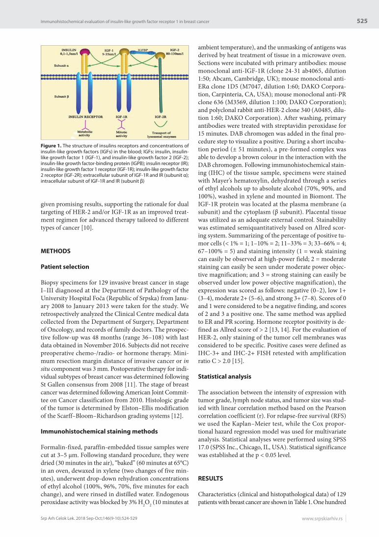
525
Srp Arh Celok Lek. 2018 Sep-Oct;146(9-10):524-529 www.srpskiarhiv.rs
given promising results, supporting the rationale for dual targeting of HER-2 and/or IGF-1R as an improved treat-ment regimen for advanced therapy tailored to different types of cancer [10].
METHODS
Patient selection
Biopsy specimens for 129 invasive breast cancer in stage I–III diagnosed at the Department of Pathology of the University Hospital Foča (Republic of Srpska) from Janu-ary 2008 to January 2013 were taken for the study. We retrospectively analyzed the Clinical Centre medical data collected from the Department of Surgery, Department of Oncology, and records of family doctors. The prospec-tive follow-up was 48 months (range 36–108) with last data obtained in November 2016. Subjects did not receive preoperative chemo-/radio- or hormone therapy. Mini-mum resection margin distance of invasive cancer or in situ component was 3 mm. Postoperative therapy for indi-vidual subtypes of breast cancer was determined following St Gallen consensus from 2008 [11]. The stage of breast cancer was determined following American Joint Commit-tee on Cancer classification from 2010. Histologic grade of the tumor is determined by Elston–Ellis modification of the Scarff–Bloom–Richardson grading systems [12].
Immunohistochemical staining methods
Formalin-fixed, paraffin-embedded tissue samples were cut at 3–5 μm. Following standard procedure, they were dried (30 minutes in the air), “baked” (60 minutes at 65°C) in an oven, dewaxed in xylene (two changes of five min-utes), underwent drop-down rehydration concentrations of ethyl alcohol (100%, 96%, 70%, five minutes for each change), and were rinsed in distilled water. Endogenous peroxidase activity was blocked by 3% H2O2 (10 minutes at
ambient temperature), and the unmasking of antigens was derived by heat treatment of tissue in a microwave oven. Sections were incubated with primary antibodies: mouse monoclonal anti-IGF-1R (clone 24-31 ab4065, dilution 1:50; Abcam, Cambridge, UK); mouse monoclonal anti-ERα clone 1D5 (M7047, dilution 1:60; DAKO Corpora-tion, Carpinteria, CA, USA); mouse monoclonal anti-PR clone 636 (M3569, dilution 1:100; DAKO Corporation); and polyclonal rabbit anti-HER-2 clone 340 (A0485, dilu-tion 1:60; DAKO Corporation). After washing, primary antibodies were treated with streptavidin peroxidase for 15 minutes. DAB chromogen was added in the final pro-cedure step to visualize a positive. During a short incuba-tion period (± 51 minutes), a pre-formed complex was able to develop a brown colour in the interaction with the DAB chromogen. Following immunohistochemical stain-ing (IHC) of the tissue sample, specimens were stained with Mayer’s hematoxylin, dehydrated through a series of ethyl alcohols up to absolute alcohol (70%, 90%, and 100%), washed in xylene and mounted in Biomont. The IGF-1R protein was located at the plasma membrane (α subunit) and the cytoplasm (β subunit). Placental tissue was utilized as an adequate external control. Stainability was estimated semiquantitatively based on Allred scor-ing system. Summarizing of the percentage of positive tu-mor cells (< 1% = 1; 1–10% = 2; 11–33% = 3; 33–66% = 4; 67–100% = 5) and staining intensity (1 = weak staining can easily be observed at high-power field; 2 = moderate staining can easily be seen under moderate power objec-tive magnification; and 3 = strong staining can easily be observed under low power objective magnification), the expression was scored as follows: negative (0–2), low 1+ (3–4), moderate 2+ (5–6), and strong 3+ (7–8). Scores of 0 and 1 were considered to be a negative finding, and scores of 2 and 3 a positive one. The same method was applied to ER and PR scoring. Hormone receptor positivity is de-fined as Allred score of > 2 [13, 14]. For the evaluation of HER-2, only staining of the tumor cell membranes was considered to be specific. Positive cases were defined as IHC-3+ and IHC-2+ FISH retested with amplification ratio C > 2.0 [15].
Statistical analysis
The association between the intensity of expression with tumor grade, lymph node status, and tumor size was stud-ied with linear correlation method based on the Pearson correlation coefficient (r). For relapse-free survival (RFS) we used the Kaplan–Meier test, while the Cox propor-tional hazard regression model was used for multivariate analysis. Statistical analyses were performed using SPSS 17.0 (SPSS Inc., Chicago, IL, USA). Statistical significance was established at the p < 0.05 level.
RESULTS
Characteristics (clinical and histopathological data) of 129 patients with breast cancer are shown in Table 1. One hundred
Figure 1. The structure of insulins receptors and concentrations of insulin-like growth factors (IGFs) in the blood; IGFs: insulin, insulin-like growth factor 1 (IGF-1), and insulin-like growth factor 2 (IGF-2); insulin-like growth factor-binding protein (IGPB); insulin receptor (IR); insulin-like growth factor 1 receptor (IGF-1R); insulin-like growth factor 2 receptor (IGF-2R); extracellular subunit of IGF-1R and IR (subunit α); intracellular subunit of IGF-1R and IR (subunit β)
Immunohistochemical evaluation of insulin-like growth factor receptor 1 in breast cancer
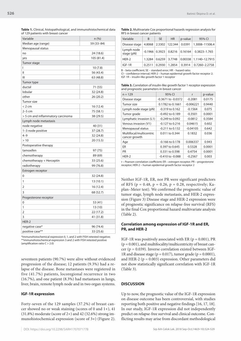
526
Srp Arh Celok Lek. 2018 Sep-Oct;146(9-10):524-529DOI: https://doi.org/10.2298/SARH170707177B
seventeen patients (90.7%) were alive without evidenced progression of the disease; 12 patients (9.3%) had a re-lapse of the disease. Bone metastases were registered in five (41.7%) patients, locoregional recurrence in two (16.7%), and one patient (8.3%) had metastases in lungs, liver, brain, remote lymph node and in two organ systems.
IGF-1R expression
Forty-seven of the 129 samples (37.2%) of breast can-cer showed no or weak staining (scores of 0 and 1+), 41 (31.8%) moderate (score of 2+) and 42 (32.6%) strong im-munohistochemical expression (score of 3+) (Figure 2).
Neither IGF-1R, ER, nor PR were significant predictors of RFS (p = 0.48, p = 0.26, p = 0.28, respectively; Ka-plan–Meier test). We confirmed the prognostic value of tumor stage, lymph node metastasis, and HER-2 expres-sion (Figure 3) Disease stage and HER-2 expression were of prognostic significance on relapse-free survival (RFS) in the final Cox proportional hazard multivariate analysis (Table 2).
Correlation among exp ression of IGF-1R and ER,
PR, and HER-2
IGF-1R was positively associated with ER (p = 0.001), PR (p = 0.001), and multifocality/multicentricity of breast can-cer (p = 0.039). Inverse correlation existed between IGF-1R and disease stage (p = 0.017), tumor grade (p = 0.0001), and HER-2 (p = 0.003) expression. Other parameters did not show statistically significant correlation with IGF-1R (Table 3).
DISCUSSION
Up to now, the prognostic value of the IGF-1R expression on disease outcome has been controversial, with studies reporting both positive and negative findings [16, 17, 18]. In our study, IGF-1R expression did not independently predict on relapse-free survival and clinical outcome. Con-flicting results may arise from discordant methodological
Table 1. Clinical, histopathological, and immunohistochemical data of 129 patients with breast cancer
Variable n (%)Median age (range) 59 (33–84)Menopausal statusno 24 (18.6)yes 105 (81.4)Tumor stageI 10 (7.8)II 56 (43.4)III 63 (48.8)Tumor typeductal 71 (55)lobular 32 (24.8)other 26 (20.2)Tumor size< 2 cm 16 (12.4)2–5 cm 75 (58.1)> 5 cm and inflammatory carcinoma 38 (29.5)Lymph node metastasisnode negative 40 (31)1–3 node positive 37 (28.7)4–9 32 (24.8)> 10 20 (15.5)Postoperative therapy
tamoxifen 97 (75)
chemotherapy 89 (69)
chemotherapy + Herceptin 33 (25.6)
radiotherapy 99 (76.8)Estrogen receptor
0 32 (24.8)
1 13 (10.1)
2 16 (12.4)
3 68 (52.7)
Progesterone receptor0 53 (41)1 13 (10)2 22 (17.2)3 41 (31.8)HER-2 negative case* 96 (74.4)positive case** 33 (25.6)
*Immunohistochemical expression 0, 1, and 2 with FISH retested negative;**immunohistochemical expression 3 and 2 with FISH retested positive (amplifi cation ratio C > 2.0)
Table 2. Multivariate Cox proportional hazards regression analysis for RFS in breast cancer patients
Variable B SE HR p-value 95% CI
Disease stage 4.8068 2.3302 122.344 0.0391 1.3008–11506.4
Lymph node stage (pN) -0.1966 0.3923 0.8216 0.16164 0.3823–1.765
HER-2 1.3284 0.6259 3.7748 0.00338 1.1140–12.7915
IGF-1R 0.2511 0.2930 1.2854 0.3914 0.7260–2.2758
B – beta coeffi cient; SE – standard error; HR – hazard ratio; CI – confi dence interval; HER-2 – human epidermal growth factor receptor 2; IGF-1R – insulin-like growth factor 1 receptor
Table 3. Correlation of insulin-like growth factor 1 receptor expression and prognostic parameters in breast cancer
n = 129 95% CI r p-valueDisease stage -0.3671 to -0.0372 -0.2081 0.0175
Tumor size 0.1782 to 0.1661 -0.006221 0.9440Lymph node stage (pN) -0.319 to 0.162 -0.1564 0.075Tumor grade -0.492 to-0.189 -0.3501 0.0001Lymphatic invasion (L1) -0.249 to 0.092 -0.0812 0.3584Venous invasion (V1) -0.127 to 0.216 0.04615 0.602Menopausal status -0.211 to 0.132 -0.04105 0.642Multifocal/multicentric cancer growth
0.011 to 0.344 0.1832 0.036
Age -0.166 to 0.178 0.006337 0.943ER 0.397 to 0.645 0.5328 0.0001PR 0.331 to 0.598 0.4754 0.0001HER-2 -0.410 to -0.088 -0.2567 0.003
r – Pearson correlation coeffi cient; ER – estrogen receptor; PR – progesterone receptor; HER-2 – human epidermal growth factor receptor 2
Batinić-Škipina D. et al.

527
Srp Arh Celok Lek. 2018 Sep-Oct;146(9-10):524-529 www.srpskiarhiv.rs
approaches, distinct molecular subtypes studied, genetic differences between different populations, and tumor het-erogeneity. Our study demonstrated high expression (score of 2+ or 3+) of IGF-1R in 64.4% of the samples. This is in line with some other studies [19]. Up to 50% of breast tumors express the activated form of IGF-1R. In our study, IGF-1R was predominantly expressed in well-differentiat-ed and hormone-dependent breast cancers. IGF-1R and the ER are critical for mammary gland development. The ER and the IGF pathway show dynamic and intricate in-terference, resulting in bidirectional regulation of expres-sion and activity. ER transcriptionally upregulates IGF-1R expression. Positive correlation exists between cyclin D1 and ER expression, which has already been explained in both experimental and clinical studies, because ER acts as the main mitogen stimulator in breast cancer [20]. The role of IGF-1R in mammary stem cell maintenance and a necessity for lineage differentiation suggest that aber-rantly expressed IGF-1R may be capable of enhancing cell potential and changing cell fate in a tumor, perhaps even in tumors composed of fully differentiated cells. As discussed
above, the IGF-1R expression is essential for driving lu-minal alveolar differentiation, linking IGF-1R to the lumi-nal lineage [21, 22]. Furthermore, many studies indicate a down-regulation of IGF-1R upon cancer progression, whereas others report elevated levels in metastatic stages. Once cancer has been confirmed, the importance of IGF-1R for disease progression remains unclear. In our study, IGF-1R was highly expressed in patients with early breast cancer and overall positively associated with good prog-nostic variables. We have indicated the decrease of IGF-1R expression with disease progression. High-level IGF-1R expression had low stage breast cancer with multiple/mul-ticentric unilateral or bilateral growth. We emphasize that IGF-1R could have effects in early phases of development of luminal breast cancer. Numerous in vitro studies dem-onstrate IGF-1R as a driver of self-renewal, stem cell sur-face markers, migration, and invasion in both normal and cancerous tissues and tumor initiation in hepatic, lung, prostate, and breast cancers [23]. Approximately 40–60% of ER-positive tumors express IGF-1R, while expression in ER-negative tumors is only 10–20%. Considering the cor-relation of IGF-1R with hormone-dependent tumor type and early stage, we assume that ER/IGF-1R axis might rep-resent a distinct proliferative pathway during breast cancer development. Other studies report that IGF-1R is a recep-tor expressed in the basaloid type breast cancer and has a role in anti-HER-2 resistance (Herceptin) [24]. We found a negative correlation between IGF-1R-overexpressed and HER-2-positive breast cancer. In general, IGF-1R correlates with good prognostic markers, such as ER and PR-positivity and HER-2-negativity. However, the IGF-1R expression has differential effects in different breast cancer subtypes. For example, its expression has been shown to be positively correlated with improved breast cancer-specific survival among patients with ER-positive tumors, while its expression was associated with an infe-rior prognosis in patients with HER2-overexpressing or triple-negative tumors. In models of breast cancer cells that overexpress HER-2, anti-HER-2 activity is disrupted by increased expression of IGF-1R. Nowadays, antibody-based molecular therapies have been developed for HER-2. IGF-1R can form heterodimers with the HER-2 tyrosine kinase and contribute to the development of resistance to HER-2 inhibition with the monoclonal antibody. An asso-ciation between IGF-1R and HER-2 in IGF-1R-dependent
a b c
Figure 2. Immunohistochemical expression of insulin-like growth factor 1 receptor in breast cancer (formalin-fixed paraffin-embedded sections, ×40); the expression was scored according to area and intensity of membranous or citoplasmatic staining: a) score 1+; b) score 2+; c) score 3+
Figure 3. Relapse-free survival analysis (Kaplan–Meier test) prognostic value of a) insulin-like growth factor 1 receptor (IGF-1R), b) human epidermal growth factor receptor 2, c) lymph node metastases, and d) disease stage
Immunohistochemical evaluation of insulin-like growth factor receptor 1 in breast cancer
528
Srp Arh Celok Lek. 2018 Sep-Oct;146(9-10):524-529
tumor transformation has been reported in mammary luminal epithelial cells, indicating that the IGF-1/HER-2 cross-talk may occur via autocrine and paracrine signal-ing. A recent study concluded that neoadjuvant therapy can induce changes in the IGF-1R expression. Therefore, there are many studies with opposite results [25, 26]. It is possible that IGF-1R expression is dependent not only on the specific cell type and disease stage, but also it is dependent on specific therapy and another factor. In some other tumors, like lung cancer, the expression of IGF-1R correlated with a less favorable outcome [27]. This indi-cates that IGF-1R activities might be not only diverse but also tissue-specific. To test this hypothesis, we evaluated the protein expression of the most important components
of the IGF-1R signaling pathway in hormone-dependent breast cancer and their significance according to the tumor subtypes. This clearly indicates other functions of IGF-1R that are not related to cell cycle progression and tumor aggressiveness, which may include cell differentiation and growth arrest.
CONCLUSION
IGF-1R is particularly important for the establishment and maintenance of the transformed phenotype and for the survival of tumor cells with anchorage-independent growth in breast carcinoma with luminal differentiation.
1. Belfiore A, Frasca F, Pandini G, Sciacca L, Vigneri R. Insulin receptor isoforms and insulin receptor/insulin-like growth factor receptor hybrids in physiology and disease. Endocr Rev. 2009; 30(6):586–623.
2. Papaioannou A, Kuyucak S, Kuncic Z. Elucidating the activation mechanism of the Insulin-family proteins with molecular dynamics simulations. PLoS One. 2016; 11(8):e0161459.
3. Pandini G, Frasca F, Mineo R, Sciacca L, Vigneri R, Belfiore A. Insulin/insulin-like growth factor I hybrid receptors have different biological characteristics depending on the insulin receptor isoform involved. J Biol Chem. 2002; 277:39684–95.
4. De Meyts P, Whittaker J. Structural biology of insulin and IGF1 receptors: implications for drug design. Nat Rev Drug Discov. 2002; 1(10):769–83.
5. Shi J, Aronson KJ, Grundy A, Kobayashi LC, Burstyn I, Schuetz JM, et al. Polymorphisms of Insulin-Like Growth Factor 1 pathway genes and breast cancer risk. Front Oncol. 2016; 6:136.
6. Parise CA, Caggiano V. Breast cancer subtypes based on ER/PR and HER-2 expression: comparison of clinicopathologic features and survival. Cancer Epidemiol. 2014; 2014:469251.
7. Yerushalmi R, Gelmon KA, Leung S, Gao D, Cheang M, Pollak M, et al. Insulin-like growth factor receptor (IGF-1R) in breast cancer subtypes. Breast Cancer Res Treat. 2012; 132(1):131–42.
8. Yan S, Jiao X, Li K, Li W, Zou H. The impact of IGF-1R expression on the outcomes of patients with breast cancer: a meta-analysis. Onco Targets Ther. 2015; 8:279–87.
9. Chan JY, LaPara K, Yee D. Disruption of insulin receptor function inhibits proliferation in endocrine-resistant breast cancer cells. Oncogene. 2016; 135(32): 4235–43.
10. de Groot S, Charehbili A, van Laarhoven HW, Mooyaart AL, Dekker-Ensink NG, van de Ven S, et al. Dutch Breast Cancer Research Group. Insulin-like growth factor 1 receptor expression and IGF1R 3129G > T polymorphism are associated with response to neoadjuvant chemotherapy in breast cancer patients: results from the NEOZOTAC trial (BOOG 2010-01). Breast Cancer Res. 2016; 18(1):3
11. Singh P, Alex JM, Bast F. Insulin receptor (IR) and insulin-like growth factor receptor 1 (IGF-1R) signaling systems: novel treatment strategies for cancer. Med Oncol. 2014; 31(1):805.
12. Goldhirsch A, Ingle JN, Gelber, RD, Coates AS, Thürlimann B, Senn HJ. Thresholds for therapies: highlights of the St Gallen international expert consensus on the primary therapy of early breast cancer 2009. Ann Oncol. 2009; 20(8):1319–29.
13. Bloom HJ, Richardson WW. Histological grading and prognosis in breast cancer; a study of 1409 cases of which 359 have been followed for 15 years. Br J Cancer. 1957; 11(3):359–77.
14. Hammond ME, Hayes DF, Dowsett M, Allred DC, Hagerty KL, Badve S, et al. American Society of Clinical Oncology/College of American Pathologists guideline recommendations for
immunohistochemical testing of estrogen and progesterone receptors in breast cancer (unabridged version). Arch Pathol Lab Med. 2010; 134(7):e48–72.
15. Wesoła M, Jeleń M. A Comparison of IHC and FISH Cytogenetic Methods in the Evaluation of HER2 Status in Breast Cancer. Adv Clin Exp Med. 2015; 24(5):899–903.
16. Sun WY, Yun HY, Song YJ, Kim H, Lee OJ, Nam SJ, et al. Insulin-like growth factor 1 receptor expression in breast cancer tissue and mammographic density. Mol Clin Oncol. 2015; 3(3):572–80.
17. Farabaugh SM, Boone DN, Lee AV. The role of IGF1R in breast cancer subtypes, stemness, and lineage differentiation. Front Endocrinol (Lausanne). 2015; 24(6):59.
18. Taunk NK, Goyal S, Moran MS, Yang Q, Parikh R, Haffty BG. Prognostic significance of IGF-1R expression in patients treated with breast-conserving surgery and radiation therapy. Radiother Oncol. 2010; 96(2):204–8.
19. Railo MJ, von Smitten K, Pekonen F. The prognostic value of insulin-like growth factor-I in breast cancer patients. Results of a follow-up study on 126 patients. Eur J Cancer. 1994; 30A(3):307–1.
20. Mohammadizadeh F, Hani M, Ranaee M, Bagheri M. Role of cyclin D1 in breast carcinoma. J Res Med Sci. 2013; 18(12):1021–5.
21. Bhargava R, Beriwal S, McManus K, Dabbs DJ. Insulin-like growth factor receptor-1 (IGF-1R) expression in normal breast, proliferative breast lesions, and breast carcinoma. Appl Immunohistochem Mol Morphol. 2011; 19(3):218–25.
22. Mountzios G, Aivazi D, Kostopoulos I, Kourea HP, Kouvatseas G, Timotheadou E, et al. Differential expression of the insulin-like growth factor receptor among early breast cancer subtypes. PLoS One. 2014; 9(3):e91407.
23. Denduluri SK, Idowu O, Wang Z, Liao Z, Yan Z, Mohammed MK, et al. Insulin-like growth factor (IGF) signaling in tumorigenesis and the development of cancer drug resistance. Genes Dis. 2015; 2(1):13–25.
24. Lu Y, Zi X, Zhao Y, Mascarenhas D, Pollak M. Insulin-like growth factor-I receptor signaling and resistance to trastuzumab (Herceptin). J Natl Cancer Inst. 2001; 93(24):1852–7.
25. Heskamp S, Boerman OC, Molkenboer-Kuenen JD, Wauters CA, Strobbe LJ, Mandigers CM, et al. Upregulation of IGF-1R expression during neoadjuvant therapy predicts poor outcome in breast cancer patients. PLoS One. 2015; 10(2):e0117745.
26. Browne BC, Eustace AJ, Kennedy S, O'Brien NA, Pedersen K, McDermott MS, et al. Evaluation of IGF1R and phosphorylated IGF1R as targets in HER2-positive breast cancer cell lines and tumours. Breast Cancer Res Treat. 2012; 136(3):717–27.
27. Yeo CD, Park KH, Park CK, Lee SH, Kim SJ, Yoon HK, et al. Expression of insulin-like growth factor 1 receptor (IGF-1R) predicts poor responses to epidermal growth factor receptor (EGFR) tyrosine kinase inhibitors in non-small cell lung cancer patients harboring activating EGFR mutations. Lung Cancer. 2015; 87(3):311–7.
REFERENCES
Batinić-Škipina D. et al.
DOI: https://doi.org/10.2298/SARH170707177B

529
Srp Arh Celok Lek. 2018 Sep-Oct;146(9-10):524-529 www.srpskiarhiv.rs
САЖЕТАКУвод/Циљ Активација рецептора инсулину-сличног фактора раста 1 (IGF-1R) изазива покретање ћелијског циклуса из фазе раста (G1) у фазу синтезе (S). Oболели од карцинома дојке се деле на специфичне терапијске и прогностичке групе у зависности од експресије хормонских рецептора, естрогених (ER) и прогестеронских (PR), и експресије рецеп-тора хуманог епидермалног фактора раста 2 (HER-2).Циљ рада је откривање степена експресије IGF-1R у тумор-ском ткиву код одређених терапијских група оболелих од карцинома дојке и његова корелација са важећим патохис-толошким и имунохистохемијским прогностичким пара-метрима. Методе Истраживање је спроведено на 129 укалупљених узорака инвазивног карцинома дојке код жена (у стадијуму болести I–III) уз постоперативно праћење тока болести 48 (36–108) месеци. За имунохистохемијско бојење коришћена су моноклонска антитела за визуализацију: ER, PR, IGF-1R и поликлонално антитело за HER-2. Резултати Експресија IGF-1R је била у негативној корелацији са стадијумом болести (p = 0,017), степеном диферентова-
ности тумора (p = 0,001) и експресијом HER-2 (p = 0,003). Позитивна корелација овог рецептора налазила се између мултифокалног/мултицентричног макроскопског начина раста карцинома дојке (p = 0,036) и експресије ЕR (p = 0,001) и PR (p = 0,0001). Коксова регресиона анализа времена без прогресије болести (RFS) показала је да стадијум боле-сти (p = 0,039) и HER-2 (p = 0,033) представљају независне прогностичке варијабле. Експресија IGF-1R није имала утицај на клинички ток болести код особа са раком дојке (p = 0,488, Каплан–Мајер тест за RFS). Закључак Болесници оперисани у почетном стадијуму бо-лести са дијагностикованим добро диферентованим, хор-монски зависним раком дојке имају већу IGF-1R експресију у односу на болеснике са троструко негативним и HER-2 ам-плификованим туморима дојке. Повећана IGF-1R експресија код карцинома са мултифокалним/мултицентричним мак-роскопским начином раста указује на значајну улогу овог рецептора у фази настанка тумора.
Кључне речи: рецептор инсулину-сличног фактора раста 1 (IGF-1R); хормонски зависни рак дојке; HER-2
Процена имунохистoхемијске експресије рецептора инсулину-cличног факторa раста 1 у карциному дојке Данијела Батинић-Шкипина1, Радмил Марић1, Љиљана Тадић-Латиновић2, Дражан Ерић1, Ненад Лаловић1
1Универзитетска болница Фоча, Фоча, Република Српска, Босна и Херцеговина;2Универзитетски клинички центар Републике Српске, Бања Лука, Република Српска, Босна и Херцеговина
Immunohistochemical evaluation of insulin-like growth factor receptor 1 in breast cancer

530
Correspondence to:
Anna A. Peresypkina
85, Pobeda St.
Belgorod, 308015
Russia
Received • Примљено: August 14, 2017
Accepted • Прихваћено: January 17, 2018
Online fi rst: February 6, 2018
DOI: https://doi.org/10.2298/SARH170814006P
UDC: 617.735-085
SUMMARYIntroduction/Objective The objective of this paper was to increase the effectiveness of pharmacological correction of retinal ischemia-reperfusion by using minoxidil.Methods The research was carried out on 180 Wistar rats. A modification of the retinal ischemia-reperfu-sion model was used, in which an increase in intraocular pressure is carried out by mechanical pressure (110 mmHg) to the front chamber of the eye for 30 minutes. Protective effects of minoxidil at a dose 0.5 mg/kg on the model of retinal ischemia-reperfusion were estimated by the changes in the level of retinal microcirculation (laser Doppler flowmetry), electroretinogram amplitude, morphometry of retinal layers after 1 hour and 72 hours of reperfusion.Results Minoxidil at a dose 0.5 mg/kg of rat mass improves retinal microcirculation, its electrophysi-ological state after 1 hour and 72 hours of reperfusion, and prevents the development of degenerative changes in the retina caused by ischemic damage to a greater extent than recombinant erythropoietin at a dose of 50 IU/kg and sildenafil at a dose of 0.5 mg/kg in monotherapy. The protective effects of minoxidil were eliminated by the preliminary administration of glibenclamide at a dose of 5 mg/kg, which indicates the presence of the preconditioning effect of minoxidil, realized through adenosine triphosphate-dependent potassium channels.Conclusion Minoxidil at a dose of 0.5 mg/kg of rat mass protects the retina from ischemic-reperfusion injury. Protective effects of minoxidil are carried out by a preconditioning action, as evidenced by the lack of positive effects with the administration of glibenclamide.Keywords: ischemia-reperfusion; retina; minoxidil; erythropoietin; sildenafil; ATP-dependent potassium channels
ORIGINAL ARTICLE / ОРИГИНАЛНИ РАД
Pharmacological correction of retinal ischemia/reperfusion by minoxidilAnna A. Peresypkina, Victoria O. Gubareva, Elena A. Levkova, Anna S. Shabelnikova, Mikhail V. PokrovskiiBelgorod State University, Belgorod, Russia
INTRODUCTION
Local circulatory disorders in the branches of retinal artery are observed in diabetic reti-nopathy, hypertensive retinopathy, degenera-tive diseases of the retina, optic nerve atrophy vascular origin, traumatic eye injury, ischemic neuropathy [1, 2, 3].
Studying the way of how to improve tissue tolerance to ischemia is an actual problem of modern experimental and clinical pharmacolo-gy. Up to now, the treatment of ischemic retinal conditions was done by use of angioprotectors, antioxidants, fibrinolytics, anticoagulants and others. As the authors note, due to the insta-bility and short-term effects after using these drugs in combination with other drugs and physiotherapy treatments is necessary to seek out a more effective way to improve blood cir-culation and increase resistance to ischemic retinal tissue having a specific orientation [4].
Thus, an important task is to find new, spe-cific and highly effective means for correcting of retinal ischemia.
Therefore, the objective of the study is to increase the effectiveness of pharmacological correction of retinal ischemia-reperfusion by using minoxidil.
METHODS
Experiments were carried out on 180 Wistar rats weighing 250 ± 25 g. For the study, the rats were taken with no external signs of disease, passed quarantine regime.
Ethical principles of handling laboratory animals were observed in accordance with the European Convention for the Protection of Vertebral Animals Used for Experimental and Other Scientific Purposes, CETS No. 123.
Minoxidil, 0.5 mg/kg, was administered intragastrically (i/g) once 1 hour before isch-emia-reperfusion modeling.
Recombinant erythropoietin (EPO) was administered intraperitoneally (i/p) once at a dose of 50 IU/kg 30 minutes before pathology modeling for the purpose of preconditioning as a reference drug [5].
Animals received i/p injection of sildenafil at a dose of 0.5 mg/kg once 30 minutes before pathology modeling.
Glibenclamide was administered at a dose of 5 mg/kg i/g once 1 hour before ischemia-reperfusion modeling.
Ischemia-reperfusion injury of the retina was simulated under anesthesia (chloral hydrate, 300 mg/kg of animal body weight, i.p.) by applying mechanical pressure (110 mmHg) to the ante-rior eye chamber for 30 minutes [4].

531
Srp Arh Celok Lek. 2018 Sep-Oct;146(9-10):530-533 www.srpskiarhiv.rs
The experiment included two series of animals (with an assessment of the parameters after 1 hour and 72 hours of re-perfusion), nine groups in each series, 10 rats in each group.
The measuring of the level of retinal microcirculation of rats was carried out by laser Doppler flowmetry (LDF) after 1 hour and 72 hours of reperfusion [6]. Registration was carried out by MP150 data acquisition and analysis systems and the TSD144 needle-type sensor, with Ac-qKnowledge 4.2 software (BIOPAC Systems, Inc., Goleta, CA, USA). After animal anesthesia, assessment of mi-crocirculation level was carried out at 10 points on the circumference of the eyeball; the recording duration of the microcirculation level readings at one point was 20 seconds. From the microcirculation level results at every point, the average value was calculated, which was taken as the indicator of the microcirculation level in the retina of the experimental animal. The value of microcirculation in the animal group was calculated as the average of the val-ues obtained from each experimental animal in the group.
To perform electroretinography (ERG), after 1 hour and 72 hours of reperfusion, rats were kept in the dark for 30 minutes, then, anesthetized (chloral hydrate, 300 mg/kg, i/p), and fixed on the table isolated from electromagnetic radiation [7]. Strobe flash of white light that was connect-ed to the STM200 stimulator (Biopac Systems, Inc.) and placed behind the animal; ERG registration was carried out in response to a single stimulation. Evoked biopo-tentials were run at a frequency of 1–1000 Hz, amplified, averaged, and presented graphically on the screen using the MP150 data acquisition and analysis system and the aforementioned software. The ERG recording was carried out for 0.5 seconds in each rat in the groups. To assess the degree of retinal ischemia, the ratio of the amplitudes of the a- and b-waves of ERG, the coefficient b/a was evalu-ated [7]. From 10 values received, the mean was derived for each group, which was introduced into the protocol.
After the LDF and ERG, eyes with surrounding tissues were subjected to enucleation in both series of experi-ments. Eyes with immediately adjacent tissues were fixed in 10% formalin solution for histological research. After fixation, the eyes were cut into two parts in the meridian direction strictly through the center, and both halves were poured into paraffin according to the standard procedure. Sections for the standard histological examination were stained with hematoxylin-eosin. A descriptive study of his-tological preparations was performed under a Axio Scope A1 microscope (Carl Zeiss Microscopy GMbH, Jena, Ger-many). The morphometric studies were performed on the Mikmed-5 microscope with the use of the Micro-Analysis View software (LOMO, JSC, Saint Petersburg, Russia) [8].
RESULTS
After the pathology modeling after 1 hour and 72 hours of reperfusion, microcirculation was measured in the retina by LDF, electrophysiological condition of the retina was deter-mined by ERG, the extirpation of animals and enucleation of the eyes for morphological studies was carried out.
After the pathology modeling, microcirculation level measurement was performed after 1 hour and 72 hours of reperfusion by LDF. The results obtained after 1 hour of reperfusion are presented in Table 1.
Table 1. The level of retinal microcirculation after 1 h and 72 hour of reperfusion (M ± m), perfusion units
No. Experimental groups
Level of microcirculation
after 1 hour of reperfusion,PU (n = 10)
Level of microcirculation after 72 hours of reperfusion, PU
(n = 10)1 Intact 738.9 ± 37.6 743.9 ± 5.02 Control (ischemia) 1,155.0 ± 51.9* 353.3 ± 11.7*3 Control + MIN 751.3 ± 21.8у 739.5 ± 14.1у4 Control + EPO 798.5 ± 12.3у 724.0 ± 4.1у5 Control + SIL 832.3 ± 20.1у 711.5 ± 15.3у6 Control + Glib 1,135.8 ± 31.2* 359.4 ± 10.3*
7 Control + MIN + Glib 1,149.8 ± 18.6* 361.1 ± 10.9*
8 Control + EPO + Glib 1,148.3 ± 15.3* 372.3 ± 13.4*9 Control + SIL + Glib 1,151.2 ± 31.9* 360.3 ± 12.1*
Glib – glibenclamide; SIL – sildenafi l; PU – perfusion units; MIN – minoxidil; EPO – recombinant erythropoietin; *p < 0.05 compared to intact rats;уp < 0.05 compared to the control group
The level of microcirculation after ischemia modeling in the control group reached 1,155.0 ± 51.9 PU after 1 hour of reperfusion, which was significantly higher than the value in the group of intact animals (p < 0.05).
With the correction of pathology by minoxidil (MIN), microcirculation level in the retina after 1 hour of reperfu-sion decreased to 751.3 ± 21.8 PU, which was significantly different from the control group (p < 0.05).
With the correction of pathology by EPO, microcircula-tion level in the group was reduced to 798.5 ± 12.3 PU and was significantly different from the values in the control group (p < 0.05).
Introduction of glibenclamide, a blocker of adenos-ine triphosphate-sensitive (ATP-sensitive) potassium channels, prevented the reduction of microcirculation in groups with correction by MIN, EPO, sildenafil (SIL); this confirms the preconditioning action of these drugs in studied doses on retinal ischemia-reperfusion model on rats after 1 hour of reperfusion.
The level of microcirculation after the pathology mod-eling in the control group after 72 hours of reperfusion was 353.3 ± 11.7 PU, which was significantly lower than in the group of intact animals (p < 0.05). In the group with the correction by MIN, this rate increased to 739.5 ± 14.1 PU (p < 0.05), which was significantly different from the values in the control group.
Correction of the modeled pathology by EPO led to an increase of microcirculation level in the group to 724.0 ± 4.1 PU, which was significantly different from the values in the control group (p < 0.05).
Introduction of glibenclamide in groups with MIN-cor-rection, EPO-correction, and SIL-correction prevented the improvement of the microcirculation level after 72 hours of reperfusion.
Pharmacological correction of retinal ischemia/reperfusion by minoxidil
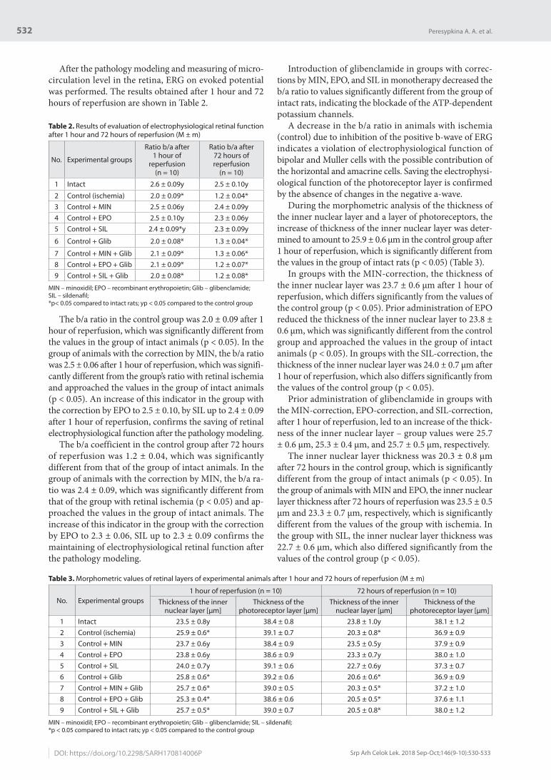
532
Srp Arh Celok Lek. 2018 Sep-Oct;146(9-10):530-533DOI: https://doi.org/10.2298/SARH170814006P
After the pathology modeling and measuring of micro-circulation level in the retina, ERG on evoked potential was performed. The results obtained after 1 hour and 72 hours of reperfusion are shown in Table 2.
Table 2. Results of evaluation of electrophysiological retinal function after 1 hour and 72 hours of reperfusion (M ± m)
No. Experimental groups
Ratio b/a after 1 hour of
reperfusion(n = 10)
Ratio b/a after 72 hours of reperfusion
(n = 10)
1 Intact 2.6 ± 0.09у 2.5 ± 0.10у
2 Control (ischemia) 2.0 ± 0.09* 1.2 ± 0.04*3 Control + MIN 2.5 ± 0.06у 2.4 ± 0.09у4 Control + EPO 2.5 ± 0.10у 2.3 ± 0.06у5 Control + SIL 2.4 ± 0.09*у 2.3 ± 0.09у
6 Control + Glib 2.0 ± 0.08* 1.3 ± 0.04*
7 Control + MIN + Glib 2.1 ± 0.09* 1.3 ± 0.06*8 Control + EPO + Glib 2.1 ± 0.09* 1.2 ± 0.07*9 Control + SIL + Glib 2.0 ± 0.08* 1.2 ± 0.08*
MIN – minoxidil; EPO – recombinant erythropoietin; Glib – glibenclamide; SIL – sildenafi l; *p< 0.05 compared to intact rats; уp < 0.05 compared to the control group
The b/a ratio in the control group was 2.0 ± 0.09 after 1 hour of reperfusion, which was significantly different from the values in the group of intact animals (p < 0.05). In the group of animals with the correction by MIN, the b/a ratio was 2.5 ± 0.06 after 1 hour of reperfusion, which was signifi-cantly different from the group’s ratio with retinal ischemia and approached the values in the group of intact animals (p < 0.05). An increase of this indicator in the group with the correction by EPO to 2.5 ± 0.10, by SIL up to 2.4 ± 0.09 after 1 hour of reperfusion, confirms the saving of retinal electrophysiological function after the pathology modeling.
The b/a coefficient in the control group after 72 hours of reperfusion was 1.2 ± 0.04, which was significantly different from that of the group of intact animals. In the group of animals with the correction by MIN, the b/a ra-tio was 2.4 ± 0.09, which was significantly different from that of the group with retinal ischemia (p < 0.05) and ap-proached the values in the group of intact animals. The increase of this indicator in the group with the correction by EPO to 2.3 ± 0.06, SIL up to 2.3 ± 0.09 confirms the maintaining of electrophysiological retinal function after the pathology modeling.
Introduction of glibenclamide in groups with correc-tions by MIN, EPO, and SIL in monotherapy decreased the b/a ratio to values significantly different from the group of intact rats, indicating the blockade of the ATP-dependent potassium channels.
A decrease in the b/a ratio in animals with ischemia (control) due to inhibition of the positive b-wave of ERG indicates a violation of electrophysiological function of bipolar and Muller cells with the possible contribution of the horizontal and amacrine cells. Saving the electrophysi-ological function of the photoreceptor layer is confirmed by the absence of changes in the negative a-wave.
During the morphometric analysis of the thickness of the inner nuclear layer and a layer of photoreceptors, the increase of thickness of the inner nuclear layer was deter-mined to amount to 25.9 ± 0.6 μm in the control group after 1 hour of reperfusion, which is significantly different from the values in the group of intact rats (p < 0.05) (Table 3).
In groups with the MIN-correction, the thickness of the inner nuclear layer was 23.7 ± 0.6 μm after 1 hour of reperfusion, which differs significantly from the values of the control group (p < 0.05). Prior administration of EPO reduced the thickness of the inner nuclear layer to 23.8 ± 0.6 μm, which was significantly different from the control group and approached the values in the group of intact animals (p < 0.05). In groups with the SIL-correction, the thickness of the inner nuclear layer was 24.0 ± 0.7 μm after 1 hour of reperfusion, which also differs significantly from the values of the control group (p < 0.05).
Prior administration of glibenclamide in groups with the MIN-correction, EPO-correction, and SIL-correction, after 1 hour of reperfusion, led to an increase of the thick-ness of the inner nuclear layer – group values were 25.7 ± 0.6 μm, 25.3 ± 0.4 μm, and 25.7 ± 0.5 μm, respectively.
The inner nuclear layer thickness was 20.3 ± 0.8 μm after 72 hours in the control group, which is significantly different from the group of intact animals (p < 0.05). In the group of animals with MIN and EPO, the inner nuclear layer thickness after 72 hours of reperfusion was 23.5 ± 0.5 μm and 23.3 ± 0.7 μm, respectively, which is significantly different from the values of the group with ischemia. In the group with SIL, the inner nuclear layer thickness was 22.7 ± 0.6 μm, which also differed significantly from the values of the control group (p < 0.05).
Table 3. Morphometric values of retinal layers of experimental animals after 1 hour and 72 hours of reperfusion (M ± m)
No. Experimental groups1 hour of reperfusion (n = 10) 72 hours of reperfusion (n = 10)
Thickness of the inner nuclear layer [μm]
Thickness of the photoreceptor layer [μm]
Thickness of the inner nuclear layer [μm]
Thickness of the photoreceptor layer [μm]
1 Intact 23.5 ± 0.8y 38.4 ± 0.8 23.8 ± 1.0у 38.1 ± 1.22 Control (ischemia) 25.9 ± 0.6* 39.1 ± 0.7 20.3 ± 0.8* 36.9 ± 0.93 Control + MIN 23.7 ± 0.6у 38.4 ± 0.9 23.5 ± 0.5у 37.9 ± 0.94 Control + EPO 23.8 ± 0.6y 38.6 ± 0.9 23.3 ± 0.7у 38.0 ± 1.05 Control + SIL 24.0 ± 0.7y 39.1 ± 0.6 22.7 ± 0.6у 37.3 ± 0.76 Control + Glib 25.8 ± 0.6* 39.2 ± 0.6 20.6 ± 0.6* 36.9 ± 0.97 Control + MIN + Glib 25.7 ± 0.6* 39.0 ± 0.5 20.3 ± 0.5* 37.2 ± 1.08 Control + EPO + Glib 25.3 ± 0.4* 38.6 ± 0.6 20.5 ± 0.5* 37.6 ± 1.19 Control + SIL + Glib 25.7 ± 0.5* 39.0 ± 0.7 20.5 ± 0.8* 38.0 ± 1.2
MIN – minoxidil; EPO – recombinant erythropoietin; Glib – glibenclamide; SIL – sildenafi l; *p < 0.05 compared to intact rats; уp < 0.05 compared to the control group
Peresypkina A. A. et al.

533
Srp Arh Celok Lek. 2018 Sep-Oct;146(9-10):530-533 www.srpskiarhiv.rs
DISC USSION
The search of new methods of retinoprotection for possible reduction of the damaging effect of ischemia, formed in various systemic diseases, is an urgent task of pharmacol-ogy and ophthalmology. Segment of drugs for the treatment of vascular diseases of the eye such as complication from hypertension, diabetes, and others, would be expedient to expand due to an increase in morbidity and lack of funds for targeted correction of ischemic lesions of the eye vessels.
Based on the fact that electrophysiological studies often have a decisive importance in the early and differential diagnosis of retinal disorders, to study the correction of functional changes in the retina, researcher must conduct a comprehensive analysis, including electroretinography and microcirculation research [9]. Analysis of the dynam-ics of retinal electrogenesis allows to evaluate the nature and topography of retinal disorders and to identify the most labile hypoxic retinal structure, as well as their reac-tion to the correction by the medications.
The most pronounced retinal protective action was ob-served in groups with pharmacological preconditioning by minoxidil at a dose of 0.5 mg/kg after 1 hour and 72 hours of reperfusion.
A single i/g injection of minoxidil at a dose of 0.5 mg/kg, 60 minutes before modeling of retinal ischemia-
reperfusion, significantly reduced the level of retinal microcirculation after 1 hour of reperfusion, and saved the retinal electrophysiological activity, which was also confirmed by morphometrics of retinal layers. We found that minoxidil prevents the retinal layers’ damages caused by ischemic injury to a greater extent than recombinant erythropoietin at a dose of 50 IU/kg and sildenafil at a dose of 0.5 mg/kg in monotherapy after 1 hour of reperfusion and degenerative changes of retinal layers after 72 hours of reperfusion.
Prior administration of glibenclamide at a dose of 5 mg/kg eliminated the positive effects of minoxidil, erythropoi-etin, and sildenafil, which confirms the implementation of retinal protection by preconditioning with the participa-tion of ATP-dependent potassium channels.
CONCLUSION
Minoxidil at a dose of 0.5 mg/kg protects the retina from ischemic-reperfusion injury better than recombinant erythropoietin at a dose of 50 IU/kg and sildenafil at a dose of 0.5 mg/kg in monotherapy.
Protective effects of minoxidil are carried out by a pre-conditioning action as evidenced by the lack of positive effects with administration of glibenclamide.
REFERENCES
1. Wong TY, Mitchell P. Hypertensive retinopathy. N Engl J Med. 2004; 351:2310–7.
2. Konstantinidis L, Guex-Crosier Y. Hypertension and the eye. Curr Opin Ophthalmol. 2016; 27(6):514–21.
3. Ojha S, Balaji V, Sadek B, Rajesh M. Beneficial effects of phytochemicals in diabetic retinopathy: experimental and clinical evidence. Eur Rev Med Pharmacol Sci. 2017; 21(11):2769–83.
4. Shabelnikova AS, Lutsenko VD, Pokrovskii MV, Peresypkina AA, Korokin MV, Gudyrev OS, et al. Protective effects of recombinant erythropoietin in ischemia of the retina: The role of mechanisms of preconditioning. Research Journal of Medical Sciences. 2015; 9(4):200–3.
5. Nekoui A, Blaise G. Erythropoietin and Nonhematopoietic Effects. Am J Med Sci. 2017; 353(1):76–81.
6. Rajkumar DS, Faitelson AV, Gudyrev OS, Dubrovin GM, Pokrovski MV, Ivanov AV. Comparative evaluation of enalapril and losartan in pharmacological correction of experimental osteoporosis and fractures of its background. J Osteoporos. 2013; 2013:325693.
7. Kurata K, Hosono K, Hotta Y. Long-Term Clinical Course in a Patient with Complete Congenital Stationary Night Blindness. Case Rep Ophthalmol. 2017; 8(1):237–44.
8. Shabelnikova AS, Peresypkina AA, Pokrovski MV, Kashuba AS, Netrebenko AS. Analysis of the protective properties of erythropoietin and nicorandil on the basis of the model of the retina ischemia/reperfusion. Res J Pharm Biol Chem Sci. 2014; 5(6):1335–39.
9. Zhiqing C, Ke Y, Wen X, Renyi W. Inhibition of synthesis of calpain by inhibitor E-64d in the retina subjected to ischemia/reperfusion. Molecular Biology. 2008; 42(2):258–64.
САЖЕТАКУвод/Циљ Циљ студије јесте да се побољша ефикасност фармаколошке корекције исхемије-реперфузијe мрежњаче миноксидилом.Методе Истраживање је спроведено на 180 пацова врсте вистар. Коришћена је модификација модела исхемије-ре-перфузије, при чему се повећање интраокуларног притис-ка вршило механичким притиском (110 mmHg) на предњу комору ока у трајању од 30 минута. Заштитни ефекти ми-ноксидила у дози од 0,5 mg/kg процењене су на основу про-мена микроциркулације мрежњаче (ласерска доплер-фло-уметрија), амплитуде електроретинограма, морфометрије слојева мрежњаче после једног и 72 сата од реперфузије.Резултати Миноксидил у дози од 0,5 mg/kg масе пацова побољшава микроциркулацију мрежњаче, њено електро-
физиолошко стање после једног и 72 сата реперфузије и спречава развој дегенеративних промена слојева мрежњаче изазваних исхемијом више него монотерапија рекомбинант-ним еритропоетином у дози од 50 IU/kg и силденафилом у дози од 0,5 mg/kg. Заштитно дејство миноксидила елими-нише се давањем глибенкламида у дози од 5 mg/kg, што доказује прекондиционирани ефекат код миноксидила, који је остварен кроз АТП-зависне калијумове канале.Закључци Миноксидил у дози од 0,5 mg/kg штити мрежњачу од исхемије / реперфузионе повреде. Заштитни ефекат ми-ноксидила се реализује посредством прекондиционирања, што доказује недостатак позитивних ефеката на примену глибенкламида.Кључне речи: исхемија-реперфузија; ретина; миноксидил; еритропоетин; силденафил; АТП-зависни канали калијума
Фармаколошка корекција ретиналне исхемије/реперфузије миноксидаломАна А. Пересипкина, Викторија О. Губарева, Јелена А. Левкова, Ана С. Шабељникова, Михаил В. ПокровскијБелгородски државни универзитет, Белгород, Русија
Pharmacological correction of retinal ischemia/reperfusion by minoxidil

534
Correspondence to:
Zorica TONČIĆ
Clinical Center of Montenegro
Ljubljanska bb, 81000 Podgorica
Montenegro
Received • Примљено: October 16, 2017
Accepted • Прихваћено: October 24, 2017
Online fi rst: October 31, 2017
DOI: https://doi.org/10.2298/SARH171016192T
UDC: 617.7-76-056.262; 159.946.4-056.262
ORIGINAL ARTICLE / ОРИГИНАЛНИ РАД
Reading performance of low vision children after using low vision aidsZorica Tončić1, Nataša Jovović1, Nada Šakotić2, Veselinka Milović2, Katarina Janićijević3, Mirjana Petrović Janićijević3, Svetlana Jovanović3
1Clinical Center of Montenegro, Clinic for Children’s Diseases, Ophthalmology Service, Podgorica, Montenegro;2University of Montenegro, Faculty of Philosophy, Nikšić, Montenegro;3University of Kragujevac, Faculty of Medical Sciences, Department of Ophthalmology, Kragujevac, Serbia
SUMMARYIntroduction/Objective The objectives of the paper are to assess the causes of low vision (LV) in pediatric population in Montenegro and to evaluate the influence of low vision aids (LVA) on reading performance regarding the speed of reading and the understanding of the read text.Methods A prospective study was conducted on 40 “treatable” LV children what represent all registered LV children in Montenegro. All participants read the same text before and after using LVA. Reading rate was calculated as the number of words read per minute. Functional speed of reading was calculated as the ratio of the rate of reading and the understanding of the read text multiplied by 100.Results The study comprised 40 LV children with the mean age of 12.60 ± 4.06 years (20 boys and 20 girls). The most common cause of LV in children were premature retinopathy (10/40 or 25%), retinitis pigmentosa (8/40 or 20%), optic nerve anomaly (5/40 or 13%), degenerative myopia (4/40 or 10%), macular dysgenesis (4/40 or 10%), Stargardt disease (3/40 or 7%), optic nerve atrophy (2/40 or 5%), and albinism (2/40 or 5%). Nystagmus was found in 11 LV children or 28% of the group. LVA were prescribed to all of them. Reading speed before vs. after LVA use was 36.58 ± 35.60 vs. 73.83 ± 27.05 words/minute (p < 0.001), while functional reading was 26.00 ± 30.43 vs. 59.41 ± 29.34 (p < 0.001).Conclusion LV children demonstrate a significant improvement in reading performance by using LVA. Keywords: low vision aid; low vision children; reading performance
INTRODUCTION
According to the International Classification of Diseases 10, there are four levels of visual func-tion: normal vision, moderate visual impair-ment (VI), severe VI, and blindness [1]. Mod-erate and severe VI are grouped under the term “low vision” (LV). “Functional” LV is defined as presenting best-corrected visual acuity (BCVA) in the better-seeing eye of less than 0.3 and more than 0.05 according to the World Health Organization (WHO) criteria, VA < 0.3 and VA ≥ 0.05 (or according to the United States (US) criteria VA < 0.5 and VA ≥ 0.1) and as blind-ness (WHO criteria VA < 0.05; US criteria VA < 0.1) [1]. LV cannot be improved or corrected with medical treatment, surgery, nor with con-ventional glasses or with contact lenses. Unlike total blindness, most individuals with LV have some degree of useful, residual sight even when vision loss is significant. The WHO estimated that 19 million children worldwide are visually impaired; of these, 1.4 million are irreversibly blind [2]. The International Classification of Functioning, Disability and Health adopted by the WHO can be used as a framework to com-prehensively describe the problems of persons with VI and the environmental factors which influence their lives [3]. Surely, LV significant-
ly interferes with the functioning of a person. Common subjective complains of LV persons include the loss of central and/or peripheral vi-sion, constricted visual filed, abnormal color perception, generalized haze, blurred vision, extreme light sensitivity, and night blindness. LV patients represent unique challenge in oph-thalmic and optometric care.
Very few LV clinics are available even in the most developed of countries. The Low Vision Service for pediatric patients at the Clinical Center of Podgorica, Montenegro (a referral tertiary health care center in Montenegro) has been established in 2013. The Service is a pio-neer of LV service in the region, covering all the needs of LV children, including education, training, and sight rehabilitation. A team of trained specialists with comprehensive, multi-disciplinary approach to LV children has the purpose to help them maximize the remaining functional vision and maintain their indepen-dence in daily living. Podgorica pediatric LV service meets the WHO recommendation for establishing LV centers to fight avoidable child-hood blindness. Namely, since 2004, the WHO in partnership with Lions Clubs International has established a global network of 45 child-hood blindness centers in 35 countries for the preservation, restoration, or rehabilitation of

535
Srp Arh Celok Lek. 2018 Sep-Oct;146(9-10):534-537 www.srpskiarhiv.rs
sight in children [2]. The LV center in Podgorica is excep-tional since all registered LV children have the use of LV aids (LVA) at no cost to them.
According to the available data, there are approximate-ly 1,000 blind and visually impaired persons of all ages registered in Montenegro. The exact number of blind LV children is unknown. There is no precise national regis-ter on the prevalence of childhood VI, but, according to available sources (referral associations, primary health care registers, etc.), it has been estimated that there are 200 VI children and 50–60 LV children in Montenegro at this moment. The assessment of the causes of VI is impor-tant to develop preventive and therapeutic strategies. The standardized protocol for reporting causes of blindness in children, with coding instructions and a database for sta-tistical analysis developed by the International Centre for Eye Health, the WHO Collaborating Centre for Blindness Prevention, and the WHO, also serves as a mechanism to monitor changing pattern of childhood blindness [4].
The objectives of this paper are to assess the causes of LV in pediatric population in Montenegro and to evalu-ate the influence of using LV aids in reading performance regarding the speed of reading and the understanding of the read text.
METHODS
A prospective study was conducted on 40 “treatable” LV children, who represent all registered LV children in Montenegro. Including criteria were the BCVA ranging 0.05–0.3 in the better-seeing eye and age less than 17 years. The term ‘treatable LV person’ represents a person who has demonstrated an improvement in reading or in distance vision using LVA. Children were recruited from registers of Association of Blind and LV Persons (one association with eight local branches), followed by two schools for special education of blind and LV children (in Podgorica and Bijela) and primary eye care registers from all over the country. All amblyopic children (215 of them) under-went complete ophthalmological examination. In total, 40 children met the criteria of a ‘treatable LV person’. The study was performed in accordance with the tenets of the Declaration of Helsinki and approved by the Institutional Review Board (decision number 03/01-12238/2). Written informed consent was obtained from all parents.
All the participants read the same text before and after using LVA. The words were printed in seven lines with 1.5-line spacing (0.8 cm), with black letters on white background to enhance contrast; the font used was Times New Roman, the letter size was N12. Reading rate was calculated as the number of read words per minute. Un-derstanding of the read text was measured by a multiple-choice test with 20 questions related to the text. Scores were rated from 0 to 20 and presented as a percentage (for example, if an LV child had 12 correct answer, the score of understanding was 60%). Functional speed of reading was calculated as the ratio of the rate of reading and the understanding of the read text multiplied by 100.
Statistical analysis
All parameters were expressed as mean ± standard de-viation (SD). Differences between pre- and post-LVA use were evaluated by the Wilcoxon signed-ranks test. The level of statistical significance was set at 0.05. All statistical analyses were performed using the IBM SPSS Statistics for Windows, Version 20.0 (IBM Corp., Armonk, NY, USA).
RESULTS
Forty LV children with the mean age of 12.60 ± 4.06 years (20 boys and 20 girls) underwent complete ophthalmologi-cal examination and socio-epidemiological assessment. The mean age of the girls was 10.95 ± 4.16 years, while that of the boys was 14.25 ± 3.28 years. All the LV children lived in families with both biological parents.
Regarding the educational profile of fathers of the LV children, 20 of them had completed primary school (50%), 19 had secondary degree (47.5%), and one (2.5%) had ob-tained higher education. Among mothers, none had fin-ished higher education, 19 mothers had secondary school finished (47.5%), and 21 had completed primary school.
In the group of children with retinal dystrophy, eight children (20%) had retinopathy pigmentosa, in three cases (7%) was diagnosed Stargardt macular dystrophy. Develop-ment anomalies of the optic nerve had four children with optic hypoplasia and one with congenital coloboma of the optic disc. Associated macular and optic nerve anomalies were found in two cases, while four children had isolated macular hypoplasia. Causes of LV were presented in Table 1.
Table 1. Causes of low vision in children
Clinical finding Number of patients %Retinal dystrophy 11 27Premature retinopathy 10 25Macular dysgenesis 4 10Optic nerve anomaly 5 13Associated macular and optic nerve anomalies 2 5
Central nervous tumor 2 5Degenerative myopia 4 10Albinism 2 5Total 40 100
Optic atrophy in two cases was a consequence of in-tracranial tumor, but it was also found in two premature children and in one with albinism. Nystagmus was found in 11 cases (nine horizontal, one rotatory, and one verti-cal). Macular scars were diagnosed in two highly myopic cases and in three children with retinitis pigmentosa. Four children had esotropia, while two had exotropia.
BCVA concerning the better seeing eye was hand moving in eight children (20% of all); in 12 children (30% of all) the score was 0.1 while 20 children (50% of all) had a score of 0.2–0.4. In three children, fellow eye was blind, 16 children (40% of all) had BCVA described as hand movement only, while 21 children had less than 0.2 according to Snellen chart.
Reading performance of low vision children after using low vision aids

536
Srp Arh Celok Lek. 2018 Sep-Oct;146(9-10):534-537DOI: https://doi.org/10.2298/SARH171016192T
Regarding the refractive error, 27/40 children had myo-pia, 3/40 (7.5% of all) had hyperopia, 2/40 (5% of all) had astigmatism, while eight children had no possibility of be-ing optically corrected.
All the children accepted LVA for reading distance. In Table 2 are listed devices which they had used.
Table 2. List of low vision aids for reading prescribed to low vision children
Low vision aid Number of children %Electronic magnifier 17 42Ready fit prism 21 53Telescope 2 5Total 40 100
The speed of reading and understanding of content was measured before and one month after using of LVA for reading. The results are presented in Table 3. As it has been shown, speed and functional speed of reading has been significantly improved.
Table 3. Speed of reading and functional reading results in low vision children before and after using low vision aids
Tested parameterBefore
x– ± sd med. (min.–max.)
Afterx– ± sd med. (min.–max.)
p
Read
ing speed 36.58 ± 35.60
21.5 (0–120)73.83 ± 27.0581 (31–121) < 0.001a
functional reading
26.00 ± 30.4313.72 (0–108)
59.41 ± 29.3462.65 (14.40–114.95) < 0.001a
aWilcoxon signed-ranks test
DISCUSSION
VI results in different degrees of difficulty in performing daily activities and tasks. Great progress has been made in the development and deployment of intraocular LVA, such as implantable monocular telescope, followed by the global positioning system-based navigation system, loca-tion-aware LVA. LV children have particular and additional tasks concerning education, and reading is one of the core activities of their studies. As it is well known, LV and VI affect their sensorial development, physical, psychological, and social well-being. Socio-epidemiological or so-called external factors (i.e. education/employment and parental influence) can either facilitate or hinder participation [3]. In our study, all the parents had completed primary and ap-proximately half of them had completed secondary school. Finishing at least primary school is a legislative obligation that can explain a relatively high percentage of parents who had completed at least primary education level.
The commonest cause of LV among children and adoles-cents in Montenegro was retinal dystrophy – retinitis pig-mentosa (20%), followed by Stargardt disease (7%). Also present are premature retinopathy (25%), macular dysgen-esis (10%), and myopic degenerative changes (10%). The etiology of childhood VI in Montenegro includes 13% of those who had VI with coexisting neurological disability. In contrast, in Brazil, the most frequent finding was congenital
glaucoma (21.1%), while in a Sao Paolo study congenital glaucoma (30.6%) was found to have higher prevalence, followed by macular retinochoroiditis due to congenital toxoplasmosis (16.7%), congenital cataract (12.8%), retinal and macular inherited disorders (11.7%), and optic atrophy (9.8%) [4, 5]. Furthermore, Haddad et al. [5] reported that only 2% of children with congenital glaucoma had nor-mal visual acuity levels, while 29% had mild VI, 28% had moderate VI, 15% had severe VI, 11% had profound VI, and 15% had near blindness. Principal causes of blindness among VI children in New Zealand were cerebral VI in 61 children (42.4%), optic nerve atrophy in 18 children (12.5%), and retinal dystrophy in 13 children (9%). The main avoidable causes of blindness in 27 children (19%) were neonatal trauma, asphyxia in nine children (33%), and non-accidental injury in six children (22%) [6].
Causes of LV in childhood and adolescence in Africa and Asia differs from those in other parts of the world. In Nigeria, the most common causes were cataract (21%), fol-lowed by glaucoma (12.9%), but in as much as 43.6% of LV children, causes of blindness were found to be treatable [7],
while Olusanya et al. [8] reported that the most common cause of LV in children was albinism (24.4%) and optic atrophy (24.4%). In Ekiti State Special Education School, Nigeria, in a study conducted in May–June 2008 it was reported that the most common causes of VI are cataract (26.7%), glaucoma (20%), retinitis pigmentosa (16.7%), and posttraumatic phthisis bulbi (6.7%); blindness was avoidable in as much as 61% of the cases[9]. In Ethiopia, the most common causes of childhood VI were corneal disease/phthisis (62.4%), followed by optic nerve lesions (9.8%), cataract/aphakia (9.2%), and lesions of the uvea (8.8%). The etiology was unknown in 45.1% of the cases, while 68% of the cases were considered to be potentially avoidable [10]. Cataract and corneal damage are the lead-ing causes of LV in children in India as well. Even though the time difference between the studies is almost 20 years, the conclusions of both studies were that in prevention of avoidable blindness, it is very important to provide measles and rubella immunization and nutrition care [11].
Among LV chi ldren in Nepal, refractive error and am-blyopia (20.1%), retinitis pigmentosa (14.9%), and macu-lar dystrophy (13.4%) were the most common causes of pediatric VI. Nystagmus (50%) was the most common cause of LV in the one to five years age group, whereas refractive error and amblyopia were the major causes in the six to 10 and 11 to 16 years age group (17.6 and 22.9%, respectively) [12].
It is a widely accepted belief in clinical practice that children with VI can benefit from the use of an LVA [13]. To LV patients at the Instituto Brasileiro de Oftalmologia e Prevencao da Cegueira, telescopic system was the only optical aid indicated for distance (44%) and glasses were the most indicated aid for near vision (54.5%), which were prescribed as such [14]. In India, only 18% of LV children with coloboma, microcornea, and microphthalmos have been using telescopes, while a stand magnifier has been prescribed in 6% of children [15]. LV children from Mon-tenegro have a special possibility to be treated with the
Tončić Z. et al.

537
Srp Arh Celok Lek. 2018 Sep-Oct;146(9-10):534-537 www.srpskiarhiv.rs
most diverse range of LVA for large distance, intermediate distance, and short distance, respectively.
CONCLUSION
According to the published data, this is the first study tar-geting the influence of LVA use in LV children on reading performance, as well as the first report on demographic data and causes of LV among LV children in Montenegro. Our results indicate that LVA for reading or short distances significantly improves reading performance in LV children and should be applied in everyday practice.
NOTE
This paper is a part of the doctoral thesis by Zorica Tončić, titled “The influence of visual rehabilitation on the quality of life using low vision aids in low vision children.”
ACKNOWLEDGMENT
Authors would like to thank Dr. Vesna Jakšić, associate professor at the Faculty of Medicine of the University of Belgrade, who established the Low Vision Service at the Clinical Center of Podgorica, Montenegro.
1. Verhoeven VJ, Wong KT, Buitendijk GH, Hofman A, Vingerling JR, Klaver CC. Visual consequences of refractive errors in the general population. Ophthalmology. 2015; 122(1):101–9.
2. Global Data on Visual Impairments 2010. World Health Organization; 2012. Available at: http://www.who.int/blindness/GLOBALDATAFINALforweb.pdf?ua=1
3. Leissner J, Coenen M, Froehlich S, Loyola D, Cieza A. What explains health in persons with visual impairment? Health Qual Life Outcomes. 2014; 12:65.
4. Silva AM, Matos MH, Lima Hde C. [Low vision service at the Instituto Brasileiro de Oftalmologia e Prevencao da Cegueira (IBOPC): analysis of the patients examined on the first year of the department (2004)]. Arq Bras Oftalmol. 2010; 73(3):266–70.
5. Haddad MA, Lobato FJ, Sampaio MW, Kara-Jose N. Pediatric and adolescent population with visual impairment: study of 385 cases. Clinics (Sao Paulo). 2006; 61(3):239–46.
6. Chong CF, McGhee CN, Dai S. A Cross-sectional Study of Prevalence and Etiology of Childhood Visual Impairment in Auckland, New Zealand. Asia Pac J Ophthalmol (Phila). 2014; 3(6):337–42.
7. Omolase CO, Aina AS, Omolase BO, Omolade EO. Causes of blindness and visual impairment at the school for the blind owo, Nigeria. Ann Ib Postgrad Med. 2008; 6(1):49–52.
8. Olusanya B, Onoja G, Ibraheem W, Bekibele C. Profile of patients presenting at a low vision clinic in a developing country. BMC Ophthalmol. 2012; 12:31.
9. Onakpoya OH, Adegbehingbe BO, Omotoye OJ, Adeoye AO. Causes of blindness in a special education school. West Afr J Med. 2011; 30(1):47–50.
10. Kello AB, Gilbert C. Causes of severe visual impairment and blindness in children in schools for the blind in Ethiopia. Br J Ophthalmol. 2003; 87(5):526–30.
11. Waddell KM. Childhood blindness and low vision in Uganda. Eye (Lond). 1998; 12(Pt 2):184–92.
12. Uprety S, Khanal S, Morjaria P, Puri LR. Profile of paediatric low vision population: a retrospective study from Nepal. Clin Exp Optom. 2016; 99(1):61–5.
13. Schurink J, Cox RF, Cillessen AH, van Rens GH, Boonstra FN. Low vision aids for visually impaired children: a perception-action perspective. Res Dev Disabil. 2011; 32(3):871–82.
14. Silva AM, Matos MH, Lima Hde C. [Low vision service at the Instituto Brasileiro de Oftalmologia e Prevencao da Cegueira (IBOPC): analysis of the patients examined on the first year of the department (2004)]. Arq Bras Oftalmol. 2010; 73(3):266–70.
15. Hornby SJ, Adolph S, Gothwal VK, Gilbert CE, Dandona L, Foster A. Requirements for optical services in children with microphthalmos, coloboma and microcornea in southern India. Eye (Lond). 2000; 14 (Pt 2):219–24.
REFERENCES
САЖЕТАК Увод/Циљ Циљ овог рада је био да се утврде разлози сла-бовидости деце у Црној Гори и утицај специјалних помагала на брзину и на функционално читање код ове деце. Методe Проспективна студија спроведена је на 40 слабови-де деце која су користила специјална помагала. Сва деца су добила да прочитају исти текст пре и после кошћења пома-гала. Брзина читања је исказана као број прочиатних речи у минути, а функционално читање као однос између брзине читања и разумевања прочитаног × 100. Резултати Средњи узраст слабовиде деце је био 12,60 ± 4,06 година (20 дечака и 20 девојчица). Најчешћи разлози слабовидости су били: прематурна ретинопатија (10/40; 25%), пигментни ретинитис (8/40; 20%), аномалије оптич-
ког нерва (5/40; 13%), дегенеративна миопија (4/40; 10%), макуларне дизгенезије (4/40; 10%), Старгард дистрофија (3/40; 7%), атрофија оптичког нерва (2/40; 5%), албинизам (2/40; 5%). Нистагмус је дијагностикован код 11 деце (28%). Свој слабовидој деци су прописана помагала. Брзина читања пре у односу на брзину читања после коришћења помагала је била 36,58 ± 35,60, тј. 73,83 ± 27,05 речи/мин. (p < 0,001), док је функционално читање било 26,00 ± 30,43, тј. 59,41 ± 29,34 (p < 0,001). Закључак Слабовида деца показују значајно побољшање читања после коришћења специјалних помагала.
Кључне речи: помагала за слабовиде; слабовида деца; бр-зина читања
Брзина читања код слабовиде деце после коришћења помагала за слабовиде Зорица Тончић1, Наташа Јововић1, Нада Шакотић2, Веселинка Миловић2, Катарина Јанићијевић3, Мирјана Петровић-Јанићијевић3, Светлана Јовановић3
1Клинички центар Црне Горе, Дечја клиника, Служба офталмологије, Подгорица, Црна Гора; 2Универзитет Црне Горе, Филозофски факултет, Никшић, Црна Гора;3Универзитет у Крагујевцу, Факултет медицинских наука, Крагујевац, Србија
Reading performance of low vision children after using low vision aids

538
Correspondence to:
Ivo KEHAYOV
Department of Neurosurgery,
Medical University of Plovdiv
15A Vassil Aprilov Blvd
Plovdiv 4002
Bulgaria
Received • Примљено: October 16, 2017
Accepted • Прихваћено: March 7, 2018
Online fi rst: March 13, 2018
DOI: https://doi.org/10.2298/SARH171016024K
UDC: 616.831-005.1
SUMMARYIntroduction/Objective Subdural hematoma is one of the most common intracranial types of bleeding with high risk of disability and mortality.The aim of this study was to determine the influence of age, sex, acuteness, and etiology of subdural hematoma on short-term clinical outcome in these patients.Methods We retrospectively studied 288 patients who were diagnosed and operated on for subdural hematomas (SDH) with different etiology (traumatic and spontaneous) and acuteness (acute, subacute, and chronic) for a period of five years. Patients scored ≤ 5 points on the Glasgow Coma Scale at hospital admission were not included in this study. Clinical outcome was assessed by the modified Rankin Scale (mRS) score at hospital discharge. Descriptive statistics and logistic regression analysis were used to determine the effect of the investigated factors on short-term clinical outcome.Results Logistic regression analysis was conducted to predict degree of recovery (good = mRS ≤1 vs. poor = mRS ≥ 2 or death) using sex, age, acuteness, and etiology of SDH as predictive factors. It was es-tablished that the following three factors made a significant contribution to the outcome: age (p = 0.004), acuteness (p < 0.001), and etiology of a hematoma (p = 0.023), with acuteness being the strongest predic-tive factor. Sex was not a significant predictor, while age under 70 years and spontaneous origin of SDH were associated with lower mRS scores and had a positive effect on recovery chances.Conclusion Age, acuteness, and etiology of hematoma are important predictive factors that influence the short-term clinical outcome in patients with SDH. These parameters should be taken into account when giving prognosis for recovery chances to a patient’s family and relatives.Keywords: subdural hematoma; outcome; predictive factors; recovery; surgery
ORIGINAL ARTICLE / ОРИГИНАЛНИ РАД
The impact of age, gender, acuteness, and etiology on short-term clinical outcome in patients with subdural hematomas – international dual-center study Ivo Kehayov1, Aleksandar Kostić2, Borislav Kitov1, Vesna Nikolov2, Hristo Zhelyazkov1, Atanas Davarski1
1Department of Neurosurgery, Faculty of Medicine, Medical University of Plovdiv, Plovdiv, Bulgaria;2Clinical Center of Niš, Clinic for Neurosurgery, Niš, Serbia
INTRODUCTION
A subdural hematoma (SDH) is a common type of intracranial hemorrhage. The prevalence and total cost for a subdural hematoma has in-creased significantly in the last decade [1]. An acute intracranial subdural hematoma (ASDH) is commonly associated with high incidence of morbidity and mortality, despite its fatality rate has begun to decline with the developments in medicine and is currently around 14% [2, 3, 4]. On the other hand, some patients develop chronic SDH (CSDH) from causes other than head injury, such as brain surgery, neovascu-larization of the hematoma capsule, or coagula-tion factors. In addition, some factors, such as old age, alcoholism, coagulopathy, neurological status at admission, hematoma density, and ir-rigation, are reported to be correlated with the outcome [5]. However, these results are still controversial, and the influence of some im-portant predictive factors on clinical outcome of SDH, regardless of its etiology and acuteness, has not yet been fully elucidated.
Therefore, the aim of this study was to de-termine the influence of age, sex, acuteness, and etiology on short-term clinical outcome in patients with subdural hematomas.
METHODS
Study design
This international study was performed in two neurosurgical centers – the Clinic for Neuro-surgery, Clinical Center of Niš, Serbia, and the Clinic for Neurosurgery, St George University Hospital, Plovdiv, Bulgaria. It was approved by the institutional review boards, and informed consent was waived. Patient information was obtained via a retrospective review of medical records for the period between January 2011 and December 2015.
Patients
We identified and included a total of 288 pa-tients diagnosed and operated on for SDHs,
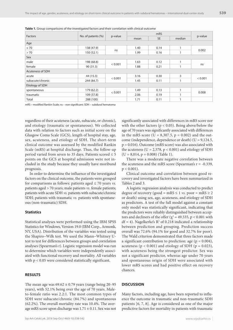
539
Srp Arh Celok Lek. 2018 Sep-Oct;146(9-10):538-542 www.srpskiarhiv.rs
regardless of their acuteness (acute, subacute, or chronic), and etiology (traumatic or spontaneous). We collected data with relation to factors such as initial score on the Glasgow Coma Scale (GCS), length of hospital stay, age, sex, acuteness, and etiology of SDH. The short-term clinical outcome was assessed by the modified Rankin Scale (mRS) at hospital discharge. Thus, the follow-up period varied from seven to 35 days. Patients scored ≤ 5 points on the GCS at hospital admission were not in-cluded in the study because they usually have moribund prognosis.
In order to determine the influence of the investigated factors on the clinical outcome, the patients were grouped for comparisons as follows: patients aged ≤ 70 years vs. patients aged > 70 years; male patients vs. female patients; patients with acute SDH vs. patients with subacute/chronic SDH; patients with traumatic vs. patients with spontane-ous (non-traumatic) SDH.
Statistics
Statistical analyses were performed using the IBM SPSS Statistics for Windows, Version 19.0 (IBM Corp., Armonk, NY, USA). Distribution of the variables was tested using the Shapiro–Wilk test. We used the Mann–Whitney U-test to test for differences between groups and correlation analyses (Spearman’s r). Logistic regression model was run to determine which variables were independently associ-ated with functional recovery and mortality. All variables with p < 0.05 were considered statistically significant.
RESULTS
The mean age was 69.62 ± 0.79 years (range being 20–95 years), with 52.1% being over the age of 70 years. Male-to-female ratio was 2.2:1. The most common types of SDH were subacute/chronic (84.7%) and spontaneous (62.2%). The overall mortality rate was 10.4%. The aver-age mRS score upon discharge was 1.71 ± 0.11. Sex was not
significantly associated with differences in mRS score nor with the other factors (p < 0.05). Being above/below the age of 70 years was significantly associated with differences in the mRS score (U = 8,307.5, p = 0.002) and the out-come (independence, dependence or death) (U = 9,124.5, p = 0.016). Outcome (mRS score) was also associated with the acuteness (U = 2,578, p < 0.001) and etiology of SDH (U = 8,014, p = 0.008) (Table 1).
There was a moderate negative correlation between the acuteness and the mRS score (Spearman’s r = -0.339, p < 0.001).
Clinical outcome and correlation between good re-covery and investigated factors have been summarized in Tables 2 and 3.
A logistic regression analysis was conducted to predict degree of recovery (good = mRS ≤ 1 vs. poor = mRS ≥ 2 or death) using sex, age, acuteness, and etiology of SDH as predictors. A test of the full model against a constant only model was statistically significant, indicating that the predictors were reliably distinguished between accep-tors and decliners of the offer (χ2 = 49.535, p < 0.001 with df = 4). Nagelkerke’s R2 of 0.218 indicated a relationship between prediction and grouping. Prediction success overall was 72.6% (94.1% for good and 32.7% for poor). The Wald criterion demonstrated that three factors made a significant contribution to prediction: age (p = 0.004), acuteness (p < 0.001) and etiology of SDH (p = 0.023), with acuteness being the strongest predictor. Sex was not a significant predictor, whereas age under 70 years and spontaneous origin of SDH were associated with lower mRS scores and had positive effect on recovery chances.
DISCUSSION
Many factors, including age, have been reported to influ-ence the outcome in traumatic and non-traumatic SDH patients [6, 7, 8]. Age is considered as one of the major predictive factors for mortality in patients with traumatic
Table 1. Group comparisons of the investigated factors and their correlation with clinical outcome
Factors No. of patents (%) p-valuemRS
p-valuemean SE median
Age≤ 70 138 (47.9)
ns1.40 0.14 1
0.002> 70 150 (52.1) 1.99 0.16 1Sexmale 198 (68.8)
< 0.0011.63 0.12 1
nsfemale 90 (31.3) 1.88 0.21 1Acuteness of SDHacute 44 (15.3)
< 0.0013.16 0.30 3
< 0.001subacute/chronic 244 (84.7) 1.45 0.11 1Etiology of SDHspontaneous 179 (62.2)
< 0.0011.49 0.13 1
0.008traumatic 109 (37.8) 2.06 0.19 1Total 288 (100) 1.71 0.11 1
mRS – modifi ed Rankin Scale; ns – non-signifi cant; SDH – subdural hematoma
The impact of age, gender, acuteness, and etiology on short-term clinical outcome in patients with subdural hematomas – international dual-center study

540
Srp Arh Celok Lek. 2018 Sep-Oct;146(9-10):538-542DOI: https://doi.org/10.2298/SARH171016024K
ASDH [9]. A recent study reported that the age of less or more than 77 years had been found to be an independent prognostic factor for the functional outcome in patients with CSDH [10]. Age is also indicated as a positive risk factor for a higher perioperative morbidity and mortal-ity [11]. On the other hand, other authors shared that despite significantly higher complication rate in elderly patients with CSDH, the clinical outcome at one month after surgery in patients older than 85 years was signifi-cantly better in comparison to patients younger than 85 years [11]. On the contrary, in a large, prospective, multicenter, observational cohort study carried out in the United Kingdom by Brennan et al. [12], over 1,205 patients with CSDH, showed that increasing patient age had independently predicted unfavorable functional out-comes [12]. Another large study conducted by Toi et al. [13], which included 63,358 patients with newly diagnosed CSDH, also confirmed that the percentage of poor out-comes at discharge tended to be higher in elderly patients. Several publications demonstrated that clinical outcome of patients over 70 years old who have received surgical
treatment for traumatic ASDH was significantly worse with the increase in age [14, 15, 16]. Our study found that patients with subdural bleeding older than 70 years had poorer short-term outcomes following surgery compared to those younger than 70 years. We identified the age less than 70 years as a significant predictor for better outcome in a mixed cohort of patients with SDH, regardless of its acuteness and etiology. Similar findings have been recently reported [17].
A recent study showed that premorbid impaired ac-tivities of daily living, consciousness disturbance, acute-to-chronic subdural hematoma, and death as outcomes at discharge were significantly more frequent in women than in men. Women had less frequent instances of good recov-ery. Female sex was also identified as a predictor of death at discharge [18]. In contrast, our study did not identify sex to be significantly associated with differences in out-come. Logistic regression analysis confirmed that sex was not a significant predictor of clinical outcome of patients with SDH, a fact also observed by other authors [9]. The prognosis for patients with ASDH remains poor, especially in elderly patients [19]. It has long been recognized that ASDH is often associated with intraparenchymal injuries and brain swelling. Hence, outcomes have historically been worse for patients with ASDH with mortality rates as high as 68% [20, 21]. Pathophysiology, patient populations, management strategies, and outcomes differ significantly between ASDH and CSDH [22]. As recently reported, patients with mixed acuteness or subacute/chronic SDH had significantly better three-month mRS with surgery compared to those with only ASDH [17]. In contrast, an-other study which investigated 45 patients over the age of 70 did not establish any change in the functional status from admission to follow-up in the groups of patients with ASDH and CSDH [23]. We also confirmed that acuteness of SDH was significantly correlated with functional recov-ery and outcome. Moreover, the acuteness was found to be the strongest predictor of clinical outcome. Our study suggested that patients with ASDH tend to have poorer outcome and lower chances for good recovery compared to patients with subacute/chronic SDH.
Patients with CSDH who reported a history of head injury are susceptible to poorer outcome [5]. However, SDH can develop spontaneously, without any history of sustained cranial trauma as a result of brain surgery, neovascularization of the hematoma capsule, or coagula-tion factors [5, 24, 25]. The etiology remains unknown in over 25% of cases because many patients have not experienced a prior traumatic event [26, 27]. We found significant correlation between etiology of SDH and clinical outcome. The etiology of SDH was also found to be a significant predictor of outcome. Our results in-dicated that patients with spontaneous (non-traumatic) SDH had better outcome and greater recovery chances after surgery than patients with traumatic SDH. One possible explanation is that traumatic SDH are often accompanied by a variety of diffuse parenchymal in-juries and cerebral edema that increase brain damage and worsen prognosis.
Table 2. Summary of clinical outcome and functional recovery of pa-tients with a subdural hematoma at discharge
Clinical outcome No. of patient (%) mRSno symptoms 68 (23.6)no significant disability 119 (41.3)slight disability 38 (13.2)moderate disability 17 (5.9)moderate severe disability 14 (4.9)severe disability 2 (0.7)death 30 (10.4)Outcome (based on mRS)independent (mRS = 0–2) 225 (78.1)dependent (mRS = 3–5) 33 (11.5)death 30 (10.4)Recovery of surviving patients (n = 258)good/full recovery (mRS ≤ 1) 187 (72.5)poor/disability (mRS = 2–5) 71 (27.5)
mRS – modifi ed Rankin Scale
Table 3. Correlations between the investigated factors and good clini-cal outcome
Factors Functional recovery (mRS ≤ 2)n (% within group) p-value
Age≤ 70 116 (84.1)
0.016> 70 109 (72.7)Sexmale 156 (78.8)
nsfemale 69 (76.7)Acuteness of SDHacute 21 (47.7)
< 0.001subacute/chronic 204 (83.6)Etiology of SDHspontaneous 149 (83.2)
< 0.05traumatic 76 (69.7)Total 225 (78.1)
mRS – modifi ed Rankin Scale; ns – non-signifi cant; SDH – subdural hematoma
Kehayov K. et al.
541
Srp Arh Celok Lek. 2018 Sep-Oct;146(9-10):538-542 www.srpskiarhiv.rs
We could not identify any other publication in the literature that discusses and compares clinical outcome in patients with spontaneous versus traumatic SDH with heterogeneous acuteness. Therefore, we consider this our original finding that could have social, economic and, chiefly, personal significance. Recovery of such patients and their ability to adequately participate in everyday life has a great impact on their quality of life.
CONCLUSION
In this study we documented that age, acuteness, and etiology of hematomas are important predictors of short-term clinical outcome in patients with SDH. Based on this, neurosurgeons can give a more accurate prognosis about the disease course and the outcome. Further studies are needed to elucidate the influence of these factors on long-term clinical outcome.
REFERENCES
1. Jennifer JA, Egorova N, Moskowitz, AJ. National trend in prevalence, cost, and discharge disposition after subdural hematoma from 1998–2007. Crit Care Med. 2011; 39(7):1619–25.
2. Moussa WMM, Khedr WM, Elwany AH. Prognostic significance of hematoma thickness to midline shift ratio in patients with acute intracranial subdural hematoma: a retrospective study. Neurosurg Rev. 2018; 41(2):483–8.
3. Lee JJ, Won Y, Yang T, Kim S, Choi CS, Yang J. Risk factors of chronic subdural hematoma progression after conservative management of cases with initially acute subdural hematoma. Korean J Neurotrauma. 2015; 11(2):52–7.
4. Ryan CG, Thompson RE, Temkin NR, Crane PK, Ellenbogen RG, Elmore JG. Acute traumatic subdural hematoma: Current mortality and functional outcomes in adult patients at a Level I trauma center. J Trauma Acute Care Surg. 2012; 73(5):1348–54.
5. Kim DH, Park ES, Kim MS, Park SH, Park JB, Kwon SC, et al. Correlation between Head Trauma and Outcome of Chronic Subdural Hematoma. Korean J Neurotrauma. 2016; 12(2):94–100.
6. El-Fiki M. Acute traumatic subdural hematoma outcome in patients older than 65 years. World Neurosurg. 2012; 78(3–4):228–30.
7. Valadka AB, Sprunt JM. Craniotomy for acute subdural hematoma in the elderly: Not as bad as you thought. World Neurosurg. 2012; 78(3-4):231–2.
8. Schoedel P, Bruendl E, Hochreiter A, Scheitzach J, Bele S, Brawanski A, et al. Restoration of Functional Integrity After Evacuation of Chronic Subdural Hematoma-An Age-Adjusted Analysis of 697 Patients. World Neurosurg. 2016; 94:465–70.
9. Alagoz F, Yildirim AE, Sahinoglu M, Korkmaz M, Secer M, Celik H, et al. Traumatic Acute Subdural Hematomas: Analysis of Outcomes and Predictive Factors at a Single Center. Turk Neurosurg. 2017; 27(2):187–91.
10. Katsigiannis S, Hamisch C, Krischek B, Timmer M, Mpotsaris A, Goldbrunner R, et al. Independent predictors for functional outcome after drainage of chronic subdural hematoma identified using a logistic regression model. J Neurosurg Sci. 2017; DOI: 10.23736/S0390-5616.17.04056-5 [Epub ahead of print]
11. Munoz-Bendix C, Steiger HJ, Kamp MA. Outcome following surgical treatment of chronic subdural hematoma in the oldest-old population. Neurosurg Rev. 2017; 40(3):527–8.
12. Brennan PM, Kolias AG, Joannides AJ, Shapey J, Marcus HJ, Gregson BA, et al. The management and outcome for patients with chronic subdural hematoma: a prospective, multicenter, observational cohort study in the United Kingdom. J Neurosurg. 2017; 127(4):732–9.
13. Toi H, Kinoshita K, Hirai S, Takai H, Hara K, Matsushita N, et al. Present epidemiology of chronic subdural hematoma in Japan: analysis of 63,358 cases recorded in a national administrative database. J Neurosurg. 2018; 128(1):222–8.
14. Benedetto N, Gambacciani C, Montemurro N, Morganti R, Perrini P. Surgical management of acute subdural haematomas in elderly: report of a single center experience. Br J Neurosurg. 2017; 31(2):244–8.
15. Hanif S, Abodunde O, Ali Z, Pidgeon C. Age related outcome in acute subdural haematoma following traumatic head injury. Ir Med J. 2009; 102(8):255–7.
16. Servadei F. Prognostic factors in severely head injured adult patients with acute subdural haematoma’s. Acta Neurochir (Wien). 1997; 139(4):279–85.
17. Weimer JM, Gordon E, Frontera JA. Predictors of Functional Outcome after Subdural Hematoma: A Prospective Study. Neurocrit Care. 2017; 26(1):70–9.
18. Hotta K, Sorimachi T, Honda Y, Matsumae M. Chronic Subdural Hematoma in Women. World Neurosurg. 2017; 105:47–52.
19. McGinity MJ, Michalek JE, Rodriguez JS, Floyd JR. Surgical evacuation of acute subdural hematoma in octogenarians: a ten-year experience from a single trauma center. Br J Neurosurg. 2017; 31(6):714–7.
20. Bullock MR, Chesnut R, Ghajar J, Gordon D, Hartl R, Newell DW, et al. Surgical Management of Traumatic Brain Injury Author Group. Surgical Management of Traumatic Brain Injury Author Group. Neurosurgery. 2006; 58(3Suppl):S16–24; discussion Si-iv.
21. Fountain DM, Kolias AG, Lecky FE, Bouamra O, Lawrence T, Adams H, et al. Survival Trends After Surgery for Acute Subdural Hematoma in Adults Over a 20-year Period. Ann Surg. 2017; 265(3):590–6.
22. Kalanithi P, Schubert RD, Lad SP, Harris OA, Boakye M. Hospital costs, incidence, and inhospital mortality rates of traumatic subdural hematoma in the United States. J Neurosurg. 2011; 115(5):1013–8.
23. Mulligan P, Raore B, Liu S, Olson JJ. Neurological and functional outcomes of subdural hematoma evacuation in patients over 70 years of age. J Neurosci Rural Pract. 2013; 4(3):250–6.
24. Kageyama H, Toyooka T, Tsuzuki N, Oka K. Nonsurgical treatment of chronic subdural hematoma with tranexamic acid. J Neurosurg. 2013; 119(2):332–7.
25. Xu C, Chen S, Yuan L, Jing Y. Burr-hole irrigation with closed system drainage for the treatment of chronic subdural hematoma: a meta-analysis. Neurol Med Chir (Tokyo). 2016; 56(2):62–8.
26. Gelabert-Gonzáles M, Iglesias-Pais M, García-Allut A, Martínez-Rumbo R. Chronic subdural haematoma: surgical treatment and outcome in 1000 cases. Clin Neurol Neurosurg. 2005; 107(3):223–9.
27. Albanese A, Tuttolomondo A, Anile C, Sabatino G, Pompucci A, Pinto A, et al. Spontaneous chronic subdural hematomas in young adults with a deficiency in coagulation factor XIII. Report of three cases. J Neurosurg. 2005; 102(6):1130–2.
The impact of age, gender, acuteness, and etiology on short-term clinical outcome in patients with subdural hematomas – international dual-center study

542
Srp Arh Celok Lek. 2018 Sep-Oct;146(9-10):538-542
САЖЕТАКУвод/Циљ Субдурални хематоми су један од најчешћих ви-дова интракранијалног крварења, са високим процентом морталитета и морбидитета. Циљ нашег истраживања је био да утврдимо утицај старо-сти, пола, динамике настанка и етиологије субдуралних хе-матома на краткорочни клинички исход код ових болесника.Методe Ретроспективно смо анализирали 288 болесника који су оперисани од субдуралних хематома (СДХ) различите етиологије (трауматски или спонтани) и динамике настанка (акутни, субакутни и хронични) у периоду од пет година. У студију нису укључени сви болесници са Глазгов кома ска-лом ≤5. Клинички исход је утврђиван помоћу модификова-не Ранкин скале (мРС) непосредно пре отпуста са клинике. Описна статистичка и логистичка регресиона анализа су коришћене за утвђивање ефекта испитиваних фактора на краткорочни клинички исход.
Резултати За предвиђање степена опоравака коришћена је метода логистичке регресионе анализе (добар = мРС ≤ 1 насупрот лошем мРС ≥ 2 или смрти), која је узимала у обзир старост, пол, динамику настанка и етилогију СДХ као факто-ре прогнозе. Утврђено је да су три фактора од статистичког значаја за степен опоравка: старост (р = 0,004), динамика настанка (р < 0,001) и етиологија хематома (р = 0,023). Пол болесника није био од прогностичког значаја, док су старост испод 70 година и нетрауматско порекло хематома удру-жени са ниским мРС имали позитиван ефект на опоравак.Закључак Старост, динамика настанка и етилогија хематома су битни прогностички фактори који утичу на краткорочни клинички исход код болесника са СДХ. Ови параметри би требало да се узму у обзир када се даје прогноза опоравка.
Кључне речи: субдурални хематом; исход; прогностички фактори; опоравак; операција
Утицај старости, пола, динамике настанка и етилогије на краткорочни клинички исход код болесника са субдуралним хематомима – међународна двоцентрична студијаИво Кехајов1, Александар Костић2, Борислав Китов1, Весна Николов2, Христо Жељазков1, Атанас Даварски1
1Медицински универзитет у Пловдиву, Медицински факултет, Катедра за неурохирургију, Пловдив, Бугарска;2Клинички центар Ниш, Клиника за неурохирургију, Ниш, Србија
Kehayov K. et al.
DOI: https://doi.org/10.2298/SARH171016024K

543
Correspondence to:
Milan M. MITKOVIĆ
Romanijska 19/52
18000 Niš, Serbia
Received • Примљено: February 20, 2018
Revised • Ревизија: May 28, 2018
Accepted • Прихваћено: June 7, 2018
Online fi rst: June 12, 2018
DOI: https://doi.org/10.2298/SARH180220042M
UDC: 616.718.4-001.5-089
ORIGINAL ARTICLE / ОРИГИНАЛНИ РАД
Operation time and intraoperative fluoroscopy time in different internal fixation methods for subtrochanteric fractures treatmentMilan M. Mitković1,2, Saša S. Milenković1,2, Ivan D. Micić1,2, Igor M. Kostić1, Predrag M. Stojiljković1,2, Milorad B. Mitković2
1Clinical Center of Niš, Clinic for Orthopaedics and Traumatology, Niš, Serbia; 2University of Niš, Faculty of Medicine, Niš, Serbia
SUMMARYIntroduction/Objective Subtrochanteric fractures are unstable, tending to a varus, antecurvatum, and shortening deformity.The aim of this paper was to compare operation time and fluoroscopy time between different internal fixation methods in the treatment of subtrochanteric fractures.Method The prospective study of the group of 27 patients with a subtrochanteric fracture treated by the SIF (selfdynamisable internal fixator with a trochanteric unit) method had been done. Operation time and fluoroscopy time values from this group were compared to the same parameters data from the literature for intramedullary (IM) nails, proximal femur locking plates (PF-LCP), dynamic condylar screws (DCS), and the 95°-angled blade plate.Results In the SIF group, operation time was 62.2 (25–140) minutes and fluoroscopy time was 43 (20–95) s. Average operation time from the literature data was: 102.1 (43–181) minutes for IM nail, 94.2 (75–129) minutes for PF-LCP, 105.3 (70–166) minutes for DCS and 221.5 (171–272) minutes for blade plate. Average fluoroscopy time from the literature data was: 109.6 (34–250) seconds for IM nail, 102.3 (47–180) seconds for PF-LCP, 238 seconds for DCS. Operation time and intraoperative fluoroscopy time were higher in IM nail, PF-LCP, DCS and blade plate comparing to SIF method (p < 0.05).Conclusion The above mentioned difference could be explained by a degree of required accuracy in the initial operative technique maneuvers, by used number of screws and by the type of the fracture reduction performance in different fixation methods. Operation time during IM nailing of subtrochanteric fractures sometimes can be shorter than average operation time in SIF method, what could be explained by the skill of the surgeon to perform as fast closed reduction for insertion of guide wire.Keywords: selfdynamisable internal fixator; subtrochanteric fractures; dynamisation
INTRODUCTION
Subtrochanteric fractures occur in 3.2/100,000 population per year and are often pathologi-cal in nature [1]. They are more common in females and in patients who have been taking bisphosphonates. They are defined as extend-ing from the lesser trochanter to 3–5 cm dis-tally although there are other definitions [1, 2, 3]. Subtrochanteric fractures are almost always displaced, being in antecurvatum, varus, and external rotation position by the effect of mus-cles attached to the fractured area. That is the reason for a frequent occurrence of malunion with hip contracture in non-operative treat-ment of these fractures, thus giving poor func-tional results. External fixation can provide good final results after proper postoperative treatment. Disadvantages of external fixation are postoperative discomfort for the patient and a risk of infection around the pins; hence, this fixation method is used predominately when the operative intervention is considered a big life risk factor or for the treatment of open subtrochanteric fractures. Internal fixation is
the most used treatment method for subtro-chanteric fractures today [4].
These fractures are commonly managed with intramedullary (IM) nails, proximal fe-mur locking plates (PF-LCP), dynamic condy-lar screws (DCS), and 95°-angled blade plates [5–15]. Selfdynamisable internal fixator (SIF) with a trochanteric unit (Figure 1) is a new-generation implant used in the treatment of several thousand patients in many clinics in-cluding our institution [16–21].
In this paper, operation time and intra-operative fluoroscopy time between the SIF method and IM nail, PF-LCP plate, DCS, and blade plate have been compared.
METHODS
Operation time and intraoperative fluoroscopy time were analyzed in the group of 27 consecu-tive cases with SIF internal fixation of a subtro-chanteric fracture. These surgical interventions were performed at the Clinic for Orthopaedics and Traumatology of the Clinical Center of Niš

544
Srp Arh Celok Lek. 2018 Sep-Oct;146(9-10):543-548DOI: https://doi.org/10.2298/SARH180220042M
between March 1, 2011 and November 1, 2012. We had analyzed the series of patients treated during 2011 and 2012 because the registration of accurate data of intraop-erative fluoroscopy time was being performed on the regu-lar bases at that time. SIF internal fixation is the method of choice at our center. In our and other 24 centers, this method has already been applied to 2,500 patients for the internal fixation of trochanteric and subtrochanteric frac-tures. Aforementioned parameters were calculated for the average values and evaluated for linear correlation.
Operation time and intraoperative fluoroscopy time were also evaluated for values taken from other published papers regarding internal fixation of subtrochanteric frac-tures with IM nail, PF-LCP, DCS, and 95°-angled blade plate. Average parameters values for each fixation method were analyzed statistically in relation to the values of the SIF group.
Above-mentioned implants are classified into two groups: implants without axial dynamic fixation feature (PF-LCP, DCS, and blade plate) and implants with axial dynamic fixation feature (IM nail and SIF). Axial dynamic fixation of subtrochanteric fractures includes the possibil-ity of controlled fractured fragments sliding along the long axis of the femur, which is a desirable factor to provide compression and further healing of the fracture in some patients. It is still not possible to predict which fracture (patient) will require dynamization in the post-surgery time. Nevertheless, fixation has to be rigid in the initial after-surgery time and the dynamization could be needed later, after several weeks. IM nail method provides the transition from initially rigid to dynamic fixation mode by additional later surgery (interlocking screw removal). In the SIF method, this transition happens spontaneously, without any need for additional surgery, by the clamps spontaneous “unlocking” resulted from the effect of bio-mechanical forces on initially locked clamps (if the heal-
ing process is slow or absent, resulting in longer implant load-bearing time) [16–22].
Statistical analysis was performed by the use of Stu-dent’s t-test and linear correlation analysis in IBM SPSS Statistics, Version 22.0 (IBM Corp., Armonk, NY, USA) with the significance level set at p < 0.05.
RESULTS
Average operation time was 62.2 (25–140) minutes, and average intraoperative fluoroscopy time was 43.9 (21–95) seconds in the group of patients with subtrochanteric frac-ture treated by the SIF method.
Averages of values taken from the literature for sub-trochanteric fracture fixation regarding operation time were as follows: 102.1 (43–181) minutes for IM nail, 94.2 (75–129) minutes for PF-LCP, 105.3 (70–166) minutes for DCS, and 221.5 (272–171) minutes for 95°-angled blade plate. Average values from the literature regarding fluo-roscopy time were as follows: 109.3 (34–250) seconds for IM nail, 102.3 (47–180) seconds for PF-LCP, and 238 sec-onds for DCS. No values were found for fluoroscopy time in subtrochanteric fracture fixation using the 95°-angled blade plate (Table 1) [5–15].
Table 1. Average operation time and intraoperative fluoroscopy time for different internal fixation methods in subtrochanteric fractures treatment; the values for the intramedullary nail, proximal femur lock-ing plates, dynamic condylar screws and 95°-angled blade plate were taken from the literature and are placed inside the parentheses
Method Operation time (minutes)
Fluoroscopy time (seconds)
SIF (trochanteric) 62.2 43.9
IM nail 102.1 (181, 166, 93, 82, 48, 43) 109.6 (250, 45, 34)
PF-LCP 94.2 (129, 91, 82, 75) 102.3 (180, 80, 47)DCS 105.3 (166, 80, 70) 238 (238)95° blade plate 221.5 (272, 171)
IM – intramedullary nails; PF-LCP – proximal femur locking plates; DCS – dynamic condylar screws
The average operative time and average fluorosco-py time from the SIF group were significantly shorter (p < 0.05) in relation to the average values for IM nail, PF-LCP, DCS, and 95°-angled blade plate calculated by the use of data taken from the literature.
Pearson correlation coefficient for correlation between operative time and fluoroscopy time in the SIF group was r = 0.482.
The results of this study can however be compared as a reference statement, rather than a real indication that the SIF is better.
DISCUSSION
Longer intraoperative time in subtrochanteric fractures treatment using the 95°-angled blade plate and DCS could be explained by the need for achieving reduction before the implant placement procedure. This is required due to
Figure 1. Selfdynamisable internal fixator with a trochanteric unit: (1) lag screws; (2) implant body; (3) clamps with screws for clamps; (4) dynamic antirotational screw; clamps are initially locked, but biome-chanical forces can lead to their spontaneous unlocking (without the need for additional surgery) if the union is delayed or absent
Mitković M. M. et al.

545
Srp Arh Celok Lek. 2018 Sep-Oct;146(9-10):543-548 www.srpskiarhiv.rs
the necessity for proximal part of the implant to be at a certain angle to the previously displaced femoral shaft. This statement is supported by the fact that the average intraoperative time was longer in the blade plate in com-parison to the DCS method. Actually, the blade plate is not an adjustable implant and its placement requires more pre-cise 3D orientation of the surgeon than the use of the DCS method (DCS is somewhat adjustable due to the rotation of its cylindrical part introduced in a trochanteric mass). Thus, it could be suggested that higher adjustability of the implant impacts the average operation time.
In addition to the aforementioned reasons, longer oper-ative time in PF-LCP in relation to the SIF method of sub-trochanteric fractures treatment could be explained by the higher number of screws in the PF-LCP method. Higher number of screws affects both the operation time and the fluoroscopy time. Longer fluoroscopy time is here primar-ily caused by the implantation of screws for the proximal femoral fragment, as it is important for the hip screws not to pass behind the medial cortex or into the hip joint.
IM nail fixation requires at least partially closed reduc-tion of the subtrochanteric fracture before introducing a guide-wire into the distal fragment medullary canal. Be-cause of the type of subtrochanteric fracture displacement, closed reduction is often hard to be performed, resulting in repeated fracture reduction and guide-wire introduction maneuvers, and hence in longer operation time and longer fluoroscopy time. However, in some papers, the average operation time of the IM nail method is similar to the SIF group in this paper.
It should be kept in mind that the introduction of the IM nail in the distal medullary canal does not always pro-vide the correct reduction of some forms of the subtro-chanteric fracture. There are some papers presenting the subtrochanteric fracture with a varus reduction after the IM nail fixation and with good final results after a switch to an extramedullary fixation (Figure 2) [23].
The SIF implantation does not require previous reduc-tion of a subtrochanteric fracture. It could be enough to
introduce one lag screw parallel to the femoral neck axis. Afterwards, fracture reduction is performed indirectly – by leaning of the implant body to the femoral shaft; the implant body position is adjusted by its rotation around the axis of the implante d lag screw (Figure 3). This type of reduction and fixation could be considered as a factor for a shorter average operation time. In this reduction and fixation method, fluoroscopy can be needed during the insertion of lag screws in the femoral neck only, contribut-ing to a shorter average fluoroscopy time.
Pearson’s coefficient was > 0.3, supporting the state-ment that there is a correlation between the operative time and the fluoroscopy time in the SIF group (longer opera-tion time is followed by longer fluoroscopy time). How-ever, the coefficient of < 0.8 rejected this correlation as a strong one, and this is supported by the fact that some of the highest values for the fluoroscopy time were in the cas-es with almost average values of the operation time. This could be explained by the occasional need for repeated K-wire insertions in the femoral neck before taking a good position for the lag screw, requiring more intraoperative fluoroscopy in a not too long operative time.
Dynamic hip screw (DHS) was not suggested in this paper as one of the most used methods in the treatment of subtrochanteric fractures, due to already confirmed higher frequency of postoperative complications in relation to other methods of internal fixation. Results of earlier stud-ies referred that these complications were almost always associated with medial cortex comminution, which is a very common condition, making subtrochanteric fractures unstable [7, 24, 25].
Excessive sliding of a la g screw in unstable subtrochan-teric fractures treated by the DHS can result in medializa-tion of the femoral shaft. Medialization of more than one third of the femoral shaft diameter is followed by a seven times more likely fixation failure, including implant break-age [26, 27].
In an earlier study on 49 consecutive patients with a subtrochanteric fracture treated by the SIF method it was
Figure 2. (A) Possible malreduction after intra-medullary nailing of a reverse subtrochanteric fracture; (B) extramedullary fixation provides a more accurate and reliable reduction of this fracture type
Figure 3. A scheme and an X-ray of subtrochanteric fracture reduction using the selfdyna-misable internal fixator method; the first lag screw is positioned parallel to the femoral neck axis, and other screws are implanted after “joystick” reduction of the fracture and adjusting of the implant body position
Operation time and intraoperative fl uoroscopy time in diff erent internal fi xation methods for subtrochanteric fractures treatment

546
Srp Arh Celok Lek. 2018 Sep-Oct;146(9-10):543-548
stated that bone healing was achieved in all cases, without the need for surgical revision, and three patients had bone union in varus angulation of less than 10° [19].
The difference in after-surgery complications rate be-tween the DHS and SIF implants can be explained by the fact that the DHS method provides dynamization in just one axis (femoral neck axis) and the SIF implant provides dynamization in two axes (both femoral neck axis and fem-oral shaft axis). Stabilization of an unstable subtrochanteric fracture after SIF surgery is achieved by the dynamization more in the femoral shaft axis and less in the femoral neck axis. Thus, the excessive medialization of the femoral shaft is rarely obtained in the SIF in comparison to the DHS method in subtrochanteric fractures treatment. Biaxial dy-namization could also be the reason for the lower rate of complications in the IM gamma nail method (surgery is performed after the interlocking screw removal) in relation to the DHS method, for subtrochanteric fractures.
The fact that, according to the literature, results of subtrochanteric fractures treatment are more acceptable for the DCS than for the DHS method could today be explained by the need for these fractures to have compres-sion in the area of the medial cortex. Actually, implants without the feature for dynamization in the femoral shaft axis, such as DHS and DCS, can provide this compres-sion only by their own cyclical elastic deformations in the varus direction as a result of everyday biomechanical forces in the hip region. Most of the biomechanical load is transferred to the proximal femur when one leg is standing during walking. It had been determined that the angle of this force vector makes an angle of 159° in relation to the femoral shaft [28, 29].
Due to the difference between the DCS and DHS im-plants’ body angles, force-inducing varus bending elastic
deformation (component of the hip load force) has differ-ent values between these two fixation types, higher in the DCS method. Thus, the compression force in the medial cortex of the subtrochanteric fracture is higher in the DCS than in the DHS method (Figure 4).
It could be considered that the absence of dynamiza-tion in the femoral shaft axis in the DCS and DHS meth-ods is partially “compensated” by the above-mentioned cyclic elastic deformations of the implant. However, cyclic bending forces are relatively high risk for implant fatigue breakage, especially in patients with delayed bone union.
In the treatment of subtrochanteric fractures, some surgeons sometimes use the SIF with the condylar unit. This implant has two thick locking screws with an angle of 95° to the body of the implant. The principle of cycli-cal elastic deformations, described above for DCS, can be regarded as a risk for fatigue breakage of the condylar SIF implant, but only for a few weeks, during the initial (rigid) phase of the fixation (before spontaneous “unlocking” of the clamps and consequent dynamization of the implant). Higher range of the cyclical varus deformation in the con-dylar SIF implant may be considered as a factor for earlier “unlocking” of the clamps’ initiation, in relation to when trochanteric SIF implant is used. This would be a hypoth-esis in some further studies.
Entry-point for condylar SIF locking screws in this way is located more proximally than entry-point for trochan-teric SIF lag screws. This feature can make condylar SIF more desirable in some types of subtrochanteric fractures than the trochanteric SIF implant.
CONCLUSION
Operative time and fluoroscopy time in internal fixation of subtrochanteric fractures using a trochanteric SIF implant have in average lower values than in the use of DCS, PF-LCP, IM gamma nail, or a 95°-angled blade plate.
It was observed that the operation time in subtrochan-teric fractures treatment can be similar between the tro-chanteric SIF and IM gamma nail fixation. Despite rela-tively short operation time and minimally invasive surgery in the IM nail method, one should have in mind that ex-tramedullary fixation can provide more accurate reduction and fixation in some shapes of subtrochanteric fractures.
ACKNOWLEDGEMENT
This work is a part of the project titled “Virtual human osteoarticular system and its application in preclinical and clinical practice” (Project No. III41017) funded by the Ministry of Education, Science and Technological Devel-opment of the Republic of Serbia.
Figure 4. Compression strength in the medial cortex area of a sub-trochanteric fracture is higher in a 95°-angled blade plate (B) than in the dynamic hip screw method (A) due to the difference of bending force intensity; F – hip load force at the moment of one leg standing during walking; Fb – bending force that induces a varus cyclic elastic deformation and hence the compression in the medial cortex area
Mitković M. M. et al.
DOI: https://doi.org/10.2298/SARH180220042M
547
Srp Arh Celok Lek. 2018 Sep-Oct;146(9-10):543-548 www.srpskiarhiv.rs
REFERENCES
1. Weinlein JC. Fractures and dislocations of the hip. In: Canale ST, Beaty JH. Campbell’s operative orthopedics. 12th ed. Philadelphia: Mosby; 2013:2725–74.
2. Schuetz M, Mettyas T, Pichler R, Bail H. Subtrochanteric fractures. In: Rommens P, Hessmann M. Intramedullary nailing. London: Springer; 2015:215–43.
3. Choi JY, Sung YB, Yoo JH, Chung SJ. Factors affecting time to bony union of femoral subtrochanteric fractures treated with intramedullary devices. Hip Pelvis. 2014; 26(2):107–14.
4. Milenković S. Prelomi kuka. Niš: Medicinski fakultet u Nišu; 2011.5. Rahme DM, Harris IA. Intramedullary nailing versus fixed angle
blade plating for subtrochanteric femoral fractures: a prospective randomized controlled trial. J Orthop Surg (Hong Kong). 2007; 15(3):278–81.
6. Naiyer A, Sohail A, Owais AQ, Latif ZJ, Tajdar H, Tariq J. Unstable intertrochanteric fracture fixation – Is Proximal Femoral Locked Compression Plate better than Dynamic Hip Screw. J Clin Diag Res. 2016; 10(1):RC09–13.
7. Saarenpaa I. Treatment of subtrochanteric fractures. A comparison of the Gamma nail and the dynamic hip screw: short-term outcome in 58 patients. Int Orthop. 2007. 31(1):65–70.
8. Van Meeteren MC, Van Riet YE, Roukema JA, Van der Werken C. Condylar plate fixation of subtrochanteric femoral fractures. Injury. 1996; 27(10):715–7.
9. Sadowski C, Lubbeke A, Saudan M, Riand N, Stern R, Hoffmeyer P. Treatment of reverse oblique and transverse intertrochanteric fractures with use of an intramedullary nail or a 95° screw-plate. J Bone Joint Surg Am. 2002; 84-A(3):372–81.
10. El-Desouky II, Mohamed MM, Kandil AE. Clinical outcome of conventional versus biological fixation of subtrochanteric fractures by proximal femoral locked plate. Injury. 2016; 47(6):1309–17.
11. Sharma SB, Meena AP, Gautam Y, Tyagi M. Management of complex proximal femoral fractures with the use of proximal femoral locking compression plate. The Journal of Orthopaedics, Traumatology and Rehabilitation. 2012; 5(2):108–12.
12. Brein WW, Wiss DA, Becker V Jr, Lehman T. Subtrochanteric femur fractures: a comparison of the Zickel nail, 95 degrees blade plate, and interlocking nail. J Orthop Trauma. 1991; 5(4):458–64.
13. Lunsjo K, Ceder L, Thorngren KG, Skytting B, Tidermark J, Berntson PO, et al. Extramedullary fixation of 569 unstable intertrochanteric fractures. Acta Orthop Scand. 2001; 72(2):133–40.
14. Halwai MA, Dhar SA, Wani MI, Butt MF, Mir BA, Ali MF, et al. The dynamic condylar screw in the management of subtrochanteric fractures: does judicious use of biological fixation enhance overall results? Strategies Trauma Limb Reconstr. 2007; 2(2):77–81.
15. Georgiannos D, Lampridis V, Bisbinas I. Subtrochanteric femoral fractures treated with the Long Gamma3® nail: A historical
control case study versus Long trochanteric Gamma nail®. Orthop Traumatol Surg Res. 2015; 101(6):675–80.
16. Mitković MB, Milenković S, Micić I, Mladenović D, Mitković MM. Results of the femur fractures treated with the new selfdynamysable internal fixator (SIF). Eur J Trauma Emerg Surg. 2012; 38(2):191–200.
17. Mitković MM, Milenković S, Micić I, Stojiljković P, Kostić I, Milenković S, et al. Application of the new self-dynamisable internal fixator in the tratment of femoral shaft fractures. Acta Fact Med Naiss. 2017; 34(2):129–36.
18. Kostić I, Mitković MM, Mitković MB. Treatment of stable and unstable intertrochanteric fractures with selfdynamisible internal fixator (concept of double dynamisation). Vojnosanit Pregl. 2015; 72(7):576–82.
19. Micić I, Mitković MB, Park I, Mladenović D, Stojiljković P, Golubović Z, et al. Treatment of subtrochanteric femoral fractures using selfdynamisable internal fixator. Clin Orthop Surg. 2010; 2(4):227–31.
20. Mitković MB, Bumbaširević M, Milenković S, Micić I, Mitković MM, Mitković MM, et al. Fractures of the upper part of the femur treated with Mitkovic selfdynamisable internal fixator (SIF). Acta Chir Iugosl. 2010; 57(4):103–7.
21. Mitković M, Bumbaširević M, Golubović Z, Mladenović D, Milenković S, Mićić I, et al. New biological method of internal fixation of the femur. Acta Chir Iugosl. 2005; 52(2):113–6.
22. Stojiljković P, Micić I, Milenković S, Mitković MM, Golubović I, Pešić S. Minimalno invazivna primena samodinamizirajućeg unutrašnjeg fiksatora (SIF) u lečenju obostranog preloma butne kosti kod politraume. Acta Chir Iug. 2013; 60(2):59–64.
23. Park J, Yoo JH. Selection of plate in internal fixation of fractures: locking plate and compression plate. J Korean Fract Soc. 2013; 26(1):92–102.
24. Saarenpaa I. Extracapsular hip fractures – aspects of intramedullary and extramedullary fixation. Acta Univ Oulu. 2008; D990.
25. Pakuts AJ. Unstable subtrochanteric fractures – gamma nail versus dynamic condylar screw. Int Orthop. 2004; 28(1):21–4.
26. Hu SJ, Zhang SM, Yu GR. Treatment of femoral subtrochanteric fractures with proximal lateral femur locking plates. Acta Ortop Bras. 2012; 20(6):329–33.
27. Massoud EI. Fixation of subtrochanteric fractures: Does a technical optimization of the dynamic hip screw application improve the results? Strat Traum Limb Recon. 2009; 4(2):65–71.
28. Mitković MM, Manić M, Petković D, Milenković S, Mitković MB. Dynamic forces of Mitkovic self-dinamysible trochanteric internal fixators (SIF). Acta Chir Iug. 2013; 60(2):87–91.
29. Loch DA, Kyle RF, Bechtold JE, Kane M, Anderson K, Sherman RE. Forces required to initiate sliding in second-generation intramedullary nails. J Bone Joint Surg. 1998; 80(11):1626–31.
Operation time and intraoperative fl uoroscopy time in diff erent internal fi xation methods for subtrochanteric fractures treatment

548
Srp Arh Celok Lek. 2018 Sep-Oct;146(9-10):543-548
САЖЕТАКУвод/Циљ Суптрохантерни преломи су нестабилни преломи са тенденцијом ка варусу, антекурватуму и деформитету скраћења.Циљ овог рада је упоређивање дужине операције и времена интраоперативне флуороскопије између различитих метода унутрашње фиксације суптрохантерних прелома.Методе Анализирана је група од 27 болесника са суптрохан-терним преломом, који су лечени унутрашњом фиксацијом СУФ методом (самодинамизирајући унутрашњи фиксатор са трохантерном јединицом). Ове вредности су потом упо-ређиване са вредностима истих параметара из литературе за ИМ клин (интрамедуларни клин), PF-LCP, DCS и угаону плочу од 95°.Резултати У СУФ групи просечна дужина операције је била 62,2 (25–140) минута, а време интраоперативне флуороско-пије је било 43 (20–95) секунде. Средње вредности резултата из литературе у вези са временом операције су биле: 102,1 (43–181) минута за ИМ клин, 94,2 (75–129) минута за PF-LCP,
105,3 (70–166) минута за DCS и 221,5 (171–272) минута за угаону плочу. Просечно трајање интраоперативне флуорос-копије, према литератури, било је: 109,6 (34–250) секунди за ИМ клин, 102,3 (47–180) секунде за PF-LCP и 238 секунди за DCS. Време операције и интраоперативне флуороскопије је било значајно краће код СУФ групе у односу на резултате осталих наведених метода из литературе (p < 0,05).Закључак Наведена разлика у трајању операције и интра-оперативне флуороскопије би могла да се објасни потреб-ним степеном прецизности у почетној фази имплантације фиксационог материјала, коришћеним бројем фиксационих завртњева, као и начином репозиције прелома који одређе-на фиксациона метода омогућава. Време трајања операције код фиксације ИМ клином некада може бити краће него код СУФ методе, што се може објаснити умешношћу хирурга да у краћем року изврши затворену репозицију суптрохантерног прелома адекватну за пласирање игле водиље.Кључне речи: самодинамизирајући унутрашњи фиксатор; суптрохантерни прелом; динамизација
Време операције и интраоперативне флуороскопије код различитих метода унутрашње фиксације суптрохантерних преломаМилан М. Митковић1,2, Саша С. Миленковић1,2, Иван Д. Мицић1,2, Игор М. Костић1, Предраг М. Стојиљковић1,2, Милорад Б. Митковић2
1Клинички центар Ниш, Клиника за ортопедију и трауматологију, Ниш, Србија;2Универзитет у Нишу, Медицински факултет, Ниш, Србија
Mitković M. M. et al.
DOI: https://doi.org/10.2298/SARH180220042M

549
Correspondence to:
Goran ARANĐELOVIĆ
Castello 6777
30122 Venice, Italy
Received • Примљено: May 16, 2018
Accepted • Прихваћено: June 7, 2018
Online fi rst: June 14, 2018
DOI: https://doi.org/10.2298/SARH180516043A
UDC: 616.69-008.14-085
SUMMARYIntroduction/Objective Although phosphodiesterase 5 (PDE 5) inhibitors represent the gold stan-dard for medical treatment of erectile dysfunction (ED), they are not curative. Over the recent years, low-intensity extracorporeal shock wave therapy (LI-ESWT) has been proposed as a valid non-invasive therapy approach for ED.The aim of our work is to assess the shortened, three-week low-intensity extracorporeal shock wave therapy of vasculogenic ED.Methods The study involved 32 patients with an International Index of Erectile Function (IIEF) score between 5 and 20, and whose vasculogenic ED had been proven through Doppler ultrasound. All the patients had a washout period of one month after previous therapy and agreed to discontinue the PDE5-I therapy during the follow-up. The LI-ESWT was applied for three weeks, twice weekly, without repeating. The patients were evaluated at baseline, after one, three, and six months with the IIEF, Doppler ultrasound, and the Beck Depression Inventory.Results All investigated parameters (International Index of Erectile Function, Beck Depression Inventory and penile Doppler ultrasound parameters) showed statistically significant improvement just one month after the treatment, compared to pre-treatment values, in all investigated domains. The international index of erectile function passed from baseline values of 12.75 ± 4.62 to 14.87 ± 5.04 at one month after treatment (p < 0.01). This trend remained positive in IIEF and all the parameters tested at the three-month and six-month follow-up.Conclusion The shortened three-week low-intensity shock wave treatment of vasculogenic erectile dysfunction proved to be clinically effective.Keywords: erectile dysfunction; low-intensity extracorporeal shock wave therapy; International Index of Erectile Function (IIEF); Doppler ultrasound; Beck Depression Inventory (BDI)
ORIGINAL ARTICLE / ОРИГИНАЛНИ РАД
Low-intensity extracorporeal shock wave therapy of vasculogenic erectile dysfunction – three-week treatment in a cohort of North Italian patientsGoran Aranđelović1, Fedra Gottardo2, Ivan Ignjatović3,4
1Saint Giovanni and Paolo Hospital, Department of Urology, Venice, Italy;2ULSS 6 Euganea, Padova, Italy;3Clinical Centre of Niš, Department of Urology, Niš, Serbia;4University of Niš, Faculty of Medicine, Niš, Serbia
INTRODUCTION
Erectile dysfunction (ED) is a problem of the male population with both high prevalence and incidence worldwide. The Massachusetts Male Aging Study has reported a prevalence of ED between 5% and 35% (Northern Europe) [1]. ED is commonly associated with aging and age-related health problems, such as vascular, hormonal, neural, psychogenic factors, and side effects of therapeutic drugs [2].
Phosphodiesterase-5 (PDE-5) inhibitors, although not curative, have become a stan-dard way of treating ED [3–6]. However, there are still some significant shortcomings of this treatment, such as side effects, drug intoler-ance, etc.
Low-intensity extracorporeal shock wave therapy (LI-ESWT) was introduced by Vardi et al. [7, 8] as an innovative and promising curative treatment of ED, with the possibility of avoiding side effects and drug intolerance. The majority of therapy protocols studied with non-linear LI-ESWT were based on treatments
lasting six weeks, with a three-week pause in between [8–11].
The aim of this pilot study was to investi-gate the effects of a three-week LI-ESWT in a cohort of patients from north-eastern Italy, all with vasculogenic ED.
METHODS
An open-label, single-arm, prospective pilot study was performed in a private urological care structure (Studio Urologico FG) in ac-cord with the Helsinki Declaration. Thirty-two patients were enrolled in the study after obtaining their written informed consent. The inclusion criteria were as follows: history of ED for at least six months, an International Index of Erectile Function for ED (IIEF-ED) domain score between 5 and 20, responders and non-responders to PDE-5 inhibitor ther-apy and vasculogenic ED proven by Doppler ultrasound. All the patients had a washout pe-riod of one month after previous therapy and
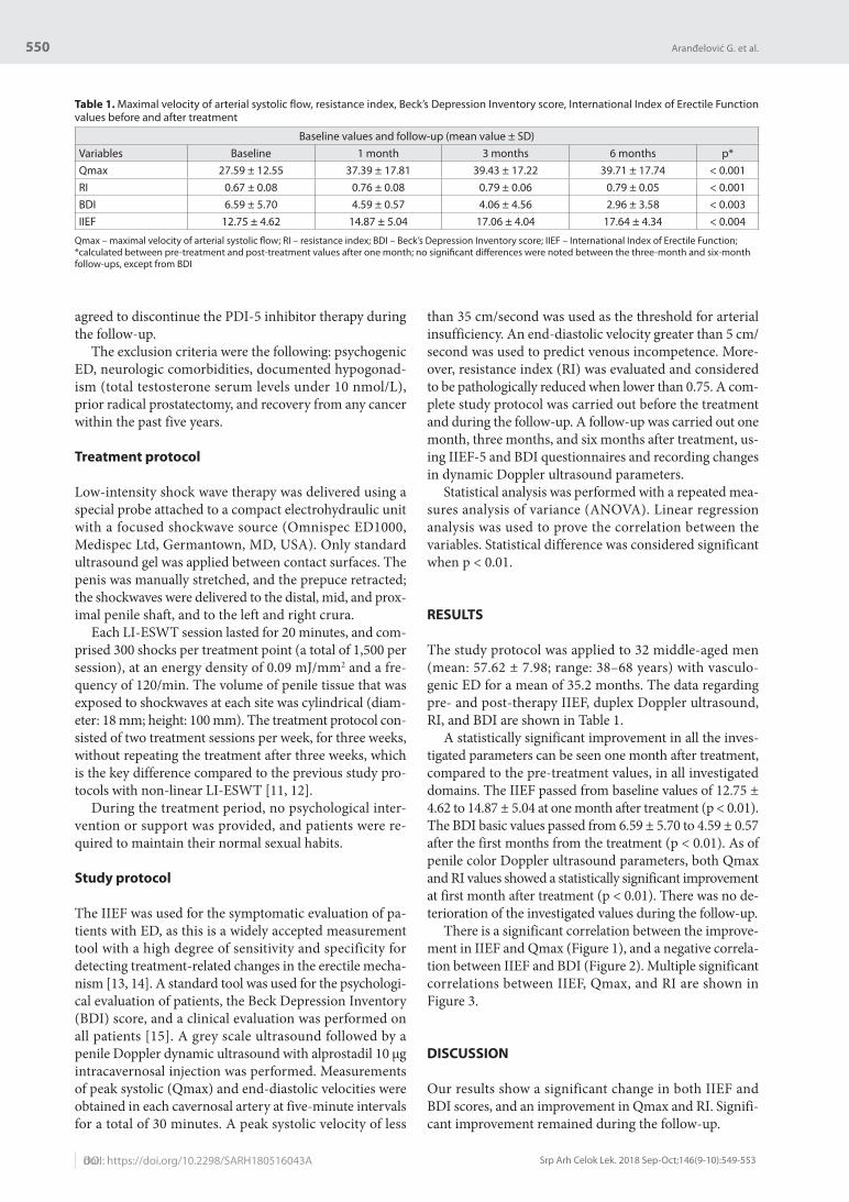
550
doi: Srp Arh Celok Lek. 2018 Sep-Oct;146(9-10):549-553
agreed to discontinue the PDI-5 inhibitor therapy during the follow-up.
The exclusion criteria were the following: psychogenic ED, neurologic comorbidities, documented hypogonad-ism (total testosterone serum levels under 10 nmol/L), prior radical prostatectomy, and recovery from any cancer within the past five years.
Treatment protocol
Low-intensity shock wave therapy was delivered using a special probe attached to a compact electrohydraulic unit with a focused shockwave source (Omnispec ED1000, Medispec Ltd, Germantown, MD, USA). Only standard ultrasound gel was applied between contact surfaces. The penis was manually stretched, and the prepuce retracted; the shockwaves were delivered to the distal, mid, and prox-imal penile shaft, and to the left and right crura.
Each LI-ESWT session lasted for 20 minutes, and com-prised 300 shocks per treatment point (a total of 1,500 per session), at an energy density of 0.09 mJ/mm2 and a fre-quency of 120/min. The volume of penile tissue that was exposed to shockwaves at each site was cylindrical (diam-eter: 18 mm; height: 100 mm). The treatment protocol con-sisted of two treatment sessions per week, for three weeks, without repeating the treatment after three weeks, which is the key difference compared to the previous study pro-tocols with non-linear LI-ESWT [11, 12].
During the treatment period, no psychological inter-vention or support was provided, and patients were re-quired to maintain their normal sexual habits.
Study protocol
The IIEF was used for the symptomatic evaluation of pa-tients with ED, as this is a widely accepted measurement tool with a high degree of sensitivity and specificity for detecting treatment-related changes in the erectile mecha-nism [13, 14]. A standard tool was used for the psychologi-cal evaluation of patients, the Beck Depression Inventory (BDI) score, and a clinical evaluation was performed on all patients [15]. A grey scale ultrasound followed by a penile Doppler dynamic ultrasound with alprostadil 10 μg intracavernosal injection was performed. Measurements of peak systolic (Qmax) and end-diastolic velocities were obtained in each cavernosal artery at five-minute intervals for a total of 30 minutes. A peak systolic velocity of less
than 35 cm/second was used as the threshold for arterial insufficiency. An end-diastolic velocity greater than 5 cm/second was used to predict venous incompetence. More-over, resistance index (RI) was evaluated and considered to be pathologically reduced when lower than 0.75. A com-plete study protocol was carried out before the treatment and during the follow-up. A follow-up was carried out one month, three months, and six months after treatment, us-ing IIEF-5 and BDI questionnaires and recording changes in dynamic Doppler ultrasound parameters.
Statistical analysis was performed with a repeated mea-sures analysis of variance (ANOVA). Linear regression analysis was used to prove the correlation between the variables. Statistical difference was considered significant when p < 0.01.
RESULTS
The study protocol was applied to 32 middle-aged men (mean: 57.62 ± 7.98; range: 38–68 years) with vasculo-genic ED for a mean of 35.2 months. The data regarding pre- and post-therapy IIEF, duplex Doppler ultrasound, RI, and BDI are shown in Table 1.
A statistically significant improvement in all the inves-tigated parameters can be seen one month after treatment, compared to the pre-treatment values, in all investigated domains. The IIEF passed from baseline values of 12.75 ± 4.62 to 14.87 ± 5.04 at one month after treatment (p < 0.01). The BDI basic values passed from 6.59 ± 5.70 to 4.59 ± 0.57 after the first months from the treatment (p < 0.01). As of penile color Doppler ultrasound parameters, both Qmax and RI values showed a statistically significant improvement at first month after treatment (p < 0.01). There was no de-terioration of the investigated values during the follow-up.
There is a significant correlation between the improve-ment in IIEF and Qmax (Figure 1), and a negative correla-tion between IIEF and BDI (Figure 2). Multiple significant correlations between IIEF, Qmax, and RI are shown in Figure 3.
DISCUSSION
Our results show a significant change in both IIEF and BDI scores, and an improvement in Qmax and RI. Signifi-cant improvement remained during the follow-up.
Table 1. Maximal velocity of arterial systolic flow, resistance index, Beck’s Depression Inventory score, International Index of Erectile Function values before and after treatment
Baseline values and follow-up (mean value ± SD)Variables Baseline 1 month 3 months 6 months p*Qmax 27.59 ± 12.55 37.39 ± 17.81 39.43 ± 17.22 39.71 ± 17.74 < 0.001RI 0.67 ± 0.08 0.76 ± 0.08 0.79 ± 0.06 0.79 ± 0.05 < 0.001BDI 6.59 ± 5.70 4.59 ± 0.57 4.06 ± 4.56 2.96 ± 3.58 < 0.003IIEF 12.75 ± 4.62 14.87 ± 5.04 17.06 ± 4.04 17.64 ± 4.34 < 0.004
Qmax – maximal velocity of arterial systolic fl ow; RI – resistance index; BDI – Beck’s Depression Inventory score; IIEF – International Index of Erectile Function;*calculated between pre-treatment and post-treatment values after one month; no signifi cant diff erences were noted between the three-month and six-month follow-ups, except from BDI
DOI: https://doi.org/10.2298/SARH180516043A
Aranđelović G. et al.

551
Srp Arh Celok Lek. 2018 Sep-Oct;146(9-10):549-553 www.srpskiarhiv.rs
Low intensity shock wave therapy is promising, al-though the exact mechanism used by LI-ESWT to induce the tissue changes is not known. Thanks to the release of neo-angiogenic factors and the subsequent neovascu-larization of the treated tissue, LI-ESWT leads to tissue regeneration [16]. Indeed, it has been shown that this low-intensity energy acts on vascularization, inducing a non-enzymatic production of physiologic amounts of nitric oxide [17]. Thus, the latest studies show that IIEF scores increase after treatment with LI-ESW [18]. Even though PDI-5 inhibitors represent the first-line therapy for ED by increasing the IIEF score, they do not represent a curative approach. The increase in IIEF does not last over time; rather, it is strictly limited to the assumption of PDE-5 in-hibitors. In addition, some ED patients respond poorly to PDI-5 and need other, more invasive treatments [9, 11, 12].
After the pilot study by Vardi et al. [7], which took into consideration the patients who had previously responded to PDE-5 inhibitor therapy, Gruenwald et al. [10] applied LI-ESWT to patients who had responded poorly to PDE-5 inhibitor therapy. By using the same protocol as in the first study on 29 patients, they concluded that LI-ESWT was again beneficial for this kind of patient, as it had a physi-ological effect on the erectile mechanism [10].
After those preliminary studies, others were done, but almost all studies on LI-ESWT for ED had the same dura-tion and applied the same treatment doses. The treatment protocol mostly considered the application of LI-ESWT twice a week for three weeks, with re-treatment for an-other three weeks after a three-week pause. The number of shocks applied was almost always 300 per treatment point with an energy flux density of 0.09 mJ/mm2 [9].
A very recent study carried out at the Mayo Clinic ap-plied low-energy shock wave therapy to alleviate renal dys-function in renovascular disease. They treated pigs’ kidneys with renal artery stenosis using low-energy shock waves twice a week for only three consecutive weeks, without re-peating. Twenty-six pigs were randomized to atherosclerotic renal artery stenosis (ARAS) or normal controls, treated or untreated with LI-ESWT. The results were amazingly posi-tive. A three-week low-intensity shockwave therapy attenu-ated renovascular hypertension, normalized stenotic kidney microvascular density and oxygenation, stabilized function, and alleviated fibrosis. This was associated with upregula-tion of the vascular endothelial growth factor expression that was decreased by ARAS, with increased angiopoietin-1, and downregulation of hypoxia-induced factor-1. Moreover, LI-ESWT improved the expression of endothelial nitric ox-ide synthase that was diminished in ARAS. No detectable injury to the kidney was observed [19].
The encouraging results from this and all the previ-ous studies led us to conduct a prospective pilot study, applying the three-week low-intensity shockwave therapy to patients with ED in order to examine if the shortened therapy could have the same effect as the repeated three-week therapy.
Our primary end points were the change in IIEF-5 and in the penile color Doppler values given through the Qmax and RI values, as well as the BDI score.
Figure 1. Correlation between the adjusted maximal velocity of arte-rial systolic flow parameters and the changes in International Index of Erectile Function scores
Qmax – maximal velocity of arterial systolic fl ow score; IIEF – International Index of Erectile Function
Figure 2. Correlation between the adjusted Beck’s Depression Inven-tory scores and the changes in International Index of Erectile Function scores
BDI – Beck’s Depression Inventory score; IIEF – International Index of Erectile Function
Figure 3. Multiple correlation between the changes in the maximal velocity of arterial systolic flow and resistance index values and the changes in International Index of Erectile Function scores
Qmax – maximal velocity of arterial systolic fl ow score; RI – resistance index; IIEF – International Index of Erectile Function
Low-intensity extracorporeal shock wave therapy of vasculogenic erectile dysfunction – three-week treatment in a cohort of North Italian patients
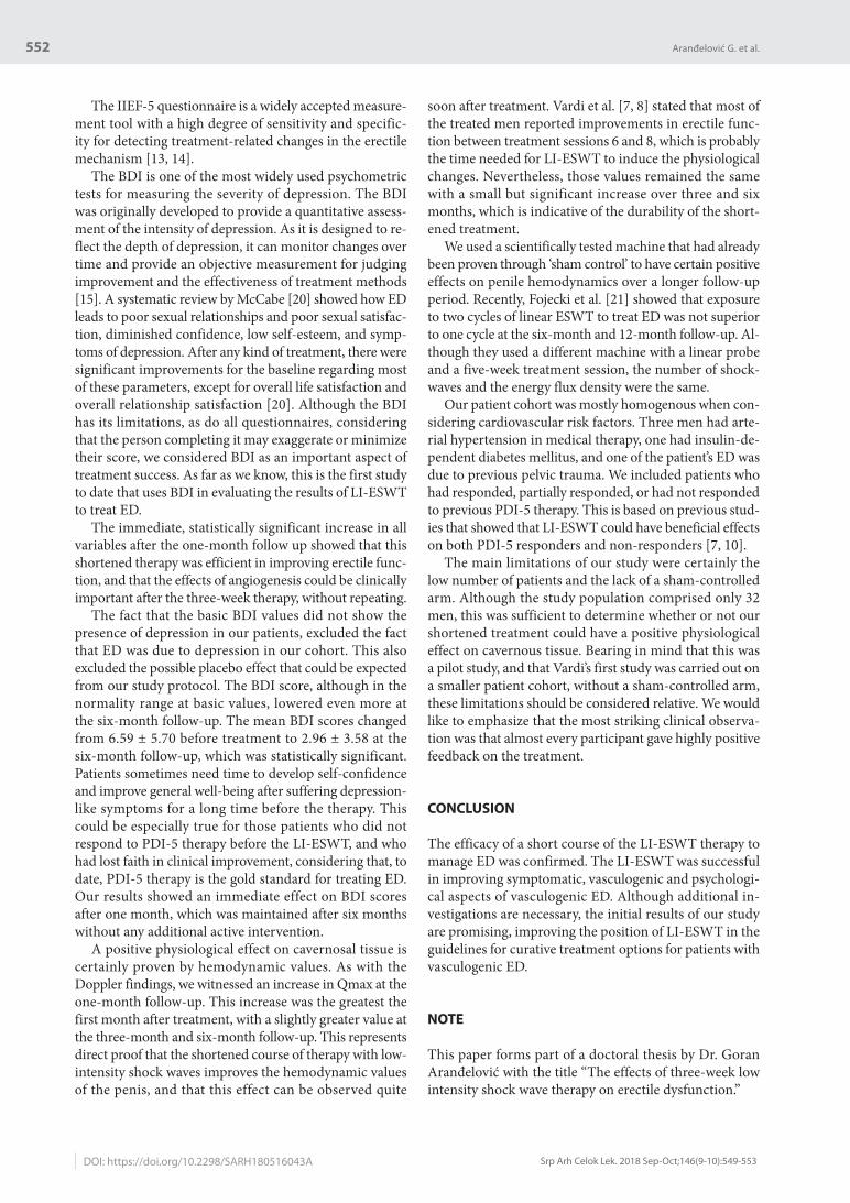
552
Srp Arh Celok Lek. 2018 Sep-Oct;146(9-10):549-553DOI: https://doi.org/10.2298/SARH180516043A
The IIEF-5 questionnaire is a widely accepted measure-ment tool with a high degree of sensitivity and specific-ity for detecting treatment-related changes in the erectile mechanism [13, 14].
The BDI is one of the most widely used psychometric tests for measuring the severity of depression. The BDI was originally developed to provide a quantitative assess-ment of the intensity of depression. As it is designed to re-flect the depth of depression, it can monitor changes over time and provide an objective measurement for judging improvement and the effectiveness of treatment methods [15]. A systematic review by McCabe [20] showed how ED leads to poor sexual relationships and poor sexual satisfac-tion, diminished confidence, low self-esteem, and symp-toms of depression. After any kind of treatment, there were significant improvements for the baseline regarding most of these parameters, except for overall life satisfaction and overall relationship satisfaction [20]. Although the BDI has its limitations, as do all questionnaires, considering that the person completing it may exaggerate or minimize their score, we considered BDI as an important aspect of treatment success. As far as we know, this is the first study to date that uses BDI in evaluating the results of LI-ESWT to treat ED.
The immediate, statistically significant increase in all variables after the one-month follow up showed that this shortened therapy was efficient in improving erectile func-tion, and that the effects of angiogenesis could be clinically important after the three-week therapy, without repeating.
The fact that the basic BDI values did not show the presence of depression in our patients, excluded the fact that ED was due to depression in our cohort. This also excluded the possible placebo effect that could be expected from our study protocol. The BDI score, although in the normality range at basic values, lowered even more at the six-month follow-up. The mean BDI scores changed from 6.59 ± 5.70 before treatment to 2.96 ± 3.58 at the six-month follow-up, which was statistically significant. Patients sometimes need time to develop self-confidence and improve general well-being after suffering depression-like symptoms for a long time before the therapy. This could be especially true for those patients who did not respond to PDI-5 therapy before the LI-ESWT, and who had lost faith in clinical improvement, considering that, to date, PDI-5 therapy is the gold standard for treating ED. Our results showed an immediate effect on BDI scores after one month, which was maintained after six months without any additional active intervention.
A positive physiological effect on cavernosal tissue is certainly proven by hemodynamic values. As with the Doppler findings, we witnessed an increase in Qmax at the one-month follow-up. This increase was the greatest the first month after treatment, with a slightly greater value at the three-month and six-month follow-up. This represents direct proof that the shortened course of therapy with low-intensity shock waves improves the hemodynamic values of the penis, and that this effect can be observed quite
soon after treatment. Vardi et al. [7, 8] stated that most of the treated men reported improvements in erectile func-tion between treatment sessions 6 and 8, which is probably the time needed for LI-ESWT to induce the physiological changes. Nevertheless, those values remained the same with a small but significant increase over three and six months, which is indicative of the durability of the short-ened treatment.
We used a scientifically tested machine that had already been proven through ‘sham control’ to have certain positive effects on penile hemodynamics over a longer follow-up period. Recently, Fojecki et al. [21] showed that exposure to two cycles of linear ESWT to treat ED was not superior to one cycle at the six-month and 12-month follow-up. Al-though they used a different machine with a linear probe and a five-week treatment session, the number of shock-waves and the energy flux density were the same.
Our patient cohort was mostly homogenous when con-sidering cardiovascular risk factors. Three men had arte-rial hypertension in medical therapy, one had insulin-de-pendent diabetes mellitus, and one of the patient’s ED was due to previous pelvic trauma. We included patients who had responded, partially responded, or had not responded to previous PDI-5 therapy. This is based on previous stud-ies that showed that LI-ESWT could have beneficial effects on both PDI-5 responders and non-responders [7, 10].
The main limitations of our study were certainly the low number of patients and the lack of a sham-controlled arm. Although the study population comprised only 32 men, this was sufficient to determine whether or not our shortened treatment could have a positive physiological effect on cavernous tissue. Bearing in mind that this was a pilot study, and that Vardi’s first study was carried out on a smaller patient cohort, without a sham-controlled arm, these limitations should be considered relative. We would like to emphasize that the most striking clinical observa-tion was that almost every participant gave highly positive feedback on the treatment.
CONCLUSION
The efficacy of a short course of the LI-ESWT therapy to manage ED was confirmed. The LI-ESWT was successful in improving symptomatic, vasculogenic and psychologi-cal aspects of vasculogenic ED. Although additional in-vestigations are necessary, the initial results of our study are promising, improving the position of LI-ESWT in the guidelines for curative treatment options for patients with vasculogenic ED.
NOTE
This paper forms part of a doctoral thesis by Dr. Goran Aranđelović with the title “The effects of three-week low intensity shock wave therapy on erectile dysfunction.”
Aranđelović G. et al.

553
Srp Arh Celok Lek. 2018 Sep-Oct;146(9-10):549-553 www.srpskiarhiv.rs
REFERENCES
1. Feldman HA, Goldstein I, Hatzichristou DG, Krane RJ, McKinlay JB. Impotence and its medical and psychosocial correlates: Results of the Massachusetts Male Aging Study. J Urol. 1994; 151(1):54–61.
2. Shabsigh R, Kimoto Y, Amar E, Hackett G, Jarow JP, Mirone V, et al. Economical aspects in sexual dysfunctions. In: Lue TF, Basson R, Rosen R, Giuliano F, Khoury S, Montorsi F (Eds.) Sexual Medicine, Sexual Dysfunctions in Men and Women. Health Publications, Paris; 2004:139–160.
3. Aversa A, Bruzziches R, Vitale C, Marazzi G, Francomano D, Barbaro G, et al. Chronic sildenafil in men with diabetes and erectile dysfunction. Expert Opin Drug Metab Toxicol. 2007; 3(3):451–64.
4. Porst H, Rajfer J, Casabe A, Feldman R, Ralph D, Vieiralves LF, et al. Long-term safety and efficacy of tadalafil 5 mg dosed once daily in men with erectile dysfunction. J Sex Med. 2008; 5(9):2160–9.
5. Vardi Y, Appel B, Ofer Y, Gruenwald I, Dayan L, Jacob G. Effect of chronic sildenafil treatment on penile endothelial function: a randomized, double-blind, placebo-controlled study. J Urol. 2009; 182(6):2850–5.
6. Hatzichristou D, d’Anzeo G, Porst H, Buvat J, Henneges C, Rossi A, et al. Tadalafil 5mg once daily for the treatment of erectile dysfunction during a 6-month observational study (EDATE): impact of patient characteristics and comorbidities. BMC Urol. 2015; 15:111.
7. Vardi Y, Appel B, Jacob G, Massarwi O, Gruenwald I. Can low-intensity extracorporeal shockwave therapy improve erectile function? A 6-month follow-up pilot study in patients with organic erectile dysfunction. Eur Urol. 2010; 58(2):243–8.
8. Vardi Y, Appel B, Kilchevsky A, Gruenwald I. Does low-intensity extracorporeal shock wave therapy have a physiological effect on erectile function? Short-term results of a randomized, double-blind, sham-controlled study. J Urol. 2012; 187(5):1769–75.
9. Clavijo RI, Kohn TP, Kohn JR, Ramasamy R. Effects of Low-Intensity Extracorporeal Shockwave Therapy on Erectile Dysfunction: A Systematic Review and Meta-Analysis. J Sex Med. 2017; 14(1):27–35.
10. Gruenwald I, Appel B, Vardi Y. Low-intensity extracorporeal shock wave therapy-a novel effective treatment for erectile dysfunction in severe ED patients who respond poorly to PDE5 inhibitor therapy. J Sex Med. 2012; 9(1):259–64.
11. Zou ZJ, Tang LY, Liu ZH, Liang JY, Zhang RC, Wang YJ, et al. Short-term efficacy and safety of low-intensity extracorporeal shock wave
therapy in erectile dysfunction: a systematic review and meta-analysis. Int Braz J Urol. 2017; 43(5):805–21.
12. Lu Z, Lin G, Reed-Maldonado A, Wang C, Lee YC, Lue TF. Low-intensity Extracorporeal Shock Wave Treatment Improves Erectile Function: A Systematic Review and Meta-analysis. Eur Urol. 2017; 71(2):223–33.
13. Rosen RC, Riley A, Wagner G, Osterloh IH, Kirkpatrick J, Mishra A. The international index of erectile function (IIEF): a multidimensional scale for assessment of erectile dysfunction. Urology. 1997; 49(6):822–30.
14. Rosen RC, Cappelleri JC, Gendrano N 3rd. The international Index of Erectile Function (IIEF): a state of the science review. Int J Impot Res. 2002; 14(4):226–44.
15. Beck AT, Ward C, Mendelson M, Mock J, Erbaugh J. An inventory for measuring depression. Arch Gen Psychiatry. 1961; 4:561–71.
16. Romeo P, Lavanga V, Pagani D, Sansone V. Extracorporeal shock wave therapy in musculoskeletal disorders: a review. Med Princ Pract. 2014; 23(1):7–13.
17. Mariotto S, Cavalieri E, Amelio E, Ciampa AR, de Prati AC, Marlinghaus E, et al. Extracorporeal shock waves: from lithotripsy to anti-inflammatory action by NO production. Nitric Oxide. 2005; 12(2):89–96.
18. Olsen AB, Persiani M, Boie S, Hanna M, Lund L. Can low-intensity extracorporeal shockwave therapy improve erectile dysfunction? A prospective, randomized, double-blind, placebo-controlled study. Scand J Urol. 2015; 49(4):329–33.
19. Zhang X, Krier JD, Amador Carrascal C, Greenleaf JF, Ebrahimi B, Hedayat AF, et al. Low-Energy Shockwave Therapy Improves Ischemic Kidney Microcirculation. J Am Soc Nephrol. 2016; 27(12):3715–24.
20. McCabe MP, Althof SE. A systematic review of the psychosocial outcomes associated with erectile dysfunction: does the impact of erectile dysfunction extend beyond a man's inability to have sex? J Sex Med. 2014; 11(2):347–63.
21. Fojecki GL, Tiessen S, Osther PJS. Effect of Linear Low-Intensity Extracorporeal Shockwave Therapy for Erectile Dysfunction: 12-Month Follow-Up of a Randomized, Double-Blinded, Sham-Controlled Study. Sex Med. 2018; 6(1):1–7.
САЖЕТАКУвод/Циљ Инхибитори фосфодиестаразе типа 5 (PDE5-Is) представљају златни стандард у медикаментозној тера-пији еректилне дисфункције. Упркос томе, медикаментоз-ни третман не представља терапију којом се може трајно излечити еректилна дисфункција. Циљ наше студије је ис-питати скраћену тронедељну терапију екстракорпоралним ударним таласима ниског интензитета васкулогене ерек-тилне дисфункије.Методе Студија је укључила 32 болесника са међународним индексом еректилне функције између 5 и 20, са потврђеном васкулогеном еректилном дисфункцијом на доплер ултраз-вуку. Болесници су имали паузу у трајању од једног месеца од претходне терапије и били су сагласни да прекину тера-пију PDE5 инхибиторима у периоду праћења после третма-на. Применили смо екстракорпоралне ударне таласе ниског интензитета у трајању од три недеље, два пута недељно, без понављања. Болесници су оцењивани пре терапије, један месец, три и шест месеци после терапије међународним ин-
дексом еректилне функције, доплер ултразвуком и Бековим упитником за депресију.Резултати Сви испитани параметри (Међународни индекс еректилне функције, Беков упитник за депресију, пара-метри доплер ултразвука пениса) показали су позитивну статистички значајну промену после само једног месеца од терапије. Међународни индекс еректилне функције је забележио пораст првог месеца после терапије и попео се са основних вредности од 12,75 ± 4,62 на 14,87 ± 5,04 (p < 0,01). Овај позитиван тренд се задржао како на Међународ-ном индексу еректилне функције тако и на свим тестираним параметрима после три и шест месеци праћења.Закључак Скраћена тронедељна терапија екстракорпорал-ним ударним таласима ниског интензитета примењена на васкулогену еректилну дисфункцију показала се клинички ефективном.Кључне речи: еректилна дисфункција; екстракорпорални ударни таласи ниског интензитета; међународни индекс ерек-тилне функције; доплер ултразвук; Беков упитник за депресију
Екстракорпорални ударни таласи ниског интензитета за третман васкулогене еректилне дисфункције – тронедељна терапија на групи болесника из североисточне ИталијеГоран Аранђеловић1, Федра Готардо2, Иван Игњатовић3,4
1Болница Светог Јована и Павла, Клиника за урологију, Венеција, Италија;2ULSS 6 Euganea, Падова, Италија;3Клинички центар Ниш, Клиника за урологију, Ниш, Србија;4Универзитет у Нишу, Медицински факултет, Ниш, Србија
Low-intensity extracorporeal shock wave therapy of vasculogenic erectile dysfunction – three-week treatment in a cohort of North Italian patients

554
Correspondence to:
Elvis MAHMUTOVIĆ
State University of Novi Pazar
Vuka Karadžića bb
36300 Novi Pazar, Serbia
Received • Примљено: October 2, 2017
Revised • Ревизија: February 5, 2016
Accepted • Прихваћено: October 17, 2017
Online fi rst: October 27, 2017
DOI: https://doi.org/10.2298/SARH171002189M
UDC: 613-056.24:616.711-089
SUMMARYIntroduction/Objective The quality of life (QL) is a modern concept of observing the outcome of the disease and the success of the therapeutic procedure in all fields of medicine. The aim was to assess the QL of surgically treated patients with lumbar radiculopathy (LR) at the beginning of treatment and three and six months after the initiation of prescribed and applied medical rehabilitation.Methods The study group included randomized and stratified sample of 50 patients treated with lumbar microdiscectomy (LM). Conservative treatment was carried out using physical therapy procedures, and kinetic and ergonomic therapeutic procedures and educational training program in ergonomics were carried out in all the patients. To assess the condition of the patients, the QL and the efficacy of the rehabilitation treatment, we used two standardized questionnaires, the Short Form Survey Instrument (SF-36) and the Oswestry Disability Index (ODI).Results The lowest values of the SF-36 – PCS, SF-36 – MCS, and of the ODI were recorded at the begin-ning of the rehabilitation (PCS: 28.8; MCS: 37.8; ODI: 56.1%). The most significant improvements of the scores were observed three months after the treatment initiation (PCS: 42.8; MCS: 45.2; ODI: 38.9%). At six months of treatment, the scores were slightly higher (PCS: 49.2; MCS: 52.5; ODI: 23.7%) (p < 0.001).Conclusion The QL and the functional status of patients after LM are significantly better after three and six months in comparison with the beginning of rehabilitation, and the state for six months compared to the state for three months.Keywords: lumbar radiculopathy; microdiscectomy; quality of life; SF-36; ODI; treatment outcome
ORIGINAL ARTICLE / ОРИГИНАЛНИ РАД
The quality of life of patients after lumbar microdiscectomy Elvis Mahmutović1,2, Radoslava Doder2,3, Zana Dolićanin1, Ksenija Bošković2,4
1State University of Novi Pazar, Department of Biomedical Sciences, Novi Pazar, Serbia;2University of Novi Sad, Faculty of Medicine, Novi Sad, Serbia;3Clinical Center of Vojvodina, Clinic for Infectious Diseases, Novi Sad, Serbia;4Clinical Center of Vojvodina, Clinic for Medical Rehabilitation, Novi Sad, Serbia
INTRODUCTION
The main symptom of lumbar radiculopathy (LR) is pain in the lumbo-sacral region with propagation to the lower extremities. The in-tensity of the neuropathic pain depends on the extent of the local damage and on the individu-al characteristics of the patient and experiential pain perception [1].
Healthy functioning and the quality of life (QL) of patients with LR depend on the sever-ity of the disease, the intensity of the symptoms and on the degree of incapacity. It has been also largely dependent on the applied therapeutic methods and protocols. In addition, the socio-economic implication plays a relevant role [2]. The QL provides valuable information about functional ability, level and quality of social in-teraction, mental state, somatic sensations, and satisfaction with life, reflecting the definition of health by the World Health Organization and reflecting the previous scientific data about the impact of the disease and treatment on disabil-ity and daily functioning [3].
Questionnaires, as instruments for measur-ing the QL, regarding their structure, may be general (generic) questionnaires that are struc-tured to express the extent of injury from the standpoint of patients, and questionnaires for
a specific disease. The latter ones are formed with an objective to provide a higher sensitivity and specificity [4]. The choice of instrument should be determined by the clinician, ac-cording to the clinical problem and measuring characteristics of the instrument [5].
The aim of this study was to evaluate the QL of patients immediately after lumbar microdis-cectomy (LM) at the beginning of the rehabili-tation, and then after three months and after six months of the prescribed supervised regular physical rehabilitation treatment. For the eval-uation, we utilized both general questionnaire and the questionnaire specific for lumbar pain syndrome (LPS).
METHODS
This randomized prospective clinical study included 50 patients with LR of disc genesis who were treated with LM. In all the patients, rehabilitation treatment was carried out under the regular protocol with the use of physical therapy procedures and ergonomic physical training.
Inclusion criteria for the patients in this study were the following: age between 20 and 65 years, patients of both sexes, orientated in
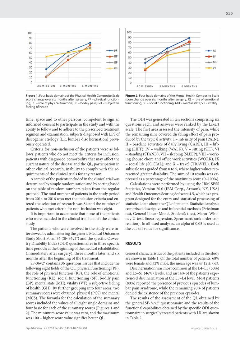
555
Srp Arh Celok Lek. 2018 Sep-Oct;146(9-10):554-560 www.srpskiarhiv.rs
time, space and to other persons, competent to sign an informed consent to participate in the study and with the ability to follow and to adhere to the prescribed treatment regimen and examination, subjects diagnosed with LPS of discogenic etiology (LR, lumbar disc herniation) previ-ously operated.
Criteria for non-inclusion of the patients were as fol-lows: patients who do not meet the criteria for inclusion, patients with diagnosed comorbidity that may affect the current nature of the disease and the QL, participation in other clinical research, inability to comply with the re-quirements of the clinical trials for any reason.
A sample of the patients included in the clinical trial was determined by simple randomization and by sorting based on the table of random numbers taken from the regular protocol. The total number of patients in the study period from 2014 to 2016 who met the inclusion criteria and en-tered the selection of research was 84 and the number of patients who met criteria for non-inclusion was eight.
It is important to accentuate that none of the patients who were included in the clinical trial had left the clinical study.
The patients who were involved in the study were in-terviewed by administering the generic Medical Outcomes Study Short Form 36 (SF-36v2®) and the specific Oswes-try Disability Index (ODI) questionnaires in three specific time periods: at the beginning of the medical rehabilitation (immediately after surgery), three months later, and six months after the beginning of the treatment.
SF-36v2® contains 36 questions, issues that include the following eight fields of the QL: physical functioning (PF), the role of physical function (RF), the role of emotional functioning (RE), social functioning (SF), bodily pain (BP), mental state (MH), vitality (VT), a subjective feeling of health (GH). By further grouping into four areas, two summary scores were obtained: physical (PCS) and mental (MCS). The formula for the calculation of the summary scores included the values of all eight single domains and four basic for each of the summary scores (Figures 1 and 2). The minimum score value was zero, and the maximum was 100 – higher score value signifies better QL.
The ODI was generated in ten sections comprising six questions each, and answers were ranked by the Likert scale. The first area assessed the intensity of pain, while the remaining nine covered disabling effect of pain pro-duced by the typical activity: I – intensity of pain (PAIN); II – baseline activities of daily living (CARE); III – lift-ing (LIFT); IV – walking (WALK); V – sitting (SIT); VI – standing (STAND); VII – sleeping (SLEEP); VIII – work-ing (house chore and office work activities (WORK); IX – social life (SOCIAL); and X – travel (TRAVEL). Each subscale was graded from 0 to 5, where higher values rep-resented greater disability. The sum of 10 results was ex-pressed as a percentage of the maximum score (0–100%).
Calculations were performed by using the IBM SPSS Statistics, Version 20.0 (IBM Corp., Armonk, NY, USA) and Health Outcomes Scoring Software 4.5, which is a pro-gram designed for the entry and statistical processing of statistical data about the QL of patients. Statistical analysis comprised descriptive and inferential methods (Friedman test, General Linear Model, Student’s t-test, Mann–Whit-ney U-test, linear regression, Spearman’s rank order cor-relation). In all used analyses, an alpha of 0.05 is used as the cut-off value for significance.
RESULTS
General characteristics of the patients included in the study are shown in Table 1. Of the total number of patients, 68% were female and 32% male, the mean age was 47.12 ± 7.63.
Disc herniation was most common at the L4–L5 (50%) and L5–S1 (46%) levels, and just 4% of the patients expe-rienced disc herniation at the L3–L4 level. Most patients (80%) reported the presence of previous episodes of lum-bar pain syndrome, while the remaining 20% of patients denied the existence of the previous episodes.
The results of the assessment of the QL obtained by the general SF-36v2® questionnaire and the results of the functional capabilities obtained by the specific ODI ques-tionnaire in surgically treated patients with LR are shown in Table 2.
Figure 1. Four basic domains of the Physical Health Composite Scale score change over six months after surgery; PF – physical function-ing; RF – role of physical function; BP – bodily pain; GH – subjective feeling of health
Figure 2. Four basic domains of the Mental Health Composite Scale score change over six months after surgery; RE – role of emotional functioning; SF – social functioning; MH – mental state; VT – vitality

556
Srp Arh Celok Lek. 2018 Sep-Oct;146(9-10):554-560DOI: https://doi.org/10.2298/SARH171002189M
At the start of rehabilitation, we recorded a very low value of the PCS 28.8, while the value of the MCS was slightly higher, but also at a low level (37.8). After three months of rehabilitation, value of all scores on the SF-36V2® questionnaire were significantly increased (PCS = 42.8; MCS = 45.2), and after 6 months, the val-ues approximately reached the levels that characterize the general population (PCS = 49.22; MCS = 52.5). Analysis of variance for repeated measures (RM ANOVA) showed that the values of PCS (Figure 1) and MCS (Figure 2) scores have significantly changed during the study (F = 490.721, p < 0.001). Both summary scores showed the greatest reg-istered progress in the first three months from the start of rehabilitation treatment.
Furthermore, after examining the results of the six-month research, we found that the domains that partici-pate in the formation of the total PCS and MCS scores and after comparing them with the values given for the general population in different countries, we concluded that the values of the domains moved closer to the general population after six months of rehabilitation. Compared with the general population of Switzerland, Great Britain, United States and China, a statistically significant differ-ence (p < 0.05) was registered among the majority of the domain, except for two domains: Switzerland (pSF = 0.26 and pVT = 0.88); United Kingdom (pSF = 0.14 and pGH = 0.27); United States (pSF = 0.35 and pBP = 0.08); China (pSF = 0.96 and pGH = 0.18). When compared with the general population in Australia, a statistically significant difference (p < 0.05) was observed in PF, RE,
MH, VT, and GH domains, while in the remaining three domains no statistically significant difference was found (pRF = 0.16, pSF = 0.76, pBP = 0.93). The analysis of com-parisons of the results of our research with the results in the general population of different countries are shown in Table 3.
Multivariate linear regression analysis showed that the values of PCS and MCS were not significantly related with the monitored characteristics of our patients.
Immediately after surgery, we registered a high ODI score of 56.10%; however, each following test recorded sig-nificant improvements in the functionality of the patients, and the ODI score was 38.9% after three months, which decreased to 23.7% after six months of the rehabilitation treatment (Figure 3).
ODI domain values during six months of the follow-up after LM are given in Table 4. The analysis of the presence of individual responses in ODI domain Friedman’s test revealed statistically significant differences among three measurements (p < 0.001). The biggest improvement was registered in the first three months from the beginning of the rehabilitation treatment.
Multivariate linear regression analysis confirmed that the value of the ODI were significantly associated with marital status. In patients who were married or in a com-mon law marriage, after controlling the effects of all other demographic characteristics, the score was greater by 6.452
Table 1. General characteristics of the patients (n = 50)
Characteristics Number (%) ± SD
SexMale 16 (32)Female 34 (68)
Age (years) Mean 47.12 ± 7.63
Education
No primary education 3 (6)Primary 15 (30)Secondary / high school 22 (44)University degree 10 (20)
Marital status
Married / in a relationship 38 (76)Divorced/separated 3 (6)Widowed 2 (4)Single 7 (14)
Level of discus hernia
L3–L4 2 (4)L4–L5 25 (50)L5–S1 23 (46)
Earlier episodesNo 10 (20)Yes, one episodes 11 (22)Yes, more episodes 29 (58)
Table 2. Results of the Medical Outcomes Study Short Form 36 and Oswestry Disability Index questionnaire scores
Questionnaire Admission 3 months 6 months F-value* p-value
SF-36PCS 28.8 42.8 49.2 490.721 < 0.001MCS 37.8 45.2 52.5 72.055 < 0.001
MCS 56.1% 38.9% 23.70% 1341.180 < 0.001
SF-36 – Medical Outcomes Study Short Form 36; PCS – Physical Health Composite Scale score; MCS – Mental Health Composite Scale score;*repeated measures ANOVA
Table 3. Comparative overview of the Medical Outcomes Study Short Form 36 scores with the general population
Results of our research General populationI II III a b c d e
SF-3
6
PF 26.5 62 80.3 90.6 85 84.2 85 83.9RF 10.5 45 75.2 85.8 81.55 80.9 85 77.5RE 39.8 68.5 89.3 79.2 83.5 81.3 80.2 79.7SF 26 60.7 81.5 83.7 84.35 83.3 81.4 82.1BP 11.9 53.8 71 77.6 79.8 75.2 76.6 71.2MH 48 61.8 77.6 69.2 73.8 74.7 70.6 73.6VT 28 54.7 64.9 65.1 58.7 60.9 61.7 57.7GH 52.4 60.62 68.52 76.1 70.35 71.9 66.3 72.8
SF-36 – Medical Outcomes Study Short Form 36; PF – physical functioning; RF – role of physical function; RE – role of emotional functioning; SF – social functioning; BP – bodily pain; MH – mental state; VT – vitality; GH – subjective feeling of health; I – admission; II – 3 months; III – 6 months; a – Switzerland; b – United Kingdom; c – USA; d – China; e – Australia [6]
Figure 3. Values of the Oswestry Disability Index score change over six months after surgery
Mahmutović E. et al.
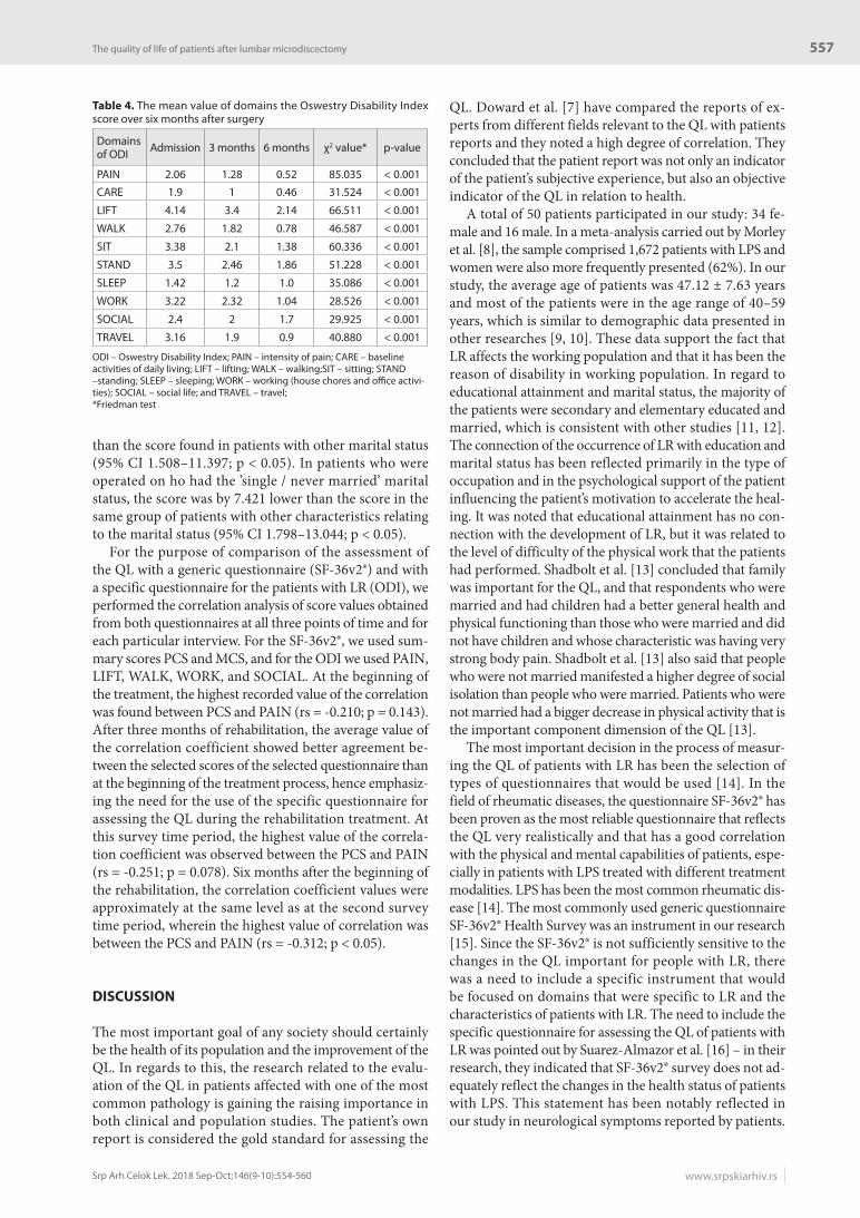
557
Srp Arh Celok Lek. 2018 Sep-Oct;146(9-10):554-560 www.srpskiarhiv.rs
than the score found in patients with other marital status (95% CI 1.508–11.397; p < 0.05). In patients who were operated on ho had the ’single / never married’ marital status, the score was by 7.421 lower than the score in the same group of patients with other characteristics relating to the marital status (95% CI 1.798–13.044; p < 0.05).
For the purpose of comparison of the assessment of the QL with a generic questionnaire (SF-36v2®) and with a specific questionnaire for the patients with LR (ODI), we performed the correlation analysis of score values obtained from both questionnaires at all three points of time and for each particular interview. For the SF-36v2®, we used sum-mary scores PCS and MCS, and for the ODI we used PAIN, LIFT, WALK, WORK, and SOCIAL. At the beginning of the treatment, the highest recorded value of the correlation was found between PCS and PAIN (rs = -0.210; p = 0.143). After three months of rehabilitation, the average value of the correlation coefficient showed better agreement be-tween the selected scores of the selected questionnaire than at the beginning of the treatment process, hence emphasiz-ing the need for the use of the specific questionnaire for assessing the QL during the rehabilitation treatment. At this survey time period, the highest value of the correla-tion coefficient was observed between the PCS and PAIN (rs = -0.251; p = 0.078). Six months after the beginning of the rehabilitation, the correlation coefficient values were approximately at the same level as at the second survey time period, wherein the highest value of correlation was between the PCS and PAIN (rs = -0.312; p < 0.05).
DISCUSSION
The most important goal of any society should certainly be the health of its population and the improvement of the QL. In regards to this, the research related to the evalu-ation of the QL in patients affected with one of the most common pathology is gaining the raising importance in both clinical and population studies. The patient’s own report is considered the gold standard for assessing the
QL. Doward et al. [7] have compared the reports of ex-perts from different fields relevant to the QL with patients reports and they noted a high degree of correlation. They concluded that the patient report was not only an indicator of the patient’s subjective experience, but also an objective indicator of the QL in relation to health.
A total of 50 patients par ticipated in our study: 34 fe-male and 16 male. In a meta-analysis carried out by Morley et al. [8], the sample comprised 1,672 patients with LPS and women were also more frequently presented (62%). In our study, the average age of patients was 47.12 ± 7.63 years and most of the patients were in the age range of 40–59 years, which is similar to demographic data presented in other researches [9, 10]. These data support the fact that LR affects the working population and that it has been the reason of disability in working population. In regard to educational attainment and marital status, the majority of the patients were secondary and elementary educated and married, which is consistent with other studies [11, 12]. The connection of the occurrence of LR with education and marital status has been reflected primarily in the type of occupation and in the psychological support of the patient influencing the patient’s motivation to accelerate the heal-ing. It was noted that educational attainment has no con-nection with the development of LR, but it was related to the level of difficulty of the physical work that the patients had performed. Shadbolt et al. [13] concluded that family was important for the QL, and that respondents who were married and had children had a better general health and physical functioning than those who were married and did not have children and whose characteristic was having very strong body pain. Shadbolt et al. [13] also said that people who were not married manifested a higher degree of social isolation than people who were married. Patients who were not married had a bigger decrease in physical activity that is the important component dimension of the QL [13].
The most important decision in the process of measur-ing the QL of patients with LR has been the selection of types of questionnaires that would be used [14]. In the field of rheumatic diseases, the questionnaire SF-36v2® has been proven as the most reliable questionnaire that reflects the QL very realistically and that has a good correlation with the physical and mental capabilities of patients, espe-cially in patients with LPS treated with different treatment modalities. LPS has been the most common rheumatic dis-ease [14]. The most commonly used generic questionnaire SF-36v2® Health Survey was an instrument in our research [15]. Since the SF-36v2® is not sufficiently sensitive to the changes in the QL important for people with LR, there was a need to include a specific instrument that would be focused on domains that were specific to LR and the characteristics of patients with LR. The need to include the specific questionnaire for assessing the QL of patients with LR was pointed out by Suarez-Almazor et al. [16] – in their research, they indicated that SF-36v2® survey does not ad-equately reflect the changes in the health status of patients with LPS. This statement has been notably reflected in our study in neurological symptoms reported by patients.
Table 4. The mean value of domains the Oswestry Disability Index score over six months after surgery
Domains of ODI Admission 3 months 6 months χ2 value* p-value
PAIN 2.06 1.28 0.52 85.035 < 0.001CARE 1.9 1 0.46 31.524 < 0.001
LIFT 4.14 3.4 2.14 66.511 < 0.001
WALK 2.76 1.82 0.78 46.587 < 0.001
SIT 3.38 2.1 1.38 60.336 < 0.001
STAND 3.5 2.46 1.86 51.228 < 0.001
SLEEP 1.42 1.2 1.0 35.086 < 0.001
WORK 3.22 2.32 1.04 28.526 < 0.001
SOCIAL 2.4 2 1.7 29.925 < 0.001
TRAVEL 3.16 1.9 0.9 40.880 < 0.001
ODI – Oswestry Disability Index; PAIN – intensity of pain; CARE – baseline activities of daily living; LIFT – lifting; WALK – walking;SIT – sitting; STAND –standing; SLEEP – sleeping; WORK – working (house chores and offi ce activi-ties); SOCIAL – social life; and TRAVEL – travel;*Friedman test
The quality of life of patients after lumbar microdiscectomy
558
Srp Arh Celok Lek. 2018 Sep-Oct;146(9-10):554-560
For the purposes of this study, as the questionnaire spe-cific to the disease we used the ODI, a specific question-naire for measuring the QL of patients with LR. It has been a very practical questionnaire for routine clinical use since it was designed as a multi-dimensional test. It measures the pain and functioning, as well as the pain during the activities causing limitations in physical activities; hence, it can be classified as a component of serious research.
Values of PCS and MCS showed statistically significant changes during our study. The biggest improvement was recorded after the first three months of the treatment in both summary scores. After six months from the start of rehabilitation, PCS value did not exceed the standard value of the SFS – 50 for the general US population. Lower val-ues of PCS were justified by the severity of the damage and by recent surgery that both contributed to physical limita-tions in the early postoperative period as well as by applied precautions for wounds, injury to back and reherniation. The patient’s fear of physical activity and body movement had contributed to lower PCS values as well. Johansson et al. [17] reported patients’ beliefs in recovery and fears of physical activity as leading factors. Authors recommended that the patients with fears of physical activity should be identified and treated appropriately.
We also concluded that the patients with psychosocial problems more frequently shortened the time spent at work and in other activities, were less efficient, had less atten-tion and motivation regarding work obligations, were more frequently nervous, in a bad mood, tired, with less energy and less active, and more irregular in maintaining social contacts. During the six-month follow-up period of the patients, the MCS values showed continuous increase and at the six-month survey period, these values exceeded the standard value of a healthy population of the United States – 50. When we compared the results of our research to some other research findings, we concluded that there is no agreement that the emotional and psychosocial factors have a major impact on success of the treatment in patients with LR. Johansson et al. [17] and Den Boer et al. [18] noted a more significant psychological impact in surgical patients when compared with nonsurgical. In contrast to these find-ings, Bošković et al. [19] and Iles et al. [20] after studying the psychosocial factors as predictive factors of the success of treatment in patients with LPS and LR concluded that depression, satisfaction/dissatisfaction with work, psycho-logical stress, and other factors have considerably smaller influence and that has been correlated to our research.
Unlike the SF-36v2® survey, in which the questions re-ferred to the time interval within the previous four weeks, the ODI questions were related to the current status of the patients. The average values of the total ODI score dur-ing the examination period changed significantly in both groups of patients, and the differences of these values three and six months after the surgery were highly statistically significant (p < 0.001) in comparison to the values at the beginning.
At the start of the hospitalization (and rehabilitation) and at three months from the start of the rehabilitation, half of the patients responded with, “The pain is very mild
at the moment.” This fact can be justified by the effect of surgery and by early rehabilitation. Six months after the beginning of the rehabilitation, we concluded that in the majority of the patients (56%) a complete relief of pain was achieved.
The decision that the assessment shall be made after the first three months of the treatment was made because it was thought that this was a long enough period for the recovery and for the assessment of the therapeutic treat-ment outcomes. Assessment in the later period (e.g. after a year or more) could provide similar but also different or inadequate results (if, for example, there was an appear-ance – emergence – of new herniation of intervertebral discs or other pathological changes of the spine).
The fact that the period of three months after the op-eration is a long enough period for the assessment of the therapeutic effect and of the degree of recovery was sup-ported by the research carried out by Häkkinen et al. [21]. They compared the score values on the ODI questionnaire administered six weeks after the surgery and then one year following the surgery for LR. They proved that the results obtained six weeks after the operation did not change sub-stantially during the coming year.
Ability to function in terms of daily activities that was covered by the ODI questionnaire at admission and at the beginning of the treatment process was limited to light activities. Three months from the start of rehabilitation, the patients showed improvement but were still limited in their daily activities in regard to performance (adjusting) within proper body position. In the last survey period, the patients were still cautious, so their answers ranged from being rigid to avoid harder activities only, while lighter activities within the proper body position could be per-formed, to being able to perform heavier activities but with additional pain. Bakker et al. [22] in the review of prospec-tive cohort studies have confirmed that sitting, walking, long standing in one place, as well as playing sports have not been significant risk factors for the development of LPS and LR, unlike most of the mechanical load of the spine during heavier work. Bending, torsion of the torso, and vibrations of the entire body were cited as significant predictors [22]. Roffey et al. [23–27] and Wai et al. [28–30] in eight systematic studies in total performed the analysis of the influence of the mechanical factors on the appear-ance of LPS and LR in a large number of workers in dif-ferent professions. In these studies, the mechanical factors that were included were prolonged sitting in an awkward body position [23, 24], prolonged standing and walking [25], lifting and moving of patients [26], pushing or pull-ing [27], bending or twisting of the body during lifting of a heavy load [28, 29], carrying heavy loads [30].
Low values of correlation coefficients in patients who were operated on and a small correlation value of the SOC (social functioning) ODI domain with other scores and domains tells about the specifics of this domain and about the evident need to assess the QL of patients with LR by us-ing batteries of generic and specific questionnaires. General generic questionnaires are needed to analyze appropriately the QL of patients in comparison to the normal population
DOI: https://doi.org/10.2298/SARH171002189M
Mahmutović E. et al.
559
Srp Arh Celok Lek. 2018 Sep-Oct;146(9-10):554-560 www.srpskiarhiv.rs
and to compare the QL in patients with different diseases, while specific questionnaires are needed in order to assess in detail the health and the QL of these patients.
CONCLUSION
Given that the QL includes all aspects of life in patients who underwent LM, we did not expect an improvement in the first days after the operation. In further monitoring of our patients, we recorded significantly higher values of physi-cal functioning and functioning in emotional and social aspects of the QL at three months and at six months when compared to the beginning of rehabilitation, and at six months when compared to three months of rehabilitation.
A statistically significant negative correlation be-tween PCS and PAIN was recorded on the third repeated
measurement. Values of domains and scores and the small values of correlation coefficients indicate that this group of patients feels differently after surgery and rehabilitation, and that this observation requires more detailed analysis and the utilization of the battery of generic and specific questionnaires.
Medical rehabilitation and ergonomic educational training have great importance in the planned structured recovery of patients after LM.
Application of the appropriate questionnaires in pa-tients with LR has been of great importance in the assess-ment of the impact of the disease on physical, psychologi-cal, functional, and work capacity and on the QL, and in patients after LM it plays an essential role in the assess-ment of the efficacy of the rehabilitation treatment and consequently in the planning of the further management of these patients.
REFERENCES
1. Kovacs F, Abraira V, Zamora J, Gil del Real TM, Llobera J, Fernández C. Correlation between pain, disabbility, and quality of life in patients with common low back pain. Spine. 2004; 29(2):206–10.
2. Bošković K, Cigić T, Grajić M, Todorović-Tomasević S, Knezević A. The quality of life of patients after a lumbar microdiscectomy: a four-year monitoring study. Clin Neurol Neurosurg. 2010; 112(7):557–62.
3. Atlas SJ, Tosteson TD, Hanscom B, Blood EA, Prannsky GS, Abdu WA, et al. What is different about workker’s compensation patients? Socioeconomic correlates of disability status among patients with lumbar radicullopathy. Spine. 2007; 32(18):2019–26.
4. Paterson C. Quality of life measures. Br J Gen Pract. 2010; 60(570):53.
5. Walker JG, Littlejohn GO. Measuring quality of life in rheumatic conditions. Clin Rheumatol. 2007; 26(5):671–3.
6. Obidoa CA, Reisine SL, Cherniack M. How Does the SF‐36 Perform in Healthy Populations? A Structured Review of Longitudinal Studies. Journal of Social, Behavioral, and Health Sciences. 2010; 4(1):1–18.
7. Doward LC, Gnanasakthy A, Baker MG. Patient reported outcomes: looking beyond the label claim. Health and Qual Life Outcomes. 2010; 8(1):89.
8. Morley S, Eccleston C, Williams A. Systematic review and meta-analysis of randomized controlled trials of cognitive behaviour therapy and behaviour therapy for chronic pain in adults, excluding headache. Pain. 1999; 80(1):1–3.
9. Thomas KC, Fisher CG, Boyd M, Bishop P, Wing P, Dvorak MF. Outcome evaluation of surgical and nonsurgical management of lumbar disc protrusion causing radiculopathy. Spine. 2007; 32(13):1414–22.
10. Hollingworth W, Deyo RA, Sullivan SD, Emerson SS, Gray DT, Jarvik JG. The practicality and validity of directly elicited and SF‐36 derived health state preferences in patients with low back pain. Health economics. 2002; 11(1):71–85.
11. Bošković K, Todorović-Tomašević S, Naumović N, Grajić M, Knežević A. The quality of life of lumbar radiculopathy patients under conservative treatment. Vojnosanit Pregl. 2009; 66(10):807–12.
12. Plata-Bello J, Pérez-Lorensu PJ, Brage L, Hernández-Hernández V, Dóniz A, Roldán-Delgado H, et al. Electrical stimulation threshold in chronically compressed lumbar nerve roots: Observational study. Clinical neurology and neurosurgery. 2015; 139:1–5.
13. Shadbolt B, Kee C, Peterson M. Health Related Quality of Life in the ACT 1994-1997. Available from: http://www.act.gov.au./18/2/2014.
14. Ražnatović M, Bojanić J, Janković S. Kvalitet života obolelih od psorijaze. Biomedicinska istraživanja. 2012; 3(1):60–7.
15. Bhosle MJ, Kulkarni A, Feldman SR, Balkrishnan R. Quality of life in patients with psoriasis. Health and quality of life outcomes. 2006; 4(1):35.
16. Suarez‐Almazor ME, Kendall C, Johnson JA, Skeith K, Vincent D. Use of health status measures in patients with low back pain in clinical settings. Comparison of specific, generic and preference‐based instruments. Rheumatology. 2000; 39(7):783–90.
17. Johansson AC, Linton SJ, Rosenblad A, Bergkvist L, Nilsson O. A prospective study of cognitive behavioural factors as predictors of pain, disability and quality of life one year after lumbar disc surgery. Disabil Rehabil. 2010; 32(7):521–9.
18. den Boer JJ, Oostendorp RA, Evers AW, Beems T, Borm GF, Munneke M. The development of a screening instrument to select patients at risk of residual complaints after lumbar disc surgery. Eur J Phys Rehabil Med. 2010; 46(4):497–503.
19. Bošković K, Naumović N, Grajić M, Tomašević-Todorović S, Potić J. Mental health of lumbar radiculopathy patients. Aktuelnosti iz neurologije, psihijatrije i graničnih područja. 2009; XVII(1-2):1–6.
20. Iles RA, Davidson M, Taylor NF. Psychosocial predictors of failure to return to work in non-chronic non-specific low back pain: a systematic review. Occup Environ Med. 2008; 65(8):507–17.
21. Häkkinen A, Kautiainen H, Järvenpää S, Arkela-Kautiainen M, Ylinen J. Changes in the total Oswestry Index and its ten items in females and males pre- and post-surgery for lumbar disc herniation: a 1-year follow-up. Eur Spine J. 2007; 16(3):347–52.
22. Bakker EW, Verhagen AP, van Trijffel E, Lucas C, Koes BW. Spinal Mechanical Load as a Risk Factor for Low Back Pain: A Systematic Review of Prospective Cohort Studies. Spine. 2009; 34(8):281–93.
23. Roffey DM, Wai EK, Bishop P, Kwon BK, Dagenais S. Causal assessment of occupational sitting and low back pain: results of a systematic review. Spine J. 2010; 10(3):252–61.
24. Roffey DM, Wai EK, Bishop P, Kwon BK, Dagenais S. Causal assessment of awkward occupational postures and low back pain: results of a systematic review. Spine J. 2010; 10(1):89–99.
25. Roffey DM, Wai EK, Bishop P, Kwon BK, Dagenais S. Causal assessment of occupational standing or walking and low back pain: results of a systematic review. Spine J. 2010; 10(3):262–72.
26. Roffey DM, Wai EK, Bishop P, Kwon BK, Dagenais S. Causal assessment of workplace manual handling or assisting patients and low back pain: results of a systematic review. Spine J. 2010; 10(7):639–51.
27. Roffey DM, Wai EK, Bishop P, Kwon BK, Dagenais S. Causal assessment of occupational pushing or pulling and low back pain: results of a systematic review. Spine J. 2010; 10(6):544–53.
28. Wai EK, Roffey DM, Bishop P, Kwon BK, Dagenais S. Causal assessment of occupational bending or twisting and low back pain: results of a systematic review. Spine J. 2010; 10(6):76–88.
29. Wai EK, Roffey DM, Bishop P, Kwon BK, Dagenais S. Causal assessment of occupational lifting and low back pain: results of a systematic review. Spine J. 2010; 10(6):554–66.
30. Wai EK, Roffey DM, Bishop P, Kwon BK, Dagenais S. Causal assessment of occupational carrying and low back pain: results of a systematic review. Spine J. 2010; 10(7):628–38.
The quality of life of patients after lumbar microdiscectomy

560
Srp Arh Celok Lek. 2018 Sep-Oct;146(9-10):554-560
САЖЕТАКУвод/Циљ Квалитет живота (КЖ) представља савремени концепт посматрања исхода обољења и лечења у свим об-ластима медицине. Циљ је био проценити КЖ оперативно лечених болесника са лумбалном радикулопатијом (ЛРП) на почетку лечења и три и шест месеци после прописане и спроведене медицинске рехабилитације.Методе Обухваћен је рандомизиран и стратификован узорак од 50 болесника лечених лумбалном микродис-цектомијом (ЛМД). Код свих болесника спроведен је кон-зервативни третман применом физикалних процедура, кинезитерапијских процедура и ергономске едукације. За процену стања болесника, квалитета живота и ефекта реха-
билитационог третмана коришћена су два стандардизована упитника – SF 36 и The Oswestry Disability Index (ODI). Резултати Најниже вредности SF-36 – PCS, SF-36 – MCS и ODI забележене су на почетку рехабилитације (PCS: 28,8; MCS: 37,8; ODI: 56,1%), после три месеца забележено је најзначај-није побољшање скорова (PCS: 42,8; MCS: 45,2; ODI: 38,9%), а после шест месеци скорови су били мало већи (PCS: 49,2; MCS: 52,5; ODI: 23,7%) (p < 0,001). Закључак КЖ и функционални статус болесника после ЛМД значајно су бољи после три и шест месеци у односу на по-четак рехабилитације, као и после шест месеци у односу на стање после три месеца.Кључне речи: лумбална радикулопатија; микродисцекто-мија; квалитет живота; SF-36; ODI; исход лечења
Квалитет живота болесника после лумбалне микродисцектомије Елвис Махмутовић1,2, Радослава Додер2,3, Зана Долићанин1, Ксенија Бошковић2,4
1Државни универзитет у Новом Пазару, Департман за биомедицинске науке, Нови Пазар, Србија; 2Универзитет у Новом Саду, Медицински факултет, Нови Сад, Србија;3Клинички центар Војводине, Клиника за инфективне болести, Нови Сад, Србија;4Клинички центар Војводине, Клиника за медицинску рехабилитацију, Нови Сад, Србија
DOI: https://doi.org/10.2298/SARH171002189M
Mahmutović E. et al.

561
DOI: https://doi.org/10.2298/SARH180214050L
UDC: 616.833-009.7-085; 615.837
SUMMARYIntroduction/Objective The aim of the paper was to evaluate the short-term effectiveness of ultrasound treatment procedure on defined clinical parameters and changes of electrodiagnostic parameters at the median nerve in carpal tunnel syndrome patients.Methods Thirty-five patients (50 hands) were randomly divided into two groups: the experimental group (EG) (20 patients (29 hands)) and the control group (CG) (15 patients (21 hands)). Twenty sessions of ultrasound treatment were performed over a period of seven weeks and control examination was performed during the eighth week from the initial session. Clinical assessment parameters (pain intensity, superficial sensibility, and Tinel sign), and electrodiagnostic parameters (motor distal latency – mDL), median sensory nerve conduction velocity (SNCV), and median sensory nerve action potential (SNAP) were assessed both at baseline (T1) and at control (T2).Results There is significant improvement of pain intensity (T1 – 10.4/58.6/31; T2 – 65.5/27.6/6.9; p < 0.001) and superficial sensibility (T1 – 3.4/69/27.6; T2 – 44.8/34.5/20.7; p < 0.001) in the EG after the treatment. In the EG, there is significant reduction in frequency of positive Tinel’s sign (T1 – 100/0; T2 – 62.1/37.9; p < 0.001), and mDL significantly decreased after the treatment (T1 – 4.7 ± 1.3; T2 – 4.5 ± 1.2; p = 0.007), while SNAP (T1 – 20.2 ± 15.4; T2 – 24.4 ± 16.5; p < 0.001) and SNCV (T1 – 36.5 ± 9.8; T2 – 42.6 ± 9.7; p < 0.001) significantly increased.Conclusion Ultrasound treatment along with exercises have positive short-term effects and benefits on improvement of clinical and electrodiagnostic findings in individuals with carpal tunnel syndrome.Keywords: carpal tunnel syndrome; ultrasound treatment; clinical findings; electrodiagnostic parameters; short-term outcome
ORIGINAL ARTICLE / ОРИГИНАЛНИ РАД
Effectiveness of combined ultrasound and exercise therapy in the treatment of carpal tunnel syndrome – randomized, placebo-controlled investigationMilica Lazović1,2, Mirjana Kocić3, Marija Hrković1,2, Dejan Nikolić1,4, Ivana Petronić1,4, Olivera Ilić-Stojanović1,2, Tamara Filipović1,2, Ivan Soldatović1
1University of Belgrade, Faculty of Medicine, Belgrade, Serbia;2Institute for Rehabilitation, Belgrade, Serbia;3University of Niš, Faculty of Medicine, Niš, Serbia;4University Children’s Hospital, Physical Medicine and Rehabilitation Department, Belgrade, Serbia
Correspondence to:
Milica LAZOVIĆ
Institute for Rehabilitation,
Sokobanjska 17
11000 Belgrade, Serbia
Received • Примљено: February 14, 2018
Revised • Ревизија: July 17, 2018
Accepted • Прихваћено: July 17, 2018
Online fi rst: August 7, 2018
INTRODUCTION
Carpal tunnel syndrome (CTS) represents the most frequent compressive neuropathy of the median nerve at the wrist level, with the prevalence of around 0.7/10,000 of working population [1]. Such state might be associ-ated with a decrease in productivity, and is the second most common cause of absence from work between 1997 and 2010 [1, 2]. It should be underlined that the frequency of CTS has temporal increase, pointing to the need for further evaluation of prevention methods and treatment modalities [1].
Numerous non-surgical options for the treatment of CTS were studied, among them ultrasound (US), splinting, exercises or mobi-lization, laser treatment, non-steroidal antiin-flammatory drugs, corticosteroids, vitamins, and complementary therapies [3, 4]. So far, there are conflicting data with regard to US treatment efficacy on improvement in patients with CTS. Previous systematic reviews stated that so far there is limited data, of poor quality evidence, suggesting therapeutic effectiveness
of US in patients with CTS [5, 6]. As a thera-peutic modality, US can be administered with various biological effects as an adjunct modal-ity in treatment of various musculoskeletal pa-thology. US therapeutic effects can be obtained via thermal (the molecular vibrations gener-ated by acoustic waves while penetrating the tissue) and/or non-thermal (cavitation, stand-ing waves, and media motion) mechanisms [7, 8]. Previous experimental studies stressed that US treatment might have anti-inflammatory and tissue-stimulating effects via numerous mechanisms, including modification of mem-brane permeability, blood flow, tissue metabo-lism, connective tissue extensibility, and nerve function [9, 10]. Yildiz et al. [11] suggested that US treatment effects on CTS are more likely due to the process of pressure formation and resolution in carpal tunnel canal, and oppos-ing anti-inflammatory effects. It is also stated that US treatment can influence the ability of nerve fibers to propagate an action potential; however, the potential physiologic mechanisms of such function are not well understood [10]. Positive effects of US therapy on the increase
562
Srp Arh Celok Lek. 2018 Sep-Oct;146(9-10):561-566DOI: https://doi.org/10.2298/SARH180214050L
Lazović M. et al.
of sensory nerve conduction velocity were reported, while there are conflicting effects on motor nerve conductions in terms of increase and decrease of the velocities. These effects on motor nerve conduction velocities are possibly due to the fact that they are intensity-dependent and might be to a certain degree the result of the relationship be-tween thermal and non-thermal effects [10]. Thus, further methodologically rigorous studies are needed in order to obtain more conclusive evidence on the optimal treatment of patients with CTS, including the role of US therapy.
The aim of our study was to evaluate in a placebo-con-trolled study the short-term effectiveness of US on defined clinical parameters and changes of electrodiagnostic pa-rameters in CTS patients.
METHODS
Patients and study design
The prospective randomized, placebo-controlled double-blind study included 39 patients (55 hands) at baseline with diagnosed CTS. Patients who met the inclusion cri-teria were included in the study. Prior to inclusion in the study, participants were informed about the study protocol and consent was obtained. The study was conducted at the Institute for Rehabilitation in Belgrade, Serbia, after the study protocol had been approved by the Institutional Review Board (number 02/2-29/2012), and was conducted according to the Declaration of Helsinki.
The patients were randomly divided into two groups: the experimental group (EG) and the control group (CG). In the CG, the US probe was applied without turning the device on. Randomization was allocated by using the “numbered envelopes” method. Printed paper with alloca-tion was put in aluminum foil to prevent possible transpar-ency in strong light. Sealed envelopes were mixed. Every enrolled patient got to pull an envelope from a pile of en-velopes. The EG was composed of 20 patients (29 hands) at baseline with no drop-off during the treatment. The CG was composed of 19 patients (26 hands) at baseline with a drop-off of four patients at random during the treat-ment. Both patient groups followed the same rehabilitation protocol.
Our calculations of the study power revealed that the study has sufficient number of patients to detect a sig-nificant difference between the groups regarding the difference between delta motor distal latency (mDL) (1-beta = 0.93), sensory nerve action potential (SNAP) (1-beta = 0.86) and sensory nerve conduction velocity (SNCV) (1-beta = 0.99) for the median nerve.
Electrophysiologic analyses
For all the patients, median and ulnar sensory and motor nerve conduction velocities (NCSs) were determined by Medelec Synergy, Oxford instruments, UK. Motor studies were recorded with supramaximal stimulation at the wrist and registration from the thenar (the abductor pollicis
brevis muscle) for the median nerve and hypothenar (the abductor digiti V muscle) for the ulnar nerve, with a dis-tance of 7 cm between these two sites. SNAPs of median and ulnar nerves were recorded antidromically, with stim-ulation at the wrist, and registration with ring-electrodes from digit 2 and digit 4 [12–15]. For the confirmation of CTS diagnosis, we followed recommendations for medi-an-to-ulnar comparison studies measured on digit 4, by stimulating both nerves at the wrist, 13 cm proximal to the detection electrode for both sensory median evaluation and sensory ulnar evaluation [13]. In motor and sensory NCSs, the latency was measured from the onset of the stimulus to the initial negative deviation, and the ampli-tudes were measured from the baseline to the negative peak. All measurements were performed bilaterally, and by the same electromyographer. Hand temperature was registered and maintained at 32–34°C. Electromyography (EMG) testing was performed using a concentric needle electrode on the abductorpollicis brevis and the abductor digiti V muscles [12]. The patients were assessed electro-physiologically with NCSs at baseline, and at eight weeks after the initial assessment.
The palmar side sensitivity of the first three fingers and half of the fourth finger was determined by the palpatoric differentiation test of the two points. The main outcome measures were pain intensity assessed by numeric rating scale (NRS) (for statistical analyses, we categorized the pain as none – NRS 0, mild – NRS 1–3, moderate – NRS 4–6, or severe – NRS 7–10) and the presence of Tinel’s sign [16].
The same board-certified physician evaluated the clini-cal assessment parameters at both baseline (T1) and eight weeks after (T2) the initial assessment.
Inclusion criteria
The study included patients aged 18 years and above, with symptoms (pain and/or numbness) in at least two digits on one hand (digits 1–4) lasting less than one year, no thenar atrophy, and mild to moderate CTS based on NCSs. Patients were eligible for the study if NCSs demon-strated any of the following: median nerve motor terminal latency above 4.4 ms with distal distance of 7 cm, and/or median nerve sensory distal latency above 3.5 ms with distal distance of 13 cm, and/or median to ulnar sensory distal latency difference from 0.5 ms and above measured on digit 4, with or without pathological EMG findings in the abductor pollicis brevis muscle [12, 13].
Exclusion criteria
Patients with severe CTS and with axonal loss of the me-dian nerve confirmed by electrodiagnostic studies (absent or low amplitude of SNAP) and/or absent or low amplitude of compound muscle action potential, and/or presence of denervation potentials and/or presence of neurogenic motor unit potentials on needle EMG examination [13], thenar atrophy, or severe pain intensity (> 7) based on the NRS [16], were excluded from the study. Other criteria

563
Srp Arh Celok Lek. 2018 Sep-Oct;146(9-10):561-566 www.srpskiarhiv.rs
Ultrasound and exercises therapy in carpal tunnel syndrome
for exclusion from the study were pregnancy, presence of diabetes mellitus, connective tissue disorders or arthritis involving hand or wrist, occlusive blood vessel disease, other neurological diseases (central and peripheral ner-vous system diseases and traumas), hypothyroidism, B12 vitamin deficiency, previous chemotherapy, previous in-juries and upper limb surgery, as well as alcoholism in the history. Individuals with the type of employment that could be considered a risk factor for CTS, and previous carpal tunnel release, were excluded.
Treatment protocol
Therapeutic US was administered in EG (In CG Sham US). Probe frequency of the therapeutic dosage of US was 1 MHz, and the intensity was 1.0 W/cm2, pulsed mode 1:4, with transducer of 5 cm2 (Eko Medico-Sono Din, Electronic Design Medical, Belgrade, Serbia), and with aquasonic gel as the couplant [17]. The US was applied in contact over the carpal tunnel area of the skin on the volar side of the wrist for 15 minutes. The 1 MHz frequency US mode was used in our study due to the fact that deeper penetration has the potential to reach the median nerve [18]. Before study inclusion of eligible participants, the US devise was calibrated. A total of 20 treatments were administered in each case, with the following schedule: 10 treatments were administered once a day, five days a week (working days only) for two weeks, followed by four treatments every other day for two weeks, and six treat-ments twice a week for three weeks. Control of eligible study participants was done eight weeks after the initial assessment. No side effects of the treatment were reported.
Individuals from the CG were not given therapeutic US treatment, but placebo (sham) treatment without affecting the normal ultrasonic output when the key was turned to the “on” position (placebo US (0.0 W/cm2 intensity)).
Patients in both groups were instructed to perform nerve and tendon gliding exercises developed by Tot-ten and Hunter [19], which they continued to perform at home during the investigation period of eight weeks. During tendon gliding exercises, the fingers were placed in five positions. During the median nerve gliding exercise, the median nerve was mobilized by putting the hand and wrist in six different positions. During these exercises, the neck and the shoulder were in a neutral position, and the elbow was in supination and in 90° of flexion. Each posi-tion was maintained for 5 seconds. These exercises were applied as five sessions daily. Each exercise was repeated 10 times at each session.
Other treatments, such as acupuncture, physical ther-apy, and wearing splints, were forbidden. The patients included in the study had neither local, nor oral adminis-tration of glucocorticoids for at least one month before or during the investigation period. Paracetamol was allowed for occasional pain relief, but non-steroidal antiinflamma-tory drugs were not allowed. None of the patients reported using paracetamol during the treatment period.
Clinical assessment and NCSs were evaluated at base-line and at eight weeks after the initial assessment.
Statistical analysis
Data are presented as counts (percentage) or means ± stan-dard deviations (SD) depending on the data type. Group comparisons were performed using Pearson χ2 test, Co-chran–Armitage test (χ2 test for trend) and Mann–Whit-ney U-test. Within the group, testing was performed using Wilcoxon signed-rank test. Data analysis was performed in IBM SPSS Statistics, Version 20.0 (IBM Corp., Armonk, NY, USA) statistical software. All p-values less than 0.05 were considered significant.
RESULTS
The EG was composed of 20 patients (29 hands) at base-line, with no drop-off during the treatment, two (10%) males and 18 (90%) females, of whom 11 (55%) patients had unilateral and nine (45%) bilateral CTS. The EG pa-tients’ age ranged 34–69 years (mean 53.5 ± 8.3 years). The CG was composed of 19 patients (26 hands) at base-line, with drop-off of four patients at random during the treatment. Therefore, we included only those (15 patients, 21 hands) who finished the study. In the CG, there were two (13.3%) males and 13 (86.7%) females, of whom nine (60%) with unilateral and six (40%) with bilateral CTS. The mean age of the CG patients was 52.6 ± 8.7 years (range 35–64 years). None of the patients reported using paracetamol during the treatment period.
In Table 1, personal characteristics and job type of the studied individuals are presented. There were no signifi-cant differences between the EG and the CG regarding observed baseline parameters (Table 1).
Table 1. Frequency distributions of demographic characteristics in patients with carpal tunnel syndrome in the ultrasound group (EG) and the control group (CG) (the results are presented as count (%) or mean ± standard deviation)
Personal characteristics
EGn = 20 (29 hands)
CGn = 15 (21 hands) p-value
Age (years) 53.5 ± 8.3 52.6 ± 8.7 0.758aSexFemale 18 (90%) 13 (86.7%)
1.000bMale 2 (10%) 2 (13.3%)Job typeManual labor 9 (45%) 6 (40%)
0.913bAdministrative work 6 (30%) 4 (26.7%)Housewife or other 5 (25%) 5 (33.3%)
at-test;bχ2 test
There was a significant improvement in the EG regard-ing pain intensity after the treatment (T2), while such dif-ference was not observed in the CG (Table 2). Significant improvement for superficial sensibility was noticed in the EG as well, after eight weeks (T2) (Table 2).
In the EG, there was a significant reduction in frequency of positive Tinel’s sign between the baseline period (T1) and eight weeks from the baseline assessment (T2) (Table 2).

564
Srp Arh Celok Lek. 2018 Sep-Oct;146(9-10):561-566
In Table 3, electrodiagnostic findings at baseline (T1) and after eight weeks (T2) are presented. There was a significant reduction in mDL values in individuals of the EG, while a significant increase in SNAP and SNCV were noticed in individuals of the EG. A significant increase in SNCV was noticed in individuals of the EG when com-pared with CG individuals, eight weeks after initial assess-ment (T2). For all evaluated electrodiagnostic parameters (distal latency, SNAP, and SNCV) there were significant differences in delta values between the EG and the CG.
DISCUSSION
In our placebo-controlled study, we aimed to evaluate the short-term effectiveness of US on defined clinical param-eters and changes of electrodiagnostic parameters in CTS patients. We demonstrated after the treatment (T2) signifi-cant improvement in pain intensity and superficial sensi-bility in the EG group versus the CG group. Furthermore, in the EG, we noticed significant reduction in frequency of positive Tinel’s sign between baseline period (T1) and eight weeks from the baseline assessment (T2).
In a recent Cochrane Systematic Review, it was sug-gested that for those individuals who are experiencing mild to moderate symptoms of CTS, therapeutic US may be offered. However, the effectiveness and duration of the benefit of such an intervention remain unclear [5].
In a systematic review of O’Connor et al. [20], it was pointed out that US treatment in patients with CTS over the course of two weeks is not considered to be beneficial, while in other studies such treatment was shown to be beneficial in improving symptoms after seven weeks [4, 11]. Ebenbichler et al. [17] also stressed positive short-term effects and even suggested satisfying medium term effects for patients with mild to moderate idiopathic CTS.
Our findings are consistent with the studies reporting positive effects of US therapy in CTS patients regarding symptoms’ improvement over the period of eight weeks [4, 11]. Our study showed that the proportion of those individuals with CTS with mild to moderate degrees of pain intensity significantly decreased, while those with no pain symptoms increased. This is also true for those with impaired superficial sensibility. Regarding the pres-ence of Tinel’s sign, a significant reduction in frequency of those individuals with the positive sign was found in the EG group.
We noticed a reduction in frequency of mild pain inten-sity symptom by almost one half, while the percentage of patients with moderate pain intensity was reduced almost three-fold. However, greater decrease in the frequency of superficial sensibility was noticed for those with a weak-ened degree (50%) than for those with extinguished degree (around 25%). These trends imply that in severe cases, US treatment might have more effect on the pain symptom rather than on superficial sensibility.
Because of possible positive effects of US on nerve function and regeneration, as previously mentioned, sig-nificant changes in electrodiagnostic evaluation might be absent despite the significantly positive effects on symp-tom improvements. In the study by Yildiz et al. [11], it was explained that such effects might be due to the fact that electrodiagnostic studies predominantly measure conduc-tion of A fibers, while C fibers, which are responsible for somatic pain, are more sensitive to US treatment. It should also be stressed that prolonged compression in the carpal tunnel canal might lead to the loss of axons along with demyelination, thus disabling significant improvement particularly in the amplitude increase, and in cases with severe axonal losses disabling improvement in conduction velocities as well. Thus, for patients with CTS, early and
Table 2. Obtained results in patients with carpal tunnel syndrome at baseline (T1) and after eight weeks (T2)
Subjective symptoms T1 (n) (%) T2 (n) (%) p-valueb
Pain intensity
No pain/Mild/Moderate
No pain/Mild/Moderate
EG 3/17/910.4/58.6/31
19/8/265.5/27.6/6.9 < 0.001*
CG 1/14/64.7/66.7/28.6
2/15/49.5/71.4/19 0.083
p-valuea 1.000 < 0.001* -Superficial sensibility
normal/weakened/extinguished
normal/weakened/extinguished p-valueb
EG 1/20/83.4/69/27.6
13/10/644.8/34.5/20.7 < 0.001*
CG 1/14/64.8/66.7/28.6
1/14/64.8/66.7/28.6 1.000
p-valuea 1.000 0.021* -Tinel sign positive/negative positive/negative p-valueb
EG 29/0100/0
18/1162.1/37.9 < 0.001*
CG 0/210/100
0/210/100 1.000
p-valuea < 0.001* < 0.001 -
CG – control group; EG – experimental group*statistically signifi cant;abetween groups;bwithin groups
Table 3. Electrodiagnostic findings at baseline (T1) and after eight weeks (T2) (means ± standard deviations)
Subjective symptoms T1 T2 p-valueb Delta
mDL (2nd finger)EG (ms) 4.7 ± 1.3 4.5 ± 1.2 0.007* 0.2 ± 0.3
CG (ms) 5.0 ± 2.0 5.0 ± 2.0 1.000 0
p-valuea 0.794 0.536 - 0.009*SNAP (2nd finger)EG (μV) 20.2 ± 15.4 24.4 ± 16.5 < 0.001* 5.0 ± 3.7CG (μV) 17.4 ± 12.4 17.9 ± 14.1 0.151 0.6 ± 5.6p-valuea 0.758 0.164 - 0.002*SNCV (2nd finger)EG (m/s) 36.5 ± 9.8 42.6 ± 9.7 < 0.001* 6.9 ± 3.2CG (m/s) 35.3 ± 9.4 36.6 ± 9.8 0.086 1.3 ± 2.9p-valuea 0.690 0.047* - < 0.001*
CG – control group; EG – experimental group; mDL – motor distal latency; SNAP – sensory nerve action potential; SNCV – sensory nerve conduction velocity;*statistically signifi cant;abetween groups;bwithin groups
Lazović M. et al.
DOI: https://doi.org/10.2298/SARH180214050L

565
Srp Arh Celok Lek. 2018 Sep-Oct;146(9-10):561-566 www.srpskiarhiv.rs
adequate diagnosis with a timely and adequate treatment modality is needed for optimal outcome.
Our results regarding electrodiagnostic evaluations in CTS patients treated with US therapy are consistent with previous reports. We obtained a significant reduction in distal latency values in the EG, along with a significant increase in SNAP and SNCV parameters in the EG, thus suggesting positive effects of US treatment on electrodi-agnostic findings.
The limitation of the study refers to the number of partic-ipants – thus, further studies on larger samples are advised.
The necessity for further research of potential benefits of non-surgical treatment options for individuals with
diagnosed CTS is advised due to the fact that despite nu-merous systematic reviews that have been published, evi-dence for many treatment modalities, among them US, is inconclusive [6, 7, 21].
CONCLUSION
Our results suggest that US treatment along with exercises has positive short-term effects and benefits on improve-ment of clinical and electrodiagnostic findings in individu-als with CTS.
REFERENCES
1. Gelfman R, Melton LJ 3rd, Yawn BP, Wollan PC, Amadio PC, Stevens JC. Long-term trends in carpal tunnel syndrome. Neurology. 2009; 72(1):33–41.
2. Mondelli M, Giannni F, Giacchi M. Carpal tunnel syndrome incidence in a general population. Neurology. 2002; 58(2):289–94.
3. Silverstein BA, Fine LJ, Armstrong TJ. Occupational factors and carpal tunnel syndrome. Am J Ind Med. 1987; 11(3):343–58.
4. Baysal O, Altay Z, Ozcan C, Ertem K, Yologlu S, Kayhan A. Comparison of three conservative treatment protocols in carpal tunnel syndrome. Int J Clin Pract. 2006; 60(7):820–8.
5. Page MJ, O’Connor D, Pitt V, Massy-Westropp N. Therapeutic ultrasound for carpal tunnel syndrome. Cochrane Database Syst Rev 2013; (3):CD009601.
6. Piazzini DB, Aprile I, Ferrara PE, Bertolini C, Tonali P, Maggi L, et al. A systematic review of conservative treatment of carpal tunnel syndrome. Clin Rehabil. 2007; 21(4):299–314.
7. Miller DL, Smith NB, Bailey MR, Czarnota GJ, Hynynen K, Makin IR; Bioeffects Committee of the American Institute of Ultrasound in Medicine. Overview of therapeutic ultrasound applications and safety considerations. J Ultrasound Med. 2012; 31(4):623–34.
8. Chang YW, Hsieh SF, Horng YS, Chen HL, Lee KC, Horng YS. Comparative effectiveness of ultrasound and paraffin therapy in patients with carpal tunnel syndrome: a randomized trial. BMC Musculoskeletal Disord. 2014; 15:399.
9. Binder A, Hodge G, Greenwood AM, Hazleman BL, Page Thomas DP. Is therapeutic ultrasound effective in treating soft tissue lesions? Br Med J (Clin Res Ed). 1985; 290(6467):512–4.
10. Oztas O, Turan B, Bora I, Karakaya MK. Ultrasound therapy effect in carpal tunnel syndrome. Arch Phys Med Rehabil. 1998; 79(12):1540–4.
11. Yildiz N, Atalay NS, Gungen GO, Sanal E, Akkaya N, Topuz O. Comparison of ultrasound and ketoprofen phonophoresis in the treatment of carpal tunnel syndrome. J Back Musculoskel Rehabil. 2011; 24(1):39–47.
12. Preston DC, Shapiro BE. Electromyography and neuromuscular disorders: clinical-electrophysiological correlations. 2nd ed., Philadelphia, Pennsylvania, USA: Elsevier; 2005.
13. Werner RA, Andary M. Electrodiagnostic evaluation of carpal tunnel syndrome. Muscle Nerve. 2011; 44(4): 597–607.
14. Keivan B, Bashar K. Practical approach to electrodiagnosis of the carpal tunnel syndrome: A review. Adv Biomed Res. 2015; 4:50.
15. Radovic D, Lazovic M, Nikolic D, Petronic I, Radosavljevic N, Hrkovic M. Electrodiagnostic evaluation of patients with carpal tunnel syndrome regarding the presence of subjective and physical findings. Arch Ital Biol. 2014; 152(1):13–9.
16. Breivik H, Borchgrevink PC, Allen SM, Rosseland LA, Romundstad L, Hals EK, et al. Assessment of pain. Br J Anaesth. 2008; 101(1):17–24.
17. Ebenbichler GR, Resch KL, Nicolakis P, Wiesinger GF, Uhl F, Ghanem AH, et al. Ultrasound treatment for treating the carpal tunnel syndrome: randomised “sham” controlled trial. BMJ. 1998; 316(7133):731–5.
18. Ahmed OF, Elkharbotly AM, Taha N, Bekheet AB. Treatment of mild to moderate carpal tunnel syndrome in patients with diabetic neuropathy using low level laser therapy versus ultrasound controlled comparative study. BBA Clin. 2017; 8:43–7.
19. Totten PA, Hunter JM. Therapeutic techniques to enhance nerve gliding in thoracic outlet syndrome and carpal tunnel syndrome. Hand Clin. 1991; 7(3):505–20.
20. O’Connor D, Marshall S, Massy-Westropp N. Non-surgical treatment (other than steroid injection) for carpal tunnel syndrome. Cochrane Database Syst Rev. 2003; (1):CD003219.
21. Huisstede BM, Hoogvliet P, Randsdorp MS, Glerum S, Van Middelkoop M, Koes BW. Carpal tunnel syndrome. Part I: Effectiveness of nonsurgical treatments - a systematic review. Arch Phys Med Rehabil. 2010; 91(7):981–1004.
Ultrasound and exercises therapy in carpal tunnel syndrome

566
Srp Arh Celok Lek. 2018 Sep-Oct;146(9-10):561-566
САЖЕТАКУвод/Циљ Циљ рада је био да се испитају краткорочни ефекти ултразвучне терапије на одређене клиничке пара-метре и промене електродијагностичких параметара сре-дишњег живца руке (n. medianus) код болесника са синдро-мом карпалног тунела.Методе Тридесет пет болесника (50 руку) методом случајног узорковања је подељено у две групе: експериментална група (EГ) (20 болесника – 29 руку) и контролна група (КГ) (15 болес-ника – 21 рука). Примењено је 20 сесија ултразвучне терапије током седам недеља и спроведена је контрола током осме недеље од почетка терапије. Праћени су клинички параме-три (интензитет бола, површински сензибилитет и Тинелов знак), електродијагностички параметри (моторна дистална латенца – мДЛ), сензорна брзина провођења n. medianusa (СБП) и сензорни акциони нервни потенцијал n. medianusa (САНП) на почетку третмана (T1) и на контроли (T2).
Резултати Дошло је до значајног побољшања у интензи-тету бола (T1 – 10,4/58,6/31; T2 – 65,5/27,6/6,9; p < 0,001) и суперфицијалног сензибилитета (T1 – 3,4/69/27,6; T2 – 44,8/34,5/20,7; p < 0,001) у ЕГ после терапије. У ЕГ је уочено значајно смањење у учесталости позитивног Тинеловог знака (T1 – 100/0; T2 – 62,1/37,9; p < 0,001), и мДЛ је значај-но снижена после терапије (T1 – 4,7 ± 1,3; T2 – 4,5 ± 1,2; p = 0,007), док су САНП (T1 – 20,2 ± 15,4; T2 – 24,4 ± 16,5; p < 0,001) и СБП (T1 – 36,5 ± 9,8; T2 – 42,6 ± 9,7; p < 0,001) значајно већи.Закључак Ултразвучна терапија са кинезитерапијом има корист и позитивне краткорочне ефекте на побољшање клиничких и електродијагностичких параметара код особа са синдромом карпалног тунела.
Кључне речи: синдром карпалног тунела; ултразвучна те-рапија; клинички параметри; електродијагностички пара-метри; краткорочни исход
Ефекти комбинованог ултразвука и кинезитерапије у терапији синдрома карпалног тунела – рандомизовано, плацебо-контролисано испитивање Милица Лазовић1,2, Мирјана Коцић3, Марија Хрковић1,2, Дејан Николић1,4, Ивана Петронић1,4, Оливера Илић-Стојановић1,2, Тамара Филиповић1,2, Иван Солдатовић1
1Универзитет у Београду, Медицински факултет, Београд, Србија;2Институт за рехабилитацију, Београд, Србија;3Универзитет у Нишу, Медицински факултет, Ниш, Србија;4Универзитетска дечја клиника, Служба физикалне медицине и рехабилитације, Београд, Србија
Lazović M. et al.
DOI: https://doi.org/10.2298/SARH180214050L

567
DOI: https://doi.org/10.2298/SARH170912209T
UDC: 616.314:616-002.52-053.2
Correspondence to:
Vladimir S. TODOROVIĆ
35 Lješka Str.
Belgrade 11000, Serbia
Received • Примљено: September 12, 2017
Revised • Ревизија: December 20, 2017
Accepted • Прихваћено: December 22, 2017
Online fi rst: December 29, 2017
CASE REPORT / ПРИКАЗ БОЛЕСНИКА
Oral rehabilitation of a patient with systemic lupus erythematosus using implant-supported fixed dentures – a case report with review of important considerationsVladimir S. Todorović1, Marija S. Milić1, Miroslav Vasović2, Živorad Nikolić3
1University of Belgrade, School of Dental Medicine, Belgrade, Serbia;2University of Kragujevac, Faculty of Medical Sciences, Department of Dentistry, Kragujevac, Serbia;3University Business Academy, Faculty of Dentistry, Pančevo, Serbia
SUMMARYIntroduction Systemic lupus erythematosus (SLE) is a chronic autoimmune inflammatory disease with a variety of oral manifestations (dry mouth, reduced salivary flow, painful mucosal lesions and restricted mouth opening, impaired oral hygiene maintenance), as well as possible far-reaching systemic implica-tions. In the context of SLE, oral rehabilitation with dental implants might be the most appropriate solu-tion. However, a lack of available literature, as well as the absence of treatment protocols, often leads to unsatisfactory management of these patients. The aim of this paper was to describe oral rehabilitation of a patient with SLE using dental implants and fixed dentures in both jaws.Case outline A 66-year-old female patient, who had suffered from SLE for over 30 years, was referred for oral rehabilitation as her chief complaints related to the existing mobile partial dentures in the jaws and poor chewing ability. Proposed oral rehabilitation with fixed dentures supported by six dental implants in the maxilla and four dental implants in the mandible, as well as prosthetic restoration of the mandibular teeth, was accepted by the patient. During the follow-up period of three years, no biological complica-tions were observed related to the performed treatment.Conclusion Dental implants might be the most suitable treatment modality for oral rehabilitation of patients suffering from SLE. Keywords: dental implants; oral rehabilitation; systemic lupus erythematosus
INTRODUCTION
Systemic lupus erythematosus (SLE) is a chronic, autoimmune, inflammatory disease with multiple organ involvement and a broad spectrum of clinical manifestations, including mucocutaneous, cardiac, renal, pulmonary, and musculoskeletal complications [1]. The hall-mark of this relapsing and remitting disease is the production of autoantibodies and immune complexes, with a consequent inflammatory response that may lead to cell death and organ failure [2].
Orofacial structures and functions may be adversely affected in the presence of SLE. Intraoral manifestations are most frequently presented as painful erythematous erosions, ulcerations and/or leukoplakic areas, localized on buccal, labial, lingual or palatal mucosa [1–3]. The most frequent complaints include xerostomia and burning mouth syndrome, while desquamative gingivitis, marginal gin-givitis, and periodontitis are among common findings [1, 3–6]. Musculoskeletal complica-tions may involve painful temporomandibular joint dysfunction, with possible repercussions on intraarticular mechanics. Additionally, im-munosuppressive therapy, including cortico-
steroids and cytotoxic agents, poses a risk of inducing osteoporosis and altered immune response, with an increased susceptibility to oral infections [3]. Regarding reduced salivary flow, painful mucosal lesions may develop and impair oral hygiene regimen; often associated with restricted mouth opening and possible ad-verse effects of immunosuppressive therapeutic agents. Therefore, providing satisfactory oral rehabilitation of patients with SLE might prove challenging.
The main objective of this paper was to pres-ent the case of a patient with SLE, for whom oral rehabilitation with implant-supported fixed dentures was chosen as a treatment mo-dality for partial edentulism. Considerations in regard to SLE complications and their pos-sible impact on oral rehabilitation with dental implants were also discussed.
CASE REPORT
A 66-year-old female patient was referred for oral rehabilitation as her chief complaints re-lated to the existing mobile partial dentures in jaws and poor chewing abilities. Medical records showed that the patient had suffered

568
Srp Arh Celok Lek. 2018 Sep-Oct;146(9-10):567-571
Todorović S. V. et al.
from SLE for over 30 years. Treatment modality for SLE included 400 mg of hydroxychloroquine per day (Plaque-nil, 200 mg tablets; Sanofi-Aventis, London, UK). Also, the patient was diagnosed with antiphospholipid syndrome treated with low doses of acetylsalicylic acid (Aspirin, 81 mg tablets; Bayer Pharma AG, Leverkusen, Germany; one tablet daily). Regarding other significant comorbidities, the patient had suffered from diabetes mellitus type 2 (DMT2) for 25 years. Metabolic control regarding DMT2 was satisfactory with glycosylated hemoglobin level < 8%, without microvascular and macrovascular complications registered in the patient’s medical record, and DMT2 ther-apy consisted of diet, oral hypoglycemic agent metformin (Glucophage SR, 750 mg tablets; Merck Pharmaceuticals, UK; two tablets daily) and long-acting insulin analogue (Lantus SoloStar 100 unit/ml solution, Sanofi-Aventis; 36 units daily). A further daily therapy regimen included nifedipine with extended release (Adalat LA, 60 mg tablets; Bayer House, UK; one tablet daily) and atenolol (Atenolol, 50 mg tablets, Actavis, UK; one tablet daily) for essential hypertension treatment, as well as calcium + vitamin D3 supplements (Calcium 600 mg +D3, 600 mg – 200-unit tablets; Major Pharmaceuticals, Livonia, MI, USA; one tablet daily) for osteopenia.
Extraoral clinical inspection did not demonstrate facial skin involvement. Intraoral clinical examination presented characteristic bilateral discoid and pigmented lesions in-volving buccal mucosa, reddened tongue with atrophy of the filiform papilla and sore mouth, with no ulcerations observed (Figure 1). The patient complained of symptoms similar to burning mouth syndrome (BMS), especially when consuming acidic or spicy food, difficulties in swal-lowing, and dry mouth. However, after salivary flow mea-surement according to the protocol described by Speight et al. [7], the obtained unstimulated saliva flow rate was 0.2 mL/minute. A problem with limited mouth opening was also reported by the patient, as well as the slight pain in the temporomandibular joints (TMJ) while chewing. Clinical examination of the TMJ did not reveal signs of dislocation, subluxation, or crepitation during mandibular movements. Maximal inter-incisal distance was 24 mm. In
the maxilla, only two teeth were present (the second molar and canine on the left side); in the mandible, both central and lateral incisors were present, as well as the canine and second premolar on the left side.
Periodontal examination in the maxilla revealed se-vere bone loss, furcation involvement, and pathological mobility of the second molar, while the canine exhib-ited pathological mobility (an average probing depth of 6.73 mm), and both were determined as irrational for fur-ther treatment. In the mandible, gingivitis was present for an average probing depth of 1.62 mm. Moreover, bleeding on probing was observed in both maxillary teeth, as well as in central and lateral incisors on the right side in the mandible.
After taking into account medical history and intra-oral status, proposed oral rehabilitation with fixed den-tures supported by six dental implants in the maxilla and four dental implants in the mandible, as well as prosthetic restoration of the mandibular teeth, was accepted by the patient.
Preoperative treatment
The patient underwent the hygienic phase of periodon-tal treatment, including extraction of the teeth that were determined as irrational for treatment (maxillary molar and canine) and scaling and polishing of the remaining teeth; root debridement was also done under local anes-thesia (the left canine and the right lateral incisor in the mandible). Additionally, chlorhexidine 0.12% solution was prescribed to the patient to rinse twice daily for four weeks. The patient was advised not to wear partial den-tures two weeks prior to surgery. After the hygienic phase and a four-week observation period, teeth preparation in the mandible was performed and temporary polymethyl methacrylate crowns were delivered.
On the morning of the surgical procedure, fasting plas-ma glucose level was determined and the obtained value was 6.9 mmol/L. The patient also confirmed that she regu-larly took prescribed therapy for autoimmune, metabolic, and cardiovascular disorders.
Surgical procedure
The surgical procedure was performed under local anes-thesia. Previous partial dentures were modified and used as a template in order to more precisely transfer prosthetic planning during implant insertion. Midline crestal inci-sion was performed in the maxilla; the mucoperiosteal flap was elevated and six implants (Straumann® Standard Plus, SLA, Basel, Switzerland) were installed according to manufacturer instructions in positions 16, 14, 12, 22, 24 (4.1 mm in diameter; 10 mm in length), and 26 (4.8 mm in diameter; 6 mm in length). In the mandible, in the same manner, four implants (Straumann® Standard Plus, SLA) were placed in positions 46, 45 (4.8 mm in diameter; 6 mm in length), 43 (4.1 mm in diameter; 10 mm in length) and 36 (4.8 mm in diameter; 8 mm in length and 6.5 mm platform). Appropriate healing abutments were positioned Figure 1. Intraoral manifestations of systemic lupus erythematosus
DOI: https://doi.org/10.2298/SARH170912209T

569
Srp Arh Celok Lek. 2018 Sep-Oct;146(9-10):567-571 www.srpskiarhiv.rs
Oral rehabilitation of a patient with systemic lupus erythematosus
and the wounds were closed with monofilament sutures. No complications were observed during the surgery. A control panoramic X-ray was obtained immediately after surgery to ensure adequate implant placement (Figure 2).
Postoperative treatment
The postoperative regimen included antimicrobial therapy with 1 g of penicillin (Panclav, Hemofarm A.D., Vršac, Serbia) with probiotic prophylaxis, twice daily for five days and an antiseptic mouthwash (chlorhexidine 0.12% solution) twice daily for ten days. For postoperative pain control, rescue analgesics (Diclofenac Duo®, 75 mg, Phar-maswiss, Nové Město, Czech Republic) were advised. The postoperative course proved uneventful and sutures were removed after eight days. A provisional denture was de-livered for the upper jaw.
Prosthetic treatment
In the mandible, both central incisors were extracted due to unsuccessful endodontic treatment. Definitive implant-supported fixed denture in the maxilla, and two implant- and one tooth-supported fixed restorations in the man-dible were delivered nine months after surgery, following a delayed implant loading protocol. Inter-arch distance was determined precisely, having in mind the TMJ problems that were previously detected. Bilaterally balanced occlu-sion was obtained during eccentric movements in order to minimize lateral forces.
During the 36-month follow-up period, no major com-plications occurred (Figure 3). After nine months, ceramic chipping was observed on one tooth, which was repaired during the same visit. Periodontal examination revealed no gingivitis, periodontitis, or periimplantitis. Additionally, the patient reported no subjective symptoms such as a burning sensation or difficulty in eating, and overall improvement and satisfaction with fixed restorations were noticeable.
DISCUSSION
Impl ant treatment for patients suffering from SLE is not documented to a satisfactory extent in current literature. Moreover, no clear clinical guidelines are available regard-ing this topic, which can lead to possible mistreatment of
patients. In this paper, the case of uneventful installation of dental implants and successful prosthetic rehabilitation of a patient with SLE was presented, and specific consider-ations with which a dentist should be familiar when treat-ing such patients were pointed out.
Pathogenesis of SLE includes deposition of autoim-mune antibody complexes in the connective tissue of various organs with subsequent immune response, and almost 90% of those affected are women ranging from young to middle age [8]. The presence of SLE may impair orofacial structures and functions in various ways. Major complaints include xerostomia, burning and tingling of oral mucosa, and painful mucosal lesions [1]. A patient’s discomfort is aggravated by mobile dentures that constant-ly irritate oral mucosa, thus leading to a poorer quality of life. In the presented case, typical bilateral, painful mucosal lesions localized on buccal mucosa were confirmed, with symptoms of xerostomia and sore mouth, as well as the presence of unsatisfactory mobile dentures. Xerostomia, as the most common oral symptom in patients with SLE, is attributed secondarily to Sjogren’s syndrome, but this diagnosis was not confirmed from the patient’s medical chart [9]. Although the patient reported a subjective feel-ing of dry mouth, hyposalivation was not confirmed by measuring the resting saliva flow, since the obtained saliva volume was higher than 0.1 ml/minute. In this case, the presence of SLE was accompanied by long-term DMT2, which could also contribute to the aggravation of orofa-cial symptoms. It is reported that DMT2 by itself, due to underlying neuropathic and microvascular changes in oral tissues, may cause xerostomia, salivary gland dysfunction, periodontal disease, tooth loss, TMJ dysfunction, and burning and tingling of oral mucosa [10–14].
SLE may affect TMJ in up to 60% of patients, with pain-ful and limited mouth opening [15]. The patient reported only slight pain bilaterally during mandibular movement with decreased interincisal distance. The observed condi-tion was most probably due to tooth loss and inadequate interocclusal dimension achieved with previous remov-able dentures, rather than SLE itself, since mouth open-ing improved and interincisal distance increased after oral rehabilitation to 26 mm during the follow-up period. Also, the patient reported improvement in chewing, while pain during TMJ movement gradually disappeared.
Frequent systemic complications of SLE include Lib-man-Sacks endocarditis, which may be present in up to
Figure 2. Postoperative panoramic X-ray Figure 3. Panoramic X-ray after three years

570
Srp Arh Celok Lek. 2018 Sep-Oct;146(9-10):567-571DOI: https://doi.org/10.2298/SARH170912209T
50% of patients. Deposition of autoimmune complexes in the endothelium of cardiac valves leads to nonbacterial thrombotic endocardial lesions, which may be colonized during transient bacteremia [16]. Therefore, oral surgi-cal treatment of such a patient would require antibiotic prophylaxis. Since there was no endocardial involvement recorded in the patient’s medical chart, antibiotic prophy-laxis was not performed. However, the usual postoperative antimicrobial regimen was prescribed.
Antiphospholipid syndrome, also known as a lupus anticoagulant syndrome, is an autoimmune prothrom-botic disorder with deep venous thrombosis as the most frequent clinical manifestation. SLE is the most common cause of secondary antiphospholipid syndrome, since it affects 30–60% of patients suffering from SLE [17]. Thrombotic tendency in venous, arterial, or microcircu-latory vascular beds is a consequence of antibodies binding with the phospholipids in the platelets’ membrane, lead-ing to increased activation and aggregation of platelets. The patient was treated daily with 81 mg of Aspirin, since these low doses (up to 100 mg per day) are effective in the prevention of thromboembolic episodes [17, 18]. Ad-verse bleeding events were not observed intraoperatively or postoperatively, and hemostasis was obtained with usual local hemostatic measures. During outpatient dental sur-gery, it is not recommended to interrupt low-dose Aspirin therapy in patients at risk of thromboembolic events, since local hemostatic measures are usually effective if intraop-erative or postoperative bleeding occurs [19].
Osteopenia and osteoporosis are considered a signifi-cant comorbidity of SLE and decreased bone mineral den-sity may be present in up to 67% of women with SLE [20]. Corticosteroid therapy is regarded as one of the major risk factors, but other factors such as early menopause, renal impairment, low levels of vitamin D, lupus dura-tion and older age may also contribute to the risk [21]. In the presented case, where the patient also suffered from osteopenia, SLE was treated with an antimalarial agent, and corticosteroids were not included in regular therapy. Antimalarial therapy is proven to be safe with respect to spine and hip-bone mineral density in female patients with SLE, although there is no data available concerning the impact of antimalarials on jaw bone metabolism [22].
While it is well documented that the presence of DMT2 may lead to altered bone metabolism, it seems that DMT2 does not impair mandibular bone mineral density [23]. In this case, it was not observed that the presence of SLE and the prescribed antimalarial therapy affected the os-seointegration of dental implants and soft tissue healing over a 36- month follow-up period. Likewise, no signs of periimplantitis were noticed during functional loading during the same observation period.
Recently, Ergun et al. [24] also reported implant-sup-ported prosthetic rehabilitation of a middle-aged female patient with SLE. The patient’s complaints were similar to those experienced by the patient in this case, includ-ing xerostomia, sore mouth, and difficult opening of the mouth. However, characteristic mucosal lesions were more pronounced, involving hard palate and lips’ mucosa. After the uneventful installation and healing of six implants in the posterior parts of both jaws, fixed implant-supported restorations were delivered. At the end of a 24-month fol-low-up period, the authors concluded that rehabilitation was successful, with improvements regarding subjective symptoms and limited mouth opening, and proposed that dental implants may be successful and preferred treatment option in patients with SLE. Correspondingly, in the pre-sented case, clinical and radiographic findings revealed that peri-implant bone levels, as well as soft tissue volume remained stable after a 36-month follow-up period.
In conclusion, on the basis of currently limited data, clinicians might consider dental implants as probably the most satisfactory treatment modality when planning pros-thetic rehabilitation for patients suffering from SLE. The present report showed an uneventful follow-up period of three years, with only minor dental complications observed (ceramic chipping). Regarding oral manifestations of SLE and imposed challenges in oral rehabilitation, fixed dentures supported by implants or teeth should be the therapeutic goal. SLE is characterized by multiple systemic complica-tions and often accompanied by concomitant chronic dis-eases, which may affect physical condition to varying de-grees, and meticulous assessment of each individual patient is necessary before any procedures are performed. Further clinical trials are warranted, to result in clear guidelines for clinicians regarding implant treatment of patients with SLE.
REFERENCES
1. Rhodus NL, Johnson DK. The prevalence of oral manifestations of systemic lupus erythematosus. Quintessence International. 1990; 21(6):461–5.
2. Louis PJ, Fe rnandes R. Review of systemic lupus erythematosus. Oral Surg Oral Med Oral Pathol Oral Radiol Endod. 2001; 91:512–6.
3. De Rossi SS, Glick M. Lupus Erythematosus – considerations for dentistry. JADA. 1998; 129:330–9.
4. Albilia JB, Lam DK, Clokie CML, Sandor GKB. Systhemic Lupus Erythematosus: A Review for Dentists. JADA. 2007; 73(9):823–8.
5. Mays JW, Sarmadi M, Moutsopoulos NM. Oral manifestations of systemic autoimmune and inflamatory diseases: diagnosis and clinical management. J Evid Base Dent Pract. 2012; 12(3 Suppl):265–82.
6. Sridevi P, Munise khar MS, Harika CH, Rama Krishna A. Oral manifestations of autoimmune diseases. Int J Oral Maxillofacial Pathology. 2012; 3(4):27–33.
7. Speight PM, Kaul A , Melsom RD. Measurement of whole unstimulated salivary flow in the diagnosis of Sjogren’s syndrome. Ann Rheum Dis. 1992; 51(4):499–502.
8. Lourenco SV, de Car valho FR, Boggio P, Sotto MN, Vilela MA, Rivitti EA, et al. Lupus erythematosus: clinical and histopathological study of oral manifestations and immunohistochemical profile of the inflamatory infiltrate. J Cutan Pathol. 2007; 34(7):558–64.
9. Andonopoulos AP, Sko pouli FN, Dimou GS, Drosos AA. Sjogren’s syndrome in systemic lupus erythematosus. J Rheumatol. 1990; 17:201–4.
10. Newrick PG, Bowman C , Green D, O’Brien IAD, Porter SR, Scully C, et al. Parotid salivary secretion in diabetic autonomic neuropathy. J Diabetic Compl. 1991; 5(1):35–7.
11. Moore PA, Guggenheime r J, Orchard T. Burning mouth syndrome and peripheral neuropathy in patients with type 1 diabetes mellitus. J Diabetes Complications. 2007; 21(6):397–402.
Todorović S. V. et al.
571
Srp Arh Celok Lek. 2018 Sep-Oct;146(9-10):567-571 www.srpskiarhiv.rs
12. Collin H, Niskanen L, Uusitupa M, Töyury J, Collin P, Koivisto A, et al. Oral symptoms and signs in elderly patients with type 2 diabetes mellitus. A focus on diabethic neuropathy. Oral Surg Oral Med Oral Pathol Oral Radiol Endod. 2000; 90(3):299–305.
13. Loe H. Periodontal dise ase. The sixth complication of diabetes mellitus. Diabetes Care. 1993; 16:329–34.
14. Arap A, Siqueira SRDT, S ilva CB, Teixeira MJ, Siqueira JTT. Trigeminal pain and quantitative sensory testing in painful peripheral diabetic neuropathy. Arch Oral Biol. 2010; 55(7):486–93.
15. Aliko A, Ciancaglini R, A lushi A, Tafaj A, Ruci D. Temporomandibular joint involvement in rheumatoid arthritis, systemic lupus erythematosus and systemic sclesrosis. Int J Oral Maxillofac Surg. 2011; 40(7):704–9.
16. Luce EB, Montgomery MT, Re dding SW. The prevalence of cardiac valvular pathosis in patients with systemic lupus erythematosus. Oral Surg Oral Med Oral Pathol Oral Radiol Endod. 1990; 70(5):590–2.
17. Yepes JF, Sullivan JA, Cast ellanos AL, Sollecito TP. Hypercoagulability syndromes: what the dentist needs to know. Oral Surg Oral Med Oral Pathol Oral Radiol Endod. 2007; 104(1):3–11.
18. Greaves M. Antiphospholipid syndrome: unusual clinical presentations. Thromb Res. 2011; 127(Suppl. 3):S47–50.
19. Aframian DJ, Lalla RV, Peters on DE. Management of dental patients taking common hemostasis-altering medications. Oral Surg Oral Med Oral Pathol Oral Radiol Endod. 2007; 103 Suppl:S45.e1–11.
20. Pineau CA, Urowitz MB, Fortin PJ, Ibanez D, Gladman DD. Osteoporosis in systemic lupus erythematosus: factors associated with referral for bone mineral density studies, prevalence of osteoporosis and factors associated with reduced bone density. Lupus. 2004; 13(6):436–41.
21. Almehed K, Forsblad d’Elia H, K vist G, Ohlsson C, Carlsten H. Prevalence and risk factors of osteoporosis in female SLE patients–extended report. Rheumatology. 2007; 46(7):1185–90.
22. Ruiz-Irastorza G, Ramos-Casals M, Brito-Zeron P, Khamashta MA. Clinical efficacy and side effects of antimalarials in systemic lupus erythematosus: a systematic review. Ann Rheum Dis. 2008; 69(1):20–8.
23. Ay S, Gursoy UK, Erselcan T, Marako glu I. Assessment of mandibular bone mineral density in patients with type 2 diabetes mellitus. Dentomaxillofac Radiol. 2005; 34(6):327–31.
24. Ergun S, Katz J, Demet Cifter E, Kor ay M, Esem Artim B, Tanyeri H. Implant-supported oral rehabilitation of a patient with systemic lupus erythematosus: case report and review of the literature. Quintessence Int. 2010; 41(10):863–7.
САЖЕТАКУвод Системски еритематозни лупус (СЛЕ) јесте хронично аутоимуно обољење са различитим системским и оралним манифестацијама (ксеростомија, болне слузокожне лезије и болно отварање уста, отежано спровођење адекватне оралне хигијене), као и могућим системским комплика-цијама.Орална рехабилитација болесника са СЛЕ фиксним зубним надокнадама ношеним зубним имплантатима може пред-стављати најприкладнији вид терапије. Међутим, услед ограничених информација из доступне литературе, као и недостатка терапијских протокола, и данас се у пракси ови болесници неадекватно протетски збрињавају мобилним надокнадама.
Циљ овог рада је био да прикаже болесницу оболелу од СЛЕ која је збринута фиксним зубним надокнадама ношеним зубним имплантатима у обе вилице.Приказ болесника Жена, 66 година стара, са еволуцијом СЛЕ од 30 година, упућена је на оралну рехабилитацију због проблема са мобилним парцијалним протезама обе вилице и немогућности жвакања. Прихватила је препоручену орал-ну рехабилитацију са шест денталних имплантата у горњој и четири у доњој вилици. Током периода праћења од три године нису уочене биолошке компликације.Закључак Терапија зубним имплантатима се може сматрати најбољим терапијским модалитетом у оралној рехабилита-цији болесника оболелих од СЛЕ. Кључне речи: зубни имплантати; орална рехабилитација; системски еритематозни лупус
Орална рехабилитација болесника са системским еритематозним лупусом фиксним зубним надокнадама на зубним имплантатима – приказ случаја и преглед значајних сазнањаВладимир С. Тодоровић1, Марија С. Милић1, Мирослав Васовић2, Живорад Николић3
1Универзитет у Београду, Стоматолошки факултет, Београд, Србија;2Универзитет у Крагујевцу, Медицински факултет, Одељење за стоматологију, Крагујевац, Србија;3Универзитет Привредна академија, Стоматолошки факултет, Панчево, Србија
Oral rehabilitation of a patient with systemic lupus erythematosus
572
DOI: https://doi.org/10.2298/SARH170605166A
UDC: 616.831-002-071-053.8
Correspondence to:
Slađana ANĐELIĆ
Municipal EMC Institute of
Belgrade
Franše d`Eperea 5
11000 Belgrade, Serbia
Received • Примљено: June 5, 2017
Revised • Ревизија: August 25, 2017
Accepted • Прихваћено: August 28, 2017
Online fi rst: September 5, 2017
SUMMARYIntroduction Rasmussen’s encephalitis (RE) represents a rare, progressive, and inflammatory disease of the brain. Its detection in adults is a great challenge in clinical medicine.The aim of this paper is to highlight the diagnostic dilemma of RE in adults.Case outline A 46-year-old woman was hospitalized due to persistent intense diffuse headaches, followed by nausea and the urge for vomiting that made her wake up during the night. On several occasions, she had transitory speech and memory disorders, and right hand numbness. Magnetic resonance (MR) imaging findings were as follows: occipitoparietal left in the deep white matter, as well as subcortical T2/flair white matter hyperintensities, T1-hypointense change involving the corpus callosum. MR spec-troscopy showed an increased level of choline/creatinine (Cr) (2.12), a reduction of N-acetylaspartate/Cr (1.27), an increased level of myo-inositol/Cr (1.20), and the presence of lactate. The patient refused lumbar puncture. Due to the described changes close to the speech center, cerebral biopsy was not taken. Even after five years, MR and spectroscopic findings are unchanged, while the clinical condition remains stable and unchanged. Conclusion This case highlights the diagnostic dilemmas that arise in adult-onset RE and suggests that this diagnosis should be considered in patients of any age with the appropriate clinical picture. Keywords: Rasmussen’s encephalitis; adult; diagnostic; dilemma
CASE REPORT / ПРИКАЗ БОЛЕСНИКА
Diagnostic dilemmas of Rasmussen’s encephalitis in adults Slađana AnđelićMunicipal Institute for Emergency Medical Care, Belgrade, Serbia
INTRODUCTION
Rasmussen’s encephalitis (RE) represents a rare, progressive, and inflammatory disease of the brain. Its detection is a great challenge in clinical medicine. It is usually associated with intractable motor seizures, mainly focal sei-zures, epilepsia partialis continua (EPC), and progressive cognitive impairment with hemi-paresis, as well as with language and cognitive disorders [1].
The disease was originally described by Rasmussen et al. [2] in 1958. According to the author’s opinion, the first RE attack most frequently occurs during childhood period between the first and the 11th year of life in previously healthy children. Forty years later, cases of chronic encephalitic epilepsy in adults and adolescents, independent of gender, were presented as RE variants [3]. The oldest patient presented in the literature was a 54-year-old female from Australia [4].
The greatest enigma connected with RE is the etiological basis of the disease. The most recent attempts in the identification of patho-genic viral agents are incomplete and contra-dictory. A great number of researches involves the identification of antibodies responsible for the development of RE. Rogers et al. [5] published a hypothesis that the antibody has a major etiological role against glutamate/AMPA subunit 3 receptor (GluR3). This theory is based on the fact that rabbits vaccinated with
GluR3 antibodies show similar clinical features as patients with RE. However, neither GluR3 nor other antibodies have been detected in all RE patients and are not strictly specific to RE but could also be found in other types of severe epilepsies. Today, autoimmune hypothesis of RE is presented more often due to transitory efficiency of plasmapheresis or other immuno-modulatory drugs in the RE treatment [6]. At-tempts to prove a genetic cause of this disease were also unsuccessful. In contrast from its un-clear etiology, there are four various pathogenic forms defined by brain biopsy findings [7].
Diagnosis is based on electroencephalogram (EEG) and magnetic resonance imaging (MRI) findings, as well as on clinical and/or histologi-cal characteristics. Bien et al. [8] (European Consensus Group) suggested diagnostic crite-ria for RE (Table 1).
Evaluation of the disease requires neuroim-aging such as positron-emission tomography (PET), single-photon emission tomography (SPECT), or spectroscopic magnetic resonance imaging (sMRI). The listed methods are invalu-able in the diagnostics and the follow-up of RE.
The aim of this paper is to highlight the di-agnostic dilemma associated with RE in adults.
CASE REPORT
We present a case of a 46-year-old female, right-handed, hospitalized at the Institute for Neuro-

573
Srp Arh Celok Lek. 2018 Sep-Oct;146(9-10):572-576 www.srpskiarhiv.rs
surgery due to headaches followed by nausea and the urge for vomiting which kept her awake at night. The disease onset occurred with the patient in full health, after a three-day subfebrile temperature (37.1°C). During the previous two months, she experienced everyday diffuse headaches rated 9–10/10. The pain occurred at the dorsal aspect of the head, left, with propagation toward the apex, resistant to analgesic therapy. In addition to the headache, she expe-rienced vertigo and unsteady gait, followed by movement to the left. She had frequent short-lasting numbness of the right hand and transitory dysphasic problems: inability to either correctly pronounce a started sentence or to recall it later. She dismissed head injury on birth or during lifetime. She had a family history of stroke and was a smoker for twenty years (20 cigarettes per day).
Physical findings of the patient were as follows: con-scious, afebrile, actively movable, psychically unremark-able, of normal vital parameters (blood pressure, heart frequency), internistic, neurological and ophthalmological findings within the normal limits). The patient underwent transcranial Doppler of cerebral blood vessels, electroen-cephalography, echocardiography, electrocardiography, heart and lungs radiography, blood analyses (glycemia, electrolytes, total blood count with thrombocytes, pro-thrombine (INR) and partial thromboplastin time, lipid status, renal and liver functions, tests for thrombophilia, hormone level in the blood). All the findings were within normal limits. Head computed tomography (CT) scan re-vealed supraventricularly parietally left, axially, a smaller zone of ischemically changed brain parenchyma /SEQ 17 et 18/. The EEG activity was normal.
PET/CT (positron emission tomography/computed tomography) finding (Figure 1) was as follows: in the projection of the periventricular brain, white matter parieto-occipitally left intensive accumulation of fluoro-deoxyglucose as compared to the level of accumulation in brain structures contralaterally at the analogue level. Hypermetabolic zone of this white matter region can cor-respond differentially-diagnostically to a benign lesion feature; however, the possibility of the presence of a low-grade tumor lesion (glial TU) cannot be excluded with absolute certainty.
MRI was performed with endocranial spectroscopy, as well as angiography of the intracranial blood vessels. Sagittal TIW, T2W transversally, FLAIR transversally, and T2W coronary of the head were also performed, as well
as multi-voxel sMRI of the pathologic process of the left cerebral hemisphere and the corresponding location of the right hemisphere. Parieto-occipitally left deep in the white matter of the brain, as well as subcortically, T2/flair hyperintense, T1 hypointense change involving the corpus callosum splenium of the left side could be visualized (Fig-ure 2). There was a mild atrophy of the left lateral horn but without strong effect on the surrounding cerebral paren-chyma, diffusion restriction, or increased post-contrast. In the surrounding region, there are signs of occipitoparietal atrophy. There are stained non-specific lesions in the pa-rietal subcortex right, and stained microvascular ischemic lesions in the medial aspect of the right thalamus. Sulci at the convexity were mildly expanded in the interparietal segment bilaterally and perilesionally. The cerebral cor-tex and two hemispheres were without any pathological
Table 1. Diagnosis criteria according to the European Consensus Statement [8]; Rasmussen’s encephalitis can be diagnosed if either all three criteria of Part A or two out of three criteria of Part B are present
Part A 1. Clinical2. EEG3. MRI
1. Focal seizures (with or without EPC) and unilateral cortical deficit(s)2. Unihemispheric slowing with or without epileptiform activity and unilateral seizure onset3. Unihemispheric focal cortical atrophy and at least one of the following:Grey or white matter T2/FLAIR hyperintense signalHyperintense signal or atrophy of the ipsilateral caudate head
Part B 1. Clinical2. MRI3. Histopathology
1. EPC or progressive unilateral cortical deficit(s) 2. Progressive unihemispheric focal cortical atrophy 3. T cell-dominated encephalitis with activated microglial cells (typically, but not necessarily, forming nodules) and reactive astrogliosisNumerous parenchymal macrophages, B cells or plasma cells or viral inclusion bodies exclude the diagnosis of RE.
EEG – electroencephalography; MRI – magnetic resonance imaging; EPC – epilepsia partialis continua; RE – Rasmussen’s encephalitis
Figure 1. Positron emission tomography/computed tomography finding
Diagnostic dilemmas of Rasmussen’s encephalitis in adults
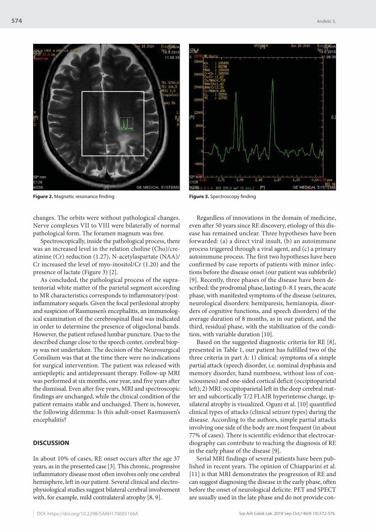
574
Srp Arh Celok Lek. 2018 Sep-Oct;146(9-10):572-576DOI: https://doi.org/10.2298/SARH170605166A
changes. The orbits were without pathological changes. Nerve complexes VII to VIII were bilaterally of normal pathological form. The foramen magnum was free.
Spectroscopically, inside the pathological process, there was an increased level in the relation choline (Cho)/cre-atinine (Cr) reduction (1.27), N-acetylaspartate (NAA)/Cr increased the level of myo-inositol/Cr (1.20) and the presence of lactate (Figure 3) [2].
As concluded, the pathological process of the supra-tentorial white matter of the parietal segment according to MR characteristics corresponds to inflammatory/post-inflammatory sequels. Given the focal perilesional atrophy and suspicion of Rasmussen’s encephalitis, an immunolog-ical examination of the cerebrospinal fluid was indicated in order to determine the presence of oligoclonal bands. However, the patient refused lumbar puncture. Due to the described change close to the speech center, cerebral biop-sy was not undertaken. The decision of the Neurosurgical Consilium was that at the time there were no indications for surgical intervention. The patient was released with antiepileptic and antidepressant therapy. Follow-up MRI was performed at six months, one year, and five years after the dismissal. Even after five years, MRI and spectroscopic findings are unchanged, while the clinical condition of the patient remains stable and unchanged. There is, however, the following dilemma: Is this adult-onset Rasmussen’s encephalitis?
DISCUSSION
In about 10% of cases, RE onset occurs after the age 37 years, as in the presented case [3]. This chronic, progressive inflammatory disease most often involves only one cerebral hemisphere, left in our patient. Several clinical and electro-physiological studies suggest bilateral cerebral involvement with, for example, mild contralateral atrophy [8, 9].
Regardless of innovations in the domain of medicine, even after 50 years since RE discovery, etiology of this dis-ease has remained unclear. Three hypotheses have been forwarded: (a) a direct viral insult, (b) an autoimmune process triggered through a viral agent, and (c) a primary autoimmune process. The first two hypotheses have been confirmed by case reports of patients with minor infec-tions before the disease onset (our patient was subfebrile) [9]. Recently, three phases of the disease have been de-scribed: the prodromal phase, lasting 0–8.1 years, the acute phase, with manifested symptoms of the disease (seizures, neurological disorders: hemiparesis, hemianopia, disor-ders of cognitive functions, and speech disorders) of the average duration of 8 months, as in our patient, and the third, residual phase, with the stabilization of the condi-tion, with variable duration [10].
Based on the suggested diagnostic criteria for RE [8], presented in Table 1, our patient has fulfilled two of the three criteria in part A: 1) clinical: symptoms of a simple partial attack (speech disorder, i.e. nominal dysphasia and memory disorder, hand numbness, without loss of con-sciousness) and one-sided cortical deficit (occipitoparietal left); 2) MRI: occipitoparietal left in the deep cerebral mat-ter and subcortically T/2 FLAIR hyperintense change, ip-silateral atrophy is visualized. Oguni et al. [10] quantified clinical types of attacks (clinical seizure types) during the disease. According to the authors, simple partial attacks involving one side of the body are most frequent (in about 77% of cases). There is scientific evidence that electrocar-diography can contribute to reaching the diagnosis of RE in the early phase of the disease [9].
Serial MRI findings of several patients have been pub-lished in recent years. The opinion of Chiapparini et al. [11] is that MRI demonstrates the progression of RE and can suggest diagnosing the disease in the early phase, often before the onset of neurological deficite. PET and SPECT are usually used in the late phase and do not provide con-
Figure 2. Magnetic resonance finding Figure 3. Spectroscopy finding
Anđelić S.

575
Srp Arh Celok Lek. 2018 Sep-Oct;146(9-10):572-576 www.srpskiarhiv.rs
crete results. Early RE diagnosis is crucial in the selection of patients who require aggressive medicamentous therapy or surgical intervention such as hemispheroctomy.
According to Rasmussen [12] and Rasmussen and An-dermann [13], standard cerebrospinal fluid tests are not reliable for the confirmation or rejection of the RE diagno-sis. Serological cerebrospinal fluid tests are usually applied in order to exclude infections by well-known neurotropic viruses. Our patient refused lumbar puncture. In most cases, the PET method detects large hypometabolic zones of the involved hemisphere, while new zones with focal hypermetabolism are found in a somewhat lower number [11, 14]. Lee et al. [15] have proposed that PET may guide brain biopsy in cases with inconclusive or normal MRI findings, especially in the early stages. sMRI investiga-tion indicates that lowering the level of N-acetylaspartate (NAA) and increasing (or normal) levels of Cho results in the increased relation NAA/Cho that indicates the loss of dysfunction [9]. Increased level of present lactates as in our case is associated with the presence of EPC. Therefore, PET, SPECT, and sMRI techniques are not adequate for defining inflammatory nature of RE. They can be helpful in the confirmation of the unihemispheric nature in the early phase of suspected RE. Cerebral biopsy is not neces-sary in all REs because other criteria could be sufficient in making the diagnosis (Table 1).
Corresponding tests should be applied to confirm RE and exclude other diseases. Most frequently used cerebral scans are MRI, SPECT, and, if necessary, fluorodeoxy-glucose-PET scans. Next, blood tests for the exclusion of infection, lumbar puncture for confirming inflammation and infection, and finally cerebral biopsy to confirm the
diagnosis are necessary. In our patient, differential-diag-nostic considerations were aimed at the following: 1) other unilateral neurologic syndromes (stroke, tumor); 2) other reasons for EPC (drugs, cerebral gliomatosis), or 3) other inflammatory or infectious diseases that mimic RE (vascu-litis, multiple sclerosis, viral or toxoplasmosis encephalitis) [9]. Although lumbar puncture can show whether there is inflammation and cerebral infection, our patient was not willing to undergo the procedure. Although cerebral biopsy is necessary in the absolute diagnostics of RE, due to the described change and the speech center, it was not performed in our patient.
After reaching the diagnosis, medicamentous or surgi-cal treatment can be applied. To treat RE, antiepileptics alone or in combination with other drugs (as in the pre-sented case) have only limited effect in the control of focal attacks and EPC; the general rule is that the number and dosage of antiepileptics should be as low as possible, as was in the presented case. Recently, long-term treatments have been attempted with corticosteroids, intravenous immunoglobulins, plasma-exchange, or tacrolimus [16]. Only a few patients have been treated with rituximab as the alternative therapy for RE [17].
Surgical treatment (hemispherectomy) remains the most efficient therapy in the prevention of attack pro-gression caused by RE. In our patient, surgery was not indicated, but only MRI follow-up.
This case highlights the diagnostic dilemmas that arise in adult-onset RE and suggests that this diagnosis should be considered in patients of any age with an appropriate clinical picture. Rasmussen’s encephalitis in adults can be a challenging diagnosis.
REFERENCES
Diagnostic dilemmas of Rasmussen’s encephalitis in adults
1. Panayiotopoulos CP. The Epilepsies: Seizures, Syndromes and Management. Oxfordshire: Bladon Medical Publishing; 2005. p. 207–15.
2. Rasmussen T, Olszewski J, Loyd-Smith D. Focal seizures due to chronic localized encephalitis. Neurology. 1958; 8(6):435–45.
3. Hart YM, Andermann F, Fish DR, Dubeau F, Robitaille Y, Rasmussen T, et al. Chronic encephalitis epilepsy in adults and adolescents: a variant of Rasmussen´s syndrome? Neurology. 1997; 48(2):418–24.
4. Vadlamudi L, Galton CJ, Jeavons SJ, Tannenberg AE, Boyle RS. Rasmussen’s syndrome in a 54 year old female: more support for an adult variant. J Clin Neurosci. 2000; 7(2):154–6.
5. Rogers SW, Andrews PI, Gahring LC, Whisenand T, Cauley K, Crain B, et al. Autoantibodies to glutamate receptor GluR3 in Rasmussen’s encephalitis. Science. 1994; 265(5172):648–51.
6. Andrews PI, Dichter MA, Berkovic SF, Newton MR, McNamara JO. Plasmapheresis in Rasmussen’s encephalitis. Neurology. 1996; 46(1):242–6.
7. Robitaille Y. Neuropathologic aspects of chronic encephalitis. In: Andermann F. Chronic encephalitis and epilepsy. Rasmussen’s syndrome. Boston: Butterworth-Heinemann; 1991. p. 79–110.
8. Bien CG, Granata T, Antozzi C, Cross JH, Dulac O, Kurthen M, et al. Pathogenesis, diagnosis and treatment of Rasmussen encephalitis. A European consensus statement. Brain. 2005; 128(Pt 3):454–71.
9. Sheybani L, Schaller K, Seeck M. Rasmussen’s encephalitis: an update. Schweizer archiv für neurologie und psychiatrie. 2011; 162(6):225–31.
10. Oguni H, Andermann F, Rasmussen TB. The natural history of the syndrome of chronic encephalitis and epilepsy: a study of the MNI series of fortyeight cases. In: Andermann F, editor. Chronic encephalitis and epilepsy. Rasmussen’s syndrome. Boston: Butterworth-Heinemann; 1991. p. 7–35.
11. Chiapparini L, Granata T, Farina L, Ciceri E, Erbetta A, Ragona F, et al. Diagnostic imaging in 13 cases of Rasmussen’s encephalitis: can early MRI suggest the diagnosis? Neuroradiology. 2003; 45(3):171–83.
12. Rasmussen T. Further observations on the syndrome of chronic encephalitis and epilepsy. Appl Neurophysiol. 1978; 41(1-4):1–12.
13. Rasmussen T, Andermann F. Update on the syndrome of ‘chronic encephalitis’ and epilepsy. Cleveland Clin J Med. 1989; 56 Suppl 2:S181–4.
14. Fiorella DJ, Provenzale JM, Edward CR, Crain BJ, Al Sugair A. (18)F-fluorodeoxyglucose positron emission tomography and MR imaging findings in Rasmussen encephalitis. Am J Neuroradiol. 2001; 22(7):1291–9.
15. Lee JS, Juhasz C, Kaddurah AK, Chugani HT. Patterns of cerebral glucose metabolism in early and late stages of Rasmussen’s syndrome. J Child Neurol. 2001; 16(11):798–805.
16. Campanille V, De Aguirre I, Crespo J, Gonzalez M, Lopez JI, Lucero C, et al. Adult Rasmussen’s Encephalitis. Clin Med Rev Case Rep. 2016; 3(12):144.
17. Thilo B, Stingele R, Knudsen K, Boor R, Bien CG, Deuschl G, et al. A case of Rasmussen encephalitis treated with rituximab. Nat Rev Neurol. 2009; 5(8):458–62.

576
doi: Srp Arh Celok Lek. 2018 Sep-Oct;146(9-10):572-576
Anđelić S.
DOI: https://doi.org/10.2298/SARH170605166A
САЖЕТАКУвод Расмусенов енцефалитис (РЕ) ретко је, прогресивно, инфламаторно обољење мозга које је тешко доказати код одраслих особа. Циљ овог рада је да истакне дијагностичке дилеме код РЕ одраслих.Приказ болесника Жена стара 46 година хоспитализована је због упорних дифузних, интензивних главобоља, праће-них мучнином и нагоном на повраћање, због којих се будила ноћу. Више пута је имала транзиторни поремећај говора и памћења и утрнулост десне руке. МР налаз је показао сле-деће: окципитопаријетално лево у дубокој белој можданој маси, као и субкортикално уочава се T2/flair хиперинтезна промена, Т1 хипоинтензна промена захвата corpus callosum.
Спектроскопски се евидентира повишена вредност односа холин / креатинин (Кр) (2,12), редукован ниво азот-ацетил-аспартата / Кр (1,27), повећана вредност миоинозитола / Кр (1,20) и присуство лактата. Болесница је одбила лумбалну пункцију. Због близине описане промене и центра за говор није урађена биопсија мозга. После пет година МР и спе-ктроскопски налази су непромењени, а стање болеснице стабилно.Закључак Овај случај наглашава дијагностичке дилеме код РЕ одраслих и указује на то да ову дијагнозу треба узети у обзир код болесника било којег узраста са одговарајућом клиничком сликом. Кључне речи: Расмусенов енцефалитис; одрасли; дифер-нецијална дијагноза; енцефалитис, дијагноза
Дијагностичке дилеме код Расмусеновог енцефалитиса одраслихСлађана АнђелићГрадски завод за хитну медицинску помоћ, Београд, Србија

577
DOI: https://doi.org/10.2298/SARH171016008J
UDC: 616.8-009.24-07:575
Correspondence to:
Dragana JANIĆ
Department of Hematology
and Oncology
University Children’s Hospital
Tiršova 10, 11000 Belgrade
Serbia
Received • Примљено: October 16, 2017
Revised • Ревизија: January 22, 2018
Accepted • Прихваћено: January 23, 2018
Online fi rst: February 6, 2018
SUMMARYIntroduction Fever of unknown origin is an important diagnostic challenge. Although rare, periodic fever syndromes may often present with a chronic or recurrent febrile condition with a variable temporal pat-tern of occurrence. Although clinical characteristics often indicate the syndrome in question, there are many atypical forms, and the genotype–phenotype relationship is highly complex, warranting in many cases the designation of a “syndrome spectrum” rather than a syndrome per se. The aim of this paper was to present a boy with recurrent fever of unknown origin.Case outline We hereby present a boy with recurrent fever of unknown origin who was by clinically guided partial exome sequencing found to have a heterozygous variant 434A>G in the TNFRSF1A gene, otherwise connected with tumor necrosis factor receptor-associated periodic fever syndrome. The patient responded well to short courses of glucocorticoids and is no longer subjected to unnecessary antibiotic treatment he had frequently received in the past. Conclusion Periodic fever syndromes should be kept in mind as a differential diagnostic possibility in children with fever of unknown origin. Keywords: fever; autoinflammatory disorders; TRAPS; genotype–phenotype correlation
CASE REPORT / ПРИКАЗ БОЛЕСНИКА
TNFRSF1A gene variant identified in a boy with recurrent episodes of fever Srđa Janković1, Goran Đuričić1, Aleksandra Radosavljević2,3, Dragana Janić1,2
1University Children’s Hospital, Belgrade, Serbia;2University of Belgrade, Faculty of Medicine, Belgrade, Serbia;3Clinical Center of Serbia, Clinic for Eye Diseases, Belgrade, Serbia
INTRODUCTION
Prolonged or recurrent fever of unknown ori-gin constitutes an important diagnostic prob-lem that may be connected to a multitude of potential etiological factors and nosological entities [1, 2]. Although exceedingly rare, au-toinflammatory disorders often present with a febrile condition of unknown origin. Depend-ing on the syndrome in question, febrile spells may occur in relatively regular intervals, or there may be no discernible time pattern in their occurrence [3, 4]. Of all periodic fever syndromes, periodic fever, aphthous stomatitis, pharyngitis and adenitis (PFAPA) syndrome exhibits by far the greatest incidence. How-ever, this syndrome is not readily explained by a defined genetic aberration(s) and is thought to be an etiologically (and to some extent also pathogenetically) heterogenous category, diag-nosed per exclusionem by the presence of fever and inflammation in a person without signs of infection and with a prompt resolution of symptoms upon glucocorticoid treatment [5]. Most common periodic fever syndromes of au-toinflammatory nature with fully characterized genetic causes include familial mediterranean fever (FMF), cryopyrin-associated periodic syndromes (CAPS), mevalonate kinase defi-ciency (MKD)/hyperimmunoglobulinemia D and periodic fever syndrome, and tumor ne-crosis factor receptor-associated periodic fever
syndrome (TRAPS) [6]. Symptoms and signs other than fever, such as joint pains, skin rash, serositis, or abdominal aches may be very help-ful in the establishment of diagnosis [7].
Treatment partly depends on the syndrome in question and in many cases may be success-fully guided by clinical tools such as the Auto-Inflammatory Diseases Activity Index (AIDAI; Table 1) [8]. Early and appropriate treatment can greatly reduce the risk of complications, including the most serious – amyloidosis [9]. It is therefore of utmost importance to systemati-cally evaluate children with fever of unknown origin for possible autoinflammatory disorders. If performed thoroughly, this very often results in a precise diagnosis [10].
We hereby present the case of a boy with recurrent episodes of fever that were eventu-ally plausibly explained by the result of genetic testing.
CASE REPORT
Repeated instances of febrile illness in an oth-erwise healthy boy began at the age of six years. They were separated by an interval of several months. Bodily temperature usually reached 40оC, while C-reactive protein was in the 40–100 mg/L range, and the erythrocyte sedi-mentation rate was typically about 30 mm/h. A mild splenomegaly was also noted. Febrile
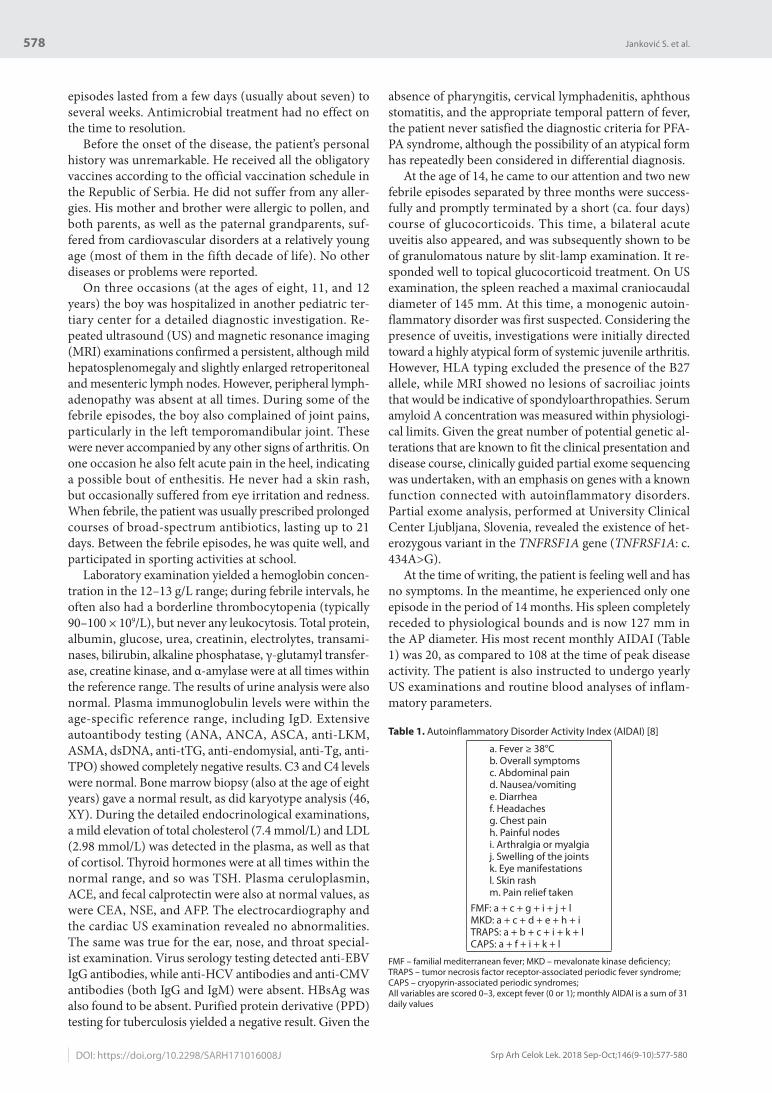
578
Srp Arh Celok Lek. 2018 Sep-Oct;146(9-10):577-580DOI: https://doi.org/10.2298/SARH171016008J
episodes lasted from a few days (usually about seven) to several weeks. Antimicrobial treatment had no effect on the time to resolution.
Before the onset of the disease, the patient’s personal history was unremarkable. He received all the obligatory vaccines according to the official vaccination schedule in the Republic of Serbia. He did not suffer from any aller-gies. His mother and brother were allergic to pollen, and both parents, as well as the paternal grandparents, suf-fered from cardiovascular disorders at a relatively young age (most of them in the fifth decade of life). No other diseases or problems were reported.
On three occasions (at the ages of eight, 11, and 12 years) the boy was hospitalized in another pediatric ter-tiary center for a detailed diagnostic investigation. Re-peated ultrasound (US) and magnetic resonance imaging (MRI) examinations confirmed a persistent, although mild hepatosplenomegaly and slightly enlarged retroperitoneal and mesenteric lymph nodes. However, peripheral lymph-adenopathy was absent at all times. During some of the febrile episodes, the boy also complained of joint pains, particularly in the left temporomandibular joint. These were never accompanied by any other signs of arthritis. On one occasion he also felt acute pain in the heel, indicating a possible bout of enthesitis. He never had a skin rash, but occasionally suffered from eye irritation and redness. When febrile, the patient was usually prescribed prolonged courses of broad-spectrum antibiotics, lasting up to 21 days. Between the febrile episodes, he was quite well, and participated in sporting activities at school.
Laboratory examination yielded a hemoglobin concen-tration in the 12–13 g/L range; during febrile intervals, he often also had a borderline thrombocytopenia (typically 90–100 × 109/L), but never any leukocytosis. Total protein, albumin, glucose, urea, creatinin, electrolytes, transami-nases, bilirubin, alkaline phosphatase, γ-glutamyl transfer-ase, creatine kinase, and α-amylase were at all times within the reference range. The results of urine analysis were also normal. Plasma immunoglobulin levels were within the age-specific reference range, including IgD. Extensive autoantibody testing (ANA, ANCA, ASCA, anti-LKM, ASMA, dsDNA, anti-tTG, anti-endomysial, anti-Tg, anti-TPO) showed completely negative results. C3 and C4 levels were normal. Bone marrow biopsy (also at the age of eight years) gave a normal result, as did karyotype analysis (46, XY). During the detailed endocrinological examinations, a mild elevation of total cholesterol (7.4 mmol/L) and LDL (2.98 mmol/L) was detected in the plasma, as well as that of cortisol. Thyroid hormones were at all times within the normal range, and so was TSH. Plasma ceruloplasmin, ACE, and fecal calprotectin were also at normal values, as were CEA, NSE, and AFP. The electrocardiography and the cardiac US examination revealed no abnormalities. The same was true for the ear, nose, and throat special-ist examination. Virus serology testing detected anti-EBV IgG antibodies, while anti-HCV antibodies and anti-CMV antibodies (both IgG and IgM) were absent. HBsAg was also found to be absent. Purified protein derivative (PPD) testing for tuberculosis yielded a negative result. Given the
absence of pharyngitis, cervical lymphadenitis, aphthous stomatitis, and the appropriate temporal pattern of fever, the patient never satisfied the diagnostic criteria for PFA-PA syndrome, although the possibility of an atypical form has repeatedly been considered in differential diagnosis.
At the age of 14, he came to our attention and two new febrile episodes separated by three months were success-fully and promptly terminated by a short (ca. four days) course of glucocorticoids. This time, a bilateral acute uveitis also appeared, and was subsequently shown to be of granulomatous nature by slit-lamp examination. It re-sponded well to topical glucocorticoid treatment. On US examination, the spleen reached a maximal craniocaudal diameter of 145 mm. At this time, a monogenic autoin-flammatory disorder was first suspected. Considering the presence of uveitis, investigations were initially directed toward a highly atypical form of systemic juvenile arthritis. However, HLA typing excluded the presence of the B27 allele, while MRI showed no lesions of sacroiliac joints that would be indicative of spondyloarthropathies. Serum amyloid A concentration was measured within physiologi-cal limits. Given the great number of potential genetic al-terations that are known to fit the clinical presentation and disease course, clinically guided partial exome sequencing was undertaken, with an emphasis on genes with a known function connected with autoinflammatory disorders. Partial exome analysis, performed at University Clinical Center Ljubljana, Slovenia, revealed the existence of het-erozygous variant in the TNFRSF1A gene (TNFRSF1A: c. 434A>G).
At the time of writing, the patient is feeling well and has no symptoms. In the meantime, he experienced only one episode in the period of 14 months. His spleen completely receded to physiological bounds and is now 127 mm in the AP diameter. His most recent monthly AIDAI (Table 1) was 20, as compared to 108 at the time of peak disease activity. The patient is also instructed to undergo yearly US examinations and routine blood analyses of inflam-matory parameters.
Table 1. Autoinflammatory Disorder Activity Index (AIDAI) [8]
a. Fever ≥ 38°Cb. Overall symptomsc. Abdominal paind. Nausea/vomitinge. Diarrheaf. Headachesg. Chest painh. Painful nodesi. Arthralgia or myalgiaj. Swelling of the jointsk. Eye manifestationsl. Skin rashm. Pain relief taken
FMF: a + c + g + i + j + lMKD: a + c + d + e + h + iTRAPS: a + b + c + i + k + lCAPS: a + f + i + k + l
FMF – familial mediterranean fever; MKD – mevalonate kinase defi ciency; TRAPS – tumor necrosis factor receptor-associated periodic fever syndrome; CAPS – cryopyrin-associated periodic syndromes;All variables are scored 0–3, except fever (0 or 1); monthly AIDAI is a sum of 31 daily values
Janković S. et al.

579
Srp Arh Celok Lek. 2018 Sep-Oct;146(9-10):577-580 www.srpskiarhiv.rs
DISCUSSION
Clinically, our patient exhibited clear signs of a long-standing inflammatory condition with elements of some autoinflammatory disorders (episodes of fever, joint pains, splenomegaly, possible enthesitis of the Achilles’ tendon, uveitis, prompt response to glucocorticoids). However, diagnostic criteria for any specific disorder were not sat-isfied, suggesting either an extremely rare nosological entity or some rather untypical clinical variant of a more common one. Bearing in mind that it is usually, if not universally, reasonable to assume that the latter is vastly more likely that the former, we decided it is worthwhile to perform clinically guided partial exome sequencing in order to identify potential gene variants that could explain the observed symptoms and signs.
Mutations in TNFRSF1A cause tumor necrosis factor (TNF) receptor-associated autoinflammatory syndrome (TRAPS) [11]. TRAPS was formerly called “familial Hi-bernian fever” because it had initially been described in a family of Scottish ancestry (Hibernia being the Roman name for Scotland) [12]. TNFRSF1A encodes a member of the TNF receptor superfamily and is therefore extensively involved in inflammatory processes associated with both innate and adaptive immune mechanisms and processes. It is mainly expressed on mononuclear phagocytes, but may also be found on a number of other cell types, such as lymphocytes, natural killer cells, granulocytes, astrocytes, and keratinocytes [13]. Numerous different variants in TNFRSF1A have been described, with a highly complex genotype–phenotype relationship [14]. Prognosis is vari-able and primarily dependent on the existence of complica-tions of chronic inflammation, such as amyloidosis. Variant 434A>G is recorded in the ClinVar database (No. 97703) and the Infevers registry, and designated as a genetic vari-ant of unknown significance [15, 16]. However, the same variant has been reported in a patient listed in the EURO-FEVER registry with clinically apparent TRAPS [17].
Although there is no possibility of final proof that the detected gene variant indeed plays a causal role in our pa-tient’s ailment, it is certainly plausible that it has at least some effect, based on obvious pathophysiological mecha-nisms (i.e. uncontrolled inflammation) and known func-tions of the TNFRSF1A gene (including inflammatory signaling). Considering the relatively innocuous disease course so far and the absence of any signs of permanent organ damage or amyloidosis, the outlook for our young patient appears to be favorable. The example we describe here could be used as a good illustration of the concept of “genomic landscape” of congenital autoinflammatory (as well as other) syndromes; the complexity of this land-scape very often does not allow a clear demarcation line to be drawn between a pathological and a physiological gene variant, particularly when seen in the light of the less-than-predictable relationship between the nature of genetic alteration and its clinical consequences, if any. Low-penetrance TNFRSF1A variants are well known and appear to cause a mild or moderate autoinflammatory condition in
some, though not all, affected persons [18]. Furthermore, the low-penetrance variants appear to produce their ef-fects through a different pathophysiological mechanism compared with clearly pathogenic gene alterations, and are, at least in part, connected to the functional status of regulatory T cells [19]. An analysis of a series of patients who carry a well characterized low-penetrance variant has shown that severity of symptoms and risk of complications are highly variable and at least partially correlated with the age of onset [20]. In the light of all this, it appears quite justified to speak of a “TRAPS spectrum” as a diagnostic category (as opposed to the diagnosis of TRAPS per se). The rationale for using the latter designation appears rather strengthened by the fact that our patient constantly exhib-ited some, but never all, features of PFAPA syndrome, beg-ging the question how many patients classified within this highly heterogenous diagnostic category are (or were) actu-ally affected by TNFRSF1A variants, among other defects in genes connected to inflammation. This and a myriad of analogous possibilities in other autoinflammatory dis-orders, such as, for instance, FMF, CAPS, and MKD (to name the most frequent ones in our population, aside from TRAPS) warrants particular attention when the physician is faced with a patient clinically exhibiting some, but not all features of a known autoinflammatory syndrome. In such instances, clinically guided partial exome sequencing, if available, generally tends to become the diagnostic method of choice. On the other hand, it is an exceedingly costly and somewhat time-consuming procedure, and this adds to the importance that all physicians, and especially pediatricians, be satisfactorily acquainted with the full range of clinical situations where it is rational to order such an analysis. This appears to be of the essence, since the usefulness of extensive genetic testing without proper clinical guidance is very doubtful, as highlighted, for instance, by the recently published experience in autoinflammatory disorders from a center in Trieste [21]. A deeper knowledge of possible genetic alterations and their complex consequences should ensure the necessary amount of critical thinking in deter-mining whether testing is indicated or warranted, and this is, indeed, more than appropriate for the practice of medi-cine in the genomic age.
The distinction between TRAPS and the proposed designation of “TRAPS spectrum” can also be viewed as highly meaningful from the treatment standpoint. While TRAPS patients are usually best treated with IL-1 antago-nists such as anakinra [22, 23] or canakinumab [24], most patients with low-penetrance TNFRSF1A alterations either require no treatment or sufficient disease control can be achieved by occasional short courses of glucocorticoids, administered as needed [18].
ACKNOWLEDGEMENT
The authors (S. J. and D. J.) received support from the Ministry of Education, Science and Technological Devel-opment of the Republic of Serbia, project No. 41004.
TNFRSF1A gene variant identifi ed in a boy with recurrent episodes of fever

580
doi: Srp Arh Celok Lek. 2018 Sep-Oct;146(9-10):577-580
1. Chusid MJ. Fever of unknown origin in childhood. Pediatr Clin North Am. 2017; 64(1):205–30.
2. Dayal R, Agarwal D. Fever in children and fever of unknown origin. Indian J Pediatr. 2016; 83(1):38–43.
3. Martorana D, Bonatti F, Mozzoni P, Vaglio A, Percesepe A. Monogenic autoinflammatory diseases with mendelian inheritance: genes, mutations, and genotype-phenotype correlations. Front Immunol. 2017; 8:344.
4. Pastore S, Vuch J, Bianco AM, Teddio A, Tommasini A. Fever tree revisited: From malaria to autoinflammatory diseases. World J Clin Pediatr. 2015; 4(4):106–12.
5. Ali NS, Sartori-Valinotti JC, Bruce AJ. Periodic fever, aphthous stomatitis, pharyngitis and adenitis (PFAPA) syndrome. Clin Dermatol. 2016; 34(4):482–6.
6. Sag E, Bilginer Y, Ozen S. Autoinflammatory diseases with periodic fevers. Curr Rheumatol Rep. 2017; 19:41.
7. Lachmann HJ. Autoinflammatory syndromes as causes of fever of unknown origin. Clin Med. 2015; 15(3):295–8.
8. Piram M, Frenkel J, Gattorno M, Ozen S, Lachmann HJ, Goldbach-Mansky R, et al. A preliminary score for the assessment of disease activity in hereditary recurrent fevers: results from the AIDAI (Autoinflammatory Diseases Activity Index) consensus conference. Ann Rheum Dis. 2011; 70(2):309–14.
9. Scarpioni R, Ricardi M, Albertazzi V. Secondary amyloidosis in autoinflammatory diseases and the role of inflammation in renal damage. World J Nephrol. 2016; 5(1):66–75.
10. Vitale A, Rigante D, Lucherini OM, De Palma A, Orlando I, Gentileschi S, et al. The diagnostic evaluation of patients with a suspected hereditary periodic fever syndrome: experience from a referral center in Italy. Intern Emerg Med. 2017; 12(5):605–11.
11. Aguado-Gil L, Irarrazaval-Armendáriz I, Pretel-Irazabal M. Advances in the diagnosis and treatment of tumor necrosis factor receptor-associated periodic syndrome. Actas Dermosifiliogr. 2013; 104(7):617–22.
12. Williamson LM, Hull D, Mehta R, Reeves WG, Robinson BH, Toghill PJ. Familial Hibernian Fever. Q J Med. 1982; 51(204):469–80.
13. Magnotti F, Vitale A, Rigante D, Lucherini OM, Cimaz R, Muscari I, et al. The most recent advances in pathophysiology and management of tumour necrosis factor receptor-associated periodic syndrome (TRAPS): personal experience and literature review. Clin Exp Rheumatol. 2013; (3 Suppl 77):141–9.
14. Ozen S, Demir S. Monogenic periodic fever syndromes: treatment options for the pediatric patient. Pediatr Drugs. 2017; 19(4):303–11.
15. ClinVar database. [Variation Report for NM_004183.3(BEST1):c.274C>T (p.Arg92Cys)] (Accessed October 12, 2017). Available at: https://www.ncbi.nlm.nih.gov/clinvar/variation/99703/
16. International Society for Systemic AutoInflammatory Diseses. Infevers registry. (Accessed January 18, 2018). Available at: http://fmf.igh.cnrs.fr/ISSAID/infevers/detail_mutation.php
17. Lachmann HJ, Papa R, Gerhold K, Obici L, Touitou I, Cantarini L, et al. The phenotype of TNF receptor-associated autoinflammatory syndrome (TRAPS) at presentation: a series of 158 cases from the Eurofever/EUROTRAPS international registry. Ann Rheum Dis. 2014; 73(12):2160–7.
18. Cantarini L, Lucherini OM, Muscari I, Frediani B, Galeazzi M, Brizzi MG. Tumour necrosis factor receptor-associated periodic syndrome (TRAPS): State of the art and future perspectives. Autoimmun Rev. 2012; 12(1):38–43.
19. Pucino V, Lucherini OM, Perna F, Obici L, Merlini G, Cattalini M, et al. Differential impact of high and low penetrance TNFRSF1A gene mutations on conventional and regulatory CD4+ cell functions in TNFR1-associated periodic syndrome. J Leukoc Biol. 2016; 99(5):761–9.
20. Ruiz-Ortiz E, Iglesias E, Soriano A, Buján-Rivas S, Español-Rego M, Castellanos-Moreira R. Disease phenotype and outcome depending on the age of disease onset in patients carrying the R92Q low-penetrance variant in TNFRSF1A gene. Front Immunol. 2017; 8:299.
21. De Pieri C, Vuch J, De Martino E, Bianco AM, Ronfani L, Athanasakis E, et al. Genetic profiling of autoinflammatory disorders in patients with periodic fever: a prospective study. Pediatr Rheumatol Online J. 2015; 13:11.
22. Grimwood C, Despert V, Jeru I, Hentgen V. On-demand treatment with anakinra: a treatment option for selected TRAPS patients. Rheumatology (Oxford). 2015; 54(9):1749–51.
23. Gentileschi S, Rigante D, Vitale A, Sota J, Frediani B, Galeazzi M, et al. Efficacy and safety of anakinra in tumor necrosis factor receptor-associated periodic syndrome (TRAPS) complicated by severe renal failure: a report after long-term follow-up and review of the literature. Clin Rheumatol. 2017; 36(7):1687–90.
24. La Torre F, Caparello MC, Cimaz R. Canakinumab for the treatment of TNF receptor-associated periodic syndrome. Expert Rev Clin Immunol. 2017; 13(6):513–23.
REFERENCES
САЖЕТАКУвод Нејасно фебрилно стање је значајан дијагностички иза-зов. Мада су ретки, синдроми повремене грознице се често могу испољити у виду фебрилног стања које се понавља у различитим интервалима и траје краће или дуже време, или је пак хронично. Премда клиничка слика неретко указује на синдром о коме се ради, однос генотипа и фенотипа је веома сложен, што често оправдава сврставање у „спектар синдрома“ уместо у посебан синдром.Циљ овог рада је био да прикажемо дечака са понављаним налетима грознице непознатог узрока.Приказ болесника Код дечака са понављаним налетима грознице непознатог узрока клинички усмерено делимично
секвенцирање егзома показало је хетерозиготну варијанту 434A>G у гену TNFRSF1A. Овај ген је иначе повезан са синдро-мом TRAPS. Болесник је показао добар терапијски одговор на краткотрајно давање глукокортикоида и више се не лечи дуготрајним давањем антибиотика, што му је често ордини-рано у прошлости. Закључак У диференцијалној дијагнози код деце са гроз-ницом непознатог узрока ваља имати на уму и синдроме повратне грознице.
Кључне речи: грозница; аутоинфламаторне болести; TRAPS; однос генотип–фенотип
Варијанта гена TNFRSF1A код дечака са рекурентном фебрилношћуСрђа Јанковић1, Горан Ђуричић1, Александра Радосављевић2,3, Драгана Јанић1,2
1Универзитетска дечја клиника Тиршова, Београд, Србија;2Универзитет у Београду, Медицински факултет, Београд, Србија;3Клинички центар Србије, Клиника за очне болести, Београд, Србија
Janković S. et al.

581
DOI: https://doi.org/10.2298/SARH170811053F
UDC: 616-056.7-06-053.2
Correspondence to:
Stojka FUŠTIK
Vodnjanska 17
1000 Skopje, R.Macedonia
Received • Примљено: August 11, 2017
Revised • Ревизија: November 20, 2017
Accepted • Прихваћено: July 25, 2018
Online fi rst: August 7, 2018
CASE REPORT / ПРИКАЗ БОЛЕСНИКА
Hyponatremic dehydration and metabolic alkalosis as dominant manifestation in cystic fibrosis infants with mild phenotype – a case seriesStojka Fuštik1, Tatjana Jakovska1, Dijana Plašeska-Karanfilska2
1University Children’s Clinic, Department for Cystic Fibrosis, Skopje, Macedonia;2Macedonian Academy of Science and Arts, Research Center for Genetic Engineering and Biotechnology, Skopje, Macedonia
SUMMARYIntroduction Due to increased losses of chloride and sodium in the sweat, children with cystic fibrosis (CF) are predisposed to develop episodes of hyponatremic/hypochloremic dehydration with hypokalemia and metabolic alkalosis when they sweat excessively. Even the patients with mild phenotype may have such episodes of dehydration and salt depletion.Outline of cases Six cases of pancreatic sufficient (PS) CF patients complicated with episodes of severe hyponatremic dehydration with metabolic alkalosis in infancy are presented. The mean age was 6.3 ± 2.16 months at admission. All the cases had no symptoms suggestive of CF before admission. The most com-mon clinical symptoms at the time of hospitalization were vomiting, anorexia, weight loss, dehydration, irritation, or lethargy. Mean values of blood pH, serum bicarbonate, sodium, chloride, and potassium (mmol/l) were as follows: 7.59 ± 0.06, 41.73 ± 5.78, 117.52 ± 2.88, 66.0 ± 11.58 and 2.62 ± 0.37, respectively. Sweat chloride test was pathological and ranged 69–120 mmol/L. The determination of fecal elastase-1 proved that they were PS (values > 200 μg/g stool). CF transmembrane conductance regulator gene analyses in six cases confirmed the diagnosis of CF; namely, patients were compound heterozygotes for F508del and other rare mutation or compound heterozygotes for two rare mutations.Conclusion Distinctive about these cases is that they were PS and had very mild presentation of CF. Without these episodes of dehydration, these patients would have remained undiagnosed until later age. CF should be considered in infants and children presenting with hypoelectrolytemia and metabolic alkalosis even in the absence of respiratory or gastrointestinal symptoms. Keywords : cystic fibrosis; CFTR genotype; hyponatremic dehydration; metabolic alkalosis
INTRODUCTION
Cystic fibrosis (CF) is a multisystem disease caused by mutations in a gene on chromo-some 7, which encodes the CF transmembrane conductance regulator (CFTR) protein. CFTR functions primarily as a chloride channel and controls the movement of salt and water into and out of epithelial cells in the affected or-gans. Almost 2,000 different CFTR mutations have been identified, resulting in different con-sequences on protein function, ranging from complete protein absence to defective protein activity at the plasma membrane [1, 2]. Hence, phenotypic expression of the disease varies widely among individuals with CF [3].
In countries without neonatal screening for CF, the disease is usually diagnosed during childhood by respiratory and/or gastro-intes-tinal symptoms. Hyponatremic hypochloremic dehydration with hypokalemia and metabolic alkalosis is a rare but typical presentation of CF in infants [4, 5, 6]. Dysfunctional CFTR in the sweat ducts are responsible for the ex-cessive chloride and sodium losses, especially during warm months. The extracellular fluid volume contraction and salt depletion will lead to activation of renin–angiotensin system and
secondary hyperaldosteronism. The resulting effect is increased renal potassium and hydro-gen losses for the exchange with sodium in the distal tubule. The consequenced hypokalemic alkalosis is a metabolic mimicry of Bartter’s syndrome; therefore, the condition is known as pseudo-Bartter’s syndrome in CF.
In our previous study of pseudo-Bartter’s syndrome in CF, all patients with metabolic al-kalosis and hypoelectrolytemia were pancreatic insufficient (PI). Respectively, they had severe mutations with regard to pancreatic exocrine function [7]. Over the last few years, we have noticed the emergence of severe hyponatremic hypochloremic dehydration with metabolic al-kalosis in pancreatic sufficient (PS) CF infants, which had very mild disease expression further on in the clinical course.
REPORT OF CASES
Clinical records of six patients, three boys and three girls, presenting in infancy with meta-bolic alkalosis and electrolyte abnormalities such as hyponatremia, hypochloremia, and hypokalemia, which were later found to have CF, were analyzed.

582
Srp Arh Celok Lek. 2018 Sep-Oct;146(9-10):581-583DOI: https://doi.org/10.2298/SARH170811053F
Fuštik S. et al.
The mean age of children was 6.3 ± 2.16 months (range being 3–9 months). Biochemical features of the patients at the admission phase are summarized in Table 1. Mean values of blood pH, serum bicarbonate, sodium, chloride, and potassium (mmol/L) at admission were as follows: 7.59 ± 0.06, 41.73 ± 5.78, 117.52 ± 2.88, 66 ± 11.58, and 2.62 ± 0.37, respectively. Urine chloride concentrations in all the patients were below 20 mmol/l. For case 1 it was the second episode, for case 4 the third episode, and for cases 2, 3, 5, and 6 the first episode of dehydration. All episodes of dehydration occurred during the summer and early autumn months. The most common clinical symp-toms in these patients were vomiting, anorexia, weight loss, dehydration, irritation, or lethargy. We did not obtain a history of recurrent chest infection or loose stools in any child. Therefore, all cases had no symptoms suggestive of CF before admission.
After rehydration and correction of metabolic abnor-malities in the blood, a subsequent sweat chloride test and genotyping confirmed the diagnosis of CF in these infants. Sweat chloride tests were pathological and ranged 69–120 mmol/L. Assessment of the pancreatic functional status by determining the values of fecal elastase-1 showed that all six infants were PS (fecal elastase values were over 200 μg/g stool). CFTR gene analyses determined that the patients were compound heterozygotes for F508del (the most common CFTR mutation, class II) and other rare mutation or compound heterozygotes for two rare muta-tion (Table 2). A new mutation c.1070C>T (A357V) was detected in case 6.
Monitoring the clinical course of the disease in these children within the next two to five years showed that they have mild expression of CF mainly manifested as recur-rent sinusitis and nasal polyps in case 3. Only case 4 had another hospitalization for treating acute exacerbation of lung disease with Pseudomonas aeruginosa infection. Very mild pulmonary involvement was found on the chest X-ray in all six cases.
DISCUSSION
Metabolic alkalosis in association with low serum electro-lyte concentration is not a common metabolic disorder in infancy. Conditions associated with repeated vomiting, especially pyloric stenosis, continuous gastric drainage without appropriate electrolyte replacement, chloride-losing diarrhea, potassium-losing nephropathy, Bartter’s syndrome, the use of thiazide diuretics, and salt depletion by sweating in CF can lead to such a disturbance [8, 9].
Metabolic abnormalities in the so-called pseudo-Bart-ter’s syndrome in CF can have mimicking biochemical features of Bartter’s syndrome. Although the biochemical hallmark of both Bartter’s and pseudo-Bartter’s syndrome is abnormally low plasma electrolyte concentrations, there are important differences between the two diseases. In Bartter’s syndrome, the sweat electrolyte profile is normal and the renal handling of electrolytes is defective. In CF, sweat electrolyte losses are increased, and intensive electro-lyte reabsorption occurs in the renal tubules. In all our CF infants presenting with electrolyte depletion and metabolic alkalosis, the initial diagnosis of Bartter’s syndrome was excluded by hypochloruria (< 20 mmol/L). Determination of urinary chloride before therapy is especially useful to distinguish these two conditions.
In our previous study, all CF infants with pseudo-Bartter’s syndrome were PI [7]. We considered that biochemical ab-normalities due to insufficient CFTR chloride canal function are more pronounced in CF patients with “severe” disease-caused mutations (class I, II, and III). Compared to the “se-vere” CFTR mutations, certain “mild” mutations tend to be associated with significantly lower sweat chloride concentra-tions [10]. However, the emergence of severe hyponatremic hypochloremic dehydration with metabolic alkalosis in PS CF infants, which had mild disease expression in the further clinical course, indicated that neither the CFTR genotype nor sweat chloride levels are correlated with the occurrence of dehydration episodes. Our present analysis showed that two rare mutations (G126D and 711+ 3A- >G) were found
Table 1. Biochemical features of the pancreatic sufficient cystic fibrosis patients with pseudo-Bartter’s syndrome
Parametars Case 1 Case 2 Case 3 Case 4 Case 5 Case 6 M ± SD*pH 7.6 7.67 7.6 7.61 7.57 7.49 7.59 ± 0.06Bicarbonate (mmol/L) 42.9 49.1 42.8 44.6 39 32 41.73 ± 5.78Sodium (mmol/L) 113 121 117 116 118 120 117.52 ± 2.88Chloride (mmol/L) 61 65 54 56 78 82 66 ± 11.58Potassium (mmol/L) 2.1 2.7 2.4 2.6 3.2 2.7 2.62 ± 0.37
*Mean values ± standard deviation
Table 2. Genotypes of the pancreatic sufficient cystic fibrosis patients with pseudo-Bartter’s syndrome
Case No. Mutation IcDNA name / legacy name
Mutation IIcDNA name / legacy name
1 c.377G>A / G126D c.1366G>T / V456F2 c.377G>A / G126D c.1753G>T / E585X3 c.1521_1523delCTT / delF508 c.579+3A>G / 711+ 3A- >G4 c.1521_1523delCTT / delF508 c.579+3A>G / 711+ 3A- >G5 c.1521_1523delCTT / delF508 c.349C>T / R117C6 c.1521_1523delCTT / delF508 c.1070C>T / A357V
583
Srp Arh Celok Lek. 2018 Sep-Oct;146(9-10):581-583 www.srpskiarhiv.rs
Hyponatremic dehydration and metabolic alkalosis as dominant manifestation in cystic fi brosis infants with mild phenotype – a case series
in two cases, each. G126D is a missense mutation in exon 4 of the CFTR gene, resulting in amino acid change (glycine to asparagine at 126) in CFTR chloride channel and 711+ 3A- >G is a splicing mutation in intron 5, resulting in an mRNA splicing defect. This may arouse doubt that certain genotypes are more predisposed to the development of this metabolic disorder. Higher rate of sweating and electrolyte losses with sweat may be the reason why some CF individu-als are biochemically more vulnerable, but the risk factors for the development of dehydration with electrolyte deple-tion in CF are still not defined.
In conclusion, any CF patient, even a patient with a mild form of the disease, may experience an episode of dehy-dration with metabolic alkalosis and hypoelectrolytemia, particularly during the hot weather conditions. CF should be considered in the differential diagnosis of infants and children presenting with these biochemical abnormali-ties, even in the absence of respiratory or gastrointestinal symptoms. Missing the diagnosis of mild forms of CF may lead to life-threatening complications, such as severe hyponatremic dehydration with hypovolemia or diffuse bronchiectasis at a later age [11].
REFERENCES
1. Cystic Fibrosis Mutation Database [Internet]. http://www.genet.sickkids.on.ca/cftr/app. (accessed: February 9, 2017).
2. Rowntree RK, Harris A. The Phenotypic Consequences of CFTR Mutations. Ann Hum Genet. 2003; 67(Pt 5):471–85.
3. Knowles MR, Drumm M. The Influence of Genetics on Cystic Fibrosis Phenotypes. Cold Spring Harb Perspect Med. 2012; 2:a009548.
4. Ballestero Y, Hernandez MI, Rojo P, Manzanares J, Nebreda V, Carbajosa H, et al. Hyponatremic Dehydration as a Presentation of Cystic Fibrosis. Pediatr Emerg Care. 2006; 22(11):725–27.
5. Scurati-Manzoni E, Fossali EF, Agostoni C, Riva E, Simonetti GD, Zanolari-Calderari M, et al. Electrolyte abnormalities in cystic fibrosis: systematic review of the literature. Pediatr Nephrol. 2014; 29(6):1015–23.
6. Vilotijević-Dautović G, Stojanović V. Pseudo-Bartter’s Syndrome in Patients with Cystic Fibrosis: A Case Series and Review of the Literature. Srp Arh Celok Lek. 2015; 143(11-12):748–51.
7. Fustik S, Pop-Jardanova N, Slaveska N, Koceva S, Efremov G. Metabolic alkalosis with hypoelectrolytemia in infants with cystic fibrosis. Pediatr Int. 2002; 44(3):289–92.
8. Kennedy JD, Dinwiddie R, Daman-Willems C, Dillon MJ, Matthew DJ. Pseudo-Bartter’s syndrome in cystic fibrosis. Arch Dis Child. 1990; 65:786–7.
9. Khanna A, Kurtzman NA. Metabolic alkalosis. J Nephrol. 2006; 19 (Suppl 9):S86–96.
10. Wilschanski M, Zielenski J, Markiewicz D, Tsui LC, Corey M, Levison H, et al. Correlation of sweat chloride concentration with classes of the cystic fibrosis transmembrane conductance regulator gene mutations. J Pediatr. 1995; 127(5):705–10.
11. Priou-Guesdon M, Malinge MC, Augusto JF, Rodien P, Subra JF, Bonneau D, et al. Hypochloremia and hyponatremia as the initial presentation of cystic fibrosis in three adults. Annales d’Endocrinologie. 2010; 71(1):46–50.
САЖЕТАКУвод Због повећаних губитака хлора и натријума у зноју, деца са цистичном фиброзом (ЦФ) предиспонирана су да развију епизоде хипонатремичне/хипохлоремичне дехидра-ције са хипокалемијом и метаболичком алкалозом када се претерано зноје. Чак и болесници са благим фенотипом могу имати такве епизоде дехидрације и исцрпљивање соли. Приказ болесника Приказано је шест случајева панкреасно суфициjентних (ПС) болесника са ЦФ који су као одојчад имали као компликацију епизоде тешке хипонатремичне дехидрације са метаболичком алкалозом. Просечна старост је била 6,3 ± 2,16 месеци на пријему. Нису сви случајеви пре пријема имали симптоме који указују на ЦФ. Најчешћи клинички симптоми у време хоспитализације били су: по-враћање, анорексија, губитак тежине, дехидрација, ирита-ција или летаргија. Средње вредности pH крви, серумских бикарбоната, натријума, хлора и калијума (mmol/l) биле су,
редом: 7,59 ± 0,06; 41,73 ± 5,78; 117,52 ± 2,88; 66 ± 11,58 и 2,62 ± 0,37. Знојни хлоридни тест био је патолошки и варирао је од 69 до 120 mmol/l. Одређивање фекалне еластазе-1 пока-зало је да су ПС (вредности > 200 μg/g столице). ЦФТР генска анализа код свих шест случајева потврдила је дијагнозу ЦФ; наиме, болесници су били сложени хетерозиготи за F508del и другу ретку мутацију или сложени хетерозиготи за две ретке мутације.Закључак Карактеристично за ове случајеве је то што су панкреасно суфицијентни и имају веома благу презентацију ЦФ. Без ових епизода дехидрације, ови болесници би остали недијагностиковани до каснијег узраста. ЦФ треба узети у обзир код одојчади и деце код којих се манифестују хипо-електролитемија и метаболичка алкалоза, чак и у одсуству респираторних или гастроинтестиналних симптома.Кључне речи: цистична фиброза; ЦФТР генотип; хипонатре-мна дехидрација; метаболичка алкалоза
Хипонатремична дехидратација и метаболичка алкалоза као доминантна манифестација код одојчади са цистичном фиброзом и благим фенотипом – серија случајеваСтојка Фуштик1, Татјана Јаковска1, Дијана Плашеска-Каранфилска2
1Универзитетска клиника за децу, Одељење за цистичне фиброзе, Скопље, Македонија;2Македонска академија наука и уметности, Истраживачки центар за генетичко инжењерство и биотехнологију, Скопље, Македонија

584
Correspondence to:
Emilija NESTOROVIĆ
Clinic for Cardiac Surgery, Clinical
Center of Serbia
Koste Todorovica 8
11000 Belgrade, Serbia
Received • Примљено: July 19, 2017
Revised • Ревизија: November 16, 2017
Accepted • Прихваћено: November 22, 2017
Online fi rst: December 8, 2017
DOI: https://doi.org/10.2298/SARH170719202N
UDC: 616.12-089.843
SUMMARYIntroduction As the waiting time for heart transplantation continues to increase due to shortage in organ donation, supporting patients with advanced heart failure with left ventricular assist device (LVAD) increases as well. The latest generation of LVAD, left ventricular assist system (LVAS) started to expand worldwide due to more promising outcomes. The aim of this article was to present the case of treating advanced heart failure in a patient who under-went implantation of HeartMate 3 LVAS as the bridge to transplantation.Case otline The patient was a 59-year-old man with advanced heart failure requiring inotropic drug sup-port, with ischemic cardiomyopathy as the underlying cause of heart failure. Therefore, in the absence of an adequate donor, it was decided to incorporate the LVAS as a bridge to transplantation. Functional capacity, cardiac, renal, and liver functions improved in the patient.Conclusion The use of the HeartMate 3 in an advanced heart failure patient results in improvements in functional capacity, cardiac, renal and liver function. Further studies should be performed in order to identify whether improved outcomes are sustained with a longer follow-up period.Keywords: LVAD; heart failure; heart transplantation
CASE REPORT / ПРИКАЗ БОЛЕСНИКА
HeartMate 3 fully magnetically levitated left ventricular assist device for advanced heart failure – initial Serbian experienceEmilija Nestorović1, Duško Terzić1, Svetozar Putnik1, Arsen Ristić2, Miljko Ristić1
1Clinical Center of Serbia, Clinic for Cardiac Surgery, Department for Heart Transplant, Belgrade, Serbia;2Clinical Center of Serbia, Cardiology Clinic, Belgrade, Serbia
INTRODUCTION
Patients with advanced heart failure (HF) have quite limited possibilities for therapy, so the morbidity and mortality in connection to the disease are unacceptably high. In light of the insufficient availability of donor hearts and a growing number of heart transplantation (HT) ineligible patients, left ventricular assist devices (LVAD) are often used to support patients with advanced HF awaiting HT or as destination therapy (DT) [1]. In order to improve clini-cal outcomes of LVAD therapy, technological advances have been made through different types of LVAD pumps over the past 30 years, from pulsatile to continuous flow (CF) and now back to artificial pulsatility. Compared to the pulsatile flow HeartMate (HM) XVE, the newer generation of LVAD and CF-LVAD is considerably smaller, with significantly lower incidence of adverse events and has become the standard of care as a bridge to transplanta-tion (BTT) [1]. HM II (Abbott Corporation, Chicago, IL, USA) is an axial pump that pro-vides CF, which requires a tissue pocket and a larger body surface of the patient, while other intrapericardial CF pumps, like HeartWare (HeartWare Inc., Framingham, MA, USA), utilize a magnetically levitating rotor system to decrease mechanical wear in order to reduce hemolysis and the incidence of pump throm-bosis. Also, due to smaller size, it is suitable
for patients with smaller body surface area [2]. Despite the fact that with the newer gen-eration of LVAD survival rate is much better, the overall adverse events are still significantly high [3]. Therefore, technology enhancements of LVAD have become increasingly important in order to improve post-LVAD outcomes and to reduce the rate of adverse events. The latest generation pump, HM 3 (Abbott Corporation) is intrapericardial fully magnetically levitated CF-LVAD with artificial pulsatility with large, consistent pump gaps designed to reduce blood trauma and minimize stasis of blood.
The aim of this article was to present a case of treating the end-stage of HF in a patient in whom the latest generation of LVAD, left ventricular assist system (LVAS), HM 3, was implanted.
CASE REPORT
The patient was a 59-year-old man with ad-vanced HF, with ischemic cardiomyopathy as the underlying cause. Ten months prior to hospitalization at our institution, the patient suffered from acute heart infarction of the an-terior wall as the first manifestation of coronary disease. Since the patient was admitted to a lo-cal hospital after 24 hours had passed, neither percutaneous coronary intervention nor fibri-nolysis was applied. At the local hospital, the

585
Srp Arh Celok Lek. 2018 Sep-Oct;146(9-10):584-587 www.srpskiarhiv.rs
patient underwent coronary angiography that showed an occlusion of the left anterior descending coronary artery and stenosis of the proximal circumflex coronary artery and he was treated with medicaments. After the acute ischemic phase, the hospital treatment was hindered by the development of left ventricular (LV) aneurysm and the appearing symptoms of HF. He complained of fatigue while at rest, of choking, chest pain, and swelling in the legs. Ten months after the myocardial infarction, he came to our institution due to symptoms of advanced HF and required inotrope support drugs. Both two-dimensional transthoracic echocardiography (TTE) and transesopha-geal echocardiography (TEE) were performed in the standard manner using Vivid E 9 (GE Medical Systems, Milwaukee, WI, USA) and showed a volume overload of LV (EDV 257 ml), increased LV end-diastolic (LVEDD) and end-systolic diameters (LVESD), 7.4 cm and 6.3 cm, respectively, and significantly decreased ejection fraction of LV (LVEF) to 18% calculated by the Biplane method. There were akinesis in the LV septum, apex, apical part of anterolateral and inferior walls, apical and medial part of the anterior wall with LV apex aneurysm without visible thrombotic masses. Severe mitral regurgitation (MR 3+) was present. The right ventricle had regular dimensions (2.6 cm) with good systolic and longitudinal functions (global fractional area change of 27%, tricuspid annular plane systolic excursion of 21 mm, and right ventricular systolic excursion velocity of 18 cm/s). There was severe functional tricuspid regurgitation (TR 3+) with high right ventricle systolic pressure (RVSP 77 mmHg). Single-pho-ton emission computed tomography showed the absence of viable myocardium of the septum, anterior and lateral wall of the LV. Preoperative hemodynamic data showed pulmonary capillary wedge pressure (PCWP) of 27 mmHg, central venous pressure (CVP) of 11 mmHg, cardiac index (CI) of 1.9 L/min./m2. In order to assess the possibility of LVAS implantation, the nature of the LV apex aneurysm was estimated by performing magnetic resonance imag-ing of the heart. At the part of LV apex aneurysm, the myocardial wall thickness was 3–5 mm with aneurysmal
extension, which was a borderline finding for pump im-plantation. Based on all the performed tests, it was con-cluded that the patient was in the end-stage of HF (New York Heart Association (NYHA) IV, Interagency Registry for Mechanically Assisted Circulatory Support 3), with significantly impaired LV function requiring continuous inotropic support; therefore, it was decided in the absence of an adequate donor to incorporate the LVAS as a BTT.
The patient underwent surgery under general anesthe-sia. The surgery included the implantation of LVAS, HM 3. After median sternotomy, in conditions of extracorporeal circulation without aortic clamping and stopping of the heart, the LV was opened at the top, the ring was sewn, and after preparation, the pump was connected to the ring. The driveline was placed through the skin in the form of double tunneling and connected to the pump controller and the energy source. Finally, a graft was sewn to the aorta (Figure 1). Unlike the LVAD HM 2 device, where it is necessary to form a tissue pocket to place the device, LVAS HM 3 was applied fully intrapericardially. The intervention was without complications.
The postoperative recovery was uneventful. After the LVAS implantation, a standardized anticoagulation regi-men was used with initiation of infusion of heparin, fol-lowed by the transition to warfarin and aspirin. Antico-agulation therapy was included in order to maintain the international normalized ratio between 2.0 and 3.0. There was normal flow through both the pump’s inflow and out-flow cannula. The pump speed was set at 4,900 rpm, with pump flow being 3.4 L/min. and pump power 3.2 w. There were stable LVAS parameters and data on the pump con-troller. The radiographic finding was regular (Figure 2). Postoperative hemodynamic data showed PCWP of 10 mmHg, CVP of 7 mmHg, CI of 3.2 L/min./m2. Functional cardiac (TTE and brain-type natriuretic peptide – BNP), renal and liver function assessments were done before the LVAD implantation, at discharge from the hospital and at 12 months. The TTE examination at discharge showed improvement in the LVEDD and the LVESD from base-line values of 7.4 and 6.3 to 5.5 and 4.5, respectively. This
Figure 2. Radiographic finding after implanted HeartMate 3 left ventricular assist system
Figure 1. Left ventricular assist system, HeartMate 3 implantation: a) left ventricle open at the top; b) sewn ring at the left ventricle; c) preparation of the pump; d) connecting the pump to the ring; e) connected pump in the pericardial cavity; f ) outflow graft sewn to the aorta
Left ventricular assist system and outcomes

586
Srp Arh Celok Lek. 2018 Sep-Oct;146(9-10):584-587DOI: https://doi.org/10.2298/SARH170719202N
Nestorović E. et al.
improvement was sustained over the time of the follow-up. For LVEF and BNP (pg/ml), the baseline values of 18% and 670 improved to 39% and 173, respectively, and con-tinued to improve during the period of 12 months to 50% and 120, respectively. The dimension of the right ventricle was sustained in the normal range (2.6 cm) with a good systolic function. At discharge, NYHA functional classifi-cation improved from IV at baseline to II and continued to improve to I during the period of 12 months. There were improvements in renal function from baseline-estimated glomerular filtration rate (ml/min./1.73 m2) and serum creatinine (nmol/L) of 40 and 155 to 57 and 114, respec-tively, and there was no change over the entire follow-up period. With regard to hepatic function, total bilirubin (μmol/L) showed a reduction at the discharge point (25.8 vs. 12.4), with no further change afterwards. There were no significant changes of aspartate transaminase (IU/L), and alanine transaminase (IU/L) values during the entire follow-up period.
DISCUSSION
HT remains the treatment of choice in cardiac replacement therapy for patients with end-stage HF. As the waiting time for HT continues to increase, supporting patients with LVAD increases as well. LVAD support enables an improved quality of life and survival compared to medical therapy [4]. However, despite all of these benefits of new technol-ogy of LVAD, there is still significant incidence of device-related complications that may have lessened enthusiasm for investigating LVAD therapy in less sick patients [5].
LVAD technology is advancing continuously and rapidly. Smaller devices, total implantability, artificial pulsatility, and remote monitoring are being investigated, which will broaden the LVAD technology application. The outcomes in patients supported by the LVAD have improved along with experience and technology advances. The HM 3 LVAS is a new centrifugal-flow device which uses full magnetic levitation allowing wide and consistent flow paths and an artificial pulse designed for enhanced hemocompatibility. It uses a fully magnetically levitated rotor with frictionless movement that excludes heat generation and wear of the moving component. During operation, blood from the LV enters the inflow cannula and pump along a central axis and is propelled through the wide-gap impeller blades of the rotor and finally out through the outflow graft. The power cable from the pump housing is externalized and connected to a modular cable that is attached to the external system controller and a power supply. The pump receives power from a power module or mobile power unit for wall power, or from a pair of 14-V lithium-ion batteries. The pump operates at a rotor speed in the range of 3,000–9,000 rpm and the maximum flow rate is 10 L/min. The LVAS operates continuously in an artificial pulse mode, where the rotor speed changes rapidly every 2 seconds to generate pulsatile flow from the pump [6].
The use of a HM 3 in advanced HF patients results in improvements in functional capacity, cardiac, renal, and liver function. LVAD technology is quickly evolving and its accessibility is increasing if compared to HT. Further studies should be performed in order to identify whether improved outcomes are sustained with a longer follow-up period.
REFERENCES
1. Kirklin JK, Naftel DC, Kormos RL, Stevenson LW, Pagani FD, Miller MA, et al. The Fourth INTERMACS Annual Report: 4,000 implants and counting. J Heart Lung Transplant. 2012; 31(2):117–26.
2. Sajgalik P, Grupper A, Edwards BS, Kushwaha SS, Stulak JM, Joyce DL, et al. Current Status of Left Ventricular Assist Device Therapy. Mayo Clin Proc. 2016; 91(7):927–40.
3. Kirklin JK, Naftel DC, Pagani FD, Kormos RL, Stevenson LW, Blume ED, et al. Seventh INTERMACS annual report: 15,000 patients and counting. J Heart Lung Transplant. 2015; 34(12):1495–504.
4. Estep JD, Starling RC, Horstmanshof DA, Milano CA, Selzman CH, Shah KB, et al. Risk Assessment and Comparative Effectiveness
of Left Ventricular Assist Device and Medical Management in Ambulatory Heart Failure Patients: Results From the ROADMAP Study. J Am Coll Cardiol. 2015; 66(16):1747–61.
5. Grimm JC, Magruder JT, Kemp CD, Shah AS. Late Complications Following Continuous-Flow Left Ventricular Assist Device Implantation. Front Surg. 2015; 2:42.
6. Netuka I, Sood P, Pya Y, Zimpfer D, Krabatsch T, Garbade J, et al. Fully Magnetically Levitated Left Ventricular Assist System for Treating Advanced HF: A Multicenter Study. J Am Coll Cardiol. 2015; 66(23):2579–89.
587
Srp Arh Celok Lek. 2018 Sep-Oct;146(9-10):584-587 www.srpskiarhiv.rs
Left ventricular assist system and outcomes
САЖЕТАКУвод Услед недостака донорских органа, време чекања на трансплантацију срца све је дуже и све је већи број бо-лесника у узнапредовалој срчаној слабости који су лечени уређајима за асистирану циркулацију левог срца. Због до-брих резултата, широм света је започела примена најновије генерације уређаја за асистирану циркулацију левог срца, система за асистирану потпору леве коморе. Циљ овог рада је био да прикаже лечење болесника у узна-предовалој фази срчане слабости имплантацијом система за асистирану потпору леве коморе – HeartMate 3, као пре-мошћавање периода до трансплантације срца. Приказ болесника Приказујемо мушкараца старог 59 го-дина на инотропној потпори лековима, у узнапредовалој
фази срчане слабости и исхемијском кардиомиопатијом као њеним основним узроком. У одсуству донора, болес-нику je уграђен систем за асистирану потпору леве коморе за премошћавање периода до трансплантације срца. Код болесника је дошло до побољшања функционалног капа-цитета, срчане, реналне и хепатичке функције.Закључак Употреба HeartMate 3 код болесника у узнапре-довалој срчаној слабости доводи до побољшања функцио-налног капацитета, срчане, реналне и хепатичке функције. Даља истраживања треба спровести у циљу откривања да ли се са временом праћења побољшани исходи одржавају.
Кључне речи: уређаји за асистирану циркулацију; срчана слабост; трансплантација срца
HeartMate 3 систем за асистирану потпору левој комори са потпуно магнетним левитирајућим мотором за узнапредовалу срчану слабост – прва искуства у Србији Емилија Несторовић1, Душко Терзић1, Светозар Путник1, Арсен Ристић2, Миљко Ристић1
1Клинички центар Србије, Клиника за кардиохирургију, Одељење за трансплантацију срца, Београд, Србија; 2Клинички центар Србије, Клиника за кардиологију, Београд, Србија

588
Correspondence to:
Aleksandra PETRIĆ
Clinic for Gyneacology and
Obstetrics
Niš Clinical Center
Bulevar dr Zorana Đinđića 48
18000 Niš, Serbia
Received • Примљено: July 18, 2017
Revised • Ревизија: November 2, 2017
Accepted • Прихваћено: November 3, 2017
Online fi rst: November 10, 2017
DOI: https://doi.org/10.2298/SARH170718196P
UDC: 618.31-071
CASE REPORT / ПРИКАЗ БОЛЕСНИКА
Unruptured tubal pregnancy in early second trimesterAleksandra Petrić1,2, Radomir Živadinović1,2, Dejan Mitić1,2, Predrag Vukomanović1,2, Milan Trenkić1,2
1University of Niš, Faculty of Medicine, Niš, Serbia;2Niš Clinical Center, Clinic for Gyneacology and Obstetrics, Niš, Serbia
SUMMARY Introduction Most ectopic pregnancies are tubal pregnancies. They are potentially life-threatening conditions with a high mortality rate if unrecognized. The diagnosis is established when the first warn-ing symptoms occur, or during the first prenatal visits to a gynecologist. The diagnosis in the second trimester is extremely rare, since clinical presentation resulting either from the expulsion of the fetus into the peritoneal cavity or from the tubal rupture is manifested by that time. If there is no rupture or the expulsion of the fetus, the pregnancy is allowed to continue and ectopic pregnancy diagnosis may be established in the second trimester.Case outline We present a case of a 31-year-old second gravida with a vital intrauterine pregnancy con-firmed at the first examination. In the early second trimester, the patient visited her doctor due to vaginal bleeding. After a gynecological examination and ultrasonography, ectopic pregnancy was suspected, so the patient underwent laparotomy. Ectopic pregnancy was confirmed and adnexectomy was performed. Conclusion Early ultrasound examinations have to confirm whether eutopic pregnancy is present. A misdiagnosis and monitoring of ectopic pregnancy as eutopic one is potentially life-threatening for a pregnant woman. Keywords: unruptured pregnancy; tubal pregnancy; early second trimester
INTRODUCTION
Ectopic pregnancy is a condition where an em-bryo implants itself in a location other than the uterus. The most common site for an ectopic implantation is the fallopian tube, but it can occur at other locations as well. Potential loca-tions of ectopic gravidities include the ovary, the abdomen, the cervix, intraligamentous lo-cations, and the intramyometrial segment [1, 2, 3].
The incidence of ectopic pregnancies is about 1.3–2.4% in relation to the total number of registered pregnancies [4]. The incidence of ectopic pregnancies has been increasing since the 1970s (0.5%) to date (2%) [5]. In the USA, the incidence of ectopic pregnancy is about 2.3%. The rate among the African-American women is almost double and the mortality risk is five-fold higher than in white women in all the states in the USA [6, 7].
Risk factors responsible for the development of ectopic pregnancy include previous pelvic inflammatory diseases, abdominal surgeries, previous ectopic pregnancy, hematoperitoneum of any etiology, the use of intrauterine devices, and previous Cesarean section [8, 9]. The prob-ability of ectopic pregnancy is 2.5–5-fold higher following assisted reproduction methods. The incidence of ectopic pregnancy after in-vitro fertilization is 1.4–5.4%. Patients with reduced ovarian reserve and confirmed tubal pathology are also at high risk [10, 11]. Previous pelvic surgery, manipulation, and surgical procedures
including the fallopian tubes increase the proba-bility of ectopic pregnancy development [3, 12].
Failure in diagnosing ectopic pregnancy, especially at uncommon sites of an embryo implantation, is one of the leading causes of maternal deaths in early pregnancy [6, 7, 13, 14]. Careful clinical monitoring and timely diagnosis reduce the probability of potentially fatal maternal risks [15]. Ectopic pregnancy is usually diagnosed in the first trimester of pregnancy and it is the most common life-threatening condition in early pregnancy [3, 6, 7, 12, 13]. However, the diagnosis can be established in the second or third trimester in patients without tubal rupture. Late diagnosis is rare, since the ultrasound examination in the first trimester confirms the diagnosis of ectopic-eutopic pregnancy during the first ex-amination [14].
CASE REPORT
Our patient was a 31-year-old primipara, sec-ond gravida. She was referred to our institution by a doctor from the local health center at the 17th week of amenorrhea, with fetal death and suspected abdominal pregnancy.
The patient had no pain and visited her gy-necologist due to vaginal bleeding. It was her second examination regarding the pregnancy. After clinical and ultrasound examinations, a practicing gynecologist suspected ectopic preg-nancy and referred the patient to the regional

589
Srp Arh Celok Lek. 2018 Sep-Oct;146(9-10):588-592 www.srpskiarhiv.rs
health center. Due to suspicion of abdominal pregnancy, the patient was transported to the tertiary healthcare center.
She has been a healthy person, non-smoker, without significant diseases, allergies, or surgeries. Menarche oc-curred at the age of 16. Menstrual cycle was with bleeding at 35–38 days. The bleeding lasted 7–10 days. The patient had one pregnancy with vaginal delivery on due date, with-out obstetric surgery or postpartum complications. The delivery had been two years before she presented to our Clinic. She reported no use of intrauterine contraception, but used a condom as a physical barrier contraception.
The patient contacted a gynecologist for pregnancy confirmation and underwent clinical and ultrasound ex-aminations. During that first examination, she reported that amenorrhea lasted nine weeks. On that occasion, the presence of a gestational sac and a vital embryo with a biometry consistent with about eight weeks were con-firmed. Extrauterine pregnancy was not suspected and it was monitored as intrauterine pregnancy.
Then, fetal biometry was performed (crown-rump length was 16 mm – corresponding to a gestational age of eight weeks and two days, gestational sac having the diam-eter of 40.1 mm, corresponded to a gestational age of nine weeks), and fetal viability was confirmed by fetal cardiac activity. Such an examination should have detected the po-sition and shape of the gestational sac, as well as a double decidual sac halo. The position of the sac and the absence of this sign on initial ultrasound examination could have raised doubts about ectopic pregnancy. Chorionic go-nadotropin values were not determined in the pregnant woman’s blood. The patient had no health problems until scarce vaginal bleeding occurred with amenorrhea lasting 16 weeks and five days. The patient visited the same gyne-cologist, who performed the first examination. On that oc-casion, fetal death and ectopic pregnancy were confirmed and the patient was referred to the General Hospital. After an ultrasound examination there, ectopic pregnancy was confirmed, as well as fetal death. Due to suspected ectopic, probably abdominal pregnancy, the patient was referred to the specialized clinic.
On admission to our clinic, the patient was conscious, afebrile, normotensive, and painless, with scarce vaginal bleeding, and was cardiocirculatory stable. The abdomen was insensitive to superficial and deep palpation. Specu-lum examination revealed cylindrical cervix, transversal orifice, moderate bleeding from external orifice, and dark blood. Proust pain sign was negative. On bimanual ex-amination, the uterus was enlarged and softened, slightly shifted to the left in retroversioflexion (RVF). The left ad-nexal region was palpable without tumefactions. In the right adnexal region and below the uterus, a soft, tense, and insensitive formation about 10 cm in diameter was palpated. Transabdominal ultrasonography examination (Toshiba Nemio, XG, 6 MHz; Toshiba, Tokyo, Japan) with the bladder not full enough revealed the presence of the gestational sac with developing anterior wall placenta and the fetus without heart action and with positive Spalding’s sign (overlapping of skull bones), Figure 1. Transvaginal ultrasonography revealed the uterus shifted to the left in
RVF (dimensions: 80 mm × 54 mm × 56 mm, endome-trium 14 mm). In the right adnexal region, a cystic for-mation was observed and suspected to be a dilated right fallopian tube. The borders and the walls of the cystic for-mation were clear, with the dimensions 92 mm × 84 mm × 65 mm). No fluid was collected around the tumefact in the pouch of Douglas. On the upper pole of the formation, a hyperechogenic formation was observed and considered to be the placenta. Fetal biometry was as follows: biparietal diameter (BPD) 28.5, head circumference (HC) 101.8 mm, abdominal circumference (AC) 85.9 mm, femur length (FL) 14.5 mm. Fetal biometry was consistent with fetal gestational age of 14 weeks and five days. Below the cystic formation (gestational sac and the embryo) the right ovary was registered, 44 mm × 38 mm × 28 mm in size. The left ovary was normal, 28 mm × 22 mm × 17 mm in size. Both ovaries had normal sonographic features. Upon the completion of the ultrasound examination, an intact right tubal pregnancy was suspected.
The patient was admitted to hospital and upon anam-nestic, clinical, and ultrasound procedures, laboratory investigations were performed. Laboratory analyses on admission were as follows: blood group A Rh (D) nega-tive; biochemical analyses and coagulation factors within referential values. Coagulation status was as follows: the prothrombin time – 93%; activated partial thromboplastin time – 25 seconds; factor I – 2.3g/L; prothrombin time –1.0. After anamnestic, clinical and ultrasound procedures and after obtaining laboratory results, we decided to per-form a laparotomy. The patient was prepared for surgery that was performed under general endotracheal anesthesia.
Surgical procedure and findings
Lower transversal laparotomy was performed, as well as the tamponade of the bowels; there was no free fluid in the abdominal cavity. A cystic, tense tumefact, with leaden-co-loured walls and about 10 cm in diameter dominated in the pelvis minor. The uterus was enlarged and shifted to the
Figure 1. The embryo, a dilated tube, the uterus – transverse imaging
Unruptured tubal pregnancy in early second trimester
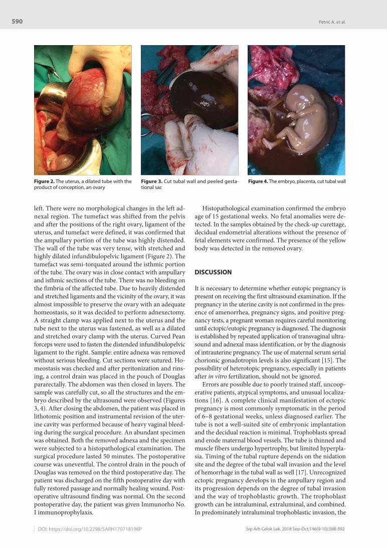
590
Srp Arh Celok Lek. 2018 Sep-Oct;146(9-10):588-592DOI: https://doi.org/10.2298/SARH170718196P
Petrić A. et al.
left. There were no morphological changes in the left ad-nexal region. The tumefact was shifted from the pelvis and after the positions of the right ovary, ligament of the uterus, and tumefact were defined, it was confirmed that the ampullary portion of the tube was highly distended. The wall of the tube was very tense, with stretched and highly dilated infundibulopelvic ligament (Figure 2). The tumefact was semi-torquated around the isthmic portion of the tube. The ovary was in close contact with ampullary and isthmic sections of the tube. There was no bleeding on the fimbria of the affected tube. Due to heavily distended and stretched ligaments and the vicinity of the ovary, it was almost impossible to preserve the ovary with an adequate homeostasis, so it was decided to perform adnexectomy. A straight clamp was applied next to the uterus and the tube next to the uterus was fastened, as well as a dilated and stretched ovary clamp with the uterus. Curved Pean forceps were used to fasten the distended infundibulopelvic ligament to the right. Sample: entire adnexa was removed without serious bleeding. Cut sections were sutured. Ho-meostasis was checked and after peritonization and rins-ing, a control drain was placed in the pouch of Douglas pararectally. The abdomen was then closed in layers. The sample was carefully cut, so all the structures and the em-bryo described by the ultrasound were observed (Figures 3, 4). After closing the abdomen, the patient was placed in lithotomic position and instrumental revision of the uter-ine cavity was performed because of heavy vaginal bleed-ing during the surgical procedure. An abundant specimen was obtained. Both the removed adnexa and the specimen were subjected to a histopathological examination. The surgical procedure lasted 50 minutes. The postoperative course was uneventful. The control drain in the pouch of Douglas was removed on the third postoperative day. The patient was discharged on the fifth postoperative day with fully restored passage and normally healing wound. Post-operative ultrasound finding was normal. On the second postoperative day, the patient was given Immunorho No. I immunoprophylaxis.
Histopathological examination confirmed the embryo age of 15 gestational weeks. No fetal anomalies were de-tected. In the samples obtained by the check-up curettage, decidual endometrial alterations without the presence of fetal elements were confirmed. The presence of the yellow body was detected in the removed ovary.
DISCUSSION
It is necessary to determine whether eutopic pregnancy is present on receiving the first ultrasound examination. If the pregnancy in the uterine cavity is not confirmed in the pres-ence of amenorrhea, pregnancy signs, and positive preg-nancy tests, a pregnant woman requires careful monitoring until ectopic/eutopic pregnancy is diagnosed. The diagnosis is established by repeated application of transvaginal ultra-sound and adnexal mass identification, or by the diagnosis of intrauterine pregnancy. The use of maternal serum serial chorionic gonadotropin levels is also significant [15]. The possibility of heterotopic pregnancy, especially in patients after in vitro fertilization, should not be ignored.
Errors are possible due to poorly trained staff, uncoop-erative patients, atypical symptoms, and unusual localiza-tions [16]. A complete clinical manifestation of ectopic pregnancy is most commonly symptomatic in the period of 6–8 gestational weeks, unless diagnosed earlier. The tube is not a well-suited site of embryonic implantation and the decidual reaction is minimal. Trophoblasts spread and erode maternal blood vessels. The tube is thinned and muscle fibers undergo hypertrophy, but limited hyperpla-sia. Timing of the tubal rupture depends on the nidation site and the degree of the tubal wall invasion and the level of hemorrhage in the tubal wall as well [17]. Unrecognized ectopic pregnancy develops in the ampullary region and its progression depends on the degree of tubal invasion and the way of trophoblastic growth. The trophoblast growth can be intraluminal, extraluminal, and combined. In predominately intraluminal trophoblastic invasion, the
Figure 2. The uterus, a dilated tube with the product of conception, an ovary
Figure 3. Cut tubal wall and peeled gesta-tional sac
Figure 4. The embryo, placenta, cut tubal wall

591
Srp Arh Celok Lek. 2018 Sep-Oct;146(9-10):588-592 www.srpskiarhiv.rs
Unruptured tubal pregnancy in early second trimester
degree and velocity of hemorrhage development depend on the degree of maternal blood vessels involvement. The tubal rupture does not necessarily occur immediately after a bleeding attack. Tubal distension occurs as a consequence of conceptual mass growth and bleeding. The possibility of intact endosalpinx epithelium and the absence or minimal hemorrhagia can be seen in intraluminal trophoblastic spread, but trophoblastic invasion of the maternal blood vessels is more intensive and tubal rupture occurs earlier in extraluminal extension [18]. The appearance and regularity of the gestational sac and the presence of crescent-shaped visible placental tissue (other authors also used similar diagnostics criteria before surgery) were the guidelines in making differential diagnosis between abdominal and tub-al pregnancy [19]. We decided to perform lower transverse laparotomy due to low probability of abdominal pregnancy. In the case of our patient fetal death occurred. There was no blood in the abdomen, nor tubal rupture. Other authors also reported their experience in patients with unruptured tubal pregnancy. There are no literature data on preserving the affected tube following an ectopic pregnancy in the sec-ond trimester. After abdominal opening and confirmation of the right tubal ectopic pregnancy diagnosis, we made an assessment and decided to perform adnexectomy as a safe alternative in our patient to maintain more effective homeostasis. The vicinity of the right ovary and the tube with the conceptus, as well as heavily distended thinned ligaments made us doubt on performing salpingectomy as the most sparing surgery, so we performed adnexectomy. Clinical manifestation of ectopic pregnancy in the second
trimester may be severe, with prominent hemorrhage and life-threatening to the mother [20, 21]. Surgical treatment of the second trimester ectopic pregnancies is an inevitable procedure, most commonly including salpingectomy, but adnexectomy may be performed as well, depending on the findings obtained [22]. Hysterectomies in patients with advanced tubal pregnancies have also been described [23]. Timely diagnosis (38th–68th gestational day) provides successful surgical treatment by using minimally invasive procedures, while preserving the affected tube [24]. Em-bryo implantation outside the uterus is always associated with endometrial changes. Proliferation, cystic hyperplasia, secretory transformation, hyper-secretory changes, asyn-chronous secretory changes between the glands and the stroma, and decidua reaction were histopathologically veri-fied [25, 26]. Instrumental curettage of the uterine cavity is not a standard post-surgical line of therapy in ectopic preg-nancy management, except in heavy or prolonged uterine bleeding, or in cases of suspected heterotopic pregnancy.
Early ultrasound examination is performed to con-firm whether the pregnancy is eutopic. A misdiagnosis and monitoring of ectopic pregnancy as eutopic one is potentially life-threatening for a pregnant woman. Surgi-cal treatment should be as sparing as possible, regarding preservation of reproductive organs, especially in patients wishing to have more children. Early diagnosis of tubal ectopic pregnancy may also enable the preservation of the tubes. The advancement of ectopic pregnancy towards the second trimester decreases the likelihood that the patient will have a sparing surgery.
REFERENCES
1. Kutlešić MR, Lukić B, Kutlešić SM, Popović J, Stefanović M, Vukomanović P, et al. Unruptured retroperitoneal pregnancy implanted in the left broad ligament: a case report. Vojnosanit Pregl. 2017; 74(2):177–83.
2. Rastogi R, Meena GL, Rastogi N, Rastog V. Interstitial ectopic pregnancy: A rare and difficult clinico-sonographic diagnosis. J Hum Reprod Sci. 2008; 1(2):81–2.
3. Sivalingam VN, Duncan WC, Kirk E, Shephard LA, Horne AW. Diagnosis and management of ectopic pregnancy. J Fam Plann Reprod Health Care. 2011; 37(4):231–40.
4. Taran FA, Kagan KO, Hübner M, Hoopmann M, Wallwiener D, Brucker S. The Diagnosis and Treatment of Ectopic Pregnancy. Dtsch Arztebl Int. 2015; 112(41):693–705.
5. Alkatout I, Honemeyer U, Strauss A, Tinelli A, Malvasi A, Jonat W, et al. Clinical diagnosis and treatment of ectopic pregnancy. Obstet Gynecol Surv. 2013; 68(8):571–81.
6. Stulberg DB, Cain LR, Dahlquisti I, Lauderdale DS. Ectopic Pregnancy Rates in the Medicaid Population. Am J Obstet Gynecol. 2013; 208(4):274e1–e7.
7. Ayaz A, Emam S, Farooq MU. Clinical course of ectopic pregnancy: A single-center experience. J Hum Reprod Sci. 2013; 6(1):70–3.
8. Vivoda M, Arsić B, Garalejić E, Ćirković I, Đukić S. [Examination of possible role of the chlamydial stress proteins in pathogenesis of ectopic pregnancy]. Srp Arh Celok Lek. 2014; 142(1–2):54–8. (Serbian)
9. Gonzalez N, Tulandi T. Cesarean Scar Pregnancy: A Systematic Review. J Minim Invasive Gynecol. 2017; 24(5):731–8.
10. Lin S, Yang R, Chi H, Lian Y, Wang J, Huang S, et al. Increased incidence of ectopic pregnancy after in vitro fertilization in women with decreased ovarian reserve. Oncotarget. 2017; 8(9):14570–5.
11. Muller V, Makhmadalieva M, Kogan I, Fedorova I, Lesik E, Komarova E, et al. Ectopic pregnancy following in vitro
fertilization: meta-analysis and single-center experience during 6 years. Gynecol Endocrinol. 2016; 32(2):69–74.
12. Panelli DM, Phillips CH, Brady PC. Incidence, diagnosis and management of tubal and nontubal ectopic pregnancies: a review. Fertil Res Pract. 2015; 1:15.
13. Creanga AA, Shapiro-Mendoza CK, Bish CL, Zane S, Berg CJ, Callaghan WM. Trends in ectopic pregnancy mortality in the United States: 1980-2007. Obstet Gynecol. 2011; 117(4):837–43.
14. Khalil MM, Badran EY, Ramadan MF, Shazly SA, Ali MK, Badee AY. An advanced second trimester tubal pregnancy: Case report. Middle East Fertil Soc J. 2012; 17(2):136–8.
15. Belics Z, Gérecz B, Csákány MG. [Early diagnosis of ectopic pregnancy]. Orv Hetil. 2014; 155(29):1158–66.
16. Wang LL, Chen X, Ye DS, Liu YD, He YX, Guo W, et al. Misdiagnosis and delayed diagnosis for ectopic and heterotopic pregnancies after in vitro fertilization and embryo transfer. J Huazhong Univ Sci Technolog Med Sci. 2014; 34(1):103–7.
17. Sowmya S, Priya R, Meenakshi P, Shwetha B. Study of trophoblasts and histological changes of fallopian tube in tubal pregnancy an anatomical considerations for its early rupture. Int J Anat Res. 2014; 2(4):609–13.
18. Zaidi T, Salahuddin MA, Kirmani F, Khan AA. A histoarchitectural study of early human ectopic pregnancy. Biomed Res. 2012; 23(1):51–4.
19. Liu J, Khan A, Johnson S, Grigorian C, Li T. The usefulness of gestational sac and placental sonographic morphology in differentiating between second-trimester tubal and abdominal pregnancy: Case report and a review of literature. J Clin. Ultrasound. 2014; 42(3):162–8.
20. Banaszek A, Ziółkowska K, Szymusik I, Wielgoś M. [Ectopic pregnancy–still a live clinical issue –a case report]. Ginekol Pol. 2006; 77(10):788–92.

592
doi: Srp Arh Celok Lek. 2018 Sep-Oct;146(9-10):588-592
21. Prajapati P, Sheikh MI. Ruptured tubal pregnancy: A rare cause of death. J Punjab Acad Forensic Med Toxicol. 2010; 10(1):48–51.
22. Gueye MD, Gueye M, Thiam I, Mbaye M, Gaye AM, Diouf AA, et al. Unruptured tubal pregnancy in the second trimestar. SSMJ. 2013; 6(4):95–6.
23. Sachan R, Gupta P, Patel ML. Second trimester unruptured ampullary ectopic pregnancy with variable presentations: Report of two cases. IJCRI. 2012; 3(8):1–4.
24. Song T, Lee DH, Kim HC, Seong SJ. Laparoscopic tube-preserving surgical procedures for ectopic tubal pregnancy. Obstet Gynecol Sci. 2016; 59(6):512–8.
25. Lopez HB, Micheelsen U, Berendtsen H, Kock K. Ectopic pregnancy and its associated endometrial changes. Gynecol Obstet Invest. 1994; 38(2):104–6.
26. Lu H, Xin L, Zhonghua Fu Chan Ke Za Zhi. [Morphologic changes of endometrium in patients with ectopic pregnancy]. 1997; 32(3):145–7.
САЖЕТАКУвод Најчешћа ектопична трудноћа је јајоводна. Ради се о могућем животно угрожавајућем стању са високом смртно-шћу код непрепознатих случајева. Дијагноза се поставља при појави првих симптома или приликом првих посета гинекологу. Дијагноза у другом триместру је значајно ређа јер до тада обично долази до манифестовања потпуне кли-ничке слике која је последица или експулзије заметка у перитонеалну шупљину или руптуре јајовода. Уколико до руптуре јајовода или експулзије заметка не дође, могућ је наставак трудноће и дијагноза ектопичног гравидитета у другом триместру.
Приказ болесника Тридесетједногодишња секундигравида, са првом виталном интраутерином трудноћом, у раном другом триместру јавила се лекару због појаве вагиналног крварења. После гинеколошког и ултрасонографског прегледа поставље-на је дијагноза ектопичне трудноће и урађена лапаратомија. Потврђена је тубарна трудноћа и урађена аднексектомија. Закључак Прве ултразвучне контроле морају да потврде еутопичност трудноће. Погрешна дијагноза и праћење ек-топичне као еутопичне трудноће може да буде угрожавајуће за трудницу.Кључне речи: неруптурирана трудноћа; тубарна трудноћа; рани други триместер
Неруптурирана тубарна трудноћа у раном другом триместру Александра Петрић1,2, Радомир Живадиновић1,2, Дејан Митић1,2, Предраг Вукомановић1,2, Милан Тренкић1,2
1Универзитет у Нишу, Медицински факултет, Ниш, Србија; 2Клинички центар Ниш, Клиника за гинекологију и акушерство, Ниш, Србија
DOI: https://doi.org/10.2298/SARH170718196P
Petrić A. et al.

593
DOI: https://doi.org/10.2298/SARH171110204P
UDC: 616.12-008.315-083.98; 615.816/.817
Received • Примљено: November 10, 2017
Revised • Ревизија: December 3, 2017
Accepted • Прихваћено: December 5, 2017
Online fi rst: December 8, 2017
Correspondence to:
Aleksandar PAVLOVIĆ
Faculty of Medicine
University of Priština
Anri Dinana bb
38220 Kosovska Mitrovica
Serbia
SUMMARYThe occurrence of cardiac arrest during anesthesia and surgery is nowadays associated with many challenges imposed by 21st century medicine. On the one hand, good education of healthcare practitioners, sophisticated anesthetic techniques and equipment, along with safer anesthetics and improved surgical techniques have significantly reduced the risk of cardiac arrest during the perioperative period. Still, the introduction of new, invasive diagnostic and therapeutic procedures in the aging patients and those with comorbidities carries along new risk and challenges. Epidemiological data indicate that intraoperative cardiac arrest is an extremely rare event. Due to variety of moral and ethical prejudices, intraoperative cardiac arrest is frequently presented as if it has happened in the immediate postoperative period, following surgery and anesthesia. The preventive measures, the etiology and diagnosis of cardiac arrest, as well as the specificities regarding organization and performance of cardiopulmonary resuscitation in the operating room, result in a better prognosis compared to other hospital departments. The article also describes the specifics of cardiopulmonary resuscitation in the catheterization laboratory, while a separate section is dedicated to cardiopulmonary resuscitation following systemic toxicity of local anesthetics.Since intraoperative cardiac arrest and death represent very rare complications, European Resuscitation Council has only recently published Guidelines for Resuscitation for performing cardiopulmonary resuscitation in the operating room – in 2015.Keywords: heart arrest, etiology, therapy; cardiopulmonary resuscitation; operating room; anesthesia, adverse effects; anesthesiology, methods, standards; medical errors, prevention, control
REWIEV ARTICLE / ПРЕГЛЕД ЛИТЕРАТУРЕ
Cardiac arrest and cardiopulmonary resuscitation in the operating roomAleksandar Pavlović1, Nevena Kalezić2,3, Slađana Trpković1, Ana Sekulić4, Olivera Marinković4
1University of Priština, Faculty of Medicine, Kosovska Mitrovica, Serbia;2University of Belgrade, Faculty of Medicine, Belgrade, Serbia;3Clinical Center of Serbia, Center for Anaesthesiology and Resuscitation, Belgrade, Serbia;4University Hospital Medical Center Bežanijska Kosa, Belgrade, Serbia
INTRODUCTION
The perioperative cardiac arrest (CA) is much less likely to happen nowadays due to con-temporary education in anesthesiology, good preoperative preparation of patients, the use of modern anesthesiologic techniques, equipment and safe anesthetics, and improved surgical techniques. On the other hand, the introduc-tion of new, invasive diagnostic and therapeu-tic procedures in the aging patients and those with comorbidities carries along new risks and challenges. CA is the most dramatic and most urgent situation for a physician in the operat-ing room (OR). A trained team of OR person-nel and specific work organization, mandatory monitoring, the availability of equipment and medications for cardiopulmonary resuscitation (CPR), secured airway and placed intravenous cannulas facilitate perioperative CPR and in-crease survival rates [1].
Since intraoperative cardiac arrest and death represent very rare complications, European Resuscitation Council (ERC) has only recently published Guidelines for Resuscitation for per-forming cardiopulmonary resuscitation in the operating room – in 2015 [2, 3, 4].
Due to a variety of moral and ethical preju-dices, intraoperative cardiac arrest is frequently presented as if it has happened in the imme-diate postoperative period, following surgery and anesthesia. When CA happens in the OR, many doctors feel unjustified guilt that some intervention during anesthesia or surgery may have contributed to CA occurrence, so they feel responsible and obligated to perform CPR, even in situations when it is completely clear that it should not be performed (i.e. terminal illnesses, signed “Do not resuscitate” protocol). That is why the operating team starts CPR in order to achieve return of spontaneous circula-tion (ROSC), finish the surgery, and transport the patient from the OR, although they are fully aware that in the immediate postopera-tive period ROSC will briefly result in a death outcome. Therefore, the majority of epidemio-logical data regarding the frequency of CA in the OR should be carefully considered.
The low incidence of CA caused by anes-thesia and surgery prevents conduction of con-trolled studies in this area [5, 6].

594
Srp Arh Celok Lek. 2018 Sep-Oct;146(9-10):593-598DOI: https://doi.org/10.2298/SARH171110204P
PROGNOSIS AND OUTCOMES OF CARDIAC ARREST
DURING ANESTHESIA AND SURGERY
In developed countries, the incidence of CA during an-esthesia is 0.2–1.1 cases per 10,000 procedures in adults, and 1.4–2.9 cases per 10,000 procedures in children (with a considerably higher incidence in newborns), which is much more rare than 30 or more years ago (20 in 10,000) [1, 7, 8, 9]. Certainly, there are higher-risk groups of pa-tients among whom perioperative CA is seen much more often (Table 1) [2, 10, 11]. Data from the Mayo Clinic in-dicate that the incidence of CA perioperatively during gen-eral anesthesia is three times higher than during regional anesthesia [2, 9].
Table 1. Risk factors for operating room cardiac arrest
Risk factors1. Urgent surgery 163/10,000 patients2. Elderly patient 54/10,000 patients3. Children < 2 years4. Male sex5. COPD6. Hypotension (shock) AP < 90 mmHg7. AKI, CKD8. Malignant disease9. Major surgery
COPD – chronic obstructive pulmonary disease; AP – arterial pressure; AKI – acute kidney injury; CKD – chronic kidney disease
The differences related to the organization and execu-tion of CPR in the OR compared to other hospital de-partments reflect different survival rates for CA patients. Documented reports show that patients’ survival following intraoperative CA is 34.5–43.9% [8]. In situations when CA was associated exclusively with anesthesia, survival rates were as high as 70–80%, which is much higher than survival rates in other hospital wards: only 15–20% [1, 12].
ETIOLOGY OF CARDIAC ARREST IN THE OPERATING
ROOM
Numerous factors are associated with perioperative CA: the preoperative factors (comorbidities), poor risk assess-ment, inadequate monitoring, mistakes during anesthesia and surgical procedures [9, 13].
The universal mnemonic “4H and 4T”, which causes of CA defined by the ERC, Moitra et al. [14] have described as many as 16 (8H and 8T) potentially reversible causes of CA in the OR (Table 2) [2].
According to the literature data, the most frequent causes of intraoperative CA are as follows:
• hemorrhage (with the highest fatal outcome rate of 10.3%);
• heart complications, myocardial infarction;• medications (anesthetics, muscle relaxants), complica-
tions of central venous catheterization;• hypoxia caused by compromised airway or complica-
tions associated with mechanical ventilation [8, 9, 15].
The most frequent causes of CA in the OR in children are the airway obstruction caused by laryngospasm and bronchospasm, hypovolemia (blood loss) and hyperkale-mia (from transfusion of stored blood) [2, 16].
Unlike seen in out-of-hospital CA and in other hospital departments, the most frequent CA rhythm seen in the OR during general anesthesia is asystole (Figure 1). Fortu-nately, the prognosis of CPR following asystole in the OR is much better than in other hospital departments [2, 7, 9].
PREVENTION OF INTRAOPERATIVE CARDIAC ARREST
The prevention of intraoperative CA begins with good pre-operative preparation of patients, assessment of co-morbidities and current physical status (according to the American Society of Anesthesiologists – ASA score), and stabilizing any concomitant chronic conditions. The pre-vention of operating room CA is affected by the choice of anesthesia technique (general or regional), which depends on the type of surgery, the condition of the patient and comorbidities, but also on the patient’s personal desires and the anesthesiologist’s experience. Furthermore, the choice of the optimal operative technique depends on the surgeon’s experience. Teamwork in the OR has contributed to more rapid recognition and timely reversal of any de-terioration of the patient, whereby the occurrence of CA is prevented [2, 17].
Pavlović A.
Table 2. Potentially reversible causes of cardiac arrest in the operating room
8H 8THypoxia Toxins (anaphylaxis/anesthesia)Hypovolemia Tension pneumothoraxHyper-/hypokalemia Thrombosis/embolus, pulmonaryHydrogen ion (acidemia) Thrombosis, coronaryHypothermia TamponadeHypoglycemia Trauma/hemorrhagic shock, CV injuryMalignant hyperthermia QT prolongationHypervagal reaction Pulmonary hypertension
CV – cardiovascular
Figure 1. The most frequent causes of a patient’s deterioration during regional anesthesia

595
Srp Arh Celok Lek. 2018 Sep-Oct;146(9-10):593-598 www.srpskiarhiv.rs
The physician’s failure to notice the deterioration of the patient’s condition in time is the most frequent cause of CA in the OR [2, 17]. The patient’s state can sometimes worsen within minutes, but also over hours during the in-traoperative period, so appropriate monitoring and timely correction of pathophysiological changes are crucial.
THE DIAGNOSIS OF CARDIAC ARREST IN
THE OPERATING ROOM AND MANDATORY
MONITORING – CONTROL OF CPR QUALITY
If the minimum mandatory monitoring standard is en-sured in the OR, there should be no delay in making a diagnosis of CA. Insertion of an arterial line for invasive blood pressure monitoring in high-risk patients is invalu-able for prompt diagnosis of CA. It is recommended that these patients should be equipped with self-adhesive defi-brillation pads prior to anesthesia induction [2, 8].
Asystole and ventricular fibrillation (VF) must be identified immediately in the OR. However, in the case of pulseless electrical activity, the diagnosis of CA should be verified by capnography (EtCO2) and pulse oximetry, and definitely confirmed by the pulse or the arterial line curve (Table 3).
Table 3. Incidence and pattern of cardiac arrest rhythms in the operat-ing room (Mayo Clinic data) [2]
Pattern of cardiac arrest rhythms Incidence (%)Asystole 41.7Ventricular fibrillation 35.4Pulseless electrical activity 14.4Unknown 8.5
Performing proper and uninterrupted chest compres-sions is essential for the success of CPR. There are multiple ways to assess the quality of chest compressions and check the success of CPR in the OR. Palpation of the carotid or femoral artery pulse during CPR is not a proper indica-tor of chest compressions’ quality. Some newer generation defibrillators can provide feedback regarding the quality of compressions. Non-invasive blood pressure monitor-ing has no relevance in the CA diagnosis, in the quality of CPR, nor in the prediction of CPR success, and can only be used following ROSC. When hypotension and hypoxia are present, pulse oximetry is not an appropri-ate monitoring touchstone for a prompt CA diagnosis or for assessing chest compressions’ quality. Capnography, a part of minimum mandatory monitoring, is very impor-tant in assessing CPR quality. When EtCO2 > 20 mmHg, ROSC is much more likely to occur than in cases when EtCO2 < 10 mmHg is achieved with chest compressions. If there is an arterial line in place, diastolic pressure (DP) over 40 mmHg is also associated with higher ROSC in-cidence. Monitoring central venous pressure (CVP) and diastolic pressure in the OR during CPR permits the calcu-lation of coronary perfusion pressure (CPP) based on the following formula: CPP = DP - CVP. CPP values of over
15 mmHg during CPR are associated with higher survival rates [8, 18, 19, 20].
However, activation of various alarms due to monitor-ing failure while the state of the patient is satisfactory is a very frequent occurrence in the OR. This may happen due to detachment of an ECG electrode from the patient’s chest, the pulse oximeter slipping off the patient’s finger, or technical problems in CO2 sampling. Such occurrences may sometimes lead the anesthesiologist to ignore the monitoring alarms and to miss a serious deterioration of the patient’s condition. Regardless of the mandatory moni-toring, “the physician is the most important monitor in the operating room” [8].
RESPONSIBILITIES OF THE OPERATING TEAM
MEMBERS IN PERFORMING CPR – THE TEAMWORK
Good communication among members of the resus-citation team in the OR is the key to the success of re-suscitation. Confusion or miscommunication must not be permitted during CPR, and no time must be wasted while resuscitation procedures are being performed. This is why teamwork, good organization, following the team leader’s instructions and the availability of equipment in good working order are crucial in the performance of CPR measures. In order to advance the teams’ professionaliza-tion, simulation sessions are held periodically to practice handling of this type of crisis situation. The responsibilities of the surgical team members in performing CPR measures are shown in Figure 2 [21, 22, 23].
Cardiac arrest and cardiopulmonary resuscitation in the operating room
Figure 2. Operating room (OR) team members’ tasks during cardiopul-monary resuscitation (CPR): 1) the anesthesiologist makes the diagno-sis of cardiac arrest, acts as the CPR team leader, executes advanced life support and removes any reversible causes; 2) the surgeon halts the surgical procedure, controls bleeding, protects vital organs, executes the anesthesiologist’s orders and performs chest compressions; 3) the anesthetist executes the anesthesiologist’s orders, prepares the defibrillator and takes part in the defibrillation, prepares and applies treatment ordered by the anesthesiologist; 4) the scrub nurse monitors and secures the sterility of the wound and the operating field, and of the surgical equipment; 5) the circulating nurse calls for additional assistance, adjusts the height of the table, oversees OR comings and goings, retrieves and delivers disposable materials and equipment [23]

596
doi: Srp Arh Celok Lek. 2018 Sep-Oct;146(9-10):593-598
PERFORMING CPR IN THE OPERATING ROOM
It is interesting to note some authors’ comparison of gen-eral anesthesia and CPR. While it is completely inaccurate to declare that general anesthesia is an “ongoing resuscita-tion,” there are certain similarities between the two medical procedures. Under general anesthesia, as in CPR, the pa-tient is unconscious. Both procedures require maintaining and monitoring of the airway and mechanical ventilation. Administering vasopressors and antiarrhythmic drugs, which are mandatory during CPR, is not unusual dur-ing general anesthesia. As in advanced life support (ALS) measures of CPR, continuous monitoring is present during general anesthesia too. Based on this, one may raise the question regarding differentiation of the routine proce-dures done during general anesthesia in the OR and the urgent skills required in CPR. According to some authors, the only difference that draws the medical line between these two procedures is the need for defibrillation and the execution of chest compressions [5].
The advantages and disadvantages of CPR in the OR are shown in Table 4.
Table 4. Advantages and disadvantages of cardiopulmonary resuscita-tion in the operating room
Advantages Disadvantages 1. CA is always observed by
witnesses;2. CA cause is usually known
and reversible;3. Airway is secured and
mechanical ventilation is ongoing;
4. Multiple vein lines, CVC in place;
5. Continuous monitoring, arterial line is often in place;
6. Entire CPR team is present;7. CPR equipment and
medications are available
1. False alarms, interruption of ECG, pulse oximetry connection;
2. Hypotension and bradycardia that may be discounted or overlooked;
3. Impossibility of adequate monitoring (obesity, the patient’s position on the operating table);
4. Contamination through non-sterile contact;
5. Medically and ethically unjustified reasons
CA – cardiac arrest; CPR – cardiopulmonary resuscitation; CVC – central venous catheter; ECG – electrocardiography
In the OR, the time of CA occurrence is always ob-served by witnesses and the direct cause of the CA is usu-ally known. That is why, along with the progress of CPR, reversible causes should be removed (bleeding, hypoxia, etc.) [3].
When non-shockable rhythms are found and accompa-nied by the inability to palpate the pulse for more than 10 seconds along with a drop in the capnographic and arterial curve, chest compressions should be started immediately, following ALS protocols for non-shockable rhythms [2, 24]. In view of the surgeon’s position relative to the patient, he or she should begin chest compressions, or direct heart compression, depending on whether the thorax is surgi-cally open [8]. The height of the operating table should be adjusted in order to permit the performance of high-qual-ity chest compressions [2]. CPR is optimally performed on a patient in a supine position, but in certain situations it is possible to perform CPR on patients in prone position if it is not feasible to place the patient in a supine position
quickly [3, 4]. Chest compressions in prone patients may be performed manually or using a mechanical chest com-pression device in continued compression (10 compres-sions/minute) if an endotracheal tube is in place [2, 3]. If a supraglottic airway device has been inserted and there is an air leak, the CPR should be performed at a ratio of 30:2 [18, 25]. Depending on the ECG rhythm (shockable or non-shockable), CPR should be performed immediately based on new ERC guidelines for CPR [2, 24].
• The decision when to terminate CPR is complex and often, in addition to the medical aspect, includes a series of moral, ethical, and legal elements. Adhering to medical positions, CPR should be terminated when the following criteria are met:fatal injuries or signs of certain death are present;
• an objective suggestion has been made by a senior – the team leader;
• an assessment has been made that all further CPR would be futile and useless;
• asystole has been present for over 20 minutes despite ALS measures and there are no reversible causes [18, 19, 20].
CARDIAC ARREST AND CPR IN THE
CATHETERIZATION LABORATORY
CA may occur during percutaneous coronary intervention in patients with myocardial infarction, but it can also arise as a complication of angiography. Most complications will lead to VF, which requires urgent defibrillation. This is why a patient in the catheterization laboratory must be continuously monitored, with a defibrillator standing by. In high-risk patients, self-adhesive radiolucent defibrillation pads should be placed prior to starting the procedure. If the defibrillation is not successful or VF re-occurs, defi-brillation should be repeated urgently two times [2]. If VF persists after the third defibrillation, begin chest compres-sions and ventilation without delay, and continue the angi-ography in order to find the cause of the CA. It is extremely important not to interrupt chest compressions during the angiography. On an angiography table, where the image intensifier is located above the patient, it is almost impos-sible to perform high-quality chest compressions, and there is the risk of exposing the practitioner to dangerous radia-tion. This is why it is strictly advised to use a mechanical chest compression device. In patients with non-shockable rhythm, immediate transthoracic echocardiography should be performed in order to diagnose the cause (pericardial tamponade or another issue) [26, 27].
CARDIAC ARREST CAUSED BY SYSTEMIC TOXICITY
OF LOCAL ANESTHETICS
CA as a consequence of systemic toxicity of a local anesthet-ic (LA) occurs in 1.8 cases per 10,000 regional anesthesia procedures [28]. Significant clinical experience is needed to find the cause of a patient’s deterioration during regional
Pavlović A.
DOI: https://doi.org/10.2298/SARH171110204P

597
Srp Arh Celok Lek. 2018 Sep-Oct;146(9-10):593-598 www.srpskiarhiv.rs
anesthesia (Figure 1), since this determines the type of the treatment. If systemic toxicity of LA is not recognized and not treated promptly, it may lead to CA.
The typical development of the clinical features is the result of a progressive biphasic effect on the central ner-vous system (CNS) and subsequently on the cardiovascular system (CVS), which are highly sensitive to changes of tissue electrophysiology. Since the CNS alterations are the first ones to occur, this compartment is considered the “mirror” of LA concentration in the blood. Initially, pa-tients become agitated and logorrheic, feel a metallic taste in their mouths, and may experience nystagmus, tinnitus, dysphagia, and confusion. Further progression of the in-toxication produces muscle tremors and the development of convulsions. Nausea and vomiting may also be present, along with breathing disturbances, respiratory vasomotor depression, and loss of consciousness [28].
The CVS is considered more resistant to LA effects than the CNS, and cardiotoxicity occurs as a consequence of the direct negative inotropic effects of the anesthetic on the heart and the direct relaxant action on the smooth muscle tissue of the blood vessels. Cardiotoxicity associ-ated with systemic LA toxicity is characterized by hypoten-sion, AV block, idioventricular rhythm, bradycardia, and cardiovascular collapse. The cardiotoxic effect of the LA is exacerbated by hypoxia, hypoventilation, acidosis, and hyperkalemia [28].
In the case of CA, perform ALS according to the ERC guidelines for CPR [2, 3, 24].
Patients with cardiovascular collapse and CA caused by systemic LA toxicity can benefit from a 20% lipid emul-sion applied intravenously during ALS [2, 28]. The widely accepted hypothesis of the mechanism of action of the 20% intravenous lipid emulsion in the treatment of car-diotoxicity is based on the absorption and removal of the circulating lipophilic toxin – the LA – from the blood,
which reduces the amount of free LA able to bind to the myocardium. This non-specific removal of LA from the plasma or heart tissue is called “lipid sink” in Anglo-Saxon literature [29]. Another possible mechanism is that the in-travenous lipid emulsion acts directly on the heart muscle by neutralizing the inhibitory effect of LA on fatty acid oxidation in mitochondria, whose ultimate purpose is creating energy – adenosine triphosphate, thus prevent-ing the depressive effect of LA on the functioning of the heart muscle [29, 30]. Some authors suggest that successful resuscitation could be due to spontaneous clearance of the instigating LA within of routine ALS without administer-ing intravenous lipid emulsion. [31, 32].
Clinical recommendations for the dosage of 20% intra-venous lipid emulsion during CPR were published in ERC guidelines for CPR in special circumstances in 2015 [2, 3, 4]. However, many questions remain open: should the lipid dose be titrated by patient weight, local anesthetic dose, or the symptoms, severity of toxicity; what is the best rate and total dose and what are the possible complications or adverse effects of lipid infusion? Furthermore, pediatric doses of IV lipid emulsion to treat systemic LA toxicity have not been defined. [30, 31, 32].
CONCLUSION
Cardiac arrest in the OR is associated with pre-existing medical or surgical diseases, the quality of preoperative evaluation and preparation for anesthesia and surgery, but also with the anesthetic and surgical technique.
Good organization and well-trained operating room team members, along with appropriate monitoring and readily available equipment and medication, result in a bet-ter prognosis when CPR is performed in the OR compared to other hospital departments.
1. Runciman WB, Morris RW, Watterson LM, Williamson JA, Paix AD. Crisis management during anaesthesia: cardiac arrest. Qual Saf Health Care. 2005; 14(3):e14.
2. Truhlar A, Deakin D, Soar J, Khalifa A, Alfonzo A, Bierens J, et al. European Resuscitation Council Guidelines for Resuscitation 2015. Section 4. Cardiac arrest in special circumstances. Resuscitation 2015; 95:148–201.
3. Trpković S, Pavlović A, Anđelić S, Sekulić A. Nove preporuke za kardiopulmonalnu reanimaciju u posebnim stanjima. Naučni časopis urgentne medicibe HALO 194. 2015; 21(3):199–211.
4. Trpković S, Pavlović A, Ilić Mostic T, Milenović M, Stevanović K, Kalezić N. Kardiopulmonalna reanimacija u posebnim stanjima. In: Kalezić N, ed. Inicijalni tretman akutnih stanja. Beograd: Medicinski fakultet u Beogradu, 2016. p. 297–347.
5. Norman GV. Do-Not-Resuscitate Orders during Anesthesia and Urgent Procedures. Ethics in medicine. University of Washington School of Medicine, Seattle. 1998–2011. Last modified: February 18, 2016. Available from: https://depts.washington.edu/bioethx/topics/dnrau.html
6. Gempeler-Rueda FE, De Brigard-Pérez AM. Do not resuscitate orders and anesthesia. Rev Colomb Anestesiol. 2016; 44(2):146–50.
7. Zuercher M. Ummehofer W. Cardiac arrest during anaesthesia. Curr Opin Crit Care. 2008; 14:269–74.
8. Andres J. Cardiac Arrest in the Operating Room. 2012. European Society of Anaesthesiology [cited 2017. Oct 12] Available from:
https://www.scribd.com/document/318455083/Cardiac-Arrest-in-the-Operating-Room-2012
9. Sprung J, Warner ME, Contrerars MG, Schroeder DR, Beighley CM, Wilson GA, et al. Predictors of survival following cardiac arrest in patients undergoing noncardiac surgery. Anesthesiology. 2003; 99(2):259–69.
10. Biboulet P, Aubas P, Dubourdieu J, Rubenovitch J, Capdevila X, d’Athis F. Fatal and non-fatal cardiac arrest related to anesthesia. Can J Anesth. 2001; 48(4):326–32.
11. Siriphuwanun V, Punjasawadwong Y, Lapisatepun W, Charuluxananan S, Uerpairojkit K. Incidence of and factors associated with perioperative cardiac arrest within 24 hours of anesthesia for emergency surgery. Risk Manag Healthc Policy. 2014; 7:155–62.
12. Larkin GL, Copes WS, Nathanson BH, Kaye W. Pre-resuscitation factors associated with mortality in 49,130 cases of in-hospital cardiac arrest: a report from the National Registry for Cardiopulmonary Resuscitation. Resuscitation. 2010; 81(3):302–11.
13. Catalani B, Sharp C, Cooper L. Life-threatening perioperative anesthetic complications: major issues surrounding perioperative morbidity and mortality. Trauma Surg Acute Care Open. 2017; 2:1–7.
14. Moitra KV, Gabrielli A, Maccioli AG, O Connor FM. Anesthesia advanced circulatory life support. Can J Anesth. 2012; 59(6):586–603.
REFERENCES
Cardiac arrest and cardiopulmonary resuscitation in the operating room
598
doi: Srp Arh Celok Lek. 2018 Sep-Oct;146(9-10):593-598
15. Ellis SJ, Newland MC, Simonson JA, Peters KR, Romberger DJ, Mercer DW, et al. Anesthesia-related cardiac arrest. Anesthesiology. 2014; 120(4):829–38.
16. Bhananker SM, Ramamoorthy C, Geiduschek JM, Posner KL, Domino KB, Haberkern CM, et al. Anesthesia-related cardiac arrest in children: update from the Pediatric Perioperative Cardiac Arrest Registry. Anesth Analg. 2007; 105(2):344–50.
17. Mhyre JM, Ramachandran SK, Kheterpal S, Morris M, Chan PS. Delayed time to defibrillation after intraoperative and periprocedural cardiac arrest. Anesthesiology. 2010; 113(4):782–93
18. Pavlović A, Trpković S, Anđelić S, Marinković O. Kardiopulmonalna reanimacija – nove preporuke 2015-2020. Naučni časopis urgentne medicibe HALO 194. 2015; 21(3):181–98.
19. Pavlović A. Kardiopulmonalno cerebralna reanimacija. 3rd ed. Beograd: Obeležja; 2011.
20. Pavlović A. KPCR kod odraslih. In: Kalezić N. (ed). Inicijalni tretman akutnih stanja. Beograd: Medicinski fakultet u Beogradu; 2016. p. 225–77.
21. Pavlović A, Kalezić N, Trpković S, Videnović N, Šulović Lj. The application of simulation in medical education – our experiences “from improvisation to simulation”. Srp Arh Celok Lek. 2018; 146(5-6):330–7
22. Pavlović A, Trpković S, Videnović N. Primena novih preporuka za kardiopulmonalno cerebralnu reanimaciju u svakodnevnoj anesteziološkoj praksi. Serbian Journal of Anesthesia and Intensive Therapy 2013; 35(1–2):51–60.
23. Porteous J. Code blue: what to do? Can Oper Room Nurs J. 2009; 27(3):22–8.
24. Soar J, Nolan JP, Bottiger BW, Perkins GD, Lott C, Carli P, et al. European Resuscitation Council Guidelines for Resuscitation 2015. Section 3. Adult advanced life support. Resuscitation. 2015; 95:100–47.
25. Trpković S, Pavlović A, Bumbaširević V, Sekulić A, Miličić B. Uspeh reanimacije osoba s akutnim zastojem srca u bolničkim uslovima. Srp Arh Celok Lek. 2014; 142(3–4):170–7.
26. Pavlović A, Kalezić N, Trpković S. Primena novih preporuka za KPCR u svakodnevnoj anesteziološkoj praksi. In Kalezić N, editor. Current Concepts in Anaesthesilogy, Resuscitation and Intensive Care: Proceedings of the 1th Belgrade Anaesthesia Forum; 2016 Apr 1–2. Lajkovac: La-pressing; 2016. p. 71–89.
27. Larsen AI, Hjornevik AS, Ellingsen CL, Nilsen DW. Cardiac arrest with continuous mechanical chest compression during percutaneous coronary intervention. A report on the use of the LUCAS device. Resuscitation 2007; 75(3):454–9.
28. Weinberg G. Treatment of local anesthetic systemic toxicity (LAST). Reg Anesth Pain Med. 2010; 35(2):188–93.
29. Weinberg G, Lin B, Zheng S, Di Gregorio G, Hiller D, Ripper R, et al. Partitioning effect in lipid resuscitation: further evidence for the lipid sink. Crit Care Med. 2010; 38(11):2268–69.
30. Ciechanowicz S, Patil V. Lipid Emulsion for Local Anesthetic Systemic Toxicity. Anesthesiol Res Pract. 2012; 131784.
31. Ozcan MS, Weinberg G. Intravenous lipid emulsion for the treatment of drug toxicity. J Intensive Care Med. 2014; 29(2):59–70.
32. Cave G, Harvey MG. Should we consider the infusion of lipid emulsion in the resuscitation of poisoned patients? Crit Care. 2014; 18:457.
САЖЕТАКАкутни застој срца током анестезије и операције је у да-нашње време повезан са многим изазовима које доноси медицина XXI века. С једне стране, добра едукација здрав-ствених радника, савремене технике анестезије и опрема, безбедни анестетици и усавршене оперативне технике зна-чајно су смањили ризик за настанак акутног застоја срца у периоперативном периоду. Међутим, увођење нових, инвазивних дијагностичких и терапијских процедура код све старијих болесника са коморбидитетом доноси нове ризике и изазове.Епидемиолошки подаци указују да се ради о изузетно ретком догађају. У медицинској пракси, због моралних и етичких предрасуда, интраоперативни акутни застој срца се често приказује у непосредном постоперативном току, после операције и анестезије. Мере превенције, етиологија
и дијагноза акутног застоја срца, као и специфичности у ор-ганизацији и извођењу кардиопулмоналне реанимације у операционој сали дају бољу прогнозу у поређењу са другим одељењима болнице. У овом раду описане су и специфичности кардиопулмонал-не реанимације у сали за катетеризацију срца и посебно после системске токсичности локалних анестетика.С обзиром на то да акутни застој срца и смртни исход то-ком интраоперативног периода представљају веома ретке компликације, Европски савет за реанимацију је први пут објавио препоруке за извођење кардиопулмоналне реани-мације у операционој сали тек 2015. године.Кључне речи: акутни застој срца, етиологија, лечење; кар-диопулмонална реанимација; операциона сала; анестезија, нежељени ефекти; анестезиологија, методе, стандарди; ме-дицинске грешке, превенција, контрола
Акутни застој срца и кардиопулмонална реанимација у операционој сали Александар Павловић1, Невена Калезић2,3, Слађана Трпковић1, Ана Секулић4, Оливера Маринковић4
1Универзитет у Приштини, Медицински факултет, Косовска Митровица, Србија;2Универзитет у Београду, Медицински факултет, Београд, Србија3Клинички центар Србије, Центар за анестезиологију и реаниматологију, Београд, Србија;4Универзитетски клиничко-болнички центар „Бежанијска коса“, Београд, Србија
Pavlović A.
DOI: https://doi.org/10.2298/SARH171110204P

599
DOI: https://doi.org/10.2298/SARH170704170P
UDC: 94(=163.41)(44)"1914/1918"; 355.415.6-055.2(100)"1914/1918"; 614.885-055.2:929(44)"1914/1918"
Correspondence to:
Slavica POPOVIĆ-FILIPOVIĆ
Ul. Koste Jovanovića 69/2
11040 Belgrade, Serbia
bobfi [email protected]
Received • Примљено: July 4, 2017
Revised • Ревизија: August 25, 2017
Accepted • Прихваћено: August 31, 2017
Online fi rst: September 8, 2017
САЖЕТАК Историографија српског народа из Великог рата одала је дужно поштовање великим биткама и славним победама, није изоставила голготу повлачења српске војске и народа, Солунски фронт и друге фронтове. Мање је позната судбина 35.000 српских регрута, расељених младих људи, сага српске избегле колоније широм европских простора и даље. Циљ овог рада је да истакне судби-не српских избеглица на Корзици и ода признање њиховим исцељитељима, који су долазили са различитих страна света као представници дипломатских, хуманитарних и медицинских мисија Србије, Француске и Велике Британије. Живот избегле српске колоније на Корзици у Француској био је организован залагањем представника краљевске Српске владе у Француској, Француског комитета за помоћ рањеницима, болесницима и избеглицама, Српског потпорног фонда, Болница шкотских жена, локалних власти и бројних појединаца на Корзици. Посебно је истакнута Болница шкотских жена на Корзици, која је за српске војнике и српске избеглице организовала Јединицу Корзика, са седиштем у Ајачу, изоловану болницу у Лазарету, док су амбуланте и диспанзери били распоређени по селима. У време када се обележава стогодишњица од Првог светског рата узвраћа-мо захвалност за посвећеност и пожртвовање свим странама, посебно Болници шкотских жена и др Елси Инглис, оснивачу и руководиоцу ове медицинске мисије. Кључне речи: Први светски рат; француска медицинска помоћ; Српски Црвени крст; Српски потпор-ни фонд; Болнице шкотских жена; Корзика; Србија; Елси Инглис
ИСТОРИЈА МЕДИЦИНЕ / HISTORY OF MEDICINE
Срби на Корзици у Великом рату – 2. деоСлавица Поповић-ФилиповићСекција за историју медицине Српског лекарског друштва, Београд, Србија
Др Елси Инглис, оснивач и
руководилац Болнице шкотских жена
(The Scottish Women’s Hospital for foreign service)
Др Елси Мод Инглис (Dr. Elsie Maud Inglis, 1864–1917), оснивач и руководилац Болница шкотских жена широм Европе, дошла је у Србију априла 1915. и заменила др Елеонор Солто, која је руководила Првом једини-цом Болнице шкотских жена у Крагујевцу. Истовремено је основала болнице у Ваље-ву, Младеновцу и Лазаревцу. Окупацијом Србије одбила је да се повуче и остала да негује преко хиљаду рањеника и болесни-ка у крушевачкој болници. Пo повратку у домовину, организовала је прославу Видо-вдана, на челу Организационог комитета промовисала српску борбу за ослобођење, основала нову болницу и отишла на Руски фронт и Добруџу, у пратњи Прве српске до-бровољачке дивизије [1, 2].
Др Елси Инглис, оснивач и „покретачки дух“ чланица Болница шкотских жена ши-ром Европе, била је прва жена носилац ви-соког српског одликовања – Ордена белог орла. Меморијална чесма на Црквенцу, у Младеновцу, најлепши је споменик пос-већен др Елси Инглис и чланицама Болни-ца шкотских жена који је српски народ по-дигао у време рата, као дар захвалности за њихову племенитост и пожртвовање. У знак сећања и поштовања на др Инглис, Болница шкотских жена је формирала Јединицу „Др Елси Инглис“ као појачање болницама на
Солунском фронту. Меморијална болница „Др Елси Инглис“ у Единбургу је још један од споменика који подсећа на дела др Елси Инглис. Биста др Елси Инглис, поклон срп-ског народа а дело Ивана Мештровића, из-ложена је у Националној галерији Шкотске, у Единбургу. После Првог светског рата ко-митети Болница шкотских жена у Лондону и Единбургу помогли су изградњу Меморијал-не болнице за мајку и дете „Др Елси Инглис“ на Дедињу, у Београду (Слика 1).
Слика 1. Др Елси Мод Инглис (1864–1917)Figure 1. Dr. Elsie Maud Inglis (1864–1917)

600
Srp Arh Celok Lek. 2018 Sep-Oct;146(9-10):599-606DOI: https://doi.org/10.2298/SARH170704170P
Поповић-Филиповић С.
Мање је познато да је у оквиру Болнице шкотских жена др Инглис основала Јединицу Корзика, која је прихватила српске избеглице на Корзици (Слика 2). Др Елси Инглис је лично посетила болницу на Корзици, била задовољна руководством др Мери Блер и органи-зацијом болнице у Ајачу, као и бројним диспанзерима. На самом почетку рада болнице др Мери Блер је са шеснаест сестара и болничарки имала тежак задатак да „излечи и поврати у нормалан живот део изнемогле српске нације... Било је то поновно стварање нације сломљене и расуте по свету – опоравити и вратити у живот људе, жене и децу, која умиру“ [3].
Извештај др Мери Блер о првој групи српских
избеглица на путу за Корзику
Извршни комитет Болница шкотских жена донео је одлуку о оснивању једне болнице на Корзици, са лока-цијом у Ајачу, која је имала циљ да се брине о српској избеглој колонији. Једно од сведочанстава о тим дани-ма показује извештај др Мери Блер, којој је поверено руководство Јединице Корзика.
На путу за Корзику, децембра 1915. године, др Блер извештава са брода „Амазон“ [4]:
„Деветог децембра др Андерсон (Dr. Catherine Emslie Anderson, 1881–1934) и ја смо отишли са сер Едвардом Бојлом и др Ђоком Ђурићем на четвородневно путо-вање у правцу Монастира (данашњег Битоља). Сер Бојл је пошао на пут у име Српског потпорног фонда да орга-низује поделу помоћи избеглицама у Водени и Флорини. Ми смо пошли да бисмо видели стање и број избеглица и проценили њихове потребе за медицинском помоћи. Након што смо посетили избеглице у разним градови-ма, добила сам идеју о њиховим болничким потребама и обиму посла. У Водени је било око 150 избеглих по-родица, њих педесеторо је требало хитну помоћ, док је другима требала помоћ за неколико недеља.“
„У Флорини смо видели најтужније призоре срп-ских војника, који су се довукли преко прелаза од
Охридског језера... Видели смо знатан број оних који су седели или лежали дуж пута, исцрпљени, гладни, рањених стопала. Неки су били тешко болесни... Сер Едвард Бојл је средио да ови људи и шест или седам стотина избеглих породица остану тамо док све не буде спремно да могу да их приме у Флорини. Један лекар и две медицинске сестре из друге јединице пос-лати су да се брину о њима привремено, све док се избеглице не сместе.“
„Док је био у Флорини, сер Едвард ми је рекао да је по доласку у Солун наш најхитнији посао био да брзо склонимо избеглице на сигурно место. За тај за-датак он се ослонио на нашу јединицу. Док смо били на путу, госпођица Хантер (Miss Hunter) организовала је рад са избеглицама у станици. Пре поласка питала сам сер Едварда Бојла, ако жели да то ми радимо, да ли ћемо имати материјалну помоћ Српског потпор-ног фонда за ту сврху. Док смо чекали наша сопствена задужења и распоред, понудили смо да обезбедимо особље. На нашем повратку из Флорине затекли смо све у најбољем реду – чај и хлеб да се окрепе избеглице по повратку, разапет шатор да се сместе преко ноћи и уговорен свакодневни превоз избеглица и њиховог пртљага у привремени логор на земљишту у околини руске болнице. Сер Едвард је био задовољан како је брзо организован и завршен посао, посебно што је то обављено упркос свим потешкоћама које су постојале у Солуну.“
„Убрзо након овог он (сер Едвард Бојл) позвао ме је и изложио своје планове. Стигла је понуда француске владе о бесплатном превозу избеглица до Ајача и за смештај тамо. Прва група требало је убрзо да крене. Замолио ме је да поведем своју групу тамо – известан број особља би ишао са сваким транспортом како би бринуо о избеглицама при путовању. Он је желео да ја преузмем управу здравствене службе избегле колоније, јер неће постојати друго болничко особље осим нас. Направио је један изузетак, који се тицао др Лилијас Хамилтон. Она се прикључује колонији и вероватно
Слика 2. Болница шкотских жена за Србе у Ајачу, на КорзициFigure 2. The Scottish Women’s Hospital in Ajaccio, on Corsica

601
Srp Arh Celok Lek. 2018 Sep-Oct;146(9-10):599-606 www.srpskiarhiv.rs
Срби на Корзици у Великом рату – 2. део
ће преузети посао у кућама, као што је санитарна ин-спекција.“
Др Лилијас Хамилтон (Dr. Lillias Hamilton, 1858–1925) рођена је у Аустралији, обучила се за медицинску сестру у Ливерпулу, завршила Медицински факултет за жене у Шкотској, стекла титулу доктора медицине 1890. Живела је и радила у Индији у Калкути (1890–94) и у Авганистану, где је радила као лични лекар еми-ра (1894–1897). Аутор је неколико књига, које су биле инспирисане њеним боравком у Авганистану. Године 1915. била је члан медицинске мисије Српског потпор-ног фонда – Јединице Комитет за бригу о рањеним савезницима (Wounded Allies Relief Committee), која је радила у Србији и Црној Гори (Подгорица). Учество-вала је у повлачењу преко Албаније, а на Солунском фронту прикључила се Болници шкотских жена, која је основала болницу у Ајачу, на Корзици.
„Ја (др Блер) преузимам здравствену бригу за избе-глице на броду. Плашим се да ће се једна жена породи-ти на броду пре него што стигнемо на Корзику. Надам се да се то неће догодити пре него што стигнемо, али, у сваком случају, мислим да можемо да се побринемо за њу. Сматрам да ће бити пуно корисног посла за нас међу Србима на Корзици. Очекује се да ће их тамо бити пет или шест хиљада“ [4].
Др Мери Алис Блер, прва управница Болнице
шкотских жена на Корзици
Др Мери Алис Блер (Mary Alice Blair) није само учест-вовала у оснивању Јединице Корзика Болнице шкот-ских жена на Корзици, већ је била и њена прва уп-равница. Болница шкотских жена у Ајачу на Корзици почела је да ради 8. октобра 1915. и радила је до априла 1919. Болница је на почетку имала капацитет од 60 постеља, од којих је 40 било намењено мушкарцима и 20 женама. Гинеколошки случаји и породиље су такође били укључени, што ће се показати од непроценљи-ве вредности. Свакодневно је велики број болесника тражио медицинску помоћ у истуреним диспанзери-ма. Кућне посете и обиласци болесника у забаченим селима су такође били у домену деловања болнице на Корзици. Мада потреба за хируршким интервенцијама није била јако заступљена, Болница шкотских жена је установила малу операциону салу, што се показало ко-рисним. У току деловања болнице обављене су 72 веће операције и 40 мањих. Поред честих појава маларије, треба истаћи да је туберкулоза била у порасту међу члановима српске избегле колоније, па отуда одлука да се оснује болница-санаторијум у Саланшеу.
Болница шкотских жена у Ајачу у току свог рада примила је око 1.700 болесника, од најстаријих до најмлађих чланова српске избегле колоније. У исту-реним амбулантама и диспанзерима лечено је 15.515 болесника, док је у породилишту рођено 79 беба.
Руководиоци Болнице шкотских жена у Ајачу биле су др Мери Блер (Dr. Mary Blair), др Мери Филипс (Dr. Mary Phillips), др Елизабет Кортолд (Dr. Elizabeth Courtauld), др Матилда Макфејл (Dr. Matilda MacPhail),
др Една Гест (Dr. Edna Guest) и др Онорија Кир (Dr. Honoria Keer). Поред лекара руководиоца, у болни-ци су радиле лекарке: др Кетрина Емсли Андерсон (Dr. Catherine Emslie Anderson), др Едит Холвеј (Dr. Edith Blake Hollway), др Софија Б. Џексон (Dr. Sophia Bangham Jackson) и др Мери Мичолина Грант Фергусон (Dr. Mary Micholina Grant Ferguson).
Августа 1915. др Мери Блер је са својим особљем пошла из Енглеске као испомоћ Болници шкотских же-нана у Ваљеву, којом је руководила др Алис Хачинсон (Dr. Alice Hutchinson). Окупацијом Србије, у великом повлачењу са српском војском су се повлачиле и са-везничке медицинске мисије. Тако је др Блер на путу за Србију остала на Солунском фронту.
У повлачење са српском војском кренули су велика колона народа, хиљаде регрута, велики број породица са децом и старима. Чланови Српског Црвеног крс-та заједно са члановима савезничких хуманитарних друштава, Српским потпорним фондом и Болницом шкотских жена обишли су логоре избеглог народа, а медицинско особље је предложило хитност решавања њиховог збрињавања. У Солуну се др Мери Блер са ос-талим члановима Француског комитета, Српског Цр-веног крста и Српског потпорног фонда организовала да прими, прехрани и пронађе привремен смештај за велики број српских избеглих породица, пре њиховог транспорта у Француску.
Симболично, на дан Божића, 25. децембра 1915. кре-нула је прва група са триста избеглица, којом је руко-водила др Мери Блер. Брод „Амазон“ са избеглицама пловио је преко Медитерана ка коначној дестинацији – острву Корзика. Убрзо је стигао још један брод са 500 српских избеглица. Док је Болница шкотских жена преузела организацију здравствене службе за српску колонију, чланице Српског потпорног фонда су имале задатак да раде на социјалним питањима.
Руководити Јединицом Корзика у Ајачу није био нимало лак задатак. Бродови су непрестано присти-зали са избеглицама, а сваки нов почетак је доносио проблем уклапања и адаптирања на климу, нове оби-чаје и законе. „Дочекани су као хероји. Али какав јадан призор, били су то скрхани људи, многи нису могли да стоје, толико истрошени и изнурени, неки без ногу, други рањени, сви јадно запуштени, али одлучни, от-ресити до последњег“ [3].
„Почели смо са организацијом болнице... То је за-иста прелепо место на дивном положају, са погледом преко мора на супротну страну планинске обале. Узимајући у обзир све околности, болница је дивна“, писала је госпођица Калбард (Miss C. M. Culbard), ад-министративна радница болнице.
У Болници шкотских жена у Ајачу са организо-вањем здравствене службе, која је била намењена ис-кључиво српској избеглој колонији, основани су муш-ко, женско и дечје одељење. У извештају др Блер од 8. фебруара 1916. године стоји да су најчешће болести пегави тифус, маларија, пнеумонија и туберкулоза и да је болница примила 42 пацијента, од којих је било 24 војника. Почетком априла 1916. у болници се лечило
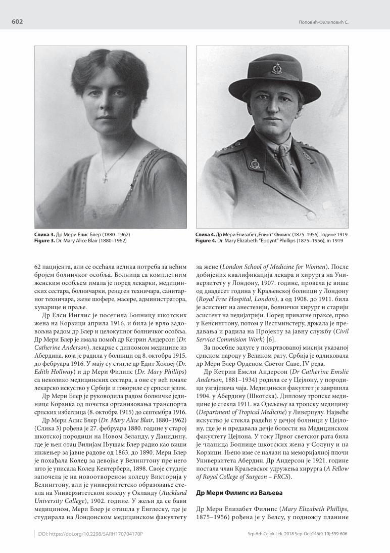
602
Srp Arh Celok Lek. 2018 Sep-Oct;146(9-10):599-606
62 пацијента, али се осећала велика потреба за већим бројем болничког особља. Болница са комплетним женским особљем имала је поред лекарки, медицин-ских сестара, болничарки, рендген техничара, санитар-ног техничара, жене шофере, масере, администратора, куварице и праље.
Др Елси Инглис је посетила Болницу шкотских жена на Корзици априла 1916. и била је врло задо-вољна радом др Блер и целокупног болничког особља. Др Мери Блер је имала помоћ др Кетрин Андерсон (Dr. Catherine Anderson), лекарке с дипломом медицине из Абердина, која је радила у болници од 8. октобра 1915. до фебруара 1916. У мају су стигле др Едит Холвеј (Dr. Edith Hollway) и др Мери Филипс (Dr. Mary Phillips) са неколико медицинских сестара, а оне су већ имале лекарско искуство у Србији и говориле су српски језик.
Др Мери Блер је руководила радом болничке једи-нице Корзика од почетка организовања транспорта српских избеглица (8. октобра 1915) до септембра 1916.
Др Мери Алис Блер (Dr. Mary Alice Blair, 1880–1962) (Слика 3) рођена је 27. фебруара 1880. године у старој шкотској породици на Новом Зеланду, у Данидину, где је њен отац Вилијам Њушам Блер радио као виши инжењер за јавне радове од 1863. до 1890. Мери Блер је похађала Колеџ за девојке у Велингтону пре него што је уписала Колеџ Кентербери, 1898. Своје студије започела је на новоотвореном колеџу Викторија у Велингтону, али је универзитетско образовање сте-кла на Универзитетском колеџу у Окланду (Auckland University College), 1902. године. У жељи да се бави медицином, Мери Блер је отишла у Енглеску, где је студирала на Лондонском медицинском факултету
за жене (London School of Medicine for Women). После добијених квалификација лекара и хирурга на Уни-верзитету у Лондону, 1907. године, провела је више од двадесет година у Краљевској болници у Лондону (Royal Free Hospital, London), а од 1908. до 1911. била је асистент на анестезији, болнички хирург и старији асистент на педијатрији. Поред приватне праксе, прво у Кенсингтону, потом у Вестминстеру, држала је пре-давања и радила на Пројекту за јавну службу (Civil Service Commision Work) [6].
За посебне залуге у пожртвованој мисији указаној српском народу у Великом рату, Србија је одликовала др Мери Блер Орденом Светог Саве, IV реда.
Др Кетрин Емсли Андерсон (Dr Catherine Emslie Anderson, 1881–1934) родила се у Цејлону, у породи-ци узгајивача чаја. Медицински факултет је завршила 1904. у Абердину (Шкотска). Диплому тропске меди-цине је стекла 1911. на Одељењу за тропску медицину (Department of Tropical Medicine) у Ливерпулу. Највеће искуство је стекла радећи у дечјој болници у Цејло-ну, где је и предавала дечје болести на Медицинском факултету Цејлона. У току Првог светског рата била је чланица Болнице шкотских жена у Солуну и на Корзици. Њено име се налази на меморијалној плочи Универзитета Абердин. Др Андерсон је 1921. године постала члан Краљевског удружења хирурга (A Fellow of Royal College of Surgeon – FRCS).
Др Мери Филипс из Ваљева
Др Мери Елизабет Филипс (Mary Elizabeth Phillips, 1875–1956) рођена је у Велсу, у подножју планине
Слика 3. Др Мери Елис Блер (1880–1962)Figure 3. Dr. Mary Alice Blair (1880–1962)
Слика 4. Др Мери Елизабет „Епинт“ Филипс (1875–1956), године 1919.Figure 4. Dr. Mary Elizabeth ”Eppynt” Phillips (1875–1956), in 1919
Поповић-Филиповић С.
DOI: https://doi.org/10.2298/SARH170704170P

603
Srp Arh Celok Lek. 2018 Sep-Oct;146(9-10):599-606 www.srpskiarhiv.rs
Епинт. Завршила је медицину на Медицинском факул-тету Универзитета Кардиф (Cardiff University, Medical College) 1900. године, а лекарску праксу обавила у Краљевској болници (Royal Free Hospital), у Лондону. На почетку Првог светског рата др Филипс се придружила медицинској мисији у саставу Болнице шкотских жена (Слика 4). Прва служба је била у болници на Малти, где је обављала дужност лекарског помоћника др Алис Хачинсон. Била је то јединица Болнице шкотских жена која је указала помоћ великом броју рањених Аустра-лијанаца и Новозеланђана после битке на Галипољу.
Др Филипс је од јуна 1915. године радила у Другој јединици Болнице шкотских жена у Ваљеву (Слика 5), као лекарски помоћник др Алис Хачинсон. У Ваљеву се разболела па је 1. септембра 1915. године враћена у домовину на опоравак. После опоравка др Филипс је путовала по Великој Британији и држала предавања за промоцију деловања Болнице шкотских жена.
После повлачења српске војске преко Црне Горе и Албаније, др Филипс ће се заједно са неколико мило-срдних сестара, које су већ биле у Србији, прикључи-ти Болници шкотских жена у Ајачу, на Корзици. Са Корзике је др Филипс писала како јој је драго што је поново међу Србима, који су је топло дочекали. Срп-ски рањеници и рековалесценти на Корзици били су срећни што је неко могао да их поздрави на матерњем језику. Др Филипс је заслужено добила титулу најпо-пуларније докторке међу Србима, јер је описана „као савршена лекарка у сваком погледу“. Отуда није било ни чудо што је др Филипс постављена да управља Бол-ницом шкотских жена, Јединицом Корзика на Кор-зици. Др Филипс је руководила Јединицом Корзика у Ајачу на Корзици од 1. маја 1916. до 4. јануара 1917.
У овом периоду др Филипс је имала велику помоћ и подршку др Едит Блејк Холвеј (Dr. Edith Blake Hollway, 1874–1948), која је радила у болници од 20. априла 1916. до 16. августа 1916. Др Холвеј је говорила српски јер је као лекарка Прве јединице Болнице шкотских жена радила у Крагујевцу, у време велике епидемије пегавог тифуса. Оснивањем Четврте јединице Болнице
шкотских жена постављена је за руководиоца једини-це у Лазаревцу. Са доласком окупације, др Холвеј се повукла у Крушевац, где ће радити у Болници „Цар Лазар“, са осталим чланицама Болнице шкотских жена, под руководством др Елси Инглис.
Популарност др Мери Филипс потврђује и испраћај, када су младићи из Лазарета приредили позоришну представу и концерт. У њену част, у Ајачу, јануара 1917. године, приказана је српска верзија Молијера. Одласком није престала њена љубав према Србима. Др Филипс је као лекар асистент (Assistant Medical Officer of Health in Merthyr Tydfil) покренула обуку српских де-војака за медицинске сестре, која је успешно обављена.
Др Елизабет Кортолд
Др Елизабет Кортолд (Dr. Elizabeth Courtauld, 1867–1947) рођена је као трећа ћерка Џорџа Кортолда (George Courtauld). Квалификовала се за медицинску сестру, али је имала јаку жељу да постане лекар. То није било лако, јер у је то време лекарска професија била привилеговано мушка професија и девојке нису имале приступ студијама медицине. Тако је Елизабет Кортолд студирала и завршила медицину на универ-зитету у Брислу. Пошто је обавила лекарски стаж у Краљевској болници у Лондону (Royal Free Hospital), добила је лиценцу лекара 1901. Након дугогодишњег боравка у Индији др Кортолд се вратила у Енглеску пре почетка Првог светског рата. Комитет Болнице шкотских жена поставио је др Кортолд за руководиоца Јединице Корзика. Запамћена је као особа која је упр-кос страхотама и трагедијама рата уносила оптимизам. Једном је рекла да она, као и остатак света, никад неће заборавити те године, те бројне тешко рањене војнике и уништене младости. Др Кортолд је руководила Једи-ницом Корзика у Ајачу највероватније од 13. јануара 1916. до краја јула 1917. По одласку са Корзике радила је у Болници шкотских жена у Рејмону, у Француској, од почетка августа 1917. до 6. марта 1919.
У болници на Корзици са др Кортолд радиле су др Хелена Џонс (Dr. Helena G. Jones, од 16. фебруара 1916. до 16. маја 1916) и др Софи Џексон (Dr. Sophie Bangham Jackson, од 30. септембра 1916. до 25. марта 1917). Др Софи Џексон је 1902. завршила медицину на Меди-цинском факултету у Њукаслу (College of Medicine, Newcastle-upon-Tyne).
Др Кортолд је носилац српских и француских од-ликовања Croix de Guerre и Legion d’Honneur. Носи-лац је два висока француска одликовања као знак признања за своју храброст да настави операцију под свећом у време непрекидне непријатељске паљбе. Као великодушан ктитор Болнице Халстeд у Халстeду (Есекс), др Кортолд је 1920. поклонила 4.000 фунти, за изградњу новог амбулантног блока у знак сећања на свог оца Џорџа Кортолда. Доцније је живела у Пер-сесу, у Гринстед Грину, где је преминула 26. децембра 1947. Хируршки блок Халстeда је недавно пресељен у новоизграђен комплекс зграда, који је понео име „Др Елизабет Кортолд“ [7].
Слика 5. Др Мери Филипс, лекар у Болници шкотских жена у Ваљеву и на КорзициFigure 5. Dr. Mary Phillips, a physician at the Scottish Women’s Hospital in Valjevo and on Corsica
Срби на Корзици у Великом рату – 2. део
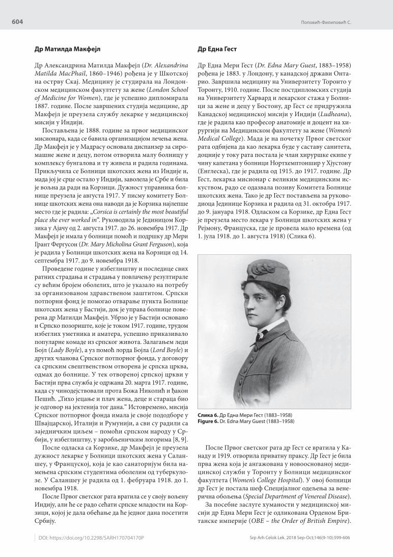
604
Srp Arh Celok Lek. 2018 Sep-Oct;146(9-10):599-606
Др Матилда Макфејл
Др Александрина Матилда Макфејл (Dr. Alexandrina Matilda MacPhail, 1860–1946) рођена је у Шкотској на острву Скај. Медицину је студирала на Лондон-ском медицинском факултету за жене (London School of Medicine for Women), где је успешно дипломирала 1887. године. После завршених студија медицине, др Макфејл је преузела службу лекарке у медицинској мисији у Индији.
Постављена је 1888. године за првог медицинског мисионара, када се бавила организацијом лечења жена. Др Макфејл је у Мадрасу основала диспанзер за сиро-машне жене и децу, потом отворила малу болницу у комплексу бунгалова и ту живела и радила годинама. Прикључила се Болници шкотских жена из Индије и, мада јој је срце остало у Индији, заволела је Србе и била је вољна да ради на Корзици. Дужност управника бол-нице преузела је августа 1917. У писму комитету Бол-нице шкотских жена она наводи да је Корзика најлепше место где је радила: „Corsica is certainly the most beautiful place she ever worked in“. Руководила је Јединицом Кор-зика у Ајачу од 2. августа 1917. до 26. новембра 1917. Др Макфејл је имала у болници помоћ и подршку др Мери Грант Фергусон (Dr. Mary Micholina Grant Ferguson), која је радила у Болници шкотских жена на Корзици од 14. септембра 1917. до 9. новембра 1918.
Проведене године у избеглиштву и последице свих ратних страдања и страдања у повлачењу резултирале су већим бројем оболелих, што је указало на потребу за организованом здравственом заштитом. Српски потпорни фонд је помогао отварање пункта Болнице шкотских жена у Бастији, док је управа болнице пове-рена др Матилди Макфејл. Убрзо је у Бастији основано и Српско позориште, које је током 1917. године, трудом избеглих уметника и аматера, успешно приказивало популарне комаде из српског живота. Залагањем леди Бојл (Lady Boyle), а уз помоћ лорда Бојла (Lord Boyle) и других чланова Српског потпорног фонда, у договору са српским свештвенством отворена је српска црква, одмах до болнице. У тек отвореној српској цркви у Бастији прва служба је одржана 20. марта 1917. године, када су чинодејствовали прота Божа Николић и ђакон Пешић. „Тихо јецање и плач жена, деце и стараца био је одговор на јектенија тог дана.“ Истовремено, мисија Српског потпорног фонда имала је своје пододборе у Швајцарској, Италији и Румунији, а сви су радили са заједничким циљем – помоћи српском народу у Ср-бији, у избеглиштву, у заробљеничким логорима [8, 9].
После одласка са Корзике, др Макфејл је преузела дужност лекарке у Болници шкотских жена у Салан-шеу, у Француској, која је као санаторијум била на-мењена српским студентима оболелим од туберкуло-зе. У Саланшеу је радила од 1. фебруара 1918. до 1. новембра 1918.
После Првог светског рата вратила се у своју вољену Индију, али ће се радо сећати српске младости на Кор-зици, којој је дала обећање да ће једног дана посетити Србију.
Др Една Гест
Др Една Мери Гест (Dr. Edna Mary Guest, 1883–1958) рођена је 1883. у Лондону, у канадској држави Онта-рио. Завршила медицину на Универзитету Торонто у Торонту, 1910. године. После постдипломских студија на Универзитету Харвард и лекарског стажа у Болни-ци за жене и децу у Бостону, др Гест се придружила Канадској медицинској мисији у Индији (Ludheana), где је радила као професор анатомије и доцент на хи-рургији на Медицинском факултету за жене (Women’s Medical College). Мада је на почетку Првог светског рата одбијена да као лекарка буде у саставу санитета, доцније у току рата постала је члан хируршке екипе у чину капетана у болници Нортхемптоншир у Хјустону (Енглеска), где је радила од 1915. до 1917. године. Др Гест, лекарка мисионар с великим медицинским ис-куством, радо се одазвала позиву Комитета Болнице шкотских жена. Тако је др Гест постављена за руково-диоца Јединице Корзика и радила од 31. октобра 1917. до 9. јануара 1918. Одласком са Корзике, др Една Гест је преузела место лекара у Болници шкотских жена у Рејмону, Француска, где је провела мало времена (од 1. јула 1918. до 1. августа 1918) (Слика 6).
После Првог светског рата др Гест се вратила у Ка-наду и 1919. отворила приватну праксу. Др Гест је била прва жена која је ангажована у новооснованој меди-цинској служби у Торонту у Болници медицинског факултета (Women’s College Hospital). У овој болници др Гест је постала шеф Специјалног одељења за вене-рична обољења (Special Department of Venereal Disease).
За посебне заслуге хуманости у медицинској ми-сији др Една Мери Гест је одликована Орденом Бри-танске империје (OBE – the Order of British Empire).
Слика 6. Др Една Мери Гест (1883–1958)Figure 6. Dr. Edna Mary Guest (1883–1958)
Поповић-Филиповић С.
DOI: https://doi.org/10.2298/SARH170704170P

605
Srp Arh Celok Lek. 2018 Sep-Oct;146(9-10):599-606 www.srpskiarhiv.rs
Као велики борац за равноправност жена и положај жена лекара у Кана-ди, бирана је 1940. и 1941. године за председницу Федерације жена лекара Канаде (Federation of Medical Women of Canada) [10].
Др Онорија Кир
Др Онорија Самервил Кир (Dr. Honoria Somerville Keer, 26. децембар 1883 – 20. март 1969) рођена је у Торонту (Ка-нада) од мајке Елајзе Самервил и оца генерал-мајора Џонатана Кира. Дипломирала је медицину и хирургију на Медицинском факултету за жене у Глазгову, 1910. године. Извесно време радила је као лекарка у Хамил-тону, а са почетком Првог светског рата придружила се Болници шкотских жена (Слика 7).
Др Кир је била у саставу Јединице Гиртон и Њунам, која је почела са радом у Француској, потом пребачена у Ђевђелију, а завршила у Солуну, на Солунском фрон-ту. Др Кир је у саставу ове јединице радила од 8. маја 1915. до 21. јануара 1918. Јединица Гиртон и Њунам у Солуну, под управом др Ен Луизe Мaкилрој (Dr. Anne Louise Mcllroy, 1874–1968), одиграла је значајну улогу у лечењу српских рањеника након Горничевске и Кајмак-чаланске битке. Касније je јединица постала позната по оснивању Ортопедског центра Калкута (Calcutta Orthopaedic Centre), који се бавио лечењем српских рањеника и инвалида. По ослобођењу, др Мaкилрој је основала Ортопедски центар за српске инвалиде, код Београда, испод Авале.
Др Кир је у Ајачу на Корзици руководила Jединицом Корзика, Болнице шкотских жена, која је лечила и не-говала српске избеглице на Корзици. Др Кир је радила на Корзици од 8. маја 1918. до 15. јануара 1919 [7, 10].
У Болници шкотских жена је приређена велика свеча-ност поводом Божића, на дан 25. децембра 1918. Рањени
и болесни војници су добили поклоне од становника Бастије, а после заједничке закуске приређен је концерт. Пригодна свечаност је окупила велики број српских избеглица, чланице Болнице шкотских жена, Српског потпорног фонда, локално становништво Бастије, срп-ско свештвенство, наставнике и ученике из Лицеја. Посебно је био запажен хор српских ђака, који је певао патриотске и народне песме из старог краја, о чему су писали и бастијски листови. У Српској цркви у Бастији припадници српске избегле колоније приредили су и свечаности за Видовдан и Петровдан 1918. са свечаним литургијама и благодарењима у част рођендана Њ. В. краља Петра I [6] (слике 8 и 9).
Др Кир је била последња управница болнице у Аја-чу, јер је болница затворена у пролеће 1919. Повратком српских избеглица у домовину престала је потреба за овом врстом болничке установе.
После Првог светског рата др Кир је краће време радила у болници у Ланарку, у Шкотској. Године 1924. стекла је диплому тропске медицине и постала лекар у државној болници у Нигерији. Носилац је два висо-ка француска одликовања: Croix de Guerre и Medaille d’ Honneur. Србија је др Онорију Кир одликовала Ор-деном Светог Саве.
Поред др Онорије Кир на Солунском фронту и Кор-зици, и др Едне Гест на Корзици, у саставу Болнице
Слика 7. Др Онорија Самервил Кир (1883–1969)Figure 7. Dr. Honoria Somerville Keer (1883–1969)
Слика 8. Српски ученици и студенти у ФранцускојFigure 8. Serbian schoolboys and students in France
Слика 9. Српски ученици у ФранцускојFigure 9. Serbian schoolboys in France
Срби на Корзици у Великом рату – 2. део
606
Srp Arh Celok Lek. 2018 Sep-Oct;146(9-10):599-606
шкотских жена велики број Канађана, лекара и дру-гог медицинског особља пожртвовано је помагао срп-ским војницима у канадским болницама на Солунском фронту [9].
Са Корзике, болничарка Агнес Патерсон (Agnes Paterson) писала је својима: „Ајачио је дивно место, палме, наранџе, лешници, и смокве поред путева. На-жалост и врло топло место. Имали смо две болнице, једну велику 'општу' која је исто тако лечила рањене војнике, а служила и као породилиште за Србе... Наша друга болница била је за инфективне болести, гроз-ницу, тифус итд. и увек смо имали неколико тешких болесника... Сви смо били јако сложни и срећни у целој јединици. Често би на крају дана, када је претопло и
кад смо сви преморени да се одвучемо на спрат, седели и певали заједно. То подстиче наше расположење и умањује носталгију за домовином“ [3]. „Медицинске сестре и болничко особље су савршено радили и за-служују све похвале“ [3].
Тако су пролазили дани, месеци, године у борби за живот избеглих и прогнаних. А њихови једини погледи наде су били упрти у небо и њихове спасиоце, лекаре и сестре.
Захвалница
Аутор захваљује Алану Камингу из Шкотске на ус-тупљеним фотографијама за овај рад.
ЛИТЕРАТУРА
1. Popović-Filipović S. Za hrabrost i humanost, Bolnice škotskih žena u Srbiji i sa Srbima za vreme Prvog svetskog rata 1914–1918, Beograd: Signature; 2007.
2. Popović-Filipović S. Elzi Inglis (1864–1917) i Bolnice škotskih žena u Srbiji u Velikom ratu – 1. deo. Srp Arh Celok Lek. 2017; 146(3–4)226–30.
3. Leneman L. In the Service of Life. Edinburgh: The Mercat Press; 1994.
4. Popović-Filipović S. Svedočanstva iz Prvog svetskog rata. Valjevo: Istorijski arhiv; 2017.
5. Popović-Filipović S. Mary Alice Blair. Scottish Women’s Hospitals. A–Z of personnel. (Accessed July 2017) Available at: http://scottishwomenshospitals.co.uk/women/
6. McLaren ES. A History of the Scottish Women’s Hospitals. London: Hodder and Stoughton; 1919.
7. Bojić D. Srpske izbeglice u Prvom svetskom ratu (1914–1921). Beograd: Zavod za udžbenike; 2007.
8. Đokić MM. Naši na Korzici. Knjiga prva. Beograd: Prosveta; 1923.9. Popović-Filipović S. Iz postojbine javora. Kanadsko-britanska
medicinska i humanitarna pomoć Srbiji u Prvom svetskom ratu. Beograd: Srpsko lekarsko društvo; 2013.
10. Sorr К. Excluded from the Record: women, refugees and relief, 1914–1929. Bern: Peter Lang, International Academic Publisher; 2010.
SUMMARYHistorians and historical research of the role of the Serbian na-tion in the Great War give ample respect and recognition of the great battles and great victories. However, the exodus of the Serbian people and its armies out of Serbia is also not forgot-ten. Neither are the Salonika Front, nor other battlefronts. Less well known and researched is the fate of 35,000 young Serbian recruits, the young people dispersed to distant lands. This research is concentrated on the fate of the Serbian refu-gees in Corsica, on those who helped them, looked after them, and treated them to recovery, and who themselves came there from other parts of the world. Those Serbian refugees in Cor-sica were looked after by the representatives of diplomatic, humanitarian, and medical missions from Serbia, France, and Great Britain. The life of the Serbian refugee colony in Corsica was organized, financed, and supported by the Royal Serbian Government in exile in France, the French Relief Committee
for the wounded, sick, and refugees, the Serbian Relief Fund, the Scottish Women’s Hospitals for Foreign Service, the local authorities, and numerous individuals in Corsica.We have paid particular attention to the Scottish Women’s Hospital in Corsica that provided a special hospital unit called “Corsica Unit,” situated in Ajaccio, with the isolation ward in Lazaret, and ambulances and dispensaries located in various villages, where the Serbian refugees were billeted. At the time of centennial commemorations of the Great War, we want to express our profound gratitude to the humanitarian and medi-cal assistance from all quarters, and in particular to the Scottish Women’s Hospitals, and Dr. Elsie Inglis, the founder and the leader of this medical mission.
Keywords: World War I; French medical help; Serbian Red Cross; Serbian Relief Fund; Scottish Women’s Hospitals; Corsica; Serbia; Inglis E
Serbs on Corsica in the Great War – Part 2Slavica Popović-FilipovićSerbian Medical Society, Section for History of Medicine, Belgrade, Serbia
Поповић-Филиповић С.
DOI: https://doi.org/10.2298/SARH170704170P

607
ПРИКАЗ КЊИГЕ / BOOK REVIEW
Споменица професору Војиславу Шуваковићу(A tribute to Professor Vojislav Šuvaković)
Received • Примљено: September 17, 2018
Accepted • Прихваћено: September 23, 2018
Online fi rst: October 11, 2018
Приредили: Стеван Баљошевић и Миодраг Павловић
Аутори: Владимир Кањух, Марија Вукотић, Стеван Баљошевић, Стеван Литвињенко, Миодраг Павловић, Бранко Брмболић, Радослав Катанић, Марија Богдановић (Библиографија радова)
Издавач: Академија медицинских наука Српског лекарског друштва, 2017.
Обим књиге: 132 стр., илустрованоISBN: 978-86-6061-082-1Доступно на: http://akademija.sld.org.rs/
global/pdf/SPOMENICA.pdf
УВОД
Могућност да напишем приказ књиге као што је „Споменица“ доживљавам као по-себну привилегију. Но, то је такође и велика одговорност која се не умањује тиме што ћу имена која помињем само по добру помиња-ти а о личностима говорити онако како су колеге и сарадници говорили.
Како сам наслов „Споменица“ каже, књи-га је посвећена проф. др Војиславу Шува-ковићу.
Иако обухвата шири контекст, то је пре-васходно књига о једном човеку „који је био и отац и син и муж и лекар, виноградар и илегалац и ратник и професор, а надасве добар човек, који је знао поштовати меди-цинску традицију, али је показивао и непо-колебљиво интересовање за све што је ново у медицини“ (Б. Брмболић, „Споменица“, страна 81)
ОПШТИ ПОДАЦИ О КЊИЗИ
„Споменица“ је штампана на 132 странице у које улази 60 фотографија, као и списак од 260 библиографских јединица чији је аутор проф. др В. Шуваковић (књига, сепарата, чланака, постера, кратких садржаја радова, рецензија и превода).
Тематски се у „Споменици“ може из-двојити пет блокова које у јединствену це-лину повезује личност и рад проф. Шувако-вића. Ти блокови су:
– Проф. др В. Шуваковићу у спомен-сећање колега и сарадника, укључујући и последње речи опроштаја;
– Инфективне болести, у чије савлађи-вање је уложено много знања, искуства и самопрегорног рада (текст о мала-рији);
– Сарадници и пријатељи (текст о проф. др Миомиру Кецмановићу);

Srp Arh Celok Lek. 2018 Sep-Oct;146(9-10):607-609
608
– Земља, народ, политичка ситуација, укупни ам-бијент у коме се живело и радило.
Наша пажња у даљем приказу биће превасходно усмерна на први блок.
ВОЈИСЛАВУ ШУВАКОВИЋУ У СПОМЕН,
СА ПИЈЕТЕТОМ
„Био је изузетно добар, племенит и хуман човек, неус-трашиви успешан организатор и борац против смрто-носних епидемија у нашој земљи, одличан стручњак-инфектолог, талентовани професор-педагог и при-знати научник у области инфектологије“ (В. Кањух, „Споменица“, стр. 19).
Основни биографски подаци нам казују да је рођен 1925. године у Београду, где је завршио основну школу и гимназију. Током немачке окупације као члан СКОЈ-а био је хапшен и затваран. Народноослободилачкој војсци Југославије прикључио се 1944. године, а 1945. постаје члан Комунистичке партије.
Медицински факултет у Београду уписао је 1945. го-дине, а завршио 1951. „Био је одличан студент и имао је лепе манире. ... Шуле је био изузетно поштен човек и познато је међу студентима да он лично никада није урадио ништа лоше другима, колико ја знам сви га памте по добру, а за остале комунисте са нашег фа-култета не би се могло рећи исто“ (М. Вукотић, „Спо-меница“, стр. 24).
Био је један од најбољих студената своје генерације, демонстратор на физици и патологији који је својим изванредним педагошким способностима приближио материју студентима. Изабран је за асистента на Ме-дицинском факултету у Београду 1959. године, 1969. за доцента, 1975. за ванредног професора и 1981. за редовног професора.
Цивилно је мобилисан и распоређен на Косово 1951. године. Тамошња здравствена ситуација била је веома тешка, а здравственог кадра било је веома мало.
„Др Војислав Шуваковић био је први лекар у При-зрену и на Косову и Метохији који је положио специја-листички испит из града где је радио савесно, предано и врло успешно...
Иако млад, др Шуваковић је својом сталном меди-цинском активношћу постао важна јавна личност у граду, омиљен у свим националним срединама. Био је врло поштован и прихваћен јер је имао слободу да изрази своје виђење ствари и у медицинској профе-сији и уопште о животним питањима тог времена“ (С. Баљошевић, „Споменица“, стр. 31, 32).
У Призрену се бавио проучавањем антракса (из ове области је и докторирао), а 1962. учествује у сузбијању тифуса у Приштини. Био је стипендиста СЗ0 1963. го-дине у Индији, где се посебно бавио проучавањем и сузбијањем великих богиња (Smallpox Control). Тада је овладао сазнањима која је на највишем професи-оналном нивоу употребио на Косову 1972. године и дао огроман допринос сузбијању епидемије ове опаке болести.
„Др Војислав Шуваковић и проф. др Миомир Кец-мановић су даноноћно опсервирали болеснике и сумњиве случајеве, прописивали терапију, интерве-нисали у свим компликованим случајевима болести, одржавали контакте са експертима из других земаља и експертима СЗО. Врло често је др Шуле био на те-рену у кућама лежећих болесника и њиховој околини и својим одлучним ставом храбрио становништво да се вакцинише на време. ... Дежурао је и био у при-правности без икаквог ограничења и био пример свим члановима карантинске заједнице у Ђаковици. Добио је значајна државна признања и у то време постао ле-гендарно име у борби против ове тешке болести. ... Истицан је као пример великог хуманисте и експер-та који је преносио своје знање, динамику и енергију у лечењу болесника од вариоле вере“ (С. Баљшевић, „Споменица“, стр. 39).
Од 1978. до 1979. године био је директор Клинике за инфективне и тропске болести у Београду. Према М. Павловићу („Споменица“, стр. 100), био је запамћен као директор који је пружао руку младима, стиму-лисао их да препознају модерне медицинске путеве, поштујући њихове напоре да се радом потврде, не за-мерајући онима који су његову благонаклоност често заборавили. Одлази у Нигерију 1979. године, где се бави лечењем тропских болести – посебно маларије.
Први је у СФРЈ схватио опасност и комплексност ХИВ инфекције/АИДС-а и написао прву популарну књигу о томе. Оснивач је првог одељења за лечење оболелих. „Посебно смо се дивили Вашој медицинској знатижељи, коју је крунисао пионирски рад на пољу АИДС-а у периоду када су се заплашени том поша-шћу сви од ње клонили“ (М. Павловић, „Споменица“, стр. 100).
Био је редовни члан Академије медицинских наука од 1983. године, омиљени и поштовани професор, мен-тор бројних магистарских и докторских теза.
Умро је фебуара 2010. године.
У ЧЕМУ ЈЕ ЗНАЧАЈ ОВЕ КЊИГЕ
„Споменица“ страну по страну, текст по текст попуња-ва портрет Војислава Шуваковића, као мозаик саста-вљен од безброј драгоцених коцкица. Многе нисмо поменули, а све их је немогуће приказати.
Тај бард српске инфектологије (М. Павловић, „Спо-меница“, стр. 100) израста кроз текстове ове књиге, надахнуто и топло написане, у ренесансни лик лекара, педагога, ЧОВЕКА. Говорио је француски, енглески, немачки и италијански језик, био је спортиста-лако-атлетичар, побеђивао на шаховским турнирима, учио студенте медицине и друге здравствене раднике, лечио људе, бавио се експерименталним радом на антраксу, гасио пожаре епидемија свуда где је позиван, бавио се, поред инфективних болести, хепатологијом и тро-пском медицином, високо поштовао и придржавао се етичких принципа своје професије, остао веран својим младалачким идејама о једнакости и социјалној пра-
Споменица професору Војиславу Шуваковићу

Srp Arh Celok Lek. 2018 Sep-Oct;146(9-10):607-609 www.srpskiarhiv.rs
609
вди, знатижељно и са ентузијазмом тежио новинама, волео људе, колеге, пријатеље и своју породицу, био прихваћен, цењен и вољен.
Велико и прелепо за један људски век.Све је то „Споменица“ сачувала на страницама књи-
ге: сећања на људе, догађаје, лепоту земље Србије, ис-трајност у борби за боље здравље људи и за савремену медицинску науку, упркос бројним препрекама. Сачу-вала је, такође, значајне информације о развоју српске (југословенске) инфектологије у другој половини XX века, те тиме представља допринос развоју историје медицине у нас.
Сећања су подстрек за младе на факултетима и шко-лама као и за све здравствне раднике, јер садрже прак-тичне примере живота и рада који су били и остали у складу са тековинама медицинске науке и принципима етике лекарског позива. Странице књиге „Споменица“ остају да подсећају будуће генерације на те вредности.
Проф. др Викторија Цуцић Српско лекарско друштво,
Академија медицинских наука, Београд, Србија[email protected]

УПУТСТВО АУТОРИМА ЗА ПРИПРЕМУ РАДА
Пре подношења рукописа Уредништву часопи-са „Српски архив за целокупно лекарство“ (СА) сви аутори треба да прочитају Упутство за ауторе (Instructions for Authors), где ће пронаћи све потребне информације о писању и припреми рада у складу са стандардима часописа. Веома је важно да аутори при-преме рад према датим пропозицијама, јер уколико рукопис не буде усклађен с овим захтевима, Уредни-штво ће одложити или одбити његово публиковање. Радови објављени у СА се не хонораришу. За чланке који ће се објавити у СА, самом понудом рада Срп-ском архиву сви аутори рада преносе своја аутор-ска права на издавача часописа – Српско лекарско друштво.
ОПШТА УПУТСТВА. СА објављује радове који до сада нису нигде објављени, у целости или делом, нити прихваћени за објављивање. СА објављује радове на енглеском и српском језику. Због боље доступности и веће цитираности препоручује се ауторима да ра-дове свих облика предају на енглеском језику. У СА се објављују следеће категорије радова: уводници, оригинални (научни и стручни) радови, метаанали-зе, прегледни радови, претходна и кратка саопштења, прикази болесника и случајева, слике из клиничке медицине, видео-чланци, радови за праксу, актуелне теме, радови из историје медицине и језика медицине, лични ставови, наручени коментари, писма уреднику, прикази књига и други прилози. Оригинални радови, претходна и кратка саопштења и прикази болесника и случајева публикују се искључиво на енглеском је-зику, а остале врсте радова се могу публиковати и на српском језику само по одлуци Уредништва. Радови се увек достављају са сажетком на енглеском и српском језику (у склопу самог рукописа). Текст рада куцати у програму за обраду текста Word, фонтом Times New Roman и величином слова 12 тачака (12 pt). Све четири маргине подесити на 25 mm, величину странице на формат А4, а текст куцати с двоструким проредом, левим поравнањем и увлачењем сваког пасуса за 10 mm, без дељења речи (хифенације). Не користити та-булаторе и узастопне празне карактере (спејсове) ради поравнања текста, већ алатке за контролу поравнања на лењиру и Toolbars. За прелазак на нову страну до-кумента не користити низ „ентера“, већ искључиво опцију Page Break. После сваког знака интерпункције ставити само један празан карактер. Ако се у тексту користе специјални знаци (симболи), користити фонт Symbol. Подаци о коришћеној литератури у тексту оз-начавају се арапским бројевима у угластим заграда-ма – нпр. [1, 2], и то редоследом којим се појављују у тексту. Странице нумерисати редом у доњем десном углу, почев од насловне стране.
При писању текста на енглеском језику треба се придр-жавати језичког стандарда American English и користи-ти кратке и јасне реченице. За називе лекова користити искључиво генеричка имена. Уређаји (апарати) се оз-
начавају фабричким називима, а име и место произ-вођача треба навести у облим заградама. Уколико се у тексту користе ознаке које су спој слова и бројева, прецизно написати број који се јавља у суперскрипту или супскрипту (нпр. 99Tc, IL-6, О2, Б12, CD8). Уколико се нешто уобичајено пише курзивом (italic), тако се и наводи, нпр. гени (BRCA1).
Уколико је рад део магистарске тезе, односно докторс-ке дисертације, или је урађен у оквиру научног проје-кта, то треба посебно назначити у Напомени на крају текста. Такође, уколико је рад претходно саопштен на неком стручном састанку, навести званичан назив ску-па, место и време одржавања, да ли је рад и како пуб-ликован (нпр. исти или другачији наслов или сажетак).
КЛИНИЧКА ИСТРАЖИВАЊА. Клиничка истра-живања се дефинишу као истраживања утицаја јед-ног или више средстава или мера на исход здравља. Регистарски број истраживања се наводи у последњем реду сажетка.
ЕТИЧКА САГЛАСНОСТ. Рукописи о истраживањи-ма на људима треба да садрже изјаву у виду писаног пристанка испитиваних особа у складу с Хелсиншком декларацијом и одобрење надлежног етичког одбора да се истраживање може извести и да је оно у складу с правним стандардима. Експериментална истраживања на хуманом материјалу и испитивања вршена на живо-тињама треба да садрже изјаву етичког одбора устано-ве и треба да су у сагласности с правним стандардима.
ИЗЈАВА О СУКОБУ ИНТЕРЕСА. Уз рукопис се при-лаже потписана изјава у оквиру обрасца Submission Letter којом се аутори изјашњавају о сваком могућем сукобу интереса или његовом одсуству. За додатне информације о различитим врстама сукоба интере-са посетити интернет-страницу Светског удружења уредника медицинских часописа (World Association of Medical Editors – WAME; http://www.wame.org) под на-зивом „Политика изјаве о сукобу интереса“.
АУТОРСТВО. Све особе које су наведене као аутори рада треба да се квалификују за ауторство. Сваки ау-тор треба да је учествовао довољно у раду на рукопису како би могао да преузме одговорност за целокупан текст и резултате изнесене у раду. Ауторство се за-снива само на: битном доприносу концепцији рада, добијању резултата или анализи и тумачењу резултата; планирању рукописа или његовој критичкој ревизији од знатног интелектуалног значаја; завршном дотери-вању верзије рукописа који се припрема за штампање.
Аутори треба да приложе опис доприноса појединачно за сваког коаутора у оквиру обрасца Submission Letter. Финансирање, сакупљање података или генерално надгледање истраживачке групе сами по себи не могу оправдати ауторство. Сви други који су допринели изради рада, а који нису аутори рукописа, требало
610

УПУТСТВО АУТОРИМА ЗА ПРИПРЕМУ РАДА
би да буду наведени у Захвалници с описом њиховог доприноса раду, наравно, уз писани пристанак.
НАСЛОВНА СТРАНА. На првој страници рукописа треба навести следеће: наслов рада без скраћеница; предлог кратког наслова рада, пуна имена и презимена аутора (без титула) индексирана бројевима; званичан назив установа у којима аутори раде, место и државу (редоследом који одговара индексираним бројевима аутора); на дну странице навести име и презиме, ад-ресу за контакт, број телефона, факса и имејл адресу аутора задуженог за кореспонденцију.
САЖЕТАК. Уз оригинални рад, претходно и кратко саопштење, метаанализу, преглед литературе, приказ случаја (болесника), рад из историје медицине, актуел-ну тему, рад за рубрику jезик медицине и рад за праксу, на другој по реду страници документа треба приложи-ти сажетак рада обима 100–250 речи. За оригиналне радове, претходно и кратко саопштење, метаанализе и прегледне радове, сажетак треба да има следећу струк-туру: Увод/Циљ, Методе, Резултати, Закључак; сваки од наведених сегмената писати као посебан пасус који почиње болдованом речи. Навести најважније резулта-те (нумеричке вредности) статистичке анализе и ниво значајности. Закључак не сме бити уопштен, већ мора бити директно повезан са резултатима рада. За приказе болесника сажетак треба да има следеће делове: Увод (у последњој реченици навести циљ), Приказ болесника, Закључак; сегменте такође писати као посебан пасус који почиње болдованом речи. За остале типове радова сажетак нема посебну структуру.
КЉУЧНЕ РЕЧИ. Испод Сажетка навести од три до шест кључних речи или израза. Не треба да се пона-вљају речи из наслова, а кључне речи треба да буду релевантне или описне. У избору кључних речи ко-ристити Medical Subject Headings – MeSH (http://www.nlm.nih.gov/mesh).
ПРЕВОД НА СРПСКИ ЈЕЗИК. На трећој по реду стра-ници документа приложити наслов рада на српском језику, пуна имена и презимена аутора (без титула) ин-дексирана бројевима, званичан назив установа у који-ма аутори раде, место и државу. На следећој – четвртој по реду – страници документа приложити сажетак (100–250 речи) с кључним речима (3–6), и то за радове у којима је обавезан сажетак на енглеском језику. Превод појмова из стране литературе треба да буде у духу срп-ског језика. Све стране речи или синтагме за које постоји одговарајуће име у нашем језику заменити тим називом.
Уколико је рад у целости на српском језику (нпр. рад из историје медицине, језика медицине и др.), потребно је превести називе прилога (табела, графикона, сли-ка, схема) уколико их има, целокупни текст у њима и легенду на енглески језик. Сажетке и радове који су у целости на српском језику аутори из Србије треба да пишу ћирилицом.
СТРУКТУРА РАДА. Сви поднаслови се пишу великим масним словима (болд). Оригинални рад, метаанали-за, претходно и кратко саопштење обавезно треба да имају следеће поднаслове: Увод (Циљ рада навести као последњи пасус Увода), Методе рада, Резултати, Диску-сија, Закључак, Литература. Преглед литературе чине: Увод, одговарајући поднаслови, Закључак, Литература. Првоименовани аутор метаанализе и прегледног рада мора да наведе бар пет аутоцитата (као аутор или коау-тор) радова публикованих у часописима с рецензијом. Коаутори, уколико их има, морају да наведу бар један аутоцитат радова такође публикованих у часописи-ма с рецензијом. Приказ случаја или болесника чине: Увод (Циљ рада навести као последњи пасус Увода), Приказ болесника, Дискусија, Литература. Не треба користити имена болесника, иницијале, нити бројеве историја болести, нарочито у илустрацијама. Прикази болесника не смеју имати више од пет аутора.
Прилоге (табеле, графиконе, слике итд.) поставити на крај рукописа, а у самом телу текста јасно назначити место које се односи на дати прилог. Крајња позиција прилога биће одређена у току припреме рада за пуб-ликовање.
СКРАЋЕНИЦЕ. Користити само када је неопходно, и то за веома дугачке називе хемијских једињења, од-носно називе који су као скраћенице већ препознатљи-ви (стандардне скраћенице, као нпр. ДНК, сида, ХИВ, АТП). За сваку скраћеницу пун термин треба навести при првом навођењу у тексту, сем ако није стандардна јединица мере. Не користити скраћенице у наслову. Избегавати коришћење скраћеница у сажетку, али ако су неопходне, сваку скраћеницу објаснити при првом навођењу у тексту.
ДЕЦИМАЛНИ БРОЈЕВИ. У тексту рада на енглеском језику, у табелама, на графиконима и другим прило-зима децималне бројеве писати са тачком (нпр. 12.5 ± 3.8), а у тексту на српском језику са зарезом (нпр. 12,5 ± 3,8). Кад год је то могуће, број заокружити на једну децималу.
ЈЕДИНИЦЕ МЕРА. Дужину, висину, тежину и запре-мину изражавати у метричким јединицама (метар – m, килограм (грам) – kg (g), литар – l) или њиховим дело-вима. Температуру изражавати у степенима Целзијуса (°C), количину супстанце у молима (mol), а притисак крви у милиметрима живиног стуба (mm Hg). Све резултате хематолошких, клиничких и биохемијских мерења наводити у метричком систему према Међу-народном систему јединица (SI).
ОБИМ РАДОВА. Целокупни рукопис рада – који чине насловна страна, сажетак, текст рада, списак литера-туре, сви прилози, односно потписи за њих и легенда (табеле, слике, графикони, схеме, цртежи), насловна страна и сажетак на српском језику – мора износити за оригинални рад, претходно и кратко саопштење, рад из
САЖЕТАК. Уз оригинални рад, претходно и кратко саопштење, метаанализу, преглед литературе, приказ случаја (болесника), рад из историје медицине, актуелну тему, рад за рубрику jезик медицине и рад за праксу, на другој по реду страници документа треба приложити сажетак рада обима 100–250 речи. За оригиналне радове, претходна и кратка саопштењa и метаанализе сажетак треба да има следећу структуру: Увод/Циљ, Методе, Резултати, Закључак; сваки од наведених сегмената писати као посебан пасус који почиње болдованом речи. Навести најважније резултате (нумеричке вредности) статистичке анализе и ниво значајности. Закључак не сме бити уопштен, већ мора бити директно повезан са резултатима рада. За приказе болесника сажетак треба да има следеће делове: Увод (у последњој реченици навести циљ), Приказ болесника, Закључак; сегменте такође писати као посебан пасус који почиње болдованом речи. За остале типове радова сажетак нема посебну структуру.
611

УПУТСТВО АУТОРИМА ЗА ПРИПРЕМУ РАДА
историје медицине и преглед литературе до 5.000 речи, а за приказ болесника, рад за праксу, едукативни чланак и рад за рубрику „Језик медицине“ до 3.000 речи; радови за остале рубрике могу имати највише 1.500 речи. Видео-радови могу трајати 5–7 минута и бити у форма-ту avi, mp4(flv). У првом кадру филма мора се навести: у наднаслову Српски архив за целокупно лекарство, наслов рада, презимена и иницијали имена и средњег слова свих аутора рада (не филма), година израде. У другом кадру мора бити уснимљен текст рада у виду апстракта до 350 речи. У последњем кадру филма могу се навести имена техничког особља (режија, сниматељ, светло, тон, фотографија и сл.). Уз видео-радове дос-тавити: посебно текст у виду апстракта (до 350 речи), једну фотографију као илустрацију приказа, изјаву потписану од свег техничког особља да се одричу ау-торских права у корист аутора рада.
ПРИЛОЗИ РАДУ су табеле, слике (фотографије, цр-тежи, схеме, графикони) и видео-прилози.
ТАБЕЛЕ. Свака табела треба да буде сама по себи лако разумљива. Наслов треба откуцати изнад табеле, а објашњења испод ње. Табеле се означавају арапским бројевима према редоследу навођења у тексту. Табеле цртати искључиво у програму Word, кроз мени Table–Insert–Table, уз дефинисање тачног броја колона и ре-дова који ће чинити мрежу табеле. Десним кликом на мишу – помоћу опција Merge Cells и Split Cells – спаја-ти, односно делити ћелије. Куцати фонтом Times New Roman, величином слова 12 pt, с једноструким проре-дом и без увлачења текста. Коришћене скраћенице у табели треба објаснити у легенди испод табеле.
Уколико је рукопис на српском језику, приложити на-зиве табела и легенду на оба језика. Такође, у једну табелу, у оквиру исте ћелије, унети и текст на српском и текст на енглеском језику (никако не правити две табеле са два језика!).
СЛИКЕ. Слике су сви облици графичких прилога и као „слике“ у СА се објављују фотографије, цртежи, схеме и графикони. Слике означавају се арапским бројевима према редоследу навођења у тексту. Примају се искључиво дигиталне фотографије (црно-беле или у боји) резолуције најмање 300 dpi и формата записа tiff или jpg (мале, мутне и слике лошег квалитета неће се прихватати за штампање!). Уколико аутори не по-седују или нису у могућности да доставе дигиталне фотографије, онда оригиналне слике треба скенирати у резолуцији 300 dpi и у оригиналној величини. Уко-лико је рад неопходно илустровати са више слика, у раду ће их бити објављено неколико, а остале ће бити у е-верзији чланка као PowerPoint презентација (свака слика мора бити нумерисана и имати легенду). Видео-прилози (илустрације рада) могу трајати 1–3 минута и бити у формату avi, mp4(flv). Уз видео дос-тавити посебно слику која би била илустрација видео-приказа у е-издању и објављена у штампаном издању.
Уколико је рукопис на српском језику, приложити на-зиве слика и легенду на оба језика.
Слике се у свесци могу штампати у боји, али додатне трошкове штампе сносе аутори.
ГРАФИКОНИ. Графикони треба да буду урађени и достављени у програму Excel, да би се виделе пратеће вредности распоређене по ћелијама. Исте графиконе прекопирати и у Word-ов документ, где се графикони означавају арапским бројевима према редоследу на-вођења у тексту. Сви подаци на графикону куцају се у фонту Times New Roman. Коришћене скраћенице на графикону треба објаснити у легенди испод графикона. У штампаној верзији чланка вероватније је да графи-кон неће бити штампан у боји, те је боље избегавати коришћење боја у графиконима, или их користити различитог интензитета.
Уколико је рукопис на српском језику, приложити на-зиве графикона и легенду на оба језика.
СХЕМЕ (ЦРТЕЖИ). Цртежи и схеме се достављају у jpg или tiff формату. Схеме се могу цртати и у програ-му CorelDraw или Adobe Illustrator (програми за рад са векторима, кривама). Сви подаци на схеми куцају се у фонту Times New Roman, величина слова 10 pt. Коришћене скраћенице на схеми треба објаснити у легенди испод схеме.
Уколико је рукопис на српском језику, приложити на-зиве схема и легенду на оба језика.
ЗАХВАЛНИЦА. Навести све сараднике који су допри-нели стварању рада а не испуњавају мерила за аутор-ство, као што су особе које обезбеђују техничку по-моћ, помоћ у писању рада или руководе одељењем које обезбеђује општу подршку. Финансијска и материјална помоћ, у облику спонзорства, стипендија, поклона, опреме, лекова и друго, треба такође да буде наведена.
ЛИТЕРАТУРА. Списак референци је одговорност ау-тора, а цитирани чланци треба да буду лако присту-пачни читаоцима часописа. Стога уз сваку референцу обавезно треба навести DOI број чланка (јединствену ниску карактера која му је додељена) и PMID број уко-лико је чланак индексиран у бази PubMed/MEDLINE.
Референце нумерисати редним арапским бројевима према редоследу навођења у тексту. Број референ-ци не би требало да буде већи од 30, осим у прегледу литературе, у којем је дозвољено да их буде до 50, а у метаанализи до 100. Број цитираних оригиналних радова мора бити најмање 80% од укупног броја ре-ференци, односно број цитираних књига, поглавља у књигама и прегледних чланака мањи од 20%. Уколико се домаће монографске публикације и чланци могу уврстити у референце, аутори су дужни да их цитирају. Већина цитираних научних чланака не би требало да
ту avi, mp4 (fl v). У првом кадру филма мора се навести:
612

УПУТСТВО АУТОРИМА ЗА ПРИПРЕМУ РАДА
буде старија од пет година. Није дозвољено цитирање апстраката. Уколико је битно коментарисати резултате који су публиковани само у виду апстракта, неопходно је то навести у самом тексту рада. Референце чланака који су прихваћени за штампу, али још нису објавље-ни, треба означити са in press и приложити доказ о прихватању рада за објављивање.
Референце се цитирају према Ванкуверском стилу (униформисаним захтевима за рукописе који се пре-дају биомедицинским часописима), који је успоставио Међународни комитет уредника медицинских часо-писа (http://www.icmje.org), чији формат користе U.S. National Library of Medicine и базе научних публика-ција. Примери навођења публикација (чланака, књига и других монографија, електронског, необјављеног и другог објављеног материјала) могу се пронаћи на ин-тернет-страници http://www.nlm.nih.gov/bsd/uniform_requirements.html. Приликом навођења литературе веома је важно придржавати се поменутог стандарда, јер је то један од најбитнијих фактора за индексирање приликом класификације научних часописа.
ПРОПРАТНО ПИСМО (SUBMISSION LETTER). Уз рукопис обавезно приложити образац који су потпи-сали сви аутори, а који садржи: 1) изјаву да рад прет-ходно није публикован и да није истовремено поднет за објављивање у неком другом часопису, 2) изјаву да су рукопис прочитали и одобрили сви аутори који ис-пуњавају мерила ауторства, и 3) контакт податке свих аутора у раду (адресе, имејл адресе, телефоне итд.). Бланко образац треба преузети са интернет-странице часописа (http://www.srpskiarhiv.rs).
Такође је потребно доставити копије свих дозвола за: репродуковање претходно објављеног материјала, употребу илустрација и објављивање информација о познатим људима или именовање људи који су допри-нели изради рада.
ЧЛАНАРИНА, ПРЕТПЛАТА И НАКНАДА ЗА ОБРАДУ ЧЛАНКА. Да би рад био објављен у часо-пису Српски архив за целокупно лекарство, сви ауто-ри који су лекари или стоматолози из Србије морају бити чланови Српског лекарског друштва (у складу са чланом 6. Статута Друштва) за годину у којој се рад предаје Уредништву. Сви домаћи аутори такође морају бити претплаћени на часопис или измирити накнаду за обраду чланака (article processing charge) за годину у којој се рад предаје Уредништву, у износу од 3.000 динара. Аутори и коаутори из иностранства су у обавези да плате накнаду за обраду чланака (article processing charge) у износу од 35 евра. Уплата у једној календарској години обухвата и све наредне, евенту-алне чланке, послате на разматрање у тој години. Сви аутори који плате ову накнаду могу, уколико то желе, да примају штампано издање часописа. Треба напо-менути да ова уплата није гаранција да ће рад бити
прихваћен и објављен у Српском архиву за целокупно лекарство. Обавеза плаћања накнаде за обраду чланка не односи се на студенте основних студија и на пре-тплатнике на часопис.
Установе (правна лица) не могу преко своје претплате да испуне овај услов аутора (физичког лица). Уз руко-пис рада треба доставити копије уплатница за члана-рину и претплату / накнаду за обраду чланка, као доказ о уплатама, уколико издавач нема евиденцију о томе. Часопис прихвата донације од спонзора који сносе део трошкова или трошкове у целини оних аутора који нису у могућности да измире накнаду за обраду чланка (у таквим случајевима потребно је часопису ставити на увид оправданост таквог спонзорства).
Додатне информације о чланарини и претплати могу се добити путем имејла ([email protected]) и на интернет-страници часописа http://srpskiarhiv.rs/en/subscription/). СЛАЊЕ РУКОПИСА. Рукопис рада и сви прилози уз рад могу се доставити имејлом ([email protected]), електронски преко система за пријављивање на ин-тернет-страници часописа (http://www.srpskiarhiv.rs), препорученом пошиљком или лично, доласком у Уред-ништво. Уколико се рад шаље поштом или доноси у Уредништво, рукопис се доставља одштампан у три примерка и нарезан на CD (снимљени материјал треба да је истоветан оном на папиру).
НАПОМЕНА. Рад који не испуњава услове овог упут-ства не може бити упућен на рецензију и биће враћен ауторима да га допуне и исправе. Придржавањем упут-ства за припрему рада знатно ће се скратити време целокупног процеса до објављивања рада у часопису, што ће позитивно утицати на квалитет чланака и ре-довност излажења часописа.
За све додатне информације, молимо да се обратите на доленаведене адресе и број телефона.
АДРЕСА:Српско лекарско друштвоУредништво часописа „Српски архив за целокупно лекарство“Ул. краљице Наталије 111000 БеоградСрбија
ТЕЛЕФОН: + 381 11 409-2776+ 381 11 409-4479
E-MAIL: [email protected]ИНТЕРНЕТ АДРЕСА: http://www.srpskiarhiv.rs
ISSN 0370-8179ISSN Online 2406-0895
613

INSTRUCTIONS FOR AUTHORS
Before submitting their paper to the Editorial Office of the Serbian Archives of Medicine, authors should read the Instructions for Authors, where they will find all the necessary information on writing their manuscript in accordance with the journal’s standards. It is essen-tial that authors prepare their manuscript according to established specifications, as failure to do so will result in paper being delayed or rejected. Serbian Archives of Medicine provides no fee for published articles. By sub-mitting a paper for publishing consideration, authors of a paper accepted for publication in the Serbian Archives of Medicine grant and assign all copyrights to the pub-lisher – the Serbian Medical Society.
GENERAL INSTRUCTIONS. Serbian Archives of Medi-cine publishes papers that have not been, either in their en-tirety or partially, previously published, and that have not been accepted for publication elsewhere. Serbian Archives of Medicine publishes papers in English and Serbian. For better availability and citation, authors are encouraged to submit articles of all types in English. The journal publishes the following article types: editorials, original papers, meta-analyses, review articles, preliminary and short communi-cations, case reports, images in clinical medicine, video-articles, articles for practitioners, current topics, history of medicine articles, language of medicine articles, opinion es-says, personal view articles, invited commentaries, letters to the editor, book reviews and other articles. Original papers, preliminary and short communications and case reports are published in English only, while other article types may be published in Serbian if the Editorial Office reaches such decision. The papers are always submitted with Summary in both English and Serbian, included in the manuscript file. The text of the manuscript should be typed in MS Word using the Times New Roman typeface, and font size 12 pt. The text should be prepared with margins set to 25 mm and onto A4 paper size, with double line spacing, aligned left and the initial lines of all paragraphs indented 10 mm, without hyphenation. Tabs and successive blank spaces are not to be used for text alignment; instead, ruler alignment control tool and Toolbars are suggested. In order to start a new page within the document, Page Break option should be used instead of consecutive enters. Only one space fol-lows after any punctuation mark. If special signs (symbols) are used in the text, use the Symbol font. References cited in the text are numbered with Arabic numerals within pa-renthesis (for example: [1, 2]), in order of appearance in the text. Pages are numbered consecutively in the right bottom corner, beginning from the title page.
When writing text in English, linguistic standard Ameri-can English should be observed. Write short and clear sentences. Generic names should be exclusively used for the names of drugs. Devices (apparatuses, instruments) are termed by trade names, while their name and place of production should be indicated in the brackets. If a let-ter-number combination is used, the number should be precisely designated in superscript or subscript (i.e., 99Tc,
IL-6, O2, B12, CD8). If something is commonly written in italics, such as genes (e.g. BRCA1), it should be written in this manner in the paper as well.
If a paper is a part of a master’s or doctoral thesis, or a re-search project, that should be designated in a separate note at the end of the text. Also, if the article was previously pre-sented at any scientific meeting, the name, venue and time of the meeting should be stated, as well as the manner in which the paper had been published (e.g. changed title or abstract).
CLINICAL TRIALS. Clinical trial is defined as any re-search related to one or more health related interventions in order to evaluate the effects on health outcomes. The trial registration number should be included as the last line of the Summary.
ETHICAL АPPROVAL. Manuscripts with human medi-cal research should contain a statement that the subjects’ written consent was obtained, according to the Declaration of Helsinki, the study has been approved by competent ethics committee, and conforms to the legal standards. Experimental studies with human material and animal studies should contain statement of the institutional ethics committee and meet legal standards.
CONFLICT OF INTEREST STATEMENT. The manu-script must be accompanied by a disclosure statement from all authors (contained within the Submission Letter) de-claring any potential interest or stating that the authors have no conflict of interest. For additional information on different types of conflict of interest, please see World Association of Medical Editors (WAME, www.wame.org) policy statement on conflict of interest.
AUTHORSHIP. All individuals listed as authors should be qualified for authorship. Every author should have par-ticipated sufficiently in writing the article in order to take responsibility for the whole article and results presented in the text. Authorship is based only on: crucial contribution to the article conception, obtaining of results or analysis and interpretation of results; design of manuscript or its critical review of significant intellectual value; final revi-sion of the manuscript being prepared for publication.
The authors should enclose the description of contribution to the article of every co-author individually (within the Submission Letter). Funding, collection of data or gen-eral supervision of the research group alone cannot justify authorship. All other individuals having contributed to the preparation of the article should be mentioned in the Acknowledgment section, with description of their contri-bution to the paper, with their written consent.
TITLE PAGE. The first page of the manuscript (cover sheet) should include the following: title of the paper without any abbreviations; suggested running title; each author’s full names and family names (no titles), indexed by numbers; official name, place and country of the institu-
614

INSTRUCTIONS FOR AUTHORS
tion in which authors work (in order corresponding to the indexed numbers of the authors); at the bottom of the page: name and family name, address, phone and fax number, and e-mail address of a corresponding author.
SUMMARY. Along with the original article, preliminary and short communication, meta-analysis, review article, case report, article on history of medicine, current topic ar-ticle, article for language of medicine and article for practi-tioners, the summary not exceeding 100–250 words should be typed on the second page of the manuscript. In original articles, the summary should have the following structure: Introduction/Objective, Methods, Results, Conclusion. Each segment should be typed in a separate paragraph using boldface. The most significant results (numerical val-ues), statistical analysis and level of significance are to be included. The conclusion must not be generalized, it needs to point directly to the results of the study. In case reports, the summary should consist of the following: Introduction (final sentence is to state the objective), Case Outline (Out-line of Cases), Conclusion. Each segment should be typed in a separate paragraph using boldface. In other types of papers, the summary has no special outline.
KEYWORDS. Below the summary, 3 to 6 keywords or phrases should be typed. The keywords need not repeat words in the title and should be relevant or descriptive. Medical Subject Headings – MeSH (http://www.nlm.nih.gov/mesh) are to be used for selection of the keywords.
TRANSLATION INTO SERBIAN. The third page of the manuscript should include: title of the paper in the Serbian language; each author’s full name and family name (no titles), indexed by numbers; official name, place and coun-try of the institution in which authors work. On the fourth page of the manuscript the summary (100–250 words) and keywords (3–6) should be typed, but this refers only to papers in which a summary and keywords are compul-sory. The terms taken from foreign literature should be translated into comprehensible Serbian. All foreign words or syntagms that have a corresponding term in Serbian should be replaced by that term.
If an article is entirely in Serbian (e.g. article on history of medicine, article for “Language of medicine”, etc.), captions and legends of all enclosures (tables, graphs, photographs, schemes) – if any – should be translated into English as well.
Summaries and articles written in Serbian by authors from Serbia need to be written in the Serbian Cyrillic alphabet.
STRUCTURE OF THE MANUSCRIPT. All section head-ings should be in capital letters using boldface. Original articles, meta-analyses and preliminary and short com-munications should have the following section headings: Introduction (objective is to be stated in the final paragraph of the Introduction), Methods, Results, Discussion, Conclu-sion, References. A review article includes: Introduction, corresponding section headings, Conclusion, References.
The firstly named author of a meta-analysis or a review ar-ticle should cite at least five auto-citations (as the author or co-author of the paper) of papers published in peer-reviewed journals. Co-authors, if any, should cite at least one auto-citation of papers also published in peer-reviewed journals. А case report should consist of: Introduction (objective is to be stated in the final paragraph of the Introduction), Case Report, Discussion, References. No names of patients, ini-tials or numbers of medical records, particularly in illustra-tions, should be mentioned. Case reports cannot have more than five authors. Letters to the editor need to refer to papers published in the Serbian Archives of Medicine within previ-ous six months; their form is to be comment, critique, or stating own experiences. Publication of articles unrelated to previously published papers will be permitted only when the journal’s Editorial Office finds it beneficial.
All enclosures (tables, graphs, photographs, etc.) should be placed at the end of the manuscript, while in the body of the text a particular enclosure should only be mentioned and its preferred place indicated. The final arrangement (position) of the enclosures will depend on page layout.
ABBREVIATIONS. To be used only if appropriate, for very long names of chemical compounds, or as well-known abbreviations (standard abbreviations such as DNA, AIDS, HIV, ATP, etc.). Full meaning of each abbreviation should be indicated when it is first mentioned in the text unless it is a standard unit of measure. No abbreviations are al-lowed in the title. Abbreviations in the summary should be avoided, but if they have to be used, each of them should be explained when first mentioned in the text of the paper.
DECIMAL NUMBERS. In papers written in English, in-cluding text of the manuscript and all enclosures, a decimal point should be used in decimal numbers (e.g. 12.5 ± 3.8), while in Serbian papers a decimal comma should be used (e.g. 12,5 ± 3,8). Wherever applicable, a number should be rounded up to one decimal place.
UNITS OF MEASURE. Length, height, weight and volume should be expressed in metric units (meter – m, kilogram – kg, gram – g, liter – l) or subunits. Temperature should be in Celsius degrees (°C), quantity of substance in moles (mol), and blood pressure in millimeters of mercury column (mm Hg). All results of hematological, clinical and biochemical measurements should be expressed in the metric system according to the International System of Units (SI units).
LENGTH OF PAPER. The entire text of the manuscript – title page, summary, the whole text, list of references, all enclosures including captions and legends (tables, photographs, graphs, schemes, sketches), title page and summary in Serbian – must not exceed 5,000 words for original articles, preliminary and short communications, review articles and articles on history of medicine, and 3,000 words for case reports, articles for practitioners, edu-cational articles and articles for “Language of medicine”; for any other section maximum is 1,500 words.
SUMMARY. Along with the original article, preliminary and short communication, meta-analysis, review article, case re-port, article on history of medicine, current topic article, arti-cle for language of medicine and article for practitioners, the summary not exceeding 100–250 words should be typed on the second page of the manuscript. In original articles, pre-liminary communications, and meta-analyses, the summary should have the following structure: Introduction/Objective, Methods, Results, Conclusion. Each segment should be typed in a separate paragraph using boldface. The most signifi-cant results (numerical values), statistical analysis and level of significance are to be included. The conclusion must not be generalized, it needs to point directly to the results of the study. In case reports, the summary should consist of the fol-lowing: Introduction (final sentence is to state the objective), Case Outline (Outline of Cases), Conclusion. Each segment should be typed in a separate paragraph using boldface. In other types of papers, the summary has no special outline.
615

INSTRUCTIONS FOR AUTHORS
Video-articles are to last 5–7 minutes and need to be sub-mitted in the flv video format. The first shot of the video must contain the following: title of the journal in the heading (Serbian Archives of Medicine), title of the work, last names and initials of first and middle names of the paper’s authors (not those of the creators of the video), year of creation. The second shot must show summary of the paper, up to 350 words long. The final shot of the video may list technical staff (director, cameraman, lighting, sound, photography, etc.). Video-articles need to be submitted along with a separate summary (up to 350 words), a single still/photograph as an illustration of the video, and a statement signed by the techni-cal staff renouncing copyrights in favor of the paper’s authors.
To check the required number of words in the manuscript, please use the menu Tools–Word Count, or File–Proper-ties–Statistics.
ARTICLE ENCLOSURES are tables, figures (photographs, schemes, sketches, graphs) and video-enclosures.
TABLES. Each table, with its legend, should be self-explan-atory. The title should be typed above the table and any explanatory information under the table. Tables should be numbered in Arabic numerals in order of citation in the text. Use MS Word, the menu Table–Insert–Table, inserting the adequate number of rows and columns. By the right click of the mouse, use the options Merge Cells and Split Cells. Use Times New Roman, font size 12 pt, with single line spacing and no indent to draw tables. Abbreviations used in tables should be explained in the legend below each respective table.
If the manuscript is entirely in the Serbian language, tables and corresponding legend should be both in Serbian and Eng-lish. Also, the table cells should contain text in both languages (do not create two separate tables with a single language!).
FIGURES. Figures are all types of visual enclosures, and photographs, schemes, sketches and graphs are published as ‘figures’ in the Serbian Archives of Medicine. Figures should be numbered in Arabic numerals in order of cita-tion in the text. Only original digital photographs (black-and-white or color), of minimum 300 dpi, and jpg or tiff format, are acceptable (small, blurry and photographs of poor quality will not be accepted for publishing!). If authors do not possess or are not able to provide digital photographs, then the original photos should be scanned in 300 dpi, and saved in original size. If a paper needs to be illustrated with a considerable number of figures, several figures will be published within the paper, and the rest will be available in the electronic version of the paper as a Pow-erPoint presentation (every figure needs to be numbered and be accompanied by legend). Video-enclosures (illus-trations of a paper) can last 1–3 minutes and are submitted in the flv format. Along with the video, a still/photograph representative of the video is also needed, as it will be used as a placeholder in the electronic version of the paper, and as an illustration in the printed version.
If the manuscript is entirely in the Serbian language, pho-tographs and corresponding legend should be both in Serbian and English.
Photographs may be printed and published in color, but pos-sible additional expenses are to be covered by the authors.
GRAPHS. Graphs should be plotted in Excel in order to see the respective values distributed in the cells. The same graphs should be copied and pasted to the Word docu-ment, numbered in Arabic numerals by order of citation in the text. The text in the graphs should be typed in Times New Roman. Abbreviations used in graphs should be ex-plained in the legend below the respective graph. In the printed versions of papers, graphs are generally published in black-and-white; therefore, it is suggested to avoid the use of colors in graphs, or to utilize colors of significant difference in brightness.
If the manuscript is entirely in the Serbian language, graphs and corresponding legend should be both in Serbian and English.
SCHEMES (SKETCHES). Schemes and sketches are to be submitted in jpg or tiff format. Schemes should be drawn in CorelDraw or Adobe Illustrator (programs for drawing vectors, curves, etc.). The text in the schemes should be typed in Times New Roman, font size 10 pt. Abbreviations used in schemes should be explained in the legend below the respective scheme.
If the manuscript is entirely in the Serbian language, schemes and corresponding legend should be both in Serbian and English.
ACKNOWLEDGMENT. List all those individuals having contributed to preparation of the article but having not met the criteria of authorship, such as individuals provid-ing technical assistance, assistance in writing the paper or running the department securing general support. Fi-nancial aid and all other support in the form of sponsor-ship, grants, donations of equipment and medications, etc., should be mentioned too.
REFERENCES. The reference list is the responsibility of the authors. Cited articles should be readily accessible to the journals readership. Therefore, following each refer-ence, its DOI number and PMID number (if the article is indexed for MEDLINE/PubMed) should be typed.
References should be numbered in Arabic numerals in order of citation in the text. The overall number of refer-ences should not exceed 30, except in review articles, where maximum of 50 is acceptable, and in meta-analysis, where up to 100 referenceas are allowed. The number of citations of original articles must be at least 80% of the total number of references, and the number of citations of books, chap-ters and literature reviews less than 20%. If monographs and articles written by Serbian authors could be included
616

INSTRUCTIONS FOR AUTHORS
in the reference list, the authors are obliged to cite them. The majority of the cited articles should not be older than five years. Use of abstracts as references is not allowed. If it is important to comment on results published solely in the form of an abstract, it is necessary to do so within the text of the article. The references of articles accepted for publication should be designated as in press with the enclosed proof of approval for publication.
The references are cited according to the Vancouver style (Uniformed Requirements for Manuscripts Submitted to Biomedical Journals), rules and formats established by the International Committee of Medical Journal Editors (http://www.icmje.org), used by the U.S. National Library of Medicine and scientific publications databases. Examples of citing publications (journal articles, books and other monographs, electronic, unpublished and other published material) can be found on the web site http://www.nlm.nih.gov/bsd/uniform_requirements.html. In citation of refer-ences the defined standards should be strictly followed, because it is one of the essential factors of indexing for classification of scientific journals.
SUBMISSION LETTER. The manuscript must be accom-panied by the Submission Letter, which is signed by all authors and includes the following: 1) statement that the paper has never been published and concurrently submit-ted for publication to any other journal; 2) statement that the manuscript has been read and approved by all authors who have met the criteria of authorship; and 3) contact information of all authors of the article (address, email, telephone number, etc.). Blank Submission Letter form can be downloaded from the journal’s web site (http://srpskiarhiv.rs/global/pdf/SubmissionletterformFINAL.pdf).
Additionally, the authors should submit the following cop-ies of all permits for: reproduction of formerly published material, use of illustrations and publication of informa-tion on known people or disclosure of the names of people having contributed to the work.
MEMBERSHIP FEE, SUBSCRIPTION RATES AND ARTICLE PROCESSING CHARGE. In order to publish their article in the Serbian Archives of Medicine, all do-mestic authors and co-authors, medical doctors and doc-tors of dental medicine, must be members of the Serbian Medical Society (according to the Article #6 of the Statute of the SMS) for the year in which the manuscript is be-ing submitted. All domestic authors and co-authors must also be subscribed to the journal for the year in which the manuscript is being submitted, or pay an article processing charge for the coverage of all editing and publishing ex-penses (3,000 RSD). Authors and co-authors from abroad need only pay the article processing charge (€35) – the fee is required for substantive editing, fact and reference vali-dations, copy editing, and publishing online and in print. An author who had already paid the fee can have more articles submitted for publishing consideration in the year the fee was paid. All authors who pay this fee may, if they
desire so, receive the printed version of the journal in the year when the fee is payed.
Please note that the payment of this charge does not guar-antee acceptance of the manuscript for publication and does not influence the outcome of the review procedure, in accordance with good publishing practice.
The journal accepts donations from sponsors to create a sum for payment reductions or waivers for authors unable to cover the article processing charge (a justification of the inability to pay should be provided in such cases).
The requirement for paying the article processing charge does not apply to students or to journal subscribers. Insti-tutions (legal entities) cannot by their subscription cover this condition on behalf of the authors (natural persons). Copies of deposit slips for membership and article pro-cessing charge should be enclosed with the manuscript. Foreign authors are under no obligation to be members of the Serbian Medical Society. All the relevant information can be obtained via email address of the Editorial Office ([email protected]) and on the journal’s web site (http://srpskiarhiv.rs/en/subscription/). SUBMISSION OF THE MANUSCRIPT. Manuscript and all enclosures can be sent by email ([email protected]) or via the journal’s web site (http://www.srpskiarhiv.rs). If sent by registered mail or delivered in person at the Editorial Office in Belgrade, it should contain two printed copies and a CD with the version identical to that on paper, as well as all the necessary accompanying documentation.
NOTE. The papers not complying with these instructions will not be reviewed and will be returned to the authors for revision. Observing the instructions for preparation of papers for the Serbian Archives of Medicine will shorten the time of the entire process of publication and will have a positive effect on the quality and timely release of the journal’s issues.
For further information, please contact us via the follow-ing address:
ADDRESS:Srpsko lekarsko društvoUredništvo časopisa „Srpski arhiv za celokupno lekarstvo“Ul. kraljice Natalije 111000 BeogradSerbia
PHONE: +381 11 409 27 76 +381 11 409 44 79E-MAIL: [email protected] SITE: www.srpskiarhiv.rs
ISSN 0370-8179ISSN Online 2406-0895
617

CIP – Каталогизација у публикацијиНародна библиотека Србије, Београд
61(497.11)
СРПСКИ архив за целокупно лекарство : званичан часопис Српског лекарског друштва = Serbian Archives of Medicine : оfficial journal of the Serbian Мedical Society / главни и одговорни уредник Гордана Теофиловски-Парапид. - Књ. 1 (1874)-књ. 2 (1875) ; књ. 3 (1879)- књ. 8 (1881) ; књ. 9 (1887)-књ. 10 (1888) ; књ. 11 (1894)-књ. 12 (1895) ; год. 1, бр. 1/2 (1895)- . - Београд : Српско лекарско друштво, 1874-1875; 1879-1881; 1887-1888; 1894-1895; 1895-(Београд : Службени гласник). - 29 cm
Двомесечно. - Текст на енгл. језику. - Имa суплемент или прилог: Српски архив за целокупно лекарство. Суплемент = ISSN 0354-2793. - Другo издањe нa другом медијуму: Српски архив за целокупно лекарство (Online) = ISSN 2406-0895ISSN 0370-8179 = Српски архив за целокупно лекарствоCOBISS.SR-ID 3378434

ГОДИ
ШТЕ
/ VO
LUM
E 14
6 •
СЕПТ
ЕМБА
Р–ОК
ТОБА
Р / S
EPTE
MBE
R–O
CTOB
ER 20
18 •
СВЕ
СКА
/ ISS
UE 9–
10
2018; 146: 9–10