Specific Ways Brain SPECT Imaging Enhances Clinical Psychiatric Practice
Upload
univ-lille2Category
view
1download
0
Psychiatry Research Neuroimaging 115(2002) 37–48
0925-4927/02/$ - see front matter� 2002 Elsevier Science Ireland Ltd. All rights reserved.PII: S0925-4927Ž02.00031-8
SPECT imaging, clinical features, and cognition before and afterlow doses of amisulpride in schizophrenic patients with the deficit
syndrome
Guillaume Vaiva *, Pierre Thomas , Pierre Michel Llorca , Sylvie Dupont ,a, a b e
Olivier Cottencin , Patrick Devos , Olivier Mazas , Claire Rascle , Marc Steinling ,a d a a c
Michel Goudemanda
Department of Psychiatry, University of Lille, Francea
Department of Psychiatry, University of Clermont Ferrand, Franceb
Department of Nuclear Medicine, Lille, Francec
Department of Statistics, Lille, Franced
Department of Radiology, Lomme, GHICL, Francee
Received 4 September 2001; received in revised form 15 April 2002; accepted 30 April 2002
Abstract
The aim of the study was to examine the action of low-dose amisulpride(100 mgyd), an atypical antipsychoticfrom the benzamide class with a high affinity for the D2 and D3 dopamine receptors, given for 4 weeks in 19schizophrenic patients with the deficit syndrome, in terms of clinical response, modifications in their cognitiveperformance and changes in brain perfusion values. A secondary objective was to distinguish between primary andsecondary deficit, according to Carpenter’s definition. Both efficacy and a relatively low rate of side effects of low-dose amisulpride in the deficit forms of schizophrenia were found as expected from earlier placebo-controlled studies.Our study found significant changes in the cerebral blood flow, before and after treatment, more marked in the frontalarea and particularly in the dorso-lateral frontal area. A significant improvement of cognitive function was foundafter treatment, without a link to any particular changes in a loco-regional perfusion value. Finally, a distinctionbetween primary and secondary deficit showed a higher percentage of clinical improvement in the patients with asecondary deficit. The psychometric and cerebral perfusion changes were no different in the two groups.� 2002 Elsevier Science Ireland Ltd. All rights reserved.
Keywords: Schizophrenia; Deficit syndrome; Amisulpride; Antipsychotic treatment; SPECT; Cognitive function; Schedule for theDeficit Syndrome; Pre-post treatment study
*Corresponding author. Clinique Michel Fontan, 6 rue du Professeur Laguesse, CHRU de Lille, 59037 Lille cedex, France. Fax:q33-3-2044-6265.
E-mail address: [email protected](G. Vaiva).
38 G. Vaiva et al. / Psychiatry Research Neuroimaging 115 (2002) 37–48
1. Introduction
Factors that are associated with the deficit syn-drome include cognitive disturbance, morphologi-cal brain abnormalities, poor premorbid socialadjustment, poor response to classical neuroleptictreatment and, therefore, unfavorable long-termprognosis(Crow, 1980). The literature provides anumber of brain imaging studies with SPECT orPET techniques(for review: Buchsbaum andHazlett, 1998). A relationship between ‘hypofron-tality’ and a negative form of schizophrenia hascommonly been found(Erkwoh et al., 1999; Sabriet al., 1997). If the deficit syndrome is in factassociated with altered brain function, the differ-ences in the findings may be due to:(1) themarked variability between studies in the assess-ment of symptoms;(2) the failure to distinguishbetween primary or enduring symptoms, and tran-sitory or secondary ones(Eugen Bleuler, 1911,cited by Carpenter et al., 1988).Certain authors, like Carpenter et al.(1988),
have discussed the value of a simple descriptionof the deficit syndrome:(i) deficit syndrome isnot specific for schizophrenia;(ii) even when it ispresent, it may have multiple causes, which arenot always linked to schizophrenia;(iii ) the prog-nosis and the response to treatment are variable;(iv) the presence of a deficit syndrome is notsynonymous with an unfavorable or uninterruptedevolution of the disease. According to theseauthors, it is therefore useful to define a so-called‘secondary’ deficit syndrome, which results fromfactors that are extrinsic to the schizophrenicprocess, or results from other psychopathologicalconditions(depression, positive symptoms, Parkin-sonism from neuroleptics, etc.). Conversely, theauthors define a ‘primary’ deficit syndrome, whichseems to be inherent to the disease; it is presentover long periods despite fluctuations in the dis-ease, often right from the beginning of the illness.The deficit syndrome actually poses complex
therapeutic problems; many types of psychotropicdrugs have been recommended, without a clear,long-lasting efficacy being observed(Miller et al.,1994). The clinical usefulness of small doses ofneuroleptics in certain forms of schizophrenia hasalready been proposed in France(Puech et al.,
1981). A particular usefulness of low doses ofsubstituted benzamides for the deficit symptomsof schizophrenia has been proposed(Meltzer etal., 1986; Petit et al., 1987). Amisulpride, anatypical antipsychotic from the benzamide class,with a high affinity for the D2 and D3 dopaminereceptors(Perrault et al., 1997), has a good effi-cacy and excellent tolerance at a dose of 50 or100 mgyday in the long-term deficit syndrome inschizophrenia(Boyer et al., 1995; Paillere-Martin-`ot et al., 1995; Loo et al., 1997; Danion et al.,1999; Leucht et al., 2002). In these studies, theefficacy of amisulpride was only measured usingclinical criteria. Currently, there are few data avail-able measuring cognitive function(Peretti et al.,1997; Ramaekers et al., 1999; Curran and Perry,2001) or functional brain imaging(Martinot et al.,1996; Trichard et al., 1998; Xiberas et al., 2001)after treatment with amisulpride. No data areavailable measuring the cognitive and functionalbrain imaging changes in schizophrenic patientswith the deficit syndrome, before and after treat-ment with amisulpride.Our follow-up study was designed as a pre-post
treatment procedure. The main objective was toexamine the action of low-dose amisulpride givenfor 4 weeks in schizophrenic patients with thedeficit syndrome, in terms of clinical response,modifications in their cognitive performance, andchanges in brain perfusion values. A secondaryobjective was to distinguish, at these three levels,between a primary and secondary deficit, accord-ing to Carpenter’s definition(Carpenter et al.,1988).
2. Methods
2.1. Patients
The study included patients who fulfilled theDSM-III-R criteria for the disorganized or residualform of ‘Schizophrenic Disorder’(American Psy-chiatric Association, 1987), and who were hospi-talized consecutively in the Psychiatry Departmentof the University Hospital of Lille. We only includ-ed patients who presented with a deficit syndrome,with an overall SANS score of more than 65y125and an overall SAPS score of less than 30(Scales
39G. Vaiva et al. / Psychiatry Research Neuroimaging 115 (2002) 37–48
for the Assessment of Negative–Positive–Symp-toms, Andreasen, 1983a,b). We excluded patientswith a comorbid psychiatric disorder, a centralnervous system disorder or arterial hypertension.This study was approved by the Ethical Committeeof the University of Lille II (decision CP 93y101).The patients had not been treated by neurolepticsfor at least 1 month before inclusion in the study.They were hospitalized in a 20-bed unit of Psy-chiatry for the duration of the study; 1 dayyweek,preferably the weekend, they were given permis-sion to stay with their family. In the unit, thenursing care was standardized and was aimed atstimulating the social activities of the patients andcalming their anxiety. From the third day onwards,they were treated with oral amisulpride at a doseof 100 mgyday taken in the morning, for 28 days.In case of severe insomnia, oxazepam could beprescribed at a dose of 50 mg at bedtime. Allmedication was taken in the presence of a nurse.
2.2. Clinical assessment
During a semi-standardized interview, clinicalassessment was performed on Day 0 and Day 28,using the Scale for the Assessment of NegativeSymptoms(SANS), the Scale of the Assessmentof Positive Symptoms (SAPS) (Andreasen,1983a,b) and the UKU side effect rating scale(Lingjaerde et al., 1987). For the SANS and theSAPS, we calculated the total scores including allindividual items and the global ratings(for exam-ple, rated 0–125 for the SANS).The patients were allocated into primary deficit
and secondary deficit subgroups, on the basis of aconsensus diagnosis made by two psychiatrists(G.V. and O.C., clinicians with well-establishedexpertise in these techniques), using the Frenchversion of the Schedule for the Deficit Syndrome(SDS, Kirkpatrick et al., 1989; Ribeyre et al.,1994). The six symptoms used in the SDS rangefrom 0 (normal) to 4 (severely affected). To fulfilcriterion 1, a score of 2 or more must be obtained,for two or more of the six items: restricted affect;diminished emotional range; poverty of speech;curbing of interest; diminished sense of purpose;and diminished social drive. Criterion 2 definesthe duration: the items from criterion 1 that have
been present continually for the last 12 months.Criterion 3 is qualitative and measures the primaryor secondary nature(depression, neuroleptic treat-ment, loss of stimulation, etc.) of the negativesymptoms present. The SDS scale has good metricproperties (unweighted ks0.73, weightedks0.66–0.74 for each of the six items, 90% interratorstability for k). The clinical assessment was per-formed by the same two investigators(G.V. andO.C.), who were different from the physiciansresponsible for treatment management.The clinical response to treatment was assessed
by the percentage of improvement of the SANStotal score, as well as by the improvement of theglobal assessment scores in each of the categoriesof the scale: affective withdrawal; alogia; avoli-tion-apathy; anhedonia; and attention.
2.3. Cognitive assessment
The cognitive function of the patients wasassessed on Day 0 and Day 28. Several neuro-psychological tasks were used to examine frontalfunction. They included verbal fluency(Luria,1976) and the Trail Making Test(Reitan, 1958),as they are simple reproducible tests that areindependent of IQ levels and are recognized asspecific for frontal function. We used alternateforms of the cognitive tests to reduce or eliminatetest-retest practice effects. Verbal fluency wasexamined for vocabulary and using different cate-gories. To assess vocabulary fluency, the patientwas asked to give 10 words beginning with ‘L’,in a maximum time of 90 s; the score wasexpressed as the number of correct answers given.To assess category fluency, the patient was askedto give 10 animal names in a maximum time of90 s. The Trail Making Test is a task which inpart A assesses the patient’s capacity for attentionand concentration. In part B, it also assesses howstrategies are maintained and the capacity to passfrom one rule to another(shifting). The resultswere expressed in seconds for both parts A and B.
2.4. SPECT imaging
A SPECT scan with 99m-Tc HMPAO wasperformed, at Day 0 and Day 28, using a Tomo-
40 G. Vaiva et al. / Psychiatry Research Neuroimaging 115 (2002) 37–48
matic 564 device from Medimatic, Copenhagen,which provides five slice planes 10 mm in width(Vaiva et al., 2002). The data were recorded duringtwo 15-min sessions, 10 min apart. The basicconditions of the examination were standardizedby asking the patient to perform a visual restingstate task(fixing his gaze on a simple geometricfigure projected on a slide).The examination was performed the day after
the admission and the clinical assessments, at theend of the morning, following a strict protocol:the patients were asked to abstain from coffee, teaand tobacco from the morning of the examination;they were kept at rest, in a dark room, lying ontheir backs and a canula was inserted into a vein(infusion of 125 ml of 5% glucose solution); theywere then asked to sit on a chair so that theycould gaze at a slide projected in front of them.They were asked to concentrate on this image,without speaking, asking questions or moving.During the following minutes, the radioisotopewas injected. The product injected(Techneti-um99m pertechneate added to dry HMPAO pow-der) was prepared there and was injected into thepatients at a dose of 7 Mbqykg (200 mCiykg ofHMPAO). Data collection was initiated 10 minafter the injection and lasted for 15 min.The position of the head allowed five 10-mm-
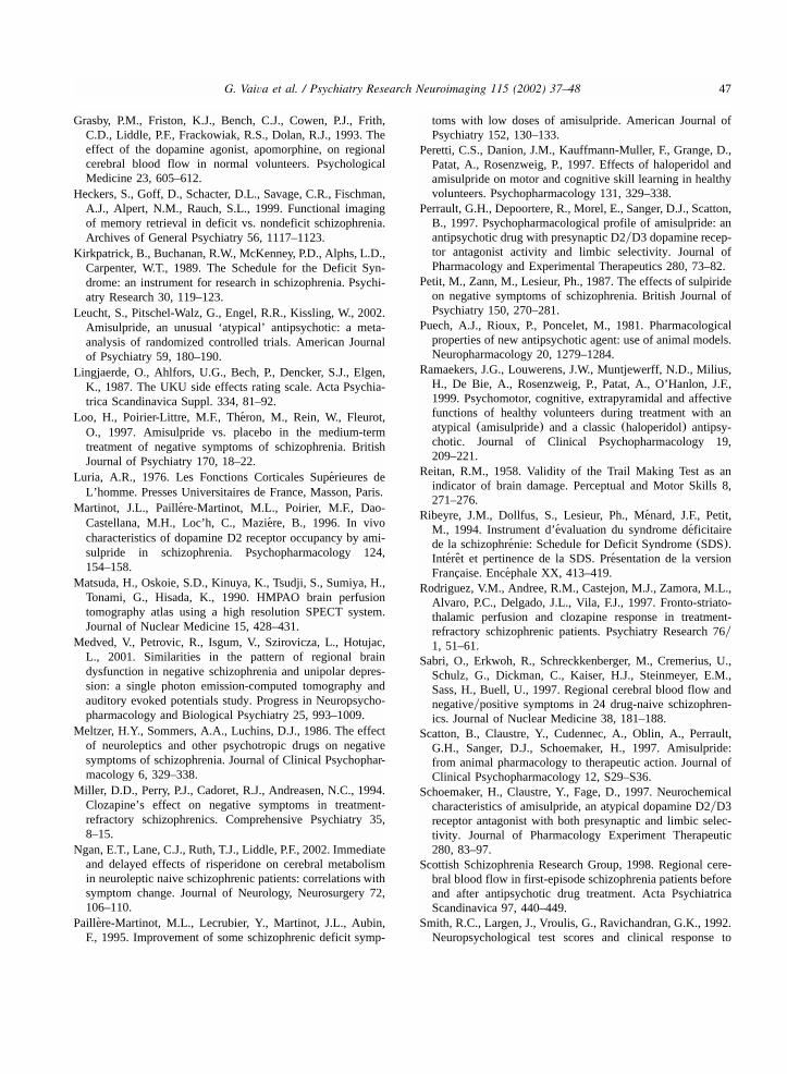
thick slices to be obtained simultaneously, passingby the plane parallel to the orbito-meatal line. Thescanner was equipped with a high sensitivity col-limator with a mean resolution of 12 mm. Differentregions of interest(ROI, Fig. 1) (right and left)were scanned with theq5 cm OM slice, referringto the atlas of Matsuda et al.(1990): frontalanterior cortex; dorso-lateral frontal cortex; frontalposterior cortex; temporo-parietal cortex; temporo-insular cortex; cross cortex; parieto-occipital cor-tex; occipital cortex; lenticular and thalamicregions.Relative quantification of the results was
obtained by reference to internal standardization(means of average ‘countypixel’ values of the 10right and the 10 left ROI, as an individual referencefor each patient: pseudo ‘whole brain’ principle)(Medved et al., 2001). Ten indices(right and left)were obtained in this way(see Fig. 1), as well as10 asymmetry indexes(left index-right index).
2.5. Statistical analysis
Statistical analysis was performed using SASfor PC software. As the patient numbered fewerthan 30, non-parametric tests were used. For com-parisons before and after treatment(blood flowvalues, clinical assessment scores and cognitiveresults), we used the Wilcoxon test for matchedpairs. Comparison of the means(clinical assess-ment scores, blood flow values, etc.) between thesubgroups(SDS criteria, for example) was madeusing the Mann-Whitney or Kruskal-Wallis tests.When there were multiple comparisons, a Bonfer-roni correction was performed. A study of thelinks between the numerical parameters was per-formed using Spearman’s non-parametric correla-tion coefficient.
3. Results
Over a period of 18 months, 35 schizophrenicpatients with the deficit syndrome were hospital-ized consecutively on our unit; nine patients hadan overall SANS score below 65, and an overallSAPS score above 30, or were already receivingantipsychotic treatment; four patients refused totake part in the study; and three others left theunit before the second assessment. Our final sam-ple included 19 patients(16 men and three women;88.5% of the patients seen); 10 presented with adisorganized form of schizophrenia according toDSM III R, 9 with a residual form. The mean agewas 31.6 years and they were all exclusively right-handed. Ten patients were drug-naıve; the other¨nine had been without treatment for a mean timeof 4.6 months(minimum 6 weeks, maximum 3years) before inclusion in the study.Out of the 19 patients, the total SANS score
was significantly reduced(ranky95, P-0.0001)after 28 days of treatment. All the subscoresreduced significantly, with a marked improvementin the Attention subscore(68%). In addition, therewere few side effects and a good overall toleranceto amisulpride. Three patients had side effectsdescribed as ‘mild’ by the UKU(one endocrinol-ogical disorder; the other two mild psychomotorexcitation, but no extrapyramidal symptoms).

41G. Vaiva et al. / Psychiatry Research Neuroimaging 115 (2002) 37–48
Table 1Clinical and cognitive data in the different subgroups, before and after amisulpride
Global Variations Variations in Psample in primary secondary(Ns19) deficits deficits
(Ns12) (Ns7)
Age 31 years 34 27 0.03(S.D.s9) (S.D.s9) (S.D.s8) zs2.2,
d.f.s1
Age of onset 26 years 27.4 23.2 0.03(S.D.s6) (S.D.s6) (S.D.s7) zs2.2,
d.f.s1
Duration of illness 6 years 7 4 0.03(S.D.s4) (S.D.s5) (S.D.s2) zs2.2,
d.f.s1Day 0 Day 28 P
SANS global 82.6 49.4 0.001 40% 56% 0.036(S.D.s16) (S.D.s23) zs2.1,
d.f.s1Affective 3.82 1.73 0.0001 36% 58% 0.008withdrawal (S.D.s8) (S.D.s1.4) zs2.6,
d.f.s1Alogia 3.59 1.64 0.0001 38% 53% 0.04
(S.D.s8) (S.D.s1.4) zs2.05,d.f.s1
Avolition 4.27 2.18 0.0001 41% 49% ns(S.D.s8) (S.D.s1.4)
Anhedonia 4.00 2.8 0.0001 45% 48% ns(S.D.s75) (S.D.s9)
Attention 3.5 1.4 0.0001 40% 56% 0.015(S.D.s9) (S.D.s1.2) zs2.4,
d.f.s1
SAPS global 21.4 19.8 ns –(S.D.s8) (S.D.s6.2)
Literal fluency 6 8 0.0001 y2.7 y1.3 0.037(S.D.s3) (S.D.s2) zsy2.1,
d.f.s1
Category fluency 7 9 0.002 y2.9 y1.2 0.012(S.D.s3) (S.D.s1) zsy2.5,
d.f.s1
TMT A (s)a 147 105 0.001 y48 y32 0.97(S.D.s115) (S.D.s63)
TMT B (s)a 216 177 0.012 y28 y42 0.35(S.D.s172) (S.D.s140)
Trail Making Test.a
Overall, the patients showed a significantimprovement in verbal fluency after 4 weeks oftreatment, and the time for the Trail Making Test
parts A and B was considerably reduced(Table1). The vocabulary and category verbal fluencywas significantly worse in the primary deficit

42 G. Vaiva et al. / Psychiatry Research Neuroimaging 115 (2002) 37–48
Fig. 1. SPECT scan(OMq5 cm). Regions of Interest(ROI).
Fig. 2. SPECT scan(OMq5 cm) of a good responder before and after low doses of amisulpride.
patients; however, the improvement in perform-ance after treatment was not different in the twogroups. We did not observe any difference betweenthe primary and secondary deficit patients for theTrail Making Test.
Data from SPECT imaging showed a significantincrease in the perfusion values, after treatmentwith amisulpride, in the following areas:
● left dorso-lateral frontal cortex(rank 73,Ps0.002);
● right dorso-lateral frontal cortex(rank 72,Ps0.0025); and
● right posterior frontal cortex(rank 65, Ps0.007).
The increase in the perfusion values was almostsignificant in the left temporo-insular area(Ps0.06). No significant changes in asymmetry werefound in the perfusion values. Analysis of thecorrelation between the changes in the clinicalscores and the changes in the perfusion valuesproduced the following relationships:
● The improvement in affective withdrawal cor-related with an increase in the left(rsq0.46,Ps0.054) and right posterior frontal values(rsq0.48, Ps0.045), as well as with an

43G. Vaiva et al. / Psychiatry Research Neuroimaging 115 (2002) 37–48
Table 2Mean regional cerebral blood flows in the different subgroups, before and after amisulpride
Global sample(Ns19) rCBF P
Day 0 Day 28 P Variations Variations inin primary secondarydeficits deficits(Ns13) (Ns6)
Left FA 0.90 (0.06) 0.91 (0.06) 0.16 y0.02 0 0.11Left FDL 0.84 (0.05) 0.86 (0.05) 0.002 y0.03 y0.02 0.38Left FP 0.90(0.06) 0.90 (0.05) 0.98 y0.01 q0.01 0.33Left TI 0.92 (0.04) 0.93 (0.06) 0.51 y0.03 y0.01 0.26Left TP 0.92(0.05) 0.93 (0.05) 0.06 y0.01 0 0.16Left TPO 0.88(0.06) 0.87 (0.04) 0.62 q0.02 q0.02 0.67Left PO 0.87(0.07) 0.86 (0.05) 0.66 q0.01 q0.03 0.70Left O 1.01(0.05) 1.02 (0.04) 0.07 y0.02 y0.01 0.67Left Lent 0.95(0.06) 0.97 (0.05) 0.18 y0.03 y0.02 0.77Left Thal 0.91(0.07) 0.93 (0.04) 0.24 y0.04 y0.01 0.92Right FA 0.91(0.07) 0.92 (0.06) 0.11 y0.02 0 0.50Right FDL 0.84 (0.05) 0.86 (0.05) 0.0025 y0.04 y0.03 0.38Right FP 0.88 (0.04) 0.89 (0.05) 0.007 y0.01 0 0.87Right TI 0.94(0.05) 0.95 (0.05) 0.19 0 0 0.26Right TP 0.94(0.06) 0.95 (0.05) 0.47 y0.01 y0.01 0.72Right TPO 0.88(0.06) 0.88 (0.04) 0.84 q0.01 q0.02 0.63Right PO 0.87(0.07) 0.86 (0.05) 0.93 q0.01 q0.02 0.95Right O 1.02(0.06) 1.04 (0.05) 0.12 y0.02 y0.01 0.48Right Lent 0.95(0.05) 0.96 (0.04) 0.27 y0.01 y0.01 0.77Right Thal 0.91(0.05) 0.93 (0.04) 0.21 y0.02 y0.01 0.80
Regions of interest: FA, frontal anterior; FDL, frontal dorso-lateral; FP, frontal posterior; TI, temporo-insular; TP, temporo-parietal;TPO, temporo-parieto-occipital; PO, parieto-occipital; O, Occipital; Lent, lenticular; and Thal, thalamus.
increase in the values for the temporo-parietal-occipital junctions(rsq0.5,Ps0.034)
● The improvement in anhedonia correlated witha reduction in the antero-internal frontal values(rsy0.51,Ps0.003).
At baseline, the patients presenting with a pri-mary deficit according to the SDS criteria had asignificantly higher total SANS score(Ps0.036),and ‘Affective Withdrawal’ (Ps0.008), ‘Alogia’(Ps0.4) and ‘Attention’ subscores(Ps0.015),which were also higher. The clinical improvementwith treatment was greater in the patients who hada secondary deficit(means56%) compared withthe patients with a primary deficit(means40%;Ps0.036). No difference was found for cerebralblood flow or perfusion asymmetry before or aftertreatment, between the primary and secondarydeficit patients(Table 2). The correlations betweenthe changes in the clinical scores and the changesin the perfusion values, found overall for all the
patients, were persistent in the primary and sec-ondary deficit patients, apart from anhedonia, forwhich a correlation with the frontal values wasonly found for the primary deficit patients(rsy0.56, Ps0.002). No difference between primaryand secondary deficit patients was found for thecorrelations between the changes in cognitive per-formance and the changes in the perfusion values.
4. Discussion
Given the psychopathological and behavioralselection criteria, our group of patients was veryhomogenous with respect to clinical features, witha dominance of psychomotor retardation, loss ofinitiative, social withdrawal, affective blunting andanhedonia. In our study, the improvement in thedeficit aspects of the illness was already verymarked after 28 days of treatment. All of thepatients were improved, which confirmed the use-

44 G. Vaiva et al. / Psychiatry Research Neuroimaging 115 (2002) 37–48
fulness of low-dose amisulpride in the deficitforms of the illness(Boyer et al., 1995; Paillere-`Martinot et al., 1995; Loo et al., 1997; Danion etal., 1999; Leucht et al., 2002). In addition, thetreatment induced only a few side effects and wasoverall well tolerated. However, in the absence ofa controlled comparison with placebo or anotherdrug treatment, the observed therapeutic effectcannot unequivocally be attributed to amisulpridein particular, since the drug treatment was associ-ated with standardized nursing management, aimedat social stimulation of the patients which also canreduce their negative symptomatology of patients.In our study, all the dimensions of negative
symptomatology assessed by the SANS wereimproved after 28 days of treatment. Our resultsnot only show improvement in the core negativesymptoms, i.e. loss of initiative, affective bluntingand social withdrawal, but more interestingly alsoa substantial improvement of cognitive function(mean improvement of 68% in the attentiondimension). Several authors have attributed manyof the cognitive disturbances found in schizophre-nia to a deficit in the attentional mechanisms(Braff, 1993). Different hypotheses have been putforward; schizophrenic patients have difficulty infocusing their attention and in selectively inhibit-ing distracting stimuli. Our study suggests thatlow-dose amisulpride may be particularly usefulfor attention phenomena in the deficit forms ofschizophrenia.This improvement of cognitive function was
confirmed by changes in verbal fluency perform-ance, after 28 days of treatment. These tests areclassically sensitive to changes in attention and tocertain frontal dyfunctions. Performance on theTrail Making Test also improved with treatment.Part A of the test is fairly non-specific and requiresless cognitive effort than part B. Some authorshave reported a wide heterogeneity in the resultsfor part B in schizophrenics, mostly correlatingwith the degree of neurological dysfunction seenin certain patients(Goldstein, 1990). These find-ings seem to link reduced part B performance withstructural brain damage, which is relatively stableover time, and fairly insensitive to correction withbiological treatment. Smith et al.(1992) evenshowed that schizophrenics who performed poorly
on the Trail Making Test, responded badly to highdoses of neuroleptics, and even became worse.Our results confirm these earlier findings, byshowing an improvement in the performance onthe Trail Making Test with low doses of amisul-pride (see Fig. 2).In our study, in which each patient was his own
control, the most significant changes in cerebralblood flow after 28 days of treatment, were foundin the frontal area, in particular in the dorso-lateralfrontal area. Several PET neuroimaging studiesimplicating dorso-lateral prefrontal cortex and infe-rior parietal cortex dysfunctions in the productionof deficit symptoms have been reported(Tammin-ga et al., 1992; Grasby et al., 1993; Heckers et al.,1999). Recently, Ngan et al.(2002) reported astudy in which eight previously unmedicated schiz-ophrenic patients received PET scans before andafter 6 weeks of risperidone treatment; they foundsignificant desactivations in frontal corticalregions. The literature provide few SPECT neu-roimaging results. Rodriguez et al.(1997) studied39 patients, who presented with resistant schizo-phrenia, before and after starting treatment withclozapine. They noted that the patients, who werenon-responders to clozapine, had lower dorso-lateral frontal perfusion with treatment, whichtherefore demonstrates the importance of this brainarea in symptomatic improvement. Yildiz et al.(2000) studied 15 schizophrenics of all types, butdid not find any changes in perfusion before andafter treatment with classical neuroleptics, at highdoses, for 1 month. A study of the ScottishSchizophrenia Research Group(1998) in 27patients who presented with their first episode ofschizophrenia did not find any modification in thehypofrontal blood flow pattern after 6 months oftreatment. Our results are therefore in favor of atargeted action of low-dose amisulpride on theincrease in the rCBF of the dorso-lateral frontalcortex.At a dose of 100 mgyday, amisulpride blocks
the high affinity presynaptic dopaminergic recep-tors preferentially(Sokoloff et al., 1987; Perraultet al., 1997); the pharmacological effect is there-fore probably an increase of dopaminergic trans-mission. This could be of particular interest in thefrontal area, as there are no postsynaptic D2—but

45G. Vaiva et al. / Psychiatry Research Neuroimaging 115 (2002) 37–48
only D1—receptors in this area: thus the effect ofamisulpride would be exclusively presynaptic, i.e.increasing dopaminergic transmission. In a studyin normal volunteers, Grasby et al.(1993) reportedresults with apomorphine, a non-selective dopa-mine agonist, used as a pharmacological probe forinvestigating central dopaminergic neurotransmis-sion. In this study, repeated measurements ofregional cerebral blood flow(rCBF) were madebefore and after the administration of apomorphine(5 or 10 mgykg) or placebo. Apomorphineincreased prefrontal rCBF(right)left).In our patients, we would suppose that it is
restimulation of the mesocortical dopaminergicbundle that underpins the clinical improvement.This dopaminergic bundle mainly goes into thedorso-lateral frontal cortex, and our hypothesiscould be compared with the changes in cerebralblood flow observed in this area. A study using aPET scan in the rat showed an increase in theregional glucose consumption after low doses ofamisulpride; this increase preferentially affectedthe cortical area, especially the limbic region(Scatton et al., 1997). This change in glucoseconsumption with amisulpride was not found withhaloperidol. The authors also compared restimula-tion of the mesocortical dopaminergic bundle withthe ‘disinhibitory’ effects of amisulpiride.We found no striatal change with low doses of
amisulpride in contrast to PET or SPECT findingsof basal ganglia increases with classical antipsy-chotics, such as haloperidol. Several authors thinkthat amisulpride preferentially interacts with dopa-mine D2 and D3 receptors located in limbicstructures rather than the striatum(Curran andPerry, 2001); data from animal studies confirmedthis hypothesis(Schoemaker et al., 1997). Martin-ot et al. (1996) have shown that low doses ofamisulpride (50–100 mgyday) induced a meanoccupancy of the striatal dopamine D2 receptorsof 14%; an occupancy rate of 85% is associatedwith an increased incidence of extrapyramidalsymptoms (Farde et al., 1995). Xiberas et al.(2001) indicated that low amisulpride plasma con-centrations(28–92mgyl) induced marked extras-triatal binding (limbic and temporal cortex) andlow striatal binding to D2 receptors. In our study,
none of the patients presented with extrapyramidalsymptoms.Comparison of the blood flow values or the
changes of these values with clinical responsereminded us of one of our previous observations(Vaiva et al., 1994): it seems to be illusory tocorrelate cerebral blood flow values with an overallassessment score of symptomatology(the overallSANS score in our study), and a more sympto-matic than syndromic approach would be moreinteresting. The improvement of the factor of‘affective withdrawal’ seemed to correlate with theincrease in frontal values in our patients. A fixedfacial expression, a reduction in spontaneousmovements, poverty of gesticulatory expression,and physical anergia are the main symptoms ofthis sort of clinical picture. An improvement inaffective withdrawal is clinically synonymous witha ‘disinhibiting effect’. The disinhibiting effect oftreatment with low-dose amisulpride may be linkedwith an increase in frontal cerebral metabolism.In our study, a distinction between the types of
deficit, according to the SDS criteria, producedtwo subgroups of very different sizes. The fewdifferences found between the groups may havebeen biased by this size effect. No differencebefore and after treatment was found between theprimary and secondary deficit patients. Despite ourfindings of differences between these two formsof schizophrenia at baseline(Vaiva et al., 2002),the impact of low-dose amisulpride treatmenttherefore seems to be purely symptomatic, havinga dimensional effect on the clinical deficiencyaspects, but non-specific for any particular aspectfrom one or the other category. Like others, wehad already observed this type of result in func-tional cerebral imaging studies(Vaiva et al., 1994;Yildiz et al., 2000).The improvement of the SANS scores was
greater in patients presenting with a secondarydeficit. This result confirms the studies by Carpen-ter et al., who considered a primary deficit as aneurodevelopmental condition, linked to severalmorphological alterations in the central nervoussystem, and therefore more or less sensitive topharmacological treatment(Carpenter et al., 1988).We also note that our patients with a primarydeficit were older, and that their illness had been
46 G. Vaiva et al. / Psychiatry Research Neuroimaging 115 (2002) 37–48
progressing for longer. These aspects may alsoexplain why the response to treatment was worse.
4.1. Limits of our study
These results must be carefully interpretedbecause of the role of possible co N foundingfactors such as a very small sample(especially inthe secondary deficit group,Ns7), different pre-vious medication status(10 neuroleptic-naive, nineneuroleptic-free) and a mixed sample of male andfemale patients. The low number of patients in ourstudy reflects the difficulty of recruiting schizo-phrenic patients with a deficit syndrome in ahospital setting. This limits the statistical interpre-tation of the results, but on the other hand, therigor of the selection criteria guarantees the symp-tomatic homogeneity of the patients.Secondly, the radioactive marker chosen
(HMPAO) only allows the cerebral metabolism tobe assessed qualitatively or semi-quantitatively(Steinling and Mazingue, 1988). The resultingvalues are by definition relative values. Moreover,the main limiting factors of our technique werethe choice of different brain areas of interest on asingle slice, as well as the absence of an absolutemorphological marker, coupled with the bloodflow examination(CT scan or MRI). Reposition-ing of the head for the second SPECT scan wastherefore made using external morphologicalcriteria.
5. Conclusion
In this uncontrolled study, the efficacy and goodtolerance of low-dose amisulpride in the deficitforms of schizophrenia were found as expectedfrom earlier placebo-controlled studies. Our studyfound significant changes in the cerebral bloodflow, before and after treatment. These changeswere more marked in the frontal area, and partic-ularly in the dorso-lateral frontal area. We alsoobserved correlations between the changes in cer-ebral blood flow, and the improvement in certainsymptomatic SANS scores. A significant improve-ment of cognitive function was found after treat-ment. This improvement did not seem to be linkedto any particular changes in a loco-regional per-
fusion value. Therefore, we prefer the hypothesisthat overall redistribution of cerebral metabolismmay occur during treatment(Fig. 2).Finally, a distinction between primary and sec-
ondary deficit, according to the SDS criteria,showed a higher percentage of clinical improve-ment in the patients with a secondary deficit. Thepsychometric changes were no different in the twogroups. No specificity in cerebral perfusion wasobserved.
References
American Psychiatric Association, 1987. Diagnostic and Sta-tistical Manual of Mental Disorders, 3rd ed., revised, Author,Washington.
Andreasen, N.C., 1983a. The Scale for the Assessment ofNegative Symptoms(SANS). University of Iowa, IowaCity, Iowa.
Andreasen, N.C., 1983b. The Scale for the Assessment ofPositive Symptoms(SAPS). University of Iowa, Iowa City,Iowa.
Boyer, P., Lecrubier, Y., Puech, A.J., Dewailly, J., Aubin, F.,1995. Treatment of negative symptoms in schizophreniawith amisulpride. British Journal of Psychiatry 166, 68–72.
Braff, D.L., 1993. Information processing and attention dys-functions in schizophrenia. Schizophrenia Bulletin 19y2,233–259.
Buchsbaum, M.S., Hazlett, E.A., 1998. Positron emissiontomography studies of abnormal glucose metabolism inschizophrenia. Schizophrenia Bulletin 24, 343–364.
Carpenter, W.T., Heinrich, D.W., Wagman, A.M.I., 1988. Def-icit and nondeficit forms of schizophrenia: the concept.American Journal of Psychiatry 145, 578–583.
Crow, T.J., 1980. Molecular pathology of schizophrenia: morethan one disease process? British Medical Journal 280,66–68.
Curran, M.P., Perry, C.M., 2001. Amisulpride. A review of itsuse in the management of schizophrenia. Drugs 61,2123–2150.
Danion, J.M., Rein, W., Fleurot, O., Amisulpride study group,1999. Improvement of schizophrenic patients with primarynegative symptoms treated with amisulpride. American Jour-nal of Psychiatry 156, 610–616.
Erkwoh, R., Sabri, O., Willmes, K., Steinmeyer, E.M., Buell,U., Sass, H., 1999. Active and remitted schizophrenia:psychopathological and regional cerebral blood flow find-ings. Psychiatry Research: Neuroimaging 90, 17–30.
Farde, L., Nordstrom, A.L., Karlsson, P., 1995. Positron emis-¨sion tomography studies on dopamine receptors in schizo-phrenia. Clinical Neuropharmacology 18, 121–129.
Goldstein, G., 1990. Neuropsychological heterogeneity inschizophrenia: a consideration of abstraction and problem-solving abilities. Archives of Clinical Neuropsychology 5y3, 251–264.
47G. Vaiva et al. / Psychiatry Research Neuroimaging 115 (2002) 37–48
Grasby, P.M., Friston, K.J., Bench, C.J., Cowen, P.J., Frith,C.D., Liddle, P.F., Frackowiak, R.S., Dolan, R.J., 1993. Theeffect of the dopamine agonist, apomorphine, on regionalcerebral blood flow in normal volunteers. PsychologicalMedicine 23, 605–612.
Heckers, S., Goff, D., Schacter, D.L., Savage, C.R., Fischman,A.J., Alpert, N.M., Rauch, S.L., 1999. Functional imagingof memory retrieval in deficit vs. nondeficit schizophrenia.Archives of General Psychiatry 56, 1117–1123.
Kirkpatrick, B., Buchanan, R.W., McKenney, P.D., Alphs, L.D.,Carpenter, W.T., 1989. The Schedule for the Deficit Syn-drome: an instrument for research in schizophrenia. Psychi-atry Research 30, 119–123.
Leucht, S., Pitschel-Walz, G., Engel, R.R., Kissling, W., 2002.Amisulpride, an unusual ‘atypical’ antipsychotic: a meta-analysis of randomized controlled trials. American Journalof Psychiatry 59, 180–190.
Lingjaerde, O., Ahlfors, U.G., Bech, P., Dencker, S.J., Elgen,K., 1987. The UKU side effects rating scale. Acta Psychia-trica Scandinavica Suppl. 334, 81–92.
Loo, H., Poirier-Littre, M.F., Theron, M., Rein, W., Fleurot,´O., 1997. Amisulpride vs. placebo in the medium-termtreatment of negative symptoms of schizophrenia. BritishJournal of Psychiatry 170, 18–22.
Luria, A.R., 1976. Les Fonctions Corticales Superieures de´L’homme. Presses Universitaires de France, Masson, Paris.
Martinot, J.L., Paillere-Martinot, M.L., Poirier, M.F., Dao-´Castellana, M.H., Loc’h, C., Maziere, B., 1996. In vivo´characteristics of dopamine D2 receptor occupancy by ami-sulpride in schizophrenia. Psychopharmacology 124,154–158.
Matsuda, H., Oskoie, S.D., Kinuya, K., Tsudji, S., Sumiya, H.,Tonami, G., Hisada, K., 1990. HMPAO brain perfusiontomography atlas using a high resolution SPECT system.Journal of Nuclear Medicine 15, 428–431.
Medved, V., Petrovic, R., Isgum, V., Szirovicza, L., Hotujac,L., 2001. Similarities in the pattern of regional braindysfunction in negative schizophrenia and unipolar depres-sion: a single photon emission-computed tomography andauditory evoked potentials study. Progress in Neuropsycho-pharmacology and Biological Psychiatry 25, 993–1009.
Meltzer, H.Y., Sommers, A.A., Luchins, D.J., 1986. The effectof neuroleptics and other psychotropic drugs on negativesymptoms of schizophrenia. Journal of Clinical Psychophar-macology 6, 329–338.
Miller, D.D., Perry, P.J., Cadoret, R.J., Andreasen, N.C., 1994.Clozapine’s effect on negative symptoms in treatment-refractory schizophrenics. Comprehensive Psychiatry 35,8–15.
Ngan, E.T., Lane, C.J., Ruth, T.J., Liddle, P.F., 2002. Immediateand delayed effects of risperidone on cerebral metabolismin neuroleptic naive schizophrenic patients: correlations withsymptom change. Journal of Neurology, Neurosurgery 72,106–110.
Paillere-Martinot, M.L., Lecrubier, Y., Martinot, J.L., Aubin,`F., 1995. Improvement of some schizophrenic deficit symp-
toms with low doses of amisulpride. American Journal ofPsychiatry 152, 130–133.
Peretti, C.S., Danion, J.M., Kauffmann-Muller, F., Grange, D.,Patat, A., Rosenzweig, P., 1997. Effects of haloperidol andamisulpride on motor and cognitive skill learning in healthyvolunteers. Psychopharmacology 131, 329–338.
Perrault, G.H., Depoortere, R., Morel, E., Sanger, D.J., Scatton,B., 1997. Psychopharmacological profile of amisulpride: anantipsychotic drug with presynaptic D2yD3 dopamine recep-tor antagonist activity and limbic selectivity. Journal ofPharmacology and Experimental Therapeutics 280, 73–82.
Petit, M., Zann, M., Lesieur, Ph., 1987. The effects of sulpirideon negative symptoms of schizophrenia. British Journal ofPsychiatry 150, 270–281.
Puech, A.J., Rioux, P., Poncelet, M., 1981. Pharmacologicalproperties of new antipsychotic agent: use of animal models.Neuropharmacology 20, 1279–1284.
Ramaekers, J.G., Louwerens, J.W., Muntjewerff, N.D., Milius,H., De Bie, A., Rosenzweig, P., Patat, A., O’Hanlon, J.F.,1999. Psychomotor, cognitive, extrapyramidal and affectivefunctions of healthy volunteers during treatment with anatypical (amisulpride) and a classic(haloperidol) antipsy-chotic. Journal of Clinical Psychopharmacology 19,209–221.
Reitan, R.M., 1958. Validity of the Trail Making Test as anindicator of brain damage. Perceptual and Motor Skills 8,271–276.
Ribeyre, J.M., Dollfus, S., Lesieur, Ph., Menard, J.F., Petit,´M., 1994. Instrument d’evaluation du syndrome deficitaire´ ´de la schizophrenie: Schedule for Deficit Syndrome(SDS).´Interet et pertinence de la SDS. Presentation de la version´ ˆ ´Francaise. Encephale XX, 413–419.´¸
Rodriguez, V.M., Andree, R.M., Castejon, M.J., Zamora, M.L.,Alvaro, P.C., Delgado, J.L., Vila, F.J., 1997. Fronto-striato-thalamic perfusion and clozapine response in treatment-refractory schizophrenic patients. Psychiatry Research 76y1, 51–61.
Sabri, O., Erkwoh, R., Schreckkenberger, M., Cremerius, U.,Schulz, G., Dickman, C., Kaiser, H.J., Steinmeyer, E.M.,Sass, H., Buell, U., 1997. Regional cerebral blood flow andnegativeypositive symptoms in 24 drug-naive schizophren-ics. Journal of Nuclear Medicine 38, 181–188.
Scatton, B., Claustre, Y., Cudennec, A., Oblin, A., Perrault,G.H., Sanger, D.J., Schoemaker, H., 1997. Amisulpride:from animal pharmacology to therapeutic action. Journal ofClinical Psychopharmacology 12, S29–S36.
Schoemaker, H., Claustre, Y., Fage, D., 1997. Neurochemicalcharacteristics of amisulpride, an atypical dopamine D2yD3receptor antagonist with both presynaptic and limbic selec-tivity. Journal of Pharmacology Experiment Therapeutic280, 83–97.
Scottish Schizophrenia Research Group, 1998. Regional cere-bral blood flow in first-episode schizophrenia patients beforeand after antipsychotic drug treatment. Acta PsychiatricaScandinavica 97, 440–449.
Smith, R.C., Largen, J., Vroulis, G., Ravichandran, G.K., 1992.Neuropsychological test scores and clinical response to
48 G. Vaiva et al. / Psychiatry Research Neuroimaging 115 (2002) 37–48
neuroleptic drugs in schizophrenic patients. ComprehensivePsychiatry 33y2, 139–145.
Sokoloff, P., Martres, M.L., Redouane, K., Schwartz, J.C.,1987. Behavioural and neurochemical profiles of discrimi-nant benzamide derivatives. Psychiatrie et Psychobiologie2, 253–264.
Steinling, M., Mazingue, A., 1988. Le HMPAO comme indi-cateur du DSC local: etude comparee avec la methode par´ ´ ´inhalation de xenon 133. Annales de Radiologie 31y4,´229–238.
Tamminga, C.A., Thaker, G.K., Buchanan, R., Kirkpatrick, B.,Alphs, L.D., Chase, T.N., Carpenter, W.T., 1992. Limbicsystem abnormalities identified in schizophrenia using PETwith fluorodeoxyglucose and neocortical alterations withdeficit syndrome. Archives of General Psychiatry 49,522–530.
Trichard, C., Paillere-Martinot, M.L., Attar-Levy, D., Recas-´sens, C., Monnet, F., Martinot, J.L., 1998. Binding ofantipsychotic drugs to cortical 5-HT2A receptors: a PETstudy of chlorpromazine, clozapine, and amisulpride inschizophrenic patients. American Journal of Psychiatry 155,505–508.
Vaiva, G., Thomas, P., Desbonnet, P., Dutoit, D.,Bianchi Decaix, I., Goudemand, M., Steinling, M., 1994.Debits sanguins cerebraux avant et apres traitement par´ ´ ´ `amisulpride a faibles doses dans les formes deficitaires de` ´schizophrenie. Circulation et Metabolisme du Cerveau 11,´ ´21–32.
Vaiva, G., Cottencin, O., Llorca, P.M., Devos, P., Dupont, S.,Mazas, O., Rascle, C., Thomas, P., Steinling, M., Goude-mand, M., 2002. Regional cerebral blood flow in deficitynondeficit types of schizophrenia according to SDS criteria.Progress in Neuropsychopharmacology and Biological Psy-chiatry 26, 481–485.
Yildiz, A., Eryilmaz, M., Gungor, F., Erkilic, M., Karayalcin,B., 2000. Regional cerebral blood flow in schizophreniabefore and after neuroleptic medication. Nuclear MedicineCommunications 21y12, 1113–1118.
Xiberas, X., Martinot, J.L., Mallet, L., Artiges, E., Canal, M.,Loc’h, C., Maziere, B., Paillere-Martinot, M.L., 2001. In´ ´vivo extrastriatal and striatal D2 dopamine receptor blockadeby amisulpride in schizophrenia. Journal of Clinical Psycho-pharmacology 21, 207–214.
Copyright © 2022 FDOKUMEN