Specificity in lipases: A computational study of transesterification of sucrose
12
Specificity in lipases: A computational study of transesterification of sucrose GLORIA FUENTES, 1,2,3 ANTHONIO BALLESTEROS, 1 AND CHANDRA S. VERMA 2,4 1 Departamento de Biocatálisis, Instituto de Catálisis, CSIC, Cantoblanco, 28049 Madrid, Spain 2 Structural Biology Laboratory, Department of Chemistry, University of York, York YO10 5DD, United Kingdom (RECEIVED March 16, 2004; FINAL REVISION August 20, 2004; ACCEPTED August 26, 2004) Abstract Computational conformational searches of putative transition states of the reaction of sucrose with vinyl laurate catalyzed by lipases from Candida antarctica B and Thermomyces lanuginosus have been carried out. The dielectric of the media have been varied to understand the role of protein plasticity in modulating the observed regioselective transesterification. The binding pocket of lipase from Candida adapts to the conformational variability of the various substates of the substrates by small, local adjustments within the binding pocket. In contrast, the more constrained pocket of the lipase from Thermomyces adapts by adjusting through concerted global motions between subdomains. This leads to the identification of one large pocket in Candida that accommodates both the sucrose and the lauroyl moieties of the transition state, whereas in Thermomyces the binding pocket is smaller, leading to the localization of the two moieties in two distinct pockets; this partly rationalizes the broader specificity of the former relative to the latter. Mutations have been suggested to exploit the differences towards changing the observed selectivities. Keywords: lipases; transesterification; sucrose; specificity; computational methods Our fundamental concepts of enzyme mechanisms are largely based on the idea of complementarity between an enzyme and the reaction transition state (Pauling 1948). Through a combination of shape and electronic properties (Goodsell and Olsen 1990; Bacon and Moult 1992; Jones and Thornton 1996), enzymes select the appropriate sub- strates for a given reaction. Transition-state stabilization appears to occur through a complex balance between favor- able (e.g., hydrogen bonding, electrostatic complementarity, burial of hydrophobic surfaces) and unfavorable (e.g., bond distortion, cavity formation, entropic penalties) interactions in the enzyme–substrate complex. Such conclusions have traditionally been based on a static description of protein conformation. With developments in spectroscopy, the role of protein mobility during catalytic or functional events (Faber and Matthews 1990; Gerstein et al. 1994; Frauen- felder and McMahon 1998) started being recognized. These include regulations of ligand diffusion into and out of pro- teins (Johnson et al. 1979; Mulder et al. 2001), coupled motions of flexible loops or “lids” (McCammon and Harvey 1987; Gunasekaran and Nussinov 2004), and rigid-body movements (“breathing motions”) of entire domains or sub- units (Gerstein et al. 1994). A large class of protein motions are now recognized as being of importance to various as- pects of functionalities (Jääskeläinen et al. 1998a; Frauen- felder et al. 2001). The fine details that characterize the spatial and temporal details of such processes only became evident with developments in computer hardware and soft- ware. Computational methods now regularly complement experiments in providing unique and incisive insights into such processes (Kazlauskas 2001; Warshel 2003; Garcia- Viloca et al. 2004). We apply computational methods in this study to inves- tigate the links between the structural plasticity of lipases and their functions. Lipases are among the most versatile enzymes currently in use (Schmid and Verger 1998; Pandey Reprint requests to: C.S. Verma, Bioinformatics Institute, 30 Biopolis Way, #07–01 Matrix, Singapore-138671; e-mail: [email protected]. edu.sg; fax: +0065-6478-9047. Present addresses: 3 Bijvoet Center for Biomolecular Research, NMR Department, Utrecht University, 3584 CH, Utrecht, The Netherlands; 4 Bio- informatics Institute, Singapore 138671. Article and publication are at http://www.proteinscience.org/cgi/doi/ 10.1110/ps.04724504. Protein Science (2004), 13:3092–3103. Published by Cold Spring Harbor Laboratory Press. Copyright © 2004 The Protein Society 3092
Transcript of Specificity in lipases: A computational study of transesterification of sucrose

Specificity in lipases: A computational studyof transesterification of sucrose
GLORIA FUENTES,1,2,3 ANTHONIO BALLESTEROS,1 AND CHANDRA S. VERMA2,4
1Departamento de Biocatálisis, Instituto de Catálisis, CSIC, Cantoblanco, 28049 Madrid, Spain2Structural Biology Laboratory, Department of Chemistry, University of York, York YO10 5DD, United Kingdom
(RECEIVED March 16, 2004; FINAL REVISION August 20, 2004; ACCEPTED August 26, 2004)
Abstract
Computational conformational searches of putative transition states of the reaction of sucrose with vinyllaurate catalyzed by lipases from Candida antarctica B and Thermomyces lanuginosus have been carriedout. The dielectric of the media have been varied to understand the role of protein plasticity in modulatingthe observed regioselective transesterification. The binding pocket of lipase from Candida adapts to theconformational variability of the various substates of the substrates by small, local adjustments within thebinding pocket. In contrast, the more constrained pocket of the lipase from Thermomyces adapts by adjustingthrough concerted global motions between subdomains. This leads to the identification of one large pocketin Candida that accommodates both the sucrose and the lauroyl moieties of the transition state, whereas inThermomyces the binding pocket is smaller, leading to the localization of the two moieties in two distinctpockets; this partly rationalizes the broader specificity of the former relative to the latter. Mutations havebeen suggested to exploit the differences towards changing the observed selectivities.
Keywords: lipases; transesterification; sucrose; specificity; computational methods
Our fundamental concepts of enzyme mechanisms arelargely based on the idea of complementarity between anenzyme and the reaction transition state (Pauling 1948).Through a combination of shape and electronic properties(Goodsell and Olsen 1990; Bacon and Moult 1992; Jonesand Thornton 1996), enzymes select the appropriate sub-strates for a given reaction. Transition-state stabilizationappears to occur through a complex balance between favor-able (e.g., hydrogen bonding, electrostatic complementarity,burial of hydrophobic surfaces) and unfavorable (e.g., bonddistortion, cavity formation, entropic penalties) interactionsin the enzyme–substrate complex. Such conclusions havetraditionally been based on a static description of proteinconformation. With developments in spectroscopy, the role
of protein mobility during catalytic or functional events(Faber and Matthews 1990; Gerstein et al. 1994; Frauen-felder and McMahon 1998) started being recognized. Theseinclude regulations of ligand diffusion into and out of pro-teins (Johnson et al. 1979; Mulder et al. 2001), coupledmotions of flexible loops or “lids” (McCammon and Harvey1987; Gunasekaran and Nussinov 2004), and rigid-bodymovements (“breathing motions”) of entire domains or sub-units (Gerstein et al. 1994). A large class of protein motionsare now recognized as being of importance to various as-pects of functionalities (Jääskeläinen et al. 1998a; Frauen-felder et al. 2001). The fine details that characterize thespatial and temporal details of such processes only becameevident with developments in computer hardware and soft-ware. Computational methods now regularly complementexperiments in providing unique and incisive insights intosuch processes (Kazlauskas 2001; Warshel 2003; Garcia-Viloca et al. 2004).
We apply computational methods in this study to inves-tigate the links between the structural plasticity of lipasesand their functions. Lipases are among the most versatileenzymes currently in use (Schmid and Verger 1998; Pandey
Reprint requests to: C.S. Verma, Bioinformatics Institute, 30 BiopolisWay, #07–01 Matrix, Singapore-138671; e-mail: [email protected]; fax: +0065-6478-9047.
Present addresses: 3Bijvoet Center for Biomolecular Research, NMRDepartment, Utrecht University, 3584 CH, Utrecht, The Netherlands; 4Bio-informatics Institute, Singapore 138671.
Article and publication are at http://www.proteinscience.org/cgi/doi/10.1110/ps.04724504.
Protein Science (2004), 13:3092–3103. Published by Cold Spring Harbor Laboratory Press. Copyright © 2004 The Protein Society3092

et al. 1999; Pleiss et al. 2000) (http://www.led.uni-stuttgart.de). One of their main uses is in synthesis, such as in theregioselective monoacylation of sucrose. This reaction iscarried out using a lipase-catalyzed process in a mediumconstituted by a mixture of tert-amyl alcohol (a non-toxicand slightly polar solvent, where most enzymes remain ac-tive) and a polar solvent, dimethyl sulfoxide (Ferrer et al.1999), and leads to different positional isomers. One suchlipase, that from Thermomyces lanuginosus (TlL), displaysa high regioselectivity for the 6-hydroxyl position in theacylation of sucrose (Ferrer et al. 1999); in contrast, lipaseB from Candida antarctica (CALB) leads to a mixture oftwo monoesters (the 6-hydroxyl and the 6�-hydroxyl posi-tion; Woudenberg et al. 1996). Experiments comparing thetransesterification reaction of sucrose with vinyl laurate forboth enzymes under identical conditions have been re-ported. It was found that (under conditions involving a mix-ture of solvents of 2-methyl 2-butanol:DMSO (4:1 v/v)) TlLwas the most efficient enzyme for this reaction—with asucrose to monolaurate conversion of 51%. On the otherhand, CALB gave rise to a sucrose conversion of 45%,which consisted of the two major monoesters (see above) inapproximately an equimolar ratio (Ferrer et al. 1999); thespeed and ratios of product formation depended on theamount of cosolvent DMSO. So what is the origin of thesedifferences? Although we cannot yet examine the influenceof the cosolvent directly (some progress has recently beenmade in this difficult area; see Elcock 2003), we can ap-proach the problem using simple computational and struc-tural models. The main advantage that computational tech-niques have over experimental ones is that structural com-putational models can be built to explore how only one or afew of the several conformations/reaction-coordinates pos-sible are observed experimentally. Structurally, both en-zymes (Fig. 1A,B) are characterized by similar overallfolds, the same catalytic triad residues, and the oxyanionhole (and the same underlying catalytic mechanism); how-ever, there are several significant differences in the size andshape of the binding pockets (Pleiss et al. 1998) that un-doubtedly will shape the reaction coordinate and hence thespecificity. For example, TlL has a helical lid region cov-ering the active site and undergoes large-scale conforma-tional rearrangements upon ligand binding (Brzozowski etal. 2000); in the case of CALB, no such conformationalrearrangement of a potential lid has been found. It is at thislevel that the microscopic interactions between various con-formational substates of the ligand/enzyme will be the dis-criminating factors between the allowed and the disallowedstates. Computational structural modeling is most powerfulat revealing this. Indeed, we have previously successfullyreproduced the above-mentioned experimental observationsof differing specificities towards the sucrose moiety and itsacylation with vinyl laureate in organic mixtures. This wascarried out by examining computationally the conforma-
tional space available to the reacting species in the activesite that were subsequently analyzed with simple models ofenthalpy and entropy (Fuentes et al. 2002; G. Fuentes, A.Ballesteros, and C. Verma, in prep.). Here, we extend ourstudy to examine the processes in different media (repre-senting different dielectric properties); our aim is to probethe extent and nature of motions and the resulting structuralplasticity in each enzyme and model the extent of dynamiccontrol exerted by the enzyme on the reaction coordinate.
Results
For each isomer, the minimum energy structures corre-sponding to the conformational searches performed underthe three dielectric media for each enzyme were examinedfor conformational variability. We examined (1) thechanges in interatomic distributions as measured throughdifference distance matrices, (2) correlations between thedistributions in space of the atomic positions, and (3) thechanges in the geometry (as computed through the areas andvolumes) of the pockets that define the ligand binding re-gions.
Difference distance matrices
Large changes in the interatomic separations between thedifferent structures, which are indicative of structural plas-
Figure 1. Secondary structures for CALB (A) and TlL (B). The catalytictriad and ligands are shown explicitly.
Transesterification of sucrose by lipases
www.proteinscience.org 3093
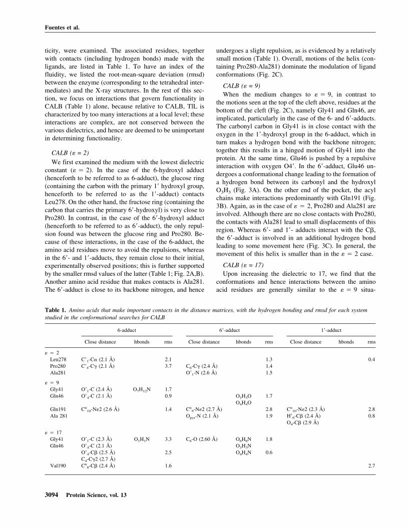
ticity, were examined. The associated residues, togetherwith contacts (including hydrogen bonds) made with theligands, are listed in Table 1. To have an index of thefluidity, we listed the root-mean-square deviation (rmsd)between the enzyme (corresponding to the tetrahedral inter-mediates) and the X-ray structures. In the rest of this sec-tion, we focus on interactions that govern functionality inCALB (Table 1) alone, because relative to CALB, TlL ischaracterized by too many interactions at a local level; theseinteractions are complex, are not conserved between thevarious dielectrics, and hence are deemed to be unimportantin determining functionality.
CALB (� = 2)
We first examined the medium with the lowest dielectricconstant (� � 2). In the case of the 6-hydroxyl adduct(henceforth to be referred to as 6-adduct), the glucose ring(containing the carbon with the primary 1� hydroxyl group,henceforth to be referred to as the 1�-adduct) contactsLeu278. On the other hand, the fructose ring (containing thecarbon that carries the primary 6�-hydroxyl) is very close toPro280. In contrast, in the case of the 6�-hydroxyl adduct(henceforth to be referred to as 6�-adduct), the only repul-sion found was between the glucose ring and Pro280. Be-cause of these interactions, in the case of the 6-adduct, theamino acid residues move to avoid the repulsions, whereasin the 6�- and 1�-adducts, they remain close to their initial,experimentally observed positions; this is further supportedby the smaller rmsd values of the latter (Table 1; Fig. 2A,B).Another amino acid residue that makes contacts is Ala281.The 6�-adduct is close to its backbone nitrogen, and hence
undergoes a slight repulsion, as is evidenced by a relativelysmall motion (Table 1). Overall, motions of the helix (con-taining Pro280-Ala281) dominate the modulation of ligandconformations (Fig. 2C).
CALB (� = 9)When the medium changes to � � 9, in contrast to
the motions seen at the top of the cleft above, residues at thebottom of the cleft (Fig. 2C), namely Gly41 and Gln46, areimplicated, particularly in the case of the 6- and 6�-adducts.The carbonyl carbon in Gly41 is in close contact with theoxygen in the 1�-hydroxyl group in the 6-adduct, which inturn makes a hydrogen bond with the backbone nitrogen;together this results in a hinged motion of Gly41 into theprotein. At the same time, Glu46 is pushed by a repulsiveinteraction with oxygen O4�. In the 6�-adduct, Glu46 un-dergoes a conformational change leading to the formation ofa hydrogen bond between its carbonyl and the hydroxylO3H3 (Fig. 3A). On the other end of the pocket, the acylchains make interactions predominantly with Gln191 (Fig.3B). Again, as in the case of � � 2, Pro280 and Ala281 areinvolved. Although there are no close contacts with Pro280,the contacts with Ala281 lead to small displacements of thisregion. Whereas 6�- and 1�- adducts interact with the C�,the 6�-adduct is involved in an additional hydrogen bondleading to some movement here (Fig. 3C). In general, themovement of this helix is smaller than in the � � 2 case.
CALB (� = 17)Upon increasing the dielectric to 17, we find that the
conformations and hence interactions between the aminoacid residues are generally similar to the � � 9 situa-
Table 1. Amino acids that make important contacts in the distance matrices, with the hydrogen bonding and rmsd for each systemstudied in the conformational searches for CALB
6-adduct 6�-adduct 1�-adduct
Close distance hbonds rms Close distance hbonds rms Close distance hbonds rms
� � 2Leu278 C�1-C� (2.1 Å) 2.1 1.3 0.4Pro280 C�4-C� (2.1 Å) 3.7 C6-C� (2.4 Å) 1.4Ala281 O�1-N (2.6 Å) 1.5
� � 9Gly41 O�1-C (2.4 Å) O7H12N 1.7Gln46 O�4-C (2.1 Å) 0.9 O3H3O 1.7
O4H4OGln191 Cn
10-N�2 (2.6 Å) 1.4 Cn9-N�2 (2.7 Å) 2.8 Cn
10-N�2 (2.3 Å) 2.8Ala 281 Opyr-N (2.1 Å) 1.9 H�6-C� (2.4 Å) 0.8
O4-C� (2.9 Å)
� � 17Gly41 O�1-C (2.3 Å) O1H1N 3.3 C6-O (2.60 Å) O6H6N 1.8Gln46 O�4-C (2.1 Å) O3H3N
O�4-C� (2.5 Å) 2.5 O4H4N 0.6C4-C�2 (2.7 Å)
Val190 Cn8-C� (2.4 Å) 1.6 2.7
Fuentes et al.
3094 Protein Science, vol. 13

tion. Gly41 is involved in a new interaction with the 6�-adduct. The carbon C6 of sucrose is close to the carbonyloxygen of Gly41, leading to larger local rearrangementsthan those observed in the case of the other dielectric con-stants. The interactions of Glu46 are conserved across thethree dielectrics (Fig. 4A). The increase in dielectric and theconcomitant change in local plasticity brings the acyl chaincarbons into close interactions with Val190 and leads torepulsions in the 6- and 1�-adducts (Fig. 4B); this situationis unique to this higher dielectric. Again, in the case ofPro280 and Ala281, the observed interactions and structuralrearrangements are quite similar to those seen in the lowerdielectrics (Fig. 4C).
Correlated motions
To obtain a global picture of the rearrangements that ac-company the conformational plasticity of the complexes, we
examined, for each enzyme separately, the average dis-placements (equivalent to fluctuations) of all of the struc-tures taken together relative to their geometrical average.From these displacements, we computed maps of the cor-relations between the directions of motions of the C� atoms(Fig. 5A,B). These maps reveal concerted motions of groupsof atoms and additionally reveal whether two groups ofatoms move away or toward each other (negatively corre-lated) or move in the same direction as each other (posi-tively correlated). We were interested in those motions thatare involved in facilitating ligand diffusion into and out ofproteins and additionally are responsible for enabling func-tions (Dvorsky et al. 2002). These motions oppose eachother in direction (the regions of negative correlations) andtypically span binding sites, that is, refer to conformationalcoupling between the residues that line the sides of bindingclefts (Miller and Agard 1999; Dvorsky et al. 2002).
Figure 2. Interactions found for the different intermediates of CALB (the6-adduct is blue; the 6�-adduct is pink; the 1�-adduct is green) for � � 2;along with the crystal structure of CALB (PDB entry 1lbs) shown inball-and-stick for reference. (A) Interactions of Leu278, Ala279, andPro280 with 6- and 6�-adducts. (B) Interactions of Leu278 and Ala279 with6- and 1�-adducts. (C) A surface representation of CALB with the ligandbound to highlight the relative locations of some key residues and the cleftwhere the active site is located.
Figure 3. Interactions found for the different intermediates of CALB (the6-adduct is shown in blue; the 6�-adduct in pink; the 1�-adduct in green) for� � 9; along with the crystal structure of CALB (PDB entry 1lbs) shownin ball-and-stick for reference. (A) Interactions of Gly41 and Gln46 with 6-and 6�-adducts; (B) interactions of Gln191 with the three adducts; (C)interactions of Pro280 and Ala281 with the three adducts.
Transesterification of sucrose by lipases
www.proteinscience.org 3095

CALB
In the case of CALB (Fig. 5A), the correlations weresmall and widespread across the enzyme. There is someevidence of movements of domains, such as between thestretches 110–140 and 160–200 (to the left of the ligands inFig. 1A) and also between these regions and the 300–318stretch (to the top left of Fig. 1A). All of these regions areon the same side of the ligand binding cleft and do not seemto directly couple the residues that form the regions in-volved in the postulated opening/closing motions across theligand binding region. Stronger correlations are observedmore locally and are largely between the areas that are closeto the acyl pocket. This arises from the fact that in contrastto the constrained alcohol pocket, the acyl pocket is gener-ally more spacious in lipases (Otto et al. 2000) and hencedeforms easily, enabling it to accommodate a highly flex-ible part of the ligand.
TlL
For T1L, the correlation plot (Fig. 5B) shows stretches ofregions (domains) that undergo correlated motions with re-
spect to each other. A strong anticorrelation characterizesthe motion of the lid/hinge (�5 helix) region against regionsof �5, a loop that delimits one of the sides of the active sitecrevice, �1, �2, �6, �7, �9, �11 and the loop connectingthem, the carboxyl terminus (the catalytic histidine is lo-cated in this region, in a loop just before �11). Many resi-dues in this domain belong to the acyl pocket. The �1-�2region delimits the putative sucrose-binding pocket and ad-ditionally contains Trp21, which is involved in the putativesucrose-binding pocket. The catalytic serine (Ser146) pro-trudes from a short loop at the carboxy-terminal end of �6helix (the so-called nucleophile elbow). As can be seen fromFigure 5C, these regions, although far apart in sequence,make up the binding pocket walls. Again, we see one stretchof contiguous correlation between the 200–250 region andseveral other regions (Fig. 5D). In particular, the motionagainst the 160–180 region reflects the motions of the resi-dues of the acyl pocket relative to those of the alcoholpocket. Furthermore, residues in �6 move relative to re-gions near the loop where the catalytic serine is locatedwhile the region of the catalytic serine (146) moves relativeto the turn between strand �8 and helix �9 (the latter con-tains the catalytic Asp201). This latter motion is probablycoupled to accommodation of the ligand and its orientationsuch that the local stereochemical requirements for catalysisare satisfied.
Geometry of the lipase pockets
The plasticity of lipase pockets was further examined bycalculating the distributions in their geometries (the areasand volumes) with the program CASTp (Table 2; the li-gands were excluded from the calculations). The dual en-tries in Table 2 refer to situations where the pocket appearssubdivided into two mainly as a result of the nature of thealgorithms used. There appears to be no correlations be-tween charge screening (dielectric) and the area/volumesspanned by the pockets. The pocket in CALB is never sub-divided, in contrast to T1L, which has a more complexstructural organization. Although this results partly from thelimitations of the algorithm used in this study, it is also aconsequence of the fact that the pocket in CALB is large butis smaller and more complex in T1L. Also, the TlL-ligandinteractions are characterized by longer range correlationsand the influence of secondary structural elements (see sec-tion above). The existence of just one pocket in CALB leadsto larger areas and volumes of its binding pockets relative tothose in TlL; the areas and volumes can be up to twice aslarge. TlL also seems to be partitioned into multiple pocketsdepending on the nature of the isomer; however, the 1�-adduct is associated with only one pocket, because the li-gand is oriented almost perpendicular to the surface of theenzyme, pointing into the binding cavity and hence definingonly one binding pocket (Fig. 6). In contrast, the 6- and
Figure 4. Interactions found for the different intermediates of CALB (the6-adduct is in blue; the 6�-adduct is in pink; the 1�-adduct in green) for� � 17; along with the crystal structure of CALB (PDB entry 1lbs) inball-and-stick for reference. (A) Interactions of Gly41 and Gln46 with 6-and 6�-adducts; (B) interactions of Val190 with the 6- and 1�-adducts; (C)interactions of Pro280 and Ala281 with 6- and 1�-adducts.
Fuentes et al.
3096 Protein Science, vol. 13

6�-adducts are “draped” along the surface of the enzymewith the lauroyl moieties occupying the same pocket in all3 cases (irrespective of the dielectric) and the two sugarrings occupying different troughs on the surface; this leadsto the formation of multiple pockets. In the case of the6-adduct, the ligand is modulated between Arg81 andArg84 and hence, as the screening of charges intensifies, thelocal surface is more easily perturbed, leading to two pock-ets (see Table 2). In the 6�-adduct, the interactions thatmodulate the conformation of the sugars are those of Trp89and dipoles of several backbones and polar side chains (andmarginally Arg81). Hence, independent of charge screen-ing, the local surface is not much affected, leading to asingle large pocket.
Discussion
We know that specificities of enzymes originate in theirplastic adaptations to external conditions by combinations
Table 2. Areas and volumes calculated for the binding pocketsof TIL and CALB
TIL CALB
Area (Å2) Volume (Å3) Area (Å2) Volume (Å3)
� � 26OH 768 991 794 10016�OH 198 229 864 1044
513 6711�OH 473 642 816 1027
� � 96OH 208 315 704 855
395 5026�OH 733 900 685 8551�OH 479 618 713 791
� � 176OH 258 344 718 877
320 4586�OH 852 1007 811 11191�OH 841 1019 627 858
Figure 5. Contour plots of the correlations between distributions of the C� positions. Positive values are shown above the diagonal and negative valuesare shown below the diagonal. The contours correspond to correlations of −0.6, −0.3, 0.3, and 0.6 in CALB (A) and TlL (B); the dashed lines show thelocation of the lid region. (C) C�-trace of TlL in blue ribbon with ligands shown in green spheres; the lid region in red is anti-correlated in motions withthe regions in yellow and the black arrows depicts this motion. (D) C�-trace of TlL in blue ribbon with ligands shown in green spheres; the 200–250subdomain region in red is anti-correlated in motion with the regions in yellow, and the black arrow depicts this motion.
Transesterification of sucrose by lipases
www.proteinscience.org 3097

of local or/and global structural and dynamic responses (see,e.g., Luo et al. 1998; Miller and Agard 1999; Baladin andOnuchic 2000; Radkiewicz and Brooks 2000; Orencia et al.2001; Young et al. 2001; Meroueh et al. 2002; Süel et al.2003). However, the underlying structural and emergentfunctional complexities have precluded the formulation ofany general rules towards an understanding of this plastic-ity. To further probe the determinants of this plasticity, wehave examined, using computational molecular models,how the lipases from two different organisms respond to aligand under varying environmental conditions. The reac-tion performed is the transesterification of sucrose, whichusually is selective for particular sites on the sugar depend-ing on the enzyme and conditions employed. Molecularmodeling is increasingly being recognized as a powerfultool to provide models for the origins of such behavior; itsuniqueness over other techniques such as nuclear magneticresonance, Fourier Transform Infrared spectroscopy, fluo-
rescence resonance energy transfer, lies in simultaneouslyproviding detailed atomic-level information in space andtime (Kazlauskas 2001; Warshel 2003; Garcia-Viloca et al.2004). Although we can only model abstractions of reality,the methods available can provide good qualitative startingpoints for experimental explorations. Indeed, we (Fuentes et al.2002; G. Fuentes, A. Ballesteros, and C. Verma, in prep.) andothers (Norin et al. 1994; Martinelle et al. 1995; Haeffner et al.1998; Kazlauskas 2001; Ottosson et al. 2002; Peters and By-water 2002) have modeled lipase–ligand interactions with en-couraging agreement with known experimental observations.
In this study, we have used rigorous conformational sam-pling (involving energy minimizations of several conform-ers yielding the enthalpic component of the free energyfollowed by vibrational entropic computations), becausetraditional molecular dynamics and energy minimizationsonly sample limited amounts of the phase space. In contrast,our method allows a much larger sampling of the phase
Figure 6. Front view of the binding pockets of TlL with the different ligands (in magenta) docked inside. The protein surface is colored according to theelectrostatic potential with blue depicting positively charged and red depicting negatively charged regions. The view is from the top looking down into thebinding site so the intervening residues have been clipped away to reveal the C� trace of the helical lid region (pink). The substrate in the putative transitionstates is shown with the lauroyl part in green and the sugar in brown. From left to right are structures for � � 2, � � 9, and � � 17. (A) 1-adduct; (B)6-adduct; (C) 6�-adduct.
Fuentes et al.
3098 Protein Science, vol. 13

space by crossing over barriers that would not be sampledotherwise. We examined the extent to which local and glob-al structural plasticity of the lipases modulates their func-tionalities. Our studies employed crystallographic structures(resolved from aqueous solutions) as the starting points forbuilding the models. This is based on the assumption thatthe structures of the enzymes in anhydrous organic solventswere essentially identical to the three-dimensional struc-tures of enzymes in water (Fitzpatrick et al. 1993, 1994;Brzozowski et al. 2000). Furthermore, this assumption hasworked well in modeling the structure–dynamics–functionsof lipases (see, e.g., Kazlauskas 2001; Fuentes et al. 2002;Ottoson et al. 2002).
The overall fold and the catalytic machinery of the twoenzymes are very similar, but TlL has a well-defined flex-ible loop covering the active site, which is linked to itsfunctionality in that it undergoes an activation in the pres-ence of substrates (Brzozowski et al. 2000); no such activelid has been reported for CALB. This is one characteristicfeature of many lipases, and although some studies suggestthat the time scales of opening and closing this lid seem tobe different from the time scales of substrate access (Zan-donella et al. 1995), they are not conclusive (Martinelle etal. 1995). What is certain is that the amino acid sequence ofthe lid affects activity (Brocca et al. 2003). We decided tocarry out the modeling studies with the lid in an openposition, based on the observation that this methodology hasbeen shown to be successful at reproducing experimentalobservations (Norin et al. 1994; Fuentes et al. 2002; Ottosonet al. 2002; Peters and Bywater 2002).
The reaction mechanism catalyzed by lipases is believedto be analogous to that of serine proteases. It has beenproposed that the tetrahedral intermediate closely resemblesthe transition state and therefore is a good model for mim-icking it (Warshel et al. 1989; Ottoson et al. 2002; Petersand Bywater 2002). During the transesterification reaction,the key intermediates seem to be the deacylation interme-diates (Norin et al. 1994), involving both the sucrose and theacyl chains together. Thus, we decided to use this interme-diate (to represent the putative transition state) in our work.Of course, our sampling methodology seeks out the lowestenergy intermediates, because these mimic the lowest en-ergy putative transition states; the lowest energy transitionstates will be the experimentally preferred conformationsas they will map on to the reaction coordinates with thehighest rates. Having identified these intermediates, we in-vestigated the concomitant conformational plasticity andthe accompanying structural responses of the enzymes byexamining the changes in interatomic distances, the corre-lations between atomic positional distributions, and thegeometric plasticity (areas and volumes) of the binding re-gions.
From the analyses of the distance matrices it is seen thatthe binding of the ligands is accompanied by a variety of
structural responses in the enzymes’ structures. This servesto illustrate how ligand binding can induce a conformationaladaptation, according to the induced-fit theory (Koshland1958). When the different ligands have diffused within theactive site into their minimum energy conformations, theaccompanying conformational changes are similar to thatseen in other complexes (Done et al. 1998). It appears thatresidues within the active sites of either enzyme can adopttwo distinct and energetically favorable conformations toaccommodate the ligands. This is not surprising as it isknown that the binding sites in lipases can roughly be par-titioned into two regions—a hydrophobic region and a hy-drophilic region (Norin et al. 1993). Naturally, the lauroylmoiety “explores” the former, whereas the more hydrophilicsucrose moiety “floats” in the latter.
In the case of CALB, the binding pocket is large and ourstudies suggest that it is pre-formed to facilitate binding inthe unliganded state. The binding region is made up pre-dominantly by hydrophobic (oily) residues. Thus, only mi-nor motions such as small-scale flexing/adaptations of thewalls of the binding pocket occur to accommodate the li-gands. The binding site is “walled” by a long helix (Glu270-Gly290) whose motions seem important (particularlyAla280–Pro281), irrespective of the dielectric screening.This feature seems to originate in the predominance of hy-drophobic residues. Although the local dynamics are quitecomplex, structural examinations of the low-energy struc-tures suggest that a mutation of Ala281 to Gly would per-turb the selectivity of the 6�-adduct (the preferred isomersthat are observed are the 6- and 6�-adducts); it is not yetclear whether it would enhance the selectivity or decrease it.A mutation of Ala283 to Ser is likely to enhance the sta-bility of the 1�-adduct by the creation of an additional hy-drogen bond between the side chain of Ser and site O11.This is analogous to the observations in other systems suchas the dynamics of the proteases seen by Miller and Agard(1999). Mutations of the two Pro residues capping the helix(Pro269, Pro290) to Gly ought to make the region moreflexible and alter the distributions of products. In the case ofTlL, the binding pocket is less spacious (Pleiss et al. 2000)and hence the fluidity that characterizes the localized dy-namics of the binding pocket region in CALB is, in general,not observed. The helical lid that is characteristic of TlL,however, occludes the hydrophobic binding pocket fromhigh dielectric media such as water, and unsurprisingly,undergoes some degree of conformational change to allowthe ligands to mould into the catalytic machinery (Brzo-zowski et al. 2000). This suggests that despite the contrast-ing time scales of lid dynamics and substrate access (Zan-donella et al. 1995), the lid motions control the dynamics ofthe substrate. In the case of TlL (where selectivity is ob-served only for the 6-adduct), structural examinations sug-gest that the selectivity of 1�-adduct might be enthalpicallyenhanced by mutating Leu259 to Asn, which would stabi-
Transesterification of sucrose by lipases
www.proteinscience.org 3099

lize the O9 and/or O10 sites on the ligand through newhydrogen bonds. Similarly, the 6�-adduct can be stabilizedby mutating Gly82 to Ser, which would stabilize O2 and/orO3 through new hydrogen bonds. The differing spaces in thebinding pockets of the two enzymes would also provide forvariations in entropic stabilizations (see, e.g., Ottoson et al.2002); however, this is beyond the scope of this paper.
The above rearrangements are again seen in analyses ofcorrelations in atomic movements. Such plots are very use-ful as they reveal how dynamical conformational couplingbetween regions far apart in structure seemingly affect thebinding sites (Sneddon and Brooks 1991; Gerstein et al.1994; Verma et al. 1997; Radkiewicz and Brooks 2000;Meroueh et al. 2002). CALB is characterized mainly bylocal couplings, that is, between residues that are close spa-tially. In stark contrast, we find delineation of the TlL struc-ture into several subdomains/substructures (Figs. 1, 5),which suggests a more global control of ligand binding. Theinterplay between these flexible subdomains provides struc-tural evidence of the expected dynamic behavior of the sur-face loops and regions surrounding the catalytic center.Such correlated motions have been postulated, in accor-dance with experimental observations, to facilitate substratebinding/egress and catalytic modulation (Weston et al.1992; Rice and Steitz 1994; Newman et al. 1995; Rad-kiewicz and Brooks 2000; Dvorsky et al. 2002; Gu-nasekaran and Nussinov 2004). The fact that one side of thebinding pocket maps the lid helix, whereas the other sidesare made up by regions of the protein that are anti-correlatedin motion to the helix, intuitively lends weight to the hy-pothesis that this kind of anti-correlation corresponds to theexpansion/contraction of the binding pocket mouth to fa-cilitate ligand binding/egress (Jääskeläinen et al. 1998b) andcatalysis (Dvorsky et al. 2002). Despite the success of suchanalyses in providing intuitively appealing models, therehave been relatively few reports of the effects of mutationson interdomain motions and the associated functionality(van Aalten et al. 1998; Miller and Agard 1999). Examiningthe structures of TlL, we suggest that the hinge-like regionthat has Gly109 and Gly177 with C� atoms separated by adistance of 3.7 Å would be a good site for mutations to Ala.Either single or double mutations would enhance the hinge-like property of this region and effect the concerted motionsof the regions that encompass the ligand binding site andaffect selectivity. Another possible mutation could beThr143Asn, which would potentially introduce a hydrogenbond between the side chain of Asn and the backbone ofPhe13, thus “gluing” the gap between the strand and thehelix (containing Phe13). This new hinge region is pre-dicted to affect concerted motions (region 4 in Fig. 5B) andaffect reactivity.
The different kinds of functional motions naturally leadto an adapting pocket volume. In CALB, these motions arelargely confined to the binding site and only the amino acids
surrounding and creating the pocket display reorientationson the binding of the putative transition state analogs. Asexpected the lauroyl moiety is stabilized by hydrophobic(and partly neutral) amino acids whereas the sucrose is sta-bilized by polar interactions; indeed the interactions that aremade by the atoms along the lauroyl moiety are quite con-servative in that it is known that the chain length can beneither too long (as it would require penetration and desta-bilization into the protein) nor too short (Ferrer et al. 2000);perhaps some kind of anchoring effect stabilizes the transi-tion state. On the other hand, in TlL, larger secondary struc-ture motions are also involved in the conformational adap-tations needed, together leading to delineation of the ligandbinding site into multiple pockets. The complexity of theinteractions that govern the binding “landscape” can be seenin Figure 6.
Together, the models suggest that optimal binding of theputative transition state requires small-scale local rearrange-ments of amino acid side chains in a preformed binding sitein CALB; in contrast in T1L, the amino acids participatingin binding and catalysis seem to undergo substantial reori-entation together with secondary structural adjustments.The availability of the larger pocket coupled to a largelylocal adaptation (and hence defined by a shallow free en-ergy surface) in CALB partly suggests why it catalyzes theformation of two isomers in contrast with T1L, which has amore constrained pocket and requires the mobilization ofdomains in its functionality (and hence a free energy surfacethat probably has multiple minima of varying depths) and soleads to the formation of just one isomer. In a low dielectricmedium, hydrogen bonding with solvent is expected to besparse, so the protein organizes itself into ‘macro-structures’to compensate for the loss of stability. As the dielectricnature of the surroundings increases, hydrogen bondingwith solvent becomes more possible and favorable and thesesubdomains are mitigated. This, on the one hand, leads toloss of global coupling as mediated by the subdomains butis compensated, on the other hand, by enhanced local plas-ticity. The two enzymes display a wide spectrum of struc-tural responses to ligand binding at the local or/and theglobal levels. This manifests itself in the range of selectivi-ties that these enzymes display in their catalytic behaviors.The complexity of the systems makes the formulation ofgeneral rules hard; however, some insights can be gained bycarrying out computational studies. This leads us to hypoth-esize that plasticity at the local level modulates functionalitythrough a largely enthalpic control (variations of electro-static and van der Waals interactions between the substrateand the surrounding amino acids), whereas at a more globallevel, entropic factors become more important (low-frequencycollective motions that are characteristic of domain motionslead to increased entropies and lowering of the free energies)(Fisher et al. 2001; Fuentes et al. 2002). We are investigatingthese effects in greater detail for these two lipases.
Fuentes et al.
3100 Protein Science, vol. 13

Materials and methods
All of the computations were performed on Silicon Graphics work-stations. The manipulations of molecules, the graphic evaluations,the energy minimizations, and the conformational analyses wereperformed using the molecular modeling program-packageQUANTA (Molecular Simulations Inc.) and the programCHARMm (Brooks et al. 1983).
General procedures and starting structures
For our studies, the coordinates for CALB were taken from a 2.6Å resolution X-ray structure cocrystallized with the inhibitor N-hexylphosphonate ethyl ester (PDB entry 1lbs; Uppenberg et al.1995), and for TlL, we used the structure with the active-site lid inan open conformation (PDB entry 1dt5; Brzozowski et al. 2000).The CHARMm force field was used to model the protein andwater molecules were modeled using the TIP3 potential (Jor-gensen et al. 1983). A neutral pH was assumed, leading to netcharges of +1 on Arg/Lys side chains and −1 on Asp/Glu sidechains, respectively. In addition, the protonated forms of His224and His258 were used. Optimization of hydrogen bonding poten-tials (which involved finding the rotamer that made the maximumnumber of hydrogen bonds) around the side chains of Asn, Gln,and His was done using CHARMm, and the lowest energy struc-tures were selected.
The lauroyl and sucrose were constructed using the moleculareditor in QUANTA with partial charges assigned using QUAN-TA’s charge template method. To account for electronic rearrange-ments in the intermediates, semi-empirical quantum mechanicalcalculations were performed and the associated changes in thepoint charges were used to scale the QUANTA-derived charges.This was carried out so that the charges derived for the interme-diates were normalized relative to the normal CHARMm-param-
eterized charges for standard amino acids. Coordinates and stan-dard geometries for missing protein atoms were constructed usingstandard geometries from CHARMm. Nonbonded interactionswere truncated at 14 Å with shift and smoothing functions oper-ating between 10 and 14 Å for the electrostatic and van der Waalsinteractions respectively; constant dielectrics of 2, 9, and 17 wereused to represent the different reaction conditions (Ferrer et al.1999). These values correspond to a conventional organic medium,a medium containing 2-methyl-2-butanol/DMSO 95:5, and a me-dium with a composition of 2-methyl-2-butanol/DMSO of 80:20,respectively; this has been the approximation chosen for the ex-perimental media used in the synthetic reactions. An initial energyminimization was carried out to remove internal strain using acombination of Steepest Descent (SD) and Adopted Basis NewtonRaphson (ABNR) algorithms.
Models for the reaction tetrahedral intermediates
A model for the lipase–lauroyl–sucrose intermediate was con-structed by removing the H atom of Ser105 and Ser146 O� (forCALB and TlL, respectively) and making a covalent link betweenthe Ser105 (or Ser146) O�, the tetrahedral C (Ct) of the lauroylmoiety and the corresponding primary hydroxyl oxygen of thesucrose (O6, O1�, or O6�) (Fig. 7). The resulting tetrahedral carbonCt and the oxygen atoms were assigned sp3 hybridization. Thisprotocol was followed for modeling all of the regioisomers in bothenzymes.
In the case of CALB, the initial location of the side chain of theester group was guided by the conformation of the crystallographicinhibitor. The position of the central part of the substrates was welldefined because of the requirements of satisfying the hydrogenbonding in the catalytic triad and the formation of the oxyanionhole. For TlL, the protein was structurally aligned with CALBusing the residues in the catalytic triad as tethers, and the proce-dure outlined above was followed.
Figure 7. Structure and nomenclature of the atoms used to model the tetrahedral intermediates and the torsional angles used for the conformational search.(Left) The structure of the lauroyl moiety. The active site serine oxygen of the lipase is the oxygen (not bolded) linked to the atom C1 shown in the lauroyl.The oxygen atom in boldface type and with a * is the oxygen at which the esterification reaction involving addition of the sucrose isomers (middle) takesplace; the arrows in the middle point to the 3 different sites of esterification (leading to the three different products that are discussed). (Right) The varioustorsion angles that were varied in the sucrose moiety (as shown in the middle of the figure) during the conformational searches.
Transesterification of sucrose by lipases
www.proteinscience.org 3101

Systematic conformational search
There are two methods that can be used to explore the conforma-tional plasticity of large molecules such as proteins: static, grid-based conformational searches for isolating the minima and mo-lecular dynamics simulations. To sample the possible conforma-tions of protein–substrate complexes, the use of moleculardynamics is inappropriate because the time scales required tosample the various conformations will be too long to be simulatedon current computers. On the other hand, it is not possible tosearch the total conformational space of an enzyme–substrate sys-tem of the size of lipases using the static grid-based methodseither. Thus, we chose to perform a local grid-based conforma-tional search. For this, the torsion angles around the five bondsshown in Figure 7 were varied systematically in each complex andthe resulting conformations were energy minimized. These angleswere varied from −60° to 180° with a grid size of 120°.
The resulting 243 conformations were minimized following aprotocol based on decremental constraints that apply to the proteinas follows: Two segments were defined, segment a, formed by theresidue 106 (or 146) belonging to the protein chain A, and segmentb with the n-laurate and sucrose moieties. Initially, a harmonicforce of 20.0 kcal/mol/Å was applied to the whole system exclud-ing the two segments, the hydrogen atoms, and water molecules.Segment a was constrained with a force constant of 5.0 kcal/mol/Å, and segment b with 1.0 kcal/mol/Å. The system was subjectedto 200 steps of Steepest Descent and 800 steps of Adopted BasicNewton Raphson minimization. Subsequent cycles of minimiza-tion were carried out, gradually decreasing the harmonic forceimposed on the system; finally, the unconstrained system wassubject to 200 steps of SD and 5000 steps of ABNR minimization,until the energy gradient was less or equal to 0.01 kcal/mol/Å. Thismethod has the advantage of sampling several conformations ofthe substrate and hence enables identification of complexes ofinterest much more quickly than is possible from molecular dy-namics simulations.
Analysis
The minimum energy structures for each combination of dielectricand acylation site were taken together for further analysis. Thiswas carried out by computing difference distance matrices, corre-lation plots, and pocket plasticities. For the difference distancematrices, the inter-C� distances in each structure were computedand the change in this index between different minimum energystructures was examined. For the correlated plots (Ichiye andKarplus 1991), the correlation between the distributions of C�positions in all minimum energy structures for each enzyme wasseparately computed. The plasticity of the enzyme pockets wascomputed through their areas and volumes, using the serverCASTp (Liang et al. 1998). This program uses weighted Delaunaytriangulations and is also based on the pocket algorithm of alphashape theory. It provides identification and measurements of sur-face-accessible pockets as well as interior-inaccessible cavities(defining a cavity as the interior empty space that is not accessibleto the solvent probe of radius 1.4 Å), and it measures analyticallythe area and volume of each pocket and cavity.
Acknowledgments
We thank the Spanish Ministry of Science and Technology and theBBSRC, UK, for the support. We thank the referees for very usefulcriticisms. BII is an A-STAR institute.
References
Bacon, D.J. and Moult, J. 1992. Docking by least-squares fitting of molecularsurface patterns. J. Mol. Biol. 225: 849–858.
Baladin, I.A. and Onuchic, J. 2000. Dynamically controlled protein tunnelingpaths in photosynthetic reaction centers. Science 290: 114–117.
Brocca, S., Secundo, F., Ossola, M., Alberghina, L., Carrea, G., and Lotti, M.2003. Sequence of the lid affects activity and specificity of Candida rugosalipase isoenzymes. Protein Sci. 12: 2312–2319.
Brooks, B.R., Bruccoleri, R.E., Olafson, B.D., States, D.J., Swaminathan, S.,and Karplus, M. 1983. CHARMM: A program for macromolecular energyminimization and dynamics calculations. J. Comput. Chem. 4: 187–217.
Brzozowski, A.M., Savage, H., Verma, C.S., Turkenburg, J.P., Lawson, D.M.,Svendsen, A., and Patkar, S. 2000. Structural origins of the interfacialactivation in Thermomyces (Humicola) lanuginosa lipase. Biochemistry 39:15071–15082.
Done, S.H., Branningan, J.A., Moody, P.C.E., and Hubbard, R.E. 1998. Ligand-induced conformational change in penicillin acylase. J. Mol. Biol. 284:463–475.
Dvorsky, R., Hornak, V., Sevcik, J., Tyrrell, G.P., Caves, L.S.D., and Verma,C.S. 2002. Dynamics of Rnase-Sa: A simulation perspective complementaryto NMR/X-ray. J. Phys. Chem. B 106: 6038–6048.
Elcock, A.H. 2003. Atomic-level observation of macromolecular crowding ef-fects: Escape of a protein from the GroEL cage. Proc. Natl. Acad. Sci. 100:2340–2344.
Faber, H.R. and Matthews, B.W. 1990. A mutant T4 lysozyme displays fivedifferent crystal conformations. Nature 348: 263–266.
Ferrer, M., Cruces, M.A., Bernabé, M., Ballesteros, A., and Plou, F.J. 1999.Lipase-catalyzed regioselective acylation of sucrose in two-solvent mix-tures. Biotechnol. Bioeng 65: 10–15.
Ferrer, M., Cruces, M.A., Plou, F.J., Bernabé, M. and Ballesteros, A. 2000. Asimple procedure for the regioselective synthesis of fatty acid esters ofmaltose, leucrose, maltotriose and n-dodecyl maltosides. Tetrahedron 56:4053–4061.
Fischer, S., Smith, J.C., and Verma, C.S. 2001. Dissecting the vibrational en-tropy change on protein/ligand binding: Burial of a water molecule in bo-vine pancreatic trypsin inhibitor. J. Phys. Chem. B 105: 8050–8055.
Fitzpatrick, P.A., Steinmetz, A.C.U., Ringe, D., and Klibanov, A.M. 1993.Enzyme crystal-structure in a neat organic solvent. Proc. Natl. Acad. Sci.90: 8653–8657.
Fitzpatrick, P.A., Ringe, D., and Klibanov, A.M. 1994. X-ray crystal structureof cross-linked subtilisin Carlsberg in water vs. acetonitrile. Biochem. Bio-phys. Res. Comm. 198: 675–681.
Frauenfelder, H. and McMahon, B. 1998. Dynamics and function of proteins:The search for general concepts. Proc. Natl. Acad. Sci. 95: 4795–4797.
Frauenfelder, H., McMahon, B.H., Austin, R.H., Chu, K., and Groves, J.T.2001. The role of structure, energy landscape, dynamics, and allostery in theenzymatic function of myoglobin. Proc. Natl. Acad. Sci. 98: 2370–2374
Fuentes, G., Cruces, M.A., Plou, F.J., Ballesteros, A., and Verma, C.S. 2002.Computational studies of subtilisin-catalyzed transesterification of sucrose:Importance of entropic effects. ChemBioChem 3: 907–910.
Garcia-Viloca, M., Gao, J., Karplus, M., and Truhlar, D.G. 2004. How enzymeswork: Analysis by modern rate theory and computer simulations. Science303: 186–195.
Gerstein, M., Lesk, A., and Chotia, C. 1994. Structural mechanisms for domainmovements in proteins. Biochemistry 33: 6739–6749.
Goodsell, D.S. and Olsen, A.J. 1990. Automated docking to proteins by simu-lated annealing. Proteins 8: 195–202.
Gunasekaran, K. and Nussinov, R. 2004. Modulating functional loop move-ments: The role of highly conserved residues in the correlated loop motions.ChemBioChem 5: 224–230.
Haeffner, F., Norin, T., and Hult, K. 1998. Molecular modeling of the enanti-oselectivity in lipase-catalyzed transesterification reactions. Biophys. J. 74:1251–1262.
Ichiye, T. and Karplus, M. 1991. Collective motions in proteins: A covarianceanalysis of atomic fluctuations in molecular dynamics and normal modesimulations. Proteins 11: 205–217.
Jääskeläinen, S., Verma, C.S., Hubbard, R.E., and Caves, L.S.D. 1998a. Iden-tifying key electrostatic interactions in Rhizomucor miehei lipase: The in-fluence of solvent dielectric. Theor. Chem. Acc. 101: 175–179.
Jääskeläinen, S., Verma, C.S., Hubbard, R.E., Linko, P., and Caves, L.S.D.1998b. Probing the conformational change in the activation of lipase. Pro-tein Sci. 7: 1359–1367.
Johnson, L.N., Stura, E.A., Wilson, K.S., Sansom, M.S.P., and Weber, L.T.
Fuentes et al.
3102 Protein Science, vol. 13
1979. Nucleotide binding to clycogen phosphorylase b in the crystal. J. Mol.Biol. 134: 639–653.
Jones, S. and Thornton, J.M. 1996. Principles of protein–protein interactionsderived from structural studies. Proc. Natl. Acad. Sci. 93: 13–20.
Jorgensen, W.L., Chandrasekhar, J., Madura, J.D., Impey, R.W., and Klein,M.L. 1983. Comparison of simple potential functions for simulating liquidwater. J. Chem. Phys. 79: 926–935.
Kazlauskas, R.J. 2001. Modeling—A tool for experimentalists. Science 293:2277–2279.
Koshland, D.E. 1958. Application of a theory of enzyme specificity to proteinsynthesis. Proc. Natl. Acad. Sci. 44: 98–104.
Liang, J., Edelsbrunner, H., and Woodward, C. 1998. Anatomy of protein pock-ets and cavities: Measurements of binding site geometry and implicationsfor ligand design. Protein Sci. 7: 1884–1897.
Luo, X., Kato, R., and Collins, J.R. 1998. Dynamic flexibility of protein-inhibi-tor complexes: A study of the HIV-1 protease/KNI-272 complex. J. Amer.Chem. Soc. 120: 12410–12418.
Martinelle, M., Holmquist, M., and Hult, K. 1995. On the interfacial activationof Candida antarctica lipase A and B as compared with Humicola lanugi-nosa lipase. Biochim. Biophys. Acta 1258: 272–276.
McCammon, J.A. and Harvey, S.C. 1987. Dynamics of proteins and nucleicacids. Cambridge University Press, Cambridge, UK.
Meroueh, S.O., Roblin, P., Golemi, D., Maveyraud, L., Vakulenko, S.B., Zhang,Y., Samama, J.-P., and Mobashery, S. 2002. Molecular dynamics at the rootof expansion of function in the M69L inhibitor-resistant TEM -lactamasefrom Escherichia coli. J. Am. Chem. Soc. 124: 9422–9430.
Miller, D.W. and Agard, D.A. 1999. Enzyme specificity under dynamic control:A normal mode analysis of �-lytic protease. J. Mol. Biol. 286: 267–278.
Mulder, F.A., Mittermaier, A., Hon, B., Dahlquist, F.W., and Kay, L.E. 2001.Studying excited states of proteins by NMR spectroscopy. Nat. Struct. Biol.8: 932–935.
Newman, M., Strzelecka, T., Dorner, L.F., Schildkraut, I., and Aggarwal, A.K.1995. Structure of bam-hi endonuclease bound to DNA—partial folding andunfolding on DNA-binding. Science 269: 656–663.
Norin, M., Haeffner, F., Achour, A., Norin, T., and Hult, K. 1994. Computermodeling of substrate-binding to lipases from Rhizomucor miehei, Humi-cola lanuginosa and Candida rugosa. Protein Sci. 3: 1493–1503.
Orencia, M.C., Yoon, J.S., Ness, J.E., Stemmer, W.P.C., and Stevens, R.C.2001. Predicting the emergence of antibiotic resistance by directed evolu-tion and structural analysis. Nat. Struct. Biol. 8: 238–242.
Otto, R.T., Scheib, H., Bornscheuer, U.T., Pleiss, J., Syldatk, C., and Schmid,R.D. 2000. Substrate specificity of lipase B from Candida antarctica in thesynthesis of arylaliphatic glycolipids. J. Mol. Catal. B: Enzym. 8: 201–211.
Ottosson, J., Fransson, L., and Hult, K. 2002. Substrate entropy in enzymeenantioselectivity: An experimental and molecular modeling study of alipase. Protein Sci. 11: 1462–1471.
Pandey, A., Benjamin, S., Soccol, C.R., Nigam, P., Krieger, N., and Soccol,V.T. 1999. The realm of microbial lipases in biotechnology. Biotechnol.Appl. Biochem. 29: 119–131.
Pauling, L. 1948. Nature of the forces between large molecules of biologicalinterest. Nature 161: 707–709.
Peters, G.H. and Bywater, R.P. 2002. Essential motions in a fungal lipase withbound substrate, covalently attached inhibitor and product. J. Mol. Recognit.15: 393–404.
Pleiss, J., Fischer, M., and Schmid, R.D. 1998. Anatomy of lipase binding sites:The scissile fatty acid binding site. Chem. Phys. Lipids 93: 67–80.
Pleiss, J., Fischer, M., Peiker, M., Thiele, C., and Schmid, R.D. 2000. Lipaseengineering database: Understanding and exploiting sequence–structure–function relationships. J. Mol. Catal. B: Enzym. 10: 491–508.
Radkiewicz, J.L. and Brooks, C.L. 2000. Protein dynamics in enzymatic ca-talysis: Exploration of dihydrofolate reductase. J. Amer. Chem. Soc. 122:225–231.
Rice, P.A. and Steitz, T.A. 1994. Refinement of �-�-resolvase reveals a strik-ingly flexible molecule. Structure 2: 371–384.
Schmid, R.D. and Verger, R. 1998. Lipases: Interfacial enzymes with attractiveapplications. Angew. Chem. Int. Ed. Eng. 37: 1609–1633.
Sneddon, S.F. and Brooks, C.L. 1991. Protein motions: Structural and functionalaspects. In Molecular structures in biology (eds. R. Diamond et al.), pp.113–160. Oxford University Press, Oxford, UK.
Süel, G.M., Lockless, S.W., Wall, M.A., and Ranganathan, R. 2003. Evolution-arily conserved networks of residues mediate allosteric communication inproteins. Nat. Struc. Biol. 10: 59–69.
Uppenberg, J., Ohrner, N., Norin, M., Hult, K., Kleywegt, G.J., Patkar, S.,Waagen, V., Anthonsen, T., and Jones, T.A. 1995. Crystallographic andmolecular-modeling studies of lipase B from Candida antarctica reveal astereospecificity pocket for secondary alcohols. Biochemistry 34: 16838–16851.
van Aalten, D.M.F., Hoff, W.D., Findlay, J.B.C., Crielaard, W., and Helling-werf, K.J. 1998. Concerted motions in the photoactive yellow protein. Prot.Eng. 11: 873–879.
Verma, C.S., Caves, L.S.D., Hubbard, R.E., and Roberts, G.C.K. 1997. Domainmotions in dihydrofolate reductase: A molecular dynamics study. J. Mol.Biol. 226: 776–796.
Warshel, A. 2003. Computer simulations of enzyme catalysis: Methods, prog-ress, and insights. Annu. Rev. Biophys. Biomol. Struct. 32: 425–443.
Warshel, A., Naray-Szabo, G., Sussman, F., and Hwang, J.K. 1989. How doserine proteases really work? Biochemistry 28: 3629–3637.
Weston, S.A., Lahm, A., and Suck, D. 1992. X-ray structure of the DNAseI-D(GGTATACC)(2) complex at 2.3-Å resolution. J. Mol. Biol. 226: 1237–1256.
Woudenberg, M., Van Rantwijk, F., and Sheldon, R.A. 1996. Regioselectiveacylation of disaccharides in tert-butyl alcohol catalyzed by C. antarcticalipase. Biotechnol. Bioeng. 49: 328–333.
Young, M.A., Gonfloni, S., Superti-Furga, G., Roux, B., and Kuriyan, J. 2001.Dynamic coupling between the SH2 and SH3 domains of c-Src and Hckunderlies their inactivation by C-terminal tyrosine phosphorylation. Cell105: 115–126.
Zandonella, G., Haalck, L., Spener, F., Faber, K., Paltauf, F., and Hermetter, A.1995. Inversion of lipase stereospecificity for fluorogenic alkyldiacyl glyc-erols. Effect of substrate solubilization. Eur. J. Biochem. 231: 50–55.
Transesterification of sucrose by lipases
www.proteinscience.org 3103