Specification of Neuronal Identities by Feedforward Combinatorial Coding
14
Specification of Neuronal Identities by Feedforward Combinatorial Coding Magnus Baumgardt, Irene Miguel-Aliaga ¤ , Daniel Karlsson, Helen Ekman, Stefan Thor * Division of Molecular Genetics, Department of Physics, Chemistry and Biology, Linkoping University, Linkoping, Sweden Neuronal specification is often seen as a multistep process: earlier regulators confer broad neuronal identity and are followed by combinatorial codes specifying neuronal properties unique to specific subtypes. However, it is still unclear whether early regulators are re-deployed in subtype-specific combinatorial codes, and whether early patterning events act to restrict the developmental potential of postmitotic cells. Here, we use the differential peptidergic fate of two lineage-related peptidergic neurons in the Drosophila ventral nerve cord to show how, in a feedforward mechanism, earlier determinants become critical players in later combinatorial codes. Amongst the progeny of neuroblast 5–6 are two peptidergic neurons: one expresses FMRFamide and the other one expresses Nplp1 and the dopamine receptor DopR. We show the HLH gene collier functions at three different levels to progressively restrict neuronal identity in the 5–6 lineage. At the final step, collier is the critical combinatorial factor that differentiates two partially overlapping combinatorial codes that define FMRFamide versus Nplp1/DopR identity. Misexpression experiments reveal that both codes can activate neuropeptide gene expression in vast numbers of neurons. Despite their partially overlapping composition, we find that the codes are remarkably specific, with each code activating only the proper neuropeptide gene. These results indicate that a limited number of regulators may constitute a potent combinatorial code that dictates unique neuronal cell fate, and that such codes show a surprising disregard for many global instructive cues. Citation: Baumgardt M, Miguel-Aliaga I, Karlsson D, Ekman H, Thor S (2007) Specification of neuronal identities by feedforward combinatorial coding. PLos Biol 5(2): e37. doi:10.1371/journal.pbio.0050037 Introduction Animals have a daunting number of different cell types, and this cellular diversity is profound in the nervous system. During the last two decades, it has become increasingly apparent that neuronal cell fate is not dictated by the action of any single regulatory gene, but results from the combina- torial action of a number of genes that comprise a regulatory ‘‘code’’ [1–3]. Given the enormous diversity of neuronal cell identities, and the great number of regulatory genes in the genome(s), the ‘‘decoding’’ of neuronal cell fate specification is only just beginning. The appearance of the proper combinatorial code in an early postmitotic neuron is the consequence of a sequence of earlier and increasingly restricted regulatory events [4,5]. How do the earlier regulatory events influence postmitotic cell fate decisions? In one extreme model, early regulators may act simply to ensure proper combinatorial coding in early postmitotic neurons. At another extreme, they may confer a state of competence that to some extent reduces the need for precise and extensive coding of cell fate. If the first model is correct, combinatorial coding should have great informational value and easily traverse many developmental boundaries—once in place, a combinatorial code should be able to act without major restrictions. If the second model is correct, combinatorial codes should have low informational value and be restricted by developmental boundaries— although the proper combinatorial code may be in place, its activity will often be restricted by the history of the cell. Given the extent of neuronal diversity in the mammalian nervous system, and the number of regulatory genes involved, distinguishing between these two models relies on the identification of specific codes in more easily accessible genetic model systems. We are focusing our studies on a specific subset of Drosophila ventral nerve cord (VNC) neurons. Within the VNC, only about 90 out of approximately 10,000 cells express the LIM-homeodomain transcription factor Apter- ous (Ap), but this group of neurons consists of several classes [6] (Figure 1), which are located in three positions in each hemisegment; the single dorsal Ap (dAp) neuron, a ventral pair of (vAp) neurons, and a lateral ‘‘Ap cluster’’ of four neurons found only in each of the six thoracic hemiseg- ments. Most Ap neurons are interneurons that project along a common fascicle. However, in each Ap cluster, the Tv neuron projects its axon out of the VNC at the dorsal midline and innervates the dorsal neurohemal organ (DNH) [7]. Tv neurons uniquely express the FMRFamide (FMRFa) neuropeptide gene [8]. A number of regulatory genes have been found to be important for Tv specification and FMRFa regulation. These include ap itself, the zinc finger gene squeeze (sqz), the bHLH gene dimmed (dimm), as well as the transcriptional co-factors encoded by the eyes absent (eya) and dachshund (dac) genes [7,9–12]. In addition, FMRFa expression is completely dependent upon retrograde BMP signaling, Academic Editor: William A. Harris, Cambridge University, United Kingdom Received August 31, 2006; Accepted December 6, 2006; Published February 6, 2007 Copyright: Ó 2007 Baumgardt et al. This is an open-access article distributed under the terms of the Creative Commons Attribution License, which permits unrestricted use, distribution, and reproduction in any medium, provided the original author and source are credited. Abbreviations: AEL, after egg laying; CNS, central nervous system; dAp, dorsal Ap; mAb, monoclonal antibody; RNAi, RNA-interference; VNC, ventral nerve cord * To whom correspondence should be addressed. E-mail: [email protected] ¤ Current address: Division of Developmental Neuroscience, Medical Research Council National Institute for Medical Research, London, United Kingdom PLoS Biology | www.plosbiology.org February 2007 | Volume 5 | Issue 2 | e37 0295 P L o S BIOLOGY
Transcript of Specification of Neuronal Identities by Feedforward Combinatorial Coding
Specification of Neuronal Identitiesby Feedforward Combinatorial CodingMagnus Baumgardt, Irene Miguel-Aliaga
¤, Daniel Karlsson, Helen Ekman, Stefan Thor
*
Division of Molecular Genetics, Department of Physics, Chemistry and Biology, Linkoping University, Linkoping, Sweden
Neuronal specification is often seen as a multistep process: earlier regulators confer broad neuronal identity and arefollowed by combinatorial codes specifying neuronal properties unique to specific subtypes. However, it is still unclearwhether early regulators are re-deployed in subtype-specific combinatorial codes, and whether early patterning eventsact to restrict the developmental potential of postmitotic cells. Here, we use the differential peptidergic fate of twolineage-related peptidergic neurons in the Drosophila ventral nerve cord to show how, in a feedforward mechanism,earlier determinants become critical players in later combinatorial codes. Amongst the progeny of neuroblast 5–6 aretwo peptidergic neurons: one expresses FMRFamide and the other one expresses Nplp1 and the dopamine receptorDopR. We show the HLH gene collier functions at three different levels to progressively restrict neuronal identity in the5–6 lineage. At the final step, collier is the critical combinatorial factor that differentiates two partially overlappingcombinatorial codes that define FMRFamide versus Nplp1/DopR identity. Misexpression experiments reveal that bothcodes can activate neuropeptide gene expression in vast numbers of neurons. Despite their partially overlappingcomposition, we find that the codes are remarkably specific, with each code activating only the proper neuropeptidegene. These results indicate that a limited number of regulators may constitute a potent combinatorial code thatdictates unique neuronal cell fate, and that such codes show a surprising disregard for many global instructive cues.
Citation: Baumgardt M, Miguel-Aliaga I, Karlsson D, Ekman H, Thor S (2007) Specification of neuronal identities by feedforward combinatorial coding. PLos Biol 5(2): e37.doi:10.1371/journal.pbio.0050037
Introduction
Animals have a daunting number of different cell types,and this cellular diversity is profound in the nervous system.During the last two decades, it has become increasinglyapparent that neuronal cell fate is not dictated by the actionof any single regulatory gene, but results from the combina-torial action of a number of genes that comprise a regulatory‘‘code’’ [1–3]. Given the enormous diversity of neuronal cellidentities, and the great number of regulatory genes in thegenome(s), the ‘‘decoding’’ of neuronal cell fate specificationis only just beginning.
The appearance of the proper combinatorial code in anearly postmitotic neuron is the consequence of a sequence ofearlier and increasingly restricted regulatory events [4,5].How do the earlier regulatory events influence postmitoticcell fate decisions? In one extreme model, early regulatorsmay act simply to ensure proper combinatorial coding inearly postmitotic neurons. At another extreme, they mayconfer a state of competence that to some extent reduces theneed for precise and extensive coding of cell fate. If the firstmodel is correct, combinatorial coding should have greatinformational value and easily traverse many developmentalboundaries—once in place, a combinatorial code should beable to act without major restrictions. If the second model iscorrect, combinatorial codes should have low informationalvalue and be restricted by developmental boundaries—although the proper combinatorial code may be in place,its activity will often be restricted by the history of the cell.Given the extent of neuronal diversity in the mammaliannervous system, and the number of regulatory genesinvolved, distinguishing between these two models relies onthe identification of specific codes in more easily accessiblegenetic model systems.
We are focusing our studies on a specific subset ofDrosophila ventral nerve cord (VNC) neurons. Within theVNC, only about 90 out of approximately 10,000 cellsexpress the LIM-homeodomain transcription factor Apter-ous (Ap), but this group of neurons consists of several classes[6] (Figure 1), which are located in three positions in eachhemisegment; the single dorsal Ap (dAp) neuron, a ventralpair of (vAp) neurons, and a lateral ‘‘Ap cluster’’ of fourneurons found only in each of the six thoracic hemiseg-ments. Most Ap neurons are interneurons that project alonga common fascicle. However, in each Ap cluster, the Tvneuron projects its axon out of the VNC at the dorsalmidline and innervates the dorsal neurohemal organ (DNH)[7]. Tv neurons uniquely express the FMRFamide (FMRFa)neuropeptide gene [8]. A number of regulatory genes havebeen found to be important for Tv specification and FMRFaregulation. These include ap itself, the zinc finger genesqueeze (sqz), the bHLH gene dimmed (dimm), as well as thetranscriptional co-factors encoded by the eyes absent (eya) anddachshund (dac) genes [7,9–12]. In addition, FMRFa expressionis completely dependent upon retrograde BMP signaling,
Academic Editor: William A. Harris, Cambridge University, United Kingdom
Received August 31, 2006; Accepted December 6, 2006; Published February 6,2007
Copyright: � 2007 Baumgardt et al. This is an open-access article distributedunder the terms of the Creative Commons Attribution License, which permitsunrestricted use, distribution, and reproduction in any medium, provided theoriginal author and source are credited.
Abbreviations: AEL, after egg laying; CNS, central nervous system; dAp, dorsal Ap;mAb, monoclonal antibody; RNAi, RNA-interference; VNC, ventral nerve cord
* To whom correspondence should be addressed. E-mail: [email protected]
¤ Current address: Division of Developmental Neuroscience, Medical ResearchCouncil National Institute for Medical Research, London, United Kingdom
PLoS Biology | www.plosbiology.org February 2007 | Volume 5 | Issue 2 | e370295
PLoS BIOLOGY
specifically mediated by the Glass bottom boat (Gbb) ligandacting on the Wishful thinking (Wit) receptor [9,13] (Figure1G). Two other ap neurons, dAp and Tvb, specificallyexpress the dopamine D1 receptor (DopR) and are alsopeptidergic [10,14], although the neuropeptide(s) expressedby these neurons was hitherto unknown. Similar to Tvneurons, Tvb/dAp express ap, dimm, and eya, and all threeregulators are necessary for DopR expression in theseneurons [14] (this study). This indicates that additionalregulators are needed to distinguish Tv neurons from Tvb/dAp neurons.
Here we identify the neuropeptide gene expressed by Tvb/dAp as the Neuropeptide like precursor protein 1 (Nplp1) gene. Wefind that these neurons also express the COE family membercollier (col; Flybase knot). In col mutants, as well as in ap, eya, anddimm mutants, expression of Nplp1 and DopR is severelyaffected or completely lost. Genetic analysis further revealsthat col is expressed in the lineage generating the Ap cluster,and acts at an early postmitotic stage to activate ap and eya,but not sqz and dac. col subsequently acts with ap and eya toactivate dimm, and together with all three regulators toactivate the Nplp1 and DopR terminal differentiation genesin a ‘‘feedforward cascade.’’ Combinatorial misexpression ofcol, ap, eya, and dimm can potently activate Nplp1 and DopR inmany neurons throughout the VNC. Although this code issimilar to the FMRFa code, simply replacing col with dac issufficient to shift the specificity of the code from Nplp1/DopRexpression to widespread activation of FMRFa. In summary,these combinatorial codes are established in a multistepfeedforward manner. Once established, they have highinformational value for the specification of neuronal identity,and potently activate specific terminal differentiation genes,with limited cross-activation of the incorrect ones. Surpris-ingly, ectopic activation of terminal differentiation genes canbe observed throughout the VNC, disregarding many knowndevelopmental boundaries such as anteroposterior, dorso-ventral, and mediolateral boundaries.
Results
A Subset of Ap Neurons Specifically Express theNeuropeptide Gene Nplp1 and the Dopamine ReceptorDopRIn the developing Drosophila VNC, approximately 90
neurons express the LIM-homeodomain regulator Apterous(Ap), and these represent at least six different cell types.Herein, we will focus on three of the Ap neurons: two cells ofthe Ap cluster—the Tv cells, which express FMRFa, and theTvb cells, which together with the dAp cells express DopR(Figure 1G). A number of regulators involved in Tv neuronspecification have been identified, but to better understandspecification of the related Tvb/dAp neurons, we wanted toidentify the putative neuropeptide gene expressed by Tvb/dAp neurons. The completion of the Drosophila genome led tothe prediction of several additional neuropeptide genes,including the Neuropeptide like precursor protein 1–4 genes(Nplp1–4; Flybase, http://flybase.bio.indiana.edu/). The validityof these predictions has been confirmed by the identificationof expressed sequence tags (ESTs) matching these genes(Flybase, http://flybase.bio.indiana.edu/), and by the detectionof amidated and secreted peptides in circulation, and/or inbrain extracts [15]. Expression of gene products from one ofthese genes, Nplp1, was found in a set of cells in the VNCreminiscent of the Tvb/dAp neurons [15]. In situ hybrid-ization for Nplp1 verified that these cells indeed correspondto the dAp neurons and to one Ap cluster neuron (FigureS1C–S1E). To further identify this Ap cluster neuron, weraised antibodies against pro-Nplp1 and against one of theprocessed and amidated peptides, IPNamide, and detected asimilar pattern (Figure S1A and S1B). We used markers forspecific subsets of Ap neurons, and could identify the Nplp1-expressing cell in the Ap cluster as the Tvb neuron (Figure1A–1E). In Tvb/dAp neurons, Nplp1 and DopR expressioncommences in the late embryo (18 h after egg laying [AEL])and persists at least into the third larval stage (unpublisheddata). Nplp1 and DopR are thus specifically expressed by the28 embryonic and larval Tvb/dAp neurons (Figure 1G).
The COE Family Member col Is Expressed in Tvb/dApNeurons and Regulates Nplp1 and DopRRecent studies have revealed that ap and dimm are
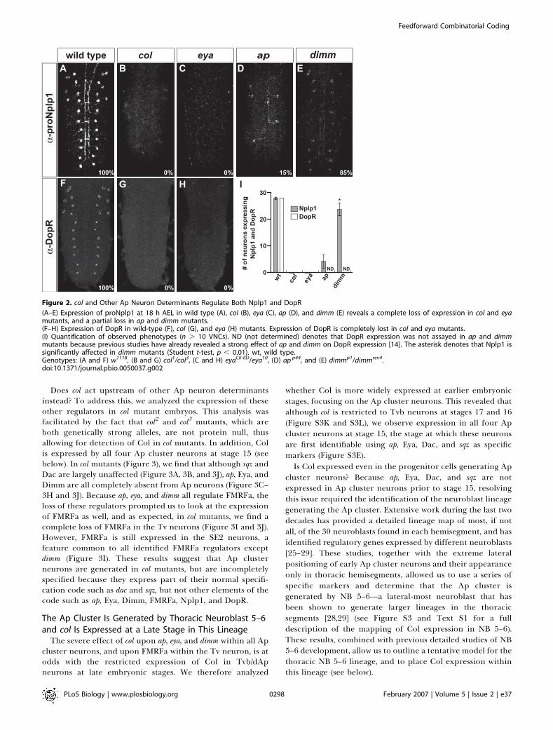
important for DopR expression in Tvb/dAp [14]. We analyzedwhether these Ap neuron determinants, as well as eya, alsoaffected Nplp1 expression (Figure 2). We found that Nplp1expression depended on eya, ap, and dimm (Figure 2C–2E), andthat eya also regulates DopR (Figure 2H–2I). How is Tv versusTvb/dAp cell fate then determined? Although both cell typesexpress ap, dimm, and eya, only Tv neurons express dac andhave activated the BMP pathway. Could the mere absence ofdac and/or BMP activation be sufficient to specify the Tvb/dApfate? To test this, we analyzed expression of Nplp1 in dac andBMP mutants, but found no evidence of ectopic Nplp1expression in Tv neurons (see below). Thus, specification ofTvb/dAp neurons likely requires additional factors restrictedto this cell type.The COE family of HLH regulators is highly evolutionary
conserved [16], and is represented in Drosophila by a singlemember, col (Flybase, knot) [17]. COE genes play importantroles during nervous system development in Caenorhabditiselegans and vertebrates [18–24], and col is expressed in the
Author Summary
The nervous system contains a daunting number of different celltypes, perhaps as many as 10,000 in mammals, far outnumberingregulatory genes in many animal species. Studies of the determi-nants of cell fate in many systems during the last decade havesupported the conclusion that cell fate is not determined by any oneregulatory gene, but results from the combinatorial action of severalregulators. Many questions about the nature of such codes,however, remain. It is not known, for example, how complex suchcodes are or how they are established. It is also unclear whetherthey are confined in their action or if they act outside of their normalcontext. To address these outstanding issues, we have used twounique subsets of Drosophila neurons, identifiable by their specificexpression of two different neuropeptide genes. We have identifiedtwo partially overlapping and relatively simple codes, consisting offour to seven regulators that act to specify these two cell types.Intriguingly, specification is achieved in a feedforward manner suchthat A activates B, followed by A/B activating C, and A/B/C activatingD. Each code is surprisingly potent, and can ectopically activateneuropeptide gene expression in a variety of neurons, with asurprising disregard for many early patterning events.
PLoS Biology | www.plosbiology.org February 2007 | Volume 5 | Issue 2 | e370296
Feedforward Combinatorial Coding

developing Drosophila central nervous system (CNS) [17],although no function has yet been assigned to it there. Theinvolvement of members of this gene family in nervoussystem development in other species, and the embryonic CNSexpression of col, prompted us to investigate the possible roleof col during Ap neuron specification. col has a dynamicexpression pattern in the VNC (below), and we initiallyfocused on its expression in mature Ap neurons, at 18 h AEL,and larval stages. We find that at these stages, col is expressedspecifically in Tvb/dAp neurons (Figure 1F), and expression ismaintained in these neurons at least into the third larval stage(see below). This raised the possibility that col plays a role inTvb/dAp cell fate specification. This notion was supported bythe complete loss of Nplp1 and DopR expression in colmutants (Figure 2A, 2B, 2F, 2G, and 2I).
col Acts Upstream of Certain ap Neuron DeterminantsPrevious studies have addressed the regulatory interactions
between several of the Ap neuron determinants [9–12].However, these had not been addressed in the case of dac,eya, and dimm. As expected from the late onset of dimm
expression, we find no evidence of dimm regulation of Dac orEya (Figure S2M and S2N). Similarly, as expected from themild effect of dac upon FMRFa, we find that dac does notregulate Dimm (Figure S2L). In contrast, in eya mutants, wefind a nearly complete loss of Dimm expression in the Tv,Tvb, and dAp neurons (Figure S2K and S2O).With a more complete picture of how previously identified
Ap neuron determinants interact genetically, we nextaddressed whether col acts upstream, downstream, or inparallel to other Ap neuron determinants. We first analyzedthe expression of Col in embryos mutant for these otherregulators. In general, we found no severe effects on Colexpression (Figures S2A–S2I). The one exception was in sqz, inwhich we found a reproducible increase in the number of Colcells (Figure S2B, S2C, and S2J). This was, however, expected,since sqz affects the composition of Ap cluster cells, with anincrease both in the number of Ap cluster cells (specifically inT1) and an increase in Tvb cells at the expense of Tv cells (inT1–T3) [9,10]. In line with this, we also find an increase inNplp1 cells in sqz mutants (Figure S2B, S2C, and S2J).
Figure 1. A Specific Subset of Ap Neurons, the Tvb and dAp Cells, Expresses the Nplp1 Neuropeptide, the DopR Dopamine Receptor and the Col
Transcription Factor
Staining for apGAL4 (green) and proNplp1 (red), and IPNamide (A), DopR (B), proFMRF (C), Dac (D), Dimm (E), and Col (F) (blue). Arrows denote the Tvbneuron, and arrowheads the Tv neuron.(A and B) Co-expression of proNplp1 with IPNamide (A) and DopR (B) in dAp neurons (top) and one Ap cluster neuron (bottom).(C and D) No co-expression between proNplp1 and proFMRFa (C) or Dac (D).(E and F) Co-expression between proNplp1 and Dimm (E) or Col (F). The Tvb neuron is identified by its expression of ap, Eya, Dimm, DopR, and Nplp1,but not Dac or FMRFa.(G) Cartoon summarizes the expression of regulatory genes and terminal identity genes in Ap neurons. DNH, dorsal neurohemal organ; vAp, ventral Apneurons.All images are from 18-h AEL VNCs.doi:10.1371/journal.pbio.0050037.g001
PLoS Biology | www.plosbiology.org February 2007 | Volume 5 | Issue 2 | e370297
Feedforward Combinatorial Coding
Does col act upstream of other Ap neuron determinantsinstead? To address this, we analyzed the expression of theseother regulators in col mutant embryos. This analysis wasfacilitated by the fact that col2 and col3 mutants, which areboth genetically strong alleles, are not protein null, thusallowing for detection of Col in col mutants. In addition, Colis expressed by all four Ap cluster neurons at stage 15 (seebelow). In colmutants (Figure 3), we find that although sqz andDac are largely unaffected (Figure 3A, 3B, and 3J), ap, Eya, andDimm are all completely absent from Ap neurons (Figure 3C–3H and 3J). Because ap, eya, and dimm all regulate FMRFa, theloss of these regulators prompted us to look at the expressionof FMRFa as well, and as expected, in col mutants, we find acomplete loss of FMRFa in the Tv neurons (Figure 3I and 3J).However, FMRFa is still expressed in the SE2 neurons, afeature common to all identified FMRFa regulators exceptdimm (Figure 3I). These results suggest that Ap clusterneurons are generated in col mutants, but are incompletelyspecified because they express part of their normal specifi-cation code such as dac and sqz, but not other elements of thecode such as ap, Eya, Dimm, FMRFa, Nplp1, and DopR.
The Ap Cluster Is Generated by Thoracic Neuroblast 5–6and col Is Expressed at a Late Stage in This Lineage
The severe effect of col upon ap, eya, and dimm within all Apcluster neurons, and upon FMRFa within the Tv neuron, is atodds with the restricted expression of Col in Tvb/dApneurons at late embryonic stages. We therefore analyzed
whether Col is more widely expressed at earlier embryonicstages, focusing on the Ap cluster neurons. This revealed thatalthough col is restricted to Tvb neurons at stages 17 and 16(Figure S3K and S3L), we observe expression in all four Apcluster neurons at stage 15, the stage at which these neuronsare first identifiable using ap, Eya, Dac, and sqz as specificmarkers (Figure S3E).Is Col expressed even in the progenitor cells generating Ap
cluster neurons? Because ap, Eya, Dac, and sqz are notexpressed in Ap cluster neurons prior to stage 15, resolvingthis issue required the identification of the neuroblast lineagegenerating the Ap cluster. Extensive work during the last twodecades has provided a detailed lineage map of most, if notall, of the 30 neuroblasts found in each hemisegment, and hasidentified regulatory genes expressed by different neuroblasts[25–29]. These studies, together with the extreme lateralpositioning of early Ap cluster neurons and their appearanceonly in thoracic hemisegments, allowed us to use a series ofspecific markers and determine that the Ap cluster isgenerated by NB 5–6—a lateral-most neuroblast that hasbeen shown to generate larger lineages in the thoracicsegments [28,29] (see Figure S3 and Text S1 for a fulldescription of the mapping of Col expression in NB 5–6).These results, combined with previous detailed studies of NB5–6 development, allow us to outline a tentative model for thethoracic NB 5–6 lineage, and to place Col expression withinthis lineage (see below).
Figure 2. col and Other Ap Neuron Determinants Regulate Both Nplp1 and DopR
(A–E) Expression of proNplp1 at 18 h AEL in wild type (A), col (B), eya (C), ap (D), and dimm (E) reveals a complete loss of expression in col and eyamutants, and a partial loss in ap and dimm mutants.(F–H) Expression of DopR in wild-type (F), col (G), and eya (H) mutants. Expression of DopR is completely lost in col and eya mutants.(I) Quantification of observed phenotypes (n . 10 VNCs). ND (not determined) denotes that DopR expression was not assayed in ap and dimmmutants because previous studies have already revealed a strong effect of ap and dimm on DopR expression [14]. The asterisk denotes that Nplp1 issignificantly affected in dimm mutants (Student t-test, p , 0.01). wt, wild type.Genotypes: (A and F) w1118, (B and G) col1/col3, (C and H) eyaCli-IID/eya10, (D) ap p44, and (E) dimmp1/dimmrev4.doi:10.1371/journal.pbio.0050037.g002
PLoS Biology | www.plosbiology.org February 2007 | Volume 5 | Issue 2 | e370298
Feedforward Combinatorial Coding

Down-Regulation of col Is Critical for Proper ap ClusterDifferentiation
Col is expressed by all four newly born Ap cluster neuronsand is essential for Ap cluster specification, as evident fromthe complete loss of ap and Eya expression in col mutants. Colis rapidly down-regulated from three Ap cluster cells andmaintained only in Tvb. Is the down-regulation of colimportant for proper Ap cluster differentiation? To test this,we misexpressed col using the apGAL4 driver, which is notexpressed until stage 16, thus maintaining col expression inall four Ap cluster neurons at the time when Col is normallydown-regulated. This experiment led to frequent activationof Nplp1 in one additional Ap cluster neuron (Figure 4A and4B), and staining for Dimm reveals that this cell is indeed theTv neuron (Figure 4C). FMRFa expression is frequently down-regulated in Tv, but we do observe Tv cells that co-express
FMRFa and Nplp1 (Figure 4B). The finding that col mis-expression in the Ap cluster only leads to one ectopic Nplp1cell and no ectopic Dimm cells indicates that col cannotinduce a peptidergic cell fate, at least not with this late driver.However, because the two unaffected cells already areexpressing ap and Eya, we predicted that co-misexpressionof dimm, together with col, should trigger Nplp1 expression inall four Ap cluster neurons. This is indeed what we find(Figure 4D and 4E). In summary, down-regulation of col inthree Ap cluster neurons is essential for proper Ap clusterspecification.
col Can Act in a Context-Dependent Manner to Generate
Additional Ap Cluster NeuronsCol is expressed prior to ap and Eya in the Ap cluster
neurons, and it is essential for ap and Eya expression within
Figure 3. col Regulates ap, eya, dimm, and FMRFa, but Not sqz and dac
(A–I) Expression of Ap neuron determinants and of FMRFa (I), in wild-type and col mutants at stage 15 (A–D) and 18 h AEL (E–I). Dashed lines in (A–D)depict the VNC midline.(A and C) In wild type, expression of Col (blue), sqzGAL4 (green), Dac (red), and Eya (red) clearly visualizes the early Ap cluster (arrows).(B and D) In col mutants, although sqzGAL4and Dac expression is largely unaffected (B), there is a complete loss of Eya expression in the Ap cluster (D,arrows).(E and F) In the late embryonic VNC, Eya and apGAL4 are readily detected in all three thoracic hemisegments in the wild type (E), whereas col mutantsshow complete loss of expression of both markers (F).(G and H) Similarly, Dimm expression (red) is lost from lateral thoracic peptidergic neurons in col mutants (bracket). Staining for other neuropeptidessuch as CCAP and Corazonin reveals that Dimm expression is specifically lost from Tv, Tvb, and dAp peptidergic neurons (unpublished data).(I) As expected from the loss of several Ap neuron determinants, FMRFa expression (green) is completely lost from Tv neurons (bracket). As with othermutations specifically affecting Tv neurons, expression of FMRFa is not lost from the SE2 neurons (SE2).(J) Quantification of the observed phenotypes (for sqzGAL4, Dac, Eya, and apGAL4, n . 65 clusters; for Dimm, n . 65 thoracic hemisegments; and forFMRFa, n . 24 VNCs). sqzGAL4, Dac, and Eya are quantified at stage 15, whereas apGAL4, Dimm, and FMRFa are quantified at 18 h AEL.Genotypes:(AandC)UAS-nmEGFP/þ; sqzGAL4/þ, (BandD)col1, UAS-nmEGFP/col 3 sqzGAL4/þ, (E)apGAL4/þ; UAS-nmEGFP/þ, (F)col1, apGAL4/col3;UAS-nmEGFP/þ,(G)w1118, and(HandI) col1/col3. (J)Controlsare:UAS-nmEGFP/þ; sqzGAL4/þfor sqz,Dac,andEyaexpression;apGAL4/þ; UAS-nmEGFP/þfor ap expression;and w1118 forDimm and FMRFa expression.doi:10.1371/journal.pbio.0050037.g003
PLoS Biology | www.plosbiology.org February 2007 | Volume 5 | Issue 2 | e370299
Feedforward Combinatorial Coding

these cells. To address whether col is also sufficient to activateap and Eya, we misexpressed col in all neurons, using the elav-GAL4 driver (Figure 5) [30]. This led to ectopic activation ofboth ap and Eya (Figure 5A–5E). In addition, we found someactivation of Dimm, Nplp1, and DopR expression (Figure 5F–5L and 5O–5Q). Although ectopic activation of ap or Eyaalone was found in several regions, co-activation was largelyconfined to neuroblast row 5—the anterior region of Gsbnexpression (Figure 5M and 5N). We typically observe six toten ap/Eya co-expressing cells in the lateral-most part of row 5(Figure 5M and 5N). Ectopic activation of ap/Eya, togetherwith Dimm, Nplp1, and DopR, was also confined to lateral-most row 5, i.e., the posterior-most part of gsblacZ cells, andfurther confined to thoracic segments (Figure 5B–5D, 5J, 5L,and 5O–5Q). Ectopic Nplp1/DopR expression is not over-lapping with FMRFa, and there is clear evidence of ectopicDimm expression (Figure 5P and 5Q), indicating that addi-tional peptidergic neurons are being generated. Ectopicgeneration of ap/Eya double-expressing cells, i.e., ectopic ‘‘Apcluster’’ neurons, was observed already at stage 13, i.e., prior
to when Ap cluster neurons are normally born (unpublisheddata).These results show that col can activate ap and Eya in a
number of neurons, but can act to generate bona fide Apcluster neurons only in a highly context-dependent manner:in lateral, thoracic, row 5 neurons. The appearance of six toten Eya-expressing cells, but only three to five Nplp1/DopR-expressing cells, and no evidence of ectopic FMRFa expres-sion, suggests that the generation of ectopic Ap clusterneurons is biased toward Tvb (Nplp1/DopR expressing) asopposed to Tv (FMRFa expressing) cell fate. In contrast,although col function depends upon these three positionalcues, our results indicate that col is able to override thetemporal coding within lateral row 5, and activate Nplp1 andDopR in earlier-born neurons.
col Can Be Partially Rescued by Expression of ap and eyaThe loss- and gain-of-function studies place col clearly
upstream of ap and eya. Does col act merely to regulate ap andeya in early postmitotic Ap cluster neurons, or does it playadditional roles during Ap cluster formation? To furtheraddress this issue, we attempted to ‘‘cross-rescue’’ col with apand eya, by expressing ap and eya in a col mutant background(Figure 6). First, as a positive control, we attempted to rescuecol by providing col activity using elav-GAL4/UAS-col. This ledto a robust rescue, both of Ap cluster determinants (Eya, ap,and Dimm) and of terminal differentiation genes (Nplp1,DopR, and FMRFa) (Figure 6A–6H, 6L, and 6M). Similar to thecolmisexpression experiments, we find a clear increase in ‘‘Apcluster’’ neurons, primarily of the Tvb type, as evident fromthe finding of six to ten ap/Eya- and three to five Nplp1/DopR-expressing neurons per hemisegment (Figure 6E–6H and 6M).Next, we attempted to ‘‘cross-rescue’’ col mutants with ap andeya, and could indeed find a significant degree of rescue of Apcluster formation, as evident both from Dimm and FMRFaexpression (Figure 6J–6M). In contrast, we found no evidenceof rescue of Nplp1 or DopR in these embryos (Figure 6I and6L). Because we can detect Col in the col2 and col3 mutantbackgrounds, we could identify a Dimm/Col-expressing celladjacent to the Tv/FMRFa neuron (Figure 6K and 6N). Thisindicates that ap/eya can partially rescue Tvb cell fate, but inthe absence of col activity, these ‘‘Tvb’’ neurons do notactivate Nplp1. In summary, the finding that in colmutants, apand eya can partially rescue the Tv cell fate, but not Tvb cellfate, suggests additional roles for col in Tvb specification.
col Acts at Multiple Steps of Tvb SpecificationOur results indicate that Tvb cell fate is not specified by a
linear col!ap/eya!dimm!Nplp1/DopR genetic cascade. Tofurther address this issue, we tested the sufficiency of col, ap,and eya to activate Dimm when misexpressed both alone andin combination. These experiments reveal that although colcan trigger some ectopic activation of Dimm, there is littleeffect upon Dimm when misexpressing ap, eya, or ap/eya(Figure 7A). In contrast, co-misexpression of col with either apor eya, and in particular, co-misexpression of all three genes,leads to striking ectopic Dimm expression (Figure 7A–7E).Does col play a role even at the final step of Tvb
differentiation, i.e., in the activation of Nplp1? We attemptedto address the possible late role of col by misexpressing italone and together with other Ap neuron determinants, andthen assay its potency in activating Nplp1. Importantly, if
Figure 4. col Misexpression in the Ap Cluster Triggers Ectopic Nplp1
Expression
(A) In the control, the Ap cluster (here visualized by Eya) contains oneFMRFa- and one Nplp1-expressing cell.(B and C) Misexpression of col in the Ap cluster triggers ectopic Nplp1expression in one additional Ap cluster neuron. Labeling for FMRFa (B)and for Dimm (C) reveals that this cell is the Tv neuron, evident from itsco-expression of Nplp1, FMRFa (B), and Dimm (C).(D) Co-misexpression of col and dimm in the Ap cluster triggers all fourAp cluster neurons to express Nplp1. Misexpression of dimm has noeffect upon Nplp1 expression (unpublished data).(E) Quantification of Nplp1, FMRFa, and Nplp1/FMRFa expression (n . 80thoracic hemisegments).All images are from 18-h AEL VNCs.Genotypes: (A) w1118, (B and C) apGAL4/þ; UAS-col/þ, and (D) apGAL4/þ;UAS-col, UAS-dimm/þ. (E) Control is w1118.doi:10.1371/journal.pbio.0050037.g004
PLoS Biology | www.plosbiology.org February 2007 | Volume 5 | Issue 2 | e370300
Feedforward Combinatorial Coding

Figure 5. Misexpression of col Activates ap, Eya, Dimm, Nplp1, and DopR, but in a Context-Dependent Manner
(A–L) Expression of aplacZ, Eya, Nplp1, DopR, and FMRFa in 18-h AEL control and col misexpression VNCs. (A) In the control, aplacZ and Eya are specificallyexpressed in Ap neurons. A small number of additional Eya-expressing cells are visible laterally; these cells express Eya at earlier stages, but typicallydown-regulate Eya expression at stages 15/16 [12].(B–D) In col misexpression embryos, robust ectopic expression is evident for both aplacZ and Eya, although ectopic co-activation of aplacZ and Eya genesis predominantly apparent in lateral thoracic areas (bracket).(E) Quantification of aplacZ and Eya expression (n . 3 VNCs).(F) Quantification of Nplp1, DopR, and FMRFa expression (n . 8 VNCs). There is no evidence of ectopic FMRFa expression.(G–I) In the wild type, Nplp1 and DopR expression is evident in dAp and Tvb neurons.(J–L) col misexpression leads to ectopic co-activation of Nplp1 and DopR expression, again restricted to lateral thoracic areas (brackets).
PLoS Biology | www.plosbiology.org February 2007 | Volume 5 | Issue 2 | e370301
Feedforward Combinatorial Coding

there is a simple linear col!ap/eya!dimmNplp1/DopR geneticcascade at work, the effect of triple co-misexpression of ap/eya/dimm should not be enhanced by addition of col to this code.However, we find a striking enhancement of ectopic Nplp1expression when adding col to this code (Figure 7F–7J). One
particular double co-misexpression combination, col/ap, wasmore potent than others in activating both Dimm and Nplp1(Figure 7E and 7J). A likely explanation for this effect is thatco-misexpression of col/ap activates significant ectopic eyaexpression (unpublished data).
(M and N) col misexpression triggers ectopic co-activation of aplacZ and Eya at stages earlier than when Ap clusters are normally generated (dashed linesdepict the VNC midline). In the control (M), only dAp (aplacZ/Eya expressing) and vAp (aplacZ expressing) neurons are visible at stage 14. (N) In colmisexpression, ectopic aplacZ and Eya cells, as well as double-positive cells, appear earlier and increase in numbers into stage 14 (brackets).(O–Q) Lateral view of the three thoracic segments in 18-h AEL VNCs; anterior is to the left. (Dotted circles depict the enlarged Ap cluster.) (O) colmisexpression leads to appearance of six to ten aplacZ/Eya-expressing cells, of which a subset is also Dimm expressing, and (P) these ectopic ‘‘Apcluster’’ cells are confined to row 5, i.e., the anterior region of gsblacZ expression. (Q) col misexpression triggers ectopic Nplp1 in these ectopic Dimmcells, while FMRFa is still only expressed in one cell per hemisegment.Genotypes: (A and M) aplacZ/þ; elav-GAL4/þ, (B–D, N, and O) aplacZ/þ; elav-GAL4/ UAS-col/þ, (G–I) w1118, (J–L) elav-GAL4/UAS-col, and (P–Q) gsblacZ/þ;elav-GAL4/ UAS-col/þ. (E) Control is aplacZ/þ; elav-GAL4/þ, and (F) control is w1118.doi:10.1371/journal.pbio.0050037.g005
Figure 6. col Rescues Both Tv and Tvb Cell Fate in col Mutants, whereas ap and eya Only Rescue Tv Cell Fate
(A and B) Wild-type expression of Nplp1 and FMRFa. (C–H) col rescue of col mutants, in 18-h AEL VNCs.(C and D) col expression in all neurons efficiently rescues back both Nplp1 and FMRFa expression in col mutants.(E–H) Similar to the misexpression results (Figure 5), col rescue results in generation of extra ‘‘Ap cluster’’ neurons, as evident from the expression ofEya, Dimm, Nplp1, and DopR, whereas there is no evidence of ectopic FMRFa expression (lateral view of three thoracic VNC hemisegments; anterior is tothe left).(I–K) ‘‘Cross-rescue’’ of col with ap and eya leads to a significant rescue of FMRFa (p , 0.01, n¼ 35 VNCs), but no rescue of Nplp1. (K) ‘‘Cross-rescue’’does lead to a partial rescue of Dimm expression in cells expressing Col (a putative ‘‘Tvb’’ cell; arrow), but this cell is never able to activate Nplp1. (Thearrowhead points to the FMRFa-expressing Tv cell.) Col can be visualized because the col3 allele is not a protein null.(L–N) Quantification of observed phenotypes (n . 16 VNCs for [L], n . 41 hemisegments for [M], and n . 9 hemisegments for [N]). (L) Expressingneurons per VNC. (M) Expressing neurons per Ap cluster. (N) Expressing neurons for the three thoracic segments combined.Genotypes: (A and B) wild type, (C–H) col1/col3; elav-GAL4/UAS-col, and (I–K) col1/col3; elav-GAL4/UAS-ap, UAS-eya. (L–N) Controls are w1118.doi:10.1371/journal.pbio.0050037.g006
PLoS Biology | www.plosbiology.org February 2007 | Volume 5 | Issue 2 | e370302
Feedforward Combinatorial Coding

To further address the late role of col, we generated atransgenic RNA-interference (RNAi) line (UAS-col-dsRNA)and attempted to suppress col gene activity by crossing thisline to apGAL4. Because apGAL4 also drives expression in thedeveloping wing disc, we first analyzed the efficiency of thisnovel tool in suppressing col gene activity in this tissue. Thisphenocopied the effects of col mutants on wing development[31], with a clear L3–L4 wing vein fusion (unpublished data),indicating that this RNAi transgene specifically blocks colgene activity. However, upon analyzing late larval (thirdinstar) CNSs, we found no effect upon Col expression in Tvbneurons, and as expected, no effect upon Nplp1 expression(unpublished data). Recent studies reveal that RNAi can beefficiently enhanced by overexpression of components ofthe RNAi pathway, in particular of the Dicer-2 (Dcr-2) gene(G. Dietzl and B. Dickson, personal communication). Wetherefore co-expressed Dcr-2 with col dsRNA (UAS-Dcr-2/þ;apGAL4/UAS-col-dsRNA), and found a clear effect not only
upon Col, but importantly, also upon Nplp1 expression. Wefound no obvious effect in the first instar larvae (unpub-lished data), but in third instar larvae, Col expression isspecifically and completely lost from all Tvb/dAp cells(Figures 8A, 8B, and 8E). This leads to a complete loss ofNplp1 in 44% of Tvb/dAp cells (Figure 8C–8E), and stronglyreduced expression in the remaining expressing cells (Figure8B). Strikingly, this strong effect upon Nplp1 is not anindirect effect of down-regulation of ap, Eya, or Dimm(Figure 8A–8E). As anticipated, col RNAi has no effect uponFMRFa expression in the third instar larvae (n ¼ 5 VNC;unpublished data).Together, the finding that col can act potently with ap and
eya to activate Dimm, that col can act potently with ap, eya, anddimm to activate Nplp1, and that postmitotic RNAi of colstrongly affects Nplp1 expression without affecting ap, Eya, orDimm, does not fit with a simple linear cascade model for Tvb
Figure 7. col Can Act in a Combinatorial Manner with ap and eya to Activate Dimm, and with All Three Regulators to Activate Nplp1 and DopR
(A–E) Expression of Dimm in 18-h AEL VNCs. Single misexpression of each regulator has limited effect upon Dimm expression, but combinatorialexpression of col/ap or col/ap/eya leads to striking ectopic Dimm expression. (E) Quantification of Dimm expression in the various misexpressionexperiments (n . 3 thoracic T1–T3 segments). Importantly, the addition of col to the different codes has greatly increased effect upon Dimm—compareap/eya with col/ap/eya.(F–J) Expression of Nplp1 in 18-h AEL VNCs. Single misexpression has limited effect upon Nplp1 expression, but combinatorial expression leads tostrong ectopic Nplp1 expression. (J) Quantification of Nplp1 expression (n . 3 VNCs; numbers denote total number of expressing cells per VNC).Importantly, the addition of col to the different codes has greatly increased effect also upon Nplp1—compare ap/eya/dimm with ap/eya/dimm/col.Genotypes: (A) w1118 and (B–J) elav-GAL4 crossed to single UAS-cDNA transgenes or combinations of the various UAS-cDNA transgenes.doi:10.1371/journal.pbio.0050037.g007
PLoS Biology | www.plosbiology.org February 2007 | Volume 5 | Issue 2 | e370303
Feedforward Combinatorial Coding

specification. Rather, it strongly suggests that col is acting atseveral levels during Tvb specification and differentiation.
The Combinatorial Codes Are Highly Potent and Highly
Specific
Previous studies have identified several regulators acting tospecify Tv fate and to control FMRFa expression. Althoughco-misexpression of parts of this code had been previouslytested, all possible combinations had not. Similar to thecombinatorial activation of Nplp1 and DopR, we find thatwhereas co-misexpression of ap/sqz, ap/dac, or ap/dimm haslimited effect upon FMRFa expression (Figure 9) [9,10,12],triple co-misexpression of these regulators, and in particular
of ap/dimm/dac, leads to a dramatic ectopic activation ofFMRFa (Figure 9G–9K and 9M).The identification of two partly overlapping and highly
potent combinatorial codes allowed us to ask an importantquestion: Does combinatorial misexpression of these regu-lators merely lead to a general confusion with a mixedneuronal identity, or are these codes truly instructive andspecific? To address this issue, we analyzed the expression ofNplp1 and FMRFa in the various misexpression backgrounds.Not surprisingly, when common and partial components ofthese codes are misexpressed, such as ap/dimm (common toboth Tv and Tvb/dAp neurons), we find ectopic activation ofboth Nplp1 and FMRFa in different subsets of cells (Figure9B, 9H, and 9M). However, as a third, and cell-type specific,regulator is added, not only does the amount of ectopic,terminal differentiation gene expression increase dramati-cally, but we find less evidence of cross-activation of theinappropriate downstream gene (Figure 9C–9F and 9I–9M).This surprising finding reveals that combinatorial misexpres-sion may act in a highly specific and instructive manner, andthat these combinatorial codes may be viewed as potentbinary switches for cell fate specification. In addition, forboth codes, ectopic activation of FMRFa and Nplp1 isobserved in neurons throughout the VNC and brain, andtraverses many developmental boundaries, such as antero-posterior, dorsoventral, and mediolateral boundaries.
Discussion
We have identified a sequential regulatory cascade ofcombinatorial coding that acts to specify two unique neuro-nal cell fates during Drosophila CNS development. Combinedwith previous studies [6,7,9–14], the findings described in thisstudy provide the following model for Ap cluster generationand specification (Figure 10). Neuroblast 5–6 forms in thefirst wave of neuroblast delamination, at late stage 8, andgenerates a mixed lineage of glia and neurons [28,29]. At stage13, Col expression is turned on specifically in thoracic NB 5–6, in two subsequent ganglion mother cells (GMCs) at stages13/14, and in the four Ap cluster neurons generated fromthese GMCs, during stages 14/15. We have not resolved thebirth order of the four Ap cluster neurons, and the siblingrelationship of Tv and Tvb is thus unclear. When Ap neuronsare born, col activates ap and eya, whereas sqz and dac areactivated by unknown regulator(s). sqz appears to play anearly postmitotic role, apparently acting in the Notchpathway, to ensure proper Ap cluster composition, and sqzmutants display both additional Ap cluster cells (in T1) andadditional Tvb cells (in T1–T3). col is rapidly down-regulatedfrom three Ap neurons, but remains expressed in the Tvb,where it acts with ap and eya to activate dimm at stage 16. Atlate embryogenesis, col acts with ap, eya, and dimm to activateNplp1 and DopR in Tvb. In the Tv neuron, ap and eya act,apparently independently of col, to activate dimm expression.In the Tv neuron, eya furthermore plays a role in setting upcompetence to respond to the BMP signal. At stage 17, the Tvaxon reaches the dorsal neurohemal organ (DNH) andreceives the BMP ligand Gbb that activates the Wit receptor,and then triggers activation of the BMP pathway in the Tvneuron. At 18 h AEL, ap, eya, sqz, dac, dimm, and BMP signalingcooperate to activate FMRFa in the Tv neuron. In addition totheir roles in neuropeptide regulation and BMP signaling,
Figure 8. Postmitotic RNA Interference of col Leads to Loss of Nplp1
Expression, without Loss of ap, Eya, or Dimm
(A and C) In the control, expression of Nplp1, ap, and Col (A), as well asEya and Dimm (C) is readily detected in the third larval instar dApneurons.(B and D) In the col RNAi, Col is completely and specifically lost from dAp(B), Nplp1 is down-regulated (B) or lost (D), whereas ap, Eya, and Dimmare not affected (B and D). As anticipated, col RNAi has no effect uponFMRFa expression in the third instar larvae (n ¼ 5 VNCs; unpublisheddata). Dashed boxes outline the cells that are magnified at the bottom.(E) Quantification of the observed effects in dAp and Ap cluster neurons,in segments T1–A2.Genotypes: (A and C) and control in (E) UAS-Dcr-2/þ; apGAL4, UAS-nmEGFP/þ and (B and D) UAS-Dcr-2/þ; apGAL4, UAS-nmEGFP/þ; UAS-col-dsRNA/þ.doi:10.1371/journal.pbio.0050037.g008
PLoS Biology | www.plosbiology.org February 2007 | Volume 5 | Issue 2 | e370304
Feedforward Combinatorial Coding

Figure 9. The Two Combinatorial Codes Act in a Highly Potent and Highly Selective Manner
Expression of Nplp1 (A–E and L) and FMRFa (F–K).(A) Wild-type expression of Nplp1.(B) Co-misexpression of ap and dimm leads to some ectopic Nplp1 expression.(C–E) Ectopic Nplp1 expression is dramatically increased by addition of col to this code (to visualize the strong activation, the confocal ‘‘stack’’ of theVNC is split into three non-overlapping merged images).(F) The same VNC as in (C–E) revealing limited ectopic activation of FMRFa.(G) Wild-type expression of FMRFa.(H) Co-misexpression of ap and dimm also leads to some ectopic FMRFa expression.(I–K) Ectopic FMRFa expression is dramatically increased by addition of dac to this code.(L) The same VNC as in (I–K) revealing limited ectopic activation of Nplp1.(M) Quantification of the ectopic activation effects (n . 3 VNCs). A single asterisk denotes that the ectopic activation of Nplp1 is significantly reducedwhen adding dac to ap/dimm (Student t-test, p , 0.01). Similarly, double asterisks denote that the ectopic activation of FMRFa is significantly reducedwhen adding col to ap/dimm (Student t-test, p , 0.01).All images are from 18-h AEL VNCs.Genotypes: (A and G) w1118. Remaining panels are elav-GAL4 crossed to single or combinations of the various UAS-cDNA transgenes.doi:10.1371/journal.pbio.0050037.g009
PLoS Biology | www.plosbiology.org February 2007 | Volume 5 | Issue 2 | e370305
Feedforward Combinatorial Coding

both ap and eya act to ensure proper axon pathfinding of Tvneurons (eya), as well as Tvb and dAp neurons (ap and eya). Therole that col may play more directly in axon pathfinding hasnot been resolved due to the fact that the expression of theappropriate axonal markers (apGAL4, Nplp1, DopR, andFMRFa) is completely absent in col mutants. Given thecomplexity of axon pathfinding, we would anticipate thatseveral other regulators are yet to be identified before acombinatorial code for ‘‘Tv-type’’ or ‘‘Tvb-type’’ axonalpathfinding is deciphered. Indeed, we find no evidence thatcombinatorial misexpression of the abovementioned regu-lators can dictate axonal projections, because ectopic Nplp1or FMRFa axons are following many different routes in theVNC (Figures 7H and 9I). Finally, dimm also plays additionalroles to those described above and is necessary for theexpression of neuropeptide-processing enzymes in peptider-gic neurons. Importantly, dimm acts independently to controlexpression of the neuroamidase gene PHM in the Tv and Tvbneurons, and dimm is sufficient to activate PHM in most, if notall, VNC neurons. Thus, during the specification and differ-entiation of the Ap neurons, there exists a remarkablediversity in the division of labor between the identifiedregulators, with most of them participating in more than one,but never all, of the identified events.
Sequential Combinatorial Coding: Feedforward LoopsProviding High-Fidelity Control of Neuronal Specification
We have identified a multistep process for specifying the Tvand Tvb cell fates. What would be the purpose of this type ofsequential combinatorial coding? In other model systems withhigher genetic resolution, such as Escherichia coli and yeast,
extensive genetic analysis has revealed that this type ofsequential gene regulation is quite common [32,33], and arecent study in C. elegans suggests it may also function duringneuronal specification [34]. These regulatory nodes, alsoknown as feedforward loops (FFL), have been shown to ensurefidelity in gene regulation [35,36]. For instance, in a simpleFFL in which gene A regulates gene B, and A/B then co-operate to regulate C, activation of C depends uponprolonged A expression such that A/B will have time toactivate C. If A is only active in a short burst, B may beactivated, but C is not, because A/B never co-express for asufficiently prolonged period of time. The role of col duringAp cluster specification provides an excellent example of aFFL used during neuronal cell fate specification. col isexpressed in all four Ap cluster cells and plays an early rolein activating ap and Eya, but is only maintained in Tvb, whereit plays a later role in activating first dimm, then Nplp1.Maintained expression of col in all four Ap cluster neurons, bydriving col expression from apGAL4, leads to activation of theTvb program also in the Tv neuron. Thus, a burst of colexpression has a different informational value than persistentcol expression—general Ap cluster specification versus Tvbspecification.
Combinatorial Coding and Global Cues: ContextDependence versus IndependenceMisexpression of each of the two identified combinatorial
codes leads to striking ectopic activation of the Nplp1 andFMRFa genes, and we are surprised by two particular aspectsof these findings. First, the global potency of these codes—co-misexpression triggers ectopic FMRFa of Nplp1 in a number
Figure 10. Model for the Thoracic NB 5–6 Lineage and for the Genetic Pathways Specifying Tv and Tvb Neurons
The thoracic NB 5–6 lineage is shown at top, and the genetic pathways specifying Tv and Tvb neurons are at the bottom. NB 5–6 is formed already atlate stage 8 and generates an early lineage of neurons (circles) and glia (uneven shape). For clarity, the early lineage is omitted from stage 14 andonward. The Ap cluster is generated between stages 13–15. The fate of the neuroblast itself is unclear after stage 15. Some of these regulatoryinteractions are occurring also in the non-peptidergic Ap cluster neurons, as well as in the dAp neurons, but for simplicity, they are not outlined here.See text for details.DNH, dorsal neurohemal organ; St, stage.doi:10.1371/journal.pbio.0050037.g010
PLoS Biology | www.plosbiology.org February 2007 | Volume 5 | Issue 2 | e370306
Feedforward Combinatorial Coding
of neurons, located in many different anteroposterior,dorsoventral, and mediolateral positions. Thus, it wouldappear that early regulators mainly act to ensure propercombinatorial coding in each neuron, and play a minor rolein restricting cell fate by limiting the cell’s competence—oncethe proper code is in place, the cell fate specificationprogram will be carried out irrespective of the history ofthe cell. Second, the striking binary effect of these codes isnoteworthy—the change of one single player in a codecompletely alters target gene choice. For instance, misex-pression of ap/dimm/dac leads almost exclusively to strongFMRFa activation, but simply replacing dac with col leads toalmost exclusively Nplp1 activation. Thus, it appears thatmore complete codes not only have great potency, but alsohave great specificity.
The Maintenance of Col, Ap, and Eya ExpressionCol shows a very dynamic expression pattern in the VNC—
exemplified in NB 5–6 by the expression in the neuroblast, intwo GMCs, in all four Ap cluster neurons, and finally only inTvb. This poses three obvious questions: what activates col inthe neuroblast, what shuts it down in three of the Ap clusterneurons, and finally, what maintains col in Tvb? As for theactivation of col in the late 5–6 neuroblast, row 5 neuroblastdeterminants, thoracic determinants, and late temporaldeterminants are obvious candidates. Indeed, our currentwork has identified input from a number of such upstreamregulators (D. Karlsson, M. Baumgardt, and S. Thor, unpub-lished data). It is less clear why col expression is lost fromthree Ap cluster cells and maintained in Tvb. It is possiblethat the initial expression in all four Ap cluster cells merelyreflects residual expression, as an effect of the activation byearlier determinants acting in the neuroblast. But why is colthen maintained in Tvb, and similarly, what maintains eya andap in all four Ap cluster cells? One simple solution would beautoregulation of each gene. But surprisingly, there is noevidence of autoregulation of col (this study) or ap [6,9] (eyahas not been addressed). In addition, we find no clearevidence of cross-regulation between col, ap, or eya, at least notduring embryonic stages. Thus, it seems likely that othermechanisms, either unidentified regulators or, perhaps,epigenetic mechanisms, act to ensure the continual expres-sion of these regulators during larval (and perhaps adult) life.
Materials and Methods
Fly stocks. The following stocks were used: apmd544 (referred to asapGAL4), apP44, aprK568 (referred to as aplacZ), sqzDF, sqzGAL4, sqzie,UAS-sqz, UAS-ap, witA12, witB11, UAS-myc-EGFP–farnesylation (referredto as UAS-mEGFPF) [9]; dimmp1, dimmrev4, UAS-dimm [11]; elav-GAL4,[30]; dac3, dac4 [37]; UAS-dac [38]; eyaCli-IID [39]; Df(2L)eya10 (referredto as eya10), PfUAS-eya.B.IIg (referred to as UAS-eya) [40]; UAS-nls-myc-EGFP (referred to as UAS-nmEGFP) [41]; ladybird fragment Kdriving lacZ (referred to as lb-lacZ) [42]; col1, col2, col3 [43]; UAS-col[31]; PflacZ-un4ghkb5953 (referred to as hkblacZ) [44]; gsb01155
(referred to as gsblacZ) [45]; and wg758 (referred to as wgGAL4)provided by K. Basler. Mutants were kept over CyO, Act-GFP, orTM3, Ser, Act-GFP balancer chromosomes. w1118 was often used aswild type.
RNAi of col. A 497–base pair (bp) fragment of the col cDNA(position 1,584–2,080 of GenBank sequence NM 080074) wasamplified by PCR, sequenced, and then inserted as an invertedrepeat into the pWIZ vector [46]. Transgenes, denoted UAS-col-dsRNA,were generated by standard methods. Six different UAS-col-dsRNAtransgenic lines were driven by apGAL4, and showed varying degrees ofcol wing effects (L3–L4 wing vein fusion). Two strong lines were usedfor CNS analysis. To enhance the col RNAi effect, a UAS-Dcr-2
transgene (on the X chromosome) was used (a gift from G. Dietzl andB. Dickson).
Immunohistochemistry. Full-length col (in pET17b, provided by M.Crozatier and A. Vincent) and C-terminal part of dimm (amino acidresidues 210–390 in pGEX-4T) were expressed in bacteria, andantibodies were raised in rabbits and guinea pigs (AgriSera, Vannas,Sweden). Antibodies to IPNamide and proNplp1 were raised in hensagainst the peptide sequences NVGTLARDFQLPIPNamide andFLGRVLPPTRATASTHRSRL, respectively (AgriSera). Antibodies toproFMRFa were raised in rabbits against the C-terminal peptidesequence GAQATTTQDGSVEQDQFFGQ (Covance, Princeton, NewJersey, United States). Peptide antibodies were affinity purified, andall polyclonal sera were pre-absorbed against pools of early embryos.Other antibodies used were: guinea pig a-Deadpan (1:1,000)(provided by J. Skeath), rat monoclonal a-Gsbn (1:10) (provided byR. Holmgren), mouse monoclonal antibody (mAb) a-Col (1:250)(provided by M. Crozatier and A. Vincent), mAb a-ELAV (1:10), mAba-Repo (1:10), mAb a-c-Myc mAb 9E10 (1:30), concentrated mAb a-b-gal mAb 40–1a (1:20), mAb a-Dac dac2–3 (1:25), mAb a-Eya 10H6(1:250) (all from Developmental Studies Hybridoma Bank, Iowa City,Iowa, United States), rabbit a-b-gal (1:5,000; ICN-Cappel, Aurora,Ohio, United States), rabbit a-GFP (1:500; Molecular Probes, Eugene,Oregon, United States), and rabbit a-phospho-histone H3-Ser10 (pH3) (1:2,500; Upstate/Millepore, Billerica, Massachusetts, United States).Immunolabeling was carried out as previously described [10].
Confocal imaging and data acquisition. Zeiss LSM 5 Confocalmicroscope was used to collect data for all fluorescent images (Zeiss,Oberkochen, Germany); confocal stacks were merged using LSM 5software or Adobe Photoshop (Adobe Systems, San Jose, California,United States). Where immunolabeling was compared for levels ofexpression, wild-type and mutant tissue was stained and analyzed onthe same slide. Bright-field images were collected on a Nikon E400microscope with a SPOT-RT digital camera (Nikon, Tokyo, Japan).Statistical analysis was performed using Microsoft Excel (Redmond,Washington, United States). Where appropriate, images were falsecolored to facilitate for color-blind readers.
Supporting Information
Figure S1. A Specific Subset of Ap Neurons, the Tvb and dAp Cells,Expresses the Nplp1 Neuropeptide Gene
Found at doi:10.1371/journal.pbio.0050037.sg001 (1.7 MB AI).
Figure S2. col Is Not Regulated by Other Ap Neuron Determinants,whereas eya Regulates Dimm
Found at doi:10.1371/journal.pbio.0050037.sg002 (8.2 MB AI).
Figure S3. Thoracic Neuroblast 5–6 Generates the Ap Cluster, and colIs Expressed in Late-Stage Thoracic NB 5–6
Found at doi:10.1371/journal.pbio.0050037.sg003 (8.0 MB PDF).
Text S1. Thoracic Neuroblast 5–6 Generates the Ap Cluster, and col IsExpressed in Late-Stage Thoracic NB 5–6
Found at doi:10.1371/journal.pbio.0050037.sd001 (52 KB DOC).
Accession Numbers
The FlyBase (http://flybase.bio.indiana.edu/search) accession numbersfor the genes and gene products discussed in this paper are Ap(CG8376), col (Flybase knot) (CG10197), dac (CG4952), dimm (CG8667),DopR (CG9652), eya (CG9554), FMRFa, (CG2346), Gbb (CG5562),Nplp1 (CG3441), PHM (CG3832), sqz (CG5557), and Wit (CG10776).
The GenBank (http://www.ncbi.nlm.nih.gov/Genbank) accessionnumber for the col cDNA from which a 497-bp fragment was takenis NM 080074.
Acknowledgments
We are grateful to M. Crozatier, A. Vincent, R. Holmgren, P.H.Taghert, J. Skeath, A. DiAntonio, G. Mardon, K. Jagla, K. Basler, G.Dietzl, B. Dickson, the Developmental Studies Hybridoma Bank at theUniversity of Iowa, and the Bloomington Stock Center for sharingantibodies, fly lines, and DNAs. We thank M. Landgraf, D. Van Meyel,G. Technau, and J. B. Thomas for critically reading the manuscript.We are grateful to A. Garces for initiating the studies of collier in ourlab. O. Ahlstrom provided excellent technical assistance.
Author contributions. MB, IMA, DK, and ST conceived anddesigned the experiments and analyzed the data. MB, IMA, and DK
PLoS Biology | www.plosbiology.org February 2007 | Volume 5 | Issue 2 | e370307
Feedforward Combinatorial Coding

performed the experiments. HE contributed reagents/materials/analysis tools. ST wrote the paper.
Funding. This work was supported by the Swedish ResearchCouncil, by the Swedish Strategic Research Foundation, by the Knut
and Alice Wallenberg foundation, and by the Swedish Royal Academyof Sciences to ST.
Competing interests. The authors have declared that no competinginterests exist.
References1. Hobert O (2005) Specification of the nervous system. In: WormBook. The C.
elegans Research Community. doi/101895/wormbook1121. Available: http://www.wormbook.org/chapters/www_specnervsys/specnervsys.html. Ac-cessed 22 December 2006.
2. Landgraf M, Thor S (2006) Development of Drosophila motoneurons:Specification and morphology. Semin Cell Dev Biol 17: 3–11.
3. Shirasaki R, Pfaff SL (2002) Transcriptional codes and the control ofneuronal identity. Annu Rev Neurosci 25: 251–281.
4. Pearson BJ, Doe CQ (2004) Specification of temporal identity in thedeveloping nervous system. Annu Rev Cell Dev Biol 20: 619–647.
5. Jessell TM (2000) Neuronal specification in the spinal cord: Inductivesignals and transcriptional codes. Nat Rev Genet 1: 20–29.
6. Lundgren SE, Callahan CA, Thor S, Thomas JB (1995) Control of neuronalpathway selection by the Drosophila LIM homeodomain gene apterous.Development 121: 1769–1773.
7. Benveniste RJ, Thor S, Thomas JB, Taghert PH (1998) Cell type-specificregulation of the Drosophila FMRF-NH2 neuropeptide gene by Apterous, aLIM homeodomain transcription factor. Development 125: 4757–4765.
8. Schneider LE, Sun ET, Garland DJ, Taghert PH (1993) An immunocyto-chemical study of the FMRFamide neuropeptide gene products inDrosophila. J Comp Neurol 337: 446–460.
9. Allan DW, Pierre SE, Miguel-Aliaga I, Thor S (2003) Specification ofneuropeptide cell identity by the integration of retrograde BMP signalingand a xombinatorial transcription factor code. Cell 113: 73–86.
10. Allan DW, Park D, St Pierre SE, Taghert PH, Thor S (2005) Regulatorsacting in combinatorial codes also act independently in single differ-entiating neurons. Neuron 45: 689–700.
11. Hewes RS, Park D, Gauthier SA, Schaefer AM, Taghert PH (2003) ThebHLH protein Dimmed controls neuroendocrine cell differentiation inDrosophila. Development 130: 1771–1781.
12. Miguel-Aliaga I, Allan DW, Thor S (2004) Independent roles of thedachshund and eyes absent genes in BMP signaling, axon pathfinding andneuronal specification. Development 131: 5837–5848.
13. Marques G, Haerry TE, Crotty ML, Xue M, Zhang B, et al. (2003) RetrogradeGbb signaling through the Bmp type 2 receptor wishful thinking regulatessystemic FMRFa expression in Drosophila. Development 130: 5457–5470.
14. Park D, Han M, Kim YC, Han KA, Taghert PH (2004) Ap-let neurons—Apeptidergic circuit potentially controlling ecdysial behavior in Drosophila.Dev Biol 269: 95–108.
15. Verleyen P, Baggerman G, Wiehart U, Schoeters E, Van Lommel A, et al.(2004) Expression of a novel neuropeptide, NVGTLARDFQLPIPNamide, inthe larval and adult brain of Drosophila melanogaster. J Neurochem 88: 311–319.
16. Dubois L, Vincent A (2001) The COE—Collier/Olf1/EBF—transcriptionfactors: Structural conservation and diversity of developmental functions.Mech Dev 108: 3–12.
17. Crozatier M, Valle D, Dubois L, Ibnsouda S, Vincent A (1996) Collier, anovel regulator of Drosophila head development, is expressed in a singlemitotic domain. Curr Biol 6: 707–718.
18. Dubois L, Bally-Cuif L, Crozatier M, Moreau J, Paquereau L, et al. (1998)XCoe2, a transcription factor of the Col/Olf-1/EBF family involved in thespecification of primary neurons in Xenopus. Curr Biol 8: 199–209.
19. Garcia-Dominguez M, Poquet C, Garel S, Charnay P (2003) Ebf genefunction is required for coupling neuronal differentiation and cell cycleexit. Development 130: 6013–6025.
20. Persson P, Manetopoulos C, Lagergren A, Nygren J, Gisler R, et al. (2004)Olf/EBF proteins are expressed in neuroblastoma cells: Potential regulatorsof the Chromogranin A and SCG10 promoters. Int J Cancer 110: 22–30.
21. Wang SS, Lewcock JW, Feinstein P, Mombaerts P, Reed RR (2004) Geneticdisruptions of O/E2 and O/E3 genes reveal involvement in olfactoryreceptor neuron projection. Development 131: 1377–1388.
22. Prasad BC, Ye B, Zackhary R, Schrader K, Seydoux G, et al. (1998) unc-3, agene required for axonal guidance in Caenorhabditis elegans, encodes amember of the O/E family of transcription factors. Development 125: 1561–1568.
23. Pozzoli O, Bosetti A, Croci L, Consalez GG, Vetter ML (2001) Xebf3 is a
regulator of neuronal differentiation during primary neurogenesis inXenopus. Dev Biol 233: 495–512.
24. Croci L, Chung SH, Masserdotti G, Gianola S, Bizzoca A, et al. (2006) A keyrole for the HLH transcription factor EBF2COE2,O/E-3 in Purkinje neuronmigration and cerebellar cortical topography. Development 133: 2719–2729.
25. Bossing T, Udolph G, Doe CQ, Technau GM (1996) The embryonic centralnervous system lineages of Drosophila melanogaster. I. Neuroblast lineagesderived from the ventral half of the neuroectoderm. Dev Biol 179: 41–64.
26. Doe CQ (1992) Molecular markers for identified neuroblasts and ganglionmother cells in the Drosophila central nervous system. Development 116:855–863.
27. Prokop A, Technau GM (1991) The origin of postembryonic neuroblasts inthe ventral nerve cord of Drosophila melanogaster. Development 111: 79–88.
28. Schmid A, Chiba A, Doe CQ (1999) Clonal analysis of Drosophila embryonicneuroblasts: Neural cell types, axon projections and muscle targets.Development 126: 4653–4689.
29. Schmidt H, Rickert C, Bossing T, Vef O, Urban J, et al. (1997) Theembryonic central nervous system lineages of Drosophila melanogaster. II.Neuroblast lineages derived from the dorsal part of the neuroectoderm.Dev Biol 189: 186–204.
30. DiAntonio A, Haghighi AP, Portman SL, Lee JD, Amaranto AM, et al. (2001)Ubiquitination-dependent mechanisms regulate synaptic growth andfunction. Nature 412: 449–452.
31. Vervoort M, Crozatier M, Valle D, Vincent A (1999) The COE transcriptionfactor Collier is a mediator of short-range Hedgehog-induced patterning ofthe Drosophila wing. Curr Biol 9: 632–639.
32. Shen-Orr SS, Milo R, Mangan S, Alon U (2002) Network motifs in thetranscriptional regulation network of Escherichia coli. Nat Genet 31: 64–68.
33. Milo R, Shen-Orr S, Itzkovitz S, Kashtan N, Chklovskii D, et al. (2002)Network motifs: Simple building blocks of complex networks. Science 298:824–827.
34. Johnston RJ Jr., Copeland JW, Fasnacht M, Etchberger JF, Liu J, et al. (2006)An unusual Zn-finger/FH2 domain protein controls a left/right asymmetricneuronal fate decision in C. elegans. Development 133: 3317–3328.
35. Mangan S, Zaslaver A, Alon U (2003) The coherent feedforward loop servesas a sign-sensitive delay element in transcription networks. J Mol Biol 334:197–204.
36. Mangan S, Alon U (2003) Structure and function of the feed-forward loopnetwork motif. Proc Natl Acad Sci U S A 100: 11980–11985.
37. Mardon G, Solomon NM, Rubin GM (1994) dachshund encodes a nuclearprotein required for normal eye and leg development in Drosophila.Development 120: 3473–3486.
38. Shen W, Mardon G (1997) Ectopic eye development in Drosophila inducedby directed dachshund expression. Development 124: 45–52.
39. Pignoni F, Hu B, Zavitz KH, Xiao J, Garrity PA, et al. (1997) The eye-specification proteins So and Eya form a complex and regulate multiplesteps in Drosophila eye development. Cell 91: 881–891.
40. Bonini NM, Leiserson WM, Benzer S (1998) Multiple roles of the eyesabsent gene in Drosophila. Dev Biol 196: 42–57.
41. Callahan CA, Yoshikawa S, Thomas JB (1998) Tracing axons. Curr OpinNeurobiol 8: 582–586.
42. De Graeve F, Jagla T, Daponte JP, Rickert C, Dastugue B, et al. (2004) Theladybird homeobox genes are essential for the specification of asubpopulation of neural cells. Dev Biol 270: 122–134.
43. Crozatier M, Valle D, Dubois L, Ibnsouda S, Vincent A (1999) Head versustrunk patterning in the Drosophila embryo; collier requirement forformation of the intercalary segment. Development 126: 4385–4394.
44. Bhat KM (1996) The patched signaling pathway mediates repression ofgooseberry allowing neuroblast specification by wingless during Drosophilaneurogenesis. Development 122: 2921–2932.
45. Duman-Scheel M, Li X, Orlov I, Noll M, Patel NH (1997) Genetic separationof the neural and cuticular patterning functions of gooseberry. Develop-ment 124: 2855–2865.
46. Lee YS, Carthew RW (2003) Making a better RNAi vector for Drosophila: useof intron spacers. Methods 30: 322–329.
PLoS Biology | www.plosbiology.org February 2007 | Volume 5 | Issue 2 | e370308
Feedforward Combinatorial Coding