Spatio-temporal patterns of Hox paralog group 3–6 gene expression during Japanese medaka ( Oryzias...
7
Spatio-temporal patterns of Hox paralog group 3–6 gene expression during Japanese medaka (Oryzias latipes) embryonic development Adam Davis * , Edmund J. Stellwag Department of Biology, Howell Science Complex, East Carolina University, Greenville, NC 27858, USA article info Article history: Received 14 April 2010 Received in revised form 6 May 2010 Accepted 8 May 2010 Available online 13 May 2010 Keywords: Japanese medaka Hox PG3-6 gene expression Embryonic development Pharyngeal arches Hindbrain Cranial neural crest cells Cis-regulatory elements Evolution Teleosts abstract Clustered Hox genes encode transcription factors that pattern the regional identities of tissues along the anterior–posterior (A-P) axis of animals during embryonic development. They are expressed along the A-P axis collinear with their physical location within a cluster. Several studies have examined the expres- sion of teleost Hox genes from paralog groups (PG) 1-4 in the rhombomeres of the hindbrain and the ante- rior pharyngeal arches. However, little is known about Hox gene expression within the posterior pharyngeal arches (PA3-7) of teleosts. Here we present the spatio-temporal expression patterns of Hox paralog group (PG) 3-6 in the neural tube and posterior arches of the Japanese medaka (Oryzias latipes). We show that medaka Hox gene expression patterns in the hindbrain are divergent spatially from orthol- ogous genes in mouse and other evolutionarily divergent teleosts, which suggests divergence of cis-reg- ulatory sequences directing hindbrain expression. Further, our study is the first to show the complete teleost Hox PG3-6 expression code in the posterior arches up to the chondrogenic stage of the cranio- facial skeletal elements. This study will provide the basis for comparative teleost Hox gene expression profiles in PA3-7 and may help in understanding the mechanisms underlying development of divergent morphological pharyngeal arch skeletal structures among evolutionarily divergent teleosts. Ó 2010 Elsevier B.V. All rights reserved. 1. Results and discussion Hox genes are a family of evolutionarily-related developmental regulatory genes that serve as critical genetic determinants of re- gional tissue identities along the anterior–posterior (A-P) axis of animal species (McGinnis and Krumlauf, 1992). They are organized in clusters in the chordate genome and are expressed along the A-P axis during animal embryonic development collinear with their physical location within a cluster (Holland and Garcia-Fernandez, 1996; Ferrier et al., 2000; Powers and Amemiya, 2004). Multiple whole-genome duplications have expanded the total number of Hox clusters to 4 in tetrapods, 7–8 in most teleosts and even 13 in salmoniformes (Stellwag, 1999; Amores et al., 2004; Moghadam et al., 2005; Hoegg et al., 2007; Mungpakdee et al., 2008a). Post- genome duplication independent gene loss has resulted in Hox gene clusters and paralog groups (PGs) that differ in gene numbers across evolutionarily divergent species depending on the historical timing of gene losses relative to genome duplications (Amores et al., 2004; Le Pabic et al., 2007; Davis et al., 2008). The bony derivatives arising from the pharyngeal arches (PA) in teleosts aid in feeding and breathing and include the oral jaws aris- ing from PA1, the oral jaw support structures arising from PA2 and the pharyngeal jaw apparatus, which originates from the posterior pharyngeal arches (PA3-7) (Schaeffer and Rosen, 1961; Kimmel et al., 2001). Many of the bony elements, especially those derived from the posterior arches, have been shown to differ in structure across evolutionarily divergent teleosts (e.g., Le Pabic et al., 2009). The difference in structure of pharyngeal jaw components among these teleosts may be related to alteration of nested expres- sion patterns of Hox genes in the posterior pharyngeal arches, which have been shown experimentally to function both cell autonomously and non cell autonomously in patterning post- migratory cranial neural crest cells (CNCCs) into specific cartilagi- nous structures within the pharyngeal arches (Santagati et al., 2005; Crump et al., 2006; Minoux et al., 2009). Unfortunately, very few studies have examined Hox gene expression patterns in the posterior pharyngeal arches of teleosts (Miller et al., 2004; Le Pabic et al., 2007, 2009; Davis et al., 2008), and of these, none were con- ducted using a comprehensive collection of the Hox genes from paralog groups 3 through 6 over a developmental period that ex- tended into the chondrogenic stages of posterior pharyngeal arch development. Until the patterns of anterior expressing Hox genes are observed and documented for divergent teleost species, little will be understood regarding how Hox genes have contributed to the morphological diversity in the posterior pharyngeal arch deriv- atives in teleosts. 1567-133X/$ - see front matter Ó 2010 Elsevier B.V. All rights reserved. doi:10.1016/j.gep.2010.05.003 * Corresponding author. Tel.: +1 484 678 9582; fax: +1 252 328 4178. E-mail address: [email protected] (A. Davis). Gene Expression Patterns 10 (2010) 244–250 Contents lists available at ScienceDirect Gene Expression Patterns journal homepage: www.elsevier.com/locate/gep
Transcript of Spatio-temporal patterns of Hox paralog group 3–6 gene expression during Japanese medaka ( Oryzias...

Gene Expression Patterns 10 (2010) 244–250
Contents lists available at ScienceDirect
Gene Expression Patterns
journal homepage: www.elsevier .com/locate /gep
Spatio-temporal patterns of Hox paralog group 3–6 gene expression duringJapanese medaka (Oryzias latipes) embryonic development
Adam Davis *, Edmund J. StellwagDepartment of Biology, Howell Science Complex, East Carolina University, Greenville, NC 27858, USA
a r t i c l e i n f o a b s t r a c t
Article history:Received 14 April 2010Received in revised form 6 May 2010Accepted 8 May 2010Available online 13 May 2010
Keywords:Japanese medakaHox PG3-6 gene expressionEmbryonic developmentPharyngeal archesHindbrainCranial neural crest cellsCis-regulatory elementsEvolutionTeleosts
1567-133X/$ - see front matter � 2010 Elsevier B.V.doi:10.1016/j.gep.2010.05.003
* Corresponding author. Tel.: +1 484 678 9582; faxE-mail address: [email protected] (A. Davis).
Clustered Hox genes encode transcription factors that pattern the regional identities of tissues along theanterior–posterior (A-P) axis of animals during embryonic development. They are expressed along theA-P axis collinear with their physical location within a cluster. Several studies have examined the expres-sion of teleost Hox genes from paralog groups (PG) 1-4 in the rhombomeres of the hindbrain and the ante-rior pharyngeal arches. However, little is known about Hox gene expression within the posteriorpharyngeal arches (PA3-7) of teleosts. Here we present the spatio-temporal expression patterns of Hoxparalog group (PG) 3-6 in the neural tube and posterior arches of the Japanese medaka (Oryzias latipes).We show that medaka Hox gene expression patterns in the hindbrain are divergent spatially from orthol-ogous genes in mouse and other evolutionarily divergent teleosts, which suggests divergence of cis-reg-ulatory sequences directing hindbrain expression. Further, our study is the first to show the completeteleost Hox PG3-6 expression code in the posterior arches up to the chondrogenic stage of the cranio-facial skeletal elements. This study will provide the basis for comparative teleost Hox gene expressionprofiles in PA3-7 and may help in understanding the mechanisms underlying development of divergentmorphological pharyngeal arch skeletal structures among evolutionarily divergent teleosts.
� 2010 Elsevier B.V. All rights reserved.
1. Results and discussion
Hox genes are a family of evolutionarily-related developmentalregulatory genes that serve as critical genetic determinants of re-gional tissue identities along the anterior–posterior (A-P) axis ofanimal species (McGinnis and Krumlauf, 1992). They are organizedin clusters in the chordate genome and are expressed along the A-Paxis during animal embryonic development collinear with theirphysical location within a cluster (Holland and Garcia-Fernandez,1996; Ferrier et al., 2000; Powers and Amemiya, 2004). Multiplewhole-genome duplications have expanded the total number ofHox clusters to 4 in tetrapods, 7–8 in most teleosts and even 13in salmoniformes (Stellwag, 1999; Amores et al., 2004; Moghadamet al., 2005; Hoegg et al., 2007; Mungpakdee et al., 2008a). Post-genome duplication independent gene loss has resulted in Hoxgene clusters and paralog groups (PGs) that differ in gene numbersacross evolutionarily divergent species depending on the historicaltiming of gene losses relative to genome duplications (Amoreset al., 2004; Le Pabic et al., 2007; Davis et al., 2008).
The bony derivatives arising from the pharyngeal arches (PA) inteleosts aid in feeding and breathing and include the oral jaws aris-
All rights reserved.
: +1 252 328 4178.
ing from PA1, the oral jaw support structures arising from PA2 andthe pharyngeal jaw apparatus, which originates from the posteriorpharyngeal arches (PA3-7) (Schaeffer and Rosen, 1961; Kimmelet al., 2001). Many of the bony elements, especially those derivedfrom the posterior arches, have been shown to differ in structureacross evolutionarily divergent teleosts (e.g., Le Pabic et al.,2009). The difference in structure of pharyngeal jaw componentsamong these teleosts may be related to alteration of nested expres-sion patterns of Hox genes in the posterior pharyngeal arches,which have been shown experimentally to function both cellautonomously and non cell autonomously in patterning post-migratory cranial neural crest cells (CNCCs) into specific cartilagi-nous structures within the pharyngeal arches (Santagati et al.,2005; Crump et al., 2006; Minoux et al., 2009). Unfortunately, veryfew studies have examined Hox gene expression patterns in theposterior pharyngeal arches of teleosts (Miller et al., 2004; Le Pabicet al., 2007, 2009; Davis et al., 2008), and of these, none were con-ducted using a comprehensive collection of the Hox genes fromparalog groups 3 through 6 over a developmental period that ex-tended into the chondrogenic stages of posterior pharyngeal archdevelopment. Until the patterns of anterior expressing Hox genesare observed and documented for divergent teleost species, littlewill be understood regarding how Hox genes have contributed tothe morphological diversity in the posterior pharyngeal arch deriv-atives in teleosts.
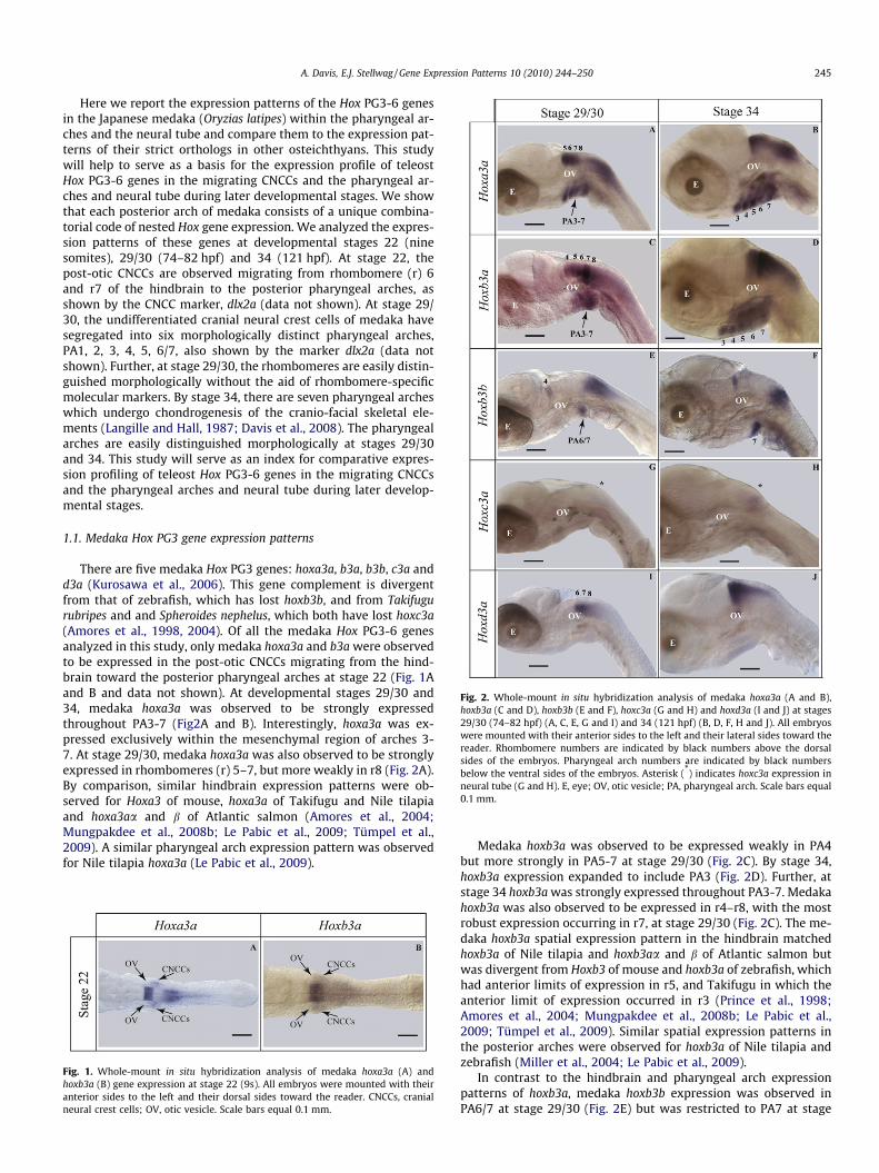
Fig. 2. Whole-mount in situ hybridization analysis of medaka hoxa3a (A and B),hoxb3a (C and D), hoxb3b (E and F), hoxc3a (G and H) and hoxd3a (I and J) at stages29/30 (74–82 hpf) (A, C, E, G and I) and 34 (121 hpf) (B, D, F, H and J). All embryoswere mounted with their anterior sides to the left and their lateral sides toward thereader. Rhombomere numbers are indicated by black numbers above the dorsalsides of the embryos. Pharyngeal arch numbers are indicated by black numbersbelow the ventral sides of the embryos. Asterisk (*) indicates hoxc3a expression inneural tube (G and H). E, eye; OV, otic vesicle; PA, pharyngeal arch. Scale bars equal0.1 mm.
A. Davis, E.J. Stellwag / Gene Expression Patterns 10 (2010) 244–250 245
Here we report the expression patterns of the Hox PG3-6 genesin the Japanese medaka (Oryzias latipes) within the pharyngeal ar-ches and the neural tube and compare them to the expression pat-terns of their strict orthologs in other osteichthyans. This studywill help to serve as a basis for the expression profile of teleostHox PG3-6 genes in the migrating CNCCs and the pharyngeal ar-ches and neural tube during later developmental stages. We showthat each posterior arch of medaka consists of a unique combina-torial code of nested Hox gene expression. We analyzed the expres-sion patterns of these genes at developmental stages 22 (ninesomites), 29/30 (74–82 hpf) and 34 (121 hpf). At stage 22, thepost-otic CNCCs are observed migrating from rhombomere (r) 6and r7 of the hindbrain to the posterior pharyngeal arches, asshown by the CNCC marker, dlx2a (data not shown). At stage 29/30, the undifferentiated cranial neural crest cells of medaka havesegregated into six morphologically distinct pharyngeal arches,PA1, 2, 3, 4, 5, 6/7, also shown by the marker dlx2a (data notshown). Further, at stage 29/30, the rhombomeres are easily distin-guished morphologically without the aid of rhombomere-specificmolecular markers. By stage 34, there are seven pharyngeal archeswhich undergo chondrogenesis of the cranio-facial skeletal ele-ments (Langille and Hall, 1987; Davis et al., 2008). The pharyngealarches are easily distinguished morphologically at stages 29/30and 34. This study will serve as an index for comparative expres-sion profiling of teleost Hox PG3-6 genes in the migrating CNCCsand the pharyngeal arches and neural tube during later develop-mental stages.
1.1. Medaka Hox PG3 gene expression patterns
There are five medaka Hox PG3 genes: hoxa3a, b3a, b3b, c3a andd3a (Kurosawa et al., 2006). This gene complement is divergentfrom that of zebrafish, which has lost hoxb3b, and from Takifugurubripes and and Spheroides nephelus, which both have lost hoxc3a(Amores et al., 1998, 2004). Of all the medaka Hox PG3-6 genesanalyzed in this study, only medaka hoxa3a and b3a were observedto be expressed in the post-otic CNCCs migrating from the hind-brain toward the posterior pharyngeal arches at stage 22 (Fig. 1Aand B and data not shown). At developmental stages 29/30 and34, medaka hoxa3a was observed to be strongly expressedthroughout PA3-7 (Fig2A and B). Interestingly, hoxa3a was ex-pressed exclusively within the mesenchymal region of arches 3-7. At stage 29/30, medaka hoxa3a was also observed to be stronglyexpressed in rhombomeres (r) 5–7, but more weakly in r8 (Fig. 2A).By comparison, similar hindbrain expression patterns were ob-served for Hoxa3 of mouse, hoxa3a of Takifugu and Nile tilapiaand hoxa3aa and b of Atlantic salmon (Amores et al., 2004;Mungpakdee et al., 2008b; Le Pabic et al., 2009; Tümpel et al.,2009). A similar pharyngeal arch expression pattern was observedfor Nile tilapia hoxa3a (Le Pabic et al., 2009).
Fig. 1. Whole-mount in situ hybridization analysis of medaka hoxa3a (A) andhoxb3a (B) gene expression at stage 22 (9s). All embryos were mounted with theiranterior sides to the left and their dorsal sides toward the reader. CNCCs, cranialneural crest cells; OV, otic vesicle. Scale bars equal 0.1 mm.
Medaka hoxb3a was observed to be expressed weakly in PA4but more strongly in PA5-7 at stage 29/30 (Fig. 2C). By stage 34,hoxb3a expression expanded to include PA3 (Fig. 2D). Further, atstage 34 hoxb3a was strongly expressed throughout PA3-7. Medakahoxb3a was also observed to be expressed in r4–r8, with the mostrobust expression occurring in r7, at stage 29/30 (Fig. 2C). The me-daka hoxb3a spatial expression pattern in the hindbrain matchedhoxb3a of Nile tilapia and hoxb3aa and b of Atlantic salmon butwas divergent from Hoxb3 of mouse and hoxb3a of zebrafish, whichhad anterior limits of expression in r5, and Takifugu in which theanterior limit of expression occurred in r3 (Prince et al., 1998;Amores et al., 2004; Mungpakdee et al., 2008b; Le Pabic et al.,2009; Tümpel et al., 2009). Similar spatial expression patterns inthe posterior arches were observed for hoxb3a of Nile tilapia andzebrafish (Miller et al., 2004; Le Pabic et al., 2009).
In contrast to the hindbrain and pharyngeal arch expressionpatterns of hoxb3a, medaka hoxb3b expression was observed inPA6/7 at stage 29/30 (Fig. 2E) but was restricted to PA7 at stage

Fig. 3. Whole-mount in situ hybridization analysis of medaka hoxa4a (A and B),hoxb4a (C and D), hoxc4a (E and F), hoxd4a (G and H) and hoxd4b (I and J) at stages29/30 (74–82 hpf) (A, C, E, G and I) and 34 (121 hpf) (B, D, F, H and J). All embryoswere mounted with their anterior sides to the left and their lateral sides toward thereader. Rhombomere numbers are indicated by black numbers above the dorsalsides of the embryos. Pharyngeal arch numbers are indicated by black numbersbelow the ventral sides of the embryos. E, eye; OV, otic vesicle; PA, pharyngeal arch;PF, pectoral fin. Scale bars equal 0.1 mm.
246 A. Davis, E.J. Stellwag / Gene Expression Patterns 10 (2010) 244–250
34 (Fig. 2F). In the hindbrain hoxb3b was observed solely in r4 atstage 29/30, which matched the pattern documented for hoxb3baof Atlantic salmon (Mungpakdee et al., 2008b). Interestingly, likehoxb3a, hoxb3b of medaka was shown to have a more anterior limitof expression than Hoxb3 in mouse, which was expressed with ananterior limit at the r4/r5 boundary (reviewed by Tümpel et al.,2009).
Medaka hoxc3a expression was not observed in the hindbrain orpharyngeal arches, although it was observed to be faintly ex-pressed in the neural tube immediately posterior to the hindbrain(see asterisk (*) in Fig. 2G and H). By comparison, Atlantic salmonhoxc3aa and b were expressed in the neural tube differently fromhoxc3a of medaka with anterior limits for both genes in r7 of thehindbrain (Mungpakdee et al., 2008b).
Like medaka hoxc3a, hoxd3a was not observed to be expressedin the pharyngeal arches (Fig. 2I and J), although hoxd3a showedhindbrain expression within r6–r8 at stage 29/30 (Fig. 2I). Thehindbrain expression of medaka hoxd3a was observed to be similarto hoxd3a of Nile tilapia and zebrafish, but divergent from Hoxd3 ofmouse, hoxd3a of Takifugu and hoxd3aa and b of Atlantic salmon,which showed an anterior limit of expression at r5 (Prince et al.,1998; Amores et al., 2004; Mungpakdee et al., 2008b; Le Pabicet al., 2009; Tümpel et al., 2009). For the pharyngeal arches, hoxd3aof Nile tilapia differed from hoxd3a of medaka in that it was ob-served to be expressed in PA4 and 5 (Le Pabic et al., 2009).
1.2. Medaka Hox PG4 gene expression patterns
There are five medaka Hox PG4 genes: hoxa4a, b4a, c4a, d4a andd4b (Kurosawa et al., 2006). This gene complement is identical tothat of Takifugu and S. nephalus but divergent from zebrafish,which lacks hoxd4b (Amores et al., 2004). All five genes were ex-pressed in the hindbrain and pharyngeal arches at both develop-mental stages 29/30 and 34. Medaka hoxa4a was expressed inPA5-7 at stages 29/30 and 34, with expression being the faintestin PA5 (Fig. 3A and B). It was also expressed in the neural tube withan anterior limit at the r7/r8 boundary (Fig. 3A and B). Similar hox-a4a hindbrain expression patterns were observed for Takifugu andNile Tilapia (Amores et al., 2004; Le Pabic et al., 2009). However,medaka hoxa4a expression in the hindbrain was observed to bedivergent from Hoxa4 in mouse, which was expressed with ananterior limit at the r6/r7 boundary (Tümpel et al., 2009). Further,medaka hoxa4a showed more anterior expression in the pharyn-geal arches than that of Nile tilapia, which was only expressed inPA6 and 7 (Le Pabic et al., 2009).
Medaka hoxb4a was expressed in PA5-7 with the strongestexpression observed in the unsegmented primordia of PA6/7 atstage 29/30 (Fig. 3C). By stage 34 the expression became robustin the dorsal domains of PA5 and 6 and both the dorsal and ventraldomains of PA7 (Fig. 3D). Medaka hoxb4a was also expressed in thehindbrain with an anterior limit at the r6/r7 boundary (Fig. 3C),which was similar to the hindbrain expression patterns of Hoxb4in mouse and hoxb4a in zebrafish (Prince et al., 1998; Tümpelet al., 2009). Interestingly, the pharyngeal arch expression patternof medaka hoxb4a diverged from zebrafish hoxb4a, which was ex-pressed in PA4-7 (Miller et al., 2004).
Medaka hoxc4a was expressed faintly in the unsegmented pri-mordia of PA6/7 at stage 29/30 (Fig. 3E). By stage 34, hoxc4a wasobserved to be expressed faintly in PA7 (Fig. 3F). Medaka hoxc4awas expressed more robustly in the hindbrain and posterior neuraltube, where it showed an anterior limit of expression at the r6/r7boundary (Fig. 3E). A similar spatio-temporal expression patternin the pharyngeal arches was observed for tilapia hoxc4a (Le Pabicet al., 2009). However, medaka hoxc4a showed a divergent hind-brain expression pattern from that of hoxc4a of tilapia and Taki-fugu, which both showed an anterior limit of expression at the
r7/r8 boundary, and hoxc4aa and b of Atlantic salmon, which wereboth expressed in the neural tube posterior to the hindbrain(Amores et al., 2004; Mungpakdee et al., 2008b; Le Pabic et al.,2009).
Medaka hoxd4a and d4b were expressed in similar patterns inthe hindbrain and neural tube with anterior limits of expressionat the r6/r7 boundary (Fig. 3G and I). However, these duplicatesshowed divergent expression patterns in the pharyngeal arches,such that hoxd4a was expressed in PA4-7 with the faintest levelsof expression in PA4 at stages 29/30 and 34 (Fig. 3G and H) whilehoxd4b was expressed in PA5-7 at stages 29/30 and 34 with thefaintest levels of expression in PA7 at stage 34 (Fig. 3I and J). Sim-ilar hindbrain expression patterns to medaka hoxd4a were ob-served for Hoxd4 of mouse and hoxd4a of Takifugu and tilapia(Amores et al., 2004; Le Pabic et al., 2009; Tümpel et al., 2009).By contrast, hoxd4b of Takifugu and tilapia differed from that ofmedaka, such that both genes showed an anterior limit of expres-sion at the r7/r8 boundary (Amores et al., 2004; Le Pabic et al.,2009). For the pharyngeal arches, a similar expression pattern to

A. Davis, E.J. Stellwag / Gene Expression Patterns 10 (2010) 244–250 247
medaka hoxd4a was observed in tilapia (Le Pabic et al., 2009). Bycontrast, medaka hoxd4b showed a divergent pharyngeal archexpression pattern from tilapia hoxd4b, which was was observedto be expressed in PA5-7 during early developmental stages but re-stricted to PA5 at chondrogenesis (Le Pabic et al., 2009). In additionto the divergent expression patterns of medaka hoxd4a and d4bwithin the pharyngeal arches, these genes also showed divergencein expression at the level of the pectoral fin buds, such that hoxd4a,but not hoxd4b, expression was observed in these embryonic do-mains at stage 34 (Fig. 3H inset), which suggests that there aredivergent cis-regulatory elements between these medaka Hox geneduplicates that are involved in directing gene expression in thepectoral fin buds.
1.3. Medaka Hox PG5 gene expression patterns
The medaka Hox PG5 gene complement is composed of fourgenes, hoxa5a, b5a, b5b and c5a, and is identical to the Hox PG5gene complements of Takifugu, S. nephalus and zebrafish (Amoreset al., 2004; Kurosawa et al., 2006). All Hox PG5 genes of medakawere observed to be expressed in the neural tube posterior tothe hindbrain (Fig. 4A–H). In the pharyngeal arches, medaka hox-a5a was expressed strongly in PA6-7 at stages 29/30 and 34(Fig. 4A and B). Hoxb5a and b5b were both expressed weakly inthe unsegmented primordia of PA6/7 at stage 29/30 (Fig. 4C andE). By stage 34, medaka hoxb5a expression expanded robustly toinclude PA5-7 (Fig. 4D), whereas hoxb5b was restricted to PA7 with
Fig. 4. Whole-mount in situ hybridization analysis of medaka hoxa5a (A and B),hoxb5a (C and D), hoxb5b (E and F) and hoxc5a (G and H) at stages 29/30 (74–82 hpf)(A, C, E and G) and 34 (121 hpf) (B, D, F and H). All embryos were mounted withtheir anterior sides to the left and their lateral sides toward the reader. Pharyngealarch numbers are indicated by black numbers below the ventral sides of theembryos. E, eye; OV, otic vesicle; PA, pharyngeal arch. Scale bars equal 0.1 mm.
the most robust expression observed in the dorsal domain of thisarch (Fig. 4F). Medaka hoxc5a was not expressed in the pharyngealarches (Fig. 4G and H). A similar hoxc5a expression pattern in theneural tube was observed in tilapia (Le Pabic et al., 2009).
1.4. Medaka Hox PG6 gene expression patterns
The medaka Hox PG6 gene complement is composed of threegenes, hoxb6a, b6b and c6a, and is identical to the Hox PG6 comple-ments of Takifugu, S. nephalus and zebrafish (Amores et al., 2004;Kurosawa et al., 2006). Each of the medaka Hox PG6 genes were ex-pressed in the neural tube posterior to the hindbrain (Fig. 5A–F).Medaka hoxb6a was not expressed in the pharyngeal arches atstage 29/30 (Fig. 5A), but weak expression was detected in the ven-tral domain of PA7 at stage 34 (Fig. 5B). By comparison, medakahoxb6b showed weak expression in the unsegmented primordiaof PA6/7 at stage 29/30 (Fig. 5C) and was restricted to PA7 at stage34, where it was expressed faintly in the dorsal domain and morerobustly in the ventral domain (Fig. 5D). Hoxc6a was not observedto be expressed in the pharyngeal arches (Fig. 5E and F). A similarhoxc6a expression pattern in the neural tube was observed in tila-pia (Le Pabic et al., 2009).
1.5. Evolution of pharyngeal arch expression in medaka
In this paper, we provided evidence for an extensive nestedexpression pattern for the medaka Hox PG3-6 genes within theposterior pharyngeal arches prior to and during chondrogenesisof post-migratory CNCCs into their derivative bony elements. Ourresults show that the medaka Hox genes are expressed with dis-tinct A–P boundaries in the pharyngeal arches, the patterns ofwhich, in many cases, change in position in a developmentalstage-dependent manner. Thus, as in tilapia, the use of Hox genesas molecular markers of the medaka posterior arches is feasible
Fig. 5. Whole-mount in situ hybridization analysis of medaka hoxb6a (A and B),hoxb6b (C and D) and hoxc6a (E and F) at stages 29/30 (74–82 hpf) (A, C and E) and34 (121 hpf) (B, D and F). All embryos were mounted with their anterior sides to theleft and their lateral sides toward the reader. Pharyngeal arch numbers areindicated by black numbers below the ventral sides of the embryos. E, eye; OV, oticvesicle; PA, pharyngeal arch. Scale bars equal 0.1 mm.

248 A. Davis, E.J. Stellwag / Gene Expression Patterns 10 (2010) 244–250
but stage-dependent (Le Pabic et al., 2009). By combining the re-sults of this study with those reported by us previously for HoxPG2 genes (Davis et al., 2008), we provide a combinatorial codeof expressed medaka Hox genes that specifies the identity of eachof the posterior pharyngeal arches (3–7) during the chondrogene-sis of the post-migratory CNCCs into their bony derivatives (stage34) (Fig. 6). The characteristics of this code are that at stage 34,PA3 expresses hoxa2a, b2a, a3a and b3a. PA4 expresses hoxa2a,b2a, a3a, b3a and d4a. PA5 expresses hoxa2a, b2a, a3a, b3a, a4a,b4a, d4a, d4b and b5a. PA6 expresses hoxa2a, b2a, a3a, b3a, a4a,b4a, d4a, d4b, a5a and b5a. PA7 expresses hoxa2a, b2a, a3a, b3a,b3b, a4a, b4a, c4a, d4a, d4b, a5a, b5a, b5b, b6a and b6b, and is theonly arch to express hoxb3b, c4a, b5b, b6a and b6b. Provided thatthe Hox PG2-6 gene expression patterns in the posterior pharyn-geal arches exhibited by medaka are similar to those of other clo-sely related beloniform fishes but not to more evolutionarilydivergent teleosts, it would be tantalizing to suggest that theexpression of the medaka Hox PG2-6 genes in the posterior pha-ryngeal arches may have, in part, resulted in the autopomorphicpharyngeal jaw apparatus structures characteristic of beloniformfishes, including the reduction in size of the second and third epi-branchials (arising from the dorsal regions of the fourth and fifthPAs), the great expansion of the articular surface of the fourth epi-branchial (arising from the dorsal region of PA6) and the presenceof large ventral flanges on the fifth ceratohyal (arising from PA7)(Rosen and Parenti, 1981; Parenti, 1987). Interestingly, severalHox genes in tilapia, but not medaka, were observed to be ex-pressed in the pharyngeal arches including hoxa2b which wasshown to be expressed in PA2 and the posterior arches and hoxd3awhich was shown to be expressed in PA4 and 5 (Le Pabic et al.,2007, 2009). However, it must be stressed that a more completeinvestigation of pharyngeal arch Hox expression in tilapia andother evolutionarily divergent teleosts is necessary to better pre-dict the role played by Hox genes in shaping divergent morpholo-gies derived from the posterior pharyngeal arches.
The stage-dependent medaka Hox gene expression patterns re-ported in this study are suggestive of dynamic cis-regulatory cir-cuitry that direct Hox genes in the migratory and post-migratoryCNCCs. Taking into account the whole-mount in situ hybridizationresults in Davis et al. (2008) we only observed the expression of
Fig. 6. Combinatorial code of Hox PG2-6 gene expression in the posteriorpharyngeal arches of medaka at stage 34 (121 hpf).
four medaka Hox genes, hoxa2a, b2a, a3a and b3a, in the post-oticCNCC stream and throughout all of the posterior pharyngeal ar-ches, which suggests that their expression in these embryonic do-mains may be caused by cis-regulatory sequences that are similarto those that direct mouse Hoxa2 expression in the post-otic CNCCstream and the pharyngeal arches (Maconochie et al., 1999). Bycontrast, all of the other medaka Hox PG3-6 genes analyzed in thisstudy were only observed to be expressed in post-migratory CNCCsat variable A-P boundaries in the posterior pharyngeal arches (seeFig. 6), which suggests that post-migratory CNCC Hox gene expres-sion may be regulated differentially between individual arches. Bycomparison, several cis-regulatory sequences have been shown tobe associated with directing or repressing Hox gene expression inspecific rhombomeres of the hindbrain, such as Krox20-binding se-quences which direct the expression of Hoxa2 and b2 in r3 and r5but repress Hoxb1 from being expressed in these rhombomeres andHox/Pbx- and Prep/Meis-binding sequences which have beenshown to direct the expression of Hoxb1, a2 and b2 in r4 (reviewedby Tümpel et al., 2009). Given that individual pharyngeal archesand rhombomeres give rise to distinct morphological derivatives,it is likely that the medaka Hox PG3-6 gene expression patternsin the post-migratory CNCCs in the posterior phraryngeal archesare patterned by distinct cis-regulatory modules in a similar fash-ion to the rhombomeres of the hindbrain.
1.6. Evolution of hindbrain expression in teleosts
In addition to the medaka Hox PG3-6 expression patterns inthe pharyngeal arches, we observed the spatial expression pat-terns of these genes in the neural tube at stage 29/30. Taking intoaccount the medaka hoxb1a and b1b expression patterns reportedby Hurley et al. (2007) and the Hox PG2 expression patterns re-ported by Davis et al. (2008), we present the spatial expressionpatterns of medaka Hox PG1-4 in the hindbrain during laterdevelopmental stages (stage 29/30 and on) (Fig. 7). r2 expresseshoxa2a alone. r3 expresses hoxa2a and b2a. r4 expresses hoxb1a,b1b, a2a, b2a, b3a and b3b. r5 expresses hoxa2a, b2a, a3a and b3a.r6 expresses hoxa2a, b2a, a3a, b3a and d3a. r7 expresses hoxa2a,b2a, a3a, b3a, d3a, b4a, c4a, d4a and d4b. r8 expresses hoxa2a,b2a, a3a, b3a, d3a, a4a, b4a, c4a, d4a and d4b, which is indicativethat the more posterior rhombomeres express ever increasingnumbers of Hox paralog 2–6 genes. Hoxc3a and all of the HoxPG5-6 genes of medaka are expressed in the neural tube withanterior limits caudally to the hindbrain, results that are consis-tent with the expression patterns documented for Hox PG5-6genes of tetrapods (see Tümpel et al., 2009). Interestingly, manyof the medaka Hox PG2-4 genes exhibit divergent spatial expres-sion patterns in the hindbrain from their strict orthologs in sev-eral evolutionarily divergent teleosts, including zebrafish (hoxb2aand b3a), striped bass (hoxb2a), tilapia (hoxc4a and d4b), Takifugu(hoxa2a, b3a, d3a, c4a and d4b) and salmon (hox c3a and d3a)(Prince et al., 1998; Scemama et al., 2002, 2006; Amores et al.,2004; Le Pabic et al., 2007; Davis et al., 2008; Mungpakdeeet al., 2009a, b) (Fig. 7). Further, in comparison to the Hox PG1-4 spatial expression patterns in the hindbrain of tetrapods (sum-marized in Tümpel et al., 2009), several orthologous Hox genes ofteleosts exhibit divergent anterior limits of expression in thehindbrain, including hoxa2a (Takifugu) b3a (medaka, tilapia andsalmon), b3b (medaka and salmon), d3a (medaka, zebrafish andtilapia), a4a (medaka, tilapia and Takifugu) and d4b (tilapia andTakifugu) (Fig. 7). The cis-regulatory circuitry responsible fordirecting tetrapod Hox PG1-4 gene expression in the hindbrainis summarized in Tümpel et al. (2009). Based on the divergentanterior limits of expression of Hox PG2-4 genes in the hindbrainexhibited by several teleosts in comparison to mouse, it can bededuced that several of the cis-regulatory modules that direct

Fig. 7. Comparison of mouse and teleost Hox PG1-4 gene expression in therhombomeres of the developing hindbrain. Black bars correspond to mouse Hoxgene expression. Blue bars correspond to teleost Hox gene expression. b, stripedbass; f, Takifugu; m, medaka; M, mouse; s, salmon; t, tilapia; z, zebrafish.
A. Davis, E.J. Stellwag / Gene Expression Patterns 10 (2010) 244–250 249
Hox genes in specific rhombomeres of the hindbrain have di-verged between tetrapods and teleosts. Given that the hindbrainexpression patterns of Hox PG2-4 genes appear to be largely con-served in tetrapods but highly divergent in teleosts, future func-tional genomic studies in teleosts are necessary in order to
Table 1Primers used for riboprobe production.
Gene Forward primer (50–30)
hoxa3a GAGATGGCGGAGGGCTGChoxb3a CTCCTCCACAGTCTCCAAGTChoxb3b CAGCAGGCACCGACGCAGhoxc3a CTGGATGACCCTTCGGAChoxd3a GCAACCTATTACGACAACTChoxa4a GCGATTACTACGAGCGAChoxb4a GACTCCCTCTACCACCCCCAChoxc4a TATTTGATGGAGTCTAAChoxd4a GGGCTCGGACTACTACAGhoxd4b TACGCAGAGCCGCAGTTChoxa5a AGCAAACGAGCAATACAGhoxb5a TGTGAACTCGCTGTCGGGGChoxb5b TCTTTCGGCTACAACTAChoxc5a ATTTACTCCTGTCCCGTChoxb6a TCTTATTTTGTGAACCCATChoxb6b CAGCCTCTGTTTGTAACThoxc6a TTTCGTGCCATTTATCCGdlx2a CAACCAGATTACCTCAAACAG
understand how the ray-finned fish-specific whole-genomeduplication resulted in divergent cis-regulatory activity in Hoxgenes of teleosts.
2. Experimental procedures
2.1. Medaka Hox PG3-6 partial cDNA cloning
Medaka Hox PG3-6 partial cDNAs were generated from RT-PCRusing total RNA isolated from stage 29/30 medaka embryos accord-ing to the manufacturer’s procedure (Totally RNA�, Applied Biosys-tems, Foster City, CA). The primers used for the amplification of allmedaka Hox PG3-6 and dlx2a partial cDNAs are listed in Table 1and were designed based on published medaka genomic sequences(Accession numbers: AB232918, AB232920, AB232921, AB232922,AB232923, AB232924 and NM001104820) (Kurosawa et al., 2006;Stock et al., 2006). The PCR products generated from RT-PCR-med-iated amplification of medaka embryonic total RNA were cloned inpCR II vectors (Invitrogen, Carlsbad, CA), according to the manufac-turer’s instructions. Confirmation and orientation of PCR productscorresponding to inserts from plasmid cDNA clones were deter-mined by restriction endonuclease digestion and DNA sequencingusing dideoxyterminator sequencing chemistry (Big Dye v. 3.0, Ap-plied Biosystems, Foster City, CA).
2.2. Whole-mount in situ hybridization
Cultivation of medaka fish and collection, raising, anesthetizing,fixation and dehydration of medaka embryos were performed asdescribed in Davis et al. (2008). Medaka embryos were develop-mentally staged according to Iwamatsu (2004). Whole-mountin situ hybridization was performed according to Davis et al.(2008). All experiments used digoxigenin (DIG)-labeled sense andantisense riboprobes that were produced and purified accordingto Scemama et al. (2006). Sense riboprobes were used in controlexperiments to assess nonspecific binding. Development of DIG-la-beled probe signal, examination of embryos and digital photogra-phy of embryos was performed as described in Scemama et al.(2006). In comparing medaka Hox PG3-6 gene expression patternsto those of other teleosts, morphological features, including themidbrain/hindbrain boundary, rhombomeres (r), otic vesicles(OV), pectoral fins (PF), pharyngeal arches (PA) and somites withindeveloping embryos were used as morphological landmarks.
Reverse primer (50–30) Amplicon length (bp)
TTTCATCCTGCGGTTCTG 477GGGACACCAACTAACATACAG 745GGCTTCAGAGGAGGGGAG 536GGGATAATGATGACCACC 466AGTTCTGTCGGGTCTCCTTC 406TTTCCACTTCATCCTCCG 518AGATGTCCAGAGGGGCGGTTC 611TTTCATCCTGCGGTTCTG 624TTGATTTGCCTCTCGGAC 477ACTTTTACTTGCCGTTCG 569TTCTCCAGTTCCAGGGTC 573ATCTGAGGTGTGTGGGGGTC 636TTCCCATCAGGTCCAGTC 491CTTCATCCGTCTGTTCTG 525TTCAGCAAACAGTCCCTTCC 471TTTTTTCCGCCTGTTCCT 391CTCTCCTTCTTCCATTTC 565AGATGCGTGGTAGAGTTCGTC 732

250 A. Davis, E.J. Stellwag / Gene Expression Patterns 10 (2010) 244–250
Acknowledgements
We would like to thank Dr. Jean-Luc Scemama and Dr. TimChristensen for their technical help.
References
Amores, A., Force, A., Yan, Y.L., Joly, L., Amemiya, C., Fritz, A., Ho, R.K., Langeland, J.,Prince, V., Wang, Y.L., Westerfield, M., Ekker, M., Postlethwait, J.H., 1998.Zebrafish hox clusters and vertebrate genome evolution. Science 282, 1711–1714.
Amores, A., Suzuki, T., Yan, Y.L., Pomeroy, J., Singer, A., Amemiya, C., Postlethwait,J.H., 2004. Developmental roles of pufferfish Hox clusters and genome evolutionin ray-fin fish. Genome Res. 14, 1–10.
Crump, J.G., Swartz, M.E., Eberhart, J.K., Kimmel, C.B., 2006. Moz dependent Hoxexpression controls segment-specific fate maps of skeletal precursors in theface. Development 133, 2661–2669.
Davis, A., Scemama, J.L., Stellwag, E.J., 2008. Japanese medaka Hox paralog group 2:insights into the evolution of Hox PG2 gene composition and expression in theOsteichthyes. J. Exp. Zool. (Mol. Dev. Evol.) 310B, 623–641.
Ferrier, D.E., Minguillon, C., Holland, P.W., Garcia-Fernandez, J., 2000. Theamphioxus Hox cluster: deuterostome posterior flexibility and Hox14. Evol.Dev. 2, 284–293.
Hoegg, S., Boore, J.L., Kuehl, J.V., Meyer, A., 2007. Comparative phylogenomicanalyses of teleost fish Hox gene clusters: lessons from the cichlid fishAstatotilapia burtoni. BMC Genomics 8, 317.
Holland, P.W., Garcia-Fernandez, J., 1996. Hox genes and chordate evolution. Dev.Biol. 173, 382–395.
Hurley, I.A., Scemama, J., Prince, V.E., 2007. Consequences of Hoxb1 duplication inteleost fish. Evol. Dev. 9, 540–554.
Iwamatsu, T., 2004. Stages of normal development in the medaka Oryzias latipes.Mech. Dev. 121, 605–618.
Kimmel, C.B., Miller, C.T., Keynes, R.J., 2001. Neural crest patterning and theevolution of the jaw. J. Anat. 199, 105–120.
Kurosawa, G., Takamatsu, N., Takahashi, M., Sumitomo, M., Sanaka, E., Yamada, K.,Nishii, K., Matsuda, M., Asakawa, S., Ishiguro, H., Miura, K., Kurosawa, Y.,Shimizu, N., Kohara, Y., Hori, H., 2006. Organization and structure of hox geneloci in medaka genome and comparison with those of pufferfish and zebrafishgenomes. Gene 370, 75–82.
Langille, R.M., Hall, B.K., 1987. Development of the head skeleton of the Japanesemedaka, Oryzias latipes (Teleostei). J. Morphol. 193, 135–158.
Le Pabic, P., Stellwag, E.J., Brothers, S.N., Scemama, J.L., 2007. Comparative analysisof Hox paralog group 2 gene expression during Nile tilapia (Oreochromisniloticus) embryonic development. Dev. Genes Evol. 217, 749–758.
Le Pabic, P., Stellwag, E.J., Scemama, J., 2009. Embryonic development andskeletogenesis of the pharyngeal jaw apparatus in the cichlid Nile tilapia(Oreochromis niloticus). Anat. Rec. 292, 1780–1800.
Maconochie, M.K., Krishnamurthy, R., Nonche, S., Meier, P., Manzanares, M.,Mitchell, P.J., Krumlauf, R., 1999. Regulation of Hoxa2 in cranial neural crestcells involves members of the AP-2 family. Development 126, 1483–1494.
McGinnis, W., Krumlauf, R., 1992. Homeobox genes and axial patterning. Cell 68,283–302.
Miller, C.T., Maves, L., Kimmel, C.B., 2004. Moz regulates Hox expression andpharyngeal segmental identity in zebrafish. Development 131, 2443–2461.
Minoux, M., Antonarakis, G.S., Kmita, M., Duboule, D., Rijli, F.M., 2009. Rostral andcaudal pharyngeal arches share a common neural crest ground pattern.Development 136, 637–645.
Moghadam, H., Ferguson, M., Danzmann, R., 2005. Evolution of Hox Clusters inSalmonidae: a comparative analysis between Atlantic salmon (Salmo salar) andrainbow trout (Oncorhynchus mykiss). J. Mol. Evol. 61, 636.
Mungpakdee, S., Seo, H., Angotzi, A.R., Dong, X., Akalin, A., Chourrout, D., 2008a.Differential evolution of the 13 Atlantic salmon hox clusters. Mol. Biol. Evol. 25,1333–1343.
Mungpakdee, S., Seo, H., Chourrout, D., 2008b. Spatio-temporal expression patternsof anterior Hox genes in Atlantic salmon (Salmo salar). Gene Expr. Patterns 8,508–514.
Parenti, L.R., 1987. Phylogenetic aspects of tooth and jaw structure of the Medaka,Oryzias latipes, and other beloniform fishes. J Zool. London 211., 561–572.
Powers, T.P., Amemiya, C.T., 2004. Evidence for a Hox14 paralog group invertebrates. Curr. Biol. 14, R183–R184.
Prince, V.E., Moens, C.B., Kimmel, C.B., Ho, R.K., 1998. Zebrafish hox genes:expression in the hindbrain region of wild-type and mutants of thesegmentation gene, valentino. Development 125, 393–406.
Rosen, D.E., Parenti, L.R., 1981. Relationships of Oryzias, and the groups ofatherinomorph fishes. Am. Mus. Novit. 2719, 1–25.
Santagati, F., Minoux, M., Ren, S.Y., Rijli, F.M., 2005. Temporal requirement of Hoxa2in cranial neural crest skeletal morphogenesis. Development 132, 4927–4936.
Scemama, J.L., Hunter, M., McCallum, J., Prince, V., Stellwag, E., 2002. Evolutionarydivergence of vertebrate Hoxb2 expression patterns and transcriptionalregulatory loci. J. Exp. Zool. 294, 285–299.
Scemama, J.L., Vernon, J.L., Stellwag, E., 2006. Differential expression of Hoxa2a andHoxa2b genes during striped bass embryonic development. Gene Expr. Patterns6, 843–848.
Schaeffer, B., Rosen, D.E., 1961. Major adaptive levels in the evolution of theactinopterygian feeding mechanism. Am. Zool. 1, 187–204.
Stellwag, E.J., 1999. Hox gene duplication in fish. Semin. Cell Dev. Biol. 10, 531–540.Stock, D.W., Jackman, W.R., Trapani, J., 2006. Developmental genetic mechanisms of
evolutionary tooth loss in cypriniform fishes. Development 133, 3127–3137.Tümpel, S., Wiedemann, L.M., Krumlauf, R., 2009. Hox genes and segmentation of
the vertebrate hindbrain. Curr. Top. Dev. Biol. 88, 103–137.