Southern Blotting Technique (Southern Analysis) (NOTES)
10
1 Southern Blotting Technique (Southern Analysis) (NOTES) There are three blotting techniques routinely used in molecular biology for detection of a specific DNA, RNA and protein sequence. In DNA and RNA samples, sequence of nucleotides is detected, while in protein samples, amino acid sequence is detected through blotting technique. Southern blotting is a method for detection of a specific DNA sequence (nucleotide sequence) in DNA samples. Edwin Southern The Southern Blotting Technique was named after its inventor, Edwin Southern. He was a British biochemist who developed this technique in Edinburgh (Scotland) in the 1970s. Edwin Southern
-
Upload
khangminh22 -
Category
Documents
-
view
0 -
download
0
Transcript of Southern Blotting Technique (Southern Analysis) (NOTES)

1
Southern Blotting Technique
(Southern Analysis)
(NOTES)
There are three blotting techniques routinely used in molecular biology for
detection of a specific DNA, RNA and protein sequence. In DNA and RNA samples,
sequence of nucleotides is detected, while in protein samples, amino acid sequence
is detected through blotting technique.
Southern blotting is a method for detection of a specific DNA sequence (nucleotide
sequence) in DNA samples.
Edwin Southern
The Southern Blotting Technique was
named after its inventor, Edwin Southern.
He was a British biochemist who developed
this technique in Edinburgh (Scotland) in
the 1970s.
Edwin Southern

2
Procedure
High molecular-weight DNA strands (large-sized DNA strands) are cut into smaller
fragments using Restriction Endonucleases (RE). Then, the RE-digested DNA
fragments are subjected to Agarose gel electrophoresis in order to separate them
according to size.
Steps in Agarose Gel Electrophoresis
If some of the DNA fragments are larger than 15 kb, then prior to blotting, the gel
may be treated with an acid (such as dilute HCl), which breaks the DNA into smaller
pieces, thus allowing more efficient transfer from the gel to nitrocellulose
membrane (discussed in the nest steps).
The DNA gel is placed into an alkaline solution (NaOH) to denature the double-
stranded DNA fragments into single strands. The denaturation in an alkaline
environment may improve binding of the negatively charged DNA to a positively
charged nitrocellulose membrane (discussion in next steps). It will also help the
hybridization of the radioactive probe with single stranded DNA fragments
(discussion in next steps).

3
Agarose gel (ready to use)
Gel with separated DNA fragments
Transfer of Single stranded DNA fragments to nitrocellulose membrane is
facilitated using buffer solution or salt solution system, which is as follows:
Southern Blotting (1st step)
Filter paper towel
Nitrocellulose membrane
Gel Filter paper wick
Salt solution or buffer solution
Weight (0.75 kg)

4
A piece of sponge (or filter paper wick) is placed in a tank containing the buffer or
salt solution. Later, Agarose gel, carrying single stranded DNA fragments, is put on
top of the sponge.
A sheet of nitrocellulose membrane (nitrocellulose filter) is placed on top of the
gel. To ensure good and even contact between gel and membrane, pressure is
applied evenly to the gel by placing a stack of filter paper-towels (absorbent
material) and a weight (0.5 to 0.75 kg) on top of the membrane and gel.
Buffer solution (or salt solution) is used to prevent drying of the gel and to transfer the DNA fragments from gel to nitrocellulose membrane. Buffer-transfer by capillary action from a region of high water potential to a region of low water potential (usually facilitated by filter paper tissues) is then used to move the DNA from the gel on to the membrane; ion exchange interactions bind the DNA to the nitrocellulose membrane due to the negative charge of the DNA and positive charge of the membrane.
Positions of the DNA fragments on the nitrocellulose membrane are identical to
their positions in the gel.
Southern Blotting Procedure
5
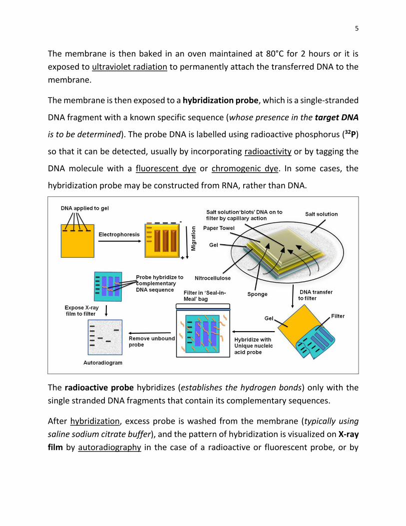
The membrane is then baked in an oven maintained at 80°C for 2 hours or it is
exposed to ultraviolet radiation to permanently attach the transferred DNA to the
membrane.
The membrane is then exposed to a hybridization probe, which is a single-stranded
DNA fragment with a known specific sequence (whose presence in the target DNA
is to be determined). The probe DNA is labelled using radioactive phosphorus (32P)
so that it can be detected, usually by incorporating radioactivity or by tagging the
DNA molecule with a fluorescent dye or chromogenic dye. In some cases, the
hybridization probe may be constructed from RNA, rather than DNA.
The radioactive probe hybridizes (establishes the hydrogen bonds) only with the
single stranded DNA fragments that contain its complementary sequences.
After hybridization, excess probe is washed from the membrane (typically using
saline sodium citrate buffer), and the pattern of hybridization is visualized on X-ray
film by autoradiography in the case of a radioactive or fluorescent probe, or by
6
development of color on the membrane if a chromogenic detection method is
used.
The photographic film (x-ray film), when developed, reveals the DNA fragments
from the original gel that hybridize with the probe
Thus, the procedure allows the specific identification of restriction fragments
containing DNA sequences similar to specific radioactive probe.
Sequences that hybridize with the hybridization probe are further analyzed, for
example, to obtain the full length sequence of the targeted gene. Southern blotting
can also be used to identify methylated sites in particular genes.
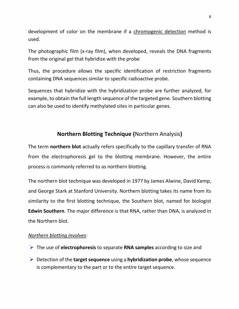
Northern Blotting Technique (Northern Analysis)
The term northern blot actually refers specifically to the capillary transfer of RNA
from the electrophoresis gel to the blotting membrane. However, the entire
process is commonly referred to as northern blotting.
The northern blot technique was developed in 1977 by James Alwine, David Kemp,
and George Stark at Stanford University. Northern blotting takes its name from its
similarity to the first blotting technique, the Southern blot, named for biologist
Edwin Southern. The major difference is that RNA, rather than DNA, is analyzed in
the Northern blot.
Northern blotting involves:
The use of electrophoresis to separate RNA samples according to size and
Detection of the target sequence using a hybridization probe, whose sequence
is complementary to the part or to the entire target sequence.
7
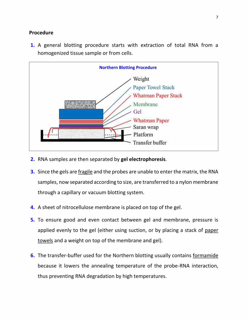
Procedure
1. A general blotting procedure starts with extraction of total RNA from a
homogenized tissue sample or from cells.
Northern Blotting Procedure
2. RNA samples are then separated by gel electrophoresis.
3. Since the gels are fragile and the probes are unable to enter the matrix, the RNA
samples, now separated according to size, are transferred to a nylon membrane
through a capillary or vacuum blotting system.
4. A sheet of nitrocellulose membrane is placed on top of the gel.
5. To ensure good and even contact between gel and membrane, pressure is
applied evenly to the gel (either using suction, or by placing a stack of paper
towels and a weight on top of the membrane and gel).
6. The transfer-buffer used for the Northern blotting usually contains formamide
because it lowers the annealing temperature of the probe-RNA interaction,
thus preventing RNA degradation by high temperatures.

8
Northern Blotting Procedure
Buffer-transfer by capillary action from a region of high water potential to a region
of low water potential (usually filter paper and paper tissues) is then used to move
the RNA from the gel on to the membrane; ion exchange interactions bind the RNA

9
to the membrane due to negative charge of the RNA and positive charge of the
membrane.
Once the RNA has been transferred to the membrane, it is immobilized through covalent linkage to the membrane by UV light or heat.
After a probe has been labeled, it is hybridized to the RNA on the membrane. Experimental conditions that can affect the efficiency and specificity of hybridization include ionic strength, viscosity, duplex length, mismatched base pairs, and base composition.
The membrane is washed to ensure that the probe has bound specifically and to avoid background signals from arising.
The hybrid signals are then detected by X-ray film and can be quantified by densitometry.
Separation of RNA
The RNA samples are most commonly separated on agarose gels containing formaldehyde as a denaturing agent for the RNA to limit secondary structure. The gels can be stained with ethidium bromide (EtBr) and viewed under UV light to observe the quality and quantity of RNA before blotting. Polyacrylamide gel electrophoresis with urea can also be used in RNA separation but it is most commonly used for fragmented RNA or micro RNAs.
Detection with Probes
Probes for northern blotting are composed of nucleic acids with a complementary
sequence to all or part of the RNA of interest, they can be DNA, RNA, or
oligonucleotides with a minimum of 25 complementary bases to the target
sequence. Commonly, cDNA is created with labelled primers for the RNA sequence
of interest to act as the probe in the northern blot.
The probes need to be labelled either with radioactive isotopes (32P) or with
chemiluminescence, which produces detectable emission of light. X-ray film can
detect both the radioactive and chemiluminescent signals and many researchers
prefer the chemiluminescent signals because they are faster, more sensitive, and

10
reduce the health hazards that go along with radioactive labels. The same
membrane can be probed up to five times without a significant loss of the target
RNA.
Applications
1. Northern blotting allows one to observe expression pattern of a particular gene
between tissues, organs, developmental stages, environmental stress levels,
pathogen infection, and over the course of treatment.
2. The expression patterns obtained under given conditions can provide insight
into the function of that gene.
3. Using northern blotting, RNA size can be detected. The quality and quantity of
RNA can be measured on the gel prior to blotting, and the membranes can be
stored and re-probed for years after blotting.