Solution X-ray Absorption Fine Structure Study of the Eu 2+ and Sr 2+ Ions: Unexpected Solvent and...
11
Solution X-ray Absorption Fine Structure Study of the Eu 2+ and Sr 2+ Ions: Unexpected Solvent and Metal Ion Dependencies of the Solvation Numbers Gilles Moreau, ² Rosario Scopelliti, ² Lothar Helm, ² Juris Purans, ²,‡ and Andre ´ E. Merbach* ,² Institute of Molecular and Biological Chemistry, Swiss Federal Institute of Technology Lausanne, EPFL-BCH, CH-1015 Lausanne, Switzerland, and Institute of Solid State Physics, Kengaraga str. 8, LV-1063 Riga, LatVia ReceiVed: May 3, 2002; In Final Form: August 9, 2002 Structural parameters of the Sr 2+ and, for the first time, of the Eu 2+ ions in nonaqueous solutions were determined by the X-ray absorption fine structure (XAFS) method and compared with those of the aqua ions. For both Eu 2+ and Sr 2+ ions, a decrease in coordination number (N) and metal-to-solvent distances was found along the increasingly solvating MeCN < H 2 O , DMF < DMSO solvent series: for strontium, N ) 8 with [Sr(MeCN) 8 ] 2+ (2.665(6) Å) and [Sr(H 2 O) 8 ] 2+ (2.600(3) Å) and N ) 7 with [Sr(DMF) 7 ] 2+ (2.555(5) Å) and [Sr(DMSO) 7 ] 2+ (2.540(7) Å); for europium(II), N ) 8 with [Eu(MeCN) 8 ] 2+ (2.640(4) Å), N ) 7 with [Eu- (H 2 O) 7 ] 2+ (2.584(5) Å), and N ) 6 with [Eu(DMF) 6 ] 2+ (2.541(3) Å) and by extrapolation [Eu(DMSO) 6 ] 2+ (2.525 Å). Smaller coordination numbers are observed for the Eu 2+ ion in O-coordinating solvents. The ionic radii of both Sr 2+ and Eu 2+ ions are very similar, but the slightly softer character of the Eu 2+ ion leads to shorter M-N and longer M-O bonds. Introduction Solvation is one of the most important chemical properties of a metal ion in solution because the solvent exchange reaction is often used as a model for the interpretation of the substitution reaction mechanisms. As a prerequisite, it is important to know the structure of the solvated ion in solution to interpret and understand the thermodynamics and kinetics of the complex formation reactions. The relative strength of the metal ion- solvent interactions is dependent on the electron-pair donor and acceptor properties of both solvent and metal ion. To better understand the solvation process of an ion in solution, it is therefore necessary to study it in a series of solvents with different properties. Pursuing the parallel structural study of the Eu 2+ and Sr 2+ ions in aqueous medium, 1 we have investigated the solvation structure of these two ions in dimethyl formamide (DMF), dimethyl sulfoxide (DMSO), and acetonitrile (MeCN) solutions in the present work. Whereas water is a hard polar and protic solvent with extensive hydrogen bonding, DMF and DMSO are aprotic solvents with high dipole moments and medium permittivities. 2 For these three solvents, the solvation of hard metal ions such as Sr 2+ and presumably Eu 2+ occurs via the solvent oxygen atom. MeCN is a softer nitrogen-binding solvent with dipole moment and permittivity similar to those of the DMF and DMSO. 2 Hard metal ions are readily dissolved by water, DMF, and DMSO and have a relatively low solubility in MeCN. The available information on the properties of the Eu 2+ ion is poor, especially in nonaqueous solutions, owing to its redox instability. It has been pointed out that strong similarities exist between divalent lanthanide ions and the alkaline earth metals. 3 However, until now, quantitative assessment of the similarity of these groups of elements remains extremely limited. 3 In the present work, the structures of the solvated Eu 2+ and Sr 2+ ions are studied in the oxygen-donor solvents dimethyl sulfoxide and N,N-dimethyl formamide and in the nitrogen- donor acetonitrile. The experimental L 3 -edge X-ray absorption fine structure (XAFS) spectra of Eu 2+ , as well as the Sr 2+ K-edge, have been measured, analyzed, and compared using the cumulant approach combined with efficient analysis techniques. 4-6 Both theoretical (calculated ab initio 7 ) and experimental (extracted from crystalline reference 8 ) phases and amplitudes have been used. Experimental Section Chemicals. Sr(DMF) 2 (O 3 SCF 3 ) 2 and Sr(DMSO) 2 (O 3 SCF 3 ) 2 were prepared under nitrogen according to the following procedure. Two equivalents of 98% CF 3 SO 3 H (triflic acid) was added to a suspension of 1 g of SrCO 3 in 10 equiv of HC- (OEt) 3 . After filtration, 2.2 equiv of the desired solvent was added, and the solution was evaporated gently under vacuum until precipitation. The precipitate was redissolved by heating, and crystals were filtered off by centrifugation after slow cooling. Anal. Calcd for Sr(DMF) 2 (O 3 SCF 3 ) 2 : C, 18.1; H, 2.6; N, 5.3; Sr, 16.5. Found: C, 18.1; H, 2.4; N, 5.0; Sr, 16.9. Anal. Calcd for Sr(DMSO) 2 (O 3 SCF 3 ) 2 : C, 13.3; H, 2.2; Sr, 16.2. Found: C, 13.6; H, 2.0; Sr, 17.0. The following procedure was used to obtain the starting materials used in the preparation of the Eu 2+ and Sr 2+ solutions. Two equivalents of 98% triflic acid was added to a suspension of 1 g of EuCO 3 9 or SrCO 3 in 10 equiv of HC(OEt) 3 . After filtration, pentane was added to the solution to precipitate quantitatively the metal triflate. After filtration and 72 h under vacuum, Eu(O 3 SCF 3 ) 2 ‚EtOH or Sr(O 3 SCF 3 ) 2 ‚1/2EtOH salts were obtained. Anal. Calcd for Eu(O 3 SCF 3 ) 2 ‚EtOH: C, 9.6; H, 1.2; Eu, 30.6. Found: C, 9.5; H, 1.1; Eu, 29.0. Anal. Calcd for Sr(O 3 SCF 3 ) 2 ‚1/2EtOH: C, 8.8; H, 0.7; Sr, 21.4. Found: C, 8.8; H, 0.7; Sr, 22.0. For all compounds, the absence of water was checked by Karl Fischer titration. Triflic acid (98%) was purchased from Aldrich Chemicals. All commercial compounds were used as * To whom correspondence should be addressed. Phone: +41-21-693 98 71. Fax: +41-21-693 98 75. E-mail: [email protected]. ² Swiss Federal Institute of Technology Lausanne. ‡ Institute of Solid State Physics. 9612 J. Phys. Chem. A 2002, 106, 9612-9622 10.1021/jp026061w CCC: $22.00 © 2002 American Chemical Society Published on Web 09/19/2002
Transcript of Solution X-ray Absorption Fine Structure Study of the Eu 2+ and Sr 2+ Ions: Unexpected Solvent and...

Solution X-ray Absorption Fine Structure Study of the Eu2+ and Sr2+ Ions: UnexpectedSolvent and Metal Ion Dependencies of the Solvation Numbers
Gilles Moreau,† Rosario Scopelliti,† Lothar Helm, † Juris Purans,†,‡ and Andre E. Merbach*,†
Institute of Molecular and Biological Chemistry, Swiss Federal Institute of Technology Lausanne, EPFL-BCH,CH-1015 Lausanne, Switzerland, and Institute of Solid State Physics, Kengaraga str. 8, LV-1063 Riga, LatVia
ReceiVed: May 3, 2002; In Final Form: August 9, 2002
Structural parameters of the Sr2+ and, for the first time, of the Eu2+ ions in nonaqueous solutions weredetermined by the X-ray absorption fine structure (XAFS) method and compared with those of the aqua ions.For both Eu2+ and Sr2+ ions, a decrease in coordination number (N) and metal-to-solvent distances was foundalong the increasingly solvating MeCN< H2O , DMF < DMSO solvent series: for strontium,N ) 8 with[Sr(MeCN)8]2+ (2.665(6) Å) and [Sr(H2O)8]2+ (2.600(3) Å) andN ) 7 with [Sr(DMF)7]2+ (2.555(5) Å) and[Sr(DMSO)7]2+ (2.540(7) Å); for europium(II),N ) 8 with [Eu(MeCN)8]2+ (2.640(4) Å),N ) 7 with [Eu-(H2O)7]2+ (2.584(5) Å), andN ) 6 with [Eu(DMF)6]2+ (2.541(3) Å) and by extrapolation [Eu(DMSO)6]2+
(2.525 Å). Smaller coordination numbers are observed for the Eu2+ ion in O-coordinating solvents. The ionicradii of both Sr2+ and Eu2+ ions are very similar, but the slightly softer character of the Eu2+ ion leads toshorter M-N and longer M-O bonds.
Introduction
Solvation is one of the most important chemical propertiesof a metal ion in solution because the solvent exchange reactionis often used as a model for the interpretation of the substitutionreaction mechanisms. As a prerequisite, it is important to knowthe structure of the solvated ion in solution to interpret andunderstand the thermodynamics and kinetics of the complexformation reactions. The relative strength of the metal ion-solvent interactions is dependent on the electron-pair donor andacceptor properties of both solvent and metal ion. To betterunderstand the solvation process of an ion in solution, it istherefore necessary to study it in a series of solvents withdifferent properties. Pursuing the parallel structural study of theEu2+ and Sr2+ ions in aqueous medium,1 we have investigatedthe solvation structure of these two ions in dimethyl formamide(DMF), dimethyl sulfoxide (DMSO), and acetonitrile (MeCN)solutions in the present work.
Whereas water is a hard polar and protic solvent withextensive hydrogen bonding, DMF and DMSO are aproticsolvents with high dipole moments and medium permittivities.2
For these three solvents, the solvation of hard metal ions suchas Sr2+ and presumably Eu2+ occurs via the solvent oxygenatom. MeCN is a softer nitrogen-binding solvent with dipolemoment and permittivity similar to those of the DMF andDMSO.2 Hard metal ions are readily dissolved by water, DMF,and DMSO and have a relatively low solubility in MeCN.
The available information on the properties of the Eu2+ ionis poor, especially in nonaqueous solutions, owing to its redoxinstability. It has been pointed out that strong similarities existbetween divalent lanthanide ions and the alkaline earth metals.3
However, until now, quantitative assessment of the similarityof these groups of elements remains extremely limited.3
In the present work, the structures of the solvated Eu2+ andSr2+ ions are studied in the oxygen-donor solvents dimethylsulfoxide andN,N-dimethyl formamide and in the nitrogen-donor acetonitrile. The experimental L3-edge X-ray absorptionfine structure (XAFS) spectra of Eu2+, as well as the Sr2+
K-edge, have been measured, analyzed, and compared usingthe cumulant approach combined with efficient analysistechniques.4-6 Both theoretical (calculated ab initio7) andexperimental (extracted from crystalline reference8) phases andamplitudes have been used.
Experimental Section
Chemicals.Sr(DMF)2(O3SCF3)2 and Sr(DMSO)2(O3SCF3)2
were prepared under nitrogen according to the followingprocedure. Two equivalents of 98% CF3SO3H (triflic acid) wasadded to a suspension of 1 g of SrCO3 in 10 equiv of HC-(OEt)3. After filtration, 2.2 equiv of the desired solvent wasadded, and the solution was evaporated gently under vacuumuntil precipitation. The precipitate was redissolved by heating,and crystals were filtered off by centrifugation after slowcooling. Anal. Calcd for Sr(DMF)2(O3SCF3)2: C, 18.1; H, 2.6;N, 5.3; Sr, 16.5. Found: C, 18.1; H, 2.4; N, 5.0; Sr, 16.9. Anal.Calcd for Sr(DMSO)2(O3SCF3)2: C, 13.3; H, 2.2; Sr, 16.2.Found: C, 13.6; H, 2.0; Sr, 17.0. The following procedure wasused to obtain the starting materials used in the preparation ofthe Eu2+ and Sr2+ solutions. Two equivalents of 98% triflicacid was added to a suspension of 1 g of EuCO3
9 or SrCO3 in10 equiv of HC(OEt)3. After filtration, pentane was added tothe solution to precipitate quantitatively the metal triflate. Afterfiltration and 72 h under vacuum, Eu(O3SCF3)2‚EtOH orSr(O3SCF3)2‚1/2EtOH salts were obtained. Anal. Calcd forEu(O3SCF3)2‚EtOH: C, 9.6; H, 1.2; Eu, 30.6. Found: C, 9.5;H, 1.1; Eu, 29.0. Anal. Calcd for Sr(O3SCF3)2‚1/2EtOH: C,8.8; H, 0.7; Sr, 21.4. Found: C, 8.8; H, 0.7; Sr, 22.0.
For all compounds, the absence of water was checked byKarl Fischer titration. Triflic acid (98%) was purchased fromAldrich Chemicals. All commercial compounds were used as
* To whom correspondence should be addressed. Phone:+41-21-69398 71. Fax:+41-21-693 98 75. E-mail: [email protected].
† Swiss Federal Institute of Technology Lausanne.‡ Institute of Solid State Physics.
9612 J. Phys. Chem. A2002,106,9612-9622
10.1021/jp026061w CCC: $22.00 © 2002 American Chemical SocietyPublished on Web 09/19/2002

received. Solvents were freshly distilled before use accordingto the literature-recommended methods.2 They were distilledunder oxygen-free nitrogen, degassed by a triple freeze-pump-thaw cycle, and stored in an oxygen-free and water-freeglovebox. The Karl Fischer coulometric titration gave for DMF,DMSO, and MeCN a respective maximum water amount of 18,12, and 10 ppm.
Preparation of the Samples.The Sr2+ solutions (0.15 M inDMF, 0.15 M in DMSO, 0.09 M in MeCN) were prepared bydissolution of the Sr(O3SCF3)2‚1/2EtOH salt into pure solvent.The Eu2+ solutions (0.15 M in DMF, 0.10 M in MeCN) wereprepared by dissolution of the Eu(O3SCF3)2‚EtOH salt into puresolvent. Additionally, the solutions were treated by amalgamatedzinc just before the XAFS measurements to avoid traces ofEu3+.1 Concentrations were checked by complexometric titra-tion. The L3-edge X-ray absorption near-edge structure (XANES)measurements have confirmed that sealed oxygen-free samplesof ca. 0.1 M Eu(O3SCF3)2 nonaqueous solutions are stable inthe multipurpose cell that we used for at least 5 h (less than0.2% Eu3+).1
The reference crystalline samples Sr(DMF)2(O3SCF3)2 andSr(DMSO)2(O3SCF3)2 were finely ground and mechanicallymixed with cellulose powder to give pressed pellets withthickness chosen to obtain an absorption jump value of about1.
All of the compounds and solutions were prepared, handled,and stored in the dry nitrogen atmosphere of a glovebox to avoidboth oxidation of the Eu2+ and water contamination of thecompounds and solutions.
X-ray Experimental Section.Suitable crystals of Sr(DMF)2-(O3SCF3)2 and Sr(DMSO)2(O3SCF3)2 were obtained as de-scribed and mounted in glass capillaries. Crystal data andstructure refinement details are listed in Table 1. Data collectionswere performed on an Oxford diffraction Kuma4 sapphire CCDand data reductions were carried out with CrysAlis RED.10
Absorption correction was applied to both data sets. For theSr(DMF)2(O3SCF3)2, the MULTI-SCAN semiempirical method11
has been employed, whereas for the Sr(DMSO)2(O3SCF3)2, theDIFABS empirical method12 has been used. Structure solution
for both compounds was performed with SIR97.13 The structureswere refined using the full-matrix least-squares onF2 with allnon-H atoms anisotropically defined. H atoms were placed incalculated positions using the “riding model” for Sr(DMSO)2-(O3SCF3)2, whereas they have been treated as free isotropicatoms for Sr(DMF)2(O3SCF3)2 (in this latter case, likely becauseof disorder combined with special position for a methyl group,the distances C-H and H‚‚‚H have been restrained to get areasonable geometry). Space group determination, structuresolution, refinement, molecular graphics, and geometricalcalculation have been carried out on both structures with theSHELXTL software package.14
XAFS Measurements.XAFS measurements were performedat the LURE synchrotron radiation facility (Orsay, France) onthe DCI D21 (XAS 2) beam line. Positron-beam energy andaverage current were 1.85 GeV and 320-250 mA, respectively.The XAFS spectra of the Eu L3-edge (6976 eV; scan 6900-7650 eV) and Sr K-edge (16 105 eV; 16 000-17 000 eV) weremeasured in transmission mode. The synchrotron radiation wasmonochromatized using the Si(311) double-crystal monochro-mator, and in the case of the Eu L3-edge, harmonic rejectionwas achieved using dedicated mirrors. The experimental spectrawere measured using two ionization chambers (filled with airfor Eu and with Ar for Sr measurements) with a count rate of2 s per point, an energy resolution of 1 or 2 eV, a 0.5 or 1 eVstep in the XANES region, and a 1 or 2 eVstep in the extendedx-ray absorption fine structure (EXAFS) region for the Eu L3-edge and the Sr K-edge, respectively. A multipurpose X-rayabsorption cell15 was used for the in-situ XAFS measurementsof sealed oxygen- and water-free solutions. The measurementswere done at an optical length of 1-1.5 mm for Eu and 4-8mm for Sr, resulting in values of the absorption jump of about0.5 (WL amplitude about 2) for Eu and about 1 for Sr. At leasttwo complete and identical XAFS scans were collected for eachsolution. All samples were measured at room temperature (20-25 °C).
Data Analysis. The experimental XAFS data were treatedusing the EDA software package16 in a way similar to the oneused in the study of the Eu2+ and Sr2+ aqua ions.1 The obtainedXAFS spectra,ø(E), were converted to thek-space of the
photoelectron wavevector, defined ask ) x(2m/p2)(E-E0),where (E - E0) is the photoelectron kinetic energy measured.The experimental XAFS spectraø(k) of both Eu and Sr weremultiplied by a factork3 to compensate for the decrease ofamplitude with increasing wavevector value.
The experimental XAFS spectra (Figures 1 and 2) wereFourier-transformed (FT) with a Kaiser-Bessel window in the0-12 Å-1 range for Eu and 0-13 Å-1 for Sr using aphotoelectron phase-shift correction. The first shell XAFScontributions were singled out by back FT procedure in the 1.9-3.2 Å range for both Eu2+ and Sr2+. Use of the phase-shiftcorrection allowed us to reduce the nonstructural peaks distortingthe baseline and led to a significant sharpening of the first shellpeak, allowing a more precise Fourier filtering.1 The use of thecorrected Fourier-filtering procedure led to a real increase inthe fitting reproducibility when playing on the parameters.
The first-shell XAFS spectra were fitted using the single-scattering curved-wave formalism with cumulant expansion:4
TABLE 1: Crystal Data and Details of the StructureDetermination
Sr(DMF)2(O3SCF3)2 Sr(DMSO)2(O3SCF3)2
chemical formula C8H14F6N2O8S2Sr C6H12F6O8S4Srfw 531.95 542.02cryst syst orthorhombic monoclinicspace group Ibam I2/aa (Å) 11.6800(10) 8.4221(13)b (Å) 20.5299(18) 11.5766(16)c (Å) 8.0429(15) 18.420(3)â (deg) 90 93.263(12)vol (Å3) 1928.6(4) 1793.0(4)Z 4 4Dcalcd (g cm-3) 1.832 2.008F(000) 1056 1072µ (mm-1) 3.105 3.564temp (K) 143 143wavelength (Å) 0.710 73 0.710 73measd reflns 5620 5084unique reflns 874 1512unique reflns [I > 2σ(I)] 815 1165data/params 874/93 1512/115Ra [I > 2σ(I)] 0.0346 0.0919wR2a (all data) 0.0934 0.2626GOFb 1.086 1.179
a R) ∑||Fo| - |Fc||/∑|Fo|; wR2) {∑[w(Fo2 - Fc
2)2]/∑[w(Fo2)2]}1/2.
b GOF) {∑[w(Fo2 - Fc
2)2]/(n - p)}1/2, wheren is the number of dataandp is the number of parameters refined.
ø(k) ) N
kC12f(π,k) exp(-
2C1
λ(k)) exp(- 2C2k2 + 2
3C4k
4) ×
sin(2kC1 - 43C3k
3 + φ(π,k)) (1)
Solution XAFS Study of the Eu2+ and Sr2+ Ions J. Phys. Chem. A, Vol. 106, No. 41, 20029613

whereN is the coordination number andC2 ) σ2 is the Debye-Waller (DW) factor (in harmonic approximation). The higher-order cumulantsC3 and C4 characterize the deviation of thedistribution of distances from a Gaussian shape. The first
cumulantC1 is closely related to the interatomic distanceR.17
λ(k) ) k/Γ is an adjustable function that models the lowkdamping factors. As in previous works,1 the Γ parameter wasallowed to vary during the fitting procedure for fine adjustmentbetween theoretical calculations and experiment. This parameteralso allows us to compensate for the FT boundary effects andwas found within classical range (from 0 to 0.1).
Phases and Amplitudes.In this paper, the strontium XAFSdata were analyzed using two different approaches: the phasesφ(π,k) and amplitudesf(π,k) were either calculated or obtainedexperimentally.
We showed in a precedent paper1 the importance in the choiceof the reference cluster used for the theoretical backscatteringamplitudes and phases calculations using the FEFF6 code.7 Tofind a cluster that mimics well the possible environment of theEu2+ and Sr2+ ions in solution, we checked the literature forcrystallographic structures in which a cation is surrounded onlyby the solvent molecules, that is, DMF, DMSO, or MeCN.Because of their ionic radii close to the Eu2+ and Sr2+ ones,the following compounds have been retained: (1) [Sr(H2O)8]-(OH)2,1,18 (2) Sr(DMF)2(O3SCF3)2, (3) [Cd(DMSO)6][CdI4],19
(4) [Gd(DMSO)8][Fe(CN)6],20 (5) [Na(MeCN)6][TaCl6],21 and(6) [Yb(MeCN)8](AlCl 4)3.22 The ionic radii of these ions beingslightly different than those of the Eu2+ and Sr2+, scaling factorswere applied for the M-O or M-N distances to match thedistances in the system studied (see Table 2 for detail). Usingthe respective crystallographic coordinates of the selectedcompounds, we calculated the following clusters to obtaintheoreticalφ(π,k) andf(π,k) functions: (1) MO8(H2O)16(OH)8,(2) M(OCNC2)4(O3SCF3)4, (3) M(OSC2)6, (4) M(OSC2)8, (5)M(NCC)6, and (6) M(NCC)8. The computed muffin-tin radiifor the different clusters are detailed in Table 2.
Because the threshold energy of the photoelectronE0 isdefined in the FEFF6 code7 relative to the Fermi level anddepends on the muffin-tin radii, the spectra have to be correctedto avoidE0 difference errors in the fitting process. Consequently,the phase differences between theoretical and experimentalspectra were set to zero at lowk, according to Bunker andStern’s criterion,23 and E0 was allowed to vary for fineadjustment during the fitting procedure.
Experimental f(π,k) and φ(π,k) were extracted from theexperimental XAFS data obtained on the crystalline Sr(DMF)2-(O3SCF3)2. These functions were obtained assuming Gaussiandistribution of distances withN ) 8 andR ) 2.585 Å, fromthe crystallographic data, andσ2 ) 0.0106 Å2 from the fit withtheoretical phase and amplitude. Experimentalf(π,k) andφ(π,k)were also extracted from the experimental XAFS spectra of theEu2+ and Sr2+ aqueous solution and normalized according tothe structural parameters determined in ref 1.
Note that use of experimental phases and amplitudes allows,to a certain extent, the compensation of systematic errors
Figure 1. Experimental XAFS spectra of Sr(O3SCF3)2 0.14 M in H2O,0.09 M in MeCN, 0.15 M in DMF, and 0.15 M in DMSO (arrows, seetext).
Figure 2. Experimental XAFS spectra of Eu(O3SCF3)2 0.15 M in H2O,0.10 M in MeCN, and 0.15 M in DMF (arrows, see text).
TABLE 2: Muffin-Tin Radii ( RMT) for the First-ShellAtomsa
computed cluster RMT (Sr) RMT (O/N) RMT (Eu) RMT (O/N)
MO8(H2O)16(OH)8 b 1.491 1.143 1.481 1.126M(OCNC2)4(O3SCF3)4
c 1.658 0.730 1.662 0.732M(OSC2)6
d 1.563 0.820 1.566 0.819M(OSC2)8
e 1.634 0.758 1.637 0.758M(NCC)6 f 1.724 0.634 1.726 0.636M(NCC)8 g 1.666 0.689 1.668 0.690
a All of the muffin-tin radii are given in Å.b Based on [Sr(H2O)8](OH)2
XRD structure, ref 18 (scaling factor, SF) 1). c Sr(DMF)2(O3SCF3)2,this study (SF ) 1). d [Cd(DMSO)6][CdI4], ref 19 (SF ) 1.1).e [Gd(DMSO)8][Fe(CN)6], ref 20 (SF) 1.05). f [Na(MeCN)6][TaCl6],ref 21 (SF) 1.05). g [Yb(MeCN)8](AlCl 4)3, ref 22 (SF) 1.1).
9614 J. Phys. Chem. A, Vol. 106, No. 41, 2002 Moreau et al.

because they include the contribution of the mean free path, ofthe multielectron amplitude reduction factorS0
2, of glitches andof resolution, reducing the number of adjustable parameters andconsequently increasing the reliability of the fitted results. Toensure the phases and amplitudes transferability and to allowestimation of systematic errors, all of the data were analyzedin a similar way, using the same theoretical phases andamplitudes, filtering procedures, and parameters.
Multielectron Transition Effect. The same kind of anoma-lous features observed in the Sr2+ aqua ion XAFS spectrum1,18,24
are present in the XAFS spectra of the Sr2+ ion in the DMF,DMSO, and MeCN solutions (Figure 1). This anomalous featuredue to the simultaneous excitation of 1s3d electrons is indicatedby an arrow in Figure 1 and arises around 6.4 Å-1. Similar
multielectron transition effects (MET) features are found forthe Eu2+ solutions at ca. 6.2 Å-1 (Figure 2). As in the case ofthe Eu2+ aqua ion,1 we attribute this sharp contribution to a2p4d double-electron transition.
As we already stated,1 these anomalies result after Fouriertransform in humps distorting the base of the major peakstanding for the first-shell Sr-O or Eu-O peak, especially atlow distances. But the use of the new 2001 version of the EDAsoftware package16 combined with the phase-shift correctedFourier filtration allowed us a greater precision in the zero-lineremoval and in the Fourier-filtering processes, so no subsequentadditional MET removal was necessary.
Results
X-ray Crystal Structure of Sr(DMF) 2(O3SCF3)2 andSr(DMSO)2(O3SCF3)2. The crystal structure of Sr(DMF)2-(O3SCF3)2 (Figure 3) consists of eight coordinate Sr2+ cationslying on a 222 symmetry site. Each metal ion is linked to fourDMF ligands and to four CF3SO3
-. Each ligand lies on asymmetry plane and bridges two Sr2+ ions by means of oneoxygen atom in the case of the DMF and by means of twodifferent oxygen atoms in the case of CF3SO3
-. Thus, the overallstructure (Figure 4) is formed by infinite one-dimensional chainsalong thec axis. The eight oxygen atoms around each Sr2+ ionform an almost perfect square antiprism geometry (with acharacteristicR angle25 of 57.5° for O1 and 57.7° for O3). Sr-Obond distances reflect the different nature of the two ligands[Sr1-O1, 2.567(2) Å; Sr1-O3, 2.578(2) Å], leading to anaverage Sr-O bond length of 2.572 Å. Within the polymericchains responsible for the crystal packing, the Sr‚‚‚Sr distanceis 4.021(1) Å. The chains are held together mostly by electro-static and steric interactions involving, respectively, the CF3 andthe CH3 groups, which lie at the surface of such tubular chains[F‚‚‚F, 2.864(5)-2.867(4) Å]. Noteworthy is the presence of a
Figure 3. Ball-and-stick representation of the Sr(DMF)2(O3SCF3)2
coordination polyhedron.
Figure 4. Crystal packing of the Sr(DMF)2(O3SCF3)2 compound along thec axis.
Solution XAFS Study of the Eu2+ and Sr2+ Ions J. Phys. Chem. A, Vol. 106, No. 41, 20029615

weakintramolecularhydrogen bond C-H‚‚‚F [C2-H2, 0.93(5)Å; H2‚‚‚F2, 2.53(5) Å; C2‚‚‚F2, 3.456(6) Å; C2-H2‚‚‚F2,176(5)°]. Geometrical parameters dealing with the same struc-ture collected at room temperature instead of low temperatureshow the following features: (1) larger gap between the Sr-Obond lengths [Sr1-O1, 2.578(3) Å; Sr1-O3, 2.600(2) Å;average Sr-O, 2.589 Å]; (2) larger Sr‚‚‚Sr distance [4.067(1)Å] due to the increase of the volume of the cell.
The structure of Sr(DMSO)2(O3SCF3)2 (Figure 5) is similarto the one already discussed but shows less symmetry. It is madeof one-dimensional chains along thea axis (Figure 6) with themetal cation lying on a binary axis. The coordination geometryaround the Sr2+ ion is square antiprism showing some slight
deviation [R values ranging from 54.4° to 63.3°]. As forSr(DMF)2(O3SCF3)2, the Sr-O bond distances are different forthe two ligands [Sr-O(O3SCF3)av, 2.587(8) Å and Sr-O(DM-SO)av, 2.649(8) Å], but the difference is greatly enhanced.However, the way that ligands bridge the Sr2+ ions is exactlythe same as that observed in Sr(DMF)2(O3SCF3)2. Within thepolymeric chains, the Sr‚‚‚Sr distance is 4.215(1) Å. The crystalpacking shows steric and electrostatic interactions among theCH3 and CF3 groups [F‚‚‚F distances vary from 2.80(1) to2.84(1) Å], as well as weakintra- andintermolecularhydrogenbonds between the methyl groups and the CF3SO3
- O2 and O3atoms.
The homologous Eu2+ compounds have not been reported inthat study; all attempts to precipitate a Eu2+ DMF solvate leadto an oxidation characterized by a Eu3+ signal in the XANESspectrum. However, no detectable oxidation occurs for at least3 h during the Eu2+ DMF solution measurement.
XAFS Analysis of the Eu2+ and Sr2+ Ions in DMF, DMSO,and MeCN Solutions. In a previous paper, we reported theXAFS study of the Eu2+ and Sr2+ ions first coordination spherein aqueous solution. The Sr2+ ion was found to be octahydratedin aqueous solution, whereas the Eu2+ ion occurs as anequilibrium between a highly predominant [Eu(H2O)7]2+ ionand a minor [Eu(H2O)8]2+ species.1
In aqueous solutions, it is well established that the triflateion does not enter the first coordination sphere of solvated metalions. In the nonaqueous DMF, DMSO, and MeCN solvents withsmaller dielectric constants than water, it is important to ensurethat no inner sphere ion pairing occurs between the metal andthe triflate ions. Ion pair formation would strongly complicatethe analysis of the XAFS data. Fortunately, the CF3SO3
- ion isa weak nucleophile, although there is some evidence that it isa slightly stronger coordinator than ClO4
- in solution. However,
Figure 5. Ball-and-stick representation of the Sr(DMSO)2(O3SCF3)2
coordination polyhedron.
Figure 6. Crystal packing of the Sr(DMSO)2(O3SCF3)2 compound along thec axis.
9616 J. Phys. Chem. A, Vol. 106, No. 41, 2002 Moreau et al.

CF3SO3- is the best counterion for our purpose as ClO4
- ispotentially explosive in nonaqueous solutions or in the presenceof a highly reducing agent such as Eu2+. The increasingsolvation series MeCN< H2O ∼ DMF < DMSO26 has beenestablished for hard metal ions such as trivalent lanthanides.Therefore, the triflate ion will not enter the first coordinationsphere in DMF and DMSO, which are better coordinatingsolvents than water. Note that the coordination of the triflateion in the solid solvates Sr(DMF)2(O3SCF3)2 and Sr(DMSO)2-(O3SCF3)2 may be explained by packing interactions in thecrystals. Ion pairing is more likely to happen in the less-coordinating solvent MeCN. However, in this solvent, thepresence of a triflate oxygen atom in the first shell of scatterersaround the metal ion should lead to severe distortions into theXAFS spectra. Such distortions were not encountered duringthe XAFS analyses. Moreover, the very small amount of verypoorly coordinating ethanol (EtOH) introduced during thepreparation of the solutions does not compete with DMF andDMSO toward Eu2+ and Sr2+ coordination. It has been shownthat despite the low coordination properties of the MeCNmolecule, EtOH-free acetonitrile solvates were obtained bytrans-solvation from alcoholic solvates.27 As a consequence,interactions between metal and triflate ions or EtOH could beneglected in this study.
Table 3 summarizes the results from the analyses withtheoretical phases and amplitudes and shows that no realimprovement is attained by the use of one cluster over another.
Consequently, the values obtained with the different clustershave been arithmetically averaged and are presented in Table4. The errors presented have been evaluated accounting for bothsystematical and statistical errors. Whereas the systematicalerrors were accounted for by a change of reference cluster,statistical errors (including correlations among parameters) wereestimated by extensive fitting of the experimental first-shellXAFS spectra. Outside the fitting intervals indicated, the fittingerrors were at least doubled.
In the high-energy XAFS region (EXAFS), the singlescattering from the first shell of scatterers is dominant and canbe analyzed quantitatively, as in the case of the Eu2+ and Sr2+
aqua ions.1 In solvents such as DMSO, the contribution of thesecond shell of scatterers is not negligible and can be used toextract information about the configuration of the coordinatedligands.28 Among these contributions, it has been shown thatonly the contributions of the M‚‚‚S single scattering (SS) andof the M-O-S multiple scattering (MS) pathways aresignificant.18,29-31 The FT of the experimental XAFS spectrumof the Sr2+ ion in the DMSO solution (Figure 7) presents afirst peak at 2.530(7) Å and a second peak at 3.73(2) Åcorresponding to the DMSO sulfur atom (3.647(3) Å in theSr(DMSO)2(O3SCF3)2 crystalline compound). Assuming a S-Obond distance of 1.529 Å in the coordinated DMSO,32 weobtained a mean Sr-O-S angle of 132(2)° in DMSO solution,typical for a hard acceptor,32 close to the one observed in theSr(DMSO)2(O3SCF3)2 crystalline compound [133.6(2)°] andidentical to the one observed for Y3+ in DMSO solution.29 Notethat the XAFS spectrum corresponding to the Eu2+ ion inDMSO solution has not been reported in this study because ofthe extreme absorption of the solvent in this energy region,preventing us from obtaining any XAFS signal in transmissionmode.
The FT of the Eu2+ and Sr2+ experimental XAFS spectra inDMF solution consists in only one contribution at 2.530(3) and2.545(5) Å, respectively (Figure 7), corresponding to the firstshell of scatterers. In the liquid phase, no significant contributionfrom a second shell of scatterers or multiple scattering isobserved in the XAFS FT, whereas the DMF carbonyl carbonatom should have a contribution. The carbon atom being a lightscatterer, the intensity of the SS M‚‚‚C contribution is small. Itis additionally partly canceled by destructive interference withthe MS M-O-C path, which is in antiphase. This, associatedto a disordered first solvation sphere leading to a broaddistribution of M‚‚‚C distances, would make the carbon atomhardly detectable by XAFS. Therefore, taking into account onlythe first shell of scatterers is sufficient in first approximation tointerpret the experimental XAFS spectra for both ions.
TABLE 3: First-Shell Structural Data Obtained from XAFSAnalysis with Theoretical Phase and Amplitude at RoomTemperaturea
cluster N C1 (Å) C2 (Å2) C3 (Å3)× 10-4
∆k ε ×10-2
Sr(DMF)2(O3SCF3)2 PowderSrO8(H2O)16(OH)8 8.0 2.570 0.0107 1 1.5-11.5 1.7Sr(DMF)4(O3SCF3)4 8.0 2.576 0.0108 2 1.5-11.5 0.8Sr(DMSO)6 7.8 2.569 0.0102 2 1.5-11.5 2.0Sr(DMSO)8 7.8 2.574 0.0104 3 1.5-11.5 1.6
Sr(DMSO)2(O3SCF3)2 PowderSrO8(H2O)16(OH)8 7.5 2.590 0.0115 3 1.5-9.5 0.6Sr(DMF)4(O3SCF3)4 7.9 2.592 0.0115 3 1.5-10 0.7Sr(DMSO)6 7.5 2.580 0.0109 3 2-9.5 1.7Sr(DMSO)8 7.7 2.588 0.0113 5 1.5-9.5 1.6
Sr(O3SCF3)2 0.15 M in DMFSrO8(H2O)16(OH)8 7.0 2.535 0.0115 1 1.5-9 2.0Sr(DMF)4(O3SCF3)4 6.9 2.544 0.0108 3 1.5-9 1.5Sr(DMSO)6 6.8 2.546 0.0107 4 1.5-9 1.4Sr(DMSO)8 6.7 2.546 0.0110 3 1.5-9 1.6
Sr(O3SCF3)2 0.15 M in DMSOSrO8(H2O)16(OH)8 6.6 2.522 0.0100 0 1.5-11.5 1.1Sr(DMF)4(O3SCF3)4 7.1 2.529 0.0105 1 1.5-11.5 0.8Sr(DMSO)6 7.0 2.530 0.0103 2 2-10.5 0.9Sr(DMSO)8 6.9 2.529 0.0103 1 1.5-11.5 0.8
Sr(O3SCF3)2 0.09 M in MeCNSr(MeCN)6 8.1 2.649 0.0153 2.9 1.5-8 2.5Sr(MeCN)8 8.2 2.652 0.0157 3.3 1.5-8 3.0
Eu(O3SCF3)2 0.15 M in DMFEuO8(H2O)16(OH)8 5.8 2.531 0.0118 0 2.5-11.5 1.6Eu(DMF)4(O3SCF3)4 6.2 2.531 0.0115 0 2.3-10 2Eu(DMSO)6 6.1 2.529 0.0120 0 2.9-10.5 0.4Eu(DMSO)8 5.8 2.527 0.0119 0 2.3-10 0.9
Eu(O3SCF3)2 0.10 M in MeCNEu(MeCN)6 8.0 2.625 0.0174 2.5 2-10.5 0.3Eu(MeCN)8 7.7 2.623 0.0170 2.7 1.7-11 0.3
a N is the number of atoms located in the first shell,C1 is the firstcumulant, C2 ) σ2 is the DW factor, C3 is the third cumulantcharacterizing the asymmetry of the RDF,∆k is the fitting interval,andε is the fitting error.
TABLE 4: First-Shell Structural Data Obtained from XAFSAnalysis with Theoretical Phase and Amplitude at RoomTemperature, Synthetic Tablea
sample N C1 (Å) R (Å) C2 (Å2) C3 (Å3)×10-4
Sr(DMF)2(O3SCF3)2 7.9(3) 2.572(4) 2.582(4) 0.0106(4) 2(1)Sr(DMSO)2(O3SCF3)2 7.8(3) 2.588(8) 2.598(8) 0.0112(8) 3(2)Sr2+ DMF solution 6.9(3) 2.545(5) 2.555(5) 0.0110(8) 3(2)Sr2+ DMSO solution 7.0(3) 2.530(7) 2.540(7) 0.0103(4) 1(1)Sr2+ MeCN solution 8.1(8) 2.651(6) 2.665(6) 0.0155(4) 3(1)Eu2+ DMF solution 6.0(5) 2.530(3) 2.541(3) 0.0118(8) 0.0(5)Eu2+ MeCN solution 7.9(4) 2.624(4) 2.640(4) 0.0172(4) 3(1)
a N is the number of atoms located in the first shell at a distanceRfrom the metal,C1 is the first cumulant,C2 ) σ2 is the DW factor,C3
is the third cumulant characterizing the asymmetry of the RDF. Thesedata have been arithmetically averaged from the fitting results presentedin Table 3. Estimated errors are presented within parentheses.
Solution XAFS Study of the Eu2+ and Sr2+ Ions J. Phys. Chem. A, Vol. 106, No. 41, 20029617

The FT of the Sr2+ experimental XAFS spectrum in MeCNsolution also features a first peak at 2.651(6) Å and a secondaround 3.8 Å (Figure 7). The relative intensities of these peaksmatch well the two principal shells of low-Z scatterers observedin the case of Cu(I) and Cu(II)31 and attributed, respectively, toCu-N and Cu‚‚‚C distances. Because of the MeCN lineargeometry, the Sr2+ ion, the nitrogen, and the carbon atoms arealmost collinear. Consequently, the SS and MS contributionsof the nitrile group carbon atom are strongly enhanced becauseof a classical “focusing effect”, whereas they were negligiblein the case of the DMF solution. The 3.78(3) Å Sr‚‚‚C distance
found suggests that the Sr-N-C angle is quasi-linear insolution. The XAFS FT spectrum of the Eu2+ ion in MeCNsolution features a first Eu-N peak at 2.624(4) Å (Figure 7).Moreover, a characteristic pattern in the imaginary part, veryclose to the one observed for the Sr2+ ion, shows that the doublepeak around 3.8 Å is not part of the noise but due to the presenceof C atoms in a second scatterer shell. The 3.76(2) Å Eu‚‚‚Cdistances found also suggests that the Eu-N-C angle is quasi-linear in solution.
Solutions in DMF and DMSO have also been analyzed usingexperimental phases and amplitudes extracted from theSr(DMF)2(O3SCF3)2 crystalline reference and using the Eu2+
and Sr2+ aqua ions in solution1 as an experimental reference.The obtained values (Table 5) are very closed to the onesobtained with the theoretical approach (Table 4) with respectto the experimental errors. And because experimental phasesand amplitudes are not available for the MeCN solutions, wedecided for consistency sake to consider the values presentedin Table 4 as the reference values through the rest of this paper.
In the study of the Eu2+ and Sr2+ aqua ions,1 only one shellof scatterers was fitted. With a second shell of scatterers, thenumber of fitted parameters is doubled. Thus, the direct fittingof the unfiltered experimental spectra including two shells ofscatterers leads to smaller fitting errors than those reported inTable 3. However, we observed in that case that the correlationsamong parameters were greatly enhanced. As a consequence,this fitting error decrease is thought to be only due to theincrease in the number of adjustable parameters, and the twoshells of scatterers were finally fitted separately. The XAFSexperimental spectra after first-shell filtering are presented forSr2+ and Eu2+ solutions in Figure 8, together with the fittedspectra with theoretical phases and amplitudes correspondingto the M(OSC2)6 and M(NCC)8 clusters (Table 3). The lowresidual intensities (dotted lines) demonstrate the quality of thesefits.
The radial distribution functions (RDF) for the first shell ofscatters of the Sr2+ and Eu2+ solutions were also simulated fromthe fitted parameters (Table 4) using the asymmetric approxima-tion. They are compared in Figure 9. TheC3 cumulant measuringthe skewing of distribution is rather low in all cases, showingthe small asymmetric character of the RDF. TheC4 cumulant,which measures the weight in the tails of distribution, was fittedand found negligible in all solutions.
X-ray Absorption Edge Analysis. The low-energy part ofthe XAFS spectrum is known to contain information about the
TABLE 5: Fit with Experimental Phase and Amplitude of Sr 2+ and Eu2+a
N R(Å) C2 (Å2) C3 (Å3) × 10-4 ∆k ε × 10-2
Fit with Experimental Phase and Amplitude from the Solid Sr(DMF)2 (OTf)2
Sr(DMF)2(O3SCF3)2 8.0b 2.589b 0.0106c 2c
Sr(DMSO)2(O3SCF3)2 8.0(2) 2.603(2) 0.0116(2) 2(2) 1.5-10.5 0.6Sr2+ DMF solution 7.0(2) 2.556(3) 0.0110(5) 3(2) 1.5-9 1.4Sr2+ DMSO solution 7.0(2) 2.548(3) 0.0110(5) 0.5(5) 1.5-10.5 1.3
Fit with Experimental Phase and Amplitude from the Aqueous Sr2+ IonSr2+ aqua ion 8.0d 2.600d 0.0126d 2.5d
Sr(DMF)2(O3SCF3)2 7.8(3) 2.596(4) 0.0112(5) 2(2) 1.5-11.5 2.1Sr(DMSO)2(O3SCF3)2 7.6(4) 2.607(2) 0.0122(5) 2(2) 1.5-11.5 1.5Sr2+ DMF solution 6.9(4) 2.560(4) 0.0107(8) 3(2) 1.5-9 2.4Sr2+ DMSO solution 6.7(4) 2.552(3) 0.0115(5) 1(1) 1.5-9 2.7
Fit with Experimental Phase and Amplitude from the Aqueous Eu2+ IonEu2+ aqua ion 7.2d 2.584d 0.0138d 1.5d
Eu2+ DMF solution 6.1(5) 2.542(4) 0.0123(8) 0.5(5) 2-10 1.4
a N is the number of atoms located in the first shell at a distanceR from the metal,C1 is the first cumulant,C2 ) σ2 is the DW factor,C3 is thethird cumulant characterizing the asymmetry of the RDF,∆k is the fitting interval, andε is the fitting error. Estimated errors are presented withinparentheses.b Crystallographic parameters.c Data from Table 4.d Data from ref 1.
Figure 7. Comparison between Fourier transforms (modulus andimaginary parts) of the experimental XAFS,ø(k)k3, spectra of (a) 0.15M Eu(O3SCF3)2 in DMF (s), 0.15 M Sr(O3SCF3)2 in DMF (- - -),and 0.15 M Sr(O3SCF3)2 in DMSO (-‚-) and (b) 0.10 M Eu(O3SCF3)2
in MeCN (s) and 0.09 M Sr(O3SCF3)2 in MeCN (- - -). TheseFourier transforms have been corrected for the photoelectron phase shiftusing the theoretical phase and amplitude, and the Eu2+ solutionimaginary part has been inverted for clarity.
9618 J. Phys. Chem. A, Vol. 106, No. 41, 2002 Moreau et al.
valence and coordination polyhedron around the absorbingatom.33 The local density of vacant electronic states in anabsorbing atom determines the X-ray absorption edge regionextending a few electronvolts below and above the edge. Thisregion is therefore very sensitive to the valence state of theabsorbing atom. The X-ray absorption near-edge structure(XANES) region extending from the edge up to about 50 eVabove corresponds to the full multiple resonances of the excitedphotoelectron scattered by neighboring atoms.33 It containsstereochemical information and is consequently characteristicof the symmetry and coordination polyhedron of the absorbingatom. In this paper, we will confine ourselves to an empiricalapproach of the XANES region based on earlier works.34,35
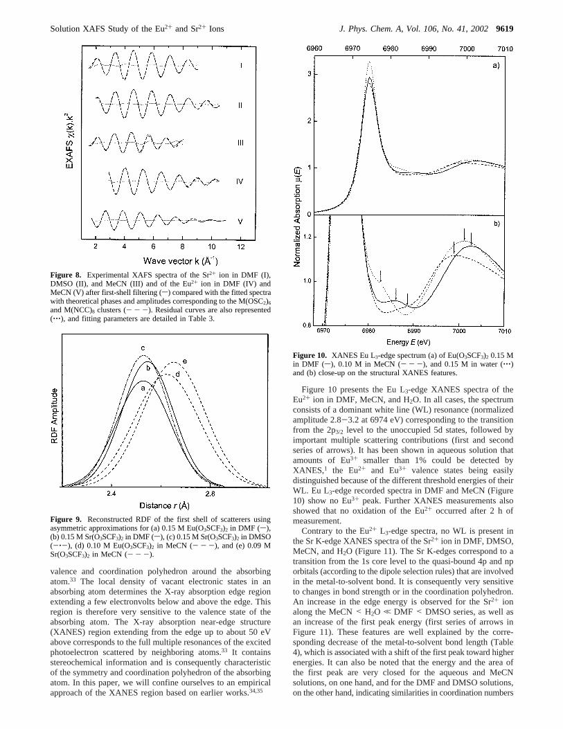
Figure 10 presents the Eu L3-edge XANES spectra of theEu2+ ion in DMF, MeCN, and H2O. In all cases, the spectrumconsists of a dominant white line (WL) resonance (normalizedamplitude 2.8-3.2 at 6974 eV) corresponding to the transitionfrom the 2p3/2 level to the unoccupied 5d states, followed byimportant multiple scattering contributions (first and secondseries of arrows). It has been shown in aqueous solution thatamounts of Eu3+ smaller than 1% could be detected byXANES,1 the Eu2+ and Eu3+ valence states being easilydistinguished because of the different threshold energies of theirWL. Eu L3-edge recorded spectra in DMF and MeCN (Figure10) show no Eu3+ peak. Further XANES measurements alsoshowed that no oxidation of the Eu2+ occurred after 2 h ofmeasurement.
Contrary to the Eu2+ L3-edge spectra, no WL is present inthe Sr K-edge XANES spectra of the Sr2+ ion in DMF, DMSO,MeCN, and H2O (Figure 11). The Sr K-edges correspond to atransition from the 1s core level to the quasi-bound 4p and nporbitals (according to the dipole selection rules) that are involvedin the metal-to-solvent bond. It is consequently very sensitiveto changes in bond strength or in the coordination polyhedron.An increase in the edge energy is observed for the Sr2+ ionalong the MeCN< H2O , DMF < DMSO series, as well asan increase of the first peak energy (first series of arrows inFigure 11). These features are well explained by the corre-sponding decrease of the metal-to-solvent bond length (Table4), which is associated with a shift of the first peak toward higherenergies. It can also be noted that the energy and the area ofthe first peak are very closed for the aqueous and MeCNsolutions, on one hand, and for the DMF and DMSO solutions,on the other hand, indicating similarities in coordination numbers
Figure 8. Experimental XAFS spectra of the Sr2+ ion in DMF (I),DMSO (II), and MeCN (III) and of the Eu2+ ion in DMF (IV) andMeCN (V) after first-shell filtering (s) compared with the fitted spectrawith theoretical phases and amplitudes corresponding to the M(OSC2)6
and M(NCC)8 clusters (- - -). Residual curves are also represented(‚‚‚), and fitting parameters are detailed in Table 3.
Figure 9. Reconstructed RDF of the first shell of scatterers usingasymmetric approximations for (a) 0.15 M Eu(O3SCF3)2 in DMF (s),(b) 0.15 M Sr(O3SCF3)2 in DMF (s), (c) 0.15 M Sr(O3SCF3)2 in DMSO(-‚-), (d) 0.10 M Eu(O3SCF3)2 in MeCN (- - -), and (e) 0.09 MSr(O3SCF3)2 in MeCN (- - -).
Figure 10. XANES Eu L3-edge spectrum (a) of Eu(O3SCF3)2 0.15 Min DMF (s), 0.10 M in MeCN (- - -), and 0.15 M in water (‚‚‚)and (b) close-up on the structural XANES features.
Solution XAFS Study of the Eu2+ and Sr2+ Ions J. Phys. Chem. A, Vol. 106, No. 41, 20029619

and in coordination structure. According to observation per-formed on plutonium aqua ions (oxidation states ranging from+III to +VI),35 the decrease in symmetry associated with thedecrease of coordination number from 8 to 7 could explain thebroadening of the first peak observed between these two couplesof solvents.
The first part of the Eu L3-edge XANES spectra of the Eu2+
ion in DMF, MeCN, and H2O (first series of arrows in Figure10) cannot be interpreted without proper calculation. However,the important changes in their general shape tend to show thatthe coordination structure of the Eu2+ ion in H2O, DMF, andMeCN solutions is different. Note that for both Eu2+ and Sr2+
XANES spectra, the first EXAFS oscillation (second series ofarrows in Figures 10 and 11) is very similar even if invertedbecause of phase inversion between K- and L-edges. For bothcations, the oscillation corresponding to the MeCN solutionoccurs first, then the oscillation for water, and finally that for
DMF and DMSO, according to the respective metal-to-solventbond length.
Discussion
We recently studied the solvation of the Eu2+ and Sr2+ ionsin water1 and now report their solvation in DMF, DMSO, andMeCN. In the literature, only two references relevant to ourstudy were found. Both of them were XAFS studies, one aboutSr2+ solvation in DMSO18 and the other about Sr2+ solvationin MeCN.36 Results for the Sr2+ aqua ion by the sameauthors18,24 are also reported in Table 6 for comparison.
Persson et al.18 performed LAXS and XAFS measurementson the solvated Sr2+ ion in DMSO. The radial distributionfunction (RDF) obtained by LAXS showed two peaks at2.54(1) and 3.77(1) Å, leading to an average Sr-O-S angle of136°. Because of the strong correlation between the fittingparameters, the authors resolved to test the followingN values:4, 5, 6, 8, and 10. The lowest error square sum was obtainedwith N ) 6. In the LAXS measurements, high absorption ofthe DMSO solvent made it necessary to use short path lengthsand high solute concentration (1.3 M in Sr(O3SCF3)2 comparedto the 11.3 M of pure DMSO). Not accounting for the METeffects, the authors could not fit the coordination number inthe XAFS analysis. TheN value was therefore fixed to 6, leadingto a DW value of 0.0082(4) Å2 and a Sr-O bond length of2.54(1) Å. Our MET-corrected XAFS analysis in diluted Sr2+
DMSO solution led to anN value of 7.0(3). UsingN ) 7 andtaking into account a linear correlation betweenN and the DWfactor, we can calculate a DW factor of 0.0096 Å2 fromPersson’s data. This value is very close to the 0.0103(4) Å2
value reported here. Besides the MET correction, the differencein coordination number could also be interpreted as a concentra-tion effect. The Sr-O distances that we found are very closeto those obtained by Persson et al. in both aqueous and DMSOsolutions (Table 6).
D’Angelo et al.36 performed an XAFS analysis on a 0.06 MSr(O3SCF3)2 in MeCN solution. They fitted the XAFS raw data(without FT) using a cluster based on a classical moleculardynamics simulation. They obtained a coordination number of8.0(2) associated with a Sr-N distance of 2.710(3) Å and aDW factor of 0.024(1) Å2. Our results are very similar even ifthe absolute values are different. We measured a metal-to-solvent distance increase of 0.065 Å and DW factor increaseof 0.0029 Å2 when going from water to the dry MeCN solution,compared to the reported values of 0.067 and 0.003 Å2. Wefound a Sr‚‚‚C contribution at 3.78(3) Å (they report 3.76(2)Å) and found an almost linear Sr-N-C angle, whereas theyreported 149°.
Alkaline-earth metal ions and trivalent lanthanides arestrongly solvated by hard donor solvents such as DMF (DN)
TABLE 6: XAFS Structural Data for Sr 2+ and Eu2+ Ions in Aqueous and Nonaqueous Solution, Comparison with Literaturea
sample N R(Å) C2 (Å2) C3 (Å3) × 10-4 ref
0.14 M Sr(O3SCF3)2 in H2O 8.0(3) 2.600(3) 0.0126(5) 2.7(5) 10.8 M Sr(ClO4)2 in H2O 8b 2.61(1) 0.0116(5) 180.1 and 3 M SrCl2 in H2O 10.3(1) 2.643(2) 0.021(2) âc 240.15 M Sr(O3SCF3)2 in DMF 6.9(3) 2.555(5) 0.0110(8) 3(2) this work0.15 M Sr(O3SCF3)2 in DMSO 7.0(3) 2.540(7) 0.0103(4) 1(1) this work1.30 M Sr(O3SCF3)2 in DMSO 6b 2.54(1) 0.0082(4) 180.09 M Sr(O3SCF3)2 in MeCN 8.1(8) 2.665(6) 0.0155(4) 3(1) this work0.06 M Sr(O3SCF3)2 in MeCN 8.0(2) 2.710(3) 0.024(1) âc 360.15 M Eu(O3SCF3)2 in H2O 7.2(3) 2.584(5) 0.0138(5) 1.5(5) 10.15 M Eu(O3SCF3)2 in DMF 6.0(5) 2.541(3) 0.0118(8) 0.0(5) this work0.10 M Eu(O3SCF3)2 in MeCN 7.9(4) 2.640(4) 0.0172(4) 3(1) this work
a N is the number of atoms located in the first shell at a distanceR from the metal,C2 ) σ2 is the DW factor,C3 is the third cumulant characterizingthe asymmetry of the RDF, and total errors are presented within parentheses.b Parameter fixed.c Parameter related to the RDF asymmetry.
Figure 11. XANES Sr K-edge spectrum (a) of Sr(O3SCF3)2 0.15 Min DMF (s), 0.15 M in DMSO (-‚-), 0.09 M in MeCN (- - -),and 0.14 M in water (‚‚‚) and (b) close-up on the structural XANESfeatures.
9620 J. Phys. Chem. A, Vol. 106, No. 41, 2002 Moreau et al.

26.6) and DMSO (DN) 29.8) and weakly solvated by softdonor solvents such as MeCN (DN) 14.1). H2O is intermediatewith a donor number (DN) value of 18.0.37 Actually, thedecrease of the structural parameters (metal-to-solvent distance,coordination number, and DW factor) is correlated with increas-ing DN along the MeCN< H2O , DMF < DMSO series forboth the Eu2+ and Sr2+ cations (Table 6).
Our results show a one-unit decrease in coordination numberwhen going from H2O to DMF and DMSO solutions for bothEu2+ and Sr2+ ions. Such a decrease is expected, because DMFand DMSO are at the same time bulkier solvents and muchbetter coordinators than H2O. A decrease in coordination numberfrom 7 for Sr2+ to 6 for Eu2+ is also observed in DMF. Thisdecrease is not surprising, because it has already been observedfrom 8 to 7 in the aqueous solution.1 In the MeCN solution,the sameN value of 8 was found for both Eu2+ and Sr2+ ions.This suggests that in the case of the MeCN solution, thecoordination number is mostly dependent on the electrostaticinteractions and not on the steric hindrance. A tendency towardcoordination number decrease is also observed between aqueousand DMF solutions for trivalent lanthanide ions.30,38In the solidstate, trivalent lanthanides are preferentially coordinated to 9MeCN,22,39 to 9 H2O,40,41 to 8 DMF,42 and to 8 or 7 DMSOligands,43 depending of the size of the lanthanide cation. Hence,a decrease in coordination number is observed in the solid stateof trivalent lanthanide solvates when increasing the solvationstrength of the solvent along the series MeCN< H2O , DMF< DMSO.37 Such an effect is consequently not specific to theEu2+ and the Sr2+ ions in solution but could be observed forother typical hard ions. Note that a steric hindrance effect oncoordination number has also been observed among trivalentlanthanides in the very similar DMF and DMA (N,N-dimethylacetamide) solvents (respective DN values of 26.6 and 27.8).37
The decrease in coordination number occurring from DMF toDMA38 shows that hard ions are subjected to strong solvationsteric effects in bulky aprotic oxygen-donor solvents.
For both Eu2+ and Sr2+ ions, the metal-to-solvent distancesdecrease along the MeCN> H2O > DMF ≈ DMSO series(Table 6). Moreover, in all of the solvents considered, thesedistances are slightly shorter for Eu2+ than for the Sr2+ ion(without coordination number correction). In the MeCN solu-tions, a 0.025 Å decrease is observed (larger than the 0.01 Ådecrease derived from Shannon radii44 for N ) 8). In theO-coordinating solvents, a 0.014-0.016 Å decrease is observed,whereas the associated coordination number decrease shouldlead to more severe shortening of the bond lengths (0.04-0.06Å according to Shannon44). The Eu2+ ion seems to have aslightly higher affinity toward MeCN and a slightly loweraffinity toward O-coordinating solvents when compared withthe Sr2+ ion. Actually, we observe in the X-ray diffraction(XRD) structures of the Eu2+ and Sr2+ diethylenetriaminepentaacetic acid (DTPA) complexes,45 on average, 0.013 Åshorter M-N bond lengths and 0.011 Å longer M-O bondlengths for the Eu2+ over the Sr2+ ion. In isomorphousacetates,46,47the Eu-O bond lengths were 0.015 Å longer thentheir Sr2+ homologues. Hence, the comparison of crystal-lographic data of the solid state, as well as the comparison ofmetal-to-solvent bond length in solution, suggests that the Eu2+
and Sr2+ ionic radii are equivalent and that Eu2+ is a slightlysofter ion than Sr2+, leading to shorter M-N bonds and longerM-O bonds. Eu2+ being the same size as Sr2+, its relativesoftness could be due to the presence of 25 extra electrons. Thedifference in hardness could therefore explain the rather smallbond length decrease observed in the O-coordinating solvents,
because it partly compensates the decrease associated in thecoordination number diminution. A higher polarizability wouldalso explain the tendency toward lower coordination numbersobserved for the Eu2+ ion in O-donor solvents.
The Debye-Waller (DW) factor can be related to the widthof the radial distribution function (RDF); as the DW factorbecomes larger, the distribution of the bond lengths becomeslarger. An increase of the DW factor could therefore eithercorrespond to a low symmetry polyhedron or to an equilibriumbetween species with different coordination numbers (i.e.,different metal-to-solvent bond lengths). In solution, the DWfactor increases significantly from the highly coordinating DMFand DMSO to the small H2O, and then to the low-coordinatingMeCN. In the same solvent, the Eu2+ DW factor is 7%-10%larger than that of the Sr2+ ion. The coordination numbers andlow DW values found in DMF and DMSO solutions suggestthat the [Sr(DMF)7]2+, [Eu(DMF)6]2+, and [Sr(DMSO)7]2+ ionsare the only solvates present in solution. The coordinationnumbers found in MeCN solutions suggest that the octa-coordinated ions [Sr(MeCN)8]2+ and [Eu(MeCN)8]2+ are thepredominant species in solution, but the associated high DWvalues do not exclude an equilibrium with minor 7 or 9coordinated solvates or both.
To the best of our knowledge, the same coordination numberwas found in pure DMF and in pure DMSO, whatever the metalion.48,49Therefore, the 6 coordination number found for the [Eu-(DMF)6]2+ ion would suggest hexa-coordination for the Eu2+
ion in DMSO solution, for which no experimental data areavailable. From the structural parameters found for the Sr2+ ionin the MeCN, H2O, DMF, and DMSO solutions and from thecomparison between Eu2+ and Sr2+ ions in these differentsolvents, we can deduce for the [Eu(DMSO)6]2+ ion a Eu-Obond length of 2.525 Å and a DW factor close to 0.0112 Å2.
Conclusion
Structural parameters of the Eu2+ ion were established inDMF and MeCN solutions, as well as the structural parametersfor Sr2+ in DMF, DMSO, and MeCN solutions, using the XAFSmethod with picometer accuracy. We obtained for the Eu2+ ionin DMF and MeCN solutions coordination numbers of 6.0(5)and 7.9(4) and metal-to-solvent distances of 2.541(3) and2.640(4) Å, respectively. A coordination number of 6 and ametal-to-solvent distance of 2.525 Å were extrapolated for theEu2+ ion in DMSO solution, for which no experimental dataare available. We obtained for the Sr2+ ion in DMF, DMSO,and MeCN solutions coordination numbers of 6.9(3), 7.0(3),and 8.1(8) and metal-to-solvent distances of 2.555(5), 2.540(7),and 2.665(6) Å, respectively.
For both Eu2+ and Sr2+ ions, a decrease in coordinationnumber and metal-to-solvent distances was found along theincreasingly solvating MeCN< H2O , DMF < DMSO solventseries: for europium(II),N ) 8 with [Eu(MeCN)8]2+, N ) 7with [Eu(H2O)7]2+, and N ) 6 with [Eu(DMF)6]2+ and byextrapolation [Eu(DMSO)6]2+; for strontium,N ) 8 with [Sr-(MeCN)8]2+ and [Sr(H2O)8]2+ andN ) 7 with [Sr(DMF)7]2+
and [Sr(DMSO)7]2+. However, an equilibrium with a minoramount of 7 or 9 coordinated solvates or both cannot beexcluded in MeCN solution.
As is the case for the trivalent lanthanides,38 Eu2+ and Sr2+
feature a strong solvation steric effect in solution when movingfrom water to bulky aprotic donor solvents, whereas in theMeCN solution, the interaction is mostly electrostatic and notvery dependent on the steric hindrance effects. We also showedthat Eu2+ and Sr2+ ionic radii should be considered as equivalent
Solution XAFS Study of the Eu2+ and Sr2+ Ions J. Phys. Chem. A, Vol. 106, No. 41, 20029621

and that Eu2+ seems to be a slightly softer ion than Sr2+, leadingto shorter M-N and longer M-O bonds and to smallercoordination numbers in O-coordinating solvents.
Acknowledgment. We thank F. Bouamrane, the LURE D21beam line staff (R. Cortes, S. Be´nazeth, I. Ascone) for theirtechnical collaboration, and A. Kuzmin for his comments. Wealso thank the “Laboratoire pour l’Utilisation du RayonnementElectromagne´tique” (LURE) for the beam time allocation andlaboratory facilities. We finally acknowledge the Swiss NationalScience Foundation, the Latvian National Science Foundation,and the COST actions D9 (Advanced Computational Chemistryof Increasingly Complex Systems) and D18 (Lanthanide Chem-istry for Diagnosis and Therapy) for financial support.
Supporting Information Available: Crystallographic datain CIF format for the Sr(DMF)2(O3SCF3)2 (143 and 293 K) andthe Sr(DMSO)2(O3SCF3)2 (143 K) compounds.
References and Notes
(1) Moreau, G.; Helm, L.; Purans, J.; Merbach, A. E.J. Phys. Chem.A 2002, 106, 3034.
(2) IUPAC Recommended Methods for Purification of SolVents andTests for impurities; Coetzee, J. F., Ed.; Pergamon Press: Elmsford, NY,1982.
(3) Mikheev, N. B.; Kamenskaya, A. N.Coord. Chem. ReV. 1991, 109,1.
(4) Bunker, G.Nucl. Instrum. Methods1983, 207, 437.(5) Sayers, D. E.; Bunker, B. A. InX-ray Absorption; Koningsberger,
D. C., Prins, R., Eds.; Chemical Analysis, Vol. 92; Wiley & Sons: NewYork, 1988.
(6) Crozier, E. D.; Rehr, J. J.; Ingalls, R. InX-ray Absorption;Koningsberger, D. C., Prins, R., Eds.; Chemical Analysis, Vol. 92; Wiley& Sons: New York, 1988.
(7) Rehr, J. J.; Mustre de Leon, J.; Zabinsky, S. I.; Albers, R. C.J.Am. Chem. Soc.1991, 113, 5135.
(8) Dalba, G.; Fornasini, P.; Grisenti, R.; Purans, J.Phys. ReV. Lett.1999, 82, 4240.
(9) Cooley, R. A.; Yost, D. M.Inorg. Synth.1946, II , 69.(10) CrysAlis RED, release 1.6.9â; Oxford Diffraction Ltd.: Abingdon,
Oxfordshire, U.K., 2001.(11) Blessing, R. H.Acta Crystallogr., Sect. A1995, 51, 33.(12) Walker, N.; Stuart, D.Acta Crystallogr., Sect. A1983, 39, 158.(13) Altomare, A.; Burla, M. C.; Camalli, M.; Cascarano, G. L.;
Giacovazzo, C.; Guagliardi, A.; Moliterni, A. G. G.; Polidori, G.; Spagna,R. J. Appl. Crystallogr.1999, 32, 115.
(14) SHELXTL, release 5.1; Bruker AXS, Inc.: Madison, WI, 53719,1997.
(15) Villain, F.; Briois, V.; Castro, I.; Helary, C.; Verdaguer, M.Anal.Chem.1993, 65, 2545.
(16) Kuzmin, A.Physica B1995, 208-209, 175.(17) Dalba, G.; Fornasini, P.J. Synchrotron Radiat.1997, 4, 243.(18) Persson, I.; Sandstrom, M.; Yokoyama, H.; Chaudhry, M.Z.
Naturforsch.1995, 50a, 21.
(19) Nieuwenhuyzen, M.; Wen, H.; Wilkins, C. J.Z. Anorg. Allg. Chem.1992, 615, 143.
(20) Klinga, M.; Cuesta, R.; Moreno, J. M.; Dominguez-Vera, J. M.;Colacio, E.; Kivekas, R.Acta Crystallogr., Sect. C1998, 54, 1275.
(21) Muller, U.; Noll, A. Z. Kristallogr. 2000, 215, 191.(22) Deacon, G. B.; Go¨rtler, B.; Junk, P. C.; Lork, E.; Mews, R.;
Petersen, J.; Zemva, B.J. Chem. Soc., Dalton Trans.1998, 3887.(23) Bunker, B. A.; Stern, E. A.Phys. ReV. B 1983, 27, 1017.(24) D’Angelo, P.; Nolting, H.-F.; Pavel, N. V.Phys. ReV. A 1996, 53,
798.(25) Kepert, D. L. InAspects of the stereochemistry of 8-coordination;
Lippard, S. J., Ed.; Progress in Inorganic Chemistry, Vol. 24; Wiley: NewYork, 1978; p 179.
(26) Bunzli, J.-C. G., Milicic-Tang, A., Gschneidner, K. A. J., Eyring,L., Eds.Hanbook on the Physics and Chemistry of Rare Earths; ElsevierScience B. V.: Amsterdam, 1995; Vol. 21.
(27) Gnaegi, F. Diploma Work, University of Lausanne, Lausanne,Switzerland, 1967.
(28) Teo, B. K. In EXAFS and Near Edge Structure; Bianconi, A.,Incoccia, L., Stipcich, S., Eds.; Springer: Berlin, 1983; p 11.
(29) Lindqvist-Reis, P.; Naslund, J.; Persson, I.; Sandstrom, M.J. Chem.Soc., Dalton Trans.2000, 2703.
(30) Naslund, J.; Lindqvist-Reis, P.; Persson, I.; Sandstrom, M.Inorg.Chem.2000, 39, 4006.
(31) Persson, I.; Penner-Hahn, J. E.; Hodgson, K. O.Inorg. Chem.1993,32, 2497.
(32) Calligaris, M.; Carugo, O.Coord. Chem. ReV. 1996, 153, 83.(33) Bianconi, A. InEXAFS and Near Edge Structure; Bianconi, A.,
Incoccia, L., Stipcich, S., Eds.; Springer: Berlin, 1983; p 118.(34) Purans, J.; Kuzmin, A.; Parent, P.; Laffon, C.Electrochim. Acta
2001, 46, 1973.(35) Ankudinov, A. L.; Conradson, S. D.; Mustre de Leon, J.; Rehr, J.
J. Phys. ReV. B 1998, 57, 7518.(36) D’Angelo, P.; Pavel, N. V.J. Chem. Phys.1999, 111, 5107.(37) Gutmann, V.The Donor-Acceptor Approach to Molecular Interac-
tions; Plenum Press: New York, 1978.(38) Ishiguro, S.; Umebayashi, Y.; Komiya, M.Coord. Chem. ReV. 2002,
226, 103.(39) Shen, Q.; Hu, J.-Y.; Jin, Z.-S.; Sun, Y.J. Chin. Rare Earth Soc.
1990, 8, 359.(40) Gerkin, R. E.; Reppart, W. J.Acta Crystallogr., Sect. C1984, C40,
781.(41) Harrowfield, J. M.; Kepert, D. L.; Patrick, J. M.; White, A. H.Aust.
J. Chem.1983, 36, 483.(42) Krishnamurthy, S. S.; Soundararajan, S.Can. J. Chem.1969, 47,
995.(43) Krishnamurthy, V. N.; Soundararajan, S.J. Inorg. Nucl. Chem.
1967, 29, 517.(44) Shannon, R. D.Acta Crystallogr.1976, A32, 751.(45) Burai, L.; Toth, E.; Seibig, S.; Scopelliti, R.; Merbach, A. E.
Chem.sEur. J. 2000, 6, 3761.(46) Starynowicz, P.Polyhedron1995, 14, 3573.(47) Trunov, V. K.; Chubinidze, A. D.; Efremov, V. A.; Velikodni, Y.
A. Koord. Khim.1984, 10, 403.(48) Lincoln, S. F.; Merbach, A. E. InSubstitution reactions of solVated
metal ions; Sykes, A. G., Ed.; Advances in Inorganic Chemistry, Vol. 42;Academic Press: San Diego, CA, 1995; p 1.
(49) Ohtaki, H.Monatsh. Chem.2001, 132, 1237.
9622 J. Phys. Chem. A, Vol. 106, No. 41, 2002 Moreau et al.