Solange Cristina Bastos da Costa Tese de Candidatura ao ...
284
Solange Cristina Bastos da Costa OCCUPATIONAL EXPOSURE TO FORMALDEHYDE. GENOTOXIC DAMAGE AND SUSCEPTIBILITY EVALUATION IN ANATOMICAL PATHOLOGY LABORATORY WORKERS Tese de Candidatura ao grau de Doutor em Ciências Biomédicas, submetida ao Instituto de Ciências Biomédicas Abel Salazar da Universidade do Porto. Orientador – Doutor João Paulo Teixeira Categoria – Investigador Auxiliar Afiliação – Instituto Nacional de Saúde Doutor Ricardo Jorge Co-orientadora – Doutora Beatriz Porto Categoria – Professora Auxiliar Afiliação – Instituto de Ciências Biomédicas Abel Salazar da Universidade do Porto
-
Upload
khangminh22 -
Category
Documents
-
view
4 -
download
0
Transcript of Solange Cristina Bastos da Costa Tese de Candidatura ao ...

Solange Cristina Bastos da Costa
OCCUPATIONAL EXPOSURE TO FORMALDEHYDE.
GENOTOXIC DAMAGE AND SUSCEPTIBILITY EVALUATION IN ANATOMICAL
PATHOLOGY LABORATORY WORKERS
Tese de Candidatura ao grau de Doutor em
Ciências Biomédicas, submetida ao Instituto de
Ciências Biomédicas Abel Salazar da
Universidade do Porto.
Orientador – Doutor João Paulo Teixeira
Categoria – Investigador Auxiliar
Afiliação – Instituto Nacional de Saúde Doutor
Ricardo Jorge
Co-orientadora – Doutora Beatriz Porto
Categoria – Professora Auxiliar
Afiliação – Instituto de Ciências Biomédicas
Abel Salazar da Universidade do Porto


The experimental work presented in this thesis was performed at the:
Environmental Health Department of the National Institute of Health, Porto,
Portugal;
Department of Nutrition of the Institute of Basic Medical Sciences, University of
Oslo, Oslo, Norway;
Toxicology Unit of the Department of Psychobiology, University of A Coruña, A
Coruña, Spain;
Toxicology Laboratory and Farmacognosia Laboratory of the Faculty of
Pharmacy, University of Porto, Porto, Portugal;
Department of Genetics of the Faculty of Medical Sciences, New University of
Lisbon, Lisbon, Portugal.
Statistical analysis was performed at the Toxicology Unit of the Department of
Psychobiology, University of A Coruña, Spain.
This work was supported by the Portuguese Foundation for Science and Technology
(FCT) under the grant SFRH/BD/46929/2008, under the QREN - POPH - Type 4.1 -
Advanced Training, subsidized by the European Social Fund and national funds from the
MCTES.
.


CONTENTS
Original Publications v
List of Figures vii
List of Tables viii
List of Abbreviations x
Abstract xi
Resumo xiii
Preamble xvii
I. REVIEW OF THE LITERATURE
1. Introduction 3 1.1 Genotoxicity and Carcinogenicity 4
1.2 Human exposure assessment 5 2. Formaldehyde 9 2.1 Physical and Chemical properties 10
2.2 Economic importance 10
2.2.1 Production 10
2.2.2 Commercial uses and Applications 11
2.3 Human Exposure 12
2.3.1 Environmental 12
2.3.2 Occupational 14
2.4 Toxicokinetic and Metabolism 15
2.5 Health Effects 17
2.5.1 Sensory irritation and related symptoms 17
2.5.2 Sensitisation 19
2.5.3 Genotoxicity 20
2.5.4 Carcinogenicity 23
2.5.4.1 Epidemiological studies 23
2.5.4.2 Animal studies 29
2.5.4.3 Mechanisms of carcinogenesis 30
2.6 Exposure to formaldehyde in Portuguese workplaces 32 II. PRESENT STUDY
1. Aim of the study 37 2. Study Population 39 2.1 General characterisation 39
2.1.1 Contact with study subjects 40
2.1.2 Professional activity 41
i

2.2 Biologic samples collection 41
2.3 Statistical analysis 42
3. Air monitoring and biomarkers of exposure 45 3.1 Overview 45 3.1.1 Air monitoring (guidelines and standards) 45
3.1.1.1 Formaldehyde occupational exposure and regulation
46
3.1.2 Biomarker of exposure-internal dose 47
3.1.2.1 Formic acid in urine as a biomarker of formaldehyde internal dose
47
3.2 Material and Methods 50 3.2.1 Determination of formaldehyde airborne concentration 50
3.2.2 Determination of formic acid concentrations in urine samples
50
3.3 Results 53 3.3.1 Formaldehyde air monitoring and exposure biomarker 53
3.3.1.1 Effect of exposure, lifestyle and work-related factors 54
3.4 Discussion 55 4. Biomarkers of Effect 59 4.1 Overview 59 4.1.1 Chromosomal Aberrations 60
4.1.2 Micronucleus assay 64
4.1.2.1 Micronucleus assay in lymphocytes 65
4.1.2.2 Micronucleus assay in exfoliated buccal cells 67
4.1.3 Sister chromatid exchanges 69
4.1.4 Single cell gel electrophoresis (Comet assay) 72
4.1.5 T Cell Receptor mutation assay 75
4.1.6 Immune markers: Lymphocytes subpopulations 76
4.2 Material and Methods 80 4.2.1 Lymphocytes culture 80
4.2.2 Chromosomal Aberrations (CAs) Assay 80
4.2.3 Lymphocyte Cytokinesis-Block Micronucleus Test 82
4.2.4 Buccal Micronucleus Cytome Assay 82
4.2.5 Sister Chromatid Exchange (SCE) Test 83
4.2.6 Comet Assay 84
4.2.7 TCR mutation assay 85
4.2.8 Lymphocyte subpopulations assessment 85
4.3 Results 86 4.3.1 Biomarkers of genotoxicity 86
ii

4.3.1.1 Effect of exposure, lifestyle factors and parameter-specific confounders
87
4.3.2 Immune Markers: Lymphocytes subpopulations 91
4.3.2.1 Effect of exposure, lifestyle factors and parameter-specific confounders
91
4.3.3 Effect of work-related factors 93
4.3.4 Correlations between biomarkers 95
4.4 Discussion 97 4.4.1 Biomarkers of genotoxicity 97
4.4.2 Immune-markers: Lymphocytes subpopulations 105
4.4.3 Effect of lifestyle factors and parameter-specific confounders on the biomarkers studied
109
4.4.3.1 Biomarkers of genotoxicity 109
4.4.3.2 Lymphocyte subpopulations 111
4.4.4 Correlations between biomarkers 112
5. Biomarkers of Susceptibility 114 5.1 Overview 114 5.1.1 Genetic polymorphisms as susceptibility biomarkers 114
5.1.2 Genetic Polymorphisms, DNA damage and cancer risk 114
5.1.3 Xenobiotic-metabolising enzymes 116
5.1.3.1 Polymorphisms of phase I enzymes studied: Cytochrome P450 family
116
5.1.3.2 Polymorphisms of Phase II enzymes studied: Glutathione S-transferases
119
5.1.4 DNA Repair 122
5.1.4.1 DNA Repair pathways 123
5.1.4.2 Polymorphisms of the BER pathway studied 128
5.1.4.3 Polymorphisms of the DSBs repair pathway studied 131
5.1.4.4 Polymorphisms of the FANC repair pathway studied 134
5.2 Material and Methods 138 5.2.1 DNA extraction 138
5.2.2 Genotyping of polymorphisms in gene involved in the metabolism
138
5.2.2.1 CYP2E1 138
5.2.2.2 GSTM1 and GSTT1 139
5.2.2.3 GSTP1 139
5.2.3 Genotyping of polymorphisms in genes involved in DNA repair.
140
5.2.3.1 XRCC1, PARP1, MUTYH, RAD51, BRIP1 and FANCA 140
5.2.3.2 XRCC2 and XRCC3 141
iii

5.3 Results 144 5.3.1 Genotype distribution of polymorphisms in the study
population 144
5.3.2 Analysis of association between the genetic polymorphisms and biomarkers studied
146
5.4 Discussion 148 III. GENERAL CONCLUSIONS 155 IV. REFERENCES 161 V. ANNEXES ANNEX I ANNEX II ANNEXIII PUBLICAÇÕES DE APOIO À TESE DE DOUTORAMENTO
ACKNOWLEDGMENTS 261
iv

ORIGINAL PUBLICATIONS
The present work contains techniques and/or data also presented in the following
scientific papers:
Costa S., García-Lestón J., Coelho M., Coelho P., Costa C., Silva S., Porto B., Laffon
B., Teixeira J.P. (2013) Cytogenetic and immunological effects of formaldehyde in a group
of exposed workers. Journal of Toxicology and Environmental Health Part A 76:217–229.
Costa S., Brandão F., Coelho M., Costa C., Coelho P., Silva S., Porto B., and Teixeira
J.P. 2013. Micronucleus frequencies in lymphocytes and buccal cells in formaldehyde
exposed workers. WIT Transactions on Biomedicine and Health, 16: 83-94.
Costa, S., Pina, C., Coelho, P., Costa, C., Silva, S., Porto, B., Laffon, B. and Teixeira,
J.P. 2011. Occupational exposure to formaldehyde: genotoxic risk evaluation by comet
assay and micronucleus test using human peripheral lymphocytes. Journal of Toxicology
and Environmental Health Part A, 74(15-16): 1040-1051.
Costa, S., Coelho, P., Costa, C., Silva, S., Mayan, O., Santos, L.S, Gaspar, J.and
Teixeira, J.P. 2008. Genotoxic damage in pathology anatomy laboratory workers exposed
to formaldehyde. Toxicology, 252(1-3): 40-48.
Ersson, C., Møller P.; Forchhammer, L.; Loft, S., Azqueta, A., Godschalk, R.W.L., van
Schooten, F.J., Jones, G.D., Higgins, J.A., Cooke, M.S., Mistry, V., Karbaschi, M., Phillips,
D.H., Sozeri O, Routledge, M.N., Nelson-Smith, K., Riso, P., Porrini, M., Matullo, G.,
Allione, A., Stepnik, M., Ferlińska, M., Teixeira, J.P., Costa, S., Corcuera, L.A. López de
Cerain, A., Laffon, B., Valdiglesias., V., Collins, A., Möller, L. 2013. An ECVAG inter-
laboratory validation study of the comet assay: inter-laboratory and intra-laboratory
variation of DNA strand breaks and FPG-sensitive sites in human mononuclear cells.
Mutagenesis, 28: 279–286.
Forchhammer, L., Ersson, C., Loft, S., Möller, L., Godschalk, R.W.L., Jones, G.D.,
Higgins, J.A., Cooke, M., Mistry, V., Karbaschi, M., Collins, A.R., Azqueta, A., Phillips,
D.H., Sozeri, O., Routledge, M.N., Nelson-Smith, K., Riso, P., Porrini, M., Matullo, G.,
Allione, A., Stępnik, M., Komorowska, M., Teixeira, J.P., Costa, S., Corcuera, L.A., López
de Cerain, A., Laffon, B., Valdiglesias. V., Møller P. 2012. Inter-laboratory variation in
DNA damage using a standard comet assay protocol. Mutagenesis, 27: 665-672.
Lorenzo, Y., Costa, S., Collins, A.R., Azqueta, A. 2012. The comet assay, DNA damage,
DNA repair and cytotoxicity: Hedgehogs are not dead. Mutagenesis 28(4):427-32.
v

Forchhammer, L., Johansson, C., Loft, S., Möller, L., Godschalk, R., Langie, S., Jones,
G.D., Kwok, R.W., Collins, A.R., Azqueta, A., Phillips, D.H., Sozeri, O., Stepnik, M., Palus,
J., Vogel, U., Wallin, H., Routledge, M.N., Handforth, C., Allione, A., Matullo, G., Teixeira,
J.P., Costa, S., Riso, P., Porrini, M., Møller, P. 2010. Variation in the measurement of
DNA damage by comet assay measured by the ECVAG inter-laboratory validation trial.
Mutagenesis, 25: 113-123.
And book chapters:
Costa, S., and Teixeira, J. P. 2013. Toxicology. In Encyclopedia of Toxicology, 3rd
Edition, Elsevier (in press). DOI: 10.1016/B978-0-12-386454-3.00440-1
Costa, S., and Teixeira, J. P. 2013. Comet Assay. In Encyclopedia of Toxicology, 3rd
Edition, Elsevier (in press). DOI:10.1016/B978-0-12-386454-3.00672-2
vi

LIST OF FIGURES
Figure 1. A simplified diagram of the cascade of events from exposure to clinical disease showing the relationships among exposure, effect and susceptibility biomarkers
8
Figure 2. Formaldehyde 2D and 3D chemical structure 10
Figure 3. Fate and metabolism of formaldehyde 17
Figure4. Frequency of the daily consumption of tea, coffee, fruit, vegetables, fish, meat and vitamin supplements by study groups
40
Figure 5. Distribution ratio (%) of the FA-exposed workers by professional category
41
Figure 6. Example of formation of a Chromosome-type aberration (CSAs) 62
Figure 7. Example of formation of Chromatid-type aberration (CTAs) 62
Figure 8. Photomicrograph (A) and a scheme (B) representation of a section of buccal mucosa showing the various cell layers
68
Figure 9. Diagrammatic representation of the sequential spatio-temporal origins of the different buccal cell-stages
69
Figure 10. Mechanism of SCEs formation 70
Figure 11. Schematic picture representing a comet of a damaged cell and image
73
Figure 12. Comet assay images 73
Figure 13. Chromosomal aberrations observed in metaphase lymphocytes (1250x)
81
Figure 14. Scored micronucleus (MN) images (500x) 82
Figure 15. Images of cell metaphases scored for SCE test (1250x) 84
Figure 16. Both environmental and host factors acts in concert in individual susceptibility (genetic and acquired ) for disease development
115
Figure 17. DNA Repair pathways 125
Figure 18. Interaction of the FANC proteins in the recognition of an ICL in S phase
127
Figure 19. Simplified model of the mechanism of ICL repair process involving NER, TLS and HR
128
Figure 20. Association among the various factors involved in individual’s response to an exogenous insult and potential biologic effects
137
vii

LIST OF TABLES
Table I. General physical and chemical properties of formaldehyde 10
Table II. Characteristics of the study population 39
Table III. Occupational exposure limits for airborne formaldehyde (standards and guidelines)
46
Table IV. Results of FA-external exposure and biomarker of exposure 53
Table V. Effect of exposure, gender, age and smoking habits on the levels of FMU with estimates of mean ratios (MR)
54
Table VI. Results of biomarkers of genotoxicity in the study groups 86
Table VII. Results of aberrant cells and multiaberrant cells in the study groups
87
Table VIII. Effect of exposure, gender, age and smoking habits on the frequencies of CA-total, CSAs, CTAs, gaps and aneuploidies with estimates of mean ratios (MR)
88
Table IX. Effect of exposure, gender, age and smoking habits on the frequencies of MNL, SCE, MNB and BNbud with estimates of mean ratios (MR)
89
Table X. Effect of exposure, gender, age and smoking habits on the frequencies of %TDNA and TCR-Mf with estimates of mean ratios (MR)
90
Table XI. Results of lymphocyte subpopulations in the study groups 91
Table XII. Effect of FA-exposure, gender, age and smoking habits on lymphocyte subpopulations with estimates of mean ratios (MR)
92
Table XIII. Influence of FA exposure level, exposure duration and professional activity on micronuclei in lymphocytes (MNL), nuclear buds in buccal cells (BNbud) and %CD16+56+ cells, with estimates of mean ratios (MR)
94
Table XIV. Influence of FA-exposure level of exposure categorized by tertiles on micronuclei in lymphocytes (MNL), nuclear buds in buccal cells (BNbud) and %CD16+56+ cells, with estimates of mean ratios (MR)
94
Table XV. Association between biomarkers of genotoxicity 95
Table XVI. Detailed information on selected SNPs 141
Table XVII. PCR-RFLP details for the XRCC2 (rs3218536), XRCC3 (rs861539), GSTP1 (rs1695) and CYP2E1 (rs6413432) polymorphisms
143
Table XVIII. Frequency of genotypes involved in xenobiotic metabolism in the study population
144
viii

Table XIX. Genotype frequency of genes involved in DNA repair in the study population
145
Table XX. Influence of biomarkers of susceptibility on genotoxicity parameters and immune markers (only significant effect are included)
146
ix

LIST OF ABBREVIATIONS
ADH alcohol dehydrogenase MN micronuclei ALDH aldehyde dehydrogenases MNB micronuclei in buccal cells BMCyt buccal micronucleus cytome
assay MNL micronuclei in lymphocytes
BM buccal mucosa NAD+ nicotinamide adenine dinucleotide
BNbud nuclear buds in buccal cells NADPH nicotinamide adenine dinucleotide phosphate
CAs chromosomal aberrations Nbud nuclear buds CBMN cytokinesis-block micronucleus
assay OECD Organisation for Economic
Co-operation and Development
CD clusters of differentiation %TDNA percentage DNA in tail CD3+ total T cells PBLs peripheral blood lymphocytes CD4+ T-helper cells ROS reactive oxygen species CD8+ T-cytotoxic cells SCE sister chromatid exchange CD19+ B cells CD16+56+ natural killer cells SCGE single cell gel electrophoresis CSAs chromosome-type aberrations SD standard deviation CTAs chromatid-type aberrations SE Standard error mean DSB double strand break SSB single strand break EH environmental health SNPs single nucleotide
polymorphisms EU European Union Tc T-cytotoxic cells FA formaldehyde TCR T Cell Receptor FDH glutathione-dependent
formaldehyde-dehydrogenase TCR-Mf TCR mutation frequency
FEMA US Federal Emergency Management Agency
Th T-helper cells
FEV forced expiratory volume TWA time-weighted average FISH fluorescence in situ
hybridization US United States of America
FVC forced vital capacity WHO World Health Organization GSH glutathione HR homologous recombination IARC International Agency for
Research on Cancer
MHC major histocompatibility complex
x

ABSTRACT
Formaldehyde (FA) is a high-volume production chemical produced worldwide with a large
range of industrial and medical uses. At room temperature is a flammable and colourless
gas with a strong pungent odour. Given its economic importance and widespread use,
many people are exposed to FA both environmentally and/or occupationally. Over the last
two decades, several epidemiological studies have revealed an increased risk of cancer
development among workers exposed to FA, namely nasopharyngeal cancer and myeloid
leukaemia. Based on these findings plus supporting evidence from animal studies and
data on mechanisms of carcinogenesis FA status was recently revised and classified as a
human carcinogen. FA’s genotoxicity is confirmed in a variety of experimental systems
ranging from bacteria to rodents. However, data from human studies is conflicting and
needs further investigation in particular the biological evidence of FA’s ability to induce
(geno)toxicity on distant-site tissues. In addition, published studies on the immunological
effects of FA indicate that this compound may be able to modulate immune responses,
although data in exposed subjects are still preliminary. The highest level of human
exposure to FA occurs in occupational settings. Several studies point to pathology and
anatomy laboratories as one of the occupational settings where workers are frequently
exposed to levels of FA near or superior to recommended limit values.
The aim of the present study was to evaluate the occupational exposure to FA in a
multistage approach relating the exposure with different biomarkers (dose and effect) and
individual susceptibility. Eighty-five workers from hospital anatomical pathology
laboratories exposed to FA and eighty-seven non-exposed controls took part in the study.
Air monitoring was performed in worker’s breathing zone for representative working
periods and the level of FA-exposure in workplace air was estimated. Formic acid in urine
was investigated as a potential biomarker of internal dose for FA occupational exposure.
Genotoxicity was evaluated in peripheral blood lymphocytes (PBLs) by means of
cytogenetic alterations (chromosomal aberrations, CAs; micronucleus, MN; sister-
chromatid exchange, SCE), DNA damage (comet assay, percentage of tail DNA, %TDNA)
and T-cell receptor (TCR) mutation assay. The frequency of MN in exfoliated buccal cells
a first contact tissue was also assessed. Percentages of different lymphocyte
subpopulations were selected as immunotoxicity biomarkers. In addition, the effect of
polymorphic genes of xenobiotic metabolising enzymes (CYP2E1, GSTM1, GSTT1,
GSTP1) and DNA repair enzymes (FANCA, RAD51, XRCC2, XRCC3, XRCC1, PARP1,
MUTYH, BRIP1) on the endpoints studied were determined.
xi

The mean level of FA-exposure was 0.38±0.03 ppm, exceeding recommended limit
values, as observed in other studies. The concentration of formic acid in urine increased
significantly in workers compared to controls, but no significant correlation was found with
the FA-level of exposure. All cytogenetic endpoints and DNA damage evaluated by comet
assay were significantly increased in PBLs of FA-exposed workers compared to controls.
Further, exposed workers showed higher frequencies of MN formation in both PBLs and
exfoliated buccal cells. A significant positive correlation was found between MN frequency
in these two tissues and FA-levels of exposure and duration, which confirms MN
sensitivity to evaluate the genotoxic action of FA in occupational exposed subjects. The
exposed group also presented significant alterations in the percentage of cytotoxic T
lymphocytes, natural killer (NK) cells and B lymphocytes in comparison with control group.
Concerning the effect of susceptibility biomarkers on the different endpoints studied,
results suggest that polymorphisms in CYP2E1, GSTP1 and GSTM1 metabolic genes are
associated with increased genetic damage in FA-exposed subjects. Furthermore, a
polymorphic gene involved in FANC repair pathway, FANCA, significantly altered the level
of genetic damage induced by FA exposure, revealing a potential novel repair pathway
involved in the repair of genetic lesions caused by FA-occupational exposure.
Data obtained in the present study show that workers in anatomical pathology laboratories
are exposed to air-levels of FA greater than recommended criteria, indicating a potential
risk to workers’ health. Further, FA-exposed workers presented higher levels of genetic
damage compared to controls. Genotoxic endpoints analysis are of great interest in risk
assessment of occupational carcinogens because they precede by a long time the
potential health effects, thus offering a greater potential for preventive measures. The
results of the present biomonitoring study emphasise the need of more effective measures
in order to protect the workers from potentially hazard health effects due to occupational
exposure to FA. Implementation of security and hygiene measures, such as periodic air
sampling and medical surveillance, as well as good practice campaigns may be crucial to
decrease risk. The obtained information may also provide new important data to be used
by health care programs and by governmental agencies responsible for setting the
acceptable levels for occupational exposure to FA.
xii

RESUMO
O Formaldeído (FA) é um dos compostos químicos mais utilizados em todo o mundo,
apresentando-se, em condições normais de pressão e temperatura, como um gás incolor
de odor característico e intenso. A sua aplicação é multifacetada e transversal a
praticamente todas as atividades, sendo que a exposição humana a este composto
assume particular importância a nível industrial e no sector da saúde, onde é bastante
utilizado. Ao longo das últimas duas décadas vários estudos epidemiológicos revelaram
uma associação entre a exposição ocupacional a este aldeído e incidência de cancro
nasofaríngeo e leucemia (do tipo mieloide) entre trabalhadores do sector industrial e da
saúde. Com base nestes estudos e em dados obtidos em ensaios experimentais com
animais o FA foi recentemente reclassificado agente carcinogénico humano pelas
principais agências mundiais. O potencial genotóxico do FA está largamente descrito
tanto in vitro como in vivo desde bactérias até roedores. No entanto, em estudos de
biomonitorização humana, o caracter genotóxico do FA é inconclusivo, e carece de mais
investigação em especial no que concerne ao seu efeito em tecidos distantes do local de
absorção. Por outro lado, alguns autores reportaram que o FA poderá influenciar
parâmetros imunológicos e por conseguinte afetar a resposta imunitária. Vários estudos
apontam os laboratórios de anatomia patológica como sendo um dos cenários
ocupacionais em que os trabalhadores estão expostos a elevados níveis de FA. Nestes
laboratórios decorrem atividades de rotina que implicam a libertação de vapores de FA,
com consequente inalação por parte dos trabalhadores, pela utilização de formol usado
em procedimentos anatomopatológicos para conservar e armazenar biópsias e peças
cirúrgicas.
O objetivo do presente estudo foi avaliar a exposição ocupacional a FA em profissionais
dos Serviços Hospitalares de Anatomia Patológica, utilizando para isso uma abordagem
múltipla de modo a relacionar diferentes tipos de biomarcadores de dose, efeito e
suscetibilidade. A integração dos resultados dos diferentes biomarcadores estudados
permitir-nos-á investigar a relação entre a exposição ao FA e possíveis efeitos biológicos
adversos, consequentes da exposição profissional a este composto.
Para o efeito foi estudada uma população de 85 profissionais dos Serviços Hospitalares
de Anatomia Patológica, expostos ao FA no seu ambiente de trabalho e 87 indivíduos
com historial ocupacional de não exposição ao FA e com características demográficas,
de idade, sexo e hábitos tabágicos, semelhantes ao grupo exposto.
xiii

A avaliação da exposição ocupacional a FA foi realizada pela medição da concentração
do FA no ar no local de trabalho e pela análise de diferentes indicadores biológicos. Para
estimar o nível de exposição do FA nos Serviços de Anatomia recorreu-se à amostragem
contínua de ar no posto de trabalho, representativa do ar inalado pelos trabalhadores. Os
níveis de ácido fórmico na urina foram quantificados, por forma a estudar a sua potencial
aplicação como biomarcador de dose interna de exposição ocupacional a FA. O estudo
do dano genético foi avaliado em linfócitos do sangue periférico através de diversos
biomarcadores de efeito, nomeadamente, alterações citogenéticas (frequência de
aberrações cromossómicas, AC; micronúcleos, MN; e trocas entre cromatídeos irmãos,
SCE), dano a nível do ADN, mediante o teste do cometa, e o teste de mutação do recetor
das células T (TCR). A frequência de MN em células da mucosa bucal foi também
avaliada. As possíveis alterações no sistema imunitário foram estudadas pela análise das
percentagens das diferentes subpopulações linfocitárias. A fim de detetar diferenças
genéticas responsáveis por variações na suscetibilidade dos indivíduos à exposição a FA,
foram investigados polimorfismos de genes relacionados com o metabolismo (CYP2E1,
GSTM1, GSTT1, GSTP1) e polimorfismos envolvidos no mecanismo de reparação de
lesão do ADN (FANCA, RAD51, XRCC2, XRCC3, XRCC1, PARP1, MUTYH, BRIP1).
Para o grupo de trabalhadores estudados, tendo em conta a jornada de trabalho obteve-
se um valor médio de exposição ao FA de 0.38 ± 0.03 ppm. O grupo exposto apresentou
em média valores urinários de ácido fórmico estatisticamente superiores ao grupo
controlo, no entanto não foi observada associação com os níveis de exposição no ar. Os
resultados obtidos para os indicadores biológicos de genotoxicidade revelaram que os
indivíduos expostos apresentaram, em média, maior dano genético comparativamente
aos indivíduos do grupo controlo. Foi observado no grupo exposto um aumento
significativo na frequência de AC, de SCE e no dano do ADN avaliado pelo teste do
cometa. Observou-se ainda, nos trabalhadores, um aumento significativo na frequência
de MN em linfócitos e em células da mucosa bucal. Foram encontradas associações
(positivas) estatisticamente significativas entre a frequência de MN em ambos os tecidos,
a duração (MN em linfócitos) e os níveis de exposição a FA (MN bucais), confirmando a
sensibilidade deste biomarcador para avaliar o efeito genotóxico do FA em indivíduos
ocupacionalmente expostos. Os profissionais dos serviços de Anatomia Patológica
apresentaram também alterações nas percentagens de subpopulações linfocitárias,
designadamente em células T citotóxicas, células natural killer e linfócitos B,
comparativamente com o grupo controlo. No que concerne ao efeito dos polimorfismos
genéticos na frequência dos diferentes biomarcadores, observou-se um aumento
significativo do dano genético associado aos polimorfismos estudados nos genes
xiv

CYP2E1, GSTM1 e GSTT1 que codificam enzimas envolvidas na metabolização de
xenobióticos. O mesmo efeito foi observado num dos genes que integram o sistema de
reparação FANC, FANCA, revelando assim o envolvimento deste mecanismo na
reparação da lesão genética induzida pela exposição ocupacional a FA.
No presente estudo constatou-se que os profissionais dos Serviços de Anatomia
Patológica estão expostos a níveis de FA superiores a valores recomendados. Foi
também observado um aumento significativo na frequência de biomarcadores de dano
genético comparativamente com o grupo controlo. Face ao exposto, torna-se necessário
tomar medidas de controlo. A primeira abordagem no controlo da exposição a um produto
químico perigoso é analisar a possibilidade da sua substituição. Na sua impossibilidade,
devem adotar-se medidas que reduzam ao mínimo o risco de exposição profissional,
nomeadamente a implementação de boas práticas de trabalho e a formação dos
trabalhadores nesses princípios. Os trabalhadores deverão também ser informados sobre
a correta utilização dos meios de prevenção (coletiva e individual) e sua manutenção, de
modo a minimizar os riscos inerentes às suas atividades. Paralelamente deverá proceder-
se a melhorias nos locais de trabalho nomeadamente no que diz respeito às condições
de exaustão e ventilação. Deve ser também assegurado um programa de vigilância da
saúde dos trabalhadores que permita uma avaliação contínua dos riscos na saúde de
natureza profissional e subsidie medidas e ações preventivas adequadas ao controle da
exposição.
xv

xvi

PREAMBLE
The modern way of life, along with population growth, is sustained by an industrial
production never before attained, in which chemicals have become essential elements for
the well-being and functioning of contemporary societies. Global production has increased
over the time for 1 million tonnes in 1930 to 400 million tonnes today. Nonetheless, in
spite of the value and usefulness of chemical products, their production, use and disposal
can be harmful to human health and the environment. An important aspect of public health
protection is the prevention or reduction of exposures to environmental agents that
contribute, either directly or indirectly, to increased rates of premature death, disease,
discomfort or disability. This assumes particular importance in occupational settings where
the exposure to potential hazardous agents (in intensity, frequency and duration) is more
significant. Chemical substances constitute the more extensive group of occupational risk
factors, contributing significantly to the morbidity of the working population. The number of
hazardous substances to which workers are exposed grows on a daily basis, reaching
more than 100 000 in nowadays figures. For the most, there is little or none information
about toxicity and potential health effects.
Measurement or estimation of actual worker exposure, along with appropriate evaluation
of biologic parameters and assumptions about health effects or safety limits, is the
standard method used to determine whether intervention is necessary and which forms of
intervention will be more effective. During recent years, measures and strategies designed
to prevent, control, reduce or eliminate occupational hazards and risks have been
developed and applied, yet, despite continuous if slow improvements, work related
diseases are still frequent (Alli, 2008) and in some cases, such as cancer, are rising
(Clapp, 2008) thus their cost in terms of human suffering and economic burden continues
to be significant.
In a recent survey conducted by the Portuguese Toxicology Association (ApTox) on the
emerging issues in Environmental Health (EH) in Portugal, chemical exposure was
identified by EH professionals as one of the principle hazards that currently contribute to
most health problems in occupational settings. It was further predicted that in the next 5 to
10 years exposure to airborne chemicals will be the primary source of concern.
Furthermore, during this period, the health points of concern are most likely to be
respiratory and neoplastic disorders.
Formaldehyde is a high-volume production chemical produced worldwide with a large
range of industrial and medical applications. It was recently reclassified as a human
xvii

carcinogen, based on increased evidence of cancer risk among subjects exposed to high
measures (exposure level or duration) of airborne formaldehyde in the workplace. Several
studies point the pathology and anatomy laboratories as one of the occupational settings
where workers are more exposed to this aldehyde. Although formaldehyde genotoxicity
and carcinogenic potentials are well documented in mammalian cells and in rodents,
genotoxic effects and carcinogenic properties in humans are still conflicting and need
more investigation.
The present study addresses the need for a policy-oriented research focused on the
development of integrated approaches regarding the health risk assessment of
populations occupationally exposed to hazard compounds. The prime objective is to
provide rigorous information on the potential genotoxic effects of formaldehyde
occupational exposure, towards a better understanding of the mechanisms of genotoxicity
that may contribute to formaldehyde carcinogenicity in workers.
xviii

I. REVIEW OF THE LITERATURE


REVIEW OF THE LITERATURE
1. INTRODUCTION
“Preventing cancer is possible when we act on what we know.” (Clapp et al., 2008)
Humans are daily exposed to a variety of potentially harmful agents in the air they
breathe, the liquids they drink, the food they eat and products they use (IPCS, 2002a).
Long-standing evidence of the bond between health and the environment has led to the
recognition of the need for sustainable development. On the other hand, there is an
increasing global awareness of the inevitable limits of individual health care and the need
to complement such services with effective public health strategies.
According to the World Health Organisation (WHO), cancer is a leading cause of death
worldwide (WHO, 2013). Additionally, at least 200,000 people die every year from
occupational or work-related cancers (WHO, 2007).
Most individuals spend one-third of their life in the workplace. Workers are often in contact
with carcinogenic agents at higher levels than the general population, resulting in
accumulation of exposure effects over lifetime. Many occupational diseases, including
work-related cancers, are characterised by long latency periods and therefore are difficult
to recognise until the clinical manifestation of their symptoms. Early detection of a
professional risk situation can significantly decrease the occurrence of deleterious effects
on worker’s health. Thus, preventing individual exposure to carcinogens may prevent the
disease.
Indeed, estimations show that at least one–third of all cancer cases of environmental and
occupational origin can be prevented based on current knowledge (Espina et al., 2013).
This is best exemplified by the decline in lung cancer cases in males from the reduction in
tobacco smoking, or by the drop in bladder cancer among dye workers from the
elimination of exposure to specific aromatic amines (Clapp et al., 2008; Jemal et al.,
2011).
Exposure assessment aims at prevention. Establishing the health effects of various
activities and exposures requires information about the levels of exposure and the
biological effects resulting from the interaction between the organism and the chemical
agent. The resulting data provides a basis for designing effective prevention and
mitigation strategies.
3

REVIEW OF THE LITERATURE
The current global labour force stands at about 45% of the world’s population. Their work
sustains the economic and material basis of society which is critically dependent on their
working capacity. Thus health at work and healthy work environments are among the
most valuable assets of individuals, communities and countries, and are critical
components for sustainable development.
1.2 GENOTOXICITY AND CARCINOGENICITY
Genotoxicity is a comprehensive term that refers to the ability of an agent to damage DNA
and/or cellular components regulating the fidelity of the genome and includes all adverse
effects on genetic material (Eastmond et al., 2009). Genomic damage may be induced by
several exogenous (exposure to genotoxic agents, lifestyle factors) and endogenous
factors (genetic variants) (Holland et al., 2008). If the damage induced is not properly
repaired it may give rise to genetic alterations that may ultimately lead to neoplasia and
cancer (Rueff et al., 2002). Thus, genomic damage is probably the most important
fundamental cause of developmental and degenerative disease.
Carcinogenesis is a multistep process that results from the accumulation of several
genetic alterations controlling either cell death or cell proliferation leading to a progressive
transformation of normal cells into highly malignant derivatives (Hanahan and Weinberg,
2000). Cancer is characterised by karyotypic instability, aberrant gene expression and
uncontrolled cellular proliferation. Neoplasias may arise either from inherited mutations or
from de novo mutations in somatic tissue (Jefford and Irminger-Finger, 2006). However,
they are not only a consequence of DNA sequence mutations, but also from changes in
the activities of genes and chromosomal regions (epimutations1) (Banno et al., 2012).
Human carcinogenesis is the outcome of a complex series of interactions between
exogenous and endogenous processes (Kyrtopoulos, 2006). Although risk factors such as
tobacco use, genetic predisposition, alcohol consumption, unhealthy diet, among others,
play a major role in carcinogenesis process, a range of environmental variables and
occupational exposures also contribute significantly to the global cancer burden (Clapp et
al., 2008; Espina et al., 2013). Indeed, over the last years, several epidemiological studies
have revealed an increased risk of cancer development at various sites among subjects
exposed to chemicals namely in occupational settings.
1 abnormal transcriptional repression in active genes and/or abnormal activation of usually repressed genes (Banno et al.,
2012).
4

REVIEW OF THE LITERATURE
1.3 HUMAN EXPOSURE ASSESSMENT
Exposure assessment is a crucial procedure for the identification, evaluation and control
of workplace hazards.
Human monitoring is a frequently used approach to provide early warning signals of
excessive exposure to toxic substances and for prediction of health risks. Overall, it
consists on a routine evaluation and interpretation of environmental and/or biological
parameters, in order to detect possible health hazards before the poisoning or illness
occur. In addition, it may contribute to define exposure limits for minimising the likelihood
of significant health risks (Teixeira, 2004).
The exposure can be assessed by measuring the concentration of the chemical agent in
environmental samples, such as air (Environmental Monitoring) and/or by measuring
biological endpoints (Biological monitoring), called biomarkers or biological indicators.
Generally, these two methods are used simultaneously, since they provide different but
complementary information (IPCS, 2002a).
Environmental monitoring is the measurement of agents present at the workplace to
evaluate ambient exposure and health hazard compared to an appropriate reference
value. It quantifies or estimates the level of the chemical pollutant to which a worker is
exposed during a task and/or workday. However, it represents only a snapshot of
exposure in time and may not adequately reflect fluctuations in exposures. Furthermore,
external exposure assessment does not take into consideration personal working habits or
individual characteristics that may influence the uptake of the chemical agent.
Biological monitoring is any measurement reflecting an interaction between a biological
system and an environmental agent, which may be chemical, physical or biological (IPCS,
1993). It provides an integrated measure of the level of exposure to chemicals through
different pathways and exposure routes. Hence, it is essential not only for evaluating
hazards to human health but also for developing strategies to improve occupational health
and safety conditions and to assess the efficiency of the implemented policies.
The aim of human biomonitoring studies is to assess the risk of deleterious health effects
by analysing the relationship between internal exposure and the biological effects in target
cells. Selection of the optimal assay and tissue for analysis for any particular exposure
situation should be based, whenever possible, on exposure type and duration, exposed
population characteristics, endpoint mechanism, and the expected target tissue. Most of
the time surrogate tissues (e.g. lymphocytes) rather than target tissues are studied for
practical, methodological and ethical reasons (Albertini et al., 2000).
5

REVIEW OF THE LITERATURE
Biological monitoring encompasses the use of biomarkers as indicators to signal events of
exposure to potential carcinogenic chemicals. Biomarkers are valuable tools in human
biomonitoring studies, because they are intermediates between exposure and clinical
manifestation of the disease, representing the whole continuum from external exposure to
effect. It should be added that the detection of a biomarker not necessarily indicate the
presence of a disease or toxic process, it may only indicate exposure of the organism to a
substance (Juberg, 1999).
Biomarkers are traditionally classified into three types, biomarker of exposure, effect and
susceptibility.
Biomarker of exposure- is the xenobiotic chemical itself, its metabolites or the product of
an interaction between a chemical and its target molecule or cell that is measured in a
compartment within an organism (IPCS, 1993). Generally, their presence only indicates
that an exposure has occurred. A biomarker of exposure can be defined as a biomarker of
internal dose, when the amount of a chemical or its metabolites is quantified in a biologic
tissue or fluid (e.g. lead in blood) or as a biomarker of biologically effective dose, when it
concerns the interactions between the chemical or its metabolites with target cellular
macromolecules forming measurable covalent complexes (e.g. DNA-adducts or protein-
adducts) (Teixeira, 2004).
Biomarker of effect- is defined as a measurable biochemical, physiological, behavioural,
functional or other alteration within an organism that might be elicited by the exposure and
depending upon the magnitude can be recognized as associated with an established or
possible health impairment or disease (IPCS, 1993). It may represent an event that can
be correlated with, and possibly predictive of, a deleterious health effect (NRC, 2006).
These biomarkers are extensively used tools in occupational studies to evaluate the
genotoxic potential of hazardous chemicals. Cytogenetic alterations, such as
chromosomal aberrations and micronuclei are the most used endpoints. Moreover, it is
well-established that chromosome aberrations in human lymphocytes predict a risk of
cancer (Hagmar et al., 1998) the same association was recently confirmed for micronuclei
in lymphocytes (Bonassi et al., 2007). Furthermore, the increased frequency of these
indicators in exposed workers is a quite consistent finding in most studies (Suspiro and
Prista, 2011), including in cancer-patients (Hagmar et al., 1998). The measurements of
biomarkers of effect generally reflect events that take place at the latter end of the
continuum between exposure and disease manifestation. Effect biomarkers, such as
chromosomal aberrations or somatic mutations are not chemical specific, and therefore
the association with an exposure must be established by an independent measure, such
as environmental monitoring or simultaneous measurement of a biomarker of exposure.
6

REVIEW OF THE LITERATURE
Although they are less specific in identifying a single causative agent, they can be more
predictive of ultimate toxicity. However, this will also depend on the individual variation in
response to the exposure (Lutrell et al., 2008).
Biomarker of susceptibility- is an indicator of an inherent or acquired limitation of an
organism to respond to the challenge of exposure to a specific chemical agent(s) (IPCS,
1993). Generally, it is used to identify whether a person or groups of people are
susceptible to damage caused by certain chemical or other toxicant. This class of
biomarkers serves to describe individual variations in the relationship of biomarkers of
exposure and biomarkers of effect. These variations may help explain why different
individuals with similar experiences of environmental exposure sometimes produce
markedly different levels of biomarkers of exposure and/or biomarkers of effect, which in
turn may correlate with different severities of clinical symptoms. These differences may
arise from genetic or non-genetic factors. Genetic factors include polymorphisms that may
lead to different individual capacities to activate, detoxify or repair DNA lesions arising
from exposure to a chemical. Non-genetic indicators include among others, nutritional
status, health status, lifestyle and age. These factors are independent of exposure but
they identify those individuals in a population who may be more susceptible or resistant to
the effects of a hazardous exposure.
Each of these biomarkers provides different information and consequently has its own set
of advantages and disadvantages relating to its specificity, relevance to the toxic pathway,
existence following exposure and difficulty of analysis (Teixeira, 2004). Ideally, it should
be used various combinations of biomarkers or apply the same biomarker in different
tissues or biological fluids in order to obtain more complex and comprehensive information
and further expand the possibility to investigate the biological consequences of the
exposure.
Given the above, estimation of the external exposure to an agent, the response of the
organism to that agent and its potential susceptibility to toxic effects are all crucial
parameters when assessing potential health effects associated to the exposure to an
agent in the workplace. The basic tenets of biomarker-based environmental genotoxicity
assessment are illustrated in Figure 1.
7

REVIEW OF THE LITERATURE
Figure 1. A simplified diagram of the cascade of events from exposure to clinical disease showing the relationships among exposure, effect and susceptibility biomarkers (adapted from Manini et al. (2007))
8

REVIEW OF THE LITERATURE
2. FORMALDEHYDE
“You can run, but you can’t formalde(hide)”
In a carton by Mike Baldwin
Formaldehyde was first synthesised by Aleksandr Butlerov in 1859, but was only in 1868
that it was conclusively identified by August von Hofmann, as the product obtained from
passing a mixture of air and methanol over a heated platinum spiral. In fact, this
production method laid the foundation for the modern manufacturing process of
formaldehyde, via methanol oxidation using a metal catalyst (copper, molybdenum alloy,
platinum or silver). At the end of the 19th century, formaldehyde was already used as a
regular medical preservative and embalming agent. By the early 20th century driven by
science and technology development, Leo Baekeland used a phenolformaldehyde resin to
invent the first synthetic polymeric material, a hard moldable plastic, precursor of modern
synthetic plastics. In 1940s, the first commercial particleboard was produced and a
revolution was launched in the furniture and construction industries. Commercial
applications continue to grow and today formaldehyde has an important role in both
medical laboratory procedures and in the production of domestic and industrial goods that
have become decisive in everyday life.
9

REVIEW OF THE LITERATURE
2.1 PHYSICAL AND CHEMICAL PROPERTIES
Formaldehyde (FA) is the most simple of all aliphatic aldehydes. It is a low-molecular
weight organic compound, highly reactive, that readily combines with many chemical
substances. At room temperature, FA is a flammable and colourless gas with a strong and
distinctive odour. It is also known as methanal, methylaldehyde or oxymethylene.
FA is soluble in water, alcohols, and other polar solvents, but has a low degree of
solubility in non-polar fluids. In dilute aqueous solution, the predominant form of FA is its
monomeric hydrate, methylene glycol (or methanediol). Since it is rapidly polymerised at
room temperature, methanol and other substances (e.g. amine derivatives) are frequently
added as stabilisers to reduce intrinsic polymerisation. FA chemical structure is shown in
Figure 2, its physical and chemical properties are listed in Table I.
2.2 ECONOMIC IMPORTANCE
2.2.1 Production
FA is a building-block of numerous compounds that are used in a wide array of materials
and products. Due to its versatile properties and inexpensive production, FA is an
economically important chemical produced worldwide. Generally FA is produced near the
point of consumption because while it is easy to make, is costly to transport and can
develop stability-associated problems during transportation and storage. As a result,
international trade is minimal (NTP, 2011) and FA demand is provided by domestic
industry.
IUPAC Name Methanal
CAS Number 50-00-0
Structural Formula CH2O
Relative molecular mass 30.03
Density (-20ºC) 0.82
Boiling point -19.1 ºC
Melting point -92 ºC
Convertion 1ppm = 1.2 mg/m3 Figure 2. Formaldehyde 2D
and 3D chemical structure.
Table I. General physical and chemical properties of formaldehyde.
Adapted from NIOSH (1994) and IARC (2006)
10

REVIEW OF THE LITERATURE
FA is a high-volume production chemical with a global annual production and
consumption of approximately 29 million tonnes (IHS, 2012). The European Union (EU) is
the second largest producer, after Asia, producing over 7 million tonnes of FA each year,
which accounts for about 25% of global production. China is the largest asian market,
accounting alone for 34% of the world demand. The third larger market is the United
States (U.S.), with an annual production of about 5 million tonnes (NTP, 2011). In fact, FA
and FA-related products are allegedly responsible for more than 5% of the annual U.S
Gross National Product (Zhang et al., 2009).
Moreover, it is forecast that the world consumption of FA will continue to grow, reaching
an average rate of almost 5% per year throughout 2011–2016, especially due to
increasing demand of fast growing economy countries industry (IHS, 2012).
2.2.2 Commercial uses and applications
FA is the most commercially important aldehyde, usually marketed as a 37 % (by weight)
aqueous solution named formalin. Formalin also contains methanol (up to 15%) and other
substances (several hundred milligrams per litre) that act as stabilisers. FA is also sold, to
a lesser extent, in its solid form, either as a polymer, paraformaldehyde, or as a cyclic
trimer, 1,3,5-trioxane.
The main industrial application of FA is in the manufacture of resins, namely, phenol-,
urea-, melamine- and polyacetal resins.
Phenolformaldehyde and urea-formaldehyde resins are extensively used in furniture and
construction industry as binders or adhesives for wood composites (sterling board,
plywood, particleboard, fiberboard, hardwood plywood). Other applications include
structural and acoustical insulation, foundry materials and heat-resistant components for
automotive and aerospace applications and housewares. In addition, urea-formaldehyde
resins are used in glass fiber roofing mats and paints. Melamine-formaldehyde resins are
the raw material of several decorative applications including furniture, tableware, flooring,
acrylic clear coats and paper finishing. The polyacetal resins are the most diversified with
applications ranging from knife handlers, zippers, insulin pens, guitar picks and Bagpipes
to automotive and consumer electronics. The leather, textile, rubber and cement
industries are other examples where FA-based resins are widely used.
Another major application of FA is as an intermediate in the synthesis of other industrial
chemicals including numerous organic compounds (e.g. 1,4-butanediol, pentaerythritol)
and derivative substances, that in turn, will be used to make other products downstream
(e.g. dyes, perfumes, vitamins).
11

REVIEW OF THE LITERATURE
FA is also used in agriculture, in controlled-release nitrogen fertilisers, crop protection
agents and as a preservative in animal feeds. In a recent resolution, EU has decided to
ban from the market FA-containing biocidal products for use as feed preservatives, from
July 2015 (EU, 2013).
This aldehyde can be further applied as an anti-corrosive agent in electroplating
processes and in the development of photographic films.
FA is also used in cosmetic products as a preservative and antimicrobial agent, namely in
shampoos, deodorants, lotions, toothpaste, lipstick, mouthwashes, nail polish and hair
products. Although FA per se is now rarely used in cosmetics, the use of FA-releasing
preservatives is common.
FA application further extends to the health sector where it is used: i) in vaccines (as an
inactivating agent) and pharmaceutical products; ii) to disinfect and sterilised hospital
wards and medical equipment; iii) to preserve and embalm biological specimens.
In many instances, because FA is a long-established material with versatile properties,
few compounds can replace it without compromising quality, performance and cost, which
hinders its substitution to both producers and users.
2.3 HUMAN EXPOSURE
Given its economic importance and widespread use, many people are exposed to FA both
environmentally and occupationally. The major route of exposure to FA is inhalation
(IARC, 2006). Although environmental exposure typically occurs at much lower levels than
occupational exposure, people are exposed to FA at some concentration in their daily
lives. FA is also a naturally occurring biological compound present in all cells, tissues and
body fluids (IARC, 2006).
According to WHO (2000) the estimates for the daily intake of FA in ambient air can vary
between 0.002 and 0.04 mg/day mean values. On the other hand, for indoor air the
average intake levels can differ between 0.3 and 0.6 mg/day. However, indoor air
concentrations can be substantially increased with tobacco smoking, rising up to 0.5 to 3.5
mg/day. WHO also predicts that the mean daily intake of FA from occupational exposure
is up to 8 mg/day, being variable according to the work in question.
2.3.1 Environmental
FA is ubiquitous in the environment and has been detected in indoor and oudoor air, soil,
food, surface water, groundwater, treated and bottled water (NTP, 2011).
12

REVIEW OF THE LITERATURE
FA is formed primarily by the combustion of organic materials and by a variety of natural
and anthropogenic activities.
Combustion sources include automobiles and other internal combustion engines, power
plants, waste incinerators, refineries, wood stoves, forest fires and tobacco smoking. FA is
also formed in the early stages of residual plant decomposition in the soil.
Secondary formation of FA occurs in the atmosphere through the photochemical oxidation
of natural and anthropogenic hydrocarbon pollutants present in the air. In some instances,
given the diversity and abundance of FA precursors in urban air, secondary production
may exceed direct air emissions (IPCS, 2002b).
For the general population the major sources of FA exposure include combustion sources,
tobacco smoke, offgassing from numerous construction and home-furnishing products,
and offgassing from consumer goods.
FA levels in indoor air are often higher by one order of magnitude or more than those
outdoors (IARC, 2006). The most significant source of indoor contamination is the release
from newly home furnishings, primarily from wood pressed products made with urea-
formaldehyde resins. These emissions are able to cause FA-exposure related symptoms
in residents or occupants especially irritation of the eyes and upper airways (Salthammer
et al., 2010). The most known example has occurred in the US, involving hurricane victims
and government-provided FEMA (Federal Emergency Management Agency) trailers.
Numerous victims of Hurricanes Katrina and Rita suffered adverse health effects
(respiratory and other symptoms) as a result of being housed in FEMA trailers containing
levels of FA 2-fold higher than the reference level. The FA was emitted from the materials
used to build the mobile homes (CDC, 2008).
The general population can also be exposed to FA from consumer goods containing FA
and from ingestion of food and water (IARC, 2006). Some of the products that often
contain FA-releasers are cosmetics, household cleaning agents, soaps, shampoos, paints
and lacquers (IPCS, 2002b).
Food and water contain measureable concentrations of FA, but the significance of
ingestion as a source of FA exposure is not considered relevant (IPCS, 2002b; NTP,
2011). In food FA can be present either naturally (e.g. fruit, vegetables, shellfish) or as
result of inadvertent contamination. FA-hydrated form, methylene glycol, can be found in
surface water or groundwater but usually it is biodegraded within 1 to 15 days (IPCS,
2002b). In treated and drinking water FA is formed primarily through oxidation of organic
matter during ozonation or chlorination treatment (NTP, 2011), yet its concentration is
usually less than 100 μg/L (IPCS, 2002b).
13

REVIEW OF THE LITERATURE
2.3.2 Occupational
The major source of exposure to airborne FA occurs in the work environment.
Occupational exposure to FA is transversal to a wide variety of professions and industries.
In the early 1990s it was estimated that approximately 1 million workers in the EU (near
36 000 in Portugal) and 2 million in the US were exposed to FA (FIOH, 1999; NTP, 2011).
Although no current data is available in the literature, given that FA-world demand has
grown over the last decades, it is safe to speculate that the number of individuals
occupationally exposed to FA is now higher.
In occupational settings, FA occurs mainly as a gas, hence the major route of exposure is
inhalation. Nevertheless, workers may also be exposed through dermal contact (formalin
solutions) or by inhalation of FA-particulates such as powdered resins or wood dust (NTP,
2011).
Given its economic importance and wide spread use, occupational exposure to FA
involves not only individuals employed in the direct manufacture of FA and products
containing it, but also those using the products. Overall, the exposure to FA occurs in
three main work-related scenarios:
- production and use of formalin in chemical industry (e.g., synthesis of resins) and
in medical laboratories as a preservative and disinfectant agent.
- release from FA-based resins in which it is present as a residue and/or through
their hydrolysis and decomposition by heat, during the manufacture of wood
products, textiles and plastics.
- the pyrolysis or combustion of organic matter, e.g. in engine exhaust gases or
during firefighting.
Another workplace circumstance that in the last few years has raised some concern is the
FA emission from hair straightening products during hair treatment in hairdresser salons;
although as shown by Pierce et al. (2011) this appeared to be limited to some specific
products.
The occupational groups considered to be more at risk, either by duration or level of
exposure, are industrial workers from chemical industries and plywood factories,
embalmers and pathology and anatomy laboratory workers (IARC, 2006).
According to IARC (2006) the highest exposures to this aldehyde were measured in the
past in industrial settings (e.g. varnishing of furniture and wooden floors, garment
industry). However, recent legislation (e.g. resins that release less FA) and improved
ventilation have resulted in decreased levels of exposure in many industrial workplaces.
14

REVIEW OF THE LITERATURE
Exposures to levels of up to 3 parts per million (ppm) have been reported for embalmers
and for hospital laboratory professionals, such as anatomy and pathology workers (IARC,
2006). A recent study assessing the airborne exposure to FA in an industrial unit and
anatomy pathology laboratory reveal higher levels of FA in the lab compared to the
industrial setting (Viegas et al., 2010).
Furthermore, numerous epidemiological studies indicate a significant association between
cancer risk and occupational exposure to FA (Hauptmann et al., 2004; Zhang et al.,
2009). In fact, a recent study reported that the estimated cancer risk of laboratory
technicians exposed to FA was 20% higher than the general population (Pilidis et al.,
2009).
2.4 TOXICINETIC AND METABOLISM
The toxicokinetic behavior of FA has been the subject of several studies in a number of
mammal species, and although there may be quantitative differences between them, the
fate of FA is qualitatively similar among species (IARC, 2006).
The major route of exposure to FA is inhalation. At room and body temperatures, FA
hydrates rapidly and it is in equilibrium with its hydrated form methylene glycol (or
methanediol) (NTP, 2011). Owing to its reactivity and solubility, FA is readily absorbed
from the respiratory tract following inhalation. In obligate nose breathers, such as rodents,
absorption and deposition occur primarily in the nasal passages. In humans, which are
oronasal breathers, it is likely to occur primarily in the nasal passages and oral mucosa,
but also in the trachea and bronchus (Kimbell et al., 2001). Species-specific differences of
FA uptake and consequent associated effects (such as cell proliferation or reaction with
other macromolecules) are determined by nasal airflow (nasal anatomy, ventilation) and
breathing patterns rather than metabolism of the parent compound (Herausgegeben et al.,
2006).
After oral exposure, FA is rapidly absorbed from the gastrointestinal tracts. In contrast,
dermal uptake following application appears to be very slight, however it is able to
penetrate skin (IARC, 2006).
FA is present at low levels in most living organisms. Physiological amounts of FA are
endogenously formed from serine, glycine, methionine and choline, and also by
demethylation of N-, O- and S-methyl compounds (IARC, 2006).
Nevertheless, regardless of its origin, endogenous or exogenous, FA has the same
metabolic pathway in the organism (Figure 3) (ATSDR, 1999; IARC, 2006).
15

REVIEW OF THE LITERATURE
After absorption, FA is rapidly metabolised by oxidation to formate. The detoxification
process takes place in all cells and can involve different enzyme systems. The primary
and most important enzyme for the metabolic inactivation of FA is the glutathione-
dependent formaldehyde-dehydrogenase (FDH) (also known as alcohol dehydrogenase
class 32; Koivusalo et al., 1989). FDH is highly specific for the FA-glutathione adduct, S-
hydroxymethylglutathione, formed spontaneously in the cytosol from FA and glutathione
conjugation. Formation of FA-glutathione adduct efficiently counteracts the presence of
free FA, a reaction that is determined by the fact that glutathione is an abundant molecule
in the cell. FDH oxidises S-hydroxymethylglutathione to S-formylglutathione, in the
presence of NAD. This intermediate is then metabolised by S-formylglutathione hydrolase
to yield formate and reduced glutathione (IPCS, 1989).
The activities of FDH are two to three orders of magnitude lower than those of S-
formylglutathione hydrolase and thus the FDH-catalysed step is rate-limiting (IARC, 2006).
A secondary pathway involves the two aldehyde dehydrogenases (ALDH), ALDH1A1 (in
cytosol) and ALDH2 (in mitochondria). Both enzymes have affinity for free FA but their Km
values are higher than the Km displayed by FDH for S-hydroxymethylglutathione (Just et
al., 2011). Therefore, FDH is probably the predominant enzyme responsible for the
oxidation of FA at physiologically relevant concentrations, while ALDH enzymes contribute
increasingly when the concentrations of FA increase (Just et al., 2011). Furthermore,
some studies showed that FDH activity do not increase in response to FA exposure
(Casanova-Schmitz et al., 1984; Øvrebø et al., 2002) thus no increase in metabolism
occurs and non-metabolised FA is probably bound to macromolecules.
Other alternative metabolic pathways include, FA oxidation to formate by catalase, but
only in the presence of hydrogen peroxide (H2O2); and FA reduction to methanol by a
cytosolic alcohol dehydrogenase class 1 (ADH1) (IARC, 2006) (Figure 3).
Being normal components of intermediary metabolism, neither FA nor formate are stored
to any significant extent in any tissue. Formate is either excreted in the urine, as formic
acid, or oxidised to carbon dioxide and exhaled.
Moreover, both FA and formate can be further incorporated into other cellular molecules,
entering the 1 carbon intermediary metabolic pool, which involves the folic acid metabolic
pathway for synthesis of nucleic acids, amino acids, and macromolecules (ATDSR, 1999).
2the systematic name for the enzyme is formaldehyde: NAD+ oxidoreductase (IUBMB code EC 1.2.1.46). Other names for
this enzyme are formaldehyde dehydrogenase, NAD-linked formaldehyde dehydrogenase and NAD-dependent
formaldehyde dehydrogenase.
16

REVIEW OF THE LITERATURE
The kinetics of FA metabolism and the effects on its rate of various enzyme inhibitors
have been studied (IARC, 2006). In isolated rat hepatocytes, depletion of glutathione or
inhibition of the metabolic enzymes, FDH, ALDH2 or ADH1 were found to decrease FA
metabolism and to increased cytotoxicity in a dose-dependent manner, through reactive
oxygen species (ROS) production (Teng et al., 2001). Recent studies have identified
ALDH2 as the primary enzyme responsible for the detoxication of aldehydes in
haematopoietic stem cells, the lack of this enzyme results in hypersensitivity of
haematopoietic stem cells to exogenous aldehydes, by accumulating DNA damage
(Langevin et al., 2011; Garaycoechea et al., 2012). Thus, it is reasonable to assume that
individuals with lower levels of glutathione as well as those with deficiencies in any of the
above enzymes (by altered genes or polymorphisms) will be more susceptible to FA
toxicity.
Figure 3. Fate and metabolism of formaldehyde (adapted from Teng et al. (2001) and IARC (2006)).
2.5 HEALTH EFFECTS
2.5.1 Sensory irritation and related symptoms
FA has a pungent and suffocating odour and at certain concentrations is a strong irritating
gas (ATDSR, 2010).
17

REVIEW OF THE LITERATURE
The respiratory tract, especially the upper respiratory tract, is a critical target of the irritant
action of inhaled-FA, as shown by human acute controlled exposure studies, by studies
on humans exposed acutely or repeatedly under occupational or residential conditions,
and by studies of animals exposed by inhalation for acute, intermediate, and chronic
durations (IARC, 2006; ATDSR, 2010).
Most humans detect and/or recognise FA odour at concentrations below 1 ppm (ATDSR,
2010), but the odour threshold has been reported to be between 0.05 and 0.18 ppm (Arts
et al., 2006). The most common exposure effects to FA vapours comprise sensory
irritation of the eyes, nose and throat, with eye irritation generally accepted as the most
sensitive endpoint.
Odour perception and the threshold for sensory irritation are sometimes not clearly
differentiated, it generally exist on a concentration gradient, but may overlap (Gaffney and
Paustenbach, 2007). In FA case, however, the progression of responses is clear, with
human studies showing that for most individuals, odour perception generally precedes
sensory irritation (Golden, 2011). Importantly, odour perception and sensory irritation are
different and distinct physiological processes. Upon substance inhalation odour is the
sensation of smell carried by the olfactory nerve (first cranial nerve), while sensory
irritation involves stimulation of the trigeminal nerve endings, and near triggering
concentrations is generally considered to be a physiological and not a toxic response
(Gaffney and Paustenbach, 2007; Golden, 2011).
Sensory irritation encompasses a graded series of responses related to the concentration
of FA in the air and is generally categorised as ranging from slight to moderate (mucosa
irritation) to severe (e.g. difficulty in breathing). The response also depends on the length
of exposure and individual sensitivity.
The most common symptoms, irritation of the eyes, nose, and throat, along with increased
tearing, occurs at FA-concentrations of about 0.3 - 3 ppm in the air.
Other symptoms described by individuals exposed to FA include sneezing, coughing,
sinus, sore throat and nausea (Lang et al., 2008). In fact, clinical symptoms of eye
soreness (68%), lacrimation (60%), eye fatigue (45%), rhinorrhea (38%), and throat
irritation (43%) were reported in a survey involving 143 medical students exposed to a
mean FA concentration of 2.4 ppm, during a course (15 hours/week for 2 months)
(Takahashi et al., 2007). Similar symptoms were also described by students but at a lower
airborne FA concentration, 0.72 ppm (6-8 hours/day for 3 months) (Wei et al., 2007).
Neurological effects due to FA exposure were also reported by a few studies namely,
decrease performance in short-term memory tests and in the ability to concentrate (Bach
18

REVIEW OF THE LITERATURE
et al., 1990; ATDSR, 2010). Indeed, in animal studies impaired learning/memory and
altered motor activity were reported following acute- and intermediate-duration exposure
to FA (ATDSR, 2010).
Changes in human pulmonary function, detected by spirometry tests (FEV, FVC), are
assumed to occur at 0.6 ppm (ATDSR, 2010), but probably as protective response of the
organism to the exposure, since the exposure to sensory irritants of the upper respiratory
tract often cause slower breathing or even breath holding (Gaffney and Paustenbach,
2007). Volunteers (healthy and asthmatic) in acute controlled exposure studies showed no
significant changes in pulmonary function after exposure to FA. In chronic exposures,
some authors reported a decrease in pulmonary function of workers exposed to FA
(Akbar-Khanzadeh et al., 1994; Fransman et al., 2003; Tang et al., 2009), while others
found no exposure-related differences (Arts et al., 2006; ATDSR, 2010).
Increased numbers of eosinophils and protein exudation following FA exposure (0.4 ppm)
were found in nasal lavage studies confirming the irritative effect of FA; a non-specific pro-
inflammatory effect of FA was suggested by the authors (Pazdrak et al., 1993).
Nasal epithelial lesions have been observed consistently across a few occupational
studies (ATDSR, 2010). In addition, clinical findings of upper respiratory tract inflammation
were reported in 41% of workers exposed to FA (0.71 ppm) namely, hypertrophy or
atrophy of the upper respiratory mucous membranes, chronic pharyngitis, rhinitis,
rhinosinusitis, and rhinopharyngitis (Lyapina et al., 2004).
2.5.2 Sensitisation
FA (vapour) is considered an etiologic factor in occupational asthma, but although it may
cause asthma in some individuals, this occurs rarely (Ezratty et al., 2007) and it cannot be
regarded as a potent asthmogenic agent (Kim et al., 2001).
In fact, over the years, the association between FA-exposure and asthma (or exacerbation
of) has been controversial, with investigations providing limited evidence of a causal
relationship.
Nevertheless, a few case-reports of asthma associated to occupational exposure to FA
were confirmed by bronchial provocation tests (Kim et al., 2001; IARC, 2006; Tang et al.,
2009). The mechanism of FA-induced asthma remains unclear (Kim et al., 2001).
However, it seems to be related to a delayed-type hypersensitivity reaction, since the
subjects showed a latent period of symptomless reaction after exposure (IARC, 2006).
19

REVIEW OF THE LITERATURE
A review on controlled chamber studies involving asthmatic volunteers found that acute
exposures to FA up to 3 ppm are unlikely to provoke asthma in an unsensitised asthmatic
(Liteplo and Meek, 2003), yet high levels of FA may probably cause asthmatic reactions
by an irritant mechanism (IARC, 2006).
Interestingly, a non-occupational study reported a significant relationship between asthma
in children and residential indoor concentration of FA, at low levels (above 0.05 ppm)
(Rumchev et al., 2002). This finding was recently explored by a meta-analysis of seven
peer reviewed studies which confirmed the positive association between indoor inhalation
of FA and induction/exacerbation of asthma in children (McGwin et al., 2010). Some
authors however stated that these reports were confounded by potential co-exposures
and other factors (Arts et al., 2006; Golden, 2011). Among adults, a few studies have
reported a positive association between indoor levels of FA and asthma while others have
not (McGwin et al., 2010). The effects of FA-indoor levels appear to be less evident in
adults than in children (especially in asthmatics) living in the same household
(Krzyzanowski et al., 1990).
The question whether FA vapour exposure is a risk factor for occupational or childhood
asthma (or its exacerbation), or not, or may act in concert with other identified or as yet
unidentified factors, still remains unanswered (Golden, 2011). Further data from other
studies suggested a possible association between exposure to FA and allergic
sensitisation to common aeroallergens in children and in asthmatic subjects (Garrett et al.,
1999; Casset et al., 2006) however a recent study found no significant deleterious effect
on airway allergen responsiveness of subjects with intermittent asthma (Ezratty et al.,
2007).
FA (formalin solution) is a known skin sensitizer and one of the common causes of allergic
contact dermatitis (IARC, 2006). Skin sensitivity to FA has been associated with many
situations of dermal exposure, including contact with formalin, FA-containing resins, FA-
treated fabrics and FA containing household products (Kim et al., 2001). Furthermore, FA
has been widely reported to cause dermal allergic reactions in occupationally exposed
workers, namely, laboratory workers, hairdressers, textile workers and industry workers
(Kim et al., 2001; Aalto-Korte, 2008).
2.5.3 Genotoxicity
In the last two decades, a large number of studies were published on the genotoxicity of
FA in a variety of biological systems and endpoints. The vast majority demonstrate FA
20

REVIEW OF THE LITERATURE
genotoxicity in a multiple of experimental in vitro and in vivo models and in exposed
humans.
In fact, FA has given positive results for almost all genetic endpoints evaluated in bacteria,
non-mammalian eukaryotes (yeast, fungi, plants, insects, nematodes), in vitro studies with
proliferating mammalian cell lines, and in vivo animal studies (IARC, 2006; NTP, 2011).
The evidence of FA-induced mutations in vitro experimental systems is consistent and
generally associated with clastogenic effects and direct DNA mutation.
FA showed mutagenic potential in Salmonella typhimurium and Escherichia coli, with or
without metabolic activation, including forward and reverse mutations (Schmid et al.,
1986; IARC, 2006) and microsatellite instability (Wang et al., 2007).
Studies in non-mammalian eukaryotes and plants were also positive for forward and
reverse mutations, lethal mutations and DNA single-strand breaks (IARC, 2006). For
example, yeast strains deficient in repair pathways showed to be more susceptible to the
mutagenic effects of FA (Magaña-Schwencke and Ekert, 1978).
Similarly, in vitro treatment of mammalian cells with different concentrations of FA induced
several genotoxic endpoints including, deletions, point mutations, insertions, cell
transformation, cytogenetic alterations (chromosomal aberrations, sister chromatid
exchanges and micronucleus), DNA-protein crosslinks, DNA single-strand breaks and
DNA- adducts (IARC, 2006).
In fact, chromosomal aberrations (CAs), sister chromatid exchanges (SCEs) and
micronucleus (MN) were all increased in vitro in several human and rodent primary cells
and cell lines (IARC, 2006, 2012). Schmid and Speit (2007) observed elevated MN
formation in human lymphocytes isolated from whole blood cultures after in vitro exposure
to FA (at 44h). Further evidence of FA-induced MN was found in DNA-repair-deficient cell
lines (Ridpath et al., 2007), indicating the importance of certain repair pathways in the
prevention of DNA lesions caused by FA exposure. A549 human lung cells and V79
Chinese hamster cells showed increased frequency of SCEs after treatment with different
concentrations of FA. One hour after the addition of the agent to the A549 cells, the
culture medium still retained the capacity to produce SCEs in non-exposed V79 cells,
suggesting that genotoxicity persists despite the high reactivity of FA with macromolecules
in the culture medium (Neuss and Speit, 2008; IARC, 2012).
Other in vitro studies described the formation of DNA-protein crosslinks in mammalian
cells treated with FA (Conaway et al., 1996). Some of the proteins enrolled in these
lesions were identified in cellular functions, namely transcription, gene regulation, DNA
replication, and DNA repair (Qiu and Wang 2009).
21

REVIEW OF THE LITERATURE
It has also been reported that FA is able to induce hydroxymethyl adducts in mammalian
DNA. For instance, DNA adducts were formed in an experimental reaction with FA and
deoxyribonucleosides (Cheng et al., 2008a), DNA from human placenta (Zhong and Hee
2004) and DNA from human nasal epithelial cells (Speit et al., 2008). In addition, Lu et al.
(2009) demonstrated the formation in vitro of an adduct between DNA and S-
hydroxymethylglutathione, a key intermediate in cellular FA detoxication (please see 2.4).
Laboratory animals inhaling FA have generally shown more genotoxic effects in the nasal
tissues. In rats FA vapour inhalation resulted in CAs in pulmonary lavage cells (Dallas et
al., 1992), MN induction in the gastrointestinal tract (Migliore et al., 1989), DNA-protein
crosslinks in the nasal mucosa and fetal liver (IARC, 2006), DNA strand breaks in
lymphocytes and liver (Im et al., 2006; Wang and Liu, 2006) and dominant lethal
mutations (Odeigah PG, 1997). However, no significant increase in CAs, MN or SCE
frequencies were found in peripheral blood of rats (IARC, 2006, 2012).
A number of human biomonitoring studies have examined the genotoxic effect of FA in
occupationally exposed populations, with both positive and negative outcomes. A wide
variety of genotoxic endpoints has been investigated, particularly cytogenetic alterations,
DNA damage and more recently DNA adducts. A fundamental aspect in the assessment
of genotoxic effects of FA in exposed humans is whether genotoxicity is limited to first
contact tissues or not. Therefore, genotoxic evaluations had focused on local-site tissues
such as nasal and buccal mucosa cells or in circulating blood lymphocytes, nevertheless,
there are few studies published that combined these two type of tissues (Ye et al., 2005;
Viegas et al., 2010; Costa et al., 2013).
Several studies have reported a significant increased frequency of MN (buccal, nasal and
lymphocytes) (Suruda et al., 1993; He et al., 1998; Orsière et al., 2006), SCEs (He et al.,
1998; Shaham et al., 1997, 2002), CAs (He et al., 1998, Jakab et al., 2010), DNA-protein
crosslinks (Shaham et al., 1997, 2003) and DNA damage (by Comet assay) (Yu et al.,
2005; Costa et al., 2008) in FA-exposed workers, namely, industry workers, embalmers
and pathology and anatomy workers.
A few studies have identified and quantified FA adducts in occupational exposed
individuals and in smokers (FA is one of the main components of tobacco). Pala et al.
(2008) reported a significant increase in FA- serum albumin conjugates in a small group of
FA- exposed laboratory workers compared to referents. Further, a recent study by Bono et
al. (2010) found a significant increase on the frequency of leukocyte malondialdehyde-
deoxyguanosine adducts (M1-dG), a biomarker of oxidative stress and lipid peroxidation
in a group of FA-exposed pathologists compared to controls. In smokers (≥ 10
22

REVIEW OF THE LITERATURE
cigarettes/day), Wang et al. (2009a) has identified a specific FA-DNA adduct. The N6-
hydroxymethyldeoxyadenosine (N6-HOMe-dAdo) adduct was detected in 29 of the 32
smokers and in 7 of the 30 non-smokers, the difference was statistically significant.
Furthermore, besides the tobacco smoke, authors proposed other minor sources of FA,
namely as a by-product of another tobacco-specific compound or as result of lipid
peroxidation or inflammation.
Also of note, a set of 89 miRNAs were significantly dysregulated in human lung cells
treated with FA. Mapping of changed miRNAs and predicted mRNA targets revealed an
association between FA exposure and inflammatory response pathways (Rager et al.,
2011).
The formation of FA-DNA adducts in leukocytes of FA exposed subjects demonstrate that
inhaled FA is able to enter systemic circulation and produce genotoxic effects in
circulating cells, which may ultimately lead to carcinogenesis in distal sites.
Furthermore, it is believed that FA-genotoxicity and cytotoxicity have a key role in FA-
related human carcinogenesis (NTP, 2011).
Genotoxic endpoints analysis, either cytogenetic alterations or DNA adducts, are of great
interest in risk assessment of occupational carcinogens because they precede by a long
time the potential health effects, thus offering a greater potential for preventive
intervention (Mayeaux R., 2004).
2.5.4 Carcinogenicity
In 2004, the International Agency for Research on Cancer, IARC, reclassified FA from
Group 2A, probably carcinogenic to humans, to Group 1, carcinogenic to humans (IARC,
2006). Recently, the US National Toxicology Programme (NTP) has revised FA
carcinogenic status and after a rigorous scientific review, FA was listed in the NTP
12th Report on Carcinogens as known to be a human carcinogen (NTP, 2011). Both
reclassifications are based on sufficient epidemiological evidence from human studies,
experimental evidence in animals and supporting studies on mechanisms of
carcinogenesis.
2.5.4.1 Epidemiological studies
Numerous epidemiological studies have evaluated the relationship between cancer risk
and exposure to FA in occupational settings, with focus on industry workers, embalmers
and health professional groups namely physicians, pathologists and anatomists.
23

REVIEW OF THE LITERATURE
Epidemiological studies have demonstrated a causal relationship between exposure to FA
and cancer in humans (NTP, 2011). Causality is indicated by consistent findings of
increased risks of nasopharyngeal cancer and leukaemia, among individuals with higher
measures (exposure level and duration) of exposure to FA, which cannot be explained by
chance, bias or unrecognized confounding effects (NTP, 2011; IARC, 2012). In addition, a
positive association was observed between exposure to FA and sinonasal cancer (IARC,
2012). A number of studies have also found associations between FA-exposure and
cancer at other sites, including the oral cavity, oro- and hypopharynx, pancreas, larynx,
lung and brain, however, the overall evidence does not support a causal role for FA in
these cancers (NTP, 2011; IARC, 2012).
Some of the more important studies implicated in FA carcinogenicity in humans are briefly
described below.
Nasopharyngeal cancer
The nasopharynx comprises the upper part of the throat that lies behind the nose. Studies
have shown that all three histological types of nasopharyngeal cancer (undifferentiated
carcinoma and keratinising and non-squamous cell carcinoma) start from the epithelial
cells of the nasopharynx. The nasopharyngeal cancer is a rare cancer representing about
0.7% of the global cancer burden (Jemal et al., 2011).
The sufficient epidemiological evidence that FA causes nasopharyngeal cancer comes
from five case–control studies and from the largest and most informative cohort study
available to date involving workers from ten US industries (25 619 subjects), conducted by
the National Cancer Institute (NCI).
In the NCI cohort study, a statistically significant excess of deaths from nasopharyngeal
cancer was found in subjects occupationally exposed to FA in comparison with the
general population (Hauptmann et al., 2004). Furthermore, a highly significant exposure–
response correlation was observed between cancer mortality rate and peak (ppm) and
cumulative exposures (ppm-years). Controlling for co-exposure to other potential
occupational carcinogens and bias did not alter the exposure-response relationship. All
cases were in the highest level of exposure category, most of which occurred in one plant,
Plant 1.
A separate case-control study nested in NCI cohort was conducted to investigate if the
excess mortality from nasopharyngeal cancer found in Plant 1 were related to other
factors, namely, occupational history outside the FA industry. Marsh et al. (2007) found
that silversmithing, prior to or after employment in Plant 1, was associated to
24

REVIEW OF THE LITERATURE
nasopharyngeal cancer risk and concluded that the observed excess of nasopharyngeal
cancers may be due to other occupational exposures rather than FA exposure, e.g
silversmithing. However, several authors observed that while there was some indication of
influence by activities as a silversmith, confounding alone did not explain the relatively
high number of deaths from nasopharyngeal cancer found in this plant (Nielson et al.,
2010; IARC, 2012). Furthermore, in Hauptmann et al. (2004) risks related to duration,
cumulative, and average intensity of FA exposure did not decrease when silversmithing or
other metal work were added as adjustment variables, indicating that silversmithing did
not confound the association between FA and nasopharyngeal cancer.
Recently, the NCI cohort study was extended by 10 years (through 2004) the results from
this update continue to indicate a link between FA occupational exposure and
nasopharyngeal cancer, consistent with previous findings (Beane et al., 2013).
Increase incidences of deaths from nasopharyngeal cancer were also found in two other
cohort studies: in a Danish study of proportionate cancer incidence, among subjects
working in companies that manufactured or used FA (Hansen and Olsen, 1995) and in a
proportionate mortality analysis of the largest US cohort of embalmers (Hayes et al.,
1990).
Conversely, no excesses of nasopharyngeal cancer mortality was detect in other large
cohort studies, two among industrial workers (Coggon et al., 2003; Pinkerton et al., 2004)
and one among embalmers and funeral directors (Walrath and Fraumeni 1983). However,
in these studies the number of cases were fewer than expected and the statistical power
to detect an effect on nasopharyngeal cancer (rare cancer) was low (IARC, 2006).
Five of seven case-control studies showed consistent findings of increased risk of
nasopharyngeal cancer among individuals with the highest or overall exposure to FA, but
not all were statistically significant (Vaughan et al., 1986, 2000, Roush et al., 1987, West
et al., 1993, Hildesheim et al., 2001).
The studies by Vaughan et al. (2000) (multi-center) and Hildesheim et al. (2001) are
regarded as the most informative because of their size, their exposure assessment and
more importantly the evaluation of potential confounders (IARC, 2012). In both case-
control studies, risks of nasopharyngeal cancers increased with exposure duration (years)
and cumulative (ppm-years) exposure to FA.
Furthermore, Vaughan et al. (2000) stratified the analysis by histological subtype of
nasopharyngeal cancer and used several different measures of exposure to evaluate risk.
A statistically significant exposure-response trend was found for squamous and
unspecified epithelial carcinomas. The risk of nasopharyngeal cancer, all types, was also
25

REVIEW OF THE LITERATURE
increased, but only for workers whose exposure duration were the longest, and not
necessarily the highest.
On the other hand, Hildesheim et al. (2001) found some evidence of increasing risk of
nasopharyngeal cancer with duration of exposure, but the observed trend did not reach
statistical significance. However, a strong effect was observed for Epstein-Barr
seropositive (EBV) individuals. In fact, a significant increase in risk of nasopharyngeal
cancer was found among those ever-exposed to FA and EBV positive. It has been
suggested that FA may have an indirect influence in EBV reactivation through
deregulation of nitrosothiol homeostasis and in addition may also interact with the virus to
promote epithelial cell transformation (Thompson and Grafström, 2009).
Overall, in the five case-control studies higher risks were found among FA-exposed
individuals in the high-exposure groups (Vaughan et al., 1986, Roush et al., 1987) or with
more years since first exposure (West et al., 1993). Some studies also reported that the
risk remained elevated after consideration of/or adjustment for tobacco smoking (West et
al., 1993; Vaughan et al., 2000; Hildesheim et al., 2001) or exposure to wood dust
(Vaughan et al., 2000; Hildesheim et al., 2001).
However, other case-control studies did not find an association between FA exposure and
nasopharyngeal cancer (Armstrong et al., 2000, Hauptmann et al., 2009).
A few meta-analyses with some of the above studies, cohort and/or case-controls, are
available in the literature, but with contrasting results. Bosetti et al. (2008) did not found a
significant increase in nasopharyngeal cancer risk in three cohort mortality studies. On the
other hand, Bachand et al. (2010) found a near significant risk in a meta-analysis of seven
case-control studies, while for six cohort studies no significant increase was detected
(after exclusion of Plant 1 data of NCI cohort).
Lymphohematopoietic cancer
An excess of mortality from leukaemia or combined lymphohematopoietic cancer (cancer
of the lymphatic and blood-forming systems) has been consistently observed in
epidemiological studies involving embalmers, funeral directors and health professionals,
namely, pathologists and anatomists (IARC, 2006, 2012).
Several of these studies, however, only examined cancer outcomes by occupation and
occupational characteristics (e.g., duration of employment) without exposure assessment.
A recent nested case-control study by Hauptmann et al. (2009) examined exposure-
response relationships for lymphohematopoietic malignancies among embalmers and
funeral directors by metrics of exposure (average, cumulative, peak, and duration of
26

REVIEW OF THE LITERATURE
embalming) based on lifetime work histories, practices and predicted FA-exposure levels.
Mortality from myeloid leukaemia was significantly elevated among ever-embalmers and
significant trends were observed with duration and peak exposure to FA.
Three of the largest cohorts in industrial settings were updated, and two found evidence of
an excess of deaths from leukaemia and lymphohematopoietic cancer associated with FA
exposure, namely in the US NCI industrial cohort (Hauptmann et al., 2003; Beane et al.,
2009) and in the US cohort of garment workers (Pinkerton et al., 2004). Conversely, no
excess of mortality for leukaemia was found among workers exposed to FA in a British
industrial cohort study (Coggon et al., 2003).
Overall, the above studies found positive exposure-response relationships for combined
lymphohematopoietic cancer or specific subtypes, but the strongest association observed
was for myeloid leukaemia (NTP, 2011). For instance, in Meyers et al. (2013) although
myeloid leukaemia mortality was elevated in garment workers compared to the US
population, the overall leukaemia mortality was not.
The large embalmers nested case-control study by Hauptmann et al. (2009) and the
cohort studies updates of NCI industrial workers (Hauptmann et al., 2003; Beane et al.,
2009) are considered to be the most informative studies for myeloid risk evaluation related
to FA exposure (NTP, 2011). In these studies, statistically significant exposure-response
relationships were observed with peak, average exposure and duration of exposure to FA.
Controlling for co-exposure to other potential occupational carcinogens did not alter the
findings.
In Beane’s et al., (2009) follow-up study the overall risk of myeloid leukaemia although
elevated was declined compared to previous reports. This finding is consistent with largest
risks occurring closer in time to relevant exposure (Beane et al., 2009) and resembles the
wavelike pattern of the relative risks over time seen for known leukaemogenic agents (e.g.
benzene) (Beane et al., 2009; NTP, 2011).
Few case control studies have assessed the relationship between FA-exposure and risk
for leukaemia (Linos et al., 1990; Partanen et al., 1993; Blair et al., 2001). No clear
association was found in any of the studies, probably due to the small number of exposed
cases and limited statistical power to analyse exposure-response associations (Zhang et
al., 2009).
Three meta-analysis were recently published with contrasting results. The meta-analysis
by Zhang et al. (2009), which included 15 cohort and case control studies found a
significantly elevated risk of leukaemia, specially, myeloid leukaemia. In opposition, a
27

REVIEW OF THE LITERATURE
meta-analysis by Bachand et al. (2010) did not find a significantly elevated risk for
leukaemia or subtypes associated to FA-exposure in the studies selected for analysis.
Bosetti et al. (2008) found an elevated risk of leukaemia across studies of professional
groups (pathologists, anatomists and embalmers) but not across studies of industrial
workers. This finding is consistent with observations by Hauptmann et al. (2009) that
embalmers have longer duration of FA-exposure, higher cumulative exposure and are
more likely to be exposed to peak exposure levels greater than 4 ppm than industrial
workers.
Sinonasal cancer
Sinonasal cancer is a rare disease with an annual incidence of about 1 per 100 000 and it
comprise cancers of the nasal cavity and paranasal sinus (Luce et al., 2002).
A number of case–control studies have shown a positive association between sinonasal
cancers and occupational exposure to FA (Olsen et al., 1984, Olsen and Asnaes 1986,
Roush et al., 1987, Luce et al., 1993). In most studies, the significant increase of
sinonasal cancer risk was associated with FA exposure–response patterns, even among
workers with low or no exposure to wood dust or after adjusting for exposure to wood
(Olsen et al., 1984, Olsen and Asnaes 1986, Luce et al., 1993, 2002).
Elevated risks were found for the two major types of sinonasal cancer types,
adenocarcinoma and squamous-cell carcinoma, but in some studies, the association was
stronger for the adenocarcinoma (Luce et al., 1993, 2000; NTP, 2011).
The increased risk of adenocarcinoma among FA-exposed workers was confirmed in a
pooled analysis study, which collected and re-analysed data from 12 case-control studies
(Luce et al., 2002), furthermore the increased risk was followed by an increased
cumulative exposure (ppm-years).
Other case-control studies did not find an association between FA exposure and
sinonasal cancer (Vaughan et al., 1986; IARC, 2012) due probably to the small sample
size (NTP, 2011).
Collins et al. (1997) meta-analysis found a significant association between FA-exposure
and mortality from sinonasal cancers, nonetheless the author pointed out that the result
was probably confounded by concomitant wood dust exposure detected in few of the
studies included.
Mix results were found with regard to cohort studies. A statistically significant excess of
sinonasal cancer was obtained in a Danish cohort study among workers exposed to FA
28

REVIEW OF THE LITERATURE
(Hansen and Olsen, 1995) and a non-significant excess of deaths from sinonasal cancer
was found in the NCI cohort, among industry workers exposed to FA (Hauptmann et al.,
2004).
No increase in deaths from sinonasal cancers was found in other large cohort studies,
(industry workers, Coggon et al., 2003; garment workers, Pinkerton et al., 2004) nor in a
meta-analyses of eight industrial cohort studies (Bosetti et al., 2008).
It should be noted that in these cohort studies the statistical power to detect an
association between FA exposure and sinonasal cancer (rare cancer) was limited, and
even the larger cohort studies had insufficient numbers of exposed workers and expected
deaths (e.g. approximately three in the NCI cohort) (NTP, 2011).
Nonetheless, based on the data available the NTP commission found that there was
sufficient epidemiological evidence that FA occupational exposure cause sinonasal cancer
in humans (NTP, 2011). On the other hand, the IARC (2006, 2012) working group
concluded that the existing studies only offered limited positive evidence of a causal
relationship between FA exposure and sinonasal cancer (IARC, 2006). The major
limitations identified were the concomitant exposure to wood-dust, strongly associated
with sinonasal cancer in some case-control studies, and the discordant results between
the cohort and case control studies, which may be due to lack of statistical power or could
indicate the confounding effect of wood dust (IARC, 2012).
2.5.4.2 Animal studies
FA exposure caused tumours in two rodent species (rats and mice) at several different
tissue sites and by two different routes of exposure (inhalation and ingestion) (NTP,
2011).
First evidence of the carcinogenic potential of FA came from long-inhalation studies with
rodents (IPCS, 1989). These studies demonstrate that high concentrations of FA can
cause irreversible damage to the nasal epithelium of rats and that in some cases rats
exposed continually to these concentrations developed neoplasia (Merck and Speit,
1998).
In five studies the long-term inhalation exposure to FA caused nasal tumours, both benign
(polypoid adenoma) and malignant (predominantly squamous-cell carcinoma but also
adenocarcinoma and carcinoma) in two rats strains: male and female F344 rats
(Monticello et al., 1996, Kamata et al.,1997) and in male Sprague-Dawley rats
(Sellakumar et al., 1985; IARC, 2006). Nasal lesions were also observed in male B6C3F1
29

REVIEW OF THE LITERATURE
mice (Kerns et al., 1983) and in male Wistar rats in a short-term exposure study (13
weeks) (Feron et al., 1988). Although the increased incidences of nasal tumours were not
statistically significant in these last studies, they were considered exposure-related and
biologically relevant because of the rarity of this type of tumour (NTP, 2011).
Studies on oral administration of FA in rats via drinking water showed to increase the
incidence of fore-stomach tumours in male rats (IARC, 2006). Moreover, increased
incidences of rare malignant gastrointestinal tumors were observed in female Sprague-
Dawley rats exposed to FA in utero and throughout life via drinking water (Soffritti et al.,
1989; IARC, 2006). In a similar study, the life-long ingestion of FA caused increased
incidences of total malignant tumors in Sprague-Dawley rats including, testicular tumors in
males, mammary-gland tumors in females and hemolymphoreticular tumors in both male
and females of the high-exposure groups (Soffritti et al., 2002). IARC (2006) noted several
limitations in these last two studies, namely the lack of historical control data, the
misclassifications of lymphomas and leukaemia and discrepancies in tumour incidences.
No tumors were observed in skin by topical applications of FA solutions in mice (Iversen
et al., 1986). With respect to other species, no tumors were found in monkeys exposed to
FA by inhalation; probably because of the small number of animals exposed and short
exposure duration (NTP, 2011).
2.5.4.3 Mechanisms of Carcinogenesis
The mechanisms by which FA causes cancer are not completely understood, but most
likely involve multiple modes of action, such as DNA reactivity, gene mutation, epigenetic
effects, glutathione depletion, oxidative stress, chromosomal breakage, cytotoxicity and
aneuploidy (NTP, 2011).
Upper airway cancers
Mechanistic studies in humans and experimental animals support the evidence that FA
causes nasopharyngeal (IARC, 2006; NTP, 2011) and sinonasal cancer (NTP, 2011). The
existing data strongly indicate that genotoxicity plays a key role in the carcinogenesis of
FA in nasal tissues and also that cellular replication (in response to FA-induced
cytotoxicity) promotes the carcinogenic response of the upper airway tissues (Conaway et
al., 1996; McGregor et al., 2006).
Indeed, several studies show that inhaled FA is capable of causing genetic damage in the
nasal epithelium of both experimental animals (e.g. DNA- protein cross-links above 6
30

REVIEW OF THE LITERATURE
ppm) and humans (e.g. buccal and nasal MN) (Conaway et al., 1996; Viegas et al., 2010;
Costa et al., 2013).
Moreover, computational fluid dynamic model studies that have predict and compare FA-
local airflow in the nasal passages of experimental animals showed that airway deposition
and cytotoxicity-induced by cellular proliferation are important variables in the
carcinogenicity of FA to nasal cells (Kimbel et al., 2001).
In rats, regional FA flux was correlated with the anatomical distribution of FA-induced
lesions (squamous metaplasia) and DNA-protein crosslinks (Georgieva et al., 2003). FA-
cytotoxicity in the nasal epithelium may result in cellular proliferation and in the promotion
of chemically induced spontaneous mutations which may contribute to a carcinogenesis
process (Monticello and Morgan, 1997). Indeed, cell-proliferation rates were correlated
with the site specificity of nasal tumors (Monticello et al., 1989; Monticello and Morgan,
1997). However, FA exposure also causes cytotoxicity and cellular proliferation at
anatomical sites that are not thought to be the origin of the squamous-cell carcinoma,
suggesting that factors other than cellular proliferation may play a role in FA-induced
nasal cancers (NTP, 2011).
Lymphohematopoietic cancer
As discussed above, several epidemiological studies found evidence of an association
between exposure to FA and leukaemia. These findings have raised some discussion in
the scientific community. Owing to its reactive nature it has been considered unlikely that
inhaled FA is able to damage directly the stem cells in bone marrow as classical
leukaemogens and cause leukaemia (Zhang et al., 2010a). In fact, some authors strongly
stated that the fact of inhaled FA enter systemic circulation, reach bone marrow and
cause leukaemia is biologically implausible and FA genotoxic and carcinogenic action are
limited to local effects, in the area of first contact where is rapidly metabolised (Heck and
Casanova, 2004; Franks, 2005; Speit, 2006). In addition, some studies have showed that
the endogenous concentration of FA in the blood of humans, monkeys, and rats (2-3 μg/g)
does not increase after inhalation (Heck and Casanova, 2004). Furthermore, the biological
evidence of systemic (geno-) toxic effects of FA in both animal and human studies is still
insufficient and conflicting, with both positive and negative outcomes (IARC, 2006, 2012).
However, it has been previously suggested that FA could potentially reach bone marrow
directly in its hydrate methanediol form. Although endogenous FA is usually rapidly
metabolised by GSH-dependent pathways, excess levels such as those encountered
31

REVIEW OF THE LITERATURE
occupationally could in fact saturate metabolic capacity and lead to genotoxicity in DNA
and chromosomes (Zhang et al., 2010b).
Additionally, there is consistent evidence for systemic distribution of FA in humans
showed by increased levels of FA-albumin adducts in serum in occupational exposed
subjects (Pala et al., 2008; Bono et al., 2010) and in smokers (Wang et al., 2009a), as
well as, numerous studies in humans and experimental animals that have demonstrated
that inhaled FA can cause hematologic effects, genotoxicity in circulating lymphocytes
(Costa et al., 2008, 2011), and cancer at distal sites (e.g. myeloid leukaemia).
Nevertheless, the mechanisms by which FA causes myeloid leukaemia in humans are not
known. A recent work by Zhang et al. (2009, 2010b) suggested two possible pathways by
which FA could act as a leukaemogen in humans. One, by damaging hematopoietic
stem/progenitor cells circulating in peripheral blood and the other, by damaging primitive
pluripotent stem cells present within the nasal turbinates and/or olfactory mucosa (Zhang
et al., 2009). In both models, the damaged stem/progenitor cells would then travel to the
bone marrow and become initiated leukemic stem cells, potentially developing into
leukaemia following a latency period (Zhang et al., 2010b). Given the likely dynamics of
stem cell turnover between the nasal/oral passages, blood and bone marrow, sufficient
stem cells could be targeted through these two alternative models, focused around
genotoxicity, to induce leukaemia, under conditions of chronic high FA exposure (such as
occupational exposure) (Zhang et al., 2009).
Overall, the available evidence taken together does not indicate that such mechanisms
are implausible (NTP, 2011; IARC, 2012). New in vitro data demonstrate FA’s ability to
induce genotoxicity in mouse erythropoietic cells and suppress human erythroid
progenitor cells expansion supporting the potential of adverse effects of FA on
hematopoietic stem/progenitor cells (Ji et al., 2014). Furthermore, MN formation in mouse
erythrocytes occurred in a dose-dependent manner, suggesting that chromosomal
damage may be one potential mechanism underlying FA induced leukemogenesis.
Additionally, it was recently reported that a FA-detoxifying enzyme (ALDH2) is involved on
aldehyde detoxication in haematopoietic stem cells (Langevin et al., 2011; Garaycoechea
et al., 2012). Thus, the biological plausibility of FA-induced leukemogenesis cannot be
excluded.
2.7 EXPOSURE TO FORMALDEHYDE IN PORTUGUESE WORKPLACES
To our knowledge, Mayan et al. (1995) conducted the first study in Portugal on
occupational exposure to FA. The aim was to assess the working conditions of health
32

REVIEW OF THE LITERATURE
professionals exposed to FA from histology, pathology and anatomy laboratories and to
evaluate the neurobehavioral impact of such exposure on workers. Results on
neurobehavioral indices were inconclusive. However, the values of FA concentration
found in the labs were near or above 1 ppm, the higher levels were obtained during
specific pathology and anatomy tasks. A later study by Ferro et al. (2005) confirmed these
findings in five hospital pathology anatomy laboratories located in Lisbon. Similarly, an
independent study conducted in the north and centre of Portugal found that the level of
exposure of these professionals exceed the national recommended criteria of 0.3 ppm
(Costa et al., 2008). Furthermore, in this study, the authors also evaluated the biologic
effects of FA-exposure and for all the genotoxic endpoints studied significant increases
were found in FA-exposed group compared with controls (Costa et al., 2008). These
findings were further replicated in two other studies on FA-exposed health professionals
and industrial workers (Viegas et al., 2010; Ladeira et al., 2011).
In a survey to assess the symptomatology of a group of medical students exposed to FA
during an anatomy class (2h) most students reported symptoms of eye (96%), nose (83%)
and throat irritation (65%) (Almeida et al., 2000) in accordance to described elsewhere
(Takahashi et al., 2007; Wei et al., 2007).
Viegas and Prista (2007) in order to identify a possible relationship between occupational
exposure to FA and nasopharyngeal cancer investigated by means of personal interviews
the occupational history of a group of patients of the Portuguese Institute of Oncology
diagnosed with nasopharyngeal cancer. In spite of some limitations, identified by the
authors, the study allowed to verify a possible relationship between the exposure to this
aldehyde and the development of the considered effect.
33

REVIEW OF THE LITERATURE
34

II. PRESENT STUDY


PRESENT STUDY
1. AIM
Exposure to a chemical carcinogen involves a continuum of events from absorption,
metabolism, DNA damage and DNA repair, resulting, in the worst case, in the
development of cancer (Perera and Weinstein, 2000). Various biomarkers may be used to
follow these events in order to elucidate the mechanisms of the genotoxic/carcinogenic
process as well as the individual response to hazard chemical compounds.
The prime objective of the present study was to evaluate the occupational exposure to
formaldehyde (FA). In particular, to assess the potential genotoxic effects of FA exposure,
through differentiation of external versus internal levels and taking into account individual
susceptibility (polymorphic genes in xenobiotic metabolising and DNA repair pathways) on
the biomarkers of effect.
Therefore the proposed objectives for this study were:
i. To estimate the mean level of environmental FA exposure to which the anatomical
pathology laboratory workers were exposed during their work shift;
ii. To investigate the use of formic acid in urine as a biomarker of exposure to FA;
iii. To assess if exposure to FA could cause cytogenetic alterations in peripheral blood
lymphocytes (PBLs) evaluated by means of chromosomal aberrations (CAs), sister-
chromatid exchange (SCE) tests, and DNA damage, evaluated by means of comet
assay;
iv. To evaluate if FA exposure could cause cytogenetic alterations measured by means
of micronuclei frequency in exfoliated buccal cells, a first contact tissue, and in PBLs, a
systemic tissue;
v. To assess if FA exposure could induce somatic mutations evaluated by T-cell
receptor mutation assay (TCR-Mf);
vi. To examine if FA exposure could influence the major lymphocyte subsets, namely
total T lymphocytes (%CD3+), T-helper lymphocytes (%CD4+), T-cytotoxic lymphocytes
(%CD8+), B lymphocytes (%CD19+) and natural killer (NK) cells (%CD16+56+);
vii. To clarify the potential role of the genetic polymorphisms in genes related to the
metabolic pathway of FA (CYP2E1, GSTM1, GSTT1, GSTP1) and the repair of DNA
lesions (FANCA, RAD51, XRCC2, XRCC3, XRCC1, PARP1, MUTYH, BRIP1) in
modulating individual levels of biomarkers related to FA exposure.
37

PRESENT STUDY
In all the exposure and effect biomarkers studied, the role of potential confounding factors
(age, diet, gender and smoking status) was also evaluated.
38

PRESENT STUDY
2. STUDY POPULATION
2.1 GENERAL CHARACTERISATION
Study population consisted of 172 subjects, 85 workers exposed to formaldehyde (FA)
from nine Hospital Anatomical Pathology laboratories, located in the North and centre of
Portugal working for at least one year, and 87 non-exposed control employees working in
the same area in administrative offices and without occupational exposure history to FA.
Health conditions, general medical history, medication, diagnostic tests (X-rays, etc.) and
relevant individual information such as, age, smoking habits and alcohol consumption,
were assessed by means of questionnaires. The questionnaire (Annex I) also included
information on dietary habits according to Bonassi et al. (2011). Subjects of the exposed
group also gave information related to working practices namely use of personal
protective equipment, years of employment, specific symptoms related to FA exposure
and chronic respiratory diseases such as asthma or others. All individuals who agreed to
participate in the study were fully informed about the procedures and objectives of the
work in progress and each subject prior to the study signed an informed consent form.
Ethical approval for this study was obtained from the Ethical Board of the National Institute
of Health.
The general characteristics of both groups are described in Table II.
Table II. Characteristics of the study population
Controls Exposed (N=87) (N=85) p-value
Gender 0.921b
Females 67 (77%) 66 (78%) Males 20 (23%) 19 (22%)
Age (years) a 38.9 ± 11.0 39.8 ± 9.5 0.571c
Years of employment a 12.0 ± 8.2
Smoking Habits
Non-smokers 65 (75%) 64 (75%)
Smokers 22 (25%) 21 (25%) Years smoking a
21.7 ± 11.3
20.4 ± 11.0 0.704c
Cigarettes/day a
13.7 ± 6.9
11.0 ± 5.8 0.160c Packs/year a
14.8 ± 11.5
11.2 ± 8.1 0.248c a Mean± standard deviation b Chi-square test (bilateral) c t- Student test
39

PRESENT STUDY
Smoking habits groups were established as non-smokers and smokers, since the number
of ex-smokers (subjects that stop smoking for at least 2 years) was low and the average
years as ex-smokers was higher than 9 years (controls,17 years; exposed, 10 years).
Data regarding dietary habits namely the daily consumption of tea, coffee, fruits,
vegetables (salad and soup), protein intake (fish and meat) and vitamin supplements
show similar dietary habits in control and exposed groups (Figure 4).
Figure 4. Frequency of the daily consumption of cups of tea/coffee, fruit (units), vegetables/fish/meat (meal) and vitamin supplements (intake) by study groups.
2.1.1 Contact with study subjects
The hospital units were identified through the National Healthcare Service web portal.
Fifteen institutions with Anatomical Pathology Departments were selected within the
desired geographic areas. A brochure with a brief description of the study and aims
(Annex II) accompanied the first contact by letter. Besides promoting the study and
generating interest, the main objective was to seek institutional approval to conduct the
study and authorisation to contact the target professionals. Nine hospitals agreed to
participate, two declined, one was not included in the study by time restraints, and three
did not respond to contact. After institutional consent, the second step was the direct
contact with the professionals to describe the project and to clarify any questions about
the study and the nature of the participation required. A second visit was necessary to
characterise the workplace and identify additional monitoring sites relevant for subsequent
FA air evaluation. Overall, 63% of the workers agreed to participate in the study. The
same procedure was followed for control subjects.
Dai
ly c
onsu
mpt
ion
(mea
n)
40
0,0
0,5
1,0
1,5
2,0
2,5
3,0
Tea Coffe Fruit Vegetables Fish Meat Vitaminsupplements
Controls
Exposed

PRESENT STUDY
2.1.2 Professional activity (brief description)
Specialised and non-specialised health professionals compose a Hospital Anatomical
Pathology Department. The non-specialised professionals include hospital orderlies
(cleaning of equipment and facilities) and administrative staff (logistic activities, reception
of biological specimens). The specialised health professionals comprise anatomo-
pathology physicians and anatomo-pathology technicians. In general, there are four main
lab areas or activities namely cytology, histology, macroscopic analysis (or registry) of
biopsy specimens and microscopic examination. Anatomo-pathology physicians are
mainly engaged in macroscopic and microscopic examination of biological specimens,
whereas anatomo-pathology technicians are allocated to all lab activities mentioned
above. Depending on the task and work location in the lab, all four professional categories
are potentially exposed to FA.
The distribution by professional activity of the FA-exposed workers participating in this
study is shown in Figure 5.
Figure 5. Distribution ratio (%) of the FA-exposed workers by professional category.
2.2 BIOLOGICAL SAMPLES COLLECTION
All biological samples were coded and analysed under blind conditions. All biological
samples were collected simultaneously. Venous blood samples (15 mL) were collected
from each donor, between 10 and 11 am. Prior to buccal cells collection subjects were
asked to rinse the mouth with tap water to removed unwanted debris. For every subject,
an individual sample from each cheek (left and right) was collected, and suspended in
buccal cell buffer. Both cheeks were sampled using separate cytobrushes (Deltalab). A
spot urine sample was also collected at the end of the shift in sterile polyethylene
containers (50mL) and frozen at –20 ºC until analysis. After collection, all biological
samples were transported in a cooler (4 ºC, transport within 2h maximum). Blood samples
and buccal cells suspensions were immediately processed in the laboratory for the
different methodologies used in the study.
41
47% 36%
12% 5%
TechnicianPhysicianHospital orderliesAdministrative

PRESENT STUDY
2.3 STATISTICAL ANALYSIS
A general description of the study population was performed through univariate analysis.
The distribution within the study groups of socio-demographic and lifestyle factors was
evaluated with the Student's t-test for continuous variables and the Pearson's Chi-square
test for categorical variables.
The effect of exposure was preliminarily assessed by Student's t-test. A logarithmic
transformation of the data was applied to the following variables: urinary formic acid
(FMU), percentage of DNA in the comet tail (% TDNA), TCR-Mf (T-cell receptor mutation
frequency), CD4+/CD8+ ratio and B lymphocytes (%CD19+) to achieve a better
approximation to the normal distribution. No transformation was needed for SCE (sister
chromatid exchange), total T lymphocytes (%CD3+), T-helper lymphocytes (%CD4+),
T-cytotoxic lymphocytes (%CD8+) and natural killer (NK) cells (%CD16+56+). As no
improvement was achieved with transformation, the Mann–Whitney U-test was applied to
all parameters obtained from chromosomal aberrations (CAs) test (total-CAs;
chromosome-type aberrations, CSAs; chromatid-type aberrations, CTAs; gaps;
aneuploidies; aberrant cells and multiaberrant cells), micronuclei in PBLs (MNL),
micronuclei in exfoliated buccal cells (MNB) and nuclear buds in exfoliated buccal cells
(BNbud).
A multiple regression analysis was performed to estimate the effect of exposure and
possible confounders. For each biomarker, the method best fitting the data was chosen
after an investigation of the data distribution. Linear regression was applied on the log-
transformed %TDNA and all lymphocyte subsets; Poisson regression on non-transformed
data was fitted for SCE, MNB, BNbud and multiaberrants, and on log-transformed data for
TCR-Mf; lastly, negative binomial regression on non-transformed data was carried out for
MNL, total-CAs, CTAs, CSAs, gaps, aberrants and aneuploidies. All models included age,
gender and smoking habits, and parameter-specific actual confounders. Mean ratio (MR)
was used as the point estimate of effect accompanied by its 95% confidence interval (CI).
Moreover, independent variables such as, alcohol consumption, vaccination status and
oral intake of contraceptives or hormonal replacement were evaluated, however due to
extremely small number of subjects or lack of responsiveness/ dubious answers these
variables were not included in the final regression models.
An ancillary regression analysis was carried out to assess the effect of FA exposure level,
exposure duration and professional activity, only in the exposed population. Adjustment
for age, gender, smoking habits and actual confounders was applied. Since the personal
42

PRESENT STUDY
protective equipment (PPE) used by professionals (masks) was not the appropriate one
for FA-airborne exposure this factor was not included in the analysis.
A possible role as effect modifiers of genetic polymorphisms, as candidate biomarkers of
susceptibility, on the alterations induced by the exposure was also tested. As the number
of homozygous variant individuals was low (or inexistent) for most genes studied, these
were merged with the group of heterozygous subjects, assuming a dominant model for
their inheritance. Mann-Whitney U-test was employed to test for associations between
biomarkers of exposure and effect and a particular genotype. Differences in genotype
distributions were evaluated by the Pearson's Chi-square test.
Associations between variables were analysed by Spearman's rank correlation. The level
of statistical significance was set at 0.05. All analyses were performed using the IBM
SPSS Statistics V. 20 software and STATA/SE 12.0 for Windows software.
43

PRESENT STUDY
44

PRESENT STUDY
3. AIR MONITORING AND BIOMARKERS OF EXPOSURE
3.1 OVERVIEW
3.1.1 Air monitoring (guidelines and standards)
The basis for the prevention and control of health risks due to occupational exposure to
chemicals are guidelines and standards. Their primary aim is to protect human health
from adverse effects of indoor exposure to air pollution and to eliminate or minimise
exposure to agents that are or are likely to be hazardous. The guidelines are based on a
comprehensive review and evaluation of the accumulated scientific knowledge available
at the time of their development and often include expert judgment. They have the
character of recommendations and may serve as scientific basis to assist policy-makers
and regulatory authorities in setting legally binding limits, the standards. Other factors
beyond the toxicological and epidemiological data are taken into account in setting
regulatory standards. These factors include environmental, social, cultural and economic
conditions, as well as technical feasibility. Nevertheless, governments and regulatory
authorities may adopt the recommended limits as legally enforceable standards.
Whether it is guideline or standard, they represent the highest concentration in the air of a
chemical under which it is believed that nearly all workers may be repeatedly exposed,
day after day, over a working lifetime, without adverse health effects. The nearly all refers
to individual susceptibility (genetic predisposition), social-demographic factors (age,
gender, ethnicity, lifestyle) or others (previous exposures) that may induce an adverse
biological response to a particular chemical at a concentration equal or lower than the limit
level (ACGIH, 2013).
Three categories of occupational exposure limits (OELs) are usually used to classify the
exposure: i) time-weighted average (TWA), concentration that should never be exceeded
during workday (8h) or workweek (40h); ii) short-term exposure limit (STEL),
concentration that should never be exceeded in 15 min exposure at any time during a
workday; iii) ceiling (C), concentration that should never be exceeded during any part of
working exposure (direct value). For some substances, a TWA alone or with a STEL is
relevant, for others only a C-level is applicable, it depends on the toxicological properties
of the chemical and nature of the work.
OELs are express in parts per million (ppm) or mg/m3 for compounds that normally exist
as a gas or vapour at room temperature and pressure.
45

PRESENT STUDY
A “healthy” workplace environment also includes providing instructions, procedures,
training and supervision to encourage people to work safely and responsibly. When
engineering controls or safe systems are not feasible or effective to reduce the risk to
acceptable levels the use of personal protective equipment (PPE) as a control measure
may aid to prevent the exposure and thus minimise the risk. Nevertheless, PPE does not
eliminate/control the hazard at source and therefore if the equipment fails or if it is not
suitable for the chemical in question the workers will not be protected.
3.1.1.1 Formaldehyde occupational exposure and regulation
Occupational exposure standards and guidelines for airborne-formaldehyde (FA) set by
countries or major international organisations are listed in Table III. The exposure limits
recommended by the European Scientific Committee were also included (SCOEL, 2008).
FA-air concentration in workplace is legally regulated all over the world with defined TWA,
STEL and C-levels of exposure ranging all three from 0.3 to 2 ppm (IARC, 2006).
Table III. Occupational exposure limits for airborne formaldehyde (standards and guidelines.
Country/Organisations OELs (category) References
China 0.4 ppm (C) Tang et al. (2009)
European Commission*
SCOEL 0.2 ppm (TWA) SCOEL (2008)
0.4 ppm (STEL)
France 0.5 ppm (TWA) AFSSET (2008)
1 ppm (STEL)
Germany 0.3 ppm (TWA) IARC (2006)
0.6 ppm (STEL)
1 ppm (C)
Norway 0.5 ppm (TWA) IARC (2006) Portugal 0.3 ppm (C) IPQ (2007)
Spain 0.3 ppm (C) IARC (2006) United Kingdom 2 ppm (TWA) HSE (2011)
2 ppm (STEL)
United States of America
ACGIH 0.3 (C) ACGIH (2013)
OSHA 0.75 ppm (TWA) OSHA (2011)
2 ppm (STEL) * values recommended by the Scientific Committee on Occupational Exposure Limit Values (SCOEL). C, ceiling; TWA, time-weighted average; STEL, short-term exposure limit.
46

PRESENT STUDY
Over the time the expansion of scientific knowledge, production of new studies and the
updating of evaluation criteria have led to the global review of the FA’s occupational
exposure limits (OELS) resulting in a reduction of the limit levels (Binetti et al., 2006).
Nevertheless, although the number of scientific data on FA toxicity and potential cancer
risk has increased in the last decade, the OELs in most countries remain the same (in
some the values are not revised since the early 1990s, e.g. US, France), which impairs
the establishment of more rigorous protective measures of the worker´s health. Thus, the
review by policy-makers of the exposure limit values in light of recent implication of FA
carcinogenicity is essential to minimise the risk.
Portuguese Standard 1796:2007 establishes the OELs of chemical agents. The OEL for
FA is 0.3 ppm (IPQ, 2007), meaning the maximum safe FA-airborne concentration that
should never be exceeded during any length of time.
3.1.2 Biomarker of exposure-internal dose
A biomarker of internal dose indicates that exposure to a particular compound has taken
place by measuring the compound or its metabolite(s) in body fluids. Although human
exposure to a particular chemical may be estimated by using workplace air monitoring
there is individual variability in absorption, distribution, metabolism and excretion.
Therefore, it is preferable to measure the actual amount of the compound, or its
metabolite(s) in a tissue or fluid from an individual in order to estimate the actual exposure
rather than the expected exposure. However, biomarkers of internal dose do not reveal to
what extent the metabolised agent has affected the target tissues or cells.
3.1.2.1 Formic acid in urine as a biomarker of FA internal dose
For occupational biomonitoring, the investigation of potential dose biomarkers that might
give information on the actual exposure of the body to a specific chemical is of paramount
importance. Ideally, any biomarker of exposure should be specific for the exposure of
interest, detectable in small quantities, correlated with prior exposure, measurable by non-
invasive techniques and able to be accurately quantified (Manini et al., 2007). The aim is
to use them as a routine monitoring method allowing a quick assessment of the worker’s
exposure and if needed a rapid implementation of safety measures. In addition, it also
provides information about the efficacy of any preventive measure already adopted.
Nowadays, sensitive analytical techniques allow accurate identification and measurement
of stable elements with relative small sample volumes in a relative short time. Urine is the
preferred biological matrix since it is non-invasive and easily accessible allowing a more
47

PRESENT STUDY
systematic and regular monitoring. Although variation in liquid intake and liquid loss may
result in large differences in concentrations of substances in urine, this variation can be
corrected by creatinine concentration of the urine (Herber et al., 2001).
Over the years, methods for the determination of FA in blood (Lou et al., 2001), breath
(Lin et al., 1995; Moser et al., 2005), and urine (Takeuchi et al., 2007) have been
published. However, FA is a very unstable chemical molecule, highly reactive and readily
metabolised, so its concentration in body fluids or expired air is not expected to be a
reliable biomarker of exposure, even for acute exposures (ATSDR, 1999).
A more promising indicator for FA exposure is the level of formic acid in urine. After
absorption, FA is rapidly metabolised by oxidation to formate. Formate is excreted in
urine, as formic acid, or oxidised to carbon dioxide and exhaled. Therefore, theoretically
the exposure to FA may potentially generate a shift in the formic acid levels in the urine of
exposed individuals.
In fact, a few studies have reported higher concentrations of formic acid concentrations in
urine of FA-exposed subjects (Schmid et al., 1994; Cheng et al., 2008b; Mautempo et al.,
2010). However, the suitability of the formic acid excretion in the urine as a parameter for
the biological monitoring of exposure to FA is a quite controversial issue (Schmid et al.,
1994). Formic acid is a metabolite of other compounds (e.g. methanol) and an
endogenous product of several metabolic pathways. In addition, a diet rich in
carbohydrates and proteins (glycine) appears to alter its concentration in urine
(Gottschling et al., 1984). Human baseline concentrations of formic acid in urine may
range between 6.5 mg/g creatinine and 23 mg/g creatinine (Heinzow and Ellrott, 1992;
Schmid et al., 1994; HCN, 2005; IARC, 2006).
An analytical method using gas chromatography with flame ionization detection (GC/FID)
was developed and applied by Mautempo et al. (2010) for quantify formic acid in human
urines. The sample preparation involves a derivatization process (with a methanolic
solution) and solid phase micro-extraction (SPME). The developed method was shown to
be selective, accurate, precise and linear for formic acid determination in urine of FA-
exposed workers from an industrial unit.
For GC analysis of organic acids a derivatization step is generally needed to yield
compounds more volatile and less polar (Vas et al., 2004). This can be achieved by an
esterification reaction of the acid with an alcohol, which involves a nucleophilic substitution
usually catalysed by a strong acid (H2SO4) or a Lewis acid (Boucharat et al., 1998).
Currently, the analytical techniques applied for extraction and concentration of volatile
compounds from biological matrices are based on headspace analysis by solid-phase
48

PRESENT STUDY
micro-extraction (SPME). The non-solvent SPME technique first developed by Pawliszyn
and co-workers integrates sampling, extraction, concentration and sample introduction
into a single procedure (Pawliszyn, 1999). A number of reports are present in the literature
addressing the usefulness of SPME analyses of drugs and xenobiotic substances in body
fluids, including formic acid in urine (Lee et al., 1999).
49

PRESENT STUDY
3.2 MATERIAL AND METHODS
3.2.1 Determination of Formaldehyde airborne concentration
Air sampling was performed in the workers breathing zone for representative working
periods (60 min each sample) during formaldehyde (FA) specific related tasks. Other
workplace sites considered relevant to the assessment were also sampled. Air sampling
and FA analysis were performed according to the NIOSH method no. 3500 (NIOSH,
1994), with minor adjustments. All chemicals used were of analytical grade. Briefly, air
samples were collected in impingers bottles with 1% sodium bisulfite solution connected
to a battery-powered air sampling pumps operating at a flow rate of 1L/min. Each
sampling was done in duplicate. An aliquot of the sampled solution (4mL) was reacted
with 1% chromotropic acid solution (Merck) in the presence of H2SO4 (95%-97%, Merck)
to form a purple mono-cationic chromogen. Freshly prepared standard solutions and blank
followed the same treatment. The absorbance of the coloured complex was analysed
using a UV/VIS spectrophotometer (ATI UNICAM) at 580 nm (Fagnani et al., 2003).
Analysis of the samples allowed the calculation of the eight hour (8h) time weighted
average (TWA) level of exposure to FA for each subject.
3.2.2 Determination of formic acid concentrations in urine samples
Urinary formic acid was quantified according to Mautempo et al. (2009) with few
modifications. Urinalysis was performed by gas chromatography with flame ionization
detection (GC-FID).
Based on previous studies the agent selected for formic acid derivatization was methanol
(Mautempo et al., 2009). This reaction, in the presence of boron trifluoride (BF3), yields
methyl formate, the target analyte.
The extraction and concentration of the analyte was achieved by solid-phase
microextraction (SPME). Several commercial fibres for SPME are currently available to
extract volatile compounds. According to supplier recommendations (Supelco, Bellefonte,
PA, USA) the fibre used in this procedure was coated with divinylbenzene/
polydimethylsiloxane (DVB/PDMS), 65 µm.
Samples and Standards preparation
All chemicals used were of analytical grade. Acetonitrile was the compound chosen as
internal standard (Lee et al., 1999; Berode et al., 2000; Lee and Berode, 2005).
50

PRESENT STUDY
Prior to the analysis, the urine samples were thawed at 4 ºC and centrifuged (453xg, 5
min).
All calibration standard solutions were prepared in urine by different dilutions of an
intermediary solution of ammonium formate salt (Alpha-Aesar), also in urine. These
solutions were all freshly prepared. The working range of standard concentrations (5–50
mg/L) was selected based on the human baseline content of formic acid in urine reported
in literature (Heinzow and Ellrott, 1992; Schmid et al., 1994; HCN, 2005; IARC, 2006). All
samples (and duplicates), standard solutions (and blank) and fortified test samples
followed the same analysis procedure.
Procedure
To a 20-mL vial containing 4660 μL of urine and a small PTFE-coated stirring bar, was
added 300 μL of BF3-methanol 10% (w/w, Fluka) and 40 μL of acetonitrile solution
10mg/L (Merck). The vial was then sealed with a polypropylene cap with PTFE/silicon
septum and stirred vigorously in a vortex mixer for 10 sec. This mixture was heated for 10
min on an aluminium block heater at 80°C. After derivatization the vials were cooled at
room temperature.
Headspace-Solid Phase Microextraction
The piercing needle of the SPME device was passed through the septum. The sample
was stirred (260 rpm) at 50°C on an aluminium block heater for 5 min to reach headspace
equilibrium. The fibre was then exposed to the headspace for 10 min (260 rpm, 50°C) to
allow adsorption of the interest compounds (methyl formate, acetonitrile). Afterward, the
fiber was carefully pulled into the needle sheath and the SPME device was removed from
the vial. It was immediately inserted into the GC port and exposed in the injection port for
1 min for complete desorption.
GC conditions and sample analysis
GC analyses were carried out on a ThermoFinnigan Model Focus gas chromatograph
equipped with a flame ionization detection (FID) system.
The injection port of the chromatograph was installed with a glass liner appropriated for
split analysis. The analyses were performed under the following chromatographic
conditions: Varian WCOT silica capillary column, CP-Wax 57-CB, 25 m × 0.25 mm i.d, DF
= 0.2 μm; sample injection in a split mode with 1:7 rate and 11mL/min flow; helium as the
carrier gas at a constant flow of 1.5 mL/min. The FID temperature was 220 ºC, and the
51

PRESENT STUDY
injector temperature was 220°C. The oven temperature was 32 ºC followed by an
increase of 5 ºC/min until 200 ºC. The total GC analysis run time was 12 min.
SPME fibre was conditioned in GC injector port in the beginning of analyses, between
samples and at the end of each analysis session (8-10 injections) for 10 min, at 250 °C.
Prior analyses allowed the identification of the interest compounds in the chromatogram
by comparing their retention times with those obtained from reference samples, and by
comparison with data reported in the Mautempo et al. (2009) study. Retention time for
methyl formate and acetonitrile (internal standard) was respectively 1.9 min and 4.9 min.
Area peak ratios of methyl formate to internal standard were calculated for each sample,
formic acid concentration was determined by linear regression performing calibration
curves in the concentration ranges between 5–50 mg/L. As quality control criteria, only
calibration curves with correlation coefficients equal or superior to 0.995 were accepted.
For each subject the formic acid concentration was corrected with the creatinine value and
expressed as mg/g creatinine. Creatinine was determined using CREAJ Gen2 kit (Roche
Diagnostics) on COBAS INTEGRA 800, according to manufacturer instructions.
Validation and acceptance criteria
The method was validated following the analytical performance parameters established by
Eurachem validation guide (1998).
The line of best fit was determined by linear regression performing calibration curves in
the concentration ranges between 5–50 mg/L. The variance across the linear range was
statistically evaluated using the two limit concentration standards, 5 and 50 mg/L; no
significant differences were found, the working range was well fitted. The linearity of the
working range was also assessed and proved to be linear with a correlation coefficient of
0.999. Relative percentage difference (RPD) of inter-day and intra-day duplicate samples
were equal or below 10%. Precision studies under repeatability conditions were also done
across the working range. For each standard concentration the calculated coefficient of
variation (CV%) (n=10) was equal or below 15%. The percentage of recovery (%R) of the
analyte from fortified samples ranged between the acceptable criteria interval of
80%-120%. The method Limit of Detection (LOD) and Limit of Quantitation (LOQ) were
estimated to be respectively, 0.8 mg/L and 2.6 mg/L. During method validation,
parameters and acceptance criteria were defined for calibration slopes, correlation
coefficients (≥0.995) internal standard peak areas and repeatability (RPD≤10%,
CV≤15%).
52

PRESENT STUDY
3.3 RESULTS
3.3.1 Formaldehyde air monitoring and exposure biomarker
Analysis of air samples collected during FA-exposure allowed the calculation of the 8h-
TWA (time weighed average) level of exposure for each subject. Urinalysis of formic acid
concentration in samples from exposed and control subjects were performed to
investigate this assay as a potential exposure biomarker for FA occupational exposure.
Results of the air level of exposure to FA and the level of formic acid in urine (FMU) by
study group are reported in Table IV.
Table IV. Results of FA-external exposure and biomarker of exposure
Controls Exposed
N mean ± SE (range) N mean ± SE
(range) p-value
FA-level of exposure (air) ppm
__ ________ 85
0.38 ± 0.03 (0.08-1.39)
FMU (formic acid in urine) mg/g creatinine
70 11.53 ± 0.95
(0-44.01) 63 17.46 ± 1.33
(1.16-52.79) 0.002
The mean TWA-level of worker´s exposure to FA was 0.38 ± 0.03 ppm (range 0.08-1.39
ppm). The peak emission of FA occurred mainly during two routine tasks, macroscopic
examination of FA-preserved specimens (average FA concentration in air 1.4 ppm; range
0.3-3.2 ppm) and disposal of specimens and waste solutions (average FA concentration in
air 1.3 ppm; range 0.3-2.8 ppm). The current Portuguese occupational exposure limit is
0.30 ppm (ceiling level), meaning the maximum safe FA concentration that should never
be exceeded during any length of time in a worker’s breathing zone. Germany and the
American Conference of Governmental Industrial Hygienists (ACGIH) also set for FA
occupational exposure a maximum limit concentration of 0.30 ppm (TWA and ceiling,
respectively). On the other hand, the Scientific Committee on Occupational Exposure
Limit Values (SCOEL) appointed by European Commission has advised a 0.2 ppm (TWA)
occupational exposure limit for FA in the workplace. The results obtained show that
professionals of the anatomical pathology laboratories studied are exposed to levels of FA
higher than admissible air standards and guidelines, both national and international.
Regarding the levels of formic acid in urine the results obtained showed a significant
increase of this compound in FA-exposed workers compared to controls.
53

PRESENT STUDY
3.3.1.1 Effect of exposure, lifestyle and work-related factors
To understand the possible effect of recognised host factors such as gender, age and
smoking habits on the dose biomarker concentration in urine (FMU) a multivariate
analysis was applied. The influence of dietary habits described on chapter 2 (present
study) was also evaluated but none of the parameters was found to be a confounder and
therefore were not included in the final multivariable model. The data obtained is
summarised in Table V.
The significant effect of exposure on FMU was confirmed with exposed workers showing
an increase of 43% on the levels of urinary formic acid compared to controls. In addition, a
borderline significant (p=0.05) increase was found among subjects with more than 45
years-old. In fact, a positive significant association was found between this biomarker and
age (r=0.206, p=0.018) confirming this trend. No effect of gender or smoking habits were
found.
Table V. Effect of exposure, gender, age and smoking habits on the levels of FMU with estimates of mean ratios (MR)
FMU
N MR [95% CI] Exposure
Controls 70 1.00 Exposed 63 1.43** [1.12-1.83]
Gender Females 101 1.00 Males 32 0.73 [0.54-1.00]
Age < 35 years 49 1.00 35-45 years 48 1.26 [0.95-1.68] > 45 years 36 1.37 [1.00-1.86]
Smoking Habits Non-smokers 95 1.00 Smokers 38 1.01 [0.97-1.06]
**p<0.01
Concerning the exposed group no significant influence was observed on FMU levels of
work related factors namely FA-levels of exposure (air), professional activity and time of
exposure (data not showed). Further, no significant association was found between dose
biomarker levels and work-related parameters.
54

PRESENT STUDY
3.4 DISCUSSION
In anatomical pathology laboratories, FA is a well-known compound traditionally used as a
fixative and tissue preservative for over 100 years. Numerous studies have consistently
shown that the levels of airborne-FA in these settings are often higher than the
recommended limit values (IARC, 2012). Indeed, indoor air analysis performed by Keil et
al. (2001) in anatomy laboratories revealed average daily room concentrations of airborne
FA ranging from 0.5 to 1.5 ppm. Because of its sensitising properties, irritating effects and
cancer implication, FA accounts probably for the most important occupational chemical
exposure hazard concerning this professional group. Inhaled FA primarily affects the
upper airways; the severity and extent of physiological response depends on its
concentration in the air.
In the present study, the mean level of FA exposure found (0.38 ± 0.03 ppm) exceeds the
current national recommended limit value of 0.3 ppm (ceiling). Furthermore, 56% of the
professionals had levels of exposure higher or equal to 0.30 ppm, half of which were
exposed to FA-concentrations above 0.5 ppm. Therefore, the data obtained show that
professionals were exposed to levels of FA superior to both national and international
workplace limit values (Table III). The main FA vapour emissions occurred during
macroscopic examination of FA-preserved specimens and during the disposal of
specimens and waste solutions. In most cases during these tasks, the professionals were
only using facemasks for biological hazard, not appropriate to protect from FA vapours.
The main reason given by professionals for not using goggles and the appropriate
respirators (when available) was the interference in the efficiency of the activities taken,
namely communication difficulties, taking notes and handling the biologic material.
Our finding is in accordance with previous studies reporting for these professionals high
levels of exposure to FA, often above permissible exposure limit values. Shaham et al.
(2002) reported for 14 pathology departments a mean low level and high level of FA
exposure of 0.4 ppm and 2.2 ppm, respectively. Personal air sampling performed during a
gross anatomy course by Akbar-Khanzadeh and Pulido (2003) revealed an individual FA-
exposure ranging from 0.6 to 1.3 ppm. Ohmichi et al. (2006) also evaluated personal
exposure levels and indoor FA concentrations in a gross anatomy laboratory during 3
lecture sessions, the average personal FA-exposure levels for instructors and students
ranged between 0.4 and 1.1 ppm, while the room averages of FA concentrations varied
from 0.4 to 0.7 ppm. Two recent Portuguese studies in anatomical pathology and
histopathology laboratories reported a time-weighed FA level of exposure ranging from
0.05 to 0.5 ppm, and a mean ceiling FA concentration of 1.1 ppm (Viegas et al., 2010;
Ladeira et al., 2011).
55

PRESENT STUDY
Overall, our data, in agreement with other studies, shows anatomical pathology
laboratories as one of the occupational settings where workers are regularly exposed to
levels of FA near or higher than recommended limit values, which indicates a potential risk
to worker’s health.
Given the above, implementation of security and hygiene measures, such as periodic air
sampling and biological monitoring, as well as good practice campaigns may be crucial to
decrease the risk associated to FA occupational exposure in anatomical pathology
laboratories.
In this regard, the level of formic acid in urine was evaluated as a potential tool for
biological monitoring of occupational exposure to FA.
In fact, our results showed that the concentration of formic acid in urine increased
significantly in FA-exposed workers compared to controls, although no significant
correlation was found with the FA- level of exposure.
Studies that monitor FA occupational exposure based on urinary formic acid shifts have
also reported similar findings. In most studies, workers exposed to FA showed higher
concentrations of urinary formic acid compared to controls (Boeniger, 1987; Mautempo et
al, 2010) however, no consistent correlation was found between the formic acid
concentrations in urine and FA exposure levels (Gottschling et al., 1984; Schmid et al,
1994). Some authors suggested that other external or individual factors beyond FA-
exposure might influence the excretion of formic acid. Therefore, its suitability as a
biological index for the biological monitoring of exposure to FA is a controversial topic.
To determine possible influencing factors on the physiological excretion of formic acid a
group of subjects not occupationally exposed to FA was investigated. Schmid et al (1994)
found a considerable inter and intra-individual fluctuations on urinary formic acid levels
among participants. One possible influencing factor highlighted by the authors, although
not assessed, was food composition. The excessive intake of carbohydrates and proteins
appears to increase formic acid excretion levels (Boeniger, 1987). However, Heinzow and
Ellrott (1992) detected no influence of dietary habits on formic acid excretion among a
group of healthy adults.
In our study, the influence of dietary parameters in the level of urinary formic acid was
also evaluated, but no effect was found, confirming previous results (Gottschling et al.,
1984; Heinzow and Ellrott, 1992).
The metabolic activity is other plausible factor contributing to the fluctuations observed.
FA is not the only chemical precursor to endogenous formic acid formation, other
compounds include methanol, acetone and halomethanes (ATSDR, 1999, Shaham et al.,
56

PRESENT STUDY
2003). However, it should be added that FA is a ubiquitous compound and metabolite of
some common chemicals (e.g. methanol; ATSDR, 1999), therefore the variations obtained
in non-occupational exposed subjects may still be influenced by FA.
Further, polymorphic genes in major FA-metabolic enzymes namely FDH, ALDH2 and
ADH1 may influence the kinetics of FA metabolism (Teng et al., 2001). A recent
occupational study in China (Cheng et al., 2008b) found significant differences in urinary
formic acid excretion among workers exposed to FA carrying different genotypes of
ALDH2 polymorphic gene. Moreover, data showed that FA exposure concentration and
ALDH2 genotypes may influence the urinary formic acid increment. This study clearly
indicates an association between FA exposure levels in the workplace and formic acid
excretion in urine, expressed by specific FA-enzymatic metabolism.
Concerning the influence of gender, age and smoking habits on urinary formic acid
concentrations, a positive association was found only with age. This effect was also
observed in a previous study (Heinzow and Ellrott, 1992) and may be related to
micronutrient status (e.g. folate) or disturbance on the tetrahydrofolic acid pathway
(Boeniger, 1987) with increasing age.
In summary, under the present state of knowledge, the level of formic acid in urine should
be used to estimate occupational group exposures to FA, rather than individual
exposures, since its measurement effectively indicated the exposure to FA, but was not
correlated with the FA-level of exposure.
57

PRESENT STUDY
58

PRESENT STUDY
4. BIOMARKERS OF EFFECT
4.1 OVERVIEW
Biomarkers of effect are biological indicators of the body’s response to exposure and
indicate early sub-clinical changes, which if sustained, may go on to have pathological
consequences (Links et al., 1995). They either indicate early processes preceding disease
or predict the development and presence of disease (by altered structure and/or function)
(Kyrtopoulos, 2006). In terms of prevention, it is considered ideally if the biomarker is able
to detect a biological alteration that is reversible. Therefore, although the early effect
biomarkers are less associated with disease, their usage is much more useful in terms of
disease prevention.
Biomarkers of effect include several markers, such as, reported gene mutations (e.g.
HPRT, TCR gene mutation assays), altered gene expression (e.g., protein expression of
metabolizing genes, DNA repair genes or specific enzymes), DNA strand breaks
(quantified by comet assay) or cytogenetic alterations, namely chromosomal aberrations
(CAs), micronucleus (MN) and sister chromatid exchange (SCE) (Au, 2007). They may
also involve immunological responses (Lutrell et al., 2008).
Cytogenetic endpoints have long been applied in surveillance of human genotoxic
exposure and early effects of genotoxic carcinogens. Assays measuring CAs, MN and
SCEs in lymphocytes are well-established techniques used extensively in human
biomonitoring studies to assess DNA damage at the chromosomal level. The relevance of
cytogenetic alterations as a cancer risk biomarker is further supported by epidemiologic
data linking CAs and MN with cancer risk in human populations (Hagmar et al., 1998;
Bonassi et al., 2007).
During the last years, the single cell gel electrophoresis assay or comet assay has been
proven to be a very sensitive tool in human biomonitoring for the detection of different
levels of DNA damage at the individual cell level (Collins, 2004). The simultaneous use of
cytogenetic tests and comet assay in occupational studies allows a comparison of the
presence of DNA strand breaks due to both acute and chronic exposure and of
chromosome damage due to clastogenic and aneugenic events. In the case of chronic
exposure, comparing the levels of damage from a cytogenetic technique with the comet
assay could provide information about past versus current exposure (Albertini et al.,
2000).
59

PRESENT STUDY
Critical for using cellular phenotypes as biomarkers in human populations is the selection
of biological specimens to be investigated (Mateuca et al., 2006). The general approach is
assess these endpoints in readily available surrogate cells to estimate events occurring in
target tissues and to provide early warning signals for adverse health outcome. The most
frequently used sentinel cells in human studies are peripheral blood lymphocytes (PBLs).
The main reason for using lymphocytes is that these cells circulate throughout the body
and have reasonably long life-span; therefore, they can be damaged in any tissue/organ-
specific toxic environment (Au, 2007). Furthermore, assuming the mechanisms of
chromosome damage formation are similar in the different tissues, the level of damage in
PBLs can be expected to reflect the level of damage in cancer-prone tissues and to
indicate cancer risk (Norppa et al., 2006). Their preference is also associated with the
relatively easy harvest, the large number in the human body and resistance even after
exposure to cytotoxic agents (Ramalho et al., 1995).
In recent years, other non-blood cells obtained by non-invasive methods, such as buccal,
nasal or urothelial cells have received a large interest by the scientific community. These
cells are easily and rapidly collected and in some cases are better models than
lymphocytes, since they are target tissues of some reactive chemicals. There has been
some debate about whether the biological events observed in surrogate cells also occur in
target cells and whether the observed events predict cancer or not. For particular
exposures, when possible, using biomarkers of effect that can be evaluated both in target
tissue as in surrogate tissue would help to clarify these questions (Salama et al., 1999).
One of main issues concerning biomarkers of effect is the interpretation of altered levels
of these indicators at individual level. Traditionally, risk predictions are only valid at a
group level and therefore effect of inter-individual variability is removed (Bonassi and Au,
2002). Also of note, the detection of a biomarker not necessarily indicate the presence of
a disease, it may only indicate an effect of exposure of the organism to a substance.
4.1.1 Chromosomal Aberrations
Chromosomal aberrations (CAs) are changes in normal chromosome structure or number
that can occur spontaneously or as a result of chemical or radiation exposure (Mateuca et
al., 2006).
The frequency of structural CAs in peripheral blood lymphocytes (PBLs) has been used
for decades as a biomarker of the early effects of genotoxic carcinogens in occupational
and environmental settings (Norppa et al., 2006).
60

PRESENT STUDY
Structural CAs may be induced by direct DNA breakage, replication on a damaged DNA
template, inhibition of DNA synthesis or other mechanisms (e.g. topoisomerase II
inhibitors) (Albertini et al., 2000). Usually, structural CAs are divided into two broad
categories, chromosome-type aberrations and chromatid-type aberrations visible in
metaphase-arrested cells (Savage, 1975). Although each class differs from each other
morphologically and mechanistically, both include breaks and exchanges being breaks the
more abundant CAs found in peripheral lymphocytes (Norppa et al., 2006), since they
may also occur “spontaneously” at a low level (Savage, 2004)
While morphologically chromosome-type aberrations (CSAs) involve the same locus on
both sister chromatids on one or multiple chromosomes, chromatid-type aberrations
(CTAs) affect only one of the sister chromatids on one or more chromosomes (Suspiro
and Prista, 2011).
Mechanistically, the formation of structural CAs requires one or several DNA double
strand breaks (DSBs). The main differences between CSAs and CTAs formation is the
timing and the type of initial lesion, and the DNA repair mechanisms (Hagmar et al.,
2004).
Depending on the inducer agent, DSBs may result directly or indirectly by preexisting DNA
lesions. Generally, the lesions are repaired by three main mechanisms: homologous
recombination repair (HR), non-homologous end joining (NHEJ) repair and single-strand
annealing (SSA). HR is an error-free repair process, highly accurate that precisely
restores the original sequence at the break. NHEJ is an error-prone repair process that
joins directly the two broken ends which usually leads to small-scale alterations at the
break site. SSA mainly leads to the formation of interstitial deletions (Obe et al., 2002).
CSAs are mostly generated in vivo in G0/G1 lymphocytes by S-phase independent
clastogens and reflect DSBs which are incompletely repaired or unrepaired by the NHEJ
repair and SSA mechanisms. After DNA synthesis and chromosome duplication, the CAs
formed in G0/G1 are doubled and chromosome-type breaks and exchanges (e.g. dicentric
and ring chromosomes) are seen in metaphase (Figure 6).
CTAs arise predominantly in vitro during the S-phase of the cultured lymphocytes, in
response to base modifications and single-strand breaks induced in vivo by S-phase-
dependent clastogens. DSBs are formed from these initial lesions during DNA replication
(Figure 7). Incomplete or failed repair of these lesions by conservative HR will trigger
CTAs formation in a subsequent metaphase (Albertini et al., 2000; Hagmar et al., 2004).
61

PRESENT STUDY
Figure 6. Example of formation of a Chromosome-type aberration (CSAs) (adapted from Mateuca et al., 2012)
The type of chromosomal aberration will be decisive for the fate of the cell. DSBs lesions
are potential inducers of carcinogenesis through activation of proto-oncogenes,
inactivation of tumor suppressor genes or loss of heterozygosity (Obe et al., 2002). Cells
with unstable aberrations that can pose problems on transcription, replication and
chromosome segregation are eliminated by apoptosis. Stable aberrations (e.g. deletions,
translocations) on the other hand may have deleterious consequences for the organism
since they are much less effective in causing apoptotic cell death.
Figure 7. Example of formation of Chromatid-type aberration (CTAs) (adapted from Mateuca et al., 2012)
In human biomonitoring studies, detection of structural CAs is performed in PBLs by the
CA assay. Information on the type of aberrations induced (S-phase independent versus S-
phase dependent) following occupational and/or environmental exposure can be used to
MECHANISMS OF CSAs FORMATION
G0 / early G1 S-phase
independent clastogens
Late G1 S-phase G2 Metaphase
NHEJ, SSA
DSB not repaired
Chromosome-type break
DSB conservative HR
MECHANISMS OF CTAs FORMATION
G0 / early G1 Late G1 S-phase G2 Metaphase
S-phase dependent clastogens
SSB
SSB
8-OxoG
SSB
BER
BER
BER
SSB not repaired
BER
SSB not repaired
conservative HR
conservative HR
Chromatid-type break
Chromatid-type break
62

PRESENT STUDY
identify into some extent the nature of the clastogenic damage produced via misrepair or
misreplication (Palitti et al.,1998; Mateuca and Kirsch-Volders, 2007).
However, the CAs assay does not allow the detection of symmetrical chromosome-type
rearrangements such as translocations and inversions. CSAs scored in humans are
mostly chromosome-type breaks and to a lesser extent dicentric chromosomes or rings.
CTAs are practically chromatid breaks, since chromatid-type exchanges are very rare
(Norppa et al., 2006).
Among the biomarkers of early effect CAs is the most widely used and best validated
biomarker (Bonassi and Au, 2002). This is because the mechanisms for CAs induction are
better understood and because most environmental toxic substances have been shown to
induce CAs (Au, 2007). CAs in PBLs are believed to represent a surrogate endpoint for
more specific chromosome alterations in target tissues undergoing carcinogenesis.
Due to their extensive use and accumulated data it was possible to examine the potential
association between structural CAs frequency and subsequent cancer outcome (Bonassi
and Au, 2002). Indeed, CAs in lymphocytes revealed to be predictive of overall cancer risk
in human populations (Hagmar et al., 1998). Both CSAs and CTAs were considered to
have similar predictive values yet in some studies CSAs showed a stronger association
(Norppa et al., 2006). During the last two decades, a number of epidemiologic studies
showing that high CAs frequencies in PBLs of healthy individuals are associated with
increased cancer risk (Hagmar et al., 2004; Rossi et al., 2009) have enriched evidence
concerning the role of CAs in carcinogenesis. The role of CAs on the etiology of cancer is
further supported by the elevated frequencies of CAs found in most cancer cells and
developmental abnormalities (Rossner et al., 2005; Au, 2007).
Numerical CAs refers to changes in chromosome number that occur due to abnormal cell
division. Cells are classified as aneuploid when they contain an extra (hyperploid) or
missing (hypoploid) chromosome. Chemically induced aneuploidy may arise by a variety
of mechanisms which may include damage to the mitotic spindle and associated
elements, damage to chromosomal sub-structures, alterations in cellular physiology and
mechanical disruption (Bourner et al., 1998; Albertini et al., 2000). Aneuploid cells can be
scored in metaphase preparations prepared for structural CAs analysis, although possible
technical errors may occur during slide preparation. Therefore, assessment of aneuploidy
is often considered a minor component of structural CAs studies, so only limited attention
has been given to developing the most appropriate study design for this endpoint
(Albertini et al., 2000).
63

PRESENT STUDY
CAs assay has a key position in the test battery for genotoxic compounds and its protocol
is defined by OECD guidelines (OECD 473). The combination with fluorescence in situ
hybridization (FISH) chromosome painting methods can help to detect structural and
numerical CAs and provide increased efficiency and specificity for identifying certain kinds
of CAs induced in vivo (Albertini et al., 2000).
4.1.2 Micronucleus assay
Micronuclei arise from acentric chromosome/chromatid fragments or whole chromosomes
that are not included in the daughter nuclei at the completion of telophase during mitosis
because they fail to attach properly with the spindle, lagging behind during the
segregation process at anaphase. The displaced DNA material is eventually enclosed by
a nuclear membrane and except for its smaller size is morphologically similar to the nuclei
after conventional nuclear staining (Fenech et al., 2011). Consequently, micronucleus
(MN) contains either chromosomal fragments or whole chromosomes.
Chromosomal fragments may result from direct double strand breakage, conversion of
single strand breaks into DSBs after cell replication or inhibition of DNA synthesis
(Albertini et al., 2000). In addition, misrepair of DSBs may lead to the production of a
dicentric chromosome with an acentric fragment (Fenech et al., 2011). Frequently, at
anaphase the acentric fragment results in a MN while the dicentric chromosome forms a
nucleoplasmic bridge between the two daughter nuclei (Mateuca et al., 2006). Further
evidence suggests that in these conditions some MN may also originate from the break of
these anaphase bridges during cytokinesis (Fenech, 2006). Whole chromosomes are
generally lag behind due to defects in the chromosome segregation machinery, such as
deficiencies in the cell cycle checkpoint genes, failure of the mitotic spindle, kinetochore or
other parts of the mitotic apparatus. Other causes may be associated to chromosomal
substructures damage or hypomethylation of centromeric DNA (Albertini et al., 2000;
Fenech, 2006).
Once formed, a micronucleated cell may undergo apoptosis or may expel the MN, retain it
in the cytoplasm or reincorporate it into the main nucleus. Nonetheless, the post-mitotic
fate of MN and micronucleated cell is still a poorly understood event (Mateuca and Kirsch-
Volders, 2007).
The use of MN as a biomarker of early genotoxic effects has become a standard assay in
biomonitoring studies. In comparison with CA, the scoring of MN is simpler, requires
shorter training and is less time consuming. MN assay is one of the best validated
cytogenetic techniques for evaluating chromosomal damage in humans (Bolognesi and
64

PRESENT STUDY
Fenech, 2013). Since MN represents a measure of both chromosome breakage and
chromosome loss, an increased frequency of micronucleated cells can reflect exposure to
genotoxic agents with clastogenic or aneugenic modes of action (Albertini et al., 2000).
Recently the in vitro mammalian cell MN test protocol for testing of chemicals was defined
by OECD (OECD 487).
The further combination of MN assay with FISH probes allows the distinction between MN
containing a whole chromosome (centromere positive MN) or an acentric chromosome
fragment (centromere negative MN) contributing to a higher sensitivity and specificity of
the method.
Besides its capacity to detect chromosome breakage/loss, the MN assay can provide
additional parameters of genetic damage, namely nucleoplasmic bridges (NPB) and
nuclear buds (Nbud). NPB generally results from dicentric chromosomes, when
centromeres are pulled to the opposite poles of the cell at anaphase and are indicative of
asymmetrical chromosome rearrangements and/or telomere end-fusions (Fenech, 2006).
Nbud are characterised by having the same morphology as a MN, apart from being
attached to the nucleus by a narrow or wide nucleoplasmic connection (Fenech, 2006).
The process of nuclear budding occurs during S-phase and cells are sometimes referred
as “broken egg” cells. Current evidence suggests that nuclear budding is the mechanism
by which cells remove amplified and/or excess DNA and is therefore a marker of gene
amplification and/or altered gene dosage (Fenech et al., 2011).
MN frequency can be assessed in both peripheral blood lymphocytes (PBLs) or in
exfoliated epithelial cells (buccal or nasal mucosa, or urine). MN can also be examined on
erythrocytes but it is not a current practice (Albertini et al., 2000). Frequencies of this
biomarker can be influenced by age, sex, nutritional status or smoking habits (Fenech,
1998; Bonassi et al., 2001). In human studies, PBLs are usually the most frequently used
tissue for MN test. In occupational studies, this endpoint has been used successfully to
assess the genetic damage in subjects exposed to potential carcinogens in the workplace.
4.1.2.1 Micronucleus assay in lymphocytes (CBMN)
The cytokinesis-block micronucleus (CBMN) assay is the most extensively used method
for measuring MN in cultured human lymphocytes because scoring is specifically
restricted to cells in interphase that have divided once after mitogen stimulation. Since the
induction of cytogenetic damage as MN can only be expressed in cells that have
completed nuclear division, it is necessary to be able to distinguish these cells from
resting cells (Mateuca et al., 2006). By adding cytochalasin-B (actin-polymerisation
65

PRESENT STUDY
inhibitor) into the culture medium before the first mitosis the cytokinesis is blocked and
once-divided cells are able to be identified by their binucleate appearance. Hence, CBMN
assay allows to distinguish between mononucleated lymphocytes, which did not divide,
and binucleated lymphocytes, which completed one nuclear division during in vitro culture
and are able to expressed the inflicted damage in vivo (Farmer and Emeny, 2006).
Consequently, the assessment of MN frequencies on binucleated cells represents
measure of the damage accumulated before cultivation plus mutations expressed during
the first in vitro mitosis (Mateuca and Kirsh-Volders, 2006). CBMN method also provides
the opportunity to rapidly measure the progression of nuclear division (nuclear division
index) by registering the number of mono, bi, tri and tetranucleated cells. Obtained result
offers useful information regarding cytostatic potential of chemicals under study,
identifying compounds that stimulate or delay cell division (Fenech, 1997).
Increased frequency of MN in lymphocytes of otherwise healthy subjects was recently
associated to cancer risk (Bonassi et al., 2007), indicating that MN formation is associated
with early events in carcinogenesis. MN may originate from CTAs formed during DNA
replication on a damaged template or CSAs initiated before the mitosis and duplicated at
replication (Albertini et al., 2000). Hence, considering the cancer risk predictivity of CAs
and the mechanistic similarities between CAs and MN formation, an association between
MN frequency and cancer risk was somewhat expected (Mateuca and Kirsch-Volders,
2007). The link between MN induction in PBLs and cancer development is further
confirmed by recent case control and meta-analysis studies (Iarmarcovai et al., 2008;
Murgia et al., 2008). A significantly higher MN frequency was found in PBLs of subjects
who developed cancer within 14 years after blood sampling (cases) as compared to those
who were still cancer free at the end of the follow-up period (controls) (Murgia et al.,
2008). Moreover, the meta-analysis showed a 28–64% increase in the baseline MN level
of untreated cancer patients compared to cancer-free referents (Iarmarcovai et al., 2008).
All the recently accumulated data on the cancer predictive value of elevated MN
frequencies makes the CBMN assay a good candidate for wide application in human
biomonitoring studies, especially those populations more susceptible to exposure to
hazard chemicals. Furthermore, recent scientific evidence suggests an association
between elevated MN frequency in lymphocytes and age-related degenerative and
neurodegenerative diseases (Petrozzi et al., 2002). Therefore, sensible biomarkers such
as MN frequency that may identify individuals who are at an early stage of degenerative
diseases would be useful since it would allow timely preventative intervention (Thomas et
al., 2007).
66

PRESENT STUDY
4.1.2.2 Micronucleus assay in exfoliated buccal cells (BMCyt)
Other increasingly popular cell type used to assess the formation of MN in biomonitoring
studies are exfoliated epithelial cells (urothelial, buccal or nasal cells). Exfoliated epithelial
cells have several advantages regarding PBLs, are easily collected, minimally invasive
and therefore do not cause undue stress to the study subjects. In some cases, these cells
are better models than lymphocytes, since they are target tissues of particular cancers.
Furthermore, approximately 90% of all cancers appear to have an epithelial origin (Rosin,
1992)
Buccal cells are the first barrier to numerous potential hazard substances entering the
body through ingestion and inhalation, and are capable of metabolizing carcinogens to
reactive products (Vondracek et al., 2001; Spivack et al., 2004). Therefore, exfoliated
buccal mucosa (BM) cells are ideal to monitor early genotoxic events resulting from the
specific exposure to these agents. MN assay in BM cells have been successfully used to
show the genotoxic effects of lifestyle factors such as tobacco smoking and diet, medical
treatments (radiotherapy) as well as occupational exposure to potential carcinogenic
chemicals (Bloching et al., 2000; Burgaz et al., 2002; Holland et al., 2008).
The mechanism of MN formation in BM cells is the same of lymphocytes but the buccal
micronucleus cytome assay (BMCyt) has a few advantages. The MN assay in a rapidly
dividing tissue, such as the buccal mucosa, allows the assessment of DNA damage
without the need for an ex vivo cell replication step, so the establishment of cell cultures
required for CBMN or for classical metaphase analyses, such as CA assay and SCE test
are not needed (Bonassi et al., 2011). The collection of buccal exfoliated cells from the
inner wall of the cheek is minimally invasive and therefore this assay is well suited for
large biomonitoring studies.
The buccal mucosa is composed of four layers of structural, progenitor and maturing cell
populations, namely the connective tissue (lamina propria), the basal cell layer of active
dividing cells (stratum basale), the prickle cell layer containing differentiated, apoptotic
and necrotic cells (stratum spinosum), and the keratinised layer at the surface (stratum
corneum). Finger-like structures called ‘‘rete pegs’’ project up from the lamina propria into
the epidermal layer produces an undulating basal cell layer effect (Holand et al., 2008;
Thomas et al., 2009). A photomicrograph of a cross section of normal buccal mucosa
along with a diagrammatic representation of the different cell layers are presented in
Figure 8.
67

PRESENT STUDY
Figure 8. Photomicrograph (A) and a scheme (B) representation of a section of buccal mucosa showing the various cell layers (adapted from Holland et al. (2008) and Thomas et al. (2009)).
The oral epithelium maintains itself by continuous cell renewal by migration of new cells
from the basal layer to the surface. The basal layer contains the stem cells that may
express genetic damage as MN (chromosome breakage or loss) during nuclear division.
These cells may also exhibit Nbud. However, the MN in buccal exfoliated cells is a
relatively rare event, with a mean background frequency of 1 micronucleated cells every
1000 exfoliated cells (Ceppi et al., 2011). The daughter cells, which may or may not
contain MN, in time differentiate into the prickle cell layer then into the keratinised
superficial layer and finally exfoliate into the buccal cavity. Eventually, a few cells may be
blocked in a binucleated stage and show two nucleus. Some cells may degenerate into
cells with condensed chromatin, fragmented nuclei (karyorrhectic cells), pycnotic nuclei or
completely lose their nuclear material (karyolitic cells). The molecular mechanisms leading
to the various cell deaths events and their inter-relationships are unknown (Holland et al.,
2008). Figure 9 illustrates the sequential spatio-temporal origins of the various buccal cell
stages and the different genetic damage that can occur induced by exogenous agents
such as inhaled carcinogens.
The evaluation of biomarkers of DNA damage (MN and Nbud) and cell death (condensed
chromatin, karyorrhexis, pycnotic and karyolitic cells) provide a more comprehensive
assessment of the cytogenetic damage induced by genotoxic agents. Therefore, the
BMCyt assay may provide information on chromosome instability, cell proliferation (basal
cell frequency), cytokinetic defects (binucleated cells) and cell death.
As before mentioned, it was recently established a positive association between MN in
human lymphocytes and cancer risk. Regarding MN in BM the extent of epidemiological
data available is quite limited because of its relatively recent adoption. Whether an
elevated MN frequency in BM is predictive of cancer risk, or whether the relationship is
primarily site-specific (oral cavity), or extends to overall cancer risk, remains for the
Connective tissue
stratum basale
stratum corneum
stratum spinosum
ORAL CAVITY A B
ORAL CAVITY
68

PRESENT STUDY
moment unknown. Validation and prospective studies on the BMCyt assay (Holland et al.,
2008), as well as data on a strong correlation between MN in PBLs and BM, would help to
evaluate this endpoint cancer predictability (Bonassi et al., 2011). Furthermore, similarly to
MN in lymphocytes, recent scientific evidence suggests an association between BMCyt
parameters and degenerative disorders, namely Alzheimer’s disease (Thomas et al.,
2007).
Figure 9. Diagrammatic representation of the sequential spatio-temporal origins of the different buccal cell-stages (adapted from Thomas et al., 2009).
4.1.3 Sister-chromatid exchanges
Sister-chromatid exchanges (SCEs) result from the reciprocal exchanges of DNA regions
between two sister chromatids of a duplicated chromosome in response to a damaged
DNA template during the S-phase of the cell cycle. The interchange process involves DNA
breakage and reunion at apparently identical loci of the sister chromatids (Latt and
Schreck, 1980). This process is considered to be conservative and error-free, since no
information is generally altered during reciprocal interchange by homologous
recombination (HR) (Wilson and Thompson, 2007). Although, its biological meaning and
mechanism has not been fully elucidated, SCEs appear to be the consequence of DNA
replication errors on a damaged template, possibly at the replication fork (Albertini et al.,
2000). The mechanism by which SCEs are likely to occur involves the initial collapse of a
replication fork when it encounters a pre-existing single-strand break (SSB) or gap in one
parental strand (Wilson and Thompson, 2007). As a result, a replication-associated
Karyolytic cells
Condensed chromatin cell
Binucleated basal cell
Basal cell with MN
Basal cell with Nbud
Basal cell
Karyolytic cells
Differentiated cell with MN
Binucleated cell Differentiated cell with Nbud
Basal cell
Karyorrhectic cell Pyknotic cell
Differentiated cell
ORAL CAVITY
7-21 days for cells to migrate from basal layer to oral cavity
69

PRESENT STUDY
double-strand break is formed with a 3’ free end, which initiates the repair process via HR
pathway and the invasion of the intact sister strand to serve as template for DNA
synthesis. Resolution of the resulting Holliday junction by non-crossing over results in
SCEs formation (Helleday, 2003) (Figure 10). Another possible pathway of SCEs
induction involves the initial stop of the replication fork due to obstacles on the DNA
template (e.g. adducts). The repair process of the stalled replication fork gives rise to a
double-strand break with one free end, which can be further repaired by HR and may
result, as described above, in SCEs formation (Helleday, 2003).
Figure 10. Mechanism of SCEs formation. A) Replication fork approaches a SSB. B) Collapsed replication fork; fork breaks; repair synthesis. C) 5’ to 3’ double-strand break resection. D) HR pathway, strand invasion. E) Resolution of the Holliday junction in the orientation shown by the green arrows results in SCE, as illustrated by the red/blue color junctions in the new “parental” strands. F) The replication fork is restored (adapted from Wilson and Thompson (2007) and Mateuca et al. (2012)).
Currently, the standard fluorescence assay used to visualise SCEs is based on the
differential staining of the sister chromatids after two rounds of replication in the presence
of 5-bromo-2´-deoxyuridine (BrdU) (Natarajan et al., 1994). BrdU is an analogue of the
DNA thymidine that is efficiently incorporated into the DNA strands during replication.
Because replication of DNA is a semiconservative process, when cells are cultured
through a single replication cycle in the presence of this thymidine analogue, one DNA
strand in each daughter chromatid is substituted with BrdU (Wilson and Thompson, 2007).
After a second round of replication in BrdU medium, one chromatid contains one
substituted DNA strand, while both strands of its sister chromatid are substituted. The
chromatids can be further differentiated by treatment with the Hoechst dye, which
fluoresces at a lower intensity when bound to DNA substituted with BrdU than when
bound to unsubstituted DNA (Perry and Wolff, 1974). Following Giemsa staining the
asymmetric distribution of BrdU-substituted DNA in sister chromatids can be observed by
light microscopy. BrdU has the effect of “bleaching” so that the chromatid with more BrdU
is lighter in appearance (Wilson and Thompson, 2007). Any SCEs appears as a
G0 / G1 S-phase
Gap/SSB A. B.
D.
C.
E. F.
SCEs
70

PRESENT STUDY
discontinuity of the stain along the chromatid. The differential staining of the chromatids is
related to the photosensitisation by UV light used in the methodology which seems to
preferentially break the bonds present in non-histone proteins of the chromatid containing
more BrdU, giving rise to a lighter staining intensity of this chromatid (Mateuca et al.,
2012). The SCE assay yields quantifiable data from every cell scored, which increases the
efficiency of data collection and the identification of DNA damage resulting from exposure
to genotoxic carcinogens (Albertini et al., 2000). In addition, scoring SCEs is more rapid
compared to traditional chromosomal aberration analysis.
Upon the advent of non-radiolabel detection methods for SCEs, these cytogenetic
alterations were applied as genetic indicators for potential genotoxins/mutagens in
laboratory toxicology tests, since most forms of DNA damage induce chromatid exchange
upon replication fork collapse (Wilson and Thompson, 2007; Rodriguez-Reyes and
Morales-Ramirez, 2003). Indeed, SCEs showed to be efficiently induced by numerous
mutagenic or carcinogenic agents, especially those which form covalent adducts to the
DNA or interfere directly or indirectly with DNA replication (Natarajan et al., 1994).
This test has gained further popularity when it was noted that cells derived from patients
with Bloom’s syndrome, characterised by a high propensity for cancer, exhibit a hallmark
high occurrence of SCE (Chaganti et al., 1974) likely due to dysregulation of HR by a
defective helicase function (Wilson and Thompson, 2007). It is believed that defects in HR
process may presumably be responsible for the genetic instability, increased SCE and
high incidence of cancer in early life associated with this disorder. In fact, markedly
increased SCE is used clinically as a diagnostic marker for Bloom’s syndrome (Wilson
and Thompson, 2007).
The readily quantifiable nature of SCEs with high sensitivity for detecting toxicant-DNA
interaction and the demonstrated ability of genotoxic chemicals to induce a significant
increase in SCEs in cultured cells have resulted in the application of this endpoint in
human studies as a biomarker of chromosomal damage in PBLs of individuals exposed to
genotoxic agents (Albertini et al., 2000).
This cytogenetic endpoint has revealed to be a highly sensitive tool for monitoring
exposure to environmental contaminants in the workplace. Exposure to different chemical
pollutants, including FA (Shaham et al., 2002; Costa et al., 2008), has been associated
with increased frequency of SCEs (Teixeira et al., 2004 (styrene); Costa et al., 2006
(pesticides); Wang et al., 2012 (polycyclic aromatic hydrocarbons)).
Despite its good performance as a biomarker of effect to genotoxic/mutagenic chemicals,
SCEs frequency does not appear to have cancer predictive value (Bonassi and Au 2002;
71

PRESENT STUDY
Norppa et al., 2006). A possible association between SCEs level and cancer risk is more
difficult to evaluate compared to chromosomal aberrations since the baseline levels of
SCEs showed to differ among individuals and across studies, which may partly due to
technical variation (Norppa et al., 2006). Therefore, a general classification of subjects
into a high, medium, or low SCE group is difficult and needs further investigation.
4.1.4 Single cell gel electrophoresis (Comet assay)
Comet assay or single cell gel electrophoresis (SCGE) is a versatile and simple method
for measuring DNA damage and repair at individual cell level. It is based on the ability of
negative charged loops/fragments of DNA to be pulled through an agarose gel in
response to an electric field, appearing like a 'comet'.
During the last two decades, the comet assay became very popular and today is probably
one of the most used tests for the assessment of DNA damage and repair. Although
initially developed within a population of mammalian cells, comet assay applications now
range from human and ecological biomonitoring (e.g., DNA damage in mussels living in
polluted estuarine sites, earthworms in toxic waste sites or rodents near coal mines) to
measurement of DNA damage in specific genomic sequences (Cotelle and Férard, 1999;
Collins, 2004; Cortés-Gutiérrez et al., 2011).
Comet assay combines the simplicity of biochemical techniques for detecting DNA strand
breaks, alkali-labile sites, and cross-linking, with the single cell approach typical of
cytogenetic assays. The assay has also a few limitations, for example it is not able to
detect aneugenic effects and epigenetic mechanisms (indirect) of DNA (effects on cell-
cycle checkpoints) (Dhawan et al., 2009).
The main advantages of the comet assay include: (a) sensitivity for detecting low levels of
damage, (b) single cell data collection, allowing more robust statistical analyses, (c)
requirement for a small number of cells per sample, (d) low cost, rapid and ease of
application (e) flexibility to use fresh or frozen samples and (f) use of any eukaryote
monodispersed cell population, proliferating as well as nonproliferating, e.g. cells from
tissue biopsies that can be homogenised, buccal cells, and also yeast, plant and sperm
cells (with few modifications) (Piperakis, 2009; Azevedo et al., 2011). For most purposes,
however, well-characterised cell lines or primary cells (e.g. peripheral blood cells) used in
classical genetic toxicology testing assays, are preferred.
Several versions of the comet assay are currently in use being the most popular the
alkaline version, first introduced by Singh and co-workers (1988). Briefly, cells embedded
in agarose are placed on a microscope slide and lysed with detergent at high salt
72

PRESENT STUDY
concentration. During lysis, cellular membranes, cytoplasmic and nucleosplasmic
constituents and most histones are removed. After lysis, what remains is the nucleoid
containing RNA, proteins and negatively supercoiled DNA attached to the nuclear matrix.
The immobilised nucleoid is then denaturated in an alkali buffer and electrophoresed in
same buffer. Electrophoresis results in structures resembling comets, with a distinct head,
comprising intact DNA and a tail of extended DNA loops and DNA fragments (Figure 11).
The “comet” can be visualised with a fluorescence microscope after staining with a DNA
staining dye (e.g: ethydium bromide, acridine orange). The size and shape of the comet
and the distribution of DNA within the comet correlate with the extent of DNA damage
present in the individual cell (Figure 12).
Comets can be analysed by visual scoring, by computerised image analysis or by recently
developed fully automatic computerised image analysis. There is a range of different
software available to analyse comets. In visual scoring comets are divided into five
categories (from class 0 to 4) representing increasing relative tail intensities and therefore
increasing damaged cells (Collins, 2004). Computerised scoring is reported using a range
of different parameters. The most frequently used endpoints are percentage DNA in tail
(%TDNA), tail moment and tail length. The different ways of scoring cells have different
advantages and limitations. Azqueta et al. (2011a) recently compared visual scoring,
computerised scoring and automatic computerised scoring and concluded that all three
scoring methods are reliable and results from the three methods are, to a certain extent,
comparable.
Figure 12. Comet assay images. (A) Untreated cell exhibiting intact DNA, without a tail. (B) Cells exhibiting increased DNA migration after treatment with a DNA damaging agent; images 1 to 4 represent different classes of DNA damage.
Figure 11. Schematic picture representing a comet of a damaged cell and image.
Comet “head” Comet “tail”
A B1 B2 B3 B4
73

PRESENT STUDY
Some technical sources of variability were detected in the assay that can have an impact
on the results such as the agarose gel density, electrophoresis time or voltage across the
gel. Several guidelines have been published (Azqueta et al., 2011a, 2011b), however a
standardised protocol is still lacking.
During the last decade, the comet assay was introduced as a useful biomarker for risk
assessment in human biomonitoring studies as an indicator of carcinogen exposure. It has
been shown to be a valuable tool for acquiring knowledge about current levels of
exposure to toxicants agents and for identifying hot spots or trends in exposure risks of
human populations. Applications include monitoring occupational exposure to genotoxic
chemicals or radiation, environmental exposure (air pollutants) and detection of DNA
damage associated with smoking. Recently the comet assay has been used to investigate
DNA damage and repair efficiency in a wide range of tumour cells in response to a variety
of DNA-damaging agents. These studies include both investigations on human tumour cell
lines and on tumour cells extracted from cancer patients. In addition, the Comet assay has
also the potential to be used to aid in clinical diagnosis (e.g. xeroderma pigmentosum and
Nijmegen breakage syndrome).
Besides is applicability to virtually any target tissue/cells, another reason for the increased
use of the comet assay as a biomarker for risk assessment is the similarity of results
between this assay and validated biomarkers extensively used in human studies (Kassie
et al., 2000). Nevertheless, the usefulness of this assay depends on the toxicants
involved, their molecular mechanisms of action, as well as the experimental design (e.g.,
use of control groups, timing of sample collection).
The growing popularity of comet assay among researchers along with the increasing
awareness of its versatility and potential use raised the need to validate this assay as a
valuable tool in risk assessment. In this sense, several collaboration efforts were made
and a number of international workgroups and networks (ICCVAM, ECVAG, JaCVAM,
ComNet Project) were established to address this important issue, within the current
OECD regulatory strategy for genotoxicity testing. Recent international inter-laboratory
studies have contributed greatly to the validation of this assay (Forchhammer et al., 2010,
2012; Ersson et al., 2013). The main objectives are to thoroughly evaluate the reliability
and accuracy of the assay and to produce a standardised protocol with maximum
acceptability by international regulatory agencies.
74

PRESENT STUDY
4.1.5 T Cell Receptor mutation assay
The T Cell Receptor (TCR) mutation assay detects somatic mutations, unlike other
biomarkers of effect here described that provide information on chromosomal alterations.
These types of mutations reflect genomic instability induced by inherited or acquired
changes (Balmain et al., 2003) that result in easily recognised cellular phenotypic
alterations (Albertini et al., 1993). Somatic mutations are also considered to constitute a
biomarker of carcinogenesis, since a number of mutational events are thought to be
necessary to convert a normal cell into a malignant cancer cell (Compton et al., 1991).
The TCR is a disulfide-linked heterodimer composed of two highly variable chains, α and
β, that is expressed on the cell surface of the vast majority of peripheral blood T
lymphocytes (Janeway et al., 2001). The genes coding for the two chains are located on
chromosomes 14 and 7 respectively, and are phenotypically hemizygous. It is believed
that only one of the two alleles is expressed actively as a result of allelic exclusion
mechanism similar to that observed in the genes of immunoglobulins in B cells (Akiyama
et al., 1995). Therefore, it is expected that a single mutation in the functional TCR genes
results in the absence of phenotypic expression of the TCR on the cell surface.
The α:β TCR can only be expressed on the cell surface in complex with CD3 (cluster of
differentiation 3). TCR and CD3 play an important role in antigen (small surface peptides)
recognition and signal transduction so that the junction of the two components is critical to
these functions (Clevers et al., 1988). If the expression of any of the TCR genes (α or β) is
inactivated, the α:β TCR/ CD3 complex cannot be transported to the surface of the cell
membrane and defective complexes accumulate in the cytoplasm (Akiyama et al., 1995).
In addition, most of the T cell mutants obtained in vitro lacking surface expression of TCR/
CD3 were found to have defects in TCR expression rather CD3 suggesting that TCR
genes are more susceptible to mutagenesis than CD3 genes (Kyoizumi et al., 1990).
TCR is essential to immune response. Through α:β TCR/CD3 complex T-cells specifically
recognise protein antigens bound to the major histocompatibility complex (MHC)
molecules at the surface of antigen-presenting cells. This ability to inspect degradation
products of antigens creates a detection system that is intrinsically difficult for pathogens
to avoid, as all proteins must eventually be degraded. It is also an extremely sensitive
system, as very few peptide-MHC complexes on an antigen-presenting cell are needed to
trigger a T cell response (Costa, 2012). The inactivation of a gene encoding a protein of
the α:β TCR/CD3 complex should lead to loss of surface α:β TCR/CD3 expression and
thereby result in defective antigen recognition and cell activation.
75

PRESENT STUDY
T cells, aside the α:β TCR/CD3 complex also express at the surface either CD4 or CD8
antigens. The CD3+CD4+ α:β T cells constitute 60% of the α:β T cells and, in general , are
T-helper cells recognizing peptides bound to MHC class II molecules. CD3+CD8+ α:β T
cells represent 30%; they are able to identify peptides (generated by endogenous
processing) bound to MHC class I molecules and are mostly cytotoxic T cells (Costa,
2012).
A minority of T cells bear an alternative, but structurally similar, receptor composed by
different polypeptide chains, γ and δ chain. γ:δ T-cell receptors appear to have different
antigen-recognition properties than the α:β T-cell receptors, including nonpeptide ligands.
The biological function of γ:δ T cells in immune responses is not yet entirely clear
(Janeway et al., 2001).
The TCR mutation assay is a simple and quick method for evaluating the mutagenic
effects in human populations requiring only a small volume of peripheral blood. The half-
life of the majority of mutated cells is around 2-3 years (Taooka et al., 2006). Mutations in
the TCR genes can be detected by flow cytometry using antibodies against CD3
molecules. The principle of the assay is to label lymphocytes with fluorescently tagged
anti-CD3 and anti-CD4 antibodies to detect α:β TCR mutants in the population of T-helper
cells expressing CD4; it measures the frequency of mutant cells CD4+CD3- among normal
CD4+ T cells (Akiyama et al., 1995). This method considers the total number of mutations
in the TCR chain genes, being unable to differentiate between mutations occurring in the
α chain gene and occurred in the β chain gene.
The TCR mutation assay has been employed in human biomonotoring studies to detect
the genotoxic effects of environmental mutagens, namely radiation exposure (atomic
bomb and accidental events), tobacco use, chemotherapeutic exposure and mining
contamination (Kubota et al., 2000; Taooka et al., 2006; Coelho et al., 2011).
Occupational exposure studies includes exposure to lead (García-Lestón et al., 2011),
vincristine (Jiang et al., 2008) and pesticides (Costa, 2012), but none, to our knowledge,
assessed formaldehyde (FA) exposure.
4.1.6 Immune markers: Lymphocytes subpopulations
Evaluation of potential adverse effects on the immune system is an important component
of the overall evaluation of a compound toxicity. The immune response is a complex
process involving the interaction of various components from anatomical barriers to
specialised cells. The interaction among the various cellular components of the immune
system (i.e., lymphocytes, macrophages, granulocytes, and natural killer cells) is
76

PRESENT STUDY
extremely advantageous for the organism and through the continuing dialogue between
innate and acquired immune response, efficiency is ensured. The immune system cells
continuously undergo proliferation and differentiation; its main responsibility is the
organism defence against invasion by pathogenic microbial agents and spontaneously
arising neoplasms. In doing so, the intensity and specificity of the immune response must
be highly regulated and capable of discerning self from no self (Luster et al., 1989).
A number of xenobiotic agents have the ability to alter the normal functionality of the
immune system, potentially compromising the organism’s ability to recognise, control or
eliminate infectious agents or neoplastic cells (Veraldi et al., 2006). The sensitivity of the
immune system to chemicals relies both on the general properties of the xenobiotic (e.g.
reactivity with macromolecules) and on the complex nature of the immune system, which
encompasses antigen recognition and processing; cellular interactions involving
cooperation, regulation, and amplification; cell activation, proliferation, and differentiation;
and mediator production (Luster et al., 1989).
Due to the structural and functional complexity of the immune system, the identification of
substances that potentially modulate the human immune system requires the examination
of multiple variables. To assess the immunotoxic effects in humans exposed to chemical
agents, one of the most used approaches is the determination of lymphocyte phenotypic
subsets. This includes the quantification of CD3+ (total T cells), CD4+ (T-helper cells),
CD8+ (T-cytotoxic cells), CD19+ (B cells) and CD16+56+ (natural killer cells) subsets. The
results obtained from this simple panel are sufficient to display the balance among
lymphocyte subpopulations and highlight possible alterations (Colosio et al., 1999).
Lymphocytes are the primary cells involved in acquired immunity and their number may
vary from 20 to 40% of the total cells in human blood. They are highly specialised cells
that interact with other cells (e.g. macrophage, dendritic cells) to initiate an immune
response. The receptor specificity and functional heterogeneity allow them to respond to
virtually any antigen (Descotes, 2004; Tryphonas et al., 2005). Furthermore, antigen-
specific response results in cell differentiation into memory cells, which remain in the
organism and enables a quick response to further insults of that specific antigen, and in
effector cells that act by destroying or inactivating the antigen.
Lymphocytes are divided in T and B cells depending on whether cell maturation occurred
in the thymus (T) or bone marrow (B).
The use of monoclonal antibodies reacting with CD markers on the lymphocyte surface
associated with flow cytometry, allows the identification of T and B cells subpopulations.
77

PRESENT STUDY
CD3 marker along with TCR is expressed on the cell surface of all T lymphocytes (or T
cells). These cells are engaged in cell-mediated immune response. T cells are divided into
two subpopulations, CD4+ and CD8+, differentiated by their surface marker and effector
function. Presence of CD4+ allows the identification of T-helper (Th) cells while CD8+
expression indicates T-cytotoxic lymphocytes (Tc).
These two types of T cells also differ in the class of MHC molecule they recognise; CD4
binds to the MHC class II molecule and CD8 to the MHC class I molecule. MHC class II is
only expressed in the cell surface of antigen-presenting cells (e.g. dendritic cells) whereas
MHC class I is present on the cell surface of all nucleated cells. During antigen
recognition, the TCR associated to either CD4 or CD8 molecules specifically binds to
MHC/antigen complex, which activates T-cells leading to an effective response depending
on the type of T-cell (Janeway et al., 2001).
CD19 marker is present on the cell surface of all B lymphocytes (or B cells). B cells are
precursors of antibody-secreting plasma cells and therefore are involved in the humoral
immune response. These cells are activated directly when an antigen specifically binds to
a surface immunoglobulin receptor (B-cell receptor) expressed on the cell membrane or
indirectly by interaction with T-helper cells, through MHC II B cell, since they are also
antigen-presenting cells. Upon activation, B cells differentiate into effector-B cells, called
plasma cells, producing antibody molecules of the same antigen specificity as this
receptor (Janeway et al., 2001).
T cells and B cells play different roles in the adaptive immune response. Broadly, when a
foreign substance enters the organism, it is recognised by professional antigen-presenting
cells (e.g. dendritic cells) that internalise, degrade and present them as antigens in their
surface bound to MHC II. T-helper cells identify that specific antigen in the surface of the
antigen-presenting cells, which, as described above, trigger the Th cells to proliferate and
differentiate into effector-Th cells. Effector-Th cells main function is to assist B cells
activation by bounding to their antigen/MHCII complex or releasing cytokines stimulating
cell proliferation. In turn activated effector-B cells (or plasma cells) produce and release
antibodies in response to the antigenic attack; their function is to destroy (by tagging for
phagocyte action) or inactivate the stimulating antigen (Janeway et al., 2001).
T-cytotoxic (Tc) lymphocytes function is to destroy compromised or stressed cells, namely
infected cells or tumour cells, by recognition on the cell surface of a specific antigen
bound to MHC class I molecule (Janeway et al., 2001). Effector-Tc cell capture the target
cell through mechanisms of adhesion and release the contents of their granules into it.
78

PRESENT STUDY
This induces apoptosis or disruption of the membrane and the death of the target cell
(Abbas and Lichtman, 2004).
A third class of lymphocytes involved in immune responses is natural killer (NK) cells.
These cells are closely related to T cells, lacking their receptor and expressing CD16 and
CD56 markers on their surface (Tryphonas et al., 2005).
NK cells are effector lymphocytes of the innate immune system with cytotoxic and
cytokine-production functions. The main role of NK cells is to kill compromised target cells,
such as virus-infected cells or tumour cells (Vivier et al., 2008). NK cells are able to detect
either antibody-coated target cells and exert antibody-dependent cell cytotoxicity by
releasing cytotoxic mediators (Descotes , 2004) or “missing self” cells deficient in surface
MHC I (Montaldo et al., 2013). In fact, the ability to recognise “missing self” cells underlies
the mechanism by which NK cells are able to kill stressed cells, such as tumour cells,
while sparing normal cells (Vivier et al., 2008). Furthermore, it is believed that NK cells are
also regulatory cells engaged in reciprocal interactions with dendritic cells, macrophages,
T cells and endothelial cells (Vivier et al., 2008).
The importance of lymphocyte subpopulations assessment in human biomonitoring is
highlighted by several studies linking the exposure to certain substances with
immunotoxic properties with altered lymphocytes subsets and their effect on the immune
response (Biró et al., 2002; Boscolo et al., 1999, Oh et al., 2005; Tulinska et al., 2004).
Furthermore, it was recently demonstrated that an absent, increased or diminished
function of immune system cells is clearly associated with autoimmune diseases,
deregulation of the immune response, and defective immune response against neoplastic
cells or different pathogens (Hernandez- Castro et al., 2009). Nevertheless, it should be
carefully added that an immunotoxic effect does not always imply clinical consequences
(Colosio et al., 1999).
79

PRESENT STUDY
4.2 MATERIAL AND METHODS
4.2.1 Lymphocytes culture
For each cytogenetic assay in lymphocytes, aliquots of heparinised whole blood were
cultured in 4.5 mL Hams F10 medium supplemented with 24% foetal bovine serum (FBS,
Sigma), penicillin (100 IU/mL) (Sigma), streptomycin (100 μg/mL) (Sigma), 1% L-
glutamine (Sigma) and 1% heparin (50 IU/mL) (B. Braun). All cultures were established in
duplicate. Lymphocytes were stimulated using 2% (v/v) of phytohaemaglutinin (Gibco
BRL) and incubated at 37 ºC.
4.2.2 Chromosomal Aberrations (CAs) Assay
Aliquots of 0.5 mL of heparinised whole blood were cultured as described in 4.2.1. Each
culture was established in duplicate and incubated at 37 ºC for 48h. To arrest cells at
metaphase stage, demecolcine (Sigma) was added (0.11μg/mL) 3h before the cell
harvest. Cells were collected by centrifugation, submitted to hypotonic treatment (KCl
0.075 mol/L), 37 °C for 10 min, fixed twice with ice-cold methanol:acetic acid (3:1, v/v)
freshly prepared (454xg, 5 min), dropped on coded slides and stained with 4% Giemsa
(Merck) in phosphate buffer (PBS) (pH 6.8, Merck) for 10 min. Stained slides were then
covered with coverslips and mounted with Entellan®.
CAs analyses were performed on a Nikon Eclipse E400 light microscope, slides were
scored blindly by the same reader. One hundred metaphases with well-spread
chromosomes were analysed for each individual, fifty from each culture duplicate, using a
500x and 1250x magnification. Gaps, chromosome-type aberrations (CSAs; e.g.
chromosomal-type breaks and dicentric and ring chromosomes) and chromatid-type
aberrations (CTAs; e.g. chromatid-type breaks, symmetrical homologous figures, radial
figures) were identified and classified according to Savage et al. (1975). Gaps were not
included in CAs parameters. The criteria for distinguishing chromatid-type breaks from
gaps were the acentric piece displaced with respect to the chromosome axis and the size
of the discontinuity exceeded the width of the chromatid. Acentric fragments were
considered together with chromatid-type breaks. A dicentric with an acentric fragment was
scored as one aberration. Numerical CAs was also scored and the number of aneuploid
metaphases assessed (46 ± 1). Some of the CAs scored are collected in Figure 13.
80

PRESENT STUDY
Figure 13. Chromosomal aberrations observed in metaphase (1250x). A-C) dicentric chromosome with accompanying fragment; D) ring chromosome; E-G) break in one chromatid; H) tetraradial figure; I-K) symmetrical homologous figures and L) symmetrical interchange figure.
In addition, known variables of chromosome fragility were also evaluated for each subject
namely the number of aberrant and multiaberrant cells according to Castella et al. (2011)
and Oostra et al. (2012). Exchanges (figures, dicentric and ring chromosomes) were
converted into the number of breaks necessary to form each figure, two breaks. A
metaphase exhibiting one break (corresponding to a chromatidic-break or a chromosomic-
break) was considered aberrant. A metaphase presenting exhanges or two or more
fragments or breaks in different chromosomes were accounted as multiaberrant.
81

PRESENT STUDY
4.2.3 Lymphocyte Cytokinesis-Block Micronucleus Test (CBMN)
The CBMN test was performed as described elsewhere (Teixeira et al., 2004). Briefly,
aliquots of 0.5 mL of heparinised whole blood were used to establish duplicate lymphocyte
cultures as outlined in 4.2.1. Cytochalasin B (Sigma), a cell-division inhibitor, was added
after 44 h at a final concentration of 6 μg/mL. After a total of 72-h culture, cells were
collected by centrifugation and treated twice with a mixture (pH 7.2) of RPMI 1640
(Sigma) supplemented with 2% FBS (202xg, 7 min). Cells were then submitted to a mild
hypotonic treatment in a mixture (pH 7.2) of RPMI:deionised water (1:4, v/v),
supplemented with 2% FBS (202xg, 5 min). After centrifugation, a drop of the pellet was
placed on dry slides and smears were performed. Air-dried slides were fixed (-20 ºC, 20
min) with freshly prepared ice-cold methanol:acetic acid (3:1, v/v) and dried overnight.
Slides were stained with 4% Giemsa in pH 6.8 PBS. Stained slides were then covered
with coverslips and mounted with Entellan®. Microscopic analyses were performed on a
Nikon Eclipse E400 light microscope. To determine the total number of micronucleus in
binucleated (BN) cells, a total of 1000 BN cells (500 from each culture duplicate) with well-
preserved cytoplasm were scored for each subject. The % of mononucleated, binucleated,
trinucleated and tetranucleated cells in 1000 cells was also registered. Micronucleus were
scored blindly by the same reader and identified according to the criteria defined by Caria
et al. (1995) and Fenech (2007) using a 500x and 1250x magnification. Figure 14 are
showed some of the images of cells scored in the CBMN assay.
Figure 14. Scored micronucleus (MN) images (500x). A) mononucleated and binucleated cells; and B-D) binucleated cells with micronucleus.
4.2.4 Buccal Micronucleus Cytome Assay (BMCyt)
BMCyt was performed as described by Thomas et al. (2009) with few modifications.
Buccal sampling was performed separately inside of both cheeks, left and right, to
eliminate any unknown bias that may be caused by sampling one cheek only. Samples
collected were individually suspended in buccal cell buffer (0.01M Tris-HCl, 0.1M EDTA,
0.02 NaCl, pH7). After cells centrifugation, supernatant was removed and replaced with
fresh buffer and washed again two more times (728xg, 7 min). After the last wash, cells
82

PRESENT STUDY
were resuspended in the small volume of pellet and placed into clean coded slides (3
slides for each cheek). After air-drying, slides were fixed (-20 ºC, 20 min) with cold
absolute ethanol: acetic acid (3:1, v/v) freshly prepared. Air-dried fixed slides were treated
in 5M HCl for 30 min and washed in running tap water for 3 min. Slides were then stained
with Schiff’s reagent (Merck) at room temperature in the dark for 60-150 min. Stained
slides were washed in running tap water for 5 min and 1 min in deionised water and left to
dry in the dark, for 10 min. Slides were then counterstained for 5 sec in 1% Fast Green
(Merck) solution, washed in absolute ethanol (3 times, 2 min each) and allowed to air dry.
Afterwards, slides were covered with coverslips and mounted with Entellan®. Slides were
scored blindly by the same reader on a Nikon Eclipse E400 light microscope using a 500x
and 1250x magnification. For each subject the percentage of basal cells, differentiated
cells, BN cells and cell death parameters such as condensed chromatin, karyorrhectic,
pyknotic and karyolitic cells were scored in 1000 cells. Differentiated cells were scored for
micronuclei, nuclear buds and nucleoplasmic bridges. Cells containing micronuclei were
confirmed under fluorescence to eliminate false positives. The scoring criteria were based
in Tolbert et al. (1992) and Thomas et al. (2009).
4.2.5 Sister Chromatid Exchange (SCE) Test
Aliquots of 0.5 mL of heparinised whole blood were cultured, in duplicate, as described
above (4.2.1.) and 5 μL of 5-bromo-2´-deoxyuridine (BrdU, Sigma) were added into the
culture medium at a final concentration of 10 μg/mL. Lymphocytes were incubated at
37 ºC in the dark. At 56 h, demecolcine was added (0.11 μg/mL), to arrest cells at
metaphase stage, and the cells were incubated for more 1h. Afterwards cells were
submitted to a hypotonic treatment (KCl 0.075 mol/L) at 37°C for 10 min and fixed twice
with ice-cold methanol:acetic acid (3:1, v/v) freshly prepared (454xg, 5 min). Cells were
ressuspended in the small volume of pellet and dropped onto clean coded slides. Slides
were stained with fluorescence-plus-Giemsa procedure (Perry and Wolff, 1974) for
differential chromatid staining. Stained slides were then covered with coverslips and
mounted with Entellan®. Microscopic analyses were performed on a Nikon Eclipse E400
light microscope using a 500x and 1250x magnification. A single observer scored a total of
50 well-spread second division metaphases for each subject (half from each duplicate) to
determine the number of SCE per cell. Every switch of staining between the sister
chromatids was scored as an SCE (Figure 15).
83

PRESENT STUDY
Figure 15. Images of cell metaphases scored for SCE (indicated by arrows) test (1250x).
4.2.6 Comet assay
Peripheral blood mononuclear cells were isolated using BD Vacutainer™ CPT™ Cell
Preparation Tubes with sodium heparin (Becton Dickinson), following manufacturer's
instructions. Cell viability, determined by trypan blue exclusion, was higher than 85% in all
cases.
The alkaline comet assay was performed as described by Singh et al. (1988) with minor
modifications. Briefly, cells collected by centrifugation (4990xg for 3 min) and suspended
in 100 μL of 0.6% low-melting-point agarose (LMA) in PBS (pH 7.4) were dropped onto a
frosted slide precoated with a 1% layer of normal melting point agarose. Slides were
placed on ice for 4 min and allowed to solidify. Coverslips were then removed and slides
were immersed in freshly prepared lysis solution (2.5 M NaCl, 100 mM Na2EDTA, 10 mM
Tris-base, 0.25 M NaOH, pH 10; 1% Triton X100) for 1 h at 4 °C, in the dark. After lysis,
slides were placed on a horizontal electrophoresis tank in an ice bath. The tank was filled
with freshly made alkaline electrophoresis solution (1 mM Na2EDTA, 300 mM NaOH, pH
13) and left in the dark for 20 min to allow DNA unwinding. Electrophoresis was carried
out for 20 min at 30 V and 300 mA (1.2 V/cm). Slides were then washed for 10 min with
cold neutralizing solution (0.4 M TrisBase, pH 7.5) and 10 min with cold deionised water.
After neutralisation, slides were left to air-dry in the dark, overnight. Gels were re-hydrated
for 20 min with cold deionised water, stained with ethidium bromide solution (20 μg/mL),
washed again for 20 min with cold deionised water and left in the dark for air-drying. For
scoring 1 to 2 drops of water were dropped onto each gel and covered with coverslips.
Two gels were prepared for each donor and a ‘blind’ scorer examined 50 randomly cells
from each gel (100 cells/donor) using a magnification of x 400. Microscopic analyses were
performed on a Nikon Eclipse E400 Epi-fluorescence microscope (G2A filter, Nikon C-
SH61). Image capture and analysis were performed with Comet Assay IV software
(Perceptive Instruments). The percentage of DNA in the comet tail (%TDNA) was the DNA
84

PRESENT STUDY
damage parameter evaluated. A mix of lymphocytes preserved at -80 ºC, was introduced
as an internal standard, the percentage coefficient variation (%CV) obtained was less than
15%.
4.2.7 TCR mutation assay
Peripheral blood mononuclear leukocytes were isolated in BD VacutainerTM Cell
Preparation Tubes (CPTTM) with sodium heparin according to manufacturer’s instructions.
After centrifugation, cells were washed three times with ice-cold pH 7.4 phosphate buffer
solution. TCR mutation assay was performed by a flow cytometric methodology following
Akiyama et al. (1995) with minor modifications (García-Lestón et al., 2011). Cell
suspensions were analysed by a FACScalibur flow cytometer with Cell Quest Pro
software (Becton Dickinson). A minimum of 2.5×10⁵ lymphocyte-gated events were
acquired, and mutation frequencies of TCR (TCR-Mf) were calculated as the number of
events in the mutant cell window (CD3−CD4+ cells) divided by the total number of events
corresponding to CD4+ cells.
4.2.8 Lymphocyte subpopulations assessment
Cell percentages of total T lymphocytes (%CD3+), T-helper (Th) lymphocytes (%CD4+),
T-cytotoxic (Tc) lymphocytes (%CD8+), B lymphocytes (%CD19+) and natural killer (NK)
cells (%CD16+56+) were determined by flow cytometric measurements using a three-color
direct immunofluorescence surface marker methodology, as described by García-Lestón
et al. (2011). The following antibodies were used: fluorescein isothiocyanate (FITC)-
labelled antiCD3, phycoerytrin (PE)-labelled antiCD4, phycoerytrin-cyanin 5 (PE-Cy5)-
labelled antiCD8, PECy5-labelled antiCD19, and PE-labelled CD16 and CD56. Analyses
were carried out in a FACScalibur flow cytometer using Cell Quest Pro software (Becton
Dickinson). After gating the lymphocytes based on forward/side scatter plots, fluorescence
data from FL1 (FITC), FL2 (PE) and FL3 (PE-Cy5) were obtained. At least 104 events in
the lymphocytes window were acquired.
85

PRESENT STUDY
4.3 RESULTS
4.3.1 Biomarkers of genotoxicity
Univariate comparisons of effect biomarkers by study group are reported in Table VI. All
genotoxicity endpoints, with the exception of TCR-Mf, were significantly increased in the
FA-exposed workers compared to control subjects.
Table VI. Results of biomarkers of genotoxicity in the study groups.
Controls Exposed
N mean ± SE (range) N mean ± SE
(range) p-value
CA-total 87
2.09 ± 0.25 (0-13) 84
3.96 ± 0.34 (0-13)
<0.001
CSAs CA-chromosome type
87 0.48 ± 0.10
(0-4) 84 0.98 ± 0.14
(0-5) 0.004
CTAs CA-chromatid type
87 1.61 ± 0.19
(0-10) 84 3.00 ± 0.28
(0-12) <0.001
Gaps 87
3.49 ± 0.32 (0-14) 84
5.70 ± 0.31 (0-13)
<0.001
Aneuploidies 87
2.13 ± 0.19 (0-6)
84 3.49 ± 0.19
(0-8) <0.001
MNL (‰) MN in lymphocytes
87 2.79 ± 0.30
(0-10) 84
4.38 ± 0.38 (0-18)
<0.001
MNB (‰) MN in buccal cells
69 0.17 ± 0.05
(0-2) 63
0.65 ± 0.09 (0-4)
<0.001
BNbud (‰) Nbud in buccal cells
69 0.32 ± 0.06
(0-2) 63
0.94 ± 0.11 (0-3)
<0.001
SCE/cell
87 4.01 ± 0.11 (1.82-6.16)
84 5.08 ± 0.12 (2.00-8.78)
<0.001
%TDNA Comet assay
87 7.50 ± 0.47 (0.86-24.40)
83 11.67 ± 0.72 (0.23-28.07)
<0.001
TCR-Mf (x10-4) TCR mutation frequency
64 3.69 ± 0.43 (1.08-27.95)
61 3.90 ± 0.45 (1.28-26.29) 0.733
With regard to CAs types, both CSAs (chromosome-type aberrations) and CTAs
(chromatid-type aberrations) were significantly higher in FA-exposed workers compared to
controls. The latter was the most frequent type of aberration found in both groups, which
is consistent to the fact that most of spontaneous aberrations are of chromatid-type,
although with low frequency (Savage, 1975). In addition, other parameters regarding the
CAs were assessed namely the frequency of aberrant (with one break) and multiaberrant
cells (with two or more breaks). The results are showed in Table VII. Both variables were
significantly higher in the exposed group than in the control group.
86

PRESENT STUDY
Table VII. Results of aberrant cells and multiaberrant cells in the study groups
Controls Exposed
N mean ± SE (range) N mean ± SE
(range) p-value
Aberrant cells
87 1.90 ± 0.19
(0-9) 84
3.18 ± 0.28 (0-11) 0.001a
Multiaberrant cells
87 0.14 ± 0.04
(0-2) 84
0.55 ± 0.09 (0-3) <0.001a
4.3.1.1 Effect of exposure, lifestyle factors and parameter-specific confounders
To evaluate the genotoxic effect of exposure and the possible influence of some known
confounders on the frequencies of the endpoints studied a multivariate modelling was
carried out. All models included gender, age and smoking habits. A parameter-specific
confounder was found for %TDNA, fruit consumption, and therefore it was included on the
analysis. The influence of the level of formic acid in urine was also evaluated, however no
significant effect was found in any of the considered biomarkers. Consequently, this
variable was not included on the final statistical models. The result on genotoxicity
biomarkers, adjusted for gender, age, smoking habits and actual parameter-specific
confounders, are reported in Table VIII, Table IX and Table X.
The significant effect of exposure was confirmed in all the cytogenetic biomarkers studied
(CA-total, CSAs, CTAs, gaps, aneuploidies, MNL, MNB, BNbud and SCE; Tables VIII and
IX) and in the DNA damage parameter (%TDNA; Table X) with FA-exposed workers
showing increased frequencies compared to controls. No significant influence of gender
was observed on the frequencies of the endpoints studied.
Age was found to be a significant confounder but only for CSAs, MNL and MNB. CSAs
frequency was significantly increased in subjects between ages 35-45 (Table VIII). It was
also elevated in individuals above 45 years-old, but with a near significance (p= 0.06).
Concerning MNL and MNB, both endpoint frequencies were significantly higher in
subjects above 35 years old (Tables IX). Indeed, a significant positive correlation was
found between age and MNL frequency (r=0.287, p<0.001) and MNB frequency (r=0.319,
p<0.001).
With regard to smoking habits, a significant effect was found for aneuploidies (decrease)
and MNB (increase) frequencies. Significant associations found between these variables
and packs/year further confirmed the influence of smoking on the frequencies of these
endpoints. A negative correlation with aneuploidies (r=-0.270, p<0.001) and a positive with
MNB (r=0.195, p=0.025).
87

PRESENT STUDY
Table VIII. Effect of exposure, gender, age and smoking habits on the frequencies of CA-total, CSAs, CTAs, gaps and aneuploidies with estimates of mean ratios (MR)
CA-total CSAs CTAs Gaps Aneuploidies
N MR [95% CI] MR [95% CI] MR [95% CI] MR [95% CI] MR [95% CI]
Exposure Controls 85 1.00 1.00 1.00 1.00 1.00 Exposed 87 1.91*** [1.44-2.53] 2.07** [1.27-3.38] 1.86*** [1.39-2.48] 1.65*** [1.34-2.03] 1.64*** [1.36-1.98]
Gender Females 133 1.00 1.00 1.00 1.00 1.00 Males 39 0.85 [0.60-1.21] 0.53 [0.27-1.05] 0.98 [0.68-1.42] 1.03 [0.80-1.33] 0.89 [0.70-1.13]
Age <35 years 60 1.00 1.00 1.00 1.00 1.00 35-45 years 63 1.11 [0.80-1.55] 1.89* [1.03-3.46] 0.97 [0.69-1.37] 0.98 [0.77-1.24] 0.89 [0.71-1.11] >45 years 49 1.20 [0.84-1.70] 1.84 [0.98-3.46] 1.08 [0.76-1.54] 0.96 [0.75-1.25] 0.99 [0.79-1.24]
Smoking Habits Non-smokers 129 1.00 1.00 1.00 1.00 1.00 Smokers 43 0.82 [0.58-1.15] 0.88 [0.48-1.60] 0.79 [0.56-1.14] 0.81 [0.63-1.05] 0.67** [0.52-0.86]
* p<0.05 ** p<0.01 *** p<0.001
88

PRESENT STUDY
Table IX. Effect of exposure, gender, age and smoking habits on the frequencies of MNL, SCE, MNB and BNbud with estimates of mean ratios (MR)
MNL (‰) SCE/cell MNB (‰) BNbud (‰)
N MR [95% CI] MR [95% CI] N MR [95% CI] MR [95% CI]
Exposure Controls 85 1.00 1.00 69 1.00 1.00 Exposed 87 1.55** [1.20-1.99] 1.27** [1.10-1.46] 63 4.08*** [2.12-7.87] 2.88*** [1.76-4.71]
Gender Females 133 1.00 1.00 100 1.00 1.00 Males 39 1.07 [0.77-1.48] 1.06 [0.89-1.26] 32 1.41 [0.76-2.63] 1.02 [0.58-1.76]
Age < 35 years 60 1.00 1.00 49 1.00 1.00 35-45 years 63 1.40* [1.03-1.91] 1.11 [0.94-1.32] 47 2.32* [1.05-5.11] 1.00 [0.57-1.75] > 45 years 49 1.97*** [1.44-2.71] 1.10 [0.92-1.31] 36 3.54** [1.64-7.66] 1.61 [0.95-2.73]
Smoking Habits Non-smokers 129 1.00 1.00 94 1.00 1.00 Smokers 43 0.88 [0.64-1.20] 1.13 [0.96-1.34] 38 2.12** [1.22-3.69] 0.87 [0.52-1.46]
* p<0.05 ** p<0.01 *** p<0.001
89

PRESENT STUDY
Table X. Effect of exposure, gender, age and smoking habits on the frequencies of %TDNA and TCR-Mf with estimates of mean ratios (MR)
%TDNA TCR-Mf (x10-4)
N MR [95% CI] N MR [95% CI]
Exposure Controls 85 1.00 69 1.00 Exposed 87 1.50** [1.14-1.96] 61 1.03 [0.74-1.43]
Gender Females 133 1.00 98 1.00 Males 39 1.05 [0.75-1.47] 32 1.20 [0.81-1.77]
Age < 35 years 60 1.00 48 1.00 35-45 years 63 1.22 [0.88-1.67] 47 0.96 [0.65-1.42] > 45 years 49 1.10 [0.78-1.56] 35 1.02 [0.68-1.54]
Smoking Habits Non-smokers 129 1.00 93 1.00 Smokers 43 0.88 [0.64-1.20] 37 0.87 [0.59-1.28]
Fruit consumption (no. pieces/day) 130 0.88* [0.78-0.99] - - -
* p<0.05 ** p<0.01
90

PRESENT STUDY
In addition, fruit consumption was found to significantly decrease the DNA damage
evaluated by comet assay (%TDNA) (Table X). Regarding aberrant and multiaberrant
cells parameters no significant effect was obtained for gender, age or smoking habits
(data not showed).
4.3.2 Immune Markers: Lymphocytes subpopulations
Data obtained for univariant analysis of lymphocyte subpopulations in FA-exposed
subjects and controls are reported in Table XI. Statistically significant differences were
found for %CD8+ (T-cytotoxic cells), %CD19+ (B lymphocytes) and %CD16+56+ (NK cells).
The results found for CD4+/CD8+ ratio (Th/Tc) confirmed the effect on %CD8+. No
significant differences were observed for %CD3+ (total T lymphocytes) and %CD4+ (T-
helper cells).
Table XI. Results of lymphocyte subpopulations in the study groups.
Controls Exposed
N mean ± SE (range) N mean ± SE
(range) p-value
Total T lymphocytes %CD3+ 64 73.82 ± 0.96
(49.99-88.6) 61 72.73 ± 1.10
(46.14-87.42) 0.455
T-helper cells (Th) %CD4+
64 44.54 ± 1.10 (26.35-73.2)
61 45.03 ± 1.08 (23.76-63.48) 0.754
T-cytotoxic cells (Tc) %CD8+ 64 16.85 ± 0.90
(5.49-39.33) 61 24.89 ± 1.11
(10.79-50.71) <0.001
B cells %CD19+
64 23.31 ± 1.13 (4.64-44.4)
61 11.30 ± 0.47 (3.02-20.08) <0.001
Natural killer (NK) cells %CD16+56+ 64 9.95 ± 0.78
(1.66-39.17) 61 12.56 ± 0.95
(1.92-44.59) 0.035
Th /Tc ratio CD4+ /CD8+ ratio
64 3.34 ± 0.25 (1.04-8.74)
61 2.10 ± 0.13 (0.47-5.2) <0.001
4.3.2.1 Effect of exposure, lifestyle factors and parameter-specific confounders
Table XII summarises the results obtained in the multivariant analysis of the immune
markers taking into account gender, age and smoking habits in addition to exposure. The
analysis was also adjusted for actual parameter-specific confounders, namely vitamin
supplement intake for T-cytotoxic cells and fruit consumption for T-helper cells. Formic
acid in urine was not included in the final multivariate models, since it proved not to be a
modifying factor on the lymphocyte subsets.
Significant effect of exposure were confirmed for percentages of T-cytotoxic cells
(increase), B lymphocytes (decrease), NK cells (increase) and Th/Tc ratio (decrease).
91

PRESENT STUDY
Table XII. Effect of FA-exposure, gender, age and smoking habits on lymphocyte subpopulations with estimates of mean ratios (MR)
Total T lymphocytes
%CD3+
T-helper cells %CD4+
T-cytotoxic cells %CD8+ B cells
%CD19+ NK cells %CD16+56+
Th/Tc
CD4+ /CD8+
N MR [95% CI] MR [95% CI] MR [95% CI] MR [95% CI] MR [95% CI] MR [95% CI]
Exposure Controls 64 1.00 1.00 1.00 1.00 1.00 1.00 Exposed 61 0.98 [0.94-1.03] 1.02 [0.96-1.09] 1.47** [1.29-1.68] 0.51** [0.44-0.59] 1.30* [1.02-1.64] 0.69** [0.59-0.82]
Gender Females 95 1.00 1.00 1.00 1.00 1.00 1.00 Males 30 1.01 [0.96-1.06] 1.01 [0.93-1.10] 0.81* [0.69-0.96] 1.11 [0.92-1.34] 1.20 [0.90-1.61] 1.22 [1.00-1.50]
Age < 35 years 43 1.00 1.00 1.00 1.00 1.00 1.00 35-45 years 47 1.08** [1.03-1.13] 1.06 [0.98-1.14] 1.07 [0.92-1.26] 0.84 [0.70-1.00] 0.85 [0.64-1.12] 0.98 [0.81-1.20] > 45 years 35 1.06* [1.01-1.12] 1.19** [1.10-1.30] 0.77** [0.65-0.91] 0.90 [0.74-1.08] 0.82 [0.61-1.10] 1.56** [1.26-1.93]
Smoking Habits Non-smokers 90 1.00 1.00 1.00 1.00 1.00 1.00 Smokers 35 1.01 [0.97-1.06] 1.04 [0.96-1.12] 0.95 [0.81-1.11] 1.02 [0.86-1.22] 1.05 [0.79-1.38] 1.11 [0.91-1.34]
Fruit consumption (no. pieces/day) 125
_ _ 1.03 [1.00-1.06] _ _ _ _ _ _ _ _ Vitamin suppl. intake (Yes/No) 125
_ _ _ _ 1.30* [1.05-1.60] _ _ _ _ _ _ *p<0.05 **p<0.01
92

PRESENT STUDY
Regarding gender, males had a significantly lower percentage of T-cytotoxic cells
compared with females.
Furthermore, age significantly influenced total T lymphocyte, T-helper cells and
T-cytotoxic cells subsets. Total T lymphocytes were higher in subjects above 35 years-old
while T-helper cells were also significantly increased but only in individuals above age 45.
A contrasting effect in the T-cytotoxic cells subset was found for that group age (> 45
years-old) with a significant decrease. The effect of age in these endpoints was further
confirmed by correlation analysis. Indeed, a significant positive association was found for
Total T lymphocytes (r=0.259, p=0.004) and T-helper cells (r=0.375, p<0.001), whereas
for T-cytotoxic cells a negative association (r=-0.192, p=0.032) was found with age.
No significant effect of smoking status was observed on lymphocyte subpopulations.
Regarding actual parameter-specific confounders, vitamin supplement intake was found to
significantly increase T-cytotoxic cells and fruit consumption was found to have a near
significant increase on % T-helper cells (p=0.05).
4.3.3 Effect of work-related factors
In the exposed population the influence of FA-level of exposure corresponding to each
exposed individual, time of exposure and professional activity on genotoxicity and immune
markers was also evaluated by multivariate regression, results are described in Table XIII
(only parameters for which at least one significant result was obtained are shown).
With regard to genotoxicity biomarkers, MNL and BNbud frequencies were significantly
increased with FA exposure level. Concerning lymphocyte subpopulations, NK cells
(%CD16+56+) were significantly decreased with the FA-level of exposure. This effect was
further confirmed with a nearly significant association between %NK cells and FA
exposure level (r= -0.245, p=0.054).
No significant effects were obtained with exposure duration or professional activities in
any of the endpoints studied. However, a positive significant correlation was found
between MNL frequencies and exposure duration (r=0.277, p=0.011).
To better investigate the effect of the estimated FA-levels of exposure on the frequencies
of MNL, BNbud and %NK cells, the variable was categorised into tertiles: T1, ranged 0.08-
0.22 ppm; T2, ranged 0.23-0.34 ppm; and T3 ranged 0.35-1.39 ppm, results are shown in
Table XIV. Subjects in the categories T2 and T3, with higher exposure levels, had a
significant increase in MNL frequency compared to reference category T1 (lowest
exposure).
93

PRESENT STUDY
Table XIII. Influence of FA-exposure level, exposure duration and professional activity on micronuclei in lymphocytes (MNL), nuclear buds in buccal cells (BNbud) and %CD16+56+ cells, with estimates of mean ratios (MR)
MNL (‰) BNbud (‰) %CD16+56+
N MR [95% CI] N MR [95% CI] N MR [95% CI]
FA-level of exposure (ppm) 84 2.71* [1.18-6.23] 63 5.27* [1.27-21.90] 61 0.32* [0.12-0.84]
Exposure duration <8 years 28 1.00 25 1.00 24 1.00 8-14 years 28 0.78 [0.51-1.23] 18 0.74 [0.30-1.78] 17 1.00 [0.63-1.60] >14 years 28 0.68 [0.40-1.15] 20 1.00 [0.37-2.74] 20 1.31 [0.75-2.28]
Professional activity Physician 30 1.00 23 1.00 21 1.00 Technician 40 0.91 [0.61-1.35] 30 1.90 [0.94-3.86] 30 0.92 [0.60-1.41] Hospital orderlies 10 0.78 [0.43-1.42] 7 1.07 [0.35-3.20] 7 1.76 [0.96-3.23] Administrative 4 0.95 [0.44-2.07] 3 - - 3 0.89 [0.39-2.03]
*p<0.05
Table XIV. Influence of FA-exposure level of exposure categorized by tertiles on micronuclei in lymphocytes (MNL), nuclear buds in buccal cells (BNbud) and %CD16+56+ cells, with estimates of mean ratios (MR)
MNL (‰) BNbud (‰) %CD16+56+
N MR [95% CI] N MR [95% CI] N MR [95% CI]
FA-level of exposure T1 (0.08-0.22 ppm) 27 1.00 20 1.00 20 1.00 T2 (0.23-0.34 ppm) 29 1.50** [1.12-2.00] 16 1.42 [0.64-3.14] 25 1.05 [0.69-1.60] T3 (0.35-1.39 ppm) 28 1.37* [1.04-1.81] 17 1.96 [0.91-4.24] 16 0.50** [0.32-0.79]
*p<0.05 **p<0.01
94

PRESENT STUDY
The same effect was observed for BNbud, but it did not reach significance. For %NK cells,
no significant difference was observed between T1 and T2. In contrast, a marked and
significant decrease in %NK cells was found in the highest tertile, T3, corresponding to
workers exposed to estimated levels of FA equal or higher than 0.35 ppm. This trend is
further confirmed by the mean %NK values found in these three groups: T1, 12.78%; T2,
15.22% and T3, 8.12% (this last value even lower than the observed for the control group,
9.95%).
4.3.4 Correlations between biomarkers
The associations between the different studied biomarkers were evaluated and some
were found to be significant. As expected, CA-total parameter was significantly well
correlated with CSAs (r=0.632 p<0.001), CTAs (r=0.930 p<0.001), aberrant (r=0.953
p<0.001) and multi-aberrant cells (r=0.580 p<0.001). A significant positive association was
also found between gaps and CA-total (r=0.521 p<0.001), CSAs (r=0.291 p<0.001), CTAs
(r=0.498 p<0.001), aberrant (r=0.511 p<0.001) and multi-aberrant cells (r=0.315 p<0.001).
In contrast, no significant correlations were found for aneuploidies and TCR-Mf with any of
the biomarkers. Further associations found between the genotoxicity endpoints are
gathered in Table XV.
Table XV. Associations between biomarkers of genotoxicity
Endpoint 1 Endpoint 2 Correlation coefficient (r)
CTAs CSAs 0.343***
Gaps MNL 0.233**
%TDNA 0.273***
MNL
Total-CAs 0.299***
CSAs 0.162*
CTAs 0.295***
MNB 0.359***
MNB BNbud 0.261**
SCE
Total-CAs 0.172*
CTAs 0.159*
Gaps 0.156*
%TDNA CSAs 0.174* * p<0.05 ** p<0.01 *** p<0.001
Concerning lymphocyte subpopulations a significant inverse association was found
between T-cytotoxic cells and B cells (r=-0.577 p<0.001). Analysis of correlations between
95

PRESENT STUDY
the different biomarkers of genotoxicity and lymphocyte subpopulations showed that these
were significant for CA-total and T-cytotoxic cells (r=0.376, p<0.001) and B cells (r=-0.313
p<0.001). Similarly, between SCE frequency and T-cytotoxic cells (r=0.204 p<0.05) and B
cells (r=-0.352 p<0.001). And lastly for %TDNA and T-cytotoxic cells (r=0.296 p<0.001)
and B cells (r=-0.350, p<0.001).
96
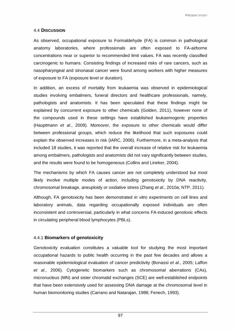
PRESENT STUDY
4.4 DISCUSSION
As observed, occupational exposure to Formaldehyde (FA) is common in pathological
anatomy laboratories, where professionals are often exposed to FA-airborne
concentrations near or superior to recommended limit values. FA was recently classified
carcinogenic to humans. Consisting findings of increased risks of rare cancers, such as
nasopharyngeal and sinonasal cancer were found among workers with higher measures
of exposure to FA (exposure level or duration).
In addition, an excess of mortality from leukaemia was observed in epidemiological
studies involving embalmers, funeral directors and healthcare professionals, namely,
pathologists and anatomists. It has been speculated that these findings might be
explained by concurrent exposure to other chemicals (Golden, 2011), however none of
the compounds used in these settings have established leukaemogenic properties
(Hauptmann et al., 2009). Moreover, the exposure to other chemicals would differ
between professional groups, which reduce the likelihood that such exposures could
explain the observed increases in risk (IARC, 2006). Furthermore, in a meta-analysis that
included 18 studies, it was reported that the overall increase of relative risk for leukaemia
among embalmers, pathologists and anatomists did not vary significantly between studies,
and the results were found to be homogeneous (Collins and Lineker, 2004).
The mechanisms by which FA causes cancer are not completely understood but most
likely involve multiple modes of action, including genotoxicity by DNA reactivity,
chromosomal breakage, aneuploidy or oxidative stress (Zhang et al., 2010a; NTP, 2011).
Although, FA genotoxicity has been demonstrated in vitro experiments on cell lines and
laboratory animals, data regarding occupationally exposed individuals are often
inconsistent and controversial, particularly in what concerns FA-induced genotoxic effects
in circulating peripheral blood lymphocytes (PBLs).
4.4.1 Biomarkers of genotoxicity
Genotoxicity evaluation constitutes a valuable tool for studying the most important
occupational hazards to public health occurring in the past few decades and allows a
reasonable epidemiological evaluation of cancer predictivity (Bonassi et al., 2005; Laffon
et al., 2006). Cytogenetic biomarkers such as chromosomal aberrations (CAs),
micronucleus (MN) and sister chromatid exchanges (SCE) are well-established endpoints
that have been extensively used for assessing DNA damage at the chromosomal level in
human biomonitoring studies (Carrano and Natarajan, 1988; Fenech, 1993).
97

PRESENT STUDY
In this study, all cytogenetic parameters evaluated (Total-CA, CSAs, CTAs, gaps,
aneuplodies, MNL, MNB, BNbud and SCE) were significantly elevated in pathological
anatomy professionals exposed to FA (0.38 ppm) compared with control subjects (Table
VI).
In fact, FA-exposed individuals showed an increase of 91% on Total-CAs frequency
compared to controls (Table VIII). Furthermore, mean frequencies of both CAs types,
CSAs (chromosome-type aberrations) and CTAs (chromatid-type aberrations) were
significantly higher in exposed workers than controls (Table VI). Although there is a
paucity of studies assessing CAs in FA occupational exposed subjects, our findings are in
agreement with most of the published literature. Indeed, increased frequencies of CAs
were found in PBLs of anatomy students exposed to FA (mean level, 2.37 ppm) during a
12-week (10h per week) anatomy class (He et al., 1998). Similarly, in a recent study
involving FA-exposed personnel working in pathology departments (n=21; mean
level=0.72 ppm) Total-CA and CTAs were significantly elevated compared to controls, no
significant differences were found for CSAs (Jakab et al., 2010). Moreover, in vitro
experiments of Schmid et al. (1986) with human lymphocytes showed that FA induces
CAs, mostly of chromatid-type and gaps, as observed in occupational exposed subjects. A
significant increase on CAs frequencies were also observed in industrial workers, namely
in a group of paper factory workers, i.e. dicentric and rings chromosomes (Bauchinger and
Schmid, 1985), and among workers manufacturing nitrogen fertiliser and exposed to FA
concentrations exceeding permissible levels (Kitaeva et al., 1996). In addition, a study in
former Czechoslovakia reported increased levels of CAs in PBLs of children exposed to
FA in prefabricated schools (Neri et al., 2006).
In opposition, Thomson et al. (1984) found no significant differences in the incidences of
CAs in a group of six pathology workers compared to unexposed controls. The same
result was obtained among FA-exposed medical students working in an anatomy
dissection laboratory (Vasudeva and Anand, 1996). Further, no significant differences
were found on CAs frequencies between individuals working in different laboratories of a
Cancer Research Institute including an anatomical pathology laboratory (Pala et al.,
2008), however the results obtained may be attributed to the low level of FA exposure
(range, 0.01-0.05 ppm) and the reduced number of subjects evaluated.
In the present study, both CAs sub-types were statistically elevated in PBLs of FA-
exposed workers. As referred earlier, the generation of structural CAs requires one or
several DNA double-strand breaks (DSBs), CSAs and CTAs formation appear to occur in
different cellular phases, involve specific DNA repair mechanisms and induce by different
agents. While CSAs are thought to be formed by direct double stranded breakage in vivo
98

PRESENT STUDY
in G0/G1 lymphocytes (being duplicated during replication), CTAs formation requires
DNA-replication and arise in vitro from other lesions pre-existing in DNA. However, CTAs
that survive to an earlier division can be converted by duplication into secondary
chromosome-types, and in next cell generation appear as CSAs (Savage, 1975; Musak et
al., 2013). Indeed the significant positive association found in our study between CTAs
and CSAs (r=0.343, p<0.001) may confirm this association and explain our results.
Nevertheless, it should be noted that mechanistically, for instance, two close single-strand
breaks in vivo, not repaired in G1, might produce in metaphase a chromosome type
break.
Hence, our findings indicates that FA- exposure is able to induce lesions in chromosomal
DNA which during repair or DNA synthesis generates DSBs and CAs formation. In
addition, some stable forms of CTAs seem to survive division and pass in a modified form
to next cell generation.
Other variables were analysed in order to obtain more information about the biological
impact of our results. Therefore, we assessed the number of aberrant cells (cells with one
CSAs or CTAs break) and multiaberrant cells (cells with CSAs or CTAs type exchanges or
with more of two breaks/fragments). Significant differences were obtained in both
variables, with FA-exposed subjects showing elevated number of aberrant and
multiaberrant cells compared to controls.
While the majority of CAs are lethal to the cell, others may lead to oncogenic
transformation, for instance by inactivation of a tumour suppressor gene or activation of
an oncogene by generating novel fusion proteins capable of initiating carcinogenesis
(Pfeiffer et al., 2000). Indeed, accumulated data confirms that high CAs frequencies in
lymphocytes of healthy individuals are predictive of cancer risk (Hagmar et al., 1998). A
number of factors may collectively influence the association between CAs and cancer
including exposure to genotoxic carcinogens and internal generation of genotoxic species
(e.g. oxidative stress) (Norppa et al., 2006). In fact, a significant association was found
between cancer incidence and CAs (chromatid breaks) and aberrant cells frequency in a
group of miners exposed to radon – the authors estimated that an increase of 1% in the
frequency of CAs was associated with an increased cancer incidence of 64%
(Smerhovsky et al., 2001, 2002).
Our results also showed that FA-exposed workers had significantly higher frequencies of
gaps (a 65% increase) than controls (Table VIII). Schmid et al. (1986) and Jakab et al.
(2010) found similar results in vitro experiments and in PBLs of pathologists exposed to
FA, respectively. Previous reports of individuals occupationally exposed to other
99

PRESENT STUDY
chemicals also found significant increases on gaps compared to referents (Tunca and
Egeli, 1996; Lander et al., 2000; Goud et al., 2004; Almeida-Santos et al., 2005; Skjelbred
et al., 2011). The validity of scoring and analysing gaps in human biomonitoring studies
has been the subject of much discussion. One reason is the visual distinction of a gap
from a break. Although a consensus was reach on scoring criteria their inclusion in studies
remain controversial (Oostra et al., 2012). Other point of discussion is whether a gap
represents a true double-stranded break in the DNA of a chromatid, a staining
discontinuity or an error in chromosome condensation process (“folding defect”) (Savage,
2004). Our findings, revealed significant positive correlations between gaps and all
structural CAs endpoints, showing an association with established CAs parameters, and
confirming this parameter sensitivity for FA genotoxicity evaluation. In fact, results from a
recent comparative analysis of different types of CAs observed under light microscopy
and by means of atomic force microscopy showed that most gaps are chromosome
alterations and should be included in genotoxicity studies (Koleva et al., 2013). This
statement is further supported by evidence that DNA lesions such as DSBs may affect
chromatin condensation (Savage, 2004), and by the observation that chromatin relaxation
is a fundamental pathway in the DNA-damage response (Ziv et al., 2006). Therefore, the
increase of gaps in FA-exposed subjects may occur as a response to double-stranded
breakage. Also of note, in opposition to CAs, gaps are reparable, and produce no further
structural damage on transmission (Savage, 2004), so by being a reversible form of
damage it may reflect relatively recent genotoxic exposure (Lander et al., 2000). Paz-y-
Miño et al. (2002) tested the correlation between gaps and comet assay, a biomarker of
recent damage, with positive results. Indeed, we also found a significant positive
association between gaps and the comet assay parameter (r=0.273; p<0.001), which
confirm the above hypothesis of the possible biological significance of gaps, and
reinforces the inclusion of this event as a parameter in biomonitoring studies.
In our study, a significant increase in aneuploid PBLs was found among FA-exposed
workers compared to controls (Table VI). However, there are limited data available
evaluating the frequency of numerical CAs in occupational subjects exposed to FA and
published results are conflicting (Zhang et al. 2010b, Speit et al., 2011). An earlier study
reported an increase in aneuploid cells in a group of pathologists compared to controls,
but it did not reach significance (Thomson et al., 1984). Conversely, a significant
decrease in aneuploidy was found among workers exposed to FA (0.72 ppm) in a
pathology department, but the observed lower frequency of aneuploid cells may be
attributed to the significant increase of apoptotic cells found in the FA-exposed workers
investigated (Jakab et al., 2010). In fact, in vitro experiments on cell lines showed that at
100

PRESENT STUDY
concentrations around 1mM, FA enhanced apoptosis and decreased cell proliferation,
whereas at lower doses (0.1 mM) it decreased apoptosis and increased cell proliferation
(Tyihák et al., 2001). Although apoptosis induction was not evaluated in our study, this
finding may explain the different outcome found in our group of workers exposed to 0.38
ppm compared to Jakab et al. (2010) study (0.72 ppm). Furthermore, in a recent study FA
showed to deregulate the expression of miRNAs involved in apoptosis signalling within the
nasal rats epithelium exposed to FA (Rager et al., 2013), which was in accordance with
previous findings in pulmonary cells of rats also exposed to FA (Sul et al., 2007).
Other studies based on a different assay (e.g. fluorescence in situ hybridisation) have also
yielded mixed results, with cells of professionals exposed to FA showing a significant
increase of chromosome loss in one study (Orsière et al., 2006) and no significant effect
in another (Titenko-Holland et al., 1996).
Nevertheless, it should be noted that aneuploidy is a natural event occurring in healthy
subjects. Indeed, different studies have showed that in humans about 3% of lymphocytes
are aneuploid (Fang and Zhang, 2011). It is possible that aneuploid cells are present in all
tissue types and because of their presence in low percentages they do not post any
significant pathological danger including oncogenic transformation. On the other hand,
aneuploidy is a prominent phenotype of cancer, and it has been discussed whether
aneuploidy is only a byproduct of the oncogenic processes or if it can induce
tumorigenesis (Fang and Zhang, 2011). Zhang et al. (2010b) reported leukaemia-specific
chromosome changes (monosomy 7 and trisomy 8) in cultured peripheral blood myeloid
progenitor cells of workers exposed to FA, this finding suggest that FA- exposure may
have an adverse impact through an aneugenic effect on the hematopoietic system.
However, the mechanisms behind aneuploidy are difficult to dissect due to countless
factors that may be involved. Therefore, taking in consideration the above mentioned
further studies on the FA potential aneugenic activity are needed before reaching to any
conclusion.
The results of micronucleus (MN) frequency in PBLs and buccal cells are shown in Table
VI. Compared to controls, MN frequency was significantly increased in FA-exposed
workers in both PBLs and exfoliated buccal cells. MN frequency in PBLs was found to be
approximately 1.6-times higher in the exposed workers compared to control subjects,
whereas in buccal cells an around 3.8-fold increase was observed. Furthermore, a
significant higher frequency of nuclear buds (BNbud) in exfoliated buccal cells was also
found among workers in comparison to controls (2.9-times higher). The increase of
101

PRESENT STUDY
damage was higher in buccal cells, which is not surprising since the oral mucosa
constitutes a site of first contact for inhaled FA.
Our findings comply with others that reported an increased frequency of MN in PBLs
and/or epithelial cells among mortuary students and, more recently, in hospital staff
exposed to FA. Indeed, Suruda et al. (1993) found significant increased frequencies of
MN in lymphocytes (26%) and buccal mucosa cells (12-fold) in a group of mortician
students exposed to FA (8h-TWA level, 0.33 ppm) before and after attending to an
embalming course. A dose-response relationship was observed with cumulative exposure,
but only for buccal micronuclei. Ying et al. (1997) also reported a significant higher
frequency of MN in nasal and oral exfoliated cells in anatomy students exposed to FA
after an 8-week course (8h-TWA level, 0.41 ppm), but no significant difference was found
for lymphocytes. However, a significant elevated frequency in micronucleated
lymphocytes was found among 59 pathology and anatomy laboratory workers in a study
conducted by Orsière et al. (2006). Burgaz et al. (2001, 2002) evaluated MN induction in
buccal and nasal epithelium from pathology and anatomy workers exposed to FA, an
increased frequency in both epithelial cells was found in exposed subjects. More recently
in a group of Portuguese histopathology professionals Ladeira et al. (2011) reported
elevated MN frequencies in PBLs and buccal epithelial cells, confirming previous reports
by Costa et al. (2008, 2011) and Viegas et al. (2010). Ladeira et al. (2011) also evaluated
the frequency of BNbud, finding a significant increase in FA- exposed workers compared
to controls. BNbud formation in cells is associated to the elimination of amplified DNA
and/or DNA-repair complexes (Fenech et al., 2011). Indeed, a recent study showed a
significant association between a polymorphic gene in repair pathway and nuclear buds
formation (Ladeira et al., 2013). Elevated frequencies of MN were also found among FA-
exposed workers from industrial units both in PBLs (Jiang et al., 2010) and in nasal
mucosa cells (Ballarin et al. 1992; Ye et al., 2005).
In the present work the correlation between MN frequencies in buccal cells and PBLs
showed to be highly significant (r=0.359, p<0.001). The concomitant increase of MN
formation in buccal cells (a FA-target tissue) and in PBLs indicates not only that inhaled
FA is able to induce cytogenetic alterations in circulating systemic cells (distal tissue) but
also that the damage between this two tissues may be associated, reinforcing the
biological plausibility of inhaled FA to induce systemic genotoxic effects.
Finally, in our study, a positive significant correlation was observed between MNL
frequencies and FA exposure duration (r=0.277, p=0.011). Also, by correlation testing a
significant positive association was found between FA-level and MNB (r=0.407, p=0.001).
Moreover, MNL and BNbud frequencies were increased with increasing FA-level of
102

PRESENT STUDY
exposure classes (Table XIV), confirming MN as a sensible bioindicator for the evaluation
of the genotoxic action of FA.
SCEs are known to be increased by exposure to various genotoxic carcinogens (Albertini
et al., 2000). Our results on SCE frequency (Table VI) agrees with data from other studies
which reported a higher frequency of this cytogenetic biomarker in FA exposed workers
compared to referents. Yager et al. (1986) were the first to observe an increase of SCE in
subjects exposed to FA (mean level, 1.2 ppm) after a 10 week anatomy class compared
to samples obtained from the same individuals immediately before FA exposure began.
Shaham et al. (1997, 2002) reported similar findings in two studies involving pathology
workers exposed to FA, both groups of workers showed significantly higher SCE
frequencies compared to controls. In the larger study that enrolled 90 professionals,
workers were divided into a “low exposure group” (mean level, 0.40 ppm) and a “high-
exposure group” (mean level, 2.24 ppm); no significant differences on SCE frequencies
were found between the two FA-exposed groups. More recently, Ye et al. (2005)
examined two different types of workers exposed to FA, with different outcomes. One
group was composed of 18 workers of a FA manufacturing facility (8h-TWA level, 0.82
ppm) whereas the second group included 16 waiters who were exposed to low levels of
FA while working in a new ballroom for 12 weeks (5h TWA level, 0.09 ppm). The first
group showed a significantly increased frequency of SCE in lymphocytes when compared
to controls, whereas no significant difference was found in waiters, probably due to the
low level of exposure to FA. Nevertheless, other studies also did not find increased SCE
rates in workers exposed to FA (Thomsom et al., 1984; Suruda et al., 1993; Ying et al.,
1999). Despite some negative findings, most of the studies have reported positive results,
confirming this biomarker sensitivity to evaluate occupational exposure to FA (IARC,
2012).
The increase of SCE in PBLs of FA-exposed individuals may indicate the inability of the
cell to efficiently repair replication-blocking lesions induced by the exposure, which results
in fork collapse and the initiation of homologous recombination (HR) for repair. Moreover,
there is some evidence that implicates the presence of single-strand breaks (SSBs) in
DNA as the primary lesion that leads to SCE formation. The mechanism is likely to
involved an unrepaired SSB, a DSB formation upon replication fork collapse, and HR (with
the sister chromatid) to facilitate accurate repair (Wilson and Thompson, 2007). An
increase of repair activity leads to an increase of SCE, which may explain the elevated
frequency of this biomarker in PBLs of FA-exposed workers. Interestingly, we found
significant positive associations, although weak, between this endpoint and double-strand
103

PRESENT STUDY
breakage parameters, namely Total-CAs (r=0.172; p<0.05), CTAs (r=0.159; p<0.05) and
gaps (r=0.156; p<0.05). In conclusion, the increase on SCEs frequency found in the
present study and previous reports may reflect the repair of FA- induced DNA lesions by
HR.
Compared to the cytogenetic endpoints above described, the comet assay was recently
introduced as a useful technique in human biomonitoring studies, allowing the evaluation
of DNA damage (single and double strand breaks and abasic sites) at the single cell level.
Hence, there are only a few studies published on FA occupational exposure in which this
biomarker is used. In our study, the levels of DNA damage measured as percentage of tail
DNA (%TDNA) were significantly increased in PBLs of FA-exposed professionals
compared to controls (Table VI). This result agrees with Yu et al. (2005) and Jiang et al.
(2010) who reported a significantly increase in comet assay parameters, comet tail length
and olive tail moment, in workers exposed to FA (mean level, 0. 83 ppm) from two
plywood factories. In contrast, no significant differences in comet assay endpoints was
found in PBLs of workers from fiberboard plants compared to controls, however the
results obtained in this study may be attributed to the low level of exposure to FA, 0.2 ppm
(Aydin et al., 2013).
Comet assay has also proved to be a sensitive biological indicator in the evaluation of the
genotoxic effect of FA in several in vitro experiments using cell lines or cells from FA-
exposed rodent or humans (Frenzilli et al., 2000; Emri et al., 2004; Im et al., 2006; Liu et
al., 2006; Sul et al., 2007). Interestingly, in most of these studies FA showed a “two-
phase” dose-response relationship. At low doses, FA induced an increase in DNA
migration whereas at higher doses a decrease in DNA migration was observed.
The DNA damaging and crosslinking effects of FA may explain this finding. Hence, at low
concentrations, it induces strand breaks whilst at higher concentrations crosslinking
activity seems to become the dominant lesion (Frenzilli et al., 2000). This is further
explained by excess of FA-induced lesions at high concentrations which may overwhelm
the cell repair capacity and result in the accumulation of cross-link lesions and the
decrease in DNA migration; while the strand breaks observed at low concentrations are
indicative of repair processes in progress or incomplete. Indeed, SSBs are often
intermediates during repair of other DNA lesions. In fact, Saccharomyces cerevisiae
strains defective in excision-repair of pyrimidine dimers showed to be more susceptible to
the lethal effects of FA and a reduced capacity to undergo single-strand breaks compared
to the wild type, which seems to suggest that SSBs are a step in the repair process of FA-
induced lesions (Magaña-Schwencke et al., 1978; IARC, 2006).
104

PRESENT STUDY
In the present study, no significant differences were found regarding TCR-Mf (Table VI).
To our knowledge, this is the first study in which TCR-Mf was evaluated in workers
exposed to FA. Although this assay has shown to be a sensitive indicator for exposure to
radiation and chemicals in some studies (Kubota et al., 2000; García-Lestón et al., 2012)
others did not find the same result (Chen et al., 2006).
4.4.2 Immune-markers: Lymphocytes subpopulations
The percentages of the different lymphocyte subsets in peripheral blood of exposed and
control subjects were assessed as indicators of possible FA exposure-related
immunotoxicity. The reference ranges for lymphocyte subsets in Caucasian population are
approximately, 60-87% for total T lymphocytes (CD3)+, 32-61% for T-helper cells (CD4+),
14-43% for T-cytotoxic cells (%CD8+), 5-20% for B cells (%CD19+) and 4-28% for NK cells
(CD16+-56+) (Santagostino et al., 1999). Our results in both groups fall within these
expected ranges and are also in accordance with recent data published for a Portuguese
population by García-Lestón et al. (2011).
Regarding the FA exposure effect when comparing both groups, no statistically significant
differences were observed for total T lymphocyte and T-helper cells (Table XI). However,
in the exposed group a significant increase in the percentages of T-cytotoxic cells and NK
cells were found along with a significant decrease of percentage of B cells (Table XI). A
lower ratio of CD4+/CD8+ (Th/Tc) was also observed with statistical significance, due to
increase of T-cytotoxic cell subpopulation in the exposed group.
Our findings are in agreement with other studies, which showed that FA exposure may
affect immunological parameters. Tang et al. (2009) summarised eight studies concerning
FA-induced immunotoxicity and hematotoxicity. In one of the studies regarding clinical
pathology personnel (FA exposure-levels between 0.2 and 0.8 ppm) a significantly higher
proportion of the exposed subjects (14%) showed abnormal white blood cell (WBC)
counts compared to controls (5%). Moreover, an investigation by Zhang et al. (2010b) on
the ability of FA to disrupt haematopoiesis revealed a significant decrease in total WBC
counts, granulocytes and lymphocytes in a group of FA-exposed workers (mean FA
exposure-level, 1.28 ppm). In a recent study involving fibreboard plant workers exposed to
low-levels of FA (mean level, 0.2 ppm) no significant differences in peripheral blood cells
in terms of total WBC, red blood cells, neutrophils, and monocytes were found;
nevertheless significant alterations in total lymphocyte and lymphocyte subpopulations
were detect between workers and controls (Aydin et al., 2013). Conversely, in other
studies lymphocyte subsets and WBC counts were not significantly influenced by FA
105

PRESENT STUDY
exposure (Tang et al., 2009). It should be noted, however, that the available data on
immunotoxicity and hematotoxicity in FA-exposed populations are still sparse, which may
contribute to the inconsistent data.
In our study, the increased percentages of T-cytotoxic cells and NK cells may indicate an
activation of the immune response in workers exposed to FA at a mean level of 0.38 ppm.
T-cytotoxic lymphocytes are cells enrolled in cell-mediated immune responses. Their
function is to destroy compromised or stressed cells (e.g. infected cells or tumour cells) by
recognition on the cell surface of a specific antigen presented by MHC class I (present in
all nucleated cells). NK cells are effector lymphocytes of the innate immune system that
have also cytotoxic function but do not form immune memory. NK cells are able to detect
compromised cells tagged by antibodies or deficient in MHC class I surface marker.
Recent research highlights the fact that NK cells are also regulatory cells engaged in
reciprocal interactions with dendritic cells, macrophages, T cells and endothelial cells
(Viver et al., 2008). Upon activation, T-cytotoxic lymphocytes and NK cells migrate to the
site of inflammation or injury where they may exert their cytolytic function depending on
the combination of inhibitory and activating signals conveyed by the target.
In fact, mild nasal epithelium lesions and a decrease in nasal epithelial cells have been
consistently reported in workers exposed to FA (Edling et al., 1988; IARC, 2006).
Moreover, FA is a mucous membrane irritant as demonstrated in experimental and human
studies (IARC, 2006). A non-specific, non-allergic pro-inflammatory effect of inhaled FA
was suggested (Pazdrak et al., 1993), and clinical findings of upper respiratory tract
inflammation were reported in workers exposed to FA (Lyapina et al., 2004). Substances
that elicit inflammatory events may provide a ‘danger’ signal to the immune system
leading to activation of lymphocytes and other cells involved in immune response.
Therefore, it is possible to speculate that the observed increase of T-cytotoxic
lymphocytes and NK cells in exposed subjects may occur in response to the irritant
properties of FA in the upper respiratory tract mucosa and tissue inflammation. Similarly, a
significant increase on percentages of total T lymphocytes and NK cells were recently
reported in workers exposed to 0.2 ppm, confirming our findings (Aydin et al., 2013).
Our data also show a significant decrease of %B cells in the exposed individuals
compared to controls. In workers exposed to benzene and polycyclic aromatic
hydrocarbons, B cells revealed to be more sensitive than T cells to the genotoxic effects
of these compounds (Sul et al., 2002, 2003), which may probably be the case of FA and
would explain our findings.
106

PRESENT STUDY
Further, the observed decrease in B cells suggests immunosuppression and decreased
ability to respond to exogenous aggressions (e.g. virus, bacteria) in FA-exposed
individuals. B cells are involved in organism’s humoral response against antigens and
engaged in the adaptive immune response system that enables a quick response of the
organism to a relapsed infection. Upon activation, B cells differentiate into plasma cells
producing specific molecules, antibodies, which eliminate targeted antigens (Janeway et
al., 2001).
Although FA-exposed subjects showed as a whole increased %NK cells with regard to the
controls, when analysing the effect of work-related factors on the endpoints studied, we
found a clear decrease in these cells associated with the highest FA-levels of exposure
(Table XIV), specifically at levels equal or above 0.35 ppm, showing percentages lower
than the control population, which may indicate that at high exposure levels FA induce a
decrease in immune response.
Indeed, a decrease in NK cells was reported for workers exposed to mean-airborne
concentrations of 1.28 ppm (Hosgood et al., 2012). In a recent study, FA exposure in mice
(10 ppm for 6h/day and 5 days/week) caused a selective NK cell deficiency, without any
defect in other lymphocytes, by impairing NK cell differentiation (Kim et al., 2013).
NK cells function comprises the control of several types of tumours and microbial
infections by limiting their spread and subsequent tissue damage (Vivier et al., 2008).
Our findings suggest that workers exposed to FA are potentially more susceptible to
infectious agents. Indeed, several studies describe a reduced resistance to infections
(upper respiratory tract infections, recurrent rhinitis and pneumonitis) in individuals
occupationally exposed to FA (IARC, 2006). In the study of Zhang et al. (2010b), 40% of
the exposed subjects had recent respiratory infections. Lyapina et al. (2004) measured
neutrophil respiratory burst activity (NRBA) in 29 workers exposed to FA (mean level, 0.71
ppm). Exposed workers showed a significant increase in upper respiratory tract
inflammations compared to controls, but no significant differences in spontaneous or
stimulated NRBA assays were found between them.
Further, viral associated cancers are increased in immunosuppressed individuals, most
likely due to the inability of the organism to limit viral replication and/or expansion of
infected cells (Schulz, 2009; Hosgood et al., 2012). The Epstein-Barr virus (EBV) is
strongly implicated in the aetiology of nasopharyngeal carcinoma (Gullo et al., 2008). In
fact, the risk of nasopharyngeal carcinoma in workers exposed to FA was more
pronounced among EBV seropositive individuals than non-seropositive (Hildesheim et al.,
2001).
107

PRESENT STUDY
Although our results are in agreement with published data, lymphocyte subpopulations
were differently affected in few other studies. For example, in a group of 23 medical
students exposed to FA (mean FA-level of exposure, 0.41ppm) during an 8-week anatomy
class, Ying et al. (1999) found a significant increase in B cells and a significant decrease
in total T cells, T-helper-inducer cells and T-cytotoxic-suppressor cells. Similarly, Ye et al.
(2005) reported a significant increase in B cells and decrease in total T cells and T-
cytotoxic-suppressor cells in 18 workers from a FA manufacturing facility (mean FA-level
of exposure, 0.98 ppm). Interestingly in both studies, the pattern of immune alteration was
the same at lower (0.41 ppm) and higher (0.98 ppm) levels of FA exposure. The small
sample size or/and the FA level of exposure may have contributed to the different
outcomes observed between these studies and the present work. Another factor that may
also contribute to explain the different results is individual susceptibility. One of the
enzymes involved in FA detoxification is the mitochondrial aldehyde dehydrogenase-2
(ALDH2) (Teng et al., 2001). ALDH2 gene has an inactive ALDH2*2 allele; the presence
of the mutant allele leads to the decrease or absence of ALDH2 catalytic activity (Wang et
al., 2002; Brennam et al., 2004). Approximately 50% of East Asians carry the mutant
inactive ALDH2*2 allele (Brennam et al., 2004; Oota et al., 2004), whereas nearly all
Caucasians carry the functional ALDH2*1/1 genotype (Chambers et al., 2002; Brennam et
al., 2004). Furthermore, in a recent Chinese study FA-exposed workers carrying the wild-
type genotype had a significant increment in formic acid in urine compared with workers
carrying the mutant genotype, confirming that ALDH2 genotypes may influence FA
metabolism (Cheng et al., 2008b). Therefore, genetic variables may modulate the
response to FA exposure and affect differently the populations exposed.
Overall, our findings indicate that inhaled FA may induce alterations in lymphocyte
subsets in exposed workers.
These results should be carefully interpreted until confirmation in further studies. Although
study population was homogenous regarding major confounding factors, and the
statistical analysis took them into account, it is known that the lymphocyte subpopulations
can vary depending on different life situations (Moszczynski et al. 2001).
Also of note, modifications in the immune parameters do not always give measurable
effects. Moreover, even if an immunotoxic effect is shown, it does not always imply clinical
consequences (Vial et al., 1996).
108

PRESENT STUDY
4.4.3 Effect of lifestyle factors and parameter-specific confounders on the biomarkers studied
In human biomonitoring studies is important to assess the influence of major potential
confounding factors such as gender, age and smoking habits in the biomarkers studied.
4.4.3.1 Biomarkers of genotoxicity
In the present study, age was found to be a confounder for CSAs, MNL and MNB, with
frequencies increasing significantly with aging (Tables VIII and IX). This influence was
further confirmed by the significant positive correlation found between age and MNL and
MNB frequency. Our data is in accordance with previous studies (Carbonell et al., 1996;
Bolognesi et al., 1997; Bonassi et al., 2001, 2011) reporting a positive association
between age and cytogenetic biomarkers. The observed age-related effect is probably
associated with a progressive increase in spontaneous chromosome instability and the
loss of efficiency in DNA repair mechanisms, which may result in the accumulation of
genetic lesions with increasing age (Bolognesi et al., 1999).
Cigarette smoking had a significant influence on aneuploidy in PBLs (decrease) and MNB
frequencies in oral mucosa (increase) (Table IX). Further, the significant correlations
found between these biomarkers and packs/year confirmed the observed effect of tobacco
smoking on these endpoints.
Tobacco smoke contains a high number of mutagenic and carcinogenic substances, such
as benzene, arsenic and FA. Epidemiologically it has been associated with a higher risk
for cancer development, especially in the oral cavity, larynx, and lungs, as these are
places of direct contact with the carcinogenic tobacco compounds (IARC, 2004).
With regard to our data, the significant decrease of aneuploid PBLs found in smokers
might be related with a higher apoptotic activity in these subjects induced by the increased
levels of DNA damage resulting from tobacco smoking. Indeed, exposure of rats to
mainstream cigarette smoke produced a significant and time-dependent increase in the
proportion of apoptotic cells in the bronchial and bronchiolar epithelium (D’Agostini et al.,
2000). Some authors have reported lower damage in PBLs of healthy smokers compared
to never-smokers (Wang et al., 2003; Lao et al., 2008). Furthermore, smokers have
showed an increase on baseline repair capacity (Wei et al., 2000a), probably as an
adaptation resulting from the increased demand for repair stimulated by the continuous
damage caused by tobacco carcinogens. The genotoxicity of mainstream tobacco smoke
and cigarette smoke condensate has been demonstrated in vitro and in vivo experiments,
although human studies have produced mixed results (DeMarini, 2004). Nevertheless, it is
109

PRESENT STUDY
important to note that the unbalanced number of smokers and non-smokers in our study
limits the value of the data obtained and restricts possible conclusions, so larger studies
are necessary to confirm this result.
Several studies have described an increase on MN formation in exfoliated buccal cells of
smokers (Goud et al., 2004; Gabriel et al., 2006; Proia et al., 2006; Nersesyan et al.,
2011). In fact, smoking was the only lifestyle variable that significantly influenced the level
of micronuclei in the oral mucosa of 120 healthy individuals, with smokers showing higher
frequency of MNB compared to controls (Konopacka et al., 2003). Furthermore, our
results showed a significant positive association with packs/year. Similarly, among a
group of tobacco smokers Wu et al. (2004) found a significant positive trend between
MNB frequency and either daily cigarette consumption or cumulative smoking pack-years.
Yet other publications report no difference between smokers and non-smokers (Karahalil
et al., 1999; Nersesyan et al., 2006; Holland et al., 2008). Nevertheless, a recent meta-
analysis found significant increases in the MNB frequency but only in heavy smokers (40
cigarettes/day) (Bonassi et al., 2011).
In our study, fruit consumption was found to significantly decrease the DNA damage
measured by comet assay %TDNA parameter (Table X). Epidemiological studies
consistently report that a balanced diet rich in fruit and vegetables is associated with a
reduced risk of cancer and heart disease (Duthie et al., 2006). Indeed, fruits are rich in
several phytochemicals and antioxidants such as vitamin C and flavonoids that inactivate
reactive oxygen species (ROS) involved in the initiation or progression of these chronic
diseases (Brevik et al., 2011a). Therefore regular fruit consumption protects against the
oxidative damage of DNA and thus might prevent mutation and cancer. Human
supplementation trials investigating the anti-oxidant effect of fruit consumption measured
by comet assay (classical and modified versions) have, however, given ambiguous results
(Hoelzl et al., 2009). In some studies, kiwifruit (juice or whole fruit) consumption showed to
significantly decrease the levels of DNA breakage in lymphocytes (induced by H2O2) and
formation of endogenous oxidised bases (measured by incubation with lesion-specific
enzymes) indicating an increased antioxidant capacity with fruit intake (Collins et al.,
2001; Collins et al., 2003; Brevik et al., 2011b). Conversely, in other trials, no effect in
oxidative damage or repair in PBLs was observed after fruit consumption (Duthie et al.,
2006; Yuan et al., 2011). Our data suggest that regular consumption of fruit may actually
protect against DNA damage, however, one shortcoming of our study was the lack of
information concerning the type of fruit consumed which would enable further conclusions.
110

PRESENT STUDY
4.4.3.2 Lymphocyte subpopulations
In the present study significant influence of the major confounding factors, gender and
age, were observed for some lymphocyte subpopulations (Table XII).
Previous studies have demonstrated gender-related differences in immune cell
percentages (Santagostino et al., 1999; Jentsch-Ullrich et al., 2005; Yan et al., 2010). The
total lymphocyte count is generally similar in males and females, but the percentage of T
cells within the lymphocyte population tends to be lower in males (Yan et al., 2010) mostly
because of total T lymphocyte and T-helper cells decreases (García-Dabrio et al., 2012).
Percentage of T-cytotoxic cells usually does not differ between males and females
(Jentsch-Ullrich et al., 2005). However, in our study males had a significantly lower
percentage of T-cytotoxic cells compared to females. No significant differences were
found between genders for T lymphocytes and T-helper cells. Some evidence suggests
that the influence of gender on T lymphocyte subsets is related to sex hormones, although
the processes involved are not well understood. Two different mechanisms have been
proposed: acceleration of thymocyte apoptosis by androgens, which may mediate
processes of thymocyte selection and shape the peripheral T cell repertoire (Olsen et al.,
1998) or the effect of binding to cell receptors for the sex steroids present on T cells
(Chng et al., 2004, Pierdominic et al., 2010).
The influence of age in the immune system has been described in several studies
(Jentsch-Ullrich et al., 2005; Saule et al., 2006; Andreu-Ballestar et al., 2012). Age is
known to affect the immune function and the percentages of circulating lymphocytes in a
process often designated as "immunosenescence" (Yan et al., 2010). Aging of the
immune system is characterised by a decrease in immunocompetence. Hence, it may
lead to infections or autoimmune diseases and be involved in the pathogenesis of several
age-related disorders, such as cardiovascular or neurodegenerative disorders (Laffon et
al., 2013). In our study, a significant decrease with age was found for T-cytotoxic cells
whereas total T lymphocytes and T-helper cells were significantly increased (Table XII).
The effect of age in these endpoints was further confirmed by correlation analysis. Other
authors (Tollerud et al., 1990; Chng et al., 2004) found similar results. Moreover, in a
recent study carried out in a Mediterranean population (Spain) a significant positive
correlation with age was found for T-helper cells, in addition, an inverse trend was
observed for T-cytotoxic cells, but it did not reach statistical significance (García-Dabrio et
al., 2012). The impact of age on lymphocyte subsets is not well established with different
studies reporting contrasting results (Bisset et al., 2004; Coelho et al., 2011; García-
Lestón et al., 2011; Andreu-Ballester et al., 2012). T lymphocyte generation in aging is
known to result from dynamic changes in thymic as well as extrathymic functions, along
111

PRESENT STUDY
with sequential developmental steps from stem cells to ultimately mature cells (Jentsch-
Ullrich et al., 2005). Therefore, the age range of subjects studied, methodological
features, environmental and genetic factors may influence the results and account for the
differences (Jentsch-Ullrich et al., 2005; Andreu-Ballester et al., 2012).
In our study, multivitamin supplement intake was found to significantly increase
T-cytotoxic cells percentage (Table XII). Furthermore, fruit consumption was also found to
increase T-helper cells percentage, but it did not reach significance. The link between diet
and immunity has long been established. Systematic studies have consistently shown that
nutritional deficiency impairs immune response, particularly cell-mediated immunity by
reducing number and functions of T-cells, phagocyte function, cytokine production,
secretory antibody response, antibody affinity and complement system (Chandra, 1999).
Similar findings have been reported for moderate deficiencies of individual nutrients such
as trace minerals and vitamins, particularly Zn, Fe, Se, vitamins A, B6, C and E (Chandra,
1999; Liu et al., 2011). The use of nutrient supplements, singly or in combination,
stimulates immune response and may lower the incidence of infection or reverse some of
the changes associated with impaired immune responses in some chronic diseases
(Grzegorzewska and Leander, 2005) and in elderly people (High, 2001). In a randomised
placebo-controlled intervention study, healthy volunteers consumed probiotic multivitamin
and mineral supplement for 3-5 months during winter/spring period. Leukocytes,
lymphocytes, in particular T-lymphocytes including T-cytotoxic cells and T-helper cells, as
well as monocytes were significantly increased in the “supplementation” group, during the
first two weeks of supplementation compared to placebo. Additionally, in the
“supplementation” group the incidence and severity of symptoms related to common cold,
frequent at that time of year, were reduced. Overall, according to published data,
multivitamin supplementation intake and a balanced diet (rich in vitamins and anti-
oxidants) improve the immune status, which is in agreement with our findings.
4.4.3 Correlations between biomarkers studied
The association found between MNL frequencies with CAs parameters and gaps is
related to the fact that both endpoints are able to indicate chromosomal and chromatidic
breaks, which suggests chromosome breakage as the primary mechanism of FA-related
micronuclei formation. In fact, in an earlier study by Titenko-Holland et al. (1996) it was
detected a greater increase in centromere-negative micronuclei content in buccal and
nasal tissues of FA-exposed students, however Orsière et al. (2006) found higher
frequencies of centromere-positive micronuclei (monocentric) in health professionals
112

PRESENT STUDY
exposed to FA. Our findings, comply with recent studies by Speit et al. (2011)
(mammalian cell lines) and Costa et al. (2011) (FA-exposed pathology anatomy workers)
indicating a clastogenic action of FA, thus confirming previous results from Titenko-
Holland et al. (1996). A significant association was found between CSAs and %TDNA, but
the coefficient was relatively low. This association is not surprising since %TDNA detects
DNA strand breaks, including double-strand breaks.
Concerning the immune markers, T-cytotoxic lymphocytes were inversely correlated with
B cells (r=-0.577, p<0.001). As mentioned earlier, B lymphocytes seem to be more
sensitive to genotoxicity induced by organic compounds exposure than T lymphocytes
(Sul et al., 2002, 2003). The primary function of T-cytotoxic lymphocytes is to remove
compromised or stressed cells. The inverse correlation found may result as a response to
stressed B cells. Further, T-cytotoxic lymphocytes were positively associated with
genotoxicity endpoints, while B cells were inversely correlated. Genotoxicity events have
been shown to alter immune responses (Gasser and Raulet, 2006). No previous studies
were found reporting similar results. Nevertheless, our findings indicate that increased
levels of DNA damage are associated with alterations in immune system.
113

PRESENT STUDY
5. BIOMARKERS OF SUSCEPTIBILITY
5.1 OVERVIEW
5.1.1. Genetic polymorphisms as susceptibility biomarkers
About 99.9% of the DNA is identical in every human genome (Hirvonen et al., 2008). The
0.1% difference is responsible for the inter-individual variation and the unique phenotype
of each individual. These minor differences correspond to genetic variations in the DNA
sequence, known as polymorphisms. The majority of polymorphisms are single nucleotide
polymorphisms (SNPs), changes at a single DNA base, which occur with a frequency of
10-6 throughout the human genome (Rueff et al., 2002). SNPs play an important role in
modulating susceptibility to diseases as well as in the individual response to various drugs
and environmental carcinogens (Hemminki and Shields, 2002).
In simple terms, SNP’s can alter an individual’s response to a chemical exposure by
interfering with the expression or function of proteins involved in their metabolism or in the
repair of the chemical-induced DNA lesions. SNPs in gene coding regions can lead to
changes in the biological properties of the encoded protein. In fact, about 50% of the
SNPs in the coding regions results in a change in the amino acid protein sequence
(Hirvonen et al., 2008). On the other hand, SNPs in non-coding regulatory regions may
affect gene expression levels in an allele-specific manner (Wang et al., 2005).
Altered function of low penetrance genes due to SNPs may affect the gene-environment
and gene-gene interaction, thereby increasing the risk of cancer development (Hirvonen
et al., 2008). Therefore, the identification of sequence polymorphisms that modulate gene
expression or function is crucial for understanding human genetic differences and
diseases (Pastinen et al., 2004) and to recognise susceptible groups in the population.
Genetic polymorphisms are thus considered to play a primary role in individual response
to carcinogen-induced disease (Teixeira et al., 2004). Therefore, in recent years, they
have been used as susceptibility biomarkers in human biomonitoring studies to assess the
risk of exposure to genotoxic chemicals.
5.1.2. Genetic Polymorphisms, DNA damage and Cancer Risk
Cancer is a multifactorial disease that results from complex interactions between genetic
and environmental factors (Hirvonen, 2008). This means that with few exceptions (e.g.
Mendelian cancer syndromes such as retinoblastoma) the cause is not entirely genetic or
114

PRESENT STUDY
entirely environmental (Rueff et al., 2002) and is not a single gene or single environmental
factor that has large effects on cancer susceptibility (Kiyohara et al., 2007). Instead,
various genetic and biologic aspects (e.g. gender, ethnicity) and exogenous factors (e.g.
environmental/occupational exposure to carcinogenic compounds, lifestyle factors,
nutritional status) play a joint role in the process of carcinogenesis (Rueff et al., 2002)
(Figure 16).
Figure 16. Both environmental and host factors acts in concert in individual susceptibility (genetic and acquired) for disease development (retrieved from Perera, 1997)
Biologically, cancer is considered to be a genetic disease, caused by genetic alterations at
either chromosomal or gene levels, as identified in most cancers (Wei et al., 2000b). It is
also agreed, that genetic polymorphisms are associated with most common disorders with
a genetic component (Rueff et al., 2002). Nevertheless, as mentioned before,
susceptibility-conferring alleles are not sufficient to cause disease, but modulate the risk in
combination with other alleles and environmental exposures.
It is known that environmental carcinogens, as a result of complex metabolism, are able to
interact with DNA and produce measurable alterations evaluated by several biomarkers of
lesion (e.g. DNA adducts, chromosomal aberrations, micronucleus) (Rueff et al., 2002;
Mateuca et al., 2008). Interestingly, it has also been observed that the correlation between
the levels of the different biomarkers and levels of exposure are not always linear,
suggesting that intra-individual susceptibility play an important role in processing the
damage (Rueff et al., 2002). Indeed, several studies have shown that the level of
biomarkers arising from environmental and/or occupational exposure may be influenced
by polymorphic genes coding for enzymes engaged in metabolic or repair processes
(Pavanello and Clonfero, 2000; Hung et al., 2005; Mateuca et al., 2008). Therefore, the
115

PRESENT STUDY
complex metabolism of carcinogens, involving different polymorphic genes, as well as
gene variants of DNA repair mechanisms, can modulate the individual susceptibility for
cancer (Pavanello and Clonfero, 2000). This is best exemplified by tobacco smoking;
cigarette smoking is one main cause of lung cancer but only a relative minority of smokers
develop pulmonary cancers.
5.1.3. Xenobiotic-metabolising enzymes
The balance between phase I and phase II metabolic enzymes is an important
determinant of whether exposure to carcinogens will result in toxicity or increased levels of
genetic damage (Talalay et al., 1990).
Most carcinogens undergo activation by phase I enzymes, often as an oxidation reaction,
and detoxification by phase II enzymes. The cytochrome P450 enzyme superfamily
constitutes the majority of phase I enzymes, whilst the glutathione S-transferases (GSTs)
and N-acetyltransferases (NATs) are primarily responsible for detoxification of xenobiotics
in phase II.
Phase I enzymes are generally involved in the metabolism of xenobiotic compounds with
hydrophobic character, producing more polar intermediates that are easily excreted from
the cell. However, phase I metabolism may also result in the activation of carcinogenic
compounds, yielding reactive electrophilic metabolites that can react with cellular
macromolecules, like DNA. The action of phase II enzymes, generally contributes to
detoxifying metabolism of more hydrophilic carcinogens. Phase II enzymes, also act on
phase I carcinogenic metabolites, transforming them into inactivated compounds that are
then excreted. Less frequently, GSTs and NATs can also mediate activating steps for
some carcinogen metabolites.
Genetic polymorphisms exist in a number of phase I (activating) and phase II (inactivating)
enzymes. Hence, it is plausible that individuals with genotypes associated with a more
efficient activating enzyme and/or a less efficient inactivating enzyme might be at
particularly high risk of adverse health effects, if exposed to toxicants. Indeed,
accumulated data suggest that polymorphisms in genes controlling carcinogen
metabolism and thus the metabolic capacity may in fact play a primary role in
susceptibility to environmentally induced diseases, such as cancer.
5.1.3.1. Polymorphisms of phase I enzymes studied: Cytochrome P450 family
The Cytochrome P450 (CYPs) system is the most important enzymatic system involved in
phase I of human metabolism (Rueff et al., 2002). CYPs constitute a superfamily of a
116

PRESENT STUDY
highly diverse group of haem-containing enzymes responsible for the oxidative
metabolism of a wide range of drugs and toxic hydrocarbons. All CYPs exhibit similarity in
their structure and general mechanism of action; NADPH is required as a coenzyme and
O2 is used as a substrate. However, there are significant differences in substrate
specificity due to structural variations at the active site. Based on sequence homologies,
human CYPs are divided into families and subfamilies.
The highest concentration of CYPs is in the liver, where it determines the intensity and
duration of drug action and promotes the detoxication of some xenobiotics. Extrahepatic
tissues, especially those that are portals of entry for foreign compounds, such as the
respiratory tract, also express xenobiotic-metabolising CYPs. In these tissues, CYPs not
only contribute to the first-pass clearance but may also influence the tissue burden of
foreign compounds or bioavailability of therapeutic agents. In the cell, these enzymes are
primarily located in the membrane of the endoplasmatic reticulum (microsomes), but they
are also found in the mitochondrial membrane.
During biotransformation, xenobiotics are either detoxified or activated into reactive
intermediate substances that might be more toxic than the parent compounds. However,
not all xenobiotic-metabolising CYPs subfamilies show the same propensity in the
bioactivation of chemicals. For example, the CYP2B, 2C and 2D subfamilies play virtually
no role in the bioactivation of toxic and carcinogenic chemicals, whereas the CYP1A, 1B
and 2E subfamilies are responsible for the bioactivation of the majority of xenobiotics
(Ioannides C, 2007).
These reactions can also result in the transfer of electrons to produce oxygen radicals.
Cytochrome P450‐catalysed reactions can thus produce xenobiotic metabolites and
oxygen species capable of interfering with cell homeostasis. Over 60 key forms of CYPs
are known, with hundreds of genetic variations that correspond to different metabolic
activities and therefore to a wide variety of susceptibility to potential deleterious effects
induced by toxicants (Autrup H, 2000; Godschalk and Schooten, 2008).
CYP2E1 gene (CYP2E subfamily)
The CYP2E subfamily has only be described in mammals, and only one CYP2E gene,
CYP2E1 has been identified in humans (Godschalk and Schooten, 2008).
CYP2E1 gene encodes the enzyme N,N-dimethylnitrosamino-N-dimethylase, which
catalyses the metabolism of a wide variety of therapeutic agents, toxicants and low
molecular weight pro-carcinogens, including benzene, N-nitrosamines and vinyl chloride
(Anzenbacher et al., 2001). It has a clear toxicological role, since it may activate pro-
117

PRESENT STUDY
carcinogens, organic solvents and drugs, and potentiate their toxicity by converting them
into cytotoxic or carcinogenic products (Nedelcheva et al., 1996).
For instance, this enzyme is involved in the oxidation of compounds, such as ethanol,
producing reactive oxygen species (ROS), which can affect target tissue and ultimately
lead to carcinogenesis (Guo et al., 2008). Indeed, a number of studies in cells lines have
established the importance of this enzyme in oxidative stress (Cederbaum, 2006). During
ethanol oxidation, the CYP2E1 enzyme produces ROS (O2– and H2O2) that can deplete
glutathione and cause cellular damage (Gonzalez, 2007). In addition, ROS can further
react and produce oxidants or result in elevated lipid peroxides that can form adducts with
cellular nucleophiles such as proteins and nucleic acids (Gonzalez, 2007).
The enzyme is mostly expressed in the hepatic tissue and can be induced by xenobiotics
such as ethanol, acetone or isoniazid (Novak and Woodcroft, 2000) all of which are
substrates. It was also detected, at low levels in human lung and oesophagus (Ding and
Kaminsky, 2003), and evidence suggests that it can be present in kidney and nasal
mucosa (Lucas et al., 2001).
CYP2E1 enzyme was detected by immunochemistry in the white cell fraction of human
peripheral blood (Lucas et al., 2001). Expression of the CYP2E1 enzyme in lymphocytes
appears to be influenced by the same factors that affect the concentration of the hepatic
enzyme, including xenobiotics and physiological status (Raucy et al., 1995; 1997).
The CYP2E1 gene is present in the human population in various polymorphic forms.
Although there are a number of SNPs in the CYP2E1 sequence, no known variant results
in the complete inactivation of the gene, suggesting that the enzyme is essential for
humans (Gonzalez, 2007).The gene is located in the 10q24.3-qter region of the
chromosome 10 (Shahriary et al., 2012). It spans 11,413 base pairs and contains 9 exons,
8 introns, and a typical TATA box (Shahriary et al., 2012). The first alleles were identified
by restriction fragment length polymorphism (RFPL) analysis. The best studied CYP2E1
polymorphisms were detected by PstI/RsaI and DraI restriction enzymes, which
corresponds to SNPs in the 5'-flanking region and in the intron 6 of the CYP2E1 gene. It
has been shown that polymorphisms detectable by PstI/RsaI can affect the transcription of
CYP2E1 in vitro. In contrast, DraI polymorphism does not appear to affect gene
transcription, but rather the CYP2E1 enzyme catalytic activity (Shahriary et al., 2012).
The allele frequencies of CYP2E1 polymorphisms may differ remarkably among different
human populations (Garte et al., 2001; Bolt et al., 2003). For instance, the DraI
polymorphism occurs at a higher frequency in Taiwanese when compared with European
and African-Americans (Stephens et al., 1994). Additional information on genotype
118

PRESENT STUDY
frequency distribution of the polymorphism studied is shown in Annex III. Polymorphisms
in the CYP2E1 gene have been associated with increased risk of cancer in different sites,
including breast cancer (Anderson et al., 2012; CYP2E1 intron, allele A), lung cancer (Wu
et al., 1997; RsaI, allele), nasopharyngeal carcinoma (Kongruttanachok et al., 2001; RsaI,
allele C) and gastric cancer (Boccia et al., 2007; PsI/RsaI, allele C).
5.1.3.2 Polymorphisms of Phase II enzymes studied: Glutathione S-transferases
The glutathione S-transferases (GSTs) play a critical role in providing protection against
electrophiles and products of oxidative stress (Hayes and Pulford, 1995).
The GSTs are a superfamily of phase II enzymes which catalyse the conjugation of
reduced glutathione (GSH) with different species of electrophilic compounds, including
many environmental mutagens and carcinogens and their reactive metabolites (Strange et
al., 2001). In addition to their role in phase II detoxification, GSTs also play an important
role in modulating the induction of other enzymes and proteins for cellular functions, such
as DNA repair (Hayes and Pulford, 1995). Because of their role in the detoxification of
potentially carcinogenic compounds, these enzymes and the genes encoding them may
play an important role in cancer susceptibility.
GST enzymes are present in the cytosol of all eukaryotic cells, but are also bound to
membranes (Landi, 2000). The human cytosolic GSTs are grouped into eight families of
GST isoenzymes, including: GSTA (α), GSTM (μ), GSTP (π), GSTS (σ), and GSTT (θ)
(Raimondi et al., 2006; Hirvonen, 2008). The classification is in accordance with the
substrate specificity, chemical affinity, structure, aminoacid sequence and kinetic behavior
of the enzyme (Hayes and Pulford, 1995). Because GSTs have overlapping substrate
specificities, the deficiencies of a given GST isoenzyme may be compensated by other
isoenzyme and use of alternative metabolic pathways is possible (Hirvonen, 2008).
Polymorphisms have been identified in the GSTM1, GSTT1 and GSTP1 genes coding for
GSTs enzymes in the mu (μ), theta (θ), and pi (π) classes, respectively. These enzymes
detoxify a wide variety of chemicals, such as reactive epoxides and carcinogens produced
by tobacco smoke. Genetic polymorphisms in GSTs encoding genes have been
associated with susceptibility to genetic lesions in populations exposed to several
environmental carcinogens, including polycyclic aromatic hydrocarbons (PAHs; Butkiewicz
et al., 2000), styrene (Teixeira et al., 2004) and hydroquinone (Silva et al., 2004).
Many studies have suggested a predisposing role of GSTs polymorphisms for various
cancers in different tissues, such as lung, breast and colon (Hirvonen, 2008). However,
there are also various reports contradicting these findings (Raimondi et al., 2006). One of
119

PRESENT STUDY
the reasons of the controversial data may be the overlapping substrate specificities of the
GSTs enzymes. Therefore, simultaneous determination of different GSTs genotypes
relevant for a given exposure may be crucial for a reliable interpretation of the role of the
GST gene family in cancer development (Hirvonen, 2008). Indeed, meta-analyses studies
have indicated that carriers of both GSTM1 and GSTT1 null alleles have a slightly higher
risk of developing lung cancer compared to carriers of at least one functional allele
(Benhamou et al., 2002; Raimondi et al., 2006).
Additional information on frequency distribution of the GSTs studied among different racial
groups is available in Annex III.
Glutathione S-transferase T1 gene (GSTT1)
The encoded GSTT1 enzyme (θ subfamily) is involved in the detoxification of
monohalomethanes and reactive epoxide metabolites of butadiene, both of which are
constituents of tobacco smoke (Landi et al., 2000; Hirvonen, 2008).
GSTT1 gene is located on 22q11.23 region of the chromosome 22; it spans 8092 base
pairs and contains 5 exons and 4 introns (Yang et al., 2013). The most common
polymorphism in GSTT1 consists of a deletion of the whole gene (GSTT1*0, null
genotype) resulting in the lack of active enzyme (Raimondi et al., 2006). The risk
associated with the null genotype is difficult to predict since the enzyme may have both
detoxification and toxification activities toward different industrial and environmental
chemicals. The enzyme in human erythrocytes detoxifies the carcinogen ethylene oxide
but may enhance the formation of the genotoxic metabolites from dichloromethane (To-
Figueiras et al., 1997).
The prevalence of GSTT1 null genotype shows a wide variation among different racial
populations and ethnic groups. For instance, in Caucasians the prevalence is 10–20%
(Hirvonen, 2008) and in Asians it can vary between 45-60% (Cho et al., 2005).
The null genotype has been correlated with increased risk of several types of
malignancies including lung and larynx cancers (Yang et al., 2013, Sørensen et al., 2004;
Russo et al., 2013). A modest increase in the risk of haematological malignancies
(lymphoma, leukaemia) was also observed (Ye and Song, 2005; Bin and Luo, 2013).
However, other studies have reported negative results (López-Cima et al., 2012;
Sørensen et al., 2007). Some of the observed heterogeneity may be partly due to ethnicity
(Raimondi et al., 2006).
120

PRESENT STUDY
Glutathione S-transferase M1 gene (GSTM1)
A gene cluster at 1p13.3 region of chromosome 1 encodes the GST µ subfamily
(Hirvonen, 2008). One of the gene variants includes a deletion and homozygotes
(GSTM1*0, null genotype) do not express the protein, thus lacking the enzyme activity
(Hayes and Pulford et al., 1995). GSTM1 enzyme is involved in the detoxification of large
hydrophobic electrophiles, such as benzo(a)pyrene and other PAHs found in tobacco
smoke.
Several epidemiological studies on GSTM1 null genotype have suggested, although
moderate, an association between this genotype and cancer risk, namely bladder cancer,
lung cancer, prostate cancer and head and neck cancer (Hirvonen, 2008). Moreover, this
genotype appears to be associated with a modest increase in the risk of acute
lymphoblastic leukaemia (Ye and Song, 2005). Some conflicting reports also exist (López-
Cima et al., 2012; Bin and Luo, 2013). However, recent pooled and meta-analyses
support previous findings suggesting that GSTM1 deficiency can contribute substantially
to the incidence of cancer at the population level (Engel et al., 2002; Hiyama et al., 2008;
Mo et al., 2009).
The distribution of the GSTM1 null genotype varies among racial and ethnic groups. For
instance the mean reported frequency for the null genotype among Caucasians is about
50%, while for African descendants is 29 % (Hirvonen, 2008).
Glutathione S-transferase P1 gene (GSTP1)
The glutathione S-transferase P1 (GSTP1) belongs to GSTP (π) subfamily. A single 2.8
kb long GSTP1 gene at 11q13 region of chromosome 11, encodes an isoform that is
known to metabolise many carcinogenic compounds, including polycyclic aromatic
hydrocarbons (PAHs). GSTP1 is the most abundant glutathione S-transferase in the lungs
(Cote et al., 2009) and therefore it is thought to be of particular importance in the
detoxification of inhaled carcinogens (Hirvonen, 2008).
An A>G transition at nucleotide 313 of the GSTP1 gene results in an isoleucine (Ile) to
valine (Val) amino acid change at codon 105 (Ile105Val). This produces a variant enzyme,
which differs in enzymatic activity (more or less effective detoxification) depending on the
substrate and alters their thermo stability (Mo et al, 2009). The presence of the GSTP1
Val variant (GG or GA genotypes) is associated with a lower conjugating activity when
compared to the homozygous GSTP1 Ile variant (AA genotype) (Cote et al., 2009).
Moreover, having at least one copy of the G allele at this locus is also associated with
121

PRESENT STUDY
increased levels of hydrophobic adducts in the lung and higher levels of PAH-DNA
adducts in human lymphocytes (Butkiewicz et al., 2000).
The GSTP1 Ile105Val polymorphism has been investigated for associations with breast,
head and neck cancers, with both positive and negative outcomes (Egan et al., 2004;
Ruwali et al., 2011). In addition, epidemiological studies of the impact of the
GSTP1 Ile105Val polymorphism on lung cancer risk including two meta-analyses show
inconsistent results (Cote et al., 2009; López-Cima et al., 2012).
5.1.4. DNA Repair
DNA repair is a ubiquitous process throughout the living world and is essential for the
maintenance of genome integrity. Several identified genes and sequence variants are
involved in human DNA repair (Au et al., 2003; Rueff et al., 2002). These genes function
in a complex diverse set of pathways that involve recognition and removal of DNA lesions
caused by endogenous or exogenous agents and/or correction of DNA errors occurred
during DNA replication or DNA repair (Ronem and Glickman, 2001). Other genes are
indirectly involved in DNA repair, by regulating the cell cycle, providing an opportunity for
repair or controlling apoptosis mechanisms (Moses et al., 2001; Ronem and Glickman,
2001).
Both endogenous and exogenous agents are continually inducing cellular DNA damage. If
unrepaired, the damage can interfere with important cellular functions potentially leading
to genetic instability, unregulated cell growth and cancer (Au et al., 2003). On the
contrary, if damage is recognised by cell machinery it can be repaired enabling the cell to
replicate as planned. At cell level, several responses may occur to prevent replication in
the presence of genetic errors: checkpoints can be activated to arrest the cell cycle,
transcription can be up-regulated to compensate for the damage or cell can trigger
apoptosis (Goode et al., 2002).
Polymorphisms in DNA repair genes may be innocuous or may change protein function
and therefore alter individual’s capacity to repair DNA lesions caused by exogenous or
endogenous agents (Au et al., 2003). A deficient or unrepaired damage can ultimately
lead to cancer (Ronem and Glickman, 2001).
Mutations that affect the function of DNA repair enzymes are rare in the human population
because they cause severe health consequences including higher proneness for cancer
development (Au et al., 2003). Examples of cancer syndromes where the disease-causing
mutations occur in DNA repair genes include xeroderma pigmentosum, Fanconi anaemia
122

PRESENT STUDY
and Bloom’s syndrome; each disorder shows specific damage sensitivities at the cellular
level and specific disease phenotypes (Moses et al., 2001).
The etiologic association between deficient DNA repair and increased cancer risk is also
supported by evidence from epidemiologic studies of sporadic cancers of the lung (Goode
et al., 2002), head and neck (Gresner et al., 2012), bladder (Stern et al., 2002) and skin
(Mocellin et al., 2009). These data strongly suggest that it is important for normal tissues
to have efficient DNA repair in order to eradicate DNA lesions caused by carcinogens,
thereby reducing the risk of mutation fixation and subsequent development of cancer (Wei
et al., 2000b).
5.1.4.1 DNA Repair pathways
A variety of complex pathways have evolved to perform the critical repair functions. The
main classic pathways of DNA repair are base excision repair (BER), nucleotide excision
repair (NER), mismatch repair (MMR) and double-strand break repair (Jeppesen et al.,
2011). Each pathway operates on specific types of DNA lesion and involves numerous
molecules in the repair processes (Goode et al., 2002) (Figure 17).
BER (Figure 17A) operates on small lesions (e.g. oxidised or reduced bases; fragmented
or nonbulky adducts) caused by a variety of factors including ionising radiation and
alkylating agents (Goode et al., 2002). The process which aims to remove and replace a
damaged nucleotide, is typically initiated by one of several substrate-selective DNA
glycosylases, which recognise and excise base modifications, such 8-oxo-7,8-dihydro-2'-
deoxyguanosine (8-oxo-dG), among others (Wilson et al., 2011). The resulting abasic site
is incised by an AP endonuclease (APE1). The baseless phosphodiester backbone and
adjacent nucleotides are then removed and the gap is filled by DNA polymerase β that
uses the other strand as a template for the DNA synthesis (Collins, 1996). The final step is
ligation of repair patch into the pre-existing DNA strand by a DNA ligase, which in
mammals is typically ligase III in complex with x-ray cross-complementing protein 1
(XRCC1) (Almeida and Sobol, 2007).
NER (Figure 17B) is the major pathway in humans for the repair of bulky lesions, such as
UV-induced pyrimidine dimers and chemical adducts, induced by environmental
carcinogens (Au et al., 2003). In the NER repair process, the distortion of the double helix
(e.g. bulky adduct) is recognised by a complex of bound proteins including XPC; the
formation of the transcription factor complex, TFIIH, allows the unwinding of the DNA and
the removal of the damaged single-stranded fragment (usually about 27–30 nucleotides);
123

PRESENT STUDY
the DNA synthesis is carried out by DNA polymerase δ using the other strand as a
template (Goode et al., 2002).
The MMR (Figure 17C) maintains genomic stability by correcting replication errors, such
as mispairings (base-base or insertion-deletion mismatched) caused by DNA polymerase
during replication (Aquilina and Bignami, 2001; Goode et al., 2001). The process begins
with specific repair proteins recognising and binding to the mismatched bases, further
enzymatic action proceeds to exonucleolytic degradation, DNA resynthesis and ligation
(Aquilina and Bignami, 2001).
Double-strand breaks, DSBs, (Figure 17D) can be produced by replication errors and by
exogenous agents (e.g. ionising radiation) (Goode et al., 2002). DSBs are the most
serious form of DNA damage because they pose problems for transcription, replication,
and chromosome segregation. DSBs affect both strands of the DNA duplex and therefore
prevent the use of the complementary strand as a template for repair. Cells have evolved
at least two different mechanisms of DSBs repair: Homologous recombination (HR) and
Non-homologous end-joining (NHEJ). The first is a highly accurate repair process that
corrects DSBs defects in an error-free manner using a mechanism that retrieves genetic
information from the homologous, undamaged DNA molecule. The majority of HR-based
repair takes place in late S- and G2-phases of the cell cycle when an undamaged sister
chromatid is available for use as repair template (Khanna and Jackson, 2001).
Conversely, the NHEJ repair mechanism is error prone, since it re-joins the two broken
ends of DNA directly, which frequently can lead to small alterations (base pair
substitutions, insertions and deletions) in the DNA sequence (Khanna and Jackson,
2001).
The Fanconi Anaemia (FANC) pathway has emerged recently as an important model for
studying the mechanisms surrounding DNA damage response/DNA repair, genomic
instability and ubiquitin signalling (Moldovan and D'Andrea, 2009; Kottemann and
Smogorzewska, 2013).
FANC repair pathway coordinates the action of multiple distinct repair processes, in
particular, NER, HR and translesion synthesis (TLS; a mode of damage tolerance that
uses specialised polymerases to insert a base across from a lesion or abasic site)
(Moldovan and D'Andrea, 2009). Studying the FANC repair pathway provides a unique
window into the elaborate interplay of multiple cellular networks involved in DNA repair.
The FANC proteins work together to maintain genomic stability during DNA replication
and to repair DNA interstrand crosslinks (ICLs), a severe lesion that blocks both DNA
124

PRESENT STUDY
A. Base excision repair (BER) B. Nucleotide excision repair (NER) C. Mismatch repair (MMR)
D. Double-strand break (DSBs) repair Non-homologous end-joining Homologous recombination
replication and transcription (Moldovan and D'Andrea, 2009; Kottemann and
Smogorzewska, 2013).
Figure 17. DNA Repair pathways. A) BER mediates the removal and replacement of small lesions, such as, damaged bases and uracil residues caused by reactive oxygen species (UNG for uracil is shown in the figure); the resulting abasic site is incised by an AP endonuclease (APEX1); the baseless phosphodiester backbone is removed and the gap filled by DNA polymerase β (Pol β). B) NER acts on larger lesions or adducts and involves lesion recognition, formation of the TFIIH complex, removal of 25–30 nucleotides and gap filling by a DNA polymerase. C) MMR removes mismatched bases; after recognition of specific proteins, a segment of DNA is excised between the mismatch and a nearby nick, the gap left is filled by DNA polymerase δ. D) DSBs can be repaired by Non-homologous end joining (NHEJ), a error prone process that joins directly the DNA ends or Homologous recombination (HR), a error free repair processes that operates when a double-stranded copy of the sequence is available (adapted from Lange et al., 2011)
FANC is a rare genetic disease caused by germline mutations that disrupt the FANC
pathway leading to a deficient repair function (Moldovan and D'Andrea, 2009).
125

PRESENT STUDY
FANC patients manifest a high propensity for cancer, namely squamous cell carcinomas
(e.g. head and neck, gynecologic system) and acute myeloide leukaemia (AML) (Alan and
D'Andrea A, 2010). This phenotype has launched the question as to whether inherited or
acquired mutations in FANC genes might be involved in the development of sporadic
cancers (Tischkowitz et al., 2004). Moreover, several genes involved in the FANC
pathway also predispose individuals to breast and ovarian cancer, which, together with
the cancer susceptibility of patients with FANC, suggests FANC proteins have an
important role in suppressing tumorigenesis (Kottemann and Smogorzewska, 2013).
The association between a chromosomal instability syndrome and cancer predisposition is
not surprising since excessive chromosome breakage might be expected to lead to
unrepaired DNA damage and apoptosis, or to mutations which could confer a selective
growth advantage to certain cells (Mathew, 2006). However, the reason why cells of
myeloid origin are particularly affected in FANC case leading to AML is not known
(Mathew, 2006; Moldovan and D'Andrea, 2009). Recent work has begun to gain
tantalising insight into not only FANC pathogenesis, but also the intersection between
DNA repair and stem-cell maintenance and the general role of FANC repair pathway
genes (Kottemann and Smogorzewska, 2013). One explanation is that the myeloid
precursors are particularly susceptible to forms of DNA damage (e.g. crosslinks), that are
recognised or repaired by the FANC pathway. It is likely that the cellular endogenous
metabolism might specifically yield agents that cause DNA lesions (Moldovan and
D'Andrea, 2009), and circulating metabolites such as formaldehyde (FA) may account for
the sensitivity of FANC hematopoietic stem cells (Ridpath et al., 2007). Indeed, recent
advances have identified endogenous aldehydes, such as acetaldehyde and FA, as
possible inducers of the DNA damage that may lead to the chromosomal instability seen
in FANC pathway deficient cells (Rosado et al., 2011; Kottemann and Smogorzewska,
2013). More importantly, there is also recent evidence that genes in the FANC repair
pathway have a crucial role in counteracting acetaldehyde-induced genotoxicity in mice,
furthermore, mice mutant in both FANC repair genes and aldehyde-catabolism gene
spontaneously developed acute leukaemia (Langevin et al., 2011).
Taking into account the above, it is reasonable to speculate the existence of polymorphic
FANC genes that may modulate individual susceptibility to the effects of exposure to FA,
particularly in the workplace. This association would also agree with Zhang et al. (2010)
hypothesis of FA ability to affect haemotopoietic system in exposed individuals and help to
elucidate the increase of AML cases among FA-exposed workers. Therefore, it was
relevant in our study a screening of polymorphic genes associated with FANC repair
pathway.
126

PRESENT STUDY
At least fifteen FANC complementation groups of proteins/genes (FANC-A, -B, -C, -
D1/BRCA2, -D2, -E, -F, -G, -I, -J/BRIP1, -L -M, -N/PALB2, -P and -O/RAD51C) were
found to constitute the FANC pathway (Kim and D'Andrea, 2012).
The FANC pathway is turned on during the S phase of the cell cycle or following DNA
damage, by FANC protein monoubiquination (the addition of one ubiquitin, an amino acid
tag, to the internal lysine residue of a protein). During activation, multiple FANC proteins
undergo phosphorylation by checkpoint kinases (ATR-CHK1) (Kim and D’Andrea, 2012).
Once active, the FANC proteins work in concert to regulate the monoubiquitinated state of
the FANCD2 and FANCI proteins and the downstream functions of the repair process
(Alan and D'Andrea, 2010).
A few models on the FANC-mediated crosslink mechanism have been proposed, but
generally it is described as follows. During replication, the presence of an ICL that
covalently links both strands of a DNA duplex, blocks the DNA unwinding and stops the
progression of the replication fork. A complex formed by FANCM and FAAP24 proteins
recognises the stalled replication fork structure and recruits the FANC core complex to the
damaged DNA region initiating the FANC pathway (Kim and D'Andrea, 2012) (Figure 18).
Figure 18. Interaction of the FANC proteins in the recognition of an ICL in S phase. A) Two replication forks converge on the DNA. B) The FANCM–FAAP24 complex recognises the stalled replication fork structure. FANCM stabilises the stalled replication fork, and initiates the ATR-CHK1-dependent checkpoint response, which in turn phosphorylates multiple FANC proteins, including proteins in the FANC core complex and FANCD2/FANCI unit. C) The FANC core complex monoubiquitinates FANCD2/FANCI complex, and it is recruited to the DNA lesion (adapted from Kim and D’Andrea, 2012)
FANCM prevents the collapse of replication fork, independently of FANC pathway
activation. The FANC core complex is an ubiquitin ligase multi-subunit composed by eight
FANC proteins (FANCA,-B,-C,-E,-F,-G,-L,-M) that monoubiquitinates the FANCD2/FANCI
unit (Alan and D'Andrea A, 2010). Lastly, the ubiquitin-tagged FANCD2/FANCI complex
interacts with other downstream FANC proteins (FANCD1, FANCN, and BRIP1) and other
DNA repair proteins to resolve the ICL and the stalled replication fork (Kim and D’Andrea,
2012).
A. B. C.
127

PRESENT STUDY
In the DNA, two nucleolytic incisions flanking the ICL region release it from one of the
DNA strands. The unhooking process forms a double-strand break and leaves cross-
linked oligonucleotides in the complementary strand. The latter is bypassed by TLS
restoring the strand and NER removes the remaining adduct and fills the gap. The double-
strand break is repaired by HR mechanism. An ubiquitin-specific protease removes the
ubiquitin from the FANCD2/FANCI complex, and completes the FANC repair pathway
(Kim and D’Andrea, 2012) (Figure 19).
Figure 19. Simplified model of the mechanism of ICL repair process involving NER, TLS and HR. A) An ICL blocks the DNA unwinding and stops the progression of the replication fork; FANC complex stabilises the staled fork. B) The crosslink unhooking from one of the DNA strands forms a DSB and leaves a monoadduct in the complementary strand. C) FANC recruits TLS to bypass the remaining monoadduct. D) NER excises the monoadduct and repairs the strand. E) The DSB is repaired by HR mechanism. F) Intermediate DNA crossover structures are resolved and the replication fork is re-established (adapted from Kennedy and D'Andrea, 2005)
5.1.4.2. Polymorphisms of the BER pathway studied
BER (base excision repair) repairs DNA lesions such as non-bulky base modifications,
abasic sites and a range of chemically distinct single-strand breaks (Wilson et al., 2011).
Genetic polymorphisms in BER pathway have been associated with cancer risk at several
sites (Wilson et al., 2011; Hung et al., 2005). Further, deficiencies in BER gene products
have been linked to neurodegenerative disorders and immunodeficiency (Doseth et al.,
2011; Wilson et al., 2011). Because polymorphisms of BER genes may affect the level of
DNA repair induced by chemicals, leading to carcinogenesis, it is important to establish
the molecular causes of the carcinogenetic process. The major genes involved in the BER
pathway include, among others, XRCC1, PARP1 and MUTYH (Almeida and Sobol et al.,
2007). Genotype frequencies may vary markedly across race, supplementary information
on the frequency distribution of the studied polymorphisms is available in Annex III.
XRCC1
The XRCC1 (x-ray repair cross-complementing group 1) protein forms complexes with
DNA polymerase β, DNA ligase III and poly-ADP-ribose-polymerase (PARP) in the repair
A. B. C. D. F.
E.
NER HR TLS
128

PRESENT STUDY
of nicks or gaps left in BER pathway and in the repair of single-strand breaks, SSBs (Aka
et al., 2008). It is an important protein for co-ordination of DNA damage repair (Thaker
and Zdzienickab, 2003).
The XRCC1 gene has been located at 19q13.2–13.3 region of human chromosome 19.
The importance of this gene is highlighted by the description of its null mutant mice,
whose embryonic development is arrested (Aka et al., 2008). Cell lines defective in this
nonenzymatic scaffold protein show reduced SSBs repair, increased sensitivity to DNA-
damaging agents that produce SSBs (Kulkarni et al., 2008), and elevated chromosomal
aberrations and sister chromatid exchanges frequencies (Thompson and West et al.,
2000).
Two common polymorphisms of XRCC1 that lead to amino acid substitutions are located
in exon 10 (G to A, arginine to glutamine, Arg399Gln) and in exon 6 (C to T, arginine to
tryptophane, Arg194Trp).
In humans, while the XRCC1 Arg399Gln substitution is associated with increased genetic
instability the XRCC1 Arg194Trp substitution is suggested to enhance individual DNA
repair capability (Tuimala et al., 2004; Aka et al., 2008; Chang et al., 2009).
Indeed, published data on XRCC1 gene and cancer risk show associations between: a)
the XRCC1 399Gln/Gln genotype and increased risk of tobacco-related cancers among
light smokers, but decreased risk among heavy smokers (Hung et al., 2005); b) the
XRCC1 Arg194Trp substitution and reduced risk of various types of cancer, including
breast and lung cancer (Goode et al., 2002). Nonetheless, recent data from a Polish study
suggest that XRCC1 399Gln allele is a potential risk factor for breast cancer (Romanowicz
et al., 2010). Yet, there are other studies with contradictory findings (Hirvonem et al.,
2008). The XRCC1 Arg194Trp substitution is more prevalent among Asians (2.6-13.2%)
than among Caucasians (≤ 2.3 %) (Hung et al., 2005). Regarding XRCC1 Arg399Gln
genotype frequencies, there is no major variation across race and ethnicity (Hung et al.,
2005; Chang et al., 2009).
PARP1
Human Poly(ADP-ribose) polymerase-1 (PARP1) is a major member of the PARP family
proteins. PARP1 functions as a DNA damage sensor in the DNA repair process.
Generally, in response to DNA damage caused by exogenous or endogenous agents,
PARP1 binds to the DNA lesion through its DNA-binding domain. This activates the
poly(ADP-ribosyl)ation of target proteins involved in the DNA repair pathways, including
BER and DSBs systems, and recruits the repair proteins to the sites of DNA damage (Yu
129

PRESENT STUDY
et al., 2012). PARP1 complex interacts preferentially with XRCC1, triggering the BER
repair machinery (Masutani et al., 2003).
PARP1 is also involved in other cellular processes, such as modulation of gene
transcription, cell proliferation and death. Accumulated data from in vitro and in vivo
studies indicates that PARP1 deficiency leads to DNA repair defects, genomic instability
and failure to induce cell apoptosis, which may contribute to cancer development (Yu et
al., 2012).
The human PARP1 gene is located on 1q41-42; it spans 47.3 kb and comprises 23 exons.
PARP1 Val762Ala is a polymorphism in exon 17 (T to C transition) that may result in
altered activity of PARP1, due to an amino acid substitution, alanine (Ala) for valine (Val),
in the protein catalytic domain. PARP1 Val762Ala has been implicated in human
carcinogenesis, but with inconclusive results. Nevertheless, data of a recent meta-
analysis suggest that this polymorphism may modulate cancer risk differently depending
on ethnicity and type of cancers. The study found an association between PARP1
Val762Ala and increased risk of cancer among Asians but a decreased risk among
Caucasians, particularly of glioma (Yu et al., 2012).
MUTYH
The MUTYH gene encodes a DNA glycosylase involved in the repair of oxidative DNA
lesions such as 8-oxo-7,8-dihydro-2'-deoxyguanosine (8-oxo-dG), a stable oxidation
product of guanine. If left unrepaired, 8-oxo-dG can mispair with adenine during DNA
replication, leading to a G:C>T:A transversion (Santos et al., 2012). The MUTYH protein
prevents these transversions by excising any adenine residue misincorporated in the
newly synthesized DNA strand opposite to 8-oxo-dG, thus allowing the repair process to
be continued by downstream BER enzymes (Mazzei et al., 2013). MUTYH dysfunction
may, therefore, be especially problematic for tumorigenesis in humans since there are no
other mechanisms for repairing 8-oxo-dG/adenine mismatches (Santos et al., 2012).
Mutations in this gene strongly predispose humans to a rare hereditary form of colorectal
cancer (MAP) (Out et al., 2012). MAP tumours display an unusually high proportion of
somatic G:C>T:A transversion mutations in certain genes, probably reflecting defective
MUTYH protein activity and consequent failure to correct A:8-oxo-dG mispair (Santos et
al., 2012). Several MUTYH germline mutations and SNPs have been identified (Wilson et
al., 2001). MUTYH is located on the short arm of the chromosome 1 at 1p34. It spans a
region of 11,147 base pairs with 16 exons.
130

PRESENT STUDY
MUTYH Gln335His (rs3219489) is a common polymorphism located on exon 12. The
variant genotype implicates a G> C transition and a substitution of the amino acid
glutamine (Gln) for histidine (His) at the 335 codon of the MUTYH protein. The enzyme
encoded by this variant has been demonstrated to have partially impaired glycosylase
activity in vitro (compared to the wild-type, the variant enzyme was 34% less active in
removing adenine from substrates containing an A:GO mismatch) and could therefore
contribute to cancer susceptibility (Santos et al., 2012).
The 335His variant allele has been suggested to be associated with increased risk of
colorectal cancer in some studies (Picelli et al., 2010; Santos et al., 2012). A positive
association was also found for lung cancer, but only when gene-gene interactions were
considered (Qian et al., 2011). Also, in a bladder cancer susceptibility study, gene-gene
interactions among BER polymorphisms (including MUTYH Gln335His) were observed in
ever smokers (Huang et al., 2007), suggesting that BER genetic variation might contribute
to cancer risk through gene-gene and gene-environmental interactions (Santos et al.,
2012).
5.1.4.3. Polymorphisms of the DSBs repair pathway studied
Unrepaired or misrepaired, double-strand breaks (DSBs) may result in cell death,
chromosome rearrangements and genome instability, all processes involved in
carcinogenesis. The DSBs repair by homologous recombination (HR) is a key pathway for
the maintenance of genetic stability in mammalian cells (Tambini et al., 2010). HR is a
high fidelity process that uses homologous sequence as a template to synthesise new
error-free DNA. It acts in coordination with the S and G2 checkpoint machinery to
eliminate chromosomal breaks before the cell division occurs (Thompson and Schild,
2001). Two major cancer genes, BRCA1 and BRCA2, associated with breast cancer are
involved in HR pathway. Gene variants of other participants in HR have showed to be
associated with cancer risk at several sites, pointing out the relevance of HR and DSBs in
cancer susceptibility. The frequency distribution of DSBs repair genotypes varies across
racial groups, supplementary information for each polymorphic gene studied is available in
Annex III.
Rad51
The RAD51 protein is responsible for the central activity of the HR repair pathway, in
which it catalyses the invasion of the broken ends of the DSBs into the intact sister
131

PRESENT STUDY
chromatid, that will serve as template to reconstruct the missing part of the damaged
strand (Gresner et al., 2012).
In this process, the generated single-stranded DNA ends are bound by Rad51 to form a
nucleoprotein filament that searches the undamaged DNA on the sister chromatid for a
homologous repair template. Other proteins including the breast cancer predisposition
proteins (BRCA1 and BRCA2), and several additional Rad51-related proteins (e.g.
XRCC2, XRCC3) serve as accessory factors in filament assembly and subsequent Rad51
activities (Richardson, 2005; Thacker, 2005). Furthermore, BRCA2 is shown to regulate
both the intracellular localisation and the DNA-binding ability of this protein (Davies et al.,
2001). Several studies of Rad51 among BRCA1/2 mutation carriers have found
associations with breast cancer risk (Thacker, 2005; Ricks-Santi et al., 2011). In addition,
several genetic assays have also demonstrated that p53 can down-regulate Rad51-
mediated HR events; however, the process by which it occurs remains unclear
(Richardson, 2005)
The role of Rad51 in genomic integrity is strongly supported by Rad51 mouse knockouts
which result in early embryonic lethality (Richardson, 2005). Experimental studies have
also show that the loss of Rad51 may drive to genetic instability, chromosomal
aberrations, and carcinogenesis by facilitating an accumulation of genetic changes
(Richardson, 2005). A recent case control found evidence that Rad51 gene variability may
be of relevance with respect to head and neck cancer risk modulation (Gresner et al.,
2012)
The RAD51 gene is located at 15q15.1 in chromosome 15. Multiple transcript variants
encoding different isoforms have been found for this gene (Thacker, 2005).
The RAD51 SNP in the 5′ untranslated region (G > T, rs1801321, RAD51 5’UTR) is a
polymorphism whose association with cancer risk is scarcely explored. The few available
epidemiological data concerns breast cancer risk, but no association was found (Silva et
al., 2010).
XRCC2
The X-ray repair cross-complementing protein group 2 (XRCC2) is a RAD51-related
protein, essential for efficient HR repair process of DSBs (Tambini et al., 2010). The
degree of sequence homology to RAD51 or to other RAD51-like proteins is relatively
small, around 45% similarity and 25% identity (Thaker and Zdzienickab, 2003).
XRCC2 role is still not clear, but there are indications that it acts as a cofactor for the
RAD51 in strand invasion and exchange activities and also in late stages of the HR repair
132

PRESENT STUDY
pathway (Richardson, 2005; Silva et al., 2010). The loss of XRCC2 has showed to result
in a severe delay in the early response of RAD51 to DNA damage (Tambini et al., 2010).
In addition, XRCC2-deficient cell lines have chromosome missegregation associated with
fragmentation of the centrosome, indicating that the protein is also required for correct
chromosome segregation (Griffin et al., 2000).
The disruption of XRCC2 gene in mice resulted in early embryonic lethality (Thaker and
Zdzienickab, 2003).
Human XRCC2 gene was first identified by the ability to complement the irs1 mutated
hamster cell line phenotype, which is highly sensitive to mitomycin C, a DNA cross-linking
agent, and suffer increased frequency of chromosomal aberrations, i.e., breaks (Cui et al.,
1999). Further studies showed that XRCC2 is highly conserved, and most truncations of
the protein destroy its ability to protect cells from the effects of the DNA cross-liking
agents (Park et al., 2012).
XRCC2 gene is localised on chromosome 7q36.1. The XRCC2 Arg188His polymorphism
(G > A, substitution of arginine to histidine, rs3218536) has been linked to small but
significant increased risk to breast (Thompson and Schild, 2002; Silva et al., 2010),
colorectal (Vineis et al., 2009) and cervical cancer (Pérez et al., 2013). Other studies
reported null results (Yu et al., 2010; Park et al., 2012). Therefore, additional studies with
larger sample size are required to confirm the role of this polymorphism in cancer
susceptibility.
XRCC3
The XRCC3 (x-ray repair cross-complementing group 3) protein plays a critical role in
repair of DSBs in homologous recombination pathway. It is involved in the assembly and
direct stabilisation of RAD51 protein multimers at DSBs sites (Aka et al., 2008, Tambini et
al., 2010); however the precise function of XRCC3 is not fully understood (Richardson,
2005). Additionally, there is evidence that XRCC3 protein is required for correct
chromosome segregation (Griffin et al., 2000). Analysis of sequence similarities showed
that XRCC3 is a RAD51-like protein, but it only shares around 25% sequence identity to
RAD51 and to other RAD51-related proteins (Thaker and Zdzienickab, 2003).
As in XRCC2, the human XRCC3 gene was originally identified by the ability to correct the
phenotype of mutated rodent cell lines (irs1SF) for sensitivity to ionising radiation and
other DNA damaging agents (e.g. cisplatin, mitomycin C) (Thaker and Zdzienickab, 2003).
Without XRCC3 activity, the irs1SF cells displayed highly elevated levels of deletions,
breaks, and translocations (Cui et al., 1999).
133

PRESENT STUDY
The human XRCC3 gene is located at 14q32.3 region of chromosome 14. Rare
microsatellite polymorphism in the XRCC3 have been reported, including a C>T non-
conservative change (Thr241Met, rs861539) in exon 8, which gives rise to an amino acid
substitution (threonine for methionine) (Aka et al., 2008). The XRCC3 241Met allele was
associated with significant increases in chromosome deletions in x-ray-challenged blood
lymphocytes (Manuguerra et al., 2006). Several studies have evaluated the influence of
XRCC3 Thr241Met on cancer risk in different tissues (Goode et al., 2002). Positive
associations were obtained for bladder cancer (Matullo et al., 2001), breast cancer (Silva
et al., 2010), lung cancer and colorectal cancer (Aka et al., 2008). In some studies, it was
found a potential gene-gene interaction with genes of metabolic enzymes (Matullo et al.,
2001) and other DNA repair pathways (Aka et al., 2008). Nevertheless, other studies have
reported contrasting results (Manuguerra et al., 2006; Aka et al., 2008). The prevalence of
Met/Met homozygosis is around 4.6 % in African Americans, 0.2 % in Asians, and 12.4 %
in Caucasians (Manuguerra et al., 2006).
5.1.4.4 Polymorphisms of the FANC repair pathway studied
FANC (Fanconi anaemia) is a genetic recessive disorder, caused by mutations in genes
regulating replication-dependent removal of DNA crosslinks (Moldovan and D’Andrea,
2009).
At least fifteen FANC complementation groups (FANC-A, -B, -C, -D1/BRCA2, -D2, -E, -F,
-G, -I, -J/BRIP1, -L -M, -N/PALB2, -P and -O/RAD51C) and their corresponding genes
have been identified to cooperate in the FANC pathway (Su and Huang, 2011).
The members of the FANC complementation group do not share sequence similarity. Four
of the FANC pathway genes (FANCD1/BRCA2, -J/BRIP1, -N/PALB2, and -O/RAD51C)
are known breast cancer susceptibility genes and other gene products, that connects
FANC proteins to HR (homologous recombination) of DSBs repair pathway (Castella et
al., 2011). Proteins encoded by these genes participate in the repair of ICLs during
replication and are essential for accurate processing of stalled replication forks (Castella
et al., 2011).
Mutations in FANC-A, -C and -G are the most common and account for approximately
85% of patients with FANC (Su and Huang et al., 2011). FANC individuals have greatly
increased risks of cancer, including acute myeloid leukaemia (AML).
It has long been hypothesized that heterozygotes for autosomal recessive DNA repair
disorders may have reduced efficiency of their DNA repair systems that could cause an
increased risk of cancer (Tischkowitz et al., 2008). Epidemiological studies assessing
134

PRESENT STUDY
incidence of malignancies (including AML) in FANC families have failed to demonstrate an
increased incidence in heterozygous individuals (Mathew, 2006; Tischkowitz et al., 2008).
However, this raises the question as to whether inherited or acquired mutations in FANC
genes might be involved in the development of sporadic cancers, e.g. AML (Tischkowitz et
al., 2004). The major role of BRCA2/FANCD1 in susceptibility to breast and other
cancers, such as cancer of the ovarian , prostate and pancreas, is well established
(Mathew, 2006). There have been several studies of the other known FANC genes in
relation to breast cancer susceptibility, some of which found an association with
FANCN/PALB2, FANCJ/BRIP1 and FANCD2 (Mathew, 2006; Barroso et al., 2009). There
is also some limited data suggesting an association with sporadic hematological
malignancies (Tischkowitz et al., 2004). It is important to point out that these studies had
some limitations and therefore replication in larger studies is needed to confirm these
findings (Mathew, 2006). Additional information on frequency distribution of the
polymorphisms studied among different racial groups is available in Annex III.
FANCA
FANCA (or Fanconi anemia complementation group A) protein is one of the eight FANC
proteins that comprise the FANC core complex, a multisubunit complex required for
FANCD2 ubiquitination, essential for resolving ICL and stalled replication fork, during
replication.
Furthermore, evidence suggests that FANCA phosphorylation is DNA damage–inducible
and essential to the function of the FANC pathway (Collins et al., 2008).
Mutation in FANCA gene accounts for more than 60% of all cases of FANC (Moldovan
and D'Andrea, 2009). The mutation profile of FANCA is highly heterogeneous with over
100 different mutations described, a large part being deletions (Castella et al., 2011).
Hence is one of the most studied genes in the FANC family in relation to cancer
proneness.
Human FANCA gene is located at 16q24.3 in chromosome 16. The FANCA Thr266Ala
polymorphism (rs7190823) results in a aminoacid substitution, in codon 266, of threonine
to alanine. The current evidence regarding the role of FANCA mutations in sporadic
malignancies is limited. Nonetheless, some findings suggest that acquired FANCA
dysfunction and/or FANCA variants may be involved in the promotion of genetic instability
in cases of sporadic AML (Lensch et al., 2003; Tischkowitz et al., 2004). Additionally, a
recent epidemiologic study have found an increased risk of cervical cancer associated
with FANCA variants (Wang et al., 2009b).
135

PRESENT STUDY
BRIP1
BRIP1 (BRCA1-interacting protein 1) also known as BACH1 (BRCA1-associated C-
terminal helicase-1) or FANCJ (for Fanconi anemia complementation group J) is a DNA
helicase that interacts directly with BRCA1 and thus is essential for DNA repair and
genomic stability (Cantor and Guillemette, 2011). It is universally expressed and co-
localised with BRCA1 in the nuclear foci (Ma et al., 2013).
Phosphorylated BRIP1 functions together with BRCA1 protein to mediate proper and
efficient repair of DNA double-strand break via HR repair subpathway (Cantor et al.,
2001). BRIP1 interacts with BRCA1, unwinds DNA structures that block replication forks
and facilitates checkpoint signalling (Constantinou, 2012). The complex formed by BRIP1
and BRCA1 is important not only for HR-mediated double-strand break repair but also for
tumour suppressor function (Ma et al., 2013).
This specific interaction between BRIP1 and BRCA1 is regulated by the cell cycle and is
essential for execution of the G2/M cell cycle checkpoint and for normal progression
through S phase by assisting in the resolution of stalled replication forks (Kumaraswamy
and Shiekhattar, 2007)
It is well established that a deficient DNA helicase function can result in human genetic
disorders in which genomic instability and predisposition to cancer are common features
(e.g. Bloom Syndrome) (Cantor et al., 2004). Accumulating data suggests an anti-
oncogenic role of BRIP1, and downregulation of BRIP1 gene has been reported in
multiple cancers (Cantor et al., 2001; Ma et al., 2013).
The BRIP1 gene is located on chromosome 17q22, it spans 180 kb and comprises 20
exons (Cantor et al., 2001). In the last few years, common SNPs in the BRIP1 gene have
been associated with susceptibility to breast cancer (Cantor et al., 2004), ovarian cancer
(Pennington and Swisher, 2012) cervical cancer (Ma et al., 2013) and prostate cancer
(Kote-Jarai et al., 2009).
Recently, the rare polymorphism BRIP1 Ser919Pro (substitution of serine to proline,
rs4986764) was reported to be a risk factor of breast cancer (Sigurdson et al., 2004)
especially in women up to age 50, however contrasting results was also found (Vahteristo
et al., 2006) and therefore this association needs further research.
A final model linking the various factors involved in individual’s response to an exogenous
insult and potential biologic effects is outlined in Figure 20.
In short, most of the carcinogens present in the environment undergo bio-activation into
ultimately reactive metabolites, and this transformation is modulated by the individual
136

PRESENT STUDY
genetic profile of metabolic genes. In addition, common polymorphisms in DNA repair
genes may alter protein function and eventually the individual ability to repair DNA
damage (Iarmacovai et al., 2007).
Therefore, the study of the interaction of different genetic polymorphisms involved both in
the metabolism of environmental carcinogens and in the DNA repair is crucial to evaluate
the possible combined effect of several genetic variants in relation to a specific chemical
exposure (Mateuca et al., 2008).
Figure 20. Association among the various factors involved in individual’s response to an exogenous insult and potential biologic effects (retrieved from Iarmacovai et al. (2007)
137

PRESENT STUDY
5.2 MATERIAL AND METHODS
5.2.1 DNA extraction
Genomic DNA was obtained from heparinised whole blood samples (350 μL) using a
commercially available kit (Qiagen EZ1 DNA Blood kit; Qiagen BioRobot EZ1 System)
according to the manufacturer’s instructions. DNA samples were stored at -20 ºC until
analysis.
5.2.2 Genotyping of polymorphisms in gene involved in the metabolism
Detailed information regarding selected SNP’s and corresponding amino acid exchanges
are presented in Table XVI. Genotype determinations were carried out twice in
independent experiments and all the inconclusive samples were reanalysed.
5.2.2.1 CYP2E1
The CYP2E1 intronic polymorphism (rs6413432) was determined by PCR-RFLP as
described elsewhere (Lin, 1998; Teixeira, 2004) with minor modifications. Primer design
and PCR conditions were optimised in order to achieve the best possible results:
OligoAnalyzer 3.1 (http://eu.idtdna.com/analyzer/Applications/OligoAnalyzer/) was used to
determine melting temperatures, GC contents, hairpins, and dimmer formation. Basic
Local Alignment Search Tool (BLAST) resource (http://blast.ncbi.nlm.nih.gov/Blast.cgi)
was used to confirm low similarity with other human sequences. This polymorphism
results in the loss of a restriction site, which allows the common and variant alleles to be
discriminated by RFLP after appropriate restriction enzyme digestion. Specific primers,
restriction enzyme site details and restriction patterns for each of these SNPs are shown
in Table XVII. PCR was performed in a 50 µL reaction volume containing: 100 ng of
genomic DNA, 1 µM of each primer, MgCl2 5 mM, 0.5 U of Immolase (Bioline), 0.2 mM of
each dNTP (Bioline) and 2% dimethyl sulfoxide (DMSO), in 1x PCR buffer. The
amplification was accomplished in a GeneAmp® PCR System 9700 thermal cycler
(Applied Biosystems) and consisted of an initial activation step (7 min at 95 ºC); 35 cycles
of denaturation (30 sec at 94 ºC), annealing (30 sec at 62 ºC) and extension (30 sec at
72 ºC); and a final extension step (10 min at 72 ºC). For restriction analysis, 10 µL of the
PCR product were digested with 5 units of the SexAI (Fermentas) restriction enzyme
(12.5 µl final reaction volume), during 210 min at 37 ºC. After restriction enzyme
inactivation (20 min at 65 ºC), restriction fragments were subjected to electrophoresis in
138

PRESENT STUDY
2% agarose gel with ethidium bromide (1 μg/mL) for visualisation under ultraviolet light.
HyperLadder IV (Bioline) was used as molecular marker.
5.2.2.2 GSTM1 and GSTT1
GSTM1 and GSTT1 genotyping for gene deletions were carried out through a multiplex
PCR technique described elsewhere (Lin et al., 1998; Teixeira et al., 2004) with minor
modifications. DNA samples were amplified for GSTM1 with the primers: 5´-GAA CTC
CCT GAA AAG CTA AAG C-3´ (forward) and 5´-GTT GGG CTC AAA TAT ACG GTG G-
3´ (reverse) which produced a 219-bp product. For GSTT1, DNA samples were amplified
with the primers 5´-TCA CCG GAT CAT GGC CAG CA-3´ (forward) and 5´-TTC CTT ACT
GGT CCT CAC ATC TC-3´ (reverse) which produced a 459-bp product. For either gene,
the success of the amplification procedure depended on the absence of the polymorphic
deletion. The amplification of the albumin gene with the primers 5′-GCC CTC TGC TAA
CAA GTC CTA C-3′ (forward) and 5′-GCC CTA AAA AGA AAA TCC CCA ATC-3′
(reverse) was used as an internal control and produced a 350-bp product. PCR was
performed in a final volume of 50 μL, consisting of 100 ng of genomic DNA, 0.2 mM of
each dNTP (Bioline), MgCl2 5 mM, 0.5 μM of each GSTM1 primer, 0.15 μM of each
GSTT1 primer, 0.1 μM of each albumin primer, 0.5 U of Immolase (Bioline), 1x PCR buffer
and 2% dimethyl sulfoxide (DMSO). Amplification was performed with an initial
denaturation at 95 ºC for 7 min, followed by 35 cycles of amplification performed at 94 ºC
for 1 min, 62 ºC for 1 min, 72 ºC for 2 min, and a final extension at 72 ºC for 10 min using
a GeneAmp® PCR System 9700 thermal cycler (Applied Biosystems). After
electrophoretic separation in 2% agarose gel stained with ethidium bromide (1µg/mL), the
amplified products were visualised under ultraviolet light.
5.2.2.3 GSTP1
The GSTP1 (rs1695) polymorphism was determined by PCR-RFLP as described
elsewhere (Harries, 1997; Teixeira, 2004), with minor modifications. PCR was performed
in a 50 µL reaction volumes containing: 100 ng of genomic DNA, 1 µM of each primer,
MgCl2 10 mM, 0.5 U of Immolase (Bioline), 0.2 mM of each dNTP (Bioline) and 2%
dimethyl sulfoxide (DMSO), in 1x PCR buffer. The amplification was accomplished in a
GeneAmp® PCR System 9700 thermal cycler (Applied Biosystems) and consisted of an
initial activation step (7 min at 95 ºC); 35 cycles of denaturation (1 min at 94 ºC),
annealing (1 min at 62 ºC) and extension (1 min at 72 ºC); and a final extension step (10
min at 72 ºC). For restriction analysis, 10 µL of the PCR product were digested with 5
units of the BsmAI (Fermentas) restriction enzyme (12.5 µL final reaction volume),
139

PRESENT STUDY
overnight (16-18 hours) at 37 ºC. After restriction enzyme inactivation (20 min at 65 ºC),
restriction fragments were subjected to eletrophoresis in 4% agarose gel with ethidium
bromide (1 μg/mL) for visualisation under ultraviolet light. HyperLadder V (Bioline) was
used as molecular marker. Primer design and PCR conditions were optimised in order to
achieve the best possible results: OligoAnalyzer 3.1
(http://eu.idtdna.com/analyzer/Applications/ OligoAnalyzer/) was used to determine
melting temperatures, GC contents, hairpins, and dimmer formation. Basic Local
Alignment Search Tool (BLAST) resource (http://blast.ncbi.nlm.nih.gov/Blast.cgi) was
used to confirm low similarity with other human sequences. For GSTP1 polymorphism the
nucleotide change resulted in gain of a restriction site, which therefore allowed the
common and variant alleles to be discriminated by RFLP after appropriate restriction
enzyme digestion. Detailed information and expected products for each genotype of the
tested gene are shown in Table XVII.
5.2.3 Genotyping of polymorphisms in genes involved in DNA repair
Detailed information regarding selected SNP’s and corresponding base and amino acid
exchanges are presented in Table XVI.
5.2.3.1 XRCC1, PARP1, MUTYH, RAD51, BRIP1 and FANCA
XRCC1 (rs1799782 and rs25487), PARP1 (rs1136410), MUTYH (rs3219489), RAD51
(rs1801321), BRIP1 (rs4986764) and FANCA (rs7190823) were genotyped by real-time
PCR, using TaqMan SNP Genotyping Assays (Applied Biosystems). In order to assure
uniformity in genomic DNA content (2.5 ng/µL) in all samples, DNA was quantified using
the fluorimetric Quant-iT™ Picogreen® dsDNA Assay Kit (Invitrogen) and a Zenyth 3100
plate reader (Anthos Labtech Instruments), according to the manufacturer’s
recommendations. The PCR amplification was performed in a 7300 Real-Time PCR
System thermal cycler (Applied Biosystems), with 96-well microplates containing 10 ng of
genomic DNA, 1x SNP Genotyping Assay mix (Applied Biosystems) and 1x TaqMan
Universal PCR Master Mix (Applied Biosystems) per well (final volume, 10 µL/well). The
specific Genotyping Assay mix used for each polymorphism is identified in Table XVI.
The amplification conditions consisted of an initial enzyme activation step (10 min at
95 ºC), followed by 40 or more amplification cycles consisting of denaturation (15 sec at
92 ºC) and annealing/extension (1 min at 60 ºC). Allelic discrimination was then performed
by measuring fluorescence emitted by both VIC and FAM dyes in each well (60 sec) and
computing the results into the System SDS software version 1.3.1. The procedure was
140

PRESENT STUDY
repeated for all inconclusive samples. Also, 10-15% of the genotype determinations were
carried out twice in independent experiments with 100% of concordance between
experiments.
Table XVI. Detailed information on selected SNPs
Gene a Location db SNP cluster ID (rs no.) Amino acid change AB Assay ID
CYP2E1 10q26.3 rs6413432 -- c -- d
GSTP1 11q13.2 rs1695 Ile105Val -- d
XRCC1 19q13.2 rs1799782 Arg194Trp C__11463404_10
XRCC1 19q13.2 rs25487 Gln399Arg C____622564_10
PARP1 1q41-q42 rs1136410 Val762Ala C___1515368_1_
MUTYH 1p34.1 rs3219489 Gln335His C__27504565_10
RAD51 15q15.1 rs1801321 -- b C___7482700_10
XRCC2 7q36 rs3218536 Arg188His -- d
XRCC3 14q32.3 rs861539 Thr241Met -- d
FANCA 16q24.3 rs7190823 Thr266Ala C__30590701_10
BRIP1 17q22.2 rs4986764 Ser919Pro C___2547422_10 a Deletions at GSTM1 (1p13.3) and GSTT1 (22q11.23) were also analysed but are not considered here, as this table pertains only to SNPs. b SNP located on 5’ UTR. c Intronic SNP. d not applicable (genotyping performed by PCR-RFLP).
5.2.3.2 XRCC2 and XRCC3
XRCC2 (rs3218536) and XRCC3 (rs861539) polymorphisms were determined by PCR-
RFLP as described previously (Bastos et al., 2009; Silva et al., 2010) with slight
modifications. Primer design and PCR conditions were optimised in order to achieve the
best possible results: OligoAnalyzer 3.1 (http://eu.idtdna.com/analyzer/Applications/
OligoAnalyzer/) was used to determine melting temperatures, GC contents, hairpins, and
dimmer formation. Basic Local Alignment Search Tool (BLAST) resource
(http://blast.ncbi.nlm.nih.gov/Blast.cgi) was used to confirm low similarity with other human
sequences. For these polymorphisms the nucleotide change resulted in the gain of a
restriction site, which therefore allowed the common and variant alleles to be
discriminated by RFLP after appropriate restriction enzyme digestion. Specific primers,
restriction enzyme site details and restriction patterns for each of these SNPs are shown
in Table XVII. PCR was performed in a 50 µL reaction volumes containing: 100 ng of
genomic DNA, 0.6 µM of each primer (Stabvida), MgCl2 2.5 mM, 0.75 U of Immolase
(Bioline) and 0.2 mM of each dNTP (Bioline), in 1.3x PCR buffer. The amplification was
accomplished in a GeneAmp® PCR System 9700 thermal cycler (Applied Biosystems)
and consisted of an initial activation step (7 min at 95 ºC); 32 (XRCC2) or 35 (XRCC3)
cycles of denaturation (30 sec at 94 ºC), annealing (30 sec at 62 ºC or 64 ºC for XRCC2
141

PRESENT STUDY
or XRCC3, respectively) and extension (30 sec at 72 ºC); and a final extension step
(10 min at 72 ºC). For restriction analysis of the XRCC2 (rs3218536) polymorphism, 10 µL
of the PCR product were digested with 2 units of the SexAI (Fermentas) restriction
enzyme (12.5 µL final reaction volume), overnight (16-18 hours) at 37 ºC. For restriction
analysis of the XRCC3 (rs861539) polymorphism, 10 µL of the PCR product were
digested with 2.5 units of the NlaIII (Fermentas) restriction enzyme (12.5 µL final reaction
volume), during 180 min at 37 ºC. After restriction enzyme inactivation (20 min at 65 ºC),
restriction fragments were subjected to eletrophoresis in 4% agarose gel with ethidium
bromide (1 μg/mL) for visualisation under ultraviolet light. HyperLadder V (Bioline) was
used as molecular marker. Genotype determinations were carried out twice in
independent experiments and all the inconclusive samples were reanalysed.
142

PRESENT STUDY
Table XVII. PCR-RFLP details for the XRCC2 (rs3218536), XRCC3 (rs861539), GSTP1 (rs1695) and CYP2E1 (rs6413432) polymorphisms
SNPs Primers PCR product (bp)
SNP effect on restriction enzyme site
Restriction patterns after enzyme digestion
XRCC2 5’-GGT GTA CTG CAG TAG TAG CAC CCA CTT AC-3’ (forward) 307 creates a SexAI site G/G: 307 bp; G/A: 307, 214, 93 bp; A/A: 214, 93 bp. 5’-CAC ATC ACA CAG TCG TCG AGA GGC-3’ (reverse)
XRCC3 5’-GTA CTG CTG TCT CGG GGC ATG-3’ (forward) 315 creates a NlaIII site C/C: 22, 293 bp; C/T: 22, 105, 188, 293 bp; T/T: 22, 105, 188 bp. 5’-CGA TGG TTA GGC ACA GGC TGC-3’ (reverse)
GSTP1 5’-ACC CCA GGG CTC TAT GGG AA-3’ (forward) 176 creates a BsmAI site A/A: 176 bp; A/G: 85, 91, 176 bp; G/G: 85, 91 bp. 5’-TGA GGG CAC AAG AAG CCC CT-3’ (reverse)
CYP2E1 5’-CTG CTG CTA ATG GTC ACT TG-3’ (forward) 688 eliminates a DraI site T/T: 338, 350 bp; T/A: 338, 350, 688 bp; A/A: 688 bp. 5’-GGA GTT CAA GAC CAG CCT AC-3’ (reverse)
143

PRESENT STUDY
5.3 RESULTS
5.3.1 Genotype distribution of polymorphisms in the study population
The genotypic frequencies of polymorphisms in genes involved in the metabolism and
DNA repair pathways for sets of exposed workers, controls, and whole population are
presented in Tables XVIII and XIX. Moreover, the distribution frequencies of the
genotyped polymorphisms in the study population were in agreement with Hardy-
Weinberg equilibrium.
Table XVIII. Frequency of genotypes involved in xenobiotic metabolism in the study population
All Controls Exposed
Gene Allele N % N % N % p-value
CYP2E1 intron 0.552 T/T 132 82.0 68 79.1 64 85.3 T/A 27 16.8 17 19.8 10 13.3 A/A 2 1.2 1 1.2 1 1.3
GSTM1 deletion 0.923 Present 78 45.6 40 46.0 38 45.2
Null 93 54.4 47 54.0 46 54.8 GSTT1 deletion 0.626
Present 140 81.9 70 80.5 70 83.3 Null 31 18.1 17 19.5 14 16.7
GSTP1 Ile105Val 0.499 Ile/ Ile 69 40.4 32 36.8 37 44.0 Ile/Val 84 49.1 44 50.6 40 47.6
Val/Val 18 10.5 11 12.6 7 8.3
Between the groups studied, there was no significant difference in the frequency of the
genetic polymorphisms of xenobiotic metabolising enzymes. The frequencies of the
metabolic polymorphisms found herein are in accordance with previous studies carried out
in Caucasians (Garte et al., 2001; Laffon et al., 2006; López-Cima et al., 2012) and
related to those already described by other authors on Portuguese populations (Gaspar et
al., 2004; Teixeira et al., 2004; Costa et al., 2012).
Regarding the DNA repair genes, there was no significant difference between study
groups in the frequency of studied polymorphisms except for RAD51 5'UTR and XRCC2
Arg188His (Table XIX).
The differences observed on genotypic distribution of RAD51 5'UTR may be associated to
the total number of exposed subjects genotyped compared to controls, whereas for
XRCC2 Arg188His the reason is possibly the rare incidence of the homozygous variant
genotype among Caucasians.
144

PRESENT STUDY
The distribution in the study population of the genotypes involved in DNA repair pathways
are consistent with those reported by other authors for Caucasian (Figueiroa et al., 2007;
Frank et al., 2007; Wang et al., 2009b; Santonocito et al., 2012) and more specifically for
Portuguese populations (Garcia-Léston et al., 2012; Silva et al., 2010). Regarding
XRCC1, statistical analysis revealed that the two studied genotypes are not in linkage
disequilibrium. In another study conducted in a Portuguese population the XRCC1
polymorphisms (Arg194Trp and Gln399Arg) also showed that these loci were not in
linkage disequilibrium.
Table XIX. Genotype frequency of genes involved in DNA repair in the study population
All Controls Exposed Gene Allele N % N % N % p-value
XRCC1 Arg194Trpa 0.058 Arg/Arg 151 91.5 83 95.4 68 87.2 Arg/Trp 14 8.5 4 4.6 10 12.8
XRCC1 Gln399Arg 0.917 Gln/Gln 63 38.2 32 36.8 31 39.7 Gln/Arg 68 41.2 37 42.5 31 397 Arg/Arg 34 20.6 18 20.7 16 20.5
PARP1 Val762Alaa 0.505 Val/Val 141 85.5 75 87.2 66 83.5 Val/Ala 24 14.5 11 12.8 13 16.5
MUTYH Gln335His 0.992 Gln/Gln 81 54.0 45 53.6 36 54.5 Gln/His 55 36.7 31 36.9 24 36.4 His/His 14 9.3 8 9.5 6 9.1
RAD51 5'UTR 0.003b G/G 44 27.2 22 25.6 22 28.9 G/T 82 50.6 36 41.9 46 60.5 T/T 36 22.2 28 32.6 8 10.5
XRCC2 Arg188His 0.030b Arg/Arg 126 73.7 57 65.5 69 82.1 Arg/His 43 25.0 28 32.2 15 47.9 His/His 2 1.2 2 2.3 0 0
XRCC3 Thr241Met 0.051 Thr/Thr 49 28.8 24 27.9 25 29.8 Thr/Met 93 54.1 42 48.8 51 60.7 Met/Met 28 16.3 20 23.3 8 9.5
FANCA Thr266Ala 0.498 Thr/Thr 21 12.5 9 10.5 12 14.6
Thr/Al 66 38.4 32 37.2 34 41.5 Ala/Ala 81 47.1 45 52.3 36 43.9
BRIP1 Ser919Pro 0.146 Ser/Ser 16 9.7 6 7.0 10 12.7 Ser/Pro 77 46.7 46 53.5 31 39.2 Pro/Pro 72 43.6 34 39.5 38 48.1
a no variant homozygous b between controls and exposed
145

PRESENT STUDY
5.3.1 Analysis of association between the genetic polymorphisms and biomarkers studied
The influence of genetic polymorphisms coding for metabolic and DNA repair enzymes on
the level of genotoxicity and lymphocyte subsets are reported in Table XX (only the data
with statistically significant or near to significance are shown).
Nuclear buds (BNbud) formation was significantly higher in buccal cells of exposed
individuals carrying the CYP2E1 intron variant allele (A) compared to homozygous wild
type individuals. Amongst controls, CYP2E1 intron genotypes had no effect in the
frequency of this biomarker.
Table XX. Influence of biomarkers of susceptibility on genotoxicity parameters (only significant effect are included)
Controls Exposed
N Mean ± SE p-value N Mean ± SE p-value CYP2E1 intron BNbud
T/T 53 0.36 ± 0.077 0.542 51 0.80 ± 0.12 0.022 T/A+A/A 15 0.20 ± 0.11 7 1.57 ± 0.20
GSTM1 deletion %TDNA
Null 47 7.66 ± 0.71 0.923 45 13.23 ± 0.99 0.011
Present 40 7.33 ± 0.61 38 10.36 ± 0.98
GSTT1 deletion
Aberrants
Null 17 1.12 ± 0.30 0.050 14 2.79 ± 0.60 0.649
Present 70 2.09 ± 0.22 70 3.26 ± 0.32
GSTP1 Ile105Val
MNB
Ile/ Ile 28 0.14 ± 0.07 0.729 29 0.45 ± 0.11 0.050
Ile/Val+Val/Val 41 0.20 ± 0.07 33 0.82 ± 0.15
FANCA Thr266Ala
MNL Thr/Thr 9 2.33 ± 0.93 0.577 12 2.33 ± 0.57 0.019
Thr/Ala+ Ala/Ala 77 2.84 ± 0.32 70 4.74 ± 0.44
Further differences were found in genotoxicity biomarkers frequencies among exposed
workers and control individuals carriers of GSTM1 and GSTT1 null alleles, respectively.
Indeed, exposed individuals carrying the null GSTM1 genotype had a significant increase
on the percentage of tail DNA (%TDNA) compared to referents expressing the enzyme. In
the control group, subjects with the active GSTT1 genotype showed a near significant
increase on aberrant cells frequency compared to null subjects.
146

PRESENT STUDY
The frequency of micronucleus on buccal cells (MNB) was higher in exposed subjects
carrying the GSTP1 Val allele compared to homozygous wild type; however, the increase
was near statistical significance.
Moreover, exposed subjects carrying the FANCA Ala variant allele showed a significant
increase on micronucleus frequency in peripheral blood lymphocytes (MNL) compared to
carriers of the wild type genotype (Thr/Thr). No significant differences were found for
FANCA genotypes among individuals from the control group.
147

PRESENT STUDY
5.4 DISCUSSION
It is generally agreed that exposure assessment and biomonitoring also requires
information on the genetic make-up of each individual. Given the fact that genetic
polymorphisms in metabolic enzymes may cause inter-individual variability in the
genotoxic damage induced by xenobiotics, individual risk assessment has to be evaluated
by taking into account individual genetics. Hence, genotyping xenobiotic metabolising
enzymes, such as cytochrome P450 (CYPs) and glutathione S-transferases (GSTs) is of
particular importance in risk assessment to hazardous chemicals.
In the present study, we evaluated the impact of major polymorphic metabolising enzymes
on the different biomarkers of genotoxicity. Our results showed that metabolic
polymorphisms in CYP2E1, GSTP1 and GSTM1 genes were able to influence the level of
DNA damage induced by FA-exposure (Table XX).
Concerning CYP2E1 intronic polymorphism, a significant increase on the frequency of
BNbud was found in carriers of the variant A allele compared to homozygous wild type
individuals, suggesting a possible protective effect of the wild-type genotype with regard to
genotoxic effects induced by FA inhalation.
As mentioned earlier, the CYP2E1 enzyme is responsible for the oxidation of various
compounds, producing reactive oxygen species (ROS) that can deplete glutathione
(Gonzalez, 2007). Buccal mucosa is the first tissue of contact to several agents and
probably more susceptible to oxidative stress. The cellular glutathione content is
determinant for FA-detoxification since it is the co-factor of the most important enzyme in
the metabolic inactivation of FA. Therefore, glutathione depletion may lead to a decrease
on FA detoxication and an increase on FA toxicity.
Indeed, in vitro experiments showed an increment of FA cytotoxicity with glutathione
depletion (Saito et al., 2005; Teng et al., 2001). Curiously, in glutathione-depleted rat
hepatocytes, also inhibited for ADH1 activity, the CYP2E1 inhibitor isoniazid was effective
at inhibiting FA metabolism and increasing cytotoxicity, showing that FA can also be a
subtract for CYP2E1 enzyme (Teng et al., 2001). Data also described ROS formation
during cellular treatment with several FA concentrations. Saito et al. (2005) reported that
FA together with free radicals induced a synergistic increment of the cellular ROS by
glutathione depletion that led to an increased cell death, but FA cytotoxicity was
associated to cross-link formation.
In conclusion, the increase on cytogenetic damage found in buccal cells of FA-exposed
individuals carrying the variant allele may be related to the activity of the encoded
CYP2E1 enzyme, probably more effective than the protein expressed by wild type
148

PRESENT STUDY
individuals. The consequent increase of intracellular ROS due to enzyme activity causes
glutathione decrease as a result FA detoxication may decrease, thus increasing FA-
induced genotoxicity. To our knowledge, this is the first study investigating the influence of
CYP2E1 polymorphism on genotoxicity biomarkers induced by FA-exposure. Therefore,
these findings must be cautiously interpreted, as further studies are needed to confirm our
results.
With regard to GSTs polymorphic genes, our results showed a significant increase of DNA
damage, %TDNA, associated with GSTM1 null allele among FA-exposed workers. This
result is not surprising since null individuals, do not express the protein and therefore
lacks the enzyme activity. Similarly, subjects carrying GSTP1 variant Val allele had higher
frequency of MNB compared to homozygous wild type (p=0.05). The GSTP1 Val allele is
associated to an enzyme with a lower conjugating activity when compared to the GSTP1
Ile allele phenotype (Cote et al., 2009). Moreover, GSTP1 gene polymorphism is
associated with bronchial asthma and airway hypersensitivities (Mapp et al., 2002; Lee et
al., 2004), which implicate that GSTP1 genotypes may be a risk factor for FA exposure.
Our results are in accordance with a recent study by Jiang et al. (2010) who found non-
significant increases on tail moment (comet assay) in FA-exposed subjects carrying the
GSTM1 null genotype and on MNL frequency in GSTP1 Val allele carriers.
The results found seem to indicate an important role of GSTs enzymes on the genotoxic
effect induced by FA exposure, since the lack or diminished function of the enzymes led to
an increase of genetic damage in exposed individuals.
GSTs constitute the major defensive antioxidant system against oxidative stress,
catalysing the conjugation of glutathione with different species of reactive molecules; their
activity, as FA detoxication process is dependent of cell’s glutathione content. Some in
vitro studies indicates that FA can decrease glutathione cellular levels (Teng et al., 2001;
Zhou et al., 2006) suggesting that it may disrupt the intracellular balance between
oxidants and antioxidants and cause oxidative stress (Sul et al., 2007) through the build-
up of ROS levels, observed by Saito et al. (2005) and Teng et al. (2001). On the other
hand, the increase of oxidative stress may activate intracellular signalling pathways
leading to increased production of detoxification enzymes such as GSTs.
Our findings together with other authors seems to indicate that the DNA damaged found in
null GSTM1 individuals and carriers of GSTP1 variant Val allele are not only related to a
decrease in the effectiveness of antioxidant defenses but also to oxidative stress induced
indirectly by FA exposure, since both processes are glutathione-dependent.
149

PRESENT STUDY
The increase of intracellular ROS may induce oxidative damage to lipids, proteins and
DNA (Dalle-Donne et al., 2003). Increased levels of malondialdehyde, a biomarker of
oxidative stress and lipid peroxidation, was found in the lung tissues of rats exposed to FA
(Sul et al., 2007). Moreover, in a group of FA- exposed pathologists Bono et al. (2010)
reported an increase on leukocyte malondialdehyde-deoxyguanosine adducts compared
to controls.
A borderline significant increase on aberrant frequencies was found in controls among
GSTT1 positive subjects. This finding is consistent to data indicating that GSTT1 enzyme
have both detoxification and activating activities toward different environmental chemicals
(To-Figueiras et al., 1997; Raimondi et al., 2006), therefore the effect associated with
GSTT1 null allele is difficult to predict.
DNA repair is a very important mechanism in the protection against multiple types of DNA
damage, specifically those induced by endogenous and exogenous agents. Common
polymorphisms in DNA repair genes may alter protein function and an individual’s
capacity to repair damaged DNA. A deficient repair capacity may lead to genetic instability
and ultimately to cancer initiation.
In the present study, the level of genetic damage induced by FA exposure was
significantly affected by FANCA genotypes. Subjects carrying the Ala variant allele
showed a significant increase on MNL frequency compared to those carrying the wild type
genotype. This finding suggests that wild-type homozygotes for FANCA polymorphism are
more proficient to repair efficiently the DNA damage induced by the exposure to FA than
the variant allele carriers do.
Our results are in accordance with recent in vitro data suggesting a role of FANC repair
pathway genes in counteracting FA-induced DNA damage. Chicken DT40 cells deficient
in FANC genes show a hypersensitive response to treatment with FA (Ridpath et al.,
2007). Moreover, Rosado et al. (2011) reported that DT40 cells with FANC knockouts are
synthetic lethal with mutations in the FA-catabolism ADH5 gene, suggesting that FA-
cytotoxicity, evidenced by increased CAs, is generated when it is accumulated in the cell.
FANCA integrates the FANC repair pathway as a subunit of the FANC core complex. This
complex comprises eight FANC proteins required to activate the FANC repair system, to
resolve DNA crosslinks and to stabilise stalled replication forks. The phosphorylation of
the FANCA protein seems to be an important step in the response of the FANC pathway
to damaged DNA (Collins et al., 2009). Additionally, recent in vitro data indicates FANCA
as one of the proteins involved in the stabilisation of stalled replication forks induced by
150

PRESENT STUDY
hydroxyurea treatment. The absence of functional protein led to an increase in
chromosomal instability, evidenced by chromosomal aberrations (Schlacher et al., 2012).
In addition to the present investigation, only a few studies were carried on FA exposed
populations to understand the influence of genetic polymorphisms of DNA repair and
xenobiotic-metabolising enzymes on observed genetic damage. Some studies have
reported positive outcomes, while others have not. A recent study found a significant
association between carriers of the variant allele Met for XRCC3 Thr241Met and BNbud
(Ladeira et al., 2013). However, in our study this effect was only observed for metabolic
genes and not for XRCC3 Thr241Met. Jiang et al. (2006) reported significant differences
on DNA damage for XRCC1 Arg280His; subjects with the variant genotype showed an
increase on comet tail length compared to wild-type homozygotes. In contrast, the
increase on CAs found in a group of pathologists was not affected by GSTM1 or GSTT1
null genotypes (Santovito et al., 2011). However, the result may have been influenced by
the low number of individuals included in the study, since in a previous study, with a
smaller population, we also did not find any association between GSTM1, GSTT1 and the
damage induced by FA-exposure (Costa et al., 2008).
Lastly, our results indicate that polymorphisms in DNA repair and xenobiotic metabolising
enzymes may affect an individual response to the DNA damage induced by occupational
exposure to FA, evidenced in both buccal cells and lymphocytes. An important association
was found for CYP2E1, GSTM1, GSTP1 and FANCA polymorphic genes. It is likely that a
combined action of enzymatic detoxication and DNA repair is needed to protect cells
against FA effects.
Variability in the expression and/or activity of enzymes is attributed to a number of
sources, including genetic variation. Nevertheless, the contributions of genetics are
controversial. In many cases, genotype does not correlate well with phenotype, a
phenomenon which is likely due to the fact that these enzymes are also induced to
varying degrees by external factors (lifestyle factors, drugs, stress) (Peck and Eaton,
2010).
151

PRESENT STUDY
152

III. GENERAL CONCLUSIONS


GENERAL CONCLUSIONS
“All substances are poisons; there is none which is not a poison.
The right dose differentiates a poison from a remedy.”
Paracelsus (1493–1541)
The current labour force stands about 45% of the world’s population. Their work sustains
the economic and material basis of society which is critically dependent on their working
capacity. Thus, health at work and healthy work environments are one of the most
valuable assets of individuals, communities and countries. Hence, a policy-oriented
research focused on the development of integrated approaches regarding the health risk
assessment of populations occupationally exposed to hazard compounds is of paramount
importance.
In anatomical pathology laboratories, Formaldehyde (FA) is a well-known compound
routinely used for over 100 years as a fixative and tissue preservative. Because of its
sensitizing properties, irritating effects and cancer implication, FA accounts probably for
the most important chemical-exposure hazard concerning this professional group.
Indeed data from the present study shows that subjects working in anatomical pathology
laboratories are regularly exposed to average levels of FA higher (0.38 ppm) than national
and international limit values, which indicates a potential risk to worker’s health.
Our findings also suggest that the level of formic acid in urine can be used to estimate the
occupational exposure to FA in a group of workers, since its measurement effectively
indicates the exposure to FA.
Regarding the genotoxicity evaluation, all cytogenetic and DNA damage endpoints were
significantly elevated in the peripheral blood lymphocytes (PBLs) of pathological anatomy
professionals exposed to FA (0.38 ppm) compared to control subjects. Our findings
reinforce recent data indicating the biological plausibility of inhaled FA to induce genotoxic
damage on circulating blood cells and potentially on other distant-site cells. Furthermore,
the same biological effect, higher frequency of micronucleus formation, observed in these
surrogate cells also occurs in FA-target cells from buccal mucosa. The association found
between the micronucleus formation in these two tissues and FA-levels of exposure and
duration, confirms this biomarker sensitivity to evaluate the genotoxic action of FA in
occupational exposed subjects.
Chromosome breakage (measured by CAs and comet assay) in PBLs was significantly
higher in FA-exposed workers compared to controls, which may suggest a clastogenic
155

GENERAL CONCLUSIONS
mode of action of FA, and the primary mechanism of micronucleus formation. Moreover,
the increase on SCEs frequency among workers seems to reflect the repair of FA-induced
DNA lesions by homologous repair (HR) pathway.
Concerning the immunological markers, our findings indicate that FA exposure may affect
the percentage of lymphocyte subpopulations. Moreover, this influence seems to be
associated with immunosuppression. To our knowledge, this is the first European study
evaluating these indicators in FA-exposed professionals from anatomical pathology
laboratories. Therefore, these results must be cautiously interpreted, until further
replication in other studies. In addition, the activity of the different subpopulations was not
measured in this study.
As evident from the literature, various genetic polymorphisms may modulate the incidence
of various diseases, particularly cancer, as well as genotoxic effects induced by
occupational exposure to xenobiotics. In theory, studies on individual susceptibility should
allow us to establish various favourable and adverse genotypes in order to minimise the
exposure risk in sensitive individuals. In this study, we found interesting associations
between metabolic polymorphisms in CYP2E1, GSTP1 and GSTM1 genes and the level
of DNA damage induced by FA exposure. Our results also reveal a potential novel repair
pathway involved in the repair of genetic lesions caused by FA-occupational exposure, the
FANC repair pathway. The level of genetic damage induced by FA exposure was
significantly affected by FANCA genotypes. This finding suggests the inclusion of the
FANC pathway in further human biomonitoring studies to explore potential individual
susceptibility to FA and other genotoxic agents.
The present results show the importance of individual susceptibility factors in modulating
genotoxicity, although cautious interpretations are required since the size of the studied
population limits the power of many of the analysis. Because the effects of these
polymorphisms are relatively subtle, and some important alleles are relatively rare, a
much larger study population will be necessary to evaluate their effects on biomarkers,
especially when gene-gene interactions are considered.
Overall, our data indicates a potential health risk situation of anatomical pathology
laboratory workers exposed to FA (0.38 ppm). Thus, implementation of security and
hygiene measures may be crucial to decrease the risk associated to FA occupational
exposure. Substitution of the chemical for one less hazardous is generally the first hazard
control measure considered, since it is usually the less expensive and the most positive
method for controlling hazards. However, in FA’s case because it is a long-established
material, effective and inexpensive, few compounds can replace it without compromising
156

GENERAL CONCLUSIONS
quality and cost, which delays its substitution in anatomo-pathology procedures by both
producers and users.
Control and minimisation of the exposure are the next step to protect workers from the
high levels of airborne FA in the workplace. This may be achieved by engineering control
measures, including the installation of equipment that allows an effective general
ventilation and local (benches) exhaust, a control of the room temperature and ventilated
sample cabinets or rooms. In addition, the use of appropriate masks to protect from FA
vapours, may be determinant to reduce exposure in some tasks where communication is
not required, for instance, during the disposal of waste solutions. Note that this task can
be countered by hiring specialised companies for waste disposal solutions of formalin,
which would eliminate a major source of exposure. Other crucial measures to control and
reduce the risk to FA exposure in anatomical pathology laboratories are regular
monitoring and surveillance activities, including, periodic air sampling and medical
surveillance, as well as good practice campaigns, training programs and implementation
of written policies and procedures.
“With respect to formaldehyde or any other “toxin of the moment, education and
prevention are essential to minimizing the risk of becoming
embroiled in “bet-the-company” litigation.”
Excerpt from an advertising brochure of Thompson Hine, a top US business law firm.
157

GENERAL CONCLUSIONS
158

IV. REFERENCES


REFERENCES
Alan, D., and D'Andrea, M.D. 2010. The Fanconi Anemia and Breast Cancer Susceptibility
Pathways. New England Journal of Medicine, 362(20):1909-19.
Alli, B.O. 2008. Fundamental Principles of Occupational Health and Safety. International
Labour Office (ILO), Geneva, 200 pp.
Aalto-Korte, K., Kuuliala, O., Suuronen, K., and Alanko, K. 2008. Occupational contact
allergy to formaldehyde and formaldehyde releasers. Contact Dermatitis, 59(5):280-9.
Abbas, A.K., and Lichtman, A.H. 2004. Células y tejidos del sistema inmunitario. In:
Immunología celular y molecular. 5ª edición. Elsevier, Madrid, 16-39 pp.
ACGIH (American Conference of Governmental Industrial Hygienists). 2013. TLV’s and
BEI’s – Based on the Documentation of the Threshold Limit Values for Chemical
Substances and Physical Agents and Biological Exposure Indices. ACGIH, 238 pp.
AFSSET (L'Agence française de sécurité sanitaire de l'environnement et du travail). 2008.
Valeurs limites d’exposition en milieu professionnel. Évaluation des effets sur la santé et
des methods de mesure des niveaux d’exposition sur le lieu de travail pour le
formaldehyde. Maisons-Alfort, France, 62 pp.
Albertini, R.J., Nicklas, J.A., and O'Neill, J.P. 1993. Somatic cell gene mutations in
humans: biomarkers for genotoxicity. Environmental Health Perspectives,
101(Suppl.3):193-201.
Albertini, R.J., Anderson, D., Douglas, G.R., Hagmar, L., Hemminki, K., Merlo, F.,
Natarajan, A.T., Norppa, H., Shuker, D.E., Tice, R., Waters, M.D., Aitio, A. 2000. IPCS
guidelines for the monitoring of genotoxic effects of carcinogens in humans. International
Programme on Chemical Safety. Mutation Research, 463(2): 111-72.
Almeida Santos, M.F., Ferrari, I. and Luna, H. 2005. Chromosomal aberration analysis in
workers exposed to chemical and biological hazards in research laboratories.
Environmental Research, 97(3):330-4.
Almeida, K.H., and Sobol, R.W. 2007. A unified view of base excision repair: lesion-
dependent protein complexes regulated by post-translational modification. DNA Repair
(Amst), 6(6):695-711.
Aka, P., Mateuca R., Kirsch-Volders M. 2008. DNA repair enzyme genotypes and their
toxicologically relevant phenotypes. In: State of art of genotype vs. phenotype studies.
Nofer Institute of Occupational Medicine. Lodz, Poland, 47-57 pp.
161

REFERENCES
Akbar-Khanzadeh, F., Vaquerano, M.U., Akbar-Khanzadeh, M., and Bisesi, M.S. 1994.
Formaldehyde exposure, acute pulmonary response, and exposure control options in a
gross anatomy laboratory. American Journal of Industrial Medicine, 26(1):61-75.
Akbar-Khanzadeh, F. and Pulido, E.V. 2003. Using Respirators and Goggles to Control
Exposure to Air Pollutants in an Anatomy Laboratory. American Journal Industrial
Medicine, 43:326-331.
Akiyama, M., Kyoizumi, S., Hirai, Y., Kusunoki, Y., Iwamoto, K.S., and Nakamura, N.
1995. Mutation frequency in human blood cells increases with age. Mutation Research,
338(1-6):141-9.
Anderson, L.N., Cotterchio, M., Mirea, L., Ozcelik, H., and Kreiger, N. 2012. Passive
cigarette smoke exposure during various periods of life, genetic variants, and breast
cancer risk among never smokers. American Journal of Epidemiology, 175(4):289-301.
Andreu-Ballester, J.C., García-Ballesteros, C., Benet-Campos, C., Amigó, V., Almela-
Quilis, A., Mayans, J., and Ballester, F. 2012. Values for αβ and γδ T-lymphocytes and
CD4+, CD8+, and CD56+ subsets in healthy adult subjects: assessment by age and
gender. Cytometry Part B Clinical Cytometry, 82(4):238-44.
Anzenbacher, P., and Anzenbacherová E. 2001. Cytochromes P450 and metabolism of
xenobiotics. Cellular and Molecular Life Sciences, 58(5-6):737-47.
Aquilina, G., and Bignami, M. 2001. Mismatch repair in correction of replication errors and
processing of DNA damage. Journal of Cellular Physiology, 187(2):145-54.
Armstrong, R.W., Imrey, P.B., Lye, M.S., Armstrong, M.J., Yu, M.C. and Sani, S. 2000.
Nasopharyngeal carcinoma in Malaysian Chinese: Occupational exposures to particles,
formaldehyde and heat. International Journal of Epidemiology, 29, 991–998.
Arts, J.H., Rennen, M.A., and de Heer, C. 2006. Inhaled formaldehyde: evaluation of
sensory irritation in relation to carcinogenicity. Regulatory Toxicology and Pharmacology,
44(2):144-60.
ATSDR (Agency for Toxic Substances and Disease Registry). 1999. Toxicological Profile
for formaldehyde. US Department of Health and Human Services, Public Health Service,
Atlanta GA, USA, 421 pp.
ATSDR (Agency for Toxic Substances and Disease Registry). 2010. Addendum to the
Toxicological Profile for formaldehyde. US Department of Health and Human Services,
Public Health Service, Atlanta GA, USA, 144 pp.
162

REFERENCES
Au, W.W., Salama, S.A., and Sierra-Torres, C.H. 2003. Functional characterization of
polymorphisms in DNA repair genes using cytogenetic challenge assays. Environmental
Health Perspectives, 111(15):1843-50.
Au, W.W. 2007. Usefulness of biomarkers in population studies: from exposure to
susceptibility and to prediction of cancer. International Journal of Hygiene and
Environmental Health, 210(3-4):239-46.
Autrup, H. 2000. Genetic polymorphisms in human xenobiotica metabolizing enzymes as
susceptibility factors in toxic response. Mutation Research, 464: 65–76.
Aydin, S., Canpinar, H., Ündeğer, Ü., Güç, D., Çolakoğlu, M., Kars, A., and Başaran N.
2013. Assessment of immunotoxicity and genotoxicity in workers exposed to low
concentrations of formaldehyde. Archives of Toxicology, 87(1):145-53.
Azevedo, F., Marques, F., Fokt, H., Oliveira, R., and Johansson, B. 2011. Measuring
oxidative DNA damage and DNA repair using the yeast comet assay. Yeast, 28(1):55-61.
Azqueta, A., Meier, S., Priestley, C., Gutzkow, K.B., Brunborg, G., Sallette, J., Soussaline,
F., and Collins, A. 2011a. The influence of scoring method on variability in results
obtained with the comet assay. Mutagenesis, 26(3):393-9.
Azqueta, A., Gutzkow, K.B., Brunborg, G., and Collins, A.R. 2011b. Towards a more
reliable comet assay: optimising agarose concentration, unwinding time and
electrophoresis conditions. Mutation Research, 724(1-2):41-5.
Bach, B., Pedersen, O.F., and Mohvave, L. 1990. Human Performance During
Experimental Formaldehyde Exposure, Environmental International 16:105-113 [abstract].
Bachand, A.M., Mundt, K.A., Mundt, D.J., and Montgomery, R.R. 2010. Epidemiological
studies of formaldehyde exposure and risk of leukemia and nasopharyngeal cancer: a
meta-analysis. Critical Reviews in Toxicology, 40(2):85-100.
Ballarin, C., Sarto, F., Giacomelli, L., Bartolucci, G.B., and Clonfero, E. 1992.
Micronucleated cells in nasal mucosa of formaldehyde-exposed workers. Mutation
Research, 280(1): 1-7.
Balmain, A., Gray, J., and Ponder, B. 2003. The genetics and genomics of cancer. Nature
genetics, 33 Suppl:238-44.
Banno, K., Kisu, I., Yanokura, M., Tsuji, K., Masuda, K., Ueki, A., Kobayashi, Y.,
Yamagami, W., Nomura, H., Tominaga, E., Susumu, N., Aoki, D. 2012. Epimutation and
cancer: a new carcinogenic mechanism of Lynch syndrome. International Journal of
Oncology, 41(3):793-7.
163

REFERENCES
Barroso, E., Pita, G., Arias, J.I., Menendez, .P, Zamora, P., Blanco, M., Benitez, J., and
Ribas, G. 2009. The Fanconi anemia family of genes and its correlation with breast cancer
susceptibility and breast cancer features. Breast Cancer Research and Treatment,
118(3):655-60.
Bastos, H.N., Antão, M.R., Silva, S.N., Azevedo, A.P., Manita, I., Teixeira, V., Pina, J.E.,
Gil, O.M., Ferreira, T.C., Limbert, E., Rueff, J., and Gaspar, J.F.2009. Association of
polymorphisms in genes of the homologous recombination DNA repair pathway and
thyroid cancer risk. Thyroid, 19(10):1067-75.
Bauchinger, M., and Schmid, E. 1985. Cytogenetic effects in lymphocytes of
formaldehyde workers of a paper factory. Mutation Research, 158(3):195-9.
Beane Freeman, L.E., Blair, A., Lubin, J.H., Stewart, P.A., Hayes, R.B., Hoover, R.N., and
Hauptmann, M. 2009. Mortality from lymphohematopoietic malignancies among workers
in formaldehyde industries: the National Cancer Institute Cohort. Journal of the National
Cancer Institute, 101(10):751-61.
Beane Freeman, L.E., Blair, A., Lubin, J.H., Stewart, P.A., Hayes, R.B., Hoover, R.N., and
Hauptmann, M. 2013. Mortality from solid tumors among workers in formaldehyde
industries: an update of the NCI cohort. American Journal of Industrial Medicine,
56(9):1015-26
Benhamou, S., Lee, W.J., Alexandrie, A.K., Boffetta, P., Bouchardy, C., Butkiewicz, D.,
Brockmöller, J., Clapper, M.L., Daly, A., Dolzan, V., Ford, J., Gaspari, L., Haugen, A.,
Hirvonen, A., Husgafvel-Pursiainen, K., Ingelman-Sundberg, M., Kalina, I., Kihara, M.,
Kremers, P., Le Marchand, L., London, S.J., Nazar-Stewart, V., Onon-Kihara, M., Rannug,
A., Romkes, M., Ryberg, D.,Seidegard, J., Shields, P., Strange, R.C., Stücker, I., To-
Figueras, J., Brennan, P., and Taioli, E. 2002. Meta- and pooled analyses of the effects of
glutathione S-transferase M1 polymorphisms and smoking on lung cancer risk.
Carcinogenesis, 23(8):1343-50.
Berode, M., Sethre, T., Läubli, T., and Savolainen, H. 2000. Urinary methanol and formic
acid as indicators of occupational exposure to methyl formate. International Archives of
Occupational and Environmental Health, 73(6):410-4.
Biró, A., Pállinger, E., Major, J., Jakab, M. G., Klupp, T., Falus, A., and Tompa, A. 2002.
Lymphocyte phenotype analysis and chromosome aberration frequency of workers
occupationally exposed to styrene, benzene, polycyclic aromatic hydrocarbons or mixed
solvents. Immunology Letters, 81: 133-140.
164

REFERENCES
Bisset, L.R., Lung, T.L., Kaelin, M., Ludwig, E., and Dubs, R.W. 2004. Reference values
for peripheral blood lymphocyte phenotypes applicable to the healthy adult population in
Switzerland. European Journal of Haematology, 72(3):203-12.
Bin, Q., and Luo, J. 2013. Role of polymorphisms of GSTM1, GSTT1 and GSTP1
Ile105Val in Hodgkin and non-Hodgkin lymphoma risk: a Human Genome Epidemiology
(HuGE) review. Leukemia and Lymphoma, 54(1):14-20.
Binetti, R., Costamagna, F.M., and Marcello, I. 2006. Development of carcinogenicity
classifications and evaluations: the case of formaldehyde. Annali dell'Istituto superiore di
sanità, 42(2):132-43.
Blair, A., Zheng, T., Linos, A., Stewart, P.A., Zhang, Y.W., Cantor, K.P.2001. Occupation
and leukemia: a population-based case-control study in Iowa and Minnesota. American
Journal of Industrial Medicine, 40(1):3-14.
Bloching, M., Hofmann, A., Lautenschläger, C., Berghaus, A., Grummt, T. 2000.
Exfoliative cytology of normal buccal mucosa to predict the relative risk of cancer in the
upper aerodigestive tract using the MN-assay. Oral Oncology, 36(6):550-5.
Boccia, S., De Lauretis, A., Gianfagna, F., van Duijn, C.M., and Ricciardi, G. 2007.
CYP2E1PstI/RsaI polymorphism and interaction with tobacco, alcohol and GSTs in gastric
cancer susceptibility: A meta-analysis of the literature. Carcinogenesis, 28(1):101-6.
Boeniger, M.F. 1987. Formate in urine as a biological indicator of formaldehyde exposure:
a review. American Industrial Hygiene Association Journal, 48(11):900-908.
Bolognesi, C., Abbondandolo, A., Barale, R., Casalone, R., Dalprà, L., De Ferrari, M.,
Degrassi, F., Forni, A., Lamberti, L., Lando, C., Migliore, L., Padovani, P., Pasquini, R.,
Puntoni, R., Sbrana, I., Stella, M., and Bonassi, S. 1997. Age-related increase of baseline
frequencies of sister chromatid exchanges, chromosome aberrations, and micronuclei in
human lymphocytes. Cancer Epidemiology Biomarkers and Prevention, 6(4):249-56.
Bolognesi, C., Lando, C., Forni, A., Landini, E., Scarpato, R., Migliore, L., and Bonassi S.
1999. Chromosomal damage and ageing: effect on micronuclei frequency in peripheral
blood lymphocytes. Age Ageing, 28(4): 393-7.
Bolognesi, C., and Fenech, M. 2013. Micronucleus assay in human cells: lymphocytes
and buccal cells. Methods Molecular Biology,1044:191-207.
Bolt, H.M., Roos, P.H., and Their, R. 2003. The cytochrome P-450 isoenzyme CYP2E1 in
the biological processing of industrial chemicals: consequences for occupational and
environmental medicine. International Archives Occupational Environmental Health,
76(3):174-85.
165

REFERENCES
Bonassi, S., Fenech, M., Lando, C., Lin, Y.P., Ceppi, M., Chang, W.P., Holland, N.,
Kirsch-Volders, M., Zeiger, E., Ban, S., Barale, R., Bigatti, M.P., Bolognesi, C., Jia, C., Di
Giorgio, M., Ferguson, L.R., Fucic, A., Lima, O.G., Hrelia, P., Krishnaja, A.P., Lee, T.K.,
Migliore, L., Mikhalevich, L., Mirkova, E., Mosesso, P., Müller, W.U., Odagiri, Y., Scarffi,
M.R., Szabova, E., Vorobtsova, I., Vral, A., and Zijno A. 2001. HUman MicroNucleus
project: international database comparison for results with the cytokinesis-block
micronucleus assay in human lymphocytes: I. Effect of laboratory protocol, scoring
criteria, and host factors on the frequency of micronuclei. Environmental and Molecular
Mutagenesis, 37(1):31-45.
Bonassi, S., and Au W.W. 2002. Biomarkers in molecular epidemiology studies for health
risk prediction. Mutation Research, 511(1):73-86.
Bonassi, S., Ugolini, D., Kirsch-Volders, M., Strömberg, U., Vermeulen, R., Tucker, J.D.
2005. Human population studies with cytogenetic biomarkers: review of the literature and
future prospectives. Environmental and Molecular Mutagenesis, 45: 258–70.
Bonassi, S., Znaor, A., Ceppi, M., Lando, C., Chang, W.P., Holland, N., Kirsch-Volders,
M., Zeiger, E., Ban, S., Barale, R., Bigatti, M.P., Bolognesi, C., Cebulska-Wasilewska, A.,
Fabianova, E., Fucic, A., Hagmar, L., Joksic, G., Martelli, A.,Migliore, L., Mirkova, E.,
Scarfi, M.R., Zijno, A., Norppa, H., and Fenech, M. 2007. An increased micronucleus
frequency in peripheral blood lymphocytes predicts the risk of cancer in humans.
Carcinogenesis, 28(3):625-31.
Bonassi, S., Coskun, E., Ceppi, M., Lando, C., Bolognesi, C., Burgaz, S., Holland, N.,
Kirsh-Volders, M., Knasmueller, S., Zeiger, E., Carnesoltas, D., Cavallo, D., da Silva, J.,
de Andrade, V.M., Demircigil, G.C., Domínguez O.A., Donmez-Altuntas, H., Gattas, G.,
Giri, A., Giri, S., Gómez-Meda, B., Gómez-Arroyo, S., Hadjidekova, V., Haveric, A.,
Kamboj, M., Kurteshi, K., Martino-Roth, M.G., Montero, M.R., Nersesyan, A., Pastor-
Benito, S., Favero S.D.M., Shaposhnikova, A., Stopper, H., Thomas, P., Torres-Bugarín,
O.,Yadav, A.S., Zúñiga-González G., and Fenech, M. 2011. The HUman MicroNucleus
project on eXfoLiated buccal cells (HUMN(XL): the role of life-style, host factors,
occupational exposures, health status, and assay protocol. Mutation Research, 728(3):88-
97.
Bono, R., Romanazzi, V., Munnia, A., Piro, S., Allione, A., Ricceri, F., Guarrera, S.,
Pignata, C., Matullo, G., Wang, P., Giese, R.W., and Peluso, M. 2010. Malondialdehyde-
deoxyguanosine adduct formation in workers of pathology wards: the role of air
formaldehyde exposure. Chemical Research in Toxicology, 23(8):1342-8.
166

REFERENCES
Bosetti, C., McLaughlin, J.K., Tarone, R.E., Pira, E., and La Vecchia, C. 2008.
Formaldehyde and cancer risk: a quantitative review of cohort studies through 2006.
Annals of Oncology, 19(1):29-43.
Boscolo, P., Di Gioacchino, M., Sabbioni, E., Benvenuti, F., Conti, P., Reale, M.,
Bavazzano, P., and Giuliano, G. 1999. Expression of lymphocyte subpopulations, cytokine
serum levels, and blood and urinary trace elements in asymptomatic atopic men exposed
to an urban environment. International Archives of Occupational and Environmental
Health, 72: 26-32.
Boucharat, C., Desauziers, V., and Le Cloirec, P. 1998. Experimental design for the study
of two derivatization procedures for simultaneous GC analysis of acidic herbicides and
water chlorination by-products. Talanta, 47 (2): 311–323.
Bourner, R.D., Parry, E.M., and Parry, J.M. 1998. Chemically induced aneuploidy:
investigations into chromosome specific effects in mitosis. Mutation Research, 404(1-
2):191-7.
Brennan, P., Lewis, S., Hashibe, M., Bell, D.A., Boffetta, P., Bouchardy, C., Caporaso, N.,
Chen, C., Coutelle, C., Diehl, S.R., Hayes, R.B., Olshan, A.F., Schwartz, S.M., Sturgis,
E.M., Wei, Q., Zavras, A.I, and Benhamou, S. 2004. Pooled analysis of alcohol
dehydrogenase genotypes and head and neck cancer: a HuGE review. American Journal
Epidemiology, 159(1):1-16
Brevik, A., Galvão, I., Medin, T., Jørgenesen, A., Piasek, A., Elilasson, J., Karlsen, A.,
Blomhoff, R., Veggan, T., Duttaroy, A.K., and Collins, A.R. 2011a. Supplementation of a
western diet with golden kiwifruits (Actinidia chinensis var.'Hort 16A':) effects on
biomarkers of oxidation damage and antioxidant protection. Nutrition Journal, 10:54.
Brevik, A., Karlsen, A., Azqueta, A., Tirado, A.E., Blomhoff, R., and Collins, A. 2011b.
Both base excision repair and nucleotide excision repair in humans are influenced by
nutritional factors. Cell Biochemistry and Function, 29(1):36-42.
Burgaz, S., Cakmak, G., Erdem, O., Yilmaz, M., and Karakaya, A.E. 2001. Micronuclei
frequencies in exfoliated nasal mucosa cells from pathology and anatomy laboratory
workers exposed to formaldehyde. Neoplasma, 48 (2): 144-147.
Burgaz, S., Erdem, O., Cakmak, G., Erdem, N., Karakaya, A., and Karakaya, A.E. 2002.
Cytogenetic analysis of buccal cells from shoe-workers and pathology and anatomy
laboratory workers exposed to n-hexane, toluene, methyl ethyl ketone and formaldehyde.
Biomarkers, 7(2): 151-161.
167

REFERENCES
Butkiewicz, D., Grzybowska, E., Phillips, D.H., Hemminki, K., and Chorazy M. 2000.
Polymorphisms of the GSTP1 and GSTM1 genes and PAH-DNA adducts in human
mononuclear white blood cells. Environmental and Molecular Mutagenesis, 35(2):99-105.
Cai, Z., Chen, H., Tao, J., Guo, W., Liu, X., Zheng, B., Sun, W., and Wang, Y. 2012.
Association of base excision repair gene polymorphisms with ESRD risk in a Chinese
population. Oxidative Medicine and Cellular Longevity, 2012:928421.
Cantor, S., Drapkin, R., Zhang, F., Lin, Y., Han, J., Pamidi, S., Livingston, D.M. 2004. The
BRCA1-associated protein BACH1 is a DNA helicase targeted by clinically relevant
inactivating mutations. Proceedings of the National Academy of Sciences of the United
States of America, 101(8):2357-62.
Cantor, S.B., Bell, D.W., Ganesan, S., Kass, E.M., Drapkin, R., Grossman, S., Wahrer,
D.C., Sgroi, D.C., Lane, W.S., Haber, D.A., and Livingston, D.M. 2001. BACH1, a novel
helicase-like protein, interacts directly with BRCA1 and contributes to its DNA repair
function. Cell, 105(1):149-60.
Cantor, S.B., and Guillemette, S. 2011. Hereditary breast cancer and the BRCA1-
associated FANCJ/BACH1/BRIP1. Future Oncology, 7(2):253-61.
Carbonell, E., Peris, F., Xamena, N., Creus, A., and Marcos, R. 1996. Chromosomal
aberration analysis in 85 control individuals. Mutation Research, 370(1):29-37.
Caria, H., Chaveca, T., Laires, A., and Rueff, J. 1995. Genotoxicity of quercetin in the
micronucleus assay in mouse bone marrow erythrocytes, human lymphocytes, V79 cell
line and identification of kinetochore-containing (CREST staining) micronuclei in human
lymphocytes. Mutation Research, 343: 85–94
Cariaso, M., and Lennon, G. 2012. SNPedia: a wiki supporting personal genome
annotation, interpretation and analysis. Nucleic Acids Research, 40(Database
issue):D1308-12.
Carrano, A., and Natarajan, A., 1988. Considerations for population monitoring using
cytogenetic techniques. Mutation Research: Human monitoring (special issue), 204: 379-
406.
Casanova-Schmitz, M., David, R.M., and Heck, H.D. 1984. Oxidation of formaldehyde and
acetaldehyde by NAD+-dependent dehydrogenases in rat nasal mucosal homogenates.
Biochemical Pharmacology, 33(7):1137-42
Casset, A., Marchand, C., Purohit, A., le Calve, S., Uring-Lambert, B., Donnay, C., Meyer,
P., and de Blay, F. 2006. Inhaled formaldehyde exposure: effect on bronchial response to
mite allergen in sensitized asthma patients. Allergy. 61(11):1344-50.
168

REFERENCES
Castella, M., Pujol, R., Callén, E., Trujillo, J.P., Casado, J.A., Gille, H., (…) Barbot, J.,
Tapia, M., Molinés, A., Figuera, A., Bueren, J.A., and Surrallés, J. 2011. Origin, functional
role, and clinical impact of Fanconi anemia FANCA mutations. Blood, 117(14):3759-69.
Cederbaum, AI. 2006. CYP2E1—biochemical and toxicological aspects and role in
alcohol induced liver injury. The Mount Sinai Journal of Medicine, 73:657–672.
Ceppi, M., Biasotti, B., Fenech, M., and Bonassi, S. 2010. Human population studies with
the exfoliated buccal micronucleus assay: statistical and epidemiological issues. Mutation
Research, 705(1):11-9.
Chambers, G.K., Marshall, S.J., Robinson, G.M., Maguire, S., Newton-Howes, J., Chong,
N.L. 2002. The genetics of alcoholism in Polynesians: alcohol and aldehyde
dehydrogenase genotypes in young men. Alcoholism, Clinical and Experimental
Research, 26: 949–55.
Chandra, R.K.1999. Nutrition and immunology: from the clinic to cellular biology and back
again. The Proceedings of the Nutrition Society, 58(3):681-683.
Chang, J.S., Wrensch, M.R., Hansen, H.M., Sison, J.D., Aldrich, M.C., Quesenberry, C.P.
Jr, Seldin, M.F., Kelsey, K.T., Wiencke, J.K. 2009. Base excision repair genes and risk of
lung cancer among San Francisco Bay Area Latinos and African-Americans.
Carcinogenesis, 30(1):78-87.
Chen, Z., Lou, J., Chen, S., Zheng, W., Wu, W., Jin, L., Deng, H., and He, J. 2006.
Evaluating the genotoxic effects of workers exposed to lead using micronucleus assay,
comet assay and TCR gene mutation test. Toxicology, 223(3):219-26.
Cheng, G., Wang, M., Upadhyaya, P., Villalta, P.W., and Hecht, S.S. 2008a. Formation of
formaldehyde adducts in the reactions of DNA and deoxyribonucleosides with alpha-
acetates of 4-(methylnitrosamino)-1-(3-pyridyl)-1-butanone (NNK), 4-(methylnitrosamino)-
1-(3-pyridyl)-1-butanol (NNAL), and N- nitrosodimethylamine (NDMA). Chemical Research
in Toxicology, 21(3): 746-751.
Cheng, X.M., Zhao, J., Feng, B., Wen, P.E., and Shao, H. 2008b. Relationship between
susceptibility of formaldehyde metabolism and genetic polymorphisms of ALDH2 and
cytochrome P4502E1. Zhonghua Yu Fang Yi Xue Za Zhi 42(8):582-7. [abstract]
CDC (Centers for Disease Control and Prevention). 2008 (amended in 2010). Final Report
on Formaldehyde Levels in FEMA-Supplied Travel Trailers, Park Models, and Mobile
Homes. CDC, Atlanta, USA, 58 pp.
169

REFERENCES
Chaganti, R.S., Schonberg, S., and German, J. 1974. A manyfold increase in sister
chromatid exchanges in Bloom's syndrome lymphocytes. Proceedings of the National
Academy of Sciences of the United States of America, 71(11):4508-12.
Chng, W.C., Tan, G.B., and Kuperan, P. 2004. Establishment of adult peripheral blood
lymphocyte subset reference range for an Asian population by single platform flow
cytometry: Influence of age, sex and race and comparison with other published studies.
Clinical and Diagnostic Laboratory Immunology, 11: 168-173.
Cho, E.Y., Hildesheim, A., Chen, C.J., Hsu, M.M., Chen, I.H., Mittl, B.F., Levine, P.H., Liu,
M.Y., Chen, J.Y., Brinton, L.A., Cheng, Y.J., and Yang, C.S. 2003. Nasopharyngeal
carcinoma and genetic polymorphisms of DNA repair enzymes XRCC1 and hOGG1.
Cancer Epidemiology, Biomarkers and Prevention, 12:1100-1104.
Cho, H.J., Lee, S.Y., Ki, C.S., and Kim, J.W. 2005. GSTM1, GSTT1 and GSTP1
polymorphisms in the Korean population. Journal of Korean Medical Science, 20(6):1089-
1092.
Clevers, H., Alarcon, B., Wileman, T., and Terhorst, C. 1988. The T cell receptor/CD3
complex: a dynamic protein ensemble. Annual Review of Immunology, 6:629-662.
Coelho, P., García-Lestón, J., Silva, S., Costa, C., Costa, S., Coelho, M., Laffon, B.,
Pásaro, E., and Teixeira, J.P. 2011. Geno- and Immunotoxic effects on populations living
near a mine - Case study: Panasqueira mine. Journal of Toxicology and Environmental
Health Part A, 74: 1076-1086.
Coggon, D., Harris, E.C., Poole, J., and Palmer, K.T. 2003. Extended follow-up of a cohort
of British chemical workers exposed to formaldehyde. Journal of the National Cancer
Institute, 95(21):1608-1615.
Collins, A.R. 1996. DNA damage and its repair-Molecular and Cellular Genetics.
Principles of Medical Biology volume 5, 229-254 pp.
Collins, J.J., Acquavella, J.F., and Esmen, N.A. 1997. An updated meta-analysis of
formaldehyde exposure and upper respiratory tract cancers. Journal Occupational
Environmental Medicine, 39(7):639-51.
Collins, B.H., Horská, A., Hotten, P.M., Riddoch, C., and Collins, A.R. 2001. Kiwifruit
protects against oxidative DNA damage in human cells and in vitro. Nutrition and Cancer,
39(1):148-53.
Collins, A.R., Harrington, V., Drew, J., and Melvin, R. 2003. Nutritional modulation of DNA
repair in a human intervention study. Carcinogenesis, 24(3):511-5.
170

REFERENCES
Collins, A.R. 2004. The comet assay for DNA damage and repair: principles, applications,
and limitations. Molecular Biotechnology, 26(3):249-61.
Collins, J.J. and Lineker, G.A. 2004. A review and meta-analysis of formaldehyde
exposure and leukemia. Regulatory Toxicology and Pharmacology, 40, 81-91.
Collins, N.B., Wilson, J.B., Bush, T., Thomashevski, A., Roberts, K.J., Jones, N.J., and
Kupfer, G.M. 2009. ATR-dependent phosphorylation of FANCA on serine 1449 after DNA
damage is important for FA pathway function. Blood, 113(10):2181-90.
Colosio, C., Corsini, E., Barcellini, W., and Maroni, M. 1999. Immune parameters in
biological monitoring of pesticide exposure: current knowledge and perspectives.
Toxicology Letters, 108(2-3):285-95.
Compton, P.J., Hooper, K., and Smith, M.T. 1991. Human somatic mutation assays as
biomarkers of carcinogenesis. Environmental Health Perspectives, 94:135-41.
Conaway, C.C., Whysner, J., Verna, L.K., and Williams, G.M. 1996. Formaldehyde
mechanistic data and risk assessment: endogenous protection from DNA adduct
formation. Pharmacology and Therapeutics, 71(1-2):29-55.
Constantinou, A. 2012. Rescue of replication failure by Fanconi anaemia proteins.
Chromosoma, 121(1):21-36.
Cortés-Gutiérrez, EI., Dávila-Rodríguez, M.I., Fernández, J.L., López-Fernández,
C.,Gosálbez, A., and Gosálvez, J. 2011. New application of the comet assay:
chromosome—comet assay. Journal of Histochemistry and Cytochemistry, 59(7):655-60.
Costa, C. 2012. Pesticides and health in agriculture. Genetic Damage. PhD Thesis,
Instituto de Ciências Biomédicas Abel Salazar (ICBAS), 169 pp.
Costa, C., Costa, S., Silva, S., Coelho, P., Botelho, M., Gaspar, J., Rueff, J., Laffon, B.,
and Teixeira JP. 2012. DNA damage and susceptibility assessment in industrial workers
exposed to styrene. Journal of Toxicology and Environmental Health Part A, 75(13-
15):735-46.
Costa, C., Teixeira, J.P., Silva, S., Roma-Torres, J., Coelho, P., Gaspar, J., Alves, M.,
Laffon, B., Rueff, J., and Mayan, O. 2006. Cytogenetic and molecular biomonitoring of a
Portuguese population exposed to pesticides. Mutagenesis, 21(5):343-50.
Costa, S., Coelho, P., Costa, C., Silva, S., Mayan, O., Santos, L.S, Gaspar, J.and
Teixeira, J.P. 2008. Genotoxic damage in pathology anatomy laboratory workers exposed
to formaldehyde. Toxicology, 252(1-3): 40-48.
171

REFERENCES
Costa, S., Pina, C., Coelho, P., Costa, C., Silva, S., Porto, B., Laffon, B. and Teixeira, J.P.
2011. Occupational exposure to formaldehyde: genotoxic risk evaluation by comet assay
and micronucleus test using human peripheral lymphocytes. Journal of Toxicology and
Environmental Health Part A, 74(15-16): 1040-1051.
Costa S., Brandão F., Coelho M., Costa C., Coelho P., Silva S., Porto B., and Teixeira
J.P. 2013. Micronucleus frequencies in lymphocytes and buccal cells in formaldehyde
exposed workers. WIT Transactions on Biomedicine and Health (Environmental Health
Risk, EHR 2013), 16: 83-94.
Cote, M.L., Chen, W., Smith, D.W., Benhamou, S., Bouchardy, C., Butkiewicz, D.,
Tokudome, S., Pu, Y., Yang, P., Wenzlaff, A.S., Wikman, H., and Taioli E. 2009. Meta-
and pooled analysis of GSTP1 polymorphism and lung cancer: a HuGE-GSEC review.
American Journal of Epidemiology, 169(7):802-14.
Cotelle, S., and Férard, J.F. 1999. Comet assay in genetic ecotoxicology: a review.
Environmental and Molecular Mutagenesis, 34(4):246-55.
Cui, X., Brenneman, M., Meyne, J., Oshimura, M., Goodwin, E.H., and Chen, D.J. The
XRCC2 and XRCC3 repair genes are required for chromosome stability in mammalian
cells. Mutation Research, 434(2):75-88.
D'Agostini, F., Balansky, R.M., Izzotti, A., Lubet, R.A., Kelloff, G.J., and De Flora, S. 2001.
Modulation of apoptosis by cigarette smoke and cancer chemopreventive agents in the
respiratory tract of rats. Carcinogenesis, 22(3):375-80.
Dhawan, A., Bajpayee, M., and Parmar, D. 2009. Comet assay: a reliable tool for the
assessment of DNA damage in different models. Cell Biology and Toxicology, 25(1):5-32.
Dallas, C.E., Scott, M.J., Ward, J.B. Jr, and Theiss, J.C. 1992. Cytogenetic analysis of
pulmonary lavage and bone marrow cells of rats after repeated formaldehyde inhalation.
Journal of Applied Toxicology, 12(3):199-203 [abstract].
Dalle-Donne I, Rossi R, Giustarini D, Milzani A, and Colombo R. 2003. Protein carbonyl
groups as biomarkers of oxidative stress. Clinical Chimica Acta, 329(1-2):23-38.
Davies, A.A., Masson, J.Y., McIlwraith, M.J., Stasiak, A.Z., Stasiak, A., Venkitaraman,
A.R., and West, S.C. 2001. Role of BRCA2 in control of the RAD51 recombination and
DNA repair protein. Molecular Cell, 7(2):273-82.
DeMarini, D.M. 2004. Genotoxicity of tobacco smoke and tobacco smoke condensate: a
review. Mutation Research, 567(2-3):447-74.
172

REFERENCES
Descotes, J. 2004. Classification of immunotoxicants. In Immunotoxicology of drugs and
chemicals: an experimental approach. Volume I: Principles and methods of
immunotoxicology. Amsterdam, Elsevier, 379-385 pp.
Ding, X., and Kaminsky, L.S. 2003. Human extrahepatic cytochromes P450: function in
xenobiotic metabolism and tissue-selective chemical toxicity in the respiratory and
gastrointestinal tracts. Annual Review of Pharmacology and Toxicology, 43:149-73.
Doseth, B., Visnes, T., Wallenius, A., Ericsson, I., Sarno, A., Pettersen, H.S., Flatberg, A.,
Catterall, T., Slupphaug, G., Krokan, H.E., and Kavli, B. 2011. Uracil-DNA glycosylase in
base excision repair and adaptive immunity: species differences between man and
mouse. Journal of Biological Chemistry, 286(19):16669-80.
Duthie, S.J., Jenkinson, A.M., Crozier, A., Mullen, W., Pirie, L., Kyle, J., Yap, L.S.,
Christen, P., and Duthie, G.G. 2006. The effects of cranberry juice consumption on
antioxidant status and biomarkers relating to heart disease and cancer in healthy human
volunteers. European Journal of Nutrition, 45(2):113-22.
Edling, C., Hellquist, H., and Odkvist, L. 1988. Occupational exposure to formaldehyde
and histopathological changes in the nasal mucosa. British Journal Industrial Medicine,
45(11):761-5.
Egan, K.M., Cai, Q., Shu, X.O., Jin, F., Zhu, T.L., Dai, Q., Gao, Y.T., and Zheng, W. 2004.
Genetic polymorphisms in GSTM1, GSTP1, and GSTT1 and the risk for breast cancer:
results from the Shanghai Breast Cancer Study and meta-analysis. Cancer Epidemiology,
Biomarkers and Prevention, 13(2):197-204.
Emri, G., Schaefer, D., Held, B., Herbst, C., Zieger, W., Horkay, I., Bayerl, C. 2004. Low
concentrations of formaldehyde induce DNA damage and delay DNA repair after UV
irradiation in human skin cells. Experimental Dermatology, 13(5):305-15.
Engel, L.S., Taioli, E., Pfeiffer, R., Garcia-Closas, M., Marcus, P.M., Lan, Q., Boffetta, P.,
Vineis, P., Autrup, H., Bell, D.A., Branch, R.A., Brockmöller, J., Daly, A.K., Heckbert, S.R.,
Kalina, I., Kang, D., Katoh, T., Lafuente, A., Lin, H.J., Romkes, M., Taylor, J.A., and
Rothman, N. 2002. Pooled analysis and meta-analysis of glutathione S-transferase M1
and bladder cancer: a HuGE review. American Journal of Epidemiology, 156(2):95-109.
Ersson, C., Møller P.; Forchhammer, L.; Loft, S., Azqueta, A., Godschalk, R.W.L., van
Schooten, F.J., Jones, G.D., Higgins, J.A., Cooke, M.S., Mistry, V., Karbaschi, M., Phillips,
D.H., Sozeri O, Routledge, M.N., Nelson-Smith, K., Riso, P., Porrini, M., Matullo, G.,
Allione, A., Stepnik, M., Ferlińska, M., Teixeira, J.P., Costa, S., Corcuera, L.A. López de
Cerain, A., Laffon, B., Valdiglesias., V., Collins, A., and Möller, L. 2013. An ECVAG inter-
173

REFERENCES
laboratory validation study of the comet assay: inter-laboratory and intra-laboratory
variation of DNA strand breaks and FPG-sensitive sites in human mononuclear cells.
Mutagenesis, 28: 279–286.
EU (European union). 2013. Commission Decision 2013/204/EU. Official Journal of the
European Union L 117 (ISS 1977-0677), 56:18-19.
Eurachem Working Group.1998. The fitness for purpose of analytical methods a
Laboratory guide to method validation and related topics. Eurachem, UK, 61 pp.
Ezratty, V., Bonay, M., Neukirch, C., Orset-Guillossou, G., Dehoux, M., Koscielny, S.,
Cabanes, P.A., Lambrozo, J., and Aubier, M. 2007. Effect of formaldehyde on asthmatic
response to inhaled allergen challenge. Environmental Health Perspectives, 115(2):210-4.
Fagnani E., Melios C.B., Pezza L., Pezza H.R. 2003. Chromotropic acid–formaldehyde
reaction in strongly acidic media. The role of dissolved oxygen and replacement of
concentrated sulphuric acid. Talanta, 60(1): 171–176.
Fang, X., and Zhang, P. 2011. Aneuploidy and tumorigenesis. Seminars in cell and
Developmental biology, 22(6):595-601.
Fenech, M. 1993. The cytokinesis-block micronucleus technique and its application to
genotoxicity studies in human populations. Environmental Health Perspectives, 101: 101-
7.
Fenech, M. 1997. The advantages and disadvantages of the cytokinesis-block
micronucleus method. Mutation Research, 392(1-2):11-8.
Fenech, M. 1998. Important variables that influence base-line micronucleus frequency in
cytokinesis-blocked lymphocytes-a biomarker for DNA damage in human populations.
Mutation Research, 404(1-2):155-65.
Fenech, M. 2006. Cytokinesis-block micronucleus assay evolves into a "cytome" assay of
chromosomal instability, mitotic dysfunction and cell death. Mutation Research, 600(1-
2):58-66.
Fenech, M. 2007. Cytokinesis-block micronucleus cytome assay. Nature Protocols,
2:1084–1104.
Fenech, M., Kirsch-Volders, M., Natarajan, A.T., Surralles, J., Crott, J.W., Parry, J.,
Norppa, H., Eastmond, D.A., Tucker, J.D., and Thomas, P. 2011. Molecular mechanisms
of micronucleus, nucleoplasmic bridge and nuclear bud formation in mammalian and
human cells. Mutagenesis, 26(1):125-32.
174

REFERENCES
Feron, V.J., Bruyntjes, J.P., Woutersen, R.A., Immel, H.R., and Appelman, L.M. 1988.
Nasal tumours in rats after short-term exposure to a cytotoxic concentration of
formaldehyde. Cancer Letters, 39 (1):101-11.
Ferro A., Ponte A., Silva A., Viegas C., Quintino F., Albuquerque P., Fernandes S., Viegas
S. 2005. A qualidade do ar interior nos serviços de anatomia patológica. Revista
Segurança, 167: 45-50.
FIOH (Finnish Institute of Occupational Health). 1998. CAREX- international information
system on occupational exposure to known and suspected carcinogens in 1990-93.
European Union, Helsinki.
http://www.ttl.fi/en/chemical_safety/carex/countries/portugal/pages/default.aspx.
Figueroa JD, Malats N, Real FX, Silverman D, Kogevinas M, Chanock S, Welch R,
Dosemeci M, Tardon A, Serra C. 2007. Genetic variation in the base excision repair
pathway and bladder cancer risk. Human Genetics, 121(2):233–42.
Forchhammer, L., Johansson, C., Loft, S., Möller, L., Godschalk, R., Langie, S., Jones,
G.D., Kwok, R.W., Collins, A.R., Azqueta, A., Phillips, D.H., Sozeri, O., Stepnik, M., Palus,
J., Vogel, U., Wallin, H., Routledge, M.N., Handforth, C., Allione, A., Matullo, G., Teixeira,
J.P., Costa, S., Riso, P., Porrini, M., and Møller, P. 2010. Variation in the measurement of
DNA damage by comet assay measured by the ECVAG inter-laboratory validation trial.
Mutagenesis, 25: 113-123.
Forchhammer, L., Ersson, C., Loft, S., Möller, L., Godschalk, R.W.L., Jones, G.D.,
Higgins, J.A., Cooke, M., Mistry, V., Karbaschi, M., Collins, A.R., Azqueta, A., Phillips,
D.H., Sozeri, O., Routledge, M.N., Nelson-Smith, K., Riso, P., Porrini, M., Matullo, G.,
Allione, A., Stępnik, M., Komorowska, M., Teixeira, J.P., Costa, S., Corcuera, L.A., López
de Cerain, A., Laffon, B., Valdiglesias. V., and Møller P. 2012. Inter-laboratory variation in
DNA damage using a standard comet assay protocol. Mutagenesis, 27: 665-672.
Frank B, Hemminki K, Meindl A, Wappenschmidt B, Sutter C, Kiechle M, Bugert P,
Schmutzler RK, Bartram CR, Burwinkel B. 2007. BRIP1 (BACH1) variants and familial
breast cancer risk: a case-control study. BMC Cancer, 7:83.
Franks, S.J. 2005. A mathematical model for the absorption and metabolism of
formaldehyde vapour by humans. Toxicology Applied Pharmacology, 206(3):309-20.
Fransman, W., McLean, D., Douwes, J., Demers, P.A., Leung, V., and Pearce, N.
Respiratory symptoms and occupational exposures in New Zealand plywood mill workers.
Annals of Occupational Hygiene, 47(4):287–295.
175

REFERENCES
Frenzilli, G., Bosco, E., and Barale, R. 2000. Validation of single cell gel assay in human
leukocytes with 18 reference compounds. Mutation Research, 468(2): 93-108.
Gabriel, H.E., Crott, J.W., Ghandour, H., Dallal, G.E., Choi, S.W., Keyes, M.K., Jang, H.,
Liu, Z., Nadeau, M., Johnston, A., Mager, D., and Mason, J.B. 2006. Chronic cigarette
smoking is associated with diminished folate status, altered folate form distribution, and
increased genetic damage in the buccal mucosa of healthy adults. American Journal of
Clinical Nutrition, 83(4):835-41.
Gaffney, S.H., and Paustenbach, D.J. A proposed approach for setting occupational
exposure limits for sensory irritants based on chemosensory models. Annals of
Occupational Hygiene. 51(4):345-56.
Garaycoechea, J.I., Crossan, G.P., Langevin, F., Daly, M., Arends, M.J., Patel, K.J. 2012.
Genotoxic consequences of endogenous aldehydes on mouse haematopoietic stem cell
function. Nature, 489:571-575.
García-Dabrio, M.C., Pujol-Moix, N., Martinez-Perez, A., Fontcuberta, J., Souto, J.C.,
Soria, J.M., and Nomdedéu, J.F. 2012. Influence of age, gender and lifestyle in
lymphocyte subsets: report from the Spanish Gait-2 Study. Acta Haematologica,
127(4):244-9.
García-Lestón, J., Roma-Torres, J., Vilares, M., Pinto, R., Cunha, L.M., Prista, J.,
Teixeira, J.P., Mayan, O., Pásaro, E., Méndez, J., Laffon, B. 2011. Biomonitoring of a
population of Portuguese workers exposed to lead. Mutation Research, 721(1):81-8.
García-Lestón, J., Roma-Torres, J., Vilares, M., Pinto, R., Prista, J., Teixeira, J.P., Mayan,
O., Conde, J., Pingarilho, M., Gaspar, J.F., Pásaro, E., Méndez, J., and Laffon, B. 2012.
Genotoxic effects of occupational exposure to lead and influence of polymorphisms in
genes involved in lead toxicokinetics and in DNA repair. Environmental International, 43:
29-36.
Garte, S., Gaspari, L., Alexandrie, A.K., Ambrosone, C., Autrup, H., Autrup, J.L.,
Baranova, H., Bathum, L., Benhamou, S., Boffetta, P., Bouchardy, C., Breskvar, K.,
Brockmoller, J., Cascorbi, I., Clapper, M.L., Coutelle, C., Daly, A., Dell'Omo, M., Dolzan,
V., Dresler, C.M., Fryer, A., Haugen, A., Hein, D.W., Hildesheim, A., Hirvonen, A., Hsieh,
L.L., Ingelman-Sundberg, M., Kalina, I., Kang, D., Kihara, M., Kiyohara, C., Kremers, P.,
Lazarus, P., Le Marchand, L., Lechner, M.C., van Lieshout, E.M., London, S., Manni, J.J.,
Maugard, C.M., Morita, S., Nazar-Stewart, V., Noda, K., Oda, Y., Parl, F.F., Pastorelli, R.,
Persson, I., Peters, W.H., Rannug, A., Rebbeck, T., Risch, A., Roelandt, L., Romkes, M.,
Ryberg, D., Salagovic, J., Schoket, B., Seidegard, J., Shields, P.G., Sim, E., Sinnet, D.,
Strange, R.C., Stücker, I., Sugimura, H., To-Figueras, J., Vineis, P., Yu, M.C., and Taioli
176

REFERENCES
E. 2001. Metabolic gene polymorphism frequencies in control populations. Cancer
Epidemiology Biomarkers and Prevention, 10(12):1239-48.
Garrett, M.H., Hooper, M.A., Hooper, B.M., Rayment, P.R., and Abramson, M.J. 1999.
Increased risk of allergy in children due to formaldehyde exposure in homes. Allergy, 54,
330–337.
Gaspar, J., Rodrigues, S., Gil, O.M., Manita, I., Ferreira, T.C., Limbert, E., Gonçalves, L.,
Pina, J.E., and Rueff, J. 2004. Combined effects of glutathione S-transferase
polymorphisms and thyroid cancer risk. Cancer Genetics Cytogenetics, 151: 60-67.
Gasser, S., and Raulet, D.H. 2006. The DNA damage response arouses the immune
system. Cancer Research, 66(8):3959-62.
Georgieva, A.V., Kimbell, J.S., and Schlosser, P.M. 2003. A distributed-parameter model
for formaldehyde uptake and disposition in the rat nasal lining. Inhalation Toxicology,
15(14):1435-63.
Golden, R. 2011. Identifying an indoor air exposure limit for formaldehyde considering
both irritation and cancer hazards. Critical Reviews in Toxicology, 41(8):672-721
Goode, E.L., Ulrich, C.M., and Potter, J.D. 2002. Polymorphisms in DNA repair genes and
associations with cancer risk. Cancer Epidemiology, Biomarkers and Prevention,
11(12):1513-30.
Gonzalez FJ. 2007. The 2006 Bernard B. Brodie Award Lecture. Cyp2e1. Drug
Metabolism and Disposition, 35(1):1-8.
Gottschling, L.M., Beaulieu, H.J., and Melvin, W.W. 1984. Monitoring of formic acid in
urine of humans exposed to low levels of formaldehyde. American Industrial Hygiene
Association Journal, 45(1):19-23.
Goud, K.I., Hasan, Q., Balakrishna, N., Rao, K.P., and Ahuja, Y.R. 2004. Genotoxicity
evaluation of individuals working with photocopying machines. Mutation Research,
563(2):151-8.
Gresner, P., Gromadzinska, J., Polanska, K., Twardowska, E., Jurewicz, J., and
Wasowicz, W. 2012. Genetic variability of Xrcc3 and Rad51 modulates the risk of head
and neck cancer. Gene, 504(2):166-74.
Griffin, C.S., Simpson, P.J., Wilson, C.R., and Thacker, J. 2000. Mammalian
recombination-repair genes XRCC2 and XRCC3 promote correct chromosome
segregation. Nature Cell Biology, 2(10):757-61.
177

REFERENCES
Grzegorzewska, A.E., and Leander M. 2005. Total lymphocyte count and subpopulation
lymphocyte counts in relation to dietary intake and nutritional status of peritoneal dialysis
patients. Advances in peritoneal dialysis, 21:35-40.
Gullo, C., Low, W.K., and Teoh, G. 2008. Association of Epstein-Barr virus with
nasopharyngeal carcinoma and current status of development of cancer-derived cell lines.
Annals of the Academy of Medicine Singapore, 37(9):769-77.
Guo, Y.M., Wang, Q., Liu, Y.Z., Chen, H.M., Qi, Z., and Guo, Q.H. 2008. Genetic
polymorphisms in cytochrome P4502E1, alcohol and aldehyde dehydrogenases and the
risk of esophageal squamous cell carcinoma in Gansu Chinese males. World Journal of
Gastroenterology, 14(9):1444-9.
Hagmar, L., Bonassi, S., Strömberg, U., Brøgger, A., Knudsen, L.E., Norppa, H., and
Reuterwall, C. 1998. Chromosomal aberrations in lymphocytes predict human cancer: a
report from the European Study Group on Cytogenetic Biomarkers and Health (ESCH).
Cancer Research, 58(18):4117-21.
Hagmar, L., Strömberg, U., Bonassi, S., Hansteen, I.L., Knudsen, L.E., Lindholm, C., and
Norppa, H. 2004. Impact of types of lymphocyte chromosomal aberrations on human
cancer risk: results from Nordic and Italian cohorts.Cancer Research, 64(6):2258-63.
Harries, L.W., Stubbins, M.J., Forman, D., Howard, G.C., and Wolf, C.R. 1997.
Identification of genetic polymorphisms at the glutathione S-transferase Pi locus and
association with susceptibility to bladder, testicular and prostate cancer. Carcinogenesis,
18(4):641-4.
Hansen, J., and Olsen, J.H. 1995. Formaldehyde and cancer morbidity among male
employees in Denmark. Cancer Causes Control, 6(4):354-60.
Hayes, J.D., and Pulford, D.J. 1995. The glutathione S-transferase supergene family:
regulation of GST and the contribution of the isoenzymes to cancer chemoprotection and
drug resistance. Critical Reviews in Biochemistry and Molecular Biology, 30(6):445-600.
Hayes, R.B., Blair, A., Stewart, P.A., Herrick, R.F., and Mahar, H. 1990. Mortality of U.S.
embalmers and funeral directors. American Journal of Industrial Medicine, 18(6):641-52.
Hauptmann, M., Lubin, J.H., Stewart, P.A., Hayes, R.B., and Blair, A. 2003. Mortality from
lymphohematopoietic malignancies among workers in formaldehyde industries. J Natl
Cancer Inst 95(21): 1615-1623.
Hauptmann, M., Lubin, J.H., Stewart, P.A., Hayes, R.B. and Blair, A. 2004. Mortality from
solid cancers among workers in formaldehyde industries. American Journal of Epidemiol.,
159, 1117–1130.
178

REFERENCES
Hauptmann, M., Stewart, P.A., Lubin, J.H., Beane Freeman, L.E., Hornung, R.W., Herrick,
R.F., Hoover, R.N., Fraumeni, J.F. Jr, Blair, A., and Hayes, R.B. 2009. Mortality from
lymphohematopoietic malignancies and brain cancer among embalmers exposed to
formaldehyde. Journal of the National Cancer Institute, 101(24):1696-708.
HCN (Health Council of the Netherlands). 2000. Committee on Updating of Occupational
Exposure Limits. Health-based Reassessment of Administrative Occupational Exposure
Limits. Formic acid; The Hague: Health Council of the Netherlands, publication no.
2000/15OSH ISBN 90-5549-350-3 .
He, J.L., Jin, L.F., and Jin, H.Y. 1998. Detection of cytogenetic effects in peripheral
lymphocytes of students exposed to formaldehyde with cytokinesis-blocked micronucleus
assay. Biomedical and Environmental Sciences, 11(1):87-92.
Heck, Hd., and Casanova, M. 2004. The implausibility of leukemia induction by
formaldehyde: a critical review of the biological evidence on distant-site toxicity.
Regulatory Toxicology and Pharmacology, 40(2):92-106.
Heinzow B., Ellrott T. 1992. Formic acid in urine - a significant parameter in environmental
diagnosis. Zentralbl Hyg Umweltmed, 192(5): 455-461 [abstract].
Helleday, T. 2003. Pathways for mitotic homologous recombination in mammalian cells.
Mutation Research, 532(1-2):103-15.
Hemminki, K., and Shields, P.G. 2002. Skilled use of DNA polymorphisms as a tool for
polygenic cancers. Carcinogenesis, 23:379–80.
Herausgegeben, von A.S., Bernauer, U., Madle, S., Mielke, H., Herbst, U., Richter-
Reichhelm, H.B., Appel, K.E., and Gundert-Remy U. 2006. Bundesinstitut für
Risikobewertung (BfR) - Assessment of the Carcinogenicity of Formaldehyde [CAS No.
50-00-0]. Berlin, Germany, 156 pp.
Herber, R. F. M., Duffus, J.H., Christensen, J. M., Olsen, E. and Park, M.V. 2001. Risk
assessment for occupational exposure chemicals. A review of current methodology. Pure
and Applied Chemistry, 73:993-1031.
Hernandez-Castro, B., Doniz-Padilla, L. M., Salgado-Bustamante, M., Rocha, D., Ortiz-
Perez, M. D., Jimenez-Capdeville, M. E., Portales-Pérez, D. P., Quintanar-Stephano, A.,
and González-Amaro, R. 2009. Effect of arsenic on regulatory T cells. Journal of Clinical
Immunology, 29: 461-469.
High, K.P. 2001. Nutritional strategies to boost immunity and prevent infection in elderly
individuals. Clinical Infectious Diseases, 33(11):1892-900.
179

REFERENCES
Hildesheim, A., Dosemeci, M., Chan, C.C., Chen, C.J., Cheng, Y.J., Hsu, M.M., Chen,
I.H., Mittl, B.F., Sun, B., Levine, P.H., Chen, J.Y., Brinton, L.A., and Yang, C.S. 2001.
Occupational exposure to wood, formaldehyde, and solvents and risk of nasopharyngeal
carcinoma. Cancer Epidemiology, Biomarkers and Prevention, 10(11):1145-53.
Hirvonen, A. 2008. State of art of genotype vs. phenotype studies. Nofer Institute of
Occupational medicine. Lodz, Poland, 98 pp.
Hirvonen, A., Godschalk, R., and Kirsch-Volders, M. 2008. Phase II enzyme genotypes
and their toxicologically relevant phenotypes. In: State of art of genotype vs. phenotype
studies. Nofer Institute of Occupational medicine. Lodz, Poland, 47-57 pp.
Hiyama, T., Yoshihara, M., Tanaka, S., and Chayama, K. 2008. Genetic polymorphisms
and head and neck cancer risk (Review). International Journal Oncology, 32(5):945-73.
Hoelzl, C., Knasmüller, S., Misík, M., Collins, A., Dusinská, M., and Nersesyan, A. 2009.
Use of single cell gel electrophoresis assays for the detection of DNA-protective effects of
dietary factors in humans: recent results and trends. Mutation Research, 681(1):68-79.
Holland, N., Bolognesi, C., Kirsch-Volders, M., Bonassi, S., Zeiger, E., Knasmueller, S.,
and Fenech, M. 2008. The micronucleus assay in human buccal cells as a tool for
biomonitoring DNA damage: the HUMN project perspective on current status and
knowledge gaps. Mutation Research, 659(1-2):93-108.
Hosgood, H. D. 3rd, Zhang, L., Tang, X., Vermeulen. R., Hao, Z., Shen, M., Qiu, C., Ge,
Y., Hua, M., Ji, Z., Li, S., Xiong, J., Reiss, B., Liu, S., Xin, K.X., Azuma, M., Xie, Y.,
Freeman, L.B., Ruan, X., Guo, W., Galvan, N., Blair, A., Li, L., Huang, H., Smith, M.T.,
Rothman, N., and Lan, Q. 2012. Occupational exposure to formaldehyde and alterations
in lymphocyte subsets. American Journal of Industrial Medicine, 56(2):252-7.
HSE (Health and Safety Executive). 2011. EH40/2005 (second edition, published 2011)
Workplace exposure limits. Containing the list of workplace exposure limits for use with
the Control of Substances Hazardous to Health Regulations (as amended), London, UK,
74 pp.
Huang, M., Dinney, C.P., Lin, X., Lin, J., Grossman, H.B. and Wu, X. 2007. High-order
interactions among genetic variants in DNA base excision repair pathway genes and
smoking in bladder cancer susceptibility. Cancer Epidemiology Biomarkers and
Prevention, 16: 84-91.
Hung, R.J., Hall, J., Brennan, P., and Boffetta P. 2005. Genetic polymorphisms in the
base excision repair pathway and cancer risk: a HuGE review. American Journal of
Epidemiology, 162(10):925-42.
180

REFERENCES
IARC (International Agency for Research on Cancer). 2004. IARC monographs on the
evaluation of carcinogenic risks to humans, Volume 83: Tobacco smoke and involuntary
smoking. World Health Organization, Lyon, France, 1451 pp.
IARC (International Agency for Research on Cancer). 2006. IARC Monographs on the
Evaluation of Carcinogenic Risks to Human, Volume 88: Formaldehyde, 2-Butoxyethanol
and 1-tert-Butoxy-2-propanol. World Health Organization, Lyon, France, 39-325 pp.
IARC (International Agency for Research on Cancer). 2012. A Review of Human
Carcinogens: Chemical Agents and Related Occupations, IARC Monographs on the
Evaluation of Carcinogenic Risks to Humans, Part F of Volume 100. World Health
Organization, Lyon, France, 401-435 pp.
Iarmarcovai, G., Sari-Minodier, I., Orsière, T., De Méo, M., Gallice, P., Bideau, C., Iniesta,
D., Pompili, J., Bergé-Lefranc, J.L., and Botta, A. 2006. A combined analysis of XRCC1,
XRCC3, GSTM1 and GSTT1 polymorphisms and centromere content of micronuclei in
welders. Mutagenesis, 21(2):159-65.
Iarmarcovai, G., Bonassi, S., Botta, A., Baan, R.A., and Orsière, T. 2008. Genetic
polymorphisms and micronucleus formation: a review of the literature. Mutation Research,
658(3):215-33.
IHS Chemical. 2012. Chemical Economics Handbook- Formaldehyde. http:
//www.ihs.com/products/chemical/planning/ceh/formaldehyde.aspx [abstract]. (consulted
on 17th of November, 2013)
Im H., Oh E., Mun J., Khim J.Y., Lee E., Kang H.S., Kim E., Kim H., Won N.H., Kim Y.H.,
Jung W.W., Sul D. 2006. Evaluation of toxicological monitoring markers using proteomic
analysis in rats exposed to formaldehyde. Journal of Proteome Research, 5(6): 1354-66.
Ioannides, C. 2007. Bioactivation of chemicals by cytochromes P450. Environmental
Biotechnology, 3 (1):1-9.
IPCS (International Programme on Chemical Safety).1989. Environmental Health Criteria
89: Formaldehyde. World Health Organization, Geneva, Switzerland, 219 pp.
IPCS (International Programme on Chemical Safety). 2002. Concise international
chemical assessment document (CICAD); no. 40 - Formaldehyde. World Health
Organization, Geneva, Switzerland, 74 pp.
IPQ (Instituto Português da Qualidade). 2007. NP-1796: Segurança e Saúde do Trabalho,
Valores limite de exposição profissional a agentes químicos. IPQ, Portugal, 53 pp.
181

REFERENCES
Iversen, O.H. 1986. Formaldehyde and skin carcinogenesis. Environmental International,
12:541–544.
Jacquemont, C. and Taniguchi, T. 2006. Disruption of the fanconi anemia pathway in
human cancer in the general population. Cancer Biology and Therapy, 5:1637 – 1639.
Jakab, M.G., Klupp, T., Besenyei, K., Biró, A., Major, J., and Tompa, A. 2010.
Formaldehyde-induced chromosomal aberrations and apoptosis in peripheral blood
lymphocytes of personnel working in pathology departments. Mutation Research, 698(1-
2):11-7.
Janeway CA Jr, Travers P, Walport M. 2001. Immunobiology: The Immune System in
Health and Disease, 5th edition edited by Garland Science, New York. US.
http://www.ncbi.nlm.nih.gov/books/NBK27098/
Jemal, A., Bray, F., Center, M.M., Ferlay, J., Ward, E., and Forman, D. 2011. Global
cancer statistics. CA: Cancer Journal Clinicians, 61(2):69-90.
Jentsch-Ullrich, K., Koenigsmann, M., Mohren, M., and Franke, A. 2005. Lymphocyte
subsets' reference ranges in an age- and gender-balanced population of 100 healthy
adults--a monocentric German study. Clinical Immunology, 116(2):192-7.
Jeppesen, D.K., Bohr, V.A., and Stevnsner, T. 2011. DNA repair deficiency in
neurodegeneration. Progress in Neurobiolology, 94(2):166-200.
Ji, Z., Li, X., Fromowitz, M., Mutter-Rottmayer, E., Tung, J., Smith, M.T., Zhang, L. 2014.
Formaldehyde induces micronuclei in mouse erythropoietic cells and suppresses the
expansion of human erythroid progenitor cells. Toxicology Letters, 224(2):233-9.
Jiang, S.F., Yu, L.Q., Leng, S.G., Zhang, Y.S., Cheng, J., Dai, Y.F., Niu, Y., He, F,S,, and
Zheng, Y.X. 2006. Association between XRCC1 gene polymorphisms and DNA damage
of workers exposed to formaldehyde. Wei Sheng Yan Jiu, 35(6):675-7 [abstract].
Jiang, W., Lu, Y., Chen, Z., Chen, S., Zhang, M., Jin, L., Lou, J., and He, J. 2008.
Studying the genotoxicity of vincristine on human lymphocytes using comet assay,
micronucleus assay and TCR gene mutation test in vitro. Toxicology, 252(1-3):113-7.
Jiang, S., Yu, L., Cheng, J., Leng, S., Dai, Y., Zhang, Y., Niu, Y., Yan, H., Qu, W., Zhang,
C., Zhang, K., Yang, R., Zhou, L., Zheng, Y. 2010. Genomic damages in peripheral blood
lymphocytes and association with polymorphisms of three glutathione S-transferases in
workers exposed to formaldehyde. Mutation Research, 695(1-2):9-15.
182

REFERENCES
Just, W., Zeller, J., Riegert, C., and Speit, G. 2011. Genetic polymorphisms in the
formaldehyde dehydrogenase gene and their biological significance. Toxicology Letters,
207(2):121-7.
Kamata, E., Nakadate, M., Uchida, O., Ogawa, Y., Suzuki, S., Kaneko, T., Saito, M., and
Kurokawa, Y. 1997. Results of a 28-month chronic inhalation toxicity study of
formaldehyde in male Fisher-344 rats. Journal of Toxicological Sciences, 22(3):239-54.
Karahalil, B., Karakaya, A.E., and Burgaz, S. 1999. The micronucleus assay in exfoliated
buccal cells: application to occupational exposure to polycyclic aromatic hydrocarbons.
Mutation Research, 442(1):29-35.
Kassie, F., Parzefall, W., and Knasmüller, S. 2000. Single cell gel electrophoresis assay:
a new technique for human biomonitoring studies. Mutation Research, 463: 13-31.
Keil CB, Akbar-Khanzadeh F, Konecny KA. 2001. Characterizing formaldehyde emission
rates in a gross anatomy laboratory. Applied Occupational Environmental Hygiene,
16(10):967-72.
Kennedy, R.D., and D'Andrea, A.D. 2005. The Fanconi Anemia/BRCA pathway: new
faces in the crowd. Genes and Development, 19(24):2925-40.
Kerns, W.D., Pavkov, K.L., Donofrio, D.J., Gralla, E.J., and Swenberg, J.A. 1983.
Carcinogenicity of formaldehyde in rats and mice after long-term inhalation exposure.
Cancer Research, 43(9):4382-92.
Kim, C.W., Song, J.S., Ahn, Y.S., Park, S.H., Park, J.W., Noh, J.H., and Hong, C.S. 2001.
Occupational asthma due to formaldehyde. Yonsei Medical Jounal, 42(4):440-5.
Kim, H., and D'Andrea, A.D. 2012. Regulation of DNA cross-link repair by the Fanconi
anemia/BRCA pathway. Genes and Development, 26(13):1393-408.
Kim, E.M., Lee, H.Y., Lee, E.H., Lee, K.M., Park, M., Ji, K.Y., Jang, J.H., Jeong, Y.H.,
Lee, K.H., Yoon, I.J., Kim, S.M., Jeong, M.J., Kim, K.D., Kang, H.S. 2013. Formaldehyde
exposure impairs the function and differentiation of NK cells. Toxicology Letters,
223(2):154-61.
Kimbell, J.S., Overton, J.H, Subramaniam, R.P., Schlosser, P.M., Morgan, K.T., Conolly,
R.B., and Miller, F.J. 2001. Dosimetry modeling of inhaled formaldehyde: binning nasal
flux predictions for quantitative risk assessment. Toxicological Sciences, 64(1):111-21.
Kitaeva, L.V., Mikheeva, E.A., Shelomova, L.F., and Shvartsman, PIa. 1996. Genotoxic
effect of formaldehyde in somatic human cells in vivo. Genetika, 32(9):1287-90 [abstract].
183

REFERENCES
Khanna, K.K., and Jackson, S.P. 2001. DNA double-strand breaks: signaling, repair and
the cancer connection. Nature Genetics, 27(3):247-54.
Kiyohara, C., and Yoshimasu, K. 2007. Genetic polymorphisms in the nucleotide excision
repair pathway and lung cancer risk: a meta-analysis. International Journal of Medical
Sciences, 4(2):59-71.
Koivusalo, M., Baumann, M., and Uotila, L. 1989. Evidence for the identity of glutathione-
dependent formaldehyde dehydrogenase and class III alcohol dehydrogenase. FEBS
Letters, 257(1):105-9
Kongruttanachok, N., Sukdikul, S., Setavarin, S., Kerekhjanarong, V., Supiyaphun, P.,
Voravud, N., Poovorawan, Y., and Mutirangura, A. 2001. Cytochrome P450 2E1
polymorphism and nasopharyngeal carcinoma development in Thailand: a correlative
study. BMC Cancer, 1:4.
Koleva, V.P., Dragoeva, A.P., Andreeva, AI., Burova, M.T., Georgiev, S., Enchev, D.D.
2013. Comparative analysis of clastogen-induced chromosome aberrations observed with
light microscopy and by means of atomic force microscopy. Mutation Research,
753(1):29-35.
Konopacka, M. 2003. Effect of smoking and aging on micronucleus frequencies in human
exfoliated buccal cells. Neoplasma, 50(5):380-2.
Kote-Jarai, Z., Jugurnauth, S., Mulholland, S., Leongamornlert, D.A., Guy, M., Edwards,
S., Tymrakiewitcz, M., O'Brien, L., Hall, A., Wilkinson, R., Al Olama, A.A., Morrison, J.,
Muir, K., Neal, D., Donovan, J., Hamdy, F., Easton, D.F., Eeles, R; UKGPCS
Collaborators and British Association of Urological Surgeons' Section of Oncology. 2009.
A recurrent truncating germline mutation in the BRIP1/FANCJ gene and susceptibility to
prostate cancer. British Journal of Cancer, 100(2):426-30.
Kottemann, M.C., and Smogorzewska, A. 2013. Fanconi anaemia and the repair of
Watson and Crick DNA crosslinks. Nature, 493(7432):356-63.
Krzyzanowski, M., Quackenboss, J.J., and Lebowitz, M.D. 1990. Chronic respiratory
effects of indoor formaldehyde exposure. Environmental Research, 52(2):117-25
Kubota, M., Lin, Y.W., Hamahata, K., Sawada, M., Koishi, S., Hirota, H., and Wakazono,
Y. 2000. Cancer chemotherapy and somatic cell mutation. Mutation Research, 470: 93-
102.
Kumaraswamy, E., and Shiekhattar R. 2007. Activation of BRCA1/BRCA2-associated
helicase BACH1 is required for timely progression through S phase. Molecular Cell
Biology, 27(19):6733-41.
184

REFERENCES
Kyoizumi, S., Akiyama, M., Hirai, Y., Kusunoki, Y., Tanabe, K., and Umeki, S. 1990.
Spontaneous loss and alteration of antigen receptor expression in mature CD4+ T cells.
Journal of Experimental Medicine, 171(6):1981-99.
Kyrtopoulos, S.A. 2006. Biomarkers in environmental carcinogenesis research: striving for
a new momentum. Toxicology Letters, 162(1):3-15
Ladeira, C., Viegas, S., Carolino, E., Prista, J., Gomes, M.C. and Brito, M. 2011.
Genotoxicity biomarkers in occupational exposure to formaldehyde--the case of
histopathology laboratories. Mutation Research, 721(1): 15-20.
Ladeira, C., Viegas, S., Carolino, E., Gomes, M.C., and Brito, M. 2013. The influence of
genetic polymorphisms in XRCC3 and ADH5 genes on the frequency of genotoxicity
biomarkers in workers exposed to formaldehyde. Environmental and Molecular
Mutagenesis, 54(3):213-21.
Laffon, B., Teixeira, J.P., Silva, S., Roma-Torres, J., Pérez-Cadahía, B., Méndez, J.,
Pásaro, E., Mayan, O. 2006. Assessment of occupational genotoxic risk in the production
of rubber tyres. Annals of Occupational Hygiene. 50(6):583-92.
Laffon, B., Aguilera, F., Ríos-Vázquez, J., García-Lestón, J., Fuchs, D., Valdiglesias, V.,
and Pásaro, E. 2013. Endocrine and immunological parameters in individuals involved in
Prestige spill cleanup tasks seven years after the exposure. Environment International,
59:103-11.
Lander, B.F., Knudsen, L.E., Gamborg, M.O., Järventaus, H., and Norppa, H. 2000.
Chromosome aberrations in pesticide-exposed greenhouse workers. Scandinavian
Journal of Work, Environment and Health, 26(5):436-42.
Landi, S. 2000. Mammalian class theta GST and differential susceptibility to carcinogens:
a review. Mutation Research, 463(3):247-83.
Lang, I., Bruckner, T., and Triebig, G. 2008. Formaldehyde and chemosensory irritation in
humans: a controlled human exposure study. Regulatory Toxicology and Pharmacology,
50(1):23-36.
Lange, S.S., Takata, K., and Wood, R.D. 2011. DNA polymerases and cancer. Nature
Reviews Cancer, 11(2):96-110.
Langevin, F., Crossan, G.P., Rosado, I.V., Arends, M.J., and Patel, K.J. 2011. Fancd2
counteracts the toxic effects of naturally produced aldehydes in mice. Nature,
475(7354):53-8.
185

REFERENCES
Lao, T., Gu, W., and Huang, Q. 2008. A meta-analysis on XRCC1 R399Q and R194W
polymorphisms, smoking and bladder cancer risk. Mutagenesis, 23(6): 523-32.
Latt, S.A., and Schreck, R.R. 1980. Sister chromatid exchange analysis. American Journal
of Human Genetics, 32(3):297-313.
Lee, X.P., Kumazawa, T., Kondo, K., Sato, K., and Suzuki, O. 1999. Analysis of methanol
or formic acid in body fluids by headspace solid-phase microextraction and capillary gas
chromatography. J. of Chromatogr. B: Biomedical Sciences and Applications, 734(1):
155–162.
Lee, Y.L., Lin, Y.C., Lee, Y.C., Wang, J.Y., Hsiue, T.R., and Guo, Y.L. 2004. Glutathione
S-transferase P1 gene polymorphism and air pollution as interactive risk factors for
childhood asthma. Clinical and Experimental Allergy, 34:1707-1713.
Lee, X.P., and Sato, K. 2005. Methanol and Formic Acid. In: Drugs and Poisons in
Humans - A Handbook of Practical Analysis. Springer, New York, 123-33 pp.
Lensch,, M.W., Tischkowitz, M., Christianson, T.A., Reifsteck, C.A., Speckhart, S.A.,
Jakobs, P.M., O'Dwyer, M.E., Olson, S.B., Le Beau, M.M., Hodgson, S.V., Mathew, C.G.,
Larson, R.A., Bagby, G.C. Jr. 2003. Acquired FANCA dysfunction and cytogenetic
instability in adult acute myelogenous leukemia. Blood, 102(1):7-16.
Lin, Y., Dueker, S.R., Jones, A.D., Ebeler, S.E., and Clifford, A.J. 1995. Protocol for
collection and HPLC analysis of volatile carbonyl compounds in breath. Clinical
Chemistry, 41(7):1028-32.
Lin, D.X., Tang, Y.M., Peng, Q., Lu, S.X., Ambrosone, C.B., Kadlubar, F.F. 1998.
Susceptibility to esophageal cancer and genetic polymorphisms in glutathione S-
transferases T1, P1, and M1 and cytochrome P450 2E1. Cancer Epidemiology,
Biomarkers and Prevention, 7(11):1013-8.
Linos, A., Blair, A., Cantor, K.P., Burmeister, L., VanLier, S., Gibson, R.W., Schuman, L.,
and Everett, G. 1990. Leukemia and non-Hodgkin's lymphoma among embalmers and
funeral directors. Journal of the National Cancer Institute, 82(1):66.
Links, J.M., Kensler, T.W., and Groopman, J.D. 1995. Biomarkers and mechanistic
approaches in environmental epidemiology. Annual Review of Public Health, 16:83-103
Liteplo, R.G., and Meek M.E. 2003. Inhaled formaldehyde: exposure estimation, hazard
characterization, and exposure-response analysis. Journal of Toxicology and
Environmental Health, Part B: Critical Reviews, 6(1):85-114.
186

REFERENCES
Liu, Y., Li, C.M., Lu, Z., Ding, S., Yang, X., Mo, J. 2006. Studies on formation and repair of
formaldehyde-damaged DNA by detection of DNA-protein crosslinks and DNA breaks.
Frontiers in Bioscience, 11:991-7.
Liu, Y., Jing, H., Wang, J., Zhang, R., Zhang, Y., Zhang, Y., Xu, Q., Yu, X., and Xue, C.
2011. Micronutrients decrease incidence of common infections in type 2 diabetic
outpatients. Asian Pacific of Clinical Nutrition, 20(3):375-82.
López-Cima MF, Alvarez-Avellón SM, Pascual T, Fernández-Somoano A, Tardón A.
2012. Genetic polymorphisms in CYP1A1, GSTM1, GSTP1 and GSTT1 metabolic genes
and risk of lung cancer in Asturias. BMC Cancer, 412:433.
Lu, K., Ye, W., Gold, A., Ball, L.M., and Swenberg, J.A. 2009. Formation of S-[1-(N2-
deoxyguanosinyl)methyl]glutathione between glutathione and DNA induced by
formaldehyde. Journal of the American Chemical Society, 131(10):3414-5.
Lucas, D., Ferrara, R., Gonzales, E., Albores, A., Manno, M., and Berthou, F. 2001.
Cytochrome CYP2E1 phenotyping and genotyping in the evaluation of health risks from
exposure to polluted environments. Toxicology Letters, 124(1-3):71-81.
Luce, D., Gérin, M., Leclerc, A., Morcet, J.F., Brugère, J., Goldberg, M. 1993. Sinonasal
cancer and occupational exposure to formaldehyde and other substances. International
Journal of Cancer, 53(2):224-31.
Luce, D., Leclerc, A., Bégin, D., Demers, P.A., Gérin, M., Orlowski, E., Kogevinas, M.,
Belli, S., Bugel, I., Bolm-Audorff, U., Brinton, L.A., Comba, P., Hardell, L., Hayes, R.B.,
Magnani, C., Merler, E., Preston-Martin, S., Vaughan, T.L., Zheng, W., and Boffetta, P.
2002. Sinonasal cancer and occupational exposures: a pooled analysis of 12 case-control
studies. Cancer Causes and Control, 13(2):147-57
Luo, W., Li, H., Zhang, Y., and Ang, C.Y. 2001. Determination of formaldehyde in blood
plasma by high-performance liquid chromatography with fluorescence detection. Journal
of Chromatography B, Biomedical Sciences and Applications, 753(2):253-7.
Luster, M.I., Ackermann, M.F., Germolec, D.R., and Rosenthal, G.J. 1989. Perturbations
of the immune system by xenobiotics. Environmental Health Perspectives, 81:157-62.
Luttrell, W. E., Jederberg, W.W., and Still K. R. 2008. Biomonotoring. In: Toxicology
Principles for the Industrial Hygienist. The American Industrial Hygiene Association,
Fairfax, US, 352-361 pp.
Lyapina, M., Zhelezova, G., Petrova, E., and Boev, M. 2004. Flow cytometric
determination of neutrophil respiratory burst activity in workers exposed to formaldehyde.
International Archives of Occupational and Environmental Health, 77(5): 335-340.
187

REFERENCES
Ma, X.D., Cai, G.Q., Zou, W., Huang, Y.H., Zhang, J.R., Wang, D.T., Chen, BL. 2013.
BRIP1 variations analysis reveals their relative importance as genetic susceptibility factor
for cervical cancer. Biochemical and biophysical Research Communications, 433(2):232-
6.
Madison, R.E., Broughton, and A., Thrasher, J.D. 1991. Immunologic biomarkers
associated with an acute exposure to exothermic byproducts of a ureaformaldehyde spill.
Environmental Health Perspectives, 94: 219-223.
Magaña-Schwencke, N. and Ekert, B. 1978. Biochemical analysis of damage induced in
yeast by formaldehyde. II. Induction of cross-links between DNA and protein. Mutation
Research, (51):11–19 [abstract].
Manini, P., De Palma, G., and Mutti, A. 2007. Exposure assessment at the workplace:
implications of biological variability. Toxicology Letters, 168(3):210-8.
Manuguerra, M., Saletta, F., Karagas, M.R., Berwick, M., Veglia, F., Vineis, P., and
Matullo, G. 2006. XRCC3 and XPD/ERCC2 single nucleotide polymorphisms and the risk
of cancer: a HuGE review. American journal of epidemiology, 164(4):297-302.
Mapp CE, Fryer AA, De Marzo N, Pozzato V, Padoan M, Boschetto P, Strange RC,
Hemmingsen A, and Spiteri MA. 2002. Glutathione S-transferase GSTP1 is a
susceptibility gene for occupational asthma induced by isocyanates. Journal Allergy
Clinical Immunology, 109(5):867-72.
Marsh, G.M., Youk, A.O., Buchanich, J.M., Erdal, S., and Esmen, N.A. 2007. Work in the
metal industry and nasopharyngeal cancer mortality among formaldehyde-exposed
workers. Regulatory Toxicology and Pharmacology, 48: 308–319.
Masutani, M., Nakagama, H., and Sugimura, T. 2003. Poly(ADP-ribose) and
carcinogenesis. Genes Chromosomes Cancer, 38(4):339-48.
Mateuca, R., Lombaert, N., Aka, P.V., Decordier, I., Kirsch-Volders, M. 2006.
Chromosomal changes: induction, detection methods and applicability in human
biomonitoring. Biochimie, 88(11):1515-31.
Mateuca, R. and Kirsch-Volders, M. 2007. Chromosomal damage. In: State of validation
of biomarkers of carcinogen exposure and early effects and their applicability to molecular
epidemiology. Nofer Institute of Occupational medicine. Lodz, Poland, 24–32 pp.
Mateuca, R.A., Roelants, M., Iarmarcovai, G., Aka, P.V., Godderis, L., Tremp, A.,
Bonassi, S., Fenech, M., Bergé-Lefranc, J.L., Kirsch-Volders, M. 2008. hOGG1 (326),
XRCC1(399) and XRCC3(241) polymorphisms influence micronucleus frequencies in
human lymphocytes in vivo. Mutagenesis, 23(1):35-41.
188

REFERENCES
Mateuca, R.A., Decordier, I., Kirsch-Volders, M. 2012. Cytogenetic methods in human
biomonitoring: principles and uses. Methods in Molecular Biology, 817:305-334.
Mathew, C.G. 2006. Fanconi anaemia genes and susceptibility to cancer. Oncogene.
25(43):5875-84.
Matullo, G., Guarrera, S., Carturan, S., Peluso, M., Malaveille, C., Davico, L., Piazza, A.,
and Vineis, P. 2001. DNA repair gene polymorphisms, bulky DNA adducts in white blood
cells and bladder cancer in a case-control study. International Journal of Cancer,
15;92(4):562-7.
Mautempo, C. 2009. O formaldeído em ambiente laboral: determinação do ácido fórmico
em urina de trabalhadores de um fábrica produtora de formaldeído (Formaldehyde in the
work environment: determination of formic acid in urine of workers in a factory producing
formaldehyde) (MSc Dissertation), Faculade de Farmácia, Universidade do Porto, Porto,
142 pp.
Mautempo, C., Bastos, M. D. L., Carvalho, F., Remião, F., Carmo, H., and Guedes-De-
Pinho P. 2010. Determination of formic acid in urine of workers occupationally exposed to
formaldehyde. Toxicology Letters, 196S:S74 (P104-023).
Mayan, O., Lacamblez, M., Capela, F., Pinto, L., Freitas, I., Coelho, J. 1995. Formaldeido.
Contribuição para o estudo de algumas dos seus efeitos numa população de Técnicos de
Laboratório. Revista Saúde e Trabalho, 1(1): 9-51.
Mayeux R. 2004. Biomarkers: potential uses and limitations. NeuroRx 1(2):182-8.
Mazzei, F., Viel, A., and Bignami, M. 2013. Role of MUTYH in human cancer. Mutation
Research, 743-744:33-43.
McGregor, D., Bolt, H., Cogliano, V., and Richter-Reichhelm, H.B. 2006. Formaldehyde
and glutaraldehyde and nasal cytotoxicity: case study within the context of the 2006 IPCS
Human Framework for the Analysis of a cancer mode of action for humans. Critical
Reviews of Toxicology, 36(10): 821-835.
McGwin, G., Lienert, J., and Kennedy, J.I. 2010. Formaldehyde exposure and asthma in
children:a systematic review. Environmental Health Perspectives, 118(3):313-7.
Merk, O. and Speit, G. 1998. Significance of formaldehyde-induced DNA–protein
crosslinks for mutagenesis. Environmental and Molecular Mutagenesis, 32(3):260-268.
Meyers, A.R., Pinkerton, L.E., and Hein, M.J. 2013. Cohort mortality study of garment
industry workers exposed to formaldehyde: update and internal comparisons. American
Journal of Industrial Medicine, 56(9):1027-39.
189

REFERENCES
Migliore, L., Ventura, L., Barale, R., Loprieno, N., Castellino, S., and Pulci, R.1989.
Micronuclei and nuclear anomalies induced in the gastro-intestinal epithelium of rats
treated with formaldehyde. Mutagenesis, 4(5):327-34.
Mo, Z., Gao, Y., Cao, Y., Gao, F., and Jian, L. 2009. An updating meta-analysis of the
GSTM1, GSTT1, and GSTP1 polymorphisms and prostate cancer: a HuGE review.
Prostate, 69(6):662-88.
Mocellin, S., Verdi, D., and Nitti, D. 2009. DNA repair gene polymorphisms and risk of
cutaneous melanoma: a systematic review and meta-analysis. Carcinogenesis,
30(10):1735-43.
Moldovan, G.L., and D'Andrea, A.D. 2009. How the fanconi anemia pathway guards the
genome. Annual Review of Genetics, 43:223-49.
Montaldo, E., Del Zotto, G., Della Chiesa, M., Mingari, M.C., Moretta, A., De Maria, A.,
Moretta, L. 2013. Human NK cell receptors/markers: a tool to analyze NK cell
development, subsets and function. Cytometry A, 83(8):702-13.
Monticello, T.M., Morgan, K.T., Everitt, J.I., and Popp, J.A. 1989. Effects of formaldehyde
gas on the respiratory tract of rhesus monkeys. Pathology and cell proliferation. American
Journal Pathology, 134(3):515-27.
Monticello, T.M., Swenberg, J.A., Gross, E.A., Leininger, J.R., Kimbell, J.S., Seilkop, S.,
Starr, T.B., Gibson, J.E., and Morgan KT. 1996. Correlation of regional and nonlinear
formaldehyde-induced nasal cancer with proliferating populations of cells. Cancer
Research, 56(5):1012-22.
Monticello, T.M., and Morgan, K.T. 1997. Chemically-induced nasal carcinogenesis and
epithelial cell proliferation: a brief review. Mutation Research, 380 (1-2):33-41.
Moser, B., Bodrogi, F., Eibl, G., Lechner, M., Rieder, J., and Lirk, P. 2005. Mass
spectrometric profile of exhaled breath--field study by PTR-MS. Respiratory Physiology
and Neurobiology, 145(2-3):295-300.
Moses, R.E. 2001. DNA damage processing defects and disease. Annual Review
Genomics and Human Genetics, 2:41-68.
Moszczyński P, Zabiński Z, Moszczyński P Jr, Rutowski J, Słowiński S, Tabarowski Z.
2001. Immunological findings in cigarette smokers. Toxicology Letters, 118(3):121-7.
Murgia, E., Ballardin, M., Bonassi, S., Rossi, A.M., and Barale, R. 2008. Validation of
micronuclei frequency in peripheral blood lymphocytes as early cancer risk biomarker in a
nested case-control study. Mutation Research, 639 (1-2):27-34.
190

REFERENCES
Musak, L., Smerhovsky, Z., Halasova, E., Osina, O., Letkova, L., Vodickova, L., Polakova,
V., Buchancova, J., Hemminki, K., Vodicka, P. 2013. Chromosomal damage among
medical staff occupationally exposed to volatile anesthetics, antineoplastic drugs, and
formaldehyde. Scandinavian Journal of Work, Environment and Health, 39(6):618-30.
Natarajan, A.T., Tucker, J.D., and Sasaki, M.S. 1994. Monitoring cytogenetic damage in
vivo. In: SCOPE 52 Methods to Assess DNA Damage and Repair: Interspecies
Comparisons. John Wiley and Sons, 95-116 pp.
http://dge.stanford.edu/SCOPE/SCOPE_52/SCOPE_52_2.04_Natarajan_95-116.pdf
NTP (National Toxicology Program). 2011. Report on Carcinogens, Twelfth Edition-
Formaldehyde. Research Triangle Park, NC: US Department of Health and Human
Services, Public Health Service, National Toxicology Program, 195-205 pp.
Neri, M., Bonassi, S., Knudsen, L.E., Sram, R.J., Holland, N., Ugolini, D., and Merlo, D.F.
2006. Children's exposure to environmental pollutants and biomarkers of genetic damage
I. Overview and critical issues. Mutation Research, 612(1):1-13.
Nielsen, G.D., and Wolkoff, P. 2010. Cancer effects of formaldehyde: a proposal for an
indoor air guideline value. Archives of Toxicology, 84(6):423-46.
NIOSH (National Institute for Occupational Safety and Health). 1994. Formaldehyde:
Method 3500, Issue 2 in NIOSH Manual of Analytical Method, 4th Edition. U.S.
Department of Health and Human Services, Cincinnati, Ohio.
Nedelcheva, V., Persson, I., and Ingelman-Sundberg, M. 1996. Genetic polymorphism of
human cytochrome P450 2E1. Methods in Enzymology, 272: 216-225.
Nersesyan, A., Kundi, M., Atefie, K., Schulte-Hermann, R., and Knasmüller, S. 2006.
Effect of staining procedures on the results of micronucleus assays with exfoliated oral
mucosa cells. Cancer Epidemiology, Biomarkers and Prevention, 15(10):1835-40.
Nersesyan, A., Muradyan, R., Kundi, M., and Knasmueller, S. 2011. Impact of smoking on
the frequencies of micronuclei and other nuclear abnormalities in exfoliated oral cells: a
comparative study with different cigarette types. Mutagenesis, 26(2):295-301.
Neuss, S., and Speit, G. 2008. Further characterization of the genotoxicity of
formaldehyde in vitro by the sister chromatid exchange test and co-cultivation
experiments. Mutagenesis, 23(5):355-7.
Norppa, H., Bonassi, S., Hansteen, I.L., Hagmar, L., Strömberg, U., Rössner, P., Boffetta,
P., Lindholm, C., Gundy, S., Lazutka, J., Cebulska-Wasilewska, A., Fabiánová, E., Srám,
R.J., Knudsen, L.E., Barale, R., and Fucic, A. 2006. Chromosomal aberrations and SCEs
as biomarkers of cancer risk. Mutation Research, 600(1-2):37-45.
191

REFERENCES
Obe, G., Pfeiffer, P., Savage, J.R., Johannes, C., Goedecke, W., Jeppesen, P., Natarajan,
A.T., Martínez-López, W., Folle, G.A., and Drets, M.E. 2002. Chromosomal aberrations:
formation, identification and distribution. Mutation Research, 504(1-2):17-36.
Odeigah, P.G. 1997. Sperm head abnormalities and dominant lethal effects of
formaldehyde in albino rats. Mutation Research, 389(2-3):141-8.
Oh, E., Lee, E., Im, H., Kang, H.-S., Jung, W.-W., Won, N.H., Kim, E.-M., and Sul, D.
2005. Evaluation of immuno- and reproductive toxicities and association between
immunotoxicological and genotoxicological parameters in waste incineration workers.
Toxicology, 210: 65-80.
Ohmichi K., Matsuno Y., Miyaso H., Yamamoto H., Toriuchi M., Shimane M., Mori C.
2006. Pilot study of a dissection table for gross anatomy laboratory equipped with a
photocatalytic device that decomposes formaldehyde. Journal Occupational of Health,
49(6): 499-503.
Olsen, J.H., and Asnaes, S. 1986. Formaldehyde and the risk of squamous cell carcinoma
of the sinonasal cavities. British Journal of Industrial Medicine, 43(11):769-74
Olsen, N.J., Visell, S.M., Fan, J., and Kovacs, W.J. 1998. Androgens accelerate
thymocyte apoptosis. Endocrinology, 139(2):748-52.
Oostra, A.B., Nieuwint, A.W., Joenje, H., and de Winter, J.P. 2012. Diagnosis of fanconi
anemia: chromosomal breakage analysis. Anemia, 2012:238731.
Oota, H., Pakstis, A.J., Bonne-Tamir, B., Goldman, D., Grigorenko, E., Kajuna, S.L.,
Karoma, N.J., Kungulilo, S., Lu, R.B., Odunsi, K., Okonofua, F., Zhukova, O.V., Kidd, J.R.,
and Kidd, K.K. 2004. The evolution and population genetics of the ALDH2 locus: random
genetic drift, selection, and low levels of recombination. Annals of Human Genetics, 68 (Pt
2):93-109.
Orsière, T., Sari-Minodier, I., Iarmarcovai, G., and Botta, A. 2006. Genotoxic risk
assessment of pathology and anatomy laboratory workers exposed to formaldehyde by
use of personal air sampling and analysis of DNA damage in peripheral lymphocytes.
Mutation Research, 605(1-2): 30-41.
OSHA (Occupational Safety and Health Standards). 2011. OSHA fact sheet-
formaldehyde. OSHA, 2pp. https://www.osha.gov/OshDoc/data_General_Facts/
formaldehyde-factsheet.pdf. (consulted on 17th of November, 2013).
Out, A.A., Wasielewski, M., Huijts, P.E., van Minderhout, I.J., Houwing-Duistermaat, J.J.,
Tops, C.M., Nielsen, M., Seynaeve, C., Wijnen, J.T., Breuning, M.H., van Asperen, C.J.,
192

REFERENCES
Schutte, M., Hes, F.J., and Devilee, P. MUTYH gene variants and breast cancer in a
Dutch case–control study. Breast Cancer Research Treatment, 134(1):219-27.
Øvrebø, S., Haugen, A., and Skaug, V. 2002. Biotransformation of formaldehyde in
cultured human bronchus. Environmental Research, 89(1):38-42.
Pala, M., Ugolini, D., Ceppi, M., Rizzo, F., Maiorana, L., Bolognesi, C., Schilirò, T., Gilli,
G., Bigatti, P., Bono, R., and Vecchio, D. 2008. Occupational exposure to formaldehyde
and biological monitoring of Research Institute workers. Cancer Detection and Prevention,
32(2):121-6.
Palitti, F. 1998. Mechanisms of the origin of chromosomal aberrations. Mutation
Research, 404(1-2):133-137.
Park, D.J., Lesueur, F., Nguyen-Dumont, T., Pertesi, M., Odefrey, F., Hammet, F.,
Neuhausen, S.L., John, E.M., Andrulis, I.L., Terry, M.B., Daly, M., Buys, S., Le Calvez-
Kelm, F., Lonie, A., Pope, B.J., Tsimiklis, H., Voegele, C., Hilbers, F.M., Hoogerbrugge,
N., Barroso, A., Osorio, A; Breast Cancer Family Registry; Kathleen Cuningham
Foundation Consortium for Research into Familial Breast Cancer, Giles, G.G., Devilee, P.,
Benitez, J., Hopper, J.L., Tavtigian, S.V., Goldgar, D.E., and Southey, M.C. 2012. Rare
mutations in XRCC2 increase the risk of breast cancer. American Journal of Human
Genetics, 90(4):734-9.
Partanen, T., Kauppinen, T., Luukkonen, R., Hakulinen, T., Pukkala, E. 1993. Malignant
lymphomas and leukemias, and exposures in the wood industry: an industry-based case-
referent study. International archives of occupational and environmental health 64(8):593-
6.
Pastinen, T., Sladek, R., Gurd, S., Sammak, A., Ge, B., Lepage, P., Lavergne, K.,
Villeneuve, A., Gaudin, T., Brändström, H., Beck, A., Verner, A., Kingsley, J., Harmsen,
E., Labuda, D., Morgan, K., Vohl, M.C., Naumova, A.K., Sinnett, D., and Hudson, T.J.
2004. A survey of genetic and epigenetic variation affecting human gene expression.
Physiological Genomics, 16(2):184-93.
Pavanello, S., and Clonfero, E. 2000. Biological indicators of genotoxic risk and metabolic
polymorphisms. Mutation Research, 463(3):285-308.
Pazdrak, K., Górski, P., Krakowiak, A., and Ruta, U. 1993. Changes in nasal lavage fluid
due to formaldehyde inhalation. International Archives of Occupational and Environmental
Health 64(7):515-9 [abstract].
193

REFERENCES
Paz-y-Miño, C., Dávalos, M.V., Sánchez, M.E., Arévalo, M., and Leone, P.E. 2002.
Should gaps be included in chromosomal aberration analysis? Evidence based on the
comet assay. Mutation Research, 516(1-2):57-61.
Pawliszyn J.1999. Applications of Solid-phase Microextraction. Royal Society of
Chemistry- Chromatography monographs, Cambridge, UK, 654 pp.
Peck, E.C., and Eaton, and D.L.2010. Genetic polymorphisms and Susceptibility to
Pesticides. In: Hayes’ Handbook of Pesticides Toxicology R. Krieger ed. (Elsevier Inc.),
525-529 pp.
Pennington, K.P., and Swisher, E.M. 2012. Hereditary ovarian cancer: beyond the usual
suspects. Gynecologic Oncology. 124(2):347-53.
Perera, F. P. 1997. Environment and cancer: who are susceptible? Science, 278:1068-
1073
Pérez, L.O., Crivaro, A., Barbisan, G., Poleri, L., and Golijow, C.D. 2013. XRCC2 R188H
(rs3218536), XRCC3 T241M (rs861539) and R243H (rs77381814) single nucleotide
polymorphisms in cervical cancer risk. Pathology Oncology Research,19(3):553-8.
Perry, P., and Wolff, S. 1974. New Giemsa method for the differential staining of sister
chromatids. Nature, 251: 156-158.
Petrozzi, L., Lucetti, C., Scarpato, R., Gambaccini, G., Trippi, F., Bernardini, S., Del Dotto,
P., Migliore, L., and Bonuccelli, U. 2002. Cytogenetic alterations in lymphocytes of
Alzheimer's disease and Parkinson's disease patients. Neurology Sciences, 23(Suppl
2):S97-8.
Pfeiffer, P., Goedecke, W., Obe, G. 2000. Mechanisms of DNA double-strand break repair
and their potential to induce chromosomal aberrations. Mutagenesis, 15(4):289-302.
Pierce, J.S., Abelmann, A., Spicer, L.J., Adams, R.E., Glynn, M.E., Neier, K., Finley, B.L.,
and Gaffney, S.H. 2011. Characterization of formaldehyde exposure resulting from the
use of four professional hair straightening products. Journal of Occupational and
Environmental Hygiene. 8(11):686-99.
Picelli, S., Zajac, P., Zhou, X.L., Edler, D., Lenander, C., Dalén, J., Hjern, F., Lundqvist,
N., Lindforss, U., Påhlman, L., Smedh, K., Törnqvist, A., Holm, J., Janson, M., Andersson,
M., Ekelund, S., Olsson, L., Lundeberg, J., and Lindblom, A. 2010. Common variants in
human CRC genes as low-risk alleles. European Journal of Cancer, 46(6):1041-8.
194

REFERENCES
Pierdominici, M., Maselli, A., Colasanti, T., Giammarioli, A.M., Delunardo, F., Vacirca, D.,
Sanchez, M., Giovannetti, A., Malorni, W., and Ortona, E. 2010. Estrogen receptor profiles
in human peripheral blood lymphocytes. Immunology Letters, 132(1-2):79-85.
Pilidis, G.A., Karakitsios, S.P., Kassomenos, P.A., Kazos, E.A., and Stalikas, C.D. 2008.
Measurements of benzene and formaldehyde in a medium sized urban environment.
Indoor/outdoor health risk implications on special population groups. Environmental
Monitoring Assesment, 150(1-4):285-94.
Pinkerton, L.E., Hein, M.J., Stayner, L.T. 2004. Mortality among a cohort of garment
workers exposed to formaldehyde: an update. Occupational and Environmental Medicine,
61(3): 193-200.
Piperakis, S.M. 2009. Comet assay: a brief history. Cell Biology Toxicology, 25(1):1-3.
Proia NK, Paszkiewicz GM, Nasca MA, Franke GE, Pauly JL. 2006. Smoking and
smokeless tobacco-associated human buccal cell mutations and their association with
oral cancer--a review. Cancer Epidemiology, Biomarkers and Prevention, 15(6):1061-77.
Qian, B., Zhang, H., Zhang, L., Zhou, X., Yu, H., and Chen, K. 2011. Association of
genetic polymorphisms in DNA repair pathway genes with non-small cell lung cancer risk.
Lung Cancer, 73(2):138-46.
Qiu, H., and Wang, Y. 2009. Exploring DNA-binding proteins with in vivo chemical cross-
linking and mass spectrometry. Journal Proteome Research, 8: 1983-1991.
Rager, J.E., Smeester, L., Jaspers, I., Sexton, K.G., and Fry, R.C. 2011. Epigenetic
changes induced by air toxics: formaldehyde exposure alters miRNA expression profiles
in human lung cells. Environmental Health Perspectives, 119(4):494-500.
Rager, J.E., Moeller, B.C., Doyle-Eisele, M., Kracko, D., Swenberg, J.A., and Fry, R.C.
2013. Formaldehyde and epigenetic alterations: microRNA changes in the nasal
epithelium of nonhuman primates. Environmental Health Perspectives, 121(3):339-44.
Raimondi S., Paracchini V., Autrup H., Barros-Dios J.M., Benhamou S., Boffetta P., Cote,
M.L., Dialyna, I.A., Dolzan, V., Filibert,i R., Garte, S., Hirvonen, A., Husgafvel-Pursiainen,
K., Imyanitov, E.N., Kalina, I., Kang, D., Kiyohara, C., Kohno, T., Kremers, P., Lan, Q.,
London, S., Povey, A.C., Rannug, A., Reszka, E., Risch, A., Romkes, M., Schneider, J.,
Seow, A., Shields, P.G., Sobti, R.C., Sørensen, M., Spinola, M., Spitz, M.R., Strange,
R.C., Stücker, I., Sugimura, H., To-Figueras, J., Tokudome, S., Yang, P., Yuan, J.M.,
Warholm, M., and Taioli, E. 2006. Meta- and pooled analysis of GSTT1 and lung cancer: a
HuGE-GSEC review. American Journal of Epidemiology, 164(11): 1027-42.
195

REFERENCES
Ramalho, A.T., Curado, M.P., and Natarajan, A.T. 1995. Lifespan of human lymphocytes
estimated during a six year cytogenetic follow-up of individuals accidentally exposed in the
1987 radiological accident in Brazil. Mutation Research, 331(1): 47-54.
Raucy, J.L., Curley, G., and Carpenter, S.P. 1995. Use of lymphocytes for assessing
ethanol-mediated alterations in the expression of hepatic cytochrome P4502E1. Alcohol,
Clinical and Experimental Research, 19(6):1369-75.
Raucy, J.L., Schultz, E.D., Wester, M.R., Arora, S., Johnston, D.E., Omdahl, J.L., and
Carpenter, S.P. 1997. Human lymphocyte cytochrome P450 2E1, a putative marker for
alcohol-mediated changes in hepatic chlorzoxazone activity. Drug Metabolism and
Disposition, 25(12):1429-35.
Richardson, C. 2005. RAD51, genomic stability, and tumorigenesis. Cancer Letters,
218(2):127-39.
Ridpath JR, Nakamura A, Tano K, Luke AM, Sonoda E, Arakawa H, Buerstedde JM,
Gillespie DA, Sale JE, Yamazoe M, Bishop DK, Takata M, Takeda S, Watanabe M,
Swenberg JA, Nakamura J. 2007. Cells deficient in the FANC/BRCA pathway are
hypersensitive to plasma levels of formaldehyde. Cancer Research, 67(23):11117-22.
Ricks-Santi, L.J., Sucheston, L.E., Yang, Y., Freudenheim, J.L., Isaacs, C.J., Schwartz,
M.D., Dumitrescu, R.G., Marian, C., Nie, J., Vito, D., Edge, S.B., and Shields, P.G.
Association of Rad51 polymorphism with DNA repair in BRCA1 mutation carriers and
sporadic breast cancer risk. BMC Cancer, 11:278.
Rodríguez-Reyes, R., and Morales-Ramírez, P. 2003. Sister chromatid exchange
induction and the course of DNA duplication, two mechanisms of sister chromatid
exchange induction by ENU and the role of BrdU. Mutagenesis, 18(1):65-72.
Romanowicz, H., Smolarz, B., Baszczyński, J., Zadrożny, M., and Kulig A. 2010. Genetics
polymorphism in DNA repair genes by base excision repair pathway (XRCC1) and
homologous recombination (XRCC2 and RAD51) and the risk of breast carcinoma in the
Polish population. Polish Journal Pathology, 61(4):206-12.
Ronen, A., and Glickman, B.W. 2001. Human DNA repair genes. Environmental and
Molecular Mutagenesis, 37(3):241-83.
Rosado, I. V., Langevin, F., Crossan, G. P., Takata, M. and Patel, K. J. 2011.
Formaldehyde catabolism is essential in cells deficient for the Fanconi anemia DNA-repair
pathway. Nature Structural and Molecular Biology, 18: 1432–1434.
196

REFERENCES
Rosin, M.P. 1992. The use of the micronucleus test on exfoliated cells to identify anti-
clastogenic action in humans: a biological marker for the efficacy of chemopreventive
agents. Mutation Research, 267(2):265-76.
Rossi, A.M., Hansteen, I.L., Skjelbred, C.F., Ballardin, M., Maggini, V., Murgia, E., Tomei,
A., Viarengo, P., Knudsen, L.E., Barale, R., Norppa, H., and Bonassi, S. 2009.
Association between frequency of chromosomal aberrations and cancer risk is not
influenced by genetic polymorphisms in GSTM1 and GSTT1. Environmental Health
Perspectives, 117(2):203-8.
Rossner, P., Boffetta, P., Ceppi, M., Bonassi, S., Smerhovsky, Z., Landa, K., Juzova, D.,
Srám R.J. 2005. Chromosomal aberrations in lymphocytes of healthy subjects and risk of
cancer. Environmental Health Perspectives, 113(5):517-20.
Roush, G.C., Walrath, J., Stayner, L.T., Kaplan, S.A., Flannery, J.T., Blair, A.1987.
Nasopharyngeal cancer, sinonasal cancer, and occupations related to formaldehyde: a
case-control study. Journal of the National Cancer Institute, 79(6):1221-4 [abstract].
Rueff, J., Gaspar, J., and Kranendonk, M. 2002. DNA polymorphisms as modulators of
genotoxicity and cancer. Biological Chemistry, 383(6):923-32.
Rumchev, K.B., Spickett, J.T., Bulsara, M.K., Phillips, M.R., and Stick S.M. 2002.
Domestic exposure to formaldehyde significantly increases the risk of asthma in young
children. European Respiratory Journal, 20:403–408.
Russo, A., Francelin, P.R., Galbiatti, A.L., Raposo, L.S., Maníglia, J.V., Pavarino, E.C.,
and Goloni-Bertollo, E.M. 2013. Association between GSTP1, GSTM1 and GSTT1
polymorphisms involved in xenobiotic metabolism and head and neck cancer
development. Molecular Biology Report, 40(7):4181-8.
Ruwali, M., Singh, M., Pant, M.C., and Parmar, D. 2011. Polymorphism in glutathione S-
transferases: susceptibility and treatment outcome for head and neck cancer. Xenobiotica,
41(12):1122-30.
Saito Y, Nishio K, Yoshida Y, and Niki E. 2005. Cytotoxic effect of formaldehyde with free
radicals via increment of cellular reactive oxygen species. Toxicology, 210: 235-245.
Salama, S.A., Serrana, M., and Au, W.W. 1999. Biomonitoring using accessible human
cells for exposure and health risk assessment. Mutation Research, 436(1):99-112.
Salthammer, T., Mentese, S., and Marutzky, R. 2010. Formaldehyde in the indoor
environment. Chemical Reviews, 110(4):2536-72.
197

REFERENCES
Santagostino, A., Garbaccio, G., Pistorio, A., Bolis, V., Camisasca, G., Pagliaro, P., and
Girotto, M. 1999. An Italian national multicenter study for the definition of reference ranges
for normal values of peripheral blood lymphocyte subsets in healthy adults.
Haematologica, 84(6):499-504.
Santonocito, C., Scapaticci, M., Penitente, R., Paradisi, A., Capizzi, R., Lanza-Silveri, S.,
Ficarra, S., Landi, F., Zuppi, C., and Capoluongo, E. 2012. Polymorphisms in base
excision DNA repair genes and association with melanoma risk in a pilot study on Central-
South Italian population. Clinica Chimica Acta. 413(19-20):1519-24.
Santos, L.S., Branco, S.C., Silva, S.N., Azevedo, A.P., Gil, O.M., Manita, I., Ferreira, T.C.,
Limbert, E., Rueff, J., and Gaspar, J.F. 2012. Polymorphisms in base excision repair
genes and thyroid cancer risk. Oncology Reports, 28(5):1859-68.
Saule, P., Trauet, J., Dutriez, V., Lekeux, V., Dessaint, J.P., and Labalette, M. 2006.
Accumulation of memory T cells from childhood to old age: central and effector memory
cells in CD4(+) versus effector memory and terminally differentiated memory cells in
CD8(+) compartment. Mechanisms of Ageing and Development, 127(3):274-81.
Savage, J.R.K. 2004. On the nature of visible chromosomal gaps and breaks.
Cytogenetics Genome Research, 104(1-4):46-55.
Savage, J.R.K. 1975. Classification and relationships of induced chromosomal structural
changes. Journal Medicine Genetics, 13(2):103–122.
Schmid, E., Göggelmann, W., and Bauchinger, M. 1986. Formaldehyde-induced cytotoxic,
genotoxic and mutagenic response in human lymphocytes and Salmonella typhimurium.
Mutagenesis, 1(6):427-31.
Schmid K., Schaller K.H., Angerer J, Lehnert G. 1994. The Importance of formic acid
Excretion in the urine for environmental and occupational medicine questions. Zentralbl
Hyg Umweltmed, 196(2): 139-152 [abstract].
Schmid, O., and Speit, G. 2007. Genotoxic effects induced by formaldehyde in human
blood and implications for the interpretation of biomonitoring studies. Mutagenesis, 22(1):
69-74.
Schlacher K, Wu H, Jasin M. 2012. A distinct replication fork protection pathway connects
Fanconi anemia tumor suppressors to RAD51-BRCA1/2. Cancer Cell, 22(1):106-16.
Schroeder, T.M., and German, J. 1974. Bloom's syndrome and Fanconi's anemia:
demonstration of two distinctive patterns of chromosome disruption and rearrangement.
Humangenetik, 25(4):299-306.
198

REFERENCES
Schulz, T.F. 2009. Cancer and viral infections in immunocompromised individuals.
International Journal Cancer, 15;125(8):1755-63.
SCOEL (Scientific Committee on Occupational Exposure Limit Values). 2008.
Recommendation from the Scientific Committee on Occupational Exposure Limits for
Formaldehyde. SCOEL/SUM/125, European Commission.
http://ec.europa.eu/social/keyDocuments.jsp?policyArea=&type=0&country=0&year=0&ad
vSearchKey=formaldehyde&mode=advancedSubmit&langId=en (consulted on 17th of
November, 2013).
Sellakumar, A.R., Snyder, C.A., Solomon, J.J. and Albert, R.E. 1985. Carcinogenicity of
formaldehyde and hydrogen chloride in rats. Toxicology applied Pharmacology, 81, 401–
406
Shaham, J., Bomstein, Y., Meltzer, A., and Ribak, J. 1997. DNA-protein crosslinks and
sister chromatid exchanges as biomarkers of exposure to formaldehyde. International
Journal of Occupational and Environmental Health, 3: 95-104.
Shaham, J., Gurvich, R., and Kaufman, Z. 2002. Sister chromatid exchange in pathology
staff occupationally exposed to formaldehyde. Mutation Research, 15:115-123.
Shaham, J., Bomstein, Y., Gurvich, R., Rashkovsky, M., and Kaufman, Z. 2003. DNA–
protein crosslinks and p53 protein expression in relation to occupational exposure to
formaldehyde. Occupational and Environmental Medicine, 60:403–409
Shahriary, G.M., Galehdari, H., Jalali, A., Zanganeh, F., Alavi, S.M., and Aghanoori, M.R.
2012. CYP2E1*5B, CYP2E1*6, CYP2E1*7B, CYP2E1*2, and CYP2E1*3 Allele
Frequencies in Iranian Populations. Asian Pacific Journal of Cancer Prevention,
13(12):6505-10.
Sigurdson, A.J., Hauptmann, M., Chatterjee, N., Alexander, B.H., Doody, M.M., Rutter,
J.L., and Struewing JP. 2004. Kin-cohort estimates for familial breast cancer risk in
relation to variants in DNA base excision repair, BRCA1 interacting and growth factor
genes. BMC Cancer, 12;4:9.
Silva, Mdo C., Gaspar, J., Duarte Silva, I., Faber, A., and Rueff, J. 2004. GSTM1, GSTT1,
and GSTP1 genotypes and the genotoxicity of hydroquinone in human lymphocytes.
Environmental and Molecular Mutagenesis, 43(4):258-64.
Silva, S.N., Tomar, M., Paulo, C., Gomes, B.C., Azevedo, A.P., Teixeira, V., Pina, J.E.,
Rueff, J., and Gaspar, J.F. 2010. Breast cancer risk and common single nucleotide
polymorphisms in homologous recombination DNA repair pathway genes XRCC2,
XRCC3, NBS1 and RAD51. Cancer Epidemiology, 34(1): 85-92.
199

REFERENCES
Singh, N., McCoy, M., Tice, R., and Schneider, E. 1988. A simple technique for
quantitation of low levels of DNA damage in individual cells. Experimental Cell Research,
175: 184-191.
Skjelbred, C.F., Svendsen, M., Haugan, V., Eek, A.K., Clausen, K.O., Kure, E.H., Tuimala,
J.T., Svendsen, M.V., Norppa, H., and Hansteen I.L. 2011. Influence of GSTM1, GSTT1,
GSTP1, NAT1, NAT2, EPHX1, MTR and MTHFR polymorphism on chromosomal
aberration frequencies in human lymphocytes. Carcinogenesis, 32(3):399-405.
Smerhovsky Z, Landa K, Rössner P, Brabec M, Zudova Z, Hola N, Pokorna Z, Mareckova
J, Hurychova D. 2001. Risk of cancer in an occupationally exposed cohort with increased
level of chromosomal aberrations. Environmental Health Perspectives, 109(1):41-5.
Smerhovsky, Z., Landa, K., Rössner, P., Juzova, D., Brabec, M., Zudova, Z., Hola, N.,
Zarska, H., and Nevsimalova, E. 2002. Increased risk of cancer in radon-exposed miners
with elevated frequency of chromosomal aberrations. Mutation Research, 514(1-2):165-
76.
Soffritti, M., Maltoni, C., Maffei, F., and Biagi, R. 1989. Formaldehyde: an experimental
multipotential carcinogen. Toxicology Industrial Health, 5(5):699-730.
Soffritti, M., Belpoggi, F., Lambertin, L., Lauriola, M., Padovani, M., and Maltoni, C.2002.
Results of long-term experimental studies on the carcinogenicity of formaldehyde and
acetaldehyde in rats. Annals of New York Academy of Sciences, 982:87-105.
Speit, G. 2006. The implausibility of systemic genotoxic effects measured by the comet
assay in rats exposed to formaldehyde. Journal of Proteome Research, 5(10):2523-4
Speit, G., Schmid, O., Neuss, S., and Schütz, P. 2008. Genotoxic effects of formaldehyde
in the human lung cell line A549 and in primary human nasal epithelial cells.
Environmental Molecular Mutagenesis, 49(4):300-7.
Speit, G., Kühner, S., Linsenmeyer, R., and Schütz, P. 2011. Does formaldehyde induce
aneuploidy? Mutagenesis, 26(6):805-11.
Spivack, S.D., Hurteau, G.J., Jain, R., Kumar, S.V., Aldous, K.M., Gierthy, J.F., and
Kaminsky, L.S. 2004. Gene-environment interaction signatures by quantitative mRNA
profiling in exfoliated buccal mucosal cells. Cancer Research, 64(18):6805-13.
Stern, M.C., Johnson, L.R., Bell, D.A., and Taylor, J.A. 2002. XPD codon 751
polymorphism, metabolism genes, smoking, and bladder cancer risk. Cancer
epidemiology, biomarkers and prevention, 11:1004–1011.
200

REFERENCES
Stephens, E.A., Taylor, J.A, Kaplan, N., Yang, C.H., Hsieh, L.L., Lucier, G.W,, Bell,
DA.1994.Ethnic variation in the CYP2E1 gene, polymorphism analysis of 695 African-
Americans, European Americans and Taiwanese. Pharmacogenetics, 4: 185-192.
Strange, R.C., Spiteri, M.A., Ramachandran, S., and Fryer, A.A. 2001. Glutathione-S-
transferase family of enzymes. Mutation Research, 482(1-2):21-6.
Sørensen, M., Autrup, H., Tjønneland, A., Overvad, K., and Raaschou-Nielsen, O. 2004.
Glutathione S-transferase T1 null-genotype is associated with an increased risk of lung
cancer. International journal of cancer, 110(2):219-24.
Sørensen, M., Raaschou-Nielsen, O., Brasch-Andersen, C., Tjønneland, A., Overvad, K.,
and Autrup, H. 2007. Interactions between GSTM1, GSTT1 and GSTP1 polymorphisms
and smoking and intake of fruit and vegetables in relation to lung cancer. Lung Cancer,
55(2):137-44.
Su, X., and Huang, J. 2011. The Fanconi anemia pathway and DNA interstrand cross-link
repair. Protein and Cell, 2(9):704-11.
Sul, D., Lee, D., Im, H., Oh, E., Kim, J., and Lee, E. 2002. Single strand DNA breaks in T-
and B-lymphocytes and granulocytes in workers exposed to benzene. Toxicology Letters,
134(1-3):87-95.
Sul, D., Oh, E., Im, H., Yang, M., Kim, C.W., and Lee, E. 2003. DNA damage in T- and B-
lymphocytes and granulocytes in emission inspection and incineration workers exposed to
polycyclic aromatic hydrocarbons. Mutation Research, 538(1-2):109-19.
Sul, D., Kim, H., Oh, E., Phark, S., Cho, E., Choi, S., Kang, H.S., Kim, E.M., Hwang, K.W.,
and Jung, W.W. 2007. Gene expression profiling in lung tissues from rats exposed to
formaldehyde. Archives of Toxicology, 81(8): 589-97.
Suruda, A., Schulte, P., Boeniger, M., Hayes, R.B., Livingston, G.K. and Steenland, K.
1993. Cytogenetic effects of formaldehyde exposure in students of mortuary science.
Cancer Epidemiology, Biomarkers and Prevention, 2: 453-460.
Suspiro, A., and Prista, J. 2011. Biomarkers of occupational exposure do anticancer
agents: a minireview. Toxicology Letters, 207(1):42-52.
Takahashi, S., Tsuji, K., Fujii, K., Okazaki, F., Takigawa, T., Ohtsuka, A., Iwatsuki, K.
2007. Prospective study of clinical symptoms and skin test reactions in medical students
exposed to formaldehyde gas. Journal of Dermatology, 34(5):283–289.
201

REFERENCES
Takeuchi, A., Takigawa, T., Abe, M., Kawai, T., Endo, Y., Yasugi, T., Endo, G., and
Ogino, K. 2007. Determination of formaldehyde in urine by headspace gas
chromatography. Bulletin of Environmental Contamination and Toxicology, 79(1):1-4.
Talalay, P., Prochaska, H.J., and Spencer, S.R. 1990. Regulation of enzymes that
detoxify the electrophilic forms of chemical carcinogens. Princess Takamatsu Symposium,
21:177-87.
Tambini, C.E., Spink, K.G., Ross, C.J., Hill, M.A., and Thacker, J. 2010. The importance
of XRCC2 in RAD51-related DNA damage repair. DNA Repair (Amst), 9(5):517-25.
Tang, X., Bai, Y., Duong, A., Smith, M.T., Li, L., Zhang, L. 2009. Formaldehyde in China:
Production, consumption, exposure levels, and health effects. Environmental
International, 35(8):1210-24.
Taooka, Y., Takeichi, N., Noso, Y., Kawano, N., Apsalikov, K.N., and Hoshi, M. 2006.
Increased T-cell receptor mutation frequency in radiation-exposed residents living near
the Semipalatinsk nuclear test site. Journa of Radiation Research, 47 (Suppl A):A179-81.
Teixeira, J.P., Gaspar, J., Martinho, G., Silva, S., Rodrigues, S., Mayan, O., Martin, E.,
Farmer, P.B., Rueff, J. 2002. Aromatic DNA adduct levels in coke oven workers:
correlation with polymorphisms in genes GSTP1, GSTM1, GSTT1 and CYP1A1. Mutation
Research, 517(1-2):147-55.
Teixeira J., Gaspar J., Silva S., Torres J., Silva S., Azevedo M., Neves P., Laffon B.,
Mendez J., Goncalves C., Mayan O., Farmer P., Rueff J. 2004. Occupational exposure to
styrene: modulation of cytogenetic damage and levels of urinary metabolites of styrene by
polymorphisms in genes CYP2E1, EPHX1, GSTM1, GSTT1 and GSTP1. Toxicology, 195:
231-42.
Teng, S., Beard, K., Pourahmad, J., Moridani, M., Easson, E., Poon, R., and O’Brian PJ.
2001. The formaldehyde metabolic detoxification enzyme systems and molecular
cytotoxic mechanism in isolated rat hepatocytes. Chemico-Biological Interactions, 130-
132: 285-296.
Tischkowitz, M.D., Morgan, N.V., Grimwade, D., Eddy, C., Ball, S., Vorechovsky, I.,
Langabeer, S., Stöger, R., Hodgson, S.V., and Mathew, C.G. 2004. Deletion and reduced
expression of the Fanconi anemia FANCA gene in sporadic acute myeloid leukemia.
Leukemia, 18(3):420-5.
Tischkowitz, M., Easton, D.F., Ball, J., Hodgson, S.V., and Mathew, C.G. 2008. Cancer
incidence in relatives of British Fanconi Anaemia patients. BMC Cancer, 8:257.
202

REFERENCES
Titenko-Holland, N., Levine, A.J., Smith, M.T., Quintana, P.J., Boeniger, M., Hayes, R.,
Suruda, A. and Schulte, P. 1996. Quantification of epithelial cell micronuclei by
fluorescence in situ hybridization (FISH) in mortuary science students exposed to
formaldehyde. Mutation Research, 371: 237-48.
Thacker, J., and Zdzienicka, M.Z. 2003. The mammalian XRCC genes: their roles in DNA
repair and genetic stability. DNA Repair (Amst), 2(6):655-72.
Thacker, J. 2005. The RAD51 gene family, genetic instability and cancer. Cancer Letters,
219(2):125-35.
Thomas, P., Hecker, J., Faunt, J., and Fenech, M. 2007. Buccal micronucleus cytome
biomarkers may be associated with Alzheimer's disease. Mutagenesis, 22(6):371-9.
Thomas, P., Holland, N., Bolognesi, C., Kirsch-Volders, M., Bonassi, S., Zeiger, E.,
Knasmueller, S., and Fenech M. 2009. Buccal micronucleus cytome assay. Nature
Protocols, 4(6): 825-37.
Thomson, E.J., Shackleton, S., and Harrington, J.M. 1984. Chromosome aberrations and
sister-chromatid exchange frequencies in pathology staff occupationally exposed to
formaldehyde. Mutation Research, 141(2):89-93.
Thompson, L.H., and West, M.G. 2000. XRCC1 keeps DNA from getting stranded.
Mutation Research, 459:1–18.
Thompson, L.H., and Schild, D. 2001. Homologous recombinational repair of DNA
ensures mammalian chromosome stability. Mutation Research, 477(1-2):131-53.
Thompson, C.M., and Grafström, R.C. 2009. Commentary: mechanistic considerations for
associations between formaldehyde exposure and nasopharyngeal carcinoma.
Environmental Health, 8:53.
To-Figueras, J., Gené, M., Gómez-Catalán, J., Galán, M.C., Fuentes, M., Ramón, J.M.,
Rodamilans, M., Hugue,t E., and Corbella, J. 1997. Glutathione S-transferase M1
(GSTM1) and T1 (GSTT1) polymorphisms and lung cancer risk among Northwestern
Mediterraneans. Carcinogenesis, 18(8): 1529-33.
Tolbert P.E., Shy C.M., Allen J.W. 1992. Micronuclei and other nuclear anomalies in
buccal smears: methods development. Mutation Research, 271: 69-77.
Tollerud, .DJ., Ildstad, S.T., Brown, L.M., Clark, J.W., Blattner, W.A., Mann, D.L., Neuland,
C.Y., Pankiw-Trost, L., and Hoover, R.N. 1990. T-cell subsets in healthy teenagers:
transition to the adult phenotype. Clinical Immunology Immunopathology, 56(1):88-96.
203

REFERENCES
Tryphonas, H., Fournier, M., Blakley, B., Smits, J., and Brousseau, P. 2005. Structural
and functional complexity of the immune system and its relationship to immunotoxicology.
In: Investigative Immunotoxicology, Taylor and Francis, Boca Raton, 3-10 pp.
Tuimala, J., Szekely, G., Gundy, S., Hirvonen, A., and Norppa H. 2002. Genetic
polymorphisms of DNA repair and xenobiotic-metabolizing enzymes: role in mutagen
sensitivity. Carcinogenesis, 23:1003–8.
Tulinska, J., Jahnova, E., Dusinska, M., Kuricova, M., Liskova, A., Ilavska, S., Horvathova,
M., Wsolova, L., Kyrtopoulos, S., Collins, A., Harrington, V., and Fuortes, L. 2004.
Immunomodulatory effects of mineral fibres in occupationally exposed workers. Mutation
Research, 553: 111-124.
Tunca, B.T., and Egeli, U. 1996. Cytogenetic findings on shoe workers exposed long-term
to benzene. Environmental Health Perspectives, 104(Suppl 6):1313-7.
Tyihák, E., Bocsi, J., Timár, F., Rácz, G., and Szende, B. 2001. Formaldehyde promotes
and inhibits the proliferation of cultured tumour and endothelial cells. Cell Proliferation,
34(3):135-41.
Vahteristo, P., Yliannala, K., Tamminen, A., Eerola, H., Blomqvist, C., Nevanlinna, H.
2006. BACH1 Ser919Pro variant and breast cancer risk. BMC Cancer, 24;6:19.
Vas, G., and Vekey, K. 2004. Solid-phase microextraction: a powerful sample preparation
tool prior to mass spectrometric analysis. Journal Mass Spectrometry, 39(3): 233-254.
Vasudeva, N., and Anand, C. 1996. Cytogenetic evaluation of medical students exposed
to formaldehyde vapor in the gross anatomy dissection laboratory. Journal of American
College Health, 44(4):177-9.
Vaughan, T.L., Strader, C., Davis, S., and Daling, J.R. 1986. Formaldehyde and cancers
of the pharynx, sinus and nasal cavity: I. Occupational exposures. International journal of
cancer, 38(5):677-83 [abstract].
Vaughan, T.L., Stewart, P.A., Teschke, K., Lynch, C.F., Swanson, G.M., Lyon, J.L., and
Berwick, M. 2000. Occupational exposure to formaldehyde and wood dust and
nasopharyngeal carcinoma. Occupational and environmental medicine, 57(6):376-84.
Veraldi, A., Costantini, A.S., Bolejack, V., Miligi, L., Vineis, P., and van Loveren, H. 2006.
Immunotoxic effects of chemicals: A matrix for occupational and environmental
epidemiological studies. American Journal of Industrial Medicine, 49(12):1046-55.
Vial T, Nicolas B, Descotes J. 1996. Clinical immunotoxicity of pesticides. Journal
Toxicology Environmental Health, 48(3):215-29.
204

REFERENCES
Viegas, S. and Prista, J. 2007. Cancro Nasofaringeo e Exposição a Formaldeído:
avaliação da história profissional em 63 casos registados. Revista Saúde and Trabalho,
6:13-24.
Viegas, S., Ladeira, C., Nunes, C., Malta-Vacas, J., Gomes, M., Brito, M., Mendonca, P.,
and Prista, J. 2010. Genotoxic effects in occupational exposure to formaldehyde: A study
in anatomy and pathology laboratories and formaldehyde-resins production. Journal of
Occupational Medicine and Toxicology, 5(25):1-8.
Vineis, P., Manuguerra, M., Kavvoura, F.K., Guarrera, S., Allione, A., Rosa, F., Di
Gregorio, A., Polidoro, S., Saletta, F., Ioannidis, J.P., and Matullo, G. 2009. A field
synopsis on low-penetrance variants in DNA repair genes and cancer susceptibility.
Journal of the National Cancer Institute, 101(1):24-36.
Vivier, E., Tomasello, E., Baratin, M., Walzer, T., and Ugolini, S. 2008. Functions of
natural killer cells. Nature Immunology, 9(5):503-10.
Vondracek, M., Xi, Z., Larsson, P., Baker, V., Mace, K., Pfeifer, A., Tjälve, H., Donato,
M.T., Gomez-Lechon, M.J., and Grafström, R.C. 2001. Cytochrome P450 expression and
related metabolism in human buccal mucosa. Carcinogenesis, 22(3):481-8.
Walrath J, Fraumeni JF, Jr. 1983. Mortality patterns among embalmers. International
Journal Cancer, 31(4): 407-411[abstract].
Wang, R.S., Nakajima, T., Kawamoto, T., and Honma, T. 2002. Effects of aldehyde
dehydrogenase-2 genetic polymorphisms on metabolism of structurally different
aldehydes in human liver. Drug Metabolisn Disposition, 30(1):69-73.
Wang, Y., Spitz, M.R., Zhu, Y., Dong, Q., Shete, S., and Wu, X. 2003. From genotype to
phenotype: correlating XRCC1 polymorphisms with mutagen sensitivity. DNA Repair,
2(8):901-8.
Wang, X., Tomso, D.J., Liu, X., and Bell, D.A. 2005. Single nucleotide polymorphism in
transcriptional regulatory regions and expression of environmentally responsive genes.
Toxicology Applied Pharmacology, 207(2 Suppl):84-90.
Wang, B., and Liu, D.D. 2006. Detection of formaldehyde induced developmental toxicity
assessed with single cell gel electrophoresis. Fen Zi Xi Bao Sheng Wu Xue Bao
39(5):462-6 [abstract].
Wang, W., Xu, J., Xu, L., Yue, B., and Zou, F. 2007. The instability of (GpT)n and (ApC)n
microsatellites induced by formaldehyde in Escherichia coli. Mutagenesis, 22(5): 353-357.
205

REFERENCES
Wang, M., Cheng, G., Balbo, S., Carmella, S.G., Villalta, P.W., Hecht, S.S. 2009. Clear
differences in levels of a formaldehyde-DNA adduct in leukocytes of smokers and
nonsmokers. Cancer Research, 69(18):7170-4.
Wang, S.S., Bratti, M.C., Rodríguez, A.C., Herrero, R., Burk, R.D., Porras, C., González,
P., Sherman, M.E., Wacholder, S., Lan, Z.E., Schiffman, M., Chanock, S.J., and
Hildesheim, A. 2009. Common variants in immune and DNA repair genes and risk for
human papillomavirus persistence and progression to cervical cancer. Journal Infectious
Disease, 199(1):20-30.
Wang, Y., Yang, H., Li, L., Wang, H., Xia, X., and Zhang, C. 2012. Biomarkers of
chromosomal damage in peripheral blood lymphocytes induced by polycyclic aromatic
hydrocarbons: a meta-analysis. International archives of occupational and environmental
health 85(1):13-25.
Wei, C.N., Harada, K., Ohmori, S., Wei, Q.J., Minamoto, K., and Ueda, A. 2007.
Subjective symptoms of medical students exposed to formaldehyde during a gross
anatomy dissection course. International journal of immunopathology and pharmacology,
20(2):23-25.
Wei, Q., Cheng, L., Amos, C.I., Wang, L.E., Guo, Z., Hong, W.K., and Spitz, and M.R.
2000a. Repair of tobacco carcinogen-induced DNA adducts and lung cancer risk: a
molecular epidemiologic study. Journal of the National Cancer Institute, 92(21):1764-72.
Wei, Q., Frazier, M.L., and Levin, B. 2000b. DNA repair: a double-edged sword. Journal of
the National Cancer Institute, 92(6):440-1.
West S, Hildesheim A, Dosemeci M. 1993. Non-viral risk factors for nasopharyngeal
carcinoma in the Philippines: results from a case-control study. International journal of
cancer, 55(5): 722-727 [abstract].
Wilson, D.M. 3rd, and Thompson, L.H. 2007. Molecular mechanisms of sister-chromatid
exchange. Mutation Research, 616(1-2):11-23.
Wilson, D.M. 3rd, Kim, D., Berquist, B.R., and Sigurdson, A.J. 2011. Variation in base
excision repair capacity. Mutation Research, 711(1-2):100-12.
WHO (World Health Organization). 2000. Air Quality Guidelines for Europe, 2nd edition.
WHO Regional Publications, European series No. 91, Copenhagen, 273 pp.
Wu, X., Shi, H., Jiang, H., Kemp, B., Hong, W.K., Delclos, G.L., and Spitz, M.R. 1997.
Associations between cytochrome P4502E1 genotype, mutagen sensitivity, cigarette
smoking and susceptibility to lung cancer. Carcinogenesis, 18(5):967-73.
206

REFERENCES
Wu PA, Loh CH, Hsieh LL, Liu TY, Chen CJ, Liou SH. 2004. Clastogenic effect for
cigarette smoking but not areca quid chewing as measured by micronuclei in exfoliated
buccal mucosal cells. Mutation Research, 562(1-2):27-38.
Yager, J.W., Cohn, K.L., Spear, R.C., Fisher, J.M., and Morse, L. 1986. Sister-chromatid
exchanges in lymphocytes of anatomy students exposed to formaldehyde-embalming
solution. Mutation Research, 174(2): 135-9.
Yan J, Greer JM, Hull R, O'Sullivan JD, Henderson RD, Read SJ, and McCombe PA.
2010. The effect of ageing on human lymphocyte subsets: comparison of males and
females. Immunity and Ageing, 7:4.
Yang, X., Qiu, M.T., Hu, J.W., Wang, X.X., Jiang, F., Yin, R., and Xu, L. 2013. GSTT1 null
genotype contributes to lung cancer risk in asian populations: a meta-analysis of 23
studies. PLoS One, 8(4):e62181.
Ye, X., Yan, W., Xie, H., Zhao, M., Ying, C. 2005. Cytogenetic analysis of nasal mucosa
cells and lymphocytes from high-level long-term formaldehyde exposed workers and low-
level short-term exposed waiters. Mutation Research, 588: 22-27.
Ye, Z., and Song, H. 2005. Glutathione s-transferase polymorphisms (GSTM1, GSTP1
and GSTT1) and the risk of acute leukaemia: a systematic review and meta-analysis.
European Journal Cancer, 41(7):980-9.
Ying, C.J., Yan, W.S., Zhao, M.Y., Ye, X.L., Xie, H., Yin, S.Y. and Zhu, X.S. 1997.
Micronuclei in nasal mucosa, oral mucosa and lymphocytes in students exposed to
formaldehyde vapor in anatomy class. Biomedical and Environmental Sciences, 10: 451-
455.
Ying, C.J., Ye, X.L., Xie, H., Yan, W.S., Zhao, M.Y., Xia, T., and Yin, S.Y. 1999.
Lymphocyte subsets and sister-chromatid exchanges in the students exposed to
formaldehyde vapor. Biomedical and Environmental Sciences, 12(2): 88-94.
Yuan L, Meng L, Ma W, Xiao Z, Zhu X, Feng JF, Yu H, Xiao R. 2011. Impact of apple and
grape juice consumption on the antioxidant status in healthy subjects. International
Journal Food Science Nutrition, 62(8):844-50.
Yu, H., Ma, H., Yin, M., and Wei, Q. 2012. Association between PARP-1 V762A
polymorphism and cancer susceptibility: a meta-analysis. Genetic Epidemiology, 36(1):56-
65.
Yu, K.D., Chen, A.X., Qiu, L.X., Fan, L., Yang, C., Shao, Z.M. 2010.XRCC2 Arg188His
polymorphism is not directly associated with breast cancer risk: evidence from 37,369
subjects. Breast Cancer Research Treatment, 123(1):219-25.
207

REFERENCES
Yu L.Q., Jiang S.F., Leng S.G., He F.S., and Zheng Y.X. 2005. Early genetic effects on
workers occupationally exposed to formaldehyde. Chinese Journal of Preventive
Medicine, 39(6): 392-5 [abstract].
Zhang, L., Steinmaus, C., Eastmond, D.A., Xin, X.K., and Smith, M.T. 2009.
Formaldehyde exposure and leukemia: a new meta-analysis and potential mechanisms.
Mutation Research, 681(2-3):150-68.
Zhang, L., Freeman, L.E., Nakamura, J., Hecht, S.S., Vandenberg, J.J., Smith, M.T., and
Sonawane, B.R. 2010b. Formaldehyde and leukemia: epidemiology, potential
mechanisms, and implications for risk assessment. Environmental and Molecular
Mutagenesis, 51(3):181-91.
Zhang, L., Tang, X., Rothman, N., Vermeulen, R., Ji, Z., Shen, M., Qiu, C., Guo, W., Liu,
S., Reiss, B., Freeman, L.B., Ge, Y., Hubbard, A.E., Hua, M., Blair, A., Galvan, N., Ruan,
X., Alter, B.P., Xin, K.X., Li, S., Moore, L.E., Kim, S., Xie, Y., Hayes, R.B., Azuma, M.,
Hauptmann, M., Xiong, J., Stewart, P., Li, L., Rappaport, S.M., Huang, H., Fraumeni, J.F.
Jr, Smith, M.T., and Lan, Q. 2010b. Occupational exposure to formaldehyde,
hematotoxicity, and leukemia-specific chromosome changes in cultured myeloid
progenitor cells. Cancer Epidemiology, Biomarkers and Prevention, 19(1):80-8.
Zhong, W., and Hee, S.Q. 2004. Quantitation of normal and formaldehyde-modified
deoxynucleosides by high-performance liquid chromatography/UV detection. Biomedical
Chromatography, 18(7):462-9.
Zhou, D.X., Qiu,, S.D., Zhang, J., Tian, H., and Wang, H.X. 2006. The protective effect of
vitamin E against oxidative damage caused by formaldehyde in the testes of adult rats.
Asian Journa Andrology, 8(5):584-8.
Ziv, Y., Bielopolski, D., Galanty, Y., Lukas, C., Taya, Y., Schultz, D.C., Lukas, J., Bekker-
Jensen, S., Bartek, J., and Shiloh, Y. 2006. Chromatin relaxation in response to DNA
double-strand breaks is modulated by a novel ATM- and KAP-1 dependent pathway.
Nature Cell Biology, 8(8):870-6.
208

V.ANNEXES


ANNEX I


AVALIAÇÃO DE RISCO DA EXPOSIÇÃO AO FORMALDEÍDO.
ANÁLISE DO DANO GENÉTICO E SUSCEPTIBILIDADE GENÉTICA EM
PROFISSIONAIS DE ANATOMIA PATOLÓGICA.
Declaração de Consentimento Fui informado sobre os objectivos do estudo e compreendi com clareza o que me é pedido, como participante. Fui ainda informado que:
1- Os dados que fornecer, bem como os resultados das análises que forem efectuadas sobre as amostras do meu sangue, urina e esfregaço bucal serão estritamente confidenciais. Assim que os procedimentos do estudo o permitam esses dados e resultados serão tornados anónimos, isto é, deixarão de poder ser relacionados com a minha identificação;
2- Todos os investigadores e técnicos envolvidos neste estudo e que utilizam esses dados são afectos ao Instituto Nacional de Saúde Doutor Ricardo Jorge, I.P. (INSA, I.P.) pelo que de acordo com a política da Qualidade, Ambiente e Segurança do INSA, I.P. estão obrigados a manter a confidencialidade das informações a que tem acesso no âmbito da sua actividade.
Nestas condições, declaro que aceito participar no estudo, disponibilizando-me para:
1- Ser entrevistado e prestar informações sobre vários aspectos respeitantes a condições de saúde bem como a algumas características sócio-demográficas relevantes;
2- Permitir a colheita de uma amostra de sangue, urina e esfregaço bucal para posterior análise laboratorial.
Data:_____/______/______ Nome do participante:____________________________________________________ Assinatura:_____________________________________________________________
CÓDIGO DE IDENTIFICAÇÃO : _____________
Pág. 1 de 1

Avaliação de Risco da Exposição ao Formaldeído. Análise do dano genético e susceptibilidade genética em Profissionais de Anatomia Patológica. CÓDIGO DE IDENTIFICAÇÃO : ______________________ Amostra recolhida por:__________________________ Data:_____/_____/___ Hora:____________
A) CARACTERIZAÇÃO SÓCIO-DEMOGRÁFICA :
1. Data de Nascimento : _____/_____/______ (Idade=_________anos)
2. Sexo: M � F �
3. Altura: ________ Peso: _________
4. Naturalidade:___________________ Localidade onde reside:_______________________
B) ACTIVIDADE PROFISSIONAL:
5. Actividade profissional: _______________________________________________________
6.1 Posto de trabalho: ______________________ 6.2 Há quanto tempo a exerce: __________
7. Em que consiste :
_____________________________________________________________________________
8. Usa algum tipo de equipamento de protecção individual? SIM � NÃO � 8.1 Qual? 9. Exerce a sua actividade profissional noutro local : SIM � NÃO � 9.1 Quantas horas por semana ? ___________________________________________________ 10. Profissões anteriores : Período :
C) CARACTERIZAÇÃO DE ESTILO DE VIDA E SAÚDE:
C1) Consumo de Álcool
11. Consumo ..............................................�
Que quantidade de álcool consome em média, por dia?_________________________________________
Que tipo de bebida alcoólica bebe habitualmente? ____________________________________________
12. Não consumo ..............................................�
Pág. 1 de 3

Avaliação de Risco da Exposição ao Formaldeído. Análise do dano genético e susceptibilidade genética em Profissionais de Anatomia Patológica.
C2) Hábitos Tabágicos
13. É actualmente fumador? SIM � NÃO �
14. Alguma vez fumou? SIM � NÃO �
15. Se é fumador passivo :
Tem contacto regular durante 2 ou mais horas com fumadores?
Em casa SIM � NÃO �
No trabalho SIM � NÃO �
No café ou similar SIM � NÃO �
C3) Historial Clínico
16. Sofre de alergias respiratórias? SIM � NÃO �
Se respondeu SIM :
16.1. Só desde que comecei a exercer a minha actual função...........�
16.2. Sempre sofri..........�
Se sempre sofreu, sente que houve um agravamento desde que começou a exercer a sua actual
actividade profissinal ? SIM � NÃO �
17. Costuma sentir alguma da sintomatologia seguinte?
17.1. Irritação na garganta........................�
17.2. Irritação no nariz.............................�
17.3. Irritação nos olhos...........................�
17.4. Aumento do lacrimejar....................�
17.5. Outros..............................................� Qual/Quais ?_____________________________
Se assinalou algum sintoma, pode especificar a altura do dia ou da semana em que sente mais?
_____________________________________________________________________________________
Se é fumador:
Com que idade começou a fumar?___________anos
Quantos cigarros fuma por dia?________________
Se é ex-fumador:
Com que idade começou a fumar?___________anos
Com que idade deixou de fumar?___________anos
Pág. 2 de 3
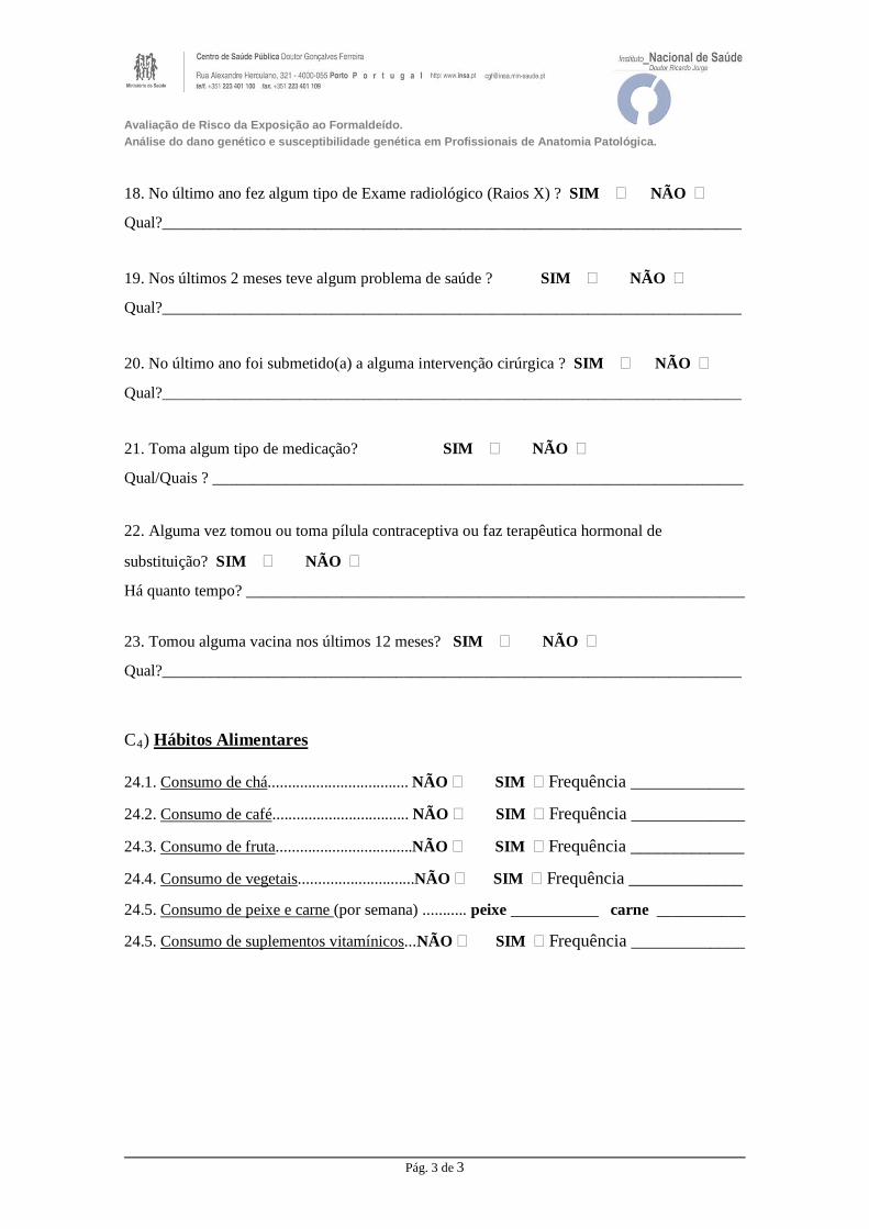
Avaliação de Risco da Exposição ao Formaldeído. Análise do dano genético e susceptibilidade genética em Profissionais de Anatomia Patológica.
18. No último ano fez algum tipo de Exame radiológico (Raios X) ? SIM � NÃO �
Qual?________________________________________________________________________
19. Nos últimos 2 meses teve algum problema de saúde ? SIM � NÃO �
Qual?________________________________________________________________________
20. No último ano foi submetido(a) a alguma intervenção cirúrgica ? SIM � NÃO �
Qual?________________________________________________________________________
21. Toma algum tipo de medicação? SIM � NÃO �
Qual/Quais ? __________________________________________________________________
22. Alguma vez tomou ou toma pílula contraceptiva ou faz terapêutica hormonal de
substituição? SIM � NÃO �
Há quanto tempo? ______________________________________________________________
23. Tomou alguma vacina nos últimos 12 meses? SIM � NÃO �
Qual?________________________________________________________________________
C4) Hábitos Alimentares
24.1. Consumo de chá................................... NÃO � SIM � Frequência _____________
24.2. Consumo de café.................................. NÃO � SIM � Frequência _____________
24.3. Consumo de fruta..................................NÃO � SIM � Frequência _____________
24.4. Consumo de vegetais.............................NÃO � SIM � Frequência _____________
24.5. Consumo de peixe e carne (por semana) ........... peixe __________ carne ___________
24.5. Consumo de suplementos vitamínicos...NÃO � SIM � Frequência _____________
Pág. 3 de 3

ANNEX II


Enquadramento
O Formaldeído (FA) é um solvente orgânico largamente usado em actividades laboratoriais.
Este produto é um fixador de tecidos pouco dispendioso e bastante eficiente, sendo, por isso, o
eleito em procedimentos de rotina anátomo-patológicos. É também um bom desinfectante e um
meio óptimo para conservar e armazenar biópsias e peças cirúrgicas.
No Serviço de Anatomia Patológica, decorrem actividades que implicam a exposição dos
trabalhadores ao FA, através da utilização e manuseamento do formol. A principal desvantagem
apontada na utilização do formol é o facto de haver libertação de vapores durante o seu
manuseamento, com consequente inalação por parte dos trabalhadores. Por ser um irritante
primário, a sintomatologia mais comum associada à inalação de FA inclui, irritação do nariz e
garganta e aumento do lacrimejar.
Recentemente, a International Agency for Research on Cancer (IARC), classificou-o agente
carcinogénico humano (Grupo 1)
Objectivos
Com este trabalho pretende-se avaliar o risco da exposição ocupacional a FA em profissionais dos
Serviços de Anatomia Patológica. A abordagem ao tema será múltipla, por forma a relacionar
diferentes tipos de biomarcadores o que permitirá caracterizar a exposição e efeitos na saúde.
A avaliação da exposição ocupacional a FA será realizada através da medição da concentração do
FA no ar (monitorização ambiental) e pela análise de diferentes indicadores biológicos
(monitorização biológica). Na monitorização biológica serão estudados biomarcadores de dose
interna (doseamento de FA na urina), biomarcadores de genotoxicidade (testes citogenéticos, teste
do cometa) e biomarcadores de susceptibilidade genética (polimorfismos genéticos de enzimas
envolvidas no metabolismo do FA e de reparação de lesões no ADN).
O conjunto de dados obtidos contribuirão para um melhor conhecimento da relação entre os
níveis ambientais do FA e os consequentes efeitos biológicos adversos na saúde humana. A
informação recolhida será um contributo para as entidades encarregues de definir os níveis
aceitáveis para a exposição humana a FA e para os serviços que têm a seu cargo a vigilância da
saúde dos trabalhadores.
AVALIAÇÃO DO RISCO DA EXPOSIÇÃO PROFISSIONAL AO FORMALDEÍDO.
ANÁLISE DO DANO GENÉTICO E SUSCEPTIBILIDADE GENÉTICA EM PROFISSIONAIS DE ANATOMIA PATOLÓGICA

ANNEX III


Table I. Genotype frequencies of studied polymorphisms coding for xenobiotic-metabolic enzymes among Caucasian and Asians.
Gene Ethnicity Genotypes Frequency (%) References
CYP2E1 (rs6413432)
Caucasians T/T T/A A/A
78.9 19.3 1.8
Teixeira et al (2004)
Asians T/T T/A A/A
54.5 36.4 9.1
SNPedia *
GSTT1 (delection)
Caucasians Present Null
84.0 16.0
Costa et al (2012)
Asians Present Null
44.5 55.5
Raimondi et al (2006)
GSTM1 (delection)
Caucasians Present Null
46.1 53.9
Lopez-Címa et al (2012)
Asians Present Null
46.2 53.8
Cho et al (2005)
GSTP1 (rs1695)
Caucasians Ile/ Ile Ile/Val Val/Val
41.9 46.4 11.7
Lopez-Címa et al (2012)
Asians Ile/ Ile Ile/Val Val/Val
70.6 25.5 2.8
Cho et al (2005)
* 1000 Genomes Project, data released on 13 December 2013; Cariaso and Lennon (2012).

Table II. Frequencies of polymorphic genes related to DNA-repair pathways among racial groups.
Gene Ethnicity Genotypes Frequency (%) References
XRCC1 (194) (rs1799782)
Caucasians Arg/Arg Arg/Trp Trp/Trp
89.0 10.0 1.0
Santonocito et al (2012)
Asians Arg/Arg Arg/Trp Trp/Trp
78.7-36.8 18.7-50.0 2.6-13.2
Hung et al (2005)
XRCC1 (399) (rs25487)
Caucasians Gln/Gln Gln/Arg Arg/Arg
42.2 44.6 13.3
Garcia-Léston et al ( 2012)
Asians Gln/Gln Gln/Arg Arg/Arg
56.6 36.7 6.6
SNPedia *
PARP1 (rs1136410)
Caucasians Val/Val Val/Ala Ala/Ala
77.0 21.0 2.0
Figueiroa et al (2007)
Asians Val/Val Val/Ala Ala/Ala
32.5 48.1 0.2
Stern et al (2007)
MUTYH (rs3219489)
Caucasians Gln/Gln Gln/His His/His
57.8 36.1 6.1
SNPedia *
Asians Gln/Gln Gln/His His/His
33.5 49.0 17.5
SNPedia *
RAD51 (rs1801321)
Caucasians G/G G/T T/T
30.6 50.2 19.2
Silva et al (2010)
Asians G/G G/T T/T
86.4 12.9 0.7
SNPedia *
XRCC2 (rs3218536)
Caucasians Arg/Arg Arg/His His/His
81.2 18.8 -
Silva et al (2010)
Asians Arg/Arg Arg/His His/His
100 - -
SNPedia *
XRCC3 (rs861539)
Caucasians Thr/Thr Thr/Met Met/Met
37.9 51.7 10.4
Iarmocovai et al (2006)
Asians Thr/Thr Thr/Met Met/Met
82.2 17.5 0.3
SNPedia *
FANCA (rs7190823)
Caucasians Thr/Thr Thr/Al Ala/Ala
17.9 43.8 38.3
SNPedia *
Asians Thr/Thr Thr/Al Ala/Ala
97.2 2.8 -
SNPedia *
BRIP1 (rs4986764) Caucasians
Ser/Ser Ser/Pro Pro/Pro
17.0 51.3 31.7
Frank et al (2007)
Asians Ser/Ser Ser/Pro Pro/Pro
5.3 33.9 60.8
SNPedia *
* 1000 Genomes Project, data released on 13 December 2013; Cariaso and Lennon (2012).

PUBLICAÇÕES DE SUPORTE À
TESE DE DOUTORAMENTO


Author's personal copy
Toxicology 252 (2008) 40–48
Contents lists available at ScienceDirect
Toxicology
journa l homepage: www.e lsev ier .com/ locate / tox ico l
Genotoxic damage in pathology anatomy laboratory workers exposedto formaldehyde
Solange Costaa,b,∗, Patrícia Coelhoa, Carla Costaa, Susana Silvaa, Olga Mayana,Luís Silva Santosc,d, Jorge Gasparc, João Paulo Teixeiraa
a National Institute of Health, Environmental Health Department, Praca Coronel Pacheco 15, 4050-453 Porto, Portugalb Escola Superior de Biotecnologia da Universidade Católica Portuguesa, Rua Dr António Bernardino de Almeida, 4200-072 Porto, Portugalc Faculty of Medical Sciences UNL, Department of Genetics, Rua da Junqueira 96, 1349-008 Lisboa, Portugald Centro Regional das Beiras, Universidade Católica Portuguesa, Estrada da Circunvalacão, 3504-505 Viseu, Portugal
a r t i c l e i n f o
Article history:Received 7 July 2008Received in revised form 20 July 2008Accepted 21 July 2008Available online 31 July 2008
Keywords:FormaldehydePathology anatomy workersMicronucleiSister chromatid exchangesComet assay
a b s t r a c t
Formaldehyde (FA) is a chemical traditionally used in pathology and anatomy laboratories as a tissuepreservative. Several epidemiological studies of occupational exposure to FA have indicated an increasedrisk of nasopharyngeal cancers in industrial workers, embalmers and pathology anatomists. There is alsoa clear evidence of nasal squamous cell carcinomas from inhalation studies in the rat. The postulatedmode of action for nasal tumours in rats was considered biologically plausible and considered likelyto be relevant to humans. Based on the available data IARC, the International Agency for Research onCancer, has recently classified FA as a human carcinogen. Although the in vitro genotoxic as well as the invivo carcinogenic potentials of FA are well documented in mammalian cells and in rodents, evidence forgenotoxic effects and carcinogenic properties in humans is insufficient and conflicting thus remains to bemore documented. To evaluate the genetic effects of long-term occupational exposure to FA a group of30 Pathological Anatomy laboratory workers was tested for a variety of biological endpoints, cytogenetictests (micronuclei, MN; sister chromatid exchange, SCE) and comet assay. The level of exposure to FA wasevaluated near the breathing zone of workers, time weighted average of exposure was calculated for eachsubject. The association between the biomarkers and polymorphic genes of xenobiotic metabolising andDNA repair enzymes was also assessed. The mean level of exposure was 0.44 ± 0.08 ppm (0.04–1.58 ppm).MN frequency was significantly higher (p = 0.003) in the exposed subjects (5.47 ± 0.76) when comparedwith controls (3.27 ± 0.69). SCE mean value was significantly higher (p < 0.05) among the exposed group(6.13 ± 0.29) compared with control group (4.49 ± 0.16). Comet assay data showed a significant increase(p < 0.05) of TL in FA-exposed workers (60.00 ± 2.31) with respect to the control group (41.85 ± 1.97). Apositive correlation was found between FA exposure levels and MN frequency (r = 0.384, p = 0.001) andTL (r = 0.333, p = 0.005). Regarding the genetic polymorphisms studied, no significant effect was found onthe genotoxic endpoints. The results of the present biomonitoring study emphasize the need to developsafety programs.
© 2008 Elsevier Ireland Ltd. All rights reserved.
1. Introduction
Formaldehyde (FA) is an important industrial compound withnumerous applications ranging from the production of resins tomedicine. At room temperature, it is a flammable and colorless
∗ Corresponding author at: National Institute of Health, Environmental HealthDepartment, Praca Coronel Pacheco 15, 4050-453 Porto, Portugal.Tel.: +351 222 046 405; fax: +351 222 046 409.
E-mail addresses: [email protected], solange [email protected](S. Costa).
gas with a strong pungent odor. It is also a naturally occurringbiological compound present in all cells, tissues and body fluids.The highest level of human exposure to this aldehyde occurs inoccupational settings. Because of its widespread use a relativelylarge number of workers are exposed to FA. Increased incidencesof nasopharyngeal cancer were found in populations occupation-ally exposed to FA (Blair et al., 1990; Partanen, 1993; Armstronget al., 2000; Vaughan et al., 2000; Hildesheim et al., 2001; Coggonet al., 2003; Hauptmann et al., 2004). Animal studies demonstratethat high concentrations of FA can cause irreversible damage tothe nasal epithelium of rats and that in some cases rats exposed tothese concentrations developed neoplasia (Merk and Speit, 1998).
0300-483X/$ – see front matter © 2008 Elsevier Ireland Ltd. All rights reserved.doi:10.1016/j.tox.2008.07.056

Author's personal copy
S. Costa et al. / Toxicology 252 (2008) 40–48 41
Based on available data the International Agency for Research onCancer (IARC) classified FA as carcinogenic to humans (group1) (IARC, 2006). Epidemiological studies of industrial workers,embalmers and pathology anatomists have associated FA expo-sure with elevated risks for cancers at various sites, including nasalcavities (Blair et al., 1990; Luce et al., 2002; Coggon et al., 2003),lung (Gardner et al., 1993; Coggon et al., 2003), brain (Hayes etal., 1990; Coggon et al., 2003), pancreas (Stone et al., 2001) andlymphohematopoietic system (Hall et al., 1991; Hauptmann et al.,2003; Pinkerton et al., 2004), however, these positive findings mayhave been confounded by concomitant exposures and remain con-troversial (Collins et al., 2001; Heck and Casanova, 2004; Marshand Youk, 2004, 2005; Golden et al., 2006). Nevertheless, it isstill unclear whether these tumours arose through a genotoxicmechanism and/or as a consequence of cytotoxicity except for car-cinogenesis in nasal tissues where FA seems to proceed both bygenotoxic and cytotoxic effects (Orsière et al., 2006). Several studieshave shown the potential genotoxic effect of FA in proliferating cul-tured mammalian cell lines and in human lymphocytes. Inductionof micronucleus (MN) (Suruda et al., 1993; He et al., 1998; Orsière etal., 2006), sister chromatid exchange (SCE) (He et al., 1998; Shahamet al., 1997, 2002), chromosomal aberrations (CA) (He et al., 1998),DNA-protein crosslinks (DPC) (Shaham et al., 1996, 1997, 2003)and DNA damage (comet assay) (Yu et al., 2005) were reported inindustrial workers, embalmers and pathology anatomists exposedto FA.
Occupational exposure to FA occurs mainly in pathologicanatomy laboratories where it is used as a cytological fixativeto preserve the integrity of cell structures for a wide variety ofpurposes, including cell, tissue and organelle descriptive exam-inations, clinical diagnoses and developmental studies. In thesesettings, absorption of FA occurs mainly through inhalation. InhaledFA primarily affects the upper airways; the severity and extent ofphysiological response depends on its concentration in the air. Sev-eral studies have consistently shown that the levels of airborneFA in anatomy laboratories exceed recommended exposure crite-ria (Shaham et al., 2002; Akbar-Khanzadeh and Pulido, 2003). Suchresults along with the recent implications of human carcinogenic-ity, point out for the need of close monitoring of FA exposures. Awide range of methods is currently used for the detection of earlybiological effects of DNA-damaging agents in occupational set-tings. Assays measuring micronucleus (MN) and sister chromatidexchanges in peripheral blood lymphocytes are well-establishedcytogenetic techniques that have been used extensively for assess-ing DNA damage at the chromosomal level in human biomonitoring(Carrano and Natarajan, 1988; Fenech, 1993). Recently it has beenreported that the MN test is also predictive of cancer risks in humanpopulations (Bonassi et al., 2007). In recent years, single cell gelelectrophoresis or comet assay has been proven to be a very sensi-tive method of human biomonitoring for the detection of levels ofDNA damage, and as a useful tool for detection of genetic damageat the individual cell level (Collins, 2004). The simultaneous use ofcytogenetic tests and comet assay in occupational studies allowsa comparison of the presence of DNA strand breaks, due to bothacute and chronic exposure, and of chromosome damage, due toclastogenic and aneugenic events. In the case of chronic exposure,comparing the levels of damage from a cytogenetic technique withthe comet assay could provide information about concurrent versuspast exposure (Albertini et al., 2000).
Polymorphic genes involved in the metabolism of xenobioticsmay modulate the levels of biomarkers arising from environmen-tal and/or occupational exposure to genotoxic agents (Pavanelloand Clonfero, 2000). Knowledge of the real impact of genetic poly-morphisms as biomarkers of susceptibility is of key significance inunderstanding the processes of genetic damage involved in muta-
genesis and carcinogenesis (Srám and Binková, 2000) and couldhelp to minimize risks for susceptible subjects (Laffon et al., 2006).Glutathione S-transferases (GSTs) are a superfamily of polymor-phic enzymes involved in the conjugation of reactive chemicalintermediates to soluble forms, and they play an important rolein the detoxification of endogenous and exogenous compounds.The polymorphic genes GSTM1 and GSTT1 code for GSTs that areinvolved in the detoxification of a variety of potentially carcinogeniccompounds, including polyaromatic hydrocarbon diol-epoxides,steroids and genotoxic lipoperoxidation products (Strange et al.,2001). The GSTM1 and GSTT1 polymorphisms are the result of thedeletion of part of the gene, and this leads to the absence of activityin individuals homozygous for the deletion (null genotype) (Strangeet al., 2001). The GSTM1 and GSTT1 null genotypes have been asso-ciated with increased risk for several cancers including, lung andcolorectal cancer (Raimondi et al., 2006; Hosgood et al., 2007; Loftet al., 2007; Yeh et al., 2007). DNA repair is a key human cellularresponse to DNA-damaging stimuli. DNA repair mechanisms arevital responses to multiple types of DNA damage, including theones caused by exposure to environmental and endogenous car-cinogens (Kiyohara and Yoshimasu, 2007; McWilliams et al., 2008).Most of the alterations caused, if not repaired, can result in geneticinstability, mutagenesis and cell death. Thus genetic variations inDNA repair genes may modulate DNA repair capacity and, thereforeinfluence risk for cancer development (Kiyohara et al., 2007). Poly-morphisms in the NER (nucleotide excision repair) pathway havebeen associated with increased risk for breast cancer (Milne et al.,2006; Costa, 2007), head and neck cancer (Shen et al., 2001), lungcancer (Kiyohara and Yoshimasu, 2007; Matakidou et al., 2007),basal cell skin cancer (Winsey et al., 2000) and pancreatic adeno-carcinoma (McWilliams et al., 2008).
The aim of the present study was to evaluate both DNA and cyto-genetic damage in peripheral lymphocytes of FA-exposed workersin pathologic anatomy laboratories. Air monitoring was performedin order to evaluate occupational exposure to FA. A multipleapproach was used in order to integrate different biomarkers (effectand susceptibility). Genetic damage was studied by means of MN,SCE and comet assay (comet tail length, TL). In addition, the influ-ence of polymorphic genes of xenobiotic metabolising enzymes(GSTM1, GSTT1) and DNA repair enzymes (ERCC1 (rs3212986: 196bp3′ of STP T > G, Q504K), ERCC4 (rs1800067: Ex8 + 31G > A R415Q)and ERCC5 (rs17655: Ex15-344G > C, D1104H; and rs2227869: Ex8-369G > C, C529S)) on the biomarkers was also analysed.
2. Methods
2.1. Subjects
The study population consisted of 30 workers exposed to FA from four hospitalpathological anatomy laboratories, located in Portugal (Oporto and Aveiro districts),and 30 non-exposed control employees, matched by age, sex, lifestyle and smokinghabits, working in the same area in administrative offices and without occupa-tional exposure history to FA. The characteristics of both groups are described inTable 1. Health conditions, medical history, medication, diagnostic tests (X-rays,etc.) and lifestyle factors were assessed by means of questionnaires. Subjects ofthe exposed group also gave information related to working practices such as useof protective measures, years of employment, specific symptoms related to FA-exposure and chronic respiratory diseases such as asthma and others. All subjectswere fully informed about the procedures and objectives of this study and each sub-ject prior to the study signed an informed consent form. Ethical approval for thisstudy was obtained from the institutional Ethical Board of the National Institute ofHealth.
2.2. Environmental monitoring
Air sampling was performed in the workers breathing zone for representativeworking periods, analysis of the samples allowed the calculation of the 8-h timeweighted average (TWA) level of exposure to FA for each subject. Air sampling andFA analysis were performed according to the NIOSH method no. 3500.

Author's personal copy
42 S. Costa et al. / Toxicology 252 (2008) 40–48
Table 1Characteristics of the study population
Control group Exposed group
Number of subjects 30 30
Gender 19 females 21 females11 males 9 males
Age (years)a 37 ± 10 (25–61) 38 ± 8 (23–53)Years of employmenta – 11 ± 7 (0.5–27)
Smoking statusNon-smokers 23 (77%) 22 (73%)Smokers 7 (23%) 8 (27%)No cigarettes/daya 14 ± 8 (6–30) 13 ± 5 (5–20)No years smokinga 21 ± 17 (4–51) 14 ± 8 (3–22)
a Mean ± S.D.
2.3. Cytogenetic assays
Heparinized venous blood samples (10 ml), were collected between 10 and11 a.m., from each donor, and were immediately processed for the different method-ologies used in this study. Workers begin their work at 9 a.m. All samples were codedand analysed under blind conditions.
2.3.1. MN testAliquots of 0.5 ml of heparinized whole blood were used to establish dupli-
cate lymphocyte cultures for cytokinesis-blocked micronucleus test, as describedin Teixeira et al. (2004). Cultures were incubated at 37 ◦C in the dark for a total of72 h, cytochalasin B (6 �g/ml) was added at 44 h to prevent cytokinesis. Cells werecollected by centrifugation and treated twice with a mixture of RPMI (pH 7.2) supple-mented with 2% fetal bovine serum. The cells were centrifuged again and submittedto a mild hypotonic treatment in a mixture of RPMI (pH 7.2):deionized water (1:4,v/v), supplemented with 2% fetal bovine serum. Then, the centrifuged cells wereplaced on dry slides and smears were performed. After air-drying, the slides werefixed with cold methanol:acetic acid (3:1, v/v). Air-dried slides were stained with4% Giemsa in pH 6.8 phosphate buffer. Microscopic analyses were performed on aNikon Eclipse E400 light microscope. To determine the total number of MN in binu-cleated cells, a total of 1000 binucleated cells with well-preserved cytoplasm (500per replicate) were scored for each subject. MN were scored blindly by the samereader and identified according to the criteria defined by Caria et al. (1995).
2.3.2. SCE testLymphocyte cultures for SCE were established in duplicate as described previ-
ously (Teixeira et al., 2004) in the micronucleus assay, except for the addition of 5 �lof 5-bromo-deoxyuridine (10 �g/ml) to the culture medium. Differential chromatidstaining was performed with the fluorescence-plus-Giemsa procedure (Perry andWolff, 1974). A single observer scored 50 s division metaphases for each donor (25from each duplicate culture) on coded slides to determine the number of SCE/cell.
2.3.3. Comet assayLymphocytes were isolated, in duplicate, from heparinized whole blood, by
centrifugation on a Ficoll density gradient. The lymphocyte layer (buffy coat) wasremoved and washed three times with ice-cold phosphate buffer solution (PBS)pH 7.4, at 1000 rpm (∼270 × g) for 10 min. Cell viability, determined by trypan blueexclusion, was higher than 80% in all cases.
The alkaline version of the comet assay was performed as described by Singhet al. (1988) with minor modifications. Briefly, cells collected by centrifugation(7500 × g for 3 min) and suspended in 100 �l of 0.6% low-melting-point agarose(LMA) in PBS (pH 7.4) were dropped onto a frosted slide precoated with a layer of1% normal melting point agarose. Slides were placed on ice for 4 min and allowed tosolidify. Coverslips were then removed and slides were immersed in freshly preparedlysing solution (2.5 M NaCl, 100 mM Na2EDTA, 10 mM TrisBase, 0.25 M NaOH, pH 10)for 1 h at 4 ◦C, in the dark. After lysis, slides were placed on a horizontal electrophore-sis tank in an ice bath. The tank was filled with freshly made alkaline electrophoresissolution (1 mM Na2EDTA, 300 mM NaOH, pH 13) to cover the slides, and they wereleft for 20 min in the dark to allow DNA unwinding and alkali-labile site expression.Electrophoresis was carried out for 20 min at 30 V and 300 mA (1 V/cm). The slideswere then washed for 10 min with 1 ml of neutralizing solution (0.4 M TrisBase, pH7.5). After neutralization, gels were stained with 100 �l of ethidium bromide solu-tion (20 �g/ml) and covered with coverslips for 20 min. After staining the slides werewashed twice with ice-cold bidistilled water for 20 min. Two slides were preparedfor each donor and a ‘blind’ scorer examined 50 randomly selected cells from eachslide (100 cells/donor) using a magnification of 400×. Image capture and analysiswere performed with Comet Assay IV software (Perceptive Instruments). Comet taillength (TL) was the DNA damage parameter evaluated. TL (�m) indicates the extentof migration of genetic material in the direction of the anode (Singh et al., 1988) and
is expected to be proportional to the level of single-strand breaks and alkali-labilesites.
2.4. Genotype analysis
Genomic DNA was obtained from heparinized whole blood samples using a com-mercially available kit according to the manufacturer’s instructions (QIAamp DNAextraction kit; Qiagen, Hilden, Germany). Each DNA sample was stored at −20 ◦Cuntil analysis.
GSTM1 and GSTT1 genotyping for gene deletions were carried out bya multiplex PCR as described by Lin et al. (1998) with minor modifi-cations described in Gaspar et al. (2004). DNA samples were amplifiedwith the primers (PerkinElmer Corp): 5′-GAACTCCCTGAAAAGCTAAAGC-3′ (upstream) and 5′-GTTGGGCTCAAATATACGGTGG-3′ (downstream) forGSTM1 which produced a 219 bp product, 5′-TCACCGGATCATGGCCAGCA-3′ (upstream) and 5′-TTCCTTACTGGTCCTCACATCTC-3′ (downstream) forGSTT1 which produced a 459-bp product. The amplification of albumingene with the primers 5′-GCCCTCTGCTAACAAGTCCTAC-3′ (upstream) and 5′-GCCCTAAAAAGAAAATCCCCAATC-3′ (downstream) was used as an internal controland produced a 350-bp product. PCR was performed in a final volume of 50 �l,consisting of DNA (0.1 �g), dNTP (0.2 mM each) (PerkinElmer), MgCl2 (2.5 mM),each primer (1.0, 0.3 and 0.2 �M for GSTM1, GSTT1 and albumin, respectively),AmplitaqGold polymerase (1.25 units) (PerkinElmer), reaction buffer and 2% DMSO.Amplification was performed with an initial denaturation at 95 ◦C for 12 min,followed by 35 cycles of amplification performed at 94 ◦C for 1 min, 62 ◦C for 1 minand 72 ◦C for 1 min, and a final extension at 72 ◦C for 10 min, using a GeneAmp9600 thermal cycler (PerkinElmer Corp). After electrophoretic separation (30 min,4 V/cm) the amplified products were visualized, under ultraviolet light, in ethidiumbromide stained (1 �g/ml) agarose gel (1.5%).
All the genotype determinations were carried out twice in independent exper-iments and all the inconclusive samples were reanalysed.
ERCC1 (rs3212986), ERCC4 (rs1800067), ERCC5 (rs17655 and rs2227869)polymorphisms were determined using the TaqMan SNP genotyping assay(Applied Biosystems) with corresponding codes of C 2532948 10, C 3285104 10,C 1891743 10 and C 15956775 10. The 5′-nuclease allelic discrimination assay, orTaqMan assay, is a PCR-based assay for genotyping single nucleotide polymorphisms.The PCR amplification was performed in 10 �l reactions containing 10 ng of genomicDNA, 1× SNP Genotyping Assay Mix, and 1× TaqMan Universal PCR Master Mixcontaining optimised buffer components and Rox reference dye. The amplificationconditions consisted of an initial AmpliTaq Gold® activation at 95 ◦C during 10 min,followed by 40 or more amplification cycles consisting of denaturation at 92 ◦C for15 s and annealing/extension at 60 ◦C for 1 min. Amplification was performed in the7300 Real-Time PCR System (Applied Biosystems) and sequences were detected bythe SDS-Sequence Detection Software (version 1.3.1). All the genotype determinationswere carried out twice in independent experiments and all the inconclusive sampleswere reanalysed.
2.5. Statistical analysis
The distribution of variables was compared with the normal distribution bymeans of the Kolmogorov–Smirnov goodness-of-fit test. MN was the only parameterthat departed significantly from normality and therefore the non-parametric testsMann–Whitney U-test and Kruskal–Wallis test were applied to data. Thus, analysesof all the other variables were performed by use of parametric tests. The effect ofoccupational exposure, gender, age, tobacco smoking, and GSTM1, GSTT1, ERCC1,ERCC4, and ERCC5 genotypes on every parameter studied was evaluated by one-wayanalysis of variance (ANOVA) and Student’s t-test. The associations between twovariables were analysed by Pearson’s (parametric) and Spearman’s (non-parametric)correlation tests. The level of significance considered was 0.05. All analyses wereconducted using the SPSS for Windows statistical package, version 11.0.
3. Results
To evaluate current exposure to FA in pathological anatomy lab-oratories air samples were collected in the workers’ breathing zone.The main FA vapor emissions occurred during the macroscopicexamination of FA-preserved specimens and during the disposal ofspecimens and waste solutions, during these tasks the mean roomFA concentrations were 1.50 and 4.43 ppm, respectively. The meanlevel of FA exposure of the 30 individuals studied was 0.44 ppm(0.04–1.58 ppm). The American Conference of Governmental Indus-trial Hygienists (ACGIH) has set for FA a ceiling limit at 0.3 ppm(ACGIH, 2008). The current Portuguese occupational exposure stan-dard is also a 0.3 ppm ceiling limit (ceiling level), meaning themaximum safe FA-airborne concentration that should never be

Author's personal copy
S. Costa et al. / Toxicology 252 (2008) 40–48 43
Table 2Results of MN, SCE, and comet assay in the populations examined
Mean, MN ± S.E. Mean, SCE ± S.E. Mean, TL (�m) ± S.E.
Controls 3.27 ± 0.69 (0–17) 4.49 ± 0.16 (3.10–3.06) 41.85 ± 1.97 (28.85–66.52)Exposed 5.47 ± 0.76* (1–17) 6.13 ± 0.29** (3.64–8.80) 60.00 ± 2.31** (33.76–99.09)
S.E.: mean standard error.* p = 0.003.
** p < 0.05.
exceeded during any length of time in a workers’ breathing zone.Thus, the FA air concentrations obtained in this study show thatworkers in pathological anatomy laboratories are exposed to levelsof airborne FA that exceed recommended exposure criteria.
Effect of exposure on frequencies of cytogenetic biomarkers andon DNA damage are shown in Table 2.
For all the variables studied significant increases were foundin the exposed workers as compared with the controls. MN fre-quency was significantly higher (p = 0.003) in the exposed subjects(5.47 ± 0.76) when compared with controls (3.27 ± 0.69). SCE meanvalue was significantly higher (p < 0.05) among the exposed group(6.13 ± 0.29) compared with control group (4.49 ± 0.16). Cometassay data showed significantly increase (p < 0.05) of comet taillength (TL) in FA-exposed workers (60.00 ± 2.31) with respect tothe control group (41.85 ± 1.97). Fig. 1 shows the distribution ofMN and SCE frequencies and TL in the study populations. The dis-tributions in the exposed group are displaced to higher values ofdamage for the three parameters studied. Moreover the exposedindividuals were the only ones to have mean frequency SCE valueshigher than 6.4 and mean TL values equal or higher than 70.4 �m.
A positive correlation was found between FA exposure levels andMN frequency (r = 0.384, p = 0.001) and TL (r = 0.333, p = 0.005).
The effect of gender on the parameters examined is shownin Table 3. MN frequency increased in females in both groupscompared with males, but the increase was not significant. Nosignificant association was found between males and femalesfrom both groups for SCE frequency. Regarding DNA damage,comet tail length was significantly (p = 0.014) increased in females(63.06 ± 2.60, n = 21) compared with males (52.84 ± 4.00, n = 9) butonly in the exposed group.
In order to examine the effect of age, exposed and non-exposedindividuals were divided in three groups: <30 years, 30–40 yearsand >40 years. No significant effect of age was found in any of thevariables studied (data not shown). MN frequency increased withage in both populations, but it did not reach statistical signification.
Smoking habits did not influence MN frequency and DNA dam-age (TL) (Table 4). However a significant increase (p = 0.014) wasfound for SCE frequency in the control group; smokers had a higher(5.24 ± 0.39, n = 7) frequency of SCE than non-smokers (4.27 ± 0.15,n = 23). In the exposed group no significant difference was observed.
In order to analyse the effect of exposure time four groups ofexposed workers were established based on their years of employ-
ment. No significant association was found between the durationof exposure and the studied biomarkers (data not shown).
Distribution of the genotype frequencies of metabolic (GSTM1,GSTT1) and DNA repair (ERCC1, ERCC4, ERCC5) genes in the studypopulation is presented in Table 5.
The GSTM1 null genotype was slightly more prevalent in con-trols (48%) than among the exposed workers (43%), whereas thedistribution of the GSTT1 null genotype was 17% in the exposedpopulation and 7% in the controls. Among the whole study popu-lation, the prevalence of GSTM1 and GSTT1 deleted genotypes was46% and 7%, respectively, a finding consistent with previous resultsdescribed by other authors for Caucasians (To-Figueras et al., 1997;Garte et al., 2001; Raimondi et al., 2006) and Portuguese popula-tions (Martins and Alves, 1998; Gaspar et al., 2004). The distributionof ERCC1, ERCC4, ERCC5 frequencies in the study population weresimilar to the ones described in other studies for Caucasian popula-tions (NCBI-SNP Database, 2008; SNP500Cancer Database, 2008).Allele frequencies of the different ERCC1 genotypes obtained in thestudy population are similar to those found for Caucasian popu-lations NCBI-SNP Database, 2008; SNP500Cancer Database, 2008).The distribution of ERCC4 genotypes in the whole studied popula-tion was 78%, 20% and 2% for GG, GA and AA, respectively. Theseresults are identical to those described for Caucasian population,GG 79%, GA 19% and AA 2% (SNP500Cancer Database, 2008). TheERCC5 (rs17655) CC genotype frequency in all studied populationwas 6%, which is in accordance with the 7% frequency describedfor the European population (NCBI-SNP Database, 2008). Concern-ing the frequency of the ERCC5 (rs2227869) genotypes among thewhole population, the prevalence of ERCC5 GG genotype was 93%,a finding consistent with the frequency registered for Caucasianpopulations (NCBI-SNP Database, 2008; SNP500Cancer Database,2008).
Regarding the effect of the genetic polymorphisms studied wedid not find any effect on the genotoxic endpoints.
4. Discussion
Occupational exposure to FA evaluated in the four pathologicanatomy laboratories shows that workers are exposed to high levelsof FA.
Several studies (Shaham et al., 1997; Dufresne et al., 2002;Akbar-Khanzadeh and Pulido, 2003) point the anatomy laboratories
Fig. 1. Distribution of MN frequency (A), SCE frequency (B), and comet tail length (C) in control and exposed populations.

Author's personal copy
44 S. Costa et al. / Toxicology 252 (2008) 40–48
Table 3Effect of gender on MN, SCE, and comet assay
Gender N MN ± S.E. (range) SCE ± S.E. (range) TL (�m) ± S.E. (range)
ControlsFemales 19 4.11 ± 1.03 (0–17) 4.33 ± 0.17 (3.27–6.16) 40.84 ± 2.75 (29.80–66.52)Males 11 1.82 ± 0.44 (0–5) 4.78 ± 0.33 (3.10–6.04) 43.60 ± 2.58 (28.85–53.70)
ExposedFemales 21 5.95 ± 0.80 (2–17) 6.10 ± 0.35 (3.72–8.80) 63.06 ± 2.60* (51.23–99.09)Males 9 4.33 ± 1.72 (1–17) 6.18 ± 0.52 (3.64–8.10) 52.84 ± 4.00 (33.76–76.18)
S.E.: mean standard error.* p = 0.014, significant difference with regard to males among exposed individuals.
Table 4Effect of smoking on MN, SCE, and comet assay
Smoking habits N MN ± S.E. (range) SCE ± S.E. (range) TL (�m) ± S.E. (range)
ControlsNon-smokers 23 3.09 ± 0.81 (0–17) 4.27 ± 0.15 (3.10–6.04) 40.89 ± 2.06 (29.80–62.14)Smokers 7 3.86 ± 1.44 (0–11) 5.24 ± 0.39* (3.12–6.16) 45.03 ± 5.17 (28.85–66.52)
ExposedNon-smokers 22 5.35 ± 0.89 (1–17) 6.17 ± 0.32 (3.64–8.80) 59.16 ± 2.84 (33.76–99.09)Smokers 8 5.70 ± 1.48 (2–17) 6.02 ± 0.64 (3.72–8.70) 61.66 ± 4.12 (41.01–66.10)
S.E.: mean standard error.* p = 0.014, significant difference with regard to non-smokers among the controls.
as one of the occupational settings where the workers are fre-quently exposed to levels of FA near or superior to recommendedlimit values, which indicate a potential risk to workers’ health.Keil et al. (2001) reported that average daily area concentrationsof airborne FA in a gross anatomy laboratory ranged from 0.635to 1.82 mg/m3 (0.51–1.46 ppm). Shaham et al. (2002) reported for14 pathology departments a mean low level and high level of FAexposure of 0.4 and 2.24 ppm, respectively. Ohmichi et al. (2006)evaluated personal exposure levels and indoor FA concentrations ina gross anatomy laboratory during 3 sessions of a total 20 sessionsover 10 weeks. Average personal FA-exposure levels for instruc-tors and students ranged between 0.45 and 1.08 ppm, room FAaverage concentrations in the three sessions were 0.45, 0.38 and0.68 ppm.
The mean level of exposure to FA obtained in this studywas 0.44 ppm (0.04–1.58 ppm). Other authors reported similarresults; Akbar-Khanzadeh et al. (1994) reported for 34 workers ofa gross anatomy laboratory a mean FA-level exposure of 1.24 ppm(0.07–2.94 ppm). Orsière et al. (2006) recently evaluated the occu-pational exposure to FA in pathology and anatomy laboratories
by personal air sampling and reported a mean concentration of0.1 ppm (0.1–0.7 ppm).
Our results show that DNA and chromosomal damage occurs inperipheral lymphocytes of pathologic anatomy laboratory workersexposed to FA. Significant increases were found in all biomarkersstudied compared with control population.
SCE frequencies were higher in the FA-exposed subjects than incontrols. These results agree with those presented by Shaham et al.(1997, 2002) who described a significant increase of SCE frequencyin peripheral lymphocytes of 90 pathology workers from 14 hos-pital pathology departments. The increase of SCE on FA exposedindividuals was previously described by Yager et al. (1986); SCEmeasured in the peripheral lymphocytes of eight non-smokinganatomy students after exposure to FA embalming solution duringa 10 week anatomy class showed a significant increase (p = 0.02)when compared with samples obtained from the same individu-als immediately before exposure began (breathing-zone samplesshowed a mean FA concentration of 1.2 ppm). Recently Ye et al.(2005) reported significantly increased SCE frequencies in a groupof 18 workers from a FA factory. The mean exposure duration
Table 5Frequency of metabolic and repair genotypes in the study population
Genes DbSNP ID Genotypes All (%) Controls (%) Exposed (%)
GSTM1 DeletionPresent 32 (54) 15 (52) 17 (57)Null 27 (46) 14 (48) 13 (43)
GSTT1 DeletionPresent 53 (88) 28 (93) 25 (83)Null 7 (12) 2 (7) 5 (17)TT 4 (7) 2 (8) 2 (7)
ERCC1 rs3212986TG 22 (42) 11 (46) 11 (38)GG 27 (51) 11 (46) 16 (55)GG 43 (78.2) 17 (68) 26 (87)
ERCC4 rs1800067GA 11 (20) 8 (32) 3 (10)AA 1 (1.8) 0 1 (3)GG 32 (59) 11 (46) 21 (70)
ERCC5 rs17655GC 19 (35) 10 (42) 9 (30)CC 3 (6) 3 (12) 0
ERCC5 rs2227869GG 50 (93) 22 (92) 28 (93)GC 4 (7) 2 (8) 2 (7)

Author's personal copy
S. Costa et al. / Toxicology 252 (2008) 40–48 45
was 8.6 years and the level of exposure was 0.985 ± 0.286 mg/m3
(0.82 ± 0.238 ppm). In contrast, in other studies no differences werefound on SCE frequencies of subjects occupationally exposed to FA(Suruda et al., 1993, level of FA exposure was 0.33 ppm; Ying et al.,1999, FA inhalation exposure was 0.508 ± 0.299 mg/m3).
Our results also showed a significant increase in MN frequencyin workers compared with the controls. This finding is consistentwith previous studies on peripheral lymphocytes and on epithe-lial cells (nasal and buccal) of FA exposed workers. Suruda et al.(1993) examined the effect of low level exposure to FA (0.33 ppm)on oral, nasal and lymphocyte biological markers in a group of 29mortician students during an embalming course. Epithelial cellsfrom the buccal mucosa showed a 12-fold increase in micronucleifrequency. Micronucleated lymphocytes frequency increased 26%and nasal epithelial micronuclei increased 22% during the studyperiod. A dose–response relationship was observed with cumu-lative exposure to FA. Ying et al. (1997) evaluated micronucleifrequency on lymphocytes and on oral and nasal mucosa cells of 25anatomy students exposed to FA over an 8-week period. A higherfrequency of micronuclei was observed in nasal and oral exfoliativecells after FA exposure, but no significant increase in the frequencyof lymphocyte micronuclei was found. He et al. (1998) appliedcytokinesis-blocked micronucleus (CBMN) assay as a biologicaldosimeter to detect abnormalities in human peripheral lympho-cytes of 13 students exposed to FA during a 12-week anatomyclass. Breathing-zone air samples collected during dissection pro-cedures showed a mean concentration of 2.37 ppm; MN frequenciesin the exposed group were significantly increased (p < 0.01) com-pared with controls. FA-exposed group also showed a significantincrease in CA (p < 0.01) and SCE (p < 0.05). A correlation betweenMN and CA in individuals was observed. The results indicated thatFA might damage the chromosomes of human lymphocytes. Yu etal. (2005) also found significantly higher frequencies of micronucleion peripheral lymphocytes in 151 workers occupationally exposedto FA from two plywood factories compared with control popu-lation (p < 0.05). Recently a study was conducted to evaluate thegenotoxic effect of occupational exposure to FA on 59 pathol-ogy and anatomy laboratory workers, assessment of chromosomaldamage was carried out by use of the CBMN assay in peripherallymphocytes. The frequency of binucleated micronucleated cellswas significantly higher in pathologists/anatomists than in controls(p = 0.001) (Orsière et al., 2006). Orsière et al. (2006) also reporteda significantly higher frequency of monocentromeric micronucleiin FA-exposed pathologists/anatomists than in controls (p < 0.001),whereas for acentromeric micronuclei frequency no differenceswere obtained between the exposed and control group, suggest-ing an aneugenic effect of FA in peripheral lymphocytes of exposedsubjects. The potential aneugenic effects of FA have also been sug-gested in vitro. Pfuhler and Wolf (2002) reported the ability of FA todisturb the kinetochore-microtubule attachment and mitotic spin-dle checkpoint using isolated tubulin from pig brains. On the otherhand, a significantly higher fraction of acentromeric micronucleiwas reported in buccal and nasal cells of mortuary science studentsexposed to FA before and after a 90-day embalming class (Titenko-Holland et al., 1996), suggesting a clastogenic effect of FA as theprimary mechanism of micronucleus formation.
Only in the last few years the comet assay has being introducedas a useful technique in human biomonitoring studies allowing theevaluation of DNA damage at the single cell level. Therefore, few arethe studies published on FA occupational exposure in which thisbiomarker is used. However, there are already some in vitro stud-ies in cellular lines and in animal and human leukocytes culturecells in which the comet assay proved to be a sensitive biologicalindicator in the evaluation of the genotoxic effect of FA (Frenzilliet al., 2000; Im et al., 2006; Liu et al., 2006; Sul et al., 2007). In
this study, the levels of DNA damage, measured as TL, were signifi-cantly increased in the exposed group compared with controls. Thisresult agrees with those presented by Yu et al. (2005) who reporteda significantly increase of TL and olive tail moment in peripheralblood lymphocyte of 151 workers from two plywood factories. Thelevels of FA-exposed workers were between 0.10 and 7.88 mg/m3
(0.08–6.54 ppm).In human biomonitoring studies it is important to assess the
influence of major confounding factors such as gender, age andsmoking habits in the endpoints studied. In this work we did notfind significant differences between females and males in MN andSCE frequencies. However, females presented an increase in MNfrequencies compared with males. This is in agreement with cur-rent knowledge on the effect of gender on genetic damage whichdetermines a 1.5-fold greater MN frequency in females than inmales (Fenech et al., 1999). The influence of gender in MN fre-quencies can be explained by supposed preferential aneugenicevents involving the X-chromosome (Costa et al., 2007). Surralléset al. (1996) reported an excessive overrepresentation of this chro-mosome in micronuclei of lymphocytes cultured from women.Significant increases were found for comet tail length only amongexposed individuals, females had higher comet tail length thanmales. Although in the past the effect of gender on the level ofDNA damage was regarded as a matter of controversy (Møller etal., 2000), recent studies have shown that this is not a factor thatinfluences the damage evaluated by this indicator (Møller, 2006;Angerer et al., 2007).
Regarding the influence of age, no differences were obtained forthe three biomarkers studied, although MN frequencies tended torise with age in both groups it did not reach statistical significance. Ithas been postulated that the age affect in MN frequency is relatedto a progressive increase in spontaneous chromosome instabilityand the loss of efficiency in DNA repair mechanisms which mayresult in the accumulation of genetic lesions with increasing age(Bolognesi et al., 1999; Kirsch-Volders et al., 2006; Orsière et al.,2006).
Tobacco smoke contains a high number of mutagenic and car-cinogenic substances, hence smoking is an important variable toconsider in biomonitoring studies (IARC, 2002; Bonassi et al., 2003).In the present study no significant differences were obtained forMN and TL. However in both groups the smokers presented slightlyhigher MN frequencies and TL than non-smokers. There are someconflicting results regarding the effect of smoking on MN frequen-cies and comet assay parameters (Laffon et al., 2002; Bhalli etal., 2006; Ergene et al., 2007), nevertheless in two recent meta-analysis studies no association between smoking and these twogenotoxic endpoints was found (Bonassi et al., 2003; Hoffmannand Speit, 2005). Significant differences were obtained in SCE fre-quencies among the control group. Smoking is known to increaseSCE frequencies, there are numerous reports describing the effectof smoking on SCE frequency of occupationally exposed subjects(Shaham et al., 1997, 2002; Cebulska-Wasilewska et al., 1999;Rowland and Harding, 1999; Ergene et al., 2007). However it isimportant to note the low number of smoker subjects included inthis study, which may have affected our results.
No association was found between time of exposure and thebiomarkers studied. One possible explanation may be an adapta-tion of various systems (induction of metabolising and detoxifyingenzymes, induction of DNA repair processes) over time.
In the present study, no association between GSTM1, GSTT1,ERCC1, ERCC4, and ERCC5 polymorphisms and the three genotoxicendopoints were detected.
Genotoxicity of FA is confirmed in a variety of experimentalsystems ranging from bacteria to rodents. Although the posi-tive findings from in vivo animal studies may provide a basis for

Author's personal copy
46 S. Costa et al. / Toxicology 252 (2008) 40–48
extrapolation to humans, the cytogenetic assays in humans havebeen conflicting with both positive and negative outcomes. Evi-dence for the genotoxic effects of FA exposure on first contact sitessuch as nose and respiratory tract have emerged increasingly, thebiological evidence of toxicity on distant-site such as peripherallymphocytes and bone marrow is still controversial.
Data obtained in this study indicate that genotoxic risk due toFA occupational exposure cannot be excluded. In the current FAlevel of exposure measured in pathologic anatomy laboratories, theDNA and chromosomal damage observed in peripheral blood lym-phocytes of exposed workers might be induced by FA exposure,and be increased with the levels of exposure (positive correlationbetween FA exposure levels and MN frequency and TL). Althoughin this study the presence of centromeric signals in the MN was notassessed, the significant increase in DNA damage obtained in thecomet assay (a clastogenicity assay) suggests a clastogenic mode ofaction of FA as the primary cause of the observed damage. Never-theless, these results must be cautiously interpreted, owing to therelatively low number of exposed and control individuals includedin this study.
The main determinants of FA exposure in the pathologicanatomy laboratories were the presence and efficiency of localexhaust systems, the efficiency of the general ventilation, thesize and number of fixed anatomical parts to be handled andwork methods. The highest room FA concentrations observed inthis study occurred during the macroscopic examination of FA-preserved specimens and during the disposal of specimens andwaste solutions. During this tasks in most of the cases the workerswere not using respirators but when they used them the respi-rators were not the appropriate ones. The main reason presentedby workers for the non-use of the respirators and goggles wasthe interference in the efficiency of the activities taken, namelycommunication difficulties, talking notes and handling material.Control aspects must take these factors into account. Implementa-tion of security and hygiene measures in this sector as well as goodpractices campaigns may be crucial to decrease professional risk ofFA exposed workers.
Conflict of interest
None.
Acknowledgements
Our current research is supported by Fundacão Calouste Gul-benkian (grant 76436).
References
ACGIH, 2008. TLV’s and BEI’s—based on the Documentation of the Threshold LimitValues for Chemical Substances and Physical Agents & Biological ExposureIndices. American Conference of Governmental Industrial Hygienists, Cincinnati.
Akbar-Khanzadeh, F., Pulido, E.V., 2003. Using respirators and goggles to controlexposure to air pollutants in an anatomy laboratory. Am. J. Ind. Med. 43, 326–331.
Akbar-Khanzadeh, F., Vaquerano, M.U., Akbar-Khanzadeh, M., Bisesi, M.S., 1994.Formaldehyde exposure, acute pulmonary response, and exposure controloptions in a gross anatomy laboratory. Am. J. Ind. Med. 26, 61–75.
Albertini, R.J., Anderson, D., Douglas, G.R., Hagmar, L., Hemminki, K., Merlo, F., Natara-jan, A.T., Norppa, H., Shuker, D.E., Tice, R., Waters, M.D., Aitio, A., 2000. IPCSguidelines for the monitoring of genotoxic effects of carcinogens in humans.International Programme on Chemical Safety. Mutat. Res. 463, 111–172.
Angerer, J., Ewers, U., Wilhelm, M., 2007. Human biomonitoring: state of the art. Int.J. Hyg. Environ. Health 210, 201–228.
Armstrong, R.W., Imrey, P.B., Lye, M.S., Armstrong, M.J., Yu, M.C., Sani, S., 2000.Nasopharyngeal carcinoma in Malaysian Chinese: occupational exposures toparticles, formaldehyde and heat. Int. J. Epidemiol. 29, 991–998.
Bhalli, J.A., Khan, Q.M., Nasim, A., 2006. DNA damage in Pakistani pesticide-manufacturing workers assayed using the comet assay. Environ. Mol. Mutagen.47, 587–593.
Blair, A., Saracci, R., Stewart, P.A., Hayes, R.B., Shy, C., 1990. Epidemiologic evidenceon the relationship between formaldehyde exposure and cancer. Scand. J. WorkEnviron. Health. 16, 381–393.
Bolognesi, C., Lando, C., Forni, A., Landini, E., Scarpato, R., Migliore, L., Bonassi, S.,1999. Chromosomal damage and ageing: effect on micronuclei frequency inperipheral blood lymphocytes. Age Ageing 28, 393–397.
Bonassi, S., et al., 2003. Effect of smoking habit on the frequency of micronuclei inhuman lymphocytes: results from the Human MicroNucleus project. Mutat. Res.543, 155–166.
Bonassi, S., Znaor, A., Ceppi, M., Lando, C., Chang, W.P., Holland, N., Kirsch-Volders,M., Zeiger, E., Ban, S., Barale, R., Bigatti, M.P., Bolognesi, C., Cebulska-Wasilewska,A., Fabianova, E., Fucic, A., Hagmar, L., Joksic, G., Martelli, A., Migliori, L., Mirkova,E., Scarfi, M.R., Zijno, A., Norppa, H., Fenech, M., 2007. An incresead micronucleusfrequency in peripheral blood lymphocytes predicts the risk of cancer in humans.Carcinogenesis 28, 625–631.
Caria, H., Chaveca, T., Laires, A., Rueff, J., 1995. Genotoxicity of quercetin in themicronucleus assay in mouse bone marrow erythrocytes, human lymphocytes,V79 cell line and identification of kinetochore-containing (CREST staining)micronuclei in human lymphocytes. Mutat. Res. 343, 85–94.
Carrano, A., Natarajan, A., 1988. Considerations for population monitoring usingcytogenetic techniques. Mutat. Res.: Human Monit. (special issue) 204, 379–406.
Cebulska-Wasilewska, A., Wierzewska, A., Nizankowska, E., Graca, B., Hughes, J.A.,Anderson, D., 1999. Cytogenetic damage and ras p21 oncoprotein levels frompatients with chronic obstructive pulmonary disease (COPD), untreated lungcancer and healthy controls. Mutat. Res. 431, 123–131.
Coggon, D., Harris, E.C., Poole, J., Palmer, K.T., 2003. Extended follow up of a cohortof British chemical workers exposed to formaldehyde. J. Natl. Cancer Inst. 95,1608–1615.
Collins, A.R., 2004. Comet assay for DNA damage and repair: principles, applicationsand limitations. Mol. Biotechnol. 26, 249–261.
Collins, J.J., Esmen, N.A., Hall, T.A., 2001. A review and meta-analysis of formaldehydeexposure and pancreatic cancer. Am. J. Ind. Med. 39, 336–345.
Costa, S., 2007. DNA repair genetic polymorphisms and breast cancer in the Por-tuguese population, Ph.D. Dissertation. Universidade do Minho, Braga.
Costa, C., Silva, S., Coelho, P., Roma-Torres, J., Teixeira, J.P., Mayan, O., 2007. Micronu-cleus analysis in a Portuguese population exposed to pesticides: preliminarysurvey. Int. J. Environ. Health 210, 415–418.
Dufresne, A., Infante-Rivard, C., Malo, J.L., Gautrin, D., 2002. Exposure to formalde-hyde among animal health students. AIHA J (Fairfax, Va) 63, 647–650.
Ergene, S., Celik, A., Cavas, T., Kaya, F., 2007. Genotoxic biomonitoring study ofpopulation residing in pesticide contaminated regions in Göksu Delta: micronu-cleus, chromosomal aberrations and sister chromatid exchanges. Environ. Int.33, 885–887.
Fenech, M., 1993. The cytokinesis-block micronucleus technique and its applicationto genotoxicity studies in human populations. Environ. Health Perspect. 101,101–107.
Fenech, M., Holland, N., Chang, W.P., Zeiger, E., Bonassi, S., 1999. The HumanMicroNucleus Project—An international collaborative study on the use of themicronucleus technique for measuring DNA damage in humans. Mutat. Res. 428,271–283.
Frenzilli, G., Bosco, E., Barale, R., 2000. Validation of single cell gel assay in humanleukocytes with 18 reference compounds. Mutat. Res. 468, 93–108.
Gardner, M.J., Pannett, B., Winter, P.D., Cruddas, A.M., 1993. A cohort study of workersexposed to formaldehyde in the British chemical industry: an update. Br. J. Ind.Med. 50, 827–834.
Garte, S., Gaspari, L., Alexandrie, A.K., Ambrosone, C., Autrup, H., Autrup, J.L., Bara-nova, H., Bathum, L., Benhamou, S., Boffetta, P., Bouchardy, C., Breskvar, K.,Brockmoller, J., Cascorbi, I., Clapper, M.L., Coutelle, C., Daly, A., Dell’Omo, M.,Dolzan, V., Dresler, C.M., Fryer, A., Haugen, A., Hein, D.W., Hildesheim, A., Hir-vonen, A., Hsieh, L.L., Ingelman-Sundberg, M., Kalina, I., Kang, D., Kihara, M.,Kiyohara, C., Kremers, P., Lazarus, P., Le Marchand, L., Lechner, M.C., van Lieshout,E.M., London, S., Manni, J.J., Maugard, C.M., Morita, S., Nazar-Stewart, V., Noda,K., Oda, Y., Parl, F.F., Pastorelli, R., Persson, I., Peters, W.H., Rannug, A., Rebbeck,T., Risch, A., Roelandt, L., Romkes, M., Ryberg, D., Salagovic, J., Schoket, B., Sei-degard, J., Shields, P.G., Sim, E., Sinnet, D., Strange, R.C., Stücker, I., Sugimura,H., To-Figueras, J., Vineis, P., Yu, M.C., Taioli, E., 2001. Metabolic gene polymor-phism frequencies in control populations. Cancer Epidemiol. Biomarkers Prev.10, 1239–1248.
Gaspar, J., Rodrigues, S., Gil, O.M., Manita, I., Ferreira, T.C., Limbert, E., Goncalves, L.,Pina, J.E., Rueff, J., 2004. Combined effects of glutathione S-transferase polymor-phisms and thyroid cancer risk. Cancer Genet. Cytogenet. 151, 60–67.
Golden, R., Pyatt, D., Shields, P.G., 2006. Formaldehyde as a potential humanleukemogen: an assessment of biological plausibility. Crit. Rev. Toxicol. 36,135–153.
Hall, A., Harrington, J.M., Aw, T.C., 1991. Mortality study of British pathologists. Am.J. Ind. Med. 20, 83–89.
Hauptmann, M., Lubin, J.H., Stewart, P.A., Hayes, R.B., Blair, A., 2003. Mortality fromlymphohematopoietic malignancies among workers in formaldehyde indus-tries. J. Natl. Cancer Inst. 95, 1615–1623.
Hauptmann, M., Lubin, J.H., Stewart, P.A., Hayes, R.B., Blair, A., 2004. Mortality fromsolid cancers among workers in formaldehyde industries. Am. J. Epidemiol. 159,1117–1130.
Hayes, R.B., Blair, A., Stewart, P.A., Herrick, R.F., Mahar, H., 1990. Mortality of U.S.embalmers and funeral directors. Am. J. Ind. Med. 18, 641–652.

Author's personal copy
S. Costa et al. / Toxicology 252 (2008) 40–48 47
He, J.L., Jin, L.F., Jin, H.Y., 1998. Detection of cytogenetic effects in peripherallymphocytes of students exposed to formaldehyde with cytokinesis-blockedmicronucleus assay. Biomed. Environ. Sci. 11, 87–92.
Heck, H., Casanova, M., 2004. The implausibility of leukemia induction by formalde-hyde: a critical review of the biological evidence on distant-site toxicity. Regul.Toxicol. Pharmacol. 40, 92–106.
Hildesheim, A., Dosemeci, M., Chan, C.C., Chen, C.J., Cheng, Y.J., Hsu, M.M., Chen, I.H.,Mittl, B.F., Sun, B., Levine, P.H., Chen, J.Y., Brinton, L.A., Yang, C.S., 2001. Occupa-tional exposure to wood, formaldehyde, and solvents and risk of nasopharyngealcarcinoma. Cancer Epidemiol. Biomarkers Prev. 10, 1145–1153.
Hoffmann, H., Speit, G., 2005. Assessment of DNA damage in peripheral blood ofheavy smokers with the comet assay and the micronucleus test. Mutat. Res. 581,105–114.
Hosgood III, H.D., Berndt, S.I., Lan, Q., 2007. GST genotypes and lung cancer suscepti-bility in Asian populations with indoor air pollution exposures: a meta-analysis.Mutat. Res. 636, 134–143.
IARC, 2002. IARC Monographs on the Evaluation of Carcinogenic Risks to Human,Volume 83: Tobacco smoke and Involuntary Smoking, Summary of DataReported and Evaluation. World Health Organization, Lyon.
IARC, 2006. IARC Monographs on the Evaluation of Carcinogenic Risks to Human,Volume 88: Formaldehyde, 2-Butoxyethanol and 1-tert-Butoxy-2-propanol.World Health Organization, Lyon.
Im, H., Oh, E., Mun, J., Khim, J.Y., Lee, E., Kang, H.S., Kim, E., Kim, H., Won, N.H., Kim,Y.H., Jung, W.W., Sul, D., 2006. Evaluation of toxicological monitoring markersusing proteomic analysis in rats exposed to formaldehyde. J. Proteome Res. 5,1354–1366.
Keil, C.B., Akbar-Khanzadeh, F., Konecny, K.A., 2001. Characterizing formaldehydeemission rates in a gross anatomy laboratory. Appl. Occup. Environ. Hyg. 16,967–972.
Kirsch-Volders, M., Mateuca, R.A., Roelants, M., Tremp, A., Zeiger, E., Bonassi, S., Hol-land, N., Chang, W.P., Aka, P.V., Deboeck, M., Godderis, L., Haufroid, V., Ishikawa,H., Laffon, B., Marcos, R., Migliore, L., Norppa, H., Teixeira, J.P., Zijno, A., Fenech,M., 2006. The effects of GSTM1 and GSTT1 polymorphisms on micronucleus fre-quencies in human lymphocytes in vivo. Cancer Epidemiol. Biomarkers Prev. 15,1038–1042.
Kiyohara, C., Yoshimasu, K., 2007. Genetic polymorphisms in the nucleotide excisionrepair pathway and lung cancer risk: a meta-analysis. Int. J. Med. Sci. 4, 59–71.
Laffon, B., Pásaro, E., Méndez, J., 2002. Evaluation of genotoxic effects in a group ofworkers exposed to low levels of styrene. Toxicology 171, 175–186.
Laffon, B., Teixeira, J.P., Silva, S., Roma-Torres, J., Pérez-Cadahía, B., Méndez, J., Pásaro,E., Mayan, O., 2006. Assessment of occupational genotoxic risk in the productionof rubber tyres. Ann. Occup. Hyg. 50, 583–592.
Lin, D., Tang, Y., Peng, Q., Lu, S., Ambrosone, C., Kadublar, F., 1998. Susceptibility toesophageal cancer and genetic polymorphisms in glutathione-S-transferase T1,P1 and M1 and cytochrome P450 2E1. Cancer Epidemiol. Biomarkers Prev. 7,1013–1018.
Liu, Y., Li, C.M., Lu, Z., Ding, S., Yang, X., Mo, J., 2006. Studies on formation and repairof formaldehyde-damaged DNA by detection of DNA-protein crosslinks and DNAbreaks. Front. Biosci. 11, 991–997.
Loft, S., Svoboda, P., Kasai, H., Tjønneland, A., Møller, P., Sørensen, M., Overvad, K.,Autrup, H., Raaschou-Nielsen, O., 2007. Prospective study of urinary excretionof 7-methylguanine and the risk of lung cancer: effect modification by mu classglutathione-S-transferases. Int. J. Cancer. 121, 1579–1584.
Luce, D., Leclerc, A., B’egin, D., Demers, P.A., G’erin, M., Orlowski, E., Kogevinas, M.,Belli, S., Bugel, I., Bolm-Audorff, U., Brinton, L.A., Comba, P., Hardell, L., Hayes,R.B., Magnani, C., Merler, E., Preston-Martin, S., Vaughan, T.L., Zheng, W., Boffetta,P., 2002. Sinonasal cancer and occupational exposures: a pooled analysis of 12case-control studies. Cancer Causes Control 13, 147–157.
Marsh, G.M., Youk, A.O., 2004. Reevaluation of mortality risks from leukemia inthe formaldehyde cohort study of the National Cancer Institute. Regul. Toxicol.Pharmacol. 40, 113–124.
Marsh, G.M., Youk, A.O., 2005. Reevaluation of mortality risks from nasopharyngealcancer in the formaldehyde cohort study of the National Cancer Institute. Regul.Toxicol. Pharmacol. 42, 275–283.
Martins, G., Alves, M., 1998. Glutathione S transferase mu polymorphism and gastriccancer in the Portuguese population. Biomarkers 3, 441–447.
Matakidou, A., el Galta, R., Webb, E.L., Rudd, M.F., Bridle, H., GELCAPS Consortium,Eisen, T., Houlston, R.S., 2007. Genetic variation in the DNA repair genes is pre-dictive of outcome in lung cancer. Hum. Mol. Genet. 16, 2333–2340.
McWilliams, R.R., Bamlet, W.R., Cunningham, J.M., Goode, E.L., Andrade, M., Board-man, L.A., Petersen, G.M., 2008. Polymorphisms in DNA repair genes, smoking,and pancreatic adenocarcinoma risk. Cancer Res. 68, 4928–4935.
Merk, O., Speit, G., 1998. Significance of formaldehyde-induced DNA-proteincrosslinks for mutagenesis. Environ. Mol. Mutagen. 32, 260–268.
Milne, R.L., Ribas, G., González-Neira, A., Fagerholm, R., Salas, A., González, E., Dopazo,J., Nevanlinna, H., Robledo, M., Benítez, J., 2006. ERCC4 associated with breastcancer risk: a two-stage case–control study using high-throughput genotyping.Cancer Res. 66, 9420–9427.
Møller, P., 2006. Assessment of reference values for DNA damage detected by thecomet assay in human blood cell DNA. Mutat. Res. 612, 84–104.
Møller, P., Knudsen, L.E., Loft, S., Wallin, H., 2000. The comet assay as a rapid testin biomonitoring occupational exposure to DNA-damaging agents and effect ofconfounding factors. Cancer Epidemiol. Biomarkers Prev. 9, 1005–1015.
NCBI-SNP Database. Available: http://www.ncbi.nlm.nih.gov/ (accessed on June 19,2008).
NIOSH (National Institute for Occupational Safety and Health), 1994. Formaldehyde:Method 3500 (Issue 2). In: NIOSH (Ed.), NIOSH Manual of Analytical Method. U.S.Department of Health and Human Services, Cincinnati, Ohio, pp. 2–5.
Ohmichi, K., Komiyama, M., Matsuno, Y., Takanashi, Y., Miyamoto, H., Kadota, T.,Maekawa, M., Toyama, Y., Tatsugi, Y., Kohno, T., Ohmichi, M., Mori, C., 2006.Formaldehyde exposure in a gross anatomy laboratory—personal exposurelevel is higher than indoor concentration. Environ. Sci. Pollut. Res. Int. 13,120–124.
Orsière, T., Sari-Minodier, I., Iarmarcovai, G., Botta, A., 2006. Genotoxic risk assess-ment of pathology and anatomy laboratory workers exposed to formaldehydeby use of personal air sampling and analysis of DNA damage in peripheral lym-phocytes. Mutat. Res. 605, 30–41.
Partanen, T., 1993. Formaldehyde exposure and respiratory cancer—a meta-analysisof the epidemiologic evidence. Scand. J. Work Environ. Health 19, 8–15.
Pavanello, S., Clonfero, E., 2000. Biological indicators of genotoxic risk and metabolicpolymorphisms. Mutat. Res. 463, 285–308.
Perry, P., Wolff, S., 1974. New Giemsa method for the differential staining of sisterchromatids. Nature 251, 156–158.
Pfuhler, S., Wolf, H.U., 2002. Effects of the formaldehyde releasing preservativesdimethylol urea and diazolidinyl urea in several short-term genotoxicity tests.Mutat. Res. 514, 133–146.
Pinkerton, L.E., Hein, M.J., Stayner, L.T., 2004. Mortality among a cohort of garmentworkers exposed to formaldehyde: an update. Occup. Environ. Med. 61, 193–200.
Raimondi, S., Paracchini, V., Autrup, H., Barros-Dios, J.M., Benhamou, S., Boffetta, P.,Cote, M.L., Dialyna, I.A., Dolzan, V., Filiberti, R., Garte, S., Hirvonen, A., Husgafvel-Pursiainen, K., Imyanitov, E.N., Kalina, I., Kang, D., Kiyohara, C., Kohno, T., Kremers,P., Lan, Q., London, S., Povey, A.C., Rannug, A., Reszka, E., Risch, A., Romkes, M.,Schneider, J., Seow, A., Shields, P.G., Sobti, R.C., Sørensen, M., Spinola, M., Spitz,M.R., Strange, R.C., Stücker, I., Sugimura, H., To-Figueras, J., Tokudome, S., Yang,P., Yuan, J.M., Warholm, M., Taioli, E., 2006. Meta- and pooled analysis of GSTT1and lung cancer: a HuGE-GSEC review. Am. J. Epidemiol. 164, 1027–1042.
Rowland, R.E., Harding, K.M., 1999. Increased sister chromatid exchange in theperipheral blood lymphocytes of young women who smoke cigarettes. Hereditas131, 143–146.
Shaham, J., Bomstein, Y., Meltzer, A., Kaufman, Z., Palma, E., Ribak, J., 1996.DNA–protein crosslinks, a biomarker of exposure to formaldehyde—in vitro andin vivo studies. Carcinogenesis 17, 121–125.
Shaham, J., Bomstein, Y., Meltzer, A., Ribak, J., 1997. DNA-protein crosslinks and sisterchromatid exchanges as biomarkers of exposure to formaldehyde. Int. J. Occup.Environ. Health. 3, 95–104.
Shaham, J., Gurvich, R., Kaufman, Z., 2002. Sister chromatid exchange in pathologystaff occupationally exposed to formaldehyde. Mutat. Res. 15, 115–123.
Shaham, J., Bomstein, Y., Gurvich, R., Rashkovsky, M., Kaufman, Z., 2003. DNA–proteincrosslinks and p53 protein expression in relation to occupational exposure toformaldehyde. Occup. Environ. Med. 60, 403–409.
Shen, H., Sturgis, E.M., Khan, S.G., Qiao, Y., Shahlavi, T., Eicher, S.A., Xu, Y., Wang,X., Strom, S.S., Spitz, M.R., Kraemer, K.H., Wei, Q., 2001. An intronic poly (AT)polymorphism of the DNA repair gene XPC and risk of squamous cell carcinomaof the head and neck: a case–control study. Cancer Res. 61, 3321–3325.
Singh, N., McCoy, M., Tice, R., Schneider, E., 1988. A simple technique for quantitationof low levels of DNA damage in individual cells. Exp. Cell Res. 175, 184–191.
SNP500Cancer Database. http://snp500cancer.nci.nih.gov/ (accessed on June 19,2008).
Srám, R.J., Binková, B., 2000. Molecular epidemiology studies on occupational andenvironmental exposure to mutagens and carcinogens, 1997–1999. Environ.Health Perspect. 108, 57–70.
Stone, R.A., Youk, A.O., Marsh, G.M., Buchanich, J.M., McHenry, M.B., Smith, T.J., 2001.Historical cohort study of US man-made vitreous fiber production workers:IV. Quantitative exposure-response analysis of the nested case-control studyof respiratory system cancer. J. Occup. Environ. Med. 43, 779–792.
Strange, R.C., Spiteri, M.A., Ramachandran, S., Fryer, A.A., 2001. Glutathione-S-transferase family of enzymes. Mutat. Res. 482, 21–26.
Sul, D., Kim, H., Oh, E., Phark, S., Cho, E., Choi, S., Kang, H.S., Kim, E.M., Hwang, K.W.,Jung, W.W., 2007. Gene expression profiling in lung tissues from rats exposed toformaldehyde. Arch. Toxicol. 81, 589–597.
Surrallés, J., Falck, G., Norppa, H., 1996. In vivo cytogenetic damage revealed by FISHanalysis of micronuclei in uncultured human T lymphocytes. Cytogenet. CellGenet. 75, 151–154.
Suruda, A., Schulte, P., Boeniger, M., Hayes, R.B., Livingston, G.K., Steenland, K., 1993.Cytogenetic effects of formaldehyde exposure in students of mortuary science.Cancer Epidemiol. Biomarkers Prev. 2, 453–460.
Teixeira, J., Gaspar, J., Silva, S., Torres, J., Silva, S., Azevedo, M., Neves, P., Laffon, B.,Mendez, J., Goncalves, C., Mayan, O., Farmer, P., Rueff, J., 2004. Occupationalexposure to styrene: modulation of cytogenetic damage and levels of urinarymetabolites of styrene by polymorphisms in genes CYP2E1, EPHX1, GSTM1,GSTT1 and GSTP1. Toxicology 195, 231–242.
Titenko-Holland, N., Levine, A.J., Smith, M.T., Quintana, P.J., Boeniger, M., Hayes, R.,Suruda, A., Schulte, P., 1996. Quantification of epithelial cell micronuclei by flu-orescence in situ hybridization (FISH) in mortuary science students exposed toformaldehyde. Mutat. Res. 371, 237–248.
To-Figueras, J., Gené, M., Gómez-Catalán, J., Galán, M.C., Fuentes, M., Ramón, J.M.,Rodamilans, M., Huguet, E., Corbella, J., 1997. Glutathione S-transferase M1(GSTM1) and T1 (GSTT1) polymorphisms and lung cancer risk among North-western Mediterraneans. Carcinogenesis 18, 1529–1533.

Author's personal copy
48 S. Costa et al. / Toxicology 252 (2008) 40–48
Vaughan, T.L., Stewart, P.A., Teschke, K., Lynch, C.F., Swanson, G.M., Lyon, J.L., 2000.Occupational exposure to formaldehyde and wood dust and nasopharyngealcarcinoma. Occup. Environ. Med. 57, 376–384.
Winsey, S.L., Haldar, N.A., Marsh, H.P., Bunce, M., Marshall, S.E., Harris, A.L., Woj-narowska, F., Welsh, K.I., 2000. A variant within the DNA repair gene XRCC3is associated with the development of melanoma skin cancer. Cancer Res. 60,5612–5616.
Yager, J.W., Cohn, K.L., Spear, R.C., Fisher, J.M., Morse, L., 1986. Sister-chromatidexchanges in lymphocytes of anatomy students exposed to formaldehyde-embalming solution. Mutat. Res. 174, 135–139.
Ye, X., Yan, W., Xie, H., Zhao, M., Ying, C., 2005. Cytogenetic analysis of nasal mucosacells and lymphocytes from high-level long-term formaldehyde exposed work-ers and low-level short-term exposed waiters. Mutat. Res. 588, 22–27.
Yeh, C.C., Sung, F.C., Tang, R., Chang-Chieh, C.R., Hsieh, L.L., 2007. Association betweenpolymorphisms of biotransformation and DNA-repair genes and risk of colorec-tal cancer in Taiwan. J. Biomed. Sci. 14, 183–193.
Ying, C.J., Yan, W.S., Zhao, M.Y., Ye, X.L., Xie, H., Yin, S.Y., Zhu, X.S., 1997.Micronuclei in nasal mucosa, oral mucosa and lymphocytes in studentsexposed to formaldehyde vapor in anatomy class. Biomed. Environ. Sci. 10,451–455.
Ying, C.J., Ye, X.L., Xie, H., Yan, W.S., Zhao, M.Y., Xia, T., Yin, S.Y., 1999. Lymphocyte sub-sets and sister-chromatid exchanges in the students exposed to formaldehydevapor. Biomed. Environ. Sci. 12, 88–94.
Yu, L.Q., Jiang, S.F., Leng, S.G., He, F.S., Zheng, Y.X., 2005. Early genetic effectson workers occupationally exposed to formaldehyde. Chin. J. Prev. Med. 39,392–395.

This article was downloaded by: [b-on: Biblioteca do conhecimento online UP]On: 22 November 2013, At: 09:01Publisher: Taylor & FrancisInforma Ltd Registered in England and Wales Registered Number: 1072954 Registered office: Mortimer House,37-41 Mortimer Street, London W1T 3JH, UK
Journal of Toxicology and Environmental Health, PartA: Current IssuesPublication details, including instructions for authors and subscription information:http://www.tandfonline.com/loi/uteh20
Cytogenetic and Immunological Effects Associated withOccupational Formaldehyde ExposureSolange Costa a , Julia García-Lestón b , Marta Coelho a , Patrícia Coelho a c , Carla Costa a d
, Susana Silva a , Beatriz Porto e , Blanca Laffon b & João Paulo Teixeira a ca National Institute of Health , Environmental Health Department , Porto , Portugalb Toxicology Unit, Department of Psychobiology , University of A Coruña, Edificio deServicios Centrales de Investigación , A Coruña , Spainc ICETA, Institute of Agricultural and Agri-foodstuffs Sciences and Technologies, University ofPorto , Porto , Portugald ISPUP, Institute of Public Health, University of Porto , Porto , Portugale ICBAS, Biomedical Sciences Institute of Abel Salazar, University of Porto , Porto , PortugalPublished online: 20 Mar 2013.
To cite this article: Solange Costa , Julia García-Lestón , Marta Coelho , Patrícia Coelho , Carla Costa , Susana Silva , BeatrizPorto , Blanca Laffon & João Paulo Teixeira (2013) Cytogenetic and Immunological Effects Associated with OccupationalFormaldehyde Exposure, Journal of Toxicology and Environmental Health, Part A: Current Issues, 76:4-5, 217-229, DOI:10.1080/15287394.2013.757212
To link to this article: http://dx.doi.org/10.1080/15287394.2013.757212
PLEASE SCROLL DOWN FOR ARTICLE
Taylor & Francis makes every effort to ensure the accuracy of all the information (the “Content”) containedin the publications on our platform. However, Taylor & Francis, our agents, and our licensors make norepresentations or warranties whatsoever as to the accuracy, completeness, or suitability for any purpose of theContent. Any opinions and views expressed in this publication are the opinions and views of the authors, andare not the views of or endorsed by Taylor & Francis. The accuracy of the Content should not be relied upon andshould be independently verified with primary sources of information. Taylor and Francis shall not be liable forany losses, actions, claims, proceedings, demands, costs, expenses, damages, and other liabilities whatsoeveror howsoever caused arising directly or indirectly in connection with, in relation to or arising out of the use ofthe Content.
This article may be used for research, teaching, and private study purposes. Any substantial or systematicreproduction, redistribution, reselling, loan, sub-licensing, systematic supply, or distribution in anyform to anyone is expressly forbidden. Terms & Conditions of access and use can be found at http://www.tandfonline.com/page/terms-and-conditions

Journal of Toxicology and Environmental Health, Part A, 76:217–229, 2013Copyright © Taylor & Francis Group, LLCISSN: 1528-7394 print / 1087-2620 onlineDOI: 10.1080/15287394.2013.757212
CYTOGENETIC AND IMMUNOLOGICAL EFFECTS ASSOCIATEDWITH OCCUPATIONAL FORMALDEHYDE EXPOSURE
Solange Costa1, Julia García-Lestón2, Marta Coelho1, Patrícia Coelho1,3, Carla Costa1,4,Susana Silva1, Beatriz Porto5, Blanca Laffon2, João Paulo Teixeira1,3
1National Institute of Health, Environmental Health Department, Porto, Portugal2Toxicology Unit, Department of Psychobiology, University of A Coruña, Edificio de ServiciosCentrales de Investigación, A Coruña, Spain3ICETA, Institute of Agricultural and Agri-foodstuffs Sciences and Technologies, University ofPorto, Porto, Portugal4ISPUP, Institute of Public Health, University of Porto, Porto, Portugal5ICBAS, Biomedical Sciences Institute of Abel Salazar, University of Porto, Porto, Portugal
Formaldehyde (FA) is a widely used industrial chemical for which exposure is associatedwith nasopharyngeal and sinonasal cancer. Based on sufficient evidence of carcinogenicityfrom human investigations, supporting studies on mechanisms underlying carcinogenesis,and experimental evidence in animals, FA status was recently revised and reclassified asa human carcinogen. The highest level of exposure to FA occurs in occupational settings.Although several studies reported FA ability to induce genotoxic responses in exposed work-ers, not all findings were conclusive. In addition, published studies on the immunologicaleffects of FA indicate that this compound may be able to modulate immune responses,although data in exposed subjects are still preliminary. In this study a group of pathologyanatomy workers exposed to FA was evaluated for cytogenetic and immunological param-eters. A control group with similar sociodemographic characteristics and without knownoccupational exposure to FA was also included. Genotoxicity was evaluated by means ofmicronucleus (MN) test, sister chromatid exchanges (SCE), and T-cell receptor (TCR) muta-tion assay. Percentages of different lymphocyte subpopulations were selected as immunotoxicbiomarkers. The mean level of FA environmental exposure was 0.36 ± 0.03 ppm. MN andSCE frequencies were significantly increased in the exposed group. A significant decrease ofthe percentage of B cells in the exposed group was also found. Data obtained in this studyindicate that genotoxic and immunotoxic increased risk due to FA occupational exposure can-not be excluded. Implementation of effective control measures along with hazard preventioncampaigns may be crucial to decrease the risk.
Formaldehyde (FA) is a high-volume pro-duction chemical produced globally with alarge range of industrial and medical pur-poses. Listed, since 2004, by IARC as a humancarcinogen (group 1), FA status was recentlyrevised by the U.S. government, which reclassi-fied this compound as known to be a humancarcinogen (group A). Both reclassifications
This work is supported by Fundação para a Ciência e a Tecnologia (FCT) under the grants SFRH/BD/46929/2008 and PTDC/SAU-ESA/102367/2008.
Address correspondence to Solange Costa, National Institute of Health, Environmental Health Department; Rua AlexandreHerculano, 321, 4000-055 Porto, Portugal. E-mail: [email protected]
are based on sufficient evidence of carcino-genicity from human investigations, supportingstudies on mechanisms underlying carcinogen-esis, and experimental evidence in animals.Numerous epidemiological studies of occu-pational exposed populations demonstrateda causal relationship between exposure toFA and cancer (IARC, 2006; NTP, 2010).
217
Dow
nloa
ded
by [
b-on
: Bib
liote
ca d
o co
nhec
imen
to o
nlin
e U
P] a
t 09:
01 2
2 N
ovem
ber
2013

218 S. COSTA ET AL.
Consistent findings of increased risks of cer-tain types of rare cancers, namely, nasopha-ryngeal and sinonasal cancer, were foundamong workers with higher measures of expo-sure to FA (exposure level or duration). Themechanisms by which FA induced cancer arenot completely understood but most likelyinvolve multiple modes of action, such as DNAreactivity, gene mutation, epigenetic effects,chromosomal breakage, aneuploidy, and oxida-tive stress (Lu et al., 2008; NTP, 2010). Zhanget al. (2010) suggested a potential causal asso-ciation between occupational exposure to FAand excess mortality from leukemia, espe-cially myeloid leukemia. However, due tomix results, evidence for FA leukemogenic-ity remains controversial (Checkoway et al.,2012). Given its economic importance andwidespread use, many individuals are envi-ronmentally and/or occupationally exposed toFA. Nonoccupational exposure includes vehi-cles emissions, tobacco smoke, and house-hold products. The highest level of humanexposure to this aldehyde occurs in occupa-tional settings. Occupational exposure involvesnot only individuals employed in the directmanufacture of FA and products containing it(Paustenbach et al., 1997), but also those usingthese products, such as those working in pathol-ogy anatomy labs where it is commonly used asa fixative and tissue preservative.
In the last decade a large number of toxi-cological studies were published regarding FA.FA-induced genotoxicity was confirmed in avariety of experimental systems ranging frombacteria to rodents. Although these positivefindings may provide a basis for extrapolation tohumans, the cytogenetic assays in humans havebeen conflicting with both positive and neg-ative outcomes. Genotoxic endpoint analysesare of great interest in risk assessment of occu-pational carcinogens because they precedeadverse health effects, thus offering a greaterpotential for preventive intervention (Mayeux,2004). Genotoxicity evaluation constitutes avaluable tool for studying the most importantoccupational and environmental hazards topublic health occurring in the past few decades
and allows a reasonable epidemiological eval-uation of cancer prediction (Bonassi et al.,2005; Laffon et al., 2006). Cytogenetic markerssuch as micronuclei (MN) and sister chromatidexchange (SCE) are well-established endpointsthat were extensively used for assessing DNAdamage at the chromosomal level in humanbiomonitoring studies (Carrano and Natarajan,1988; Fenech, 1993).
Evaluation of potential adverse effects onthe immune system is also an important com-ponent of the overall evaluation of a com-pound toxicity (Luebke et al., 2006). Theimmune response is a complex process involv-ing the interaction of various components fromanatomical barriers to specialized cells. Thisinteraction among the various componentsof the immune system is extremely advanta-geous for the organism as the continuing dia-logue between innate and acquired immuneresponse, and efficiency is ensured. A numberof biological or chemical agents have the abilityto alter the functionality of the immune system,potentially compromising the organism’s abil-ity to recognize, control, or eliminate infectiousagents or neoplastic cells (Veraldi et al., 2006).Studies on immunological effects induced byFA predominantly focused on the allergic reac-tions such as contact dermatitis and occupa-tional asthma. In fact, a few investigations onimmunological parameters (Tang et al., 2009,Hosgood et al. 2012) suggested that FA mayalter these endpoints in exposed individuals.
The aim of the present study was toevaluate both genotoxic and immunotoxicparameters using peripheral lymphocytes ofFA-exposed workers employed in pathologyanatomy labs. Air sampling was performedin order to determine FA levels of expo-sure in each worker. Genotoxic damage wasstudied by means of MN test, „ and T-cellreceptor (TCR) mutation assay. Percentagesof major lymphocyte subsets, namely, Tlymphocytes (%CD3+), T-helper lymphocytes(%CD4+), T-cytotoxic lymphocytes (%CD8+), Blymphocytes (%CD19+), and natural killer (NK)cells (%CD16-56+), were selected as immuno-toxicity markers.
Dow
nloa
ded
by [
b-on
: Bib
liote
ca d
o co
nhec
imen
to o
nlin
e U
P] a
t 09:
01 2
2 N
ovem
ber
2013

BIOMONITORING OF FA-EXPOSED WORKERS 219
METHODS
Subject Selection and Blood SampleCollectionThe study population consisted of 35 sub-
jects working for at least 1 yr in 4 hospitalpathology anatomy labs, located in Portugal,and 35 nonexposed control employees, work-ing in the same area in administrative officesbut without occupational exposure history toformaldehyde (FA). The characteristics of bothgroups are described in Table 1. Relevant indi-vidual information on age, smoking habits,health conditions, medical history, medication,and diagnostic tests (x-rays etc.) was assessed bymeans of questionnaires. Subjects that stoppedsmoking for more than 2 yr were consid-ered nonsmokers. Workers also provided infor-mation related to working practices such asuse of protective measures, years of employ-ment, specific symptoms related to FA expo-sure and chronic respiratory diseases and otherdisorders. Ethical approval for this study wasobtained from the Ethical Board of the NationalInstitute of Health. All subjects were fullyinformed about the procedures and aims ofthis study and each subject prior to the studysigned an informed consent form. Peripheralblood samples were collected by venipuncturefrom each donor between 10 and 11 a.m. Allsamples were coded and analyzed under blindconditions.
Environmental MonitoringAir sampling was performed in the work-
ers breathing zone for representative workingperiods. Analysis of the samples allowed thecalculation of the 8-h time-weighted average(TWA) level of exposure to FA for each subject.Air sampling and FA analysis were performedaccording to the NIOSH method number 3500(NIOSH, 1994).
Micronucleus (MN) TestAliquots of 0.5 ml of heparinized whole
blood were used to establish duplicatelymphocyte cultures for cytokinesis-blockedMN test as described by Teixeira et al. (2004).Microscopic analyses were performed using aNikon Eclipse E400 light microscope. To deter-mine the total number of MN in binucleatedcells, a total of 1000 binucleated cells withwell-preserved cytoplasm (500 per replicate)was scored for each subject. MN were scoredblindly by the same reader and identifiedaccording to the criteria defined by Fenech(2007).
Sister Chromatid Exchanges (SCE)Lymphocyte cultures for SCE were estab-
lished in duplicate as described by Teixeiraet al. (2004). Differential chromatid staining
TABLE 1. Characteristics of the Study Population
Control (n = 35) Exposed (n = 35) p Value
Gender .513b
Females 31 28Males 4 7
Age (yr)a 39.8 ± 10.0 (24–61) 41.2 ± 8.7 (26–56) .527c
Years of employmenta — 12.5 ± 8.1 (1–30)
BMI (kg/m2)a 24.1 ± 4.8 (19–38) 23.4 ± 3.3 (17–32) .464c
Smoking statusNonsmokers 28 (80%) 28 (80%)Smokers 7 (20%) 7 (20%)Packs per yeara 13.9 ± 10.3 (2.8–32.3) 12.9 ± 11.2 (0.6–35.0) .863c
aMean ± SD (range).bFisher’s exact test.cStudent’s t-test.
Dow
nloa
ded
by [
b-on
: Bib
liote
ca d
o co
nhec
imen
to o
nlin
e U
P] a
t 09:
01 2
2 N
ovem
ber
2013

220 S. COSTA ET AL.
was performed with the fluorescence-plus-Giemsa procedure (Perry and Wolff, 1974).Microscopic analyses were performed using aNikon Eclipse E400 light microscope. A singleobserver scored 50 second-division metaphasesfor each donor (25 from each duplicate culture)on coded slides to determine the number ofSCE per cell.
TCR Mutation AssayPeripheral blood mononuclear leukocytes
were isolated in BD Vacutainer Cell PreparationTubes (CPT) with sodium heparin according tothe manufacturer’s instructions. After centrifu-gation, cells were washed thrice with ice-coldpH 7.4 phosphate buffer solution. TCR muta-tion assay was performed by a flow cytomet-ric methodology according to Akiyama et al.(1995) with minor modifications (García-Lestónet al., 2011). Cell suspensions were analyzedby a FACScalibur flow cytometer with CellQuest Pro software (Becton Dickinson). A min-imum of 2.5 × 10⁵ lymphocyte-gated eventswas acquired, and mutation frequencies of TCR(TCR-Mf) were calculated as the number ofevents in the mutant cell window (CD3−CD4+cells) divided by the total number of eventscorresponding to CD4+ cells.
Lymphocyte SubpopulationsCell percentages of total T lymphocytes
(%CD3+), T-helper (Th) lymphocytes (%CD4+),T-cytotoxic (Tc) lymphocytes (%CD8+), Blymphocytes (%CD19+), and natural killer(NK) cells (%CD16-56+) were determinedby flow cytometric measurements using athree-color direct immunofluorescence sur-face marker methodology described by García-Lestón et al. (2011). Analyses were carried outin a FACScalibur flow cytometer using CellQuest Pro software (Becton Dickinson). Aftergating the lymphocytes based on forward/sidescatter plots, fluorescence data from FL1 (FITC),FL2 (PE), and FL3 (PECy5) were obtained.At least 104 events in the lymphocytes windowwere acquired.
Statistical AnalysisAll analyses were conducted using the
SPSS for Windows statistical package 16.0.The statistical differences between means andthe relationship between categorical variablesin the characteristics of the study populationwere assessed by means of Student’s t-testand Fisher’s exact test, respectively. All resultsobtained in the study were assessed for nor-mal distribution using the Kolmogorov–Smirnovtest and graphic evaluation (histograms, Q–Q plots, P–P plots). When the assumptionof normality was not fitted, data were trans-formed to normalize the distribution. TCR-Mf, B lymphocytes, NK cells, and Th/Tc ratiowere the only parameters that departed sig-nificantly from normality and therefore thesedata were transformed on the natural logarithmscale (ln). The effect of exposure on the levelof genotoxicity and immunological biomarkerswas preliminarily tested using Student’s t-test.Multivariant analysis was carried out to evalu-ate the contribution of exposure and potentialconfounding factors to the response variablesconsidered. Correlation between variables wasanalyzed by Pearson’s correlation test. The levelof significance considered was .05.
RESULTS
The general characteristics of the stud-ied population are summarized in Table 1.In total, 70 subjects (35 exposed and 35 con-trols) were involved in the study. Both groupswere similar in gender distribution, age,body mass index (BMI), and smoking habits.The mean level of worker’s exposure toformaldehyde (FA) was 0.36 ± 0.03 ppm(range 0.23–0.69 ppm). The peak emission ofFA occurred during two routine tasks: macro-scopic examination of FA-preserved specimens,and disposal of specimens and waste solutions.The current Portuguese occupational expo-sure limit is 0.3 ppm (ceiling level), indicatingthis is the maximum safe FA concentrationthat should never be exceeded during anylength of time in a worker’s breathing zone.The American Conference of Governmental
Dow
nloa
ded
by [
b-on
: Bib
liote
ca d
o co
nhec
imen
to o
nlin
e U
P] a
t 09:
01 2
2 N
ovem
ber
2013

BIOMONITORING OF FA-EXPOSED WORKERS 221
Industrial Hygienists (ACGIH) also set a ceil-ing exposure limit of 0.3 ppm (ACGIH, 2008).Our results show that workers in the pathol-ogy anatomy labs analyzed were exposed toairborne concentrations of FA that exceededthe national guideline limit value and ACGIH-recommended exposure criteria.
The genotoxicity biomarkers results areshown in Figure 1 as a univariant analy-sis. MN frequency was significantly 2.5-foldhigher in FA-exposed workers than in unex-posed individuals. In addition, SCE mean valuewas significantly increased in exposed groupby 1.3-fold. A positive significant correlationwas found between these two cytogeneticbiomarkers. TCR-Mf did not differ markedlybetween exposed and control subjects.
Data obtained from univariant analysis oflymphocyte subpopulations in FA-exposed sub-jects and controls are illustrated in Figure 2.No significant differences were found for Tcells—total %CD3+ T lymphocytes, %CD4+ Thelper cells (Th), %CD8+ T cytotoxic cells (Tc),Th/Tc ratio—and for %CD16-56+ NK cells.However, a significant decrease of 0.7-fold in%CD19+ B lymphocytes was noted in exposedindividuals.
Table 2 summarizes the results obtainedin the multivariant analysis of the effectbiomarkers, taking into account lifestyle fac-tors, including age, gender, and smoking habits,in addition to exposure. It should be notedthat only significant results are shown. Thesignificant effect of exposure was confirmedin the MN test and SCE as increases and in
0
1
2
3
4
5
6
7
8
MN (‰) SCE/cell TCR-Mf (x10–4)
Control Group
Exposed Group
*
*
FIGURE 1. Results of MN test, SCE, and TCR mutation assayin the study population. Asterisk indicates significantly differentfrom control group, p < .05, according to Student’s t-test.
%CD19+ B lymphocytes as a decrease. Further,a significant correlation was obtained betweenfrequencies of MN and SCE, and a nearly sig-nificant correlation was found between SCEand %CD19+. A significant effect of age wasobserved on SCE frequency; a rise was alsodetected for MN rate that did not reach signifi-cance. Indeed, a significant positive correlationwas found between age and SCE frequency.No significant influence of gender or smokinghabits was observed. Besides, exposure time(years of employment) was not significantly cor-related with any genotoxicity or immunologicalparameters.
When considering the FA environmen-tal levels corresponding to each exposedindividual, a significant influence of theFA level of exposure was found for somelymphocyte subpopulations based upon signif-icant results collected in Table 3. Total %CD3+T lymphocytes and %CD4+ Th cells was sig-nificantly elevated with FA-level of exposure,whereas %CD16-56+ NK cells was significantlyreduced. In fact, these results agree with thecorrelations found between the FA-exposurelevels and %CD3+, %CD4+, and %CD16-56+ NK cells. Significant correlations were alsoobserved between %CD16-56+ NK cells and%CD3+ T lymphocytes and %CD4+ Th cells.No significant effect of gender, smoking habits,or age was noted in these studies.
DISCUSSION
In pathology anatomy labs, FA is a well-known compound traditionally used as a fixa-tive and tissue preservative. Indoor air analysesconsistently show that the levels of airborneFA in pathology anatomy labs exceeded rec-ommended exposure criteria (Shaham et al.,2002; Akbar-Khanzadeh and Pulido, 2003).In these settings, absorption of FA occurs mainlythrough inhalation. Inhaled FA primarily affectsthe upper airways; the severity and extent ofphysiological response depend upon chemicalconcentration in the air. In the current studythe mean FA level of exposure found, 0.36± 0.03 ppm, was higher than the established
Dow
nloa
ded
by [
b-on
: Bib
liote
ca d
o co
nhec
imen
to o
nlin
e U
P] a
t 09:
01 2
2 N
ovem
ber
2013

222 S. COSTA ET AL.
*
0
10
20
30
40
50
60
70
80%
%CD3+ %CD4+ %CD8+ %CD4+/ %CD8+ %CD19+ %CD16+–56+
Control Group
Exposed Group
FIGURE 2. Percentages of lymphocyte subsets analyzed in the study population. Asterisk indicates significantly different from controlgroup, p < .05, according to Student’s t-test.
TABLE 2. Influence of Exposure, Gender, Smoking Habits, and Age on MN, SCE, and B Lymphocytes (%CD19+)
ModelUnstandardizedcoefficients β 95% CI
Partialp value R2
Modelp value
1. MN (‰) .232 .002Exposed vs. nonexposed 2.1 1.025 to 3.174 <.001Females vs. males 0.334 1.170 to 1.838 .659Smokers vs. nonsmokers 0.061 −1.302 to 1.423 .929Age (yr) 0.05 −0.008 to 0.109 .088
2. SCE/cell .250 .001Exposed vs. nonexposed 1.245 0.594 to 1.897 <.001Females vs. males 0.514 −0.398 to 1.426 .264Smokers vs. nonsmokers 0.28 −0.546 to 1.106 .501Age (yr) 0.036 0.001 to 0.071 .045
3. B lymphocytes (%CD19+) .174 .014Exposed vs. nonexposed −1.387 −1.714 to − 1.121 .003Females vs. males −1.097 −1.477 to 1.228 .536Smokers vs. nonsmokers 1.198 −1.093 to 1.568 .185Age (yr) −1.007 −1.019 to 1.004 .228
p < 0.05, significantly different.
national ceiling limit of 0.3 ppm. It is importantto note that 54% of the workers were exposedto FA levels greater than or equal to 0.3 ppm,and half of these were exposed to concentra-tions above 0.4 ppm. Therefore, data obtainedshow that workers are exposed to levels of FAthat are greater than both national and interna-tional recommended limit values (IARC, 2006),indicating a potential risk to workers’ health.The main FA vapor emissions occurred dur-ing macroscopic examination of FA-preservedspecimens and during the disposal of speci-mens and waste solutions. In most cases duringthese tasks the workers were only using masksfor biological hazard, not appropriate to protect
from FA vapors. The primary reason given byworkers for not using goggles and appropri-ate masks (when available) was interference inefficiency in performance of activities, namely,difficulties in communication, taking notes, andhandling material. Implementation of securityand hygiene measures, such as periodic airsampling and medical surveillance, as well asgood practice campaigns may be crucial tolower the risk associated with FA occupationalexposure.
As shown in Figure 1, frequencies of MNand SCE were significantly elevated in FA-exposed individuals compared to unexposedcontrols. The increase in MN formation in
Dow
nloa
ded
by [
b-on
: Bib
liote
ca d
o co
nhec
imen
to o
nlin
e U
P] a
t 09:
01 2
2 N
ovem
ber
2013

BIOMONITORING OF FA-EXPOSED WORKERS 223
TABLE 3. Influence of FA Environmental Level, Gender, Smoking Habits, and Age on T Lymphocytes (%CD3+), T-Helper Cells (%CD4+),and NK Cells (%CD16-56+), Only in the Exposed Population
Model 95% CIPartialp value R2
Modelp value
1. Lymphocytes T (%CD3+) .303 .024FA level of exposure 14.092 to 55.191 .002Females vs. males −4.570 to 11.131 .400Smokers vs. nonsmokers −9.623 to 5.928 .631Age (yr) −0.371 to 0.380 .981
2. T-helper cells (%CD4+) .280 .037FA level of exposure 8.907 to 47.963 .006Females vs. males −4.366 to 10.554 .404Smokers vs. nonsmokers −2.009 to 12.769 .147Age (yr) −0.429 to 0.285 .682
3. NK cells (%CD16-56+) .500 <.001FA level of exposure −98.593 to − 6.821 <.001Females vs. males −2.604 to 1.066 .084Smokers vs. nonsmokers −1.255 to 2.190 .270Age (yr) −1.027 to 1.022 .820
p < 0.05, significantly different.
individuals occupationally exposed to FA iswell established. Suruda et al. (1993) foundelevated frequencies of MN in lymphocytes(26%), nasal epithelial cells (22%), and buccalmucosa cells (12-fold) in a group of morti-cian students, before and after attending anembalming course. A dose-response relation-ship was observed with cumulative exposure toFA. In a population of 151 workers exposed toFA from two plywood factories, Yu et al. (2005)reported a significantly higher frequency of MNin peripheral lymphocytes. Further, a higher fre-quency of MN in lymphocytes was observedin pathology anatomy lab workers in two inde-pendent Portuguese studies (Costa et al., 2008;Viegas et al., 2010). Orsière et al. (2006) founda significantly higher frequency of monocen-tromeric MN in FA-exposed workers, suggestingan aneugenic effect of FA. However, recentstudies by Costa et al. (2011) of FA-exposedpathology anatomy workers and by Speit et al.(2011) in mammalian cell lines indicated a clas-togenic effect attributed to FA as the primarymechanism underlying MN formation, confirm-ing previous reports (Titenko-Holland et al.,1996).
Our results on SCE frequency also agreewith data from other studies that also reported ahigher frequency of this cytogenetic biomarkerin FA exposed workers. Yager et al. (1986)
were the first to describe a rise in SCE in theperipheral lymphocytes of FA-exposed individ-uals. SCE were measured in blood samplescollected from 8 nonsmoking anatomy studentsbefore and after a 10-wk anatomy class witha mean FA concentration in breathing-zonesamples of 1.2 ppm. The results showed thatSCE frequency was significantly higher in sam-ples taken at the end of the course comparedto samples obtained from the same individ-uals immediately before FA exposure began.Shaham et al. (1997, 2002) examined a groupof 90 pathology workers. Based on different FAexposure levels, the exposed group was dividedinto a low-exposure group (0.04 to 0.7 ppm)and a high-exposure group (0.72 to 5.6 ppm).Both exposed groups displayed significantlyhigher SCE frequencies. Ye et al. (2005) exam-ined two different groups of FA-exposed work-ers. One group was composed of 18 workersin a FA manufacturing facility (8-h TWA level ofexposure was 0.82 ± 0.24 ppm), whereas thesecond group included 16 waiters who wereexposed to low levels of FA while working ina new ballroom for 12 wk (5-h TWA level ofexposure was 0.09 ± 0.05 ppm). The first groupshowed a significantly elevated frequency ofSCE in lymphocytes, whereas no significant dif-ference was found in waiters, probably due tothe low level of exposure to FA. Finally, Costa
Dow
nloa
ded
by [
b-on
: Bib
liote
ca d
o co
nhec
imen
to o
nlin
e U
P] a
t 09:
01 2
2 N
ovem
ber
2013

224 S. COSTA ET AL.
et al. (2008) in a group of pathology anatomyworkers (TWA = 0.44 ppm) also noted a1.4-fold increase in SCE frequency. However,other investigators did not find greater SCErates in subjects occupationally exposed to FA(Thomson et al., 1984; Suruda et al., 1993;Ying et al., 1999). In the current study no signif-icant differences were found in TCR-Mf. To ourknowledge this is the first report in which TCR-Mf was evaluated in workers exposed to FA.Thus, more studies are needed to confirm thesefindings.
The immunological status of exposed andcontrol subjects was also addressed by assess-ing percent of different lymphocyte subsetsin peripheral blood. The reference values forlymphocyte subsets in Caucasian populationare 60–87% for total CD3+ T lymphocytes,32–61% for CD4+ T helper cells, 14–43% forCD8+ T cytotoxic cells, 5–20% for CD19+B cells, and 4–28% for NK cells (CD16+-56+) (Santagostino et al., 2009). Our results inboth groups are within these expected rangesand also in accordance with recent data ina Portuguese population (García-Lestón et al.,2011).
Regarding the FA-exposure effect whencomparing both groups, no statistical dif-ferences were found for total %CD3+ Tlymphocytes, %CD4+ T helper cells (Th),%CD8+ T cytotoxic cells (Tc), Th/Tc ratio, and%CD16-56+ NK cells. However a significantdecrease of percent B cells was found in theexposed group. In addition, a nearly significantcorrelation obtained between SCE and B cellsindicates a reliable association between thesetwo parameters influenced by FA exposure.
Our results are in agreement with otherstudies which showed that FA exposure mayaffect immunological parameters. Tang et al.(2009) summarized eight Chinese studies con-cerning FA-induced hematotoxicity. The major-ity of these reports noted a decrease in totalwhite blood cells (WBC) counts in FA-exposedworkers. In one of the studies concerningclinical pathology personnel (FA levels werebetween 0.2 and 0.8 ppm). Tang et al. (2009)found that a significantly higher proportion ofthe exposed subjects (14%) showed abnormal
WBC counts compared to controls (5%). In arecent study on the ability of FA to disrupthematopoiesis in a group of exposed work-ers with mean FA 8-h TWA of 1.28 ppm,Zhang et al. (2010) also reported a significantreduction in WBC counts, granulocytes, andlymphocytes. However, there are also studieswhere WBC counts were not markedly influ-enced by FA exposure (Madison et al., 1991;Tang et al., 2009).
In our study a decreased percentage of Bcells was found in exposed individuals, sug-gesting immunosuppression and therefore areduced immune response. B cells are involvedin organism humoral responses against anti-gens and are also engaged in adaptive immuneresponses. Further, %CD16-56+ NK cells wereinversely correlated with FA levels of expo-sure. NK cells are effector lymphocytes ofthe innate immune system that control sev-eral types of tumors and microbial infectionsby limiting their spread and subsequent tissuedamage (Vivier et al., 2009). Indeed, severalstudies describe a reduced resistance to infec-tions, including upper respiratory tract infec-tions, recurrent rhinitis, and pneumonitis inindividuals occupationally exposed to FA. In thestudy by Zhang et al. (2010), 40% of theexposed subjects had recent respiratory infec-tions. Evidence suggests that FA exposure mayresult in functional changes in neutrophils andpossibly influence the host capacity to respondto infections (Lyapina et al., 2004). Lyapinaet al. (2004) tested this hypothesis by measuringneutrophil respiratory burst activity (NRBA) in29 workers exposed to FA (mean FA level 0.71± 0.32 ppm). Exposed workers displayed a sig-nificant rise in upper respiratory tract inflamma-tions, but no significant differences were foundbetween groups in spontaneous or stimulatedNRBA assays.
In a population-based case-control studyconducted by Hildesheim et al. (2001), the riskof nasopharyngeal carcinoma and ever expo-sure to FA was higher among Epstein–Barrvirus (EBV) seropositive individuals than amongnonseropositive subjects. Viral-associated can-cers are increased in immunosuppressed indi-viduals, due most likely to the inability of
Dow
nloa
ded
by [
b-on
: Bib
liote
ca d
o co
nhec
imen
to o
nlin
e U
P] a
t 09:
01 2
2 N
ovem
ber
2013

BIOMONITORING OF FA-EXPOSED WORKERS 225
the organism to limit viral replication and/orexpansion of infected cells (Schulz, 2009).Epstein–Barr virus (EBV) is implicated in theetiology of nasopharyngeal carcinoma (Marshet al., 2007; Bosetti et al., 2008). Thompsonand Grafstrom (2009) suggested that FA mayexert an indirect influence in EBV reactivationthrough deregulation of nitrosothiol homeosta-sis and may also interact with the virus topromote epithelial cell transformation.
Although our results are in agreement withpublished data, lymphocyte subpopulationswere affected differently in some other stud-ies. Ying et al. (1999) assessed the lymphocytesubsets in 23 nonsmoking medical studentsexposed to FA during an 8-wk anatomy labcourse (8-h TWA= 0.413 ± 0.243 ppm) andfound a significant increase in B cells and asignificant fall in total T cells, T-helper-inducercells, and T-cytotoxic-suppressor cells at theend of the study. A similar result was reportedby Ye et al. (2005) for a group of workersfrom a FA manufacturing facility. In a recentcross-sectional study conducted in China, adecreased in NK cells, regulatory T cells, andCD8 effector memory T cells was reportedamong FA-exposed workers (Hosgood et al.,2012). The small sample size or/and higherFA level of exposure may have contributed tothe different outcomes observed between theseand the present study.
Another important factor that may alsoexplain the different results is individual sus-ceptibility. One of the enzymes involved inFA detoxification is the mitochondrial aldehydedehydrogenase-2 (ALDH2) (Teng et al., 2001).ALDH2 gene contains an inactive ALDH2∗2allele; the presence of the mutant allele leadsto a decrease or absence of ALDH2 catalyticactivity (Brennan et al., 2004). Approximately50% of East Asians carry the mutant inac-tive ALDH2∗2 null allele (Goedde et al.,1992; Oota et al., 2004), whereas nearly allCaucasians carry the functional ALDH2∗1/1genotype (Chambers et al., 2002; Brennanet al., 2004). Thus, larger human studies com-bining genotoxic, immunological parametersand susceptibility biomarkers are needed inorder to understand the potential relationshipbetween FA exposure and these endpoints.
Finally, significant associations were foundbetween FA level of exposure and total %CD3+T lymphocytes, %CD4+ Th cells, and %CD16-56+ NK cells in exposed individuals, indicatinga reliable relationship between level of expo-sure and alterations in these immunologicalmarkers. Regarding the influence of lifestylefactors and exposure time on the endpointsstudied, in this study no significant influence ofgender, smoking habits, or years of employmentwas observed. Age was the only confounderthat showed a significant influence but onlywith respect to SCE frequency. This effect wasconfirmed with the significant positive corre-lation noted between age and SCE frequency.Our data agree with previous studies (Kirsch-Volders et al., 2006; Teixeira et al., 2010)reporting a positive association between ageand cytogenetic biomarkers related to a pro-gressive increase in spontaneous chromosomeinstability and loss of efficiency in DNA repairmechanisms, which may result in accumulationof genetic lesions with increasing age (Bolognesiet al., 1999).
CONCLUSIONS
Data obtained in this study indicate thatincreased genotoxic risk due to FA occupa-tional exposure cannot be excluded. MN andSCE frequencies were significantly elevated inperipheral lymphocytes of pathology anatomyworkers exposed to FA. A significant decreaseof percentage of B cells in exposed group wasalso found. Further, a significant relationshipsbetween FA level of exposure and increasesin %total T lymphocytes and %Th-cells andfall in %NK cells were noted in exposedindividuals, indicating that FA exposure mayinfluence immunological parameters. However,these results need to be interpreted with cau-tion, owing to the relatively low number ofexposed and control individuals included in thisstudy.
REFERENCES
ACGIH, 2008. TLV’s and BEI’s—Based on thedocumentation of the threshold limit values
Dow
nloa
ded
by [
b-on
: Bib
liote
ca d
o co
nhec
imen
to o
nlin
e U
P] a
t 09:
01 2
2 N
ovem
ber
2013

226 S. COSTA ET AL.
for chemical substances and physical agents& biological exposure indices. Cincinnati,OH: American Conference of GovernmentalIndustrial Hygienists.
Akbar-Khanzadeh, F., and Pulido, E. V. 2003.Using respirators and goggles to controlexposure to air pollutants in an anatomylaboratory. Am. J. Ind. Med. 43: 326–331.
Akiyama, M., Umeki, S., Kusunoki, Y.,Kyoizumi, S., Nakamura, N., and Mori,T. 1995. Somatic-cell mutations as a possiblepredictor of cancer risk. Health Phys. 68:643–649.
Bolognesi C., Lando C., Forni A., Landini E.,Scarpato R., Migliore L., and Bonassi S. 1999.Chromosomal damage and ageing: Effect onmicronuclei frequency in peripheral bloodlymphocytes. Age Ageing. 28: 393–397.
Bonassi, S., Ugolini, D., Kirsch-Volders, M.,Strömberg, U., Vermeulen, R., and Tucker,J. D. 2005. Human population studies withcytogenetic biomarkers: Review of the liter-ature and future prospectives. Environ. Mol.Mutagen. 45: 258–270.
Bosetti, C., McLaughlin, J. K., Tarone, R. E., Pira,E., and La Vecchia, C. 2008. Formaldehydeand cancer risk: A quantitative review ofcohort studies through 2006. Ann Oncol 19:29–43.
Brennan, P., Lewis S., Hashibe, M., Bell, D.A., Boffetta, P., Bouchardy, C., Caporaso, N.,Chen, C., Coutelle, C., Diehl S. R., Hayes,R. B., Olshan, A. F., Schwartz, S. M., Sturgis,E. M., Wei Q., Zavras, A. I., and Benhamou,S. 2004. Pooled analysis of alcohol dehydro-genase genotypes and head and neck can-cer: A HuGE review. Am. J. Epidemiol. 159:1–16.
Carrano, A., and Natarajan, A., 1988.Considerations for population monitor-ing using cytogenetic techniques. Mutat.Res. Human Monitoring (special issue) 204:379–406.
Chambers, G. K., Marshall, S. J., Robinson,G. M., Maguire, S., Newton-Howes, J., andChong, N. L. 2002. The genetics of alco-holism in Polynesians: Alcohol and aldehydedehydrogenase genotypes in young men.Alcohol Clin. Exp. Res. 26: 949–55.
Checkoway, H., Boffetta, P., Mundt, D. J.,and Mundt, K. A. 2012. Critical reviewand synthesis of the epidemiologic evi-dence on formaldehyde exposure and riskof leukemia and other lymphohematopoi-etic malignancies. Cancer Causes Control.doi:10.1007/s10552-012-0055-2.
Costa, S., Coelho, P., Costa, C., Silva, S., Mayan,O., Santos, L. S., Gaspar, J., and Teixeira,J. P. 2008. Genotoxic damage in pathol-ogy anatomy laboratory workers exposed toformaldehyde. Toxicology 252: 40–48.
Costa, S., Pina, C., Coelho, P., Costa, C.,Silva, S., Porto, B., Laffon, B., and Teixeira,J. P. 2011. Occupational exposure toformaldehyde: genotoxic risk evaluation bycomet assay and micronucleus test usinghuman peripheral lymphocytes. J ToxicolEnviron Health A 74: 1040–1051.
Fenech, M., 1993. The cytokinesis-blockmicronucleus technique and its applica-tion to genotoxicity studies in human pop-ulations. Environ. Health Perspect. 101:101–107.
Fenech, M. 2007. Cytokinesis-block micro-nucleus cytome assay. Nat. Protocol 2:1084–1104.
García-Lestón, J., Roma-Torres, J., Vilares, M.,Pinto, R., Cunha, L. M., Prista, J., Teixeira,J. P., Mayan, O., Pásaro, E., Méndez, J., andLaffon, B. 2011. Biomonitoring of a popula-tion of Portuguese workers exposed to lead.Mutat. Res.18: 81–88.
Goedde, H. W., Agarwall, D. P., Fritze, G.,Meier-Tackmann, D., Singh, S., Beckmann,G., Bhatia, K., Chen, L. Z., Fang, B., andLisker, R. 1992. Distribution of ADH2 andALDH2 genotypes in different populations.Human Genet. 88: 344–346.
Hildesheim, A., Dosemeci, M., Chan, C. C.,Chen, C. J., Cheng, Y. J., Hsu, M. M.,Chen, I. H., Mittl, B. F., Sun, B., Levine,P. H., Chen, J. Y., Brinton, L. A., andYang, C. S. 2001. Occupational exposureto wood, formaldehyde, and solvents andrisk of nasopharyngeal carcinoma. CancerEpidemiol. Biomarkers Prev. 10: 1145–1153.
Hosgood, H. D. 3rd, Zhang, L., Tang, X.,Vermeulen, R., Hao, Z., Shen, M., Qiu, C.,
Dow
nloa
ded
by [
b-on
: Bib
liote
ca d
o co
nhec
imen
to o
nlin
e U
P] a
t 09:
01 2
2 N
ovem
ber
2013

BIOMONITORING OF FA-EXPOSED WORKERS 227
Ge, Y., Hua, M., Ji, Z., Li, S., Xiong, J.,Reiss, B., Liu, S., Xin, K. X., Azuma, M.,Xie, Y., Freeman, L. B., Ruan, X., Guo, W.,Galvan, N., Blair, A., Li, L., Huang, H., Smith,M. T., Rothman, N., and Lan, Q. 2012.Occupational exposure to formaldehyde andalterations in lymphocyte subsets. Am. J. Ind.Med. doi:10.1002/ajim.22088.
IARC. 2006. Formaldehyde, 2-butoxyethanoland 1-tert-butoxy-2-propanol. IARC Monogr.Eval. Carcinogen. Risks Hum. 88. Lyon,France: World Health Organization.
Kirsch-Volders, M., Mateuca, R. A., Roelants,M., Tremp, A., Zeiger, E., Bonassi, S.,Holland, N., Chang, W. P., Aka, P. V.,Deboeck, M., Godderis, L., Haufroid, V.,Ishikawa, H., Laffon, B., Marcos, R., Migliore,L., Norppa, H., Teixeira, J. P., Zijno, A., andFenech, M. 2006. The effects of GSTM1 andGSTT1 polymorphisms on micronucleus fre-quencies in human lymphocytes in vivo.Cancer Epidemiol. Biomarkers Prev. 15:1038–1042.
Laffon, B., Teixeira, J. P., Silva, S., Roma-Torres,J., Pérez-Cadahía, B., Méndez, J., Pásaro,E., and Mayan, O. 2006. Assessment ofoccupational genotoxic risk in the produc-tion of rubber tyres. Ann. Occup. Hyg. 50:583–592.
Lu, K., Boysen, G., Gao, L., Collins, L. B.,and Swenberg, J. A. 2008. Formaldehyde-induced histone modifications in vitro.Chem. Res. Toxicol. 21: 1586–1593.
Luebke, R. W., Chen, D. H., Dietert, R., Yang,Y., and Luster, M. I., with ImmunotoxicologyWorkshop. 2006. The comparative immuno-toxicity of five selected compounds followingdevelopmental or adult exposure. J. Toxicol.Environ. Health B 9: 1–26.
Lyapina, M., Zhelezova, G., Petrova, E., andBoev, M. 2004. Flow cytometric determina-tion of neutrophil respiratory burst activity inworkers exposed to formaldehyde. Int. Arch.Occup. Environ. Health 77: 335–340.
Madison, R. E., Broughton, A., and Thrasher,J. D. 1991. Immunologic biomarkers associ-ated with an acute exposure to exothermicbyproducts of a urea formaldehyde spill.Environ. Health Perspect. 94: 219–223.
Marsh, G. M., Youk, A. O., and Morfeld,P. 2007. Mis-specified and non-robustmortality risk models for nasopharyngealcancer in the National Cancer Instituteformaldehyde worker cohort study. Regul.Toxicol. Pharmacol. 47: 59–67.
Mayeux., R. 2004. Biomarkers: Potential usesand limitations. NeuroRx 1: 182–188.
National Toxicology Program. 2010. Finalreport on carcinogens background docu-ment for formaldehyde. Rep. CarcinogenBackground Doc. 10-5981:i-512.
National Institute for Occupational Safety andHealth. 1994. Formaldehyde: Method 3500(Issue 2). In NIOSH manual of analyti-cal methods, 2–5. Cincinnati, OH: U.S.Department of Health and Human Services.
Oota, H., Pakstis, A. J., Bonne-Tamir, B.,Goldman, D., Grigorenko, E., Kajuna, S .L.,Karoma, N. J., Kungulilo, S., Lu, R. B.,Odunsi, K., Okonofua, F., Zhukova, O. V.,Kidd, J. R., and Kidd, K. K. 2004. Theevolution and population genetics of theALDH2 locus: Random genetic drift, selec-tion, and low levels of recombination. Ann.Hum. Genet. 68: 93–109.
Orsière, T., Sari-Minodier, I., Iarmarcovai, G.,and Botta, A. 2006. Genotoxic risk assess-ment of pathology and anatomy laboratoryworkers exposed to formaldehyde by use ofpersonal air sampling and analysis of DNAdamage in peripheral lymphocytes. Mutat.Res. 605: 30–41.
Paustenbach, D., Alarie, Y., Kulle, T., Schachter,N., Smith, R., Swenberg, J., Witschi, P., andHorowitz, S. B., 1997. A recommendedoccupational limit for formaldehyde basedon irritation. J. Toxicol. Environ. Health 50:217–263.
Perry, P., and Wolff, S., 1974. New Giemsamethod for the differential staining of sisterchromatids. Nature 251: 156–158.
Santagostino, A., Garbaccio, G., Pistorio, A.,Bolis, V., Camisasca, G., Pagliaro, P., andGirotto, M. 1999. An Italian national mul-ticenter study for the definition of refer-ence ranges for normal values of peripheralblood lymphocyte subsets in healthy adults.Haematologica 84: 499–504.
Dow
nloa
ded
by [
b-on
: Bib
liote
ca d
o co
nhec
imen
to o
nlin
e U
P] a
t 09:
01 2
2 N
ovem
ber
2013

228 S. COSTA ET AL.
Schulz, T. F. 2009. Cancer and viral infectionsin immunocompromised individuals. Int. J.Cancer 15: 1755–1763.
Shaham, J., Bomstein, Y., Meltzer, A., andRibak, J. 1997. DNA-protein crosslinks andsister chromatid exchanges as biomarkers ofexposure to formaldehyde. Int. J. Occup.Environ. Health 3: 95–104.
Shaham, J., Gurvich, R., and Kaufman, Z. 2002.Sister chromatid exchanges in pathology staffoccupationally exposed to formaldehyde.Mutat. Res. 514: 115–123.
Speit, G., Kahner, S., Linsenmeyer, R., andSchatz, P. 2011. Does formaldehyde induceaneuploidy? Mutagenesis 26: 805–811.
Suruda, A., Schulte, P., Boeniger, M., Hayes,R. B., Livingston, G. K., and Steenland, K.,1993. Cytogenetic effects of formaldehydeexposure in students of mortuary sci-ence. Cancer Epidemiol. Biomarkers Prev. 2:453–460.
Tang, X., Bai, Y., Duong, A., Smith, M.T., Li,L., and Zhang, L. 2009. Formaldehyde inChina: Production, consumption, exposurelevels, and health effects. Environ. Int. 35:1210–1224.
Teixeira, J., Gaspar, J., Silva, S., Torres,J., Silva, S., Azevedo, M., Neves, P.,Laffon, B., Mendez, J., Goncalves, C.,Mayan, O., Farmer, P., and Rueff, J. 2004.Occupational exposure to styrene: mod-ulation of cytogenetic damage and lev-els of urinary metabolites of styrene bypolymorphisms in genes CYP2E1, EPHX1,GSTM1, GSTT1 and GSTP1. Toxicology 195:231–242.
Teixeira, J. P., Gaspar, J., Coelho, P., Costa,C., Pinho-Silva, S., Costa, S., Da Silva, S.,Laffon, B., Pásaro, E., Rueff, J., and Farmer,P. 2010. Cytogenetic and DNA damage onworkers exposed to styrene. Mutagenesis 25:617–621.
Teng, S., Beard, K., Pourahmad, J., Moridani,M., Easson, E., Poon, R., and O’Brian,P. J. 2001. The formaldehyde metabolicdetoxification enzyme systems and molec-ular cytotoxic mechanism in isolated rathepatocytes. Chem. Biol. Interact. 130–132:285–296.
Thompson, C., and Grafstrom, R. 2009.Commentary: mechanistic considerations forassociations between formaldehyde expo-sure and nasopharyngeal carcinoma. Environ.Health 8: 53.
Thomson, E. J., Shackleton, S., and Harrington,J. M. 1984. Chromosome aberrations andsister-chromatid exchanges frequencies inpathology staff occupationally exposed toformaldehyde. Mutat. Res. 141: 89–93.
Titenko-Holland, N., Levine, A. J., Smith, M.T., Quintana, P. J., Boeniger, M., Hayes,R., Suruda, A., and Schulte, P. 1996.Quantification of epithelial cell micronucleiby fluorescence in situ hybridization (FISH)in mortuary science students exposed toformaldehyde. Mutat. Res. 371: 237–248.
Veraldi, A., Costantini, A. S., Bolejack, V.,Miligi, L., Vineis, P., and van Loveren, H.2006. Immunotoxic effects of chemicals: Amatrix for occupational and environmentalepidemiological studies. Am. J. Ind. Med. 49:1046–1055.
Viegas, S., Ladeira, C., Nunes, C., Malta-Vacas,J., Gomes, M., Brito, M., Mendonca, P., andPrista, J. 2010. Genotoxic effects in occu-pational exposure to formaldehyde: A studyin anatomy and pathology laboratories andformaldehyde-resins production. J. Occup.Med. Toxicol. 5: 25.
Vivier, E., Tomasello, E., Baratin, M., Walzer,T., and Ugolini, S. 2009. Functions of naturalkiller cells. Nat. Immunol. 9: 503–510.
Yager, J. W., Cohn, K. L., Spear, R. C., Fisher,J. M., and Morse, L., 1986. Sister-chromatidexchanges in lymphocytes of anatomy stu-dents exposed to formaldehyde-embalmingsolution. Mutat. Res. 174: 135–139.
Ye, X., Yan, W., Xie, H., Zhao, M., and Ying, C.,2005. Cytogenetic analysis of nasal mucosacells and lymphocytes from high-level long-term formaldehyde exposed workers andlow-level short-term exposed waiters. Mutat.Res. 588: 22–27.
Ying, C. J, Ye, X. L., Xie, H., Yan, W. S., Zhao, M.Y., Xia, T., and Yin, S. Y. 1999. Lymphocytesubsets and sister-chromatid exchanges inthe students exposed to formaldehyde vapor.Biomed. Environ. Sci. 12: 88–94.
Dow
nloa
ded
by [
b-on
: Bib
liote
ca d
o co
nhec
imen
to o
nlin
e U
P] a
t 09:
01 2
2 N
ovem
ber
2013

BIOMONITORING OF FA-EXPOSED WORKERS 229
Yu, L. Q., Jiang, S. F., Leng, S. G., He, F.S., and Zheng, Y. X. 2005. Early geneticeffects on workers occupationally exposedto formaldehyde. Chin. J. Prev. Med. 39:392–395.
Zhang, L., Freeman, L. E., Nakamura, J., Hecht,S. S., Vandenberg, J. J., Smith, M. T., andSonawane, B. R. 2010. Formaldehyde andleukemia: Epidemiology, potential mecha-nisms, and implications for risk assessment.Environ. Mol. Mutagen. 51: 181–191.
Dow
nloa
ded
by [
b-on
: Bib
liote
ca d
o co
nhec
imen
to o
nlin
e U
P] a
t 09:
01 2
2 N
ovem
ber
2013

Micronucleus frequencies in lymphocytes and buccal cells in formaldehyde exposed workers
S. Costa1, F. Brandão1,2, M. Coelho1, C. Costa1,3, P. Coelho1,2, S. Silva1, B. Porto4 & J. P. Teixeira1,3
1National Institute of Health, Environmental Health Department, Portugal 2Institute of Agricultural and Agri-foodstuffs Sciences and Technologies (ICETA), University of Porto, Portugal 3Institute of Public Health, University of Porto (ISPUP), Portugal 4Institute of Biomedical Sciences Abel Salazar (ICBAS), Laboratory of Cytogenetics, University of Porto, Portugal
Abstract
Formaldehyde (FA) is a high-volume production chemical produced worldwide with a large range of industrial and medical uses. Listed, since 2004, by IARC as a human carcinogen, FA status was recently revised by the US government who reclassified this compound as known to be a human carcinogen. Both reclassifications are based on sufficient evidence of carcinogenicity from epidemiologic studies, supporting data on mechanisms of carcinogenesis and experimental evidence in animals. The highest level of human exposure to FA occurs in occupational settings. Consistent findings of increased risks of certain types of rare cancers were found among workers with higher measures of exposure to FA (exposure level or duration). The aim of the present study was to assess the genotoxic effects of occupational exposure to FA. A group of pathology and anatomy workers was evaluated for micronuclei in lymphocytes and in exfoliated buccal cells. Genotoxic endpoints are of great interest in the risk assessment of occupational carcinogens because they precede by a long time the potential health effects, thus offering a greater potential for preventive measures. Micronuclei in lymphocytes and in exfoliated buccal cells were significantly higher in the exposed subjects when compared with controls. Air sampling was performed in the workers’ breathing zone for representative working periods and an 8h-time weighted average was assessed. Results
Environmental Health Risk VII 83
www.witpress.com, ISSN 1743-3525 (on-line) WIT Transactions on Biomedicine and Health, Vol 16, © 2013 WIT Press
doi:10.2495/ 13EHR 00 18

obtained confirm an association between genetic damage and occupational exposure to FA. Such results along with the recent implications of human carcinogenicity, point out the need for close monitoring of FA exposures. Implementation of effective control measures along with hazard prevention campaigns may be crucial to decrease the risk. Keywords: formaldehyde, genotoxicity, micronucleus test, lymphocytes, exfoliated buccal cells.
1 Introduction
Over the years several epidemiological studies have revealed an increased risk of cancer development among workers exposed to chemicals [1]. Human monitoring is a frequently used approach to provide early warning signals for excessive exposure to toxic substances and for prediction of health risk [2]. Establishing the health outcomes of various activities and exposures requires information about the levels of exposure and the biological effects resulting from the interaction between the exposed organism and the chemical agent. Genomic damage is probably the primary basic cause of developmental and degenerative disease. It is well established that genomic damage may result from lifestyle factors, medical procedures (e.g., chemotherapy, radiotherapy), diet, individual susceptibility and environmental/occupational exposure to genotoxic compounds. A wide range of bioindicators are currently used for the detection of early biological effects of genotoxic agents, namely cytogenetic alterations. The relevance of increased frequency of cytogenetic alterations as a cancer risk biomarker is further supported by epidemiological studies suggesting that a high frequency of chromosomal aberrations or micronucleus are predictive of an increase risk of cancer [3]. Micronucleus (MN) test is a sensitive and well established tool extensively used in human biomonitoring studies to assess DNA damage at chromosomal level [4]. Micronuclei are extra-nuclear DNA containing bodies formed as a result of chromosomal fragments or whole chromosomes not being incorporated into the daughter nuclei during nuclear division. Since MN represents a measure of both chromosome breakage and chromosome loss, an increased frequency of micronucleated cells can reflect exposure to genotoxic agents with clastogenic or aneugenic modes of action. In human studies, peripheral blood lymphocytes are usually the most frequently used tissue for MN test. However exfoliated epithelial cells (urothelial, buccal or nasal cells) are increasingly popular, as they can be easily collected and in some cases are better models than lymphocytes, since they are target tissues of some cancers. Formaldehyde (FA) is a building-block for many chemical compounds with a wide range of industrial and medical uses. It is a high-volume production chemical produced worldwide, which many people are exposed to. At room temperature it is a flammable and colorless gas with a strong pungent odor. In 2006, the International Agency for Research on Cancer, IARC, reclassified FA from Group 2A (probably carcinogenic to humans) to Group 1 (carcinogenic to
84 Environmental Health Risk VII
www.witpress.com, ISSN 1743-3525 (on-line) WIT Transactions on Biomedicine and Health, Vol 16, © 2013 WIT Press

humans) [5]. More recently, FA carcinogenic status was also revised by the US National Toxicology Programme (NTP). After a rigorous scientific review, FA was reassigned, in the NTP 12th Report on Carcinogens, as known to be a human carcinogen [6]. Both reclassifications are based on sufficient evidence of carcinogenicity from studies in humans, supporting studies on mechanisms of carcinogenesis and experimental evidence in animals. Epidemiological studies demonstrated a causal relationship between occupational exposure to FA and cancer [5, 6]. Consisting findings of increased risks of certain types of rare cancers, namely nasopharyngeal and sinonasal carcinomas were found among workers with higher measures of exposure to FA (exposure level or duration). Studies have also suggested that FA may affect the lymphatic and blood systems and that exposure to FA may cause leukemia, particularly myeloid leukemia, in humans [7–9], yet due to mix results and biological implausibility the evidence for FA leukemogenicity remains controversial and needs further investigation [5, 10]. FA also caused tumors in two rodent species (rats and mice), at several different tissue sites, and by two different routes of exposure (inhalation and ingestion) [6]. The highest level of human exposure to FA occurs in occupational settings, namely in pathology and anatomy laboratories where it is used as a fixative and tissue preservative for more than 100 years. Indoor air analyses have consistently shown that the levels of airborne FA in anatomy laboratories exceed recommended exposure criteria [11, 12]. In these settings, absorption of FA occurs mainly through inhalation, affecting primarily the upper airways. In the last decade a large number of toxicological studies were published about FA. FA’s genotoxicity is confirmed in a variety of experimental systems ranging from bacteria to rodents. Although these positive findings may provide a basis for extrapolation to humans, the cytogenetic assays in humans have been inconsistent with both positive and negative outcomes [5]. Biological evidence of toxicity on distant-site such as peripheral lymphocytes and bone marrow is still insufficient and conflicting [13]. Some authors stated that since inhaled FA is rapidly metabolized it would not be expected to enter the systemic circulation and for that reason genotoxic and carcinogenic effects (leukemia) in animals and humans are limited to local effects, in the area of first contact [14, 15]. The principal aim of the present study was to evaluate MN frequency in both peripheral blood lymphocytes (PBLs) and in exfoliated buccal cells of FA- exposed workers from pathology anatomy laboratories. An association on MN frequency in these two biological tissues, a first contact tissue and a systemic tissue, was also investigated. Air sampling was also performed in order to determine FA-level of exposure of each worker.
2 Methods
2.1 Study population
The general characteristics of the studied population are summarized in Table 1. In total, 80 women were involved in the study, 38 working for at least one year
Environmental Health Risk VII 85
www.witpress.com, ISSN 1743-3525 (on-line) WIT Transactions on Biomedicine and Health, Vol 16, © 2013 WIT Press

in pathology and anatomy laboratories located in Portugal, and 42 non-exposed control females, working in administrative offices in the same area and without occupational exposure history to formaldehyde (FA). Both groups were similar in age and smoking habits. Health conditions, medical history, medication, diagnostic tests (X-rays, etc) and important individual information namely age and smoking habits was elicited by means of questionnaires. Subjects that stopped smoking for more than two years were considered non-smokers. Workers also gave information related to working practices such as use of protective measures, years of employment, specific symptoms related to FA-exposure and chronic respiratory diseases and others. Ethical approval for this study was obtained from the Ethical Board of the National Institute of Health. All subjects were fully informed about the procedures and aims of this study and each subject prior to the study signed an informed consent form.
Table 1: Characteristics of the study population.
Control Group
(N=42) Exposed Group
(N=38) P-value
Age (years) a 38.90 ± 11.99
(20–61) 39.68 ± 8.49
(26–56) 0.74b
Years of employment a _____
11.82 ± 7.10
(1–32)
Smoking status
Non-smokers 34 (81%) 30 (79%)
Smokers 8 (19%) 8 (21%)
Packs per year a
13.14 ± 9.76 (2.85–32.25)
9.07 ± 6.32 (0.25–18.00)
0.34b
aMean + SD (range). bStudent’s t-test.
2.2 Environmental monitoring
Air sampling was performed in the workers breathing zone for representative working periods; analysis of the samples allowed the calculation of the 8h-TWA (time weighed average) level of exposure to FA for each subject. Air sampling and FA analysis were performed according to the NIOSH method no. 3500 [16].
2.3 Biologic samples collection
For lymphocytes culture peripheral blood samples were collected by venipuncture from each donor. After rinsing the mouth with tap water, buccal cells were collected from each donor. This was performed inside of both cheeks with different cytobrush to sample left and right areas of the mouth, to eliminate any unknown bias that may be caused by sampling one cheek only. All samples were collected between 10 and 11 am, coded, and analyzed under blind conditions.
86 Environmental Health Risk VII
www.witpress.com, ISSN 1743-3525 (on-line) WIT Transactions on Biomedicine and Health, Vol 16, © 2013 WIT Press

2.4 Lymphocyte cytokinesis-block micronucleus assay
Aliquots of 0.5 mL of whole blood were used to establish duplicate lymphocyte cultures for cytokinesis-blocked micronucleus (MN) test, as described by Teixeira et al. [17]. Microscopic analyses were performed on a Nikon Eclipse E400 light microscope. To determine the total number of MN in binucleated cells, a total of 1000 binucleated cells with well-preserved cytoplasm (500 per replicate) were scored for each subject. MN were scored blindly by the same reader and identified according to the criteria defined by Fenech [18].
2.5 Buccal micronucleus cytome assay
Buccal micronucleus cytome assay was performed as described by Thomas et al. [19] with minor modifications.Briefly, for every subject, an individual sample from each cheek was collected and suspended in a 10 mL tube with buccal cell buffer (0.01M Tris-HCl, 0.1M EDTA, 0.02 NaCl, pH7). Cells were then spun for 10 min at 1500 rpm. Supernatant was removed and replaced with fresh buffer and washed twice more. After the last wash, cells were resuspended in small volume of buffer and placed in clean labeled slides (3 slides for each cheek). After air-drying, slides were fixed with cold ethanol: acetic acid (3:1, v/v) solution for 20 min. Air-dried slides were then treated in 5M HCl for 30 min and washed in running tap water for 3 min. Slides were then stained with Schiff’s reagent at room temperature, in the dark, for at least 60 min. Next, slides were washed in running tap water for 5 min and 1 min in deionised water and left to dry in the dark for 10 min. Slides were then stained for 5 sec in 1% Fast Green solution and washed in ethanol 3 times, 2min each. After that, slides were allowed to air dry, then covered with coverslips and mounted with Entellan®. Slides were scored blindly by the same person using a Nikon Eclipse E400 light microscope. For each subject a total of 1000 cells were scored for basal cells, differentiated cells, binucleated cells and cell death parameters such as condensed chromatin, karyorrhectic, pyknotic and karyolitic cells. A total of 2000 differentiated cells were scored for micronuclei, nuclear buds and nucleoplasmic bridges. Cells containing micronuclei were confirmed under fluorescence to eliminate false positives. The scoring criteria were based in Tolbert et al. [20] and Thomas et al. [19].
2.6 Statistical analysis
All analyses were conducted using the SPSS for Windows statistical package 16.0. The statistical differences between means in the characteristics of the study population were assessed by means of students’ t-test. All results obtained in the study were assessed for normal distribution using Kolmogorov-Smirnov test and graphic evaluation (histograms, Q-Q plots). Since both dependent variables departed from normal distribution non-parametric tests were applied to data. The effect of exposure on the level of genotoxicity was preliminarily tested through Mann-Whitney U-test. Multivariant analysis with Negative Binomial model (with log link) was applied to evaluate the contribution of exposure and potential
Environmental Health Risk VII 87
www.witpress.com, ISSN 1743-3525 (on-line) WIT Transactions on Biomedicine and Health, Vol 16, © 2013 WIT Press

confounding factors to the response variables considered. Correlation between variables was analyzed by Spearman’s correlation test. The level of significance considered was 0.05.
3 Results and discussion
To evaluate current exposure to formaldehyde (FA) in pathology and anatomy laboratories air samples were collected in the worker’s breathing zone. The mean level of worker’s exposure to FA was 0.35 ± 0.03 ppm (range 0.18–0.69 ppm). Current Portuguese occupational exposure limit is 0.30 ppm (ceiling level), meaning the maximum safe FA concentration that should never be exceeded during any length of time in a worker’s breathing zone. The American Conference of Governmental Industrial Hygienists (ACGIH) also set a ceiling exposure limit of 0.30 ppm [21]. Our results show that in the laboratories analysed the pathology anatomy workers are exposed to air concentrations of FA that exceed national guidelines and ACGIH recommended exposure criteria. Several reports on indoor FA levels have consistently shown that the airborne concentrations in anatomy laboratories approach or exceed recommended guidelines [11, 12]. Implementation of security and hygiene measures, such as periodic air sampling, efficient air extraction, temperature control, as well as good practice campaigns, may be crucial to decrease FA exposure in this workplace. The results of MN frequency in peripheral blood lymphocytes (PBLs) and buccal cells are shown in Table 2. Compared to unexposed controls MN frequency was significantly increased in FA-exposed females in both PBLs and exfoliated buccal cells. MN frequency in PBLs was found to be 2.1-times higher in the exposed workers compared to control subjects, whereas in buccal cells a 5.9-fold increase was observed.
Table 2: Results of biomarkers of genotoxicity in studied groups (mean ± SE and range).
Control Group Exposed Group
PBLs MN (‰)
1.71 ± 0.25 (0–6)
3.51 ± 0.35* (0–8)
Buccal MN (‰) 0.12 ± 0.06
(0–2) 0.71 ± 0.12 *
(0–4)
S.E., mean standard error. *P< 0.05.
Our results are in agreement with data obtained in other studies that reported an increased frequency of MN in PBLs and/or epithelial cells among mortuary students and, more recently, in hospital staff exposed to FA. In 1993, Suruda et al. [22] reported a significant increase in post-exposure MN frequency in lymphocytes (26%) and buccal mucosa cells (12-fold) among mortician students exposed to FA (TWA=0.33ppm), whereas in nasal epithelial cells the increase
88 Environmental Health Risk VII
www.witpress.com, ISSN 1743-3525 (on-line) WIT Transactions on Biomedicine and Health, Vol 16, © 2013 WIT Press

(22%) was not significant. Also, a significant dose-response relationship was found between buccal micronuclei increase and cumulative exposure to FA, but only in male subjects. Ying et al. [23] also assessed MN frequency in lymphocytes, oral and nasal mucosa cells of 25 anatomy students exposed to FA over an 8-week period. A higher frequency of MN was found in nasal and oral exfoliated cells, but no significant increase in lymphocytes was observed. In contrast, a significant elevated frequency in micronucleated lymphocytes was found among 59 pathology and anatomy laboratory workers in a study conducted by Orsière et al. [24]. Burgaz et al. [25, 26] also evaluated MN induction on pathology and anatomy workers exposed to FA but only in buccal and nasal epithelium; an increased frequency of MN in both epithelial cells was found in exposed subjects compared to controls. More recently in a group of Portuguese histopathology laboratory workers (Ladeira et al. [27]) reported elevated MN frequencies in PBLs and buccal epithelial cells, confirming previous reports by Costa et al. [13, 28] and Viegas et al. [29]. Increased frequencies of this biomarker were also found among FA-exposed workers from industrial units. In fact, Ballarin et al. [30] were the first to describe an increase in micronucleated nasal cells collected from 15 non-smoking workers from a plywood factory. TWA levels of exposure to FA were 0.08 ppm in the saw mill and shearing-press departments and 0.32 ppm in the warehouse area, there was also a concurrent exposure to wood dust (0.19 to 0.6 ppm). Later on, in a population of 151 plywood factory workers exposed to FA, Yu et al. [31] also reported a significantly higher frequency of MN but in PBLs, compared to a control group. Overall, the majority of the studies show a link between the exposure to this chemical and the increase of this cytogenetic endpoint in lymphocytes, oral and nasal epithelium, confirming that MN is a sensitive indicator for the mutagenic action of FA. Micronuclei formation may result from aneugenic or clastogenic actions. On additional slides from Suruda et al. [22] study, Titenko-Holland et al. [32] detected a greater increase in centromere-negative micronuclei content in buccal and nasal tissues from FA-exposed subjects suggesting chromosome breakage as the primary mechanism of FA micronucleus formation. In contrast, Orsière et al. [24], in their study found higher significant frequencies of centromere-positive micronuclei (monocentric) in the FA-exposed workers. However, recent studies [28, 33] indicate a clastogenic effect of FA, confirming earlier results from Titenko-Holland et al. [32] study. In the present work we found a significant positive association (r=0.449, P<0.001) between MN frequencies in PBLs and exfoliated buccal cells. The concomitant increase of MN formation in buccal cells, a local FA-target tissue, and in PBLs indicates not only that inhaled FA is able to induce cytogenetic alterations in circulating systemic lymphocytes (distal tissue) but also that the damage between this two tissues may be associated, giving relevance to Zhang et al. [9, 34] hypothesis of FA capability to induce directly or indirectly genotoxic damage in distal cells. Although the biological mechanisms associated with FA-induced cancer are not completely understood, it is important to acknowledge
Environmental Health Risk VII 89
www.witpress.com, ISSN 1743-3525 (on-line) WIT Transactions on Biomedicine and Health, Vol 16, © 2013 WIT Press

that chemicals can act through multiple toxicity pathways and modes and/or mechanisms of action to induce cancer or other health outcomes [35]. Potential carcinogenic modes of actions for FA include DNA reactivity, gene mutation, chromosomal breakage, aneuploidy, enzyme-mediated DNA damage/repair, cell signaling other than nuclear-receptor mediated, immune response modulation, inflammation, and cytotoxicity [6]. Among the exposed group, was also found a near significant positive association (r=0.324, P=0.051) between MN frequency in PBLs and FA exposure levels, which reinforces the relevance of using this cytogenetic damage biomarker to assess FA genotoxic effects in occupational exposed populations. The evaluation of the effects of age and smoking habits in addition to exposure was performed using a multivariate model. Table 3 summarizes the results obtained.
Table 3: Influence of exposure, smoking habits and age on MN frequency in PBLs and buccal cells.
Model
β SE 95% Wald CI Wald χ2 P-value
1. PBLs MN (‰)
Exposed vs. non-exposed 0.72 0.27 0.19 to 1.25 7.20 <0.05
Smokers vs. non-smokers 0.03 0.33 -0.62 to 0.67 0.01 0.93
Age (years) 0.01 0.01 -0.01 to 0.04 1.26 0.26
2. Buccal MN (‰)
Exposed vs. non-exposed 1.92 0.56 0.81 to 3.02 11.54 <0.05
Smokers vs. non-smokers 0.85 0.46 -0.06 to 1.75 3.36 0.07
Age (years) 0.04 0.03 -0.01 to 0.09 2.25 0.13
In the present study no significant influence of age, smoking habits or years of employment was observed. A significant positive correlation was found between age and MN frequency in buccal cells (r=0.257, P<0.05). The increase of MN frequency in buccal cells with age is a documented fact [36] confirmed in a recent pooled analysis by Bonassi et al. [37] with more than 5000 subjects, being this increase significant from age forty. In conclusion, results found in the present study substantiate an association between MN formation and occupational exposure to FA, confirming this endpoint as a sensitive indicator for evaluating FA genotoxic effect in occupational exposed populations. Moreover, a significant positive correlation between MN frequency in PBLs and MN frequency in buccal cell was found. Such results along with the recent implications of human carcinogenicity, point out for the need of close monitoring of FA exposures. Development of training programs, medical surveillance programs, valuable data for program evaluation and effective purchase and implementation of control measures may be crucial to decrease the risk associated to FA occupational exposure.
90 Environmental Health Risk VII
www.witpress.com, ISSN 1743-3525 (on-line) WIT Transactions on Biomedicine and Health, Vol 16, © 2013 WIT Press

Acknowledgements
This work is supported by Fundaçãopara a Ciência e a Tecnologia (FCT) under the grants SFRH/BD/46929/2008 and PTDC/SAU-ESA/102367/2008.
References
[1] Clapp, R.W., Jacobs, M.M. and Loechler, E.L., Environmental and occupational causes of cancer: new evidence 2005–2007. Rev Environ Health, 23(1), pp. 1–37, 2008.
[2] Cebulska-Wasilewska, A., Srám, R. J. and Au, W.W., Human Monitoring for Genetic Effects (NATO ASI SERIES), IOS Press: Amsterdam, volume 351, 2003.
[3] Bonassi, S., Znaor, A., Ceppi, M., Lando, C., Chang, W.P., Holland, N., Kirsch-Volders, M. and et al., An increased micronucleus frequency in peripheral blood lymphocytes predicts the risk of cancer in humans. Carcinogenesis, 28(3), pp. 625–31, 2007.
[4] Fenech, M., Holland, N., Chang, W. P., Zeiger, E. and Bonassi, S., The Human MicroNucleus Project – An international collaborative study on the use of the micronucleus technique for measuring DNA damage in humans. Mutat. Res., 428, pp. 271–28, 1999.
[5] International Agency for Research on Cancer, IARC Monographs on the Evaluation of Carcinogenic Risks to Human, Volume 88: Formaldehyde, 2-Butoxyethanol and 1-tert-Butoxy-2-propanol. World Health Organization: Lyon, 2006.
[6] National Toxicology Program, Final Report on Carcinogens Background Document for Formaldehyde. Rep Carcinog Backgr Doc. 10-5981: i-512, 2010.
[7] Bosetti, C., McLaughlin, J.K., Tarone, R.E., Pira, E., and La Vecchia C., Formaldehyde and cancer risk: a quantitative review of cohort studies through 2006. Ann Oncol, 19(1), pp. 29–43, 2008.
[8] Beane-Freeman L.E., Blair, A., Lubin, J.H., Stewart, P.A., Hayes, R.B., Hoover, R.N. and Hauptmann, M., Mortality from lymphohematopoietic malignancies among workers in formaldehyde industries: the National Cancer Institute Cohort. J Natl Cancer Inst, 101(10), pp. 751–761, 2009.
[9] Zhang, L., Steinmaus, C., Eastmond, D.A., Xin, X.K. and Smith, M.T. Formaldehyde exposure and leukemia: a new meta-analysis and potential mechanisms. Mutat Res, 681(2–3), pp. 150–168, 2009.
[10] Speit, G., Zeller, J., Schmid, O., Elhajouji, A., Ma-Hock. L and Neuss S. Inhalation of formaldehyde does not induce systemic genotoxic effects in rats. Mutat Res, 677(1–2), pp. 76–85, 2009.
[11] Akbar-Khanzadeh, F. and Pulido, E.V., Using Respirators and Goggles to Control Exposure to Air Pollutants in an Anatomy Laboratory. Am. J. Ind. Med., 43, pp. 326–331, 2003.
[12] Ohmichi, K., Komiyama, M., Matsuno, Y., Takanashi, Y., Miyamoto, H., Kadota, T., Maekawa, M., Toyama, Y., Tatsugi, Y., Kohno, T., Ohmichi,
Environmental Health Risk VII 91
www.witpress.com, ISSN 1743-3525 (on-line) WIT Transactions on Biomedicine and Health, Vol 16, © 2013 WIT Press

M. and Mori, C., Formaldehyde exposure in a gross anatomy laboratory – personal exposure level is higher than indoor concentration. Environ. Sci. Pollut. Res. Int., 13, pp. 120–4, 2006.
[13] Costa, S., Coelho, P., Costa, C., Silva, S., Mayan, O., Santos, L.S, Gaspar, J. and Teixeira, J.P., Genotoxic damage in pathology anatomy laboratory workers exposed to formaldehyde. Toxicology, 252(1–3), pp. 40–48, 2008.
[14] Heck, H. and Casanova, M., The implausibility of leukemia induction by formaldehyde: a critical review of the biological evidence on distant-site toxicity. Regul Toxicol Pharmacol 40(2), pp. 92–106, 2004.
[15] Pyatt, D., Natelson, E. and Golden, R. Is inhalation exposure to formaldehyde a biologically plausible cause of lymphohematopoietic malignancies? Regul Toxicol Pharmacol 51(1), pp. 119–133, 2008.
[16] National Institute for Occupational Safety and Health, Formaldehyde: Method 3500 (Issue 2), in: NIOSH (Eds.), NIOSH Manual of Analytical Method. U.S. Department of Health and Human Services: Cincinnati, Ohio, pp. 2–5, 1994.
[17] Teixeira, J., Gaspar, J., Silva, S., Torres, J., Silva, S., Azevedo, M., Neves, P., Laffon, B., Mendez, J., Goncalves, C., Mayan, O., Farmer, P. and Rueff, J., Occupational exposure to styrene: modulation of cytogenetic damage and levels of urinary metabolites of styrene by polymorphisms in genes CYP2E1, EPHX1, GSTM1, GSTT1 and GSTP1. Toxicology 195, pp. 231–42, 2004.
[18] Fenech, M., Cytokinesis-block micronucleus cytome assay. Nat. Protoc. 2, pp. 1084–1104, 2007.
[19] Thomas, P., Holland, N., Bolognesi, C., Kirsch-Volders, M., Bonassi, S., Zeiger, E., Knasmueller, S. and Fenech M., Buccal micronucleus cytome assay. Nat Protoc. 4(6), pp. 825–37, 2009.
[20] Tolbert, P.E., Shy, C.M. and Allen, J.W., Micronuclei and other nuclear anomalies in buccal smears: methods development. Mutat. Res. 271, pp. 69–77, 1992.
[21] American Conference of Governmental Industrial Hygienists TLVs and BEIs, Based on the Documentation of the Threshold Limit Values for Chemical Substances and Physical Agents and Biological Exposure Indices. American Conference of Governmental Industrial Hygienists: Cincinnati, 2008.
[22] Suruda, A., Schulte, P., Boeniger, M., Hayes, R.B., Livingston, G.K. and Steenland, K., Cytogenetic effects of formaldehyde exposure in students of mortuary science. Cancer Epidemiol. Biomarkers Prev. 2, pp. 453–460, 1993.
[23] Ying, C.J., Yan, W.S., Zhao, M.Y., Ye, X.L., Xie, H., Yin, S.Y. and Zhu, X.S., Micronuclei in nasal mucosa, oral mucosa and lymphocytes in students exposed to formaldehyde vapor in anatomy class. Biomed. Environ. Sci.10, pp. 451–5, 1997.
[24] Orsière, T., Sari-Minodier, I., Iarmarcovai, G. and Botta, A. Genotoxic risk assessment of pathology and anatomy laboratory workers exposed to
92 Environmental Health Risk VII
www.witpress.com, ISSN 1743-3525 (on-line) WIT Transactions on Biomedicine and Health, Vol 16, © 2013 WIT Press

formaldehyde by use of personal air sampling and analysis of DNA damage in peripheral lymphocytes. Mutat. Res. 605 (1–2), pp. 30–41, 2006.
[25] Burgaz, S., Cakmak, G., Erdem, O., Yilmaz, M. and Karakaya, A.E., Micronuclei frequencies in exfoliated nasal mucosa cells from pathology and anatomy laboratory workers exposed to formaldehyde. Neoplasma 48 (2), pp. 144–147, 2001.
[26] Burgaz, S., Erdem, O., Cakmak, G., Erdem, N., Karakaya, A., and Karakaya, A.E., Cytogenetic analysis of buccal cells from shoe-workers and pathology and anatomy laboratory workers exposed to n-hexane, toluene, methyl ethyl ketone and formaldehyde. Biomarkers 7(2), pp. 151–61, 2002.
[27] Ladeira, C., Viegas, S., Carolino, E., Prista, J., Gomes, M.C. and Brito, M., Genotoxicity biomarkers in occupational exposure to formaldehyde – the case of histopathology laboratories.Mutat Res.,721(1), pp. 15–20, 2011.
[28] Costa, S., Pina, C., Coelho, P., Costa, C., Silva, S., Porto, B., Laffon, B. and Teixeira, J.P., Occupational exposure to formaldehyde: genotoxic risk evaluation by comet assay and micronucleus test using human peripheral lymphocytes. J Toxicol Environ Health A. 74(15–16), pp. 1040–51, 2011.
[29] Viegas, S., Ladeira, C., Nunes, C., Malta-Vacas, J., Gomes, M., Brito, M., Mendonca, P., Prista, J., Genotoxic effects in occupational exposure to formaldehyde: A study in anatomy and pathology laboratories and formaldehyde-resins production. J Occup Med Toxicol. 5(25), pp. 1–8, 2010.
[30] Ballarin C, Sarto F, Giacomelli L, Bartolucci GB, Clonfero E., Micronucleated cells in nasal mucosa of formaldehyde-exposed workers. Mutat Res 280(1), pp. 1–7, 1992.
[31] Yu, L.Q., Jiang, S.F., Leng, S.G., He, F.S., and Zheng, Y.X., Early genetic effects on workers occupationally exposed to formaldehyde. Chinese Journal of Preventive Medicine 39, pp. 392–5, 2005. [abstract].
[32] Titenko-Holland, N., Levine, A.J., Smith, M.T., Quintana, P.J., Boeniger, M., Hayes, R., Suruda, A. and Schulte, P., Quantification of epithelial cell micronuclei by fluorescence in situ hybridization (FISH) in mortuary science students exposed to formaldehyde. Mutat. Res. 371, pp. 237–48, 1996.
[33] Speit, G., Kuhner, S., Linsenmeyer, R., and Schutz, P., Does formaldehyde induce aneuploidy? Mutagenesis 26, pp. 805–811, 2011.
[34] Zhang, L., Beane-Freeman, L., Nakamura, J., Hecht, S., Vandenburg, J., Smith, M., and Sonawane, B., Formaldehyde and leukemia: Epidemiology, potential mechanisms, and implications for risk assessment. Environ Mol Mutagen., 51(3), pp. 181–91, 2009.
[35] Guyton, K.Z., Kyle, A.D., Aubrecht, J., Cogliano, V.J., Eastmond, D.A., Jackson , M., Keshava, N., Sandy, M.S., Sonawane, B., Zhang, L., Waters, M.D., and Smith, M.T. Improving prediction of chemical carcinogenicity by considering multiple mechanisms and applying toxicogenomic approaches. Mutat Res, 681(2–3), pp. 230–240, 2009.
Environmental Health Risk VII 93
www.witpress.com, ISSN 1743-3525 (on-line) WIT Transactions on Biomedicine and Health, Vol 16, © 2013 WIT Press

[36] Heuser, V.D., Erdtmann, B., Kvitko, K., Rohr, P., and da Silva, J., Evaluation of genetic damage in Brazilian footwear-workers: biomarkers of exposure, effect, and susceptibility. Toxicology, 232(3), pp. 235–47, 2007.
[37] Bonassi, S., Coskun, E., Ceppi, M., Lando, C., Bolognesi, C., Burgaz, S., Holland, N., et al. The HUman MicroNucleus project on eXfoLiated buccal cells (HUMN(XL)): the role of life-style, host factors, occupational exposures, health status, and assay protocol. Mutat Res., 728(3), pp. 88–97, 2011.
94 Environmental Health Risk VII
www.witpress.com, ISSN 1743-3525 (on-line) WIT Transactions on Biomedicine and Health, Vol 16, © 2013 WIT Press

ACKNOWLEDGMENTS
Começo por agradecer a todos os profissionais de Anatomia Patológica que aceitaram
participar neste projeto. Obrigada também pela disponibilidade e cordialidade com que
nos receberam.
Agradeço às Instituições e aos responsáveis pelos Serviços de Anatomia Patológica por
terem permitido a realização do estudo.
Qualquer percurso de aprendizagem assenta numa teia de relações que envolve um
vasto conjunto de pessoas e não num trabalho individual. Este espaço é dedicado a
todos aqueles que contribuíram para o desenvolvimento deste estudo. A todos eles deixo
aqui o meu agradecimento sincero.
In this learning journey, there were many people that helped me and gave me support,
regarding my professional as well as my personal life. Thus, I would like to take this
opportunity to thank:
All the individuals that agreed to participate in this study, as well as the competent
authorities involved in the sensitisation campaigns.
Dr. João Paulo Teixeira, my supervisor. Thank you very much for your guidance, support,
enthusiasm, advice and encouragement. It has been a pleasure to be a part of your team
during these years! Thank you for believing in me frequently more than I have believed in
myself! Thank you very much for everything!
Dr. Beatriz Porto, my co-adviser. Thank you for your constructive critics, for sharing your
knowledge, for all the interesting discussions and great support.
To Dr. Andrew Collins, I am especially grateful for giving me the chance to work and learn
with an extraordinary scientist: himself.
I also want to thank you the opportunity that you gave me to work with promising young
scientists, such as Yolanda Lorenzo, Amaya Azqueta, Naouale El Yamani and Zuzana
Magdolenova. To Yolanda and Marit Sandvik a very special Tusen Takk, you are
extraordinary and it has been a complete pleasure to work with you.
Dr. Julia García-Léston for her availability and work even during difficult times… and also
a very special GRACIAS, to the rest of the team at University of A Coruña for their
precious and vast knowledge and professional support.
My gratitude to Dr. Jorge Gaspar for bringing some light into possible sources of
susceptibility, and to Luís Silva Santos for his availability, work and support.
261

I especially want to thank the support and collaboration of Dr. Paula Guedes team
(Toxicology Laboratory) and Dr. Patrícia Valentão team (Farmacognosia Laboratory) of
the Faculty of Pharmacy, University of Porto, in the last phase of study.
Um agradecimento muito especial à Susana pelo incansável acompanhamento pessoal e
profissional e pelo valor incalculável dos seus ensinamentos e conhecimentos. À Carla
Costa e à Patrícia Coelho agradeço o acrescento tanto a nível profissional, pelo apoio
laboratorial e científico decisivo para o enriquecimento do trabalho, como a nível pessoal,
pela amizade sentida.
A todas as “meninas do formaldeído”, Carolina, Marta, Fátima e Sandra, muito obrigada
pelo vosso trabalho e suporte nesta jornada.
A todos os elementos da família DSA-UASO agradeço a ajuda, simpatia e o excelente
ambiente de companheirismo e trabalho com que me brindaram durante o período de
elaboração deste trabalho. Deste fantástico grupo de pessoas preciso destacar a
preciosa colaboração e profissionalismo da Paula Neves e Lívia Aguiar nas saídas de
campo.
Deixo também uma nota de agradecimento aos amigos “fora do laboratório” (sabem
quem são) por todo o apoio, pela compreensão nas ausências e por todos os momentos
memoráveis passados, presentes e futuros.
Agradeço sobretudo à minha Mãe e Pai, e toda a família por toda a paciência e apoio
durante mais esta jornada…
A todos um Muito Obrigada
Gracias
Thank You
Tusen Takk
Porto, Dezembro de 2013
Solange Costa
262