Sistemes bicel·lars com a nova estratègia d'aplicació tòpica
204
Sistemes bicel·lars com a nova estratègia d’aplicació tòpica Laia Rubio Toledano ADVERTIMENT. La consulta d’aquesta tesi queda condicionada a l’acceptació de les següents condicions d'ús: La difusió d’aquesta tesi per mitjà del servei TDX (www.tdx.cat) ha estat autoritzada pels titulars dels drets de propietat intel·lectual únicament per a usos privats emmarcats en activitats d’investigació i docència. No s’autoritza la seva reproducció amb finalitats de lucre ni la seva difusió i posada a disposició des d’un lloc aliè al servei TDX. No s’autoritza la presentació del seu contingut en una finestra o marc aliè a TDX (framing). Aquesta reserva de drets afecta tant al resum de presentació de la tesi com als seus continguts. En la utilització o cita de parts de la tesi és obligat indicar el nom de la persona autora. ADVERTENCIA. La consulta de esta tesis queda condicionada a la aceptación de las siguientes condiciones de uso: La difusión de esta tesis por medio del servicio TDR (www.tdx.cat) ha sido autorizada por los titulares de los derechos de propiedad intelectual únicamente para usos privados enmarcados en actividades de investigación y docencia. No se autoriza su reproducción con finalidades de lucro ni su difusión y puesta a disposición desde un sitio ajeno al servicio TDR. No se autoriza la presentación de su contenido en una ventana o marco ajeno a TDR (framing). Esta reserva de derechos afecta tanto al resumen de presentación de la tesis como a sus contenidos. En la utilización o cita de partes de la tesis es obligado indicar el nombre de la persona autora. WARNING. On having consulted this thesis you’re accepting the following use conditions: Spreading this thesis by the TDX (www.tdx.cat) service has been authorized by the titular of the intellectual property rights only for private uses placed in investigation and teaching activities. Reproduction with lucrative aims is not authorized neither its spreading and availability from a site foreign to the TDX service. Introducing its content in a window or frame foreign to the TDX service is not authorized (framing). This rights affect to the presentation summary of the thesis as well as to its contents. In the using or citation of parts of the thesis it’s obliged to indicate the name of the author.
-
Upload
khangminh22 -
Category
Documents
-
view
5 -
download
0
Transcript of Sistemes bicel·lars com a nova estratègia d'aplicació tòpica
Sistemes bicel·lars com a nova estratègia d’aplicació tòpica
Laia Rubio Toledano
ADVERTIMENT. La consulta d’aquesta tesi queda condicionada a l’acceptació de les següents condicions d'ús: La difusió d’aquesta tesi per mitjà del servei TDX (www.tdx.cat) ha estat autoritzada pels titulars dels drets de propietat intel·lectual únicament per a usos privats emmarcats en activitats d’investigació i docència. No s’autoritza la seva reproducció amb finalitats de lucre ni la seva difusió i posada a disposició des d’un lloc aliè al servei TDX. No s’autoritza la presentació delseu contingut en una finestra o marc aliè a TDX (framing). Aquesta reserva de drets afecta tant al resum de presentació de la tesi com als seus continguts. En la utilització o cita de parts de la tesi és obligat indicar el nom de la persona autora.
ADVERTENCIA. La consulta de esta tesis queda condicionada a la aceptación de las siguientes condiciones de uso: La difusión de esta tesis por medio del servicio TDR (www.tdx.cat) ha sido autorizada por los titulares de los derechos de propiedad intelectual únicamente para usos privados enmarcados en actividades de investigación y docencia. No se autoriza su reproducción con finalidades de lucro ni su difusión y puesta a disposición desde un sitio ajeno al servicio TDR. No se autoriza la presentación de su contenido en una ventana o marco ajeno a TDR (framing). Esta reserva de derechos afecta tanto al resumen de presentación de la tesis como a sus contenidos. En la utilización o cita de partes de la tesis es obligado indicar el nombre de la persona autora.
WARNING. On having consulted this thesis you’re accepting the following use conditions: Spreading this thesis by the TDX (www.tdx.cat) service has been authorized by the titular of the intellectual property rights only for private uses placed in investigation and teaching activities. Reproduction with lucrative aims is not authorized neither its spreading and availability from a site foreign to the TDX service. Introducing its content in a window or frame foreign to the TDX service isnot authorized (framing). This rights affect to the presentation summary of the thesis as well as to its contents. In the usingor citation of parts of the thesis it’s obliged to indicate the name of the author.
Laia�Rubio�Toledano
Barcelona,�2012
Sistemes�bicel·lars�com�a�nova�estratègia�d’aplicació tòpica
UNIVERSITAT�DE�BARCELONA�
FACULTAT�DE�FARMÀCIA��
��������� ������� ������������ �� �������� ��
�
�
CONSEJO�SUPERIOR�DE�INV STIGACIONES�CIENTÍFICAS�
INSTITUT�DE�QUÍMICA�AVANÇADA�DE�CATALUNYA�(IQAC)�
E�
�
���������������������� ������ �� �������� ��� ���
SISTEMES�BICEL·LARS�COM�A�NOVA�ESTRATÈGIA�
LAIA�RUBIO �
Barcelona,�2012�
D’APLICACIÓ�TÒPICA����������������������������������������������������
�TOLEDANO�

PROGRAMA�DE�DOCTORAT���
������ ��������������� ������������������������
� !"" �#$$%�&#$$'�
�
�
SISTEMES�BICEL·LARS�COM�A�NOVA�ESTRATÈGIA�D’APLICACIÓ�
TÒPICA�
�
�
���(� �����������������)* *��+� ,��,)!�*",�����������������������������������
��+� ���� �����������������
�
�
�
)� ����- ������������
�
)�� �������� ��� � �������� .�� ������ ���� �/���� ��� ��� ������������ ��� ������� ��
���� �� � ��� ���� ��� ��� ��� �0 ��� ���� ��� ���� �� *���1���� ��� �������2�� ����
�������� 3���� ��� �0 ����� �� ���� � ����4 5���6� 7 �*�&�3 �8� ����� ��� � �� 9� ��� ���
���6�,����)9��/� 3������� � ��� :��46� ;��<�)���� :����� ;��/�� � ��� ��� ����� ����� ���:��46�
������;��<�=���������������������������������� �� �������� ��������� ��
�����+� ���� ���������������������5���������� �/������������������� 96��
�
�
�
�
��6�,����)9��/�3������� :��46�;��<�)����:�����;��/��
�
�
�
:��46�������;��<�=�����������
�
����������#$>#�


ÍNDEX�

Sistemes�bicel·lars�com�a�nova�estratègia�d’aplicació�tòpica���������������������������������������������������������������������������������ÍNDEX�
�
�
ÍNDEX� �
ABREVIATURES� ?�
PLANTEJAMENT�GENERAL� ? �
1.�INTRODUCCIÓ� >�
1.1�Fosfolípids� @
>6>6>&*��������4��4�� ��� ��������� 9� A
>6>6>6>&�������������4�� ��������- �����4��4�� ��� 5���� %
>6>6>6#&3 �������- ��B����� '�
1.2�Principis�actius� C
>6#6>&������ 4 � 9����������4 ������D:)�� >$�
1.3�Tècniques�de�caracterització�dels�sistemes�bicel·lars� >>
>6@6>&��� ������ ����� 9�� ��� ����������7�)38� >>
>6@6#&����� ���� ��� 4���� ����0����-����7�3�8� >#
>6@6@&� ����� �������(� ����������� �� 9�7�!�8� >A
>6@6@6>&� ���(� �������(� ������ �4�������7!�8� >%
>6@6@6#&��� �� ���(� �������(� ����������� �� 9�7��2�&�!�8� >E
>6@6A���� 5�������� ����� 9������ ���F� >'�
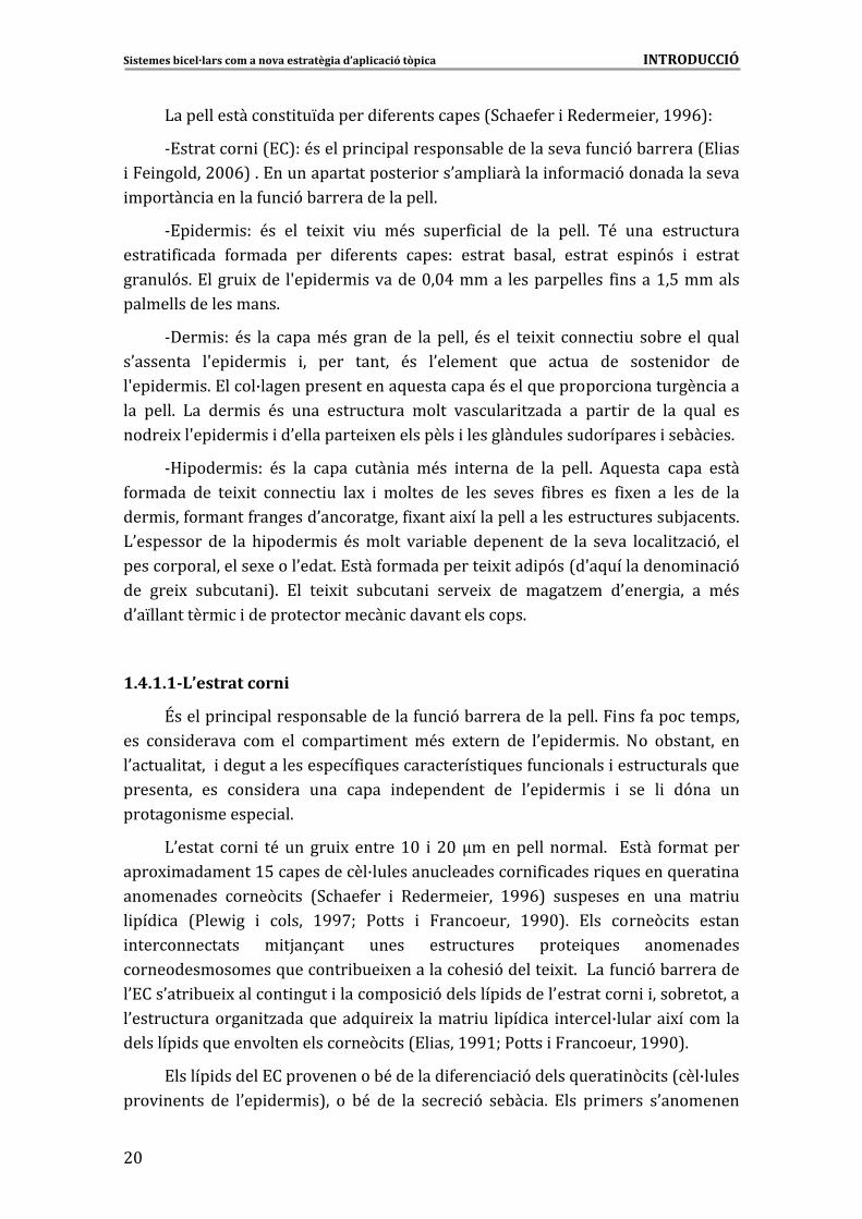
1.4�La�pell�com�a�via�d’administració�de�principis�actius�� >C
>6A6>&���������� 5��������������� >C
>6A6>6>&�)0���������� � #$
>6A6#&:������ ���������������� �4�� 9�������-����������� �� ##
>6A6#6>&�3 ���� 9�in�vitro�����������-�� �4�� 9�������-����������� �� #@
>6A6#6#&�*����� 9�����0������ 9�������4�� 9�-������������������ #A�
1.5��La�pell�i�els�sistemes�lipídics� #%�
1.6�Absorció�percutània� #'
>6E6>&�? ����0�-��� 9�������� �� #'
>6E6#&��������� ������0�-��� 9�������� ��in�vitro� #G
Sistemes�bicel·lars�com�a�nova�estratègia�d’aplicació�tòpica���������������������������������������������������������������������������������ÍNDEX�
�
1.7�Tècniques�de�caracterització�de�la�pell������� @$
>6'6>&�!�������(� �� �4����H�����������4������������� ���������4����� �����������������7*��&� �8�
@>
>6'6#&3*F3�� �����9� @@
>6'6@&�� ���-�� �� 9� ��� ���� �� ��� � ���(� �� �����(� �� ��������� �� 9�73�!�86�
@A
�
2.�OBJECTIUS� @'�
3.�RESULTATS� A>
Article� 1.� � ������ �2������ 4��� �� � ���� ������������ �-����� ��� �4�� ��4����
A%
Article�2.�3����������44�����4�4��4���� �� �� ���::�I�D:��- ������32������
%@
Article� 3.� � ������ �2������ ��� ��J� ��� ���2� �������2� 4��� ��� ������� �� ����4�4��4���� �� ���
E%
Article�4.�� �������2�������������J����� ������� ���2��������2�4����K �� >$%
Article�5.����� ��� 4��� ����4� ��������� ��� �����K ���:������������������� ����4��44� ���������� 2� �� ��
>>@
Article�6.�� �������2�����������. ���4��������������4� ��� �����K ��� >#%�
4.�DISCUSSIÓ� >A%�
4.1�Caracterització�físico�química�dels�sistemes�bicel·lars� >A'
A6>6>����� �������������� ����4��(� ������������������� �������- ��B������������� ���� 9��0���4�����
>A'
A6>6>6>� ������� 9�������!*�� >A'
A6>6>6#� ������� 9�����*�� >%$
A6>6>6@��������� ����������� ������������������ ���� 9�������!*� �����
*����� �������- ��B���������::�I�D:�� >%A
A6>6#����� ����������������������� �������- ��B����� >%A�
4.2�Estudis�in�vitro�de�l’aplicació�tòpica�dels�sistemes�bicel·lars�en�pell�sana�
>%%
A6#6>�3 �������- ��B������������. ��������!*�� �4���� ������������� 9�� ��� �������� ��������
>%%
A6#6#�3 �������- ��B������������. ������*� >%E
A6#6#6>�� �4���� ��������. �������0�-��� 9�������� ������*� >%E
Sistemes�bicel·lars�com�a�nova�estratègia�d’aplicació�tòpica���������������������������������������������������������������������������������ÍNDEX�
�
A6#6#6#� ����� 9������� �������- ��B��������*���-�������� �������0!�� >%C
A6#6@�!���� �������- ��B��������������� ������������������- � ���������������
>E$
�
4.3�Disseny�d’una�metodologia�de�simulació�in�vitro�de�pell�amb�la�funció�barrera�deteriorada�
>E$
�
4.4�Ús�dels�sistemes�bicel·lars�com�a�vehicle�en�l’aplicació�de�DDEA�sobre�pell�in�vitro�simulant�una�condició�patològica�
>E@
�
5.�CONCLUSIONS� >E'�
6.�BIBLIOGRAFIA� >'>


ABREVIATURES�

Sistemes�bicel·lars�com�a�nova�estratègia�d’aplicació�tòpica��������������������������������������������������������������ABREVIATURES�
?�
ABREVIATURES��*6:6� *-��� 9�������� ��
*3� “Automatic�Freeze�Substitution”��3 ������������� �/��������-�� �� 9��
* "!�� � *�� �4������� ����������� ����
*���� 7“Attenuated�Total�Reflection”,���4����� ������������������
��2�&�!��� �� �� ���(� �������(� ����������� �� 9��
D� � ���4 �������� 4�� 9�
d� � � ���� ���������� 9��
dc�� � � ���� �������������������L�����
��!*� �� ��4�������� �� ��� ���
�D:�� � � .�M��� ��4��4�� � ��� ����
�)3� � NDynamic�Light�Scattering”,�� ����� 9�� ��� �����������
��:���� � � � ��� ��4��4�� � ��� ���
�::��� � � ���� �� ��4��4�� � ��� ����
�3�� �NDifferential� Scanning� Calorimetry”,� ����� ���� �� � 4���� ���
�0����-�����
!��� � !��������� �
*� � O ��4��4���� ���
!��� �“Freeze�Fracture� Electron� Microscopy”,� � ���(� �� �����(� �� ���
� �4��������
3�!��� “Freeze� Substitution� Transmission� Electron� Microscopy”���� ���-�� �� 9���� ���������� ���(� �������(� ����������� �� 9��
� ��� �“Fourier� Transform� InfraredP�� !�������(� �� �4����H�� ����
�����4������������� ����
D� � ����.�M��������
D!F� � !���5���������.�M�������
D � � ����.�M��������������
D � � ����.�M������� �������
D:)�� � ����������4 ����5� ����0����������� 9��
� � ����� �������� ����� 9��
kB�� � ����������������/�����
)�� � ������� ����
)�� � ����� ����B� ���
����:�J� )���� ����������4 ����������� 9�������&� ����
)Q� � ����� �����&��5� ��
Sistemes�bicel·lars�com�a�nova�estratègia�d’aplicació�tòpica��������������������������������������������������������������ABREVIATURES�
? �
)R�S� � �������� �� �����
" �3�� � “Non�Invasive�Back�Scatter”,�������� ����� ���� ������������ ����� 9��
,��!� ,���� �/� 9����������������� 9� ��������������������!��(� �
,�� � !���5��������������(�- �
:RS� � ����N� ����P��
q�� � ?��������� ����� 9��5� ����� 9� ������ ������ ��� 4��4���� �� ��� ������ ��5��� �� ������� � ���
4��4���� ����������������
Rh� � ��� �. ���� ��� �
��"� � �������� �������� ���������
3*F3����������������NSmall�Angle�X�ray�Scattering”���� ����� 9������ ��F������������� ���
3::�� � “Short�Periodicity�Phase”������������ �� ������������ � �������������
�!��� �“Transmission� Electron� Microscopy”�� � ���(� �� �����(� �� ���
������ �� 9�
�!T)� � “Transepidermal�Water�Lloss”��:������������� ���� ���0� �����
��� � �������������������� 9����4����������� �����&��5� ���
tR��� � �������������M� 9�
+?�� � +����� ������
T*F3� � NWide�Angle�X�ray�Scattering”,�� ����� 9������ ��F�����������������
��� � �? ��� �����
�� � *��������� ����� 9��
�� � )��� �����0�����

PLANTEJAMENT�GENERAL�������������������

Sistemes�bicel·lars�com�a�nova�estratègia�d’aplicació�tòpica����������������������������������������PLANTEJAMENT�GENERAL�
? �
P�
LANTEJAMENT�GENERAL�
)�������<������/����������������<���� � �/������������� ����� � � ���� ���
��-� ���� � 9� ����� 7����� �8�� ��� ����� 7��������� ��������(� �8� �� � ���� ��
7��� -�������� ��� 4������ �� � ����� ��������� 86� ������ �� ��� ������� �� 5���
�������� ������� ��� ��5� � ��� ��� ����� ��� �� � �� �(� �� �0��� � 9� ��� ����������
�0�-�� �� 9�� ����� ��.����5� � �������������� ������ ��6�
!�� ��� 4������� ��� �0�-��� 9� ������� �� �0.��� ��� ���������� ����� ��������
����� !�� �� � � � �� ��� ��� ��. ��� � ��� ����6� �������� �0����� �<� ��� ���� ���� �
�������� �������(����������������M ��� M������������� ����� 9�����������6����4��������
���� ����� ��� ���� ��� �0�-��� 9� ������� �� ��� �9�� ���� � ������ ������� ���
�����M ���� ������������ ���4��(� �� � ��������� ��� ��� ������ ���� ��������� 5����
5��� 5���� �4�� �&5��� 5����������� � � ���� ��� �������� 4 � ����5���������� ��
������ ����������������. ���6������ M(������������ �������������4� � �������� 4�� 9�
�0�� ���������<�������� 4�������������������������6�
*5��������� ��0.��� ����2������ �������������5�����������H������������5���
�0.�� ����4��� �� ��� ��� ��� M������ �<�� ��������� ��-��� ���� ��������� 5���� 4�� �&
5��� � �5�� � ������� �������������������������. ���- ��B������ � �/��6�
)��� - ��B���� �9�� ��������������� ������ �/����� ���� ��� ����� ���� ���
����� 9� � ��� �� � ���������� - ��� ����� � M�� ��� ���� ���� ������ � ���� ����
�������� 5���6������� �� �5������� ��������� 5���� � �� ���� ����� �� ���� ����� ����
���� �/����������������������0.���������5������������ ��������������� �������������
U���� �� � (�� �. ����� ����� � 9�� � ��
!�� �5������ ��� � �0.��� ��. �� �/��� ���� �� � � �� �� ��� ��-� � 9�
��� �4�����(� �� ��� � ������� - ��B����6� *5������� � ������� �0.��� ������ �/���
�� � �/����������� �������� 5���� �������������������������� ��0���� ��������6�
!����� � � ���� �����. �� �/������������ �������- ��B������0.������ �����-���
����������-���� 4 ��� ���������� M������������������������� �0�-��� 9�������� �6�
:������ M(���0.���� � �/����������(� ��in�vitro��4 ������������������������,��!�
5��� �� � �/�� - (�� ��� ��� ����� ��-� ���� ���5����� ��� 9� 4 � ��(� �6� * M������ M��
�0.�� � ����2��� ���� ���(� �� in� vitro� ���� �� � ������ ��� ������� ���� � ��� ���
�������������0��������������(� ����-���������4�� 9�-�������� �� ��L���� ��0.��
����� ������������ ����0�� � �/�������� �������- ��B���������5������� �����������6�!���
��� ������-� ���� �����5���������������<�������-� � 9�5����0.��� ��(����������� �
�����������������������-� ����������5������ ����� �� 9�5������������� -� �������
��. �� �/� 9� �<�� �4�� ��� � ������� ��� � 4������� �� � � �� �� ��� 5��� �0��� 5� ��
�(� �����6�������
�

�
�
�
�
�
�
�
�
�
�
�
�
�
�
�
�
�
�
�
�
�
�
�
�
�
�
�
�
�
�

��������
1. INTRODUCCI�������������������

Sistemes�bicel·lars�com�a�nova�estratègia�d’aplicació�tòpica�����������������������������������������������������������������INTRODUCCIÓ�
�
@
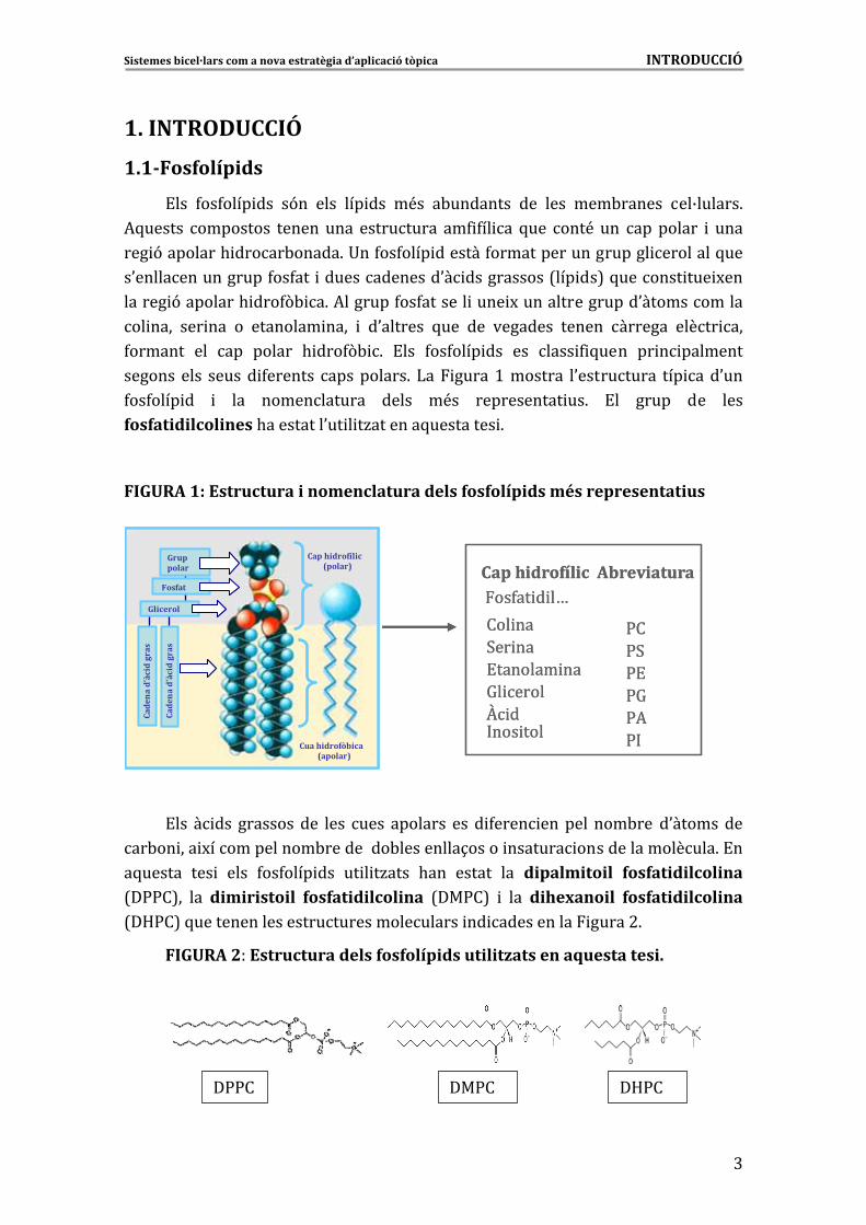
1.�INTRODUCCIÓ�
1.1�F fos olípids�
!��� 4��4���� ��� �9�� ���� ��� ��� �<�� �-�������� ��� ���� ���-������ ��B������6�
*5������ ��������� ���������� ���������� ��4 4�� �� 5��� ���<� ��� ��������� � ����
��� 9��������. �����-�����6�+��4��4���� �������4�������������������� ��������5���
�0����������������4��4��� ��������������0� �����������7��� ��8�5������� ��� M���
������ 9��������. ���4(- �6�*�������4��4������� ���� M����������������0������������
�� ���� ��� ��� �� �������� ���� � �0������� 5��� ��� �������� ������ ������� ����� ���
4������� ��� ��� ������ . ���4(- 6� !��� 4��4���� ��� ��� ���� 4 5���� �� � ��������
����������� ������ 4������� ����������6� )�� �����>�������� �0���������� ��� ���0���
4��4���� �� � ��� ������������ ����� �<�� ����������� ��6� !�� ����� ��� ����
fosfatidilcolines�.���������0�� � �/�������5��������� 6��
�
FIGURA�1:�Estructura�i�nomenclatura�dels�fosfolípids�més�representatius�
�
!��� � ��� ����������� ���� ���� �������� ���� 4���� ����������-����0���������
��-�� ��� M������������-���������-���������1����� ������� �����������������6�!��
�5������ ��� � ���� 4��4���� ��� �� � �/���� .��� ������ ��� dipalmitoil� fosfatidilcolina�
7�::�8�� ��� dimiristoil� fosfatidilcolina� 7��:�8� � ��� dihexanoil� fosfatidilcolina�
7�D:�8�5������ �������������������������� �� ����������� �����#6�
Cap hidrofílic��4�� � �V
��� ��3�� ��!������� ��=� ����O � ��� ���
Abreviatura
:�:3:!:=:*:
Gruppolar
Fosfat
Glicerol
Cad
ena�
d’à
cid
gras
Cua hidrofòbica(apolar)
Cap hidrofílic(polar)
Cad
ena�
d’à
cid
gras
Cap hidrofílic��4�� � �V
��� ��3�� ��!������� ��=� ����O � ��� ���
Abreviatura
:�:3:!:=:*:
Gruppolar
Fosfat
Glicerol
Cad
ena�
d’à
cid
gras
Cua hidrofòbica(apolar)
Cap hidrofílic(polar)
Cad
ena�
d’à
cid
gras
��
IGURA�2��Estructura�dels�fosfolípids�utilitzats�en�aquesta�tesi.�F
�
�
��
�::�� ��:�� �D:��
�
Sistemes�bicel·lars�com�a�nova�estratègia�d’aplicació�tòpica�����������������������������������������������������������������INTRODUCCIÓ�
�
A�
����� �5���������������� ��� ������ ��� ������� ��-� �0� ���� ��� ������ M� ���
�4����. ���4(- �� ��������&��������������� ��� ��� ��-����� ���� ������� ��� �������
��-��0� ���� ���������. �����-��������� ��������������S ���� ��6��
�
1.1.1 fo �� Agregats� sfolipídics�en solució�
)0������ 9� �������� �� ����� 4��4���� ��� ��� ���<�� ����� ������ ����� ����
�0�4���� . ���4(- � ��������� ����� �������� ���-<� �0.�� ��� ��� �� ��� ������ ���
������� �� ����� 4��4���� ��6� *5������ ������� �� ������ � ���� ������ ���� �����
. ���4(- �� �0����� �4�� ��� ���� ��� ������ � ��� ���� ���� ��� ���� �������
. �����-������6�3�-�����5��������������������������� ���������������� ���������
�����������4 4�� 5���� ����� ���������������� 9��<��4�����-��6��
!�����������>��0 �� 5��������� 4�������4������������� 5��������������������
��4 4�� 5����� ��������������� 9��<��4�����-���������� 96�
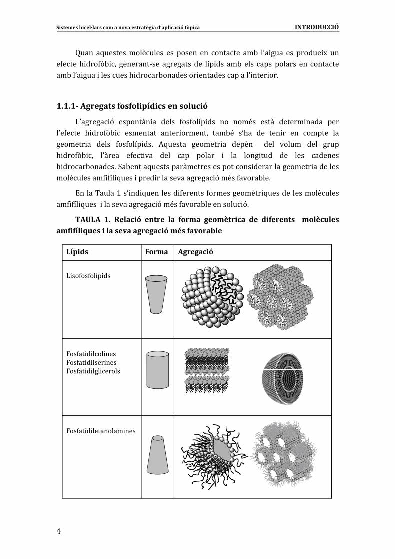
TAULA� 1.� Relació� entre� la� forma� geomètrica� de� diferents� � molècules�amfifíliques�i�la�seva�agregació�més�favorable�
��4�� � ��������� ���
��4�� � ��� �����4�� � ���� �����4�� � ��� �����
) ��4��4���� ��
AgregacióFormaLípids
Sistemes�bicel·lars�com�a�nova�estratègia�d’aplicació�tòpica�����������������������������������������������������������������INTRODUCCIÓ�
�
%�
!��� ��� ��� �0���� ����� ��� . ���4(- ��� ������ ��� ��� ������ ����� ������� �����
�������� ��-� ��� ��� � �0��������� ��� 4����� ��� � ��B���� ��4�� 5���� �� � ��B����
��-�����6�!������ ��������-���������������� ������������������������ �����-�������
��� ��-� ����� �� �0��������� ��� 4����� ��� - ����� ������� �� �������� 4�������
��������� � � �������6�!M ��� M���������� ��� ��������-��� ������5����� �������� ���
���������������������������������������������� ��������������-������ 9�������
���W��5��������� ���������4��������������������� ��B���� �������6���
!���������� ���� fosfatidilcolines� ����������4����������-� ����� � ������������
�0�������������4�������bicapes6�
�
1.1.1.1�Comportament�fàsic�de�les�bicapes�fosfolipídiques�
!���������� 9������������������������� ��� ��4������������� ��������4�����
� ����B� ���6��)����<����������9������4�����.�M������� ���� ���6�)��4����.�M�������
7D8� <�� ���� ���������� ��� (� �� ��� ����� � ���� ���� 5��� ��� ��� M� ��� � ��B����
����� 5���������B����������5��������.�M����������� ���-��������� ���� �4 � ��6�
:������ �������� ����� �����������7D 8���� �������7D 86�)��4������� ����7)8����������
� �������- �����������������������������0� ���6��
)���- ����� 4��4�� ��� 5���� 4������4����� ��� ����������� ��5�9�6� �*5�������
4�����������������-�������4�� 9����������������������������������� 4������������������
7���� ������� . �����-������� ���� ��� �� ������ ��� ���� � ���������8�� � ������ ��5� ��
� ����B��� �� 4�� �� 7���� ������� . �����-������� ����� ��� ��� ������ �(- ��86� )��
�����������������5������������� M������� ��0���������������� �����&��5� ���0��������
�������������������� 9����4����7��86�*����������������������� �4�� ����������������
��� �� ��� ���-�� ��� 4���� ���� � �� ������� ����� ����� ��� ���-�� ��� 4���� ��� � �����&��5� ��
7X����.� ���� ���>G'G86�
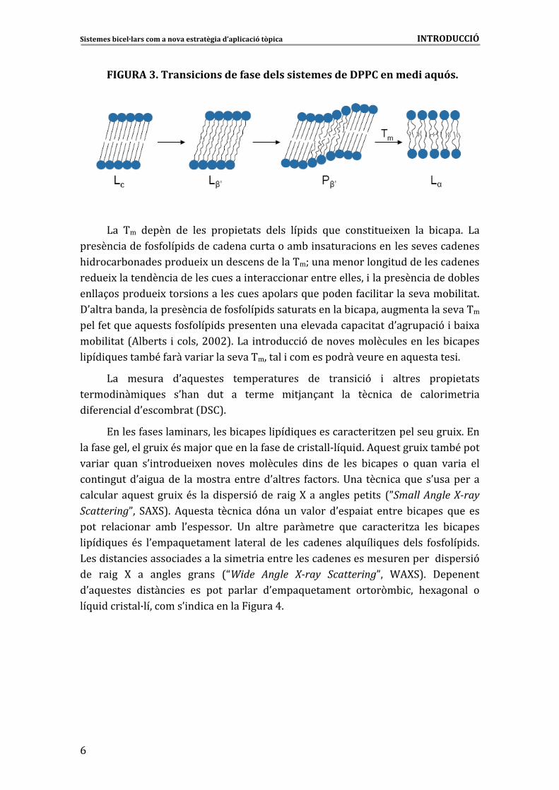
� ������ ��� 4���������M ��� M���� 4���������-4�����5������������������ �������
4��4���� ��� ��� ������6� :��� �M������� ��� ��� ��� ����� � ������� ��� �::�� ��� ��� �
�5�9��� �� -� M��� ������������� ���� - ����� ������ ��� 4���� � ����B� ��� 7)�8�� ��-�����
����������������� ����B� ��6�*��������������������������������- �����������������
4�������� �� �����7)R� S8������5������ 4�������� ��� ���������������������L��������� ���
4���� � ����B� �������(� ������-����������� �<�������� ������0����������6� ������� ��
5��� ��� �::�� �<� ��� ��� ������ ����� ����� �9��� �-���� �0�M��� ������� ��� ����� 9�
�� � ������4����� �����&��5� ��7)Q8����������������������� 9����)R�S���N� ����P�4����
7:RS8�� ��� ���� - ����� ���������� ����� ������ ���� 7D����� � ) �� >GGGW� X�2����� �
��44��2��>GGCW�:�������� ������#$$>86�!����� �����@���������������������������� 9�
������������ ����� ���������4�������� ����������4�������� �������� �����&��5� �6�
�
�
Sistemes�bicel·lars�com�a�nova�estratègia�d’aplicació�tòpica�����������������������������������������������������������������INTRODUCCIÓ�
�
E�
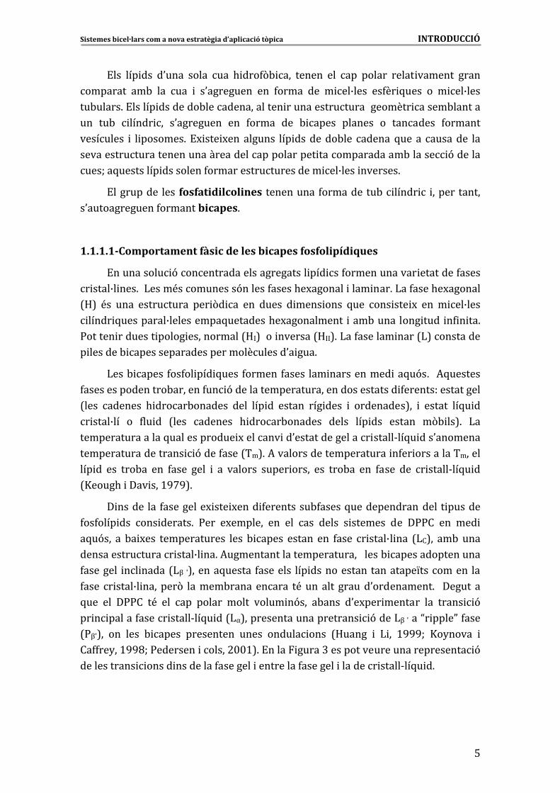
FIGURA�3.�Transicions�de�fase�dels�sistemes�de�DPPC�en�medi�aquós.�
�
)�� ��� ������ ��� ���� ���� ������ ����� ��� ��� 5��� ���� ��� M��� ��� - ���6� )��
������ �����4��4���� ���������������������-� ������� ������������������������
. �����-������������� M�������������������W��������������� ������������������
����� M���������� ��������������� ����� ������������������ ���������� �������-����
�����1��������� M����� ����������������������5���������4� � ��������������- � ���6�
�0������-��������������� �����4��4���� ������������������- �������������������������
����4���5����5������4��4���� ���������������������������� �����0������ 9� �-� M��
��- � ����7*�-����� ������#$$#86�)�� ������ 9���������������������� ����- �����
� ��� 5 ��� ����-<�4������� ������������������� �����������������������5��������� 6�
)�� ������� �0�5������� ������������� ��� ����� 9� � ������� ���� ������
������ ��� 5���� �0.��� ���� �� ������ � �H��1���� ��� ��� �� ��� ���� ���� ��
� 4���� ����0����-����7�3�86�
!������4�������� ����������- ������ ��� 5������������� �/�������������� M6�!��
���4��������������� M�<����H���5���������4�������� �����&��5� �6�*5�������� M����-<�����
��� ��� 5���� �0 ������� M��� ������ ��������� � ��� ��� ���� - ����� �� 5���� ��� �� ���
��� ������0� ������� �����������������0������� 4�����6�+��� ��� ��5��� �0����������
��������5�������� M�<�� ���� ����� 9���� �� ��F������������� ��� 7PSmall�Angle�X�ray�ScatteringP�� 3*F386� *5������ ��� �� �9��� ��� ������ �0���� ��� ������ - ����� 5��� ���
���� ���� ����� ��-� �0��������6� +�� ������ ���������� 5��� ������ �/�� ���� - �����
� ��� 5���� <�� �0����5��������� �������� ��� ���� ������� ��5��� 5���� ����� 4��4���� ��6�
)���� ���� ������� ����������� ���� ������������������������������������ ����� 9�
��� �� �� F� �� ������� ������ 7NWide� Angle� X�ray� ScatteringP�� T*F386� ���������
�0�5������� � ���� ��� ��� ���� ������� �0����5��������� �����(�- �� .�M������� ��
��5� ��� ����B��������0 �� �������� �����A6�
�
�
�
�
Sistemes�bicel·lars�com�a�nova�estratègia�d’aplicació�tòpica�����������������������������������������������������������������INTRODUCCIÓ�
�
'
FIGURA� 4.� � Empaquetament� lateral� de� les� cadenes� alquíliques� dels�fosfolípids.�
�
A6>�Y
@6'�Y
A6>�Y
@6'�Y
A6>�Y
@6'�YA6>�Y
A6>�Y
A6>�Y
A6>�Y
A6>�Y
A6>�Y�
,����(�- � D�M������� ��������)�5� ��
ZA6E�YZA6E�YZA6E�Y
�
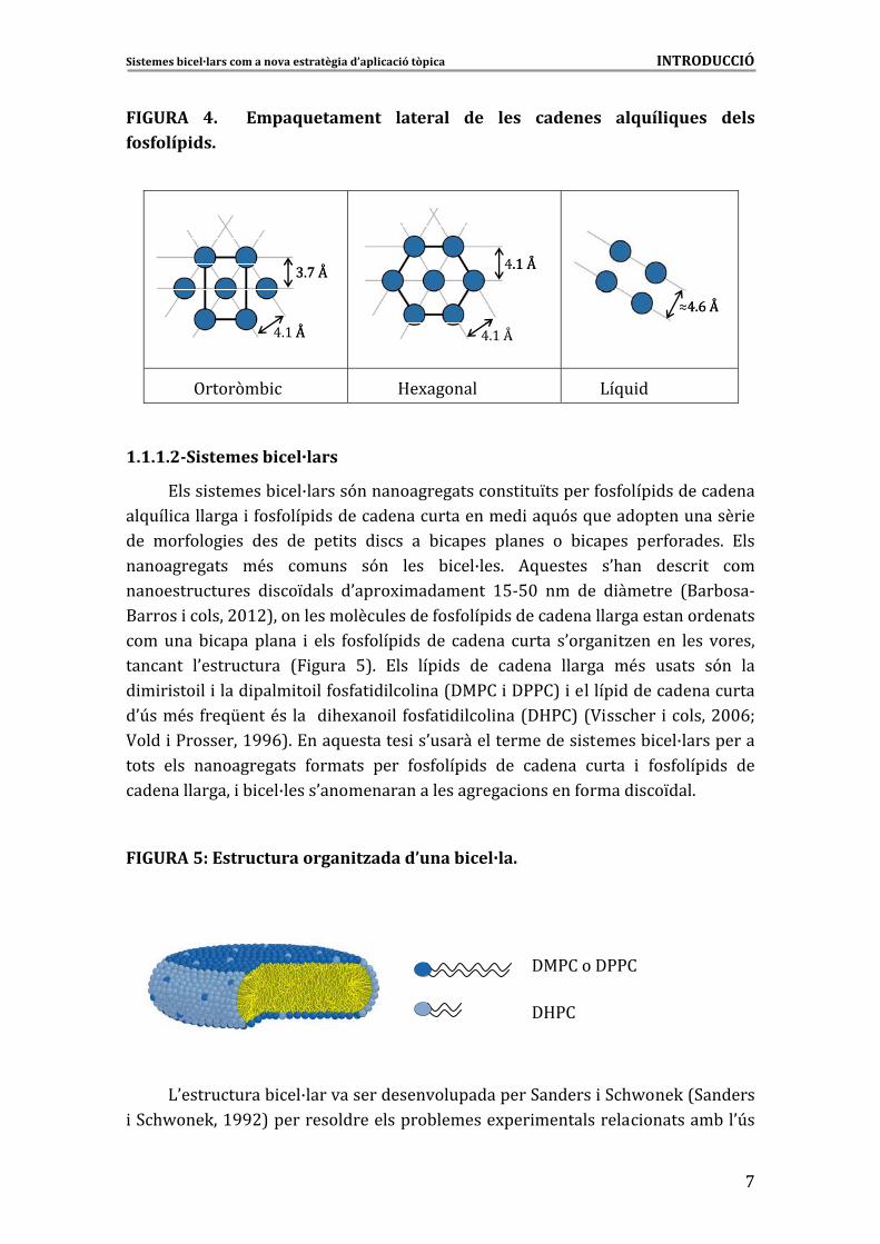
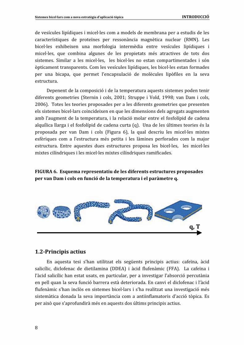
1.1.1.2�Sistemes�bicel·lars�
!���� �������- ��B������9������������������� ��L�������4��4���� ������������
��5��� ��������� �4��4���� ����������������������� ��5�9��5������������������ ��
��� ���4���� ��� ���� ��� ��� ��� � ��� �� - ����� ������� �� - ����� ���4������6� !���
������������� �<�� ������ �9�� ���� - ��B���6� *5������� �0.��� ���� �� ���
��������������� � ��L����� �0����M ��������� >%&%$� ��� ��� � ������� 7���-���&
������� ������#$>#8���������������������4��4���� ����������������������������������
�������- ���������� � ���� 4��4���� ��� ��� ������ ����� �0����� �/��� ��� ���� �������
������� �0���������� 7 ����� %86� !��� ��� ��� ��� ������ ������� �<�� ������ �9�� ���
� � � ��� �� ����� ���� �� ��4��4�� � ��� ���7��:�� ��::�8� ������� ����������������
�0U���<�� 4��5[����<�� ��� �� .�M��� �� 4��4�� � ��� ���7�D:�8�7? ��.��� ������#$$EW�
?���� �:��������>GGE86�!���5��������� ��0������������������� �������- ��B�����������
����� ���� ������������� 4������� ���� 4��4���� ��� ��� ������ ����� � 4��4���� ��� ���
�������������� �- ��B�����0����������������������� �������4������ ��L���6�
�
FIGURA�5:�Estructura�organitzada�d’una�bicel·la.�
�
�
�
�
�
�
)0����������- ��B�����������������������������3������� �3.J���K�73�������
�3.J���K��>GG#8���������������������-�������M��� ������������ ��������-��0U��
��:�����::��
�D:�
�
Sistemes�bicel·lars�com�a�nova�estratègia�d’aplicació�tòpica�����������������������������������������������������������������INTRODUCCIÓ�
�
C�
������������� ��� 5���� �� ��B����������������������-���������������� ���������
��������� 5���� ��� �����L���� ���� �������� �� ������ �� ������� 7��"86� )���
- ��B���� �M. -� M��� ���� ���4���� �� ������� �� ������ ��������� � ��� 5���� �
� ��B����� 5��� ��- ��� �������� ��� ���� ���� ������ �<�� ����� ���� ��� ����� ����
� ������6� 3 � ���� �� ���� � ��B����� � ���� - ��B���� ��� ������ ������ ��������� � �9��
(�� ������������������6������������������� ��� 5���������- ��B����������4��������
���� ���� - ����� 5��� ������� �0�������� 9� ��� ��������� � �(4 ���� ��� ��� �����
���������6�
�������������������� 9� ��������������������5������� ���������������� ��
� 4������� ������� ��� 73���� �� � ����� #$$>W� 3������� � ?����� >GGCW� �������� � �����
#$$E86���������������� ������������������������� 4�������������� ���5�������������
����� �������- ��B������ � �� M������5�������� ���� ����������������������������
��-� �0����������� ��� ������������� � ��� ���� 9���������������� 4��4���� �����������
��5��� ��������� ����4��4���� ����������������7586� �+����������U�� �������� ���<�����
���������� ���� ���� ���� � ���� 7 ����� E8�� ��� 5���� ���� �� ���� � ��B���� � M����
��4�� 5���� ��� �� �0���������� �<�� ��� ��� � ���� ��� ���� ���4������� ��� ��� ��H���
���������6� !����� �5������� ����� ����������� �������� ���� - ��B����� � ���� � ��B����
� M���� ����� 5���� ������ ��B����� M���� ����� 5������� 4 ����6��
�
FIGURA�6.��Esquema�representatiu�de�les�diferents�estructures�proposades�per�van�Dam�i�cols�en�funció�de�la�temperatura�i�el�paràmetre�q.�
�
q,�Tq,�T
�
1.2�P nri cipis�actius�
!�� �5������ ��� � �0.��� �� � �/��� ���� ���[����� �� � � �� �� ���� �4�L���� � ��
��� �� �� � ��4���� ��� � �� ��� ��� 7��!*8� � � �� 4��4���� � 7*86� � )�� �4�L��� �
�0� ����� �� �.����������������������� ������������ ����� �����0�-��� 9�������� ��
��������5������������4�� 9�-����������������� �����6�!����� ����� ��4���� ��0� ��
4��4���� ��0.��� ��(������ �������- ��B����� ��0.������ �/������� ����� �� 9��<��
� ������ �� ������� ��� ����� ������� �� ��� �� ��� �4������� �� �0� 9� �(� �6� !��
����� M(�5����0����4��� ����<������5����������U�� ����� � � ���� ��6�
Sistemes�bicel·lars�com�a�nova�estratègia�d’aplicació�tòpica�����������������������������������������������������������������INTRODUCCIÓ�
�
G
*�-�9�� ��� �4������� �� ������2��� ��� ����� ����� ��� �4������� �� ���
������ ���� 7* "!�86� � !����!*� <�������� ������� �0� �� 4�� ���� � � ��� *���� �0� ��
������ � 6�!����� �����'���������������0����������5��� �� ������������ �����4�� �&
5��� ������������� ����������4 ����������� 9�������&� ����7����:�J8�����5����
<�� �� ��������� ��4 � �����0���������6� ��
�
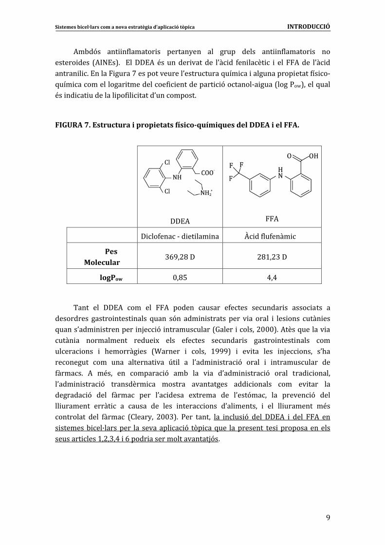
FIGURA�7.�Estructura�i�propietats�físico�químiques�del�DDEA�i�el�FFA.�
�
�
�
��!*�
�
*�
� � ��4� ��� �����&�� �� O � ��4��4����
Pes�Molecular�
@EG�#C��� #C>�#@��
logPow $�C%� A�A�
�
����� ��� ��!*� ��� ��� *� ������ ������ �4����� ������� �� ���� ���� ��
���������� ������ ����� ����� 5���� �9�� ��� � ������� ���� � �� ����� � ��� ���� ���� ���
5�����0��� � ���������� �H� 9� ������������7=����� ������#$$$86�*����5������� ��
���� �� ����������� ����� M� ���� �4����� ������� �� ������ ����� ����� ���
����� ���� � .������� ��� 7T������ � ����� >GGG8� � �� ��� ���� �H� ����� �0.��
��������� ��� ���� �������� ��� U� �� �� �0��� � ���� 9� ����� � ������������ ���
4�����6� *� �<��� ��� ������ 9� ��-� ��� � �� �0��� � ���� 9� ����� ���� ������
�0��� � ���� 9� ��������� �� ������� ����������� ��� ������ ��� �� ���� ���
������� 9� ���� 4����� ���� �0� ����� �M������ ��� �0���9���� ��� ������ 9� ����
�� �������� ����� � �� ����� ��� ���� ����� ���� �0�� ������� � ��� �� �������� �<��
��������� ���� 4����� 7�����2�� #$$@86� :��� ������ ��� ���� 9� ���� ��!*� � ���� *� ���
� �������- ��B��������� ��� �������� � 9� �(� ��5��� ����������� ��� ����������������
�������� ����>�#�@�A� �E����� ������������������H9�6�
�
�
�
�
Sistemes�bicel·lars�com�a�nova�estratègia�d’aplicació�tòpica�����������������������������������������������������������������INTRODUCCIÓ�
�
>$�
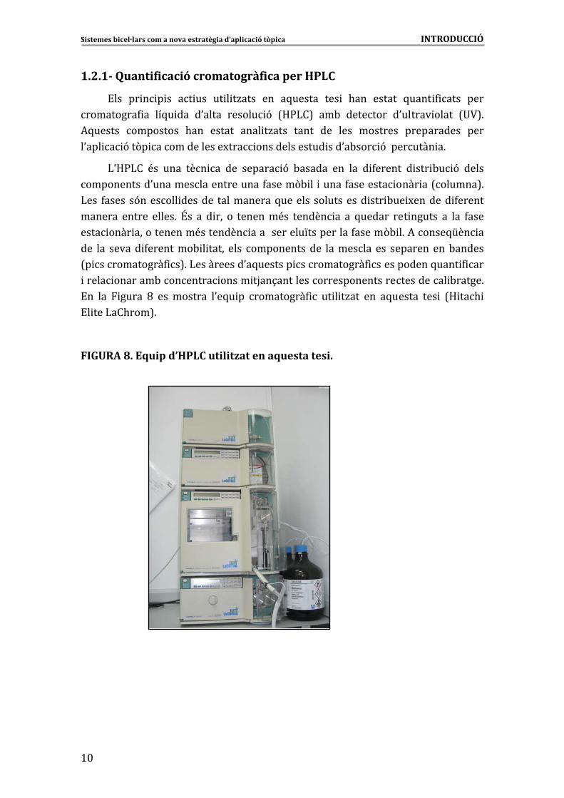
1.2.1 f��Quantificació�cromatogrà ica�per�HPLC�
!��� �� � � �� �� ��� �� � �/���� ��� �5������ ��� � .��� ������ 5���� 4 ���� ����
���������4 �� ��5� ��� �0����� ������ 9� 7D:)�8� ��-� �������� �0������ ����� 7+?86�
*5������ ��������� .��� ������ ���� �/���� ����� ��� ���� �������� ����������� ����
�0��� �� 9��(� ���� ��������M��� �������������� ���0�-��� 9��������� �6��
)0D:)�� <�� ���� ��� �� ��� ������ 9� -������ ��� ��� � 4������ � ��� -� 9� �����
�����������0��������������������4�����(- �� �����4�������� ���� ��7������86�
)��� 4����� �9������� ������� �����������5������� ����������� ��� -�� M������� 4������
������� ������ �����6� \�� �� � ��� �� �������<�� ������ �� �� 5������ ��� ������ �� ��� 4����
���� ���� ������������<�������� ������������L����������4�����(- �6�*�����5[�� ��
��� ��� ������ 4��������- � ����� ���� ������������� ��������� ��� �������� ���-������
7� �����������4 �86�)����������0�5������� �����������4 �����������5���� 4 ���
����� �������-�������� ����� �H��1�������������������������������� -�����6��
!�� ��� ����� C� ��� ������� �0�5� �� ���������4 � �� � �/��� ��� �5������ ��� � 7D ��. �
!� ���)��.���86�
�
FIGURA�8.�Equip�d’HPLC�utilitzat�en�aquesta�tesi.�
�
�
�
�
�
�
�
�
�
�
�
�
�
�
�
�
Sistemes�bicel·lars�com�a�nova�estratègia�d’aplicació�tòpica�����������������������������������������������������������������INTRODUCCIÓ�
�
>>
1.3�Tècniques�de�caracterització�dels�sistemes�bicel·lars�
1.3.1 ica�de�llum�(DLS)��Tècnica�de�dispersió�dinàm
)��� ����� 9�� ��� ����������7NDynamic�Light�Scattering”���)38��<�������������
��� 5�����<��������������������� ����������������������4�� ����������4 ����
���� 4�� 9����� �. ���� ��� ����� ����� �� ���� � ����� 9����������������������������
������� 9� 7���K���� #$$G86� � *5������ ��� �� ��� ��� M� �� 4��� � � �� ��� 4� M� ���
��� � 9� ��-� ���� ���� �0���� �� � ������ �0���� q=� 2�/�� ��� ���� ������� 9� ��-�
���������� 5��� ������ ��� ��� ����� -��J� �6� !�� ���������� ��� ��� � 9� ��-� ����
����������� ���� ����� ��� �0����� �� <�� �-���- ���� ���� ������ <�� ���������� ������
��� 4 � 9� ���� ������ �0���� � ����� � ���� ������ <�� � ��������� ��� ���� �0���� 6� )��
����� � ��������� �� ��� ���� ������ �� <�� ��B������� ���� ��� ��������
74������� �� ����86�!���� �������� ����� 9�����������������4�� 9������������0����
���� 4�� 9�q=� (4�/�)sin(�/2)� 7��������� 5��� �0����� �� � ����� <�� ����� �� �0����� ��
���������8� � ������ ������ ���� ����� ����� ��� � ����� 9� �� � 4������� ������ ��� ����
�-��� ������4�� 9��0���������� 96��
:��� ���������� ��4�� 5��������� ��������� ��� 4�� 9� �0���������� 9� ���� ����
�H���������������[����4�� 9��M����� �����������������������t����
g(t) �A�exp�(�2t/t= R)�+�B�
,��A� � <�� ���� �������� �������������B� <�� ��� ������ -���� � tR� � <�� ��� ������ ���
����M� 96��*5�����tR���������� �������-������4 �������� 4�� 9�7D8������0�5�� 9���
tR=1/Dq2
�*�� ���� �0�5�� 9� �03��K��&! ���� ��� ��� ���� ���� ��� ��� ��� � . ���� ��� �
��������� �Rh�������D���
D=k /6��RBTB h
,�� kB� <�� ��� �������� ��� ����/����� �� <�� ��� ������������ �-������� � �� <�� ���
� ���
B
��������� �����6��
!����������������������������5� ������� ����� 9������������������������� M���
��������������9������� 4������� ��� � 9������������������ ��� �����������������
� ��������S���������� ����� 9� ��������������7 �����G86�
�
�
�
�
�
�
Sistemes�bicel·lars�com�a�nova�estratègia�d’aplicació�tòpica�����������������������������������������������������������������INTRODUCCIÓ�
�
>#�
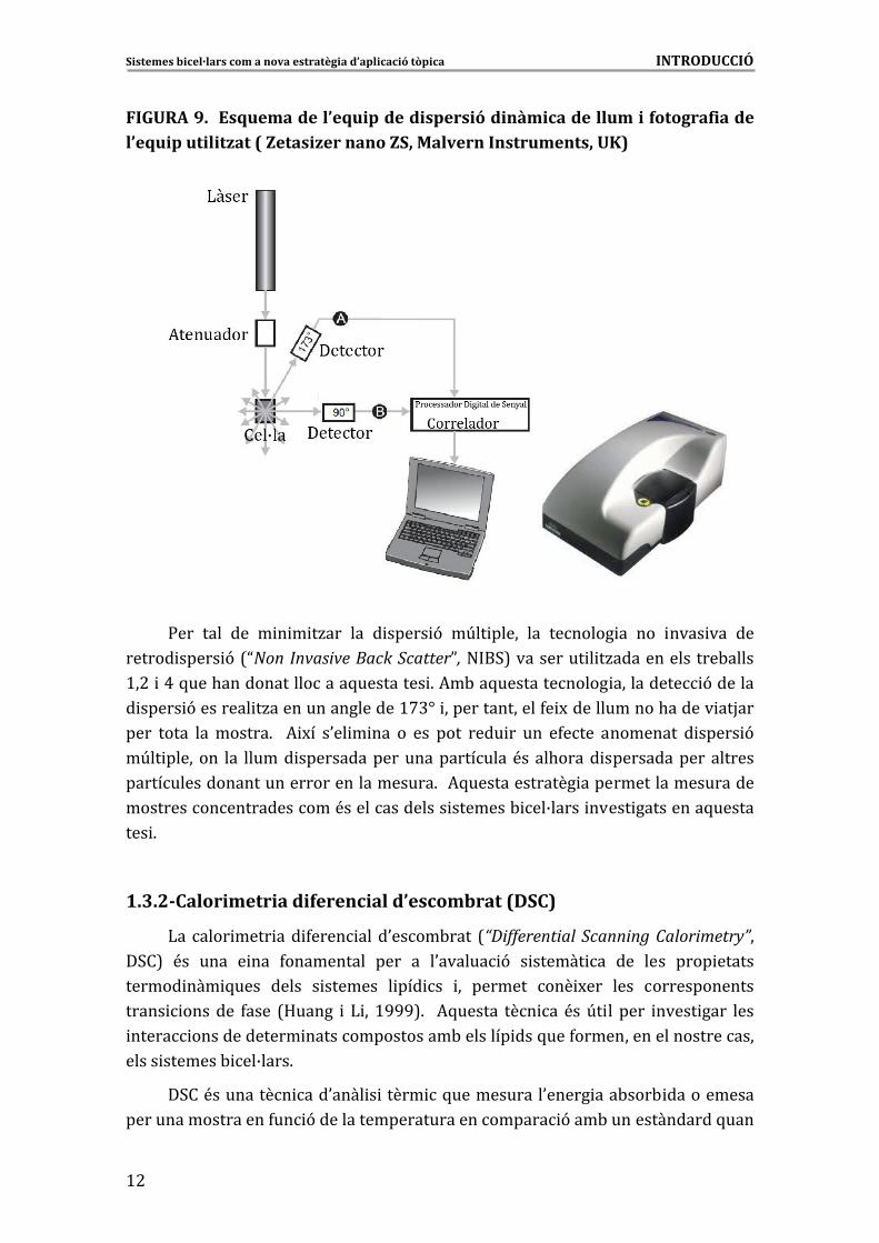
FIGURA�9.��Esquema�de�l’equip�de�dispersió�dinàmica�de�llum�i�fotografia�de�l’equip�utilitzat�(�Zetasizer�nano�ZS,�Malvern�Instruments,�UK)�
�
�
�
�
�
�
�
�
:��� ���� ��� � � � �/��� ��� � ����� 9� �U�� ����� ��� ������� �� ��� ���� ��� ���
������ ����� 9�7NNon�Invasive�Back�ScatterP,�" �38���������� � �/��������������-�����
>�#� �A�5���.����������������5��������� 6�*�-��5������������� ���������� 9�������
� ����� 9�������� �/�����������������>'@]� ���������������4� M������������.������ ��H���
���� ����� ���������6� � * M�� �0�� � ��� �� ��� ���� ���� �� ��� �4���� ��������� � ����� 9�
�U�� ����� ��� ��� ������ ��������������������������<����.����� ��������������������
��������������������������������������6��*5�������������� ����������������������
����������������������<�������������� �������- ��B����� ����� ���������5������
��� 6�
�
1.3.2 rat�(DSC)��Calorimetria�diferencial�d’escomb
)������ ���� ��� 4���� ����0����-���� 7“Differential� Scanning�Calorimetry”���3�8� <�� ���� � ��� 4���������� ���� �� �0������ 9� � ������ �� ��� ���� ���� ������
������ ��� 5���� ����� � ������� � ��� �� �� ������� ��� M��� ���� �������������
����� ������� 4���� 7D����� � ) �� >GGG86� �*5������ ��� �� <��U� �� ���� ����� ���� ����
����� ������������� ����������������-�������� ���5���4������������������������
����� �������- ��B����6�
�3��<��������� ���0���� � ����� �5�����������0����� ���-���- �����������
������������������4�� 9���������������������������� 9���-��������������5����
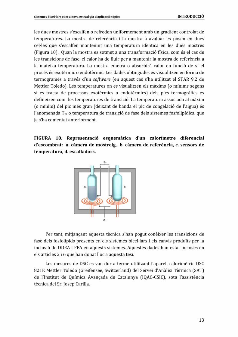
Sistemes�bicel·lars�com�a�nova�estratègia�d’aplicació�tòpica�����������������������������������������������������������������INTRODUCCIÓ�
�
>@
������������������0����4�������4�������� 4�����������-�������� ����������������
������������6� )�� ������� ��� ��4���� �� � ��� ������� �� �������� ��� ������ ��� �����
��B���� 5��� �0����4��� ������ ��� ���� ������������ ���� �� ��� ���� ����� ��������
7 �����>$86��������������������������������������4���� 9�4�� ������<�����������
��������� �������4�������������.�����4�� �������������� �����������������4���� ����
��� ���� M�� �����������6� )�� ������� ������� �� �-���- ��� ����� ��� 4�� 9� ��� � � ���
���<��<���M����� ����������� 6�)����������-� ����������� ���� �/������4��������
������������ �� ����<�� �0��� software� 7��� �5����� ��� �0.�� �� � �/��� ��� 3�*�� G6#� ���
��������������86�)����������������������� ���� �/���������M ���7����� ����������
� � ��� ������ ��� ��������� �M����� �� �� �������� �8� ����� � �� ��������4 �� ���
��4 �� M�������������������������������� 96�)������������������ ���������M ��
7����� �8������ ��<������� 7�� M�������-��������� ���� ������ 9���� �0� ���8�<��
�0����������������������������������� 9����4���������� �������4��4�� ��� ���5���
H���0.��������������� ������6��
��
FIGURA� 10.� Representació� esquemàtica� d'un� calorímetre� diferencial�d'escombrat:� �a.�càmera�de�mostreig,� �b.�càmera�de�referència,�c.�sensors�de�temperatura,�d.�escalfadors.�
�
�
�
�
�
�
�
�
:��� ������� �H��1�����5������ ��� ���0.������������ M��� ���� ����� �������
4���������4��4���� �������������������� �������- ��B����� �������� �������L����������
���� 9������!*� �*�����5������� ������6�*5�������������.��������� ����������
������� ����#� �E�5���.����������������5��������� 6�
)���������������3��������������� �������� � �/���� �0������������ ���� ��3��
C#>!����������������7=�� 4�������3J �/������8�����3���� ��0*��� � ����� ��73*�8�
��� �0 ��� ���� ��� ���� �� *���1���� ��� �������2�� 7 �*�&�3 �8�� ����� �0��� ���� ��
��� ������3�6�;�������� ���6�
�
�
�
Sistemes�bicel·lars�com�a�nova�estratègia�d’aplicació�tòpica�����������������������������������������������������������������INTRODUCCIÓ�
�
>A�
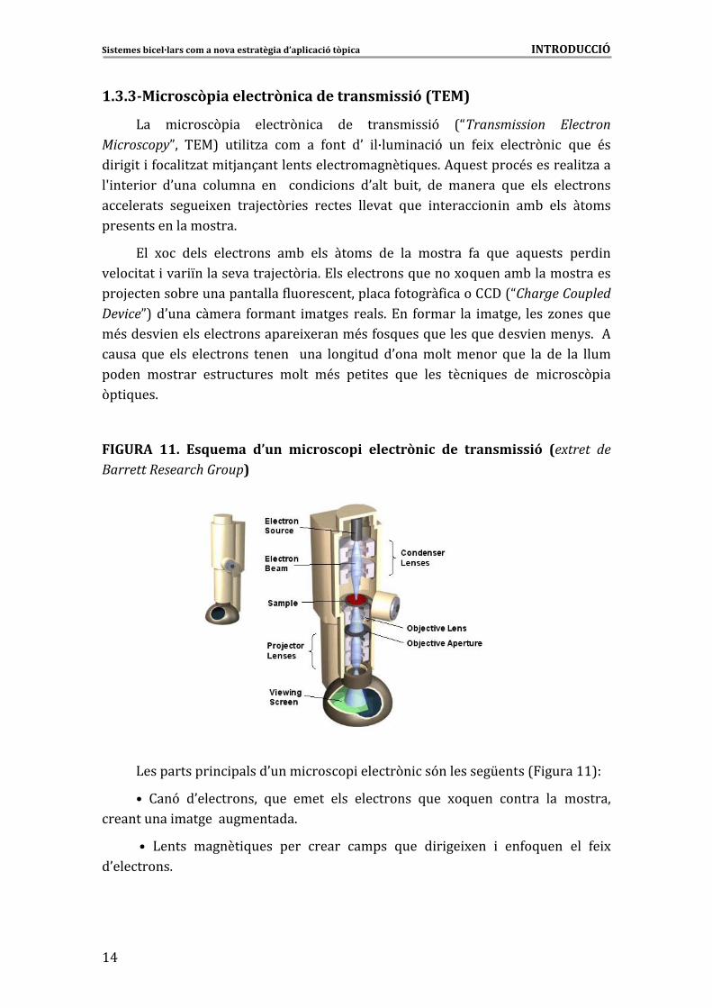
1.3.3�Microscòpia�electrònica�de�transmissió�(TEM)�
)�� � ���(� �� �����(� �� ��� ������ �� 9� 7NTransmission� Electron�MicroscopyP�� �!�8� �� � �/�� ��� �� 4���� �0� �B��� �� 9� ��� 4� M� �����(� � 5��� <��
� � � �� �4��� �/���� �H��1���������������������� 5���6�*5��������<��������� �/����
�S ���� ��� �0���� ������� ��� � ��� ���� �0���� -� ��� ��� ������� 5��� ���� ���������
��������� ����� M��� ���H��(� ��� ������ ������� 5��� ����� �� �� ��-� ���� ������
���������������������6�
!�� M�� ����� ��������� ��-� ���� ������ ��� ��� ������� 4�� 5��� �5������ ���� ���
���� ���� ���� L�������������H��(� �6�!������������5������M�5������-���������������
���H�������-����������������4����������������4������4 ��������7NCharge�Coupled�DeviceP8��0���������� 4������� ������� �����6�!�� 4������ ��� ������� ���� /�����5���
�<������ ��������������������� M������<��4��5����5�������5������� ������2�6��*�
����� 5��� ���� ��������� ������ � ���� ���� ���� �0��������������� 5��� ��� ��� ��� �����
������ �������� ����������� ����� �<�� ��� ���� 5��� ���� ��� 5���� ��� � ���(� ��
(�� 5���6�
�
FIGURA� 11.� Esquema� d’un� microscopi� electrònic� de� transmissió� (extret� de�Barrett�Research�Group)�
�
�
�
�
�
�
�
�
�
�
)����������� � ������0���� ����� ������(� ��9���������[�����7 �����>>8��
^� ���9� �0���������� 5��� ����� ���� ��������� 5��� M�5���� ������ ��� ��������
���������� �����������������6�
�^� )����� ������ 5���� ���� ����� ����� 5��� � � �� M��� � ��4�5���� ��� 4� M�
�0��������6�
Sistemes�bicel·lars�com�a�nova�estratègia�d’aplicació�tòpica�����������������������������������������������������������������INTRODUCCIÓ�
�
>%�
�^�3 ���������-� ���5���<���������������� ��������������� ����� ������(� 6�
*������5������������������������������� ��������������������������0� �����0.�����4���
���-� ���� ��-<����������0 ���� ����0���� ����� ��0�5���������������� 5���6�
�^�:����4������4 �������������4����������5���������B����������������0�-H����
��� ���� �/���������� ���������� ����������������6�
�^� 3 ��������� ��� �����5���������� ��� ������5��� ������� M������� ��������� �
5������������������ �����6�
!��� �M��� ������ ��� � ���(� �� �����(� �� �0.��� ���� �/��� ��� ��� �� ���� ���
�� �&� ���(� ��!����(� ���������������� ����4 �� ������(� ��������+� ���� ����
������������7�� �&+�8��������0��� ���� ����� �����������6��������)9��/� �������
���6�!� ����������6��
�
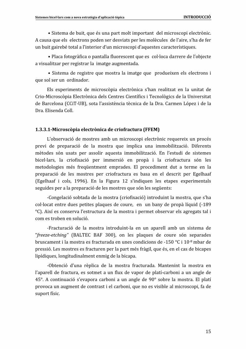
1.3.3.1�Microscòpia�electrònica�de�criofractura�(FFEM)�
)0�-����� 9��������������-����� ����� � �����(� � ��5���� M� ������<��
���� � ��� ������� 9� ��� ��� ������� 5��� ��� �� ���� ���- � �/� 96� � 4�������
�������� �9�� ������ ���� ����� �� �5������ ���- � �/� 96� !�� �0����� � ��� � �������
- ��B������ ��� � �4 M� 9� ���� ����� 9� ��� ������ � ��� � �4������� �9�� ����
��������� ��� �<�� 4��5[�������� ��������6� !�� ����� ����� ���� �� ������ ��� ���
������� 9� ��� ���� �������� ���� � �4������� ��� -���� ��� ��� ���� �� ���� !���.��4�
7!���.��4� � ����� >GGE86� !�� ��� ����� >#� �0 �� 5���� ���� ������� �M��� ��������
���� �������������������� 9����������������5����9���������[������
&������� 9���-������������������7� �4 M� 98� ������ ��������������5����0.��
��B������������������� �������5������������� ���� ����-��2���������� ��5� ��7&>CG�
]�86�* M��������������0����������������������� ���������-������������������������ �
���� 6�����-���������� 9 �
&������ 9� ��� ��� ������� ������ ��&��� ��� ��� �������� ��-� ��� � ������ ���
Nfreeze�etching”� 7�*)�!�� �*� @$$8�� ��� ���� ���5���� ��� ����� �9�� ����������
-��������� ��������������4�������������������� �������&>%$�]�� �>$&C��-������
����� 96�)��������������4���������������������<��4��� ���5���<���������������- �����
� ��� 5 ���������� �� ����������� ��������- ���6�
&,-��� 9� �0���� ���� �� ��� ��� ������� 4��������6� ������ ��� ��� ������� ���
�0�������� ��� 4�������� ��� ������������ 4��M������������������&��-�� ���������������
A%]6� *� ��� ��� 9� �0�������� ��-�� � �� ��� ������ ��� G$]� ��-��� ���������6� !�� ������
����������������������������� ������-�� ��5���������� � -������� ����� ��4�����
�������4�� 6�
�
�
Sistemes�bicel·lars�com�a�nova�estratègia�d’aplicació�tòpica�����������������������������������������������������������������INTRODUCCIÓ�
�
>E�
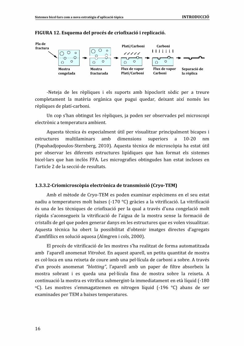
FIGURA�12.�Esquema�del�procés�de�criofixació�i�replicació.�
Pla de�fractura
Mostracongelada
Flux�de�vapor�Platí/Carboni
Mostrafracturada
Flux�de�vapor�Carboni
Separació de�la�rèplica
Platí/Carboni CarboniPla de�fractura
Mostracongelada
Flux�de�vapor�Platí/Carboni
Mostrafracturada
Flux�de�vapor�Carboni
Separació de�la�rèplica
Platí/Carboni Carboni
�
&"���H�� ��� ���� ���� 5���� � ���� �������� ��-� . ����� �� �(� � ���� �� �������
������������ ��� ����� �� ����� �� 5��� ���� � 5������� �� M���� � M�� ���<�� ����
���� 5������������&��-�� 6�
+������0.����-� ������������� 5����� H�������������-�������������� ����� �
�����(� �����������������- ���6�
*5������ ��� ��<������ �������U� ������� ���� �/����� � ��������- ����� �
����������� ���� ��� ����� ��-� � ���� ���� ����� ���� �� >$&#$� ���
7:���.��H�������&3����-����� #$>$86�*5������ ��� ������ ���(� ��.�� ������U� ��
���� �-������� ���� � 4������� ����������� � ��� 5���� 5��� .��� 4������ ���� � �������
- ��B����� 5��� .��� ��(�� *6� )���� �����4 ��� �-� ������� .��� ������ ������� ���
�0��� ���#��������� 9�������������6���
�
1.3.3.2�Criomicroscòpia�electrònica�de�transmissió�(Cryo�TEM)�
*�-����������������2�&�!������������M�� �������� ���������������������
��� ����������������������-� M���7&>'$�]�8���� ��������� �� 4 � 96�)��� �� 4 � 9�
<�� ������� ���� ��� 5���� ��� � �4 M� 9� ���� ��� 5���� �� ����<�� �0���� ������ 9������
��� ��� �0�������� M� ��� � �� 4 � 9� ��� �0� ���� ��� ��� ������� ������ ��� 4���� 9� ���
� ��������������5��������������������2��������������������5������������� ���� �/��6�
*5������ ��� �� .�� �-���� ��� ���� - � ���� �0�-��� �� ������� � ������ �0���������
�0��4 4�� ��������� 9��5�����7*������� ������#$$$86�
!�����<������ �� 4 � 9�����������������0.������ �/������4������������ �/����
��-���0�����������������Vitrobot6�!���5�������������������� ���5���� ��������������
�����B������������� M��������������-��������B�����������-�� �����-��6�*�����<��
�0��� ���<�� ��������� “blotting”�� �0�������� ��-� ��� ������ ��� 4 ����� �-���-� M� ���
������� ��-����� � ��� 5����� ���� ���B������ 4 ��� ��� ������� ��-��� ��� �� M���6� *�
��� ��� 9��������������� �� 4 ����-���� ��&��� ���� �����������������5� ��7&>C$���86� )��� �������� �0�������/����� ��� � ������� ��5� �� 7&>GE� ]�8� �-���� ��� ����
�M�� �����������!����-� M���������������6��
�
�
Sistemes�bicel·lars�com�a�nova�estratègia�d’aplicació�tòpica�����������������������������������������������������������������INTRODUCCIÓ�
�
>'
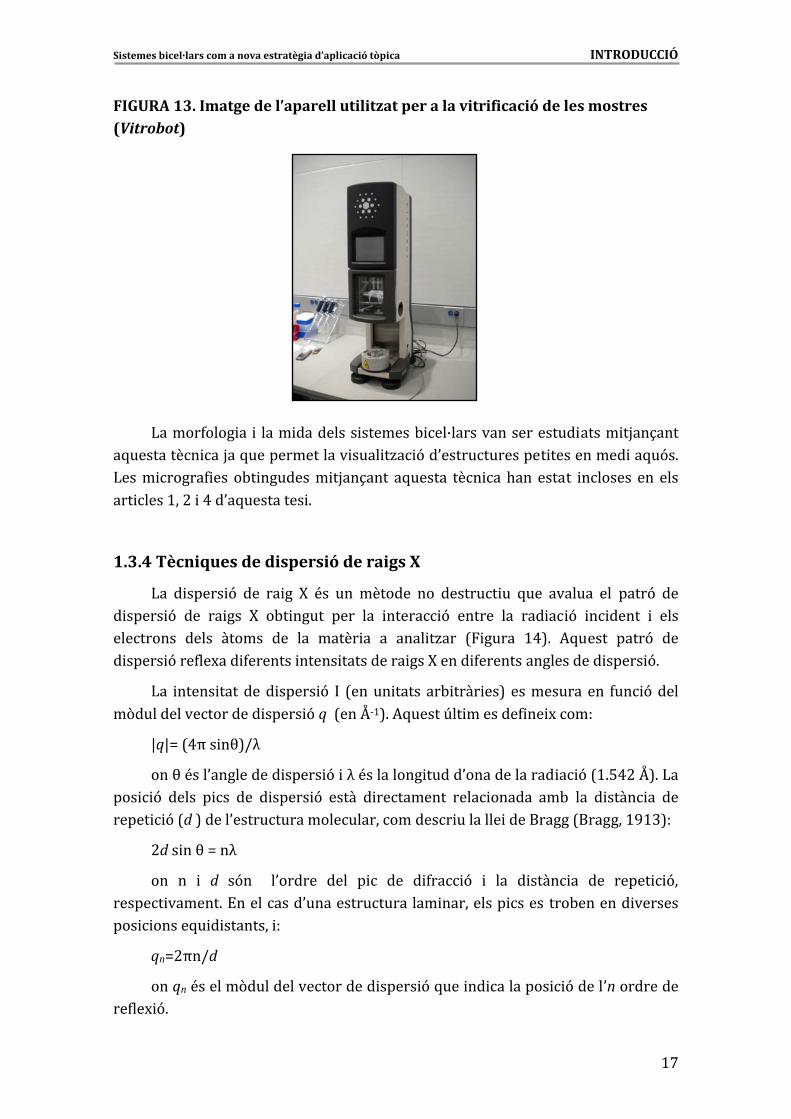
FIGURA�13.�Imatge�de�l’aparell�utilitzat�per�a�la�vitrificació�de�les�mostres�(Vitrobot)������
�
�
�
�
�
�
�
�
�
)�����4���� �� � ���� ��������� �������- ��B������������������ ����� �H��1����
�5��������� ��H��5�������������� ���� �/� 9��0�������������� ���������� ��5�9�6�
)���� �����4 ��� �-� �������� �H��1���� �5������ ��� ��.��� ������ ������� ��� ����
��� ����>��#� �A��0�5��������� 6
�
1.3.4�Tècniques�de�dispersió�de�raigs�X�
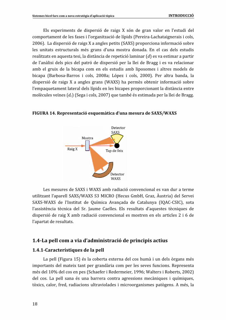
)�� � ����� 9� ��� �� �� F� <�� ��� ������� ��� ������� �� 5��� ������� ��� ����9� ���
� ����� 9� ��� �� ��� F� �-� ����� ���� ��� ����� 9� ������ ��� ��� � 9� � ����� � ����
��������� ����� ������ ��� ��� ����� �� �� ���� �/��� 7 ����� >A86� *5����� ����9� ���
� ����� 9���4��M��� 4������� ����� ���������� ���F����� 4������������������ ����� 96�
)�� ����� �������� ����� 9� � 7����� ����� ��- ���� ��8� ������������� 4�� 9�����
�(�� �� ���� 9�q��7���Y&>86�*5�����U�� �������4 �� M������ ������������� �
_q_`�7Aa�� �b8Ic��
���b�<���0���������� ����� 9� �c�<��������� �����0������������� � 9�7>6%A#�Y86�)��
��� 9� ����� � �� ��� � ����� 9� ����� � ��������� ���� ������ ��-� ��� � ���� �� ���
����� 9 ������������������������� �������� ����������7�������>G>@8�� �7d�8�����0��
#d�� ��b�`��c��
��� �� � d� �9�� � �0������ ���� � � ��� � 4�� 9� � ��� � ���� �� ��� ����� 9��
������ ������6�!���������0����������������� ���������� ��������-������� �������
��� �����5� � �������� ��
�
qn`#a�Id�
���qn�<������(������������������ ����� 9�5��� �� �������� 9�����0n����������
��4��M 96�
Sistemes�bicel·lars�com�a�nova�estratègia�d’aplicació�tòpica�����������������������������������������������������������������INTRODUCCIÓ�
�
>C�
!��� �M��� ������ ��� � ����� 9� ��� �� ��� F� �9�� ��� ����� ������ ��� �S����� � ����
�������������������4����� ��0����� �/� 9������� ���7:��� ��&)�.��� ����� �� ������
#$$E86��)��� ����� 9������ ���F������������� ���73*F38������� ���� �4���� 9���-���
���� �� ����� ������������ �<�� ������ �0���� ������� ������6� !�� ��� ��� ����� ����� ��
���� �/��������5��������� ������ ���� ���������� 9���� ����7d8���������� ���������� ��
��� �0���� � ������� ����������9����� ����� 9����� ��� ��� ���������� � ������ ���� �����
��-� ��� ��� M� ��� ��� - ���� ��� ��� ���� ����� �� ��-� � �������� � ������� ������� ���
- ���� 7���-���&������� � ����� #$$C�W� )9��/� � ����� #$$$86� :��� ������ -������ ���
� ����� 9� ��� �� ��� F� �� ������� ������ 7T*F38� .�� ������� �-��� �� �4���� 9� ��-���
�0����5������������������������� ����������- ����������� ���������� ���� ��������
�����������L����7dc8�73���� ������#$$'8�5������-<�<����� ��������������� ���������6��
�
FIGURA�14.�Representació�esquemàtica�d’una�mesura�de�SAXS/WAXS�
�
�
��
�
�
�
)��������������3*F3� �T*F3���-���� � 9������ ������������������������
�� � �/���� �0��������3*F3IT*F3�3@�� ��,�7D����=�-D��=��/��O���� �8�����3���� �
3*F3&T*F3� ��� �0 ��� ���� ��� ���� �� *���1���� ��� �������2�� 7 �*�&�3 �8�� �����
�0��� ���� �� ��� �� ���� 3�6� ;����� �������6� !��� ���������� �0�5������� ��� 5���� ���
� ����� 9������ ��F���-���� � 9������ �������������������������� ����#� �E����
�0��������������������6�
�
1.4�La�pell�com�a�via�d’administració�de�principis�actius�
1.4.1�Característiques�de�la�pell�
)�������7 �����>%8�<�� ����-������M�������������.���� ���������(�������<��
������������������ M� ���������������� ��������� ���������� 4�� ���6������������
�<������>$d���������������73.��4��� �������� ����>GGEW�T������� ���-������#$$#8�
���� ��6� )�� ����� ����� <�� ���� -������� ������ ������ ���� ���� 5���� � 5��� 5�����
�(M ��������� 4�������� � ���������� ������� �� ������� ��������(����6�*��<��� ���
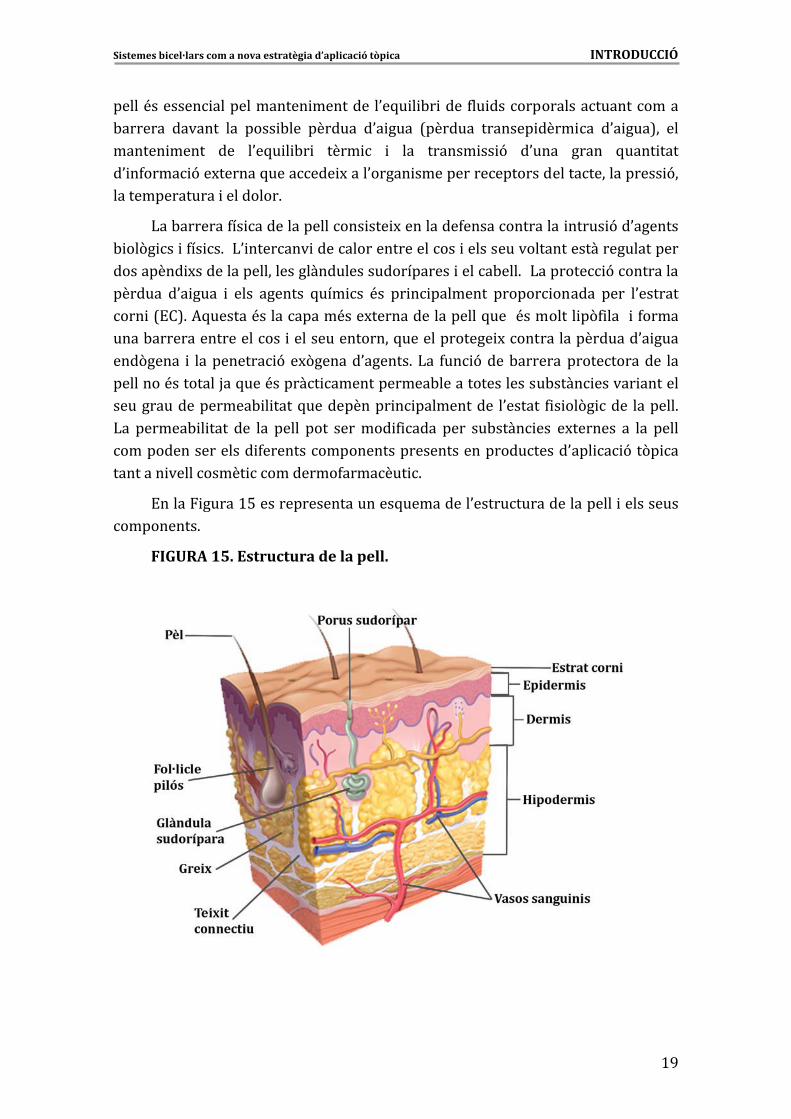
Sistemes�bicel·lars�com�a�nova�estratègia�d’aplicació�tòpica�����������������������������������������������������������������INTRODUCCIÓ�
�
>G
�����<������� ������������� �������� �0�5� � -� ����4�� ������������������������
-������� ������� ��� ���� -��� ������� �0� ���� 7������� ������� ���� �� �0� ���8�� ���
������ ����� ��� �0�5� � -� � ���� � � ��� ������ �� 9� �0���� ����� 5���� ����
�0 �4���� 9��M������5������� M����0����� ����������������������������������� 9��
��������������� ���������6��
)��-�������4�� ���������������� ��� M���������4�������������� ����� 9��0�������
- ��(� �� �4�� �6��)0 ������� ��������������������� ����������������������������������
��������� M���������������������������������������� �����-���6��)������� 9����������
������� �0� ���� � ���� ������� 5��� �� <�� �� � �������� ������ ������ ���� �0�������
��� �7!�86�*5������<����������<���M�����������������5����<�������� �(4 ���� �4�����
����-������������������� ����������������5������������� M������������������0� ����
���(����� � ���������� 9��M(������0������6�)�� 4�� 9����-�������������������� ���
��������<��������H��5���<������ ������������-�����������������-���� ������ �������
������������������- � ����5����������� � ����������� �0������ 4 � ��(� ���� �������6�
)�� ������- � ���� ��� ��� ����� ���� ������� 4 ���� ���� ��-���� ��� �M������� �� ��� �����
������������������ 4���������������������������������������0��� � 9��(� ��
�������� ���������� ���������4������� 6���
!����� �����>%��������������������5���������0��������������������� ����������
���������6�
FIGURA�15.�Estructura�de�la�pell.�
�
�
�
�
�
�
�
�
�
�
�
�
�
�
�
�
Sistemes�bicel·lars�com�a�nova�estratègia�d’aplicació�tòpica�����������������������������������������������������������������INTRODUCCIÓ�
�
#$�
)���������������� ��L�������� 4������������73.��4��� �������� ����>GGE8��
&!��������� �7!�8��<������� � ������������-��������������4�� 9�-�������7!� ���
�� �������#$$E8�6�!������������������� ����0���� ������� �4���� 9����������������
���� 4 �-�����������������6���� �������� �� 9
&!� ���� ��� <�� ��� �� M �� � �� �<�� �����4 ��� ��� ��� ����6� �<� ���� ����������
������ 4 ���� 4������� ���� � 4������� ������ ������� -������ ������� ��� �9�� � �������
������9�6�!����� M���� �S�� ���� ��������$�$A������ �������������� 4 �����>�%��������
��������������������6��
&���� ��� <�� ��� �����<�� �������� ��� ������ <�� ��� �� M �� ����� �� ��-��� ��� 5����
�0�������� �S�� ���� �� �� ���� ������ <�� �0�������� 5��� ����� ��� ������ ���� ���
�S�� ���� �6�!����B������������������5����������<�����5��������� ���������� ����
��� ����6� )�� ���� �� <�� ���� ���������� ����� ������� �/���� �� ���� �� ��� ��� 5���� ���
����� M��S�� ���� �� 0 �� ���������� M������������ ��������������������������� ���-� ��6�
&D ������ ��� <�� ��� ���� ���� �� �<�� ������� ��� ��� ����6� *5������ ���� �����
4������� ��� �� M �� ����� �� ��M� � ������� ��� ���� ������ 4 -���� ��� 4 M��� �� ���� ��� ���
���� ���4�������4��������0����������4 M����� M�����������������������������-H�����6�
)0��������� ��� ��� . ������ �� <������� ��� �-��� ��������� ��� ��� ����� ���� �/� 9�� ���
������������������M�����0����6�!����4������������� M ���� �9��7�S�5����������� �� 9�
��� ��� M� ��-���� 86� !�� �� M �� ��-���� � ����� M� ��� �����/��� �0����� ��� �� �<��
�0�L���������� � ����������������� ���������������6�
�
1.4.1.1�L’estrat�corni�
\������� � ������������-���������4�� 9�-�����������������6� ���4������������
��� ��� ������� ��� ��� ������ ����� �<�� �M����� ��� �0�� ���� �6� "�� �-������� ���
�0����� ������ ������������������4 5������������� 5����4�� ������ �������������5���
���������� ��� ��� ����� ���� ���� ����������� ��� �0�� ���� �� � ��� � � �9��� ���
�������� �������� ��6���
)0������ ��� � �<���� ��� M� ������>$� � #$�e���������� ������6� � !���� 4����������
����M ���������>%�����������B������������������� 4 ������ 5�������5����� ���
����������� ����( ��� 73.��4��� � ������� ���� >GGE8� ��������� ��� ���� ���� ��
� ��� �� 7:��J �� � ����� >GG'W� :����� � ��������� >GG$86� !��� ����( ��� ������
������������� � �H��1���� ����� ����������� ����� 5���� �����������
����������������5������� -�� M���������.�� 9������� M �6��)��4�� 9�-����������
�0!���0��� -�� M������� ����� ��������� 9��������� �������0���������� � ����-���������
�0��������������� �/����5�����5� �� M� ������� �� � ��� �� ������B������� M�� ��� ���
�������� ���5��������������������( ���7!� ����>GG>W�:����� ����������>GG$86�
!������ �������!�������������-<�������� 4���� � 9������5����� �( ���7��B������
���� ������ ��� �0�� ���� �8�� �� -<� ��� ��� ���� 9� ��-� �6� !��� �� ����� �0���������
Sistemes�bicel·lars�com�a�nova�estratègia�d’aplicació�tòpica�����������������������������������������������������������������INTRODUCCIÓ�
�
#>
��� ��� ������B������� � ������ ��� ������ ����� ��������� ��� ��� 4�� 9� -������6� )��
����� 9�� ��� ������0!��<������M ����������������[�����A$&%$�d�������� �����
#%�d���������������>%&#$�d��0� ������������� ������%�d�������4����������������2�#�
d��0�������������������6��
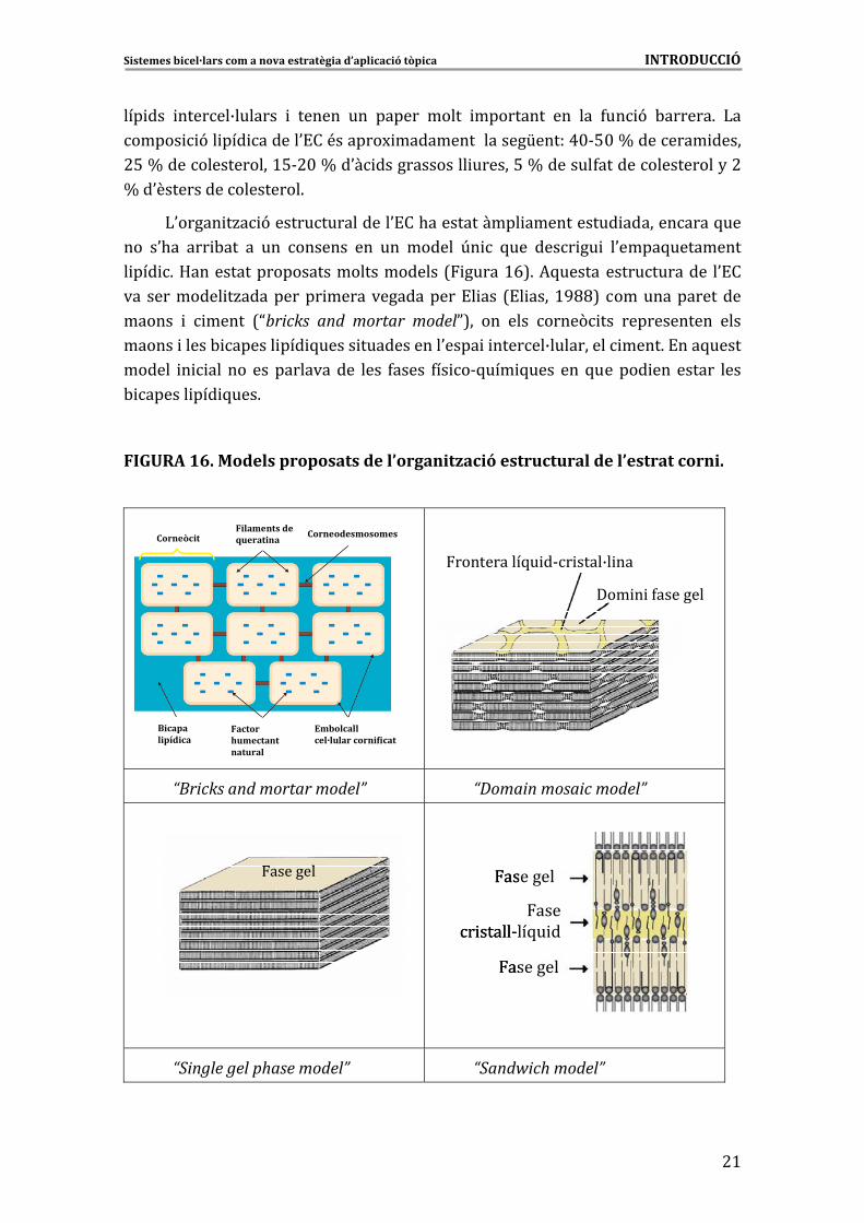
)0����� �/� 9����������������0!��.������������ ����������� �����������5���
��� �0.�� ��� -��� �� ��� ������� ��� ��� ������ U� � 5��� ���� �� � �0����5���������
� ��� 6�D��������������������������������7 �����>E86�*5������ ������������� �0!��
��� ��������� �/���������� ����������������!� ��� 7!� ����>GCC8� ����������������
������ � ����� 7Nbricks� and� mortar� modelP8�� ��� ���� ����( ��� ������������ ����
������ �����- ������ ��� 5����� �����������0���� � ������B���������� ����6�!���5�����
������ � ��� ��� �������������� ���� 4����� 4�� �&5��� 5���� ���5������ ��� ������ ����
- ������ ��� 5���6��
�
FIGURA�16.�Models�proposats�de�l’organització�estructural�de�l’estrat�corni.�
�
CorneòcitFilaments de�queratina
Corneodesmosomes
Bicapalipídica
Factor�humectantnatural
Embolcallcel·lular cornificat
Filaments de�queratina
CorneodesmosomesCorneòcit
Bicapalipídica
Embolcallcel·lular co ificat
Factor�humectantnatural
rn
����������5� �&� ����B� ��
��� � 4�������
�
“Bricks�and�mortar�model”� “Domain�mosaic�model”�
� �
“Single�gel�phase�model”� “Sandwich�model”�
��������������
����� �����&��5� �
�������
�������
����� �����&��5� �
�������
�������
�
�
Sistemes�bicel·lars�com�a�nova�estratègia�d’aplicació�tòpica�����������������������������������������������������������������INTRODUCCIÓ�
�
##�
:����� ������� ������� ������� .��� ����� ���� ��� �� ������� 5��� �0.��� �����
��� ���� ������ ��� 5���� ��� ������ �/� 96� ���� ��� 7���� ���� >GGAW� ���� ��� �
�����>GG'8����������������������������� ������� � ���Ndomain�mosaic�modelP�����
���� ��� ��� �0����� �/��������� � �� ��� 4���� ���� � ��� � ����� 4���� � �����&��5� �6�+��
���������������-�������5���������� ����������������4��������U� ���Nsingle�gel�phase�modelP� 7"���<��� #$$>86� ���J����� ��� ��������� ��� ������ NsandwichP� 7���J����� �
�����#$$$8������������������������� ������4����� �����&��5� ���������������������
���� � � ����� ����� �� ���4����������-����� �-����6�
!��� ������� ���� ��� ������ -������ ��� ���� ���������� �-� ������ ��� ����� ��
���� �/���� ��-� � 4������� ��� 5���6� ����� �5������ ������� � � �� M��� ��� ����
������ 9� ��� ��������� ��� ���5�����������-������������ ���(� �������(� �����
� ���-�� �� 9�73&�!�8W��<������������0�M�� �����-������� ���0�5������������
� ���(� 6� )0��������(� �� �4����H�� ���� �����4������� ��� ��� ��� ��-�
��4����� �� ������ ��������� 7� �&*��8� �4��� M� �4���� 9� ��-��� ��� ��- � ���� �
�0����5��������� �������� ����� ��� ��� ��� �0!�� 7,��� �������K��� � ����� >GGA86� )��
��� �� ��� 3*F3� �� � �/���� ��� � 9� � �����9� <�� ���� �M��B����� � ��� ���� ����� ���
�0����� �/� 9������������������B���������������� � �0����� �/� 9�� ��� ����� �������
�0!��7?�����������.� ������>GG'86��
�
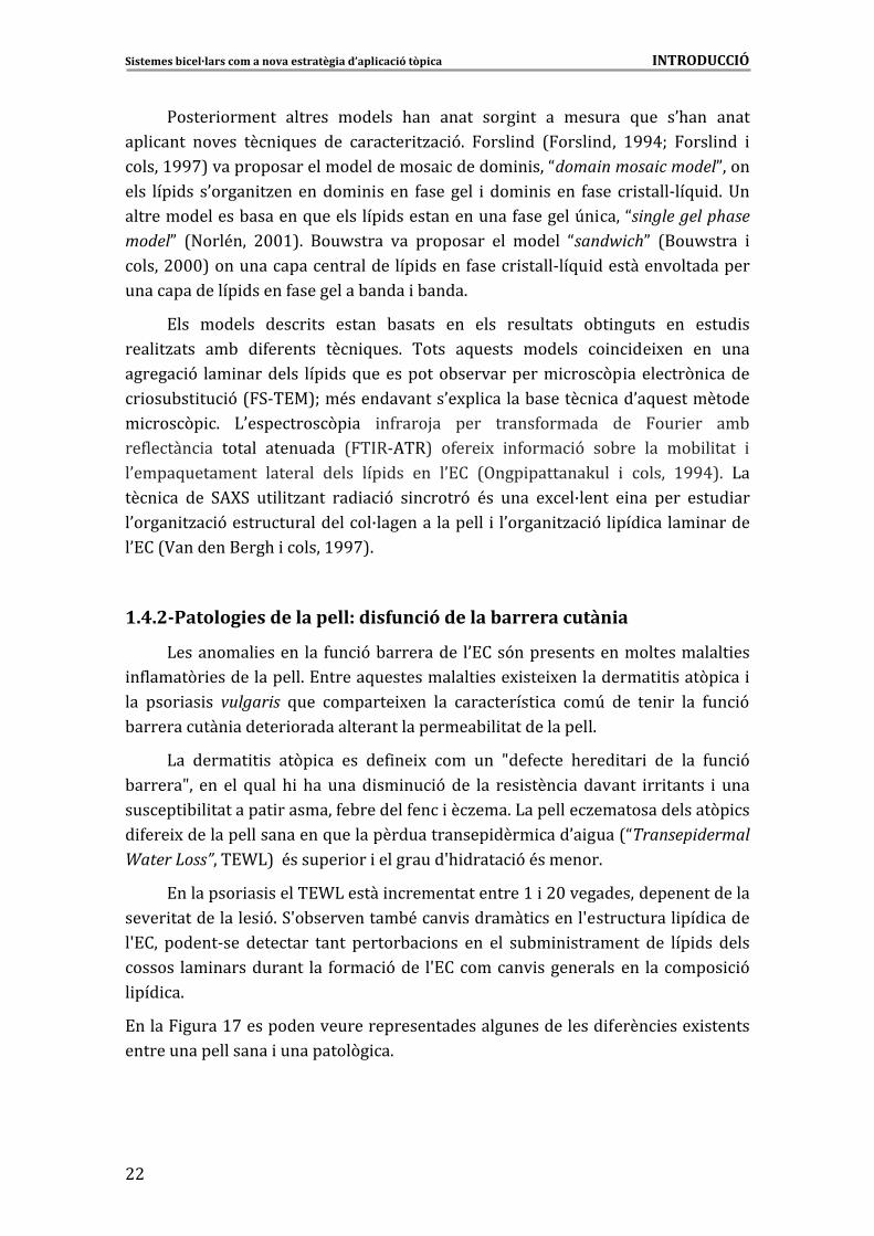
1.4.2�Patologies�de�la�pell:�disfunció�de�la�barrera�cutània�
)��������� ������ ��� 4�� 9�-���������� �0!���9��������������������������� ���
�4�����(� �������������6�!������5������������� ����M ��� M������������ � ����(� �� �
��� ���� �� �� vulgaris� 5��� ������� M��� ��� ��������� �� ��U� ��� ��� �� ��� 4�� 9�
-����������� ������� ������������������������- � ��������������6��
)�� ������ � �� ��(� �� ��� ��4 �� M� ��� ��� f��4���� .���� ��� � ��� ��� 4�� 9�
-������f�� ��� ��� 5���� . � .�� ����� �� �� 9���� ��� ��� ���� �� ������� �� ������ � ����
������ - � ��������� ��������4�-�������4��� ��/���6�)��������/���������������(� ��
� 4��� M��������������������5�������������������� ���� ���0� ����7NTransepidermal�Water�Loss”���!T)8��<������� ��� ����������S. ����� 9�<�������6�
!��������� �� ������!T)������ ����������������>� �#$�������������������������
����� ������������� 96�3S�-����������-<���� �������� ������S����������� ��� �����
�S!��� ������&��� �������� ����� ������-� ���� ��� ��� ��-� � ��������� ��� ��� ��� �����
������ ��� ������������ ��� 4���� 9���� �S!�������� ����������� ��� �������� 9�
� ��� �6�
!����� �����>'���������������������������������������������� 4���� ����M �������
�������������������� ����������(� �6�
�
�
Sistemes�bicel·lars�com�a�nova�estratègia�d’aplicació�tòpica�����������������������������������������������������������������INTRODUCCIÓ�
�
#@
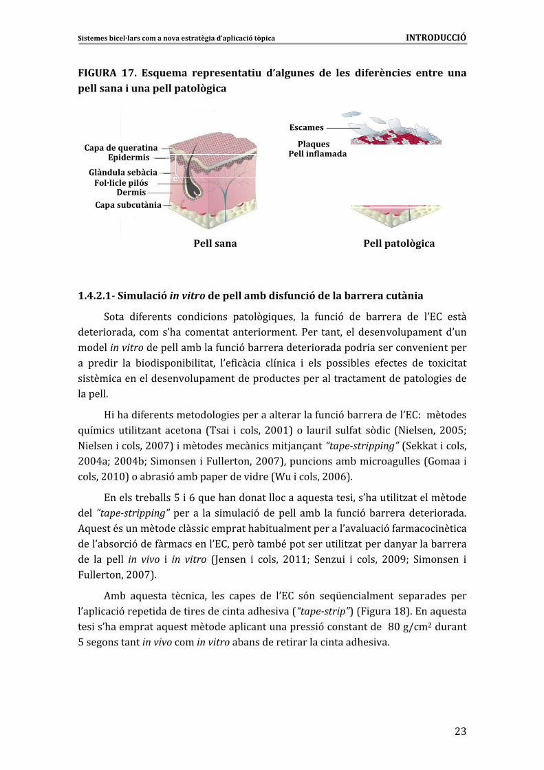
FIGURA� 17.� Esquema� representatiu� d’algunes� de� les� diferències� entre� una�pell�sana�i�una�pell�patològica�
Capa�de�queratinaEpidermis
Fol·licle�pilósGlàndula�sebàcia
DermisCapa�subcutània
Escames
PlaquesPell�inflamada
Pell sana Pell�patològica
Capa�de�queratinaEpidermis
Fol·licle�pilósGlàndula�sebàcia
DermisCapa�subcutània
Escames
PlaquesPell�inflamada
Pell sana Pell�patològica�
�
1.4.2.1��Simulació�in�vitro�de�pell�amb�disfunció�de�la�barrera�cutània�
3���� � 4������� ��� ���� �����(� 5����� ��� 4�� 9� ��� -������� ��� �0!�� �����
����� ������� ����0.�� ������������� ������6�:��� ������ ��������������������0���
������in�vitro�����������-����4�� 9�-������������ ���������� ����������� ��������
�� ���� �� ��� - �� ���� - � ����� �0�4 � �� ��� �� � ���� ���� -���� �4����� ��� ��M ����
� ���� ��������������������������������������������������������������� ������
�������6��
D �.��� 4���������������� ��������������������4�� 9�-�����������0!������������
5��� �� �� � �/���� ������� 7��� � � ����� #$$>8� �� ���� �� ���4��� �(� � 7" ������� #$$%W�
" ������ ������#$$'8� ������������� ��� �H��1����“tape�stripping”�73�KK��� ������
#$$A�W�#$$A-W�3 ������� �����������#$$'8����� ������-�� ����������7=����� �
�����#$>$8����-��� 9���-����������� ����7T�� ������#$$E86��
!���������-�����%� �E�5���.����������������5��������� ���0.���� � �/�������������
���� “tape�stripping”� ���� �� ��� � ���� 9��������� ��-� ��� 4�� 9�-������������ �����6�
*5�����<���������������� ��������.�- ����������������0������ 9�4����� ��� ��
����0�-��� 9����4����������0!������(����-<����������� � �/����������2������-�������
��� ��� ����� in� vivo� � in� vitro� 7;������ � ����� #$>>W� 3��/� � � ����� #$$GW� 3 ������� �
����������#$$'86�
*�-� �5������ ��� ��� ���� ����� ��� �0!�� �9�� ��5[�� ������� ���������� ����
�0��� � 9������ ������� ������� ������.�� ���7“tape�strip”8�7 �����>C86�!���5������
��� ��0.����������5��������������� ������������� 9�������������C$��I�#��������
%�������������in�vivo����in�vitro��-���������� ������� ������.�� ��6�
�
�
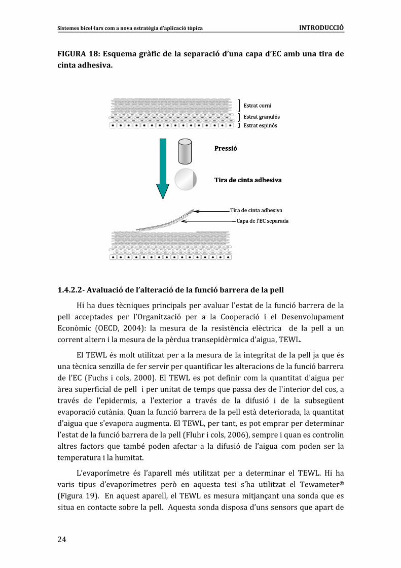
Sistemes�bicel·lars�com�a�nova�estratègia�d’aplicació�tòpica�����������������������������������������������������������������INTRODUCCIÓ�
�
#A�
FIGURA�18:�Esquema�gràfic�de�la�separació�d’una�capa�d’EC�amb�una�tira�de�cinta�adhesiva.�
�
�
Pressió
Tira�de�cinta�adhesiva
� ������ ������.�� ��
���������0!� ��������
!����� ���
!����� ������9�
!����� ��� �9�
Pressió
Tira�de�cinta�adhesiva
� ������ ������.�� ��
���������0!� ��������
!����� ���
!����� ������9�
!����� ��� �9�
�
�
�
�
�
�
�
�
�
�
�
1.4.2.2��Avaluació�de�l’alteració�de�la�funció�barrera�de�la�pell�
D �.���������� 5������ � ������������������S������������4�� 9�-�������������
����� ��������� ���� �0,���� �/� 9� ���� �� ��� ������� 9� � ��� ����������������
!��(� � 7,!���� #$$A8�� ��� ������� ��� ��� ��� ���� �� ����� �� � ��� ��� ����� �� ���
�������������� ������������������������������� ���� ���0� ������!T)6��
!���!T)�<��������� � �/������������������������� ����� ���������������H��5���<��
������� �����/ �������4������� ������5���� 4 ������������� ����������4�� 9�-�������
��� �0!�� 7�.�� � ����� #$$$86�!���!T)����������4 � �� ��� ���5���� �����S� ��������
����������4 ������������ ������� �������������5�����������������S ���� �������������
����<�� ��� �0�� ���� ��� �� �0�M��� ��� �� ����<�� ��� ��� � 4�� 9� � ��� ��� ��-���[����
������� 9����� �6���������4�� 9�-���������������������������� ����������5���� ����
�S� ����5����S����������������6�!���!T)������������������������������������ ����
�0������������4�� 9�-������������������7��.�� ������#$$E8��������� �5������������� ��
������� 4������ 5��� ���-<� ������ �4����� �� ��� � 4�� 9� ��� �0� ���� ��� ������ ���� ���
������������ ����.�� ���6�
)0������������� <�� �0�������� �<�� �� � �/��� ���� �� ������ ���� ��� �!T)6� D � .��
��� �� � ���� �0�������������� ���(� ��� �5������ ��� � �0.�� �� � �/��� ��� ��J������g�
7 �����>G86� �!���5�������������� ����!T)������������ �H��1��������������5������
� ����������������-����������6��*5������������� �������0������������5������������
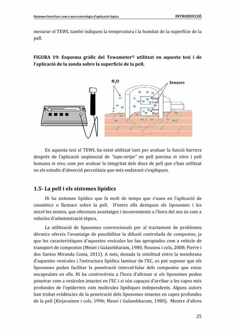
Sistemes�bicel·lars�com�a�nova�estratègia�d’aplicació�tòpica�����������������������������������������������������������������INTRODUCCIÓ�
�
#%
������������!T)����-<� �� 5������������������� ����.�� ���������������4� ��������
����6��
�
FIGURA� 19:� Esquema� gràfic� del� Tewameterg� utilitzat� en� aquesta� tesi� i� de�l’aplicació�de�la�sonda�sobre�la�superfície�de�la�pell.
�
�
�
�
�
�
�
�
!���5��������� �����!T)�.���������� � �/��� ����������������� ��� 4�� 9�-��������
�����<�� ��� �0��� � 9� ��5[�� ��� ��� “tape�strips”� ��� ����� ��� ��� in� vitro� � �����
.������ in�vivo�������������������� ����� ���������� �����������5����0.����� � �/���
������������ ���0�-��� 9�������� ��5����<������������0�M�� 5���6�
SensorsH2O SensorsH2O
�
1.5�� �La pell�i�els�sistemes�lipídics�
D � .�� � ������� � ��� �� 5��� 4�� ����� ��� ������ 5��� �0����� ��� �0��� � 9� ���
����� �� �� 4������ ��-��� ��� ����6� � �0������ ����� ����5���� ���� � �������� � ����
� ��B����� M�����5����4��� M�������������� � ������ ��������0.������������U�������
��.
�
�����0��� � ���� 9��(� �6�
)�� �� � �/� 9� ��� � �������� ����� ������ ���� ��� ���������� ��� ���-������
���� �� �4��� M� �0����������������� - � ���� ��� � 4�� 9� ������������� ���������� H��
5��� ���� ��������� 5�����0�5���������������� ���� 4�������� ������������. ������
����������������������7��/� � �=�����K.������>GC$W�"������ ������#$$CW�: ����� �
����3������� �������������#$>>86�*��<���������� ��� � � � ���������� ������-�����
�0�5���������������� � �0���������� � ��� �� ��� ������� �0!������������������5�������
� �������� ������ 4� � ���� ��� ������� 9� ������B������ ����� ��������� 5��� ������
����������� ��� ����6� D � .�� ��������� �� �� �0.���� �0�4 ����� � � ���� � �������� ������
����������������������� �����������0!�� �� ��9�����1����0��� -���������������<��
���4������ ��� �0�� ���� �� ��� ��������� � ��� 5���� �����������6� *������ �������
.������-����� ��� ���������������� 9������� �������� ������������������4������
�����������7X �H��� ���� ������>GGEW���/� � �=�����K.������>GC$86� ���������0�������
Sistemes�bicel·lars�com�a�nova�estratègia�d’aplicació�tòpica�����������������������������������������������������������������INTRODUCCIÓ�
�
#E�
����� ��.�������������5�������� ���������������������<������������0!��7X��� ��� �
�����>GG%W�)��.� ������>GG#86�)�� ����� 9� I��������� 9������� ����������-��0!��
.�� ������ �M�� ��� ���-<� � �H��1���� � 4������� � � ���� ��� ���� ����� ����� ���
�0����� 9��������� �����4����������������������0��.�� 9����� 4�� 9���0 ������� �� ��� ��
��6�7X �H��� ���� ������>GGGW�)9��/� ������#$$>86��!��5������-�����U�����5������
����� ��<��5���������� ���5���4����������� ������������� M�����5�����&������ ������
����0!�6�
!�� ���� U�� ��� >%� ��2��� ������� � ���� ��� ��������� � ��� 5���� �0.��� �����
�������������� ���� �� ���� ����� ������� ����� ������ ����� ����� � ��������
����� �����6�!������5����������������transfersomes��5����9��� �����������������
��4����-���� 7!�� ���.��-2� � ����� >GGGW� � �� � ����� #$$CW� ������� � ����� #$$A86�
*5������ ���������� ������������� 9� ���� ��� ����5�����������-����������� ���
4�� 9�-�����������0!�6�+��������� �����9������ethosomes��5��� ��������������������
���������� 9� 7* �- ����� ���� �����#$$%W�:��� ��� � �����#$$%86�)0�������<�����
� ���������5������������������ 9�� ��� ������0!��������� ������4�����M������������
�������� ��6�
+�� ������ ����� � ������� � ��� �� ������� ��� ���� ����������� �(� �� �9�� ����
� ������� ���� ��B����� M���� ��� �&���� ��� �6� *5�������� ��B���� �9�� ������ ���
�����4�����&��� ��� � �������� ��� ��� ���&��� ��� ���� ��� �� � �H��1���� � �� � � ��
� ��������� ���� � �� 9� ��-� � ���6�*5������ ���� ����� 4�� 5��� �5������ � ������� ���
������� �� ��� ���� �������� �� ����� ������������ ��� ���� ��������� ����� 9� �
������ 9���� ��������������� � ��� 5������� �������� ���. �� �/� 9������-���� �����
����<�� ��� �0!�� � �0�� ���� �� 7)9��/� � ����� #$$#86� )0�������� �� ��� �0U�� ���� ��B����
� M����4���������������� ��� ��� ���� ������-�������5��������� ������� ���������<��
��� ��� 5��� ���� � ��������� ����� ��B���� ������ ��� ��� ���� ��� ������� �� ����<�� �����
���� �� ������B�����������0!�� ��������������4���������� ������������� �� 9����������
��H��� ��� ����� �0� ���� ��� ���� ����� �<�� ���4������ ��� ��� ����6� !�� �������������
�0�5������ � ������� ����� ���� ����� ��-� ��� ������ �� ��� ���� ��� ��� ��� ��� �����
����� 96� *5������� ��������� ������ ��� ������� �4���� �� ����� ��-��� ��� �����
7)9�� � #/� � ���� $$>8������������- � �/������-�������� ��� �����������������4����6�
:��� ���� ������ ��������� 5���� ��� ����� 9� � ��� �� � ��� ������ � ���� ���
� ���� ����� ���� � ������� - ��B����� .��� ������ ����� ���� ���� ������� ����� ���
��� � ���� �(� 5���6� *5������� ����� �� ���� ���� ����1��� ���� ��������
� � � �/��������������������������� �������� ������ ��B���������� � �����(� 5���6�
�0�5������ ����� �� �0.�� ���(��5������� � �������- ��B����������� 4�� ����� �����
������� ������- � �/����� ��� ��� ����� �� ��� ������� ��� ��4��1� ��� ���� �����������
� ��� 5����������������������� � �� ������B���������� ����������<���M���������� ���
�����7���-���&������� ������#$$C-W����-���&������� ������#$$CW���������/� ������
#$>$86��
Sistemes�bicel·lars�com�a�nova�estratègia�d’aplicació�tòpica�����������������������������������������������������������������INTRODUCCIÓ�
�
#'�
!���5��������� ������������������� ����������������� �������- ��B����������
��. ���������0��� � 9��(� ������� � � ���� ��6�
�
1.6�Absorció�percutània��
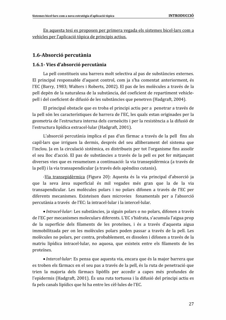
1.6.1��Vies�d’absorció�percutània�
)����������� ��� M�����-����������������� ���������������-���� ����M������6�
!�� �� � ���� ��������-��� �0�5����� �������� ��� H�� �0.�� �������� ����� �������� <��
�0!��7����2��>GC@W�T������� ���-������#$$#86�!����������������������������<��������
������������������������������������-���� ���������4 ������������� �������. ��&
����� �������4 �������� 4�� 9����������-���� ���5������������7D�����4���#$$A86�
!���� � �����-������5���������-������� � � ��� ������������������������<�����
���������9��������������� 5�������-�����������0!�������5������������� � �������������
������� ������0���������� ����������������( ��� ����������� ���� �������� 4�� 9����
�0����������� ��� ���M�����B������7D�����4���#$$>86�
)0�-��� 9�������� �� ��� ����������0��� 4������� ����<����� �������� � 4 �������
�� �B����� 5��� �� ����� ��� ���� ��� �����<�� ���� ���� ��� -�������� ���� � ������ 5���
�0 ����6�;�������� ���� 9�� ���� ������� ��� -�� M����������0����� ����4 �������� ��
��� ���� �����0� 96� !��������� ��-���� ��� �� ����<����� �������� ������� 4���� �H��1����
� �������� ���5������������ M�������� ��� 9������ ��������� ���� ��7������<�����
������ � �86��8� ����� ������������ �����7������<������������ M�����
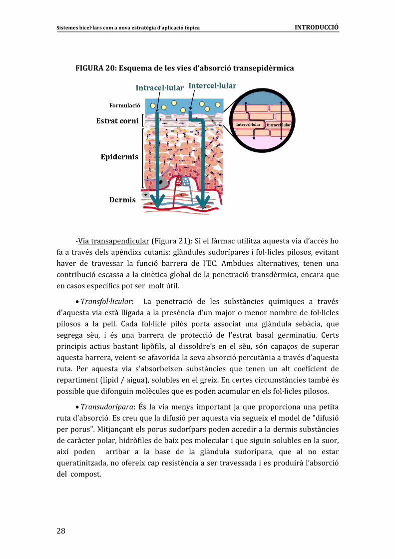
&? �� ������� ���� �� 7 ����� #$8�� *5������ <�� ��� � �� �� � ���� �0�-��� 9� H��
5��� ��� ����� ����� �����4 ��� <�� � �� �������� �<�� ����� 5��� ��� ��� ��� � ��
���������� ����6� )��� ��������� ������� � ��� ������� � 4����� �� ����<�� ��� �0!�� ����
� 4������� ���� ����6� !M ��� M��� ����� � ��� ��� � 4����������� ���� �� �0�-��� 9�
������� ��������<�������0!������ ������B������ ���� ������B�����6��
� Intracel·lular��)�����-���� ����H��� �� ����������������������� 4�����������<��
����0!���������� ���������������� 4������6�)S!���0. ��������0��������0� ���������
��� ��� �����4� �� ����� 4 �������� ��� ���� �����L����� � <�� �� ����<�� �0�5������ � ����
���- � �/���� ���� ��� ������������� ������� ������ ������� �� ����<�� ��� ��� ����6� )���
����������������������������������-�-������������ ������� �� 4�����������<��������
���� �� � ��� �� ������B������� ��� �5������ 5��� �M ��� M� ������ ���� 4 �������� ��� ����
�����L���6��
� Intercel·lular��!��������5����5������� ���������5���<�������H���-�������5���
������-�������4��������������������������<��������������<�������������������� 9�5���
�� ��� ��� ��H�� �� ����� 4������ � �(4 ��� ���� ��� �� �� ����� �<�� ���4������ ���
�S�� ���� ��7D�����4���#$$>86�\�������������������� ����� 4�� 9������� � � ��� �����
4�������������� ��� ��5���. �.��������������B����������0!�6�
Sistemes�bicel·lars�com�a�nova�estratègia�d’aplicació�tòpica�����������������������������������������������������������������INTRODUCCIÓ�
�
#C�
�
FIGURA�20:�Esquema�de�les�vies�d’absorció�transepidèrmica�
�
�
�
�
�
�
�
�
�
�
&? ������������ �����7 �����#>8��3 ����4������� � �/���5������� ���0�<��.��
4��������<������������ M������ ������������������������� �4��B� ����� ��������� �����
.����� ��� ���������� ��� 4�� 9� -������� ��� �0!�6� *�-����� �������� ����� ������ ����
���� -� 9���������� ��� ��� �����-���������������� 9���������� ���������5���
�������������4 ����������������U� �6��
� Transfol·licular�� � )�� ������� 9� ��� ���� ��-���� ��� 5��� 5���� �� ����<��
�0�5������� ������� �� ������� ��������� ���0�����H��������������-������ 4��B� ����
� ������ �� ��� ����6� ����� 4��B� ��� � �9�� ������ ���� ��� ���� ��������� ��-� ��� 5���
�������� ����� � <�� ���� -������� ��� ����� 9� ��� �0������� -����� ���� ��� �6� ������
�� � � �� �� ��� -������� � �(4 ���� ��� � �������0�� ��� ��� ����� �9�� ���1��� ��� ��������
�5������-���������� ���&����4���� ������������-��� 9�������� ��������<���0�5������
����6� :��� �5������ � �� �0�-���-� M��� ��-���� ��� 5��� ������ ��� ���� ��4 ���� ���
������ �����7��� ��I�� ���8������-������������� M6�!�������� ������� ������-<�<��
���� -���5���� 4���� �����������5���������������������������4��B� ����� �����6�
� Transudorípara�� \�� ��� � �����2�� ��������� H�� 5��� ������ ���� ���� ��� ���
������S�-��� 96�!������5������� 4�� 9������5������� ������� M�������������f� 4�� 9�
���������f6�� �H��1���������������������������������� ����������� ����-���� ���
�����������������. ��(4 �������-� M�������������� �5���� �� ������-����������������
� M�� ������ � ��� -��� �� ��� -���� ��� ��� ��������� ������������ 5��� ��� ��� ������
5����� � �/���������4��� M������� ���� ������������������� ��������� ����0�-��� 9�
�����������6��
�
�
Sistemes�bicel·lars�com�a�nova�estratègia�d’aplicació�tòpica�����������������������������������������������������������������INTRODUCCIÓ�
�
#G
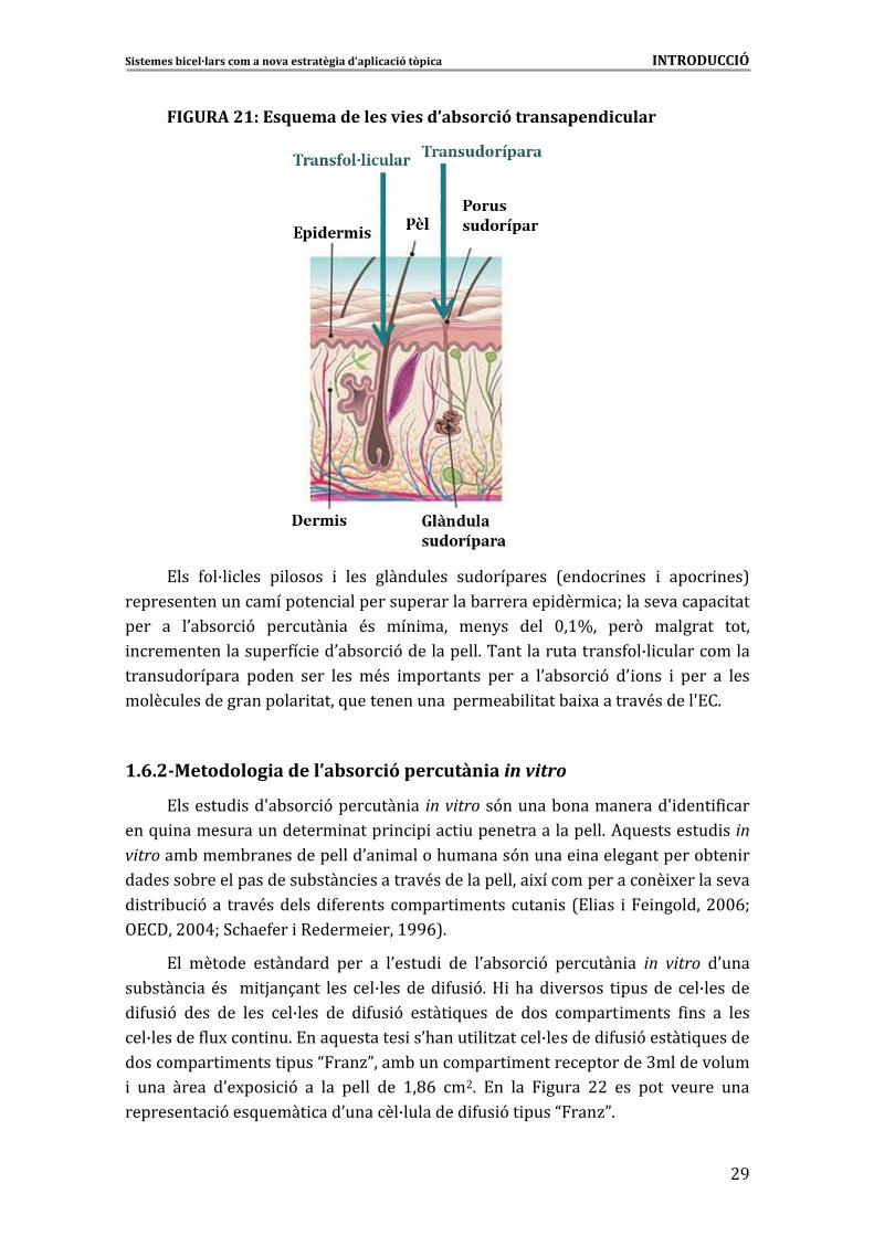
FIGURA�21:�Esquema�de�les�vies�d’absorció�transapendicular�
�
�
�
�
�
�
�
�
�
�
�
�
!��� 4��B� ���� � ������ � ���� ���������� ������������ 7����� ���� � ���� ���8�
������������������������ ������������������-��������� ���� �W������������ ����
���� �� �0�-��� 9� ������� �� <�� ��� ���� ���2�� ���� $�>d�� ���(� �������� �����
������������������4� ���0�-��� 9�����������6�������������������4��B� �����������
��������������� ������ ���� ���� �<�� ���������� ���� �� �0�-��� 9� �0 ���� � ���� �� �����
���������������������� �����5��������������������- � ����-� M��������<������S!�6�
�
1.6.2 rcutàn�Metodologia�de�l’absorció�pe ia�in�vitro�
!�������� ���S�-��� 9�������� �� in�vitro��9������-������������S ���� 4 ���
���5� ������������������� ������ � � ��� �������������������6�*5����������� ��in�vitro���-����-���������������0�� ������.�������9������� ����������������-��� ��
��������-���������������-���� ���������<��������������� M�������������� M�����������
� ��� -� 9��� ����<�������� 4������� ������ ���������� �� 7!� ��� �� �������#$$EW�
,!�� $ � �
�
��# $AW�3.��4��� �������� � ��>GGE86
!�� ������� ���������� ���� �� �0����� � ��� �0�-��� 9� ������� �� in� vitro� �0����
��-���� ��<�� �� �H��1���� ���� ��B�������� 4�� 96�D �.��� ������� � ������� ��B�������
� 4�� 9� ���� ��� ���� ��B���� ��� � 4�� 9� ����� 5���� ��� ���� ������ ������ 4 ��� �� ����
��B�������4��M���� ��6�!���5��������� ��0.����� � �/�����B�������� 4�� 9������ 5�������
���������� ������� ����N���/P����-���������� ����������������@������������
� ���� ����� �0�M��� 9� �� ��� ����� ��� >�CE� �#6� !�� ��� ����� ##� ��� ���� ������ ����
���������� 9���5����� ���0������B��������� 4�� 9�� ����N���/P6�
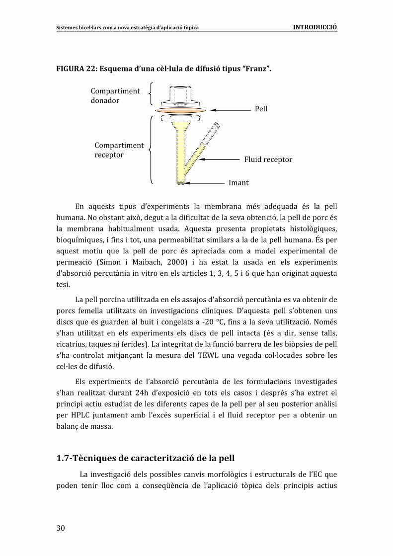
Sistemes�bicel·lars�com�a�nova�estratègia�d’aplicació�tòpica�����������������������������������������������������������������INTRODUCCIÓ�
�
@$�
�
FIGURA�22:�Esquema�d’una�cèl·lula�de�difusió�tipus�“Franz”.�
������� �����������
������� �����������
:���
�� ���������
����
������� �����������
������� �����������
:���
�� ���������
�����
!�� �5������ � ���� �0�M��� ������ ��� ���-����� �<�� ���5����� <�� ��� �����
.�����6�"���-������� M(�������������� 4 ������������������-��� 9�����������������<��
��� ���-����� .�- ��������� �����6� *5������ ��������� ���� ������ . ����(� 5�����
- �5��� 5����� �4 ��� ����������������- � ����� � ������������� ��������.�����6�\������
�5����� ��� �� 5��� ��� ����� ��� ���� <�� ���� ���� ��� �� ������ �M��� ������� ���
������ 9� 73 ���� � �� -�.�� #$$$8� � .�� ������ ��� ������ ��� ���� �M��� ������
�0�-��� 9�������� �� ��� �������������� ����>��@��A��%� �E�5���.����� � �����5������
��� 6�
)���������� ����� � �/���������������H����S�-��� 9�������� ���������-��� �����
����� 4������� �� � �/���� ��� ����� �� ���� ��� 5���6� �0�5������ ����� �0�-������ ����
� ���5�����������������-� �� � ����������� &#$�]��� 4 ����� ���������� � �/� 96�"��<��
�0.��� �� � �/��� ��� ���� �M��� ������ ���� � ��� ��� ����� ������ 7<�� �� � ��� ������ �������
��� ������5����� �4�� ���86�)�� ����� ����������4�� 9�-��������������- (�� �����������
�0.�� ��������� � �H��1���� ��� ������� ���� �!T)� ���� ������� ��B������� ��-��� ����
��B�������� 4�� 96�
!��� �M��� ������ ��� �0�-��� 9� ������� �� ��� ���� 4������ ���� ����� ������
�0.��� ���� �/��� ������� #A.� �0�M��� 9� ��� ����� ���� ����� � �����<�� �0.�� �M����� ���
�� � � ��� ������� ����������� 4���������������������������������������� ������� � �
���� D:)�� H��������� ��-� �0�M<�� �����4 ��� � ��� 4�� �� �������� ���� �� �-��� �� ���
-����1���������6��
�
1 cniques�de�caracterització�de�la�pell�������
� )�� ����� �� 9���������� -������� �����4��(� �� ���������������� �0!��5���
������ ��� �� ���� ��� �� ����5[�� �� ��� �0��� � 9� �(� �� ����� �� � � �� �� ���
.7�Tè
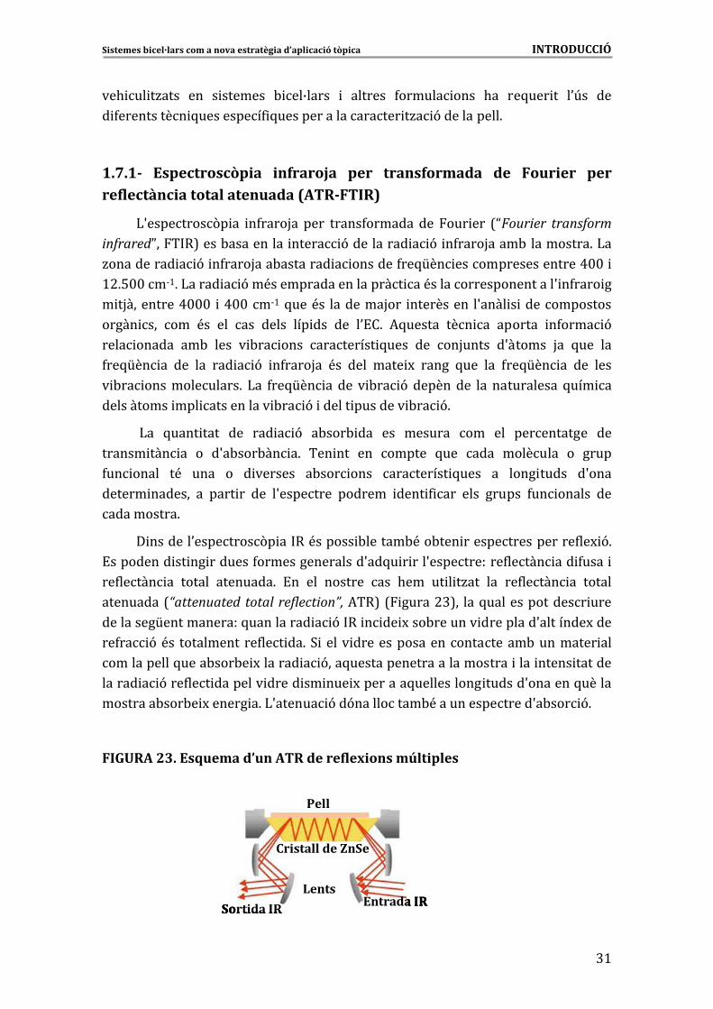
Sistemes�bicel·lars�com�a�nova�estratègia�d’aplicació�tòpica�����������������������������������������������������������������INTRODUCCIÓ�
�
@>
��. �� �/���� ��� � ������� - ��B����� � ������� 4������ ���� .�� ��5��� �� �0U�� ���
� 4���������� 5���������4 5������������������� �/� 9�����������6�
�
1.7.1�� Espectroscòpia� infraroja� per� transformada� de� Fourier� per�reflectància�total�atenuada�(ATR�FTIR)�
)S��������(� �� �4����H�� ���� �����4���������� ��� ��� 7NFourier� transform�infraredP��� �8����-���������� ����� 9���������� � 9� �4����H����-� ���������6�)��
/���������� � 9� �4����H���-�������� � �������4��5[�� ������������������A$$� �
>#6%$$��&>6�)����� � 9��<�������������������� ��<��������������������S �4���� ��
� �H���������A$$$� �A$$��&>�5���<�� ��������H��� ���������� �S���� � �������������
����� ��� ��� <�� ��� ��� ����� ��� ��� ��� �0!�6� *5������ ��� �� ������� �4���� 9�
���� ������ ��-� ���� � -�� ���� ��������� 5���� ��� ��H����� �S������ H�� 5��� ���
4��5[�� �� ��� ��� ��� � 9� �4����H�� <�� ���� ���� M� ����� 5��� ��� 4��5[�� �� ��� ����
� -�� �������������6� )�� 4��5[�� �� ��� � -�� 9� ��������� ��� ����������� 5��� ��
������ � ������ ��� ������� ��� -�� 9� ������ �������� -�� 96�
�)�� 5���� ���� ��� ��� � 9� �-���- ��� ��� ������� ��� ��� ����������� ���
������ ��� �� �� �S�-���-�� �6� ��� ��� ��� ������ 5��� ���� �������� �� �����
4�� ����� �<� ���� �� � ������� �-��� ���� ��������� 5���� �� ���� ����� �S����
������ ������� �� ���� �� ��� �S�������� ������� ���� 4 ��� ���� ������ 4�� ������ ���
����������6�
� �������0��������(� �� ��<������ -������-<��-��� �����������������4��M 96�
!��������� �� �� �������4����������������S��5� � ���S�����������4����� ��� 4���� �
��4����� �� ������ ��������6� !�� ��� ������� ��� .��� �� � �/��� ��� ��4����� �� ������
���������7“attenuated� total� reflection”,�*��8�7 �����#@8�� ���5��������������� ����
���������[������������5���������� � 9� �� � �� M���-������� ���������S��������M����
��4�� 9�<�� ������������4��� ��6�3 � ���� ������������������������-��������� ���
�����������5����-���-� M������� � 9���5�������������������������� ���� ����� �������
������ � 9���4��� �������� ����� �� ��� M��������5����������� ������S�������5������
��������-���-� M������ �6�)S������ 9��9����������-<���������������S�-��� 96��
�
FIGURA�23.�Esquema�d’un�ATR�de�reflexions�múltiples�
�
�
Entrada�IRSortida IR
Lents
Cristall de�ZnSe
Pell
Entrada�IRSortida IR
Lents
Cristall de�ZnSe
Entrada�IRSortida IR
Lents
Cristall de�ZnSe
Pell
�
�
�
�
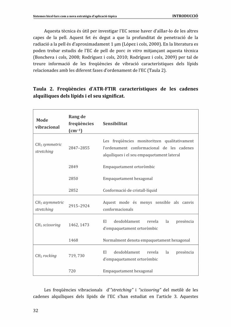
Sistemes�bicel·lars�com�a�nova�estratègia�d’aplicació�tòpica�����������������������������������������������������������������INTRODUCCIÓ�
�
@#�
*5��������� ��<��U� ������ ����� �����S!��������.������0�L����&�����������������
����� ��� ��� ����6� *5����� 4��� <�� ������ �� 5��� ��� ���4��� ���� ��� ������� 9� ��� ���
��� � 9�����������<���0����M ���������>�h��7)9��/� ������#$$$86�!������ ������������
������ ���-��� ����� �� ��� �0!�� ��� ����� ��� ���� in� vitro� � �H��1���� �5������ ��� ��
7���.���� ������#$$CW���������/� ������#$>$W���������/� ������#$$G8������������
������� �4���� 9� ��� ���� 4��5[�� ��� ��� � -�� 9� ��������� 5���� ����� ��� ���
���� ���������-������ 4������4������0���������������0!��7������#86��
�
Taula� 2.� Freqüències� d'ATR�FTIR� característiques� de� les� cadenes�alquíliques�dels�lípids�i�el�seu�significat.�
�
�Mode�������
vibracional�
Rang�de�
freqüències�
(cm�1)�
Sensibilitat�
�D#�symmetric�
stretching�#CA'i#C%%�
)��� 4��5[�� ��� ��� ��� �/��� 5��� ��� �������
�0����������� ��4���� ����� ��� ���� �������
����������������5��� 5���� ������������5���
� #CAG� !���5��������������(�- �
� #C%$� !���5���������.�M�������
� #C%#� ���4���� 9����� �����&��5� ��
�D#�asymmetric�
stretching�#G>%i#G#A�
*5����� ����� <�� ���2�� ���� -��� ���� ��� ��
��4���� ������
�D#�scissoring� >AE#��>A'@�!�� �����-������� ������� ��� ������ ��
�0����5��������������(�- �
� >AEC� "���������������������5���� �� M��������� �.�
�D#�rocking� '>G��'@$�!�� �����-������� ������� ��� ������ ��
��0����5��������������(�-
� '#$� !���5���������.�M�������
�
)��� 4��5[�� ��� � -�� ������ � �0”stretching”� � “scissoring”� ���� ��� ��� ��� ����
������� ��5��� 5���� ����� ��� ��� ��� �0!�� �0.��� ����� ��� ��� �0��� ��� @6� *5�������
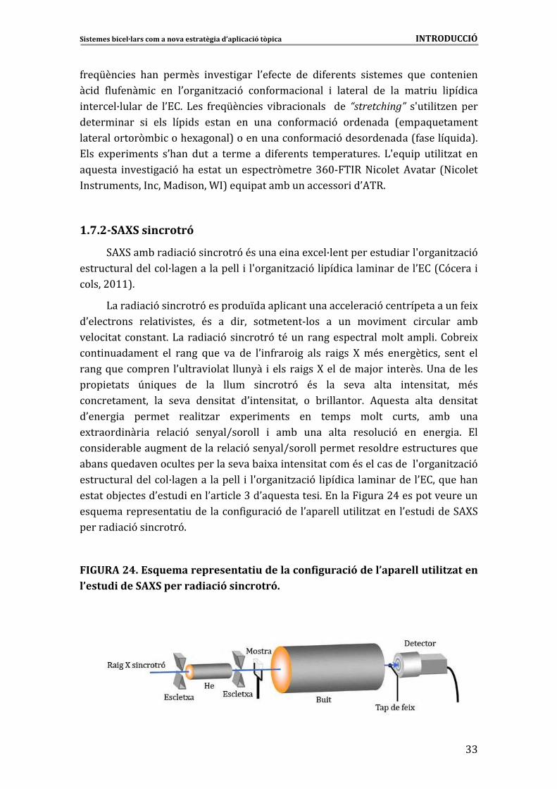
Sistemes�bicel·lars�com�a�nova�estratègia�d’aplicació�tòpica�����������������������������������������������������������������INTRODUCCIÓ�
�
@@
4��5[�� ��� .��� ������� ����� ���� �0�4���� ��� � 4������� � ������� 5��� ����� ���
� �� 4��4���� � ��� �0����� �/� 9� ��4���� ����� � �������� ��� ��� ���� �� � ��� ��
������B������ ��� �0!�6� )��� 4��5[�� ��� � -�� ������ � ��� “stretching”� �S�� � �/��� ����
������ ���� � � ���� ��� ��� ������ ��� ���� ��4���� 9� ��������� 7����5���������
�������������(�- ���.�M������8������������4���� 9�������������74������5� ��86�
!��� �M��� ������ �0.��� ���� �� ������ �� � 4������� ������������6� )S�5� �� �� � �/��� ���
�5������ ����� �� 9�.�� ������ ��� ������(������@E$&� ��" �����*������ 7" �����
������������ ������ �����T 8��5� ������-���������� ��0*��6��
�
1.7.2�SAXS�sincrotró�
3*F3���-���� � 9�� �����9�<������� ����M��B�������������� ����S����� �/� 9�
�����������������B���������������� � �S����� �/� 9�� ��� ����� ��������0!��7�9���� �
�����#$>>86�
)����� � 9�� �����9���������L������ �������������� 9����������������4� M�
�0��������� ����� � ������ <�� �� � ��� ���������&���� �� ��� ��� ����� ������ ��-�
���� ���� �������6�)�� ��� � 9� � �����9� �<���� ������������������� ���� 6���-�� M�
��� ���������� ��� ����� 5��� ��� ��� �0 �4���� �� ���� �� ��� F� �<�� ������� ��� ����� ���
�����5���������� �0������ ����� ����2�� ������� ���F���������H��� ������6�+������ ����
���� ������ U� 5���� ��� ��� ����� � �����9� <�� ��� ����� ����� ����� ����� �<��
������������ ��� ����� ���� ���� �0 ����� ����� �� -� �������6� *5������ ����� ���� ����
�0����� �� ������� ���� �/��� �M��� ������ ��� ������ ����� ������ ��-� ����
�M������ ��� �� ���� 9� ���2��I������� � ��-� ���� ����� ������ 9� ��� ����� �6� !��
��� ����-��������������������� 9����2��I����������������������������������5���
�-����5���������������������������-� M�� ����� �������<�������������S����� �/� 9�
�����������������B�������� �������� � �S����� �/� 9� � ��� ����� ��������0!���5���.���
�������-H������0����� �����0��� ���@��0�5��������� 6�!����� �����#A�����������������
��5���������������� ����������4 ���� 9�����0���������� � �/�������0����� ����3*F3�
������� � 9�� �����96���
�
FIGURA�24.�Esquema�representatiu�de�la�configuració�de�l’aparell�utilitzat�en�l’estudi�de�SAXS�per�radiació�sincrotró.�
�
�

Sistemes�bicel·lars�com�a�nova�estratègia�d’aplicació�tòpica�����������������������������������������������������������������INTRODUCCIÓ�
�
@A�
!��4������������3*F3�H���0.���M�� �������0��������>6@6A��0�5������ ������ 96��
������ ���� �������� ��� 3*F3� ���� ��� � 9� � �����9� ��� ���� ���� �/��� ��
������������ ��- ���� ��� ��� ��� �� ��� ����� �����2���� 7��>E8� ��� ��� �����6�� 9�
!������� ��� ��� � 9� 3 �����9� 7European� Synchrotron� Radiation� Facility�� !3���
=����-�������1�8�������0��� ���� ����� ��������6����1� �����.� ����������6�*���
)�-�����6���
1.7.3
� �
�
� ��Criosubstitució� aplicada a� la� microscòpia electrònica� de�transmissió�(FSTEM).�
*5������ ��� �� ����� ��� ���<�� ��� 4 M� 9� 5��� ��� ��� ��� ����� 4 M� 9�
5��� ��� ��� ���<�� ��� � �4 M� 9� ���� �� �0��� ��� � ���-�� �� 9� � ��� ������ ���
���� 9������� M ������������ ��6�*5����������������9��������� ������������������
� ���� �/� 9���� ��������������������� �������� ����� ��� ����M�����B���������� �0!�6�)���
������� �M��� �������� ���� ���� ���� �� ��� ������� 9� ��� ���� �������� .��� � ���� ����
���[������
& :��<�� ��� 4 M� 9� 5��� ��� ��� 4�� ��� �� ��� ����������. �� ��� 5���� 4 M� ����
�����L��������������6�
& �:����4 M� 9���0 ������� M�����(M ���������� ������������5���������� �������0!��
5��� ������������� �������� ������������ ����� ������(� 6�
& ��� �4 M� 9� ��� � ������� ��5� �� 7&>GE� ]�8� ������ ��� ��2����-��K� 7)� ���
? ���86���
� � � 9
8 8 � �
& �� ���-�� � 9���� 4��������� ������������� �/��������-�� � �7“Automatic�Freeze� Substitution”�� *3 � 7 ����� #% 6� *5����� �� <�� ��� ��� M� ��� �0 ����� � �
7��-� � �� 98���� �0� ����������� ��������������������� 6�!���5����� ������ ��������
����� � ������� ��� ���� ��� �������� � ���-<� ��� ��� ������� ����(M �� �0��� � 7������
���������������8������������5���������� ������������������������������������������
���� � � � ���
� � �
� �������� ��� ������ �� �-������� � �!�6� )��� �������� ��� �� M �� ���
� ���-�� ��� M���� &G$�]������� �AC�.6�
�2�� D�#$�
7>$$d
& ���� 9�� ��� �� M �� �0 ����� ��� ���� ��� ���� ��� �5����� ��� )�J
8���������#A.6��
& :�� ��� �/� 9� ��� ��� ��� ���� ���� �������� ��� ���� �����4�� �� �� ��� �������
����� ���)�J �2��� � ������� ��-����������AC�.������� &%$� ]�� ����� ��� � 9�+?*��
������������������� ��� �/� 96��
!��������������4 ��������� ��������� ���� �/� 9����� ����� ������(� ���������
���������������������� ������7+�������+����)� ���? �����O���� �86��
�����<�� ��� ��� ������� 9� ��� ������������� �5������� ��� ���� � ���� �/��� ��� ���
�!��D ��. �E$$6�
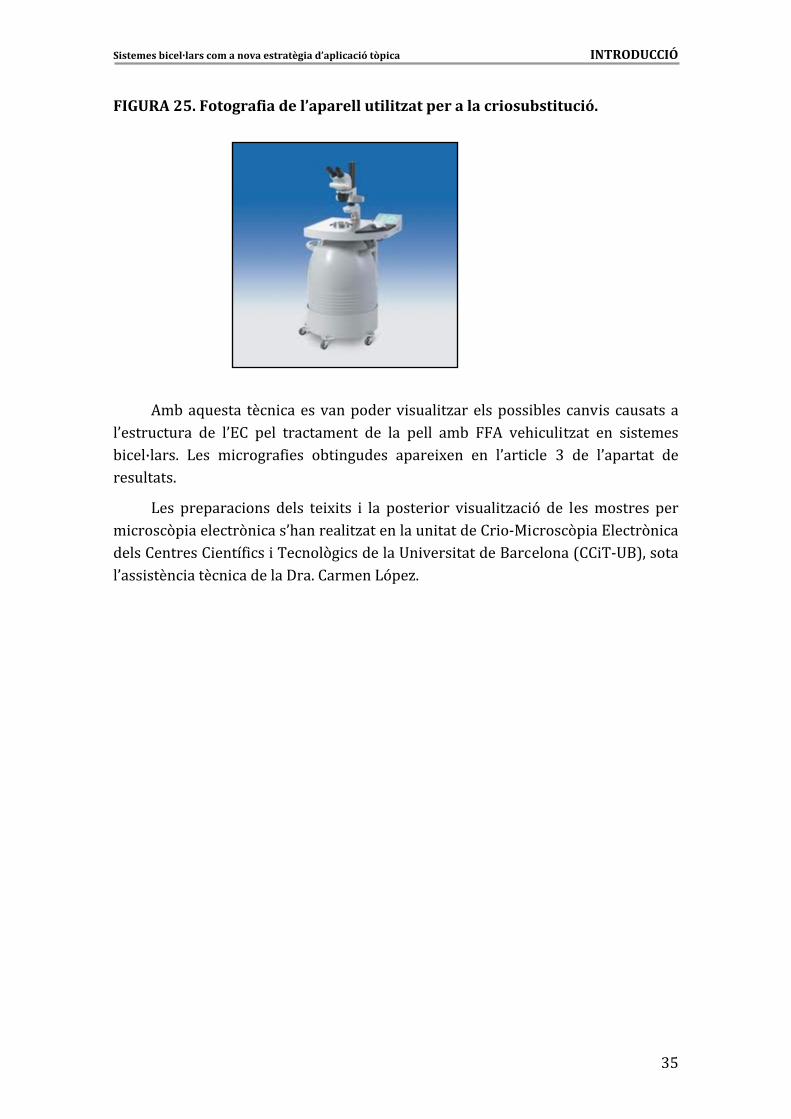
Sistemes�bicel·lars�com�a�nova�estratègia�d’aplicació�tòpica�����������������������������������������������������������������INTRODUCCIÓ�
�
@%
FIGURA�25.�Fotografia�de�l’aparell�utilitzat�per�a�la�criosubstitució.�
�
�
�
�
�
�
�
�
�
*�-��5��� ���������� -���� ��� �� ���������
�0���� ����� ��� �0!�� ���� ���������� ��� ��� ����� ��-� *� ��. �� �/��� ��� � �������
- ��B����6� )��� � �����4 ��� �-� ������� ����� M��� ��� �0��� ��� @� ��� �0�������� ���
���������6�
)��� ������� ���� ����� �� M ��� � ��� ������ ��� � ���� �/� 9� ��� ������������ ����
� ���(� �������(� ���0.������� �/����������� ��������� �&� ���(� ��!����(� ��
������ ������� ����4 �� ������(� ��������+� ���� ����������������7�� �&+�8�������
�0��� ���� ����� �����������6��������)9��/6�
��� ��� ���������������� ���� �/�
�
�
�
�
�
�
�
�
�
�
�
�
�
�
�
�

@E�

2.�OBJECTIUS�

Sistemes�bicel·lars�com�a�nova�estratègia�d’aplicació�tòpica������������������������������������������������������������������������OBJECTIUS�
@G�
2.�OBJECTIUS��!�� �� � ���� �-H�� �� ��� ��� ����� �� 9� ���� �/���� ��� ��� ���� �0�5������ ��� � .��
��� �� �� ��� ��� M��� ���� ���� - � ����� ������ ��� �0U�� ����� � ������� - ��B����� ��� ���
�. �� �/� 9������ � � ���� ������ �����(� �����6��
�
:��� �� � M(�� �0.��� � ����2��� ��� ���� �� �0����� �� ��-� ��� 4 ��� ���� �0������� �� ����
��[������-H�� ��������4 ����
�
� ������� �/��� �0���������� ����� � ������� - ��B������ ��� ����� ������ � ����
�0��-��������� M���������������� �����0 ������� 9������ � � ���� ��6�:���
�� ������� �� � M(�� �0.�� ������H��� �0U�� ��� ��� 5���� ������������� �0���� �
�������6�*5���������� 5���������������� M���������������2������������������
���� �������������(� 5��������� ����� ����� ��� 5���� �������� ��5������ ����
����0����� �/� 9��������- ��B������� ������� 9������ � � ���� ��6�
�
� ���������� ��� ����L���� ����� � ������� - ��B����� ���� �� ��. �� �/��� �� � � ��
�� ����� ����<����� ����� 4������� �������� ��������5�����9����� ���� �(� �����6�
:���������� ���5������-H�� ���0.��������H����
&)������ �/� 9��0����� ���0�-��� 9�������� ��� in�vitro�5�����������������-� ��
� ��� ����4 � �����-����0 ������������� �������- ��B������������������������� ��
�0��� -������������� � � ���� �����-����������6��
&)0����� ��������� 5������������(� 5���� ����� ���(� ���������� ����� ����
�0�5������� �������- ��B�������-�������� �������0!�6�
�
� � ����2��� ��� ������ in� vitro� ���� �� � ������ ��� ������������ �0���� �����
�����(� ����-���������4�� 9�-������������ ���������-����4 ��� ���������� M���
�0�4���������� �������- ��B�������-����5������ ������������6�
�
*5������ �-H�� ��� �0.��� �������� ������� �� � �H��1���� ��� ���� �/� 9� �0���� ��� ��
�0 ����� �� ���� ��-� ����� ��� ��� ���� ����4 �� 5��� �0 ������� ��� ��� �� 9� ���
��������6��
�
!���0*�� ���>������������ ������������ �/� 9�4�� �&5��� �������� �������- ��B�����
� ��� ����� ��� ���� ��� ��. �� �/� 9� ���� ��!*����� �� ��� ����� ��� � 9� �(� ��� � M��
��� �0U�� ����� � ������� - ��B����� ������ 4����� ��� �� ����� ������ ��� �0�-��� 9�
������� ���0�������� 9��5����������!*6��*���� ��� 9��������*�� ����#� �@�������
Sistemes�bicel·lars�com�a�nova�estratègia�d’aplicació�tòpica������������������������������������������������������������������������OBJECTIUS�
�
A$�
����� ������� ���� 9��0���������4���������*���������� �������- ��B����6�:������ M(�
���� ����� �� ��� ���� ������� ��� ��� ������ �/� 9� 4�� �&5��� �� ����� � �������
- ��B����� ��� *� � ��� ����� ��� � 9� �(� ��� H��������� ��-� �0����� � ��� �0�4����
�0�5������� �������- ��B������������� ��� ������ �0!�6�:����� ���������� �0*�� ���A����
��� 4��� ��� ����� � ������� �� ��� ��� ��� ���� ���������� ����� � ������� - ��B����� ���
�0�-��� 9�������� ��������!*� �����*6�!���0*�� ���%�������������H������������in�
vitro��������������(� ����-� ��� 4 ��� ����������� �/�������� ���0�-��� 9�������� ��
������ �����<������ ����6� �������������0*�� ���E������� ����� ����������� - � ����
���5�������� �������- ��B�����5�������������!*����� ����� ���4�����-���4 �����
���� �� �������� ��� 4�� 9� -������� � �� ��L��� �0���� ����� �����(� �6� ������� M�
�0����������-���4 �����0� 9������������0�5������� ����������������� �������M ������
� ����� � ���� 6� *� ��� ��� 9�� �0 ������� ���� ���������� ��� ���� ��������� ��-� ���
-������������������6�

3.�RESULTATS������������������������

Sistemes�bicel·lars�com�a�nova�estratègia�d’aplicació�tòpica����������������������������������������������������������������������RESULTATS�
A@�
3.�RESULTATS�!��� ���������� 5��� .��� ������ ���� �� �5������ ��� � ��� ��4���� M��� ��� ���� � �� ��� ����
����4 ��5������������������� ��� 96���������0�����H��.�����������-� ���� ����������
�����������<�������� � 96� �*5��������� �����������������������2�����0���-�����
��������������6�
������ �����9���������[������
�
!
�
Article�1.�Bicellar�systems�for�in�vitro�percutaneous�absorption�of�diclofenac6�
)6���- ����6�*�������=6���������/��)6����-���&��������)6������.��*6���������/���;6)6�
:
�
����� �,6�)9��/6�7#$>$8�Int.�J.�Pharm.,�#$>$��@CE��>$C&>>@6�
Article� 2.� Structural� effects� of� flufenamic� acid� in� DPPC/DHPC� bicellar�systems.�)6���- ���=6���������/���6�*��������6�)9��/& ���� �����6��9�����)6������.��
6���������/���;6�)6�:����� �,6�)9��/6�Soft�Matter��#$>>��'��CACC&CAG'6�*
�
Article�3.��Bicellar�systems�as�new�delivery�strategy�for�topical�application�of�flufenamic�acid6�)6���- ����6�*�������=6���������/���6��9������6�)9��/& ���� ����)6�
����.���*6���������/���;6�)6�:����� �,6�)9��/6�J.�Phys.�Chem.�B&������� � 96��
�
Article� 4.� Bicellar� systems� as� a� new� colloidal� delivery� strategy� for� skin.� )6�
�- ��� =6� ��������/�� )6� ���-���&�������� �6� *������� �6� �9����� *6� ��� ��� ��/��� ;6)6�
����� �)9��/6�Colloid�Sur.f�B��Biointerfaces,�#$>#��G#��@##i�@#E6�
�
:
�
Article� 5.� Barrier� function� of� intact� and� impaired� skin:� Percutaneous�penetration� of� caffeine� and� salicylic� acid.� )6� ��- ��� �6� *������� ,6� )9��/�� =6�
��������/��)6������.��;6�"���� �6�*6���������/������;6�)6�:����6�Int�J�Dermatol,�#$>>��
$��CC>iCCG6�%
�
Article�6.��Bicellar�systems�as�vehicle�for�treatment�of�impaired�skin.�)6���- ���
6�*�������=6���������/���6��9�����)6������.��;6�"���� �6�*6���������/���;6�)6�:����� �
6�)9��/.�J.�Control.�Release�&������� � 96�
�
,
�
�
�
�
�
�
�
�

AA�
Sistemes�bicel·lars�com�a�nova�estratègia�d’aplicació�tòpica����������������������������������������������������������������������RESULTATS�
A%�
Article�1��
Bicellar�systems�for�in�vitro�percutaneous�absorption�of�diclofenac��
Int.�J.�Pharm.,�#$>$��@CE��>$C&>>@��
!���5��������-� � 9��0������� �0�4�������� 4�������� �������- ��B�������-���
�S�-��� 9�������� ���������!*��� � �/����������4��������� 4������6�!������� ����
������� 4����������� ��������������� �������- ��B����� 4�������������:�I�D:�� �
�::�I�D:��� ������ ��� 9����������� �������������� ���in�vitro6�!���������������
�������� ������ ���������-����� � �������- ��B����� ������ 4������-������� �0��� � 9�
�0�������� 9��5�����������!*6��
:��� ���������������� �/� 9������� �������- ��B����������)3������������5���
��� ������� �� ��� ��������� � �� ��� M� 5���� ��� ��!*� <�� ����������� ����� ��� ����
� ������� ��:�I�D:�� ��� ��� ���� ��� �::�I�D:�6� ���-<�� ���� � �����4 ���
�-� �������������2�&�!�����������������5������4������������������� ����������
�::�I�D:�6��
!��� � ����� �� �0�-��� 9� ������� �� ���� ��������� ���������� ������� 9� ����
��!*�5������� 4����������� ����������- ��B���������:�� ��::��5���5����������
��� ��� ��� ���� ���� 9� �5����6� *5����� �4���� <�� ������ ���� -������� �� ���� �����
� � ����������� �������- ��B������������������� ������� 9�������!*6�)0�-��� 9�
������� �������!*��������������� ����������������-������ �������- ��B�����������
4��������������������������� 9���-� �0�-��� 9������!*���-��� �������� ������
������6�)���- ��B������������!*������ ���������������������� �/� 9�������4�� 9�
-������� ��� �0!��� ��� 5��� 4� � ��� �� ��� ������� 9� ������� �� ���� ��!*� ��� ���
������ ������6��
* M��� ��������� ����� ����� ����������� 4 � �5��� �� � ��� ���� ��� ����
�0��� � 9������� �������- ��B�������������� ���4���������� ����������������������
0�-��� 9� ������� �6�*5������ ���� �4����� ������ ���� �������� ��� ����������������
����� � �/��������4���������� � �����(� 5�����0��� � ���� 9����4�����6�
�
�
�
�
�
�
�
�
�
�
�
�

AE�
International Journal of Pharmaceutics 386 (2010) 108–113
Contents lists available at ScienceDirect
International Journal of Pharmaceutics
journa l homepage: www.e lsev ier .com/ locate / i jpharm
Bicellar systems for in vitro percutaneous absorption of diclofenac
L. Rubio ∗, C. Alonso, G. Rodríguez, L. Barbosa-Barros, L. Coderch, A. De la Maza, J.L. Parra, O. López
I.Q.A.C.-C.S.I.C., C/Jordi Girona 18-26, 08034 Barcelona, Spain
a r t i c l e i n f o
Article history:
Received 7 July 2009
Received in revised form 4 November 2009
Accepted 6 November 2009
Available online 14 November 2009
Keywords:
Bicellar systems
Diclofenac
In vitro percutaneous absorption
Vehicle
Drug delivery system
a b s t r a c t
This work evaluates the effect of different bicellar systems on the percutaneous absorption of diclofenac
diethylamine (DDEA) using two different approaches. In the first case, the drug was included in bicellar
systems, which were applied on the skin and, in the second case, the skin was treated by applying bicel-
lar systems without drug before to the application of a DDEA aqueous solution. The characterization of
bicellar systems showed that the particle size decreased when DDEA was encapsulated. Percutaneous
absorption studies demonstrated a lower penetration of DDEA when the drug was included in bicellar
systems than when the drug was applied in an aqueous solution. This effect was possibly due to a certain
rigidity of the bicellar systems caused by the incorporation of DDEA. The absorption of DDEA on skin
pretreated with bicelles increased compared to the absorption of DDEA on intact skin. Bicelles without
DDEA could cause certain disorganization of the SC barrier function, thereby facilitating the percutaneous
penetration of DDEA subsequently applied. Thus, depending on their physicochemical parameters and
on the application conditions, these systems have potential enhancement or retardant effects on per-
cutaneous absorption that result in an interesting strategy, which may be used in future drug delivery
applications.
© 2009 Elsevier B.V. All rights reserved.
1. Introduction
Over the past few decades, there has been wide interest in
exploring new techniques to modulate drug absorption through
the skin (Barry, 2001; Williams, 2003; Honeywell-Nguyen and
Bouwstra, 2005). The first lipid vesicles studied for skin delivery
were liposomes (Mezei and Gulasekharam, 1980). Over the past 15
years, a new class of lipid vesicles has been developed, the highly
deformable liposomes, termed transfersomes (El Maghraby et al.,
2001, 1999; Qiu et al., 2008; Trotta et al., 2002, 2004). Other lipid
carriers recently developed are ethosomes (Ainbinder and Touitou,
2005; Paolino et al., 2005; Touitou et al., 2000), solid lipid nanopar-
ticles (Fang et al., 2008; Puglia et al., 2008) and micelle-based
surfactants (Spernath et al., 2008). These studies on skin delivery
point to the need to obtain vehicles of appropriate sizes, high sta-
bility and biocompatibility. Bicelles are bilayered aggregates with a
discoidal shape composed of long- and short-chain phospholipids.
Long-chain lipids commonly used are dimyristoyl and dipalmitoyl
phosphatidylcholine (DMPC and DPPC) and the short-chain lipid
most frequently used is dihexanoyl phosphatidylcholine (DHPC)
(Visscher et al., 2006). The long-chain phospholipids of bicelles
form a bilayer section that is surrounded by a rim of short-chain
phospholipids (DHPC) (Vold and Prosser, 1996). These systems
have the propensity to align in magnetic fields; in fact, their use
∗ Corresponding author. Tel.: +34 400 61 00x2328; fax: +34 93 204 59 04.
E-mail address: [email protected] (L. Rubio).
is mainly based on this property (Whiles et al., 2002). Thus, bicelles
are used to orient membrane proteins that can be inserted in
the bilayer structure and also to study the superficial interactions
between proteins and the phospholipids bilayer. Considering the
structure, composition and nanodimensions of these systems, their
use as delivery systems for topical applications may be interesting.
The use of bicellar systems for skin purposes has been explored
and the results obtained indicate that depending on their composi-
tion, these systems are able to work in two ways: as permeabilizing
agents of the skin or as reinforcing agents of the lipid structures
present in the intercellular domains of the outermost layers of the
skin (Barbosa-Barros et al., 2008a,b).
In vitro percutaneous absorption studies are a good way to iden-
tify how far a given drug penetrates into the skin. These in vitro
studies with animal or human skin membranes are an elegant tool
for obtaining data on the passage of test substances through the
skin as well as on its distribution over the different cutaneous com-
partments (Elias and Feingold, 2006; OECD, 2004; Schaefer and
Redermeier, 1996). Also, these studies permit the evaluation of
the effectiveness of some vehicles and enhancers (Gwak and Chun,
2002).
Diclofenac is a potent non-steroidal anti-inflammatory drug
(NSAID) with analgesic effects. Diclofenac may cause side effects,
such as gastrointestinal disorders when it is administered by the
oral route and cutaneous lesions if it is administered by an intra-
muscular injection (Galer et al., 2000). Thus, the identification of
strategies to reduce toxicity and to increase the pharmacological
effect of the NSAID may be highly relevant.
0378-5173/$ – see front matter © 2009 Elsevier B.V. All rights reserved.doi:10.1016/j.ijpharm.2009.11.004
47
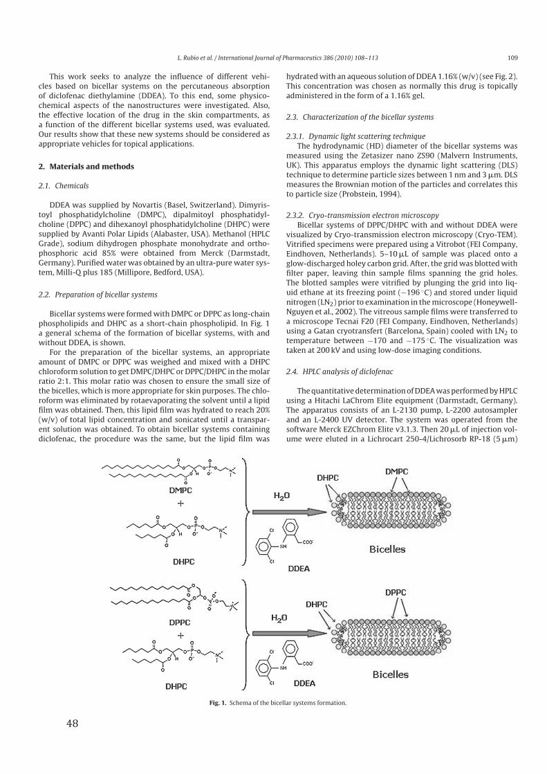
L. Rubio et al. / International Journal of Pharmaceutics 386 (2010) 108–113 109
This work seeks to analyze the influence of different vehi-
cles based on bicellar systems on the percutaneous absorption
of diclofenac diethylamine (DDEA). To this end, some physico-
chemical aspects of the nanostructures were investigated. Also,
the effective location of the drug in the skin compartments, as
a function of the different bicellar systems used, was evaluated.
Our results show that these new systems should be considered as
appropriate vehicles for topical applications.
2. Materials and methods
2.1. Chemicals
DDEA was supplied by Novartis (Basel, Switzerland). Dimyris-
toyl phosphatidylcholine (DMPC), dipalmitoyl phosphatidyl-
choline (DPPC) and dihexanoyl phosphatidylcholine (DHPC) were
supplied by Avanti Polar Lipids (Alabaster, USA). Methanol (HPLC
Grade), sodium dihydrogen phosphate monohydrate and ortho-
phosphoric acid 85% were obtained from Merck (Darmstadt,
Germany). Purified water was obtained by an ultra-pure water sys-
tem, Milli-Q plus 185 (Millipore, Bedford, USA).
2.2. Preparation of bicellar systems
Bicellar systems were formed with DMPC or DPPC as long-chain
phospholipids and DHPC as a short-chain phospholipid. In Fig. 1
a general schema of the formation of bicellar systems, with and
without DDEA, is shown.
For the preparation of the bicellar systems, an appropriate
amount of DMPC or DPPC was weighed and mixed with a DHPC
chloroform solution to get DMPC/DHPC or DPPC/DHPC in the molar
ratio 2:1. This molar ratio was chosen to ensure the small size of
the bicelles, which is more appropriate for skin purposes. The chlo-
roform was eliminated by rotaevaporating the solvent until a lipid
film was obtained. Then, this lipid film was hydrated to reach 20%
(w/v) of total lipid concentration and sonicated until a transpar-
ent solution was obtained. To obtain bicellar systems containing
diclofenac, the procedure was the same, but the lipid film was
hydrated with an aqueous solution of DDEA 1.16% (w/v) (see Fig. 2).
This concentration was chosen as normally this drug is topically
administered in the form of a 1.16% gel.
2.3. Characterization of the bicellar systems
2.3.1. Dynamic light scattering technique
The hydrodynamic (HD) diameter of the bicellar systems was
measured using the Zetasizer nano ZS90 (Malvern Instruments,
UK). This apparatus employs the dynamic light scattering (DLS)
technique to determine particle sizes between 1 nm and 3 �m. DLS
measures the Brownian motion of the particles and correlates this
to particle size (Probstein, 1994).
2.3.2. Cryo-transmission electron microscopy
Bicellar systems of DPPC/DHPC with and without DDEA were
visualized by Cryo-transmission electron microscopy (Cryo-TEM).
Vitrified specimens were prepared using a Vitrobot (FEI Company,
Eindhoven, Netherlands). 5–10 �L of sample was placed onto a
glow-discharged holey carbon grid. After, the grid was blotted with
filter paper, leaving thin sample films spanning the grid holes.
The blotted samples were vitrified by plunging the grid into liq-
uid ethane at its freezing point (−196 ◦C) and stored under liquid
nitrogen (LN2) prior to examination in the microscope (Honeywell-
Nguyen et al., 2002). The vitreous sample films were transferred to
a microscope Tecnai F20 (FEI Company, Eindhoven, Netherlands)
using a Gatan cryotransfert (Barcelona, Spain) cooled with LN2 to
temperature between −170 and −175 ◦C. The visualization was
taken at 200 kV and using low-dose imaging conditions.
2.4. HPLC analysis of diclofenac
The quantitative determination of DDEA was performed by HPLC
using a Hitachi LaChrom Elite equipment (Darmstadt, Germany).
The apparatus consists of an L-2130 pump, L-2200 autosampler
and an L-2400 UV detector. The system was operated from the
software Merck EZChrom Elite v3.1.3. Then 20 �L of injection vol-
ume were eluted in a Lichrocart 250-4/Lichrosorb RP-18 (5 �m)
Fig. 1. Schema of the bicellar systems formation.
48
110 L. Rubio et al. / International Journal of Pharmaceutics 386 (2010) 108–113
Fig. 2. Preparation of bicellar systems.
column (Merck, Germany) at room temperature. DDEA was mon-
itored by UV detection at 254 nm. A mobile phase consisting of a
66% methanol and 34% phosphate buffer (pH 2.5) was used at a flow
rate of 1 mL/min. The area under the peak was used to calculate the
concentration of DDEA using external standards that showed lin-
earity over the concentration range of 0.33–83 �g/mL. The intraday
and interday variations of the method were less than 2%.
2.5. In vitro percutaneous absorption studies
For this study pig skin from the unboiled back of Landrace large
white pigs weighing between 30 and 40 kg was used. The pig skin
was provided by the Clínic Hospital of Barcelona, Spain. The bristles
were removed carefully with an animal clipper and then the skin
was washed with tap water. The excised skin was dermatomed to
500 ± 50 �m thickness (Dermatome GA630, Aesculap, Tuttlingen,
Germany). Discs of the dermatomed skin were obtained with an
iron punch (2.5 cm inner diameter) and fitted into Franz type diffu-
sion cells. The skin discs were stored at −20 ◦C until use. One hour
prior to the diffusion experiments, the skin was thawed at room
temperature.
Franz cells (Lara-Spiral, Courtenon, France) consisted of an
upper donor compartment and a receptor chamber (3 mL of vol-
ume). These two parts were separated by the skin biopsy, leaving an
exposed surface area of 1.86 cm2. A magnetic stirring bar was intro-
duced into the receptor chamber. The skin disc was mounted with
the SC side up in the Franz cell. The receptor chamber was filled with
a receptor fluid (RF) which was PBS (pH 7.4) in distilled water, con-
taining 1% of bovine serum albumin and 0.04% gentamicin sulphate,
this was stirred continuously.
Franz cells were kept at 37 ± 1 ◦C by means of a circulating
water bath (Julabo Labortechnik GmbH, Germany) to ensure that
the surface skin was maintained at 32 ± 1 ◦C. The integrity of each
skin sample was checked by determining the transepidermal water
loss (TEWL) using a Tewameter TM210 (Courage-Khazaka, Köln,
Germany). The diffusion experiment was initiated by applying to
the entire surface, delimited by the upper cell, 10 �L of each of
the following solutions: aqueous solution of DDEA, DMPC/DHPC
bicelles with DDEA or DPPC/DHPC bicelles with DDEA. A control
cell was also used (only with the application of 10 �L of water).
After the exposure time (24 h), the test formulation remaining on
the skin surface was removed with a specific wash: first with 0.5 mL
of sodium lauryl ether sulphate solution (at 0.5%, w/v) and then
twice (2 × 0.5 mL) with distilled water. After that, the skin sur-
face was dried with a cotton swab. Water aliquots, all tips of the
micropipette, all cotton swabs as well as the top of the cell were
pooled, constituting the fraction of the active compound remain-
ing in the surface. Then, the receptor fluid was removed from the
receptor compartment and brought up to 5 mL in a volumetric flask.
The SC of the treated skin area was removed by 8 successive tape-
strippings using adhesive tape (D-Squame®, CuDerm Inc., Dallas,
USA). After that, the viable epidermis was separated from the der-
mis after heating the skin at 80 ◦C for a few seconds.
The amount of DDEA in the different layers and in the washing
solution was extracted with a solution of methanol: water (50:50)
for 20 h. Then, samples were shacked for 30 min at room tempera-
ture and sonicated for 15 min. Before the analytical determination
by HPLC, the samples were filtered through a 0.45 �m Acrodisc
filter (Pall Gelman Sciences, Northampton, UK).
2.6. Pretreatment of the skin with DMPC/DHPC and DPPC/DHPC
bicelles
To evaluate the effect of the pretreatment of skin with bicelles in
the subsequent percutaneous absorption of DDEA, 10 �L of bicelles
(without diclofenac) were topically applied for 1 h and after that a
careful aqueous washing of the skin surface was carried out. This
process was repeated four times. Then, an in vitro percutaneous
absorption test of an aqueous solution of DDEA (1.16%, w/v) was
performed as described in Section 2.5.
2.7. Statistical analysis
Each value is expressed as the mean ± S.D. for six determina-
tions. For group comparisons, analysis of variance (ANOVA) with
a one-way layout was applied. The software used was the STAT-
GRAPHICS plus 5. Significant differences in the mean values were
evaluated by the Student’s unpaired t-test. A p value of less than
0.05 was considered significant.
3. Results and discussion
3.1. Characterization of bicellar systems
The characterization of the bicellar systems have been carried
out by two different techniques: DLS and Cryo-TEM.
49
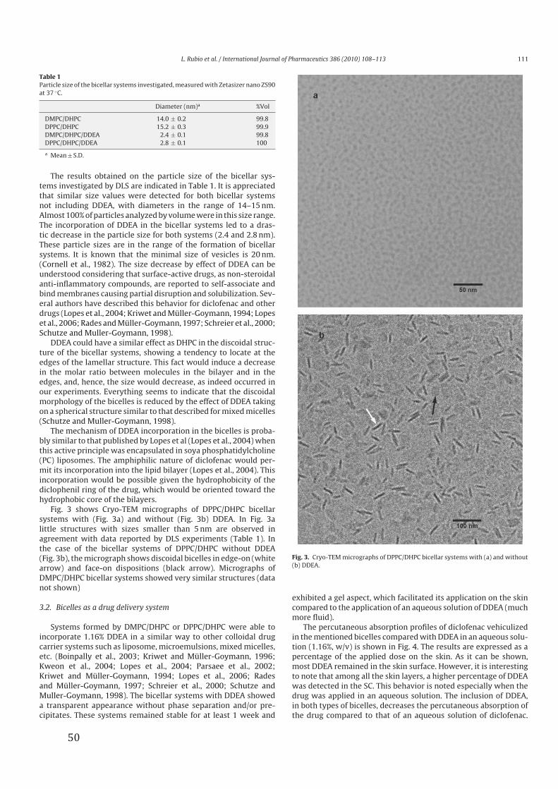
L. Rubio et al. / International Journal of Pharmaceutics 386 (2010) 108–113 111
Table 1Particle size of the bicellar systems investigated, measured with Zetasizer nano ZS90
at 37 ◦C.
Diameter (nm)a %Vol
DMPC/DHPC 14.0 ± 0.2 99.8
DPPC/DHPC 15.2 ± 0.3 99.9
DMPC/DHPC/DDEA 2.4 ± 0.1 99.8
DPPC/DHPC/DDEA 2.8 ± 0.1 100
a Mean ± S.D.
The results obtained on the particle size of the bicellar sys-
tems investigated by DLS are indicated in Table 1. It is appreciated
that similar size values were detected for both bicellar systems
not including DDEA, with diameters in the range of 14–15 nm.
Almost 100% of particles analyzed by volume were in this size range.
The incorporation of DDEA in the bicellar systems led to a dras-
tic decrease in the particle size for both systems (2.4 and 2.8 nm).
These particle sizes are in the range of the formation of bicellar
systems. It is known that the minimal size of vesicles is 20 nm.
(Cornell et al., 1982). The size decrease by effect of DDEA can be
understood considering that surface-active drugs, as non-steroidal
anti-inflammatory compounds, are reported to self-associate and
bind membranes causing partial disruption and solubilization. Sev-
eral authors have described this behavior for diclofenac and other
drugs (Lopes et al., 2004; Kriwet and Müller-Goymann, 1994; Lopes
et al., 2006; Rades and Müller-Goymann, 1997; Schreier et al., 2000;
Schutze and Muller-Goymann, 1998).
DDEA could have a similar effect as DHPC in the discoidal struc-
ture of the bicellar systems, showing a tendency to locate at the
edges of the lamellar structure. This fact would induce a decrease
in the molar ratio between molecules in the bilayer and in the
edges, and, hence, the size would decrease, as indeed occurred in
our experiments. Everything seems to indicate that the discoidal
morphology of the bicelles is reduced by the effect of DDEA taking
on a spherical structure similar to that described for mixed micelles
(Schutze and Muller-Goymann, 1998).
The mechanism of DDEA incorporation in the bicelles is proba-
bly similar to that published by Lopes et al (Lopes et al., 2004) when
this active principle was encapsulated in soya phosphatidylcholine
(PC) liposomes. The amphiphilic nature of diclofenac would per-
mit its incorporation into the lipid bilayer (Lopes et al., 2004). This
incorporation would be possible given the hydrophobicity of the
diclophenil ring of the drug, which would be oriented toward the
hydrophobic core of the bilayers.
Fig. 3 shows Cryo-TEM micrographs of DPPC/DHPC bicellar
systems with (Fig. 3a) and without (Fig. 3b) DDEA. In Fig. 3a
little structures with sizes smaller than 5 nm are observed in
agreement with data reported by DLS experiments (Table 1). In
the case of the bicellar systems of DPPC/DHPC without DDEA
(Fig. 3b), the micrograph shows discoidal bicelles in edge-on (white
arrow) and face-on dispositions (black arrow). Micrographs of
DMPC/DHPC bicellar systems showed very similar structures (data
not shown)
3.2. Bicelles as a drug delivery system
Systems formed by DMPC/DHPC or DPPC/DHPC were able to
incorporate 1.16% DDEA in a similar way to other colloidal drug
carrier systems such as liposome, microemulsions, mixed micelles,
etc. (Boinpally et al., 2003; Kriwet and Müller-Goymann, 1996;
Kweon et al., 2004; Lopes et al., 2004; Parsaee et al., 2002;
Kriwet and Müller-Goymann, 1994; Lopes et al., 2006; Rades
and Müller-Goymann, 1997; Schreier et al., 2000; Schutze and
Muller-Goymann, 1998). The bicellar systems with DDEA showed
a transparent appearance without phase separation and/or pre-
cipitates. These systems remained stable for at least 1 week and
Fig. 3. Cryo-TEM micrographs of DPPC/DHPC bicellar systems with (a) and without
(b) DDEA.
exhibited a gel aspect, which facilitated its application on the skin
compared to the application of an aqueous solution of DDEA (much
more fluid).
The percutaneous absorption profiles of diclofenac vehiculized
in the mentioned bicelles compared with DDEA in an aqueous solu-
tion (1.16%, w/v) is shown in Fig. 4. The results are expressed as a
percentage of the applied dose on the skin. As it can be shown,
most DDEA remained in the skin surface. However, it is interesting
to note that among all the skin layers, a higher percentage of DDEA
was detected in the SC. This behavior is noted especially when the
drug was applied in an aqueous solution. The inclusion of DDEA,
in both types of bicelles, decreases the percutaneous absorption of
the drug compared to that of an aqueous solution of diclofenac.
50
112 L. Rubio et al. / International Journal of Pharmaceutics 386 (2010) 108–113
Fig. 4. Percutaneous absorption profiles of all the diclofenac formulations studied
(mean values ± SD, n = 6). Distribution in the different layers of the skin: stratum
corneum (SC), epidermis (E), dermis (D), receptor fluid (RF) and total percutaneous
absorption (Perc. Abs.).
This finding suggests a retarder effect of the percutaneous absorp-
tion when the drug is included in bicellar systems. This effect would
be interesting to be applied in drugs which have a too fast percuta-
neous absorption, as it is the case of fentanyl, to prevent an overdose
of the drug (Frölich et al., 2001).
It is obvious that the use of both kinds of bicellar systems pre-
vents the passage of the drug to the deeper layers of the skin.
This inhibitory effect on skin penetration was more marked for
DMPC/DHPC bicelles. For these bicelles, Fig. 4 shows a higher
value of DDEA in SC and an absence of drug in the receptor fluid.
The results as global percentage of percutaneous absorption (con-
sidering the amount of DDEA detected in epidermis (E), dermis
(D) plus receptor fluid (RF)) show the following ranking: aque-
ous solution DDEA (4.61 ± 0.62%) > DPPC/DHPC bicelles with DDEA
(2.78 ± 1.62%) > DMPC/DHPC bicelles with DDEA (1.25 ± 0.33%).
The significant difference (p < 0.05) of skin penetration detected
between DPPC and DMPC bicelles could be due to the different
transition temperature (Tm) of these two phospholipids. The DPPC
at the experimental temperature is in a gel phase, like lipids of
the SC (Tm about 60 ◦C) (Golden et al., 1987); this fact could facil-
itate the mix between lipids from the SC and from the bicelles
promoting skin penetration, with respect to the DMPC bicelles. On
the other hand, DMPC at the experimental temperature exhibits a
liquid crystalline phase (Tm of DMPC 23 ◦C) (Lewis et al., 1987).
Therefore, the DMPC has a different phase than the lipids from
the SC and, as a consequence, the skin penetration could be more
difficult.
Due to the small particle size of the lipid systems formed after
the encapsulation of DDEA, one might expect that these systems
could penetrate more easily through the skin. But there are different
factors that are involved in the enhancer effect of a vehicle. Some of
these factors are the possible disruption of the organization of the
intercellular lipids of the SC, the affinity of the drug to the vehicle
and the rigidity of the lipid structure of the vehicle (Gwak and Chun,
2002; Thong et al., 2007). The incorporation of DDEA in a bicellar
system may cause a certain rigidity in the bicelles. In fact, the drug
is not simply dissolved in the lipophilic region of the phospholipids,
but is incorporated in the bilayer lined up with the phospholipids
and the diclofenac’s carboxyl groups increase the rigidity of the
head groups of the phospholipids (Ferreira et al., 2005; Seddon et
al., 2009). This possible rigidity would hinder the penetration of
DDEA through the skin (Kriwet and Müller-Goymann, 1996; Kriwet
and Müller-Goymann, 1994). Also, another possible reason for this
low penetration could be related to the difficulty of the DDEA to
diffuse out of the bicellar systems.
Fig. 5. Percutaneous absorption profiles of diclofenac with skin pretreated with
bicellar systems or water (blank) (mean values ± SD, n = 6). Distribution in the dif-
ferent layers of the skin: stratum corneum (SC), epidermis (E), dermis (D), receptor
fluid (RF) and total percutaneous absorption (Perc. Abs.).
3.3. Bicelles as enhancers of skin penetration
Although the bicelles with DDEA inhibit the drug penetration, in
vivo studies of Barbosa-Barros et al. (2008c) showed an increase of
TEWL after a consecutive application of phospholipids bicelles. The
effect of bicelles on the barrier function and their possible enhancer
effect on the in vitro percutaneous absorption of DDEA were inves-
tigated. To this end, we performed a pretreatment of the skin discs
with bicellar systems of DMPC/DHPC and DPPC/DHPC followed by a
topical application of a DDEA aqueous solution. Also, to discard the
possible influence of the water contained in the bicellar systems,
some skin discs were pretreated with water (blank).
Fig. 5 shows the percutaneous absorption of DDEA using skin
pretreated with bicelles, expressed in percentage of applied dose
and using the experimental conditions described earlier.
In general terms, the amount of DDEA detected in the SC was
higher in skin not pretreated with bicelles than in pretreated sam-
ples. In addition, the global results obtained on skin penetration
show that a pretreatment of the skin with bicelles promotes the
percutaneous absorption of diclofenac. There were no significant
differences (p < 0.05) in the percentage of percutaneous absorption
between the treatment with DMPC/DHPC or DPPC/DHPC bicelles.
These results have been obtained despite the fact that microscopy
studies have shown that each of these systems change, in different
ways, the microstructure of the SC. DMPC/DHPC bicelles did not
affect SC lipid microstructure (Barbosa-Barros et al., 2008a) and
DPPC/DHPC systems seem to penetrate inside the skin SC and grow
forming vesicles (Barbosa-Barros et al., 2008c). In future works it
would be interesting to study the eventual histological changes
detected not only in the SC but also in whole structure of skin
treated with bicelles.
The enhancer effect of the bicellar systems on the percutaneous
absorption of diclofenac could be due to an initial interaction of
bicelles with the SC. This interaction could cause some disorgani-
zation of intercellular lipids, responsible of the SC barrier function.
This fact would help the absorption of diclofenac through the skin.
The interaction of the specific phospholipids of bicelles with the
lipids of the SC would be the mechanism responsible for the event
detected. This mechanism is different for both systems because the
DMPC/DHPC bicellar systems, apparently, did not produce modi-
fications in the microstructure of the SC, whereas the DPPC/DHPC
system did produce changes in the lipid lamellae regions (Barbosa-
Barros et al., 2008a,c). Thus, our results seems to indicate that other
factors, in addition to the microstructural changes of the SC, must be
involved in the penetration of DDEA, since the two types of bicelles
51
L. Rubio et al. / International Journal of Pharmaceutics 386 (2010) 108–113 113
produce a similar effect. In this case, the DDEA is applied in an
aqueous solution. For this reason, the limiting rate of drug trans-
port through the SC may not be due to drug release from the vehicle,
but could probably be related more to inherent SC resistance.
4. Conclusions
This work demonstrates that bicellar systems are able to incor-
porate DDEA. This incorporation decreases the particle size with
respect to the original bicelles. These bicellar systems, including
DDEA, can act as retarders in the percutaneous absorption of the
drug; probably, the limiting of the rate of drug transport is depen-
dent on rate of the drug release from the vehicle. The results of our
work may be useful to develop bicellar systems for drugs which
have a too fast percutaneous absorption as it is the case of fentanyl
(Frölich et al., 2001). On the contrary, the previous in vitro appli-
cation of bicelles on skin discs seems to promote the passage of
diclofenac through the lipidic interstices of the SC and, as a result,
to improve the percutaneous absorption. In this case, there are no
problems of drug release since DDEA is in an aqueous solution. As a
consequence, a previous application of bicellar systems on the skin
seems to be useful to modulate the percutaneous absorption of top-
ically applied DDEA. Further investigations on the use of bicelles as
delivery systems, using other drugs with different physicochem-
ical properties and applications, should be considered in future
research.
Acknowledgments
The authors acknowledge Ministerio de Educación y Ciencia,
Spain. This work was supported by European Social Fund (Programa
Nacional de Potenciación de Recursos Humanos del Plan Nacional
de I+D+I (2004-2008)).
References
Ainbinder, D., Touitou, E., 2005. Testosterone ethosomes for enhanced transdermaldelivery. Drug Deliv. 12, 297–303.
Barbosa-Barros, L., de la Maza, A., Estelrich, J., Linares, A.M., Feliz, M., Walther, P.,Pons, R., Lopez, O., 2008a. Penetration and growth of DPPC/DHPC bicelles insidethe stratum corneum of the skin. Langmuir 24, 5700–5706.
Barbosa-Barros, L., De la Maza, A., Walther, P., Estelrich, J., Lopez, O., 2008b. Mor-phological effects of ceramide on DMPC/DHPC bicelles. J. Microsc. 230, 16–26.
Barbosa-Barros, L., Barba, C., Cocera, M., Coderch, L., Lopez-Iglesias, C., de la Maza,A., Lopez, O., 2008c. Effect of bicellar systems on skin properties. Int. J. Pharm.352, 263–272.
Barry, B.W., 2001. Novel mechanisms and devices to enable successful transdermaldrug delivery. Eur. J. Pharm. Sci. 14, 101–114.
Boinpally, R.R., Zhou, S.L., Poondru, S., Devraj, G., Jasti, B.R., 2003. Lecithin vesiclesfor topical delivery of diclofenac. Eur. J. Pharm. Biopharm. 56, 389–392.
Cornell, B.A., Fletcher, G.C., Middlehurst, J., Separovic, F., 1982. The lower limit tothe size of small sonicated phospholipid vesicles. Biochim. Biophys. Acta 690,15–19.
El Maghraby, G.M., Williams, A.C., Barry, B.W., 2001. Skin delivery of 5-fluorouracilfrom ultradeformable and standard liposomes in-vitro. J. Pharm. Pharmacol. 53,1069–1077.
El Maghraby, G.M., Williams, A.C., Barry, B.W., 1999. Skin delivery of oestradiol fromdeformable and traditional liposomes: mechanistic studies. J. Pharm. Pharmacol.51, 1123–1134.
Elias, P.M., Feingold, K.R., 2006. Skin Barrier. Taylor and Francis group, New York.Fang, J.Y., Fang, C.L., Liu, C.H., Su, Y.H., 2008. Lipid nanoparticles as vehicles for topical
psoralen delivery: solid lipid nanoparticles (SLN) versus nanostructured lipidcarriers (NLC). Eur. J. Pharm. Biopharm. 70, 633–640.
Ferreira, H., Lucio, M., Lima, J.L.F.C., Matos, C., Reis, S., 2005. Effects ofdiclofenac on EPC liposome membrane properties. Anal. Bioanal. Chem. 382,1256–1264.
Frölich, M., Giannotti, A., Modell, J.H., 2001. Opioid overdose in a patient using afentanyl patch during treatment with a warming blanket. Anesth. Analg. 93,647–648.
Galer, B.S., Rowbotham, M., Perander, J., Devers, A., Friedman, E., 2000. Topicaldiclofenac patch relieves minor sports injury pain: results of a multicenter con-trolled clinical trial. J. Pain Symptom Manage. 19, 287–294.
Golden, G.M., Guzek, D.B., Kennedy, A.H., McKie, J.E., Potts, R.O., 1987. Stratumcorneum lipid phase transitions and water barrier properties. Biochemistry 26,2382–2388.
Gwak, H.S., Chun, I.K., 2002. Effect of vehicles and penetration enhancers on the invitro percutaneous absorption of tenoxicam through hairless mouse skin. Int. J.Pharm. 236, 57–64.
Honeywell-Nguyen, P.L., Frederik, P.M., Bomans, P.H., Junginger, H.E., Bouwstra,J.A., 2002. Transdermal delivery of pergolide from surfactant-based elastic andrigid vesicles: characterization and in vitro transport studies. Pharm. Res. 19,991–997.
Honeywell-Nguyen, P.L., Bouwstra, J.A., 2005. Vesicles as a tool for transdermal anddermal delivery. Drug Discov. Today: Technol. 2, 67–74.
Kriwet, K., Müller-Goymann, C.C., 1994. Mutual interactions between diclofenacdiethylamine and phospholipids—investigation on the microstructure of thearisen systems. Pharmazie 49, 187–191.
Kriwet, K., Müller-Goymann, C.C., 1996. Diclofenac release from phospholipid drugsystems and permeation through excised human stratum corneum. Int. J. Pharm.125, 231–242.
Kweon, J.H., Chi, S.C., Park, E.S., 2004. Transdermal delivery of diclofenac usingmicroemulsions. Arch. Pharm. Res. 27, 351–356.
Lewis, R.N., Mak, N., McElhaney, R.N., 1987. A differential scanning calorimetric studyof the thermotropic phase behavior of model membranes composed of phos-phatidylcholines containing linear saturated fatty acyl chains. Biochemistry 26,6118–6126.
Lopes, L.B., Scarpa, M.V., Silva, G.V., Rodrigues, D.C., Santilli, C.V., Oliveira, A.G., 2004.Studies on the encapsulation of diclofenac in small unilamellar liposomes ofsoya phosphatidylcholine. Colloids Surf. B: Biointerfaces 39, 151–158.
Lopes, L.B., Scarpa, M.V., Pereira, N.L., Oliveira, L.C., Oliveira, A.G., 2006. Interaction ofsodium diclofenac with freeze-dried soya phosphatidylcholine and unilamellarliposomes. Rev. Bras. Cienc. Farm. 42, 497–504.
Mezei, M., Gulasekharam, V., 1980. Liposomes—a selective drug delivery sys-tem for the topical route of administration. Lotion dosage form. Life Sci. 26,1473–1477.
OECD, 2004. Guideline 428: skin absorption: in vitro method. In: Development,O.f.E.C.a. (Ed.), OECD Guidelines for the Testing of Chemicals. Paris, p. 8.
Paolino, D., Lucania, G., Mardente, D., Alhaique, F., Fresta, M., 2005. Ethosomes forskin delivery of ammonium glycyrrhizinate: in vitro percutaneous permeationthrough human skin and in vivo anti-inflammatory activity on human volun-teers. J. Control. Release 106, 99–110.
Parsaee, S., Sarbolouki, M.N., Parnianpour, M., 2002. In-vitro release of diclofenacdiethylammonium from lipid-based formulations. Int. J. Pharm. 241, 185–190.
Probstein, R.F., 1994. Physicochemical Hydrodynamics. John Wiley & Sons, NewYork.
Puglia, C., Blasi, P., Rizza, L., Schoubben, A., Bonina, F., Rossi, C., Ricci, M., 2008. Lipidnanoparticles for prolonged topical delivery: an in vitro and in vivo investigation.Int. J. Pharm. 357, 295–304.
Qiu, Y., Gao, Y., Hu, K., Li, F., 2008. Enhancement of skin permeation of docetaxel: anovel approach combining microneedle and elastic liposomes. J. Control. Release129, 144–150.
Rades, T., Müller-Goymann, C.C., 1997. Investigations on the micellisation behaviourof fenoprofen sodium. Int. J. Pharm. 159, 215–222.
Schaefer, H., Redermeier, T.E., 1996. Skin Barrier—Principles of PercutaneousAbsorption. Karger-Verlag, Basel.
Schreier, S., Malheiros, S.V., de Paula, E., 2000. Surface active drugs: self-associationand interaction with membranes and surfactants. Physicochemical and biolog-ical aspects. Biochim. Biophys. Acta 1508, 210–234.
Schutze, W., Muller-Goymann, C.C., 1998. Phase transformation of a liposomaldispersion into a micellar solution induced by drug-loading. Pharm. Res. 15,538–543.
Seddon, A.M., Casey, D., Law, R.V., Gee, A., Templer, R.H., Ces, O., 2009. Drug interac-tions with lipid membranes. Chem. Soc. Rev. 38, 2509–2519.
Spernath, A., Aserin, A., Sintov, A.C., Garti, N., 2008. Phosphatidylcholine embeddedmicellar systems: enhanced permeability through rat skin. J. Colloid InterfaceSci. 318, 421–429.
Thong, H.Y., Zhai, H., Maibach, H.I., 2007. Percutaneous penetration enhancers: anoverview. Skin Pharmacol. Physiol. 20, 272–282.
Touitou, E., Dayan, N., Bergelson, L., Godin, B., Eliaz, M., 2000. Ethosomes—novelvesicular carriers for enhanced delivery: characterization and skin penetrationproperties. J. Control. Release 65, 403–418.
Trotta, M., Peira, E., Carlotti, M.E., Gallarate, M., 2004. Deformable liposomes fordermal administration of methotrexate. Int. J. Pharm. 270, 119–125.
Trotta, M., Peira, E., Debernardi, F., Gallarate, M., 2002. Elastic liposomes for skindelivery of dipotassium glycyrrhizinate. Int. J. Pharm. 241, 319–327.
Visscher, I., Stuart, M.C., Engberts, J.B., 2006. The influence of phenyl and phenoxymodification in the hydrophobic tails of di-n-alkyl phosphate amphiphiles onaggregate morphology. Org. Biomol. Chem. 4, 707–712.
Vold, R.R., Prosser, R.S., 1996. Magnetically oriented phospholipid bilayered micellesfor structural studies of polypeptides. Does the ideal bicelle exist? J. Magn. Res.Ser. B 113, 267–271.
Whiles, J.A., Deems, R., Vold, R.R., Dennis, E.A., 2002. Bicelles in structure-functionstudies of membrane-associated proteins. Bioorg. Chem. 30, 431–442.
Williams, A.C., 2003. Transdermal and Topical Drug Delivery: From Theory to ClinicalPractice. Pharmaceutical Press, London.
52
Sistemes�bicel·lars�com�a�nova�estratègia�d’aplicació�tòpica����������������������������������������������������������������������RESULTATS�
%@�
Article�2��
Structural�effects�of�flufenamic�acid�in�DPPC/DHPC�bicellar�systems��
Soft�Matter��#$>>��'��CACC&CAG'�
�
!�� �5����� ���-���� ��� ��� ����� ���� �0�4���� ��� ��� ���� 9� ���� *� ��� ����
� ������� - ��B����� ��� �::�I�D:�6� :��� �� �5����� ����(� ��� ��� ��� �� � �/��� ����
��- �� 9����� 4���������� 5���6�)����� ������)3��������� � �/�����������������
���� 4���� ��������� ������������������������ �������- ��B�������-� �������4����6��
)�� ��� ��� ���(� �����!���������������-������� ����� 4������� �����������
� ��� 5����5����������4������5�������*�������� ��(���������� �������- ��B��������
�::�I�D:�6� *�� ���������� ��-� ��� � ���(� �� ��� ��2�&�!�� � ��� ���� ������
�-����������������������� ��� 5������� ���������� �������- ��B�������-� �������*6�
!��3*F3������������� �4���� 9���-��� ���� 4���� ����������� M���� ����- ����������
� �������- ��B����� ���������������*6�:���������-���������T*F3���������� �����
�4���� 9� ��-��� ���� 4���� ���0����5��������� ����������� ����- ����� � ��� 5����
5���� ��� 4����� ������� ��(�� �� ��6� )�� ��� �� ���� ���� �� �3�� ��� ���������
��� M�������� 4������������ �������4����5������*���� ��� ����-�������::���������
��(���������� �������- ��B����6����
)�� ������� 9�����*��������������� ��������������� ���������4���� ������
������������ �������(� � � �0������-������� ����� � ������� - ��B����� � ���6� *�
���� ����� ������������� ����� ������ ��� ���� 4������ � 4������� ����������� ��� ��
����5[�� �� ��� ��� ������� 9� ���� 4����� ��� ���� - ��B���6� !�� �0����� � �����
� ������� - ��B����� ������ *�� �� ���� ����� ������������� ����� ����� ��� ��� ������ ���
4���� 9����- ��B����� ��L���������!�� � ��2�&�!���0���� ������� �����������
�����)3��0����G��������M ��������6�!����������������3*F3� �T*F3����� �4������
5����0��������������- ���������0����%C�Y� ��0����5��������������������������(�- �
��� ���� ����� ������������6� ����� ��� 4����� ��� ��� ������� ��� ���� - ��B����� ��� ����
�-���������#%�]������4���� 9������������������������� ��� ���������!�� �������
������������ ��L�����������2�&�!�6�!����������������3*F3� �T*F3��������������
���� �� �0�� ������� ��� - ����� � �0����5��������� �������� .�M��������
������ ������6� *� @#�%� ]��� ��� ��� �-������� ��� � �� �� 9� ��� ��� ������� �� �����
� ������� � ��� �� � �0���� 9� �0����������� ���������6� ���-<� ��� ��� �������� ����
��� ��� � �� �� 9� ��� �0�������� ��� ��� - ���� � ��� �0����5��������� ��������
.�M������6� *� @'� ]�� ��� ���� ���� ��� ��� ���� ����������� ��-������� � ��L����� �
��4�� 5����� ���� �������� ��� - ���� � � ���� ���� � ������� ������ 4����� � ���
����5�����������������.�-� ��������.�M������� �� �����&��5� �6�!�������� ������3��
���������������4���� 9����������4����6�

Sistemes�bicel·lars�com�a�nova�estratègia�d’aplicació�tòpica����������������������������������������������������������������������RESULTATS�
%A�
�+����� �5������� ��� 5���� �M��� ��������� �0.��� ������ �/��� ���� � �������
��B�����5�����������*��5������������������������������������������. ����
������0��� � 9��(� ������� � � ���� ��6�
-
�
�
�
�
�����������������������������������
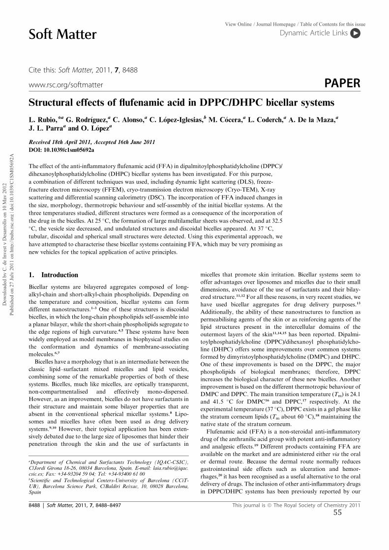
Structural effects of flufenamic acid in DPPC/DHPC bicellar systems
L. Rubio,*a G. Rodr�ıguez,a C. Alonso,a C. L�opez-Iglesias,b M. C�ocera,a L. Coderch,a A. De la Maza,a
J. L. Parraa and O. L�opeza
Received 18th April 2011, Accepted 16th June 2011
DOI: 10.1039/c1sm05692a
The effect of the anti-inflammatory flufenamic acid (FFA) in dipalmitoylphosphatidylcholine (DPPC)/
dihexanoylphosphatidylcholine (DHPC) bicellar systems has been investigated. For this purpose,
a combination of different techniques was used, including dynamic light scattering (DLS), freeze-
fracture electron microscopy (FFEM), cryo-transmission electron microscopy (Cryo-TEM), X-ray
scattering and differential scanning calorimetry (DSC). The incorporation of FFA induced changes in
the size, morphology, thermotropic behaviour and self-assembly of the initial bicellar systems. At the
three temperatures studied, different structures were formed as a consequence of the incorporation of
the drug in the bicelles. At 25 �C, the formation of large multilamellar sheets was observed, and at 32.5�C, the vesicle size decreased, and undulated structures and discoidal bicelles appeared. At 37 �C,tubular, discoidal and spherical small structures were detected. Using this experimental approach, we
have attempted to characterise these bicellar systems containing FFA, which may be very promising as
new vehicles for the topical application of active principles.
1. Introduction
Bicellar systems are bilayered aggregates composed of long-
alkyl-chain and short-alkyl-chain phospholipids. Depending on
the temperature and composition, bicellar systems can form
different nanostructures.1–3 One of these structures is discoidal
bicelles, in which the long-chain phospholipids self-assemble into
a planar bilayer, while the short-chain phospholipids segregate to
the edge regions of high curvature.4,5 These systems have been
widely employed as model membranes in biophysical studies on
the conformation and dynamics of membrane-associating
molecules.6,7
Bicelles have a morphology that is an intermediate between the
classic lipid–surfactant mixed micelles and lipid vesicles,
combining some of the remarkable properties of both of these
systems. Bicelles, much like micelles, are optically transparent,
non-compartmentalised and effectively mono-dispersed.
However, as an improvement, bicelles do not have surfactants in
their structure and maintain some bilayer properties that are
absent in the conventional spherical micellar systems.8 Lipo-
somes and micelles have often been used as drug delivery
systems.9,10 However, their topical application has been exten-
sively debated due to the large size of liposomes that hinder their
penetration through the skin and the use of surfactants in
micelles that promote skin irritation. Bicellar systems seem to
offer advantages over liposomes and micelles due to their small
dimensions, avoidance of the use of surfactants and their bilay-
ered structure.11,12 For all these reasons, in very recent studies, we
have used bicellar aggregates for drug delivery purposes.13
Additionally, the ability of these nanostructures to function as
permeabilising agents of the skin or as reinforcing agents of the
lipid structures present in the intercellular domains of the
outermost layers of the skin11,14,15 has been reported. Dipalmi-
toylphosphatidylcholine (DPPC)/dihexanoyl phosphatidylcho-
line (DHPC) offers some improvements over common systems
formed by dimyristoylphosphatidylcholine (DMPC) and DHPC.
One of these improvements is based on the DPPC, the major
phospholipids of biological membranes; therefore, DPPC
increases the biological character of these new bicelles. Another
improvement is based on the different thermotropic behaviour of
DMPC and DPPC. The main transition temperature (Tm) is 24.1
and 41.5 �C for DMPC16 and DPPC,17 respectively. At the
experimental temperature (37 �C), DPPC exists in a gel phase like
the stratum corneum lipids (Tm about 60 �C),18 maintaining the
native state of the stratum corneum.
Flufenamic acid (FFA) is a non-steroidal anti-inflammatory
drug of the anthranilic acid group with potent anti-inflammatory
and analgesic effects.19 Different products containing FFA are
available on the market and are administered either via the oral
or dermal route. Because the dermal route normally reduces
gastrointestinal side effects such as ulceration and hemor-
rhages,20 it has been recognised as a useful alternative to the oral
delivery of drugs. The inclusion of other anti-inflammatory drugs
in DPPC/DHPC systems has been previously reported by our
aDepartment of Chemical and Surfactants Technology (IQAC-CSIC),C/Jordi Girona 18-26, 08034 Barcelona, Spain. E-mail: [email protected]; Fax: +34-93204 59 04; Tel: +34-93400 61 00bScientific and Technological Centers-University of Barcelona (CCiT-UB), Barcelona Science Park, C/Baldiri Reixac, 10, 08028 Barcelona,Spain
8488 | Soft Matter, 2011, 7, 8488–8497 This journal is ª The Royal Society of Chemistry 2011
Dynamic Article LinksC<Soft Matter
Cite this: Soft Matter, 2011, 7, 8488
www.rsc.org/softmatter PAPER
Dow
nloa
ded
by C
. de
Inve
st y
Des
arro
llo o
n 10
May
201
2Pu
blis
hed
on 2
7 Ju
ly 2
011
on h
ttp://
pubs
.rsc.
org
| doi
:10.
1039
/C1S
M05
692A
View Online / Journal Homepage / Table of Contents for this issue
55
group,13 and the results showed the potential of these systems for
drug delivery. Therefore, the inclusion of FFA in bicellar systems
proposed in the present work and its topical application could be
very advantageous.
However, it is important to find out, through preliminary
studies, if some structural changes have been induced due to the
incorporation of FFA into the (DPPC/DHPC) bicelles. To do so,
these systems were characterised by the combined use of different
techniques, including dynamic light scattering (DLS), freeze-
fracture electron microscopy (FFEM), cryo-transmission elec-
tron microscopy (Cryo-TEM), X-ray scattering and differential
scanning calorimetry (DSC). Using these techniques, several
structural changes of the bicelles have been detected as a function
of temperature (from 25 to 37 �C). A separate application of
these techniques in the characterisation of colloidal systems has
reported relevant data in this field.21–23
The present work focuses on the study of structural changes
induced by the inclusion of FFA in DPPC/DHPC bicellar
systems. The evaluation of the structural changes was carried out
with the aforementioned techniques. Our work reports innova-
tive data that should be important in designing bicellar systems
as vehicles for the topical application of FFA, taking into
consideration their physicochemical behaviour at different
temperatures.
2. Materials and methods
2.1 Materials
Flufenamic acid (FFA) was purchased from Sigma Aldrich (St
Louis, MO, USA). Dipalmitoylphosphatidylcholine (DPPC) and
dihexanoyl phosphatidylcholine (DHPC) were supplied by
Avanti Polar Lipids (Alabaster, USA). Chloroform was
purchased from Merck (Darmstadt, Germany). Purified water
was obtained from an ultra-pure water system, Milli-Q plus 185
(Millipore, Bedford, USA).
2.2 Preparation of bicellar systems
An appropriate amount of DPPC was weighed and mixed with
aDHPC chloroform solution to obtainDPPC/DHPC in themolar
ratio 2 : 1, with a 20% weight/volume (w/v) total lipid concentra-
tion. In the case of FFA bicellar systems, 10 mg of FFA was
weighed and mixed with DPPC, DHPC and chloroform. The
chloroform from both systems was removed by rota-evaporation,
and the lipid filmwas dissolved in 1ml of water and sonicated until
a transparent solution was obtained. This DPPC/DHPC/FFA
solution contained 10 mg of FFA per ml (1% w/v). pH measure-
ments of the DPPC/DHPC/FFA aqueous solution were per-
formed with a Model 720 pH meter and a ROSS Model 8103 SC
pH electrode (both from Orion Research, Cambridge, MA, USA).
2.3 Characterisation of the bicellar systems
2.3.1 Dynamic light scattering technique (DLS). DLS is
a useful technique to measure the dimensions of the vesicle
structures. The hydrodynamic diameter (HD) and polydispersity
index (PI) of the aggregates were measured using the Zetasizer
nano ZS (Malvern Instruments, UK). This apparatus employs the
dynamic light scattering (DLS) technique to determine particle
sizes between 0.6 nm and 6 mm. DLS measures the Brownian
motion of the particles and correlates this phenomenon to particle
size.24 To minimise multiple scattering, non-invasive back scatter
technology (NIBS) was used. With this technology, the detection
of scattering is performed at an angle of 173�; therefore,
concentrated samples can be measured, as is the case of our
systems. The measurements were carried out at three different
temperatures (25 �C, 32.5 �C and 37 �C). The results were ana-
lysed as the percentage of scattering intensity. The data obtained
were collected and analysed with the program DTS (dispersion
technology software) provided by Malvern Instruments Ltd.
2.3.2 Microscopy techniques. FFEM and Cryo-TEM were
used to visualise the dimensions and morphologies of these
systems. Samples of DPPC/DHPC/FFA systems were equili-
brated at three different temperatures (25 �C, 32.5 �C and 37 �C)for 30 min to visualise the system at these conditions. These three
temperatures were chosen because 25 �C is the ambient temper-
ature, 32.5 �C is approximately the temperature of the skin surface
and 37 �C is the physiological temperature. For control purposes,
bicelles of DPPC/DHPC were cryofixed at 25 �C. Samples were
visualised using the microscopy techniques described below.
2.3.2.1 Freeze-fracture electron microscopy (FFEM). FFEM
is a technique that involves cryofixation, replication and trans-
mission electron microscopy. This technique is widely used to
characterise lipid bilayer and non-bilayer arrangements25 and
monolayer-based structures.26 FFEM avoids artifacts due to
composition changes that are inherent in chemical fixation,
staining and drying techniques. The procedure carried out in this
work for the FFEM study was based on that described by
Egelhaaf.27 Freeze-fracture samples were prepared by first
depositing approximately 1 mL of sample between two copper
platelets using a 400 mesh gold grid as a spacer. Then samples
were frozen by plunging into a liquid propane bath cooled at
�189 �C by liquid nitrogen and fractured by separating the two
copper plates in a BalTec BAF 300 freeze-etching device (Leica,
Vienna, Austria) at �150 �C and 10�8 mbar of pressure. The
replicas were obtained by unidirectional shadowing at 45� with 2
nm of platinum/carbon (Pt/C) and at 90� with 20 nm of carbon.
After the replication, the samples were brought to room condi-
tions. An advantage of this preparation design is that large
portions of the metal replicas were automatically attached to the
TEM grids. The grids and replicas were cleaned by immersion in
concentrated sodium hypochlorite to completely remove any
organic material from the sample, leaving only the Pt/C replica.
The replicas were examined in a Jeol 1010 Transmission electron
microscope (Jeol, Tokyo, Japan) at 80 kV.
2.3.2.2 Cryo-transmission electron microscopy (Cryo-TEM).
Cryo-TEM is a method whereby the specimen can be examined in
its native state by vitrification. This technique has opened the
possibility for the direct imaging of amphiphilic aggregates
formed in aqueous solutions.28 DPPC/DHPC bicellar systems
with and without FFA were visualised by cryo-TEM. Vitrified
specimens were prepared using a Vitrobot (FEI Company,
Eindhoven, Netherlands). Then 3–5 mL of sample was placed onto
a glow-discharged holey carbon grid. The grid was subsequently
blotted with filter paper, leaving thin sample films spanning the
This journal is ª The Royal Society of Chemistry 2011 Soft Matter, 2011, 7, 8488–8497 | 8489
Dow
nloa
ded
by C
. de
Inve
st y
Des
arro
llo o
n 10
May
201
2Pu
blis
hed
on 2
7 Ju
ly 2
011
on h
ttp://
pubs
.rsc.
org
| doi
:10.
1039
/C1S
M05
692A
View Online
56
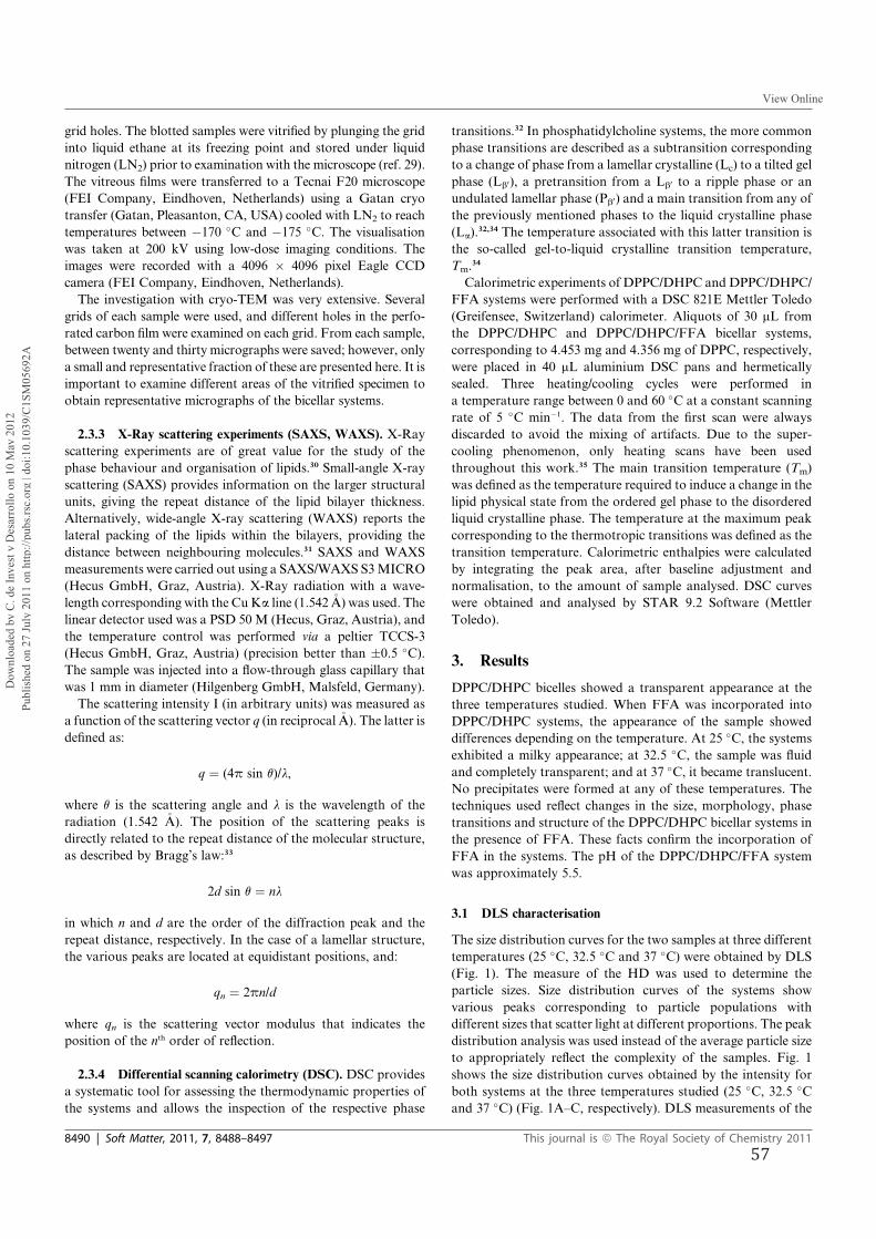
grid holes. The blotted samples were vitrified by plunging the grid
into liquid ethane at its freezing point and stored under liquid
nitrogen (LN2) prior to examination with the microscope (ref. 29).
The vitreous films were transferred to a Tecnai F20 microscope
(FEI Company, Eindhoven, Netherlands) using a Gatan cryo
transfer (Gatan, Pleasanton, CA, USA) cooled with LN2 to reach
temperatures between �170 �C and �175 �C. The visualisation
was taken at 200 kV using low-dose imaging conditions. The
images were recorded with a 4096 � 4096 pixel Eagle CCD
camera (FEI Company, Eindhoven, Netherlands).
The investigation with cryo-TEM was very extensive. Several
grids of each sample were used, and different holes in the perfo-
rated carbon film were examined on each grid. From each sample,
between twenty and thirty micrographs were saved; however, only
a small and representative fraction of these are presented here. It is
important to examine different areas of the vitrified specimen to
obtain representative micrographs of the bicellar systems.
2.3.3 X-Ray scattering experiments (SAXS, WAXS). X-Ray
scattering experiments are of great value for the study of the
phase behaviour and organisation of lipids.30 Small-angle X-ray
scattering (SAXS) provides information on the larger structural
units, giving the repeat distance of the lipid bilayer thickness.
Alternatively, wide-angle X-ray scattering (WAXS) reports the
lateral packing of the lipids within the bilayers, providing the
distance between neighbouring molecules.31 SAXS and WAXS
measurements were carried out using a SAXS/WAXS S3MICRO
(Hecus GmbH, Graz, Austria). X-Ray radiation with a wave-
length corresponding with the CuKa line (1.542�A) was used. The
linear detector used was a PSD 50 M (Hecus, Graz, Austria), and
the temperature control was performed via a peltier TCCS-3
(Hecus GmbH, Graz, Austria) (precision better than �0.5 �C).The sample was injected into a flow-through glass capillary that
was 1 mm in diameter (Hilgenberg GmbH, Malsfeld, Germany).
The scattering intensity I (in arbitrary units) was measured as
a function of the scattering vector q (in reciprocal�A). The latter is
defined as:
q ¼ (4p sin q)/l,
where q is the scattering angle and l is the wavelength of the
radiation (1.542 �A). The position of the scattering peaks is
directly related to the repeat distance of the molecular structure,
as described by Bragg’s law:33
2d sin q ¼ nl
in which n and d are the order of the diffraction peak and the
repeat distance, respectively. In the case of a lamellar structure,
the various peaks are located at equidistant positions, and:
qn ¼ 2pn/d
where qn is the scattering vector modulus that indicates the
position of the nth order of reflection.
2.3.4 Differential scanning calorimetry (DSC). DSC provides
a systematic tool for assessing the thermodynamic properties of
the systems and allows the inspection of the respective phase
transitions.32 In phosphatidylcholine systems, the more common
phase transitions are described as a subtransition corresponding
to a change of phase from a lamellar crystalline (Lc) to a tilted gel
phase (Lb0), a pretransition from a Lb0 to a ripple phase or an
undulated lamellar phase (Pb0) and a main transition from any of
the previously mentioned phases to the liquid crystalline phase
(La).32,34 The temperature associated with this latter transition is
the so-called gel-to-liquid crystalline transition temperature,
Tm.34
Calorimetric experiments of DPPC/DHPC and DPPC/DHPC/
FFA systems were performed with a DSC 821E Mettler Toledo
(Greifensee, Switzerland) calorimeter. Aliquots of 30 mL from
the DPPC/DHPC and DPPC/DHPC/FFA bicellar systems,
corresponding to 4.453 mg and 4.356 mg of DPPC, respectively,
were placed in 40 mL aluminium DSC pans and hermetically
sealed. Three heating/cooling cycles were performed in
a temperature range between 0 and 60 �C at a constant scanning
rate of 5 �C min�1. The data from the first scan were always
discarded to avoid the mixing of artifacts. Due to the super-
cooling phenomenon, only heating scans have been used
throughout this work.35 The main transition temperature (Tm)
was defined as the temperature required to induce a change in the
lipid physical state from the ordered gel phase to the disordered
liquid crystalline phase. The temperature at the maximum peak
corresponding to the thermotropic transitions was defined as the
transition temperature. Calorimetric enthalpies were calculated
by integrating the peak area, after baseline adjustment and
normalisation, to the amount of sample analysed. DSC curves
were obtained and analysed by STAR 9.2 Software (Mettler
Toledo).
3. Results
DPPC/DHPC bicelles showed a transparent appearance at the
three temperatures studied. When FFA was incorporated into
DPPC/DHPC systems, the appearance of the sample showed
differences depending on the temperature. At 25 �C, the systems
exhibited a milky appearance; at 32.5 �C, the sample was fluid
and completely transparent; and at 37 �C, it became translucent.
No precipitates were formed at any of these temperatures. The
techniques used reflect changes in the size, morphology, phase
transitions and structure of the DPPC/DHPC bicellar systems in
the presence of FFA. These facts confirm the incorporation of
FFA in the systems. The pH of the DPPC/DHPC/FFA system
was approximately 5.5.
3.1 DLS characterisation
The size distribution curves for the two samples at three different
temperatures (25 �C, 32.5 �C and 37 �C) were obtained by DLS
(Fig. 1). The measure of the HD was used to determine the
particle sizes. Size distribution curves of the systems show
various peaks corresponding to particle populations with
different sizes that scatter light at different proportions. The peak
distribution analysis was used instead of the average particle size
to appropriately reflect the complexity of the samples. Fig. 1
shows the size distribution curves obtained by the intensity for
both systems at the three temperatures studied (25 �C, 32.5 �Cand 37 �C) (Fig. 1A–C, respectively). DLS measurements of the
8490 | Soft Matter, 2011, 7, 8488–8497 This journal is ª The Royal Society of Chemistry 2011
Dow
nloa
ded
by C
. de
Inve
st y
Des
arro
llo o
n 10
May
201
2Pu
blis
hed
on 2
7 Ju
ly 2
011
on h
ttp://
pubs
.rsc.
org
| doi
:10.
1039
/C1S
M05
692A
View Online
57
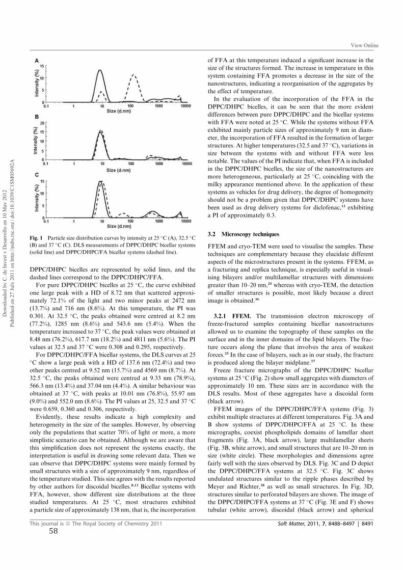
DPPC/DHPC bicelles are represented by solid lines, and the
dashed lines correspond to the DPPC/DHPC/FFA.
For pure DPPC/DHPC bicelles at 25 �C, the curve exhibited
one large peak with a HD of 8.72 nm that scattered approxi-
mately 72.1% of the light and two minor peaks at 2472 nm
(13.7%) and 716 nm (8.6%). At this temperature, the PI was
0.301. At 32.5 �C, the peaks obtained were centred at 8.2 nm
(77.2%), 1285 nm (8.6%) and 543.6 nm (5.4%). When the
temperature increased to 37 �C, the peak values were obtained at
8.48 nm (76.2%), 617.7 nm (18.2%) and 4811 nm (5.6%). The PI
values at 32.5 and 37 �C were 0.308 and 0.295, respectively.
For DPPC/DHPC/FFA bicellar systems, the DLS curves at 25�C show a large peak with a HD of 137.6 nm (72.4%) and two
other peaks centred at 9.52 nm (15.7%) and 4569 nm (8.7%). At
32.5 �C, the peaks obtained were centred at 9.33 nm (78.9%),
566.3 nm (13.4%) and 37.04 nm (4.4%). A similar behaviour was
obtained at 37 �C, with peaks at 10.01 nm (76.8%), 55.97 nm
(9.0%) and 552.0 nm (8.6%). The PI values at 25, 32.5 and 37 �Cwere 0.659, 0.360 and 0.306, respectively.
Evidently, these results indicate a high complexity and
heterogeneity in the size of the samples. However, by observing
only the populations that scatter 70% of light or more, a more
simplistic scenario can be obtained. Although we are aware that
this simplification does not represent the systems exactly, the
interpretation is useful in drawing some relevant data. Then we
can observe that DPPC/DHPC systems were mainly formed by
small structures with a size of approximately 9 nm, regardless of
the temperature studied. This size agrees with the results reported
by other authors for discoidal bicelles.8,13 Bicellar systems with
FFA, however, show different size distributions at the three
studied temperatures. At 25 �C, most structures exhibited
a particle size of approximately 138 nm, that is, the incorporation
of FFA at this temperature induced a significant increase in the
size of the structures formed. The increase in temperature in this
system containing FFA promotes a decrease in the size of the
nanostructures, indicating a reorganisation of the aggregates by
the effect of temperature.
In the evaluation of the incorporation of the FFA in the
DPPC/DHPC bicelles, it can be seen that the more evident
differences between pure DPPC/DHPC and the bicellar systems
with FFA were noted at 25 �C. While the systems without FFA
exhibited mainly particle sizes of approximately 9 nm in diam-
eter, the incorporation of FFA resulted in the formation of larger
structures. At higher temperatures (32.5 and 37 �C), variations insize between the systems with and without FFA were less
notable. The values of the PI indicate that, when FFA is included
in the DPPC/DHPC bicelles, the size of the nanostructures are
more heterogeneous, particularly at 25 �C, coinciding with the
milky appearance mentioned above. In the application of these
systems as vehicles for drug delivery, the degree of homogeneity
should not be a problem given that DPPC/DHPC systems have
been used as drug delivery systems for diclofenac,13 exhibiting
a PI of approximately 0.3.
3.2 Microscopy techniques
FFEM and cryo-TEM were used to visualise the samples. These
techniques are complementary because they elucidate different
aspects of the microstructures present in the systems. FFEM, as
a fracturing and replica technique, is especially useful in visual-
ising bilayers and/or multilamellar structures with dimensions
greater than 10–20 nm,25 whereas with cryo-TEM, the detection
of smaller structures is possible, most likely because a direct
image is obtained.36
3.2.1 FFEM. The transmission electron microscopy of
freeze-fractured samples containing bicellar nanostructures
allowed us to examine the topography of these samples on the
surface and in the inner domains of the lipid bilayers. The frac-
ture occurs along the plane that involves the area of weakest
forces.25 In the case of bilayers, such as in our study, the fracture
is produced along the bilayer midplane.37
Freeze fracture micrographs of the DPPC/DHPC bicellar
systems at 25 �C (Fig. 2) show small aggregates with diameters of
approximately 10 nm. These sizes are in accordance with the
DLS results. Most of these aggregates have a discoidal form
(black arrow).
FFEM images of the DPPC/DHPC/FFA systems (Fig. 3)
exhibit multiple structures at different temperatures. Fig. 3A and
B show systems of DPPC/DHPC/FFA at 25 �C. In these
micrographs, coexist phospholipids domains of lamellar sheet
fragments (Fig. 3A, black arrow), large multilamellar sheets
(Fig. 3B, white arrow), and small structures that are 10–20 nm in
size (white circle). These morphologies and dimensions agree
fairly well with the sizes observed by DLS. Fig. 3C and D depict
the DPPC/DHPC/FFA systems at 32.5 �C. Fig. 3C shows
undulated structures similar to the ripple phases described by
Meyer and Richter,38 as well as small structures. In Fig. 3D,
structures similar to perforated bilayers are shown. The image of
the DPPC/DHPC/FFA systems at 37 �C (Fig. 3E and F) shows
tubular (white arrow), discoidal (black arrow) and spherical
Fig. 1 Particle size distribution curves by intensity at 25 �C (A), 32.5 �C(B) and 37 �C (C). DLS measurements of DPPC/DHPC bicellar systems
(solid line) and DPPC/DHPC/FA bicellar systems (dashed line).
This journal is ª The Royal Society of Chemistry 2011 Soft Matter, 2011, 7, 8488–8497 | 8491
Dow
nloa
ded
by C
. de
Inve
st y
Des
arro
llo o
n 10
May
201
2Pu
blis
hed
on 2
7 Ju
ly 2
011
on h
ttp://
pubs
.rsc.
org
| doi
:10.
1039
/C1S
M05
692A
View Online
58
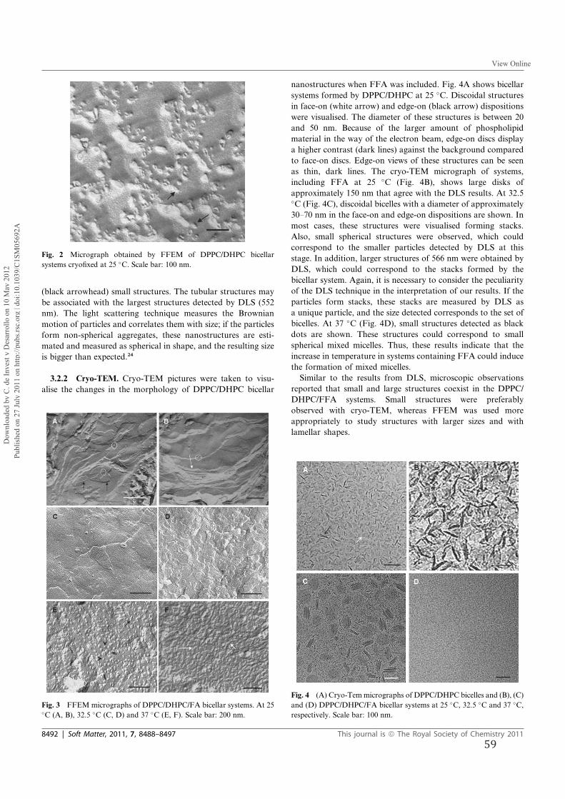
(black arrowhead) small structures. The tubular structures may
be associated with the largest structures detected by DLS (552
nm). The light scattering technique measures the Brownian
motion of particles and correlates them with size; if the particles
form non-spherical aggregates, these nanostructures are esti-
mated and measured as spherical in shape, and the resulting size
is bigger than expected.24
3.2.2 Cryo-TEM. Cryo-TEM pictures were taken to visu-
alise the changes in the morphology of DPPC/DHPC bicellar
nanostructures when FFA was included. Fig. 4A shows bicellar
systems formed by DPPC/DHPC at 25 �C. Discoidal structures
in face-on (white arrow) and edge-on (black arrow) dispositions
were visualised. The diameter of these structures is between 20
and 50 nm. Because of the larger amount of phospholipid
material in the way of the electron beam, edge-on discs display
a higher contrast (dark lines) against the background compared
to face-on discs. Edge-on views of these structures can be seen
as thin, dark lines. The cryo-TEM micrograph of systems,
including FFA at 25 �C (Fig. 4B), shows large disks of
approximately 150 nm that agree with the DLS results. At 32.5�C (Fig. 4C), discoidal bicelles with a diameter of approximately
30–70 nm in the face-on and edge-on dispositions are shown. In
most cases, these structures were visualised forming stacks.
Also, small spherical structures were observed, which could
correspond to the smaller particles detected by DLS at this
stage. In addition, larger structures of 566 nm were obtained by
DLS, which could correspond to the stacks formed by the
bicellar system. Again, it is necessary to consider the peculiarity
of the DLS technique in the interpretation of our results. If the
particles form stacks, these stacks are measured by DLS as
a unique particle, and the size detected corresponds to the set of
bicelles. At 37 �C (Fig. 4D), small structures detected as black
dots are shown. These structures could correspond to small
spherical mixed micelles. Thus, these results indicate that the
increase in temperature in systems containing FFA could induce
the formation of mixed micelles.
Similar to the results from DLS, microscopic observations
reported that small and large structures coexist in the DPPC/
DHPC/FFA systems. Small structures were preferably
observed with cryo-TEM, whereas FFEM was used more
appropriately to study structures with larger sizes and with
lamellar shapes.
Fig. 2 Micrograph obtained by FFEM of DPPC/DHPC bicellar
systems cryofixed at 25 �C. Scale bar: 100 nm.
Fig. 3 FFEM micrographs of DPPC/DHPC/FA bicellar systems. At 25�C (A, B), 32.5 �C (C, D) and 37 �C (E, F). Scale bar: 200 nm.
Fig. 4 (A) Cryo-Temmicrographs of DPPC/DHPC bicelles and (B), (C)
and (D) DPPC/DHPC/FA bicellar systems at 25 �C, 32.5 �C and 37 �C,respectively. Scale bar: 100 nm.
8492 | Soft Matter, 2011, 7, 8488–8497 This journal is ª The Royal Society of Chemistry 2011
Dow
nloa
ded
by C
. de
Inve
st y
Des
arro
llo o
n 10
May
201
2Pu
blis
hed
on 2
7 Ju
ly 2
011
on h
ttp://
pubs
.rsc.
org
| doi
:10.
1039
/C1S
M05
692A
View Online
59
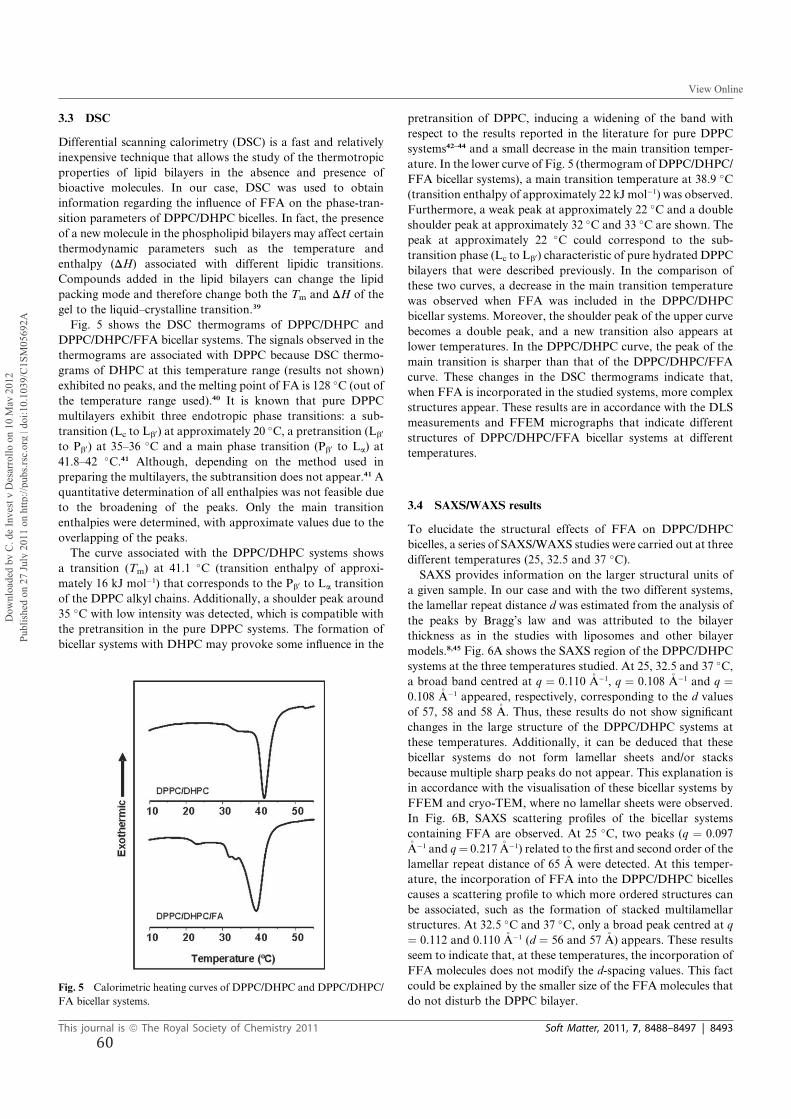
3.3 DSC
Differential scanning calorimetry (DSC) is a fast and relatively
inexpensive technique that allows the study of the thermotropic
properties of lipid bilayers in the absence and presence of
bioactive molecules. In our case, DSC was used to obtain
information regarding the influence of FFA on the phase-tran-
sition parameters of DPPC/DHPC bicelles. In fact, the presence
of a new molecule in the phospholipid bilayers may affect certain
thermodynamic parameters such as the temperature and
enthalpy (DH) associated with different lipidic transitions.
Compounds added in the lipid bilayers can change the lipid
packing mode and therefore change both the Tm and DH of the
gel to the liquid–crystalline transition.39
Fig. 5 shows the DSC thermograms of DPPC/DHPC and
DPPC/DHPC/FFA bicellar systems. The signals observed in the
thermograms are associated with DPPC because DSC thermo-
grams of DHPC at this temperature range (results not shown)
exhibited no peaks, and the melting point of FA is 128 �C (out of
the temperature range used).40 It is known that pure DPPC
multilayers exhibit three endotropic phase transitions: a sub-
transition (Lc to Lb0) at approximately 20 �C, a pretransition (Lb0
to Pb0) at 35–36�C and a main phase transition (Pb0 to La) at
41.8–42 �C.41 Although, depending on the method used in
preparing the multilayers, the subtransition does not appear.41 A
quantitative determination of all enthalpies was not feasible due
to the broadening of the peaks. Only the main transition
enthalpies were determined, with approximate values due to the
overlapping of the peaks.
The curve associated with the DPPC/DHPC systems shows
a transition (Tm) at 41.1 �C (transition enthalpy of approxi-
mately 16 kJ mol�1) that corresponds to the Pb0 to La transition
of the DPPC alkyl chains. Additionally, a shoulder peak around
35 �C with low intensity was detected, which is compatible with
the pretransition in the pure DPPC systems. The formation of
bicellar systems with DHPC may provoke some influence in the
pretransition of DPPC, inducing a widening of the band with
respect to the results reported in the literature for pure DPPC
systems42–44 and a small decrease in the main transition temper-
ature. In the lower curve of Fig. 5 (thermogram of DPPC/DHPC/
FFA bicellar systems), a main transition temperature at 38.9 �C(transition enthalpy of approximately 22 kJ mol�1) was observed.
Furthermore, a weak peak at approximately 22 �C and a double
shoulder peak at approximately 32 �C and 33 �C are shown. The
peak at approximately 22 �C could correspond to the sub-
transition phase (Lc to Lb0) characteristic of pure hydrated DPPC
bilayers that were described previously. In the comparison of
these two curves, a decrease in the main transition temperature
was observed when FFA was included in the DPPC/DHPC
bicellar systems. Moreover, the shoulder peak of the upper curve
becomes a double peak, and a new transition also appears at
lower temperatures. In the DPPC/DHPC curve, the peak of the
main transition is sharper than that of the DPPC/DHPC/FFA
curve. These changes in the DSC thermograms indicate that,
when FFA is incorporated in the studied systems, more complex
structures appear. These results are in accordance with the DLS
measurements and FFEM micrographs that indicate different
structures of DPPC/DHPC/FFA bicellar systems at different
temperatures.
3.4 SAXS/WAXS results
To elucidate the structural effects of FFA on DPPC/DHPC
bicelles, a series of SAXS/WAXS studies were carried out at three
different temperatures (25, 32.5 and 37 �C).SAXS provides information on the larger structural units of
a given sample. In our case and with the two different systems,
the lamellar repeat distance d was estimated from the analysis of
the peaks by Bragg’s law and was attributed to the bilayer
thickness as in the studies with liposomes and other bilayer
models.8,45 Fig. 6A shows the SAXS region of the DPPC/DHPC
systems at the three temperatures studied. At 25, 32.5 and 37 �C,a broad band centred at q ¼ 0.110 �A�1, q ¼ 0.108 �A�1 and q ¼0.108 �A�1 appeared, respectively, corresponding to the d values
of 57, 58 and 58 �A. Thus, these results do not show significant
changes in the large structure of the DPPC/DHPC systems at
these temperatures. Additionally, it can be deduced that these
bicellar systems do not form lamellar sheets and/or stacks
because multiple sharp peaks do not appear. This explanation is
in accordance with the visualisation of these bicellar systems by
FFEM and cryo-TEM, where no lamellar sheets were observed.
In Fig. 6B, SAXS scattering profiles of the bicellar systems
containing FFA are observed. At 25 �C, two peaks (q ¼ 0.097�A�1 and q¼ 0.217�A�1) related to the first and second order of the
lamellar repeat distance of 65 �A were detected. At this temper-
ature, the incorporation of FFA into the DPPC/DHPC bicelles
causes a scattering profile to which more ordered structures can
be associated, such as the formation of stacked multilamellar
structures. At 32.5 �C and 37 �C, only a broad peak centred at q
¼ 0.112 and 0.110 �A�1 (d ¼ 56 and 57 �A) appears. These results
seem to indicate that, at these temperatures, the incorporation of
FFA molecules does not modify the d-spacing values. This fact
could be explained by the smaller size of the FFA molecules that
do not disturb the DPPC bilayer.Fig. 5 Calorimetric heating curves of DPPC/DHPC and DPPC/DHPC/
FA bicellar systems.
This journal is ª The Royal Society of Chemistry 2011 Soft Matter, 2011, 7, 8488–8497 | 8493
Dow
nloa
ded
by C
. de
Inve
st y
Des
arro
llo o
n 10
May
201
2Pu
blis
hed
on 2
7 Ju
ly 2
011
on h
ttp://
pubs
.rsc.
org
| doi
:10.
1039
/C1S
M05
692A
View Online
60
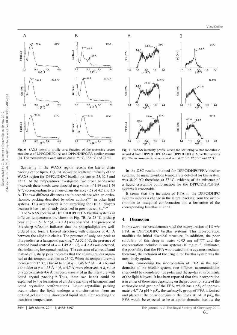
Scattering in the WAXS region reveals the lateral chain
packing of the lipids. Fig. 7A shows the scattered intensity of the
WAXS region for DPPC/DHPC bicellar systems at 25, 32.5 and
37 �C. At the temperatures investigated, two broad bands were
observed; these bands were detected at q values of 1.49 and 1.79�A�1, corresponding to a chain–chain distance (dc) of 4.2 and 3.5�A. The two different distances are in accordance with an ortho-
rhombic packing described by other authors46,47 in other lipid
systems. This arrangement is not surprising for DPPC bilayers
because it has been already described in previous works.42,48
The WAXS spectra of DPPC/DHPC/FFA bicellar systems at
different temperatures are shown in Fig. 7B. At 25 �C, a sharp
peak at q ¼ 1.53 �A�1 (dc ¼ 4.1 �A) was observed. The presence of
this sharp reflection indicates that the phospholipids are well-
ordered and form a layered structure, with distances of 4.1 �A
between the aliphatic chains. The presence of only one peak at
this q indicates a hexagonal packing.46At 32.5 �C, the presence ofa broad band centred at q ¼ 1.49 �A�1 (dc ¼ 4.2 �A) was detected,
also indicating hexagonal packing. The existence of a broad band
instead of a sharp peak indicates that the chains are less organ-
ised at this temperature than at 25 �C.When the temperature was
increased to 37 �C, a broad band at q¼ 1.46 �A�1 (dc ¼ 4.3 �A) and
a shoulder at q ¼ 1.33 �A�1 (dc ¼ 4.7 �A) were observed. A dc value
of approximately 4.6 �A has been associated in the literature with
liquid crystal packing.46 Thus, these two bands could be
explained by the formation of a hybrid packing of hexagonal and
liquid crystalline conformations. Liquid crystalline packing
occurs when the lipids undergo a transformation from an
ordered gel state to a disordered liquid state after reaching the
transition temperature.
In the DSC results obtained for DPPC/DHPC/FFA bicellar
systems, the main transition temperature detected for this system
was 38.90 �C; therefore, at 37 �C, evidence of the existence of
a liquid crystalline conformation for the DPPC/DHPC/FFA
systems is reasonable.
It seems that the inclusion of FFA in the DPPC/DHPC
systems induces a change in the lateral packing from the ortho-
rhombic to hexagonal conformation and a formation of the
corresponding lamellae at 25 �C.
4. Discussion
In this work, we have demonstrated the incorporation of 1% w/v
FFA in DPPC/DHPC bicellar systems. This incorporation
modifies the initial discoidal structure. In addition, the poor
solubility of this drug in water (0.03 mg ml�1)49 and the
concentration included in our systems (10 mg ml�1) eliminated
the possibility that the FFA was located in the aqueous medium;
therefore, the inclusion of the drug in the bicellar system was the
most likely option.
Thus, considering the incorporation of FFA in the lipid
domains of the bicellar system, two different accommodation
sites could be considered: the polar and the apolar environments
of the lipid bilayers. It has been reported that this incorporation
is in either of these sites depending on the protonation state of the
carboxylic acid group of the FFA, which has a pKa of approxi-
mately 4.50 At pH > pKa, the carboxylic group of FFA is ionised
and placed at the polar domains of the lipids. At pH < pKa, the
FFA would be expected to be at apolar domains because the
Fig. 6 SAXS intensity profile as a function of the scattering vector
modulus q of DPPC/DHPC (A) and DPPC/DHPC/FA bicellar systems
(B). The measurements were carried out at 25 �C, 32.5 �C and 37 �C.
Fig. 7 WAXS intensity profile versus the scattering vector modulus q
recorded from DPPC/DHPC (A) and DPPC/DHPC/FA bicellar systems
(B). The measurements were carried out at 25 �C, 32.5 �C and 37 �C.
8494 | Soft Matter, 2011, 7, 8488–8497 This journal is ª The Royal Society of Chemistry 2011
Dow
nloa
ded
by C
. de
Inve
st y
Des
arro
llo o
n 10
May
201
2Pu
blis
hed
on 2
7 Ju
ly 2
011
on h
ttp://
pubs
.rsc.
org
| doi
:10.
1039
/C1S
M05
692A
View Online
61
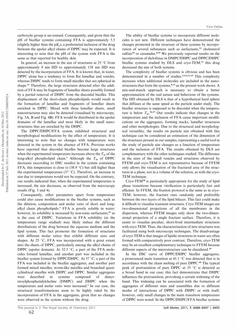
carboxylic group is un-ionised. Consequently, and given that the
pH of bicellar systems containing FFA is approximately 5.5
(slightly higher than the pKa), a preferential inclusion of the drug
between the apolar alkyl chains of DPPC may be expected. It is
interesting to note that the pH of the system with FFA is the
same as that reported for healthy skin.
In general, an increase in the size of structures at 25 �C from
approximately 9 nm HD to approximately 138 nm HD was
detected by the incorporation of FFA. It is known that, in water,
DPPC alone has a tendency to form flat lamellae and vesicles,
whereas DHPC tends to form small micelles that are spherical in
shape.51 Therefore, the large structures detected after the addi-
tion of FFAmay be fragments of lamellar sheets possibly formed
by a partial removal of DHPC from the discoidal bicelles. This
displacement of the short-chain phospholipids would result in
the formation of lamellae and fragments of lamellar sheets
enriched in DPPC. Mixed with these lamellar sheets, small
nanostructures may also be detected (visualised by microscopy,
Fig. 3A, B and Fig. 4B). FFA would be distributed in the apolar
domains of the lamellae and most likely in the small nano-
structures that are enriched by the DHPC.
The DPPC/DHPC/FFA system exhibited structural and
morphological modifications by the effect of temperature. It is
interesting to note that no changes with temperature were
detected in the system in the absence of FFA. Previous works
have reported that discoidal bicelles become large structures
when the experimental temperature increases above the Tm of the
long-alkyl phospholipid chain.3 Although the Tm of DPPC
decreases (according to DSC results) in the system containing
FFA, this temperature is close to (38.9 �C) but still higher thanthe experimental temperature (37 �C). Therefore, an increase in
size due to temperature would not be expected. On the contrary,
our results indicated that, when the experimental temperature is
increased, the size decreases, as observed from the microscopy
results (Fig. 3 and 4).
Consequently, other parameters apart from temperature
could also cause modifications in the bicellar systems, such as
the dilution, composition and molar ratio of short and long-
alkyl chain phospholipids.3 FFA is poorly soluble in water;
however, its solubility is increased by non-ionic surfactants,52 as
is the case of DHPC. Variations in FFA solubility (at the
temperature range studied) may likely induce the different
distributions of the drug between the aqueous medium and the
lipid system. This fact promotes the formation of structures
with different molar ratios that exhibit different sizes and
shapes. At 25 �C, FFA was incorporated with a great extent
into the sheets of DPPC, particularly among the alkyl chains of
DPPC (apolar domain). At 32.5 �C, a part of the FFA mole-
cules formed lamellae, and another part was included in the
bicellar system formed by DPPC/DHPC. At 37 �C, a part of the
FFA was included in the bicellar aggregates, and another part
formed mixed micelles, worm-like micelles and branched quasi-
cylindrical micelles with DHPC and DPPC. Similar aggregates
were described in systems composed of dimyr-
istoylphosphatidylcholine (DMPC) and DHPC when the
temperature and molar ratio were increased.3 In our case, the
structural transformations seem to be highly ruled by the
incorporation of FFA in the aggregates, given that no changes
were observed in the system without the drug.
The ability of bicellar systems to incorporate different mole-
cules is not new. Different techniques have demonstrated the
changes promoted in the structure of these systems by incorpo-
ration of several substances such as surfactants,53 cholesterol
sulfate54 or ceramides.8,55 We previously reported results on the
incorporation of diclofenac in DMPC/DHPC and DPPC/DHPC
bicellar systems studied by DLS and cryo-TEM;13 this drug
decreased the size of both systems.
The complexity of bicellar systems is obvious and has been
demonstrated in a number of studies.2,3,6,11,14 This complexity
increases when additional molecules are included in the nano-
structures that form the systems,8,13 as the present work shows. A
mix-and-match approach is necessary to obtain a better
approximation of the real nature and behaviour of the systems.
The HD obtained by DLS is that of a hypothetical hard sphere
that diffuses at the same speed as the particle under study. The
bicellar structure is supposed to be discoidal when the tempera-
ture is below Tm.56,57 Our results indicate that changes in the
temperature and the inclusion of FFA cause important modifi-
cations on the aggregates, forming stacks, lamellar structures
and other morphologies. Due to the structural and morpholog-
ical versatility, the results on particle size obtained with this
technique can be considered an estimation of the dimension of
the structures present in our samples, representing a useful tool in
the study of particle size changes as a function of temperature
and the inclusion of FFA. The results obtained by DLS are
complementary with the other techniques studied. The difference
in the sizes of the small vesicles and structures observed by
FFEM and cryo-TEM is not representative because of FFEM
only allows the visualisation of the fractured area of the struc-
tures in a plane, not in a volume of the solution, as with the cryo-
TEM technique.
Cryo-TEM58 is particularly appropriate for the study of lipid
phase transitions because vitrification is particularly fast and
efficient. In FFEM, the fixation protocol is the same as in cryo-
TEM; however, the fracture runs randomly and preferably
between the two layers of the lipid bilayer. This fact could make
it difficult to visualise transient structures. Cryo-TEM images are
two-dimensional projections of all the membranes in the
dispersion, whereas FFEM images only show the two-dimen-
sional projection of a single fracture surface. Therefore, it is
easier to visualise peculiar, short-lived intermediate structures
with cryo-TEM. Thus, the characterisation of new structures was
facilitated using both microscopy techniques. The disadvantage
of cryo-TEM is that images of lipidic structures in water are often
formed with comparatively poor contrast. Therefore, cryo-TEM
may be an excellent complementary technique to FFEM because
different aspects of the microstructure can be elucidated.59
In the DSC curve of DPPC/DHPC bicellar aggregates,
a pronounced main transition at 41.1 �C was detected that is in
accordance with the chain melting of pure DPPC.60 The typical
peak of pretransition of pure DPPC at 35 �C is detected as
a broad band in our case; this fact demonstrates that DHPC
influences the pretransition, provoking a certain widening of the
band. This widening can be associated with the formation of
aggregates of different sizes and assemblies due to different
modes of interactions of DPPC with DHPC or with itself;
however, only small changes in the main transition temperature
of DPPC were noted. In the DPPC/DHPC/FFA bicellar systems
This journal is ª The Royal Society of Chemistry 2011 Soft Matter, 2011, 7, 8488–8497 | 8495
Dow
nloa
ded
by C
. de
Inve
st y
Des
arro
llo o
n 10
May
201
2Pu
blis
hed
on 2
7 Ju
ly 2
011
on h
ttp://
pubs
.rsc.
org
| doi
:10.
1039
/C1S
M05
692A
View Online
62
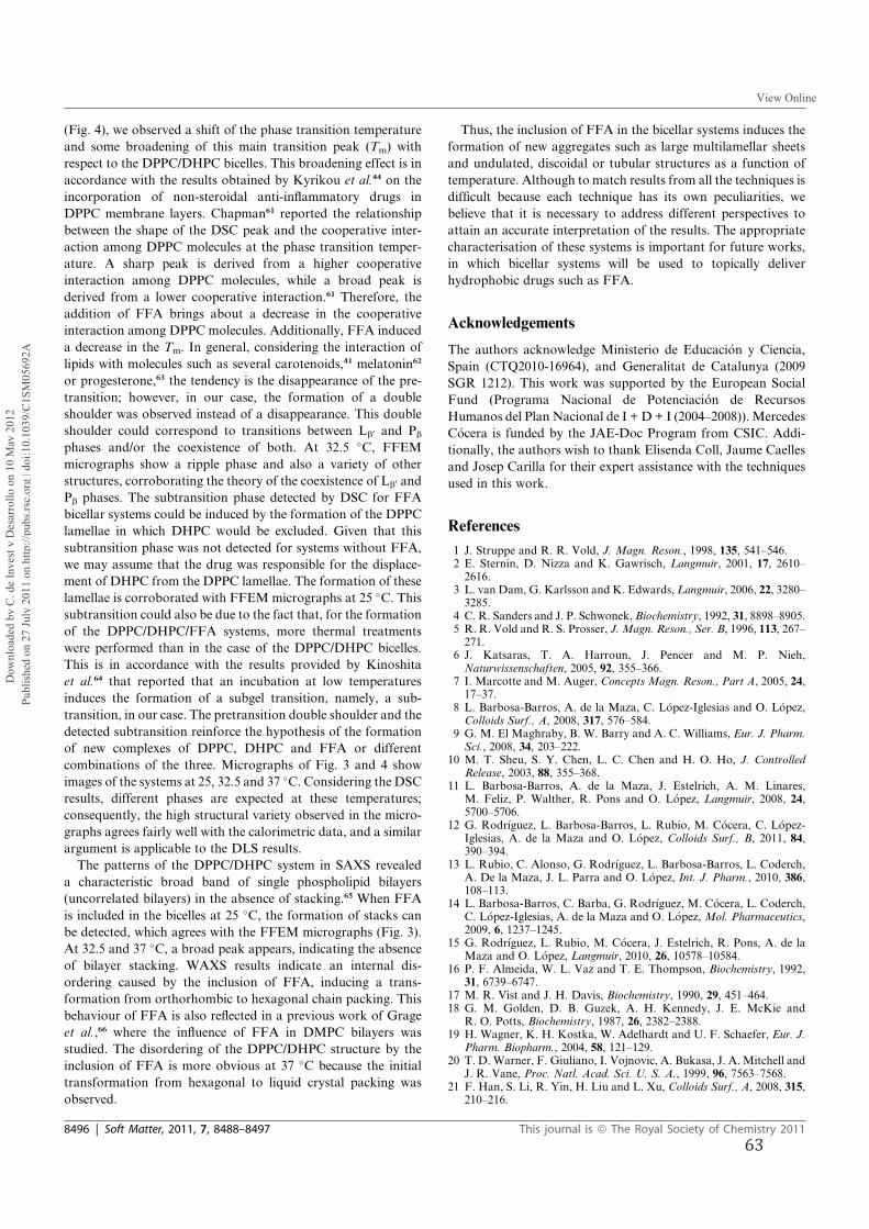
(Fig. 4), we observed a shift of the phase transition temperature
and some broadening of this main transition peak (Tm) with
respect to the DPPC/DHPC bicelles. This broadening effect is in
accordance with the results obtained by Kyrikou et al.44 on the
incorporation of non-steroidal anti-inflammatory drugs in
DPPC membrane layers. Chapman61 reported the relationship
between the shape of the DSC peak and the cooperative inter-
action among DPPC molecules at the phase transition temper-
ature. A sharp peak is derived from a higher cooperative
interaction among DPPC molecules, while a broad peak is
derived from a lower cooperative interaction.61 Therefore, the
addition of FFA brings about a decrease in the cooperative
interaction among DPPC molecules. Additionally, FFA induced
a decrease in the Tm. In general, considering the interaction of
lipids with molecules such as several carotenoids,41 melatonin62
or progesterone,63 the tendency is the disappearance of the pre-
transition; however, in our case, the formation of a double
shoulder was observed instead of a disappearance. This double
shoulder could correspond to transitions between Lb0 and Pb
phases and/or the coexistence of both. At 32.5 �C, FFEM
micrographs show a ripple phase and also a variety of other
structures, corroborating the theory of the coexistence of Lb0 and
Pb phases. The subtransition phase detected by DSC for FFA
bicellar systems could be induced by the formation of the DPPC
lamellae in which DHPC would be excluded. Given that this
subtransition phase was not detected for systems without FFA,
we may assume that the drug was responsible for the displace-
ment of DHPC from the DPPC lamellae. The formation of these
lamellae is corroborated with FFEM micrographs at 25 �C. Thissubtransition could also be due to the fact that, for the formation
of the DPPC/DHPC/FFA systems, more thermal treatments
were performed than in the case of the DPPC/DHPC bicelles.
This is in accordance with the results provided by Kinoshita
et al.64 that reported that an incubation at low temperatures
induces the formation of a subgel transition, namely, a sub-
transition, in our case. The pretransition double shoulder and the
detected subtransition reinforce the hypothesis of the formation
of new complexes of DPPC, DHPC and FFA or different
combinations of the three. Micrographs of Fig. 3 and 4 show
images of the systems at 25, 32.5 and 37 �C. Considering the DSC
results, different phases are expected at these temperatures;
consequently, the high structural variety observed in the micro-
graphs agrees fairly well with the calorimetric data, and a similar
argument is applicable to the DLS results.
The patterns of the DPPC/DHPC system in SAXS revealed
a characteristic broad band of single phospholipid bilayers
(uncorrelated bilayers) in the absence of stacking.65 When FFA
is included in the bicelles at 25 �C, the formation of stacks can
be detected, which agrees with the FFEM micrographs (Fig. 3).
At 32.5 and 37 �C, a broad peak appears, indicating the absence
of bilayer stacking. WAXS results indicate an internal dis-
ordering caused by the inclusion of FFA, inducing a trans-
formation from orthorhombic to hexagonal chain packing. This
behaviour of FFA is also reflected in a previous work of Grage
et al.,66 where the influence of FFA in DMPC bilayers was
studied. The disordering of the DPPC/DHPC structure by the
inclusion of FFA is more obvious at 37 �C because the initial
transformation from hexagonal to liquid crystal packing was
observed.
Thus, the inclusion of FFA in the bicellar systems induces the
formation of new aggregates such as large multilamellar sheets
and undulated, discoidal or tubular structures as a function of
temperature. Although to match results from all the techniques is
difficult because each technique has its own peculiarities, we
believe that it is necessary to address different perspectives to
attain an accurate interpretation of the results. The appropriate
characterisation of these systems is important for future works,
in which bicellar systems will be used to topically deliver
hydrophobic drugs such as FFA.
Acknowledgements
The authors acknowledge Ministerio de Educaci�on y Ciencia,
Spain (CTQ2010-16964), and Generalitat de Catalunya (2009
SGR 1212). This work was supported by the European Social
Fund (Programa Nacional de Potenciaci�on de Recursos
Humanos del Plan Nacional de I + D + I (2004–2008)). Mercedes
C�ocera is funded by the JAE-Doc Program from CSIC. Addi-
tionally, the authors wish to thank Elisenda Coll, Jaume Caelles
and Josep Carilla for their expert assistance with the techniques
used in this work.
References
1 J. Struppe and R. R. Vold, J. Magn. Reson., 1998, 135, 541–546.2 E. Sternin, D. Nizza and K. Gawrisch, Langmuir, 2001, 17, 2610–2616.
3 L. van Dam, G. Karlsson and K. Edwards, Langmuir, 2006, 22, 3280–3285.
4 C. R. Sanders and J. P. Schwonek, Biochemistry, 1992, 31, 8898–8905.5 R. R. Vold and R. S. Prosser, J. Magn. Reson., Ser. B, 1996, 113, 267–271.
6 J. Katsaras, T. A. Harroun, J. Pencer and M. P. Nieh,Naturwissenschaften, 2005, 92, 355–366.
7 I. Marcotte and M. Auger, Concepts Magn. Reson., Part A, 2005, 24,17–37.
8 L. Barbosa-Barros, A. de la Maza, C. L�opez-Iglesias and O. L�opez,Colloids Surf., A, 2008, 317, 576–584.
9 G. M. El Maghraby, B. W. Barry and A. C. Williams, Eur. J. Pharm.Sci., 2008, 34, 203–222.
10 M. T. Sheu, S. Y. Chen, L. C. Chen and H. O. Ho, J. ControlledRelease, 2003, 88, 355–368.
11 L. Barbosa-Barros, A. de la Maza, J. Estelrich, A. M. Linares,M. Feliz, P. Walther, R. Pons and O. L�opez, Langmuir, 2008, 24,5700–5706.
12 G. Rodr�ıguez, L. Barbosa-Barros, L. Rubio, M. C�ocera, C. L�opez-Iglesias, A. de la Maza and O. L�opez, Colloids Surf., B, 2011, 84,390–394.
13 L. Rubio, C. Alonso, G. Rodr�ıguez, L. Barbosa-Barros, L. Coderch,A. De la Maza, J. L. Parra and O. L�opez, Int. J. Pharm., 2010, 386,108–113.
14 L. Barbosa-Barros, C. Barba, G. Rodr�ıguez, M. C�ocera, L. Coderch,C. L�opez-Iglesias, A. de la Maza and O. L�opez, Mol. Pharmaceutics,2009, 6, 1237–1245.
15 G. Rodr�ıguez, L. Rubio, M. C�ocera, J. Estelrich, R. Pons, A. de laMaza and O. L�opez, Langmuir, 2010, 26, 10578–10584.
16 P. F. Almeida, W. L. Vaz and T. E. Thompson, Biochemistry, 1992,31, 6739–6747.
17 M. R. Vist and J. H. Davis, Biochemistry, 1990, 29, 451–464.18 G. M. Golden, D. B. Guzek, A. H. Kennedy, J. E. McKie and
R. O. Potts, Biochemistry, 1987, 26, 2382–2388.19 H. Wagner, K. H. Kostka, W. Adelhardt and U. F. Schaefer, Eur. J.
Pharm. Biopharm., 2004, 58, 121–129.20 T. D. Warner, F. Giuliano, I. Vojnovic, A. Bukasa, J. A. Mitchell and
J. R. Vane, Proc. Natl. Acad. Sci. U. S. A., 1999, 96, 7563–7568.21 F. Han, S. Li, R. Yin, H. Liu and L. Xu, Colloids Surf., A, 2008, 315,
210–216.
8496 | Soft Matter, 2011, 7, 8488–8497 This journal is ª The Royal Society of Chemistry 2011
Dow
nloa
ded
by C
. de
Inve
st y
Des
arro
llo o
n 10
May
201
2Pu
blis
hed
on 2
7 Ju
ly 2
011
on h
ttp://
pubs
.rsc.
org
| doi
:10.
1039
/C1S
M05
692A
View Online
63
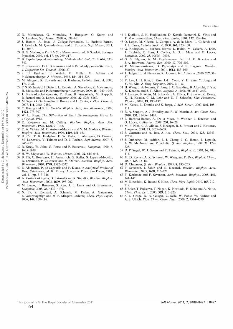
22 D. Momekova, G. Momekov, S. Rangelov, G. Storm andN. Lambov, Soft Matter, 2010, 6, 591–601.
23 J. Ramos, A. Imaz, J. Callejas-Fernandez, L. Barbosa-Barros,J. Estelrich, M. Quesada-Perez and J. Forcada, Soft Matter, 2011,11, 5067.
24 H. G.Merkus, in Particle SizeMeasurements, ed. B. Scarlett, SpringerPijnacker, 2009, vol. 17, pp. 299–317.
25 B. Papahadjopoulos-Sternberg, Methods Mol. Biol., 2010, 606, 333–349.
26 C. Brancewicz, D. H. Rasmussen and B. Papahadjopoulos-Sternberg,J. Dispersion Sci. Technol., 2006, 27.
27 S. U. Egelhaaf, E. Wehrli, M. M€uller, M. Adrian andP. Schurtenberger, J. Microsc., 1996, 184, 214–228.
28 M. Almgren, K. Edwards and G. Karlsson, Colloids Surf., A, 2000,174, 3–21.
29 P. S. Mohanty, H. Dietsch, L. Rubatat, A. Streadner, K. Matsumoto,H. Matsuoka and P. Schurtenberger, Langmuir, 2009, 25, 1940–1948.
30 J. Pereira-Lachataignerais, R. Pons, H. Amenitsch, M. Rappolt,B. Sartori and O. L�opez, Langmuir, 2006, 22, 5256–5260.
31 M. Sega, G. Garberoglio, P. Brocca and L. Cantu, J. Phys. Chem. B,2007, 111, 2484–2489.
32 C. Huang and S. Li, Biochim. Biophys. Acta, Rev. Biomembr., 1999,1422, 273–307.
33 W. L. Bragg, The Diffraction of Short Electromagnetic Waves bya Crystal, 1913.
34 R. Koynova and M. Caffrey, Biochim. Biophys. Acta, Rev.Biomembr., 1998, 1376, 91–145.
35 R. A. Videira, M. C. Antunes-Madeira and V. M. Madeira, Biochim.Biophys. Acta, Biomembr., 1999, 1419, 151–163.
36 H. Cui, T. K. Hodgdon, E. W. Kaler, L. Abezgauz, D. Danino,M. Lubovsky, Y. Talmon and D. J. Pochan, Soft Matter, 2007, 3,945–955.
37 R. Strey, W. Jahn, G. Porte and P. Bassereau, Langmuir, 1990, 6,1635–1639.
38 H. W. Meyer and W. Richter, Micron, 2001, 32, 615–644.39 B. Pili, C. Bourgaux, H. Amenitsch, G. Keller, S. Lepetre-Mouelhi,
D. Desmaele, P. Couvreur and M. Ollivon, Biochim. Biophys. Acta,Biomembr., 2010, 1798, 1522–1532.
40 E. Abignente, P. de Caprariis and F. Klaus, in Analytical Profiles ofDrug Substances, ed. K. Florey, Academic Press, San Diego, 1982,vol. 11, pp. 313–346.
41 A. Kostecka-Gugala, D. Latowski and K. Strzalka, Biochim. Biophys.Acta, Biomembr., 2003, 1609, 193–202.
42 M. Lucio, F. Bringezu, S. Reis, J. L. Lima and G. Brezesinski,Langmuir, 2008, 24, 4132–4139.
43 N. Fa, S. Ronkart, A. Schanck, M. Deleu, A. Gaigneaux,E. Goormaghtigh and M. P. Mingeot-Leclercq, Chem. Phys. Lipids,2006, 144, 108–116.
44 I. Kyrikou, S. K. Hadjikakou, D. Kovala-Demertzi, K. Viras andT. Mavromoustakos, Chem. Phys. Lipids, 2004, 132, 157–169.
45 O. L�opez, M. C�ocera, L. Campos, A. de la Maza, L. Coderch andJ. L. Parra, Colloids Surf., A, 2000, 162, 123–130.
46 G. Rodr�ıguez, L. Barbosa-Barros, L. Rubio, M. C�ocera, A. D�ıez,J. Estelrich, R. Pons, J. Caelles, A. D. l. Maza and O. L�opez,Langmuir, 2009, 25, 10595–10603.
47 G. S. Pilgram, A. M. Engelsma-van Pelt, H. K. Koerten andJ. A. Bouwstra, Pharm. Res., 2000, 17, 796–802.
48 T. Mavromoustakos, D. Papahatjis and P. Laggner, Biochim.Biophys. Acta, Biomembr., 2001, 1512, 183–190.
49 J. Hadgraft, J. d. Plessis and C. Goosen, Int. J. Pharm., 2000, 207, 31–37.
50 Y. Lee, I. H. Kim, J. Kim, J.-H. Yoon, Y. H. Shin, Y. Jung andY. M. Kim, J. Drug Targeting, 2010, 0, 1–8.
51 H. Wang, J. de Joannis, Y. Jiang, J. C. Gaulding, B. Albrecht, F. Yin,K. Khanna and J. T. Kindt, Biophys. J., 2008, 95, 2647–2657.
52 J. Luengo, B. Weiss, M. Schneider, A. Ehlers, F. Stracke, K. Konig,K. H. Kostka, C. M. Lehr and U. F. Schaefer, Skin Pharmacol.Physiol., 2006, 19, 190–197.
53 M. Kozak, L. Domka and S. Jurga, J. Mol. Struct., 2007, 846, 108–111.
54 R. A. Shapiro, A. J. Brindley and R. W. Martin, J. Am. Chem. Soc.,2010, 132, 11406–11407.
55 L. Barbosa-Barros, A. De la Maza, P. Walther, J. Estelrich andO. L�opez, J. Microsc., 2008, 230, 16–26.
56 M.-P. Nieh, C. J. Glinka, S. Krueger, R. S. Prosser and J. Katsaras,Langmuir, 2001, 17, 2629–2638.
57 S. Gaemers and A. Bax, J. Am. Chem. Soc., 2001, 123, 12343–12352.
58 J. Dubochet, M. Adrian, J. J. Chang, J. C. Homo, J. Lepault,A. W. McDowall and P. Schultz, Q. Rev. Biophys., 1988, 21, 129–228.
59 D. P. Siegel, W. J. Green and Y. Talmon, Biophys. J., 1994, 66, 402–414.
60 M. D. Reeves, A. K. Schawel, W. Wang and P. Dea, Biophys. Chem.,2007, 128, 13–18.
61 D. Chapman, Q. Rev. Biophys., 1975, 8, 185–235.62 F. Severcan, I. Sahin and N. Kazanci, Biochim. Biophys. Acta,
Biomembr., 2005, 1668, 215–222.63 F. Korkmaz and F. Severcan, Arch. Biochem. Biophys., 2005, 440,
141–147.64 M. Kinoshita, K. Ito and S. Kato,Chem. Phys. Lipids, 2010, 163, 712–
719.65 J. Bolze, T. Fujisawa, T. Nagao, K. Norisada, H. Saito and A. Naito,
Chem. Phys. Lett., 2000, 329, 215–220.66 S. L. Grage, D. R. Gauger, C. Selle, W. Pohle, W. Richter and
A. S. Ulrich, Phys. Chem. Chem. Phys., 2000, 2, 4574–4579.
This journal is ª The Royal Society of Chemistry 2011 Soft Matter, 2011, 7, 8488–8497 | 8497
Dow
nloa
ded
by C
. de
Inve
st y
Des
arro
llo o
n 10
May
201
2Pu
blis
hed
on 2
7 Ju
ly 2
011
on h
ttp://
pubs
.rsc.
org
| doi
:10.
1039
/C1S
M05
692A
View Online
64
Sistemes�bicel·lars�com�a�nova�estratègia�d’aplicació�tòpica����������������������������������������������������������������������RESULTATS�
E%�
Article�3��
Bicellar�systems�as�new�delivery�strategy�for�topical�application�of�flufenamic�acid�
�
J.�Phys.�Chem.�B&������� � 96�
�
!���5����� ���-����� ���� � �������- ��B������.�������������������������������
�������� ���������0��� -������������0��� �4������� �*���-����������6���
)�� ������ �/� 9� ���� �� ����� � ������� - ��B����� ��-� *� 7*�� ��� #8�� ���
��4 ����� ��� ���� 9� ���� 4����� ��� �5������ � ������6� !�� �5����� ���-���� �0.��
����� ���������� ��������0�-��� 9�������� ��in�vitro��������������������*����
� 4������� ��. ���� 7� ������� - ��B������ ���� ���� 9� ����(� �� � ��� ��������
���� ��86� ���-<� �0.�� ����� ���� ���� *��&� �� �0�4���� ����� � 4������� ��. ����
��-��� ��� ����6� � *�� ���������� � �H��1���� ��� ��� �� � ���(� �� 3�!�� �0.���
� ���� �/����������� -������� ��� �������������5������� �������������������0!�����
��� ����� ���� �0��� � 9� ����� � �������- ��B����� ��-�*6� )�� ��� �� 3*F3&3�� ��� ���
�� � �/��� ��-� ��� ����� ������� ���� �� � ���� �/��� ��� �� ��� ��� ��B������ ��� ��� ����� W�
�0����� � ��� �0!�� �L����� ��� ������ �4���� 9� ��-��� ���� ���� -���� ��� �� ��� ��� �����
����� �/� 9�� ��� ��������������0��� � 9������� �������- ��B�������-�*6�
!��� � ������� - ��B����� ���� �M. - �� ��� �4���� ����������� ��� �S�-��� 9�
������� ������*����������������������. ���6�!��������������0*��&� �� ����3*F3&
3���������������������������������������� �������0!�����������������������������
5������������-��0�4����������������� �0�-��� 9�������� �6�!�� ����� �����4 ���
���3�!�������� �����-������� ��� �� ��� �0���������� �����B��������� ��� ������ �0!�6�
:�������������� �������- ��B�������-�*�����������4�������������������0�-��� 9�
���� ���� �������������������� ��������� ����������������0!�� ���������6��
��� ��� ��� ������ 5��� �0������ 9� ��� ��� ����� <�� ��� ����� �� � ����� �4�����
������� ���� ��� �4���� 9�� ��� ������ 9� � ��� ������ 9� ��� ���� ������������ ��� ���
����� 5��� �������� ���� � ������� - ��B����� ��-� *� ���� ��� ���� �������� ���� ��
-���4 �������������������� � 9��(� �6��
�
�
������

EE�
67
Bicellar systems as new delivery strategy for
topical application of flufenamic acid
L. Rubio*,a, C. Alonsoa, G. Rodrígueza, M. Cóceraa, C. López-Iglesiasb, L. Codercha, A.
De la Mazaa, J. L. Parraa and O. Lópeza.
aDepartment of Chemical and Surfactants Technology (IQAC-CSIC), C/Jordi Girona
18-26, 08034 Barcelona, Spain.
bScientific and Technological Centers-University of Barcelona (CCiT-UB), Barcelona
Science Park, C/Baldiri Reixac, 10, 08028 Barcelona, Spain.
* Corresponding author: Laia Rubio, Department of Chemical and Surfactants
Technology (IQAC-CSIC) C/ Jordi Girona 18-26, 08034, Barcelona, Spain. Phone: 34-
93400 61 00. Fax: 34-93204 59 04. E-mail: [email protected]
EC�
ABSTRACT
In this work, bicellar systems, bilayered disk-shaped nanoaggregates formed in water by
phospholipids, are proposed as a novel strategy for delivery the anti-inflammatory
flufenamic acid (FFA) in the skin. A comparative percutaneous penetration study of this
drug in bicellar systems and other different vehicles has been carried out. Also, the
effects induced on the skin by the application of FFA in the different vehicles have been
analyzed by ATR-FTIR. Additionally, using the microscopic technique FSTEM and
SAXS-SR technique has been studied the possible microstructural and organizational
changes that may be caused to the SC lipids and the collagen of the skin by the
application of FFA bicellar systems. Bicellar systems exhibited a retarder effect on the
percutaneous absorption of FFA with respect to the other vehicles without promoting
disruption in the SC barrier function of the skin. Given that skin disruption is one of the
main effects caused by inflammation, prevention and repair of the skin microstructure
should be one of the goals to get in anti-inflammatory formulations.
Keywords:
Bicellar systems, flufenamic acid, ATR-FTIR, FS-TEM, in vitro percutaneous
absorption, drug delivery.

69
1. Introduction
Bicellar systems are mixtures of two types of phospholipids in aqueous medium, one
with long alkyl hydrophobic chains and another with short alkyl hydrophobic chains.
These phospholipids are able to self-assembly forming different nanoaggregates. The
most common nanostructures formed are discoidal bicelles, which are intermediate
forms between lipid vesicles and classical mixed micelles 1,2. In bicelles, the long-chain
phospholipids constitute the bilayer structure in the central part and the short-chain
phospholipids complete the margins of the disk 3,4. Considering the structure, chemical
composition avoiding surfactants and nanodimensions of these systems, their use as
delivery systems for topical applications emerges as an interesting strategy. The use of
bicellar systems has been previously explored by our group for the transdermal delivery
of the amphiphilic drug diclofenac 5. Additionally, in other works we have
demonstrated that these systems are able to work for skin applications in two ways: as
permeabilizing agents of the skin or as reinforcing agents of the lipid structures present
in the intercellular domains of the outermost layer of the skin, the stratum corneum (SC)
6,7. The skin represents the largest and most easily accessible organ of the body and it is
readily available to drug application. Transdermal drug delivery offers many advantages
compared to the conventional routes of application including avoidance of the first pass
effect, stable blood levels, easy application and higher compliance of the patient 8,9 10. In
order to evaluate data on the passage of substances through the skin as well as on its
distribution over the different cutaneous compartments 11-13 in vitro percutaneous
absorption studies are performed. These studies also permit the evaluation of the
effectiveness of some vehicles and of the permeability enhancer effect to different
compounds 14.
'$�
Flufenamic acid (FFA) is a non-steroidal anti-inflammatory drug of the anthranilic
acid group with potent anti-inflammatory and analgesic effects 15. This drug, that is
commonly included in products for topical application, is insoluble in water and,
organic solvents able to solubilise this drug could impair the skin. Given that skin
disruption is one of the major proinflammatory stimulators 16, prevention and reparation
of the skin microstructure should be one of the goals reach by anti-inflammatory topical
formulations. Thus, in these formulations not only drug should be effective, but also the
vehicle must work in the same address potentiating the drug effect. Additionally, the
own deleterious effects of some anti-inflammatory molecules on the skin structure 17
should be considered and avoided by using appropriate skin repairing vehicles.
Considering these arguments, we think that the use of bicellar systems as carriers of
FFA may be a good alternative for dermal applications. Bicellar systems including FFA
have been previously characterized in a recent work 18 using a combination of different
techniques like dynamic light scattering (DLS), freeze-fracture electron microscopy
(FFEM), Cryo-transmission electron microscopy (Cryo-TEM), x-ray scattering and
differential scanning calorimetry (DSC).
The present study investigates the percutaneous penetration of FFA in different
vehicles, as well as the effects induced on the skin by the application of FFA bicellar
systems. Attenuated total reflectance-Fourier transform infrared (ATR-FTIR)
spectroscopy, small angle X-ray scattering (SAXS) using synchrotron radiation (SR)
and electron microscopy were used. ATR-FTIR is a highly suitable technique to
determine the vibrational characteristic frequencies of the alkyl chain lipids related to
differently ordered phases 19-21. SAXS is an excellent tool to study the structural
organization of the collagen in the skin and the lipid lamellar organization of the SC 22
and freeze-substitution transmission electron microscopy (FSTEM) has been commonly
71
used to visualize the microstructure of the skin 23. In this work, the mentioned
techniques were used to evaluate the microstructure of skin after treatment with FFA
bicellar systems in vitro.
2. Materials and methods
2.1. Materials
Flufenamic acid (FFA) was purchased from Sigma- Aldrich (St. Louis, MO, USA).
Dipalmitoyl phosphatidylcholine (DPPC) and dihexanoyl phosphatidylcholine (DHPC)
were supplied by Avanti Polar Lipids (Alabaster, USA). Bovine Serum Albumin (BSA),
Phosphate Buffer Saline (PBS) and gentamicin were obtained from Sigma Aldrich (St
Louis, MO, USA). Methanol (HPLC Grade), sodium dihydrogen phosphate
monohydrate and ortho-phosphoric acid 85% were acquired from Merck (Darmstadt,
Germany). Purified water was obtained by an ultra-pure water system, Milli-Q plus 185
(Millipore, Bedford, USA). Ethanol absolute was purchased from Carlo Erba (Milano,
Italy). Commercial topic gel containing FFA, salicylic acid and excipients as
isopropanol (Laboratorio Stada, Sant Just Desvern, Spain) was purchased at a local
pharmacy. Trypsin (from porcine pancreas) was obtained from Sigma Aldrich (St Louis,
MO, USA).
The chemicals for preparing microscopy samples were: ruthenium tetroxide (RuO4),
lowicryl HM20, glutaraldehyde and sodium cacodylate buffer which were supplied by
Electron Microscopy Sciences (Hatfield, PA, USA), methanol and potassium
ferrocyanide (K4Fe(CN)6) provided by Merck (Darmstadt, Germany), and osmium
tetroxide (OsO4) purchased from Pelco International (Redding, CA, USA).
'#�
2.2. Methods
2.2.1. Preparation of bicellar systems
Bicellar systems including FFA were formed with dipalmitoyl phosphatidylcholine
(DPPC) as long-alkyl-chain phospholipid and dihexanoyl phosphatidylcholine (DHPC)
as short-alkyl-chain phospholipid. An appropriate amount of DPPC was weighed and
mixed with a DHPC chloroform solution to get DPPC/DHPC in the molar ratio 2:1. 10
mg of FFA were weighed and mixed with the DPPC/DHPC chloroformic solution. The
chloroform was removed by rota-evaporation and the lipid film was dissolved in 1 ml of
water to reach 20% (w/v) of total lipid concentration and sonicated until a transparent
solution was obtained. PH measurements of FFA bicellar systems were performed with
a Model 720 pH meter and a ROSS Model 8103 SC pH electrode, (both from Orion
Research, Cambridge, MA, USA).
2.2.2. HPLC analysis
The quantitative determination of FFA was carried out by High-performance liquid
chromatography (HPLC) using a Hitachi LaChrom Elite equipment (Darmstadt,
Germany). The equipment consisted of an L-2130 pump, L-2200 autosampler and an L-
400 UV detector. The system was operated from the software Merck EZChrom Elite
v3.1.3. The chromatographic separation was performed at room temperature using a
Lichrocart 250-4/Lichrosorb RP-18 (5 μm) column (Merck, Germany) with a flow rate
of 1.0 ml/min. Twenty microlitres of samples or calibration standards were injected into
the column and eluted with a mobile phase of methanol and phosphate buffer (pH 2.5)
(79:21, v/v). Detection was carried out by monitoring the absorbance signals at 284 nm.
The calibration curve exhibited linear behaviour (> 0.99%) over the concentration range
of 0.078–97 �g/mL.
73
2.2.3. In vitro percutaneous absorption studies
The in vitro permeation experiments through porcine skin were performed using
Franz-type diffusion cells (Lara-Spiral, Courtenon, France). The porcine skin was
excised from the dorsal region of female Landrace large white pigs weighing between
30–40 Kg. The pig skin was provided by the Clínic Hospital of Barcelona, Spain. The
bristles were removed carefully with an animal clipper and subsequently the skin was
washed with water. The excised skin was dermatomed to a thickness of 500±50 �m
(Dermatome GA630, Aesculap, Tuttlingen, Germany). Circular pieces of the
dermatomed skin were obtained using an iron punch (2.5 cm of inner diameter) in such
a way that they fit into the Franz diffusion cells. The skin discs were stored at �20 ºC.
Before the experiments, the skin discs were thawed and sandwiched securely between
the two halves of the Franz cells (diffusion area of 1.86 cm2) with the SC side facing the
donor compartment. The receptor chamber was filled with a receptor fluid containing
PBS (pH 7.4) in distilled water, 4% BSA, and 0.04% of gentamicin sulphate.
The receptor phase was kept under constant stirring at 37 ± 1 °C by means of a
circulating water bath (Julabo Labortechnik GmbH, Germany) to ensure that the surface
skin was maintained at 32 ± 1 ºC. The integrity of each skin sample was checked by
determining the transepidermal water loss (TEWL) using a Tewameter TM210
(Courage-Khazaka, Köln, Germany). The diffusion experiment was initiated by
applying to the entire surface, delimited by the upper cell, 10 μL of each of the
ethanolic or bicellar solution of FFA and about 4 mg of the commercial product of FFA.
The doses applied are based on the OCDE recommendations12. The permeation
experiments were conducted in six replicates over 24 h. After the exposure time, the
content of the donor compartment was washed with sodium lauryl ether sulphate and
Milli-Q water following usual procedure, and the washing solution was collected. Then,
'A�
the receptor fluid was removed from the receptor compartment and brought up to 5 ml
in a volumetric flask. Most of the SC of the treated skin area was removed by 8
successive tape-strippings using an adhesive tape (D-Squame®, CuDerm Inc., Dallas,
USA). After that, the viable epidermis was separated from the dermis by heat treatment
at 80 ºC for a few seconds.
The FFA content in the washing solution and in the different skin layers was extracted
with methanol overnight. The different samples extracted and the receptor fluid were
filtered through a 0.45 μm Acrodisc filter (Pall Gelman Sciences, Northampton, UK)
and analyzed by HPLC as it is described above.
2.2.4. ATR-FTIR spectroscopy
Four circular pieces of dermatomed porcine skin were placed in Franz diffusion cells,
as above described. Three of them were treated with 10 μL of FFA ethanolic solution,
FFA bicellar systems or about 10mg of FFA commercial product, respectively. The
other sample was used as a control (untreated). The skin exposure was done during 24h;
after that, the skin was washed with sodium lauryl ether sulphate and Milli-Q water and
removed from the diffusion cells.
Infrared spectra of the porcine skin samples were obtained using a 360-FTIR
spectrophotometer Nicolet Avatar (Nicolet Instruments, Inc., Madison, WI) equipped
with an attenuated total reflection (ATR) accessory that uses a ZnSe crystal with an
angle of incidence of 45° in a horizontal orientation.
Before analysis, the skin was placed with the SC facing onto the ZnSe ATR crystal. In
order to ensure reproducible contact between the sample and the crystal, a pressure of
10 kPa was applied on the samples. The IR spectra were collected at 25 ºC, 32.5 ºC and
37 ºC. These three temperatures have been chosen because 25 ºC is the room
75
temperature, 32.5 ºC is approximately the temperature of the skin surface and 37 ºC is
the physiological temperature. For a correct equilibration of the temperature, the skin
samples were placed in the equipment 30 min before collecting the spectra. The
temperature was controlled by a temperature controller integrated in the ATR device
and by an external thermostatic jacket.
All spectra analysed represent an average of 256 scans obtained in 7 min with a
resolution of 2 cm-1, and the wavenumber range used was 4000-700 cm-1. Peak
positions were determined with the aid of OMNIC software version 7.3 (Nicolet,
Madison, WI).
2.2.5. Freeze-substitution transmission electron microscopy (FSTEM)
Two porcine skin discs, one as a control and one treated with FFA bicellar systems,
were processed as for percutaneous absorption studies. Then, the porcine skin samples
were cut into small rectangular pieces with a size of approximately 2 × 1 mm. The
pieces were fixed with 5% (w/v) glutaraldehyde buffered with 0.1 M sodium cacodylate
(pH 7.2). The postfixation was performed using 0.2% (w/v) of ruthenium tetroxide
(RuO4) in sodium cacodylate buffer (pH 6.8) with 0.25% (w/v) potassium ferrocyanide
(K4Fe (CN)6) for 1 h. After rinsing in buffer and prior to freeze-substitution, the tissue
samples were cryofixed by liquid nitrogen (-196 ºC) using a Cryovacublock (Leica,
Vienna). The freeze-substitution method was done in an Automatic Freeze Substitution
(AFS) system (Leica Microsystems, Vienna, Austria). The freeze-substitution fluid
consisted of 1.0% (w/v) osmium tetroxide (OsO4), 0.5% (w/v) uranyl acetate and 3.0%
(w/v) glutaraldehyde in methanol. The tissue samples were cryosubstituted at -90 °C for
48 h. After the substitution period, the temperature was raised to -50 °C and the samples
were washed 3 times in 100% methanol. After that, the methanol solution was gradually
'E�
replaced by the embedding medium, Lowicryl HM20 (100%). This resin was replaced
after 24 and 48 h by freshly prepared embedding medium. Finally, the samples were
transferred to a mold containing Lowicryl, and were incubated for 48 h at -50 °C under
UVA-radiation, to allow polymerization. Ultrathin sections were prepared on an
ultramicrotome (Ultracut UCT, Leica, Vienna, Austria), transferred to Formvar-coated
grids and examined in a Hitachi 600 transmission electron microscope.
2.2.6. X-ray scattering measurements
2.2.6.1. Whole dermatomed skin
The SAXS profile of control skin and skin treated with FFA bicellar systems as
described in the percutaneous absorption studies section were analysed to characterise
the organisation of the collagen of the skin. SAXS measurements were performed using
the Spanish beamline (BM16) of the European Synchrotron Radiation Facility (ESRF,
Grenoble, France) at room temperature. All of these skin samples were cut into pieces
sections 1 mm thick with a 10 mm diameter and were placed in the holder with the skin
surface perpendicular to the beam. In the holder, skin pieces were sandwiched between
two aluminium plates with two holes, allowing the beam to pass directly through the
samples. The energy of the beam was 12 keV (�= 0.9795 Å). The scattering data were
collected with a SAXS 2D detector MARCCD (Marresearch GmbH, Norderstedt,
Germany) with an active surface area of 165 mm in diameter24 and a single exposure
time of about 60 seconds. The sample-to-detector distance at 5 m (long set-up) was
used. Calibration was performed using silver behenate. Spacing was determined from
axially integrated 2D images using the FIT2D program
(http://www.esrf.eu/computing/scientific/FIT2D) from ESRF. The data were normalised
with transmitted intensity.
77
2.2.6.2. Isolated SC
A control skin and a skin treated with FFA bicellar systems for 24h were heated with
the SC side in contact with a metal plate for 10 seconds at 80 ºC and the epidermis was
scraped off in sheets. To isolate the SC, the epidermal sheets were incubated for 2 h at
37 ºC with the epidermal side in contact with a solution of 0.5 % Trypsin in PBS at pH
7.4. Trypsin is used to remove adherent cells from epidermis. After the 2h, the Trypsin
was removed by several washes of the SC with Milli-Q water.
SAXS measurements of the isolated SC were performed using the same technical
specifications as in the case of whole skin but changing the sample-to-detector distance
to 1.4 m (short set-up) in order to achieve a q range between 0.2 and 2 nm-1.
The SAXS technique provides information about the larger structural units in the
sample, namely, the repeat distance of one structure (collagen for whole dermatomed
skin and lipid lamellar structure for isolated SC). The scattering intensity (I, in arbitrary
units) was measured as a function of the scattering vector q (in reciprocal nm), whose
modulus is defined as:
q= (4� sin�)/�
where � is the scattering angle and � the wavelength of the radiation (0.9795 Å). The
position of the diffraction peaks is directly related to the repeat distance of the
molecular structure, as described by Bragg’s law 25:
2d sin� = n�
in which n and d are the order of the diffraction peak and the repeat distance,
respectively. The different orders of peaks are calculated by:
qn=2�n/d
'C�
where qn is the scattering vector modulus that indicates the position of the nth order
reflection.
2.2.7. Statistical analysis
Percutaneous absorption values are expressed as the mean ± standard deviation (SD)
for six determinations. For group comparisons, analysis of variance (ANOVA) with a
one-way layout was applied. The software used was the STATGRAPHICS plus 5.
Significant differences in the mean values were evaluated by the Student's unpaired t-
test. A p value of less than 0.05 was considered significant. The positions of IR peaks
were obtained as the mean values ± SD of three measurements.
3. Results Visual inspection of FFA bicellar systems showed differences in the appearance
depending on the temperature. At 25 ºC the systems exhibited milky appearance, at 32.5
ºC this sample was fluid and completely transparent and at 37 ºC it became translucent.
No precipitates were appreciated in the bicellar systems at any of these temperatures.
The pH of the FFA bicellar system was around 5.5, in this sense, bicellar systems are an
appropriate vehicle to use in topical applications because under normal conditions, the
skin has a pH between 4 and 5.5 26.
3.1. In vitro percutaneous studies
HPLC analysis showed that in the three cases the extraction efficiency of FFA from
the different skin layers, washing solution and receptor fluid was higher than 97 %. The
higher amount of FFA remained on the skin surface after application using all the
vehicles. This effect was specially noted in the case of FFA bicellar systems, in which
94.5 ± 1.7% of the total amount applied was removed as surface excess. FFA in ethanol
showed the lower value, 67.2 ± 4.6%, and the commercial product showed an
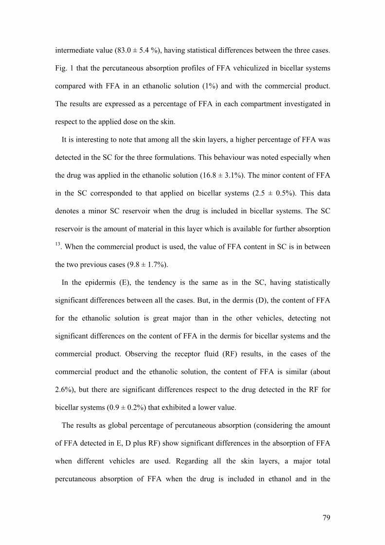
79
intermediate value (83.0 ± 5.4 %), having statistical differences between the three cases.
Fig. 1 that the percutaneous absorption profiles of FFA vehiculized in bicellar systems
compared with FFA in an ethanolic solution (1%) and with the commercial product.
The results are expressed as a percentage of FFA in each compartment investigated in
respect to the applied dose on the skin.
It is interesting to note that among all the skin layers, a higher percentage of FFA was
detected in the SC for the three formulations. This behaviour was noted especially when
the drug was applied in the ethanolic solution (16.8 ± 3.1%). The minor content of FFA
in the SC corresponded to that applied on bicellar systems (2.5 ± 0.5%). This data
denotes a minor SC reservoir when the drug is included in bicellar systems. The SC
reservoir is the amount of material in this layer which is available for further absorption
13. When the commercial product is used, the value of FFA content in SC is in between
the two previous cases (9.8 ± 1.7%).
In the epidermis (E), the tendency is the same as in the SC, having statistically
significant differences between all the cases. But, in the dermis (D), the content of FFA
for the ethanolic solution is great major than in the other vehicles, detecting not
significant differences on the content of FFA in the dermis for bicellar systems and the
commercial product. Observing the receptor fluid (RF) results, in the cases of the
commercial product and the ethanolic solution, the content of FFA is similar (about
2.6%), but there are significant differences respect to the drug detected in the RF for
bicellar systems (0.9 ± 0.2%) that exhibited a lower value.
The results as global percentage of percutaneous absorption (considering the amount
of FFA detected in E, D plus RF) show significant differences in the absorption of FFA
when different vehicles are used. Regarding all the skin layers, a major total
percutaneous absorption of FFA when the drug is included in ethanol and in the
C$�
commercial formulation than when the drug is included in the bicellar systems was
detected.
3.2. ATR-FTIR spectroscopy
Attenuated total reflectance-Fourier transform infrared (ATR-FTIR) is a noninvasive
spectroscopic technique whereby the sample is placed directly onto an ATR crystal.
This technique is useful to investigate the SC without isolation from the other skin
layers. This fact is because the depth of penetration of the radiation to the skin is about
1 �m 27. Previous works have presented IR spectra of porcine skin in vitro 19,21,28 in
order to report the vibrational characteristic frequencies of the alkyl chain lipids related
to differently ordered phases of the SC. In the present work these studies have been
focused in order to investigate the effect of the different systems containing FFA on the
SC lipids.
The skin control and the skin treated with FFA in the different vehicles were analyzed
at 25, 32.5 and 37 ºC. The spectra analysis has been focused on bands associated with
the alkyl chain of SC lipid vibrations: CH2 stretching (around 2920 and 2850 cm-1) and
CH2 scissoring (between 1480 and 1460 cm-1).
3.2.1. CH2 Stretching Region
The methylene stretching vibrations (region between 2800 and 3000 cm-1) provide
information on the conformation ordering (e.g., gel or liquid crystalline) of the SC lipid
alkyl chains 19,21. In our ATR-FTIR spectra and in accordance with other authors 29,30,
the asymmetric and the symmetric CH2 stretching vibrations are observed near 2920
and 2850 cm-1, respectively. In all spectra obtained for all the skin samples, both modes
were intense and not overlapped by other vibrational modes in this region. Tables 1 and
2 show values obtained for the symmetric and asymmetric stretching modes,
81
respectively, for the skin control and for the skin treated with FFA in the different
vehicles and at the three different temperatures. In general, in the case of CH2
symmetric stretching a vibrational frequency of 2849 cm-1 indicates an orthorhombic
chain conformation, of 2850 cm-1 an hexagonal chain conformation and of 2852 cm-1 a
liquid crystalline chain conformation19. The increase of the vibrational frequency
indicates an increase of the disordering. The same occurs with the CH2 asymmetric
stretching, an increase of vibrational frequency denotes a disordering.
Concerning the symmetric mode (Table 1), in the control skin, a small increase of the
CH2 stretching frequency (2850.1 ± 0.6 to 2850.8 ± 0.2 cm-1 ) is observed when the
temperature is increased from 25 to 37 ºC. These results are compatible with a
hexagonal (HEX) packing lattice with a tendency to an increase of disorder packing in
agreement with results published by other authors19,28,31,32. The skin treated with FFA
bicellar systems, at 25 ºC shows a vibrational band for CH2 at 2849.2 ± 0.3 cm-1
corresponding to an orthorhombic (OR) phase. This fact indicates a major ordering of
the SC lipids in the sample treated with the FFA bicellar systems respect to the control
sample. The increase of temperature induces a shift of the CH2 symmetric stretching
frequency to higher values, indicating a transition from OR to HEX phase. At 37 ºC the
skin treated with FFA bicellar systems are in HEX phase. This fact agrees with the
results obtained for Rodriguez et al.28 when DPPC/DHPC bicelles were applied to
porcine skin although those systems did not contain FFA.
When the ethanolic solution of FFA is applied on the porcine skin, the CH2 symmetric
stretching frequency remains practically unaltered respect to the control and also with
the increase of the temperature (a lower increase from 2850.0 ± 0.2 to 2850.3 ± 0.1 cm-
1), corresponding in all cases to an HEX phase 21,28. Results obtained for the application
of the FFA commercial product show a transition from an OR phase to a HEX phase
C#�
with the increase of temperature. This transition was also detected in the case of FFA
bicellar systems as above described.
Although the stretching symmetric vibrations of CH2 are described as more sensitive
than the asymmetric vibrations to packing changes 33, these latter are also used in some
studies for similar purposes 19,27. Table 2 shows the values detected for the CH2
asymmetric stretching mode. Concerning the skin control, from 25 to 32.5 ºC the
frequency remains unaltered (from 2920.1 ± 0.5 to 2920.2 ± 0.4 cm-1) but when the
temperature is increased until 37 ºC a change to higher values of frequency (2922.1 ±
0.5 cm-1) was observed indicating a higher fluidity of the SC lipids at this temperature.
In the case of the skin treated with FFA bicellar systems, the CH2 asymmetric stretching
frequency at 25 ºC was 2917.1 ± 0.2 cm-1, quite bellow to that detected in the control
skin, indicating that treatment with FFA bicellar systems induces an ordering in the SC
lipid lattice. When the temperature is increased at 32.5 ºC the frequency is maintained
but at 37 ºC this value is increased until 2919.9 ± 0.3 cm-1, denoting also an augment of
the lipid fluidization when the temperature increases. In the case of the skin treated with
FFA ethanolic solution, at 25 ºC the value of the frequency was 2917.3 ± 0.5 cm-1, at
32.5 ºC the frequency increased until 2918.3 ± 0.2 cm -1 and at 37 ºC was maintained.
The augment in the disordering is observed when the temperature increases from 25 to
32.5 ºC. When the porcine skin was treated with the FFA commercial, also an increase
of the disordering is noted when the temperature is changed from 25 ºC (2918.5 ± 0.5
cm-1) to 32.5 ºC (2920.1 ± 0.7 cm-1) and, at 37 ºC the increase is slightly smaller (2920.3
± 0.8 cm-1).
Summarizing, respect to the control, FFA bicellar systems and FFA commercial
product induce an ordering of the SC lipids, this fact is noted in both the symmetric and
the asymmetric stretching vibrations. When FFA is applied with the ethanolic solution,
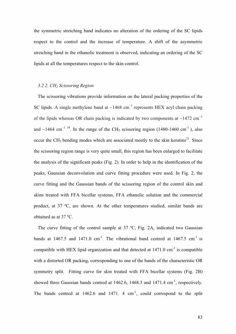
83
the symmetric stretching band indicates no alteration of the ordering of the SC lipids
respect to the control and the increase of temperature. A shift of the asymmetric
stretching band in the ethanolic treatment is observed, indicating an ordering of the SC
lipids at all the temperatures respect to the skin control.
3.2.2. CH2 Scissoring Region
The scissoring vibrations provide information on the lateral packing properties of the
SC lipids. A single methylene band at ~1468 cm�1 represents HEX acyl chain packing
of the lipids whereas OR chain packing is indicated by two components at ~1472 cm�1
and ~1464 cm�1 34. In the range of the CH2 scissoring region (1480-1460 cm-1 ), also
occur the CH3 bending modes which are associated mostly to the skin keratins21. Since
the scissoring region range is very quite small, this region has been enlarged to facilitate
the analysis of the significant peaks (Fig. 2). In order to help in the identification of the
peaks, Gaussian deconvolution and curve fitting procedure were used. In Fig. 2, the
curve fitting and the Gaussian bands of the scissoring region of the control skin and
skins treated with FFA bicellar systems, FFA ethanolic solution and the commercial
product, at 37 ºC, are shown. At the other temperatures studied, similar bands are
obtained as at 37 ºC.
The curve fitting of the control sample at 37 ºC, Fig. 2A, indicated two Gaussian
bands at 1467.5 and 1471.0 cm-1. The vibrational band centred at 1467.5 cm-1 is
compatible with HEX lipid organization and that detected at 1471.0 cm-1 is compatible
with a distorted OR packing, corresponding to one of the bands of the characteristic OR
symmetry split. Fitting curve for skin treated with FFA bicellar systems (Fig. 2B)
showed three Gaussian bands centred at 1462.6, 1468.3 and 1471.4 cm-1, respectively.
The bands centred at 1462.6 and 1471. 4 cm-1, could correspond to the split
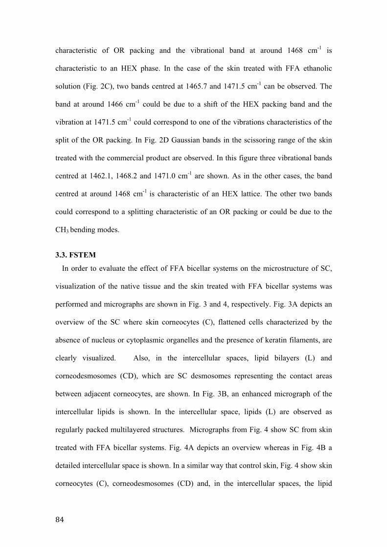
CA�
characteristic of OR packing and the vibrational band at around 1468 cm-1 is
characteristic to an HEX phase. In the case of the skin treated with FFA ethanolic
solution (Fig. 2C), two bands centred at 1465.7 and 1471.5 cm-1 can be observed. The
band at around 1466 cm-1 could be due to a shift of the HEX packing band and the
vibration at 1471.5 cm-1 could correspond to one of the vibrations characteristics of the
split of the OR packing. In Fig. 2D Gaussian bands in the scissoring range of the skin
treated with the commercial product are observed. In this figure three vibrational bands
centred at 1462.1, 1468.2 and 1471.0 cm-1 are shown. As in the other cases, the band
centred at around 1468 cm-1 is characteristic of an HEX lattice. The other two bands
could correspond to a splitting characteristic of an OR packing or could be due to the
CH3 bending modes.
3.3. FSTEM
In order to evaluate the effect of FFA bicellar systems on the microstructure of SC,
visualization of the native tissue and the skin treated with FFA bicellar systems was
performed and micrographs are shown in Fig. 3 and 4, respectively. Fig. 3A depicts an
overview of the SC where skin corneocytes (C), flattened cells characterized by the
absence of nucleus or cytoplasmic organelles and the presence of keratin filaments, are
clearly visualized. Also, in the intercellular spaces, lipid bilayers (L) and
corneodesmosomes (CD), which are SC desmosomes representing the contact areas
between adjacent corneocytes, are shown. In Fig. 3B, an enhanced micrograph of the
intercellular lipids is shown. In the intercellular space, lipids (L) are observed as
regularly packed multilayered structures. Micrographs from Fig. 4 show SC from skin
treated with FFA bicellar systems. Fig. 4A depicts an overview whereas in Fig. 4B a
detailed intercellular space is shown. In a similar way that control skin, Fig. 4 show skin
corneocytes (C), corneodesmosomes (CD) and, in the intercellular spaces, the lipid
85
lamellae (L) were also clearly visualized. No differences were observed in the different
areas of SC after treatment with FFA bicellar systems in comparison to native tissue.
This fact indicates that the treatment with these systems did not induce apparent
miscrostructural changes in the SC organization.
3.4. X-ray scattering results
Dermatomed whole skin
Fig. 5 shows the 1D scattering patterns from control skin and skin treated with FFA
bicellar systems resulting from the average of several images after integration with
FIT2D software. The dermatomed skin samples used has a dermis thickness of about
400 μm. The profiles of both samples show the characteristic peaks of the typical
scattering pattern of collagen. In both cases, the characteristic peak centred at q�0.1 nm-
1 corresponding to a d-spacing around 63 nm associated to the axial periodicity of
collagen 22 (marked region) and their reflections are clearly visualized. The typical axial
periodicity of collagen is the result of the displacement of each molecule with respect to
the adjacent molecules in a particular direction 22.
At low q, differences in the intensity between both samples can be observed. In Fig. 6,
a magnification of the SAXS profiles at low q (grey area in Fig. 5) has been plotted in
order to elucidate these differences.
Fig. 6 shows the experimental (squared) and fitted (continuous line) curves of the
SAXS profile of the control skin (A) and the skin treated with FFA bicellar systems (B),
at low q. Differences in intensity and number of bands between these two samples are
observed. The curves were fitting to two (control skin) or three (skin treated) Gaussian
bands (discontinuous lines) using the Origin software. The resulting bands for control
skin were centred at q around 0.055 and 0.099 nm-1 corresponding to a d-spacing around
114, and 63 nm. For treated skin, the resulting bands were centred at q around 0.054,
CE�
0.075 and 0.098 nm-1 corresponding to spacings of around 116, 84, and 64 nm.
Mérigoux C. and co-workers 35 found the same spacings in the study of the skin, which
were associated with the axial (65 nm), the lateral packing of collagen fibrils (115 nm),
and the scattering from the cylindrical shape of the fibrils (85 nm) in skin collagen.
Thus, reflexions detected in our experiments are probably associated to the organization
of skin collagen. Modification in the collagen scattering pattern after treatment with the
bicellar system including FFA is indicative of the ability of the system to reach the
dermis in which collagen is located. Differences between scattering patterns in both
samples could be attributed to an effect of the anti-inflammatory molecules on the
collagen arrangement.
Isolated SC
Fig. 7 shows the SAXS pattern of SC in the control skin and the skin treated with
FFA bicellar systems. The pattern of the control skin shows a band centred at a q around
1.05 nm-1 corresponding to a d value around 6 nm. This d-spacing is associated to the
lamellar arrangement of the lipids. When the skin is treated with the FFA bicellar
systems, a shift of the position to lower q (around 0.87 nm-1 ) corresponding to a d value
of about 7.2 nm and an increase of the intensity are observed. Also, a small shoulder
centred at a q about 1.26 nm-1 (d�5 nm) appears. It can be deduced that the treatment
with FFA bicellar systems induces some ordering in the SC lipid structure.
4. Discussion
Bicellar systems containing FFA have been widely characterized in a previous work
18. These lipid systems allow the inclusion of the lipophilic drug FFA without using
organic solvents and, have demonstrated to exhibit an interesting morphological
versatility. Due to these facts and considering that similar bicellar systems have been
evaluated for skin applications5, the present work proposes to study the potential of
C'�
bicellar systems as drug delivery strategy through the skin without damaging the tissue.
Discussion of the results is carried out from two different viewpoints: the effect of the
bicellar system as retarder of the drug absorption and the effect of the bicellar system on
the skin structure and functionality.
4.1. Bicellar systems as retarder of FFA percutaneous absorption
The results obtained in the percutaneous absorption studies of FFA in the different
vehicles regarding all the skin layers show the lowest value when the drug is included in
bicellar system and the highest when FFA is dissolved in ethanol.
The high percutaneous absorption values obtained for the ethanolic solution are
probably related to the well known penetration enhancer effect of this solvent 36. Merle
et al. 37 reported a reduction of the SC thickness associated to a solubilisation of the SC
lipids by effect of ethanol. These effects on the SC could potentiate the permeability of
the tissue. An enhancement in the permeability is associated in most cases to a
fluidification of SC lipids that involves higher movement of the lipid alkyl chains in
these areas 38. However, this fact does not seem to occur in samples treated with FFA
ethanolic solution considering ATR-FTIR results. The position of the symmetric and
asymmetric stretching bands of skin lipids after treatment with FFA ethanolic solution
showed a slight ordering rather than the expected disordering of the intercellular lipid
alkyl chains respect to the skin control. These results are in accordance with a previous
work of Bommannan et al.39 which indicates that while ethanol extracts appreciable
amounts of lipids from the SC, it does not appear to induce lipid disorder. Our FTIR
scissoring results, indicate a shift of the characteristic HEX peak to lower values, this
fact could be associated with the extraction of some of the SC lipids. The change in the
lipid composition would induce a certain disturbance in the SC barrier function that it is
evidenced in our results as an increase of the skin permeability. The band that remains
CC�
at 1465.7 cm-1 could be related to the ceramides more resistant to the ethanol
extraction37.The results obtained corroborate that lipid extraction rather than disruption
of lipid order, may be the predominant mechanism of ethanol as enhancer. Another
possible complementary explanation was suggested by Lewis et al.40. This group
indicated that the apparent ordering produced by ethanol treatment is due to an
interdigitation effect of the lipid hydrocarbon chains by displacement of bound water
molecules at the lipid headgroup/membrane interface region.
The percutaneous absorption of FFA included in the commercial product was lower
than the percutaneous absorption when the drug was included in the ethanolic solution,
although higher than in the case of FFA in bicellar systems. This is probably due to the
composition of the commercial product that, apart from FFA, consists in 2% of salicylic
acid. This compound is a keratolytic molecule able to alter the barrier properties of the
skin and provokes a desquamation of corneocytes that reduces the SC barrier function41-
43. Therefore, it could enhance the permeability of the SC. The ATR-FTIR results
showed an ordering of the SC lipids respect to the control when the commercial product
is applied. This apparent ordering could be due to alcoholic excipients of the
commercial product, such as isopropanol and polypropylene glycol, that could extract
part of the SC lipids and could induce similar effects on ATR-FTIR bands, as ethanol.
When FFA is included in bicellar systems and is applied on the skin, the total
percutaneous absorption results indicate the lowest absorption respect to the treatments
with the other two vehicles. These results suggest a retarder effect of the percutaneous
absorption when the drug is included in bicellar systems. The concept of retarder was
described by Schaefer and co-workers 13 and it is associated to agents which reduce the
percutaneous absorption of compounds through augmentation of skin barrier function.
Possibly, lipids from bicelles interact with the SC lipids modifying the organization of
CG�
the lipids bilayers and increasing the SC barrier function. This effect was also described
by some authors for certain compounds such as some iminosulfurane analogs44,45 and
laurocapram analog N091545,46. The phospholipid dimyristoyl phosphatidylcholine
(DMPC) was also described as retarder of the percutaneous absorption of the drug
flurbiprofen in comparison to other compounds such as unsaturated fatty acids and
cyclic monoterpenes able to work as enhancers 47. In that paper, the retarder effect of
DMPC was associated to the gel-liquid phase transition temperature (Tm) 23 ºC, of this
phospholipid, higher than that described for the other molecules used in the study. In the
case of FFA bicellar systems, the Tm studied by DSC in a previous work was higher
(38.9 ºC) 18 than the described for DMPC. This fact involves that at the experimental
temperature the system applied is in the gel state. It is well known that gel-state
phospholipids do not affect, or even increase, the skin lipid barrier 47,48, resulting in our
case, in a reduction of the drug permeation. The retarder effect of FFA bicellar systems
is in accordance with the results obtained by our group5 in which diclofenac
diethylamine was included in DPPC/DHPC bicellar systems. In that case the same
retarder effect was observed. This behaviour could be useful in the topical application
of drugs that exhibit too fast percutaneous absorption rates, as it is the case of fentanyl,
to prevent an overdose of the drug 49, or the vehiculization of cosmetic ingredients that
should not penetrate in a great extend or in dermatological formulations to be applied on
skin with the barrier function impaired because could prevent the penetration to the
systemic level. This last topic is being currently studied in our laboratory.
4.2. FFA bicellar systems did not disturb the SC lipid organization Images of SC from control and treated FFA bicellar systems showed similar
microstructures comparable to those published previously by other authors for native
skin 50,51. The well organized lamellar lipid structure in both samples indicates that no
G$�
structural modifications in the SC were induced by the FFA bicellar system treatment.
The effect of different bicellar systems on the SC microstructure has been previously
studied by our group. We reported that DMPC/DHPC bicelles did not promoted
changes in the organization of the SC lipid lamellae 52,53 whereas DPPC/DHPC bicelles
induced formation of vesicles in the lipid areas 51,53. This different behaviour may be
understood regarding the Tm associated to the long alkyl chain phospholipids of each
system (23 ºC for DMPC and 41 ºC for DPPC). Another factor to be considered is the
ability of bicellar system to increase their size as the water content in the system
increases 6. When DMPC/DHPC bicelles are applied on the skin at physiological
temperature (37 ºC) this system suffers a phase transition from gel to liquid crystalline
state. This change induces an increase in the size of the bicellar structures that hinders
the pass of bicelles through the SC. In the case of DPPC/DHPC system, with higher Tm,
the contact with the skin at 37 ºC does not promote the phase transition and no
modification of the size occurs allowing that these systems penetrate through the SC.
According to our experience, inside the skin these bicelles grow as a consequence of a
dilution caused by the water located into the skin6. In the present work, although FFA
bicellar systems are composed by DPPC and DHPC no changes in the SC lipid areas
were detected. This is due to the fact that when DPPC/DHPC includes FFA, the size of
the nanostructures and phase behaviour of the system is modified. The new bicellar
system with FFA forms larger aggregates than those detected in the system without FFA
18. This could hinder the pass thought the SC and, as a consequence, no new lipid
structures are visualized in the SC after treatment, as our FSTEM micrographs showed.
In spite of the absence of new lamellar or vesicular structures after treatment with
bicellar systems containing FFA, a certain effect was detected in the lipid organization
of this sample. Thus, a partial diffusion of lipids and of FFA from bicellar systems into
G>�
the SC could be assumed. ATR-FTIR and SAXS results showed an ordering of the SC
lipids respect to the control that agrees with the retarder effect in the permeability
described above. This increase in the order the SC lipids could be induced by the
inclusion of DPPC from the bicellar system into the lipid bilayers of the tissue. This
phospholipid in a gel ordered phase at the experimental temperature could promote an
additional order in the lamellar SC structure 28. SAXS experiments performed in SC
samples (control and treated with FFA bicellar systems) showed an increase of the d-
spacing corresponding to the short periodicity phase (SPP) of the SC lamellar phases
reported by Bouwstra et al.54. The d-spacing is the sum of the lipid bilayer thickness and
the thickness of one adjacent interlamellar water layer 55. For this reason, the increase of
the d-spacing when the SC is treated with FFA bicellar systems could be promoted by
the incorporation of water on lamellae, or incorporation of lipids in gel phase. This fact
corroborates the interaction of bicelles with SC lipids. The higher intensity of the SPP
band in the case of the SC treated respect to the control indicates an increase of the
ordering of the SC lipid structure. In any case, no extraction of SC lipids should be
expected after treatment with the bicellar systems; on the contrary, an enrichment of
these areas could be envisaged by effect of the treatment and, considering results from
percutaneous absorption, also a reinforcement of the SC barrier function.
Our results indicate that using bicellar systems is possible to introduce small amounts
of an anti-inflammatory drug in the skin without disturb the tissue. It has been
documented that skin barrier disruption causes chronic inflammation 16. Because of this,
it becomes crucial to adjust treatment and prevention strategies for reversing and
preventing skin barrier disruption. In this sense, the use of anti-inflammatory drugs
should be linked to the use of vehicles able to avoid skin damage. Results presented in
this work should stimulate to skin care providers to be more focussed on optimizing the
G#�
SC barrier function to prevent and reverse the skin damage, which is cause and effect of
chronic inflammation. The use of bicellar systems as vehicle, in spite of reducing the
amount of anti-inflammatory drug absorbed, could be more effective on treatment of
skin inflammation by improving skin structure and functionality. Future in vivo
research studies are open to explore the potential of this strategy.
5. Conclusions
Ethanol as vehicle and salicylic acid contained in the commercial product alter the
skin barrier function in different ways, as lipid solubilising or as keratolytic agent,
respectively. Instead, bicellar systems did not disturb the SC barrier function but seems
to prevent and reinforce skin microstructure and functionality. The results of our work
may be useful to develop bicellar systems as an alternative vehicle for drugs that need to
be solubilised by organic solvents and that have a too fast percutaneous absorption.
These drugs could be applied on the skin without the danger of potential toxicity and of
delirious effects on the skin structure.
Acknowledgements
The authors acknowledge Ministerio de Educación y Ciencia, Spain (CTQ2010-
16964), and Generalitat de Catalunya (2009 SGR 1212). This work was supported by
the European Social Fund (Programa Nacional de Potenciación de Recursos Humanos
del Plan Nacional de I+D+I (2004-2008)). Mercedes Cócera is funded by the JAE-Doc
Program from CSIC (co-funded by FSE). Additionally, the authors wish to thank
Elisenda Coll for her expert assistance with the techniques used in this work.
G@�
References
(1) Sanders, C. R.; Prosser, R. S. Structure 1998, 6, 1227.
(2) Seddon, A. M.; Curnow, P.; Booth, P. J. Biochim Biophys Acta 2004,
1666, 105.
(3) Sanders, C. R.; Schwonek, J. P. Biochemistry 1992, 31, 8898.
(4) Vold, R. R.; Prosser, R. S. J Magn Reson, Ser B 1996, 113, 267.
(5) Rubio, L.; Alonso, C.; Rodríguez, G.; Barbosa-Barros, L.; Coderch, L.;
De la Maza, A.; Parra, J. L.; López, O. Int J Pharm 2010, 386, 108.
(6) Barbosa-Barros, L.; de la Maza, A.; Estelrich, J.; Linares, A. M.; Feliz,
M.; Walther, P.; Pons, R.; Lopez, O. Langmuir 2008, 24, 5700.
(7) Barbosa-Barros, L.; De la Maza, A.; Walther, P.; Estelrich, J.; Lopez, O.
J Microsc 2008, 230, 16.
(8) Thomas, B. J.; Finnin, B. C. Drug Discov Today 2004, 9, 697.
(9) Prausnitz, M. R.; Mitragotri, S.; Langer, R. Nat Rev Drug Discov 2004,
3, 115.
(10) Bal, S. M.; Ding, Z.; van Riet, E.; Jiskoot, W.; Bouwstra, J. A. J Control
Release 2010, 148, 266.
(11) Elias, P. M.; Feingold, K. R. Skin barrier; Taylor and Francis group:
New york, 2006.
GA�
(12) OECD. Guideline 428: Skin Absorption: In Vitro Method. In OECD
Guidelines for the Testing of Chemicals; Development, O. f. E. C. a., Ed. Paris, 2004;
pp 8.
(13) Schaefer, H.; Redermeier, T. E. Skin Barrier—Principles of
Percutaneous absorption; Karger-Verlag: Basel, 1996.
(14) Gwak, H. S.; Chun, I. K. Int J Pharm 2002, 236, 57.
(15) Wagner, H.; Kostka, K. H.; Adelhardt, W.; Schaefer, U. F. Eur J Pharm
Biopharm 2004, 58, 121.
(16) Rahman, S.; Collins, M.; Williams, C. M.; Ma, H. L. Inflamm Allergy
Drug Targets 2011, 10, 486.
(17) Schacke, H.; Docke, W. D.; Asadullah, K. Pharmacol Ther 2002, 96, 23.
(18) Rubio, L.; Rodríguez, G.; Alonso, C.; López-Iglesias, C.; Cócera, M.;
Coderch, L.; De la Maza, A.; Parra, J. L.; López, O. Soft Matter 2011, 7, 8488.
(19) Rodríguez, G.; Barbosa-Barros, L.; Rubio, L.; Cócera, M.; Díez, A.;
Estelrich, J.; Pons, R.; Caelles, J.; De la Maza, A.; López, O. Langmuir 2009, 25,
10595.
(20) Obata, Y.; Utsumi, S.; Watanabe, H.; Suda, M.; Tokudome, Y.; Otsuka,
M.; Takayama, K. Int J Pharm 2010, 389, 18.
(21) Boncheva, M.; Damien, F.; Normand, V. Biochim Biophys Acta 2008,
1778, 1344.
G%�
(22) Cócera, M.; Rodríguez, G.; Rubio, L.; Barbosa-Barros, L.; Benseny-
Cases, N.; Cladera, J.; Sabes, M.; Fauth, F.; de la Maza, A.; López, O. Soft Matter 2011,
7, 8605.
(23) Van den Bergh, B. A.; Swartzendruber, D. C.; Bos-Van der Geest, A.;
Hoogstraate, J. J.; Schrijvers, A. H.; Boddé, H. E.; Junginger, H. E.; Bouwstra, J. A. J
Microsc 1997, 187 ( Pt 2), 125.
(24) Rueda, D. R.; García-Gutiérrez, M. C.; Nogales, A.; Capitán, M. J.;
Ezquerra, T. A.; Labrador, A.; Fraga, E.; Beltrán, D.; Juanhuix, J.; Herranz, J. F.;
Bordas, J. Rev Sci Instrum 2006, 77, 033904.1.
(25) Bragg, W. L. “The Diffraction of Short Electromagnetic Waves by a
Crystal”; Proceedings of the Cambridge Philosophical Society, 1913.
(26) Parra, J. L.; Paye, M. Skin Pharmacol Appl Skin Physiol 2003, 16, 188.
(27) Coderch, L.; de Pera, M.; Perez-Cullell, N.; Estelrich, J.; de la Maza, A.;
Parra, J. L. Skin Pharmacol Appl Skin Physiol 1999, 12, 235.
(28) Rodriguez, G.; Rubio, L.; Cocera, M.; Estelrich, J.; Pons, R.; de la Maza,
A.; Lopez, O. Langmuir 2010, 26, 10578.
(29) Gooris, G. S.; Bouwstra, J. A. Biophys J 2007, 92, 2785.
(30) Prasch, T.; Knubel, G.; Schmidt-Fonk, K.; Ortanderl, S.; Nieveler, S.;
Forster, T. Int J Cosmet Sci 2000, 22, 371.
(31) Furuishi, T.; Fukami, T.; Suzuki, T.; Takayama, K.; Tomono, K. Biol
Pharm Bull 2010, 33, 294.
GE�
(32) Pensack, R. D.; Michniak, B. B.; Moore, D. J.; Mendelsohn, R. Appl
Spectrosc 2006, 60, 1399.
(33) Mendelsohn, R.; Flach, C. R.; Moore, D. J. Biochim Biophys Acta 2006,
1758, 923.
(34) Kaushik, D.; Michniak-Kohn, B. AAPS PharmSciTech 2010, 11, 1068.
(35) Mérigoux, C.; Durand, D.; Doucet, J.; Eugéne, M.; Diat, O. Newsletter
ESRF 1997, July, 18.
(36) Thong, H. Y.; Zhai, H.; Maibach, H. I. Skin Pharmacol Physiol 2007, 20,
272.
(37) Merle, C.; Baillet-Guffroy, A. Biochimica et Biophysica Acta (BBA) -
Biomembranes 2009, 1788, 1092.
(38) Williams, A. C.; Barry, B. W. Chemical Permeation Enhancement. In
Enhancement in Drug Delivery; Touitou, E., Barry, B. W., Eds.; CRC Press, 2006.
(39) Bommannan, D.; Potts, R. O.; Guy, R. H. Journal of Controlled Release
1991, 16, 299.
(40) Lewis, E. N.; Levin, I. W.; Steer, C. J. Biochim Biophys Acta 1989, 986,
161.
(41) Lebwohl, M. Int J Dermatol 1999, 38, 16.
(42) Dias, M.; Hadgraft, J.; Lane, M. E. Int J Pharm 2007, 340, 65.
(43) Bashir, S. J.; Dreher, F.; Chew, A. L.; Zhai, H.; Levin, C.; Stern, R.;
Maibach, H. I. Int J Pharm 2005, 292, 187.
G'�
(44) Kim, N.; El-Khalili, M.; Henary, M. M.; Strekowski, L.; Michniak, B. B.
Int J Pharm 1999, 187, 219.
(45) Asbill, C. S.; Michniak, B. B. PSTT 2000, 3, 36.
(46) Hadgraft, J.; Peck, J.; D.G., W.; Pugh, W. J.; Allan, G. Int. J. Pharm.
1996, 141 17.
(47) Fang, J. Y.; Hwang, T. L.; Leu, Y. L. Int J Pharm 2003, 250, 313.
(48) Kirjavainen, M.; Monkkonen, J.; Saukkosaari, M.; Valjakka-Koskela, R.;
Kiesvaara, J.; Urtti, A. J Control Release 1999, 58, 207.
(49) Frolich, M. A.; Giannotti, A.; Modell, J. H. Anesth Analg 2001, 93, 647.
(50) López, O.; Cócera, M.; Walther, P.; Wehrli, E.; Coderch, L.; Parra, J. L.;
de la Maza, A. Micron 2001, 32, 201.
(51) Rodríguez, G.; Barbosa-Barros, L.; Rubio, L.; Cócera, M.; López-
Iglesias, C.; de la Maza, A.; López, O. Colloids Surf B Biointerfaces 2011, 84, 390.
(52) Barbosa-Barros, L.; Barba, C.; Cocera, M.; Coderch, L.; Lopez-Iglesias,
C.; de la Maza, A.; Lopez, O. Int J Pharm 2008, 352, 263.
(53) Barbosa-Barros, L.; Barba, C.; Rodriguez, G.; Cocera, M.; Coderch, L.;
Lopez-Iglesias, C.; de la Maza, A.; Lopez, O. Mol Pharm 2009, 6, 1237.
(54) Bouwstra, J. A.; Gooris, G. S.; van der Spek, J. A.; Bras, W. J Invest
Dermatol 1991, 97, 1005.
(55) de Jager, M. W.; Gooris, G. S.; Ponec, M.; Bouwstra, J. A. J Lipid Res
2005, 46, 2649.
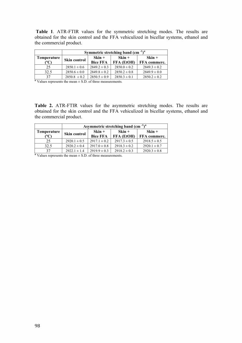
GC�
Table 1. ATR-FTIR values for the symmetric stretching modes. The results are obtained for the skin control and the FFA vehiculized in bicellar systems, ethanol and the commercial product.
Symmetric stretching band (cm -1)� Temperature
(ºC) Skin control Skin + Bice FFA
Skin + FFA (EtOH)
Skin + FFA commerc.
25 2850.1 ± 0.6 2849.2 ± 0.3 2850.0 ± 0.2 2849.3 ± 0.2 32.5 2850.6 ± 0.0 2849.8 ± 0.2 2850.2 ± 0.8 2849.9 ± 0.0 37 2850.8 ± 0.2 2850.5 ± 0.9 2850.3 ± 0.1 2850.2 ± 0.2
� Values represents the mean ± S.D. of three measurements.
Table 2. ATR-FTIR values for the asymmetric stretching modes. The results are obtained for the skin control and the FFA vehiculized in bicellar systems, ethanol and the commercial product.
Asymmetric stretching band (cm -1)� Temperature
(ºC) Skin control Skin + Bice FFA
Skin + FFA (EtOH)
Skin + FFA commerc.
25 2920.1 ± 0.5 2917.1 ± 0.2 2917.3 ± 0.5 2918.5 ± 0.5 32.5 2920.2 ± 0.4 2917.0 ± 0.8 2918.3 ± 0.2 2920.1 ± 0.7 37 2922.1 ± 1.4 2919.9 ± 0.3 2918.2 ± 0.3 2920.3 ± 0.8
� Values represents the mean ± S.D. of three measurements.
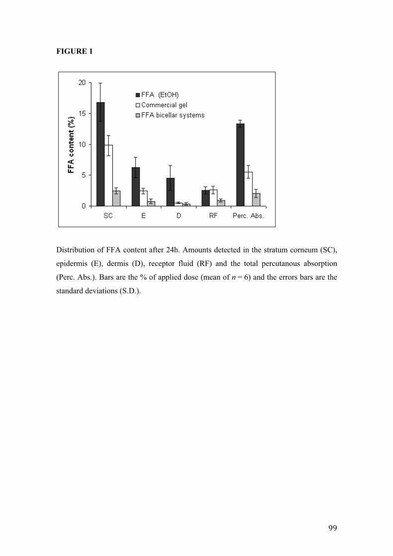
FIGURE 1
Distribution of FFA content after 24h. Amounts detected in the stratum corneum (SC),
epidermis (E), dermis (D), receptor fluid (RF) and the total percutanous absorption
(Perc. Abs.). Bars are the % of applied dose (mean of n = 6) and the errors bars are the
standard deviations (S.D.).
GG�
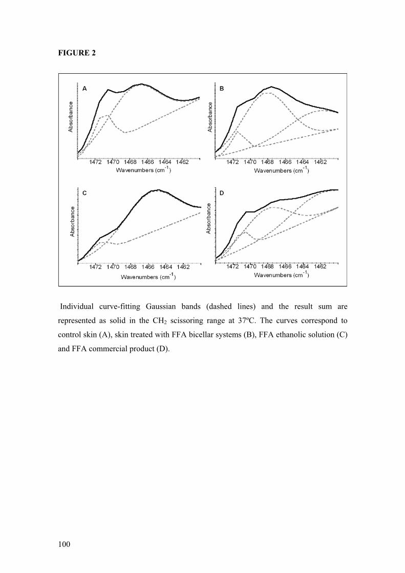
FIGURE 2
Individual curve-fitting Gaussian bands (dashed lines) and the result sum are
represented as solid in the CH2 scissoring range at 37ºC. The curves correspond to
control skin (A), skin treated with FFA bicellar systems (B), FFA ethanolic solution (C)
and FFA commercial product (D).
100
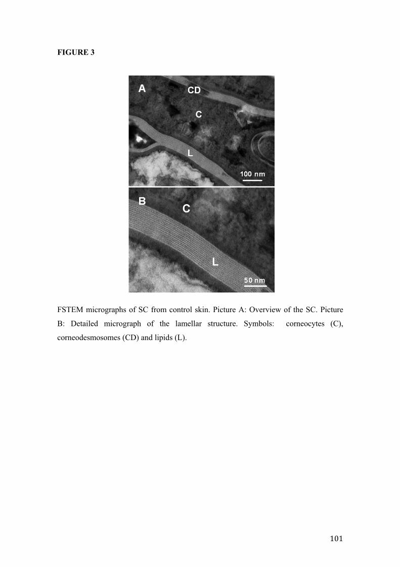
FIGURE 3
FSTEM micrographs of SC from control skin. Picture A: Overview of the SC. Picture
B: Detailed micrograph of the lamellar structure. Symbols: corneocytes (C),
corneodesmosomes (CD) and lipids (L).
>$>�
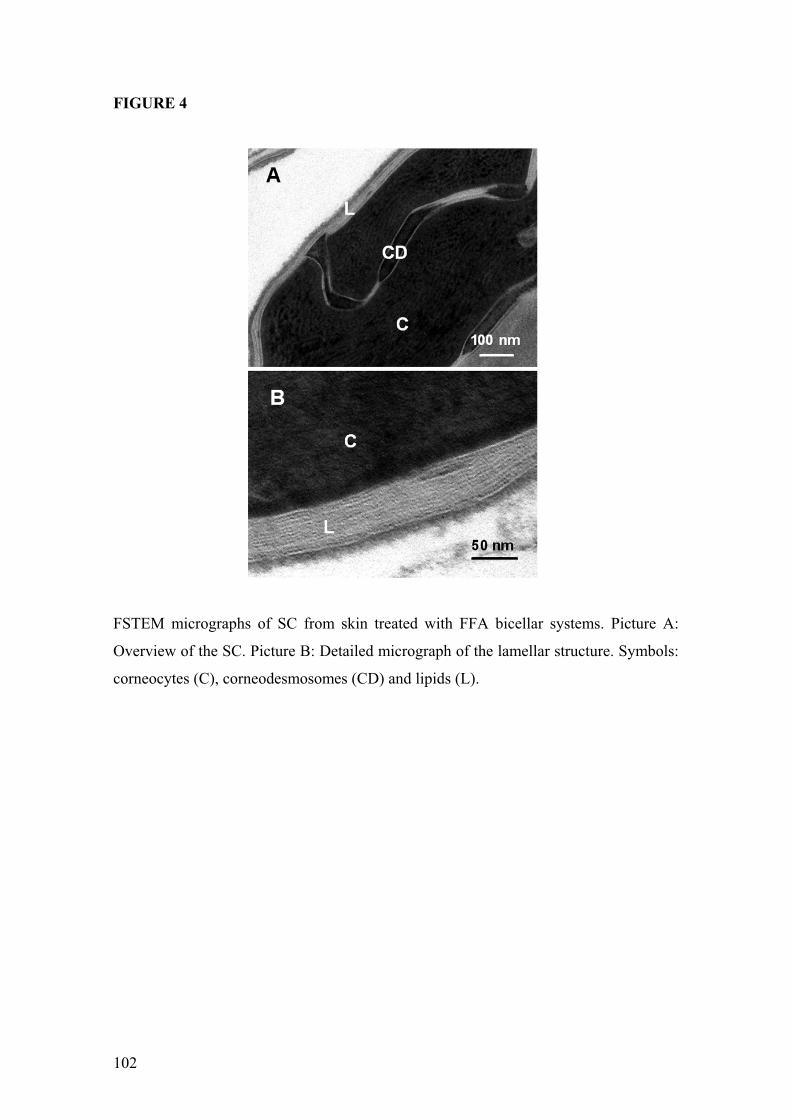
FIGURE 4
FSTEM micrographs of SC from skin treated with FFA bicellar systems. Picture A:
Overview of the SC. Picture B: Detailed micrograph of the lamellar structure. Symbols:
corneocytes (C), corneodesmosomes (CD) and lipids (L).
102
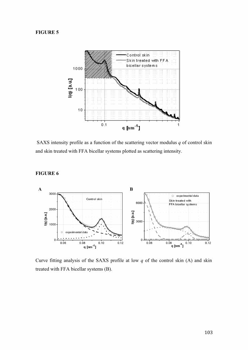
FIGURE 5
SAXS intensity profile as a function of the scattering vector modulus q of control skin
and skin treated with FFA bicellar systems plotted as scattering intensity.
FIGURE 6
Curve fitting analysis of the SAXS profile at low q of the control skin (A) and skin
treated with FFA bicellar systems (B).
>$@�
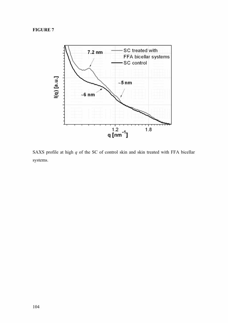
FIGURE 7
SAXS profile at high q of the SC of control skin and skin treated with FFA bicellar
systems.
104
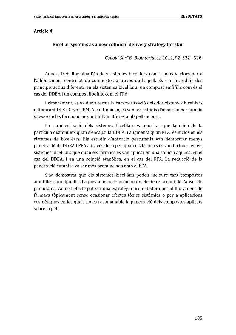
Sistemes�bicel·lars�com�a�nova�estratègia�d’aplicació�tòpica����������������������������������������������������������������������RESULTATS�
>$%�
Article�4��
Bicellar�systems�as�a�new�colloidal�delivery�strategy�for�skin��
Colloid�Surf�B��Biointerfaces,�#$>#��G#��@##i�@#E6�
�
*5����� ���-���� ������� �0U������� � �������- ��B����� ���������� ����������� ��
�0��� -�������� ��������� ��� ��������� �� ����<�� ��� ��� ����6� !�� ���� ������ �� ����
�� � � ���� ���� 4��������������� �������- ��B������������������4 4�� ����<�����
���������!*� ������������ ��4�� �������*6�
:� ������������������������������������� �/� 9����������� �������- ��B�����
� �H��1�����)3� ���2�&�!�6�*���� ��� 9���������4�������� ���0�-��� 9�������� ��
in�vitr � ( � �o��������4������ ������ �4����� � �����-� ����������6�
)�� ������ �/� 9� ����� � ������� - ��B����� ��� �������� 5��� ��� � ��� ��� ���
���������� �� ��� M�5�����0�����������!*�� ����������5����*��<�� ��(���������
� ������� ��� - ��B����6� !��� ����� �� �0�-��� 9� ������� �� ���� ���������� ���2��
������� 9������!*� �*�������<�������������5��������4������������� ��������������
� �������- ��B�����5���5��������4���������������� �������������� 9��5������������
��� ���� ��!*�� � ��� ���� ���� 9� ����(� ��� ��� ��� ��� ���� *6� )�� ���� 9� ��� ���
������� 9����� ����������<�������� ������-����*6��
30.�� ���������� 5��� ���� � ������� - ��B����� ������ ������� ����� ���������
��4 4�� ������ ��4�� �� ��5������ ���� 9������������4������������������S�-��� 9�
������� �6�*5������4������������������������ ����������������������� �����������
4������ �(� ������ ������ ��� ����� �4����� �(M �� � ���� �� �� ���� �� ��� � ����
����� 5�����������5������������������-������������� 9������������������ ����
�-�� �������6��� �
�
�
�
�
�
�
�
�
�
�
�
�
�
�

>$E�
Colloids and Surfaces B: Biointerfaces 92 (2012) 322– 326
Contents lists available at SciVerse ScienceDirect
Colloids and Surfaces B: Biointerfaces
j our na l ho me p age: www.elsev ier .com/ locate /co lsur fb
Bicellar systems as a new colloidal delivery strategy for skin
L. Rubio, G. Rodríguez, L. Barbosa-Barros, C. Alonso, M. Cócera, A. de la Maza, J.L. Parra, O. López ∗
Departament de Tecnología Química i de Tensioactius, Institut de Química Avanc ada de Catalunya (I.Q.A.C.), Consejo Superior de Investigaciones Científicas (C.S.I.C.), C/Jordi Girona
18-26, 08034 Barcelona, Spain
a r t i c l e i n f o
Article history:
Received 6 July 2011
Received in revised form 7 October 2011
Accepted 6 December 2011
Available online 14 December 2011
Keywords:
Bicellar systems
In vitro percutaneous absorption
Diclofenac
Flufenamic acid
Dipalmitoyl-glycero-phosphatidylcholine
Dimyristoyl-glycero-phosphatidylcholine
Dihexanoyl-glycero-phosphatidylcholine
Cryo-transmission electron microscopy
a b s t r a c t
The presented work evaluates the use of bicellar systems as new delivery vectors for controlled release
of compounds through the skin. Two different active principles were introduced into the bicellar sys-
tems: diclofenac diethylamine (DDEA) and flufenamic acid (Ffa). Bicellar systems are discoidal aggregates
formed by long and short alkyl chain phospholipids. Characterization of the bicellar systems by dynamic
light scattering (DLS) and cryogenic transmission electron microscopy (Cryo-TEM) showed that parti-
cle size decreased when DDEA was encapsulated and increased when Ffa was included in the bicellar
systems. Percutaneous absorption studies demonstrated a lower penetration of DDEA and Ffa through
the skin when the drugs were included in the bicellar systems than when the drugs were applied in an
aqueous solution (DDEA) and in an ethanolic solution (Ffa); the reduction in penetration was more pro-
nounced with Ffa. These bicellar systems may have retardant effects on percutaneous absorption, which
result in a promising strategy for future drug or cosmetic delivery applications.
© 2011 Elsevier B.V. All rights reserved.
1. Introduction
Over the past few decades, there has been interest in explor-
ing new techniques to modulate drug absorption through the skin
[1–3]. These investigations into skin delivery highlight the need
to obtain vehicles of appropriate sizes, high stability and bio-
compatibility. Bicelles are bilayered aggregates with a discoidal
shape composed of long- and short-chain phospholipids. The long-
chain lipids commonly used for bicelle formation are dimyristoyl
and dipalmitoyl phosphatidylcholine (DMPC and DPPC), and the
short-chain lipid most frequently used is dihexanoyl phosphatidyl-
choline (DHPC) [4]. The long-chain phospholipids of bicelles form a
bilayer section that is surrounded by a rim of short-chain phospho-
lipids (DHPC). These nanostructures are between 10 and 30 nm in
diameter and approximately 5–7 nm thick [5]. Bicelles are used to
orient membrane proteins that are inserted into the bilayer struc-
ture and to study the superficial interactions between proteins
and the phospholipid bilayer [6]. The structure, composition and
Abbreviations: DHPC, 1,2-dihexanoyl-sn-glycero-3-phosphatidylcholine;
DMPC, 1,2 dimyristoyl-sn-glycero-3-phosphatidylcholine; DPPC, 1,2 dipalmitoyl-
sn-glycero-3-phosphatidylcholine; DLS, dynamic light scattering; Cryo-TEM,
cryo-transmission electron microscopy; DDEA, diclofenac diethylamine; Ffa,
flufenamic acid; NSAID, non-steroidal anti-inflammatory drug; PI, polydispersity
index.∗ Corresponding author Tel.: +34 93 400 61 00; fax: +34 93 204 59 04.
E-mail address: [email protected] (O. López).
nanodimensions of these systems make them applicable for use as
delivery systems for topical applications. The use of bicellar systems
for skin purposes has been explored previously, and results indicate
that depending on the composition, these systems can work as skin
permeabilizing agents or as agents that reinforce the lipid struc-
tures present in the intercellular domains of the outermost layers
of the skin [7–9]. The most common bicellar systems are formed
with DMPC and DHPC. In the present work, DPPC was selected
instead of DMPC because it is the major phospholipid in biological
membranes and would thus form bicelles of greater biological char-
acter. In addition, DPPC has a more favorable thermotropic behavior
than DMPC. The primary transition temperatures (Tm) are 24.1 and
41.5 ◦C for DMPC [10] and DPPC [11], respectively. At the experi-
mental temperature (37 ◦C), DPPC exists in a gel phase, similar to
stratum corneum lipids (Tm approximately 60 ◦C)[12], thus it would
maintain the native state of the stratum corneum. The use of DHPC
over other conventional surfactants minimizes possible disruption
of the barrier function of the skin.
In vitro percutaneous absorption studies are useful in identifying
the depth that a given drug penetrates into the skin. In vitro studies
using animal or human skin are an elegant tool to obtain data on the
permeability of test substances through the skin and distribution
across the different cutaneous compartments [13–15]. Addition-
ally, these studies permit the evaluation of the effectiveness of some
vehicles and permeability enhancers [16].
Diclofenac diethylamine (DDEA) and flufenamic acid (Ffa)
are potent non-steroidal anti-inflammatory drugs (NSAIDs) with
0927-7765/$ – see front matter © 2011 Elsevier B.V. All rights reserved.doi:10.1016/j.colsurfb.2011.12.005
107
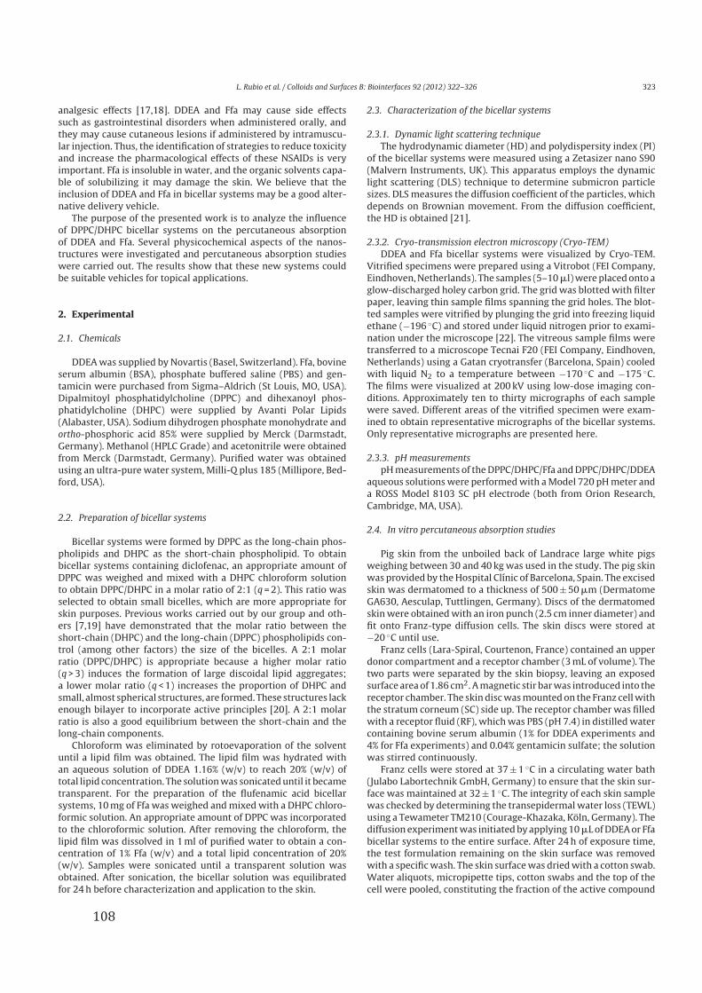
L. Rubio et al. / Colloids and Surfaces B: Biointerfaces 92 (2012) 322– 326 323
analgesic effects [17,18]. DDEA and Ffa may cause side effects
such as gastrointestinal disorders when administered orally, and
they may cause cutaneous lesions if administered by intramuscu-
lar injection. Thus, the identification of strategies to reduce toxicity
and increase the pharmacological effects of these NSAIDs is very
important. Ffa is insoluble in water, and the organic solvents capa-
ble of solubilizing it may damage the skin. We believe that the
inclusion of DDEA and Ffa in bicellar systems may be a good alter-
native delivery vehicle.
The purpose of the presented work is to analyze the influence
of DPPC/DHPC bicellar systems on the percutaneous absorption
of DDEA and Ffa. Several physicochemical aspects of the nanos-
tructures were investigated and percutaneous absorption studies
were carried out. The results show that these new systems could
be suitable vehicles for topical applications.
2. Experimental
2.1. Chemicals
DDEA was supplied by Novartis (Basel, Switzerland). Ffa, bovine
serum albumin (BSA), phosphate buffered saline (PBS) and gen-
tamicin were purchased from Sigma–Aldrich (St Louis, MO, USA).
Dipalmitoyl phosphatidylcholine (DPPC) and dihexanoyl phos-
phatidylcholine (DHPC) were supplied by Avanti Polar Lipids
(Alabaster, USA). Sodium dihydrogen phosphate monohydrate and
ortho-phosphoric acid 85% were supplied by Merck (Darmstadt,
Germany). Methanol (HPLC Grade) and acetonitrile were obtained
from Merck (Darmstadt, Germany). Purified water was obtained
using an ultra-pure water system, Milli-Q plus 185 (Millipore, Bed-
ford, USA).
2.2. Preparation of bicellar systems
Bicellar systems were formed by DPPC as the long-chain phos-
pholipids and DHPC as the short-chain phospholipid. To obtain
bicellar systems containing diclofenac, an appropriate amount of
DPPC was weighed and mixed with a DHPC chloroform solution
to obtain DPPC/DHPC in a molar ratio of 2:1 (q = 2). This ratio was
selected to obtain small bicelles, which are more appropriate for
skin purposes. Previous works carried out by our group and oth-
ers [7,19] have demonstrated that the molar ratio between the
short-chain (DHPC) and the long-chain (DPPC) phospholipids con-
trol (among other factors) the size of the bicelles. A 2:1 molar
ratio (DPPC/DHPC) is appropriate because a higher molar ratio
(q > 3) induces the formation of large discoidal lipid aggregates;
a lower molar ratio (q < 1) increases the proportion of DHPC and
small, almost spherical structures, are formed. These structures lack
enough bilayer to incorporate active principles [20]. A 2:1 molar
ratio is also a good equilibrium between the short-chain and the
long-chain components.
Chloroform was eliminated by rotoevaporation of the solvent
until a lipid film was obtained. The lipid film was hydrated with
an aqueous solution of DDEA 1.16% (w/v) to reach 20% (w/v) of
total lipid concentration. The solution was sonicated until it became
transparent. For the preparation of the flufenamic acid bicellar
systems, 10 mg of Ffa was weighed and mixed with a DHPC chloro-
formic solution. An appropriate amount of DPPC was incorporated
to the chloroformic solution. After removing the chloroform, the
lipid film was dissolved in 1 ml of purified water to obtain a con-
centration of 1% Ffa (w/v) and a total lipid concentration of 20%
(w/v). Samples were sonicated until a transparent solution was
obtained. After sonication, the bicellar solution was equilibrated
for 24 h before characterization and application to the skin.
2.3. Characterization of the bicellar systems
2.3.1. Dynamic light scattering technique
The hydrodynamic diameter (HD) and polydispersity index (PI)
of the bicellar systems were measured using a Zetasizer nano S90
(Malvern Instruments, UK). This apparatus employs the dynamic
light scattering (DLS) technique to determine submicron particle
sizes. DLS measures the diffusion coefficient of the particles, which
depends on Brownian movement. From the diffusion coefficient,
the HD is obtained [21].
2.3.2. Cryo-transmission electron microscopy (Cryo-TEM)
DDEA and Ffa bicellar systems were visualized by Cryo-TEM.
Vitrified specimens were prepared using a Vitrobot (FEI Company,
Eindhoven, Netherlands). The samples (5–10 �l) were placed onto a
glow-discharged holey carbon grid. The grid was blotted with filter
paper, leaving thin sample films spanning the grid holes. The blot-
ted samples were vitrified by plunging the grid into freezing liquid
ethane (−196 ◦C) and stored under liquid nitrogen prior to exami-
nation under the microscope [22]. The vitreous sample films were
transferred to a microscope Tecnai F20 (FEI Company, Eindhoven,
Netherlands) using a Gatan cryotransfer (Barcelona, Spain) cooled
with liquid N2 to a temperature between −170 ◦C and −175 ◦C.
The films were visualized at 200 kV using low-dose imaging con-
ditions. Approximately ten to thirty micrographs of each sample
were saved. Different areas of the vitrified specimen were exam-
ined to obtain representative micrographs of the bicellar systems.
Only representative micrographs are presented here.
2.3.3. pH measurements
pH measurements of the DPPC/DHPC/Ffa and DPPC/DHPC/DDEA
aqueous solutions were performed with a Model 720 pH meter and
a ROSS Model 8103 SC pH electrode (both from Orion Research,
Cambridge, MA, USA).
2.4. In vitro percutaneous absorption studies
Pig skin from the unboiled back of Landrace large white pigs
weighing between 30 and 40 kg was used in the study. The pig skin
was provided by the Hospital Clínic of Barcelona, Spain. The excised
skin was dermatomed to a thickness of 500 ± 50 �m (Dermatome
GA630, Aesculap, Tuttlingen, Germany). Discs of the dermatomed
skin were obtained with an iron punch (2.5 cm inner diameter) and
fit onto Franz-type diffusion cells. The skin discs were stored at
−20 ◦C until use.
Franz cells (Lara-Spiral, Courtenon, France) contained an upper
donor compartment and a receptor chamber (3 mL of volume). The
two parts were separated by the skin biopsy, leaving an exposed
surface area of 1.86 cm2. A magnetic stir bar was introduced into the
receptor chamber. The skin disc was mounted on the Franz cell with
the stratum corneum (SC) side up. The receptor chamber was filled
with a receptor fluid (RF), which was PBS (pH 7.4) in distilled water
containing bovine serum albumin (1% for DDEA experiments and
4% for Ffa experiments) and 0.04% gentamicin sulfate; the solution
was stirred continuously.
Franz cells were stored at 37 ± 1 ◦C in a circulating water bath
(Julabo Labortechnik GmbH, Germany) to ensure that the skin sur-
face was maintained at 32 ± 1 ◦C. The integrity of each skin sample
was checked by determining the transepidermal water loss (TEWL)
using a Tewameter TM210 (Courage-Khazaka, Köln, Germany). The
diffusion experiment was initiated by applying 10 �L of DDEA or Ffa
bicellar systems to the entire surface. After 24 h of exposure time,
the test formulation remaining on the skin surface was removed
with a specific wash. The skin surface was dried with a cotton swab.
Water aliquots, micropipette tips, cotton swabs and the top of the
cell were pooled, constituting the fraction of the active compound
108
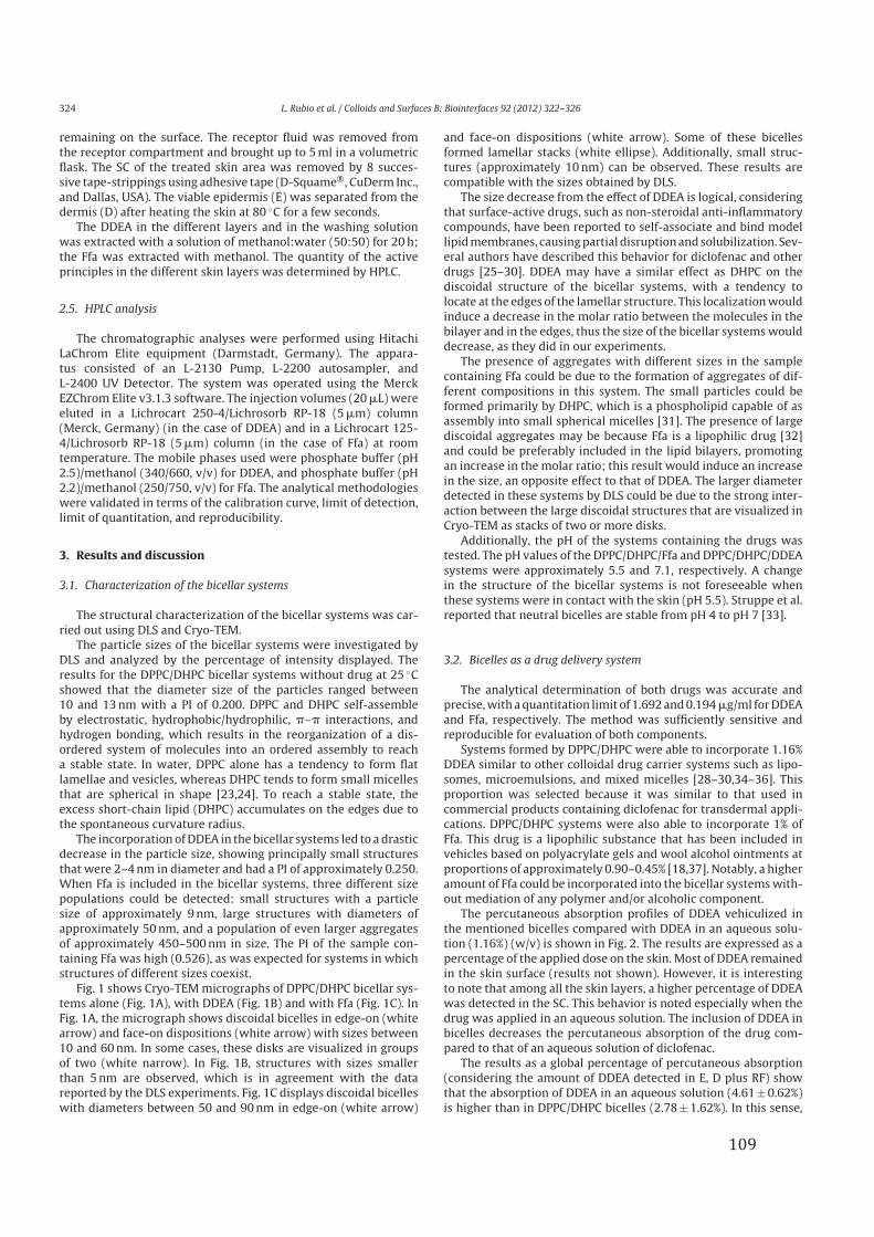
324 L. Rubio et al. / Colloids and Surfaces B: Biointerfaces 92 (2012) 322– 326
remaining on the surface. The receptor fluid was removed from
the receptor compartment and brought up to 5 ml in a volumetric
flask. The SC of the treated skin area was removed by 8 succes-
sive tape-strippings using adhesive tape (D-Squame®, CuDerm Inc.,
and Dallas, USA). The viable epidermis (E) was separated from the
dermis (D) after heating the skin at 80 ◦C for a few seconds.
The DDEA in the different layers and in the washing solution
was extracted with a solution of methanol:water (50:50) for 20 h;
the Ffa was extracted with methanol. The quantity of the active
principles in the different skin layers was determined by HPLC.
2.5. HPLC analysis
The chromatographic analyses were performed using Hitachi
LaChrom Elite equipment (Darmstadt, Germany). The appara-
tus consisted of an L-2130 Pump, L-2200 autosampler, and
L-2400 UV Detector. The system was operated using the Merck
EZChrom Elite v3.1.3 software. The injection volumes (20 �L) were
eluted in a Lichrocart 250-4/Lichrosorb RP-18 (5 �m) column
(Merck, Germany) (in the case of DDEA) and in a Lichrocart 125-
4/Lichrosorb RP-18 (5 �m) column (in the case of Ffa) at room
temperature. The mobile phases used were phosphate buffer (pH
2.5)/methanol (340/660, v/v) for DDEA, and phosphate buffer (pH
2.2)/methanol (250/750, v/v) for Ffa. The analytical methodologies
were validated in terms of the calibration curve, limit of detection,
limit of quantitation, and reproducibility.
3. Results and discussion
3.1. Characterization of the bicellar systems
The structural characterization of the bicellar systems was car-
ried out using DLS and Cryo-TEM.
The particle sizes of the bicellar systems were investigated by
DLS and analyzed by the percentage of intensity displayed. The
results for the DPPC/DHPC bicellar systems without drug at 25 ◦C
showed that the diameter size of the particles ranged between
10 and 13 nm with a PI of 0.200. DPPC and DHPC self-assemble
by electrostatic, hydrophobic/hydrophilic, �–� interactions, and
hydrogen bonding, which results in the reorganization of a dis-
ordered system of molecules into an ordered assembly to reach
a stable state. In water, DPPC alone has a tendency to form flat
lamellae and vesicles, whereas DHPC tends to form small micelles
that are spherical in shape [23,24]. To reach a stable state, the
excess short-chain lipid (DHPC) accumulates on the edges due to
the spontaneous curvature radius.
The incorporation of DDEA in the bicellar systems led to a drastic
decrease in the particle size, showing principally small structures
that were 2–4 nm in diameter and had a PI of approximately 0.250.
When Ffa is included in the bicellar systems, three different size
populations could be detected: small structures with a particle
size of approximately 9 nm, large structures with diameters of
approximately 50 nm, and a population of even larger aggregates
of approximately 450–500 nm in size. The PI of the sample con-
taining Ffa was high (0.526), as was expected for systems in which
structures of different sizes coexist.
Fig. 1 shows Cryo-TEM micrographs of DPPC/DHPC bicellar sys-
tems alone (Fig. 1A), with DDEA (Fig. 1B) and with Ffa (Fig. 1C). In
Fig. 1A, the micrograph shows discoidal bicelles in edge-on (white
arrow) and face-on dispositions (white arrow) with sizes between
10 and 60 nm. In some cases, these disks are visualized in groups
of two (white narrow). In Fig. 1B, structures with sizes smaller
than 5 nm are observed, which is in agreement with the data
reported by the DLS experiments. Fig. 1C displays discoidal bicelles
with diameters between 50 and 90 nm in edge-on (white arrow)
and face-on dispositions (white arrow). Some of these bicelles
formed lamellar stacks (white ellipse). Additionally, small struc-
tures (approximately 10 nm) can be observed. These results are
compatible with the sizes obtained by DLS.
The size decrease from the effect of DDEA is logical, considering
that surface-active drugs, such as non-steroidal anti-inflammatory
compounds, have been reported to self-associate and bind model
lipid membranes, causing partial disruption and solubilization. Sev-
eral authors have described this behavior for diclofenac and other
drugs [25–30]. DDEA may have a similar effect as DHPC on the
discoidal structure of the bicellar systems, with a tendency to
locate at the edges of the lamellar structure. This localization would
induce a decrease in the molar ratio between the molecules in the
bilayer and in the edges, thus the size of the bicellar systems would
decrease, as they did in our experiments.
The presence of aggregates with different sizes in the sample
containing Ffa could be due to the formation of aggregates of dif-
ferent compositions in this system. The small particles could be
formed primarily by DHPC, which is a phospholipid capable of as
assembly into small spherical micelles [31]. The presence of large
discoidal aggregates may be because Ffa is a lipophilic drug [32]
and could be preferably included in the lipid bilayers, promoting
an increase in the molar ratio; this result would induce an increase
in the size, an opposite effect to that of DDEA. The larger diameter
detected in these systems by DLS could be due to the strong inter-
action between the large discoidal structures that are visualized in
Cryo-TEM as stacks of two or more disks.
Additionally, the pH of the systems containing the drugs was
tested. The pH values of the DPPC/DHPC/Ffa and DPPC/DHPC/DDEA
systems were approximately 5.5 and 7.1, respectively. A change
in the structure of the bicellar systems is not foreseeable when
these systems were in contact with the skin (pH 5.5). Struppe et al.
reported that neutral bicelles are stable from pH 4 to pH 7 [33].
3.2. Bicelles as a drug delivery system
The analytical determination of both drugs was accurate and
precise, with a quantitation limit of 1.692 and 0.194 �g/ml for DDEA
and Ffa, respectively. The method was sufficiently sensitive and
reproducible for evaluation of both components.
Systems formed by DPPC/DHPC were able to incorporate 1.16%
DDEA similar to other colloidal drug carrier systems such as lipo-
somes, microemulsions, and mixed micelles [28–30,34–36]. This
proportion was selected because it was similar to that used in
commercial products containing diclofenac for transdermal appli-
cations. DPPC/DHPC systems were also able to incorporate 1% of
Ffa. This drug is a lipophilic substance that has been included in
vehicles based on polyacrylate gels and wool alcohol ointments at
proportions of approximately 0.90–0.45% [18,37]. Notably, a higher
amount of Ffa could be incorporated into the bicellar systems with-
out mediation of any polymer and/or alcoholic component.
The percutaneous absorption profiles of DDEA vehiculized in
the mentioned bicelles compared with DDEA in an aqueous solu-
tion (1.16%) (w/v) is shown in Fig. 2. The results are expressed as a
percentage of the applied dose on the skin. Most of DDEA remained
in the skin surface (results not shown). However, it is interesting
to note that among all the skin layers, a higher percentage of DDEA
was detected in the SC. This behavior is noted especially when the
drug was applied in an aqueous solution. The inclusion of DDEA in
bicelles decreases the percutaneous absorption of the drug com-
pared to that of an aqueous solution of diclofenac.
The results as a global percentage of percutaneous absorption
(considering the amount of DDEA detected in E, D plus RF) show
that the absorption of DDEA in an aqueous solution (4.61 ± 0.62%)
is higher than in DPPC/DHPC bicelles (2.78 ± 1.62%). In this sense,
109
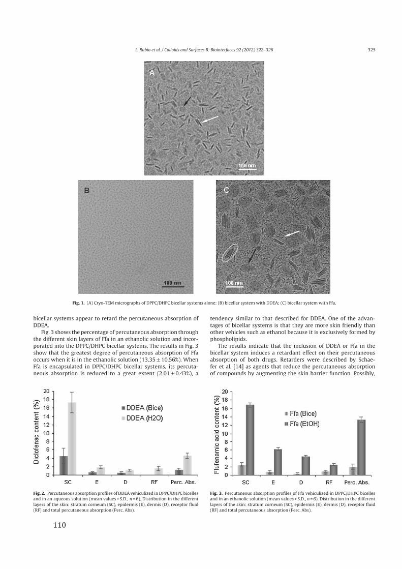
L. Rubio et al. / Colloids and Surfaces B: Biointerfaces 92 (2012) 322– 326 325
Fig. 1. (A) Cryo-TEM micrographs of DPPC/DHPC bicellar systems alone: (B) bicellar system with DDEA; (C) bicellar system with Ffa.
bicellar systems appear to retard the percutaneous absorption of
DDEA.
Fig. 3 shows the percentage of percutaneous absorption through
the different skin layers of Ffa in an ethanolic solution and incor-
porated into the DPPC/DHPC bicellar systems. The results in Fig. 3
show that the greatest degree of percutaneous absorption of Ffa
occurs when it is in the ethanolic solution (13.35 ± 10.56%). When
Ffa is encapsulated in DPPC/DHPC bicellar systems, its percuta-
neous absorption is reduced to a great extent (2.01 ± 0.43%), a
Fig. 2. Percutaneous absorption profiles of DDEA vehiculized in DPPC/DHPC bicelles
and in an aqueous solution (mean values + S.D., n = 6). Distribution in the different
layers of the skin: stratum corneum (SC), epidermis (E), dermis (D), receptor fluid
(RF) and total percutaneous absorption (Perc. Abs).
tendency similar to that described for DDEA. One of the advan-
tages of bicellar systems is that they are more skin friendly than
other vehicles such as ethanol because it is exclusively formed by
phospholipids.
The results indicate that the inclusion of DDEA or Ffa in the
bicellar system induces a retardant effect on their percutaneous
absorption of both drugs. Retarders were described by Schae-
fer et al. [14] as agents that reduce the percutaneous absorption
of compounds by augmenting the skin barrier function. Possibly,
Fig. 3. Percutaneous absorption profiles of Ffa vehiculized in DPPC/DHPC bicelles
and in an ethanolic solution (mean values + S.D., n = 6). Distribution in the different
layers of the skin: stratum corneum (SC), epidermis (E), dermis (D), receptor fluid
(RF) and total percutaneous absorption (Perc. Abs).
110
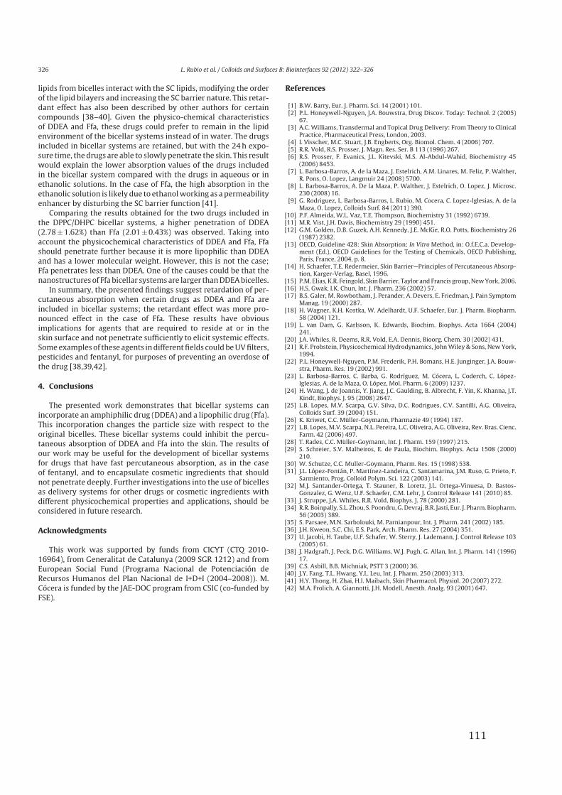
326 L. Rubio et al. / Colloids and Surfaces B: Biointerfaces 92 (2012) 322– 326
lipids from bicelles interact with the SC lipids, modifying the order
of the lipid bilayers and increasing the SC barrier nature. This retar-
dant effect has also been described by other authors for certain
compounds [38–40]. Given the physico-chemical characteristics
of DDEA and Ffa, these drugs could prefer to remain in the lipid
environment of the bicellar systems instead of in water. The drugs
included in bicellar systems are retained, but with the 24 h expo-
sure time, the drugs are able to slowly penetrate the skin. This result
would explain the lower absorption values of the drugs included
in the bicellar system compared with the drugs in aqueous or in
ethanolic solutions. In the case of Ffa, the high absorption in the
ethanolic solution is likely due to ethanol working as a permeability
enhancer by disturbing the SC barrier function [41].
Comparing the results obtained for the two drugs included in
the DPPC/DHPC bicellar systems, a higher penetration of DDEA
(2.78 ± 1.62%) than Ffa (2.01 ± 0.43%) was observed. Taking into
account the physicochemical characteristics of DDEA and Ffa, Ffa
should penetrate further because it is more lipophilic than DDEA
and has a lower molecular weight. However, this is not the case;
Ffa penetrates less than DDEA. One of the causes could be that the
nanostructures of Ffa bicellar systems are larger than DDEA bicelles.
In summary, the presented findings suggest retardation of per-
cutaneous absorption when certain drugs as DDEA and Ffa are
included in bicellar systems; the retardant effect was more pro-
nounced effect in the case of Ffa. These results have obvious
implications for agents that are required to reside at or in the
skin surface and not penetrate sufficiently to elicit systemic effects.
Some examples of these agents in different fields could be UV filters,
pesticides and fentanyl, for purposes of preventing an overdose of
the drug [38,39,42].
4. Conclusions
The presented work demonstrates that bicellar systems can
incorporate an amphiphilic drug (DDEA) and a lipophilic drug (Ffa).
This incorporation changes the particle size with respect to the
original bicelles. These bicellar systems could inhibit the percu-
taneous absorption of DDEA and Ffa into the skin. The results of
our work may be useful for the development of bicellar systems
for drugs that have fast percutaneous absorption, as in the case
of fentanyl, and to encapsulate cosmetic ingredients that should
not penetrate deeply. Further investigations into the use of bicelles
as delivery systems for other drugs or cosmetic ingredients with
different physicochemical properties and applications, should be
considered in future research.
Acknowledgments
This work was supported by funds from CICYT (CTQ 2010-
16964), from Generalitat de Catalunya (2009 SGR 1212) and from
European Social Fund (Programa Nacional de Potenciación de
Recursos Humanos del Plan Nacional de I+D+I (2004–2008)). M.
Cócera is funded by the JAE-DOC program from CSIC (co-funded by
FSE).
References
[1] B.W. Barry, Eur. J. Pharm. Sci. 14 (2001) 101.[2] P.L. Honeywell-Nguyen, J.A. Bouwstra, Drug Discov. Today: Technol. 2 (2005)
67.[3] A.C. Williams, Transdermal and Topical Drug Delivery: From Theory to Clinical
Practice, Pharmaceutical Press, London, 2003.[4] I. Visscher, M.C. Stuart, J.B. Engberts, Org. Biomol. Chem. 4 (2006) 707.[5] R.R. Vold, R.S. Prosser, J. Magn. Res. Ser. B 113 (1996) 267.[6] R.S. Prosser, F. Evanics, J.L. Kitevski, M.S. Al-Abdul-Wahid, Biochemistry 45
(2006) 8453.[7] L. Barbosa-Barros, A. de la Maza, J. Estelrich, A.M. Linares, M. Feliz, P. Walther,
R. Pons, O. Lopez, Langmuir 24 (2008) 5700.[8] L. Barbosa-Barros, A. De la Maza, P. Walther, J. Estelrich, O. Lopez, J. Microsc.
230 (2008) 16.[9] G. Rodriguez, L. Barbosa-Barros, L. Rubio, M. Cocera, C. Lopez-Iglesias, A. de la
Maza, O. Lopez, Colloids Surf. 84 (2011) 390.[10] P.F. Almeida, W.L. Vaz, T.E. Thompson, Biochemistry 31 (1992) 6739.[11] M.R. Vist, J.H. Davis, Biochemistry 29 (1990) 451.[12] G.M. Golden, D.B. Guzek, A.H. Kennedy, J.E. McKie, R.O. Potts, Biochemistry 26
(1987) 2382.[13] OECD, Guideline 428: Skin Absorption: In Vitro Method, in: O.f.E.C.a. Develop-
ment (Ed.), OECD Guidelines for the Testing of Chemicals, OECD Publishing,Paris, France, 2004, p. 8.
[14] H. Schaefer, T.E. Redermeier, Skin Barrier—Principles of Percutaneous Absorp-tion, Karger-Verlag, Basel, 1996.
[15] P.M. Elias, K.R. Feingold, Skin Barrier, Taylor and Francis group, New York, 2006.[16] H.S. Gwak, I.K. Chun, Int. J. Pharm. 236 (2002) 57.[17] B.S. Galer, M. Rowbotham, J. Perander, A. Devers, E. Friedman, J. Pain Symptom
Manag. 19 (2000) 287.[18] H. Wagner, K.H. Kostka, W. Adelhardt, U.F. Schaefer, Eur. J. Pharm. Biopharm.
58 (2004) 121.[19] L. van Dam, G. Karlsson, K. Edwards, Biochim. Biophys. Acta 1664 (2004)
241.[20] J.A. Whiles, R. Deems, R.R. Vold, E.A. Dennis, Bioorg. Chem. 30 (2002) 431.[21] R.F. Probstein, Physicochemical Hydrodynamics, John Wiley & Sons, New York,
1994.[22] P.L. Honeywell-Nguyen, P.M. Frederik, P.H. Bomans, H.E. Junginger, J.A. Bouw-
stra, Pharm. Res. 19 (2002) 991.[23] L. Barbosa-Barros, C. Barba, G. Rodríguez, M. Cócera, L. Coderch, C. López-
Iglesias, A. de la Maza, O. López, Mol. Pharm. 6 (2009) 1237.[24] H. Wang, J. de Joannis, Y. Jiang, J.C. Gaulding, B. Albrecht, F. Yin, K. Khanna, J.T.
Kindt, Biophys. J. 95 (2008) 2647.[25] L.B. Lopes, M.V. Scarpa, G.V. Silva, D.C. Rodrigues, C.V. Santilli, A.G. Oliveira,
Colloids Surf. 39 (2004) 151.[26] K. Kriwet, C.C. Müller-Goymann, Pharmazie 49 (1994) 187.[27] L.B. Lopes, M.V. Scarpa, N.L. Pereira, L.C. Oliveira, A.G. Oliveira, Rev. Bras. Cienc.
Farm. 42 (2006) 497.[28] T. Rades, C.C. Müller-Goymann, Int. J. Pharm. 159 (1997) 215.[29] S. Schreier, S.V. Malheiros, E. de Paula, Biochim. Biophys. Acta 1508 (2000)
210.[30] W. Schutze, C.C. Muller-Goymann, Pharm. Res. 15 (1998) 538.[31] J.L. López-Fontán, P. Martínez-Landeira, C. Santamarina, J.M. Ruso, G. Prieto, F.
Sarmiento, Prog. Colloid Polym. Sci. 122 (2003) 141.[32] M.J. Santander-Ortega, T. Stauner, B. Loretz, J.L. Ortega-Vinuesa, D. Bastos-
Gonzalez, G. Wenz, U.F. Schaefer, C.M. Lehr, J. Control Release 141 (2010) 85.[33] J. Struppe, J.A. Whiles, R.R. Vold, Biophys. J. 78 (2000) 281.[34] R.R. Boinpally, S.L. Zhou, S. Poondru, G. Devraj, B.R. Jasti, Eur. J. Pharm. Biopharm.
56 (2003) 389.[35] S. Parsaee, M.N. Sarbolouki, M. Parnianpour, Int. J. Pharm. 241 (2002) 185.[36] J.H. Kweon, S.C. Chi, E.S. Park, Arch. Pharm. Res. 27 (2004) 351.[37] U. Jacobi, H. Taube, U.F. Schafer, W. Sterry, J. Lademann, J. Control Release 103
(2005) 61.[38] J. Hadgraft, J. Peck, D.G. Williams, W.J. Pugh, G. Allan, Int. J. Pharm. 141 (1996)
17.[39] C.S. Asbill, B.B. Michniak, PSTT 3 (2000) 36.[40] J.Y. Fang, T.L. Hwang, Y.L. Leu, Int. J. Pharm. 250 (2003) 313.[41] H.Y. Thong, H. Zhai, H.I. Maibach, Skin Pharmacol. Physiol. 20 (2007) 272.[42] M.A. Frolich, A. Giannotti, J.H. Modell, Anesth. Analg. 93 (2001) 647.
111

>>#�
Sistemes�bicel·lars�com�a�nova�estratègia�d’aplicació�tòpica����������������������������������������������������������������������RESULTATS�
>>@�
Article�5��
Barrier�function�of�intact�and�impaired�skin:�Percutaneous�penetration�of�caffeine�and�salicylic�acid�
�
Int.�J.�Dermatol.,�#$>>��%$��CC>iCCG6�
�
!�� �5����� ���-���� �0������� �0�-��� 9� ������� �� in� vitro� ��� ���� ���������
7�4�L��� � � �� ��� �� 8�� ��-� �0�-H�� �� �0����� ��� ��� ������ 9� �M ������ ������ ���
���-����������� ����� Ntape�stripsP���� ������ �������� � ��� �����!T)�� � �����������
����� ��-� � 4������� � ������ ��� ����� �������� ��� ��� 4�� 9� -������6� !�� ��������� ����
������� �S�-��� 9� ������� �� ��� ���� �/��� � �H��1���� - (�� ��� ��� ����� ��-� ����
��� 9� 4 � ��(� �� �����-��6� "�� �-������ � M(�� ����� � 4������� ��� ����
�����(� 5��������4�� 9����-�����������0!����������4������ ���������5[�� ����������
���4 ���0�-��� 9�������� �6�:����5�������� �����������������4����5���������� 6��
)�� 4�� 9� -������� ��� �S!�� ��� ��� �������� � �H��1���� ���� ������ 9� ������ ���
���-����S��� � ��������� �������� ������� ������.�� �����-�����������7��� ���in�vitro� �.������in�vivo8� ���������!T)6�*5������4�� 9�-������������������������-<���
����<�� �0����� �� �S�-��� 9� ������� �� in� vitro� ����� ���� ���������� �� � �/����
��B������ ��� � 4�� 9� ������/� � ����� 4������� ������ ��� ���� ��-���� ����� � ����� ���
������-� 9�����0!�6��
+��� � �� �� 9� �������� ��� ��� ��� 4�� 9� ��� -������� ��� ��� ����� .�� ������
��������������!T)������in�vitro����in�vivo�5����������-������Ntape�strippingsP���� ��� ����� ��������6� �0������ -������ �0�-��� 9� ������� �� ����� ���������
����� ����� ��������� �0���� ������� � 4������ 5���� ��������� ��� ���-��� ��� � ����
��.�� ���6� )0� �� ��� �� � ��� ���������� ��� �������� � ����� ��� 4�� 9� ��� ���
������-� 9�������-����������� �6�!���������������4�L���� �S�-��� 9�������� �����
���������������������M����� ��6�!������������� �� 5����5����������� �����������
��� -������� ��� ��� ����� ��������� ������� ��� ������������ ��� ������� 9� �0���
������� ������� ���(�� ��� -����1� . ��(4 �&� �(4 �� ����� ��������� �� 4������ ����
�� � �/����� ������ ��� 4 ��� ��� ����� ������� ��� ���� ���4 �� ��� ������� 9��
���� � � �/��� �������5�����0� ����������-����4�� 9�-���������� 4 ���6��
*5�������������in�vitro���������U� ������� �������0������������<������ �������
���4 ���S�-��� 9�������� ���0��������� � � ���� ������ ������-�������������-�����
4�� 9�-���������������� ����� �������� � �/��������� ����������������������0U��in�vivo��0�� ������0�M��� ����� 9����������������4������� 6�
����

>>A�
Pharmacology and therapeutics
Barrier function of intact and impaired skin: percutaneous
penetration of caffeine and salicylic acid
Laia Rubio1, BChem, Cristina Alonso1, PhD, Olga Lopez1, PhD, Gelen Rodrıguez1, BBiol,Luisa Coderch1, PhD, Jaime Notario2, MD, Alfonso de la Maza1, PhD, andJose L. Parra1, PhD, MD
1Department of Chemical and
Surfactants Technology, IQAC-CSIC,
and 2Department of Dermatology,
Bellvitge University Hospital, Barcelona,
Spain
Correspondence
Laia Rubio, BChem
Department of Chemical Technology
IQAC-CSIC
C/ Jordi Girona, 18–26
08034 Barcelona
Spain
E-mail: [email protected]
Conflicts of interest: None.
Abstract
Background Normally, percutaneous absorption tests are carried out using skin biopsies
for an apparent and acceptable physiological condition. However, under different
pathological conditions, the stratum corneum (SC) barrier function is impaired.
Methods The barrier function of the SC was assessed by correlation between the number
of repeated applications of tape strips on the skin and its transepidermal water loss
(TEWL), as well as by in vitro percutaneous absorption studies of different compounds,
using Franz diffusion cells and porcine skin previously stripped.
Results A progressive diminution of the skin barrier function has been detected by TEWL
both in vitro and in vivo as the number of skin tape strips increases. On the other hand,
the percutaneous absorption of the compounds tested increases in a different way as the
number of strips increases. Salicylic acid increases linearly depending on the barrier
disturbance. However, percutaneous absorption of caffeine exponentially increased with
barrier disturbance. Our results indicate that the barrier impairment of skin always
increases the penetration behavior of a given compound; however, the hydrophilic–
lipophilic balance of the compounds or formulations used could greatly modify its
penetration profile, especially when a modified skin is used.
Conclusions This in vitro protocol may be useful to simulate the percutaneous absorption
profile of some drugs applied onto skin with an impaired SC barrier function and could
be used to avoid, to some extent, the use of in vivo experimental animal models in the
dermopharmaceutical field.
Introduction
The stratum corneum (SC) is the outermost layer of theskin. One of its most important roles is as the limitingbarrier to water loss by evaporation, as well as its role asa barrier against the penetration of foreign physical,chemical, and pathogenic components from the environ-ment.1–3 The physical state and organization of the SC,particularly its lipid intercellular matrix, provide adequatebarrier function.4–6
Percutaneous absorption can be studied using in vitro
methods, as it is possible to maintain the barrier proper-ties of the SC in excised skin.7–9 There is suitable evi-dence that in vitro data may be predictive for in vivo
percutaneous absorption in both animals and humans.10
Furthermore, the usefulness of the pig skin as an experi-mental model for skin permeation in humans is widely
appreciated.11 In fact, this in vitro strategy may be veryuseful to substitute or reduce in vivo animal experiments.
Normally, percutaneous absorption tests are carriedout using skin biopsies for an apparent and acceptablephysiological condition. However, under different patho-logical conditions, the SC barrier function is impaired.The skin pathologies most frequently cited are atopic der-matitis,12 ichthyosis,13 psoriasis,14 and severe xerosis.15
In all these dermatological pathologies, a reduced skinbarrier function has been detected owing to elevatedtransepidermal water loss (TEWL) values. The measure-ment of TEWL allows total water loss to be determinedfrom viable epidermis and dermis, diffusing throughthe SC to the skin surface.16 The TEWL measurementin vitro is a good biophysical indicator of the integrity ofbarrier function.17,18 The relationship between TEWL, asa measurement of barrier function efficiency, and the 881
ª 2011 The International Society of Dermatology International Journal of Dermatology 2011, 50, 881–889
115
degree of barrier disruption after skin damage is generallyaccepted using different skin models.19–22
In the literature, it has also been shown that a less thanperfect SC (across which TEWL is elevated) is a morepermeable barrier to drug permeation.23 An overviewabout the existence of a correlation between TEWL andpercutaneous absorption has been reported by Levin andMaibach.24 The TEWL and percutaneous absorptionrates increase when the integrity of the SC barrier is com-promised. In general, the majority of earlier studies pre-sented a quantitative correlation between TEWL andpercutaneous absorption,25,26 although in a few, no quan-titative correlation was observed.27,28
Measurements of TEWL on previously stripped skinbiopsies might be an interesting strategy to know, ata preliminary stage, the behavior of diseased skin duringthe percutaneous absorption process. A correlation hasbeen reported between the decrease in barrier functionas indicated by TEWL and increase in the percutaneousabsorption of hydrocortisone;25 this correlation wasreconfirmed by a study on patients with widespread der-matitis.29 Based on this idea, we tried to generalize thepredictability of this model by evaluating the skin bar-rier function using the correlation between sequentialtape stripping on SC and its TEWL. This barrier func-tion was assessed by in vitro percutaneous absorption oftwo compounds (caffeine and salicylic acid) using por-cine skin samples subjected to previous progressiveimpairment of the SC by sequential tape stripping. Sali-cylic acid is widely used in the treatment of many com-mon dermatological conditions, owing to its keratolyticproperties.30 In addition, caffeine is well known for itsanticellulite effect, but recent studies also use it as atreatment for psoriasis.31 These compounds with differ-ent octanol–water partition coefficient32 may be used intopical applications.In this paper, a more realistic scenario should be envis-
aged using skin biopsies with a progressive deteriorationof the SC barrier function, when an in vitro percutaneousabsorption test is performed to develop a transdermaldrug to be used on impaired skin.
Materials and methods
Materials
Salicylic acid (99%) was purchased from Aldrich (Gillingham,
Dorset, UK). Caffeine (pure), bovine serum albumin, phosphate-
buffered saline, and gentamicin were purchased from Sigma
Aldrich (St Louis, MO, USA). Methanol (high-performance liquid
chromatography grade), sodium dihydrogen phosphate
monohydrate, acetonitrile, and orthophosphoric acid (85%) were
supplied by Merck (Darmstadt, Germany). Ethanol was
obtained from Carlo Erba (Milan, Italy), and purified water was
obtained by an ultra-pure water system, Milli-Q plus 185
(Millipore, Bedford, MA, USA).
Methods
In vivo tape stripping of the skin and evaluation of its
transepidermal water loss
A total of six volunteers were used for this study. Tape stripping
was carried out using a different number of D-Squame (0–50
strips) by applying a constant pressure (80 g/cm2) for
5 seconds, in all the cases. Furthermore, the tearing-off
direction was also varied by 90� for each strip. The TEWL was
carried out using a Tewameter TM210 (Courage & Khazaka,
Cologne, Germany) for 1 minute, and the mean value was
determined. This study was approved by the Ethics Committee
of the Bellvitge University Hospital, Barcelona, Spain.
In vitro tape stripping of the skin and evaluation of its
transepidermal water loss
Pig skin obtained from the unboiled back of Landrace large
white pigs weighing 30–40 kg was used for this study. The pig
skin was provided by the Clınic Hospital of Barcelona, Spain.
Bristles were removed carefully with an animal clipper, and
subsequently the skin was washed with water. The excised skin
was dermatomed to a thickness of 500 ± 50 lm (Dermatome
GA630; Aesculap, Tuttlingen, Germany). The discs of the
dermatomed skin were obtained using an iron punch (2.5 cm of
inner diameter) in such a way that they fit into the Franz
diffusion cells. The skin discs were stored at )20 �C and
thawed immediately for subsequent usage.
Several skin discs with their SC undisturbed were positioned
in a simple model of Franz static diffusion cells (Lara-Spiral,
Courtenon, France). The Franz cells consisted of an upper
donor compartment and a receptor chamber (3 ml of volume).
These two parts were separated by the skin biopsy with an
exposed surface area of 1.86 cm2. A small magnetic stirring bar
was introduced into the receptor chamber. Subsequently, the
skin discs were mounted in the Franz cell, with the SC side on
top. The receptor chamber was filled with a receptor fluid
containing phosphate-buffered saline (pH 7.4) in distilled water,
1% bovine serum albumin, and 0.04% of gentamicin sulfate.
The cells were placed in a circulating water bath (Julabo,
Seelbach, Germany) to ensure that the surface skin was
maintained at 32 �C. After stabilization of the cells for 1 hour,
the measurement of TEWL was carried out for 1 minute, and
the mean value was determined.
The same procedure was followed for the measurement of
TEWL in the previously stripped skin discs. The in vitro
stripping was carried out using D-Squame adhesive discs
(CuDerm Corporation, Dallas, TX, USA). The adhesive tapes
were applied in a sequential manner onto the skin discs (from
0 to 50). Each strip was applied onto the skin with a constant
International Journal of Dermatology 2011, 50, 881–889 ª 2011 The International Society of Dermatology
Pharmacology and therapeutics Barrier function of intact and impaired skin Rubio et al.882
116
pressure (80 g /cm2) for 5 seconds. For each repeated strip, the
tearing-off direction was varied by 90� to obtain a homogeneous
removal of the SC cell layers.
In vitro percutaneous absorption studies of previously
stripped skin
The variedly stripped skins were positioned in the Franz
diffusion cells. Each active principle (caffeine or salicylic acid)
was applied on the progressively stripped skin samples, and
in vitro permeation studies were performed for 24 hours. The
Franz cells were kept at 37 ± 1 �C by means of a thermostated
water bath with a magnetic stirring. The receptor fluid was
continuously stirred. In all the cases, the integrity of the skin
was checked by TEWL. A minimum of six samples was
required for each experimental assay.
The diffusion experiment was initiated by applying 10 ll of
ethanol–water (1:1) solution of caffeine or salicylic acid (both
about 3%, w/v) to the entire surface delimited by the upper cell.
After an exposure time of 24 hours, the test formulation
remaining on the skin surface was removed with a specific
wash: first with 0.5 ml of sodium lauryl ether sulfate solution
(at 0.5%, w/v) and then twice with distilled water (2 · 0.5 ml).
Subsequently, the skin surface was dried with a cotton swab.
Water aliquots from all the tips and cotton swabs used, as well
as the top of the cell, were pooled, which constituted the fraction
of active compound remaining on the surface. Subsequently, the
receptor fluid was removed from the receptor compartment and
brought to 5 ml in a volumetric flask. The SC of the treated area
was removed by eight successive tape strippings, carried out
under controlled conditions. The tape strips were grouped as
strips 1–4 and strips 5–8, and were placed in glass vials. After
that, the viable epidermis was separated from the dermis after
heating the skin at 80 �C for a few seconds, and subsequently
the dermis was cut into small pieces.
The amount of caffeine or salicylic acid in the different
layers and washing solution was extracted with methanol–
water (50:50) solution. After one night of contact with the
solvent, all the samples were shaken for 30 minutes at room
temperature, and the dermis, epidermis, and SC samples
were sonicated for 15 minutes. Before the analytical
determination of the samples, they were filtered through a
0.45-lm Acrodisc filter (Pall Gelman Sciences, Northampton,
UK). Subsequently, the different compartments, for each
active principle, were analyzed using high-performance liquid
chromatography.
High-performance liquid chromatography analysis
The chromatographic analyses were performed using Hitachi
LaChrom Elite equipment (Darmstadt, Germany). The
apparatus consisted of an L-2130 Pump, L-2200 Autosampler,
and L-2400 UV Detector. The system was operated using the
software EZChrom Elite v3.1.6 (Scientific Software Inc.,
Pleasanton, CA, USA). The mobile phases used were 20 mM
phosphate buffer (pH 2.5)/methanol (340/660, v/v) for caffeine,
and 25 mM phosphate buffer (pH 2.33)/acetonitrile/methanol
(650/200/150, v/v/v) for salicylic acid. The analytical
methodologies were validated in terms of calibration curve,
limit of detection, limit of quantitation, and reproducibility.
Table 1 shows the analytical results obtained on validations.
Results
The use of in vitro methods and non-invasive techniquesmay be two suitable alternatives to the in vivo animalexperiments. In the case of percutaneous absorption, anin vitro guideline has been adopted by the Organizationfor Economic Co-operation and Development (OECD).8
On the other hand, TEWL measurements are considereda good indicator of the integrity of skin barrier func-tion.20,33 It has been demonstrated that TEWL is predic-tive of the permeability barrier, and as such, can be usedto develop suitable and realistic transdermal drug deliverysystems, taking into account the physiological or patho-logical conditions of the skin.19,20,23,34 In this paper, theexisting correlation between progressive SC tape strippingand TEWL variation were applied to the in vitro percuta-neous absorption of two compounds that have differentpenetration profiles on intact skin. The conditions ofcompromised skin, particularly the impaired barrier func-tion of SC were simulated to develop a suitable approachcapable of predicting the drug penetration on a compro-mised skin.
Correlation between stratum corneum strips and
transepidermal water loss
In Figure 1, the influence of the number of strips onTEWL measurements performed on pig skin discs in vitro
and on the forearm of human volunteers in vivo isshown. The in vitro results showed that the TEWL valuecorresponding to the baseline (mean ± SD, n = 6), whereno damage had been inflicted to the skin barrier, was8.1 ± 2 g/m2 per hour. During the first 10 strips, only a
Table 1 Analytical validation data obtained for the differentcompounds analyzed
Caffeine Salicylic acid
Detection limit (lg/ml) 0.402 lg/ml 0.376
Quantification limit (lg/ml) 0.804 lg/ml 0.762
Calibration curve A271nm = 9194.1
[conc])1085.3A237nm = 13889
[conc])15452Linearity coefficient (r2) 1 0.999
Repeatability (CVintra) 0.5% 1.4%
Intermediate precision (CVinter) 1.5% 2.7%
ª 2011 The International Society of Dermatology International Journal of Dermatology 2011, 50, 881–889
Rubio et al. Barrier function of intact and impaired skin Pharmacology and therapeutics 883
117
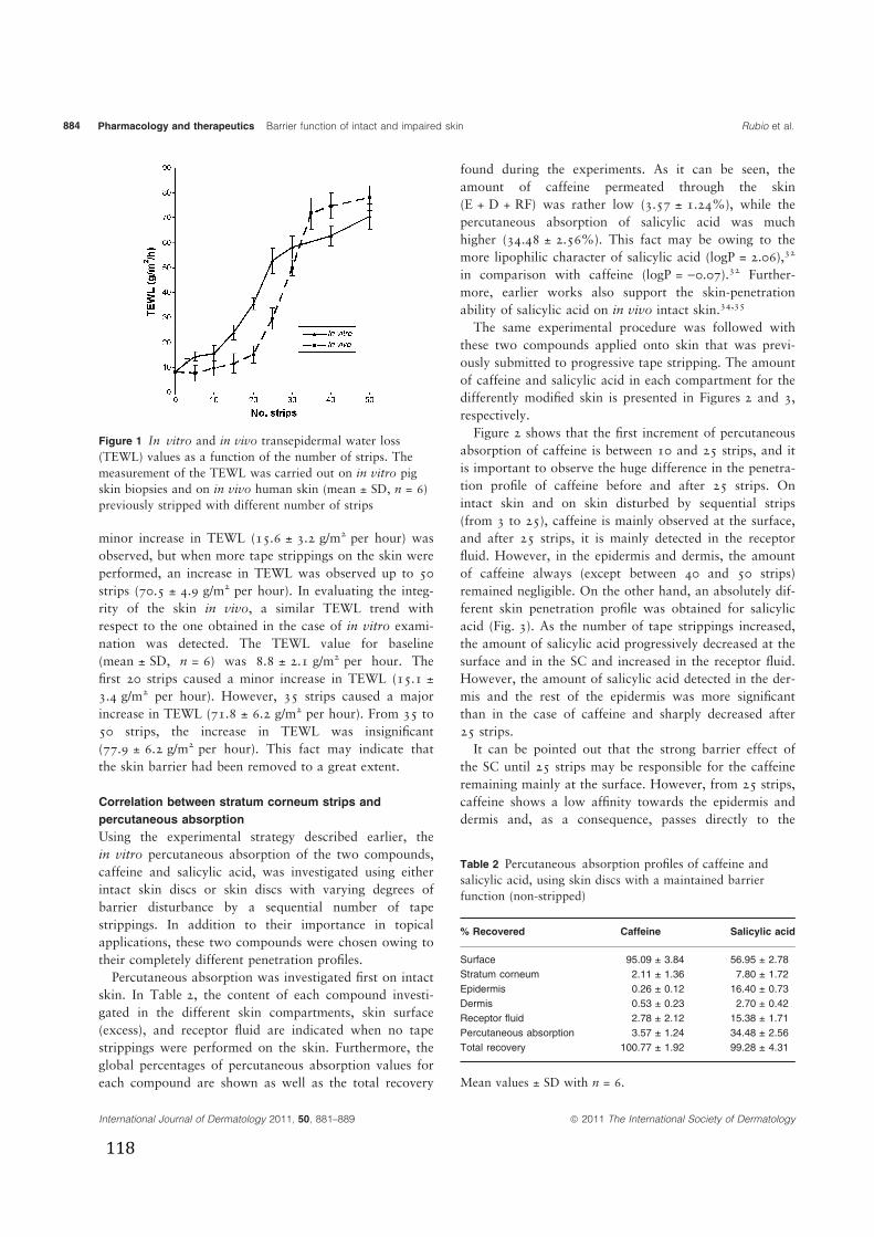
minor increase in TEWL (15.6 ± 3.2 g/m2 per hour) wasobserved, but when more tape strippings on the skin wereperformed, an increase in TEWL was observed up to 50strips (70.5 ± 4.9 g/m2 per hour). In evaluating the integ-rity of the skin in vivo, a similar TEWL trend withrespect to the one obtained in the case of in vitro exami-nation was detected. The TEWL value for baseline(mean ± SD, n = 6) was 8.8 ± 2.1 g/m2 per hour. Thefirst 20 strips caused a minor increase in TEWL (15.1 ±3.4 g/m2 per hour). However, 35 strips caused a majorincrease in TEWL (71.8 ± 6.2 g/m2 per hour). From 35 to50 strips, the increase in TEWL was insignificant(77.9 ± 6.2 g/m2 per hour). This fact may indicate thatthe skin barrier had been removed to a great extent.
Correlation between stratum corneum strips and
percutaneous absorption
Using the experimental strategy described earlier, thein vitro percutaneous absorption of the two compounds,caffeine and salicylic acid, was investigated using eitherintact skin discs or skin discs with varying degrees ofbarrier disturbance by a sequential number of tapestrippings. In addition to their importance in topicalapplications, these two compounds were chosen owing totheir completely different penetration profiles.Percutaneous absorption was investigated first on intact
skin. In Table 2, the content of each compound investi-gated in the different skin compartments, skin surface(excess), and receptor fluid are indicated when no tapestrippings were performed on the skin. Furthermore, theglobal percentages of percutaneous absorption values foreach compound are shown as well as the total recovery
found during the experiments. As it can be seen, theamount of caffeine permeated through the skin(E + D + RF) was rather low (3.57 ± 1.24%), while thepercutaneous absorption of salicylic acid was muchhigher (34.48 ± 2.56%). This fact may be owing to themore lipophilic character of salicylic acid (logP = 2.06),32
in comparison with caffeine (logP = )0.07).32 Further-more, earlier works also support the skin-penetrationability of salicylic acid on in vivo intact skin.34,35
The same experimental procedure was followed withthese two compounds applied onto skin that was previ-ously submitted to progressive tape stripping. The amountof caffeine and salicylic acid in each compartment for thedifferently modified skin is presented in Figures 2 and 3,respectively.
Figure 2 shows that the first increment of percutaneousabsorption of caffeine is between 10 and 25 strips, and itis important to observe the huge difference in the penetra-tion profile of caffeine before and after 25 strips. Onintact skin and on skin disturbed by sequential strips(from 3 to 25), caffeine is mainly observed at the surface,and after 25 strips, it is mainly detected in the receptorfluid. However, in the epidermis and dermis, the amountof caffeine always (except between 40 and 50 strips)remained negligible. On the other hand, an absolutely dif-ferent skin penetration profile was obtained for salicylicacid (Fig. 3). As the number of tape strippings increased,the amount of salicylic acid progressively decreased at thesurface and in the SC and increased in the receptor fluid.However, the amount of salicylic acid detected in the der-mis and the rest of the epidermis was more significantthan in the case of caffeine and sharply decreased after25 strips.
It can be pointed out that the strong barrier effect ofthe SC until 25 strips may be responsible for the caffeineremaining mainly at the surface. However, from 25 strips,caffeine shows a low affinity towards the epidermis anddermis and, as a consequence, passes directly to the
Figure 1 In vitro and in vivo transepidermal water loss(TEWL) values as a function of the number of strips. Themeasurement of the TEWL was carried out on in vitro pigskin biopsies and on in vivo human skin (mean ± SD, n = 6)previously stripped with different number of strips
Table 2 Percutaneous absorption profiles of caffeine andsalicylic acid, using skin discs with a maintained barrierfunction (non-stripped)
% Recovered Caffeine Salicylic acid
Surface 95.09 ± 3.84 56.95 ± 2.78
Stratum corneum 2.11 ± 1.36 7.80 ± 1.72
Epidermis 0.26 ± 0.12 16.40 ± 0.73
Dermis 0.53 ± 0.23 2.70 ± 0.42
Receptor fluid 2.78 ± 2.12 15.38 ± 1.71
Percutaneous absorption 3.57 ± 1.24 34.48 ± 2.56
Total recovery 100.77 ± 1.92 99.28 ± 4.31
Mean values ± SD with n = 6.
International Journal of Dermatology 2011, 50, 881–889 ª 2011 The International Society of Dermatology
Pharmacology and therapeutics Barrier function of intact and impaired skin Rubio et al.884
118
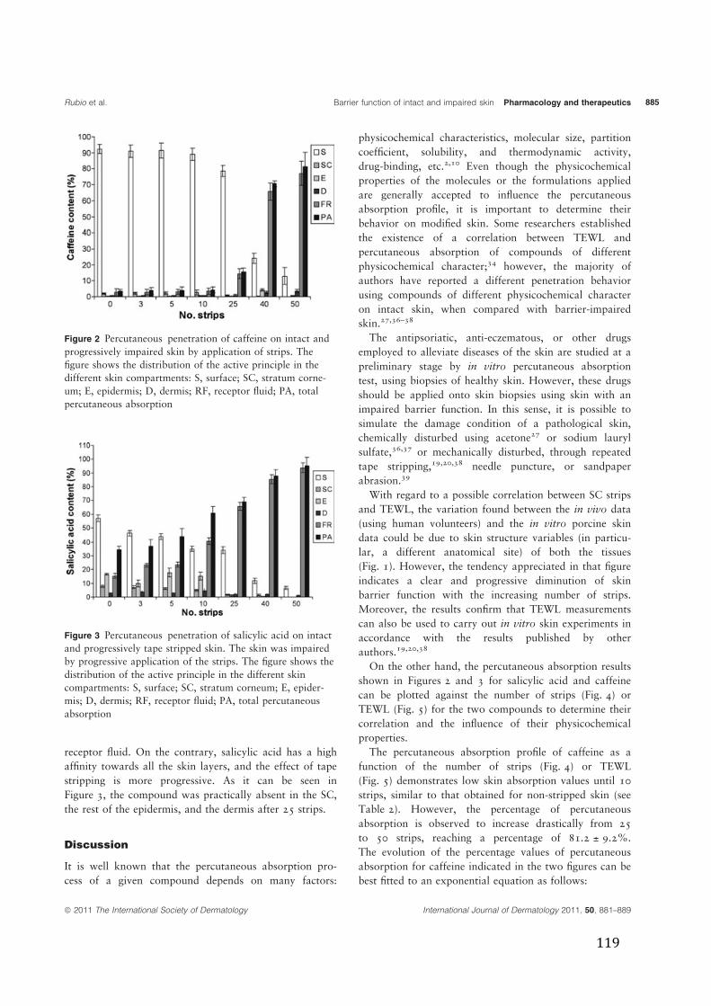
receptor fluid. On the contrary, salicylic acid has a highaffinity towards all the skin layers, and the effect of tapestripping is more progressive. As it can be seen inFigure 3, the compound was practically absent in the SC,the rest of the epidermis, and the dermis after 25 strips.
Discussion
It is well known that the percutaneous absorption pro-cess of a given compound depends on many factors:
physicochemical characteristics, molecular size, partitioncoefficient, solubility, and thermodynamic activity,drug-binding, etc.2,10 Even though the physicochemicalproperties of the molecules or the formulations appliedare generally accepted to influence the percutaneousabsorption profile, it is important to determine theirbehavior on modified skin. Some researchers establishedthe existence of a correlation between TEWL andpercutaneous absorption of compounds of differentphysicochemical character;34 however, the majority ofauthors have reported a different penetration behaviorusing compounds of different physicochemical characteron intact skin, when compared with barrier-impairedskin.27,36–38
The antipsoriatic, anti-eczematous, or other drugsemployed to alleviate diseases of the skin are studied at apreliminary stage by in vitro percutaneous absorptiontest, using biopsies of healthy skin. However, these drugsshould be applied onto skin biopsies using skin with animpaired barrier function. In this sense, it is possible tosimulate the damage condition of a pathological skin,chemically disturbed using acetone27 or sodium laurylsulfate,36,37 or mechanically disturbed, through repeatedtape stripping,19,20,38 needle puncture, or sandpaperabrasion.39
With regard to a possible correlation between SC stripsand TEWL, the variation found between the in vivo data(using human volunteers) and the in vitro porcine skindata could be due to skin structure variables (in particu-lar, a different anatomical site) of both the tissues(Fig. 1). However, the tendency appreciated in that figureindicates a clear and progressive diminution of skinbarrier function with the increasing number of strips.Moreover, the results confirm that TEWL measurementscan also be used to carry out in vitro skin experiments inaccordance with the results published by otherauthors.19,20,38
On the other hand, the percutaneous absorption resultsshown in Figures 2 and 3 for salicylic acid and caffeinecan be plotted against the number of strips (Fig. 4) orTEWL (Fig. 5) for the two compounds to determine theircorrelation and the influence of their physicochemicalproperties.
The percutaneous absorption profile of caffeine as afunction of the number of strips (Fig. 4) or TEWL(Fig. 5) demonstrates low skin absorption values until 10strips, similar to that obtained for non-stripped skin (seeTable 2). However, the percentage of percutaneousabsorption is observed to increase drastically from 25to 50 strips, reaching a percentage of 81.2 ± 9.2%.The evolution of the percentage values of percutaneousabsorption for caffeine indicated in the two figures can bebest fitted to an exponential equation as follows:
Figure 3 Percutaneous penetration of salicylic acid on intactand progressively tape stripped skin. The skin was impairedby progressive application of the strips. The figure shows thedistribution of the active principle in the different skincompartments: S, surface; SC, stratum corneum; E, epider-mis; D, dermis; RF, receptor fluid; PA, total percutaneousabsorption
Figure 2 Percutaneous penetration of caffeine on intact andprogressively impaired skin by application of strips. Thefigure shows the distribution of the active principle in thedifferent skin compartments: S, surface; SC, stratum corne-um; E, epidermis; D, dermis; RF, receptor fluid; PA, totalpercutaneous absorption
ª 2011 The International Society of Dermatology International Journal of Dermatology 2011, 50, 881–889
Rubio et al. Barrier function of intact and impaired skin Pharmacology and therapeutics 885
119
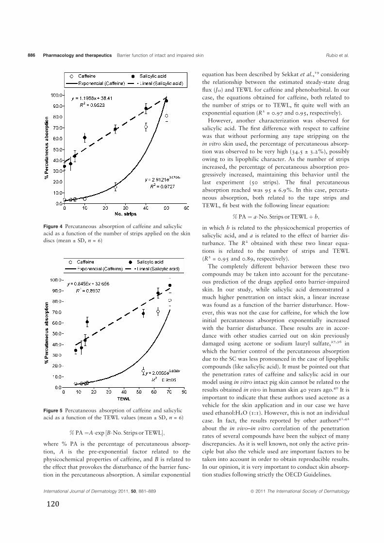
% PA ¼A�exp ½B�No: Strips or TEWL�;where % PA is the percentage of percutaneous absorp-tion, A is the pre-exponential factor related to thephysicochemical properties of caffeine, and B is related tothe effect that provokes the disturbance of the barrier func-tion in the percutaneous absorption. A similar exponential
equation has been described by Sekkat et al.,19 consideringthe relationship between the estimated steady-state drugflux (Jss) and TEWL for caffeine and phenobarbital. In ourcase, the equations obtained for caffeine, both related tothe number of strips or to TEWL, fit quite well with anexponential equation (R2 = 0.97 and 0.95, respectively).
However, another characterization was observed forsalicylic acid. The first difference with respect to caffeinewas that without performing any tape stripping on thein vitro skin used, the percentage of percutaneous absorp-tion was observed to be very high (34.5 ± 3.2%), possiblyowing to its lipophilic character. As the number of stripsincreased, the percentage of percutaneous absorption pro-gressively increased, maintaining this behavior until thelast experiment (50 strips). The final percutaneousabsorption reached was 95 ± 6.9%. In this case, percuta-neous absorption, both related to the tape strips andTEWL, fit best with the following linear equation:
% PA ¼ a�No: Strips or TEWLþ b;
in which b is related to the physicochemical properties ofsalicylic acid, and a is related to the effect of barrier dis-turbance. The R2 obtained with these two linear equa-tions is related to the number of strips and TEWL(R2 = 0.95 and 0.89, respectively).
The completely different behavior between these twocompounds may be taken into account for the percutane-ous prediction of the drugs applied onto barrier-impairedskin. In our study, while salicylic acid demonstrated amuch higher penetration on intact skin, a linear increasewas found as a function of the barrier disturbance. How-ever, this was not the case for caffeine, for which the lowinitial percutaneous absorption exponentially increasedwith the barrier disturbance. These results are in accor-dance with other studies carried out on skin previouslydamaged using acetone or sodium lauryl sulfate,27,36 inwhich the barrier control of the percutaneous absorptiondue to the SC was less pronounced in the case of lipophiliccompounds (like salicylic acid). It must be pointed out thatthe penetration rates of caffeine and salicylic acid in ourmodel using in vitro intact pig skin cannot be related to theresults obtained in vivo in human skin 40 years ago.40 It isimportant to indicate that these authors used acetone as avehicle for the skin application and in our case we haveused ethanol:H2O (1:1). However, this is not an individualcase. In fact, the results reported by other authors41,42
about the in vivo–in vitro correlation of the penetrationrates of several compounds have been the subject of manydiscrepancies. As it is well known, not only the active prin-ciple but also the vehicle used are important factors to betaken into account in order to obtain reproducible results.In our opinion, it is very important to conduct skin absorp-tion studies following strictly the OECD Guidelines.
Figure 5 Percutaneous absorption of caffeine and salicylicacid as a function of the TEWL values (mean ± SD, n = 6)
Figure 4 Percutaneous absorption of caffeine and salicylicacid as a function of the number of strips applied on the skindiscs (mean ± SD, n = 6)
International Journal of Dermatology 2011, 50, 881–889 ª 2011 The International Society of Dermatology
Pharmacology and therapeutics Barrier function of intact and impaired skin Rubio et al.886
120
On the other hand, the need to quantify (or, at least, tohave some idea of the amount of) the SC removed is notclear. Herkenne et al.43 published a paper considering theimportance of some experimental aspects. Different tapesand different methods will inevitably strip the SC in avariable fashion making inter- and intralaboratory com-parisons quite difficult. Several alternative attempts havebeen proposed, but no single method has achieved univer-sal acceptance.Our results indicate that the barrier impairment of the
skin always increases the penetration behavior of a givencompound; however, the hydrophilic–lipophilic balanceof the compounds or formulations used could greatlymodify its penetration profile, especially when a modifiedskin is used. It is clear that in the case of drugs to beapplied on a skin with a compromised barrier function, apreliminary knowledge about their in vivo TEWL valuesis necessary, as reported by Simonsen and Fullerton38 onatopic dermatitis. In fact, the skin permeation resultsobtained by these authors for fusidic acid and bethameth-asone-17-valerate, using the stripped-skin model, wereobserved to be correlated with clinical findings. Based onTEWL measurements obtained in vivo for different typesof damaged skin, the in vitro percutaneous absorptionstudies should be performed using skin discs simulating asimilar disturbance of their SC barrier function, by tapestripping.Very recently, Gattu and Maibach44 have published a
review considering the research carried out on percutane-ous absorption by different authors using several methodsof skin damage: mechanical, chemical, biocidal as well asin vitro tests with clinical disease. In general, all methodsof damage resulted in some enhancement of percutaneousabsorption of some molecules, most pronounced withhydrophilic molecules and less so with lipophilics. Thatenhancement was modest although very variable betweenthe different authors implying that despite mechanical,chemical, biocidal-damaged, and clinical disease, not allbarrier properties were removed. In our work, theincrease of percutaneous absorption depends on the num-ber of strips performed on the skin, and it is different forboth compounds investigated. As an example, in the caseof caffeine when 25 strips, are previously performed, theincrease of percutaneous absorption with respect to theintact skin is about five times; however, in the case ofsalicylic acid when using 25 strips, the increase ofpercutaneous absorption is about two times. These resultsare in accordance with the results presented in thatreview44 because for lipophilic molecules, as is the casefor salicylic acid, the increase of the percutaneous absorp-tion in damaged skin is minor. In any case, it is importantto design precise reproducible in vitro skin damagemodels considering, in particular, how permeability was
measured. This can be a prerequisite to adopt in thefuture broad comparisons and even correlations to thein vivo studies.
Conclusions
Evaluation of SC integrity (intact or disturbed) can becontrolled in vitro by the measurement of TEWL. By per-forming a sequential number of tape strippings on porcineskin discs, it is possible to progressively disturb SC barrierfunction. Moreover, by means of in vitro percutaneousabsorption experiments, it is possible to observe the influ-ence of a progressive disturbance of the SC on the percu-taneous penetration of different compounds topicallyapplied. In our study, caffeine and salicylic acid showed acompletely different behavior on intact compared withprogressively barrier-impaired skin. Salicylic acid showeda much higher penetration on intact skin, demonstratinga linear increase with the barrier disturbance. It seemsthat salicylic acid has a higher affinity towards all theskin layers and that the effect of tape stripping is progres-sive. However, this was not the case for caffeine, whoselow initial percutaneous absorption exponentiallyincreased with barrier disturbance. It is observed thatthe strong barrier effect of SC until 25 strips may beresponsible for the caffeine remaining mainly in theSC. However, after a strong barrier disturbance,caffeine shows a low affinity towards the epidermis anddermis and, consequently, passes directly to the receptorfluid.
This in vitro protocol may be useful to simulate the per-cutaneous absorption profile of some drugs applied ontothe skin with a progressive impaired SC barrier function.Moreover, this in vitro approach could avoid, to someextent, the use of in vivo experimental animal models.
Acknowledgment
The authors acknowledge the financial support of Minis-terio de Educación y Ciencia of Spain and the EuropeanSocial Fund (Programa de Personal Técnico de Apoyo[PTA2007-0335-I]).
References
1 Elias PM. Stratum corneum defensive functions: anintegrated view. J Invest Dermatol 2005; 125: 183–200.
2 Schaefer H, Redemeier TE. Skin Barrier – Principles of
Percutaneous Absorption. Basel: Karger-Verlag, 1996.3 Elias PM, Feingold KR. Stratum corneum barrier
function: definitions and broad concepts. In: Elias PM,Feingold KR, ed. Skin Barrier. New York: Taylor andFrancis Group, 2006: 1–4.
ª 2011 The International Society of Dermatology International Journal of Dermatology 2011, 50, 881–889
Rubio et al. Barrier function of intact and impaired skin Pharmacology and therapeutics 887
121
4 Bouwstra JA, Ponec M. The skin barrier in healthy anddiseased state. Biochim Biophys Acta 2006; 1758:280–295.
5 Fartasch M. The nature of the epidermal barrier: structuralaspects. Adv Drug Deliv Rev 1996; 18: 273–282.
6 Kessner D, Ruettinger A, Kiselev MA, et al. Properties ofceramides and their impact on the stratum corneumstructure. Part 2: stratum corneum lipid model systems.Skin Pharmacol Physiol 2008; 21: 58–74.
7 Howes D, Guy R, Hadgraft J, et al. Methods forassessing percutaneous absorption – the report andrecommendations of ECVAM workshop 13. Altern Lab
Anim 1996; 24: 81–106.8 OECD. Guideline 428: skin absorption: in vitro method.
In: Organisation for Economic Cooperation andDevelopment, eds. OECD Guidelines for the Testing of
Chemicals. Paris: OECD, 2004: 1–8.9 OECD. Guidance document for the conduct of skin
absorption studies number 28. In: Organisation forEconomic Cooperation and Development, eds. OECD
Series on Testing and Assessment. Paris: OECD, 2004:9–31.
10 Williams A. Transdermal and Topical Drug Delivery:
From Theory to Clinical Practice. London:Pharmaceutical Press, 2003.
11 Simon GA, Maibach HI. The pig as an experimentalanimal model of percutaneous permeation inman: qualitative and quantitative observations – anoverview. Skin Pharmacol Appl Skin Physiol 2000; 13:229–234.
12 Imokawa G, Abe A, Jin K, et al. Decreased level ofceramides in stratum corneum of atopic dermatitis: anetiologic factor in atopic dry skin? J Invest Dermatol
1991; 96: 523–526.13 Paige DG, Morse-Fisher N, Harper JI. Quantification of
stratum corneum ceramides and lipid envelope ceramidesin the hereditary ichthyoses. Br J Dermatol 1994; 131:23–27.
14 Motta S, Monti M, Sesana S, et al. Abnormality of waterbarrier function in psoriasis. Role of ceramide fractions.Arch Dermatol 1994; 130: 452–456.
15 Rawlings AV, Watkinson A, Rogers J. Abnormalities instratum corneum structure, lipid composition anddesmosome degradation in soap-induced winter xerosis.J Soc Cosmet Chem 1994; 45: 203–220.
16 Rogiers V. EEMCO guidance for the assessment oftransepidermal water loss in cosmetic sciences. SkinPharmacol Appl Skin Physiol 2001; 14: 117–128.
17 Rogiers V, Balls M, Basketter D, et al. The potential useof non-invasive methods in the safety assessment ofcosmetic products. The report and recommendations ofan ECVAM/EEMCO workshop (ECVAM workshop 36).Altern Lab Anim 1999; 27: 515–537.
18 Nangia A, Berner B, Maibach HI. Transepidermal waterloss measurements for assessing skin barrier functionsduring in vitro percutaneous absorption studies. In:Bronaugh RL, Maibach HI, eds. Percutaneous
Absorption: Drugs-Cosmetics-Mechanisms-Methodology.New York: Marcel Dekker, 1999: 587–594.
19 Sekkat N, Kalia YN, Guy RH. Porcine ear skin as amodel for the assessment of transdermal drug deliveryto premature neonates. Pharm Res 2004; 21: 1390–1397.
20 Sekkat N, Kalia YN, Guy RH. Development of an invitro model for premature neonatal skin: biophysicalcharacterization using transepidermal water loss. J PharmSci 2004; 93: 2936–2940.
21 Kalia YN, Nonato LB, Guy RH. The effect ofiontophoresis on skin barrier integrity: non-invasiveevaluation by impedance spectroscopy andtransepidermal water loss. Pharm Res 1996; 13: 957–960.
22 Kalia YN, Alberti I, Sekkat N, et al. Normalization ofstratum corneum barrier function and transepidermalwater loss in vivo. Pharm Res 2000; 17: 1148–1150.
23 Rougier A, Lotte C, Maibach HI. In vivo relationshipbetween percutaneous absorption and transepidermalwater loss. In: Wester RC, Maibach HI, eds.Percutaneous Absorption: Drug-Cosmetics-Mechanisms-
Methodology. New York: Marcel Dekker, 1999:117–132.
24 Levin J, Maibach H. The correlation betweentransepidermal water loss and percutaneous absorption:an overview. J Control Release 2005; 103: 291–299.
25 Aalto-Korte K, Turpeinen M. Transepidermal water lossand absorption of hydrocortisone in widespreaddermatitis. Br J Dermatol 1993; 128: 633–635.
26 Rougier A, Lotte C, Corcuff P, Maibach HI. Relationshipbetween skin permeability and corneocyte size accordingto anatomic site, age and sex in man. J Soc Cosmet
Chem 1988; 39: 15–26.27 Tsai JC, Sheu HM, Hung PL, Cheng CL. Effect of barrier
disruption by acetone treatment on the permeability ofcompounds with various lipophilicities: implications forthe permeability of compromised skin. J Pharm Sci 2001;90: 1242–1254.
28 Chilcott RP, Dalton CH, Emmanuel AJ, et al.Transepidermal water loss does not correlate with skinbarrier function in vitro. J Invest Dermatol 2002; 118:871–875.
29 Aalto-Korte K, Turpeinen M. Pharmacokinetics of topicalhydrocortisone at plasma level after applications once ortwice daily in patients with widespread dermatitis. Br JDermatol 1995; 133: 259–263.
30 Bashir SJ, Dreher F, Chew AL, et al. Cutaneous bioassayof salicylic acid as a keratolytic. Int J Pharm 2005; 292:187–194.
31 Vali A, Asilian A, Khalesi E, et al. Evaluation of theefficacy of topical caffeine in the treatment of psoriasisvulgaris. J Dermatolog Treat 2005; 16: 234–237.
32 Dias M, Hadgraft J, Lane ME. Influence of membrane-solvent-solute interactions on solute permeation in skin.Int J Pharm 2007; 340: 65–70.
International Journal of Dermatology 2011, 50, 881–889 ª 2011 The International Society of Dermatology
Pharmacology and therapeutics Barrier function of intact and impaired skin Rubio et al.888
122
33 Kalia YN, Nonato LB, Lund CH, Guy RH. Developmentof skin barrier function in premature infants. J InvestDermatol 1998; 111: 320–326.
34 Lotte C, Rougier A, Wilson DR, Maibach HI. In vivorelationship between transepidermal water loss andpercutaneous penetration of some organic compounds inman: effect of anatomic site. Arch Dermatol Res 1987;279: 351–356.
35 Klimowicz A, Farfal S, Bielecka-Grzela S. Evaluation ofskin penetration of topically applied drugs in humans bycutaneous microdialysis: acyclovir vs. salicylic acid. J ClinPharm Ther 2007; 32: 143–148.
36 Nielsen JB. Percutaneous penetration throughslightly damaged skin. Arch Dermatol Res 2005; 296:560–567.
37 Nielsen JB, Nielsen F, Sorensen JA. Defense againstdermal exposures is only skin deep: significantlyincreased penetration through slightly damaged skin.Arch Dermatol Res 2007; 299: 423–431.
38 Simonsen L, Fullerton A. Development of an in vitro skinpermeation model simulating atopic dermatitis skin for
the evaluation of dermatological products. SkinPharmacol Physiol 2007; 20: 230–236.
39 Wu XM, Todo H, Sugibayashi K. Effects of pretreatmentof needle puncture and sandpaper abrasion on the invitro skin permeation of fluorescein isothiocyanate(FITC)-dextran. Int J Pharm 2006; 316: 102–108.
40 Feldmann RJ, Maibach HI. Absorption of some organiccompounds through the skin in man. J Invest Dermatol
1970; 54: 399–404.41 Franz TJ. Percutaneous absorption. On the relevance of
in vitro data. J Invest Dermatol 1975; 64: 190–195.42 Franz TJ, Lehman PA, Raney SG. Use of excised human
skin to assess the bioequivalence of topical products. SkinPharmacol Physiol 2009; 22: 276–286.
43 Herkenne C, Alberti I, Naik A, et al. In vivo methods forthe assessment of topical drug bioavailability. Pharm Res
2008; 25: 87–103.44 Gattu S, Maibach HI. Enhanced absorption through
damaged skin: an overview of the in vitro human model.Skin Pharmacol Physiol 2010; 23: 171–176.
ª 2011 The International Society of Dermatology International Journal of Dermatology 2011, 50, 881–889
Rubio et al. Barrier function of intact and impaired skin Pharmacology and therapeutics 889
123

>#A�
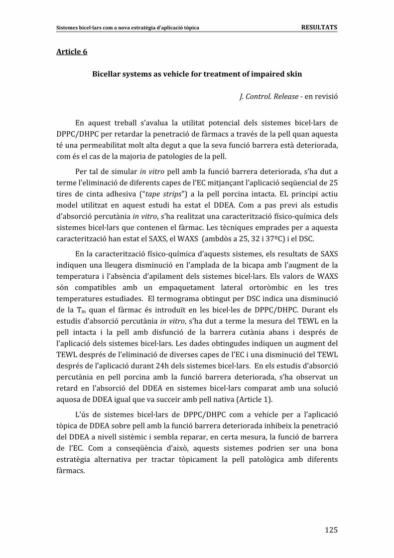
Sistemes�bicel·lars�com�a�nova�estratègia�d’aplicació�tòpica����������������������������������������������������������������������RESULTATS�
>#%�
Article�6��
Bicellar�systems�as�vehicle�for�treatment�of�impaired�skin��
�J.�Control.�Release�&������� � 9�
�
!�� �5����� ���-���� �0������� ��� �� � ���� ����� ��� ����� � ������� - ��B����� ���
�::�I�D:������������������������� 9����4������������<�������������5�����5������
�<�����������- � ����������������������5�����������4�� 9�-����������������� �������
���< � ����������������� �����H�� ����� �������������6��
:����������� ������ in�vitro��������-���� 4�� 9�-������������ ��������0.��������
�������0�� � �� 9����� 4����������������0!��� �H��1�����0��� � 9���5[�� ������#%�
� ���� ��� ���� ��.�� ��� 7Ntape� stripsP8� �� ��� ����� ��� ��� �����6� !)� �� � � � �� ��
������ �� � �/��� ��� �5����� ����� � .�� ������ ��� ��!*6� ���� �� ���� ���� � ���� ����� ��
�S�-��� 9�������� ��in�vitro���0.������ �/������������� �/� 9�4�� �&5��� �������
� �������- ��B�����5��������������4����6�)������ 5��������������������5������
������ �/� 9�.������������3*F3�����T*F3��7��-�(����#%��@#� �@'t�8� �����3�6��
!����������� �/� 9�4�� �&5��� ���0�5������� �������������������������3*F3�
�� 5�������� ���������� �� �� 9���� �0����������� ���- ������-� �0����������� ���
������������ � �0�-��� ���0�� ������������ � �������- ��B����6�!�������������T*F3�
�9�� ����� -���� ��-� ��� ����5��������� �������� �����(�- � ��� ���� �����
������������������ ����6��!��������������-� ����������3�� �� ������� �� �� 9�
��� ��� ��� 5���� ��� 4����� <�� ������L�� ��� ���� - ��B���� ��� �::�I�D:�6� ������� ����
����� ���0�-��� 9�������� �� in�vitro���0.�����������������������������!T)�������
����� ������ � ��� ����� ��-� � �4�� 9� ��� ��� -������� ���� �� �-���� � �����<�� ���
�0��� � 9������� �������- ��B����6�)����������-� ������� �� 5�������������������
�!T)������<������0�� � �� 9����� ����������������0!�� ������ �� �� 9������!T)�
�����<������0��� � 9��������#A.������� �������- ��B����6��!����������� ���0�-��� 9�
������� �� ��� ����� ��� ��� ��-� ��� 4�� 9� -������� ����� ������� �0.�� �-������� ���
������� ��� �0�-��� 9� ���� ��!*� ��� � ������� - ��B����� �������� ��-� ���� ���� 9�
�5��� !������� *� �����5��������� ����-��������� ���7*�� ���>86�
)0U�� ��� � ������� - ��B����� ��� �::�I�D:�� ��� �� ��. ��� ���� �� �0��� � 9�
�(� �������!*���-����������-����4�� 9�-������������ ������ �. -� M����������� 9�
������!*���� ������ ���� � ����-�������������������������������4�� 9����-�������
��� �0!�6� ���� �� ����5[�� �� �0� M(�� �5������ � ������� ���� ��� ���� ���� -����
�������� �� �������� ��� ���� ������� �(� ������ ��� ����� �����(� �� ��-� � 4�������
4�����6�
�
�

>#E�

>#'�
Bicellar systems as vehicle for treatment of impaired skin L. Rubio1, C. Alonso1, G. Rodríguez1, M. Cócera1, L. Coderch1, A. de la Maza1 J. L. Parra1 and O. López1
1IQAC-CSIC, Department of Chemical and Surfactants Technology, Barcelona, Spain. * Corresponding author: Laia Rubio, Department of Chemical and Surfactants Technology (IQAC-CSIC)
C/ Jordi Girona 18-26, 08034, Barcelona, Spain. Phone: 34-93400 61 00. Fax: 34-93204 59 04. E-mail:
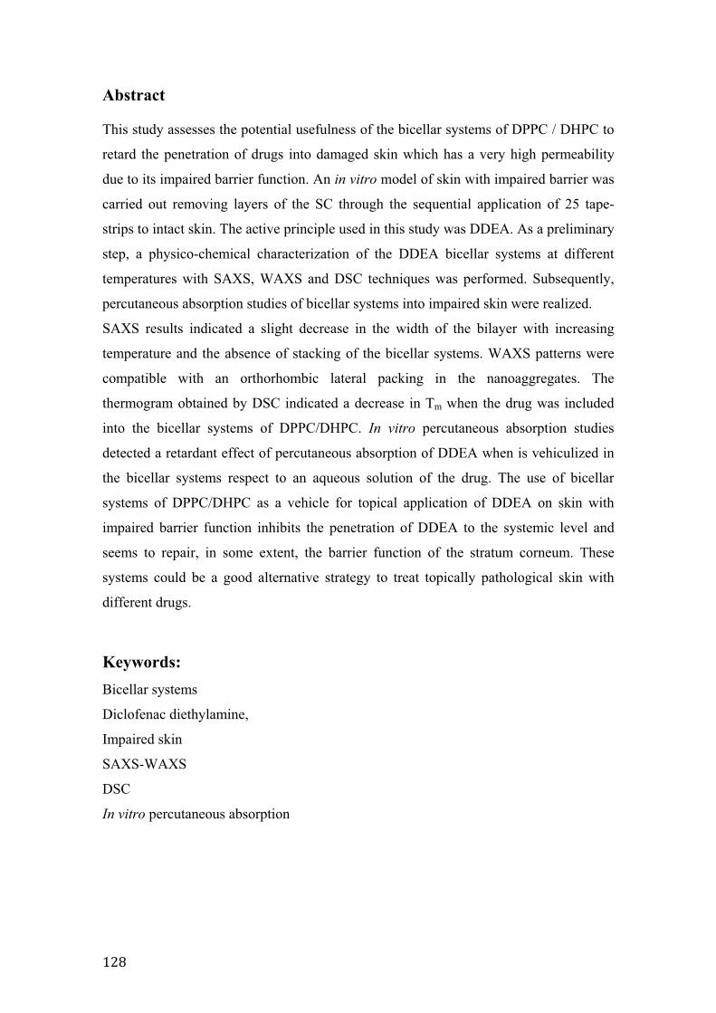
>#C�
Abstract
This study assesses the potential usefulness of the bicellar systems of DPPC / DHPC to
retard the penetration of drugs into damaged skin which has a very high permeability
due to its impaired barrier function. An in vitro model of skin with impaired barrier was
carried out removing layers of the SC through the sequential application of 25 tape-
strips to intact skin. The active principle used in this study was DDEA. As a preliminary
step, a physico-chemical characterization of the DDEA bicellar systems at different
temperatures with SAXS, WAXS and DSC techniques was performed. Subsequently,
percutaneous absorption studies of bicellar systems into impaired skin were realized.
SAXS results indicated a slight decrease in the width of the bilayer with increasing
temperature and the absence of stacking of the bicellar systems. WAXS patterns were
compatible with an orthorhombic lateral packing in the nanoaggregates. The
thermogram obtained by DSC indicated a decrease in Tm when the drug was included
into the bicellar systems of DPPC/DHPC. In vitro percutaneous absorption studies
detected a retardant effect of percutaneous absorption of DDEA when is vehiculized in
the bicellar systems respect to an aqueous solution of the drug. The use of bicellar
systems of DPPC/DHPC as a vehicle for topical application of DDEA on skin with
impaired barrier function inhibits the penetration of DDEA to the systemic level and
seems to repair, in some extent, the barrier function of the stratum corneum. These
systems could be a good alternative strategy to treat topically pathological skin with
different drugs.
Keywords: Bicellar systems
Diclofenac diethylamine,
Impaired skin
SAXS-WAXS
DSC
In vitro percutaneous absorption
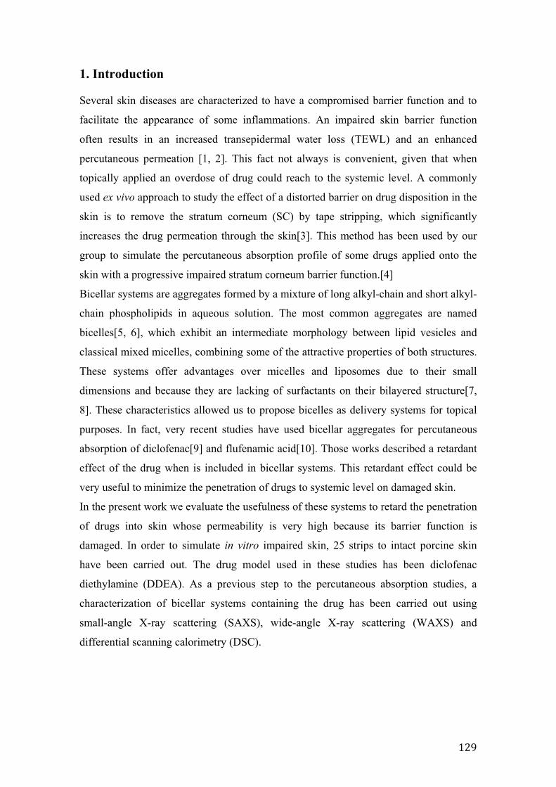
>#G�
1. Introduction
Several skin diseases are characterized to have a compromised barrier function and to
facilitate the appearance of some inflammations. An impaired skin barrier function
often results in an increased transepidermal water loss (TEWL) and an enhanced
percutaneous permeation [1, 2]. This fact not always is convenient, given that when
topically applied an overdose of drug could reach to the systemic level. A commonly
used ex vivo approach to study the effect of a distorted barrier on drug disposition in the
skin is to remove the stratum corneum (SC) by tape stripping, which significantly
increases the drug permeation through the skin[3]. This method has been used by our
group to simulate the percutaneous absorption profile of some drugs applied onto the
skin with a progressive impaired stratum corneum barrier function.[4]
Bicellar systems are aggregates formed by a mixture of long alkyl-chain and short alkyl-
chain phospholipids in aqueous solution. The most common aggregates are named
bicelles[5, 6], which exhibit an intermediate morphology between lipid vesicles and
classical mixed micelles, combining some of the attractive properties of both structures.
These systems offer advantages over micelles and liposomes due to their small
dimensions and because they are lacking of surfactants on their bilayered structure[7,
8]. These characteristics allowed us to propose bicelles as delivery systems for topical
purposes. In fact, very recent studies have used bicellar aggregates for percutaneous
absorption of diclofenac[9] and flufenamic acid[10]. Those works described a retardant
effect of the drug when is included in bicellar systems. This retardant effect could be
very useful to minimize the penetration of drugs to systemic level on damaged skin.
In the present work we evaluate the usefulness of these systems to retard the penetration
of drugs into skin whose permeability is very high because its barrier function is
damaged. In order to simulate in vitro impaired skin, 25 strips to intact porcine skin
have been carried out. The drug model used in these studies has been diclofenac
diethylamine (DDEA). As a previous step to the percutaneous absorption studies, a
characterization of bicellar systems containing the drug has been carried out using
small-angle X-ray scattering (SAXS), wide-angle X-ray scattering (WAXS) and
differential scanning calorimetry (DSC).
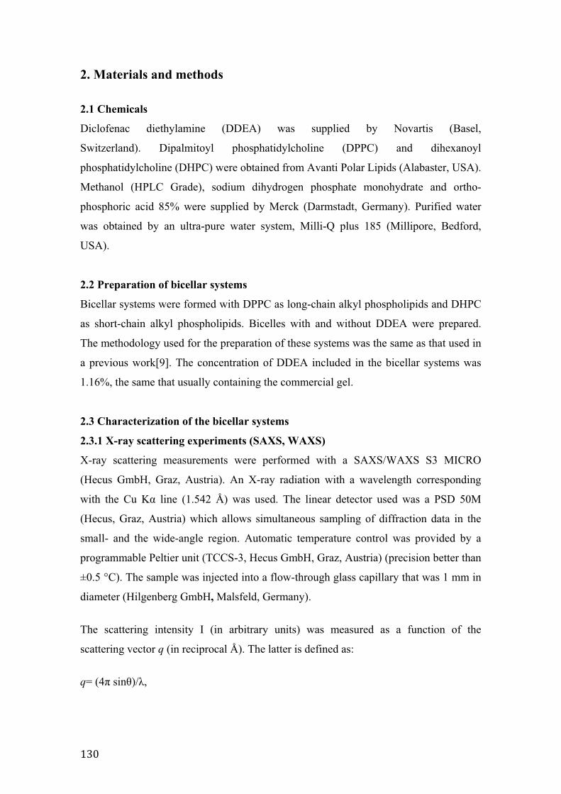
>@$�
2. Materials and methods
2.1 Chemicals
Diclofenac diethylamine (DDEA) was supplied by Novartis (Basel,
Switzerland). Dipalmitoyl phosphatidylcholine (DPPC) and dihexanoyl
phosphatidylcholine (DHPC) were obtained from Avanti Polar Lipids (Alabaster, USA).
Methanol (HPLC Grade), sodium dihydrogen phosphate monohydrate and ortho-
phosphoric acid 85% were supplied by Merck (Darmstadt, Germany). Purified water
was obtained by an ultra-pure water system, Milli-Q plus 185 (Millipore, Bedford,
USA).
2.2 Preparation of bicellar systems
Bicellar systems were formed with DPPC as long-chain alkyl phospholipids and DHPC
as short-chain alkyl phospholipids. Bicelles with and without DDEA were prepared.
The methodology used for the preparation of these systems was the same as that used in
a previous work[9]. The concentration of DDEA included in the bicellar systems was
1.16%, the same that usually containing the commercial gel.
2.3 Characterization of the bicellar systems
2.3.1 X-ray scattering experiments (SAXS, WAXS)
X-ray scattering measurements were performed with a SAXS/WAXS S3 MICRO
(Hecus GmbH, Graz, Austria). An X-ray radiation with a wavelength corresponding
with the Cu K� line (1.542 Å) was used. The linear detector used was a PSD 50M
(Hecus, Graz, Austria) which allows simultaneous sampling of diffraction data in the
small- and the wide-angle region. Automatic temperature control was provided by a
programmable Peltier unit (TCCS-3, Hecus GmbH, Graz, Austria) (precision better than
±0.5 °C). The sample was injected into a flow-through glass capillary that was 1 mm in
diameter (Hilgenberg GmbH, Malsfeld, Germany).
The scattering intensity I (in arbitrary units) was measured as a function of the
scattering vector q (in reciprocal Å). The latter is defined as:
q= (4� sin�)/�,
>@>�
where � is the scattering angle and � the wavelength of the radiation (1.542 Å). The
position of the scattering peaks is directly related to the repeat distance of the molecular
structure, as described by Bragg’s law:[11]
2d sin � = n�
in which n and d are the order of the diffraction peak and the repeat distance,
respectively. In the case of a lamellar structure, the various peaks are located at
equidistant positions, then
qn=2�n/d
where qn is the scattering vector modulus that indicates the position of the nth order
reflection.
2.3.2 Differential Scanning Calorimetry (DSC)
Differential scanning calorimetry (DSC) measurements of DPPC/DHPC and
DPPC/DHPC/DDEA systems were performed with a DSC 821E Mettler Toledo
(Greifensee, Switzerland) calorimeter. Aliquots of 30�L from the DPPC/DHPC and
DPPC/DHPC/DDEA bicellar systems, corresponding to 4.356 mg and 4.074 mg of
DPPC respectively, were placed in 40 �L aluminium DSC pans and hermetically sealed.
Three heating/cooling cycles were performed in a temperature range between 0 and 60
°C at a constant scanning rate of 5 °C/min. The data from the first scan were always
discarded to avoid mixing artifacts. Due to the supercooling phenomenon only heating
scans have been used throughout this work.[12] DSC curves were obtained and
analyzed by STAR 9.2 Software (Mettler Toledo).
2.4 HPLC analysis of diclofenac
The quantitative determination of DDEA was performed by HPLC using a Hitachi
LaChrom Elite equipment (Darmstadt, Germany). The apparatus consists of an L-2130
pump, L-2200 autosampler and an L-2400 UV detector. The system was operated from
the software Merck EZChrom Elite v3.1.3. Then 20 �L of injection volume were eluted
in a Lichrocart 250-4/Lichrosorb RP-18 (5 μm) column (Merck, Germany) at room
temperature. DDEA was monitored by UV detection at 254 nm. A mobile phase
consisting of a 66% methanol and 34% phosphate buffer (pH 2.5) was used at a flow
rate of 1 ml/min. The area under the peak was used to calculate the concentration of
>@#�
DDEA using external standards that showed linearity over the concentration range of
0.33 to 83 �g/ml. The intraday and interday variations of the method were less than 2%.
2.5 In vitro percutaneous absorption methodology using impaired skin
Following a conventional methodology, pig skin from the unboiled back of Landrace
large white pigs weighing between 30–40 Kg is used. The pig skin was provided by the
Clínic Hospital of Barcelona, Spain. The methodology used to obtain the skin biopsies
was the same as used in previous works[4, 9].
Franz cells (Lara-Spiral, Courtenon, France) were used to perform the percutaneous
absorption studies and PBS (pH 7.4) in distilled water, containing 1% of bovine serum
albumin and 0.04% gentamicin sulphate was used as the receptor fluid (RF). Franz cells
were kept at 37 ± 1 ºC by means of a circulating water bath (Julabo Labortechnik
GmbH, Germany) to ensure that the surface skin was maintained at 32 ± 1ºC. The
integrity of each skin sample was checked by determining the transepidermal water loss
(TEWL) using a Tewameter TM210 (Courage-Khazaka, Köln, Germany).
In this work, percutanous absorption experiments were carried out using samples of
impaired skin. The skin barrier impairment was induced by use of the tape stripping
method. To do that, after checking the skin integrity, each intact biopsy was subjected
to 25 sequential strips using D-Squame® tape discs (22 mm diameter, CuDerm Inc.,
Dallas, USA). ). Each tape was applied to the skin surface using a specific device
securing a standard pressure of 80 g/cm2 (for 5 s) and removed from the test area in one
gentle movement. For each repeated strip, the tearing-off direction was varied to obtain
a homogeneous removal of the SC cell layers. After that, the skin biopsies were
mounted to the Franz diffusion cells and after 1h, TEWL was measured to verify the
impairment of the skins.
The diffusion experiment was initiated by applying to the entire surface, delimited by
the upper cell, 10 μL of aqueous solution of DDEA or DDEA bicellar systems. After the
exposure time (24h), TEWL was measured. Then, the test formulation remaining on the
skin surface was removed. The receptor fluid was collected and brought up to 5 ml in a
volumetric flask. The SC of the treated skin area was removed by 8 successive tape
strips. After that, the viable epidermis was separated from the dermis after heating the
skin at 80ºC for a few seconds.
The amount of DDEA in the different layers and in the washing solution was extracted
with a solution of methanol: water (50:50). Before the analytical determination by
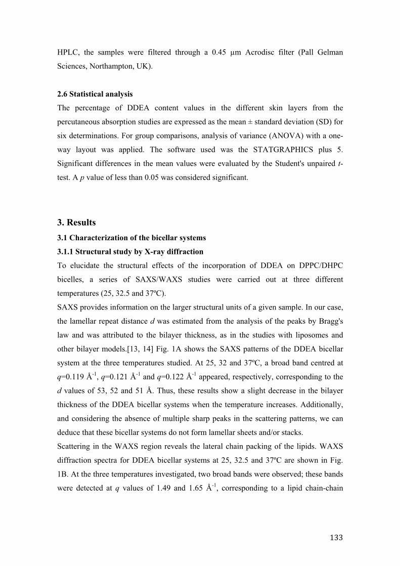
>@@�
HPLC, the samples were filtered through a 0.45 μm Acrodisc filter (Pall Gelman
Sciences, Northampton, UK).
2.6 Statistical analysis
The percentage of DDEA content values in the different skin layers from the
percutaneous absorption studies are expressed as the mean ± standard deviation (SD) for
six determinations. For group comparisons, analysis of variance (ANOVA) with a one-
way layout was applied. The software used was the STATGRAPHICS plus 5.
Significant differences in the mean values were evaluated by the Student's unpaired t-
test. A p value of less than 0.05 was considered significant.
3. Results 3.1 Characterization of the bicellar systems
3.1.1 Structural study by X-ray diffraction
To elucidate the structural effects of the incorporation of DDEA on DPPC/DHPC
bicelles, a series of SAXS/WAXS studies were carried out at three different
temperatures (25, 32.5 and 37ºC).
SAXS provides information on the larger structural units of a given sample. In our case,
the lamellar repeat distance d was estimated from the analysis of the peaks by Bragg's
law and was attributed to the bilayer thickness, as in the studies with liposomes and
other bilayer models.[13, 14] Fig. 1A shows the SAXS patterns of the DDEA bicellar
system at the three temperatures studied. At 25, 32 and 37ºC, a broad band centred at
q=0.119 Å-1, q=0.121 Å-1 and q=0.122 Å-1 appeared, respectively, corresponding to the
d values of 53, 52 and 51 Å. Thus, these results show a slight decrease in the bilayer
thickness of the DDEA bicellar systems when the temperature increases. Additionally,
and considering the absence of multiple sharp peaks in the scattering patterns, we can
deduce that these bicellar systems do not form lamellar sheets and/or stacks.
Scattering in the WAXS region reveals the lateral chain packing of the lipids. WAXS
diffraction spectra for DDEA bicellar systems at 25, 32.5 and 37ºC are shown in Fig.
1B. At the three temperatures investigated, two broad bands were observed; these bands
were detected at q values of 1.49 and 1.65 Å-1, corresponding to a lipid chain-chain
distance (dc) of 4.2 and 3.8 Å. These spacings are compatible with an orthorhombic
packing such as other authors described [15, 16].
0.0 0.2 0.4
25ºC53 Å
I(q) [
a.u.
]
q [Å-1] 1.4 1.6 1.8
3.8 Å4.2 Å
I(q) [
a.u.
]
q [Å-1]
25ºC
0.0 0.2 0.4
32ºC52 Å
q [Å-1]
I(q) [
a.u.
]
1.4 1.6 1.8
3.8 Å4.2 Å
32ºC
I(q) [
a.u.
]
q [Å-1]
0.0 0.2 0.4q [Å-1]
I(q) [
a.u.
]
37ºC51 Å
1.4 1.6 1.8
I(q) [
a.u.
]
q [Å-1]
37ºC
3.8 Å4.2 Å
A B
Fig. 1. SAXS (A) and WAXS (B) intensity profile versus the scattering vector modulus q of
DPPC/DHPC/DDEA bicellar systems. The measurements were carried out at 25 ºC, 32.5 ºC and 37 ºC.
3.1.2 Differential Scanning Calorimetry
The calorimetric analysis was performed in order to determine the heat flow associated
with the lipid phase transitions of the long alkyl chain phospholipid in DPPC/DHPC
bicelles with and without DDEA. When phosphatidylcholine systems are investigated,
the more common phase transitions described in the literature are a subtransition
corresponding to a change of phase from lamellar crystalline (Lc) to a tilted gel phase
>@A�
(L’), a pretransition from L’ to ripple phase or undulated-lamellar phase (P’) and a
main transition from whatever of the previously mentioned phases to liquid crystalline
phase (L�).[17, 18] The temperature associated with this latter transition is the so called
gel-to-liquid crystalline transition temperature, Tm [18]. DSC can be used to detect
interactions between the DDEA and the DPPC forming the bicellar systems. It is well
known that the inclusion of a new molecule in the phospholipid bilayers can modify the
lipid packing mode provoking a change in the temperature and in some cases in the
enthalpy (H) of the different lipid phase transitions.[19, 20] The results of the
experiments are shown in Fig. 2.
10 20 30 40 50
DPPC/DHPC
DPPC/DHPC/DDEA
Temperature (ºC)
Exot
herm
ic
10 20 30 40 50
DPPC/DHPC
DPPC/DHPC/DDEA
Temperature (ºC)
Exot
herm
ic
Fig. 2. DSC thermogram of DPPC bicellar systems with (solid line) and without (dashed line) DDEA.
>@%�
The signals observed in the thermograms are only associated to DPPC because DSC
thermograms of DHPC at this temperature range (results not shown) exhibited no peaks
and the melting point of DDEA is 127 ºC (out of the temperature range
investigated).[21] A quantitative determination of all enthalpies was not feasible, due to
the broadening peaks. Only the main transition enthalpies were determined giving
approximate values owing to the overlapping of the peaks.
The thermogram for DPPC/DHPC bicelles shows a large peak and a small shoulder
peak. The large peak at higher temperature (41.1 ºC) corresponds to the main transition
(Tm) associated to the P’ to L� transition of the DPPC alkyl chains and has an associated
enthalpy of about 16 KJ/mol. The small shoulder peak at around 35ºC is compatible
with the pretransition of the pure DPPC systems. The curve associated to the DPPC
transitions when DDEA is included in the DPPC/DHPC bicelles present also a large
peak and a small shoulder peak. The large peak associated to the main transition was
detected at 39ºC with an enthalpy value of about 18 KJ/mol. The small shoulder
associated with the pretransition was detected at around 29 ºC. It is noted that when
DDEA is included in the DPPC/DHPC bicellar systems, the temperatures associated to
the transitions decrease.
3.2 TEWL results before and after skin impairment TEWL measurements before and after impairment of skin barrier by 25 tape strippings
of the SC are shown in Fig. 3. Also, in this figure the TEWL values measured on
damaged skin after topical application of DDEA bicellar systems are presented. The
TEWL value of intact skin was 6.5 ± 1.3 g/m2/h and after 25 tape strippings this value
increases up to 33.9 ± 6.5 g/m2/h. As it could be expected a clear increase of the TEWL
was induced by the impairment of the SC barrier. When this damaged skin was treated
with the DDEA bicellar systems a decrease of TEWL until 22.4 ± 7.5 g/m2/h was
detected after 24h of exposure time. This result indicates certain restoring of the barrier
function by effect of treatment with DDEA bicellar systems.
05
1015202530354045
Intact skin Damaged skinbefore bicellar
treatment
Damaged skinafter bicellar
treatment
TEW
L(g/
m2 /h
)
Figure 3. TEWL values of intact skin and damaged skin before and after of DDEA bicellar systems treatment. Bars are the TEWL values (mean of n = 6) and the errors bars are the standard deviations (S.D.).
>@E�
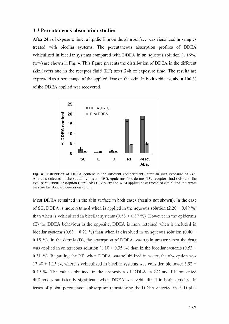
3.3 Percutaneous absorption studiesAfter 24h of exposure time, a lipidic film on the skin surface was visualized in samples
treated with bicellar systems. The percutaneous absorption profiles of DDEA
vehiculized in bicellar systems compared with DDEA in an aqueous solution (1.16%)
(w/v) are shown in Fig. 4. This figure presents the distribution of DDEA in the different
skin layers and in the receptor fluid (RF) after 24h of exposure time. The results are
expressed as a percentage of the applied dose on the skin. In both vehicles, about 100 %
of the DDEA applied was recovered.
0
5
10
15
20
25
SC E D RF Perc.Abs.
% D
DEA
con
tent
DDEA (H2O)
Bice DDEA
Fig. 4. Distribution of DDEA content in the different compartments after an skin exposure of 24h. Amounts detected in the stratum corneum (SC), epidermis (E), dermis (D), receptor fluid (RF) and the total percutanous absorption (Perc. Abs.). Bars are the % of applied dose (mean of n = 6) and the errors bars are the standard deviations (S.D.).
Most DDEA remained in the skin surface in both cases (results not shown). In the case
of SC, DDEA is more retained when is applied in the aqueous solution (2.20 ± 0.89 %)
than when is vehiculized in bicellar systems (0.58 ± 0.37 %). However in the epidermis
(E) the DDEA behaviour is the opposite, DDEA is more retained when is included in
bicellar systems (0.63 ± 0.21 %) than when is dissolved in an aqueous solution (0.40 ±
0.15 %). In the dermis (D), the absorption of DDEA was again greater when the drug
was applied in an aqueous solution (1.10 ± 0.35 %) than in the bicellar systems (0.53 ±
0.31 %). Regarding the RF, when DDEA was solubilized in water, the absorption was
17.40 ± 1.15 %, whereas vehiculized in bicellar systems was considerable lower 3.92 ±
0.49 %. The values obtained in the absorption of DDEA in SC and RF presented
differences statistically significant when DDEA was vehiculized in both vehicles. In
terms of global percutaneous absorption (considering the DDEA detected in E, D plus
>@'�
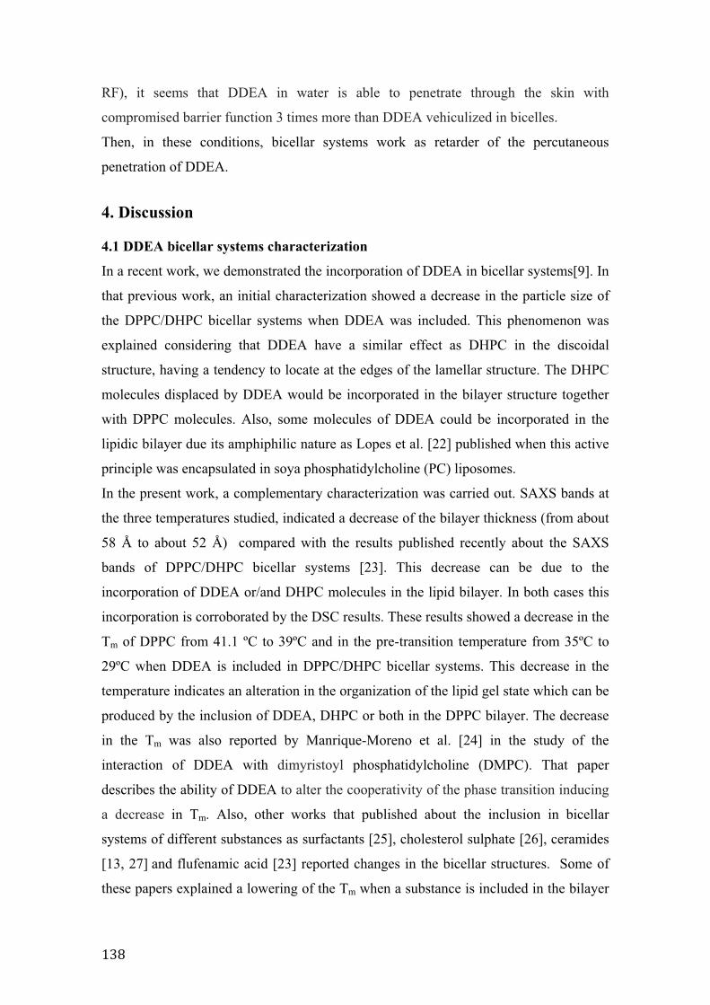
>@C�
RF), it seems that DDEA in water is able to penetrate through the skin with
compromised barrier function 3 times more than DDEA vehiculized in bicelles.
Then, in these conditions, bicellar systems work as retarder of the percutaneous
penetration of DDEA.
4. Discussion
4.1 DDEA bicellar systems characterization
In a recent work, we demonstrated the incorporation of DDEA in bicellar systems[9]. In
that previous work, an initial characterization showed a decrease in the particle size of
the DPPC/DHPC bicellar systems when DDEA was included. This phenomenon was
explained considering that DDEA have a similar effect as DHPC in the discoidal
structure, having a tendency to locate at the edges of the lamellar structure. The DHPC
molecules displaced by DDEA would be incorporated in the bilayer structure together
with DPPC molecules. Also, some molecules of DDEA could be incorporated in the
lipidic bilayer due its amphiphilic nature as Lopes et al. [22] published when this active
principle was encapsulated in soya phosphatidylcholine (PC) liposomes.
In the present work, a complementary characterization was carried out. SAXS bands at
the three temperatures studied, indicated a decrease of the bilayer thickness (from about
58 Å to about 52 Å) compared with the results published recently about the SAXS
bands of DPPC/DHPC bicellar systems [23]. This decrease can be due to the
incorporation of DDEA or/and DHPC molecules in the lipid bilayer. In both cases this
incorporation is corroborated by the DSC results. These results showed a decrease in the
Tm of DPPC from 41.1 ºC to 39ºC and in the pre-transition temperature from 35ºC to
29ºC when DDEA is included in DPPC/DHPC bicellar systems. This decrease in the
temperature indicates an alteration in the organization of the lipid gel state which can be
produced by the inclusion of DDEA, DHPC or both in the DPPC bilayer. The decrease
in the Tm was also reported by Manrique-Moreno et al. [24] in the study of the
interaction of DDEA with dimyristoyl phosphatidylcholine (DMPC). That paper
describes the ability of DDEA to alter the cooperativity of the phase transition inducing
a decrease in Tm. Also, other works that published about the inclusion in bicellar
systems of different substances as surfactants [25], cholesterol sulphate [26], ceramides
[13, 27] and flufenamic acid [23] reported changes in the bicellar structures. Some of
these papers explained a lowering of the Tm when a substance is included in the bilayer
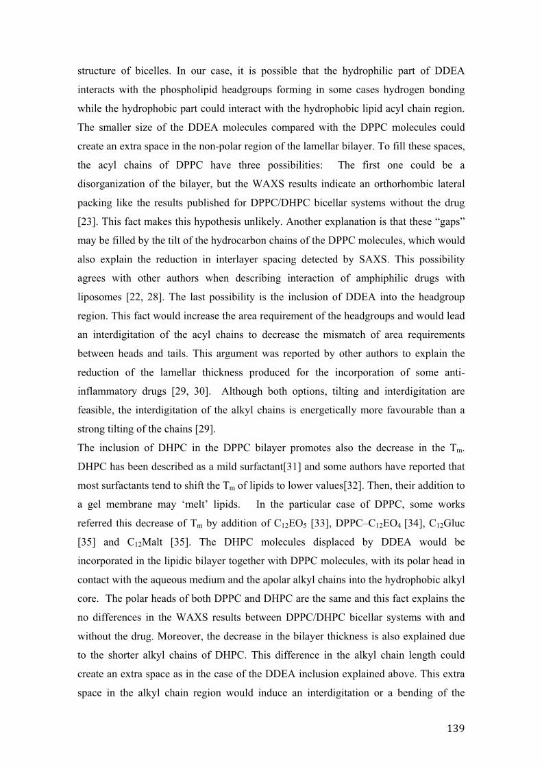
>@G�
structure of bicelles. In our case, it is possible that the hydrophilic part of DDEA
interacts with the phospholipid headgroups forming in some cases hydrogen bonding
while the hydrophobic part could interact with the hydrophobic lipid acyl chain region.
The smaller size of the DDEA molecules compared with the DPPC molecules could
create an extra space in the non-polar region of the lamellar bilayer. To fill these spaces,
the acyl chains of DPPC have three possibilities: The first one could be a
disorganization of the bilayer, but the WAXS results indicate an orthorhombic lateral
packing like the results published for DPPC/DHPC bicellar systems without the drug
[23]. This fact makes this hypothesis unlikely. Another explanation is that these “gaps”
may be filled by the tilt of the hydrocarbon chains of the DPPC molecules, which would
also explain the reduction in interlayer spacing detected by SAXS. This possibility
agrees with other authors when describing interaction of amphiphilic drugs with
liposomes [22, 28]. The last possibility is the inclusion of DDEA into the headgroup
region. This fact would increase the area requirement of the headgroups and would lead
an interdigitation of the acyl chains to decrease the mismatch of area requirements
between heads and tails. This argument was reported by other authors to explain the
reduction of the lamellar thickness produced for the incorporation of some anti-
inflammatory drugs [29, 30]. Although both options, tilting and interdigitation are
feasible, the interdigitation of the alkyl chains is energetically more favourable than a
strong tilting of the chains [29].
The inclusion of DHPC in the DPPC bilayer promotes also the decrease in the Tm.
DHPC has been described as a mild surfactant[31] and some authors have reported that
most surfactants tend to shift the Tm of lipids to lower values[32]. Then, their addition to
a gel membrane may ‘melt’ lipids. In the particular case of DPPC, some works
referred this decrease of Tm by addition of C12EO5 [33], DPPC–C12EO4 [34], C12Gluc
[35] and C12Malt [35]. The DHPC molecules displaced by DDEA would be
incorporated in the lipidic bilayer together with DPPC molecules, with its polar head in
contact with the aqueous medium and the apolar alkyl chains into the hydrophobic alkyl
core. The polar heads of both DPPC and DHPC are the same and this fact explains the
no differences in the WAXS results between DPPC/DHPC bicellar systems with and
without the drug. Moreover, the decrease in the bilayer thickness is also explained due
to the shorter alkyl chains of DHPC. This difference in the alkyl chain length could
create an extra space as in the case of the DDEA inclusion explained above. This extra
space in the alkyl chain region would induce an interdigitation or a bending of the
>A$�
DPPC alkyl chains leading a decrease of the bilayer thickness as it can be see in the
SAXS results.
These arguments support that some molecules of DDEA induce a displacement of
DHPC from the edges to the DPPC bilayer. Also, other DDEA molecules are
incorporated in the DPPC bilayer. In summary, the inclusion of DDEA induces the
mixture of these components in the bicellar structure promoting the reduction of the Tm
values and the bilayer thickness. This finding is in concordance with the small and
spherical shape of these DPPC/DHPC/DDEA systems detected by transmission electron
microscopy (TEM) and published in a previous work [9].
4.2 Topical application of DDEA bicellar systems in impaired skin
The simulation of in vitro impaired skin by tape stripping has been previously described
by our group [4] and other authors [1, 3] due its accessibility and simplicity. This
methodology induces and impairment of the SC increasing significantly the TEWL.
Simonsen et al.[3] developed an in vitro skin permeation model simulating atopic
dermatitis skin by applying 25 tape strips to intact skin, the barrier impairment was
evaluated by TEWL. The skin permeation results obtained through this impaired skin
were found to correlate with clinical findings. According to the results obtained in that
work, we decided to use 25 strips to simulate in vitro impaired skin.
The application of bicelles containing DDEA on the in vitro impaired skin induced a
certain occlusion of the skin with the corresponding diminution of the TEWL. This
occlusion is due to a lipidic film formed after the evaporation of the water of the bicellar
systems. This effect was also detected by other authors in the use of liposomes topical
applied in vivo which reduces TEWL giving to the patient immediate relief from the
discomfort associated with dry skin [36, 37]. The TEWL diminution envisaged in Fig. 3
can also be due to a certain restoration effect promoted by DDEA bicellar systems. This
system (without drug) has demonstrated in previous papers to work as repairing agent of
the SC lipid structure [7].
As regards to the percutaneous absorption studies, DDEA in bicelles has a favourable
tendency to pass through the SC (proportion of DDEA in SC in very low, Fig. 4). This
fact could be due to a higher affinity of DDEA with the lipids of the bicelle than with
the intercellular lipids of the SC. This affinity can be explained by the hydrogen
bonding possibly formed between DDEA and the phospholipids from bicelles. In the
case of E and D layers, the content of DDEA detected when the drug is applied in both
>A>�
vehicles is very similar. Therefore, the content of DDEA available in E and D does not
decrease much when is vehiculized in bicellar systems.
In RF the content of DDEA in water is statistically very high as compared with DDEA
in the bicellar system. This particular behaviour of DDEA vehiculized in water can be
considered normal because the barrier function of the skin has been compromised due to
the tape stripping and consequently the percutaneous absorption is enhanced [38]. Then,
it is interesting the retardant effect of bicellar systems on the DDEA topical absorption
in damaged skin. This behaviour is consistent with the work of Bouwstra et al[39] who
reports that gel-state vesicles can inhibit drug permeation across the skin. Considering
the DPPC/DHPC/DDEA Tm (39ºC) and the temperature of application (32ºC), DDEA
bicellar systems were also applied on a gel state. In addition, the retardant effect
detected on percutaneous absorption of DDEA included in a bicellar system may be
justified by the preferential interaction of the active principle with the lipids constituent
of the bicelles. This retardant effect of bicellar systems agrees with previous published
data in which this effect was described when drugs are applied on native skin [40].
This retardant effect of bicelles (decreasing the percutaneous absorption) as well as their
occlusive action on the skin surface (lowering the TEWL results) can be useful for the
improvement of the therapeutic characteristics of DDEA when this drug is applied on
impaired skin. This is specially interesting because, in most of the skin diseases, the
skin inflammatory process induces a deterioration of the EC [41].
5. Conclusions The use of DPPC/DHPC bicellar systems as vehicle for topical application of DDEA on
impaired skin inhibits the penetration of DDEA to systemic level and seems to repair, in
some extent, the barrier function. As a consequence, these systems may be a good
alternative strategy to treat damaged skin with different drugs.
Acknowledgements The authors acknowledge Ministerio de Educación y Ciencia, Spain (CTQ2010-16964)
and Generalitat de Catalunya (2009 SGR 1212). This work was supported by European
Social Fund (Programa Nacional de Potenciación de Recursos Humanos del Plan
Nacional de I+D+I (2004-2008)). Mercedes Cócera is funded by JAE-Doc Program
>A#�
from CSIC. Also, the authors wish to thank Jaume Caelles and Josep Carilla for expert
assistance with the techniques used in this work.
References
[1] S. Gattu, H.I. Maibach, Enhanced absorption through damaged skin: an overview of the in vitro human model, Skin Pharmacol Physiol, 23 (2010) 171-176. [2] J. Levin, H. Maibach, The correlation between transepidermal water loss and percutaneous absorption: an overview, J Control Release, 103 (2005) 291-299. [3] L. Simonsen, A. Fullerton, Development of an in vitro skin permeation model simulating atopic dermatitis skin for the evaluation of dermatological products, Skin Pharmacol Physiol, 20 (2007) 230-236. [4] L. Rubio, C. Alonso, O. López, G. Rodríguez, L. Coderch, J. Notario, A. de la Maza, J.L. Parra, Barrier function of intact and impaired skin: percutaneous penetration of caffeine and salicylic acid, International Journal of Dermatology, 50 (2011) 881-889. [5] J. Katsaras, T.A. Harroun, J. Pencer, M.P. Nieh, "Bicellar" lipid mixtures as used in biochemical and biophysical studies, Naturwissenschaften, 92 (2005) 355-366. [6] C.R. Sanders, 2nd, G.C. Landis, Reconstitution of membrane proteins into lipid-rich bilayered mixed micelles for NMR studies, Biochemistry, 34 (1995) 4030-4040. [7] L. Barbosa-Barros, A. de la Maza, J. Estelrich, A.M. Linares, M. Feliz, P. Walther, R. Pons, O. López, Penetration and growth of DPPC/DHPC bicelles inside the stratum corneum of the skin, Langmuir, 24 (2008) 5700-5706. [8] G. Rodríguez, L. Barbosa-Barros, L. Rubio, M. Cócera, C. López-Iglesias, A. de la Maza, O. López, Bicellar systems as modifiers of skin lipid structure, Colloids and Surfaces B: Biointerfaces, 84 (2011) 390-394. [9] L. Rubio, C. Alonso, G. Rodríguez, L. Barbosa-Barros, L. Coderch, A. De la Maza, J.L. Parra, O. López, Bicellar systems for in vitro percutaneous absorption of diclofenac, Int. J. Pharm., 386 (2010) 108-113. [10] L. Rubio, G. Rodríguez, L. Barbosa-Barros, C. Alonso, M. Cócera, A. de la Maza, J.L. Parra, O. López, Bicellar systems as a new colloidal delivery strategy for skin, Colloids and Surfaces B: Biointerfaces, 92 (2011) 322-326. [11] W.L. Bragg, The Diffraction of Short Electromagnetic Waves by a Crystal, in: Proceedings of the Cambridge Philosophical Society, 1913, pp. 43-57. [12] R.A. Videira, M.C. Antunes-Madeira, V.M. Madeira, Perturbations induced by alpha- and beta-endosulfan in lipid membranes: a DSC and fluorescence polarization study, Biochim. Biophys. Acta, Biomembr., 1419 (1999) 151-163. [13] L. Barbosa-Barros, A. de la Maza, C. López-Iglesias, O. López, Ceramide effects in the bicelle structure, Colloids and Surfaces A: Physicochemical and Engineering Aspects, 317 (2008) 576-584. [14] O. López, M. Cócera, L. Campos, A. de la Maza, L. Coderch, J.L. Parra, Use of wide and small angle X-ray diffraction to study the modifications in the stratum corneum induced by octyl glucoside, Colloids Surf., A, 162 (2000) 123-130. [15] G.S. Pilgram, A.M. Engelsma-van Pelt, H.K. Koerten, J.A. Bouwstra, The effect of two azones on the lateral lipid organization of human stratum corneum and its permeability, Pharm. Res., 17 (2000) 796-802. [16] G. Rodríguez, L. Barbosa-Barros, L. Rubio, M. Cócera, A. Díez, J. Estelrich, R. Pons, J. Caelles, A.D.l. Maza, O. López, Conformational Changes in Stratum Corneum Lipids by Effect of Bicellar Systems, Langmuir, 25 (2009) 10595-10603.
>A@�
[17] C. Huang, S. Li, Calorimetric and molecular mechanics studies of the thermotropic phase behavior of membrane phospholipids, Biochim. Biophys. Acta, Rev. Biomembr., 1422 (1999) 273-307. [18] R. Koynova, M. Caffrey, Phases and phase transitions of the phosphatidylcholines, Biochim. Biophys. Acta, Rev. Biomembr., 1376 (1998) 91-145. [19] M. Lucio, F. Bringezu, S. Reis, J.L. Lima, G. Brezesinski, Binding of nonsteroidal anti-inflammatory drugs to DPPC: structure and thermodynamic aspects, Langmuir, 24 (2008) 4132-4139. [20] B. Pili, C. Bourgaux, H. Amenitsch, G. Keller, S. Lepetre-Mouelhi, D. Desmaele, P. Couvreur, M. Ollivon, Interaction of a new anticancer prodrug, gemcitabine-squalene, with a model membrane: coupled DSC and XRD study, Biochim. Biophys. Acta, Biomembr., 1798 (2010) 1522-1532. [21] L.F. Manli Wang, Percutaneous absorption of diclofenac acid and its salts from emulgel, AJPS, 3 (2008) 131-141. [22] L.B. Lopes, M.V. Scarpa, G.V. Silva, D.C. Rodrigues, C.V. Santilli, A.G. Oliveira, Studies on the encapsulation of diclofenac in small unilamellar liposomes of soya phosphatidylcholine, Colloids Surf B Biointerfaces, 39 (2004) 151-158. [23] L. Rubio, G. Rodriguez, C. Alonso, C. Lopez-Iglesias, M. Cocera, L. Coderch, A. De la Maza, J.L. Parra, O. Lopez, Structural effects of flufenamic acid in DPPC/DHPC bicellar systems, Soft Matter, (2011). [24] M. Manrique-Moreno, P. Garidel, M. Suwalsky, J. Howe, K. Brandenburg, The membrane-activity of Ibuprofen, Diclofenac, and Naproxen: a physico-chemical study with lecithin phospholipids, Biochim Biophys Acta, 1788 (2009) 1296-1303. [25] M. Kozak, L. Domka, S. Jurga, The effect of selected surfactants on the structure of a bicellar system (DMPC/DHPC) studied by SAXS, J. Mol. Struct., 846 (2007) 108-111. [26] R.A. Shapiro, A.J. Brindley, R.W. Martin, Thermal Stabilization of DMPC/DHPC Bicelles by Addition of Cholesterol Sulfate, J. Am. Chem. Soc., 132 (2010) 11406-11407. [27] L. Barbosa-Barros, A. De la Maza, P. Walther, J. Estelrich, O. López, Morphological effects of ceramide on DMPC/DHPC bicelles, J. Microsc. (Oxford, U. K.), 230 (2008) 16-26. [28] I. Stoye, K. Schroder, C.C. Muller-Goymann, Transformation of a liposomal dispersion containing ibuprofen lysinate and phospholipids into mixed micelles - physico-chemical characterization and influence on drug permeation through excised human stratum corneum, Eur J Pharm Biopharm, 46 (1998) 191-200. [29] C. Nunes, G. Brezesinski, J.L. Lima, S. Reis, M. Lucio, Synchrotron SAXS and WAXS study of the interactions of NSAIDs with lipid membranes, J Phys Chem B, 115 (2011) 8024-8032. [30] C. Nunes, G. Brezesinski, J.L.F.C. Lima, S. Reis, M. Lucio, Effects of non-steroidal anti-inflammatory drugs on the structure of lipid bilayers: therapeutical aspects, Soft Matter, 7 (2011) 3002-3010. [31] H. Helmut, Short-chain phospholipids as detergents, Biochimica et Biophysica Acta (BBA) - Biomembranes, 1508 (2000) 164-181. [32] H. Heerklotz, Interactions of surfactants with lipid membranes, Quarterly Reviews of Biophysics, 41 (2008) 205-264. [33] H. Pfeiffer, G. Klose, K. Heremans, C. Glorieux, Thermotropic phase behaviour of the pseudobinary mixtures of DPPC/C12E5 and DMPC/C12E5 determined by differential scanning calorimetry and ultrasonic velocimetry, Chemistry and Physics of Lipids, 139 (2006) 54-67.
>AA�
[34] B. Mädler, G. Klose, A. Möps, W. Richter, C. Tschierske, Thermotropic phase behaviour of the pseudobinary mixture DPPC/C12E4 at excess water, Chemistry and Physics of Lipids, 71 (1994) 1-12. [35] B. Carion-Taravella, S. Lesieur, J. Chopineau, P. Lesieur, M. Ollivon, Phase Behavior of Mixed Aqueous Dispersions of Dipalmitoylphosphatidylcholine and Dodecyl Glycosides:A Differential Scanning Calorimetry and X-ray Diffraction Investigation, Langmuir, 18 (2001) 325-335. [36] J. De Leeuw, H.C. De Vijlder, P. Bjerring, H.A.M. Neumann, Liposomes in dermatology today, Journal of the European Academy of Dermatology and Venereology, 23 (2009) 505-516. [37] H. Lautenschlager, Liposomes in dermatological preparations. Part 1, Cosmet Toiletries, 105 (1990) 89-96. [38] D. Frank, Z. Hongbo, L. Harald, C. Myeong Jun, M. Howard, Effect of Tape Stripping on Percutaneous Penetration and Topical Vaccination, in: Percutaneous Absorption, Informa Healthcare, 2005, pp. 811-821. [39] J.A. Bouwstra, P.L. Honeywell-Nguyen, G.S. Gooris, M. Ponec, Structure of the skin barrier and its modulation by vesicular formulations, Progress in Lipid Research, 42 (2003) 1-36. [40] L. Rubio, G. Rodríguez, L. Barbosa-Barros, C. Alonso, M. Cócera, A. de la Maza, J.L. Parra, O. López, Bicellar systems as a new colloidal delivery strategy for skin, Colloids and Surfaces B: Biointerfaces, 92 (2012) 322-326. [41] S. Rahman, M. Collins, C.M. Williams, H.L. Ma, The pathology and immunology of atopic dermatitis, Inflamm Allergy Drug Targets, 10 (2011) 486-496.

�������
4.�DISCUSSIÓ�

Sistemes�bicel·lars�com�a�nova�estratègia�d’aplicació�tòpica������������������������������������������������������������������������DISCUSSIÓ�
>A'�
4.�DISCUSSIÓ�
4.1�Caracterització�físico�química�dels�sistemes�bicel·lars�
� ����������������� �������������������������:�I�D:�� ��::�I�D:�����
�� ����� � ������� ��� ��. �� �/� 9W� ��-�9�� ��-� ���� ���� 9� ������ ������ ���
4��4���� �� ��� ������ ��5��� �� ������� � ��� ��� ������ ��5��� �� ������ 5`� #�� � ����
������� 9� � ��� �� ���� #$d6� !��� ���������� ��� �)3� 7������ >� ��� �0��� ��� >8� ����
������������ �����������������0������>A� �>%�������������������6�:������������������
� ������� ��� ��:�I�D:�� ��� ���� 4������� ���� �::�I�D:�� ��� ��� ����� ��� ���
�� � � ��-������� ������������ ����U���������. ������������ ��� ���������� 9�
� ��� �� � ��� ����� � ��� ��� ��������6� :��(� �� ������� 5��� .��� ����� ����1���� ����
����� �� �����0�5��������� ��������B������������-<��0���������� M����������������
�0��� � 9� �0�5������ � ������� - ��B����� in� vivo� ���� ��� ���� ������� �����
�0 ����� �� 9� 7���-���&������� � ����� #$$G86� ����� ���������� �-� ������ ��� �5������
����� ������������������������ 9����5�������� �������- ��B���������::���9��� ������
���� �� �0��� � 9� �(� �� 5��� ���� ��� ��:�� H�� 5��� �5������ U�� ��� �����L��� �<��
��� � 9���������������4�� �&5��� �������������5�����������::�6��*��<�������������
�M�� � 9������������ �� �������� �����������::�������������������:��<��5�����
������������4 � ��(� ��7����M6�@'�]�8������::����������4���������H��5����<�����������
A>�]��7������� ������#$$'8�� �����5������� ��� ������ �0!��7����0����M ���������E$�
]�87=������ � ����� >GC'86� !�� ��� �� ��� ��:�� �<� ���� ��� � �0����M ��������� #@� ]��
73���� �� ������#$$>8�� ������������������������������5���������-��������������� �������
��� ��:�I�D:�� ������ ��� 4���� 4��L��6� :��� �5���� ��� ��� ���� - ��B���� ��� �::�� ���
��� ���� ����4����� �������4���� �����0��� ��&�����-����������W������� ������- ��B����
�����:��� �5������� M������ �����4���� ����4��(� �6�
�
4.1.1� Canvis� estructurals� i� morfològics� provocats� en� els� sistemes�bicel·lars�per�la�inclusió�d’un�principi�actiu�
4.1.1.1�Incorporació�del�DDEA��
)�� ������� 9� ���� ��!*� ��� ���� � ������� - ��B����� ��� ���� �� �� ����
� �� �� 9� ����� �� ��� ��� � ��� ��� ��������� ��������� ���� �)3� ���� ��-�9��
� �����������>A�$������#�A�������������������:�I�D:�� ����>%�#������#�C�������
��� ��� ����� � ������� ��� �::�I�D:�� 7������ >� ��� �0��� ��� >86� *5������� � ���� ���
����������������� �������0 �����������4���� 9����� �������- ��B�����H��5������� ���
��� ��� ��� ���� ��������� <�� ��� �0������� ��� #$� ��� 7�������� � ����� >GC#86� )��
� �� �� 9� ��� ���� ��� ���� �4���� ���� ��!*� ��� ���� ��������� ��� ������� 5��� ����
4��������-�������� ������� ���� ��� ��� ������� ������������ �4������� �����
������ ����� ��� ������ ����&���� ��� �� ���-������ �� ��&��� �� ������ � ������� ����
� ���� 9���� ����0�5������6�*5�������M�� � 9������������ ������������ ����������
Sistemes�bicel·lars�com�a�nova�estratègia�d’aplicació�tòpica������������������������������������������������������������������������DISCUSSIÓ�
>AC�
�::�I�D:�� � ��:�I�D:�� H�� 5��� ��-�9�� ��� ��� ������ ������� ��� ���-����6�
� ������� ������� .��� ���� �� �5����� ������������ ���� ��!*� � ������� 4������
7X� J��� ��[����&=�2������>GGAW�)����� ������#$$EW�)����� ������#$$A-W������� �
�[����&=�2������>GG'W�3.�� ��� ������#$$$W�3.��/�� �������&=�2������>GGC86��!��
��!*� ���� �� ��� �� ��� �4���� � � ���� ��� ���� �D:�� ��� �0���������� � ��L���� ��� ����
- ��B������������������������ �������� �/��&�������������������0������������� ���6�
*5�����4������� ��������������� �� �� 9����������� 9��������������������������
������- ���� ������������������� ��������������������� ���������������������� � ������
���4�������9���������������������M��� �����6��������-��� �� ���5���������4���� ��
� ��L�����������- ��B������������ M������S�4���������!*������������������������
��4�� �� � � ������ ������� ������� ����� ��B����� M���� 73.��/�� �������&=�2������
>GGC86� � !������ ���� �S ������� 9� ���� ��!*� ��� ���� - ��B���� <�� ���-�-�������
� � ���������-� �������)�����7)����� ������#$$A-8��� �5�����5������� � � ��� �����
����������������� �����������4��4�� � ��� ��������H�6�)���������������4 4�� ������
��!*�������� �� ��� ����� ������� 9� ��� ��� - ���� � ��� �6� *5������ ������� 9�
��� ������ -�������������. ���4�- ��������0������� ��4�� ������4������5����0�� �����
��������� �. ��(4�-��������- ����6�
!�����������������)3� ���2�&�!����������������::�I�D:��7 �6�@�����0��� ���
>8���������������� �� �� 9�������� ��������� �������- ��B�����5���������!*����
�����������������������������6��
)��� ��� 5���� ��� 3*F3�� T*F3� � �3�� ���� ��������� ���� ������ �/� 9�
�M.���� ��� ����� � ������� - ��B����� ��������� ���� �::�I�D:�� ��-� ��� ��!*�
���������7��� ���E86��
)���-�������-� �����������3*F3��7 �6�>�����0��� ���E8�������������������������
����� ����� 7#%�� @#� � � @'� ]�8� �� 5���� ���� � �� �� 9� ���� ��� M� ��� ��� - ����
7�S����M ��������� %C� Y� �� ����M ��������� %#� Y8� ��� ������ 9� ��-� ����
�����������-� ����������� �������- ��B���������::�I�D:��������4�����7 �6�E����
�0��� ���#86�*5������� �� �� 9������������������ ��� ������� 9����������������
��!*� I������D:��������- ����� ��� �6�!����-�9���������5������ ������� 9����
��������-�������������������������3��7 �6�#�����0��� ���E86�!��������������-� ������
���� �������� ���� � �� �� 9� ��� ��� ��� ���� �::�� ��� A>�>� ]�� �� @G� ]�� � ��� ���
������������������&����� 9����@%�]����#G�]��5���������!*��S ��������� �������
- ��B����� ��� �::�I�D:�6� *5������ � �� �� 9� ��� ��� ������������ �� �� ����
������ 9� ��� �S����� �/� 9� ��� �S������ ���� ����� ��� ��� 5��� ���� ���� �����L��� ���� ���
���� 9�������!*������D:������������������- ��������::�6�)��� �� �� 9����������H��
.������������ �������7���� 5��&������� ������#$$G8�����S����� ������� ����� 9����
��!*���-���:�6�!��������������0�5���������� ����� ���������� ����������!*�����
������������������ � ��������������� 9����4���� ��� �������� �� �� 9���������6�*�
�<��� ������� ��� ���� ��-� ���� ��-��� ��� ���� 9� ��� ���� � ������� ��� - ��B����� ���
� 4������� ��-���� ��� ��� ���� ��� ��� 7X�/�K� � ����� #$$'8�� ���4��� ��� ����������
Sistemes�bicel·lars�com�a�nova�estratègia�d’aplicació�tòpica������������������������������������������������������������������������DISCUSSIÓ�
>AG�
73.�� ��� ������#$>$8� ����� ����7���-���&������� ������#$$C�W����-���&������� �
�����#$$C-8����� ����� ���� ���� ����� ���������������- ��B����6�*�������0�5������
���-������M�� 5�������� �� �� 9�����������5��������������<�� ��(������0����������
������- �����������- ��B���6�!���������������� �<������ -���5�����������. ��(4 �������
��!*� �������L� ��-� ���� ���� ������� ����� 4��4���� ��� 4������� ��� ������� �����
�����1��������������S. ���������������5�����������. ��(4�-������ ������������-����
��� 9� . ��(4�-�� ��� ��� ������ ��5��� �� ����� ��� ��6� )�� ������ ������� �� ��� ����
�����������!*���������� 9���-������������������::������ �������������� �
�M������������ 9����������������- ������� ���6�:������� ���5��������� �������������
��5��� 5���� ��� �::�� ������ ����� ���� - � ������ )�� �� ����� ���� �� ���� ����
�������� �/� 9� ��� ��� - ����� ���(� ���� ���������� ��� T*F3� 7 =�� >� ��� �0��� ��� E8�
�� 5���� ��� ����5��������� �������� �����(�- � 7,�8� ����� 5��� ���� ����������
�-� ������ ��� �0��� ��� #� ����� � ������� - ��B����� ��� �::�I�D:�� ������ ��� �� � � �
�� �6�*5�����4���4��5����5������. �(��� ��� �� � ����-�-��6�+����������M�� � 9�<��
5����5��������� ������������������������ �� �� 9���������������. �����-�������
��������������������::���4���5������-<��M�� �� ��������� 9�����0��������������
- ����������������3*F36�*5���������� - � ����������S�������-���������������5���
���� ���� ��� ����� 9� ��� ��������� ��4 4�� �� ��-� � �������� 7)����� � �����
#$$A-W�3��2�� ������>GGC86�)0U�� ������� - � ����<�� ��� ���� 9�������!*��� ������ 9�
���� ��� �����6� *5����� 4��� ��������� �� �0����� ��� ��� ��� 9� ������ � ������ �� ����
����� � �� 9� ��� ���� ������� ��5��� 5���� ���� �::�� ���� � �� �� �� ��� ����5� � -� �
������ ���� ����� ����� �0���� � ����� ���� ������� � � ���� ���� . ���4(- 5���� ��� ����
�������������::��������- ���6�*5��������������.���������������������������������
���� �M�� ��� ��� ���� 9� ���� ��� M� ��� ��� - ���� �����L��� ���� ��� ������� 9�
�0������� ��������� ��� �4������� �� 7"����� � ����� #$>>�W� "����� � ����� #$>>-86�
!�����5�������������� �����0 �� �� 9� � ����� � �� 9�����������������5��� 5����
�����::���9��4�� -����� ��� ����� � �� 9�<��������� �������<��4�����-���5�������
�� �� 9�4�������������������7"����� ������#$>>-86�
)�� ���� 9�����������������D:�������- ��������::�����-<�����������������
� �� �� 9� ��� ��� ��6� !�� �D:�� .�� ������ ���� �� ������ ���� ��� �� ����� 7D�������
#$$$8� � ������� ������� .��� ���� �� 5��� ��� ��H�� �� ����� ���� ��� ��� ����� M��� ��
������1��� ��� ��� ����� ��� ��� �� ������� �<�� -� M��� 7D���K���/�� #$$C86� :���� ��� ����
��������� ��� �D:�� ������1����� ���� ��!*� �0 �������� ��� �� ��� - ���� � ��� ��
H�����������-������������������::�����-��������������������:��������������-�
������ ��5�9�� � ������������������� ���������������� �. ��(4�-���� ���- ���6�!���
����������������::�� ������D:���9���������� M��� ��5�����4����M�� ��5������. �.�� �
� 4���� ���������� �������������T*F3������������ �������- ��B���������::�I�D:��
��-� ���������!*6��S������-������ ���� �� �� 9���������� M����- �����0�M�� �� ��
���-<�����������5��� �������������5��� 5���������D:���9���<�������� ��5������
� 4���� ������������ ���������������������� �������������� ��M����������������
��� ��� ���� 9�������!*�������- ����5����0.���M�� �������� ������6�*5��������� �
Sistemes�bicel·lars�com�a�nova�estratègia�d’aplicació�tòpica������������������������������������������������������������������������DISCUSSIÓ�
>%$�
��� ����� �� ��� ��� 9� ������� ��� � �� ���� ����� � �� 9� �� 4��M 9� ��� ���� �������
��5��� 5���������::���5������� � ��������� �� �� 9�������� M�������- ����������
�������� ��������������������3*F36�
������������������������ ����������� ��������/���5�����������������������
��!*� ��� � ��� ��� ������1������ ��� ��������� ��� �D:�� ��� ���� ������ ��� ����
- ��B���������- ��������::�6�*��<������������������������!*��0 �������� ��������
- ���� ��� �::�6� !�� ������� ��� ���� 9� ���� ��!*� ��� ���� � ������� - ��B����� ���
�::�I�D:�� ���� M� ���� ���- � 9� ����� ����� ���������� ��� ���� �����������
- ��B����� ���������� ��� ���� 9������ �������������� ���� ��� M� ��� ��� - ���� � ��� ���
� ������ ����- ��B���6�*5������ ���-��������������������1����-� ��� 4�������� ��� �
��4�� ��5����������5������� �����������::�I��D:�I��!*���������������2�&�!��
7��� ����>� �A86��
�
4.1.1.2�Incorporació�del�FFA��
!�� ��� ������ �/� 9� 4�� �&5��� �� ����� � ������� - ��B����� ���
�::�I�D:�I*� �0.��� 4��� ���� �� � ������� ��� 5���� ����������� ��6� *5�������
��� 5����.�������������)3�����!��������2�&�!������3*F3�����T*F3� �����3�6�
!������ ����� �� �������� �/���������0��� ���#��0.��������������� ������� 9�
�0���>d����*����� �������- ��B������::�I�D:�6�*5������ ������� 9���� 4 ��
�0���������� � ��L���� � ��6� *��<��� ��� -� M�� ����- � ���� �0�5����� 4����� ��� � ����
7$�$@���I��8�7D�����4�� ������#$$$8� � ���������� 9� �������������� �������7>$�
��I��8��� � ���������� - � �������5������*�������- ���������� ��5�9�W��������������
���� 9�����4������������ ������- ��B���������0�� 9��<�����-�-��6�
��� ��������������� ������� 9�����*����������� � ��� ��� ������� ������
- ��B��������� ���������� ����������������� -����/������0�����H������� 4�����������
������� �������- ���������������� ���- ���� � ��� �6�!�� �����0 ������� 9����������
�0������ ��� ������� 9� ���� ����� ��-�M�� � ��� ��� *�� 5��� �<� ��� �X��
�0����M ��������� A� 7)��� � ����� #$>$86� !�� ����5[�� ��� � ����� 5��� ��� �D� �����
� ������� 5��� �������� *� <�� �0����M ��������� %�%� 7������������� �<�� �������
5�������X������*8��������������������� ���� 9�����4��������4��������������������
��������������������::�6�\�� ������������-�������5�������D������ ��������-�*�
<��������� M�5���������� ��������������������6�
:��� �)3� 7 �6� >� ��� �0��� ��� #8�� �� #%� ]�� ��� ��� �������� ��� �������� ��� ���
������� ���������������������0����G����������>@C��������������� ������� 9�����
*6� 3�� ���� 5���� ��� � ����� ��� �::�� �<� ���� ������ �� �� 4������ - ����� ������� �
�����������������5�������D:������� M���4������� ��B������4�� 5������� ����7T����
������#$$C86�:��������� �����������������������������������!��7 �6�@�*� ������
�0��� ��� #8� �����<�� ��� �0��� 9� ���� *� ������ ���� 4��������� ��� ��� �����
���� -������� 4�������� ���� ���� ��� ����� ��� ��� ���� �D:�� ��� ���� - ��B����
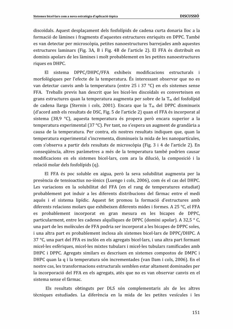
Sistemes�bicel·lars�com�a�nova�estratègia�d’aplicació�tòpica������������������������������������������������������������������������DISCUSSIÓ�
>%>�
� ��L����6�*5�����������1����������� 4��4���� ���������������������� �� ������ ���
4���� 9������� ���� �4����������0�5��������������������� 5� �������::�6����-<�
�������������������� ���(� ������ �������������������-����H�������-��5�������
����������� ��� ����� 7 �6� @*�� �� � �6� A�� ��� �0��� ��� #86� !�� *� <�� � ��� -�L�� ���
��� � �������������������� ���� ���������-�-����������������� �������������������
� 5�� ����� �D:�6��
!�� � ������ �::�I�D:�I*� �M. -� M� ��� 4 � ���� ������������ �
���4��(� 5���� ���� �0�4���� ��� ��� �����������6� \�� ����������� �-������� 5��� ��� ���
������������ ��� ����-� ��� ������������ 7������#%� �@'� ]�8�������� � ������� ������
*6� � ���-����� ���� �� .��� ���� �� 5��� ���� - ��B���� � ��L����� ��� ������� M��� ���
�����������������5����������������������������������-����������������4��4���� ��
��� ������ ������� 73���� �� � ����� #$$>86� !����� 5��� ��� ��� ���� �::�� � �� ��� M�
7�0�������-�������������������3��� �6�%�����0��� ���#8�5�������*�<�� ������������
� ������ 7@C�G� ]�8�� �5������ ������������ <�� �������� ���(� ������ ����� ��� �� ���
�������������M��� �������7@'�]�86�:�������������0���������������������������� ����
����� ��� ��� �����������6� :��� ������� ���� �������� ���������� �� 5���� 5���� 5���� ���
�������������M��� ��������0 ����������� �� ��� M����� �������������������������
����0�-������������ ������� �������������� ���(� �� 7 �6�@� � A���� �0��� ���#86�!��
����5[�� ��� ������� ����������� ���<�� ��� ��� ������������ ���-<� ���� ��� ������
��� 4 � ���� ��� ���� � ������� - ��B������ ��� ���� ��� � �� 9�� ��� ����� 9� � ���
���� 9������������4��4���� ���7586��
!�� *� <�� ��� ����-��� ��� � ����� ���(� ��� ����� ����- � ���� ��������� ���� ���
������ ��������� ��� �����& (� ��7)������ ������#$$E8�����<�������������D:�6�
)��� ��� � ���� ��� ��� ����- � ���� ���� *� 7��� ��� ����� ��� ������������� ����� ��8�
���-�-������� ���� ��� �� �� ���� � 4������� � ��� -� ���� ���� 4����� ������ ��� ��� �
�5�9�� � ��� � ������ � ��� 6� *5����� 4��� ������� ��� 4���� 9� �S����������� ��-�
� 4����������� �����������5����M. -� M���� 4�������� ���� �4�����6�*�#%�]������*�
��� ���-�-������� ��������� ��� ����� ������� ��� ���� - ����� ��� �::���
���� �����������������������������5��� 5��������::��7��� � �������86�*�@#�%�]����
����������������������������*����� ������ ���������������- ���������::���������
����������������������-�-������� ����������� �������- ��B���������::�I�D:�6�*�
@'�]����������������*���� ��(������������������- ��B������ ����������������4�������
� ��B������4�� 5������ ��B����� M������-������ �� ��B������-��������� 4 �������-�
�D:�� � �::�6� *�������� � � ����� ��� ���� ���� ��� � ������� ��������� ��� ��:�� �
�D:��5�������5� �����������������9�� ������������7�������� ������#$$E86�!�����
��������������������4���� �������������������-���������������������� ����������
��� ������� 9�����*�����������������������5��������������-���������� ��������
� ���������������4����6�
�!��� ���������� �-� ������ ���� �)3� �9�� ����������� �� ���� ��� ���� �������
��� 5���� ����� ����6� )�� � 4���� �� ��� ��� � ��� ��� ���� ��� ���� ��������� � ����
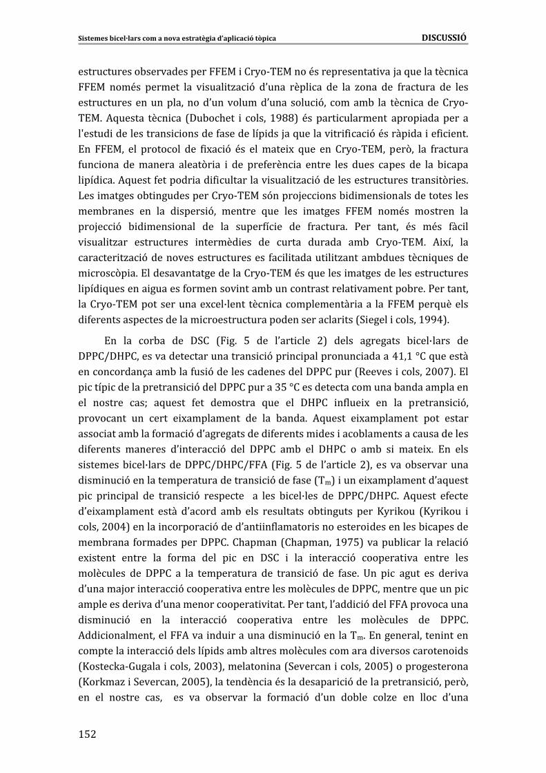
Sistemes�bicel·lars�com�a�nova�estratègia�d’aplicació�tòpica������������������������������������������������������������������������DISCUSSIÓ�
>%#�
������������-�������������!�� ���2�&�!�����<������������� ���H��5��������� ��
!�� ���<�� ������� ��� � ���� �/� 9� �0���� ���� �� ��� ��� /���� ��� 4������� ��� ����
��������������������������0����������0���� ���� 9�� �����-� ��� ��� �������2�&
�!�6�*5������ ��� �� 7��-�.��� � ����� >GCC8� <������ ��������� ����� �������� ��
�S����� ������������� �������4���������� ���H��5������� �� 4 � 9�<����� ��� ��4 ���6�
!�� !��� ��� �������� ��� 4 M� 9� <�� ������� M� 5��� ��� ��2�&�!��� ���(�� ��� 4�������
4�� ���� ���������� �����(� �� � ��� ���4���� �� ������ ���� ����� ����� ��� ��� - ����
� ��� �6�*5�����4������� ��� 4 ���������� ���� �/� 9������������������������ �(� ��6�
)��� ��������-� �������������2�&�!���9�����H� ����- � ���� �������������������
���-������ ��� ��� � ����� 9�� ������� 5��� ���� ������� !�� ���<�� �������� ���
���H� 9� - � ���� ����� ��� ��� �����4� �� ��� 4������6� :��� ������ <�� �<�� 4� ��
� ���� �/��� ����������� ������� ��� ��� ����� ������� ��-� ��2�&�!�6� * M��� ���
������ �/� 9������������������������ 4� � ������� � �/������-�������� 5�������
� ���(� �6�!�����������������������2�&�!��<��5������� �������������������������
� ��� 5�������� �������4��������� �����-����������������� ���������-��6�:���������
�����2�&�!���������������M��B����� ��� ������������� ���� ��� !�����5�������
� 4���������������������� �������������������������� ���73 ����� ������>GGA86�
!�� ��� ��-�� ��� �3�� 7 �6� %� ��� �0��� ��� #8� ����� ��������� - ��B����� ���
�::�I�D:�������������������������� 9��� � ���������� ������A>�>�]��5��������
����������1����-����4�� 9��������������������::������7������� ������#$$'86�!��
� ���� ��������������� 9������::��������@%�]�������������������-��������������
��� ������� ��W� �5����� 4��� ��������� 5��� ��� �D:�� �4��� M� ��� ��� �������� 9��
��������� ��� ���� � M���������� ��� ��� -����6� *5����� � M���������� ���� ������
���� �����-����4���� 9��0������������� 4�������� ���� ���-����������������������
� 4������� �������� �0 ����� 9� ���� �::�� ��-� ��� �D:�� �� ��-� � � ���� M6� !�� ����
� �������- ��B���������::�I�D:�I*� 7 �6�%���� �0��� ���#8�� �������-�����������
� �� �� 9��������������������������� 9����4����7��8� ����� M�����������0�5�����
� � �� � ���� ��� ����� 9� �������� � �� ���� - ��B���� ��� �::�I�D:�6� *5����� �4����
�0� M���������� ����� �0����� ��-� ���� ���������� �-� ������ ���� X2� K��� 7X2� K��� �
�����#$$A8������� ������� 9�����0��� �4������� ����������� �����������- ��������
���-����� 4�������������::�6��.������7�.�������>G'%8������-� ��� ������� 9�
�M ������ ������ ��� 4����� ���� � � ��� �3�� � ��� ����� 9� ������� ��� ������ ����
��������� ��� �::�� �� ��� ������������ ��� ����� 9� ��� 4���6� +�� � � ����� ��� ��� ���
�0������H��� ����� 9�������� ��������������������������::����������5������� �
������������ ����0����������������� � ���6�:����������0��� 9�����*������������
� �� �� 9� ��� ��� ����� 9� ������� ��� ������ ���� ��������� ��� �::�6�
*�� �������������*���� ��� ��������� �� �� 9���������6�!�������������� ������
��������� ����� 9��������� �����-������������������������� �������������� ���
7X����K�&=������ ������#$$@8��������� ���73������� ������#$$%8����������������
7X��K��/� �3��������#$$%8����������� ��<������������ 9��������������� 9�����(��
��� ��� ������� ���� � ��� ��� �-������� ��� 4���� 9� �0��� ��-��� ��/�� ��� ���� �0����
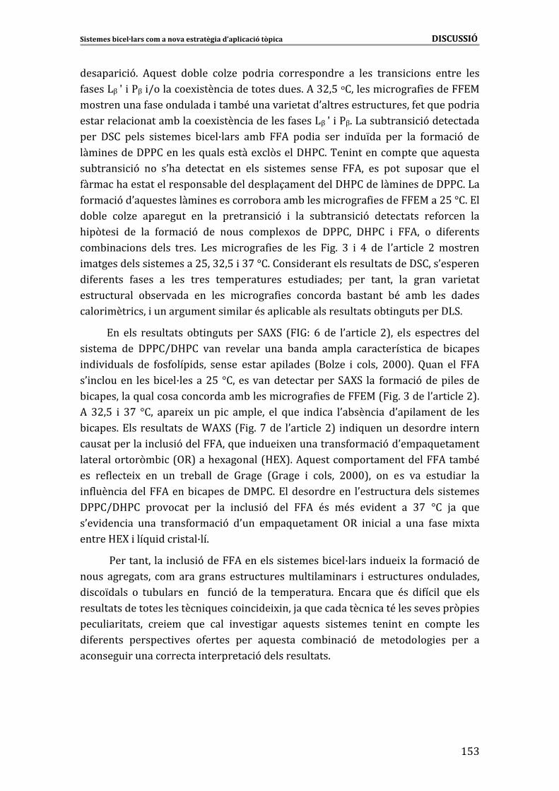
Sistemes�bicel·lars�com�a�nova�estratègia�d’aplicació�tòpica������������������������������������������������������������������������DISCUSSIÓ�
>%@�
������� 96� *5����� ��-��� ��/�� ���� �� ������������ �� ���� ����� ���� ������ ����
4�����)R�S� �:R� I�������M ���� ���������������6�*�@#�%���������� �����4 ������!��
������������4������������� ����-<�������� ������0�������������������4���5������� ��
���������� �������-������M ���� ���������4�����)R�S� �:R6�)����-����� 9����������
���� �3�� ����� � ������� - ��B����� ��-� *� ��� �� ���� ���L��� ���� ��� 4���� 9� ���
��� ��������::���������5�����������M�(������D:�6���� ������������5����5������
��-����� 9� ��� �0.�� �������� ��� ���� � ������� ������ *�� ��� ���� �������� 5��� ���
4�����.�������������������-�������������1�����������D:�������� ��������::�6�)��
4���� 9��0�5���������� �����������-������-������ �����4 ������!����#%�]�6�!��
��-��� ��/�� ��������� ��� ��� �������� 9� � ��� ��-����� 9� ��������� ��4����� ���
. �(��� � ��� ��� 4���� 9� ��� ����� �����M��� ��� �::��� �D:�� � *�� �� � 4�������
��- �� ���� ����� ����6� )��� � �����4 ��� ��� ���� �6� @� � A� ��� �0��� ��� #� ��������
������������� ���������#%��@#�%� �@'�]�6����� �������������������������3����0��������
� 4������� 4����� �� ���� ����� ������������� ����� ����W� ���� ������ ��� ����� ��� �����
����������� �-�������� ��� ���� � �����4 ��� ������� -������� -<� ��-� ���� ������
���� ���� ��� �������������� � ����<����� �-������������������-� �����������)36�
!�� ���� ���������� �-� ������ ���� 3*F3� 7 =�� E� ��� �0��� ��� #8�� ���� ��������� ����
� ������ ��� �::�I�D:�� ���� �������� ���� -����� ������ ��������� �� ��� - �����
�� � ������ ��� 4��4���� ���� ������ ������ �� ������ 7���/�� � ����� #$$$86� ����� ��� *�
�0 �������� ����- ��B������#%� ]����������������������3*F3� ��� 4���� 9����� �������
- ���������5�����������������-������ �����4 ������!��7 �6�@�����0��� ���#86�
*� @#�%� � @'� ]��� ����� M� ��� � � ������� ��� 5��� �� �� �0�-��� �� �0�� ������� ��� ����
- ����6�!����������������T*F3�7 �6�'���� �0��� ���#8� �� 5���������������� ������
������������� ���� 9�����*��5��� ���� M������������4���� 9��0����5���������
�������������(�- �7,�8���.�M�������7D!F86�*5���������������������*����-<�
��� ��4���� M� ��� ��� ���-���� ��� =����� 7=����� � ����� #$$$8�� ��� ��� ��� ����� ��� ���
�4���� ������*����- ����������:�6�!�������������� �0���������������� �������
�::�I�D:�� �������� ���� ��� ���� 9� ���� *� <�� �<�� �� ����� �� @'� ]�� H�� 5���
�0�� ��� �� ���� �����4���� 9� �0��� ����5��������� ,�� � ��� �� ���� 4���� � M���
������D!F� ���5� ��� ����B��6��
�:��������� ��� ���� 9����*��������� �������- ��B����� ���� M����4���� 9����
����� ���������� ��� ���� ������ ��������������� ��� ����� � ����������� ������������
� ��L����� �� ��-������ ��� � 4�� 9� ��� ��� �����������6� !����� 5��� <�� � 4� �� 5��� ����
�������������������������� 5����� � �� M ���H��5���������� ���<�������������(� ���
���� �� ������ �� ��� 5��� ��� ����� ���� �5������ � ������� ��� ��� ��� ������ ����
� 4������� ������� ���� �4������ ���� �5������ ��- �� 9� ��� ��������� ��� ���� ��
������� ������������� ��������� 9���������������6��
�
�
Sistemes�bicel·lars�com�a�nova�estratègia�d’aplicació�tòpica������������������������������������������������������������������������DISCUSSIÓ�
>%A�
4.1.1.3�Comparativa�dels�canvis�provocats�per�la�inclusió�del�DDEA�i�del�FFA�en�sis �temes�bicel·lars de�DPPC/DHPC�
)�� ������� 9� ���� ��!*� ��� �5������ � ������� - ��B����� ������� ����
���� 9������������������� ����������� �� �4���� ������������������������������
�)3���������5���������-��� ������������������������::��7A>�]�86�:���������-����������
��������������)3���������������� ���� 9�����*� �� 5���������������������� ���
������ ������� ���L������*6��
)���� �����4 ����-� �������������2�&�!������-������������ 9�������� ���
������������5���������!*�<�� ��9���������� �������- ��B���������::�I�D:�6�!�����
��� ��� ��� ���� 9� ���� *�� ���� � ���� �/� ���� ��� ����� �����4 ��� �-� ������� ����
��2�&�!�� � !�� �� 5���� ��� 4���� 9� ��� � 4������� ������������� ��� ��� ����
�� �������� ��B������-������ ��0�����������4�� 9������������������6��
������������������3*F3������������ ��5������ ���� 9�������!*��������� �������
- ��B����������������� �� �� 9�����0��������������- ����������������������������
����� ������������� ����� ����6�:��� ������-������ ��� ��� ���� 9����� *� � �� #%� ]�� ���
����������4���� 9������� ������ ������ ���@#�%� �@'�]������0�-����������� �������
� ����� 9�����0��������������- ����������������� �������������4����6�)����� ��
���T*F3� ��� �������� ��� ����5��������� ��������,�� ����� 5��� ���� - ��B���� ������
4������������ �������� � ��������::�I�D:�I��!*6� �!����� ������ ��� ������� 9�
���� � ��*�����4��� � ���� M������ ���������0����5������������������0,����D!F6�
*5������� � 4���� ��� ��� ��� ��� 4 � 9� ����������� � ���4��(� �� ��� ����
- ��B���� � �����������������������4�������9���������������� 4������� ��4 � ����
������������������H��5��������!*�<����4 4�� � ����*�<��� ��4�� 6�
!����-�9��������0.���������� ��� ���� 9����� 4����������5���<�� ���������
����������������� -���U�������� � �����(� 5�������4�����6�
!�����5����������������������4�������������<�������� ���� 9������4������
�9�� � 4�������� ��� ��-�9�� ����� ��� ��������� ���� ����� �/� 9� ��� - ����� 5���
��������0����� �/� 9�� ��� ������0!�6�*5�����4���4��������������� ����L�����0�5������
� ��������������������������(� �6��
�
4.1.2 es�Capacitat�encapsuladora�dels�sistem �bicel·lars�
!��� � ������� 4������� ���� ��:�I�D:�� � �::�I�D:�� ���� ���� ���1���
�0 ���������>�>E�d�7JI�8� 7������� 9���U��������� M� 4����� ��������������
���� ��8� ��� ��!*� �0���� ������� � � ���� �� ������� � ������� ��B��L����� ��� ����
� ���������� ������� ������ ��B����� M����� ��� 7�� �����2� � �����#$$@W�X� J��� �
�[����&=�2������>GGAW�XJ���� ������#$$AW�)����� ������#$$EW�)����� ������#$$A�W�
:������� ������#$$#86�!���� �������- ��B�������-���!*�����������������������1��
������������ ������ ������ 9� ��� 4����� I�� ��� � ����6� *5������ � ������� ��� ����
Sistemes�bicel·lars�com�a�nova�estratègia�d’aplicació�tòpica������������������������������������������������������������������������DISCUSSIÓ�
>%%�
������ ������-����������������2��>��������� ������M. - ��������������������5���
4� � ��� ����������� � 9���-��� ����������������� 9���-��0��� � 9��0�������� 9�
�5��� � �������!*�7������<� 4�� ��86�
!��� � ������� � ��� �� ��� �::�I�D:�� .��� ������� ��� ���� 9� �0��� �������
� �(4 ��� ��� <�� ��� ��� ���� *�� ������ ��� ����� ���� �0�� � �/��� � ���������� ����� ��
�������� ��� ���� ��������� ���� ���� � .��� ���������� 5��� �M. -� M��� ����
������ � �������4��(� �� ����������6� � )�� ������� 9� �� � �/���� .�� ������ ��� �0>d��
� � ����������� � �/�������������������!*6�? ��������������� �������- ��B��������*�
���� �������� � 4���� ��� ��� �S������1�� ��������� ��� ��� �����������6� *� #%� ]�� ����
� ���������� �����������������9�W���@#�%�]����5�����������������4�� ��� �������������
�������� ���@'�]������������������������U ��6�"������������� ������ � ������������
�������������� �������- ��B��������5����������S�5�������������������6��
�
4.2�Estudis�in�vitro�de�l’aplicació�tòpica�dels�sistemes�bicel·lars�en�pell�sana�
30.����� � �/��� ���� � �����0�M��� ���������� �� ����� ��� �0��� � 9� �(� �������
� �������- ��B����6�!������� ����� ���������� �������- ��B�����.��������������������
��. ����������!*�7��� ����>� �A8�������*�7��� ����@� �A86�!������������� ���������
� ������� - ��B����� ������ 4����� �0.��� ����� ��� �� ��� 4 ������ ��� ��� -�������
���� ���-��������0��� � 9��0�������� 9��5����������!*�7��� ���>86�
�
4.2.1� Sistemes� bicel·lars� com� a� vehicle� del� DDEA:� influència� de� la�composició�lipídica�dels�sistemes��
)�� ���� 9� �����!*� ��� - ��B���� ����::�I�D:�� � ��:�I�D:�� � �� ��� M�
�0�-��� 9� ������� �� ���� 4����� ��� ������ 9� ��-� ��� �0���� ���� 9� �5����� ���
��!*� 7 �6� A� ��� �0��� ��� >86� *5������ ���-����� ������� M� ��� �4���� ����������� ���
�0�-��� 9�������� ��5������� 4���������� ��(���������� �������- ��B����6�*5�����
�4���� ��� �� ����������� ���� ���� ��� ��� ��� 4������ 5��� ������ ���� �-��� 9�
������� �� ������ ��� ���� ��� <�� ��� ��� ���� 4����� ��� ���� �� ���� ���� ��-����� � ��
� ������ ���� �7�u� .� ������#$$>86�
\���� �����5����0U���0��-�9��� �������� �������- ��B����� ����� M������������
4����� �� ���� ����� �<�� ���4������ ��� ��� ����6� *5����� �4���� �. - ��� � ��-��� ���
������� 9� �� ��� ����� ��� ���� �<�� ������ ��� ��� ��� ����� � ������� - ��B����� ���
��:�I�D:�6�)��� 4���� ������������ 9�����������������!*� ��(�����- ��B�������
�::�� � ��� - ��B���� ��� ��:�� � ���� �� ���� �� ����� ��� ��� � 4������ ������������ ���
����� 9� �0�5������ ���� 4��4���� ��6� � ���� �0.�� �������� �-����� �� ��� ������������
�M��� �����������::����������4�������� �����5���������� �������0!�� ������:����������
Sistemes�bicel·lars�com�a�nova�estratègia�d’aplicació�tòpica������������������������������������������������������������������������DISCUSSIÓ�
>%E�
4����� �����&��5� �6�!����:���������������4����� 4������������������� �������0!�� �������
����5[�� ������������� 9�������<����������������� �������<��� 4� �6�
!������ ���������5����������������������� ���� 9��������- ��B����5�����0.��
���������������!*��5������� ����������� �������������<��4� ������������<��������
������ ����5��������� ������������������M��� �����6�!����� �� ��� �������� 4������
5���. �.��� 4�������4������5��������� ��� ��������0�4��������� ����I�����������
�0��� ��. ��6� *������ �0�5������ 4������ �9�� ��� ���� -��� � ���� 9� ��� �0����� �/� 9�
�������� ��� ������B�����������0!����0�4 � �����������������-������. ��� ����� � �����
��� �0���������� � ��� �� ���� ��. ��� 7=J�K� � �.���� #$$#W� �.���� � ����� #$$'86� )��
������� 9�������!*������� � ������- ��B�������������������� ����� � � ��������
����- ��B���6���� 4���� ��� 4������������� � ���������� ������ ��� ��� ��� 9� � �(4 ��� �����
4��4���� ���� � �9� 5��� ���-�-������� �S �������� �� ��� - ���� �� ����� ��-� ����
4��4���� ���W����<���������������-�M ������� ��4�������� ����������������� � �����
����� ���� ������� ����� 4��4���� ��� 7���� ��� � ����� #$$%W� 3������ � ����� #$$G86�
*5������ � � ������ � 4 ����� �� ��� ������� 9� ���� ��!*� ����<�� ��� ��� ����� 7X� J��� �
�[����&=�2������ >GGA86� +��� ������ ���� -��� ��9� ���� �� �M�� ��� �5������ -� M��
������� 9� ���� �� ������ ���� ������ ��-� ��� � 4 ������ 5��� �<� ��� ��!*� �� � 4������
4���������� �������- ��B����6�
�
4.2.2�Sistemes�bicel·lars�com�a�vehicle�del�FFA�
)��� ����� �� ��������������������-�- ��B��������� ���*��0.���� ����2������ ��
��� ������ ���� �������� � 4���� ���6� :��� ���� ������ �0.�� ����� ��� ��� �4���� �� ���
� 4���������. ��������0�-��� 9�������� ������*� �������������������0.�� ����� ����
��� ����� 9������ ����������� ��������� �������- ��B�������-������� � �� ��� ����
�0!�6�
4.2.2. del1�Influència�del�vehicle�en�l’absorció�percutània� �FFA�
!�� ���� 4��� ����� �� �0�-��� 9� ������� �� ���� *� ��. �� �/��� ��� � �������
- ��B���������::�I�D:�������������� 9�����(� �� ������������������� ���7 �6�
A�����0��� ���E86�
!��� ���������� �-� ������ ������������5��� �0�-��� 9�������� ����� ����������
5�������4����������� ��������������� �������- ��B������ ���H���5�������*��������
� ���������������6��
!��� ����� ������� �0�-��� 9� ������� �� �-� ������ ���� �� ��� � ����� 9� ����(� ��
���-�-����������������� ��������-������������4��������� ������������������ 9�
�0�5������ ��������6�������7������ ��� ����&=�44��2��#$$G8�����-��������������� 9�
���� ��� M� ��� �0!�� ���� ��� �� ���� ����- � �/� 9� ����� ��� ��� ��� �0!�� ���� �4���� ���
�0������6�*5����� �4���� ��-��� �S!������ ������� ��� ���������- � ���� ��� ��� ����6�+��
�������� ��� ��� ������- � ���� ����� ���� ���� ��� ��� ��H�� �� ����� ������ �� ����
Sistemes�bicel·lars�com�a�nova�estratègia�d’aplicació�tòpica������������������������������������������������������������������������DISCUSSIÓ�
>%'�
4��L� 4 � 9� ����� ��� ��� ��� �0!�� 5��� ��� �� ��� ��H��� ��� ����� ��� ���� �������
��5��� 5����� ��� 5��������5�������������7T �� ���� �����2��#$$E86�"���-������� M(��
��� ����������������������0*��&� ����5�����4���������-����9��������������������
�����������-��������� 9�����(� ������*6�)����� 9��������-������� ���� 5���� �
�� ���� 5���� �S“stretching”� ����� ��� ��� ��� �0!�� �����<�� ���� ���������� ��-� ����
���� 9� ����(� �� ��� *� ��� �������� ��� �������� ����������� ��� ���� �������
��5��� 5������������ ��� ������B�����������0!����������� 9���-��������������������
���������������������������6�*5��������������������������-�������-�������� ����
���������� 7���������� � ����� >GG>8� 5��� �� �� 5��� ������ 5��� �0������� ����
�M�������5���� ��������� �-���������� �������0!���� M(�������-��� ��� ��������������
������� ��� ������ �0!�6�!����������������������0*��&� ����� ������ 9���� �0“scissoring”�� �� 5���� ��� ��� � ���� � � ��������� � ��� �0����5��������� D!F� �� ������� �<��
-� M����4���5������� ������������ �����-��0�M��� 9��0���������� �������0!�6�!����� �
����������� 9�� ��� �� ��� � �����������������-� 9�������4�� 9����-����������
�0!��� 4��� 5��� �0�� ��� �� ��� ���� �������� ���������� �0�-��� 9� ������� �� ��� ���
�������� ��� ��� ������- � ���� ��� ��� ����6� !��� ���������� �-� ������ ����-����� 5���
�0�M��� 9������� ��������0���������������������� ������������-�������� ��������
���������- � ��������������������������0������ 9�����0�������������� ��� ������B�������
����0!�6��
)0�-��� 9�������� ������*� ��(�������������������� ����������������
5��� �0�-��� 9� ������� �� 5���� ��� 4����� ��� ��� ������� ��� ��� ���� 9� ����(� ���
������ 5��� �<�� ����� 5��� ��� ��� ��� ��� *� ��� � ������� - ��B����6� * M(� <��
���-�-������� ������ �� ��� ����� 9� ���� �������� ���� ��� 5���� �� ����� ��� *��
���<����#d��0� ����� �� 6�*5������������<��������������5��������� �����1����
������������������ 9����������( ������� ������4�� 9�-�����������0!��7���. �� �
�����#$$%W�� ��� ������#$$'W�)�-J�.���>GGG86�!��������������0*��&� ��������������
���� ������������������������ �������0!��5������������� ������������������ ���
���*���������� 9���-���������������6�*5����������������������� ��������������
�M � ��������.(� ������������������ ������������0 ����������� ������� ���� ���
�� ����5������� ����M�������������������� �������0!�� ����� ��� ��� ���4������ � �����
���0���������-�������-�������*��&� �6�
��������*������ ��(���������� �������- ��B����� � �0��� ����-��� ��������� ����
��������������0�-��� 9�������� �������� �� 5����������������<��-� M������������
��-� �0��� � 9����*���-������������������. ���6�*5����������������������� M���
��� �4���� ����������� ��� �0�-��� 9� ������� �� 5���� ��� 4����� ����� ��(�� ��� ����
� ������� ��� - ��B����6� !�� ������� ��� ����������� ��� ���� ���� �� ���� 3.��4���
73.��4��� �������� ����>GGE8� � ���������� ��������������5�������� M����0�-��� 9�
������� �� ����� ���������� �H��1���� �0�������� � ��� ��� 4�� 9� -������� ��� ��� ����6�
:��� -�������� ���� ��� ��� ��� ���� - ��B���� ���������� ��-� ���� ��� ��� ��� �0!��
��� 4 ���� �0����� �/� 9���� ����- ����� � ��� 5���� ������������ ��� 4�� 9�-�������
Sistemes�bicel·lars�com�a�nova�estratègia�d’aplicació�tòpica������������������������������������������������������������������������DISCUSSIÓ�
>%C�
��� �0!�6� *5����� �4���� ���-<� ��� ���� ���� �� ���� ������� ������� ���� �� ������ �����
�����������������������������������0 � �����4����7*�- ��� �� .� �K��#$$$W�X �� �
�����>GGG8� � ������������ �����������"$G>%�7*�- ��� �� .� �K��#$$$W�D�����4�� �
����� >GGE86� !�� ��:�� ���-<� .�� ������ ���� �� ��� �� ���������� ��� �0�-��� 9�
������� �� ���� 4����� 4��- ���4�� ��� ������ 9� ��-� ������� ��������� ��� ����
� ����������� ���������� ��������������� �����1����0���������������� ������
��� �0�-��� 9� 7���� � �����#$$@86�!�� �0��� ���@�� �0�4���� ����������������:��������
���� ����� ����������������������� 9�������4���������� ��5� �� 7��`�#@�]�8��0�5�����
4��4���� ����<�������5���������� ������������������������������� � �/���������0����� 6�
!�� ��� ��� ����� � ������� - ��B����� ��� *�� ��� ��� ����� ���� ���� �3�� ��� �0��� ��� #�
�0�5��������� �<���<�������7@C�G�]�8�5���������� ��������:�6�*5�����4��� ��� ��5���
������������������M��� ������������ ��������� �������������������-�����������������6�
\��-�����-���5�������0��������������4��4���� �������4��������4 ��� �������������������
-�������� ��� �������������7���� ������#$$@W�X �H��� ���� ������>GGG8��4���5���������
������������������������� 9���� ���������� 9����� 4����6�)S�4���� �����������
������ �������- ��B��������*�������0�������-����������������-� ����������0��� ���
>���������� �������- ��B���������::�I�D:�I��!*�������-<��������-��������5�����
�4��� ��������6����
�
4.2.2.2�Interacció�dels�sistemes�bicel·lars�de�FFA�amb�els�lípids�de�l’EC��
)��� ����������3�!������0!�������������������� �������������������-�� �������
- ��B��������*� 7 �6� @� � A���� �0��� ��� @8� ������������� ������������� � � �����
������-���� �� ���� ��-� ����� ����� ������� ���� ������� ������� ���� �� ����� ��� ���
7�9���� ������#$>>W�)9��/� ������#$$>86�)0������������� ����-�������� �/���������
��� �������0!��5����0�-�����������-������������� �� ��5������. �.����� 4 � ����
������������ ��� �0!�� ���L���� ���� ���������� ��-� ���� � ������� - ��B����� ��� *6�
)0�4�������� 4������� � �������- ��B����� ��-��� ���� ��������������� �0!��.��������
���� ������ ����� ��� ���� ������� ����6� �0�5������ ����� �� ��� ��� �������� 5��� ����
- ��B���������:�I�D:����������������� ������0����� �/� 9���� ������������ ���
����0!��7���-���&������� ������#$$CW����-���&������� ������#$$G8��������5�������
- ��B��������::�I�D:�� ���� M��� ��� 4���� 9���������������� ���/��������� ��� ���
��� �0!�� 7���-���&������� � ����� #$$GW� �9���� � ����� #$>>86� *5����� � 4������
������������ ���� ���� ������ ��� ��� H�� �0.�� � �� ����� �������� ��� � 4������ ���
���� ���� ���� ��� ��� ��� ������ ��5��� �� ������6� +�� ������ 4����� �� ��� ������ <�� ���
��� ���������� �������- ��B������0������������������������� ��5���������� �����
�0� ������������ ����������������7���-���&������� ������#$$C�86����������- ��B����
�����:�I�D:���0��� 5������-��� ���������� ������������ 4 � ��(� �� 7@'�]�8���5�����
� ������ �M��� ������ ���� ����� 9� ��� 4���� ��� ���� �� ������ ��5� �6� *5����� ��� �
���� M������������������������� ��������������������- ��B�����5����� �����������
���- ��B������ ����<����� �0!�6�!����� ��������� �����������::�I�D:�����-���������
Sistemes�bicel·lars�com�a�nova�estratègia�d’aplicació�tòpica������������������������������������������������������������������������DISCUSSIÓ�
>%G�
�<�����������������������-�����������@'�]�������������������� 9����4��������������
��� ��� ������ M� ��� ��� 4 � 9� ��� ���� ������ � ���� ���� ���������� 5��� �5������
� ������������� ��������<������0!�� �5����������������� �� 9���������������0� ����
5���. �.���� �������� ����- ��B���� �� M �� 4������&�����������6�!�� �0��� ���@�� ������
5������� � �������- ��B�������� *��������������������::�� ��D:��������������
��� �� ��� ��� /���� ��� ��� ��� ��� �0!�6� * M(� <�� �� ����� ��� 5��� 5���� ���� � ������� ���
�::�I�D:�� ������� *�� ��� ��� 4 �� ��� � ��� ��� ���� ��������������� � ���
������������4�� ������ �����6�!������� ������- ��B������-�*�4��������������
�����H���������� ��5����������������������� ������������*���������������������
�0��� ��� #6� * M(� ���� �� � 4 ������ ��� ���� �� ����<�� ��� �0!�� � <�� ���� � M(� 5��� ��� ���
������������������������� � ��� 5������� �0!�������<������ ������������-��5�����
� �������� �������- ��B�������-����4����6��
���� ��0�-��� ������������������������� ���������� ������������������������
����� �����<�� ���� ���������� ��-� �5������ �������������5��� ����� ��� *�� ��� ���
�������� ��� ���� �4���� ��� �0����� �/� 9� ��� ��� ��� ��� �0!�6� :��� ������ ��� ���� ��
�������� 5��� . � .�� .����� ���� � 4�� 9� ��� ��� ����� ��� ��� � ��� *� ����� � �������
- ��B����� �� �0!�6� !��� ���������� �0*��&� �� � ��� 3*F3� ������������ ��� �����������
����� ��� ��� ��� �0!�� ��� ������ 9� ��-� ��� ����� ������� 5��� ������� ��-� �0�4����
����������������������- � �������� ������� ������6�)0������������0�������������� ���
����0!������ ������ ���L��������� ���� 9�����::������� ������- ��B����������- �����
� ��� 5���� ���� �� M �� ���� 6� *5����� 4��4���� �� ����� ��� ���� 4���� ���� ��������� �� ���
������������ �M��� ������� � ���� �� ��������� ��� ����������� ��� ����� ���
�0������������� ��������0!��7��������/� ������#$$G86��
!��� �M��� ������ ��� 3*F3� ��-� ��� � 9� � �����9� ���� �/���� �� �������� �0!���
������� � �����������-�� �������- ��B�������-�*����������������������������
�0���� ���d� ������������ �� ��� 4���� ������ �� ���� ����� 7“Short� Periodicity�Phase”��3::8��������4�������� ���������0!������ �����������J�����7���J����� ������>GG>86�
*5����� ���� ��� d� <�� ��� ����� ���� ��� M� ��� ��� - ���� � ��� �� � ��� ��� M� �S���� ����
��H������0� ���� ������� ���� 7��� ;����� � �����#$$%86�)0����������� ���� ���� ��d������ ��� ��� ��� �0!�� 5���� ��� ����� ��� ������ ��-� � ������� - ��B����� ��� *� ���� ����
�M�� ���������� ������� 9��0� ��������������- ����������� ������� 9��������� ���
�::����� 4����������6�*5����� 4�������-���� ��� ����� 9������ ��� ������ ����- ��B����
��-� �0!�6� )����H��� ����� ���� ��� ��� -����� ������������ ��� SPP� ��� ��� ��� ��� �0!��
�������������4���� ����� �������� �� ���������������� �0������ 9���� �0����������
� ��� ������0!�6�!��5���������������-����������������-� ����������0.��������������
�M��� 9��������� �������0!�������<������������������-������ �������- ��B����W�����
������� ������� ��������� ��� 5� ����� �0�5������ ��� ��� ���� �4���� ���� ���������� ��
��� ������� ���� ���������� ��� �0�-��� 9� ������� ��� ���-<� ��� ��4��1� ��� ��� 4�� 9�
-�����������0!�6�
�
Sistemes�bicel·lars�com�a�nova�estratègia�d’aplicació�tòpica������������������������������������������������������������������������DISCUSSIÓ�
>E$�
4.2.3�Els�sistemes�bicel·lars�com�a�potenciadors�de�la�permeabilitat�de�la�pell�
��������5��� ����- ��B������-�4�����7��!*� �*8� �. -� M��� ���������� 9�
�0�5��������������� �� in�vivo�������-���&�������7���-���&������� ������#$$C8�����
�������� ��� �������� ���� �!T)� �����<�� �0���� ��� � 9� �9� �� ������ ��� ���
- ��B����4��4�� ��� 5���6�!���0��� ���>������� ����� �����0�4�����������- ��B������-���
��� 4�� 9� -������� ��� ��� ����� � ��� ���� ���� -��� �4���� ����� ����� ��-��� �0�-��� 9�
������� �� in�vitro�������!*6�*�-��5������4 ��� ��������������� �/����������������
���� ��������������������������� �����-�� �������- ��B����������:�I�D:�������
�::�I�D:������ ���0������� � 9��(� ���0�������� 9��5����������!*6��
!����������������-�������������� ���0�-��� 9�������� ��7 �6�%���� �0��� ���>8�
�� 5���� 5��� ��� ���������� ���� � ��� ��� ����� ��-� - ��B���� ������� �0�-��� 9�
������� ��������!*�������� 9��5����6�"��. ����.������ 4���� ���� �� 4 �� �������
���������������0�-��� 9�������� ��������������������������-�- ��B���������:��
� ��-�- ��B���� ����::��� ���� � 5���. � .�� ����� �� ���� ���(� �� 5���.�����������
��������0�5������� �������������� �������������� 4���������� ���������������
�0!��7���-���&������� ������#$$CW����-���&������� ������#$$C�W����-���&������� �
�����# �$$GW���������/� ������#$$G8�����H���0.��������������0������� A6#6#6#6��
)0�4��������� ���������� � �������- ��B����� ��-��� �0�-��� 9� ������� ������
��!*����� ������������������ ����� 9� � ����������- ��B������-��0!��5������� ��
������ ���� �������� �/� 9� ����� ��� ��� ������B�������� ��������-���� ��� ��� 4�� 9�
-������� ��� �0!�6� *5����� 4��� 4� � ��� �� �0�-��� 9� ���� ��!*� �� ����<�� ��� ��� ����6� )��
����� 9� ����� 4��4���� ��� �����4 �� ��� - ��B���� ��-� ���� ��� ��� ��� �0!�� ��� �� ���
���� ������������-�������0�4�����������6�:��(����5����5��������� ������� ��
� 4������������������ ����- ��B���������:��5������������ ����- ��B��������::�������
���������� �-� ������ ���-���� �� ��� 5��� ������� 4������� �� �<�� ����� ��� ��
� ������������������0!���.���������� ����������������� 9�������!*�� H��5�������
���� �������- ��B���������� M�������4����� � ������� ������0�-��� 96���
�
4.3�Disseny�d’una�metodologia�de�simulació�in�vitro�de�pell�amb�la�funció�barrera�deteriorada�
"����������� ���� ����� �� �0�-��� 9� ������� �� in� vitro� ��� ����� �� ������
�� � �/����- (�� �������������-�������� 9�4 � ��(� �������-��6�"���-������� M(��
������ 4���������� ���������(� 5���������������� ���4�� 9����-�����������0!�������
����� �����6�:�����������������������������0���������in�vitro�����������-����4�� 9�
-������� ����� ������ ���� �� ���� ����� ���� ���� �� ���� ��� ��� ��� ����
�M��� �������� �<�� ���� ������ ��� - �� ���� - � ����� �0�4 � �� ��� �� � ���� ���� -����
Sistemes�bicel·lars�com�a�nova�estratègia�d’aplicació�tòpica������������������������������������������������������������������������DISCUSSIÓ�
>E>�
�4����� ��� ��M ���� � ���� �� ��� ��� ���������������� ��� ��������� ���� ���
�������������������� �������������6��
\�� -��� ��-��� 5��� ��� ���<�� �0�-��� 9� ������� �� �0��� ������� ������ ���
������4�������������������������� 5����4 � �5��� 5��������������� ���������������
��4 ���� ��� ���� 9�� ��� ����- � ���� � �0�� � ���� ������ ��� ��� ��� �� 9� 4����&
��. ������6� 7!� ��� �� �������#$$EW�3.��4��� �������� ����>GGE86�!�����5��� ����
���� ������4 � �5��� 5�����������������������������4������ ������� ������9������
4������ 5��� �9�� ������������ �������� �� �0.���� �0 �4�� �� ��� ��� ���4 �� �0�-��� 9�
������� ��� <�� ��������� ������ ���� ��� ���� ������������ ��� ����� ��-� ��� 4�� 9�
-���������� 4 ���6�*������ ����� ���������������-� ���0�M ���� ���0���������� 9�
������ ��� �!T)� � �0�-��� 9� ������� �� ��� ��������� ��-� � 4������� ���� ������
4�� �&5��� 5����7)����� ������>GC'8W����(�������H�� ��������������.����-����������
���������������������� 9�� 4�������� � �/���������������-���� ������������
4�� �&5��� � ��-��� ��� ����� ������ �������� ��-� ��� �� � �/� 9� ��� ����� ��-� ���
-������� ����� ������ � 7" ������� #$$%W� " ������ � ����� #$$'W� 3 ������� � ����������
#$$'W���� � ������#$$>86�
!��� ��� &���� �� ��� ���� 4������ ��� &�/���������� �� �0������ ������� 5���
�0������� ���� �����H��� ���� ������ ��� ��� ��� ����� �0��������� ��� ���� 4���� ���� � �����
� �H��1������������0�-��� 9�������� ��in�vitro���� � �/����- (�� ���������������6�"��
�-������ � M(�� �5������ 4������ �0.��� ��� �0��� ��� ��� - (�� ��� ��� ����� ��-� ���
����� ��������������4�� 9����-������������-��� ��������4 ���0�-��� 9�������� ��
�<������6�!���5��������� ���<������ -���� ������ ������ 9��������� ����������� ���
-������� ���� �� ��� ��� ����� �����(� ��� ��������� �5������ 4�� 9� 5��� ������
�� � �/���� ������� 7��� � � ����� #$$>8� �� ���� �� ���4��� ��� ��� � 73)38� 7" ������� #$$%W�
" ������ ������#$$'W���� � ������#$$>8�������� ������� �H��1����“tape�stripping”�73�KK��� � ����� #$$A�W� #$$A-W� 3 ������� � ���������� #$$'8�� � ��� ���� ��-�
� �� $E86���������7=����� ������#$>$8�����-��� 9���-����������� ����7T�� ������#$
:���5���4������������ -��������� 9�������������-������� ������.�� ����7“tape��strips”8� ��� ����� �� ��� ����� � ��� �!T)�� ��� ��� � 9� ���-���� ������ ���� ������ in� vivo�7�� � �/������������ ��.�����8� ���������������������� ���in�vitro����� �������������
�� ���� ��� �-���� ������������ ��� ��� ����� 7��� ���� ������ ��� � 4������ ���� ����(� �
�� � �/��8� �0��-�(�� �� M ��� 7 �6� >� ��� �0��� ��� %86� "�� �-������ � M(�� ��� ������ ��
���� �������������������� �� ������� �� �� 9������ ��������� ������ ��� 4�� 9�
-��������������������-��0���������������-������� ������.�� ������� ����6�*��<���
����������������4 �����5�������������������!T)����-<������������ � �/�����������
�� ������ �M��� ������ ��� ��� ����� in� vitro� �S����� ��-� ���� ���������� ��-� ���� ����
��������������73�KK��� ������#$$A�W�#$$A-W�3 ������� �����������#$$'86�
�S������-������������������������������ 9�����0� ����� �� � �����4�L���7 �6�#� �
@�����0��� ���%8��������� 4������������� ���������� ����������������-������� ����
��.�� ���� ��� ����� �� ��� ����� �������� ��� ������������ � � 4������ ��� ���� ����
Sistemes�bicel·lars�com�a�nova�estratègia�d’aplicació�tòpica������������������������������������������������������������������������DISCUSSIÓ�
>E#�
���������� ������5��� ��� ��-�9�� �����. � .����� ����������� ���������� 9������
4������ 5���� �0��������� ��� ����� �������� ��� ��� 4�� 9� -������� ��� ��� ����6� 3 �
�0�-��� 9�������� �������������������������4�� 9��������-������� ������.�� ����
7 �6� A� ��� �0��� ��� %8� �� ��� �!T)� 7 �6� %� ��� �0��� ��� %8�� �0����� 9� ����� ������� ����
����������� ���-��� �0�-��� 9� ������� �� ����� M��� � 4������� 4�� ���� ��� ����
��6��
)0�5�� 9�5���� ������0�����������������������4�L���<������4�� 9��M����� ���
����� ������
�
�*6:6�`�*�d M����M��M��v"��-���� �������!T)w�
�
,�� d� *6:6� <�� ��� ����������� �0�-��� 9� ������� ��� *� <�� ��� 4����� ���&
�M����� ��� ���� �������-� �������� ������ 4�� �&5��� 5������� ��� �4�L���� ��������
���� �������-��0�4����5�����������0������ 9�������4�� 9����-�����������0�-��� 9�
������� �6�+����5�� 9��M����� ���� � ����.������������ �������3�KK���73�KK��� �
����� #$$A-8�� ��� ��� ��� ������ ��� ���� 9� ������ ��� 4��M� ���� 4����� ��� �0������
�0�5� � -� � 7� “estimated� steady�state� flux”�� ;338� � ��� �!T)� ���� �� ��� �4�L��� � ���
4���-��- ���6� !�� ��� ������� ���� ���� �5�� ���� �-� ������� ���� �� ��� �4�L���� ����� ���
���� 9���-�������-������ � ������� ����������-�����!T)����� M���-�������-<�
��-� ���� �5�� 9� �M����� ��� 7��4 ����� ��� ������ 9�� �#`� $�G'� � $�G%��
������ ������8�
!�� ���� ��� ��� �0� �� ��� �� � �0�5�� 9� 5��� � ����� ���� �� ��� ������������
�0�5�����4�����<������4�� 9�� ���������� �����
�
�*6:6��`�a�d M�"��-���� �������!T)��x�b��
�
,��a���������� ��������-��0�4����������������-� 9�������-����������� �� �b���������� �������-��������� ������4�� �&5��� 5��������0� ����� �� 6�!���#��-� �����
��-�����������5�� ����� ���������� ���������-�������-������� ������� ������ ����
�!T)��9��$�G%� �$�CG�������� ������6�
!�� ��� ��� ��� �0� �� ��� �� �� �0 �������� ��� �0�-��� 9� ������� �� �������� ���
����� ��������������4�� 9�-������������������<��������5�������������������4�L��6��
*5������ ���������� ������ �S����� ��-� ������� ����� �� ����� �� ������ ��-��� ��� �����
���� ���������2������ � �/�������������3)3�7" �������#$$%W���� � ������#$$>8����
5�����������������0�-��� 9�������� ���������������4�� 9�-�����������0!���������
���2�������� ������������������������� ��4�� ��7����0� ����� �� 86��
Sistemes�bicel·lars�com�a�nova�estratègia�d’aplicació�tòpica������������������������������������������������������������������������DISCUSSIÓ�
>E@�
!����������� ���������� �� 5����5����������� ���������������� �� ��� ��� 4�� 9�
-����������������������������������������������������������� 9��0����������
������� ���(�� ��� -����1� . ��(4 �&� �(4 �� ����� ��������� �� 4������ ���� �� � �/�����
���� ����� 4 ��� ��� ������������ ��� ���� ���4 �� ��� ������� 9�� ���� ������� 5����
�0�� � �/����������� ��-� ��� 4�� 9�-������������ �����6�\���� �����5��������� ������
4������ 5��� .��� ��� ���� ��� ���� ��-��� ���� ����� ��-� ���� 4�� 9� ��� -�������
�������������������� M���������� � ��������������������!T)�in�vivo,��������
��������� 3 ������� � ��������� ��� ��� ��� ��� ������ � �� ��(� �� 73 ������� �
���������� #$$'86� ��� 4���� ���� ���������� �0�-��� 9� ������� �� �-� ����� ���� �5������
��������������S� ��4���� � ����-�����������&>'&����������� � �/�����������-����4�� 9�
-������� ����� ������ � �H��1���� “tape�stripping”�� ��� ������ ������� ��-� ����
������������� �6�������&��������������������!T)��-� ������ in�vivo�������� 4�������
� ���� ��� ����� ���2����� ���� ����� �� in� vitro� �S�-��� 9� ������� �� �0.��� ��� 4���
�� � �/���� ���� - (�� ��� ��� ����� � ������� ���� ������-� 9� � � ���� ��� ��� 4�� 9� ���
-�����������0!��� �H��1����������-���� 4��������� �������.�� ������� �������-���
����������� ��6���
�����������=�����7=����� ��� -�.��#$>$8�.����-� ���������� � 9���� ������
������ ��� ������ ����� �� ������ ���� � 4������� ����� ������� ��-��� �S�-��� 9�
������� ���� � �/����� ������������������� �� �5��� ������� �� �� ��in�vitro����
4�� 9� -������� ��� ��� ����6� !�� ��������� ��� ����� ���� �������� ���������� �S�-��� 9�
������� ���S�������������������������6�*5�������������<���<�������� �����-�
��������� . ��(4 ���� � ���2�� ��-� � �(4 ���6� !�� ��� ������� ���-����� �S�������� ���
�S�-��� 9�������� ���������������-������� ������.�� ������� �������-����������� �
<��� 4��������������-�9����������� ����� ����6���������M������������������� ���
�4�L���� 5���� ��� ���� ��� ��� #%� � ���� ��-��� ��� ������ �S �������� ��� �S�-��� 9�
������� �������������������� ��������������0����M ���������%�������������(�����
��� ������ �S� �� ��� �� � �� � �/���� �������� M��� ��� ���� �S����������� �S�-��� 9�
������� �� ��� ���� �0����M ��������� #� �������6� *5������ ���������� ������ �S�����
��-� ���� ���������� ����������� ��� ��� ��� � 9� ��� ��� ���� =����� H�� 5��� ���� �� ����
��������� � �(4 ����� ��� <�� ��� ��� ��� �0� �� ��� �� �� �0�������� ��� �S�-��� 9�
������� ����������������2����<�������6�!��5�������������<�� ���������� ����2���
�������in�vitro���� ���� ��������L-��������������-����4�� 9�-������������ �����6��
�
4.4� Ús� dels� sistemes� bicel·lars� com� a� vehicle� en� l’aplicació� de�DDE b �A�so re�pell�in�vitro simulant�una�condició�patològica�
���� �0.��� � ��� ��� ���� ��� ���� >�� @� � A�� ���� � ������� - ��B����� ������ ���
������������ ���������� ��� �0�-��� 9� ��� 4������ ��� ��� ��!*� � ��� *6� \�� ����
�5�������� ��5������������������������ � �/� 9��0�5������� �����������0��� � 9����
�������-������ �4�� 9�������-����������� �6�
Sistemes�bicel·lars�com�a�nova�estratègia�d’aplicació�tòpica������������������������������������������������������������������������DISCUSSIÓ�
>EA�
)��� ���� 9������������ in�vitro���-����4�� 9�-������������ ����������������
�0��� � 9� ��5[�� ��� ��� � ���� ��.�� ���� ��-��� ����� ������ .�� ������ ���� ��
���� ������ ��� �0��� ��� %� � ���-<� ���� ������� ������� 7=����� � �� -�.�� #$>$W�
3 ������� � ���������� #$$'8� ���� ��� ����� ���� - � ���� � ���/ �����6� *5������
��������� �� ���� M� ��� ����� �������� �������� �� ��� �0!�� �����������
� �� 4 ������!T)6�� �� �����
)0��� � 9� in� vitro� ��� ��!*� ��. �� �/��� ��� � ������� - ��B����� ���
�::�I�D:�� ��-��� ����� ��� ��� ��-� ��� 4�� 9� -������� ����� ������ ��� ��� �� ����
����� ���� 9� ��-� ��� ������������ � �� �� 9� ���� �!T)6� * M(� <�� �� ����� ��� ���
4���� 9��0�������B������ � ��� �� 4������������<����� �0������� 9���� �0� ���������
� �������- ��B����6�*5������4�������-<��������������������������������������0U�����
� ����������� �����(� ������ in�vivo��-���������������� 9����� ��!T)� ��������
����� ������������H������ ���� ������ ���������� ������� ����� �� ������������7���
)���J� ������#$$GW�)������.�������>GG$86�)��� �� �� 9������!T)��-�����������
��� �6�@�����0��� ���E����-<��������������������������4��������������� 9������������
���������� ����� � ������� - ��B����� �����!*6� )��� - ��B���� ����::�I�D:�� ������
4����� .��� ���������� ��� ���-����� ���� �� 5��� ������ ������ ��� �� ������� ���
������ 9�����0������������������ �������0!��7���-���&������� ������#$$C�86�
:��� 5��� 4�� ���� ����� �� �0�-��� 9� ������� ��� ��� ��!*� ��. �� �/��� ��� ����
� �������- ��B������<����������� ��4�����-������������������<������S!�6�!����� �6�A�
����0��� ���E��������������5������������������������� 9�������!*�����0!��<�������
-� M�� 4��� 5��� ���� ���� ������ �� ���� ��H��� �4 � ���� ���� ��!*� ��-� ���� ��� ��� ��� ����
- ��B����5�����-�������� ��� ������B�����������0!�6�*5�������4 � �������������M�� ����
���� ��� �����1� ���� ������ �0. ������� ������ ��� ��!*� � ���� ��� ��� ����� � �������
- ��B����6�!���0�� ���� �� �������� �������� ����������!*���������5�������4�����
�0��� ���������� �������- ��B�������������� 9��5�����<�������� � ���6�:������������
��� ����� ��� ��!*� � ���� -��� �� �0�� ���� �� � ��� ���� �� ��� � �� ��� M� 5���� ���
��. �� �/��� ��� ���� � ������� - ��B����6� � !�� ��� ����� �����!*� �������� ��� ��� 4�� ��
��������5�����0��� ���5����� ���������� ���� 9��5�����<���������� �������<��
������ ��5������������������!*� �. �� �/������� �������- ��B�����
\�� ����������� ���� ��� ���-<� �0�4���� ���������� ����� � ������� ��� - ��B�����
��-��� �0�-��� 9� ������� �� ���� ��!*6�*5����� ������������ <�� �.������ ��-� ���
���-���� ��� ���J����� 7���J����� � ����� #$$@8� ��� 5���� �M�� �� 5��� ���� ��������� ���
������ ���� ������ �. - �� ��� ������� 9� ��� 4������ �� ����<�� ��� ��� ����6� ��� ��� ���
������5��������������� ��������::�I�D:�I��!*��-�������������3��<�����@G�]���
� ��� ��������������� ��� �����4� ����� ��� ����� <���0����@#� ]��� ���� � �������- ��B�����
��-���!*� ���-<���� ������� ������������������6� )0�4���� ��������������� � �������
- ��B��������������-����������������-� ����������� ����������������� ����>� �@����
�0��� � 9������!*� �*���. �� �/�������� �������- ��B����� ���� ����������������6��

Sistemes�bicel·lars�com�a�nova�estratègia�d’aplicació�tòpica������������������������������������������������������������������������DISCUSSIÓ�
>E%�
*5����� �4���� ���������� ����� � ������� - ��B������ � M�� ��� ��� ����� � 9�
���� ��� ��-��� ��� �����4� �� ��� ��� ����� ���� ���� U� �� ���� �� ��� � ������ ��� ����
��������� 5���� �������� 5���� ���� ��!*� 5���� �5����� 4����� �0��� �� ��-��� ����
����������(� �6�* M(������������� ������� ��������������5�����������H�� ���������
������� ������ ��������������<���0 �4���� 9� ���� M��������� ����������� ��� 4�� 9�
-�����������0!��7��.���� ������#$>>86�:����������0��� � 9������� �������- ��B�����
���� �� � ������� �5������ 4�� 9� -������� ��� �0!�� � �� �<�� 4�������� �0� 9�
��� � �(� �������� � � ��� ���� � �/��6�4����
�
�
�
�
�
�
�
�
�
�
�
�
�
�
�
�
�
�
�
�
�
�
�
�

>EE�

5.�CONCLUSIONS������������������������

Sistemes�bicel·lars�com�a�nova�estratègia�d’aplicació�tòpica�����������������������������������������������������������������CONCLUSIONS�
�
>EG�
5.�CONCLUSIONS��
>& !��� � ������� - ��B����� 4������� ���� �::�I�D:�� � ������ ���� � ��� ���
����������0����>%���� �����4������ ��L���6�!����������������3*F3� �T*F3� �� 5����
��������������- �������%C�Y� ��������5��������� ��������,��� ������ ������� �
������������������������������6��
�
#& !��� � ������� - ��B����� ��� ��:�I�D:�� � ��� �::�I�D:�� �9�� ���1���
�0 ��������� ��� ��!*�� ��� ������� ��4 4�� 6� *5������ ������� 9� ��� ���� ����
� �� �� 9� ���� ���� �����2� ��� ���������� �������� ���� �)3�� ��� ��-�9�� ����6� !���
���������� ��� 3*F3� ����� � ������� �::�I�D:�I��!*� �� 5���� ���� � �� �� 9� ���
�0��������������- ����5�������4�����<�� ��(�6�)����� �����T*F3���������������
����5�����������������,�� �����5�������- ��B����������4����6���
�
@& ���������������H����0�-��� 9�������� ���0.������ �������4�����������������
�0�-��� 9�������!*�5�����5���������� ��(������ �������- ��B�������������������
���� 9� �5����� ���� 4����6� *5����� �4���� <�� ������������� �<�� ������ 5���� ���
4����� ����� ��. �� �/��� ��� ���� � ������� - ��B����� ��� ��:�I�D:�� 5��� ���� ����
� �����������::�I�D:�6�
�
A& )0��� � 9� ���� �� ��� - ��B���� ������ 4����� ��-��� ��� ������ ���� ��� ���
�0��� 9�������!*�������� 9��5��������������0�-��� 9�������� ������4����6�
�
%& !��� � ������� - ��B����� ��� �::�I�D:�� �9�� ���1��� �0 ��������� *�� ���
������� � ��4�� �� ������ �0U�� ��� ��������� ����� �� ���������� �������� ���
��������� ���� ���� ��-� �5����� 4����6� )�� ����� ������� 9� ���� � �������
- ��B����� ���� M� ��� �������� ���� �����2� ��� �0����������� �������� ���� �)3�� 5���
�����������������������6�30.��� ���� �/������4���� 9����������������������������
��������������� ��� ����������������������������� ��L������� ��-����������4�� 9�
��� ��� �����������6� � !��� ���������� ��� 3*F3� �� 5���� 5��� �� #%� ]�� �5������
�������������4��������� ������ ������ ���@#�%� �@'�]�������������������� �������
� � ���� 9� ���� �����2� ��� ��� - ���� �������� ���� � ������� ������ 4����6� �����
�������������T*F3����������� ��5���� ��� ������� 9�����*� ���� M�������������
����0����5��������������������������0�����(�- �7,�8���.�M�������7D!F86�
�
E& !�� ������� �0�-��� 9� ������� ��� ���� � ������� - ��B����� ��-� *� ���-<�
������ ��� �4���� ���������6� *5����� �4���� <�� �<�� ������ 5��� ��� ��� ��� ���� ��!*�
����������-�-�������������H��������2��0�5������� ������6��
�
'& 30.�� ��������� 5��� ���� � ������� - ��B����� ��-� *� ��� ��� 4 5����
�0������������� ������������ �������0!��� ��������������������4�� 9�-������6�
Sistemes�bicel·lars�com�a�nova�estratègia�d’aplicació�tòpica�����������������������������������������������������������������CONCLUSIONS�
�
>'$�
�
C& �30.��� ����2�������������� in�vitro��0�-��� 9�������� ���� � �/���� � �����
��������� ��� ��4���� �� �0� �� ��� �� � � ��� �4�L���� 5��� ������� � ������ ���
������������ �0���� ����� �����(� �� ��-� ��� ����� 4�� 9� -������� ����� �����6�
*5�������������0�-��� 9�������� ������������������� �����<������ ���������
�-��� ��������4 ���0�-��� 9�������� ���<��� ���9������������0��������������(� �6��
�
G& +� � �/���� �5����� �������� �����4 � �0�-��� 9� ������� ��� �0.�� ������
���������5�������� �������- ��B�����5�������������!*��������M�� ������4����
�����������-������4�� 9�-�����������������6�*��<����0�4�������������������� �������
- ��B��������� ���4���� ���0� 9���� �4�����(� ��������!*���� ���������0�� ���� �� �
������� �� �� �� �� � ��������� -����4������M ��(� ���� ������ ���� 6�
�
�
Considerant� conjuntament� aquestes� conclusions� es� pot� deduir� que� els�sistemes�bicel·lars�poden�ser�bones�estratègies�de�vehiculització�de�principis�actius�per�a�la�seva�aplicació�tòpica.�

�������
6.�BIBLIOGRAFIA�

Sistemes�bicel·lars�com�a�nova�estratègia�d’aplicació�tòpica����������������������������������������������������������������BIBLIOGRAFIA�
>'@�
6.�BIBLIOGRAFIA��
* �- ������ �6�� ��� ����� !6�� #$$%6� ������������� ��.������� 4��� ��.�����
��������������� ���26��������� �6�>#��#G'&@$@6�
*�-�������6�� ;�.������*6�� )�J ��� ;6����44���6����-������X6��T�������:6�� #$$#6����������
� ����2��4��.������6�=�������3 �����"�J�y��K6�
*�������� �6�� !�J������ X6�� X��������� =6�� #$$$6� ��2�� ������ �� ��� ��������
� �����2��4�� �����������������������������6������ ���3��46��*�>'A��@&#>6�
*�- �����636��� .� �K���6�6��#$$$6�:������������������� �����.�������������������
��������������� � �26�:3���@��@E&A>6
���-���&�������� )6�� ��� �����/��� *6�� )9��/& ���� ���� �6�� )9��/�� ,6�� #$$C�6� ����� ���
�44���� ���.��- �������������6������ ���3��46��*�@>'��%'E&%CA6�
���-���&�������� )6�� ��� ��� ��/��� *6�� T���.���� :6�� !����� .�� ;6�� )9��/�� ,6�� #$$C-6�
����.���� ����44�����4����� ��������:�I�D:��- �����6� ;6�� ���6�7,M4�����+6�
X68�#@$��>E&#E6�
���-���&��������)6�����-����6���9������6�������.��)6��)9��/& ���� �����6����������/���
*6��)9��/��,6��#$$C6�!44����4�- �������2����������K ��������� ��6� ��6�;6�:.���6�@%#��
#E@&#'#6�
���-���&��������)6����������/���*6��!����� .��;6��) �������*6�6���� /���6��T���.����:6��
:������6��)9��/��,6��#$$C�6�:������� ����������J�.��4��::�I�D:��- ������ �� ���
�.������������������4��.���K �6�)����� ��#A��%'$$&%'$E6�
���-���&��������)6�����-����6����������/��=6���9������6�������.��)6��)9��/& ���� ����
�6����������/���*6��)9��/��,6��#$$G6�) � ��������������������4&�����-�2������44������
�K ��������� ��6����6�:.������� ��E��>#@'&>#A%6�
���-���&�������� )6�� ��������/�� =6�� ���-��� �6�� �9����� �6�� ��- ��� )6�� !����� .�� ;6��
)9��/& ���� ���� �6�� ��� ��� ��/��� *6�� )9��/�� ,6�� #$>#6� � ������� ) � �� ��������������
����4�����J �.������� �������������� �� ���6�3�����C��C$'&C>C6�
����2���6T6��>GC@6�:������ ��� �.��� �4�������������������-����� ��6� �����KK����
�6� 7!�68�� ���������� ��� 4������� ����� ������������ �-����� ���� "�J� y��K�� ��6�
>A%i#@@6�
���. ���36;6�����.����6���.�J��*6)6��z.� ��D6��)�� ����6��3�������6���� -�.��D6 6��#$$%6�
����������- �����2��4���� 2� �� �������K������2� 6� ��6�;6�:.���6�#G#��>C'&>GA6�
�� �����2���6�6��z.����36)6��:��������36�������H��=6��;��� ���6�6��#$$@6�)� �. ����� ����
4������ ������ ���2��4�� ��4���6�!��6�;6�:.���6�� ��.���6�%E��@CG&@G#6�
Sistemes�bicel·lars�com�a�nova�estratègia�d’aplicació�tòpica����������������������������������������������������������������BIBLIOGRAFIA�
>'A�
���/���;6���H ��J����6��"�������6��"�� ������X6��3� ����D6��"� ����*6��#$$$6�3�����������
F&��2� ������ ��� ���� @>:� "��� ���� ��� ��� �.�� �.���� -�.�� ��� �4� �.���.�� � ��
- ��2������ M���� �����6��.��6�:.2�6�)���6�@#G��#>%&##$6�
������������6��:�������6,6��=�2���6D6��>GG>6�!M�� ��� ����4��.���44����4���.��������
.����� �������� ������� �� � ��� �� ��� �4������ ���������26� ;6� �������6� ��������
>E��#GG&@$A6�
���.������6����� ���� 6��"��������?6�� #$$C6���������� ����� /�� ����4� �.�� � � ��
���� M� �� �����3���������������� ���*��&� �����������26�� �. �6�� ��.2�6�
�> &*���>''C� @AA >@%%6�
���J������ ;6*6�� =��� ��� =636�� ���� ���� 3��K�� ;6*6�� ������ T6�� >GG>6� 3���������
����� ��� �����4�.��������������������-2������&������F&��2������� ��6�;6� �����6�
��������6�G'��>$$%&>$>#6�
���J������;6*6����--�������6!6��=��� ���=636��:������6��#$$$6��.��� � ������� ��� ���
���.���K ��-��� ��6�*�������6�?�������6�3����6�73��K.8�C$��#@&@$6�
���J������ ;6*6��D���2J���&"��2����:6)6��=��� ���=636��:������6��#$$@6� 3���������4�
�.���K ��-��� ������� ���������� ���-2���� �����4������� ���6�:����6�) � �����6�A#��
>&@E6�
������� T6)6�� >G>@6� �.�� � 44��� ��� �4� �.���� ������������ � J����� -2� �� �2������
�:����� ����4��.�����-� ����:. �����. ���3� ��2����6�A@&%'6�
�����2�� =6T6�� #$$@6� ������������ ���� �����������&� K�� ��� ���2� �2�����
�������� � ��������2������.��4�����6��������� ���2���.���6�@��@%&A$6�
�9������6����������/��=6����- ���)6�����-���&��������)6��������2&�������"6�����������
;6��3�-�����6�����.��6����������/���*6��)9��/��,6��#$>>6��.������ ��� ����4��K ���������
-2����&�2����� ���� 44��� ��6�3�4���������'��CE$%&CE>>6�
����������6*6�����.����=6�6��� ����.������ ;6��3������ ��6��>GC#6��.�� ��J��� � � �����
�.��� /���4���������� ������.���.�� � ����� ���6�� �. �6�� ��.2�6�*���EG$��>%&
>G6�
�.��������6��>G'%6�:.��������� � ��������4�� � �2�.������ �� ���4�� � �����������
���-�����6��6����6�� ��.2�6�C��>C%&#@%6�
��� ;������ �6T6�� =��� ��� =636�� :����� �6�� ���J������ ;6*6�� #$$%6� ) � �� � M������
���������J �.�J���&��4 �����2��.�� ����� ���������2�� � ��.���� 5�����������
�������� � ���.����-�.�� ��6�;6�) � �����6�AE��#EAG&#E%E6�
��� )���J�� ;6�� ��� ? H������ D6�6�� �H��� ���� :6�� "�������� D6*6�6�� #$$G6� ) �������� ��
����������2�����26�;6�!��6�*��6���������6�?�������6�#@��%$%&%>E6�
� ���� �6�� D�����4��� ;6�� )����� �6!6�� #$$'6� �4������ �4� ���-����&�������&�������
������ ��������������������� ��� ���K �6� ��6�;6�:.���6�@A$��E%&'$6�
Sistemes�bicel·lars�com�a�nova�estratègia�d’aplicació�tòpica����������������������������������������������������������������BIBLIOGRAFIA�
>'%�
��-�.����;6��*�� �����6���.�����;6;6��D�����;6�6��)��������;6�����J�����*6T6��3.���/��
:6��>GCC6���2�&��������� �����2��4�� �� 4 ������ ����6��6����6�� ��.2�6�#>��>#G&
##C6�
!���.��4�� 36+6�� T�.�� �� !6�� �[������ �6�� *�� ���� �6�� 3.�����-������� :6�� >GGE6�
������ ��� ����4� �.��� /��� ��� -�� ����4� �� �. �� � ���������*�������� �������2�
�� ��� 4���/�� 4�������� �2��������� � �����2� ���� �2��� � � �.�� ������ ��6� ;6�
� ���6�7,M4�����+6�X68�>CA��#>A&##C6�
!�� ���.��-2�� =6�6�� T �� ����� *6�6�� ����2�� �6T6�� >GGG6� 3K �� ��� ���2� �4� ������� ���
4���� ��4����-��� ���� ���� � ����� � ��������� ��.�� �� � ���� ��6� ;6� :.���6�
:.������6�%>��>>#@&>>@A6�
!� ���� :6�6�� >GCC6� 3�������� ���� 4��� ��� �4� �.�� �������� ������� ������- � �2�
-��� ��6��������������������>@��G'&>$%6�
!� ����:6�6��>GG>6�!� �������-��� ���4��� ���� ���������������������� � �������������
�� � �������� � �����������-�� ��6�;6��������6���������>%��>GG&#$C6�
� �������X6!� ����:6�6�� �6��#$$E6�3K ��-��� ��6���2����������� ���������"�J�2��K6�
����� ;6y6�� DJ����� �6)6�� )���� y6)6�� #$$@6� !44��� �4� ��.������ ���� ���������� ���
������������ �-����� ����4� 4���- ���4��� 4����.2�������6� ��6� ;6� :.���6�#%$�� @>@&
@#%6�
���� ����D6�� )� ����6�� ) ���� ;6)6����������6���� ��� 36�� #$$%6�!44�����4�� ��4�������
!:��� ����������-������������ ��6�*���6�� �����6��.��6�@C#��>#%E&>#EA6�
��.��� ;6T6�� � ������� X6�6�� !� ���� :6�6�� #$$E6� ������� ������� J����� ����� ��4�����
������- � �2� -��� ��� �������� ��� ��� ��� �� .����� ���� ������� �� � ��� ���� �M� � ���
������6�!M�6���������6�>%��AC@&AG#6�
���� �����6��>GGA6�*����� ������ ��������4��.���K ��-��� ��6�*�������6�?�������6�
'A��>&E6�
���� ���� �6�� !��������� 36�� !��-����� ;6�� "������� )6�� >GG'6� *� ������ ������.� ��� �.��
���������� ����4�.������K ��-��� ���4��� ��6�;6���������6�3 6�>A��>>%&>#%6�
�u� .���6*6��= ������ ��*6����������;6D6��#$$>6�,� � ����������� ������� ������ �����
4�����2�����.���� ��� ����������J �.���J��� ���-���K��6�*����.6�*����6�G@��EA'&
EAC6�
�.��� ;6�� =���.�� "6�� D���� ���� �6�� #$$$6� �� � ���� ���� �� � ��� ����������� �4� �.��
�� ��� ��������� ����4�� 44�������� �������� ��.������K �6���M ����2�>%>��%%&E@6�
=������ �636�� ��J-��.���� �6�� :��������� ;6�� �������� *6�� � ������� !6�� #$$$6� ��� ���
� ��4���� ���.� ��� ����� � ���� ������� �H��2� �� ��� �������� �4� �� ���� ������
����������� � ����� ��6�;6�:� ��32������������6�>G��#C'&#GA6�
Sistemes�bicel·lars�com�a�nova�estratègia�d’aplicació�tòpica����������������������������������������������������������������BIBLIOGRAFIA�
>'E�
=������ 36�� �� -�.�� D6 6�� #$>$6� !�.����� �-����� ��� �.����.� �������� �K ��� ���
����� �J��4��.�� ��� ����.����������6�3K ��:.������6�:.2� ��6�#@��>'>&>'E6�
=������� =6�6�� =�/�K�� �6�6�� X�����2�� *6D6�� �X ��� ;6!6�� :������ �6,6�� >GC'6� 3�������
������� � � �� �.���� ����� � ���� ���� J����� -��� ��� ������� ��6� � �.�� ���2� #E��
#@C#&#@CC6�
=������y6*6�������J���6 6��=���������6;6���������2���66��!�&X.������ ��)6X6���� �����
?6�6�� #$>$6� !44���� �4� � ��������� �����.�� ���� �2�� ����� ��� � ��� ���� ���� ����
���� �� �������.������K ��-��� ���4��� ����������������-2�������� �������J�����
����6���M ��6� ��? ����#A��>G'>&>G'C6�
=������ 36)6�� =������� �6�6�� 3������ �6�� :�.���� T6�� � .����� T6�� +�� .�� *636�� #$$$6� �.��
���. �. � ������ 4��4���� �� ����� �������.�M��������.���� ����:����� ��� ��
������@>:&�����>G&"�������26�:.2�6��.��6��.��6�:.2�6�#��A%'A&A%'G6�
=J�K��D636���.���� 6X6��#$$#6�!44����4���. ���������������� �����.����������.�� ��
� ���� ������������ �-����� ��� �4� ����M ��� �.����.� .� ������ ������ �K �6� ��6� ;6�
:.���6�#@E��%'&EA6�
D�����4���;6��#$$>6�3K ����.��4 ����4���� ��6� ��6�;6�:.���6�##A��>&>C6�
D�����4���;6��#$$A6�3K ������6�!��6�;6�:.���6�� ��.���6�%C��#G>&#GG6�
D�����4��� ;6�� :���� ��� ;6�6�� =������� �6�� #$$$6� �.�� ����� ��� �4� ���&����� ���� ��� &
� �4��������2������� 4������������� ���26� ��6�;6�:.���6�#$'��@>&@'6�
D�����4��� ;6�� :�K�� ;6���6=6��T6�� :��.��T6;6�� *������=6�� >GGE6���.�� �����4� �� ����4�
�K ���������� �����.�����I�����������*/�����������������6� ��6�;6�:.���6�>A>��>'&
#%6�
D���K���/�� D6�� #$$C6� ������ ���� �4� ���4�������J �.� � � �����-�����6� ��������2�
��� �J���4�� ��.2� ��A>��#$%&#EA6�
D�������D6��#$$$6�3.���&.� ���.���.�� � ����������������6�� �. �6�� ��.2�6�*���
E 6>%$C��> A&>C> �
D������ �6�� ) �� 36�� >GGG6� ����� ���� � ���� ��������� ��.�� �� ���� ��� �4� �.��
�.�������� ��.����-�.�� ����4����-������.���.�� � ��6�� �. �6�� ��.2�6�*���
>A##��#'@&@$'6�
;������� )6�6�� :����������X6��" �������D6�6�� #$>>6� �� � ������������ ���������� ��� �4�
��� �� � � �� �������� ���� �� ����� ���� -��� ��& ��� ���� �K �6� !��� ;6� :.���6�
� ��.���6�'G��EC&'%6�
X����.��X6�6����� ���:6;6��>G'G6�=������ � 5� �&�2����� ����.��������� � ���� ��J�����
� ����� �����4������������ M��&� ���.���.�� �2�.�� ���6�� �.�� ���2�>C��>A%@&
>A%G6�
Sistemes�bicel·lars�com�a�nova�estratègia�d’aplicació�tòpica����������������������������������������������������������������BIBLIOGRAFIA�
>''�
X ��� "6�� !�&X.�� � �� �6�� D����2�� �6�6�� 3���K�J�K �� )6�� � .� �K�� �6�6�� >GGG6�
:����������� �������� ��� ��.�������� �� � �2� �4� ������ � 3�� 3&
� ���.2� � �����4������6� ��6�;6�:.���6�>C'��#>G&##G6�
X �H��� ������6��+��� ��*6��?��H�KK�&X��K������6��X ��������� ;6���u�KKu����� ;6��>GGG�6�
) ������&�K �� ������ ���������.� ���44��������.���K ��������� ����4������6�!��6�
;6�:.���6�3 6�'��#'G&#CE6�
X �H��� ������6�����KK������;6��3��KK����� ���6��?��H�KK�&X��K������6��X ���������;6��
+��� �� *6�� >GGG-6� :.���.�� � ��� �44��� �������� ������� � � �� - ��2��� 4�� � �2� ����
'��������� � �� ��� �����.��- ��2���6�;6��������6���������%C��#$ &#>A6�
X �H��� ����� �6�� +��� �� *6�� ;{{�K��{ ����� 6�� 3�.������ �6�6�� :�������� :6�� ?��H�KK�&
X��K����� �6�� X ��������� ;6�� �u�KKu����� ;6�� >GGE6� ������ ��� �4� � �������� J �.�
.����� �K �� �� � ���&&�.�� �4������ �4� � � �� ����� � ��� ���� ��������6� � �. �6�
� ��.2�6�*���>@$A��>'G&>CG6�
X��K��/�� 6�� 3�������� 6�� #$$%6� !44��� �4� ������������� ��� �::�� ���-������
�� ����� 4��� �������� �.���� ������� ��� ���� ������� �� ��� �� � � ���2��� �6�*�.6�
� �.��6�� ��.2�6�AA$��>A>&>A'6�
X��� ����D6�6��3���/��T6��3.� ����6D6���� ��.�4����=6��>GG%6� ������ ����4�� ��������
� � �J �.�.������� �� � � �������� ��� ��� ���6���6�;6���������6�>@#��%'>&%'G6�
X����K�&=������� *6�� )���J�K �� �6�� 3��/��K��� X6�� #$$@6� �.�������� � �.����
-�.�� ���� �4� ���.�&� ���� ��2��.���.�� �2�.�� ������� - ��2���� �� �4������� ���
��� ���� �M������ -2� ������� ��� ���� � ��� � 44������ ���������� 4�������&&�� �����
4����� 44����� ��� ���� ��� ���� ����26� � �. �6� � ��.2�6� *���� � ����-�6� >E$G��
>G@&#$#6�
X�2������ �6�� ��44��2�� �6�� >GGC6� :.����� ���� �.���� ����� � ���� �4� �.��
�.���.�� �2�.�� ���6�� �. �6�� ��.2�6�*���>@'E��G>&>A%6�
X�/�K�� �6�� ���K��� )6�� ;������ 36�� #$$'6� �.�� �44��� �4� �������� ���4������� ��� �.��
����������4���- �������2�����7��:�I�D:�8����� ���-2�3*F36� ;6����6�3����6�CAE��
>$C&>>>6�
X� J���� X6�� �[����&=�2������ �6�6�� >GGA6� ������� ������ ���� -��J���� � ��4����
� ��.2��� ��������.���.�� � ���&� ����� ��� �������.��� ������������4��.���� ����
���/ ��AG��2�����6�:.� �>C'&>G>6�
XJ����� ;6D6�� �. �� 36�6�� :��K�� !636�� #$$A6� ������������ ��� ���2� �4� � ��4���� �� ���
� ������� ���6�*�.6�:.���6����6�#'��@%>&@%E6�
X2� K���� 6�� D��H K�K���� 36X6�� X�����&������/ �� �6�� ? ����� X6�� �����������K���� �6��
#$$A6�!44�����4����&����� ����� & �4��������2������� �����-�����- ��2���6��.��6�
:.2�6�) � ���>@#��>%'&>EG6�
Sistemes�bicel·lars�com�a�nova�estratègia�d’aplicació�tòpica����������������������������������������������������������������BIBLIOGRAFIA�
>'C�
)��.��;6��)��-���6��T�.���-��T6��>GG#6�D�J��������� ������ ������������������ ����
.������K �|�;6��������6���������>C��%%&%C6�
)������.������� D6�� >GG$6� ) �������� �� ���������� ��� �������� ���6� :���� >6�
������6��� ���� ���>$%��CG&GE6�
)�-J�.����6�� >GGG6� �.�� ����� �4� ��� 2� � � �� �� �.�� ���������� �4� ���� �� �6� ��6� ;6�
��������6�@C��>E&#A6�
)����y6��X ��� 6D6��X ���;6��y�����;6&D6��3. ���y6D6��;�����y6��X ���y6�6��#$>$6�!������ ���
�4� ��M����&4��4���� � � �� ������ ��� �� ���2��� � ����&��� 4 � �������� �4�
4��4���� �� ��������� & �4��������2�������4���.�����.����26�;6������������ ���$��
>&C6�
)������)6�6��3�������6?6��:��� ����"6)6��,� �� ����)6�6��,� �� ����*6=6��#$$E6� ������ ���
�4� ��� ���� ��4����J �.� 4���/�&�� �����2���.���.�� �2�.�� ��������� ���������
� �������6����6�����6�� ��6����6�A#��AG'&%$A6�
)������ )6�6�� 3������ �6?6�� 3 ����� =6?6�� ���� ������ �6�6�� 3��� �� �� �6?6�� ,� �� ���� *6=6��
#$$A�6�3��� �������.����������� ����4�� ��4���� ���������� ���������� ���������4�
���2���.���.�� �2�.�� ��6������ ���3��46���� � ����4����@G��>%>&>%C6
)������ )6�6�� 3������ �6?6�� 3 ����� =6?6�� ���� ������ �6�6�� 3��� �� �� �6?6�� ,� �� ���� *6=6��
#$$A-6�3��� �������.����������� ����4�� ��4���� ���������� ���������� ���������4�
��2���.���.�� �2�.�� ��6������ ���3��46����@G��>%>&>%C6�
)9��/��,6���9������6����������)6����������/���*6�������.��)6��:������;6)6��#$$$6�+����4�
J ��� ���� ������ ������ F&��2�� 44��� ��� ��� ����2� �.����� 4 �� ���� �� �.�� ��������
������� ������-2���2������� ��6������ ���3��46��*�>E#��>#@&>@$6�
)9��/��,6���9������6��T���.����:6��T�.�� ��!6�������.��)6��:������;6)6����������/���*6��
#$$>6�) ����������������� �����������4������������������� ������2������� ����
������2�-��������. �.&������� ������J&���������������� ������������ �����26�
� ����@#��#$>&#$%6�
)9��/�� ,6�� �9����� �6�� )9��/& ���� ���� �6�� T������� :6�� �����.�� )6�� :������ ;6)6�� ��� ���
��/���*6��#$$#6������� ��� ����4�� �������� �� ����.�� ������������� � ������ ���4�
�.����������������6�)����� ��>C��'$$#&'$$C6�
)�������6������ ����*6��T �������6�6���� -�.��D6 6��>GC'6� ��� �������� ���. ��-��J����
������� ������� J����� ����� ���� ������������ �������� ��� �4� ����� ����� �
��������� ��������44����4������� �� ��6�*�.6���������6����6�#'G��@%>&@%E6�
)������� ;6��T� ���� �6�� 3.�� ������6�� !.������ *6�� 3���K��� 6�� X�� ��� X6�� X���K��� X6D6��
)�.��� �6�6�� 3.��4���� +66�� #$$E6� �4������ �4� ������������� ��� ��� .����� �K ��
.������6�:.2� ��6�>G��>�����������4�4��4���� �� �6�3K ��: G$&>G'6�
���� 5��&���������6�� =�� ����� :6�� 3�J���K2���6�� D�J��� ;6�� �������-����� X6�� #$$G6�
�.�� ���-����&�� � �2� �4� -����4���� � ��4����� ���� "����M���� �� �.2� �&
Sistemes�bicel·lars�com�a�nova�estratègia�d’aplicació�tòpica����������������������������������������������������������������BIBLIOGRAFIA�
>'G�
.�� ��� ����2� J �.� �� �. �� �.���.�� � ��6� � �. �6� � ��.2�6� *��� >'CC�� >#GE&
>@$@6�
���K���� D6=6�� #$$G6� �2��� � ) �.�� 3����� ��6� ��� 3�������� �6� 7!�68�� :��� ��� 3 /��
������������6�3�� �����: H��K������6�#GG&@>'6�
������� �6�� �� ����&=�44��2�� *6�� #$$G6� :.2� ��� ���� .�� ��� ������-�� ���� �4� �.��
�������������� ����� /�� ��� �4� �.�� �������� ������� � � ���� �� � ���� ��� �M� � ���
����26�� �. �6�� ��.2�6�*���>'CC��>$G#&>$GC6�
��/� ���6��=�����K.������?6��>GC$6�) ���������������� ����������� ���2��2�����4���
�.����� ����������4���� � ����� ��6�)�� ����������4���6�) 4��3 6�#E��>A'@&>A''6�
" �������;6�6��#$$%6�:������������������� ����.����.��� �.��2����������K �6�*�.6�
��������6����6�#GE��%E$&%E'6�
" ������� ;6�6��" �������6��3���������;6*6��#$$'6���4�������� ������������M�������� ��
���2��K ��������� �� 4 ����2� ���������������� ����.����.��� �.��2����������K �6�
*�.6���������6����6�#GG��A#@&A@>6�
"���<���)6��#$$>6�3K ��-��� ����������������4��� �����.��� ����������.���������6�;6�
�����6���������6�>>'��C@$&C@E6�
"������� �6�6�� !�&X.������ �� )6X6�� X.���4����.�� "6*6�� X.�� ��� 36*6�� #$$C6� ) ��������
4������� ��� 4��� ������� ���� ������������ ����� ��� ���2�� :����� �������� ���� 4�����6�
:������ ��6��������� �6������6�#��G&>C6�
"������ �6�� ���/�� ��K �� =6�� ) ���� ;6)6�� �� ��� 36�� )� ����6�� #$>>�6� 32�.������� 3*F3�
����T*F3�����2��4��.�� ������ �����4�"3* ���J �.�� � �����-�����6�;6�:.2�6��.��6�
��>>%��C$#A&C$@#6�
"������ �6�� ���/�� ��K �� =6�� ) ���� ;6)66�6�� �� ��� 36�� )U ����6�� #$>>-6� !44���� �4� ���&
����� ������� & �4��������2�����������.������������4� � � ��- ��2������.������� ���
������6�3�4���������'��@$$#&@$>$6�
,!���� #$$A�6� =� ��� ��� A#C�� 3K �� �-����� ���� �� � ���� ���.��6� ��� ,!��� 7!�68��
,!���=� ��� ����4����.������ ����4�.�� �����:�� �����6�C6�
,!���� #$$A-6� =� ����� �������� 4��� �.�� ������ �4� �K �� �-����� ��� ���� ���
���-���#C6� ���,!���7!�68��,!������ ���������� �������������������:�� �6�
,��� �������K���� �6�� ����������6)6�� :������ �6,6�� >GGA6� :��2����. ��� �� ��������
�������� � ��6�� �. �6�� ��.2�6�*���>>G$��>>%&>##6�
:��� �����6��)��� ���=6�������������6��*�.� 5����6����������6��#$$%6�!�.�������4���
�K �� ��� ���2� �4� ����� ��� ��22��. / ������ �� � ���� ������������ ������� ���
�.����.�.������K ������ ��� ������ & �4��������2��� � �2����.���������������6�;6�
�������6���������>$E��GG&>>$6�
Sistemes�bicel·lars�com�a�nova�estratègia�d’aplicació�tòpica����������������������������������������������������������������BIBLIOGRAFIA�
>C$�
:���.��H�������&3����-����� �6�� #$>$6� ���/�&4������� �������� � �����2� ���
���� ��� �� � � ������&� ����- ��2��� �������&������� ��� ����6����.�������6� � ��6�
E$E��@@@&@AG6�
:��������36��3��-����K ���6"6��:��� ���������6��#$$#6� �&� �������������4�� ��4����
� ��.2������ ���4����� � �&-�����4������� ���6� ��6�;6�:.���6�#A>��>C%&>G$6�
:��������� �6�6�� 3�-���� �6�6�� ��KH����� 36�� ���� ������ ,6=6�� ;���������� X6�� #$$>6�
*��� �� ����4��2�������� �� �������� ����J �.�� ���� ��2��.���.�� �2���� ��&
� ���� ��2��.���.�� �2�.�� ���� � �����-�����6��.��6�:.2�6�) � ���>>@��C@&G%6�
:��� ��&)�.��� ����� ��� ;6��:������6��*��� ��.��D6������������6�� 3����� ���6��)9��/��
,6�� #$$E6� !44��� �4� ��� ��� ����2�� ���4���� ��� � 44������ .2���� ��� ��� � ���� ���
� ����2�� �.���.�� �2�.�� ��� - ��2���� ���� ��� -2� ���/ ��� � ����� F&��2�
� 44��� ��6�)����� ��##��%#%E&%#E$6�
: �������6������3������� ������������� 6��#$>>6�) ���������2����������������� ���2�
��. ����4������������������������������ �� ���6�*�.6� ���������6����6�@$@��E$'&
E#>6�
:��J ���=6��;��������6��3.������"6y6��>GG'6�3�������������6�D�����/��AC��%>$&%#>W�
5� /�%#$6�
:�������6,6������������6)6��>GG$6�) � ��- ��.2� ���4�J�����������.����.��.���K �6�
:��6�"���6�*��6�3 6�+636*6�C'��@C'>&@C'@6�
� ���y6��=����y6��D���X6��) ��6��#$$C6�!�.���������4��K ��������� ����4������M������
������ ������.� ��- � ���� ������������������ � � �������6� ;6� �������6���������
>#G��>AA&>%$6�
������� �6�� �[����&=�2������ �6�6�� >GG'6� ����� ��� ���� ��� �.�� � ��� ��� ���
-�.�� �����4�4������4������ ��6� ��6�;6�:.���6�>%G��#>%&###6�
��.����� 36�� ���� ���� �6�� T �� ����� �6�6�� ���� D6)6�� #$>>6� �.�� ���.����2� ����
��������2��4����� ������� � �6� �4����6�*�����2��������������>$��ACE&AGE6�
���������6�6��3.�J����*6X6��T�����T6�������:6��#$$'6�!44�����4�-������� ����������
� ���� ��2��.���.�� �2�.�� ���- ��2������-�����6�� ��.2�6��.��6�>#C��>@&>C6�
��������/��=6����- ���)6���9������6��!����� .��;6��:������6����������/���*6��)9��/��,6��
#$>$6�*��� �� ����4�- �������2����������K ���� 44�� ��������������������� /�� ���
�44���6�)����� ��#E��>$%'C&>$%CA6�
��������/��=6�����-���&��������)6����- ���)6���9������6�����/��*6��!����� .�� ;6��:�����
�6�� ��������� ;6�� ��/��� *6�6�6�� )9��/�� ,6�� #$$G6� ���4����� ����� .������ �� ��������
�������� � ���-2��44����4�- �������2�����6�)����� ��#%��>$%G%&>$E$@6�
Sistemes�bicel·lars�com�a�nova�estratègia�d’aplicació�tòpica����������������������������������������������������������������BIBLIOGRAFIA�
>C>�
3�������� �6�6�� 3.J���K�� ;6:6�� >GG#6� �.������ /�� ��� �4� ������ ���2� �� ����-���
- ��2���� �� � M������ �4� � .�M���2��.���.�� �2�.�� ��� ����
2� �2� ��� ��.���.�� �2�.�� ���-2���� �&������"��6�� �.�� ���2�@>��CCGC&CG$%6�
3.��4���� D6�� ������� ���� �6!6�� >GGE6� 3K �� -��� ���� :� � ����� �4� ������������
�-����� ��6�X�����&?��������������@>$���6�
3.�� ����36�����.� �����36?6�����:������!6��#$$$6�3��4����� �������������4&���� �� ���
���� ������ ���J �.����-�������������4������6�:.2� �.�� �������- ���� ���
������6�� �. �6�� ��.2�6�*���>%$C��#>$&#@A6�
3.��/��� T6�� ������&=�2������ �6�6�� >GGC6� :.���� �����4����� ��� �4� �� � ��������
� ����� ��� ������� ����������� ��� ������-2�����&���� ��6�:.���6����6�>%��%@C&
%A@6�
3������� *6�6�� ����2�� �6�� )�J�� �6?6�� =���� *6�� ��������� �6D6�� ����� ,6�� #$$G6� �����
� ������ ����J �.�� � �����-�����6� .��6�3�6����6�@C��#%$G&#%>G6�
3����� �6�� =��-����� ��� =6�� ������ :6�� ������� )6�� #$$'6� � ����� � ��������� �4�
�.���.�� � ��- ��2����� ����� ����-��J��������������2��� �� � ����� ��������
FJ ��&������ &��2�������6�;6�:.2�6��.��6���>>>��#ACA&#ACG6�
3�KK���� "6�� X�� ��� y6"6�� =�2�� �6D6�� #$$A�6� :�� ��� ���� �K �� ��� �� ������ 4��� �.��
������������4� �������������������� ���2� ���������������������6�:.���6����6�#>��
>@G$&>@G'6�
3�KK���� "6�� X�� ��� y6"6�� =�2�� �6D6�� #$$A-6� ������������ �4� ��� �� � ���� ������ 4���
��������������������K ���- ��.2� ���.������ /�� ����� ���������� �������J�����
����6�;6�:.���6�3 6�G@��#G@E&#GA$6�
3��/� ���6�����������6��� �����X6�� K����. ��y6��T�����-���y6���H ���6��#$$G6�3���2����
�������� ����4�� ��� ���� �M ���7� ,#8��������� ���� ���� ������������������K ��
� � �� 6� ��� ���6�;6� �M ��6�3 6 @% >$'&>>@
3�������� 6�� 3�. ��� 6�� X�/�� �� "6�� #$$%6� ������� �� �������2� �������� J �.�
/J ���� �� � ������ ���-�����&&�� ����� 4���� ��� ��� �����4���� �4������
���������2� ���� � 44����� ��� ���� ��� ���� ����26� � �. �6� � ��.2�6� *����
� C 6�� ����- 6�>EE ��#>%&###
3.�� ���� �6*6�� �� ����2�� *6;6�� ���� ��� �6T6�� #$>$6� �.������ 3��- � /�� ��� �4�
��:�I�D:�� � ������ -2� *�� � ��� �4� �.���������� 3��4���6� ;6� *�6� �.��6� 3�6� >@#��
>>A$E&>>A$'6�
3 �������6:6��=������T6;6����������y6��>GGA6��.����.�� ����4� ��������&��& ��������
.�M������� �.���� ����� � ����� �� ����2� �� ��� �����������&H���� �2�&��������
� �����26�� ��.2�6�;6�EE��A$#&A>A6�
Sistemes�bicel·lars�com�a�nova�estratègia�d’aplicació�tòpica����������������������������������������������������������������BIBLIOGRAFIA�
>C#�
3 ����� =6*6�� �� -�.�� D6 6�� #$$$6� �.�� � �� ��� ��� �M��� ������� �� ���� ������ �4�
������������ ������� ��� �� ����� 5��� ��� ��� ���� 5���� ��� ��� �-������ ���&&���
����� �J6�3K ��:.������6�*���6�3K ��:.2� ��6�>@��##G&#@A6�
3 �������� )6�� ���������� *6�� #$$'6� ������������ �4� ��� �� � ���� �K �� ������� ���
������ � ����� ��� ���� � ������ � �� �K �� 4��� �.�� ������� ��� �4� ���������� ���
�������6�3K �6�:.������6�:.2� ��6�#$��#@$&#@E6�
3���� ���!6��" //����6��=�J� �.��X6��#$$>6������������������������4���:�I�D:��
� M ��� �� �� - ������ ����� ��� ���� ��� ���������� ��� �� ���6� )����� �� >'�� #E>$&
#E>E6�
3��2��� 6��3.�������X6��������&=�2�������6�6��>GGC6������4����� ����4��� � ��������
� ����� ������� � ��� -����4��� �2� ����������.���.�� � ��� ����� M���� ������ &�
�.2� �&.�� ��� .������ /�� ��� ���� �4������ ��� ����� ������� ��� �.����.�
�M ����.�������������������6�!��6�;6�:.���6�� ��.���6�AE��>G>&#$$6�
3�������� ;6�� ?����� �6�6�� >GGC6� � ����� - ������ ����� ���� 4��� ����������"���J��K6� ;6�
����6������6�>@%��%A>&%AE6�
�.�����D6y6��z.� ��D6���� -�.��D6 6��#$$'6�:������������������� �����.����������
����� �J6�3K ��:.������6�:.2� ��6�#$��#'#&#C#6�
���������6�� :� ���� !6�� ������� ���6!6�� =�����������6�� #$$A6� ��4����-��� � �������� 4���
�:.���6�#'$��>>G&���������� � ����� ����4����.����M���6� ��6�;6 >#%6�
��� �� ;6�6�� 3.����D6�6��D�����:6)6�� �.������6)6�� #$$>6�!44��� �4�-��� ���� ����� ���-2�
���������������������.��������- � �2��4����������J �.���� ����� ���. � � ����
��� �� ���� 4��� �.�� ������- � �2� �4� ������ ���� �K �6� ;6� :.���6� 3 6� G$�� >#A#&
>#%A6�
���������)6��X���������=6��!�J������X6��#$$E6�����.����2��4������� ���2��� �� ���
��:�I�D:������������&���4��������.����������� �K�6�)����� ��##��@#C$&@#C%6�
?�����������.���6*6��3J���/�����-�����6�6�����&?�������=������*6��D������������;6;6��
3.� H������*6D6������<��D6!6�� ;��� ������D6!6�����J������ ;6*6��>GG'6��������������4�
��� ��� ���� �������� 4��� �.�� ��������������� �M�� ��� ��� �4� �K �� -2� ������ �� ���
��������� �����26�;6�� ���6�>C'�7�:��#8��>#%&>@@6�
? ��.���� 6��3��������6�6��!��-������;6�6��#$$E6��.�� �4�������4��.��2�������.���M2�
��� 4 �� ��� �� �.�� .2����.�- � �� ��� �4� � &�&��K2�� �.���.���� ���. �. ���� ���
��������������.����26�,��6�� ����6��.��6�A��'$'&'>#6�
?����� �6�6�� :�������� �636�� >GGE6� ������ ���2� �� ������ �.���.�� � �� - ��2�����
� ������4����������������� ����4����2���� ���6�������.�� �����- ������M ��|�;6�����6�
���6�������>>@��#E'&#'>6�
Sistemes�bicel·lars�com�a�nova�estratègia�d’aplicació�tòpica����������������������������������������������������������������BIBLIOGRAFIA�
>C@�
T��������X6*6����-�������636��#$$#6��.���������������4��� ����4��K �6� ���T��������
X6*6� 7!�68�� ���������� ��� ���� ������������ 4������� ���6� ������ ��KK���� "�J�
y��K&��������6�>&A$6�
T����� D6�� ��� ;���� ��� ;6�� ; ����� y6�� =���� ���� ;6�6�� *�-��.��� �6�� y ��� 6�� X.������ X6��
X �����;6�6��#$$C6�� ��2����������������������44����������� � �� ����4�� � ���-2��� ��
�����.������ �� �� ����� ���6�� ��.2�6�;6�G%��#EA'&#E%'6�
T������� �6�6�� = �� ����� 6�� ?�H��� �� 6�� ��K����� *6�� � �.����� ;6*6�� ?����� ;6�6�� >GGG6�
"������� ������������ � � ���4���2��&�M2������&>����.����.���2��&�M2������&#�
���� ���� �����J �.� .����� ������ ����� ���� ��M �2�� �� 4���� �� � ���� ����2� �6� :��6�
"���6�*��6�3 6�+636*6�GE��'%E@&'%EC6�
T �� �����*6�6������2���6T6��#$$E6��.�� ���������� �����.�������6� ������ �����
!6������2���6T6�7!��68��!�.�������� ���������� ���26�����:����6�
T��� F6�6�� ������ D6�� 3�� -�2��. �� X6�� #$$E6� !44���� �4� ������������� �4� �������
�������� ���� ���������� �-��� ��� ��� �.�� �� � ���� �K �� ������� ��� �4� 4������� ��
���. �2������7 ��8&��M����6� ��6�;6�:.���6�@>E��>$#&>$C6�
�
�