Oracle® Transparent Gateway for Sybase Administrator's Guide
Upload
spanalumniCategory
view
0download
0
Accepted Manuscript
Title: Single-Walled Carbon Nanotube based TransparentImmunosensor for Detection of a Prostate Cancer BiomarkerOsteopontin
Author: Abhinav Sharma Seongkyeol Hong Renu SinghJaesung Jang
PII: S0003-2670(15)00184-1DOI: http://dx.doi.org/doi:10.1016/j.aca.2015.02.010Reference: ACA 233724
To appear in: Analytica Chimica Acta
Received date: 25-10-2014Revised date: 29-12-2014Accepted date: 6-2-2015
Please cite this article as: Abhinav Sharma, Seongkyeol Hong, Renu Singh,Jaesung Jang, Single-Walled Carbon Nanotube based Transparent Immunosensor forDetection of a Prostate Cancer Biomarker Osteopontin, Analytica Chimica Actahttp://dx.doi.org/10.1016/j.aca.2015.02.010
This is a PDF file of an unedited manuscript that has been accepted for publication.As a service to our customers we are providing this early version of the manuscript.The manuscript will undergo copyediting, typesetting, and review of the resulting proofbefore it is published in its final form. Please note that during the production processerrors may be discovered which could affect the content, and all legal disclaimers thatapply to the journal pertain.
1
Single-Walled Carbon Nanotube based Transparent Immunosensor for
Detection of a Prostate Cancer Biomarker Osteopontin
Abhinav Sharma1, Seongkyeol Hong1, Renu Singh1, Jaesung Jang 1,2,3,†
1Department of Mechanical Engineering, Ulsan National Institute of Science and Technology (UNIST), Ulsan 689-798, Republic of Korea
2Department of Biomedical Engineering, Ulsan National Institute of Science and Technology (UNIST), Ulsan 689-798, Republic of Korea
3School of Materials Science and Engineering, Ulsan National Institute of Science and Technology (UNIST), Ulsan 689-798, Republic of Korea
† Correspondence should be addressed to [email protected]. Tel: +82-52-217-2323 Fax: +82-52-217-2409
2
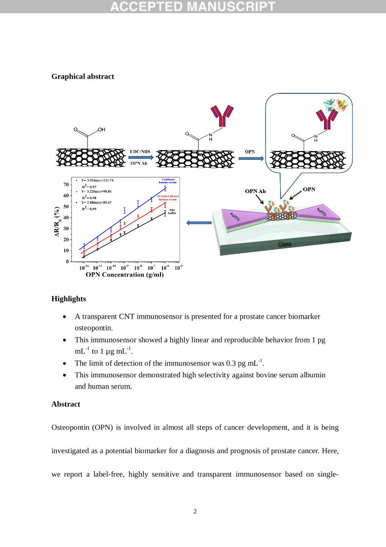
Graphical abstract
Highlights
A transparent CNT immunosensor is presented for a prostate cancer biomarker osteopontin.
This immunosensor showed a highly linear and reproducible behavior from 1 pg mL-1 to 1 µg mL-1.
The limit of detection of the immunosensor was 0.3 pg mL-1. This immunosensor demonstrated high selectivity against bovine serum albumin
and human serum.
Abstract
Osteopontin (OPN) is involved in almost all steps of cancer development, and it is being
investigated as a potential biomarker for a diagnosis and prognosis of prostate cancer. Here,
we report a label-free, highly sensitive and transparent immunosensor based on single-

3
walled carbon nanotubes (SWCNTs) for detection of OPN. A high density of -COOH
functionalized SWCNTs was deposited between two gold/indium tin oxide electrodes on a
glass substrate by dielectrophoresis. Monoclonal antibodies specific to OPN were
covalently immobilized on the SWCNTs. Relative resistance change of the immunosensors
was measured as the concentration of OPN in phosphate buffer saline (PBS) and human
serum was varied from 1 pg mL-1 to 1 µg mL-1 for different channel lengths of 2, 5, and 10
μm, showing a highly linear and reproducible behavior (R2 > 97%). These immunosensors
were also specific to OPN against another test protein, bovine serum albumin, PBS and
human serum, showing that a limit of detection for OPN was 0.3 pg mL-1. This highly
sensitive and transparent immunosensor has a great potential as a simple point-of-care test
kit for various protein biomarkers.
Keywords: cancer; osteopontin; carbon nanotubes; dielectrophoresis; transparent sensor;
field effect transistor sensors.
1. Introduction
Cancer is one of the most life-threatening diseases in humans, and several types of
cancers are reported yet among which lung, liver, blood, and prostate cancers are most
common and have caused most of the deaths worldwide [1]. Prostate cancer is the second
most common cancer in USA after lung cancer [2]. Diagnosis of prostate cancer is a critical
4
limiting factor for this disease because there is no sign of cancer progression for many years
in the initial stage. This often results in delayed diagnosis, and hence the cancer can spread
from the prostate to surrounding organs without being properly treated. There are several
techniques to diagnose prostate cancer including ultrasound, biopsy, and prostate specific
antigen (PSA) test. The PSA test is commonly used, and prostate cancer patients generally
have abnormally elevated PSA level in serum (>4 ng mL-1) [3]. However, it is well known
that PSA tests are prone to both false positives and false negatives [4].
Enzyme-linked immunosorbent assay (ELISA) is a commonly used method for
detection of a certain biomarker protein, but this assay has specific requirements such as
availability of pure samples, long processing time, special equipment and trained personnel
[5,6]. Therefore, it is highly desirable to make a simple, cost-effective, and highly sensitive
immunosensor that can detect a biomarker protein. Recently, carbon nanotube (CNT)- and
nanowire-based field effect transistor (FET) biosensors have shown excellent sensitivity
and selectivity with a low detection limit of PSA for prostate cancer [7-10]. A CNT-FET
based biosensor also showed a detection limit of 30 fM of osteopontin (OPN) for prostate
cancer using a genetically engineered single-chain variable fragment antibody [11].
OPN is a potential new biomarker of prostate cancer [12-14]. It is a secreted, 60-
kDa phosphoprotein that cancer cells use to facilitate their expansion and can be expressed
5
in a variety of tissues such as bones, brain, kidney, lung and liver [15]. It is involved in
almost all the steps of cancer development, and it is being investigated both as a new
therapeutic target and as a potential biomarker for a diagnosis and prognosis of prostate
cancer [14-16]. The measurements of free OPN, which is not bound to complement factor
H, in plasma showed an increase with the stage of prostate cancer [13] although early
detection with OPN measurements has not been fully established, requiring more studies
[17]. It should also be noticed that OPN measurements are important for the prediction of
prostate patient survival and for detection of other cancers. Traditional protein detection
methods such as ELISA have shown to be problematic for its quantification [18] and there
have been very few studies on the cost-effective, easy to implement immunosensors for
OPN detection with comparable or better sensitivity than an ELISA assay.
In this study, we report a label-free, highly sensitive and transparent electrical
immunosensor to detect OPN using single-walled carbon nanotubes (SWCNTs). The
electrical detection based on CNTs has several advantages such as ease of fabrication, well-
understood carbon surface chemistry and simple measurements, which enables miniaturized
and inexpensive biosensors. The SWCNTs were deposited between two transparent source
and drain electrodes on a glass substrate by dielectrophoresis (DEP), making a channel for
OPN attachment and detection. Although chemical vapor deposition (CVD) is a most
6
common technique to grow highly aligned SWCNTs [19] and offers good control on
positioning, it requires specialized materials and high temperature for the growth. On the
contrary, DEP allows the direct alignment of a high density of SWCNTs at room
temperature, and it is generally much simpler and more cost-effective. Monoclonal
antibodies specific to OPN were then covalently immobilized on the SWCNT surfaces by
EDC/NHS treatment. Relative resistance change of the immunosensors was measured for
different channel lengths of 2, 5, and 10 μm and constant channel width of 100 µm as the
concentration of OPN in human serum and phosphate buffer saline (PBS) was varied from
1 pg mL-1 to 1 μg mL-1. The selectivity of the immunosensors was also tested against
bovine serum albumin (BSA), human serum, and PBS.
2. Materials and Methods
2.1. Materials
SWCNTs (98% semiconducting, length: 5-30 µm, diameter: 1-2 nm, COOH
content: ~2.75 wt%) were purchased from M K Impex Corp., Canada. 1-ethyl-3-(3-
dimethylaminopropyl) carbodiimide (EDC), N-hydroxysuccinimide (NHS), and BSA were
purchased from Sigma Aldrich, USA. Normal human serum was purchased from Merck
Millipore, USA. Mouse monoclonal antibodies (AKm2A1) to OPN were purchased from
Santa Cruz Biotechnology, USA, and OPN was purchased from AbD-Serotec, USA.

7
Dimethylformamide (DMF) (98%) and tween 20 were purchased from Biosesang, South
Korea. PBS (pH 7.4, 10x) was purchased from Life Technologies, South Korea. Deionized
water (dH2O) (resistance: ~18.2 MΩ) from the Millipore water purification system was
utilized for preparation of the desired aqueous solutions (molecular biology grade). All the
solutions and glassware were autoclaved prior to being used.
2.2. Fabrication of the SWCNT immunosensor
The immunosensors were fabricated using the conventional photolithography and
lift-off process of two gold/indium tin oxide (ITO) electrodes on a glass substrate. First, 500
µm thick glass wafers (Pyrex 7740, INEXUS, Inc., South Korea) were cleaned by a piranha
solution for 10 min, rinsed with dH2O, and dried with a nitrogen gas. The two electrode
patterns were generated by photolithography followed by electron beam evaporation of
gold (thickness: 2 nm) and RF-sputtering of ITO (thickness: 100 nm) on the glass wafer.
These two electrodes were 100 µm wide and separated by 2, 5, and 10 µm. A SWCNT
suspension was prepared by suspending the SWCNTs in DMF (10 µg mL-1) by 90 min
sonication in a water bath followed by centrifugation at 5,000 rpm for 1 h, and the
supernatant was discarded. After dropping 20 µl of the SWCNT suspension between these
two Au/ITO electrodes, a thin film of parallel-aligned SWCNT channels was formed by
applying 10 V (peak to peak value) at 200 kHz for less than 1 min.
8
2.3. Bio-functionalization
After the deposition of SWCNTs, the immunosensors were rinsed with dH2O, dried
with a nitrogen gas stream, and treated with a mixed solution of 1 mL of EDC (5 mg mL-1)
and 1 mL of NHS (12 mg mL-1) in PBS buffer for 15 min at room temperature to activate
the carboxylic acid groups of SWCNTs followed by washing with PBS buffer (pH 7.4, 1x).
Monoclonal OPN antibodies (10 µg mL-1) in PBS were incubated on the CNT channels for
overnight at 4°C. After the antibody immobilization, the loosely attached antibodies were
removed by thoroughly rinsing with PBS buffer (pH 7.4, 1x) and air-dried at room
temperature. The attachment of the OPN antibodies occurred via the covalent binding of –
NH2 group with NHS succinimide ester on the SWCNTs. Tween 20 (0.5% in 1x PBS, pH
7.4) was then incubated for 30 min at room temperature to prevent the non-specific binding
on the SWCNTs [20]. After rinsing with PBS buffer (pH 7.4, 1x), OPN (1 pg mL-1 –1 µg
mL-1) in PBS (pH 7.4, 1x), undiluted human serum, and 10-fold diluted human serum with
PBS was applied to measure the resistance change of the immunosensors. During each
functionalization, all solutions were pipetted through a polydimethylsiloxane (PDMS) well
fabricated on the channel area, and all the immunosensors were rinsed with PBS buffer (pH
7.4, 1x) after each functionalization step. Fig. 1a shows a schematic of the SWCNT
immunosensor.
9
2.4 Electrical measurements
After each functionalization step, all the immunosensors were thoroughly rinsed
with PBS buffer (pH 7.4, 1x) and dried at room temperature. The current vs voltage (I-V)
characteristics, or the electrical resistance, of all the immunosensors were measured using a
Keithley 2400 semiconductor characterization system, where voltage was varied from −2.0
to 2.0 V. The resistance of the SWCNT channels increased due to each functionalization.
The relative resistance change (RRC) is defined as ΔR (= R - R0)/R0, where R is the
immunosensor resistance after the attachment of OPN, and R0 is the immunosensor
resistance after the treatment of OPN antibody and Tween 20.
2.5 Non-electrical measurements: atomic force microscopy (AFM), scanning electron
microcopy (SEM), optical transmission, and X-ray photoelectron spectroscopy (XPS)
An atomic force microscope (Bruker Instrument, USA) and a field emission
scanning electron microscope (FE-SEM) (S-4800, Hitachi, Japan) were used to characterize
the surface morphology of the SWCNT channels and OPN antibodies on the channels.
AFM images were taken in the tapping mode using a V-shaped TESP probe (Bruker
Instrument). For FE-SEM images, samples were coated by a thin layer of platinum using
ion sputter (E-1045, Hitachi, Japan) at 20 mA for 60 sec before imaging. UV-Vis-NIR (Cary
10
5000, Agilent, USA) was used to measure the optical transmission of the immunosensors.
The chemical composition of the SWCNT surface after DEP-deposition and the antibody
immobilization (10 µg mL-1) was studied by XPS (K–Alpha, Thermo Scientific, USA).
3. Results and Discussion
3.1 AFM, SEM, and XPS analyses
The SEM images of the SWCNT channels showed that many SWCNTs were
aligned between two Au/ITO electrodes by DEP (Fig. 1b and 1c). These aligned SWCNTs
contrast with a non-aligned and dense network of SWCNTs made by sedimentation [21].
The AFM images showed the deposition of the SWCNTs on a glass substrate (Fig. 1d) and
the OPN antibodies on the SWCNT surfaces (Fig. 1e). The spiky nanotube features
disappeared after the antibody attachment, and a globular surface of the antibody layer was
observed. The XPS measurements of the antibodies also showed that the antibodies were
successfully immobilized (supplementary text and Figs. S1-S3).
3.2 Electrical measurements after each functionalization and transparency measurements
Fig. 2 shows an I-V characteristic graph of the immunosensors (L- 2 µm and W-100
µm) when the OPN concentration was 1 pg mL-1. The I-V characteristic graphs for the other
channel lengths (L- 5 and 10 µm) are shown in the supplementary data (Fig S4). More than
11
five immunosensors for each channel length were evaluated. The I-V graphs showed a
linear behavior after each functionalization for all the channel lengths, indicating a good
ohmic contact between SWCNTs and Au/ITO electrodes. The resistance of the
immunosensors increased with additional functionalization: EDC/NHS, OPN antibody,
Tween 20, and OPN. The average resistances (Re) of the immunosensors immediately after
the deposition of the SWCNTs were almost linear (R2=99.5%) with respect to the channel
length (L), showing Re = 1.18 × L, where Re and L are expressed in kΩ and µm, r
espectively (Fig. 3).
Deposition of a thin layer of gold with ITO resulted in a good ohmic contact
between the SWCNTs and the ITO electrodes as well as high transparency [22]. In fact, the
work functions of ITO and CNT are into the range of 3.9 - 4.4eV and 4.7 - 5.1eV,
respectively. This difference would show a large Schottky barrier at the source and drain
electrodes without the gold layer, which has the higher work function (5.1- 5.47eV). The
optical transmittance spectra of the immunosensors exhibited high transparency (~81%) in
the wavelength range of 300 ‒ 700 nm (visible light) while the immunosensors with only
ITO electrodes showed slightly higher transparency (~90%) (Fig. S5). Transparent
biosensors are considered attractive sensing platforms because they allow both optical and
electrical measurements in various experimental set-ups for biosensing and bio-MEMS [23,
12
24].
To determine the optimal incubation time for OPN, the RRCs of the immunosensors
were measured with the incubation time for two different OPN concentrations (Fig. 4).
They drastically increased up to 20 min, and saturated after an incubation time of 60 min.
Therefore, the immunosensors were incubated for 60 min during all the experiments.
3.3 Sensitivity and selectivity studies
Fig. 5 shows the RRCs of the immunosensors as the OPN concentration in PBS was
varied from 1 pg mL-1 to 1 μg mL-1. The RRCs increased with an increase in the OPN
concentration, which is due to a decrease in the number of hole carriers of the p-type
semiconducting SWCNTs with attachment of negatively charged OPNs. For repeatability,
more than five immunosensors were tested for each concentration of OPN, which
confirmed that the immunosensor responses were reproducible. We also observed a highly
linear behavior of RRCs with the logarithm of OPN concentration for different channel
length (2, 5, and 10 µm), where the smallest R2 was 98% for 10 µm long channels.
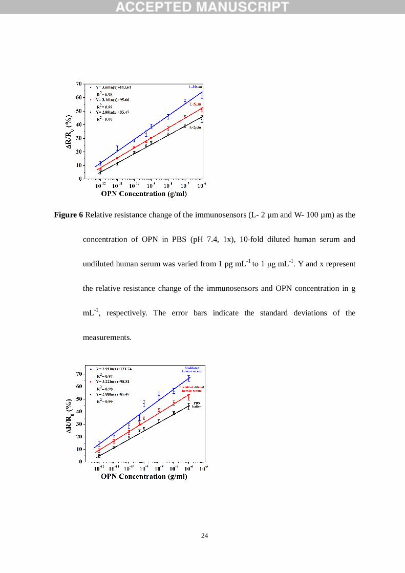
The RRCs of the immunosensors were also measured as the OPN concentration in
10-fold diluted human serum and undiluted human serum was varied from 1 pg mL-1 to 1
μg mL-1 for 2 µm long channels (Fig. 6). The OPN concentration in prostate and normal
patient sera was reported to be 653±39 ng mL-1 and 439±30 ng mL-1, respectively [12].
13
Also, the average level of free OPN in plasma was 45±20 ng mL-1 in normal samples and
127±80 ng mL-1 in prostate cancer samples [13]. The RRCs for given OPN concentration
were the smallest in a PBS medium and the largest in undiluted human serum, which can be
attributed to non-specific binding of several other proteins in the serum.
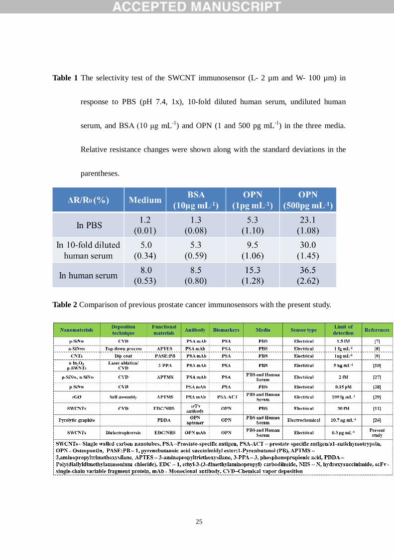
To investigate the selectivity of the immunosensors, PBS (pH 7.4, 1x), 10-fold
diluted human serum, and undiluted human serum along with high concentration BSA (10
µg mL-1) in the three media were introduced to the immunosensors (Table 1). There was
little change in the RRC of the immunosensors between the media and BSA. However,
upon the addition of OPN (1 pg mL-1) on the immunosensors, a rapid and sharp change in
RRC was observed. According to the measurements, these immunosensors showed high
selectivity for OPN, and the detection limit for OPN against BSA and the media was found
to be 0.3 pg mL-1 when the three interpolated graphs in Fig. 6 are equated with the noise
levels caused by the media in the immunosensors, corresponding to 1.5 fg mL-1 for 1 μm2 of
the sensing areas. This immunosensor is 3 orders of magnitude more sensitive than the
current clinical method, ELISA immunoassay [25], and 4-5 orders of magnitude more
sensitive than the OPN levels detected in prostate and normal patient sera. This high
sensitivity shows several advantages. For example, it allows at least 1000-fold dilution of
real samples with buffers, in which less non-specific binding may occur and it is easier to

14
make electrical or magnetic manipulations for the concentration, capture, or detection of
target proteins.
A CVD-grown CNT-based biosensor demonstrated the detection limit of 1 pg mL-1
for OPN in PBS using a single chain variable fragment antibody [11], and a homogeneous
electrochemical method showed a detection limit of 10.7 ng mL-1 in a linear range from 50
to 500 ng mL-1 [26]. The previous prostate cancer immunosensors along with the present
study are listed in Table 2. According to the table, this immunosensor is a cost-effective,
highly sensitive and selective, easy to implement and transparent sensing platform allowing
for optical measurements. Moreover, this immunosensor involved the conventional
microelectrode fabrication and the room-temperature deposition of SWCNTs, implying that
this immunosensor can also be implemented on a flexible substrate for more applications.
4. Conclusions
We presented a label-free, highly sensitive and transparent electrical immunosensors
based on SWCNTs to detect a prostate cancer biomarker OPN. The SWCNTs were
deposited between two transparent Au/ITO electrodes on a glass substrate by DEP. We
observed a highly linear and reproducible behavior over a wide range of OPN
concentrations (1 pg mL-1 to 1 µg mL-1) in PBS and human serum along with a detection
limit of 0.3 pg mL-1. These immunosensors also demonstrated high selectivity of OPN
15
against BSA, human serum and PBS. This transparent immunosensor has great potential as
a useful platform for sensing different types of protein biomarkers along with simultaneous
optical measurements.
Acknowledgments
This research was supported by Basic Science Research Program through the
National Research Foundation of Korea (NRF) funded by the Ministry of Education,
Science and Technology (2012R1A2A2A01012528).
References
[1] X. Li, Y.P. Zhang, H.S. Kim, K.H. Bae, K.M. Stantz, S.J. Lee, C. Jung, J.A. Jimenez,
T.A. Gardner, M.H. Jeng, C. Kao, Gene therapy for prostate cancer by controlling
adenovirus E1a and E4 gene expression with PSES enhancer, Cancer Res. 65 (2005)
1941-1951.
[2] United States Cancer Statistics (U.S.C.S), 1999–2010. Incidence and Mortality Web-
based Report. U.S. Department of Health and Human Services, Centers for Disease
Control and Prevention and National Cancer Institute; Atlanta, 2013.
[3] I.M. Thompson, D.K. Pauler, P.J. Goodman, C.M. Tangen, M.S. Lucia, H.L. Parnes,
L.M. Minasian, L.G. Ford, S.M. Lippman, E.D. Crawford, J.J. Crowley, C.A. Coltman,
16
Prevalence of prostate cancer among men with a prostate-specific antigen level ≤4.0 ng
per milliliter, N. Engl. J. Med. 350 (2004) 2239–2246.
[4] M.J. Barry, Prostate specific antigen testing for early diagnosis of prostate cancer, N.
Engl. J. Med. 344 (2001) 1373-1377.
[5] A. Plumer, H. Duan, S. Subramaniam, F.L. Lucas, S. Miesfeld, A.K. Ng, L. Liaw,
Development of fragment-specific osteopontin antibodies and ELISA for quantification
in human metastatic breast cancer, BMC Cancer 8 (2008) 1471-2407.
[6] A.M. Ward, J.W.F. Catto, F.C Hamdy, Prostate specific antigen: biology, biochemistry
and available commercial assays, Ann. Clin. Biochem. 38 (2001) 633– 651.
[7] X.P.A. Gao, G. Zheng, C.M. Lieber, Subthreshold regime has the optimal sensitivity for
nanowire FET biosensors, Nano Lett. 10 (2010) 547–552.
[8] A. Kim, C.S. Ah, H.Y. Yu, J.H. Yang, I.B. Baek, C.G. Ahn, C.W. Park, M.S. Jun, S.
Lee, Ultrasensitive, label-free, and real-time immunodetection using silicon field-effect
transistors, Appl. Phys. Lett. 91 (2007) 103901.
[9] J.P. Kim, B.Y. Lee, J. Lee, S. Hong, S.J. Sim, Enhancement of sensitivity and
specificity by surface modification of carbon nanotubes in diagnosis of prostate cancer
based on carbon nanotube field effect transistors, Biosen. Bioelectron. 24 (2009) 3372–
3378.
17
[10] C. Li, M. Curreli, H. Lin, B. Lei, F.N. Ishikawa, R. Datar, R.J. Cote, M.E. Thompson,
C. Zhou, Complementary detection of prostate-specific antigen using In2O3 nanowires
and carbon nanotubes, J. Am. Chem. Soc. 127 (2005) 12484-12485.
[11] M.B. Lerner, J. D’Souza, T. Pazina, J. Dailey, B.R. Goldsmith, M.K. Robinson, A.T.C.
Johnson, Hybrids of a genetically engineered antibody and a carbon nanotube transistor
for detection of prostate cancer biomarkers, ACS Nano 6 (2012) 5141-5149.
[12] N.S. Fedarko, A. Jain, A. Karadag, M.R.V. Eman, L.W. Fisher, Elevated serum bone
sialoprotein and osteopontin in colon, breast, prostate, and lung cancer, Clin. Cancer
Res. 7 (2001) 4060–4066.
[13] A. Jain, D.A. McKnight, L.W. Fisher, E.B. Humphreys, L.A. Mangold, A.W. Partin,
N.S. Fedarko, Small integrin-binding proteins as serum markers for prostate cancer
detection, Clin. Cancer Res. 15 (2009) 5199–5207.
[14] G. Castellano, G. Malaponte, M.C. Mazzarino, M. Figini, F. Marchese, P. Gangemi,
S.Travali, F. Stivala, S. Canevari, M. Libra, Activation of the osteopontin/matrix
metalloproteinase-9 pathway correlates with prostate cancer progression, Clin. Cancer
Res. 14 (2008) 7470-7480.
[15] J. Sodek, B. Ganss, M.D. McKee, Osteopontin, Crit. Rev. Oral Biol. Med. 11 (2000)
279–303.
18
[16] A. Bellahcene, V. Castronovo, K.U. Ogbureke, L.W. Fisher, N.S. Fedarko, Small
integrin-binding ligand N-linked glycoproteins (Siblings): multifunctional proteins in
cancer, Nat. Rev. Cancer 8 (2008) 212–226.
[17] R. Puzone, L. Paleari, F. Montefiore, L. Ruggiero, M. Puntoni, M. Maffezzini, B.
Bobbio, P. Marroni, R. Libener, P. G. Betta, Osteopontin plasma level does not detect
prostate cancer in patients referred for diagnostic prostate biopsy, Int. J. Biol. Markers
25 (2010) 200-206.
[18] P.H. Anborgh, S.M. Wilson, A.B. Tuck, E. Winquist, N. Schmidt, R. Hart, S. Kon, M.
Maeda, T. Uede, L.W. Stitt, A.F. Chambers, New dual monoclonal elisa for measuring
plasma osteopontin as a biomarker associated with survival in prostate cancer: clinical
validation and comparison of multiple elisas, Clin. Chem. 55 (2009) 895–903.
[19] J. Kong, H.T. Soh, A.M. Cassell, C.F. Quate, H. Dai, Synthesis of individual single-
walled carbon nanotubes on patterned silicon wafers, Nature 395 (1998) 878-88.
[20] B. Batteiger, W.J. Newhall V, R.B. Jones, The use of tween 20 as a blocking agent in
the immunological detection of proteins transferred to nitrocellulose membranes, J.
Immunolo. Met. 55 (1982) 297-307.

19
[21] R. Singh, A. Sharma, S. Hong, J. Jang, Electrical Immunosensor based on
Dielectrophoretically-deposited Carbon Nanotubes for Detection of Influenza Virus
H1N1, Analyst 139 (2014) 5415-5421.
[22] J. Zhang, C. Wang, C. Zhou, Rigid/flexible transparent electronics based on separated
carbon nanotube thin-film transistors and their application in display electronics, ACS
Nano 6 (2012) 7412–7419.
[23] F.J. Rawson, C.L. Yeung, S.K. Jackson, P.M. Mendes, Tailoring 3D single-walled
carbon nanotubes anchored to indium tin oxide for natural cellular uptake and
intracellular sensing, Nano Lett. 13 (2013) 1-8.
[24] W. Tonomura, H. Okamura, S. Konishi, Transparent biosensor with micro channel
array for optical and electrophysiological detection of luciferin–luciferase reaction,
IEEJ Trans. 2 (2007) 372–377.
[25] G. MacBeath, Protein microarrays and proteomics, Nature Genet. 32 (2002) 526-532.
[26] Y. Cao, D. Chen, W. Chen, J. Yu, Z. Chen, G. Li, Aptamer-based homogeneous protein
detection using cucurbit[7]uril functionalized electrode, Anal. Chim. Acta 812 (2014)
45-49.
20
[27] G. Zheng, F. Patolsky, Y. Cui, W. U. Wang, C. M. Lieber, Multiplexed electrical
detection of cancer markers with nanowire sensor arrays, Nat. Biotechnol. 23 (2005)
1294–1301.
[28] G. Zheng, X. P. A. Gao, C, M. Lieber, Frequency domain detection of biomolecules
using silicon nanowire biosensors, Nano Lett. 10 (2010) 3179–3183.
[29] D. J. Kim, Y. Sohn, J. H. Jung, O. J. Yoon, N. E. Lee, J. S. Park, Reduced graphene
oxide field-effect transistor for label-free femtomolar protein detection, Biosen.
Bioelectron. 41 (2013) 621–626.
21
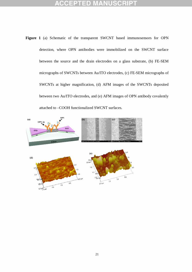
Figure 1 (a) Schematic of the transparent SWCNT based immunosensors for OPN
detection, where OPN antibodies were immobilized on the SWCNT surface
between the source and the drain electrodes on a glass substrate, (b) FE-SEM
micrographs of SWCNTs between Au/ITO electrodes, (c) FE-SEM micrographs of
SWCNTs at higher magnification, (d) AFM images of the SWCNTs deposited
between two Au/ITO electrodes, and (e) AFM images of OPN antibody covalently
attached to –COOH functionalized SWCNT surfaces.
22
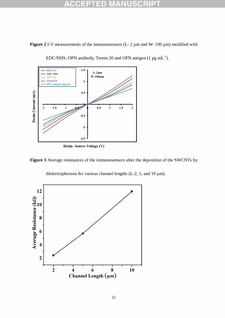
Figure 2 I-V measurements of the immunosensors (L- 2 µm and W- 100 µm) modified with
EDC/NHS, OPN antibody, Tween 20 and OPN antigen (1 pg mL-1).
Figure 3 Average resistances of the immunosensors after the deposition of the SWCNTs by
dielectrophoresis for various channel lengths (L-2, 5, and 10 µm).
23
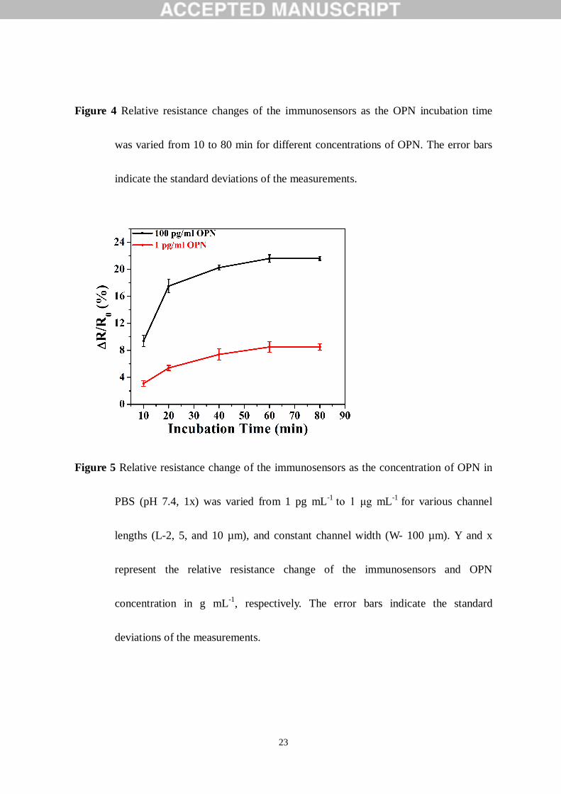
Figure 4 Relative resistance changes of the immunosensors as the OPN incubation time
was varied from 10 to 80 min for different concentrations of OPN. The error bars
indicate the standard deviations of the measurements.
Figure 5 Relative resistance change of the immunosensors as the concentration of OPN in
PBS (pH 7.4, 1x) was varied from 1 pg mL-1 to 1 μg mL-1 for various channel
lengths (L-2, 5, and 10 µm), and constant channel width (W- 100 µm). Y and x
represent the relative resistance change of the immunosensors and OPN
concentration in g mL-1, respectively. The error bars indicate the standard
deviations of the measurements.
24
Figure 6 Relative resistance change of the immunosensors (L- 2 µm and W- 100 µm) as the
concentration of OPN in PBS (pH 7.4, 1x), 10-fold diluted human serum and
undiluted human serum was varied from 1 pg mL-1 to 1 μg mL-1. Y and x represent
the relative resistance change of the immunosensors and OPN concentration in g
mL-1, respectively. The error bars indicate the standard deviations of the
measurements.
25
Table 1 The selectivity test of the SWCNT immunosensor (L- 2 µm and W- 100 µm) in
response to PBS (pH 7.4, 1x), 10-fold diluted human serum, undiluted human
serum, and BSA (10 μg mL-1) and OPN (1 and 500 pg mL-1) in the three media.
Relative resistance changes were shown along with the standard deviations in the
parentheses.
Table 2 Comparison of previous prostate cancer immunosensors with the present study.
Copyright © 2022 FDOKUMEN