Microbiome Profiling by Illumina Sequencing of Combinatorial Sequence-Tagged PCR Products
Upload
independentCategory
view
0download
0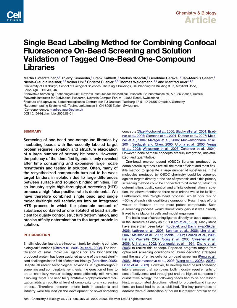
Chemistry & Biology
Article
Single Bead Labeling Method for Combining ConfocalFluorescence On-Bead Screening and SolutionValidation of Tagged One-Bead One-CompoundLibrariesMartin Hintersteiner,1,2 Thierry Kimmerlin,2 Frank Kalthoff,2 Markus Stoeckli,3 Geraldine Garavel,2 Jan-Marcus Seifert,2
Nicole-Claudia Meisner,2,3 Volker Uhl,2 Christof Buehler,2,5 Thomas Weidemann,2,4 and Manfred Auer1,2,*1University of Edinburgh, School of Biological Sciences, The King’s Buildings, CH Waddington Building 3.07, Mayfield Road,
Edinburgh EH9 3JR, UK2Innovative Screening Technologies unit, Novartis Institutes for BioMedical Research, Brunnerstrasse 59, A-1235 Vienna, Austria3Novartis Institutes for BioMedical Research, Novartis Campus Forum 1, 4056 Basel, Switzerland4Institute of Biophysics, Biotechnologisches Zentrum der TU Dresden, Tatzberg 47-51, D-01307 Dresden, Germany5Supercomputing Systems AG, Technoparkstrasse 1, CH-8005 Zurich, Switzerland*Correspondence: [email protected]
DOI 10.1016/j.chembiol.2009.06.011
SUMMARY
Screening of one-bead one-compound libraries byincubating beads with fluorescently labeled targetprotein requires isolation and structure elucidationof a large number of primary hit beads. However,the potency of the identified ligands is only revealedafter time consuming and expensive larger scaleresynthesis and testing in solution. Often, many ofthe resynthesized compounds turn out to be weaktarget binders in solution due to large differencesbetween surface and solution binding affinities. Foran industry style high-throughput screening (HTS)process a high false positive rate is detrimental. Wehave therefore combined single bead and singlemolecule/single cell techniques into an integratedHTS process in which the picomole amount ofsubstance contained on one isolated hit bead is suffi-cient for quality control, structure determination, andprecise affinity determination to the target protein insolution.
INTRODUCTION
Small molecular ligands are important tools for studying complex
biological functions (Chen et al., 2006; Xu et al., 2008). The iden-
tification of small molecular ligands for any biochemically
produced protein has been assigned as one of the most signifi-
cant challenges in the field of chemical biology (Schreiber, 2005).
Despite all recent technological advances in high-throughput
screening and combinatorial synthesis, the question of how to
probe chemistry versus biology most efficiently still remains
a moving target. The need for quantitative biophysical character-
ization adds an additional level of complexity to any screening
process. Therefore, research efforts both in academia and
industry were focused on the development of novel screening
724 Chemistry & Biology 16, 724–735, July 31, 2009 ª2009 Elsev
concepts (Diaz-Mochon et al., 2006; Blackwell et al., 2001; Brad-
ner et al., 2006; Clemons et al., 2001; Duffner et al., 2007; Meis-
ner et al., 2004; Metzger et al., 2006; Muckenschnabel et al.,
2004; Sedlacek and Chen, 2005; Urbina et al., 2006; Vegas
et al., 2008; Winssinger et al., 2008; Zehender et al., 2004).
However, none of these concepts are fully integrated, miniatur-
ized, and quantitative.
One-bead one-compound (OBOC) libraries produced by
combinatorial synthesis are still the most efficient and most flex-
ible method to generate a large number of substances. If the
molecules produced by OBOC chemistry could be screened
against targets directly at the site of synthesis and if this primary
screening method could be connected to hit isolation, structure
determination, quality control, and affinity determination in solu-
tion, the above mentioned three main criteria would be fulfilled.
Furthermore, this ‘‘single bead process’’ would only rely on
�50 ng of each individual library compound. Resynthesis efforts
would be focused on the most potent compounds. Such
a screening process would dramatically gain if it was directly
linked to validation in cells and model organisms.
The basic idea of screening ligands directly on bead appeared
in the literature as early as 1991 (Lam et al., 1991). Many steps
have since then been taken (Kodadek and Bachhawat-Sikder,
2006; Lathrop et al., 2007; Lehman et al., 2006; Lim et al.,
2007; Meisner et al., 2009; Meldal, 2002; Paulick et al., 2006;
Pei and Wavreille, 2007; Song et al., 2003; Sweeney et al.,
2006; Uhl et al., 2002; Youngquist et al., 1994; Zhang et al.,
2008) to realize this concept. Reported progress ranges from
optimized screening conditions to library decoding strategies
and the use of entire cells for on-bead screening (Peng et al.,
2006; Udugamasooriya et al., 2008; Wang et al., 2005a, 2005b;
Chen et al., 2009). However, to develop bead based screening
into a process that combines both industry requirements of
cost effectiveness and throughput and the highest standards in
quantitative biology, the following needs had to be addressed.
First, an automated detection method for protein-ligand interac-
tions on bead had to be established. The key parameters to
address were quantification of bound fluorescent protein at the
ier Ltd All rights reserved
Chemistry & Biology
Single Bead Validation of CONA On-Bead Screening Hits
Figure 1. The ICB Discovery Cycle Comprises Seven Steps and Combines On-Bead and Solution Screening
Step 1: design and synthesis of OBOC libraries by combinatorial chemistry. Step 2: hit bead detection by CONA. Step 3: semiautomated hit beads isolation and
deposition (one bead per vial). Step 4: conversion of bead bound compounds into fluorescent ligands by site-specific labeling and compound cleavage. Step 5:
affinity measurement in homogenous solution by titration of labeled hit compounds with unlabeled target protein using a miniaturized confocal fluorescence fluc-
tuation assay. Step 6: hit bead ranking. Step 7: MS-based structure determination.
bead surface and reliable distinction between hit beads and
autofluorescent beads. Second, a direct link between on-bead
binding and affinity determination in solution needed to be
developed. Third, the chemical method for combining surface
screening and solution testing needed to be flexible enough to
include the option for profiling identified ligands in cellular assays
and in validation assays further downstream.
Herein we describe a new screening methodology, ‘‘inte-
grated chemical biophysics’’ (ICB), which integrates library
design and synthesis, automated confocal nanoscanning
(CONA) for preselection of hit compounds on the solid surface
as primary screen, quantitative testing of single hit bead-
derived compounds in solution, as well as structure determina-
tion and quality control (Figure 1). This is achieved by converting
all compounds on picked hit beads into fluorescent ligands.
With a free choice of color, additional chemical tagging possibil-
ities, and an affinity (Kd) connected to each compound, cellular
validation by high resolution confocal imaging, microspectro-
scopy, or functional assays is immediately possible. The ICB
process is demonstrated by screening a >100,000 member
library of designed a,b-phosphopeptides against the Grb2
SH2 domain. The identification of ligands with nano- to micro-
molar affinity (Kd) for Grb2 SH2 in solution from this library
without prior resynthesis shows the utility and flexibility of the
ICB method and its unique power in producing a reliable and
Chemistry & Biology 1
strictly quantitative high-throughput SAR in a primary screening
campaign.
RESULTS
Automated CONA and Bead PickingCurrent on-bead screens are performed on standard (fluores-
cence) microscopes or on the COPAS bead sorter (Union Bio-
metrica).
These methods detect integrated fluorescence intensities,
from the entire volume of individual (TentaGel) beads. Hit bead
identification is hampered by the varying autofluorescence levels
occurring in combinatorial OBOC libraries (Ding et al., 2006;
Olivos et al., 2003). (Although this problem is less pronounced
with PEG-based resins, the increased mechanical stability of
TentaGel beads is highly advantageous during library synthesis
and on-bead screening.) While autofluorescence is distributed
throughout the bead matrix, the limited pore size of TentaGel
beads largely prevents the fluorescently labeled target proteins
(>10 kDa) from diffusing inside the bead in the time frame of
incubation and screening (<24 hr). Therefore, to limit detection
of fluorescence artifacts and to allow precise quantification of
target binding to compounds presented at the bead surface
the on-bead screening relevant binding signal for TentaGel
beads must be recorded within the outer few micrometers of
6, 724–735, July 31, 2009 ª2009 Elsevier Ltd All rights reserved 725
Chemistry & Biology
Single Bead Validation of CONA On-Bead Screening Hits
C D
BA
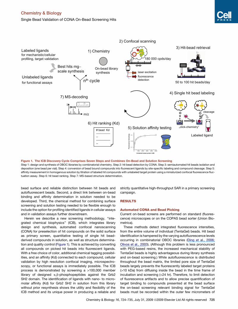
Figure 2. On-Bead Screening by CONA
(A) Schematic layout of the PickoScreen (PS0x) instruments. The PickoScreen instruments are confocal microscopes equipped with a scanning table and
a picking robot. The picking robotics operates a capillary (depicted in the inset), through which individual hit beads are sucked up from the bottom of the microtiter
plate via pneumatic vacuum.
(B) In an advanced high-speed version of the PickoScreen design (PS04), the traditional confocal optics was replaced by a Nipkow spinning disc, which uses
multiple pinholes for parallel sampling of individual picture frames. The tile pictures are finally merged to generate whole-well images.
(C) During CONA the stage is moved and the confocal focus is held just below the equatorial plane of beads. The beads appear as round objects in the scan
image. For each bead two parameters are registered: the fluorescence intensity in the bead interior (area intensity) and the intensity in a small ring on the outer
edge of the beads (ring intensity).
(D) The 2–5 mm scanning resolution in CONA allows to efficiently distinguish between beads with a high level of autofluorescence (top) and hit beads with bound
target protein on the outside (bottom).
a bead. We reasoned that by measuring spatial intensity distribu-
tions along the equatorial plane of a bead with confocal imaging,
much higher signal to noise ratios can be achieved, as compared
to standard integrative fluorescence methods.
Together with Evotec Technologies (formerly ET, now Perkin
Elmer) we have developed three fluorescence microscopes
based on the Insight Reader platform for single molecule spec-
troscopy and dedicated them to three essential steps in bead
based screening: PS02, an instrument for high resolution
confocal scanning and bead picking; PS04, an instrument for
high speed CCD-based confocal bead imaging; and PS03, an
instrument for high resolution microspectroscopy. (Figure 2A).
The technical aspects of the PS02 and PS01 instruments are
726 Chemistry & Biology 16, 724–735, July 31, 2009 ª2009 Elsevie
described in detail in Hintersteiner et al. (2009). Briefly, each of
the ‘‘PickoScreen’’ instruments consists of a modified Olympus
IX70 microscope with an integrated confocal multicolor detection
system. The PS02 instrument is equipped with a high precision
motorized scanning table holding an integrated sample compart-
ment. Other than in conventional confocal laser scanning micro-
scopes the laser beam for fluorescence excitation remains fixed
while the sample compartment is moved during image acquisi-
tion. This setup offers the advantage that a larger sample area
can be imaged without optical distortion. In an advanced modifi-
cation, fast multiparallel confocal imaging has been realized in
PS04 by using a Nipkow spinning disc with multiple pinholes
and CCD cameras for signal detection (Figure 2B).
r Ltd All rights reserved
Chemistry & Biology
Single Bead Validation of CONA On-Bead Screening Hits
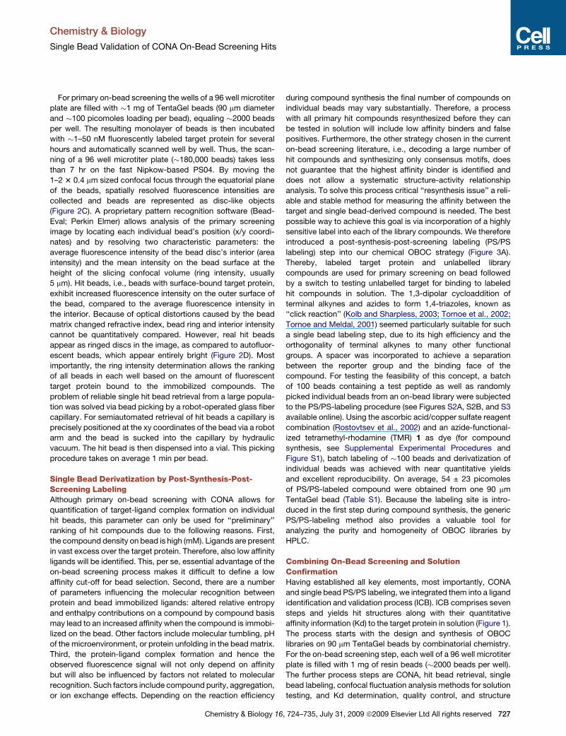
For primary on-bead screening the wells of a 96 well microtiter
plate are filled with �1 mg of TentaGel beads (90 mm diameter
and �100 picomoles loading per bead), equaling �2000 beads
per well. The resulting monolayer of beads is then incubated
with �1–50 nM fluorescently labeled target protein for several
hours and automatically scanned well by well. Thus, the scan-
ning of a 96 well microtiter plate (�180,000 beads) takes less
than 7 hr on the fast Nipkow-based PS04. By moving the
1–2 3 0.4 mm sized confocal focus through the equatorial plane
of the beads, spatially resolved fluorescence intensities are
collected and beads are represented as disc-like objects
(Figure 2C). A proprietary pattern recognition software (Bead-
Eval; Perkin Elmer) allows analysis of the primary screening
image by locating each individual bead’s position (x/y coordi-
nates) and by resolving two characteristic parameters: the
average fluorescence intensity of the bead disc’s interior (area
intensity) and the mean intensity on the bead surface at the
height of the slicing confocal volume (ring intensity, usually
5 mm). Hit beads, i.e., beads with surface-bound target protein,
exhibit increased fluorescence intensity on the outer surface of
the bead, compared to the average fluorescence intensity in
the interior. Because of optical distortions caused by the bead
matrix changed refractive index, bead ring and interior intensity
cannot be quantitatively compared. However, real hit beads
appear as ringed discs in the image, as compared to autofluor-
escent beads, which appear entirely bright (Figure 2D). Most
importantly, the ring intensity determination allows the ranking
of all beads in each well based on the amount of fluorescent
target protein bound to the immobilized compounds. The
problem of reliable single hit bead retrieval from a large popula-
tion was solved via bead picking by a robot-operated glass fiber
capillary. For semiautomated retrieval of hit beads a capillary is
precisely positioned at the xy coordinates of the bead via a robot
arm and the bead is sucked into the capillary by hydraulic
vacuum. The hit bead is then dispensed into a vial. This picking
procedure takes on average 1 min per bead.
Single Bead Derivatization by Post-Synthesis-Post-Screening LabelingAlthough primary on-bead screening with CONA allows for
quantification of target-ligand complex formation on individual
hit beads, this parameter can only be used for ‘‘preliminary’’
ranking of hit compounds due to the following reasons. First,
the compound density on bead is high (mM). Ligands are present
in vast excess over the target protein. Therefore, also low affinity
ligands will be identified. This, per se, essential advantage of the
on-bead screening process makes it difficult to define a low
affinity cut-off for bead selection. Second, there are a number
of parameters influencing the molecular recognition between
protein and bead immobilized ligands: altered relative entropy
and enthalpy contributions on a compound by compound basis
may lead to an increased affinity when the compound is immobi-
lized on the bead. Other factors include molecular tumbling, pH
of the microenvironment, or protein unfolding in the bead matrix.
Third, the protein-ligand complex formation and hence the
observed fluorescence signal will not only depend on affinity
but will also be influenced by factors not related to molecular
recognition. Such factors include compound purity, aggregation,
or ion exchange effects. Depending on the reaction efficiency
Chemistry & Biology 16
during compound synthesis the final number of compounds on
individual beads may vary substantially. Therefore, a process
with all primary hit compounds resynthesized before they can
be tested in solution will include low affinity binders and false
positives. Furthermore, the other strategy chosen in the current
on-bead screening literature, i.e., decoding a large number of
hit compounds and synthesizing only consensus motifs, does
not guarantee that the highest affinity binder is identified and
does not allow a systematic structure-activity relationship
analysis. To solve this process critical ‘‘resynthesis issue’’ a reli-
able and stable method for measuring the affinity between the
target and single bead-derived compound is needed. The best
possible way to achieve this goal is via incorporation of a highly
sensitive label into each of the library compounds. We therefore
introduced a post-synthesis-post-screening labeling (PS/PS
labeling) step into our chemical OBOC strategy (Figure 3A).
Thereby, labeled target protein and unlabelled library
compounds are used for primary screening on bead followed
by a switch to testing unlabelled target for binding to labeled
hit compounds in solution. The 1,3-dipolar cycloaddition of
terminal alkynes and azides to form 1,4-triazoles, known as
‘‘click reaction’’ (Kolb and Sharpless, 2003; Tornoe et al., 2002;
Tornoe and Meldal, 2001) seemed particularly suitable for such
a single bead labeling step, due to its high efficiency and the
orthogonality of terminal alkynes to many other functional
groups. A spacer was incorporated to achieve a separation
between the reporter group and the binding face of the
compound. For testing the feasibility of this concept, a batch
of 100 beads containing a test peptide as well as randomly
picked individual beads from an on-bead library were subjected
to the PS/PS-labeling procedure (see Figures S2A, S2B, and S3
available online). Using the ascorbic acid/copper sulfate reagent
combination (Rostovtsev et al., 2002) and an azide-functional-
ized tetramethyl-rhodamine (TMR) 1 as dye (for compound
synthesis, see Supplemental Experimental Procedures and
Figure S1), batch labeling of �100 beads and derivatization of
individual beads was achieved with near quantitative yields
and excellent reproducibility. On average, 54 ± 23 picomoles
of PS/PS-labeled compound were obtained from one 90 mm
TentaGel bead (Table S1). Because the labeling site is intro-
duced in the first step during compound synthesis, the generic
PS/PS-labeling method also provides a valuable tool for
analyzing the purity and homogeneity of OBOC libraries by
HPLC.
Combining On-Bead Screening and SolutionConfirmationHaving established all key elements, most importantly, CONA
and single bead PS/PS labeling, we integrated them into a ligand
identification and validation process (ICB). ICB comprises seven
steps and yields hit structures along with their quantitative
affinity information (Kd) to the target protein in solution (Figure 1).
The process starts with the design and synthesis of OBOC
libraries on 90 mm TentaGel beads by combinatorial chemistry.
For the on-bead screening step, each well of a 96 well microtiter
plate is filled with 1 mg of resin beads (�2000 beads per well).
The further process steps are CONA, hit bead retrieval, single
bead labeling, confocal fluctuation analysis methods for solution
testing, and Kd determination, quality control, and structure
, 724–735, July 31, 2009 ª2009 Elsevier Ltd All rights reserved 727
Chemistry & Biology
Single Bead Validation of CONA On-Bead Screening Hits
determination by MS methods. After primary CONA screening
and PS/PS validation from single beads, the best scoring hits
can be resynthesized in milligram quantities with or without the
spacer and fluorophore tagging site. This opens up multiple
ways for testing the newly identified ligands in functional, (single
cell) imaging, or model organism secondary assays. To demon-
strate the power of this process, we designed and synthesized
an SH2 domain-tailored library of mixed a/b-phosphopeptides
and screened it against SH2 domain targets, such as Grb2.
Library SynthesisSH2 domains constitute an essential class of cell signaling inter-
action domains. They recognize short peptide stretches with
only a few residues C- and N-terminal to the phosphotyrosine
(Table S2). Limited cell permeability and plasma stability are
the main limitations for using a peptides for pharmaceutical
applications. In contrast, b peptides are highly stable toward
enzymatic degradation (Wiegand et al., 2002). We have therefore
designed and synthesized a OBOC library of more than 100,000
mixed a/b-phosphopeptides on the PS/PS-labeling platform,
described above. The library design contained three distinct
branches, each intended to mimic different regions of the
consensus motif (Figure 3B and Figures S4 and S5). Two library
branches consisted of pentapeptides entailing four combinato-
rial sites plus the phosphotyrosine and the third branch
consisted of tetrapeptides entailing three combinatorial sites
plus phosphotyrosine. The library was synthesized by the split
A
B
C
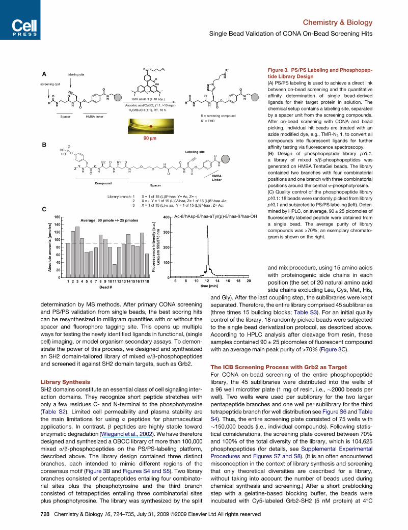
Figure 3. PS/PS Labeling and Phosphopep-
tide Library Design
(A) PS/PS labeling is used to achieve a direct link
between on-bead screening and the quantitative
affinity determination of single bead-derived
ligands for their target protein in solution. The
chemical setup contains a labeling site, separated
by a spacer unit from the screening compounds.
After on-bead screening with CONA and bead
picking, individual hit beads are treated with an
azide modified dye, e.g., TMR-N3 1, to convert all
compounds into fluorescent ligands for further
affinity testing via fluorescence spectroscopy.
(B) Design of phosphopeptide library pYL1:
a library of mixed a/b-phosphopeptides was
generated on HMBA TentaGel beads. The library
contained two branches with four combinatorial
positions and one branch with three combinatorial
positions around the central a-phosphotyrosine.
(C) Quality control of the phosphopeptide library
pYL1: 18 beads were randomly picked from library
pYL1 and subjected to PS/PS labeling (left). Deter-
mined by HPLC, on average, 90 ± 25 picomoles of
fluorescently labeled peptide were obtained from
a single bead. The average purity of library
compounds was >70%; an exemplary chromato-
gram is shown on the right.
and mix procedure, using 15 amino acids
with proteinogenic side chains in each
position (the set of 20 natural amino acid
side chains excluding Leu, Cys, Met, His,
and Gly). After the last coupling step, the sublibraries were kept
separated. Therefore, the entire library comprised 45 sublibraries
(three times 15 building blocks; Table S3). For an initial quality
control of the library, 18 randomly picked beads were subjected
to the single bead derivatization protocol, as described above.
According to HPLC analysis after cleavage from resin, these
samples contained 90 ± 25 picomoles of fluorescent compound
with an average main peak purity of >70% (Figure 3C).
The ICB Screening Process with Grb2 as TargetFor CONA on-bead screening of the entire phosphopeptide
library, the 45 sublibraries were distributed into the wells of
a 96 well microtiter plate (1 mg of resin, i.e., �2000 beads per
well). Two wells were used per sublibrary for the two larger
pentapeptide branches and one well per sublibrary for the third
tetrapeptide branch (for well distribution see Figure S6 and Table
S4). Thus, the entire screening plate consisted of 75 wells with
�150,000 beads (i.e., individual compounds). Following statis-
tical considerations, the screening plate covered between 70%
and 100% of the total diversity of the library, which is 104,625
phosphopeptides (for details, see Supplemental Experimental
Procedures and Figures S7 and S8). (It is an often encountered
misconception in the context of library synthesis and screening
that only theoretical diversities are described for a library,
without taking into account the number of beads used during
chemical synthesis and screening.) After a short preblocking
step with a gelatine-based blocking buffer, the beads were
incubated with Cy5-labeled Grb2-SH2 (5 nM protein) at 4�C
728 Chemistry & Biology 16, 724–735, July 31, 2009 ª2009 Elsevier Ltd All rights reserved
Chemistry & Biology
Single Bead Validation of CONA On-Bead Screening Hits
overnight. The on-bead screen was run on the PS04 instrument
in less than 6 hr of scanning time. The automated analysis of ring
and area intensities for all beads in each well revealed a total of
238 hit beads (hit rate 0.16%) with a ‘‘ring intensity’’ that
exceeded the background signal at least 5-fold. Counting the
number of hit beads per well allowed the prioritization of 16 wells,
as the predominantly responding sublibraries, for bead picking.
Figure 4A shows one exemplary well with several hit beads.
From the selected wells the 60 beads with the highest ring inten-
sities were picked, using the capillary based bead-picking
procedure of the PS04 instrument.
Solution Confirmation of Primary Hit Beads by ConfocalFluorescence Fluctuation AnalysisVarious confocal fluorescence methods are suitable for the solu-
tion assay after single bead cleavage of fluorescent derivatives
of hit compounds. Overall, the fastest and most reliable method
is 2D-FIDA anisotropy (Kask et al., 2000). Therefore, single bead-
derivatized TMR-labeled hit compounds, in amounts between 27
and 108 picomoles, were dissolved in 20 ml of water and 20%
acetonitrile. By taking 1 ml only of each sample and further
1:500 dilution, suitable concentrations for the 2D-FIDA-r assay
was reached. Single-point measurements for Grb2-SH2 binding
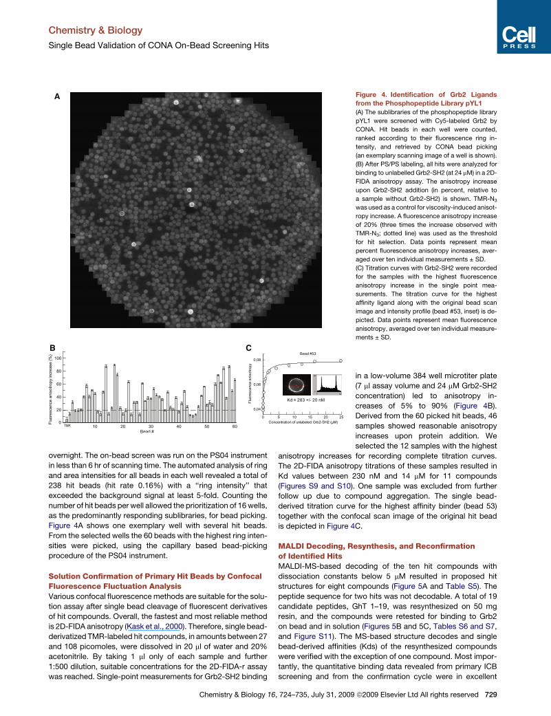
A Figure 4. Identification of Grb2 Ligands
from the Phosphopeptide Library pYL1
(A) The sublibraries of the phosphopeptide library
pYL1 were screened with Cy5-labeled Grb2 by
CONA. Hit beads in each well were counted,
ranked according to their fluorescence ring in-
tensity, and retrieved by CONA bead picking
(an exemplary scanning image of a well is shown).
(B) After PS/PS labeling, all hits were analyzed for
binding to unlabelled Grb2-SH2 (at 24 mM) in a 2D-
FIDA anisotropy assay. The anisotropy increase
upon Grb2-SH2 addition (in percent, relative to
a sample without Grb2-SH2) is shown. TMR-N3
was used as a control for viscosity-induced anisot-
ropy increase. A fluorescence anisotropy increase
of 20% (three times the increase observed with
TMR-N3; dotted line) was used as the threshold
for hit selection. Data points represent mean
percent fluorescence anisotropy increases, aver-
aged over ten individual measurements ± SD.
(C) Titration curves with Grb2-SH2 were recorded
for the samples with the highest fluorescence
anisotropy increase in the single point mea-
surements. The titration curve for the highest
affinity ligand along with the original bead scan
image and intensity profile (bead #53, inset) is de-
picted. Data points represent mean fluorescence
anisotropy, averaged over ten individual measure-
ments ± SD.
in a low-volume 384 well microtiter plate
(7 ml assay volume and 24 mM Grb2-SH2
concentration) led to anisotropy in-
creases of 5% to 90% (Figure 4B).
Derived from the 60 picked hit beads, 46
samples showed reasonable anisotropy
increases upon protein addition. We
selected the 12 samples with the highest
anisotropy increases for recording complete titration curves.
The 2D-FIDA anisotropy titrations of these samples resulted in
Kd values between 230 nM and 14 mM for 11 compounds
(Figures S9 and S10). One sample was excluded from further
follow up due to compound aggregation. The single bead-
derived titration curve for the highest affinity binder (bead 53)
together with the confocal scan image of the original hit bead
is depicted in Figure 4C.
MALDI Decoding, Resynthesis, and Reconfirmationof Identified HitsMALDI-MS-based decoding of the ten hit compounds with
dissociation constants below 5 mM resulted in proposed hit
structures for eight compounds (Figure 5A and Table S5). The
peptide sequence for two hits was not decodable. A total of 19
candidate peptides, GhT 1–19, was resynthesized on 50 mg
resin, and the compounds were retested for binding to Grb2
on bead and in solution (Figures 5B and 5C, Tables S6 and S7,
and Figure S11). The MS-based structure decodes and single
bead-derived affinities (Kds) of the resynthesized compounds
were verified with the exception of one compound. Most impor-
tantly, the quantitative binding data revealed from primary ICB
screening and from the confirmation cycle were in excellent
Chemistry & Biology 16, 724–735, July 31, 2009 ª2009 Elsevier Ltd All rights reserved 729
Chemistry & Biology
Single Bead Validation of CONA On-Bead Screening Hits
agreement. To complete the discovery cycle, outlined above, the
best compound (GhT-11, 2) was also synthesized in unlabelled
form (GhN-11, 3, without labeling site and spacer). A 2D-FIDA-
r titration of a mix of the unlabelled hit compound and the
TMR-labeled derivative with the Grb2-SH2 target derived close
Kds for tagged and untagged ligand (Figure 5D). The fact that
a higher affinity (Kd) was found for the labeled ligand GhN-11,
2, most likely indicates an additional small hydrophobic contri-
bution from TMR to the binding free energy.
To investigate whether a purification step introduced between
the PS/PS labeling and cleavage of primary on-bead hits and the
single molecule assay in solution would improve the quality of the
fluorescence titrations and the decoding efficiency, a second
screening round with Grb2-SH2 was performed. An additional
20 hit beads with compound affinities between 390 nM and
>20 mM were identified (Table S8). The HPLC purification step
proved to facilitate the decoding process by MALDI-MS. Eigh-
teen out of 20 samples were successfully decoded. The full
scope of the current ICB process can be explored by further
taking the resynthesized PS/PS-labeled ligands into cellular vali-
dation. To exemplify this, we expanded the PS/PS-labeling
strategy by incorporating a bifunctional tag for conjugating
both a cell-permeable peptide and a fluorescent label. The
best resynthesized hit compound from our a/b peptide library
was derivatized with an azide-modified, TMR-labeled TAT
peptide to produce a cell-permeable construct 4 (Figure 6).
By high resolution fluorescence imaging on the PS03 microspec-
troscopy instrument the TAT-conjugated compound 4, was
demonstrated to accumulate in the nucleus of A431 cells after
12 hr incubation (Figure 7A). Furthermore, anisotropy images
showed a significant higher anisotropy for the nuclear fraction
of the probe as compared to the cytoplasm. This indicates
a molecular interaction of the heavily positive charged probe
with components in the nucleus (Figure 7B).
The ICB process studies were further complemented by
a more standard cell biological investigation. The activity of
compound 5, which lacks a label, but contains a Kno homeodo-
main-derived cell-penetrating peptide (Balayssac et al., 2006),
was tested in human endothelial cells. In comparison to a
control peptide 6, containing only the cell-penetrating peptide
sequence, the mixed a/b-phosphopeptide 5 led to a concentra-
tion-dependent growth inhibition of HMEC-1 and HUVEC cells
(Figures 7C and 7D), demonstrating the biological significance
of the obtained screening hit compounds.
DISCUSSION
A high screening speed, fast discovery cycle times, the flexibility
to generate project-tuned libraries, and the chemical resource
efficiency makes on-bead screening a particularly attractive
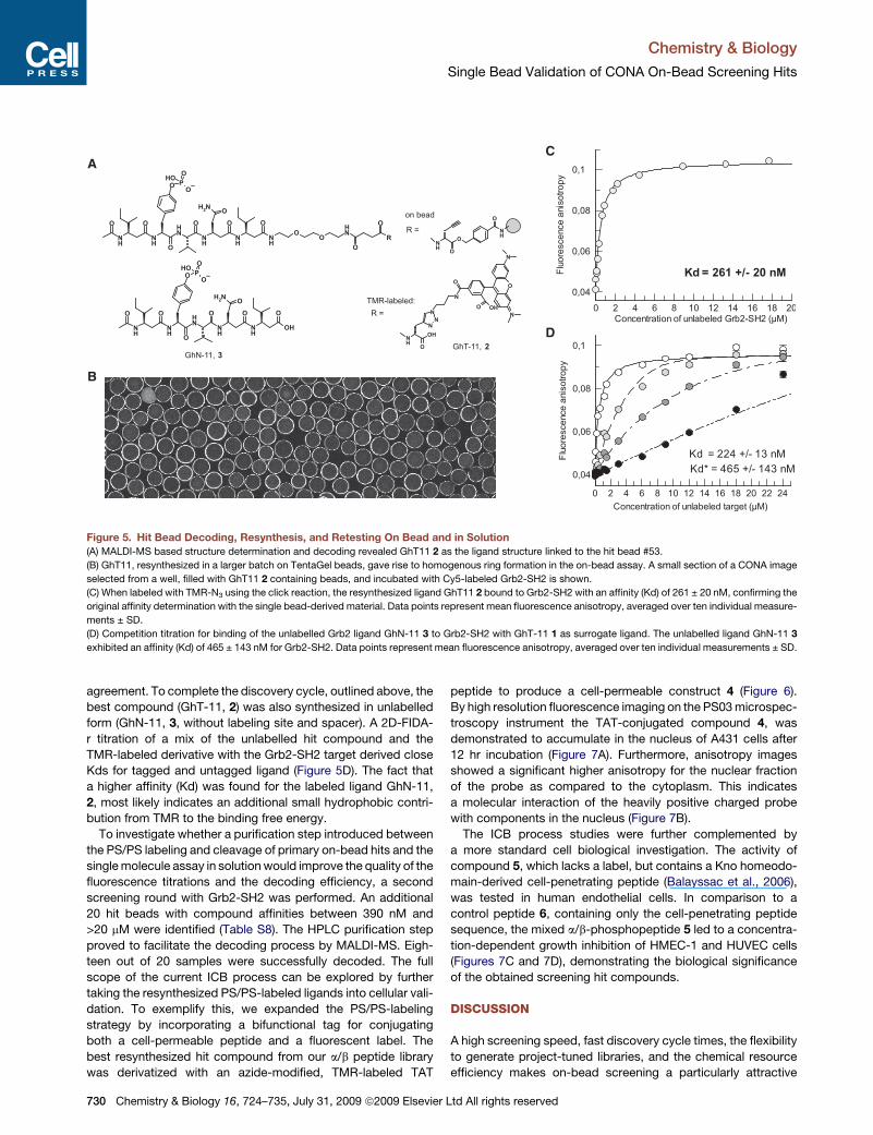
Figure 5. Hit Bead Decoding, Resynthesis, and Retesting On Bead and in Solution
(A) MALDI-MS based structure determination and decoding revealed GhT11 2 as the ligand structure linked to the hit bead #53.
(B) GhT11, resynthesized in a larger batch on TentaGel beads, gave rise to homogenous ring formation in the on-bead assay. A small section of a CONA image
selected from a well, filled with GhT11 2 containing beads, and incubated with Cy5-labeled Grb2-SH2 is shown.
(C) When labeled with TMR-N3 using the click reaction, the resynthesized ligand GhT11 2 bound to Grb2-SH2 with an affinity (Kd) of 261 ± 20 nM, confirming the
original affinity determination with the single bead-derived material. Data points represent mean fluorescence anisotropy, averaged over ten individual measure-
ments ± SD.
(D) Competition titration for binding of the unlabelled Grb2 ligand GhN-11 3 to Grb2-SH2 with GhT-11 1 as surrogate ligand. The unlabelled ligand GhN-11 3
exhibited an affinity (Kd) of 465 ± 143 nM for Grb2-SH2. Data points represent mean fluorescence anisotropy, averaged over ten individual measurements ± SD.
730 Chemistry & Biology 16, 724–735, July 31, 2009 ª2009 Elsevier Ltd All rights reserved
Chemistry & Biology
Single Bead Validation of CONA On-Bead Screening Hits
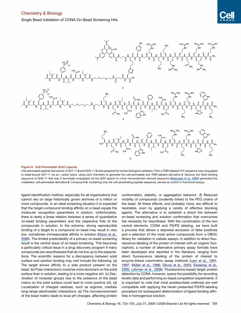
Figure 6. Cell-Permeable Grb2 Ligands
Cell-permeable peptide derivatives of GhT-11 2 and GhN-11 3 were prepared for further biological validation. First, a TMR-labeled TAT sequence was conjugated
to bead-bound GhT-11 via an 3-azido lysine, using click chemistry to generate the cell-permeable and TMR-labeled derivative 4. Second, the Grb2-binding
sequence of GhN-11 that was C-terminally conjugated via the ADO spacer to a Kno homeodomain-derived sequence (Balayssac et al., 2006) generated the
unlabelled, cell-permeable derivative 5. Compound 6, containing only the cell-penetrating peptide sequence, served as control in functional assays.
ligand identification method, especially for all organizations that
cannot rely on large historically grown archives of a million or
more compounds. In an ideal screening situation it is expected
that the target-compound binding affinity on a bead equals the
molecular recognition parameters in solution. Unfortunately,
there is rarely a linear relation between a series of quantitative
on-bead binding parameters and the respective Kds of the
compounds in solution. In the extreme, strong reproducible
binding of a target to a compound on bead may result in very
low, sometimes immeasurable affinity in solution (Dixon et al.,
2006). The limited predictability of a primary on-bead screening
result is the central issue of on-bead screening. This becomes
a particularly critical issue in a drug discovery program if many
compounds are resynthesized that do not live up to the expecta-
tions. The scientific reasons for a discrepancy between solid
surface and solution binding may well include the following. (a)
The target shows affinity to a side product present on the
bead. (b) Polar interactions could be more dominant on the solid
surface than in solution, leading to a more negative DH. (c) Des-
olvation of nonpolar groups due to the presence of the bead
matrix on the solid surface could lead to more positive DS. (d)
Localization of charged residues, such as arginine, mediate
long-range electrostatic interactions. (e) The microenvironment
of the bead matrix leads to local pH changes, affecting protein
Chemistry & Biology 1
conformation, stability, or aggregation behavior. (f) Reduced
mobility of compounds covalently linked to the PEG chains of
the bead. All these effects, and probably more, are difficult to
neutralize, even by applying a variety of effective blocking
agents. The alternative is to establish a direct link between
on-bead screening and solution confirmation that overcomes
the necessity for resynthesis. With the combination of the two
central elements, CONA and PS/PS labeling, we have built
a process that allows a stepwise exclusion of false positives
and a selection of the most active compounds from a OBOC
library for validation in cellular assays. In addition to direct fluo-
rescence labeling of the protein of interest with an organic fluo-
rophore, a number of alternative primary assay formats have
been developed and reported in the literature, ranging from
direct fluorescence labeling of the protein of interest to
enzyme-linked colorimetric assay methods (Lam et al., 1991,
1997; Muller et al., 1996; Olivos et al., 2003; Sweeney et al.,
2005; Lehman et al., 2006). Fluorescence-based target protein
detection by CONA, however, opens the possibility for recording
kinetic data and performing on-bead competition experiments. It
is important to note that most predescribed methods are well
compatible with applying the herein presented PS/PS-labeling
procedure for subsequent determination of ligand binding affin-
ities in homogenous solution.
6, 724–735, July 31, 2009 ª2009 Elsevier Ltd All rights reserved 731

Chemistry & Biology
Single Bead Validation of CONA On-Bead Screening Hits
Figure 7. Characterization of Cell-Permeable Grb2 Ligands
(A) Confocal fluorescence intensity image of A431 (human epithelial carcinoma cell line) cells treated with 5 mM of the TMR-TAT-conjugated Grb2-ligand 4 after 12
hr of incubation time, recorded on the PS03 instrument. An accumulation of ligand 4 in the nucleus is detected.
(B) Confocal fluorescence anisotropy image of the same section of A431 cells as in (A).
(C) Effect of cell-permeable Grb2 ligand 5 and control peptide 6 on HMEC-1 (human microvascular endothelial cell line-1) cells (trypan blue staining, 203 images):
20 mM peptide 5 (left top), 5 mM peptide 5 (right top), 20 mM peptide 6 (left bottom), and 5 mM peptide 6 (right bottom).
(D) Effects of peptides 5 (black dots) and 6 (gray squares) at different concentrations on proliferation of HMEC-1 (top) and HUVEC (bottom) cells. The percentage
of maximal growth response (cpm values obtained in the absence of peptides) is plotted (data points represent means from four independent experiments ± SD).
The direct solution confirmation represents a strictly quantita-
tive measure for the quality of each ligand derived from each hit
bead. The multiparameter confocal fluorescence fluctuation
spectroscopy assays used for testing on-bead hits in solution
are highly miniaturized (few microliter assay volumes) and inher-
ently sensitive (using 1 nM ligand concentration). It is often
argued that a fluorescence label on a small molecule might
impact the protein binding affinities. In our experience only small
effects of affinity reduction or enhancement of PS/PS-labeled hit
compounds are detected compared to unlabeled derivatives.
Furthermore, the 2D-FIDA anisotropy assay used for deter-
mining the ligand affinity in solution allows immediately spotting
of influences of the label on the protein binding affinity. In
experiments where the label is involved in protein binding,
pronounced effects on the molecular brightness parameter will
be observed (fluorescence quenching or brightness increase).
By analyzing the molecular brightness changes during the
binding reaction, such effects can be identified and taken into
consideration for ranking of the ligands. In addition, it is note-
worthy to point out that the PS/PS-labeling method provides
free choice of label.
732 Chemistry & Biology 16, 724–735, July 31, 2009 ª2009 Elsevier
SIGNIFICANCE
On-bead screening of large one-bead one-compound
libraries can easily result in hundreds to thousands of hit
beads. With the high compound density on commonly
used micro beads and the thermodynamic differences of
ligand-protein binding on the solid surface and in solution,
a method for the efficient identification of the best hit beads
and compounds is needed. Hence, for a full exploitation of
its potential, a bead-based screening process has to
comprise automated and quantitative methods for detecting
protein-ligand complex formation on bead and for
measuring the affinity of these complexes in solution. The
screening process reported herein combines quantitative
on-bead screening by CONA, with generic PS/PS labeling
of the chemical substance on individual hit beads. This
tagging scheme allows a direct and reliable affinity determi-
nation of primary hit compounds to their protein targets in
solution and a stepwise exclusion of optical, chemical, and
thermodynamic artifacts prior to resynthesis. Thereby, any
follow-up activities can be focused on compounds, with
Ltd All rights reserved
Chemistry & Biology
Single Bead Validation of CONA On-Bead Screening Hits
compelling target binding affinities and specificities.
Furthermore, the ICB screening process operates with
minimal chemical resources, i.e., the approximately 20–100
picomoles of compound originally contained on individual
90 mm TentaGel beads. The multiparameter confocal fluo-
rescence fluctuation spectroscopy methods used for target
binding affinity measurements in solution are highly minia-
turized (1–10 ml assay volumes) and inherently sensitive
(using 1 nM ligand concentration). Consequently, the
amount of compound derived from individual hit beads is
sufficient for hundreds of assay points opening up the possi-
bility for broad specificity testing. The fluorescent ligands
generated en route provide valuable tools for cellular valida-
tion studies and model organism imaging. Looking beyond
high throughput screening on bead, the combination of
single bead labeling and handling techniques can deliver
large fluorescently labeled small molecular libraries for
biochemical, cellular, and model organism screening.
EXPERIMENTAL PROCEDURES
For detailed procedures on chemical synthesis, single bead purification,
MALDI-MS structure decoding, cell biology, and imaging refer to the Supple-
mental Experimental Procedures.
On-Bead Screening
For on-bead screening, wells from a 96 well microtiter plate were filled with
1 mg of resin from the individual sublibraries of the phosphopeptide library.
The beads were then swollen in PBS and 0.01% Tween20 (200 ml per well,
10 mM phosphate [pH 7.4]) and sonicated for 1 min to break remaining bead
clusters. After buffer removal, the beads were treated with 150 ml of blocking
buffer at 4�C for 1 hr under constant shaking. Then, a solution of Cy5-labeled
Grb2-SH2 protein (50 ml; 20 nM in blocking buffer) was added to the samples
and incubated for 8 hr at 4�C. To generate a monolayer of beads the plate was
shortly vortexed at �600 rpm, followed by a sudden stop. This resulted in
a homogeneous sedimentation of the beads onto the well bottom. Immediately
afterwards the plate was mounted on the PS04 instrument and the confocal
scanning started.
Blocking Buffer
PBS (10 mM phosphate, 137 mM NaCl, and 3 mM KCl [pH 7.4]), 0.25% (w/v)
gelatine (dry gelatine for blocking buffer; Sigma-Aldrich), 0.01% (v/v) Tween20,
and 0.2% (w/v) BSA.
Bead Scanning, Image Evaluation, and Bead Picking
The PickoScreen04 instrument (PS04) is designed for automated scanning of
multiple wells from MTP plates. After scanning and image merging of all wells
from the screening plate was completed, image analysis was performed using
the software package Optimas (Media Cybernetics; version 6.2) to select
those wells containing hit beads.
Picking Procedure Steps
As Preparation for picking, a capillary (�2.5 cm length and 140 mm diameter)
was mounted onto the picker arm and aligned to the coordinate system of
the instrument. All tubings were then thoroughly flushed with water to remove
any air from the system.
A rescan of the well from which hits are to be picked was carried out imme-
diately before picking. The image of the rescan was then analyzed by the
BeadEval software (version 2.2; Perkin Elmer) and a bead detection per-
formed. The bead detection routine uses the Hough transformation (Ballard,
1981; Thomas et al., 1992). In the bead detection procedure, the xy coordi-
nates of all beads along with their intensity parameters are determined and
candidate beads for picking were selected.
Chemistry & Biology 1
The picker arm was consecutively positioned above the xy coordinates of
the respective hit beads from the pick list. Picked beads were deposited
into 96 well filter plates (Innovative Microplates).
PS/PS Labeling of Individual Hit Beads in 96 Well Filter Plates
Prior to labeling, the beads in the filterplates were washed with methanol using
a vacuum manifold (VWR International). After sealing the filter plates at the
bottom with a sealing film (Parafilm; 3M), each well was treated with 26 ml of
a four-component labeling solution (10 ml H2O, 10 ml tButanol, 3 ml catalyst
solution, and 3 ml dye solution) and finally sealed. The reaction was allowed
to proceed under constant agitation for at least 16 hr at room temperature.
After removal of the top and bottom sealing, the wells were drained through
the filters. The labeled beads were then washed thoroughly with methanol
and water, inspected under a microscope, and manually transferred into auto-
sampler glass vials using a micropipette (Gilson; Microman M10).
The dye solution was 2 mM methanolic solution of TMR-azide 1. The catalyst
solution was a freshly prepared mixture (1:1) of ascorbic acid (10 mg/ ml) and
copper sulfate (5 mg/ml) in water.
Cleavage of PS/PS-Labeled Compounds from Resin Beads
Labeled beads were treated with an ice-cooled solution (6 ml) of NaOH (1M)/
dioxane (1:1) for 15 min at room temperature. After neutralization with HCl
(4 ml, 1 M), the cleavage solution was evaporated under reduced pressure.
Solution Confirmation of Cleaved Compounds by Confocal
Fluorescence Fluctuation Spectroscopy
To generate stock solutions the cleaved and dried material from each hit bead
was dissolved in 20 ml of acetonitrile (20% v/v) in water. One microliter aliquots
from each sample were further diluted 1:500 in PBS (10 mM phosphate,
137 mM NaCl, and 3 mM KCl [pH 7.4]), containing 0.005% Tween20. All solu-
tion confirmation measurements were performed in a total assay volume of 7 ml
on the PS02 instrument (typical measurement time 12 3 12 s per sample)
at ambient temperature, using low volume 384 well microtiter plates (Perkin-
Elmer).
Single Point Solution Confirmation and Affinity Determination
Complex formation between the PS/PS-labeled hit compound and Grb2-SH2
was monitored by recording the fluorescence fluctuation data for each
compound in the presence and absence of Grb2-SH2 (24 mM) and by deter-
mining the fluorescence anisotropy with 2D-FIDA39. For an estimation of the
significance threshold (i.e., the anisotropy increase due to viscosity changes
at higher protein concentrations) identical measurements were performed
with free TMR-azide 1.
For affinity determination, a titration series containing 10 to 12 measurement
points of increasing Grb-SH2 protein concentration was recorded. Fluctuation
signals for individual wells were recorded in replicates of 12 3 12 s for each
titration point.
SUPPLEMENTAL DATA
Supplemental Data include Supplemental Experimental Procedures, eleven
figures, and eight tables and can be found with this article online at http://
www.cell.com/chemistry-biology/supplemental/S1074-5521(09)00209-9.
ACKNOWLEDGMENTS
The authors thank Marion Kala for editorial support and Matthew Ross for
native speaker support. The authors declare competing financial interests.
The experimental work for this manuscript was performed at Novartis Insti-
tutes for BioMedical Research.
Received: March 4, 2009
Revised: June 5, 2009
Accepted: June 11, 2009
Published: July 30, 2009
6, 724–735, July 31, 2009 ª2009 Elsevier Ltd All rights reserved 733
Chemistry & Biology
Single Bead Validation of CONA On-Bead Screening Hits
REFERENCES
Diaz-Mochon, J.J., Bialy, L., and Bradley, M. (2006). Dual colour, microarray-
based, analysis of 10,000 protease substrates. Chem. Comm. (Camb.), 14,
3984–3986.
Balayssac, S., Burlina, F., Convert, O., Bolbach, G., Chassaing, G., and
Lequin, O. (2006). Comparison of penetratin and other homeodomain-derived
cell-penetrating peptides: interaction in a membrane-mimicking environment
and cellular uptake efficiency. Biochemistry 45, 1408–1420.
Ballard, D.H. (1981). Generalizing the Hough transform to detect arbitrary
shapes. Pattern Recognit. 13, 111–122.
Blackwell, H.E., Perez, L., Stavenger, R.A., Tallarico, J.A., Cope, E.E., Foley,
M.A., and Schreiber, S.L. (2001). A one-bead, one-stock solution approach
to chemical genetics: part 1. Chem. Biol. 8, 1167–1182.
Bradner, J.E., McPherson, O.M., Mazitschek, R., Barnes-Seeman, D., Shen,
J.P., Dhaliwal, J., Stevenson, K.E., Duffner, J.L., Park, S.B., Neuberg, D.S.,
et al. (2006). A robust small-molecule microarray platform for screening cell
lysates. Chem. Biol. 13, 493–504.
Chen, S., Do, J.T., Zhang, Q., Yao, Q., Yao, S., Yan, F., Peters, E.C., Schoeler,
H.R., Schultz, P.G., and Ding, S. (2006). Self-renewal of embryonic stem cells
by a small molecule. Proc. Natl. Acad. Sci. USA 103, 17266–17271.
Chen, X., Tan, P.H., Zhang, Y., and Pei, D. (2009). On-bead screening of
combinatorial libraries: reduction of nonspecific binding by decreasing surface
ligand density. J. Comb. Chem., in press. Published online April 28, 2009.
10.1021/cc9000168.
Clemons, P.A., Koehler, A.N., Wagner, B.K., Sprigings, T.G., Spring, D.R.,
King, R.W., Schreiber, S.L., and Foley, M.A. (2001). A one-bead, one-stock
solution approach to chemical genetics: part 2. Chem. Biol. 8, 1183–1195.
Ding, H., Prodinger, W.M., and Kopecek, J. (2006). Two-step fluorescence
screening of CD21-binding peptides with one-bead one-compound library
and investigation of binding properties of N-(2-hydroxypropyl)methacrylamide
copolymer-peptide conjugates. Biomacromolecules 7, 3037–3046.
Dixon, S., Ziebart, K.T., He, Z., Jeddeloh, M., Yoo, C.L., Wang, X., Lehman, A.,
Lam, K.S., Toney, M.D., and Kurth, M.J. (2006). Aminodeoxychorismate syn-
thase inhibitors from one-bead one-compound combinatorial libraries:
‘‘staged’’ inhibitor design. J. Med. Chem. 49, 7413–7426.
Duffner, J.L., Clemons, P.A., and Koehler, A.N. (2007). A pipeline for ligand
discovery using small-molecule microarrays. Curr. Opin. Chem. Biol. 11,
74–82.
Hintersteiner, M., Buehler, C., Uhl, V., Schmied, M., Muller, J., Kottig, K., and
Auer, M. (2009). Confocal nanoscanning, bead Picking (CONA) - the
PickoScreen microscopes for automated and quantitative screening of one-
bead one compound libraries. J Comb. Chem., in press. Published online
July 15, 2009. 10.1021/cc900059q.
Kask, P., Palo, K., Fay, N., Brand, L., Mets, U., Ullmann, D., Jungmann, J.,
Pschorr, J., and Gall, K. (2000). Two-dimensional fluorescence intensity distri-
bution analysis: theory and applications. Biophys. J. 78, 1703–1713.
Kodadek, T., and Bachhawat-Sikder, K. (2006). Optimized protocols for the
isolation of specific protein-binding peptides or peptoids from combinatorial
libraries displayed on beads. Mol. Biosyst. 2, 25–35.
Kolb, H.C., and Sharpless, K.B. (2003). The growing impact of click chemistry
on drug discovery. Drug Discov. Today 8, 1128–1137.
Lam, K.S., Salmon, S.E., Hersh, E.M., Hruby, V.J., Kazmierski, W.M., and
Knapp, R.J. (1991). A new type of synthetic peptide library for identifying
ligand-binding activity. Nature 354, 82–84.
Lam, K.S., Lebl, M., and Krchnak, V. (1997). The ‘‘one-bead-one-compound’’
combinatorial library method. Chem. Rev. 97, 411–448.
Lathrop, J.T., Fijalkowska, I., and Hammond, D. (2007). The Bead blot: A
method for identifying ligand-protein and protein-protein interactions using
combinatorial libraries of peptide ligands. Anal. Biochem. 361, 65–76.
Lehman, A., Gholami, S., Hahn, M., and Lam, K.S. (2006). Image subtraction
approach to screening one-bead-one-compound combinatorial libraries with
complex protein mixtures. J. Comb. Chem. 8, 562–570.
734 Chemistry & Biology 16, 724–735, July 31, 2009 ª2009 Elsevier
Lim, H.-S., Archer, C.T., and Kodadek, T. (2007). Identification of a peptoid
inhibitor of the proteasome 19S regulatory particle. J. Am. Chem. Soc. 129,
7750–7751.
Meisner, N.C., Hintersteiner, M., Uhl, V., Weidemann, T., Schmied, M., Gstach,
H., and Auer, M. (2004). The chemical hunt for the identification of drugable
targets1. Curr. Opin. Chem. Biol. 8, 424–431.
Meisner, N.-C., Hintersteiner, M., Seifert, J.-M., Bauer, R., Benoit Roger, M.,
Widmer, A., Schindler, T., Uhl, V., Lang, M., Gstach, H., et al. (2009). Terminal
adenosyl transferase activity of posttranscriptional regulator HuR revealed by
confocal on-bead screening. J. Mol. Biol. 386, 435–450.
Meldal, M. (2002). The one-bead two-compound assay for solid phase
screening of combinatorial libraries. Biopolymers 66, 93–100.
Metzger, A., Diller, D.J., Lin, T.H., Henderson, I., and Webb, M.L. (2006).
Successful screening of large encoded combinatorial libraries leading to the
discovery of novel p38 MAP kinase inhibitors. Comb. Chem. High Throughput
Screen. 9, 351–358.
Muller, K., Gombert, F.O., Manning, U., Grossmueller, F., Graff, P., Zaegel, H.,
Zuber, J.F., Freuler, F., Tschopp, C., and Baumann, G. (1996). Rapid identifi-
cation of phosphopeptide ligands for SH2 domains. Screening of peptide
libraries by fluorescence-activated bead sorting. J. Biol. Chem. 271, 16500–
16505.
Muckenschnabel, I., Falchetto, R., Mayr, L.M., and Filipuzzi, I. (2004).
SpeedScreen: label-free liquid chromatography-mass spectrometry-based
high-throughput screening for the discovery of orphan protein ligands. Anal.
Biochem. 324, 241–249.
Olivos, H.J., Bachhawat-Sikder, K., and Kodadek, T. (2003). Quantum dots as
a visual aid for screening bead-bound combinatorial libraries. ChemBioChem
4, 1242–1245.
Paulick, M.G., Hart, K.M., Brinner, K.M., Tjandra, M., Charych, D.H., and Zuck-
ermann, R.N. (2006). Cleavable hydrophilic linker for one-bead-one-
compound sequencing of oligomer libraries by tandem mass spectrometry.
J. Comb. Chem. 8, 417–426.
Pei, D., and Wavreille, A.-S. (2007). Reverse interactomics: decoding protein-
protein interactions with combinatorial peptide libraries. Mol. Biosyst. 3,
536–541.
Peng, L., Liu, R., Marik, J., Wang, X., Takada, Y., and Lam, K.S. (2006). Combi-
natorial chemistry identifies high-affinity peptidomimetics against alpha4beta1
integrin for in vivo tumor imaging. Nat. Chem. Biol. 2, 381–389.
Rostovtsev, V.V., Green, L.G., Fokin, V.V., and Sharpless, K.B. (2002). A step-
wise huisgen cycloaddition process: copper(I)-catalyzed regioselective
‘‘ligation’’ of azides and terminal alkynes. Angew. Chem. Int. Ed. Engl. 41,
2596–2599.
Schreiber, S.L. (2005). Small molecules: the missing link in the central dogma.
Nat. Chem. Biol. 1, 64–66.
Sedlacek, R., and Chen, E. (2005). Screening for protease substrate by poly-
valent phage display. Comb. Chem. High Throughput Screen. 8, 197–203.
Song, A., Zhang, J., Lebrilla, C.B., and Lam, K.S. (2003). A novel and rapid
encoding method based on mass spectrometry for ‘‘one-bead-one-
compound’’ small molecule combinatorial libraries. J. Am. Chem. Soc. 125,
6180–6188.
Sweeney, M.C., Wavreille, A.S., Park, J., Butchar, J.P., Tridandapani, S., and
Pei, D. (2005). Decoding protein-protein interactions through combinatorial
chemistry: sequence specificity of SHP-1, SHP-2, and SHIP SH2 domains.
Biochemistry 44, 14932–14947.
Sweeney, M.C., Wang, X., Park, J., Liu, Y., and Pei, D. (2006). Determination of
the sequence specificity of XIAP BIR domains by screening a combinatorial
peptide library. Biochemistry 45, 14740–14748.
Thomas, A.D., Davies, T., and Luxmoore, A.R. (1992). The Hough transform for
locating cell nuclei. Anal. Quant. Cytol. Histol. 14, 347–353.
Tornoe, C.W., and Meldal, M. (2001). Peptidotriazoles: copper(I)-catalyzed
1,3-dipolar cycloadditions on solid-phase. Peptides: The Wave of the Future,
Proceedings of the Second International and the Seventeenth American
Peptide Symposium. San Diego, CA, June 9–14, 2001, pp. 263–264.
Ltd All rights reserved
Chemistry & Biology
Single Bead Validation of CONA On-Bead Screening Hits
Tornoe, C.W., Christensen, C., and Meldal, M. (2002). Peptidotriazoles on solid
phase: [1,2,3]-triazoles by regiospecific copper(I)-catalyzed 1,3-dipolar cyclo-
additions of terminal alkynes to azides. J. Org. Chem. 67, 3057–3064.
Udugamasooriya, D.G., Dineen, S.P., Brekken, R.A., and Kodadek, T. (2008).
A peptoid ‘‘antibody surrogate’’ that antagonizes VEGF receptor 2 activity. J.
Am. Chem. Soc. 130, 5744–5752.
Uhl, V., Gstach, H., Seifert, J.M., Jung, T., Falchetto, R., Fetsch, A., Stange, R.,
Schmied, M., Graf, C., Adam, A., et al. (2002). Confocal nanoscanning - bead
picking - AIDA technology. A methodology for drug discovery with orphan
molecular targets and combinatorial chemistry on the solid surface. Single
Molecules 3, 174–175.
Urbina, H.D., Debaene, F., Jost, B., Bole-Feysot, C., Mason, D.E., Kuzmic, P.,
Harris, J.L., and Winssinger, N. (2006). Self-assembled small-molecule micro-
arrays for protease screening and profiling. ChemBioChem 7, 1790–1797.
Vegas, A.J., Fuller, J.H., and Koehler, A.N. (2008). Small-molecule microarrays
as tools in ligand discovery. Chem. Soc. Rev. 37, 1385–1394.
Wang, X., Peng, L., Liu, R., Gill, S.S., and Lam, K.S. (2005a). Partial alloc-
deprotection approach for ladder synthesis of ‘‘one-bead one-compound’’
combinatorial libraries. J. Comb. Chem. 7, 197–209.
Wang, X., Peng, L., Liu, R., Xu, B., and Lam, K.S. (2005b). Applications of topo-
logically segregated bilayer beads in ‘one-bead one-compound’ combinato-
rial libraries. J. Pept. Res. 65, 130–138.
Chemistry & Biology 1
Wiegand, H., Wirz, B., Schweitzer, A., Camenisch Gian, P., Rodriguez Perez
Maria, I., Gross, G., Woessner, R., Voges, R., Arvidsson Per, I., Frackenpohl,
J., et al. (2002). The outstanding metabolic stability of a 14C-labeled beta-
nonapeptide in rats in vitro and in vivo pharmacokinetic studies. Biopharm.
Drug Dispos. 23, 251–262.
Winssinger, N., Pianowski, Z., and Barluenga, S. (2008). Chemical technolo-
gies: probing biology with small molecules. In Chemical and Functional
Genomic Approaches to Stem Cell Biology and Regenerative Medicine, S.
Ding, ed. (Hoboken, NJ: John Wiley & Sons, Inc.), pp. 109–144.
Xu, Y., Shi, Y., and Ding, S. (2008). A chemical approach to stem-cell biology
and regenerative medicine. Nature 453, 338–344.
Youngquist, R.S., Fuentes, G.R., Lacey, M.P., and Keough, T. (1994). Matrix-
assisted laser desorption ionization for rapid determination of the sequences
of biologically active peptides isolated from support-bound combinatorial
peptide libraries. Rapid Commun. Mass Spectrom. 8, 77–81.
Zehender, H., Le Goff, F., Lehmann, N., Filipuzzi, I., and Mayr, L.M. (2004).
SpeedScreen: the ‘‘missing link’’ between genomics and lead discovery. J.
Biomol. Screen. 9, 498–505.
Zhang, Y., Zhou, S., Wavreille, A.-S., DeWille, J., and Pei, D. (2008). Cyclic
peptidyl inhibitors of Grb2 and tensin SH2 domains identified from combinato-
rial libraries. J. Comb. Chem. 10, 247–255.
6, 724–735, July 31, 2009 ª2009 Elsevier Ltd All rights reserved 735
Copyright © 2022 FDOKUMEN