Silencing of the Tapetum-Specific Zinc Finger Gene TAZ1 Causes Premature Degeneration of Tapetum and...
16
The Plant Cell, Vol. 14, 1–15, October 2002, www.plantcell.org © 2002 American Society of Plant Biologists Silencing of the Tapetum-Specific Zinc Finger Gene TAZ1 Causes Premature Degeneration of Tapetum and Pollen Abortion in Petunia Sanjay Kapoor, Akira Kobayashi, 1 and Hiroshi Takatsuji 2 Developmental Biology Laboratory, Plant Physiology Department, National Institute of Agrobiological Sciences, Tsukuba 305-8602, Japan TAZ1 (TAPETUM DEVELOPMENT ZINC FINGER PROTEIN1; renamed from PEThy; ZPT3-2) cDNA was first isolated as an anther-specific cDNA from petunia. Here, we report a functional characterization that includes analysis of spatial and temporal expression profiles and examination of anther phenotypes in TAZ1-silenced plants. TAZ1 showed a bi- phasic expression pattern. In the premeiotic phase, TAZ1 transcripts were found to accumulate in all cell types of the anther except the tapetum and gametophytic tissues, whereas the postmeiotic phase of anther development was char- acterized by expression exclusively in the tapetum. Silencing of TAZ1 by cosuppression resulted in aberrant develop- ment and precocious degeneration of the tapetum, followed by extensive microspore abortion that started soon after their release from pollen tetrads. A few pollen grains that survived showed reduced flavonol accumulation, defects in pollen wall formation, and poor germination rates. This study demonstrates an essential role for TAZ1 in the postmei- otic phase of tapetum development. INTRODUCTION Development of the male gametophyte (pollen) occurs within the anther compartment of the stamen and requires cooperative functional interactions between gametophytic and sporophytic tissues (Raghavan, 1997a). The tapetum is the innermost of the four sporophytic layers of the anther wall that comes in direct contact with the developing game- tophyte; therefore, it has long been considered to play a crucial role in the maturation of microspores (Shivanna et al., 1997). Early studies using microscopic and cytological techniques revealed that tapetal cells contain dense cyto- plasm with abundant nuclei and other cell organelles, indi- cating the metabolically dynamic state of these cells (Stevens and Murray, 1981). The plasma membrane facing the developing microspores in these cells was shown to de- velop membrane-lined lipid bodies (orbicules), which were thought to contribute to the synthesis of the pollen wall (Steer, 1977). Additionally, a number of male-sterile mutants were found to have defects in the structure of the tapetum (Raghavan, 1997b). The indispensability of the tapetum dur- ing microspore development was demonstrated further by the specific ablation of tapetal cells by targeted expression of cytotoxin genes, which resulted in the premature abortion of microspores (Goldberg et al., 1993). The understanding of the function of the tapetum prompted the identification of tapetum-specific genes such as those coding for callase (Hird et al., 1993) and the en- zymes involved in the biosynthesis of lipids (Foster et al., 1992) and flavonoids (Koes et al., 1990; van Tunen et al., 1990). Genes coding for oleosins, which are specialized pro- teins that enclose naked oil droplets of storage triglycerides, and those for small Gly- or Cys-rich secretory proteins that expressed specifically or preferentially in the tapetum, also were characterized (Aguirre and Smith, 1993; Chen and Smith, 1993; Robert et al., 1994; Ross and Murphy, 1996; Ruiter et al., 1997). Regarding the hierarchy of gene function during flower development, the examples cited above be- long to the category of downstream genes that are required for the function of the tapetum. On the other end of this reg- ulatory hierarchy, a number of “master” regulatory genes that are involved in the establishment of the floral meristem and the determination of floral organ identity also have been characterized (reviewed by Theiben, 2001). The most ne- glected aspect of flower development, however, has been the delineation of the middle-level regulatory mechanisms 1 Current address: Potato Breeding Laboratory, Department of Up- land Agriculture Research, National Agriculture Research Center for Hokkaido Region, Shinsei, Memuro, Hokkaido 082-0071, Japan. 2 To whom correspondence should be addressed. E-mail takatsuh@ nias.affrc.go.jp; fax 81-298-38-8383. Article, publication date, and citation information can be found at www.plantcell.org/cgi/doi/10.1105/tpc.003061.
-
Upload
delhi-south -
Category
Documents
-
view
2 -
download
0
Transcript of Silencing of the Tapetum-Specific Zinc Finger Gene TAZ1 Causes Premature Degeneration of Tapetum and...

The Plant Cell, Vol. 14, 1–15, October 2002, www.plantcell.org © 2002 American Society of Plant Biologists
Silencing of the Tapetum-Specific Zinc Finger Gene
TAZ1
Causes Premature Degeneration of Tapetum and Pollen Abortion in Petunia
Sanjay Kapoor, Akira Kobayashi,
1
and Hiroshi Takatsuji
2
Developmental Biology Laboratory, Plant Physiology Department, National Institute of Agrobiological Sciences, Tsukuba 305-8602, Japan
TAZ1
(
TAPETUM DEVELOPMENT ZINC FINGER PROTEIN1
; renamed from
PEThy; ZPT3-2
) cDNA was first isolated asan anther-specific cDNA from petunia. Here, we report a functional characterization that includes analysis of spatialand temporal expression profiles and examination of anther phenotypes in
TAZ1
-silenced plants.
TAZ1
showed a bi-phasic expression pattern. In the premeiotic phase,
TAZ1
transcripts were found to accumulate in all cell types of theanther except the tapetum and gametophytic tissues, whereas the postmeiotic phase of anther development was char-acterized by expression exclusively in the tapetum. Silencing of
TAZ1
by cosuppression resulted in aberrant develop-ment and precocious degeneration of the tapetum, followed by extensive microspore abortion that started soon aftertheir release from pollen tetrads. A few pollen grains that survived showed reduced flavonol accumulation, defects inpollen wall formation, and poor germination rates. This study demonstrates an essential role for
TAZ1
in the postmei-otic phase of tapetum development.
INTRODUCTION
Development of the male gametophyte (pollen) occurswithin the anther compartment of the stamen and requirescooperative functional interactions between gametophyticand sporophytic tissues (Raghavan, 1997a). The tapetum isthe innermost of the four sporophytic layers of the antherwall that comes in direct contact with the developing game-tophyte; therefore, it has long been considered to play acrucial role in the maturation of microspores (Shivanna etal., 1997). Early studies using microscopic and cytologicaltechniques revealed that tapetal cells contain dense cyto-plasm with abundant nuclei and other cell organelles, indi-cating the metabolically dynamic state of these cells(Stevens and Murray, 1981). The plasma membrane facingthe developing microspores in these cells was shown to de-velop membrane-lined lipid bodies (orbicules), which werethought to contribute to the synthesis of the pollen wall(Steer, 1977). Additionally, a number of male-sterile mutants
were found to have defects in the structure of the tapetum(Raghavan, 1997b). The indispensability of the tapetum dur-ing microspore development was demonstrated further bythe specific ablation of tapetal cells by targeted expressionof cytotoxin genes, which resulted in the premature abortionof microspores (Goldberg et al., 1993).
The understanding of the function of the tapetumprompted the identification of tapetum-specific genes suchas those coding for callase (Hird et al., 1993) and the en-zymes involved in the biosynthesis of lipids (Foster et al.,1992) and flavonoids (Koes et al., 1990; van Tunen et al.,1990). Genes coding for oleosins, which are specialized pro-teins that enclose naked oil droplets of storage triglycerides,and those for small Gly- or Cys-rich secretory proteins thatexpressed specifically or preferentially in the tapetum, alsowere characterized (Aguirre and Smith, 1993; Chen andSmith, 1993; Robert et al., 1994; Ross and Murphy, 1996;Ruiter et al., 1997). Regarding the hierarchy of gene functionduring flower development, the examples cited above be-long to the category of downstream genes that are requiredfor the function of the tapetum. On the other end of this reg-ulatory hierarchy, a number of “master” regulatory genesthat are involved in the establishment of the floral meristemand the determination of floral organ identity also have beencharacterized (reviewed by Theiben, 2001). The most ne-glected aspect of flower development, however, has beenthe delineation of the middle-level regulatory mechanisms
1
Current address: Potato Breeding Laboratory, Department of Up-land Agriculture Research, National Agriculture Research Center forHokkaido Region, Shinsei, Memuro, Hokkaido 082-0071, Japan.
2
To whom correspondence should be addressed. E-mail [email protected]; fax 81-298-38-8383.Article, publication date, and citation information can be found atwww.plantcell.org/cgi/doi/10.1105/tpc.003061.
2 The Plant Cell
that commence after floral organ primordia have been es-tablished and direct the completion of organ differentiation(Smyth, 2001).
We previously identified a number of genes for floral or-gan–specific zinc finger transcription factors in petunia byPCR-based screening of organ-specific cDNA libraries(Takatsuji, 1999). Seven such anther-specific genes werefound to express sequentially but transiently during thecourse of anther development, suggesting that they mightform a regulatory cascade to control anther development(Kobayashi et al., 1998). Of these, the expression peakof
TAZ1
(
TAPETUM DEVELOPMENT ZINC FINGER PRO-TEIN1
; renamed from
PEThy-ZPT3-2
), which encodes aprotein with three zinc finger motifs, preceded those of sixother genes during the early stages of anther development(Kobayashi et al., 1998). Here, we report detailed expressionprofiles and the functional characterization of
TAZ1
. Thedata show a distinctive biphasic expression pattern of
TAZ1
during the early stages of anther development in petunia.Analysis of transgenic plants, in which the
TAZ1
gene wassilenced by cosuppression, demonstrated that
TAZ1
activ-ity is essential for the postmeiotic phase of tapetum devel-opment, which in turn plays an essential role in microsporematuration. Based on these data, a possible role for
TAZ1
in the transcriptional regulation associated with the devel-opment and function of the tapetum as well as the matura-tion of pollen is discussed.
RESULTS
Temporal and Spatial Expression Pattern of
TAZ1
in Sporophytic Anther Tissues
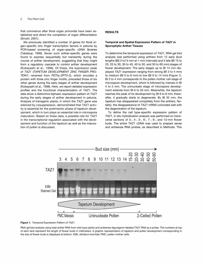
To determine the temporal expression of
TAZ1
, RNA gel blotanalysis was performed using anthers from 12 early (budlengths [Bl] 3 to14 mm at 1-mm intervals) and 5 late (Bl 15 to20, 20 to 30, 30 to 40, 40 to 50, and 50 to 60 mm) stages offlower development. The early stages up to Bl 14 mm dis-played
TAZ1
expression ranging from strong (Bl 3 to 4 mm)to medium (Bl 5 to 8 mm) to low (Bl 9 to 14 mm) (Figure 1).Bl 3 to 4 mm corresponds to the pollen mother cell stage ofmicrospore development, which is followed by meiosis in Bl4 to 5 mm. The uninucleate stage of microspore develop-ment extends from Bl 6 to 20 mm. Meanwhile, the tapetumreaches the peak of its development by Bl 6 to 8 mm; there-after, it gradually starts to degenerate. By Bl 35 mm, thetapetum has disappeared completely from the anthers. No-tably, the disappearance of
TAZ1
mRNA coincided well withthe degeneration of the tapetum.
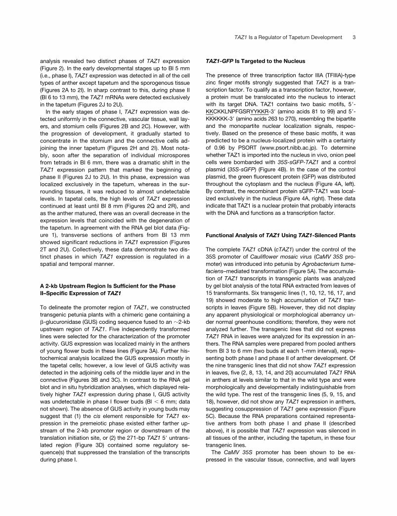
To define the cell type–specific expression pattern of
TAZ1
, in situ hybridization analysis was performed on trans-verse sections of 3-, 4-, 5-, 6-, 7-, 9-, and 13-mm flowerbuds. The entire
TAZ1
cDNA was used to prepare senseand antisense RNA probes, as described in Methods. This
Figure 1. Temporal Expression Pattern of TAZ1.
RNA gel blot analysis using total anther RNA from wild-type plants and antisense digoxigenin-labeled TAZ1 RNA as a probe. The numbers at topof each lane represent the length of flower buds in millimeters. A graphic representation of tapetum and pollen development corresponding tothe size of flower buds is displayed at bottom. EtBr, ethidium bromide; PMC, pollen mother cells.
TAZ1
Is a Regulator of Tapetum Development 3
analysis revealed two distinct phases of
TAZ1
expression(Figure 2). In the early developmental stages up to Bl 5 mm(i.e., phase I),
TAZ1
expression was detected in all of the celltypes of anther except tapetum and the sporogenous tissue(Figures 2A to 2I). In sharp contrast to this, during phase II(Bl 6 to 13 mm), the
TAZ1
mRNAs were detected exclusivelyin the tapetum (Figures 2J to 2U).
In the early stages of phase I,
TAZ1
expression was de-tected uniformly in the connective, vascular tissue, wall lay-ers, and stomium cells (Figures 2B and 2C). However, withthe progression of development, it gradually started toconcentrate in the stomium and the connective cells ad-joining the inner tapetum (Figures 2H and 2I). Most nota-bly, soon after the separation of individual microsporesfrom tetrads in Bl 6 mm, there was a dramatic shift in the
TAZ1
expression pattern that marked the beginning ofphase II (Figures 2J to 2U). In this phase, expression waslocalized exclusively in the tapetum, whereas in the sur-rounding tissues, it was reduced to almost undetectablelevels. In tapetal cells, the high levels of
TAZ1
expressioncontinued at least until Bl 8 mm (Figures 2Q and 2R), andas the anther matured, there was an overall decrease in theexpression levels that coincided with the degeneration ofthe tapetum. In agreement with the RNA gel blot data (Fig-ure 1), transverse sections of anthers from Bl 13 mmshowed significant reductions in
TAZ1
expression (Figures2T and 2U). Collectively, these data demonstrate two dis-tinct phases in which
TAZ1
expression is regulated in aspatial and temporal manner.
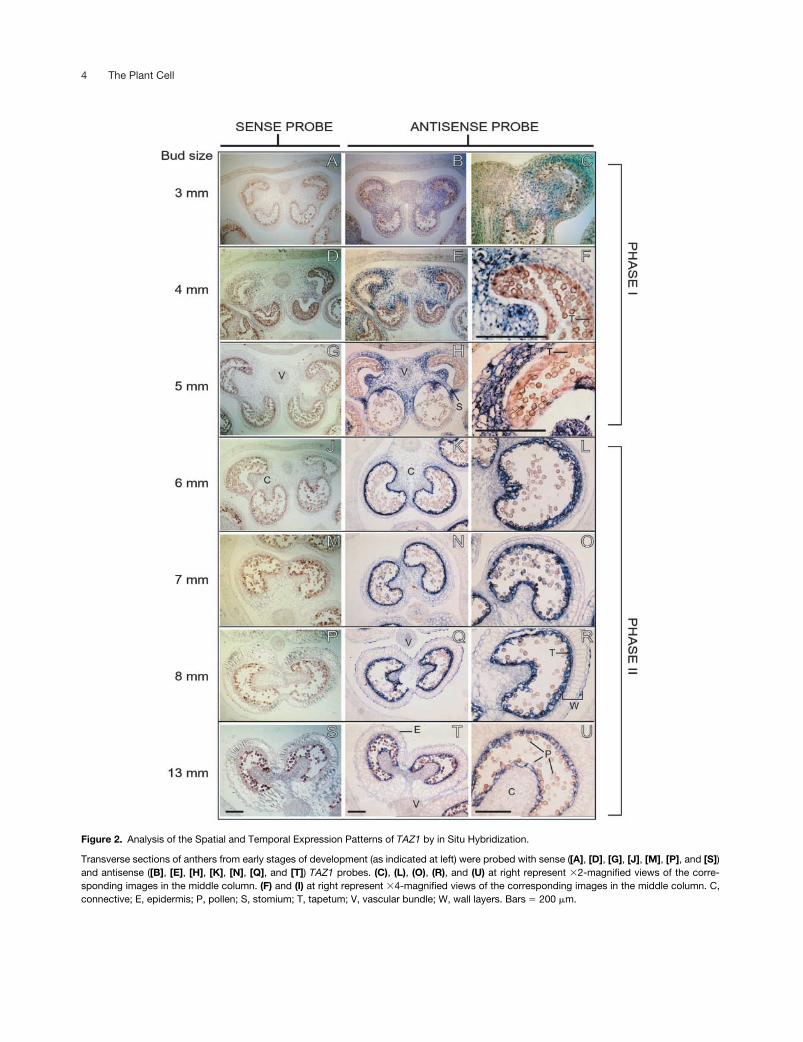
A 2-kb Upstream Region Is Sufficient for the PhaseII–Specific Expression of
TAZ1
To delineate the promoter region of
TAZ1
, we constructedtransgenic petunia plants with a chimeric gene containing a
�
-glucuronidase (GUS) coding sequence fused to an
�
2-kbupstream region of
TAZ1
. Five independently transformedlines were selected for the characterization of the promoteractivity. GUS expression was localized mainly in the anthersof young flower buds in these lines (Figure 3A). Further his-tochemical analysis localized the GUS expression mostly inthe tapetal cells; however, a low level of GUS activity wasdetected in the adjoining cells of the middle layer and in theconnective (Figures 3B and 3C). In contrast to the RNA gelblot and in situ hybridization analyses, which displayed rela-tively higher
TAZ1
expression during phase I, GUS activitywas undetectable in phase I flower buds (Bl
�
6 mm; datanot shown). The absence of GUS activity in young buds maysuggest that (1) the
cis
element responsible for
TAZ1
ex-pression in the premeiotic phase existed either farther up-stream of the 2-kb promoter region or downstream of thetranslation initiation site, or (2) the 271-bp
TAZ1
5
�
untrans-lated region (Figure 3D) contained some regulatory se-quence(s) that suppressed the translation of the transcriptsduring phase I.
TAZ1-GFP
Is Targeted to the Nucleus
The presence of three transcription factor IIIA (TFIIIA)-typezinc finger motifs strongly suggested that
TAZ1
is a tran-scription factor. To qualify as a transcription factor, however,a protein must be translocated into the nucleus to interactwith its target DNA. TAZ1 contains two basic motifs, 5
�
-KKCKKLNPFGSRYYKKR-3
�
(amino acids 81 to 99) and 5
�
-KKKKKK-3
�
(amino acids 263 to 270), resembling the bipartiteand the monopartite nuclear localization signals, respec-tively. Based on the presence of these basic motifs, it waspredicted to be a nucleus-localized protein with a certaintyof 0.96 by PSORT (www.psort.nibb.ac.jp). To determinewhether TAZ1 is imported into the nucleus in vivo, onion peelcells were bombarded with
35S-sGFP-TAZ1
and a controlplasmid (
35S-sGFP
) (Figure 4B). In the case of the controlplasmid, the green fluorescent protein (GFP) was distributedthroughout the cytoplasm and the nucleus (Figure 4A, left).By contrast, the recombinant protein sGFP-TAZ1 was local-ized exclusively in the nucleus (Figure 4A, right). These dataindicate that TAZ1 is a nuclear protein that probably interactswith the DNA and functions as a transcription factor.
Functional Analysis of
TAZ1
Using
TAZ1
-Silenced Plants
The complete
TAZ1
cDNA (
cTAZ1
) under the control of the35S promoter of
Cauliflower mosaic virus
(
CaMV
35S
pro-moter) was introduced into petunia by
Agrobacterium
tume-faciens–
mediated transformation (Figure 5A). The accumula-tion of
TAZ1
transcripts in transgenic plants was analyzedby gel blot analysis of the total RNA extracted from leaves of15 transformants. Six transgenic lines (1, 10, 12, 16, 17, and19) showed moderate to high accumulation of
TAZ1
tran-scripts in leaves (Figure 5B). However, they did not displayany apparent physiological or morphological aberrancy un-der normal greenhouse conditions; therefore, they were notanalyzed further. The transgenic lines that did not express
TAZ1
RNA in leaves were analyzed for its expression in an-thers. The RNA samples were prepared from pooled anthersfrom Bl 3 to 6 mm (two buds at each 1-mm interval), repre-senting both phase I and phase II of anther development. Ofthe nine transgenic lines that did not show
TAZ1
expressionin leaves, five (2, 8, 13, 14, and 20) accumulated
TAZ1
RNAin anthers at levels similar to that in the wild type and weremorphologically and developmentally indistinguishable fromthe wild type. The rest of the transgenic lines (5, 9, 15, and18), however, did not show any
TAZ1
expression in anthers,suggesting cosuppression of
TAZ1
gene expression (Figure5C). Because the RNA preparations contained representa-tive anthers from both phase I and phase II (describedabove), it is possible that
TAZ1
expression was silenced inall tissues of the anther, including the tapetum, in these fourtransgenic lines.
The
CaMV
35S
promoter has been shown to be ex-pressed in the vascular tissue, connective, and wall layers
4 The Plant Cell
Figure 2. Analysis of the Spatial and Temporal Expression Patterns of TAZ1 by in Situ Hybridization.
Transverse sections of anthers from early stages of development (as indicated at left) were probed with sense ([A], [D], [G], [J], [M], [P], and [S])and antisense ([B], [E], [H], [K], [N], [Q], and [T]) TAZ1 probes. (C), (L), (O), (R), and (U) at right represent �2-magnified views of the corre-sponding images in the middle column. (F) and (I) at right represent �4-magnified views of the corresponding images in the middle column. C,connective; E, epidermis; P, pollen; S, stomium; T, tapetum; V, vascular bundle; W, wall layers. Bars � 200 �m.
TAZ1
Is a Regulator of Tapetum Development 5
but not in the tapetum (Plegt and Bino, 1989; van der Meeret al., 1992). Therefore, it is unlikely that
TAZ1
expressionunder the control of the
CaMV
35S
promoter could havetriggered cosuppression directly in the tapetal cells. Be-cause the silencing signal is known to propagate throughthe plant (Vaucheret et al., 1998), one can postulate that a
TAZ1
silencing signal translocated from the site of its initia-tion, presumably from the cells of the connective and/oranther wall, to the adjoining cells of the tapetum. In theselines,
TAZ1
transgene expression was not detected even inleaves.
To verify the specificity of
TAZ1
silencing, we analyzedthe expression of another zinc finger–encoding gene,
ZPT2-5
,in
TAZ1
-silenced lines (Figure 5D). Of the seven anther-spe-cific zinc finger genes described previously, the expressionprofile of only
ZPT2-5
overlapped with that of
TAZ1
,whereas others were expressed later during anther develop-ment (Kobayashi et al., 1998). Furthermore, the conservedzinc finger–encoding regions in
TAZ1 and ZPT2-5 displayedthe highest level (�68%) of sequence similarity among all ofthe anther-specific zinc finger genes. The level of ZPT2-5
transcripts in TAZ1-silenced lines was the same as that inthe wild type, confirming that the silencing was specific toTAZ1. These plants had extremely low pollen counts at thetime of dehiscence. Apparently, however, there was no effecton female fertility, because cross-pollination of TAZ1-silencedflowers by wild-type pollen yielded normal seed set.
Development of the Tapetum in TAZ1-Silenced Plants
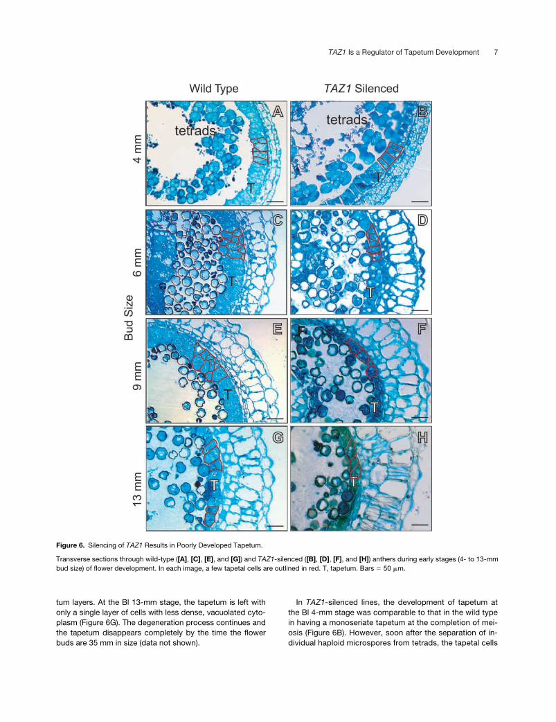
Transverse sections of anthers from wild-type and TAZ1-silenced plants were analyzed for the effect of TAZ1 silenc-ing on various cell types of the anther (Figure 6). Early stagesof anther development (phase I) showed no noticeable ef-fect on the development of the wall layers, connective, vas-cular tissue, and stomium cells. In wild-type plants, tapetumin the anthers at Bl 4 mm consists of a single layer of colum-nar cells (Figure 6A). Thereafter, the tapetum undergoes aphase of active cell proliferation to become multilayered bythe time individual microspores are released from pollen tet-rads (Bl 6 mm; Figure 6C). At this stage, the tapetum cells
Figure 3. TAZ1 Promoter-Driven GUS Expression in Transgenic Plants.
(A) to (C) GUS staining of a 25-mm flower bud (A) and transverse sections through the anther ([B] and [C]) of a TAZ1::GUS transgenic plant. P,pollen; T, tapetum. Bar in (A) � 5 mm; bars in (B) and (C) � 100 �m.(D) Scheme of a TAZ1::GUS construct. The transcription initiation site is marked with a bent arrow. The translation initiation and termination sitesare shown as ATG and TAG, respectively, and the TAZ1 5� untranslated region is shown with a dotted pattern.

6 The Plant Cell
are characterized by a dense cytoplasm and a few smallvacuoles. By Bl 9 mm, the tapetum is reduced to two layers,but the size of individual tapetal cells increases significantly(Figure 6E). The gain in cell size continues until the Bl 13-mm stage, concomitant with the degeneration of inner tape-
Figure 4. TAZ1 Is a Nucleus-Localized Protein.
(A) DNA constructs encoding sGFP (left) and the chimeric sGFP-TAZ1 (right) proteins were bombarded into onion peel cells, and thesubcellular localization of fusion proteins was observed by GFP vi-sualization with a confocal microscope.(B) Scheme of the 35S::sGFPcs::TAZ1 construct.
Figure 5. Analysis of TAZ1 Transcripts in CaMV35S::TAZ1 Trans-genic Petunia Plants.
(A) The 35S::TAZ1 construct. The 1890-bp cTAZ1 was inserted be-tween the CaMV 35S promoter (800 bp) and the nopaline synthaseterminator (Tnos). The translation initiation site of TAZ1 is markedas ATG.(B) RNA gel blot analysis using total leaf RNA and antisense digoxi-genin-labeled TAZ1 RNA as a probe. The numbers at top of eachlane represent individual transgenic lines, and the last two lanescontained total RNA from leaves (L) and anthers (A) of wild-typeplants.(C) RNA gel blot analysis using total anther RNA and antisensedigoxigenin-labeled TAZ1 RNA as a probe of those transgenic linesthat did not show TAZ1 expression in leaves (B).(D) Specific silencing of TAZ1. Total RNA samples from the anthersof wild-type and transgenic plants (lanes 5, 9, 15, and 18) wereprobed with antisense digoxigenin-labeled TAZ1 (top) and ZPT2-5(middle) RNA as probes.The bottom gels in (B) to (D) show ethidium bromide–stained RNAgels photographed before blotting. The 26S RNA (top bands) in (B)is eclipsed by comigrating bromphenol blue. WT, wild type.
TAZ1 Is a Regulator of Tapetum Development 7
tum layers. At the Bl 13-mm stage, the tapetum is left withonly a single layer of cells with less dense, vacuolated cyto-plasm (Figure 6G). The degeneration process continues andthe tapetum disappears completely by the time the flowerbuds are 35 mm in size (data not shown).
In TAZ1-silenced lines, the development of tapetum atthe Bl 4-mm stage was comparable to that in the wild typein having a monoseriate tapetum at the completion of mei-osis (Figure 6B). However, soon after the separation of in-dividual haploid microspores from tetrads, the tapetal cells
Figure 6. Silencing of TAZ1 Results in Poorly Developed Tapetum.
Transverse sections through wild-type ([A], [C], [E], and [G]) and TAZ1-silenced ([B], [D], [F], and [H]) anthers during early stages (4- to 13-mmbud size) of flower development. In each image, a few tapetal cells are outlined in red. T, tapetum. Bars � 50 �m.

8 The Plant Cell
started to degenerate. Noticeably, at the Bl 6-mm stage,the tapetum in TAZ1-silenced lines consisted of a singlelayer of vacuolated cells, instead of the multilayered tape-tum that was observed in wild-type anthers at this stage.The extent of tapetum degeneration at this stage in TAZ1-silenced anthers was comparable to that of the Bl 13-mmstage in wild-type plants (Figures 6D and 6G). The degen-eration of tapetal cells continued during the Bl 9-mmstage, and most of the cells had collapsed by the Bl 13-mmstage (Figures 6F and 6H).
Effects of TAZ1 Silencing on Pollen Development
Premature Pollen Abortion
The TAZ1-silenced plants had drastically reduced numbersof pollen (�1000/anther) compared with wild-type plants(�30,000/anther) (Figure 7). Moreover, the surviving pollengrains in these lines were found to be fragile, because mostof them burst within 10 min of incubation in 10% glycerolsolution (data not shown). In this solution, wild-type pollengrains remained intact for �24 h.
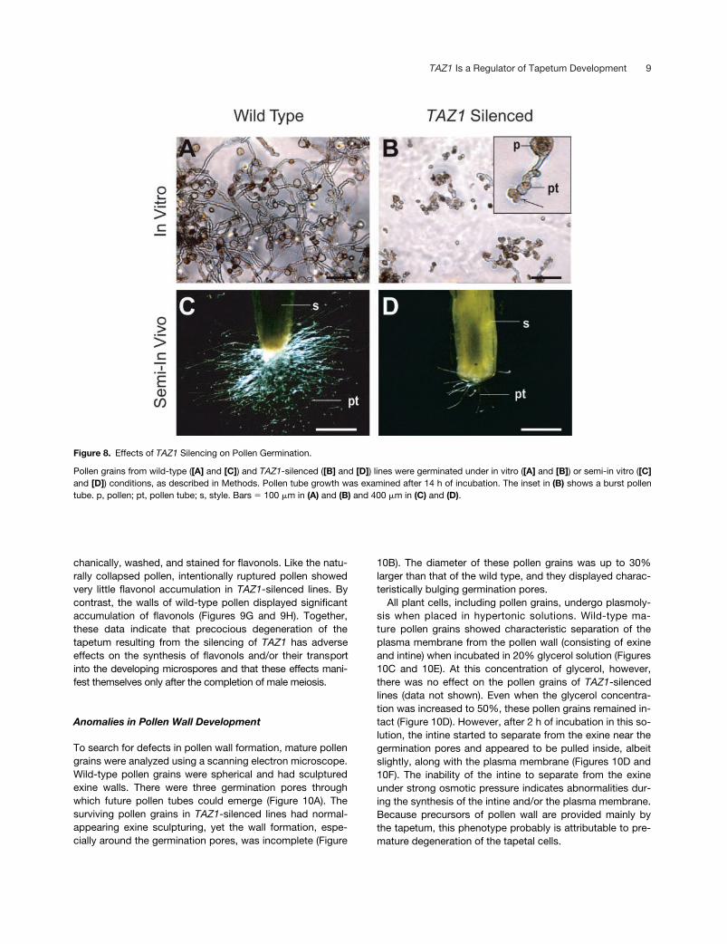
Poor Viability
To examine the viability of the surviving pollen in TAZ1-silencedlines, they were germinated under in vitro and semi-in vivoconditions, as described in Methods. As shown in Figure 8,wild-type pollen demonstrated nearly 100% germination un-der both conditions. Most of the pollen tubes grew �400�m under in vitro conditions and rarely showed bursting(Figure 6A). Under semi-in vivo conditions, the pollen tubesgrew longer than 1 cm in 14 h (Figure 8C). By contrast, pol-len grains from TAZ1-silenced lines rarely germinated. Inthe small fraction that germinated, the pollen tubes did notgrow �80 �m in length (Figure 8B). Furthermore, most ofthese pollen tubes displayed burst tips (Figure 8B, inset).Under semi-in vivo germination conditions, the pollengrains from TAZ1-silenced lines showed poor germination,because few pollen tubes were visible at the cut end of thestyle (Figure 8D).
Reduced Flavonol Accumulation
The flavonols are synthesized in tapetal cells and are re-leased subsequently into the anther locule to be taken up bythe developing microspores (Mo et al., 1992; Ylstra et al.,1994). To determine whether precocious degeneration ofthe tapetum had any effect on the supply of flavonols to thedeveloping microspores, both young and mature pollen grainsfrom wild-type and TAZ1-silenced plants were stained forflavonols. In microspore tetrads (Figures 9A and 9B), therewas no apparent difference in the accumulation of flavonolsbetween the wild-type and TAZ1-silenced lines (Figures 9Aand 9B). However, mature pollen grains in TAZ1-silencedlines displayed markedly reduced (�30-fold less) fluores-cence of flavonols compared with that in the wild type (Fig-ures 9C to 9F). Furthermore, the collapsed pollen grains inTAZ1-silenced lines fluoresced blue under UV light, sug-gesting that the walls of these microspores did not accumu-late any flavonols. To determine whether flavonols generallywere absent in the pollen wall or that this absence resultedfrom the silencing of TAZ1 expression, intact pollen grainsfrom wild-type and TAZ1-silenced lines were ruptured me-
Figure 7. Pollen Degeneration as a Result of TAZ1 Silencing.
(A) and (B) Longitudinal sections of mature anthers from 65-mmbuds of wild-type and TAZ1-silenced plants.(C) and (D) Enlarged views of (A) and (B), respectively. Bars �
100 �m.(E) Number of intact pollen grains in wild-type and TAZ1-silencedplants. The numbers at top of each bar represent the average intactpollen count of six individual anthers from three different flowerbuds. Error bars represent standard deviations.
TAZ1 Is a Regulator of Tapetum Development 9
chanically, washed, and stained for flavonols. Like the natu-rally collapsed pollen, intentionally ruptured pollen showedvery little flavonol accumulation in TAZ1-silenced lines. Bycontrast, the walls of wild-type pollen displayed significantaccumulation of flavonols (Figures 9G and 9H). Together,these data indicate that precocious degeneration of thetapetum resulting from the silencing of TAZ1 has adverseeffects on the synthesis of flavonols and/or their transportinto the developing microspores and that these effects mani-fest themselves only after the completion of male meiosis.
Anomalies in Pollen Wall Development
To search for defects in pollen wall formation, mature pollengrains were analyzed using a scanning electron microscope.Wild-type pollen grains were spherical and had sculpturedexine walls. There were three germination pores throughwhich future pollen tubes could emerge (Figure 10A). Thesurviving pollen grains in TAZ1-silenced lines had normal-appearing exine sculpturing, yet the wall formation, espe-cially around the germination pores, was incomplete (Figure
10B). The diameter of these pollen grains was up to 30%larger than that of the wild type, and they displayed charac-teristically bulging germination pores.
All plant cells, including pollen grains, undergo plasmoly-sis when placed in hypertonic solutions. Wild-type ma-ture pollen grains showed characteristic separation of theplasma membrane from the pollen wall (consisting of exineand intine) when incubated in 20% glycerol solution (Figures10C and 10E). At this concentration of glycerol, however,there was no effect on the pollen grains of TAZ1-silencedlines (data not shown). Even when the glycerol concentra-tion was increased to 50%, these pollen grains remained in-tact (Figure 10D). However, after 2 h of incubation in this so-lution, the intine started to separate from the exine near thegermination pores and appeared to be pulled inside, albeitslightly, along with the plasma membrane (Figures 10D and10F). The inability of the intine to separate from the exineunder strong osmotic pressure indicates abnormalities dur-ing the synthesis of the intine and/or the plasma membrane.Because precursors of pollen wall are provided mainly bythe tapetum, this phenotype probably is attributable to pre-mature degeneration of the tapetal cells.
Figure 8. Effects of TAZ1 Silencing on Pollen Germination.
Pollen grains from wild-type ([A] and [C]) and TAZ1-silenced ([B] and [D]) lines were germinated under in vitro ([A] and [B]) or semi-in vitro ([C]and [D]) conditions, as described in Methods. Pollen tube growth was examined after 14 h of incubation. The inset in (B) shows a burst pollentube. p, pollen; pt, pollen tube; s, style. Bars � 100 �m in (A) and (B) and 400 �m in (C) and (D).

10 The Plant Cell
DISCUSSION
TAZ1 belongs to the 5-enolpyruvylshikimate-3-phosphatesynthase promoter binding factor family of TFIIIA-type zincfinger transcription factors, in which two Cys and two Hisresidues tetrahedrally coordinate a zinc atom to form acompact structure that interacts with the major groove of
DNA in a sequence-specific manner (Takatsuji, 1996). The5-enolpyruvylshikimate-3-phosphate synthase promoterbinding factor family differs from the animal TFIIIA-type zincfinger protein in having a conserved QALGGH motif in theputative DNA-contacting domain and in the presence oflong spacers of varied lengths between zinc fingers (Takatsuji,1999). TAZ1 codes for a 444–amino acid polypeptide thatcontains three zinc finger domains. Other members of thisclass, such as ZPT2-1 and ZPT2-2, have been shown to in-teract with two tandem but separated core DNA sequencesin a manner that is dependent on the length of the spacerbetween the two core sites (Takatsuji and Matsumoto,1996). Existing homologies between these two zinc fingerproteins and TAZ1 suggest that TAZ1 is a DNA binding pro-tein. This notion is supported further by the subcellular lo-calization of TAZ1 in the nucleus.
Figure 9. TAZ1 Silencing Results in Flavonol-Deficient Pollen.
Pollen grains from young (6-mm) and mature (65-mm) flower buds ofwild-type (left) and TAZ1-silenced (right) plants were stained for fla-vonols and observed under UV light.(A) and (B) Pollen tetrads from 6-mm buds.(C) and (D) Mature pollen from 65-mm buds.(E) and (F) Single intact pollen.(G) and (H) Pollen wall remains after mechanical rupturing.Bars � 10 �m.
Figure 10. Characterization of the Pollen Wall.
(A) and (B) Scanning electron micrographs of pollen from maturebuds (Bl 65 mm) of wild-type and TAZ1-silenced plants. The place ofanomaly in pollen wall formation accompanying a bulging germina-tion pore is marked with an arrow in (B). Bars � 10 �m.(C) Pollen grains from wild-type anthers in a 20% glycerol solution.(D) Pollen grains from TAZ1 cosuppression lines in 50% glycerol.(E) and (F) Magnified views of (C) and (D), respectively.ex, exine; in, intine; pm, plasma membrane.
TAZ1 Is a Regulator of Tapetum Development 11
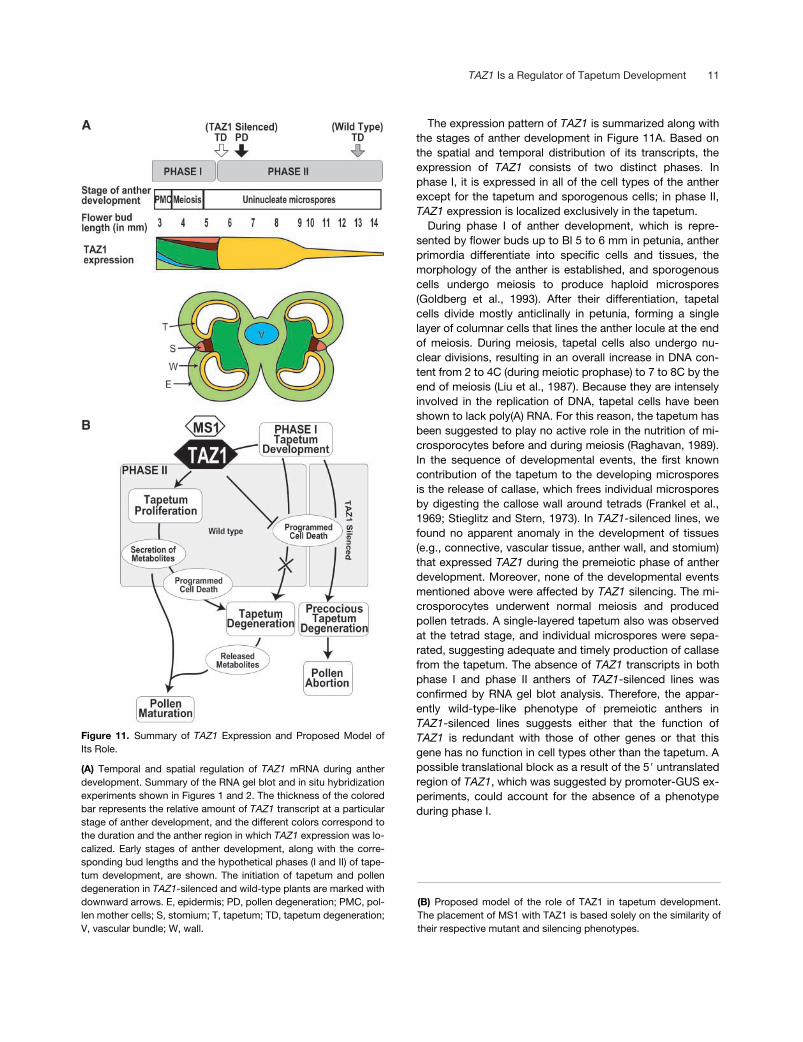
The expression pattern of TAZ1 is summarized along withthe stages of anther development in Figure 11A. Based onthe spatial and temporal distribution of its transcripts, theexpression of TAZ1 consists of two distinct phases. Inphase I, it is expressed in all of the cell types of the antherexcept for the tapetum and sporogenous cells; in phase II,TAZ1 expression is localized exclusively in the tapetum.
During phase I of anther development, which is repre-sented by flower buds up to Bl 5 to 6 mm in petunia, antherprimordia differentiate into specific cells and tissues, themorphology of the anther is established, and sporogenouscells undergo meiosis to produce haploid microspores(Goldberg et al., 1993). After their differentiation, tapetalcells divide mostly anticlinally in petunia, forming a singlelayer of columnar cells that lines the anther locule at the endof meiosis. During meiosis, tapetal cells also undergo nu-clear divisions, resulting in an overall increase in DNA con-tent from 2 to 4C (during meiotic prophase) to 7 to 8C by theend of meiosis (Liu et al., 1987). Because they are intenselyinvolved in the replication of DNA, tapetal cells have beenshown to lack poly(A) RNA. For this reason, the tapetum hasbeen suggested to play no active role in the nutrition of mi-crosporocytes before and during meiosis (Raghavan, 1989).In the sequence of developmental events, the first knowncontribution of the tapetum to the developing microsporesis the release of callase, which frees individual microsporesby digesting the callose wall around tetrads (Frankel et al.,1969; Stieglitz and Stern, 1973). In TAZ1-silenced lines, wefound no apparent anomaly in the development of tissues(e.g., connective, vascular tissue, anther wall, and stomium)that expressed TAZ1 during the premeiotic phase of antherdevelopment. Moreover, none of the developmental eventsmentioned above were affected by TAZ1 silencing. The mi-crosporocytes underwent normal meiosis and producedpollen tetrads. A single-layered tapetum also was observedat the tetrad stage, and individual microspores were sepa-rated, suggesting adequate and timely production of callasefrom the tapetum. The absence of TAZ1 transcripts in bothphase I and phase II anthers of TAZ1-silenced lines wasconfirmed by RNA gel blot analysis. Therefore, the appar-ently wild-type-like phenotype of premeiotic anthers inTAZ1-silenced lines suggests either that the function ofTAZ1 is redundant with those of other genes or that thisgene has no function in cell types other than the tapetum. Apossible translational block as a result of the 5� untranslatedregion of TAZ1, which was suggested by promoter-GUS ex-periments, could account for the absence of a phenotypeduring phase I.
Figure 11. Summary of TAZ1 Expression and Proposed Model ofIts Role.
(A) Temporal and spatial regulation of TAZ1 mRNA during antherdevelopment. Summary of the RNA gel blot and in situ hybridizationexperiments shown in Figures 1 and 2. The thickness of the coloredbar represents the relative amount of TAZ1 transcript at a particularstage of anther development, and the different colors correspond tothe duration and the anther region in which TAZ1 expression was lo-calized. Early stages of anther development, along with the corre-sponding bud lengths and the hypothetical phases (I and II) of tape-tum development, are shown. The initiation of tapetum and pollendegeneration in TAZ1-silenced and wild-type plants are marked withdownward arrows. E, epidermis; PD, pollen degeneration; PMC, pol-len mother cells; S, stomium; T, tapetum; TD, tapetum degeneration;V, vascular bundle; W, wall.
(B) Proposed model of the role of TAZ1 in tapetum development.The placement of MS1 with TAZ1 is based solely on the similarity oftheir respective mutant and silencing phenotypes.
12 The Plant Cell
From the time of completion of meiosis in the mi-crosporocytes to the stage at which the tapetum begins todisintegrate, tapetal cells are characterized by the highestrates of transcriptional activity of all of the cell types in theanther (Raghavan, 1981, 1989). During this phase (phase II),TAZ1 was expressed exclusively in the tapetum. The begin-ning of phase II also was characterized by the rapid multipli-cation of tapetal cells. As a result of both periclinal and anti-clinal divisions, the number of tapetal layers increased fromone (monoseriate) at the tetrad stage to at least three (triser-iate) by the Bl 6-mm stage. After Bl 6 mm and at least untilBl 13 mm, the size of individual tapetal cells increasednearly twofold to fivefold; however, because of the gradualdegeneration of the inner layers, the tapetum was left withonly a single layer of cells by the Bl 13-mm stage. By con-trast, the phase of tapetum proliferation was completely ab-sent in TAZ1-silenced lines. Soon after the release of indi-vidual microspores from tetrads, tapetal cells began toshrink and collapse, and by the Bl 9-mm stage, the tapetumwas represented by a single layer of darkly stained col-lapsed cells in the anthers of TAZ1-silenced lines.
The importance of the tapetum in pollen development hasbeen demonstrated using the “genetic laser” technique(Koltunow et al., 1990; Mariani et al., 1990). In these experi-ments, selective destruction of the tapetum caused by theexpression of cytotoxin genes under the control of a tape-tum-specific TA-29 promoter resulted in the complete de-generation of pollen grains in tobacco. Recently, mutationin a tapetum-specific Arabidopsis gene, MALE STERILE1(MS1), was demonstrated to cause precocious degenera-tion of the tapetum and complete male sterility (Wilson et al.,2001). MS1 is a plant homeodomain protein finger proteinthat lacks any apparent similarity to TAZ1. The reported ex-pression pattern of the MS1 gene was similar to that ofTAZ1 during phase II. As with TAZ1, the accumulation ofMS1 transcripts begins only after male meiosis is completeand individual microspores have separated from the tetrads.Furthermore, both TAZ1 and MS1 code for putative tran-scription factors. The similarity in the expression pattern andthe phenotypes resulting from the lack of their respectiveexpression suggest that both TAZ1 and MS1 could serve ascomponents of the regulatory mechanism that controls thepostmeiotic phase of tapetum development.
Based on the data presented here, we propose a hypoth-esis for the possible role of TAZ1 (Figure 11B). According tothis model, both constructive and degenerative forces actsimultaneously during phase II of tapetum development.The degenerative forces are governed by the programmedcell death mechanism, whereas TAZ1, along with MS1,forms an essential component of the constructive forcesthat cause tapetal cells to proliferate, allowing them to playtheir role in sustaining pollen development. In the absenceof TAZ1 (in silenced lines), however, the programmed celldeath mechanism proceeds unchecked and causes preco-cious degeneration of the tapetum. The absence of the cellproliferation and synthesis phase in the TAZ1-minus devel-
opment deprives the developing microspores of nutrients,resulting in their premature abortion.
METHODS
Cloning of the TAZ1 Gene and Its Promoter
The TAZ1 gene was isolated by screening a genomic DNA library ofPetunia hybrida var Mitchell in EMBL3 vector. Subsequently, a 2.2-kbEcoRI fragment containing a 1991-bp promoter region upstream ofthe ATG initiation codon and 209 bp of the coding region was sub-cloned in pBluescript SK (Stratagene, La Jolla, CA) and se-quenced. The sequence was submitted to DDBJ.
Plasmid Construction
TAZ1::GUS
The TAZ1::GUS reporter gene was constructed by fusing a 2.0-kbupstream fragment (from the 2.2-kb EcoRI fragment mentionedabove) of the TAZ1 gene to the �-glucuronidase (GUS) coding se-quence. A 20-base oligonucleotide (5�-GATCTATATGTCGACATA-TA-3�; the SalI site is underlined) was self-hybridized and introducedas a linker into the BglI site that was located 13 bp downstream ofthe TAZ1 translation initiation codon. Thereafter, the 2012-bp HindIII-SalI fragment (HindIII is of pBluescript SK; SalI is a restriction siteintroduced as part of the linker) was spliced into the same sites ofpUCAP (van Engelen et al., 1995), placing it 5� to the GUS gene thatwas cloned already in SmaI and SacI sites of this vector. This syn-thetic gene containing TAZ1(promoter)::GUS::Tnos was excisedfrom pUCAP using AscI and PacI and was inserted into pBINPLUS(van Engelen et al., 1995) using the same set of enzymes.
35S::TAZ1
A SalI-NotI fragment containing the complete TAZ1 cDNA (cTAZ1)sequence (Kobayashi et al., 1998) was excised from pSPORT vectorusing KpnI and SacI. This fragment was inserted between the 35Spromoter of Cauliflower mosaic virus (CaMV35S) and the nopalinesynthase (Nos) terminator, which were cloned already in the SalI andEcoRI sites, respectively, of pUCAP (van Engelen et al., 1995). Thissynthetic gene containing CaMV35S::cTAZ1::Tnos was excised frompUCAP using AscI and PacI and was inserted into pBINPLUS (vanEngelen et al., 1995) using the same set of enzymes.
35S::GFP::TAZ1
The complete TAZ1 coding sequence was amplified by PCR usingtwo oligonucleotide primers, 5�-ACTAGGGCCCATGGTTGATTATCA-AGATCTTCAAGTTGGG-3� and 5�-ACTAGGGCCCTTAAATTGGAA-AAAATGTAAAATACTGATGATCACGG-3�. The underlined regionsdenote XmaI sites that were added to both primers to facilitate thecloning of amplified DNA. After digestion with XmaI, the coding re-gion of TAZ1 was ligated to the XmaI-linearized psGFPcs vector
TAZ1 Is a Regulator of Tapetum Development 13
(Jiang et al., 2001), placing it between the green fluorescent protein(GFP) coding sequence and the Nos terminator.
Plant Transformation
The TAZ1::GUS and 35S::TAZ1::NosT chimeric genes were intro-duced into petunia plants by Agrobacterium tumefaciens (GV3101)–mediated transformation (Jorgensen et al., 1996). After regenerationon selective medium, transformed petunia lines were checked for thepresence of the transgene by PCR of the neomycin phosphotrans-ferase II sequence and were transferred to a greenhouse.
Transient expression of the 35S::GFP::TAZ1 gene in onion peelcells was performed as described by Jiang et al. (2001). The fluores-cence of sGFP fusion proteins was observed with a confocal micro-scope (Bio-Rad Laboratories, Hercules, CA), and the resulting digitalmicrographs were assembled using Photoshop software (AdobeSystems, San Jose, CA).
RNA Gel Blot Analysis
Total cellular RNA from petunia leaves and anthers was isolated us-ing the procedure developed by Logemann et al. (1987) and stored at70�C in 95% ethanol. Aliquots (10 �g) of RNA were separated on1.2% agarose gels containing 0.4 M formaldehyde and transferred toGene Screen membranes (DuPont–New England Nuclear Life Sci-ence Products, Boston, MA). A digoxigenin-labeled antisense RNAcorresponding to the 1172-bp 3� terminal region (3� of the EcoRI siteat position 717) of cTAZ1, prepared according to the manufacturer’sinstructions (Boehringer Mannheim), was used as a probe. Hybrid-ization of blots and detection of chemiluminescence also were per-formed according to the Boehringer Mannheim protocol.
In Situ Hybridization
In situ hybridization was essentially carried out by using Ribomap kitand Discovery automatic staining module (Ventana Medical Systems,Tucson, Arizona) according to manufacturer’s instructions. The ex-cised anthers were fixed in formaldehyde 10%, Acetic acid 5%, andEthanol 50% for four days at 4�C. The cTAZ1 was digested with EcoRIand SpeI. The 491-, 243-, 263-, 167-, 154-, 434-, and 172-bp frag-ments generated were cloned in pBluescript SK. Both antisense andsense probes from all of the cTAZ1 fragments were synthesized usingthe SP6/T7 digoxigenin RNA labeling kit (Roche Diagnostics, India-napolis, IN), according to the manufacturer’s instructions. The senseand antisense digoxigenin-labeled RNAs were pooled and used forhybridization with the fixed anther sections at 50�C overnight in 0.3 MNaCl, 10 mM Tris-HCl, pH 6.8, 1 mM EDTA, 60 mM DTT, 1 � Den-hardt’s solution (0.02% Ficoll, 0.02% polyvinylpyrrolidone, and 0.02%BSA), 166 �g/mL tRNA (type XXI; Sigma), 500 mg/mL poly(A), 10%dextran sulfate, and 50% formamide. After hybridization, sectionswere washed in 4 � SSC (three times for 15 min) (1� SSC is 0.15 MNaCl and 0.015 M sodium citrate) at 60�C for 6 in Ribohyb hybridiza-tion solution (Ventana Medical Systems, Tucson, Arizona). After hy-bridization, sections were washed in 0.1 X SSC (thrice for 6 min) at70�C. Detection of hybrids was performed using the digoxigenin nu-cleic acid detection kit (Boehringer Mannheim), according to the man-ufacturer’s instructions. Detection of hybrids with nitroblue tetrazo-lium salt and 5-bromo-4-chloro-3-indolyl phosphate 4-toluidinium saltwas performed at 20�C for 16 h in the dark. Sections were dehydrated
through an ethanol series (30, 50, 70, 90, and 100% ethanol for 2 mineach) and washed twice for 5 min in xylene before mounting in Malinolmounting medium (Muto Pure Chemicals, Tokyo, Japan).
Cultivation and Observation of Pollen Tubes
For in vitro culture, pollen grains from a single anther were dustedonto a petri dish containing solid pollen germination medium [0.05%Ca(NO3)2·4H2O, 0.01% H3BO3, 5% Suc, and 0.15% gellan gum] ac-cording to the protocol described by Higashiyama (2000). Plateswere incubated for 14 h at 25�C and then analyzed and photo-graphed using the phase-contrast option in a DMR microscope (LeicaMicroscopy Systems, Wetzlar, Germany).
For semi-in vivo germination, pollen grains of either wild-type orTAZ1-silenced plants were smeared onto the stigmas of wild-typepistils. The styles were cut (1 cm from the top) at 15 min after pollina-tion and placed onto the solid pollen germination medium. To com-pensate for the low pollen count in TAZ1-silenced lines, three an-thers were used to pollinate a single stigma, instead of one in thecase of the wild type. This experiment was performed in triplicate. Af-ter 14 h, the plates were examined using the dark-field settings of theDMR microscope.
Detection of Flavonols
Flavonol staining was performed essentially as described (Ylstra etal., 1994). Pollen from freshly dissected anthers was placed for 2 h ina saturated solution (�0.5%, w/v) of diphenylborinic acid ethanol-amine ester (Aldrich) with 0.01% Triton X-100 and 10% glycerol. Af-ter washing with a solution of 10% glycerol, the pollen was visualizedand photographed under UV light using the DMR microscope.
Pollen Plasmolysis
The anthers were pressed open (by gently pressing the cover glassto release the pollen grains) in 20-�L hypertonic solutions (13 to 50%glycerol) on glass slides. After 5 min of incubation at room tempera-ture, the solutions were covered with glass cover slips and photo-graphed using the Nomarski optics (interference microscopy) optionof the DMR microscope.
Upon request, all novel materials described in this article will bemade available in a timely manner for noncommercial research pur-poses. No restrictions or conditions will be placed on the use of anymaterials described in this article that would limit their use for non-commercial research purposes.
Accession Number
The DDBJ accession number for TAZ1 is AB063169.
ACKNOWLEDGMENTS
Hiroshi Kouchi and James Doughty are gratefully acknowledged fortheir valuable suggestions regarding in situ hybridization experi-ments. We are also grateful to Akiko Baba-Kasai for providing uswith psGFPcs vector, to Osamu Ueno and A.B.M. Siddique for their
14 The Plant Cell
help during experiments involving microtomy and light microscopy,and to Renu Wadhwa for her suggestions during the preparation ofthe manuscript. This work was supported by Center of Excellence,Special Coordination Funds for the Promotion of Science and Tech-nology from the Science and Technology Agency of Japan and aPROBRAIN grant from the Bio-Oriented Technology Research Ad-vancement Institution of Japan.
Received March 14, 2002; accepted May 31, 2002.
REFERENCES
Aguirre, P.J., and Smith, A.G. (1993). Molecular characterization ofa gene encoding a cysteine-rich protein preferentially expressed inanthers of Lycopersicon esculentum. Plant Mol. Biol. 23, 477–487.
Chen, R., and Smith, A.G. (1993). Nucleotide sequence of a sta-men- and tapetum-specific gene from Lycopersicon esculentum.Plant Physiol. 101, 1413.
Foster, G.D., Robinson, S.W., Blundell, R.P., Robert, M.R.,Hodge, R., Draper, J., and Scott, R.J. (1992). A Brassica napusmRNA encoding a protein homologous to phospholipid transferproteins is expressed specifically in the tapetum and developingmicrospores. Plant Sci. 84, 187–192.
Frankel, R., Izhar, S., and Nitsan, J. (1969). Timing of callase activ-ity and cytoplasmic male sterility in Petunia. Biochem. Genet. 3,451–455.
Goldberg, R.B., Beals, T.P., and Sanders, P.M. (1993). Antherdevelopment: Basic principles and practical applications. PlantCell 5, 1217–1229.
Higashiyama, T. (2000). Dark-field microscopy and its applicationto pollen tube culture. In Methods in Plant Electron Microscopyand Cytochemistry, W.V. Dashek, ed (Totowa, NJ: HumanaPress), pp. 113–119.
Hird, D.L., Worrall, D., Hodge, R., Smartt, S., Paul, W., and Scott,R. (1993). The anther-specific protein encoded by the Brassicanapus and Arabidopsis thaliana A6 gene displays similarity tobeta-1,3-glucanases. Plant J. 4, 1023–1033.
Jiang, C.J., Shoji, K., Matsuki, R., Baba, A., Inagaki, N., Ban, H.,Iwasaki, T., Imamoto, N., Yoneda, Y., Deng, X.W., andYamamoto, N. (2001). Molecular cloning of a novel importin alphahomologue from rice, by which constitutive photomorphogenic 1(COP1) nuclear localization signal (NLS)-protein is preferentiallynuclear imported. J. Biol. Chem. 276, 9322–9329.
Jorgensen, R.A., Cluster, P.D., English, J., Que, Q., and Napoli,C.A. (1996). Chalcone synthase cosuppression phenotypes inpetunia flowers: Comparison of sense vs. antisense constructsand single-copy vs. complex T-DNA sequences. Plant Mol. Biol.31, 957–973.
Kobayashi, A., Sakamoto, A., Kubo, K., Rybka, Z., Kanno, Y., andTakatsuji, H. (1998). Seven zinc-finger transcription factors areexpressed sequentially during the development of anthers inpetunia. Plant J. 13, 571–576.
Koes, R.E., van Blokland, R., Quattrocchio, F., van Tunen, A.J.,and Mol, J.N.M. (1990). Chalcone synthase promoters in Petuniaare active in pigmented and unpigmented cell types. Plant Cell 2,379–392.
Koltunow, A.M., Truettner, J., Cox, K.H., Wallroth, M., andGoldberg, R.B. (1990). Different temporal and spatial gene
expression patterns occur during anther development. Plant Cell2, 1201–1224.
Liu, X.C., Jones, K., and Dickinson, H.G. (1987). DNA synthesisand cytoplasmic differentiation in tapetal cells of normal and cyto-plasmic male sterile lines of Petunia hybrida. Theor. Appl. Genet.74, 846–851.
Logemann, J., Schell, J., and Willmitzer, L. (1987). Improvedmethod for the isolation of RNA from plant tissues. Anal. Bio-chem. 163, 16–20.
Mariani, C., De Beuckeleer, M., Truettner, J., Leemans, J., andGoldberg, R.B. (1990). Induction of male sterility in plants by achimeric ribonuclease gene. Nature 347, 737–741.
Mo, Y.-Y., Nagel, C., and Taylor, L.P. (1992). Biochemical comple-mentation of chalcone synthase mutants defines a role for fla-vonols in functional pollen. Proc. Natl. Acad. Sci. USA 89, 7213–7217.
Plegt, L., and Bino, R.J. (1989). �-Glucuronidase activity duringdevelopment of the male gametophyte from transgenic and non-transgenic plants. Mol. Gen. Genet. 216, 321–327.
Raghavan, V. (1981). A transient accumulation of poly(A) containingRNA in the tapetum of Hyoscyamus niger during microsporogene-sis. Dev. Biol. 81, 342–348.
Raghavan, V. (1989). mRNAs and a cloned histone gene are differ-entially expressed during anther and pollen development in rice(Oryza sativa L.). J. Cell Sci. 92, 217–229.
Raghavan, V. (1997a). Anther developmental biology. In MolecularEmbryology of Flowering Plants, V. Raghavan, ed (Cambridge,UK: Cambridge University Press), pp. 17–60.
Raghavan, V. (1997b). Pollen abortion and male sterility. In Molecu-lar Embryology of Flowering Plants, V. Raghavan, ed (Cambridge,UK: Cambridge University Press), pp. 120–151.
Robert, L.S., Gerster, J., Allard, S., Cass, L., and Simmonds, J.(1994). Molecular characterization of two Brassica napus genesrelated to oleosins which are highly expressed in the tapetum.Plant J. 6, 927–933.
Ross, J.H., and Murphy, D.J. (1996). Characterization of anther-expressed genes encoding a major class of extracellular oleosin-like proteins in the pollen coat of Brassicaceae. Plant J. 9, 625–637.
Ruiter, R.K., Van Eldik, G.J., Van Herpen, R.M., Schrauwen, J.A.,and Wullems, G.J. (1997). Characterization of oleosins in the pol-len coat of Brassica oleracea. Plant Cell 9, 1621–1631.
Shivanna, K.R., Cresti, M., and Ciampolini, F. (1997). Pollen devel-opment and pollen-pistil interaction. In Pollen Biotechnology forCrop Production and Improvement, K.R. Shivanna and V.K.Sawhney, eds (Cambridge, UK: Cambridge University Press), pp.15–39.
Smyth, D.R. (2001). Flower development. Curr. Biol. 11, R82–R84.Steer, M.W. (1977). Differentiation of the tapetum in Avena. I. The
cell surface. J. Cell Sci. 25, 125–138.Stevens, V.A., and Murray, B.G. (1981). Studies on heteromorphic
self-incompatibility systems: The cytochemistry and ultrastructureof the tapetum of Primula obconica. J. Cell Sci. 50, 419–431.
Stieglitz, H., and Stern, H. (1973). Regulation of �-1,3-glucanaseactivity in developing anthers of Lilium. Dev. Biol. 34, 169–173.
Takatsuji, H. (1996). A single amino acid determines the specificityfor the target sequence of two zinc-finger proteins in plants. Bio-chem. Biophys. Res. Commun. 224, 219–223.
Takatsuji, H. (1999). Zinc-finger proteins: The classical zinc fingeremerges in contemporary plant science. Plant Mol. Biol. 39,1073–1078.
Takatsuji, H., and Matsumoto, T. (1996). Target-sequence recog-

TAZ1 Is a Regulator of Tapetum Development 15
nition by separate-type Cys2/His2 zinc finger proteins in plants. J.Biol. Chem. 271, 23368–23373.
Theiben, G. (2001). Development of floral organ identitygenes: Stories from the MADS house. Curr. Opin. Plant Biol.4, 75–85.
van der Meer, I.M., Stam, M.E., van Tunen, A.J., Mol, J.N.M.,and Stuitje, A.R. (1992). Antisense inhibition of flavonoid biosyn-thesis in petunia anthers results in male sterility. Plant Cell 4,253–262.
van Engelen, F.A., Molthoff, J.W., Conner, A.J., Nap, J.P.,Pereira, A., and Stiekema, W.J. (1995). pBINPLUS: An improvedplant transformation vector based on pBIN19. Transgenic Res. 4,288–290.
van Tunen, A.J., Mur, L.A., Brouns, G.S., Rienstra, J.D., Koes,R.E., and Mol, J.N. (1990). Pollen- and anther-specific chi pro-
moters from petunia: Tandem promoter regulation of the chiAgene. Plant Cell 2, 393–401.
Vaucheret, H., Béclin, C., Elmayan, T., Feuerbach, F., Godon, C.,Morel, J.-B., Mourrain, P., Palauqui, J.-C., and Vernhettes, S.(1998). Transgene-induced gene silencing in plants. Plant J. 16,651–659.
Wilson, Z.A., Morroll, S.M., Dawson, J., Swarup, R., and Tighe,P.J. (2001). The Arabidopsis MALESTERILITY1 (MS1) gene is atranscriptional regulator of male gametogenesis, with homol-ogy to the PHD-finger family of transcription factors. Plant J.28, 27–39.
Ylstra, B., Busscher, J., Franken, J., Hollman, P.C.H., Mol,J.N.M., and van Tunen, A.J. (1994). Flavonols and fertilization inPetunia hybrida: Localization and mode of action during pollentube growth. Plant J. 6, 201–212.
DOI 10.1105/tpc.003061; originally published online September 16, 2002;Plant Cell
Sanjay Kapoor, Akira Kobayashi and Hiroshi TakatsujiTapetum and Pollen Abortion in Petunia
Causes Premature Degeneration ofTAZ1Silencing of the Tapetum-Specific Zinc Finger Gene
This information is current as of November 6, 2014
Permissions https://www.copyright.com/ccc/openurl.do?sid=pd_hw1532298X&issn=1532298X&WT.mc_id=pd_hw1532298X
eTOCs http://www.plantcell.org/cgi/alerts/ctmain
Sign up for eTOCs at:
CiteTrack Alerts http://www.plantcell.org/cgi/alerts/ctmain
Sign up for CiteTrack Alerts at:
Subscription Information http://www.aspb.org/publications/subscriptions.cfm
is available at:Plant Physiology and The Plant CellSubscription Information for
ADVANCING THE SCIENCE OF PLANT BIOLOGY © American Society of Plant Biologists