Removing electroencephalographic artifacts by independent components analysis
Upload
utsouthwesternCategory
view
0download
0
[Mental Illness 2012; 4:e4] [page 15]
Sex differences in slow-waveelectroencephalographicactivity (SWA) in adolescentdepressionJorge Lopez,1 Robert Hoffmann,1Graham Emslie,2 Roseanne Armitage1
1Sleep and Chronophysiology Laboratory,Department of Psychiatry, University ofMichigan; 2Department of Psychiatry,University of Texas SouthwesternMedical Center at Dallas, USA
Abstract
Sleep disturbances, present in more than90% of major depressive disorder (MDD)patients, are moderated by sex in adult MDD.In particular, slow-wave electroencephalo-graphic activity (SWA; 0.5-4 Hz) accumulationis low and dissipation impaired. This SWAabnormality in depressed adult males does notchange with age, suggesting that SWA abnor-mality appears at early ages. The present studyevaluated sex differences in SWA in adoles-cents with MDD compared to healthy controls.We evaluated regularized sleep-wake sched-ules at home for 5-7 days, followed by two con-secutive nights of sleep EEG recording. Thestudy included 104 participants, 52 sympto-matic and depressed subjects (MDD: 20 malesand 32 females) and 52 healthy controls (HC:20 males and 32 females), aged 13-18 years.SWA power and dissipation, and duration andlatencies to each Non-Rapid Eye Movement(NREM) sleep period were calculated for eachgroup. Results showed that SWA accumulationin the first NREM period was lower and its dis-sipation across the night more irregular inMDD males compared to HC males (P<0.009).By contrast, SWA was equivalent in MDD andHC females. In conclusion, as reported in adultMDD, the accumulation and dissipation ofSWA was abnormal in depressed adolescents,but only in males. SWA abnormalities in ado-lescent MDD may relate to different depressivesymptoms in females and males. These resultsunderscore the need to develop sex-specifictherapies to enhance and restore SWA indepressed adolescents.
Introduction
Major depressive disorder (MDD) is a perva-sive illness affecting nearly 11% of adoles-cents, and it has been suggested to have amore severe and chronic clinical course with
onset at this age than in adulthood.1 Higherrates of recurrence,2,3 suicide,4 and substanceabuse disorder5 are evident in early-onsetdepression compared to late-onset depres-sion.6 It is also during these stages of humandevelopment that sex differences in MDDbecome apparent, with a 2:1 female to maleratio of incidence.7,8 Several factors have beenimplicated in the etiology of MDD, but sleepdisturbances have received considerable atten-tion as a factor in the onset and maintenanceof this disorder.9
Subjective sleep complaints are evident inmore than 90% of child and adult patients.10Persistent sleep disturbances increase the riskof relapse and recurrence of depressiveepisodes and the risk of suicide.11,12 Moreover,recent research indicates that the most com-mon residual symptom of adolescent depres-sion is sleep disturbance.13 Althoughpolysomnographic (PSG) measures have beensuccessfully utilized to differentiate adultswith depression from healthy controls, resultsin adolescents have been more equivocal.14,15However, quantitative sleep electroencephalo-graphic (EEG) analysis constitutes a usefultool to examine changes in specific EEG fre-quencies through the night,16 and has general-ly provided a better discrimination betweendepressed patients from healthy individualsthan PSG measures alone.17
Power spectral analysis of delta EEG activity(0.5-4Hz) in non-rapid eye movement (NREM)sleep, also known as slow-wave activity (SWA),is considered a proxy for sleep homeostasis,with the initial SWA accumulation after sleeponset reflecting sleep drive or sleep need, andthe dissipation of SWA reflecting the progres-sion of recovery from the sleep debt that accu-mulated over the day.18,19 Sleep deprivationresults in an SWA rebound that is directly pro-portional to the amount of prior wakefulness,while napping reduces sleep pressure and SWAgeneration after sleep onset.20
Studies of SWA on MDD have proved usefulin distinguishing adults with MDD fromhealthy control even in the absence of PSG dif-ferences.21-23 Specifically, SWA power is lowerin adults with MDD and dissipates more slow-ly across the night, resulting in a shift in therelative distribution to the latter part of thenight.21-23 These SWA abnormalities arestrongly associated with severity of depressivesymptoms.24,25 Recent studies have shown thatlower SWA power and a slower dissipationacross NREM sleep is more characteristic ofmen with MDD, perhaps indicative of sex dif-ferences in the mechanisms underlying sleepdysregulation in depressed men andwomen.22,26 An earlier study evaluating ageeffects on SWA in depressed young adults(aged 18-40 years), found no relationshipbetween age and SWA measurements indepressed males, suggesting that delta
declines earlier in depressed males than inhealthy males.21 Altogether, these relevantfindings underscore the need to understandthe relationship between SWA and MDD anddevelopmental changes in brain regulationthat may impact the consequences of reducedSWA or impaired function. Studying potential
Mental Illness 2012; volume 4:e4
Correspondence: Jorge Lopez, Sleep and Chrono-physiology Laboratory, University of Michigan,4250 Plymouth Road, Ann Arbor, MI 48109, USA.Tel. +1.734.764.2256 - Fax: +1.734.764.1229.E-mail: [email protected]
Key words: sleep, depression, adolescence, sexdifferences.
Conflict of interest: RA is a paid consultant forEisai Inc.; however, her activities are unrelated tothe present work. GE receives research supportfrom Biobehavioral Diagnostics Inc., Eli Lilly,Forest Laboratories, GlaxoSmithKline, andSomerset, is a consultant for BiobehavioralDiagnostics Inc., Eli Lilly, Forest Laboratories,GlaxoSmithKline, INC Research Inc., Lundbeck,Pfizer Inc., Shire Pharmaceuticals, ValidusPharmaceuticals, and Wyeth Pharmaceticals, andis on the Speakers Bureau for ForestLaboratories, Inc.; however, these activities areunrelated to the present work. JL and RH have noconflict of interest to report.
Acknowledgment: the authors would like to thankTaryn Mayers, M.S. for valuable comments on themanuscript, and the research teams of the SleepLaboratories at the University of Michigan andUniversity of Texas Southwestern for running theparticipants. The authors extend their deepestthanks to the adolescents and families who par-ticipated in the study. This research was support-ed under R01-MH077744 and R01-MH056953 toRA, UL1RR024986 to JL, and the Cohen FamilyFund to RA.
Contributions: RA, RH, GE grant and study proto-col design. RA, JL, RH formulated the hypothesisof SWA impairment in depressed adolescents andthe procedures for statistical analysis; GE helpedwith recruiting and evaluating subjects, andsupervised the clinical staff; RH helped withacquiring and analyzing the EEG data and super-vising lab staff; JL managed the literaturesearch, conducted the statistical analysis underRA’s supervision, and wrote the first draft of themanuscript.
Received for publication: 20 September 2011.Accepted for publication: 6 December 2011.
This work is licensed under a Creative CommonsAttribution NonCommercial 3.0 License (CC BY-NC 3.0).
©Copyright J. Lopez et al., 2012Licensee PAGEPress srl, ItalyMental Illness 2012; 4:e4doi:10.4081/mi.2012.e4
Non-co
mmercial
use o
nly

[page 16] [Mental Illness 2012; 4:e4]
sex differences in SWA and its regulation mayalso be helpful in understanding differentialprevalence and risk factors in males andfemales across the life cycle. These studyresults could lead to the development of behav-ioral or pharmacological interventions toenhance SWA in depressed patients and toidentify sex-specific depressive symptomsrelated to SWA decline. Furthermore, it mayraise awareness of the need to design specifictherapies addressing sex differences underly-ing depressive symptomatology.
The present study examined the influenceof MDD on SWA in adolescents with depres-sion compared to healthy controls, with specialemphasis on sex differences. Based on theabove mentioned findings, we hypothesizedthat SWA power would be lower in depressedthan in healthy adolescents, and with a slowerdissipation across NREM sleep time. We alsoexpected males with MDD to show the lowestSWA power.
Materials and Methods
ParticipantsFor this study, 52 depressed subjects (MDD:
20 males and 32 females) and 52 healthy con-trols (HC: 20 males and 32 females) agedbetween 13-18 years with a Tanner develop-mental score of 3 or over were recruited for atotal of 104 participants. Study participantswere recruited through published advertise-ments and posted flyers at community centers,hospitals, outpatient psychiatric clinics, andpediatric clinics in Texas or Michigan. Self-referral or referral from a community clinicianwas also permitted. Specifically, 64 partici-pants were recruited and studied in Texas (35MDD and 29 HC) and 40 in Michigan (17 MDDand 23 HC). However, recruitment procedures,study design, and equipment utilized for sleeprecordings were identical in both sets of thesample. The study was approved by theInstitutional Review Board committee of theUniversity of Texas Southwestern andUniversity of Michigan. Inclusion criteria forall participants consisted of ability to provideinformed written consent (parent) and assent(child), and no medication (for at least fourweeks) or counseling at the time of the clinicalinterview. Additional inclusion criteria forhealthy controls were no personal or familyhistory of psychopathology in first-degree rela-tives. For the MDD group, inclusion required acurrent diagnosis of non-psychotic MDD, sin-gle or recurrent, according to the Diagnosticand Statistical Manual of Mental Disorders(DSM-IV, 4th ed.) and being symptomatic at thetime of enrollment. Exclusion criteria for allparticipants included significant head injuryor unconsciousness for more than five min-
utes (lifetime), independent/intrinsic sleepdisorder based on history or polysomnogram(PSG), significant previous or concurrentmedical illness determined by history, and life-time diagnosis of anorexia or bulimia, or sub-stance abuse in the last six months.
Clinical assessmentA brief telephone screen was administered
to determine potential eligibility. All partici-pants were then scheduled for a full clinicalstructured interview. Prior to the initial inter-view, the study was explained and writteninformed consent was obtained from the par-ent(s) and assent from the participant. Allstudy participants underwent the same initialpsychiatric evaluations. At the initial visit,each participant and parent(s) was inter-viewed separately using the Schedules forAffective Disorders and Schizophrenia forSchool-Aged Children: Present and Lifetime.27Additionally, depressive-symptom severity wasassessed using the Children’s DepressionRating Scale-Revised (CDRS-R).28 A minimumscore of over 40 on the CDRS-R was requiredfor entry into the study, indexing moderatedepressive-symptom severity and matching thecriterion of our previous work.29-32 While thechild was being interviewed, a separate inter-viewer obtained family history from parentsusing the Family History Diagnostic Interview.The Children’s Global Assessment Scale(CGAS)33 and the Family Global AssessmentScale (FGAS)34 assessed overall functioning ofthe child and the family, respectively. Tannermaturation (1-5 scores) was self-assessed byparticipants using the Typical Progression ofPubertal Development Chart adapted fromTanner.35 Breast and pubic hair developmentwere assessed for girls, and genital and pubichair development for boys.
ProceduresSleep recording and scoring
All participants agreed to follow their usualschool week bed- and rise-time schedulethroughout the study, established by sleephistory at enrollment. Actigraphs (Actiwatch-LTM, Mini-Mitter) were worn throughout theweek and sleep/wake diaries were kept dailyduring the home recording period. Data fromthe actigraphs were downloaded prior to theirfirst night in the laboratory to ensure partici-pant adherence to their regularized rise- andbed-times.
Each participant spent two consecutivenights in a sleep laboratory. Night 1 served aslaboratory adaptation and as an additionalscreen for the presence of independent sleepdisorders, and night 2 as the study night.Electrode placement on the study night includ-ed F3, F4, C3, C4, O1, O2, P3, P4, left and rightelectrooculogram (EOG), recorded from the
upper and lower canthi, and a bipolar, chin-cheek electromyogram (EMG). Night 1 record-ings also included chest and abdomen respira-tion bands, nasal-oral thermistors, and legelectrodes. EEG electrodes were referenced tothe ear lobes linked through a 10 KΩ resistorto minimize non-homogeneous current flowand potential artifactual hemispheric asymme-tries, as is standard in our laboratory. EEG wastransduced by Grass™ P511 A/C amplifiers setat a sensitivity of 5 (50mV, 0.5 s calibration),corresponding to a gain of 50,000. The half-amp low- and high-bandpass filters were set at0.3 and 30 Hz, respectively. A 60-Hz notch filterattenuated electrical noise.
Visual stage scoring was conducted in 30sec epochs, according to standard sleep stag-ing criteria, described in Rechtschaffen andKales,36 by research personnel trained formore than 90% agreement on an epoch-by-epoch basis. Sleep latency was defined as thefirst consecutive 10 min block of any sleepstage (except REM) with no more than 2 minof waking time, reflecting persistent sleeponset. Total sleep period (TSP) was defined asthe time from lights out to lights on. The REMlatency was defined as the minutes from sleeponset to the first epoch of REM sleep with nominimum duration criterion. Sleep efficiencyis calculated as the total amount of sleep timedivided by the total sleep period. The numberof arousals was defined as the total number ofwaking episodes of at least 30 sec duration.Percentages of each of the sleep stages withinTSP were also computed. The personnel whoscored the records were blind to the diagnosticgroup, age or gender.
Non-REM period definitionThe definition of NREM periods closely fol-
lowed that outlined by Dijk et al.37 andFeinberg and Floyd.38 NREM periods weredefined as the succession of stages 2, 3, or 4 of15 min or more duration, and terminated bystage REM or a period of wakefulness of atleast 5 min. Stage 1 sleep epochs were exclud-ed. No minimum REM duration was requiredfor the first or last REM period. Latency to eachof the NREM periods was measured in minutesfrom sleep onset. For statistical purposes, onlythe first four NREM periods were included foranalysis, since not all subjects had more than 4NREM periods through the night.
Power spectral analysis (PSA)The PSA algorithm was taken from Press et
al.,39 processing data in 2 sec epochs.Following their recommendation that PSA becomputed where the sample size is an integerpower of two, the digitized data values basedon 250 Hz were padded with six zeros to pro-duce 512 samples/2 s.40 Spectral estimateswere tapered and a Hanning window mini-
Article
Non-co
mmercial
use o
nly

[Mental Illness 2012; 4:e4] [page 17]
mized band overlap.40 The present paper wasrestricted to spectral power in the 0.5 to 3.9 Hzdelta band. SWA power was then averaged in30 sec epochs to provide identical epochlengths to the stage-score data. Data were theninspected on an epoch by epoch basis toensure that only artifact-free epochs wereincluded in the statistical analyses. To esti-mate SWA dissipation through the night, thedifference in SWA power between successiveNREM periods was calculated.
Statistical analysisData were coded for group (MDD or HC) and
sex. The statistical analysis software (SAS)version 9.1.3 for Windows was utilized for allanalyses. Repeated-measures analysis of vari-ance (ANOVA) was conducted for SWA power,SWA dissipation, and NREM period latency andduration, treating NREM periods as a four-levelrepeated measure. Separate ANOVAs evaluat-ed between-group differences for the depend-ent variables: SWA power, SWA dissipation,and duration and latency to each NREM period,if a statistical NREM period by group by sexinteraction and NREM period main effect wereobtained with the repeated-measures ANOVA.Least-squares multiple comparisons tested dif-ferences between individual means at anexperiment-wise P<0.05, only in the NREMperiods when a significant overall ANOVAeffect was obtained. Data are provided asmean±standard deviation unless otherwisespecified.
Results
Demographic and clinical dataDemographic and clinical information is
shown in Table 1. There were no significantbetween-group differences in age (F3,103=0.5;P>0.05) or Tanner developmental scores(F3,103=0.7; P>0.05). There were between-groups differences for CGAS (F3,103=495.8;P<0.0001), FGAS (F3,103=55.3; P<0.0001) andCDRS (F3,102=278.8; P<0.0001). As expected,the MDD adolescents showed lower CGAS andFGAS, and higher CDRS scores than HCs(P<0.0001). It should be noted that CDRSscores were missing for one MDD female andthus analyses were based on 31 subjects.
Sleep macroarchitectureA MANOVA of polysomnographic (PSG) vari-
ables (Table 2) yielded a significant group bysex interaction (F9,95=2.6; P<0.01) group(F9,95=2.1; P<0.04), and sex (F9,95=2.9;P<0.004) main effects. The latency to the firstREM period (F3,103=4.4; P<0.006), percentageStage 1 sleep (F3,103=5.7; P<0.002), percentage
Stage 2 (F3,103=4.7; P<0.004), and percentageREM sleep (F3,103=2.7; P<0.05) differedbetween groups. Least-square multiple com-parisons indicated that MDD males showedshorter REM latency (P<0.004) and higherpercentage REM (P<0.03) than HC males.MDD females spent more time in Stage 1 thanhealthy females (P<0.05), and more time inStage 2 than MDD males (P<0.04). MDDmales also had significantly more Stage 1 thanMDD females (P<0.03).
Slow-wave activity power anddissipation across NREM periods
Slow-wave activity powerRepeated-measures ANOVA of SWA power
yielded a significant NREM period by group bysex interaction (F9,300=2.5; P<0.009) and an
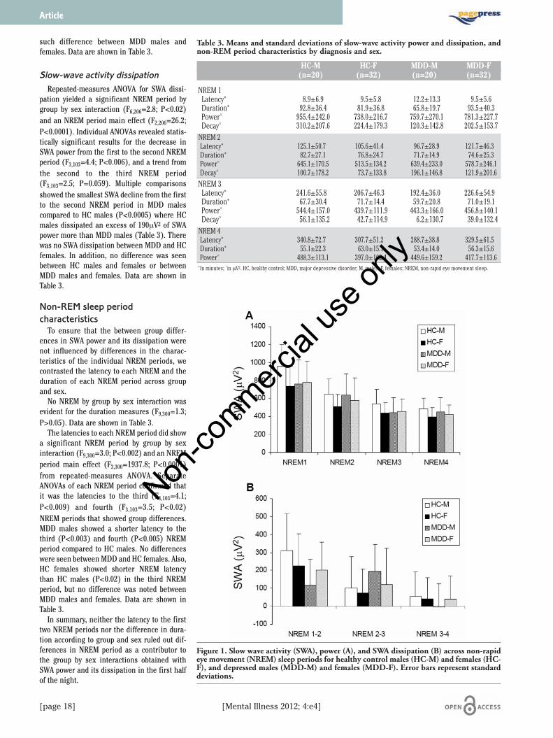
NREM main effect (F3,300=215.0; P<0.0001).Separate ANOVAs revealed between-group dif-ferences in the first (F3,103=4.1; P<0.009),third (F3,103=2.8; P<0.05), and fourth NREMperiods (F3,103=2.8; P<0.05), but only a trend inthe second NREM period (F3,103=2.6; P=0.06).Multiple comparisons showed that SWA powerwas significantly lower in MDD males com-pared to HC males in the first (P<0.007) andthird (P<0.02) NREM periods (Figure 1A).However, there were no differences in SWApower between MDD and HC females (P>0.05)in any of the NREM periods. Our results alsoconfirmed previous studies showing differentdevelopmental time periods between healthymales and females during adolescence. Thus,HC males expressed higher SWA power thanHC females in the first, third, and fourthNREM periods (P=0.002-0.03). There was no
Article
Table 1. Demographic and clinical features of the sample by diagnosis and sex.
HC-M HC-F MDD-M MDD-F(n=20) (n=32) (n=20) (n=32)
Age 14.7±1.2 15.1±1.6 15.3±1.6 15.0±1.5Tanner scores 4.2±0.7 4.4±0.5 4.4±0.7 4.4±0.6
FGAS 91.0±8.1 89.9±6.2 66.1±13.0 64.4±11.7CGAS 92.2±5.8 91.0±4.0 51.3±5.6 53.4±5.2
CDRS-R* 18.0±1.3 18.5±1.6 55.7±7.4 56.5±10.1Age of onset – – 13.4±2.9 12.9±2.6
Length of current depressive episode° – – 16.0±26.7 20.2±21.8Family history of MDD (n) – – 13 27
Comorbid psychiatric diagnosis (n) – – 6 15ADHD – – 3 6
Generalized anxiety – – 3 4Phobias – – – 2
Oppositional defiant dis. – – – 5OCD – – – 2*Missing data n=31 for MDD females. °In months.HC, healthy control; MDD, major depressive disorder; M, males; F, females; FGAS, family global assessment scale; CGAS, clinical globalassessment scale; CDRS-R, children’s depression rating scale - revised; ADHD, attention deficit hyperactivity disorder; OCD, obsessive com-pulsive disorder.
Table 2. Means and standard deviations of sleep macroarchitecture by diagnoses and sex.
Sleep variables HC-M HC-F MDD-M MDD-F(n=20) (n=32) (n=20) (n=32)
Total sleep time, min. 449.2±35.3 458.4±42.0 445.5±44.5 465.2±41.7Sleep latency, min. 9.5±9.9 13.7±10.7 14.6±11.2 18.1±20.2
REM latency, min. 111.1±57.1 84.7±38.6 69.6±23.7 107.6±50.3% Stage 1* 8.4±5.0 4.8±3.0 10.6±7.2 7.4±5.0
% Stage 2* 47.0±6.4 52.6±5.9 45.4±8.2 50.0±8.5% SW (stages 3+4)* 20.8±8.1 18.0±7.7 17.9±10.5 18.3±8.7
% REM* 18.3±3.9 21.2±4.0 22.3±4.9 20.6±5.1% awake and movement* 5.1±4.7 3.1±1.6 3.6±1.6 3.4±2.2
Sleep efficiency 93.0±4.9 93.8±2.4 93.2±2.5 93.0±4.5Number of arousals 23.4±7.7 19.1±8.9 20.4±6.1 19.7±8.4*Relative to total sleep time. Minutes of stages 3+4.HC, healthy control; MDD, major depressive disorder; M, males; F, females; REM, rapid eye movement sleep; NREM, non-rapid eye move-ment sleep; SW, slow wave sleep.
Non-co
mmercial
use o
nly
[page 18] [Mental Illness 2012; 4:e4]
such difference between MDD males andfemales. Data are shown in Table 3.
Slow-wave activity dissipation Repeated-measures ANOVA for SWA dissi-
pation yielded a significant NREM period bygroup by sex interaction (F6,206=2.8; P<0.02)and an NREM period main effect (F2,206=26.2;P<0.0001). Individual ANOVAs revealed statis-tically significant results for the decrease inSWA power from the first to the second NREMperiod (F3,103=4.4; P<0.006), and a trend fromthe second to the third NREM period(F3,103=2.5; P=0.059). Multiple comparisonsshowed the smallest SWA decline from the firstto the second NREM period in MDD malescompared to HC males (P<0.0005) where HCmales dissipated an excess of 190mV2 of SWApower more than MDD males (Table 3). Therewas no SWA dissipation between MDD and HCfemales. In addition, no difference was seenbetween HC males and females or betweenMDD males and females. Data are shown inTable 3.
Non-REM sleep periodcharacteristics
To ensure that the between group differ-ences in SWA power and its dissipation werenot influenced by differences in the charac-teristics of the individual NREM periods, wecontrasted the latency to each NREM and theduration of each NREM period across groupand sex.
No NREM by group by sex interaction wasevident for the duration measures (F9,300=1.3;P>0.05). Data are shown in Table 3.
The latencies to each NREM period did showa significant NREM period by group by sexinteraction (F9,300=3.0; P<0.002) and an NREMperiod main effect (F3,300=1937.8; P<0.0001)from repeated-measures ANOVA. SeparateANOVAs of each NREM period confirmed thatit was the latencies to the third (F3,103=4.1;P<0.009) and fourth (F3,103=3.5; P<0.02)NREM periods that showed group differences.MDD males showed a shorter latency to thethird (P<0.003) and fourth (P<0.005) NREMperiod compared to HC males. No differenceswere seen between MDD and HC females. Also,HC females showed shorter NREM latencythan HC males (P<0.02) in the third NREMperiod, but no difference was noted betweenMDD males and females. Data are shown inTable 3.
In summary, neither the latency to the firsttwo NREM periods nor the difference in dura-tion according to group and sex ruled out dif-ferences in NREM period as a contributor tothe group by sex interactions obtained withSWA power and its dissipation in the first halfof the night.
Article
Table 3. Means and standard deviations of slow-wave activity power and dissipation, andnon-REM period characteristics by diagnosis and sex.
HC-M HC-F MDD-M MDD-F(n=20) (n=32) (n=20) (n=32)
NREM 1Latency* 8.9±6.9 9.5±5.8 12.2±13.3 9.5±5.6Duration* 92.8±36.4 81.9±36.8 65.8±19.7 93.5±40.3Power° 955.4±242.0 738.0±216.7 759.7±270.1 781.3±227.7Decay° 310.2±207.6 224.4±179.3 120.3±142.8 202.5±153.7NREM 2Latency* 125.1±50.7 105.6±41.4 96.7±28.9 121.7±46.3Duration* 82.7±27.1 76.8±24.7 71.7±14.9 74.6±25.3Power° 645.1±170.5 513.5±134.2 639.4±233.0 578.7±246.1Decay° 100.7±178.2 73.7±133.8 196.1±146.8 121.9±201.6
NREM 3Latency* 241.6±55.8 206.7±46.3 192.4±36.0 226.6±54.9Duration* 67.7±30.4 71.7±14.4 59.7±20.8 71.0±19.1Power° 544.4±157.0 439.7±111.9 443.3±166.0 456.8±140.1Decay° 56.1±135.2 42.7±114.9 6.2±130.7 39.0±132.4NREM 4Latency* 340.8±72.7 307.7±51.2 288.7±38.8 329.5±61.5Duration* 55.1±22.3 63.0±15.1 53.4±14.0 56.3±15.6Power° 488.3±113.1 397.0±102.4 449.6±159.2 417.7±113.6*In minutes; °in mV2. HC, healthy control; MDD, major depressive disorder; M, males; F, females; NREM, non-rapid eye movement sleep.
Figure 1. Slow wave activity (SWA), power (A), and SWA dissipation (B) across non-rapideye movement (NREM) sleep periods for healthy control males (HC-M) and females (HC-F), and depressed males (MDD-M) and females (MDD-F). Error bars represent standarddeviations.
Non-co
mmercial
use o
nly
[Mental Illness 2012; 4:e4] [page 19]
Correlation between REM latencyand slow-wave activity measures
Pearson’s correlations were used to addressthe contribution of short or long REM latencyto changes in SWA between groups. No signif-icant correlations were obtained between SWApower or SWA dissipation and REM latency inany of the groups (r 0.05-.38; P>0.05). Thus,the reduction in SWA power observed indepressed males was not due to a shorter REMlatency.
Discussion
The major finding of the study was thatdepressed adolescent males showed SWAabnormalities, with lower SWA power in thefirst NREM period and irregular dissipation ofSWA over the night, compared to healthy con-trol males. Low SWA power at the beginningof the night suggests that depressed adoles-cent males failed to accumulate sufficientsleep drive during the daytime. The irregulardissipation or decline of SWA between con-secutive NREM periods further suggestsreduced recovery from prior sleep debt in thisgroup. Depressed adolescent males showedmaximum SWA loss between the second andthird NREM periods, not between the first andsecond as would be expected if sleep drivewas high at sleep onset. Moreover, this groupexpressed the shortest latency to the thirdand fourth NREM period of the night.Altogether, these results indicate thatdepressed adolescent males, like depressedadult males, show reduced SWA power withan abnormal time course.
SWA power and its time course are consid-ered physiological correlates of the recupera-tive aspect of sleep as evidenced by theincrease in SWA power following sleep depri-vation41 and the reduction of SWA power afternapping.42 Consequently, the SWA abnormali-ties observed in depressed adolescent malescompared to healthy males in the absence ofdifferences in total sleep time between thesegroups suggest the existence of sleep deficit ata neuronal level that may interfere with moodregulation.
In an earlier study, Reynolds and collabora-tors reported lower slow-wave sleep and deltawave count, as well as irregular temporal dis-tribution of delta waves, in depressed mencompared to depressed women, although theauthors attributed the sex differences to varia-tions in skull thickness.43 Our own group hasshown that men with depression have lowerSWA accumulation in the first NREM periodand slower dissipation through the night com-pared to healthy men.21,22 In addition,depressed males showed no relationship
between SWA changes and age (between 20-40years), suggesting the presence of SWA abnor-malities in adolescents with depression. Thesefindings and our present results support theidea that the pathophysiology of depressiondiffers for men and women, and that such dif-ferences emerge at very early ages.
The present results highlight the need todevelop sex specific therapies to improve sleepquality rather than time spent sleep in adoles-cents with MDD, and particular emphasisshould be given to the enhancement andrestoration of Slow-Wave Activity.Interpretation of our initial findings in adultswith MDD and the current work raise the pos-sibility that: i) SWA impairment antecedes theonset of MDD in males, underscoring the needto study SWA regulation in children and ado-lescents at-risk for MDD; and ii) SWA impair-ment may relate to different depressive symp-toms in females and males.
Regarding the analysis of polysomnographicdata, we observed a substantial shorter REMlatency and higher percentage of REM sleep indepressed male adolescents compared tohealthy control males. However, depressedfemales showed longer REM latency comparedto healthy females, although this differencewas not statistically significant. Our resultssuggest that changes in REM latency might besex-specific and could potentially account forthe great discrepancy in studies of sleep indepression examining changes in REM laten-cy. In agreement with our previous findings inadults,21 the relationship between REM latencyand SWA measures in either depressed orhealthy adolescents was negligible, suggestingthat changes in REM sleep and SWA mecha-nisms are not interdependent. Historically,short REM latency has been offered as anexplanation for reduced visually scored slow-wave sleep and reduced delta wave count,44,45but this suggestion does not fit the SWA datapresented here. However, we did obtain groupdifferences in REM latency in the presentstudy and in several of our previous publica-tions.32,46,47 Thus, REM sleep differences maybe evident in some MDD adolescents, but theydo not appear to contribute to SWA differencesbetween depressed and healthy control groups.
There are several limitations to the currentstudy. It is noteworthy to highlight that itdescribes baseline sleep and, therefore, onlyevaluates the time course of SWA, not its regu-lation. Sleep deprivation or sleep challengestudies will be necessary to evaluate whetherSWA regulation and sleep homeostasis is trulyimpaired in adolescents with MDD. A study ofSWA homeostasis in adults indicated that bothmen and women with MDD showed an abnor-mal SWA response to a mild sleep challenge,but the effects were inverted, with a bluntedresponse in MDD men and a hyper-response inMDD women.48 If the adolescent data are con-
sistent, one would also expect a bluntedresponse to sleep deprivation or sleep chal-lenge in adolescent males with MDD. Ongoingresearch in our laboratory is investigatingsleep homeostasis and regulation in MDD ado-lescents. Second, the cross-sectional nature ofthe present study makes it difficult to establishwhether SWA abnormalities in depressed ado-lescents persist in adulthood or whether theylead to a more severe course of illness com-pared to those with adult onset MDD. Third, wedid not analyze clinical data in relationship toSWA, and as such, the clinical relevance ofSWA impairment remains to be demonstrated.Nevertheless, the present results constitutethe basis for the development of new therapiesto improve sleep quality in depressed adoles-cents, with the caveat that sleep disturbancesin MDD may not be of the same nature formales and females.
References
1. Zisook S, Rush AJ, Albala A, et al. Factorsthat differentiate early vs. later onset ofmajor depression disorder. Psychiatry Res2004;129:127-40.
2. Kovacs M, Feinberg TL, Crouse-Novak MA,et al. Depressive disorders in childhood. I.A longitudinal prospective study of charac-teristics and recovery. Arch Gen Psychiatry1984;41:229-37.
3. Rao U, Ryan ND, Birmaher B, et al. Uni-polar depression in adolescents: clinicaloutcome in adulthood. J Am Acad ChildAdolesc Psychiatry 1995;34:566-78.
4. Weissman MM, Bland RC, Canino GJ, et al.Prevalence of suicide ideation and suicideattempts in nine countries. Psychol Med1999;29:9-17.
5. Birmaher B, Ryan ND, Williamson DE, etal. Childhood and adolescent depression: areview of the past 10 years. Part II. J AmAcad Child Adolesc Psychiatry 1996;35:1575-83.
6. Kaufman J, Martin A, King RA, Charney D.Are child-, adolescent-, and adult-onsetdepression one and the same disorder?Biol Psychiatry 2001;49:980-1001.
7. Hankin BL, Abramson LY. Development ofgender differences in depression: descrip-tion and possible explanations. Ann Med1999;31:372-9.
8. Hankin BL, Abramson LY, Moffitt TE, et al.Development of depression from preado-lescence to young adulthood: emerginggender differences in a 10-year longitudi-nal study. J Abnorm Psychol 1998;107:128-40.
9. Rao U, Chen LA. Characteristics, corre-lates, and outcomes of childhood and ado-lescent depressive disorders. Dialogues
Article
Non-co
mmercial
use o
nly

[page 20] [Mental Illness 2012; 4:e4]
Clin Neurosci 2009;11:45-62.10. Peterson MJ, Benca RM. Sleep in mood
disorders. Psychiatr Clin North Am 2006;29:1009-32.
11. Perlis ML, Giles DE, Buysse DJ, et al. Self-reported sleep disturbance as a prodromalsymptom in recurrent depression. J AffectDisord 1997;42:209-12.
12. Armitage R, Hoffmann RF, Emslie GJ, et al.Sleep microarchitecture as a predictor ofrecurrence in children and adolescentswith depression. Int J Neuropsychophar-macol 2002;5:217-28.
13. Kennard BD, Silva S, Vitiello B, et al.Remission and Residual Symptoms AfterShort-Term Treatment in the Treatment ofAdolescents With Depression Study(TADS). J Am Acad Child Adolesc Psychia-try 2006;45:1404-11.
14. Armitage R. Sleep and circadian rhythmsin mood disorders. Acta Psychiatr ScandSuppl 2007:104-15.
15. Ivanenko A, Johnson K. Sleep disturbancesin children with psychiatric disorders.Semin Pediatr Neurol 2008;15:70-8.
16. Borbely AA. New techniques for the analy-sis of the human sleep-wake cycle. BrainDev 1986;8:482-8.
17. Armitage R, Hoffmann RF. Sleep EEG,depression and gender. Sleep Med Rev2001;5:237-46.
18. Borbely AA. A two process model of sleepregulation. Hum.Neurobiol 1982;1:195-204.
19. Dijk DJ. Regulation and functional corre-lates of slow wave sleep. J Clin Sleep Med2009;5:S6-15.
20. Armitage R. Sleep and circadian rhythmsin mood disorders. Acta Psychiatr ScandSuppl 2007:104-15.
21. Armitage R, Hoffmann R, Fitch T, et al.Temporal characteristics of delta activityduring NREM sleep in depressed outpa-tients and healthy adults: group and sexeffects. Sleep 2000;23:607-17.
22. Armitage R, Hoffmann R, Trivedi M, RushAJ. Slow-wave activity in NREM sleep: sexand age effects in depressed outpatientsand healthy controls. Psychiatry Res2000;95:201-13.
23. Borbely AA, Tobler I, Loepfe M, et al. All-night spectral analysis of the sleep EEG inuntreated depressives and normal con-
trols. Psychiatry Res 1984;12:27-33.24. Riemann D, Berger M, Voderholzer U.
Sleep and depression-results from psy-chobiological studies: an overview. BiolPsychol 2001;57:67-103.
25. Kupfer DJ. Sleep research in depressiveillness: clinical implications-a tastingmenu. Biological Psychiatry 1995;38:391-403.
26. Tsuno N, Besset A, Ritchie K. Sleep anddepression. J Clin Psychiatry 2005;66:1254-69.
27. Kaufman J, Birmaher B, Brent D, et al.Schedule for Affective Disorders andSchizophrenia for School-Age Children-Present and Lifetime Version (K-SADS-PL): initial reliability and validity data. JAm Acad Child Adolesc Psychiatry1997;36:980-8.
28. Poznanski EO, Freeman LN, Mokros HB.Children's Depression Rating Scale -Revised (September 1984). Psychophar-macol Bull 1985;21:979-89.
29. Armitage R, Emslie GJ, Hoffmann RF, et al.Ultradian rhythms and temporal coher-ence in sleep EEG in depressed childrenand adolescents. Biol Psychiatry 2000;47:338-50.
30. Emslie GJ, Armitage R, Weinberg WA, et al.Sleep polysomnography as a predictor ofrecurrence in children and adolescentswith major depressive disorder. Int JNeuropsychopharmacol 2001;4:159-68.
31. Lopez J, Hoffmann R, Armitage R. Reducedsleep spindle activity in early-onset andelevated risk for depression. J Am AcadChild Adolesc Psychiatry 2010;49:934-43.
32. Robert JJ, Hoffmann RF, Emslie GJ, et al.Sex and age differences in sleep macroar-chitecture in childhood and adolescentdepression. Sleep 2006;29:351-8.
33. Shaffer D, Gould MS, Brasic J, et al. A chil-dren's global assessment scale (CGAS).Arch Gen Psychiatry 1983;40:1228-31.
34. Mrazek DA, Mrazek P, Klinnert M. Clinicalassessment of parenting. J Am Acad ChildAdolesc Psychiatry 1995;34:272-82.
35. Tanner JM. Growth at Adolescence. Vol 2.Charles C.Thomas, Springfield, 1962.
36. Rechtschaffen A, Kales A. A Manual ofStandarized Terminology, Techniques andScoring Systems for Sleep Stages ofHuman Subjects. National Institute of
Healthy Publication N. 204. US GovermentPrinting Office, Washington, DC, USA,1968.
37. Dijk DJ, Brunner DP, Borbely AA. Timecourse of EEG power density during longsleep in humans. Am J Physiol 1990;258:R650-61.
38. Feinberg I, Floyd TC. Systematic trendsacross the night in human sleep cycles.Psychophysiology 1979;16:283-91.
39. Press WH, Fannery BP, Teukolsky SA,Vetterling WT. Numerical recipes inPascal. Cambridge University Press, NewYork, USA, 1989.
40. Armitage R, Hoffmann R, Fitch T, et al. Acomparison of period amplitude and powerspectral analysis of sleep EEG in normaladults and depressed outpatients.Psychiatry Res 1995;56:245-56.
41. Borbely AA, Baumann F, Brandeis D, et al.Sleep deprivation: effect on sleep stagesand EEG power density in man. Electroen-cephalogr Clin Neurophysiol 1981;51:483-95.
42. Werth E, Dijk DJ, Achermann P, BorbelyAA. Dynamics of the sleep EEG after anearly evening nap: experimental data andsimulations. Am J Physiol 1996;271:R501-10.
43. Reynolds CF 3rd, Kupfer DJ, Thase ME, etal. Sleep, gender, and depression: ananalysis of gender effects on the electroen-cephalographic sleep of 302 depressed out-patients. Biological Psychiatry 1990;28:673-84.
44. McCarley RW. REM sleep and depression:common neurobiological control mecha-nisms. Am J Psychiatry 1982;139:565-70.
45. Kupfer DJ, Foster FG. Interval betweenonset of sleep and rapid-eye-movementsleep as an indicator of depression. Lancet1972;2:684-6.
46. Emslie GJ, Roffwarg HP, Rush AJ, et al.Sleep EEG findings in depressed childrenand adolescents. Am J Psychiatry 1987;144:668-70.
47. Emslie GJ, Rush AJ, Weinberg WA, et al.Sleep EEG features of adolescents withmajor depression. Biol Psychiatry 1994;36:573-81.
48. Armitage R. Sleep and circadian rhythmsin mood disorders. Acta PsychiatricaScandinavica Supplementum 2007:104-15.
Article
Non-co
mmercial
use o
nly
Copyright © 2022 FDOKUMEN