Intermolecular Interactions of the p85alpha Regulatory Subunit of Phosphatidylinositol 3-Kinase
Upload
independentCategory
view
2download
0
ORI GIN AL PA PER
Self-Assembly, Crystal Structure and Analysisof Intermolecular Interactions of the SupramolecularCompound Based on Hexamolybdochromate(III),Sulfate and Piperazine
Feng Yao • Ya-Guang Chen • Ali R. Salimi •
Masoud Mirzaei
Received: 28 February 2011 / Published online: 21 May 2011
� Springer Science+Business Media, LLC 2011
Abstract A new supramolecular compound based on Anderson-B hexamolybdo-
chromate, (H2Pz)3[Cr(OH)6Mo6O18H](SO4)2�12H2O (1) (Pz = piperazine) was
synthesized and characterized by elemental analysis, IR spectroscopy, and single-
crystal X-ray diffraction (Mo Ka). The compound crystallizes in monoclinic system,
P21/c space group with a = 13.5708(6) A, b = 17.3711(8) A, c = 22.2387(9) A,
b = 110.631(2)�; V = 4906.3(4) A3, Z = 4, Dc = 2.290 g/cm3, F(000) = 3364.0;
l = 1.905, S = 1.033. The final R = 0.0398 and wR = 0.0971. The H2pz2? ions and
sulfate anions in 1 are arranged through hydrogen bonds into a hexagonal network in
[202] plane and hexamolybdochromates anions (CrMo6) fill in the hexagonal
vacancies. The networks stack in such a way that each anion links two sulfate ions
from adjacent networks via hydrogen bonds with short (CrMo6)O���OSO3 distances of
2.637–2.697 A. A lot of hydrogen bonds are formed between water molecules, sul-
fate, H2pz2? ions and CrMo6 anions, which are the dominating force constructing the
supramolecular structure. Hirshfeld surface analysis of 1 gives us the details of
intermolecular interactions in the crystals of 1 in a visual manner and shows that the
CrMo6 anion acts as a stronger hydrogen bond donor than as an acceptor.
Electronic supplementary material The online version of this article
(doi:10.1007/s10876-011-0382-6) contains supplementary material, which is available to authorized
users.
F. Yao � Y.-G. Chen (&)
Key Laboratory of Polyoxometalates Science of Ministry of Education, College of Chemistry,
Northeast Normal University, Changchun 130024, People’s Republic of China
e-mail: [email protected]
A. R. Salimi � M. Mirzaei
Department of Chemistry, School of Sciences, Ferdowsi University of Mashhad,
P.O. Box 917791436, Mashhad, Iran
123
J Clust Sci (2011) 22:309–318
DOI 10.1007/s10876-011-0382-6
Keywords Anderson-B hexamolybdochromate � Crystal structure �Intermolecular interaction � Piperazine � Sulfate � Hirshfeld surface analysis
Introduction
Supramolecular compounds based on polyoxometalates (POMs) construct a small
subset of the large family of POMs which have attracted considerable attention
worldwide due to variety of their structures and potential application [1–6]. The
self-assembly of supramolecular structure performs via ligand–metal coordination
bonds, hydrogen bonds, electrostatic force and p–p stacking interaction. Among the
non-covalent motifs, hydrogen bonding and p–p stacking interactions have been
employed as synthetic paradigms to rationally design superstructures [7, 8]. There
are a lot of reports on syntheses, characterization and studies of properties of the
supramolecular compounds based on POMs [9–17] but Hirshfeld surfaces analyses
on these compounds are rare [18]. Hirshfeld surface analysis is the method
developed by Spackman et al. [19–22] for elucidating and comparing intermolecular
interactions as well as for spotting common features and trends in specific classes of
compounds. After our first report [18], we obtained another analogue
(H2Pz)3[Cr(OH)6Mo6O18H](SO4)2�12H2O (1) (Pz = piperazine) from pre-synthe-
sized hexamolybdochromate (CrMo6) and in a high concentration of sulfate through
self-assembly in solution and found that the components of this compound
undertake a such different arranging pattern that a hexagonal network in [202] plane
with CrMo6 filling forms with hydrogen bonds. Herein, we report the self-assembly,
crystal structure and the Hirshfeld surface analysis of the intermolecular interactions
of 1.
Experimental
Materials and Methods
All chemicals of reagent grade were purchased from commercial sources and used
as received. Elemental analyses (C, H, N) were performed on a Perkins-Elmer 2400
CHN Elemental Analyzer. Mo and Cr were determined on a Prodigy inductively
coupled plasma spectrometer. IR spectrum was recorded in the range of
400–4000 cm-1 on a Magna-560 FT-IR spectrophotometer.
Synthesis
An aqueous solution (20 mL) containing 0.1231 g (0.1 mmol) Na3[CrMo6H6
O24]�H2O [23, 24] was heated at approximately 60 �C. Then, into it a solution of
CuSO4�5H2O (0.0500 g, 0.2 mmol) in 5 mL water and 10 mL aqueous solution of
Pz (0.12 g, 0.6 mmol) were added dropwise. The pH of the resulting mixture was
adjusted with dilute H2SO4 solution to about 3.15 and the final mixture was stirred
310 F. Yao et al.
123
for 40 min at 60 �C, then filtered. Slow evaporation of this filtrate resulted in a
light-red block crystalline product within 2 weeks (Yield: ca. 35% based on Cr).
Anal. calcd for C12H67CrMo6N6O44S2: C 8.55, H 3.65, N 4.99, S 3.80, Cr 3.09, Mo
34.15(%); found C 8.41, H 3.45, N 4.90, S 3.69, Cr 2.91, Mo 35.01(%). IR (KBr)m:
3422.35(s), 3026(w), 2960(w), 1615.53(ms), 1458.47(m), 1314.83(w), 1083.25(m),
918.5 (s), 643.87(s), 564.85 (w), 443.1(m) cm-1.
X-ray Crystallography
X-ray diffraction data were collected on a SMART APEX II CCD diffractometer
with graphite monochromated MoKa radiation at 293 K with x scans. Empirical
absorption correction was applied. The structure was solved by direct methods and
refined by full-matrix least-squares on F2 with the SHELXL-97 program package
[25, 26]. The positions of the hydrogen atoms attached to carbon and nitrogen atoms
were fixed according to riding mode. All the non-hydrogen atoms except C2 atom
were refined anisotropically. A summary of crystallographic and structural
refinement data for 1 is provided in Table 1.
Table 1 Crystallographic and structural refinement data of 1
Empirical formula C12H67CrMo6N6O44S2
Mr 1691.43
Crystal dimension (mm3) 0.10 9 0.11 9 0.18
Range of h (�) 1.98–28.34
Crystal system Monoclinic
Space group P2(1)/c
a (A) 13.5708(6)
b (A) 17.3711(8)
c (A) 22.2387(9)
b (deg) 110.631(2)
V (A3) 4906.3(4)
Z 4
Dcalcd (g cm-3) 2.290
F(000) (e) 3364.0
l (mm-1) 1.905
Refl. measured 29824
Refl. Unique (Rint) 12237(0.0333)
R1/wR2a [I [ 2r(I)] 0.0398, 0.0971
GoF (F2)a 1.033
(D/r)max 0.001
±(Dq) (eA3) 2.030, -0.962
a w = 1/[r2(Fo2) ? (0.0485P)2 ? 6.9690P] where P = (Fo
2 ? 2Fc2)/3
Synthesis and Characterization of a Supramolecular Compound 311
123
Results and Discussions
Structural Description
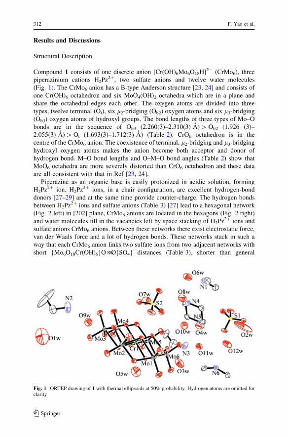
Compound 1 consists of one discrete anion [Cr(OH)6Mo6O18H]2- (CrMo6), three
piperazinium cations H2Pz2?, two sulfate anions and twelve water molecules
(Fig. 1). The CrMo6 anion has a B-type Anderson structure [23, 24] and consists of
one Cr(OH)6 octahedron and six MoO4(OH)2 octahedra which are in a plane and
share the octahedral edges each other. The oxygen atoms are divided into three
types, twelve terminal (Ot), six l2-bridging (Ob2) oxygen atoms and six l3-bridging
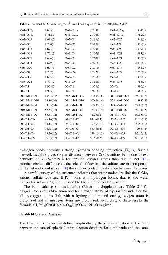
(Ob3) oxygen atoms of hydroxyl groups. The bond lengths of three types of Mo–O
bonds are in the sequence of Ob3 (2.260(3)–2.310(3) A) [ Ob2 (1.926 (3)–
2.055(3) A) [ Ot (1.693(3)–1.712(3) A) (Table 2). CrO6 octahedron is in the
centre of the CrMo6 anion. The coexistence of terminal, l2-bridging and l3-bridging
hydroxyl oxygen atoms makes the anion become both acceptor and donor of
hydrogen bond. M–O bond lengths and O–M–O bond angles (Table 2) show that
MoO6 octahedra are more severely distorted than CrO6 octahedron and these data
are all consistent with that in Ref [23, 24].
Piperazine as an organic base is easily protonized in acidic solution, forming
H2Pz2? ion. H2Pz2? ions, in a chair configuration, are excellent hydrogen-bond
donors [27–29] and at the same time provide counter-charge. The hydrogen bonds
between H2Pz2? ions and sulfate anions (Table 3) [27] lead to a hexagonal network
(Fig. 2 left) in [202] plane, CrMo6 anions are located in the hexagons (Fig. 2 right)
and water molecules fill in the vacancies left by space stacking of H2Pz2? ions and
sulfate anions CrMo6 anions. Between these networks there exist electrostatic force,
van der Waals force and a lot of hydrogen bonds. These networks stack in such a
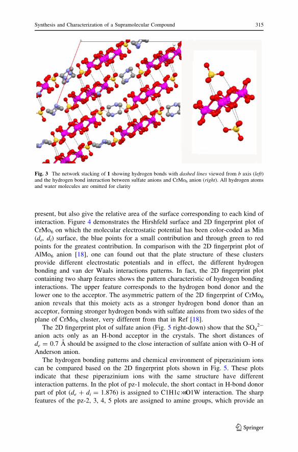
way that each CrMo6 anion links two sulfate ions from two adjacent networks with
short {Mo6O18Cr(OH)6}O���O{SO4} distances (Table 3), shorter than general
Fig. 1 ORTEP drawing of 1 with thermal ellipsoids at 50% probability. Hydrogen atoms are omitted forclarity
312 F. Yao et al.
123
hydrogen bonds, showing a strong hydrogen bonding interaction (Fig. 3). Such a
network stacking gives shorter distances between CrMo6 anions belonging to two
networks of 3.295–3.515 A for terminal oxygen atoms than that in Ref [18].
Another obvious difference is the role of sulfate: in 1 the sulfates are the component
of the networks and in Ref [18] the sulfates control the distance between the layers.
A careful survey of the structure indicates that water molecules link the CrMo6
anions, sulfate ions and H2Pz2? ions with hydrogen bonds, that is, the water
molecules act as a ‘‘glue’’ to assemble the supramolecular structure.
The bond valence sum calculation (Electronic Supplementary Table S1) for
oxygen atoms of CrMo6 anion and for nitrogen atoms of piperazines indicates that
all l3-oxygen atoms link with a hydrogen atom and one l2-oxygen atom is
protonized and all nitrogen atoms are protonized. According to these results the
formula (H2Pz)3[Cr(OH)6Mo6O18H](SO4)2�12H2O is given.
Hirshfeld Surface Analysis
The Hirshfeld surfaces are defined implicitly by the simple equation as the ratio
between the sum of spherical atom electron densities for a molecule and the same
Table 2 Selected M–O bond lengths (A) and bond angles (�) in [Cr(OH)6Mo6O18H]2-
Mo1–O12t 1.693(3) Mo1–O1b3 2.298(3) Mo1–O23b2 1.934(3)
Mo1–O11t 1.712(3) Mo1–O2b3 2.304(3) Mo1–O10b2 1.952(3)
Mo2–O19 1.693(3) Mo2–O1 2.286(3) Mo2–O23 1.941(3)
Mo2–O7 1.700(3) Mo2–O3 2.310(3) Mo2–O9 1.959(3)
Mo3–O13 1.693(3) Mo3–O3 2.270(3) Mo3–O9 1.919(3)
Mo3–O18 1.702(3) Mo3–O4 2.307(3) Mo3–O21 1.993(3)
Mo4–O17 1.694(3) Mo4–O5 2.260(3) Mo4–O21 1.926(3)
Mo4–O14 1.699(3) Mo4–O4 2.271(3) Mo4–O22 2.032(3)
Mo5–O20 1.691(3) Mo5–O5 2.261(3) Mo5–O15 1.891(3)
Mo5–O8 1.702(3) Mo5–O6 2.282(3) Mo5–O22 2.055(3)
Mo6–O16 1.693(3) Mo6–O2 2.286(3) Mo6–O10 1.929(3)
Mo6–O24 1.704(3) Mo6–O6 2.310(3) Mo6–O15 1.986(3)
O2–Cr1 1.968(3) O3–Cr1 1.970(3) O5–Cr1 1.990(3)
O1–Cr1 1.963(3) O4–Cr1 1.971(3) O6–Cr1 1.966(3)
O12–Mo1–O11 105.27(18) O12–Mo1–O23 100.89(16) O11–Mo1–O23 98.37(16)
O12–Mo1–O10 96.86(16) O11–Mo1–O10 100.26(16) O23–Mo1–O10 149.82(13)
O12–Mo1–O1 93.85(14) O11–Mo1–O1 160.07(15) O23–Mo1–O1 72.06(12)
O10–Mo1–O1 82.62(12) O12–Mo1–O2 161.07(15) O11–Mo1–O2 92.11(15)
O23–Mo1–O2 83.58(12) O10–Mo1–O2 72.23(12) O1–Mo1–O2 69.83(10)
O1–Cr1–O6 96.16(12) O1–Cr1–O2 84.05(13) O6–Cr1–O2 83.79(12)
O1–Cr1–O3 84.15(12) O6–Cr1–O3 179.59(13) O2–Cr1–O3 96.50(12)
O1–Cr1–O4 96.45(12) O6–Cr1–O4 96.44(12) O2–Cr1–O4 179.41(14)
O3–Cr1–O4 83.26(12) O1–Cr1–O5 179.15(12) O6–Cr1–O5 83.13(12)
O2–Cr1–O5 96.33(12) O3–Cr1–O5 96.56(12) O4–Cr1–O5 83.17(12)
Synthesis and Characterization of a Supramolecular Compound 313
123
sum for the entire crystal [19–22]. The molecular Hirshfeld surfaces reflect
intermolecular interactions in a novel visual manner as triangulation surfaces. The
surface shape relies on the interaction between molecules in the crystal as well as
between atoms in the molecule. Derived from the Hirshfeld surface, the 2D-
fingerprint plots, in which the points on the surface are a function of the closest
distances from the point to nuclei inside (di) and outside (de) the surface, provide a
visual summary of the frequency of each combination of de and di across the surface
of a molecule, so they not only indicate which intermolecular interactions are
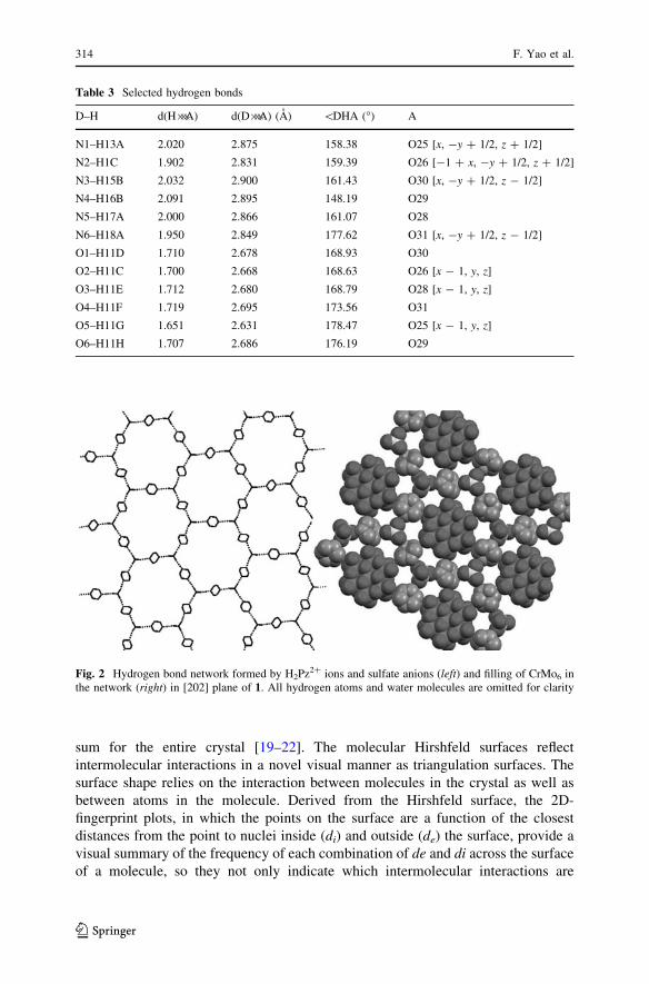
Table 3 Selected hydrogen bonds
D–H d(H���A) d(D���A) (A) \DHA (�) A
N1–H13A 2.020 2.875 158.38 O25 [x, -y ? 1/2, z ? 1/2]
N2–H1C 1.902 2.831 159.39 O26 [-1 ? x, -y ? 1/2, z ? 1/2]
N3–H15B 2.032 2.900 161.43 O30 [x, -y ? 1/2, z - 1/2]
N4–H16B 2.091 2.895 148.19 O29
N5–H17A 2.000 2.866 161.07 O28
N6–H18A 1.950 2.849 177.62 O31 [x, -y ? 1/2, z - 1/2]
O1–H11D 1.710 2.678 168.93 O30
O2–H11C 1.700 2.668 168.63 O26 [x - 1, y, z]
O3–H11E 1.712 2.680 168.79 O28 [x - 1, y, z]
O4–H11F 1.719 2.695 173.56 O31
O5–H11G 1.651 2.631 178.47 O25 [x - 1, y, z]
O6–H11H 1.707 2.686 176.19 O29
Fig. 2 Hydrogen bond network formed by H2Pz2? ions and sulfate anions (left) and filling of CrMo6 inthe network (right) in [202] plane of 1. All hydrogen atoms and water molecules are omitted for clarity
314 F. Yao et al.
123
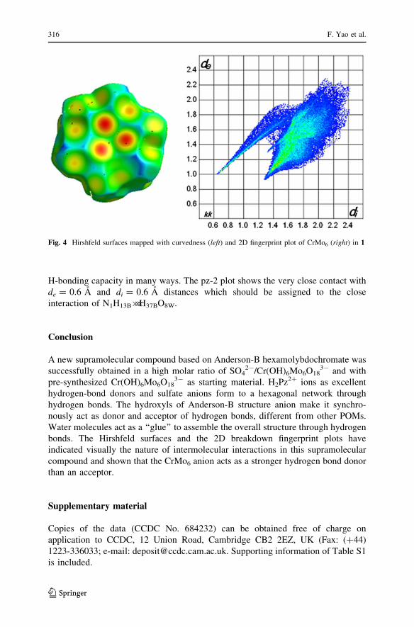
present, but also give the relative area of the surface corresponding to each kind of
interaction. Figure 4 demonstrates the Hirshfeld surface and 2D fingerprint plot of
CrMo6 on which the molecular electrostatic potential has been color-coded as Min
(de, di) surface, the blue points for a small contribution and through green to red
points for the greatest contribution. In comparison with the 2D fingerprint plot of
AlMo6 anion [18], one can found out that the plate structure of these clusters
provide different electrostatic potentials and in effect, the different hydrogen
bonding and van der Waals interactions patterns. In fact, the 2D fingerprint plot
containing two sharp features shows the pattern characteristic of hydrogen bonding
interactions. The upper feature corresponds to the hydrogen bond donor and the
lower one to the acceptor. The asymmetric pattern of the 2D fingerprint of CrMo6
anion reveals that this moiety acts as a stronger hydrogen bond donor than an
acceptor, forming stronger hydrogen bonds with sulfate anions from two sides of the
plane of CrMo6 cluster, very different from that in Ref [18].
The 2D fingerprint plot of sulfate anion (Fig. 5 right-down) show that the SO42-
anion acts only as an H-bond acceptor in the crystals. The short distances of
de = 0.7 A should be assigned to the close interaction of sulfate anion with O–H of
Anderson anion.
The hydrogen bonding patterns and chemical environment of piperazinium ions
can be compared based on the 2D fingerprint plots shown in Fig. 5. These plots
indicate that these piperazinium ions with the same structure have different
interaction patterns. In the plot of pz-1 molecule, the short contact in H-bond donor
part of plot (de ? di = 1.876) is assigned to C1H1c���O1W interaction. The sharp
features of the pz-2, 3, 4, 5 plots are assigned to amine groups, which provide an
Fig. 3 The network stacking of 1 showing hydrogen bonds with dashed lines viewed from b axis (left)and the hydrogen bond interaction between sulfate anions and CrMo6 anion (right). All hydrogen atomsand water molecules are omitted for clarity
Synthesis and Characterization of a Supramolecular Compound 315
123
H-bonding capacity in many ways. The pz-2 plot shows the very close contact with
de = 0.6 A and di = 0.6 A distances which should be assigned to the close
interaction of N1H13B���H37BO8W.
Conclusion
A new supramolecular compound based on Anderson-B hexamolybdochromate was
successfully obtained in a high molar ratio of SO42-/Cr(OH)6Mo6O18
3- and with
pre-synthesized Cr(OH)6Mo6O183- as starting material. H2Pz2? ions as excellent
hydrogen-bond donors and sulfate anions form to a hexagonal network through
hydrogen bonds. The hydroxyls of Anderson-B structure anion make it synchro-
nously act as donor and acceptor of hydrogen bonds, different from other POMs.
Water molecules act as a ‘‘glue’’ to assemble the overall structure through hydrogen
bonds. The Hirshfeld surfaces and the 2D breakdown fingerprint plots have
indicated visually the nature of intermolecular interactions in this supramolecular
compound and shown that the CrMo6 anion acts as a stronger hydrogen bond donor
than an acceptor.
Supplementary material
Copies of the data (CCDC No. 684232) can be obtained free of charge on
application to CCDC, 12 Union Road, Cambridge CB2 2EZ, UK (Fax: (?44)
1223-336033; e-mail: [email protected]. Supporting information of Table S1
is included.
Fig. 4 Hirshfeld surfaces mapped with curvedness (left) and 2D fingerprint plot of CrMo6 (right) in 1
316 F. Yao et al.
123
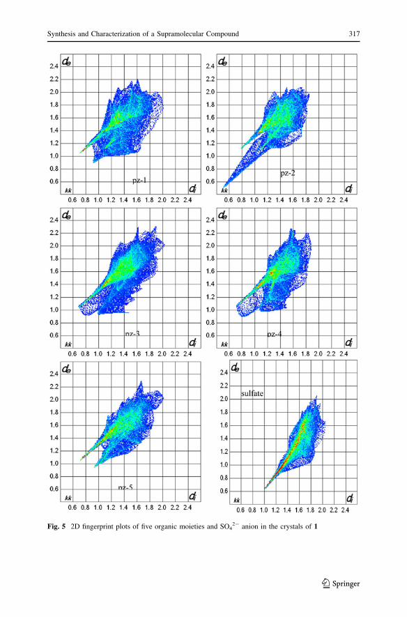
Fig. 5 2D fingerprint plots of five organic moieties and SO42- anion in the crystals of 1
Synthesis and Characterization of a Supramolecular Compound 317
123

References
1. C. L. Hill and X. Zhang (1995). Nature 373, 324.
2. D. Hagrman, C. Zubieta, D. J. Rose, J. Zubieta, and R. Haushalter (1997). Angew. Chem. Int. Ed. 36,
873.
3. G. S. Kim, H. Zeng, J. T. Rhule, I. A. Weinstock, and C. L. Hill (1999). Chem. Commun. 1651.
4. C. L. Hill (Guest ed) (1998) Chem. Rev. (Special issue on Polyoxometalates) 98, 1.
5. K. Fukaya and T. Yamase (2003). Angew. Chem. Int. Ed. 115, 678.
6. P. Kogerler and L. Cronin (2005). Angew. Chem. Int. Ed. 117, 866.
7. N. Malek, T. Maris, M. Simard, and J. D. Wuest (2005). J. Am. Chem. Soc. 127, 5910.
8. K. Uemura, K. Saito, S. Kitagawa, and H. Kita (2006). J. Am. Chem. Soc. 128, 16122.
9. S. Upreti and A. Ramanan (2005). Cryst. Growth Des. 5, 1837.
10. H. An, Y. Li, E. Wang, D. Xiao, C. Sun, and L. Xu (2005). Inorg. Chem. 44, 6062.
11. Y.-Q. Lan, S.-L. Li, X.-L. Wang, K.-Z. Shao, Z.-M. Su, and E.-B. Wang (2008). Inorg. Chem. 47,
529.
12. S. Yin, H. Sun, Y. Yan, W. Li, and L. Wu (2009). J. Phys. Chem. B. 113, 2355.
13. Y.-M. Xie, Q.-S. Zhang, Z.-G. Zhao, X.-Y. Wu, S.-C. Chen, and C.-Zg. Lu (2008). Inorg. Chem. 47,
8086.
14. V. Shivaiah, M. Nagaraju, and S. K. Das (2003). Inorg. Chem. 42, 6604.
15. H. An, Y. Li, D. Xiao, E. Wang, and C. Sun (2006). Cryst. Growth Des. 6, 1107.
16. W. Bu, H. Li, H. Sun, S. Yin, and L. Wu (2005). J. Am. Chem. Soc. 127, 8016.
17. M. Schulz-Dobrick and M. Jansen (2007). Inorg. Chem. 46, 4380.
18. X. D. Yang, Y. G. Chen, M. Mirzaei, A. R. Salimi, and F. Yao (2009). Inorg. Chem. Commun. 12,
195.
19. M. A. Spackman and P. G. Byrom (1997). Chem. Phys. Lett. 267, 215.
20. J. J. McKinnon, A. S. Mitchell, and M. A. Spackman (1998). Chem. Eur. J. 4, 2136.
21. J. J. McKinnon, M. A. Spackman, and A. S. Mitchell (2004). Acta. Crystallogr. B 60, 627.
22. S. K. Wolff, D. J. Grimwood, J. J. McKinnon, D.Jayatilaka, and M. A. Spackman (2006) Crystal-
Explorer 1.5. University of Western Australia, Perth.
23. C. Wroe Wolfe, M. Block, and L. C. W. Baker (1955). J. Am. Chem. Soc. 77, 2200.
24. A. Perloff (1970). Inorg. Chem. 9, 2228.
25. G. M. Sheldrick SHELXL 97, Program for Crystal Structure Solution (University of Gottingen,
Gottingen, 1997).
26. G. M. Sheldrick SHELXL 97, Program for Crystal Structure Refinement (University of Gottingen,
Gottingen, 1997).
27. K. Jayaraman, A. Choudhury, and C. N. R. Rao (2002). Solid State Sci. 4, 413.
28. M. Singh, S. E. Lofland, K. V. Ramanujachary, and A. Ramanan (2010). Cryst. Growth Des. 10,
5101.
29. K. Pavani, M. Singh, and A. Ramanan (2011). Aust. J. Chem. 64, 68.
318 F. Yao et al.
123
Copyright © 2022 FDOKUMEN