Seizure-Induced Changes in Place Cell Physiology: Relationship to Spatial Memory
11
Behavioral/Systems/Cognitive Seizure-Induced Changes in Place Cell Physiology: Relationship to Spatial Memory Xianzeng Liu, 1,2,3 Robert U. Muller, 4,5 Li-Tung Huang, 2 John L. Kubie, 4 Alexander Rotenberg, 2,4 Bruno Rivard, 4 Maria Roberta Cilio, 2 and Gregory L. Holmes 1,2 1 Neuroscience Center at Dartmouth, Division of Neurology, Dartmouth Medical School, Lebanon, New Hampshire 03756, 2 Children’s Hospital Boston, Harvard Medical School, Boston, Massachusetts 02115, 3 Department of Neurology, People’s Hospital, Peking University Health Science Center, Beijing, China 100044, 4 Department of Physiology and Pharmacology, State University of New York at Brooklyn, Brooklyn, New York 11203, and 5 Medical Research Council Centre for Synaptic Plasticity, Department of Anatomy, University of Bristol, Bristol, United Kingdom, B58 1TD Status epilepticus (SE) is a frequent neurological emergency associated with a significant risk of morbidity in survivors. Impairment of hippocampal-specific memory is a common and serious deficit occurring in many of the survivors. However, the pathophysiological basis of cognitive deficits after SE is not clear. To directly address the cellular concomitants of spatial memory impairment, we recorded the activity of place cells from CA1 in freely moving rats subjected to SE during early development and compared this activity to that in control rats. Place cells discharge rapidly only when the rat’s head is in a cell-specific part of the environment called the “firing field.” This firing field remains stable over time. Normal place cell function seems to be essential for stable spatial memory for the environment. We, therefore, compared place cell firing patterns with visual–spatial memory in the water maze in SE and control rats. Compared with controls, place cells from the SE rats were less precise and less stable. Concordantly, the water maze performance was also impaired. There was a close relationship between precision and stability of place cells and water maze performance. In contrast, a single, acute, chemically induced seizure produced cessation of place cell activity and spatial memory impairment in water maze performance that reversed within 24 hr. These results strongly bolster the idea that there is a relationship between abnormal place cells and spatial memory. Our findings also suggest that the defects in place cell and spatial memory after SE and acute chemically induced seizures result from different processes. Key words: water maze; epilepsy; status epilepticus; memory; place cell; seizures Introduction Status epilepticus (SE), which is defined as 30 min or more of continuous epileptic seizure activity, is a common neurological emergency in children associated with high morbidity and mor- tality (Aicardi and Chevrie, 1970, 1983; DeLorenzo et al., 1992; Working Group on Status Epilepticus, 1993; van Esch et al., 1996; Sahin et al., 2001). Although SE with or without recurrent sei- zures is associated with a wide variety of neuropsychological problems, memory deficits including impairment of episodic memory are especially prominent (Jambaque et al., 1993; Krum- holz et al., 1995; van Esch et al., 1996). It is, therefore, of para- mount interest to identify the processes by which SE leads to permanently abnormal brain function, to prevent the operation of such processes, or to reduce their effects afterward. A great deal of evidence suggests that normal declarative memory, the ability to learn and recall specific information about people, places, and events, depends on a properly functioning hippocampus (Squire, 1992). In light of the memory deficits seen after SE in humans, it is not surprising that SE is preferentially associated with histologically detectable damage of the hip- pocampus and related areas (“mesial temporal sclerosis”). This form of damage is characterized by cell loss in CA1, CA3, the hilus and dentate gyrus, and synaptic reorganization as evidence by sprouting of mossy fibers (Liu et al., 1995). Although the precise role of cell loss and synaptic reorganization in the sequelae of SE is unclear, there is evidence that other processes, including recur- rent seizures, may play important roles in memory deficiencies (Hermann et al., 2002; Stefan and Pauli, 2002). Thus, little is certain about the pathophysiological mecha- nisms responsible for the adverse events after SE, including the memory loss. It is clear, however, that the participation of cell level processes in these events is extremely difficult to study in humans. It is, therefore, hard to avoid the conclusion that progress toward a complete understanding of SE will require the use of animal models. To directly address the cellular concomitants of spatial mem- ory impairments, we recorded the activity of single hippocampal neurons in freely moving rats subjected to SE during early devel- opment and compared this activity to that in control rats. Al- Received July 15, 2003; revised Oct. 9, 2003; accepted Oct. 9, 2003. This work was supported by National Institute of Neurological Disorders and Stroke Grants NS27984 and NS44295 (G.L.H.), an American Epilepsy Foundation fellowship (X.L.), National Institutes of Health Grants NS20686 and NS37150 (R.U.M.), and a grant from the Medical Research Council (United Kingdom) (R.U.M.). We thank Emerson S. Hawley for hardware technical support. Correspondence should be addressed to Dr. Gregory L. Holmes, Center for Neuroscience at Dartmouth, Dartmouth-Hitchcock Medical Center, One Medical Center Drive, Lebanon, NH 03756. E-mail: [email protected]. Copyright © 2003 Society for Neuroscience 0270-6474/03/2311505-11$15.00/0 The Journal of Neuroscience, December 17, 2003 • 23(37):11505–11515 • 11505
Transcript of Seizure-Induced Changes in Place Cell Physiology: Relationship to Spatial Memory
Behavioral/Systems/Cognitive
Seizure-Induced Changes in Place Cell Physiology:Relationship to Spatial Memory
Xianzeng Liu,1,2,3 Robert U. Muller,4,5 Li-Tung Huang,2 John L. Kubie,4 Alexander Rotenberg,2,4 Bruno Rivard,4
Maria Roberta Cilio,2 and Gregory L. Holmes1,2
1Neuroscience Center at Dartmouth, Division of Neurology, Dartmouth Medical School, Lebanon, New Hampshire 03756, 2Children’s Hospital Boston,Harvard Medical School, Boston, Massachusetts 02115, 3Department of Neurology, People’s Hospital, Peking University Health Science Center, Beijing,China 100044, 4Department of Physiology and Pharmacology, State University of New York at Brooklyn, Brooklyn, New York 11203, and 5Medical ResearchCouncil Centre for Synaptic Plasticity, Department of Anatomy, University of Bristol, Bristol, United Kingdom, B58 1TD
Status epilepticus (SE) is a frequent neurological emergency associated with a significant risk of morbidity in survivors. Impairment ofhippocampal-specific memory is a common and serious deficit occurring in many of the survivors. However, the pathophysiological basisof cognitive deficits after SE is not clear. To directly address the cellular concomitants of spatial memory impairment, we recorded theactivity of place cells from CA1 in freely moving rats subjected to SE during early development and compared this activity to that in controlrats. Place cells discharge rapidly only when the rat’s head is in a cell-specific part of the environment called the “firing field.” This firingfield remains stable over time. Normal place cell function seems to be essential for stable spatial memory for the environment. We,therefore, compared place cell firing patterns with visual–spatial memory in the water maze in SE and control rats. Compared withcontrols, place cells from the SE rats were less precise and less stable. Concordantly, the water maze performance was also impaired. Therewas a close relationship between precision and stability of place cells and water maze performance. In contrast, a single, acute, chemicallyinduced seizure produced cessation of place cell activity and spatial memory impairment in water maze performance that reversed within24 hr. These results strongly bolster the idea that there is a relationship between abnormal place cells and spatial memory. Our findingsalso suggest that the defects in place cell and spatial memory after SE and acute chemically induced seizures result from differentprocesses.
Key words: water maze; epilepsy; status epilepticus; memory; place cell; seizures
IntroductionStatus epilepticus (SE), which is defined as 30 min or more ofcontinuous epileptic seizure activity, is a common neurologicalemergency in children associated with high morbidity and mor-tality (Aicardi and Chevrie, 1970, 1983; DeLorenzo et al., 1992;Working Group on Status Epilepticus, 1993; van Esch et al., 1996;Sahin et al., 2001). Although SE with or without recurrent sei-zures is associated with a wide variety of neuropsychologicalproblems, memory deficits including impairment of episodicmemory are especially prominent (Jambaque et al., 1993; Krum-holz et al., 1995; van Esch et al., 1996). It is, therefore, of para-mount interest to identify the processes by which SE leads topermanently abnormal brain function, to prevent the operationof such processes, or to reduce their effects afterward.
A great deal of evidence suggests that normal declarative
memory, the ability to learn and recall specific information aboutpeople, places, and events, depends on a properly functioninghippocampus (Squire, 1992). In light of the memory deficits seenafter SE in humans, it is not surprising that SE is preferentiallyassociated with histologically detectable damage of the hip-pocampus and related areas (“mesial temporal sclerosis”). Thisform of damage is characterized by cell loss in CA1, CA3, the hilusand dentate gyrus, and synaptic reorganization as evidence bysprouting of mossy fibers (Liu et al., 1995). Although the preciserole of cell loss and synaptic reorganization in the sequelae of SEis unclear, there is evidence that other processes, including recur-rent seizures, may play important roles in memory deficiencies(Hermann et al., 2002; Stefan and Pauli, 2002).
Thus, little is certain about the pathophysiological mecha-nisms responsible for the adverse events after SE, including thememory loss. It is clear, however, that the participation of celllevel processes in these events is extremely difficult to study inhumans. It is, therefore, hard to avoid the conclusion thatprogress toward a complete understanding of SE will require theuse of animal models.
To directly address the cellular concomitants of spatial mem-ory impairments, we recorded the activity of single hippocampalneurons in freely moving rats subjected to SE during early devel-opment and compared this activity to that in control rats. Al-
Received July 15, 2003; revised Oct. 9, 2003; accepted Oct. 9, 2003.This work was supported by National Institute of Neurological Disorders and Stroke Grants NS27984 and NS44295
(G.L.H.), an American Epilepsy Foundation fellowship (X.L.), National Institutes of Health Grants NS20686 andNS37150 (R.U.M.), and a grant from the Medical Research Council (United Kingdom) (R.U.M.). We thank Emerson S.Hawley for hardware technical support.
Correspondence should be addressed to Dr. Gregory L. Holmes, Center for Neuroscience at Dartmouth,Dartmouth-Hitchcock Medical Center, One Medical Center Drive, Lebanon, NH 03756. E-mail:[email protected] © 2003 Society for Neuroscience 0270-6474/03/2311505-11$15.00/0
The Journal of Neuroscience, December 17, 2003 • 23(37):11505–11515 • 11505

though such recordings in a primary locus of SE-based damageare of intrinsic interest, they have additional impact becausemany individual pyramidal cells of CA1 and CA3 in normal ratsact as “place cells.” Each place cell discharges rapidly only whenthe rat’s head is in a cell-specific part of the environment calledthe “firing field.” Moreover, firing fields are stable over long times(weeks or months) in a constant environment, implying that theacross-cell representation is remembered and not created de novoeach time the rat enters the environment (Muller and Kubie,1987; Muller et al., 1987; Thompson and Best, 1989, 1990).
We now report that adult rats who experienced SE duringearly development showed deficient performance in two variantsof a complex spatial task, the Morris swimming task, and, inparallel, have defective place cells, as expected from the spatialmapping theory. The place cells from the SE rats are defective intwo ways: (1) their firing fields are less orderly than those ofnormal rats; and (2) their firing fields are less stable than those ofnormal rats. Each of these defects provides a reasonable explana-tion of why water maze performance is deficient in SE rats.
These results demonstrate that SE has consequences that arereflected at the cellular level in the hippocampus, the brain regionimplicated in declarative memory in humans and spatial memoryin rodents. The results provide a substrate for following the timecourse over which place cell abnormalities develop in SE rats andhow these abnormalities are related to the appearance, frequency,and severity of seizures. Preliminary results with acutely inducedseizures in normal rats reveal a very different pattern of place cellpathology from that seen in SE rats, suggesting that the effects ofacute seizures may be additive to the long-term effects of SE.
Materials and MethodsOverview. The goals of the study were to evaluate the effects of SE inadolescent rats on place cell firing patterns when the animals were adults.We elected to use weanling rats because we have previously establishedthat SE in this age group is associated with substantial histological andbehavioral deficits when the animals are studied as adults (Faverjon et al.,2002; Rutten et al., 2002; Cilio et al., 2003). In addition, children have ahigh risk for SE and SE-induced damage (van Esch et al., 1996; Shinnar etal., 1997); however, the mechanisms for the cognitive impairment afterSE in children are unclear.
Once the animals were adults, place cell measurements were obtained.A subset of animals underwent EEG monitoring during place cell acqui-sition. To determine whether there was a relationship between place cellfiring patterns and cognitive function, control and SE rats underwenttesting in the water maze, a test of visual–spatial memory. Animals weretested in either a small or large swimming tank. Because rats may developspontaneous seizures after SE, monitoring for seizures was donethroughout the place cell recordings and water maze testing.
In a separate group of control rats after place cell recordings, rats wereobtained. After training to asymptote performance in the water maze,animals were subjected to brief, flurothyl-induced generalized seizures,and place cells and water maze performance were recorded during therecovery period. The study was designed to allow us to evaluate both thechronic effects of SE and the acute effects of seizures on cognitive func-tion and place cell firing.
Rats and lithium–pilocarpine-induced seizures. The experimental pro-cedures were approved by the Animal Care Committee of Children’sHospital Boston and were performed in accordance with National Insti-tutes of Health guidelines for the humane treatment of animals. MaleSprague Dawley rats were divided into a control group (n � 27) and alithium–pilocarpine seizure group (n � 28). They were maintained on a12 hr light/dark cycle and had ad libitum access to food and water, exceptduring place cell recording. For the seizure group, lithium chloride (3mEq/kg) was given intraperitoneally 18 hr before a subcutaneous injec-tion of pilocarpine (60 mg/kg) on postnatal day (P) 20 to induce SE, asdescribed previously (Faverjon et al., 2002; Rutten et al., 2002; Cilio et al.,
2003). For the control group, normal saline was given intraperitoneally18 hr before a subcutaneous saline injection on day P20. After injections,the rats were placed in group cages (three rats per cage) without the dam.
Behavioral training. Twelve control rats and 18 SE rats were selected forplace cell recordings using methods similar to those of Muller et al.(1987). Place cell recordings were performed between P80 and P110. Therecording area was a gray cylinder 76 cm in diameter and 51 cm highplaced on a piece of gray paper that was replaced between each session. Asheet of white cardboard occupied 90 o of inside arc of the cylinder andwas the only polarizing stimulus. The card was centered at 3:00 as seenfrom overhead. The cylinder was centered in a circular curtain, the bot-tom of which was just above the floor and the top of which was just belowthe ceiling. The cylinder floor was illuminated by eight 25 W bulbs evenlyspaced around the curtain.
The key requirement for place cell recordings was for the rat to visit allparts of the cylinder. To this end, hungry rats learned to chase 45 mg foodpellets dropped from an overhead feeder. On the first training day, the ratwas put in the cylinder with �50 pellets scattered on the floor. The ratremained in the cylinder for 30 min, during which time it walked aroundand ate the food. On subsequent training days, pellets were droppedrandomly in various locations throughout the cylinder at an average rateof �3/min. Training was completed when the rat spent a minimum of 12of the 16 min walking.
Electrode implantation. The movable array of 16 25 �m electrodes wasmodified from the method of Kubie (1984). Before surgery, rats wereanesthetized with intraperitoneal pentobarbital (40 mg/kg). Supplemen-tal chloral hydrate (300 – 600 mg/kg) was given intraperitoneally asneeded. Electrode implantation was done under sterile conditions. Therat was put in a stereotaxic frame, the skull was exposed, and anchorscrews were put into the skull over the right olfactory bulb, left frontalcortex, and right cerebellar hemisphere. A 2 mm hole was made in theright parietal bone. The dura was removed to expose the brain surface.The initial placement of the electrode tips was 3.8 mm posterior tobregma, 2.5 mm lateral to midline, and 1.5 mm below dura, directlyabove the dorsal hippocampus (Paxinos and Watson, 1998). Sterile pe-troleum jelly was applied to the exposed brain surface and the electrodeguide tube. Grip cement was applied over the jelly, around the tube, andonto the skull and anchor screws. Neosporin ointment was applied to thecut skin. Rats were given several days to recover from surgery beforerecordings were begun.
Electrophysiological recording. Electrical activity was first amplified(1�) on the rat’s head and then amplified (10,000�) with a differentialAC amplifier and bandpass filtered (300 –10,000 Hz). Spikes were digi-tized (33 kHz) and later discriminated with a DataWave system (Data-Wave, Boulder, CO). Discrimination was done from single wire probesusing spike height, spike width, and the voltage. Spike height was thedifference between maximum peak and depth of the waveform, and spikeduration was the time interval between the initial and final deflection.Initial discrimination was made before a session, but waveforms werealways saved to allow refinement of discrimination off-line.
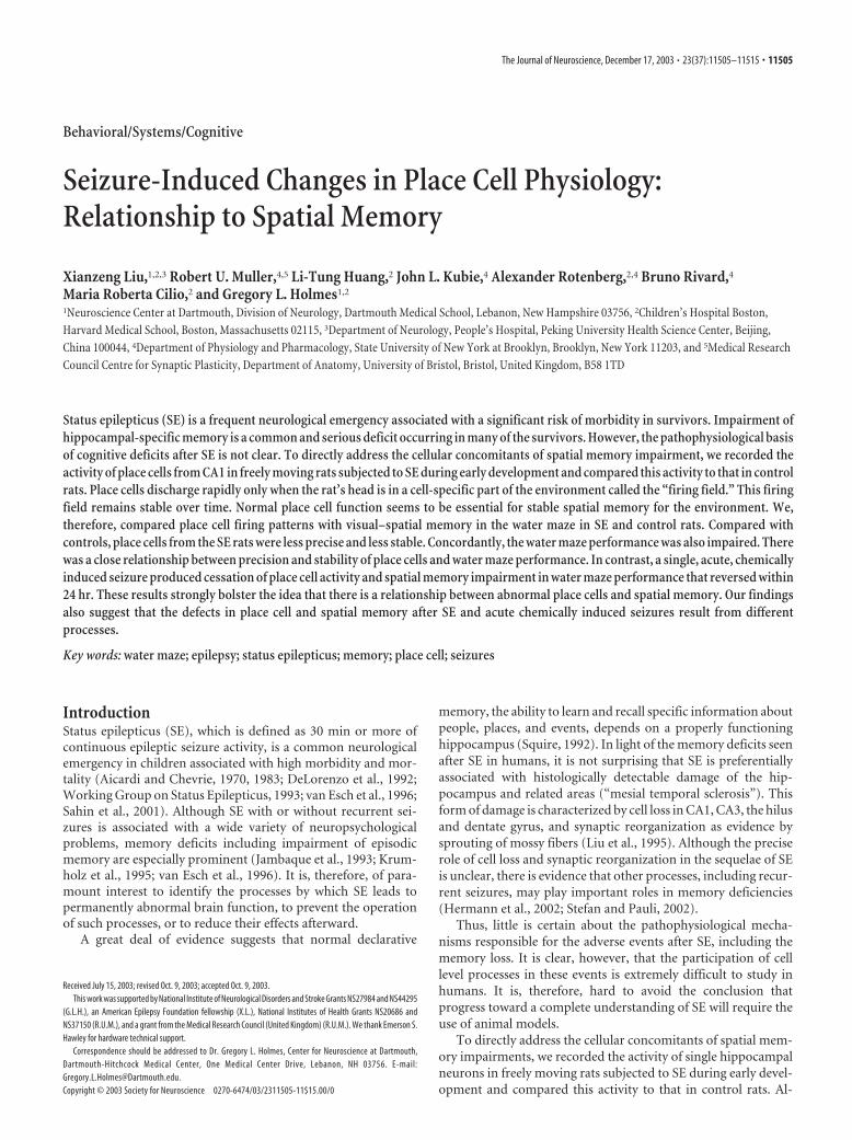
To permit unbiased estimates of the fraction of cells with differentproperties, we established a fixed rule: if the peak-to-peak amplitude ofany waveform was �150 �V, it was rejected; if the amplitude was �150�V, it was always recorded. After recording, discriminated waveformswere sorted into pyramidal cells and interneurons (Ranck, 1973; Fox andRanck, 1975, 1981). To be classified as a pyramidal cell, the unit had to:(1) fire complex spike bursts (decrementing spike sequences with inter-spike interval �5 msec) (Fig. 1); (2) have a negative initial phase �300�sec; and (3) show silent intervals of at least 1 sec in duration. To beclassified as an interneuron, the unit had to: (1) never fire complexbursts; (2) have a negative initial phase �300 �sec; and (3) never showsilent intervals �0.2 sec.
Before recordings, each electrode was checked for waveforms of suffi-cient amplitude. If none were detected, the electrode array was advanced20 �m and the rat was returned to its home cage for 4 – 6 hr. This se-quence was repeated until one or more pyramidal cells with �150 �Vwere isolated. Each cell or group of cells was recorded for 64 min in twopairs of 16 min sessions. The first pair of sessions was separated by a 2–3min interval. After the second session, the rat was returned to its home
11506 • J. Neurosci., December 17, 2003 • 23(37):11505–11515 Liu et al. • Alterations in Place Cells after Seizures
cage for 4 – 6 hr. It was then put back in the cylinder for sessions 3 and 4,which were also separated by a 2–3 min break. Thus, there were twoshort-interval session pairs (S1–S2 and S3–S4) and four long-intervalsession pairs (S1–S3, S1–S4, S2–S3, and S2–S4).
Tracking position. The position of a light-emitting diode (LED) on thehead was tracked with an overhead television camera at 60 Hz in a 64 �64 array of square pixels 2.7 cm on a side. The total time the LED wasdetected in each pixel, and the number of spikes fired in each pixel wasaccumulated. A time-averaged firing rate distribution was calculated bydividing the number of spikes in each pixel by the dwell time in that pixel.Color-coded firing rate maps were used to visualize positional firingdistributions. Pixel rates were coded in the sequence yellow, orange, red,green, blue, and purple. The firing rate was exactly zero for yellow pixels.Unvisited pixels in the cylinder and pixels outside the cylinder werecoded white.
EEG and video monitoring. In another eight SE rats and four controlrats, two additional electrodes on the same headset were used for record-ing the EEG. Two straight 75 �m nichrome wires were glued together,with one cut 0.8 –1.0 mm shorter than the other. The EEG electrodeswere fixed at 3.5 mm anteroposterior and 2.5 mm lateral, with respect tothe bregma, with the longer electrode aimed at the hippocampal fissureand the shorter electrode aimed dorsal to the CA1 stratum pyramidale.The 26 gauge cylinder through which the place cell electrodes were
threaded served as the reference electrode. The ground electrode for theEEG recordings was a skull screw placed over the right cerebellum. Thiselectrode arrangement allowed us to record fissure theta (Brazhnik et al.,2003). When the EEG was in the theta state, the two channels phasereversed relative to each other with the positive phase of the theta dis-played upward. The EEG was collected through the same preamplifierused for the place cell recording, then amplified 1000� times by anotherdifferential AC amplifier, bandpass filtered at 0.1–300 Hz, and digitizedat 1 kHz. Rats were monitored continuously for behavioral and electro-graphic seizures during the place cell recordings.
These 12 rats also underwent video monitoring every other night for 1month. Eight-hour recordings were from 6:00 P.M. to 2:00 A.M., and thetapes were reviewed the next day for behavioral seizures by experiencebehavioral technicians. All recorded seizures were also reviewed and ver-ified by G.L.H. or X.L. On scheduled recording days, rats were also videotaped for 6 hr before being placed in the behavioral chamber for the twoearly sessions, during the sessions, for 3– 4 hr between sessions, andduring the two late sessions. Animals with seizures during this period oftime were not studied. In addition, we recorded EEG during the place cellrecordings in eight rats in the experimental group.
Data analysis. Place cells were recorded in 8 of 12 control rats and 12 of18 SE rats. In four control rats and six SE rats, cells recorded did not meetour criteria for inclusion as place cells, noisy cells, or silent cells. Ourdescription of positional firing patterns is based on the idea that a placecell has a distinct, contiguous location in which it reliably discharges. Toidentify a firing field, the following rules were applied: (1) a pixel could beincluded in a field if its firing rate was �0; (2) to be part of a field, a pixelhad to share an edge with a pixel already known to be in the field; and (3)the minimum field size was nine pixels. In the present analysis, if two ormore fields were identified for a cell, only the largest field was considered.On this basis we measured: (1) field area: the number of pixels includedin the field; (2) field rate: the firing rate averaged over all pixels in thefield; (3) field center rate: for each pixel in the field, the average of its rateand the rate in its eight nearest neighbors is calculated. The peak rate isthat in the pixel for which this average is greatest; and (4) coherence: thisis a two-dimensional nearest-neighbor autocorrelation. It is calculatedby listing the firing rate in each pixel and the average firing rate in its eightnearest neighbors. Coherence is the Z-transform of the correlation be-tween these lists. It estimates the local smoothness of the positional firingpattern. An impression of the meaning of coherence can be gained fromFigure 2.
Based on positional firing rate distributions, CA1 complex–spike cellswere categorized as place cells, silent cells, or noisy cells (Rotenberg et al.,1996, 2000) (Fig. 1). Place cells had one, or in a few cases, two clear firingfields. Silent cells fired only a few action potentials without sufficientdensity to have a firing field. Noisy cells fired at an average rate �1.0spike/sec but had none of the distinct silent areas that characterize placecells recorded in these circumstances.
Using these criteria, an initial categorization was first made by visualinspection of firing rate maps by two investigators (G.L.H. and X.L.) whoagreed in all cases. To objectively classify place cells, we used criteria fromRotenberg et al. (1996). Thus, to be a place cell, the coherence was re-quired to be �0.26, and the area of the largest field had to be �70% of theapparatus area. Selection of place cells according to these criteria and byvisual inspection was in exact agreement.
The stability of firing fields across session pairs was assessed with arotational cross-correlation method. First, the pixel-by-pixel correlationwas calculated for a pair of positional firing patterns. One pattern wasthen rotated in 360 1° steps, and at each step the correlation was recalcu-lated. The magnitude of the highest of these correlations estimates thesimilarity of the two positional firing patterns. The angle of the highestcorrelation estimates how much one pattern is rotated with respect to theother. An ideal place cell would have identical firing-rate maps in eachpair of the recording sessions, so that the maximal similarity score wouldbe 1.0 and the required angular displacement would be 0°.
Theta frequency. We measured spectral power on EEG recordings us-ing Clampfit 8.2 (Axon Instruments, Union City, CA) during times therats were running in the cylinder for food pellets. Relative power for thefrequency band from 2 to 20 Hz was calculated by averaging 12 epochs of
Figure 1. Basic pyramidal cell properties. A, B, Complex spikes recorded from a control rat( A) and an SE rat ( B). Note the very short (�5 msec) time interval between spikes and thedecrementing amplitude of later spikes. No differences in the configuration of these bursts werenoted between normal and SE rats. C, D, Examples of silent, place, and noisy cells from a controlrat ( C) and an SE rat ( D). The firing field for the control place cell is the dark region at 1:00; for theplace cell from the SE rat, the field is at 8:30.
Liu et al. • Alterations in Place Cells after Seizures J. Neurosci., December 17, 2003 • 23(37):11505–11515 • 11507

10 sec duration at a spectral resolution of 0.15 Hz. Mean frequency ofhighest power in the theta range was calculated for the SE and controlgroups.
Water maze. Two hidden-platform water maze experiments were doneon SE rats and matched controls by a blinded observer using techniquesdescribed previously (Holmes et al., 1998; Faverjon et al., 2002; Rutten etal., 2002). These were identical in design, except for the diameter of thewater maze and the corresponding platform location. The first experi-ment, in a 1.15 M diameter tank, was done on the same rats from whichsingle cell recordings were obtained. The second experiment, in a 2 M
diameter tank, was done on 10 control and 19 SE rats age matched to therecording groups, so that training and testing started at day P100. Thisgroup included the animals that had EEG recordings in addition to theplace cell recordings.
In both experiments, a circular tank (50 cm high) was filled with water(26 � 1°C) to a depth of 25 cm. Milk was added to the water to preventthe rat from seeing the platform. Room cues visible from the water sur-face were constant from day to day. Four points on the perimeter of thetank were designated north (N), east (E), south (S), and west (W), divid-ing the tank into four quadrants (NW, NE, SE, and SW). For each rat, an8 � 8 cm Plexiglas escape platform was positioned in the center of one ofthe quadrants, 1 cm below the water surface. The radial distance from thecenter was 0.5 of the radius of the tank.
In our protocol, each rat was put in the tank for a 60 sec swim with noplatform on day 1. On days 2–5, the rat received six timed, hidden plat-form trials with the platform in the same quadrant across days. Thestarting quadrant for each trial was varied in pseudo-random manner.Time to platform was recorded by an observer blinded to the treatmentgroup. In each trial, the rat was allowed 2 min to find the platform, whereit could stay for 30 sec. If it failed to escape in 120 sec it was removed fromthe water and put on the platform for 30 sec. At the end of a trial, the ratwas lifted from the platform, dried with a towel, and put in the water forthe next trial. On day 6, a probe test was done without the platform. Theprobe began with the rat in the quadrant opposite the trained platformlocation. The rat swam for 60 sec, and the time spent in each quadrantwas recorded.
The testing procedure used during the 4 d of locating the hidden
platform provides a measure of spatial reference memory, whereas theprobe trial measures the strength of spatial memory (Jeltsch et al., 2001).
Place cell recording and water maze test after flurothyl-induced seizures.We used a chemical convulsant to produce acute epileptic seizures toassess: (1) the short-term consequences of epileptic seizures on place cellactivity; and (2) relationships between possible epileptically induced,short-term changes in place cells and water maze performance. Seizureswere induced in rats already trained in the water maze at a time whenstable, identified place cells were being recorded.
The sequence of experience for five control rats was: (1) using theprotocol described above plus an additional day, if necessary, rats weretrained to asymptotic performance in the 1.15 M diameter water maze.The criterion was for the average latency to the platform to be �10 sec forthree consecutive trials; (2) pellet chasing training for place cell record-ings as described above; (3) electrode implantation; (4) place cell record-ing; (5) demonstration of place cell stability. By this, we mean that theangular displacement for recognizable firing fields was �10° for three ormore consecutive days; 6) retraining to asymptotic performance in thewater maze immediately after the recordings. This was done on the re-cording day; 7) flurothyl-induced seizure (see below); and (8) alternationbetween single hidden platform trials in the water maze and place cellrecordings. Single trials followed by recording were done 30, 60, and 120min and 24 hr after the flurothyl seizure.
Brief seizures were induced with flurothyl (bis-2,2,2-triflurothyl ether;Aldrich Chemical Co., Milwaukee, WI) (Holmes et al., 1998; Huang etal., 1999; Villeneuve et al., 2000; Sogawa et al., 2001), a rapidly acting,potent CNS stimulant. After putting a rat in a small container, a piece offlurothyl-saturated filter paper was suspended from the container neckand the lid was put on. The flurothyl evaporated and was inhaled by therat. Typically, flurothyl caused, in sequence, myoclonic seizures, vigor-ous running, and tonic and clonic activity. When tonic posturing devel-oped, the rat was removed from the container to recover in room air.
Histology: Timm staining. At the end of the experiment, rats were killedwith a lethal dose of sodium pentobarbital (65 mg/kg) and perfusedtranscardially with 200 ml of normal saline, 200 ml of sodium sulfidemedium (2.925 gm of Na2S and 2.975 gm of NaH2PO4.H2O in 500 ml ofH2O), and 200 ml of 4% paraformaldehyde (PFA). The brains wereremoved, postfixed in 4% PFA for 24 hr, and placed in 30% sucrose for 24hr or longer until the brains sank. Coronal sections along the entireextent of the hippocampus were cut at 30 �M on a freezing microtomeand stored in PBS. Every fourth section was Timm stained for mossyfibers.
For Timm staining, sections were developed in the dark for 40 – 45 minin a solution of 50% gum arabic (120 ml), 10 ml of citric acid (51 gm/100ml H2O), 10 ml of sodium citrate (47 gm/100 ml H2O), 3.47 gm ofhydroquinone in 60 ml of H2O, and 212 mg of AgNO3. Slides fromcontrol and SE rats were always stained at the same time. After washing,the slides were dehydrated in alcohol, cleared in xylene, and sealed withPermount.
Timm-stained sections were analyzed using a semiquantitative scalefor terminal sprouting in CA3 and the supragranular region (Holmes etal., 1998, 1999). Timm staining in the pyramidal and infrapyramidal CA3region and supragranular region was assessed on each section from theseptal area in which the two blades of the dentate were equal and formeda V shape (2.8 mm posterior from the bregma) to a point �3.8 mmposterior to the bregma (Paxinos and Watson, 1998). Assessment of theTimm score in the supragranular region was done in the inferior blade ofthe dentate, avoiding the edge and crest of the gyrus. Both hippocampi ofthe specimens were analyzed, and the score given to the CA3 and supra-granular regions reflected the mean for the two sides. Five sections per ratwere evaluated.
Histology: thionin staining. To grade the degree of pathology present inportions of the hippocampus, thionin-stained slides were prepared fromevery fourth section. CA1, CA3, and the hilus were graded separately onthe following scale: no cell loss � 0; cell loss �25% � 1; cell loss 25–50% � 2; cell loss 50 –75% � 3; no remaining cells � 4 (Mikati et al.,1994; Schmid et al., 1999). Values for each region were taken as averagesfor the two hippocampi.
Statistical analysis. The values of all parameters in electrophysiological,
Figure 2. Organization of firing fields measured by coherence. Examples of firing rate mapsfrom control ( A) and SE rats ( B). The number above each map is the coherence for the spatialfiring pattern. The examples are chosen to reflect the coherence distributions for the twogroups. C, D, Coherence histograms for 69 place cells from control rats ( C) and 66 place cells fromSE rats ( D). Each coherence is the mean for all sessions for a given cell.
11508 • J. Neurosci., December 17, 2003 • 23(37):11505–11515 Liu et al. • Alterations in Place Cells after Seizures
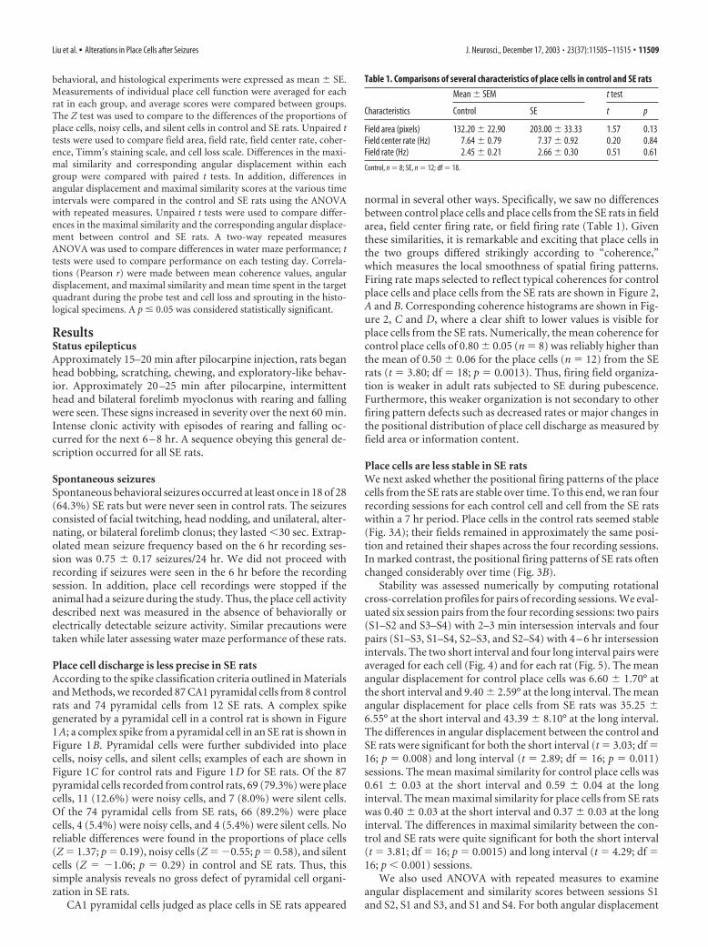
behavioral, and histological experiments were expressed as mean � SE.Measurements of individual place cell function were averaged for eachrat in each group, and average scores were compared between groups.The Z test was used to compare to the differences of the proportions ofplace cells, noisy cells, and silent cells in control and SE rats. Unpaired ttests were used to compare field area, field rate, field center rate, coher-ence, Timm’s staining scale, and cell loss scale. Differences in the maxi-mal similarity and corresponding angular displacement within eachgroup were compared with paired t tests. In addition, differences inangular displacement and maximal similarity scores at the various timeintervals were compared in the control and SE rats using the ANOVAwith repeated measures. Unpaired t tests were used to compare differ-ences in the maximal similarity and the corresponding angular displace-ment between control and SE rats. A two-way repeated measuresANOVA was used to compare differences in water maze performance; ttests were used to compare performance on each testing day. Correla-tions (Pearson r) were made between mean coherence values, angulardisplacement, and maximal similarity and mean time spent in the targetquadrant during the probe test and cell loss and sprouting in the histo-logical specimens. A p � 0.05 was considered statistically significant.
ResultsStatus epilepticusApproximately 15–20 min after pilocarpine injection, rats beganhead bobbing, scratching, chewing, and exploratory-like behav-ior. Approximately 20 –25 min after pilocarpine, intermittenthead and bilateral forelimb myoclonus with rearing and fallingwere seen. These signs increased in severity over the next 60 min.Intense clonic activity with episodes of rearing and falling oc-curred for the next 6 – 8 hr. A sequence obeying this general de-scription occurred for all SE rats.
Spontaneous seizuresSpontaneous behavioral seizures occurred at least once in 18 of 28(64.3%) SE rats but were never seen in control rats. The seizuresconsisted of facial twitching, head nodding, and unilateral, alter-nating, or bilateral forelimb clonus; they lasted �30 sec. Extrap-olated mean seizure frequency based on the 6 hr recording ses-sion was 0.75 � 0.17 seizures/24 hr. We did not proceed withrecording if seizures were seen in the 6 hr before the recordingsession. In addition, place cell recordings were stopped if theanimal had a seizure during the study. Thus, the place cell activitydescribed next was measured in the absence of behaviorally orelectrically detectable seizure activity. Similar precautions weretaken while later assessing water maze performance of these rats.
Place cell discharge is less precise in SE ratsAccording to the spike classification criteria outlined in Materialsand Methods, we recorded 87 CA1 pyramidal cells from 8 controlrats and 74 pyramidal cells from 12 SE rats. A complex spikegenerated by a pyramidal cell in a control rat is shown in Figure1A; a complex spike from a pyramidal cell in an SE rat is shown inFigure 1B. Pyramidal cells were further subdivided into placecells, noisy cells, and silent cells; examples of each are shown inFigure 1C for control rats and Figure 1D for SE rats. Of the 87pyramidal cells recorded from control rats, 69 (79.3%) were placecells, 11 (12.6%) were noisy cells, and 7 (8.0%) were silent cells.Of the 74 pyramidal cells from SE rats, 66 (89.2%) were placecells, 4 (5.4%) were noisy cells, and 4 (5.4%) were silent cells. Noreliable differences were found in the proportions of place cells(Z � 1.37; p � 0.19), noisy cells (Z � �0.55; p � 0.58), and silentcells (Z � �1.06; p � 0.29) in control and SE rats. Thus, thissimple analysis reveals no gross defect of pyramidal cell organi-zation in SE rats.
CA1 pyramidal cells judged as place cells in SE rats appeared
normal in several other ways. Specifically, we saw no differencesbetween control place cells and place cells from the SE rats in fieldarea, field center firing rate, or field firing rate (Table 1). Giventhese similarities, it is remarkable and exciting that place cells inthe two groups differed strikingly according to “coherence,”which measures the local smoothness of spatial firing patterns.Firing rate maps selected to reflect typical coherences for controlplace cells and place cells from the SE rats are shown in Figure 2,A and B. Corresponding coherence histograms are shown in Fig-ure 2, C and D, where a clear shift to lower values is visible forplace cells from the SE rats. Numerically, the mean coherence forcontrol place cells of 0.80 � 0.05 (n � 8) was reliably higher thanthe mean of 0.50 � 0.06 for the place cells (n � 12) from the SErats (t � 3.80; df � 18; p � 0.0013). Thus, firing field organiza-tion is weaker in adult rats subjected to SE during pubescence.Furthermore, this weaker organization is not secondary to otherfiring pattern defects such as decreased rates or major changes inthe positional distribution of place cell discharge as measured byfield area or information content.
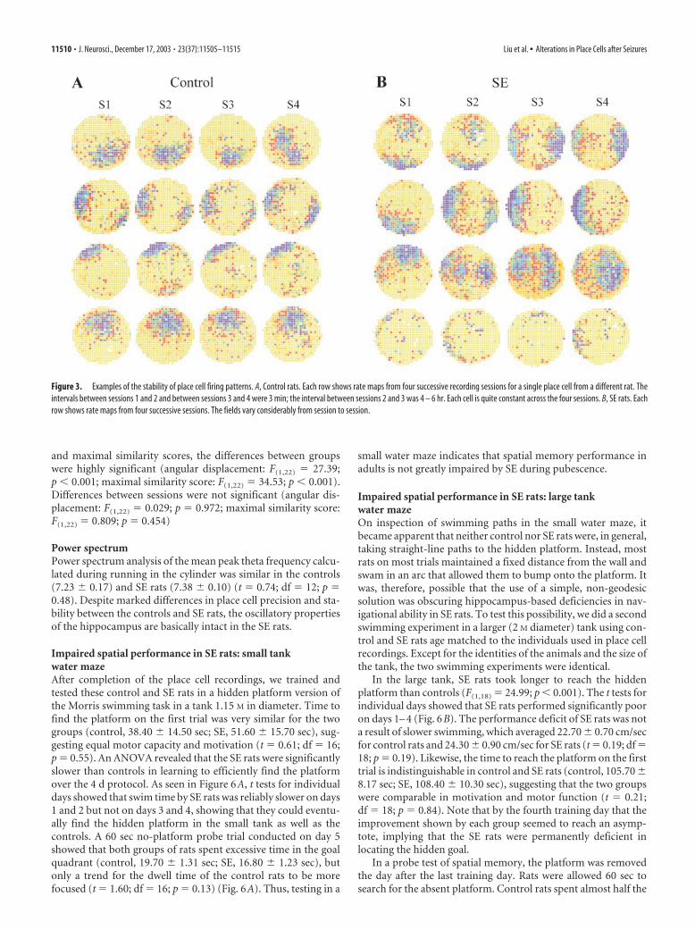
Place cells are less stable in SE ratsWe next asked whether the positional firing patterns of the placecells from the SE rats are stable over time. To this end, we ran fourrecording sessions for each control cell and cell from the SE ratswithin a 7 hr period. Place cells in the control rats seemed stable(Fig. 3A); their fields remained in approximately the same posi-tion and retained their shapes across the four recording sessions.In marked contrast, the positional firing patterns of SE rats oftenchanged considerably over time (Fig. 3B).
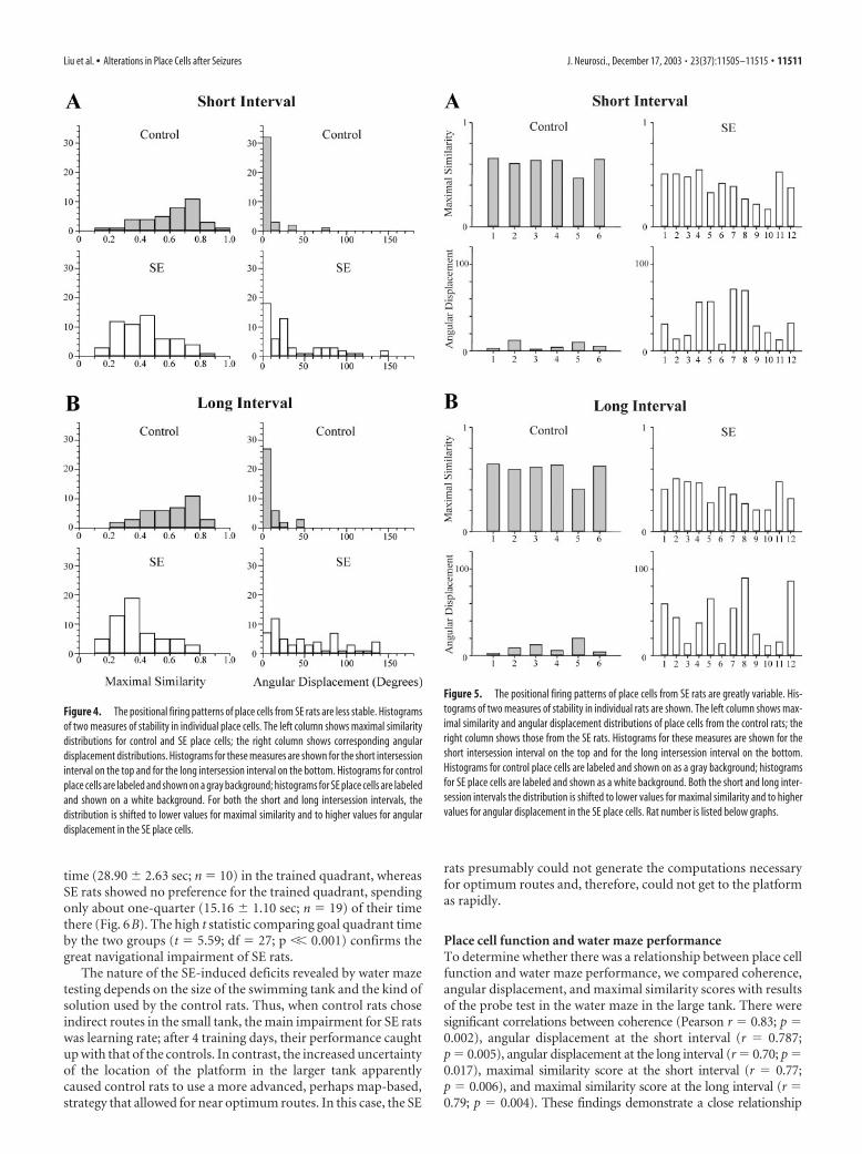
Stability was assessed numerically by computing rotationalcross-correlation profiles for pairs of recording sessions. We eval-uated six session pairs from the four recording sessions: two pairs(S1–S2 and S3–S4) with 2–3 min intersession intervals and fourpairs (S1–S3, S1–S4, S2–S3, and S2–S4) with 4 – 6 hr intersessionintervals. The two short interval and four long interval pairs wereaveraged for each cell (Fig. 4) and for each rat (Fig. 5). The meanangular displacement for control place cells was 6.60 � 1.70° atthe short interval and 9.40 � 2.59° at the long interval. The meanangular displacement for place cells from SE rats was 35.25 �6.55° at the short interval and 43.39 � 8.10° at the long interval.The differences in angular displacement between the control andSE rats were significant for both the short interval (t � 3.03; df �16; p � 0.008) and long interval (t � 2.89; df � 16; p � 0.011)sessions. The mean maximal similarity for control place cells was0.61 � 0.03 at the short interval and 0.59 � 0.04 at the longinterval. The mean maximal similarity for place cells from SE ratswas 0.40 � 0.03 at the short interval and 0.37 � 0.03 at the longinterval. The differences in maximal similarity between the con-trol and SE rats were quite significant for both the short interval(t � 3.81; df � 16; p � 0.0015) and long interval (t � 4.29; df �16; p � 0.001) sessions.
We also used ANOVA with repeated measures to examineangular displacement and similarity scores between sessions S1and S2, S1 and S3, and S1 and S4. For both angular displacement
Table 1. Comparisons of several characteristics of place cells in control and SE rats
Characteristics
Mean � SEM t test
Control SE t p
Field area (pixels) 132.20 � 22.90 203.00 � 33.33 1.57 0.13Field center rate (Hz) 7.64 � 0.79 7.37 � 0.92 0.20 0.84Field rate (Hz) 2.45 � 0.21 2.66 � 0.30 0.51 0.61
Control, n � 8; SE, n � 12; df � 18.
Liu et al. • Alterations in Place Cells after Seizures J. Neurosci., December 17, 2003 • 23(37):11505–11515 • 11509
and maximal similarity scores, the differences between groupswere highly significant (angular displacement: F(1,22) � 27.39;p � 0.001; maximal similarity score: F(1,22) � 34.53; p � 0.001).Differences between sessions were not significant (angular dis-placement: F(1,22) � 0.029; p � 0.972; maximal similarity score:F(1,22) � 0.809; p � 0.454)
Power spectrumPower spectrum analysis of the mean peak theta frequency calcu-lated during running in the cylinder was similar in the controls(7.23 � 0.17) and SE rats (7.38 � 0.10) (t � 0.74; df � 12; p �0.48). Despite marked differences in place cell precision and sta-bility between the controls and SE rats, the oscillatory propertiesof the hippocampus are basically intact in the SE rats.
Impaired spatial performance in SE rats: small tankwater mazeAfter completion of the place cell recordings, we trained andtested these control and SE rats in a hidden platform version ofthe Morris swimming task in a tank 1.15 M in diameter. Time tofind the platform on the first trial was very similar for the twogroups (control, 38.40 � 14.50 sec; SE, 51.60 � 15.70 sec), sug-gesting equal motor capacity and motivation (t � 0.61; df � 16;p � 0.55). An ANOVA revealed that the SE rats were significantlyslower than controls in learning to efficiently find the platformover the 4 d protocol. As seen in Figure 6A, t tests for individualdays showed that swim time by SE rats was reliably slower on days1 and 2 but not on days 3 and 4, showing that they could eventu-ally find the hidden platform in the small tank as well as thecontrols. A 60 sec no-platform probe trial conducted on day 5showed that both groups of rats spent excessive time in the goalquadrant (control, 19.70 � 1.31 sec; SE, 16.80 � 1.23 sec), butonly a trend for the dwell time of the control rats to be morefocused (t � 1.60; df � 16; p � 0.13) (Fig. 6A). Thus, testing in a
small water maze indicates that spatial memory performance inadults is not greatly impaired by SE during pubescence.
Impaired spatial performance in SE rats: large tankwater mazeOn inspection of swimming paths in the small water maze, itbecame apparent that neither control nor SE rats were, in general,taking straight-line paths to the hidden platform. Instead, mostrats on most trials maintained a fixed distance from the wall andswam in an arc that allowed them to bump onto the platform. Itwas, therefore, possible that the use of a simple, non-geodesicsolution was obscuring hippocampus-based deficiencies in nav-igational ability in SE rats. To test this possibility, we did a secondswimming experiment in a larger (2 M diameter) tank using con-trol and SE rats age matched to the individuals used in place cellrecordings. Except for the identities of the animals and the size ofthe tank, the two swimming experiments were identical.
In the large tank, SE rats took longer to reach the hiddenplatform than controls (F(1,18) � 24.99; p � 0.001). The t tests forindividual days showed that SE rats performed significantly pooron days 1– 4 (Fig. 6B). The performance deficit of SE rats was nota result of slower swimming, which averaged 22.70 � 0.70 cm/secfor control rats and 24.30 � 0.90 cm/sec for SE rats (t � 0.19; df �18; p � 0.19). Likewise, the time to reach the platform on the firsttrial is indistinguishable in control and SE rats (control, 105.70 �8.17 sec; SE, 108.40 � 10.30 sec), suggesting that the two groupswere comparable in motivation and motor function (t � 0.21;df � 18; p � 0.84). Note that by the fourth training day that theimprovement shown by each group seemed to reach an asymp-tote, implying that the SE rats were permanently deficient inlocating the hidden goal.
In a probe test of spatial memory, the platform was removedthe day after the last training day. Rats were allowed 60 sec tosearch for the absent platform. Control rats spent almost half the
Figure 3. Examples of the stability of place cell firing patterns. A, Control rats. Each row shows rate maps from four successive recording sessions for a single place cell from a different rat. Theintervals between sessions 1 and 2 and between sessions 3 and 4 were 3 min; the interval between sessions 2 and 3 was 4 – 6 hr. Each cell is quite constant across the four sessions. B, SE rats. Eachrow shows rate maps from four successive sessions. The fields vary considerably from session to session.
11510 • J. Neurosci., December 17, 2003 • 23(37):11505–11515 Liu et al. • Alterations in Place Cells after Seizures
time (28.90 � 2.63 sec; n � 10) in the trained quadrant, whereasSE rats showed no preference for the trained quadrant, spendingonly about one-quarter (15.16 � 1.10 sec; n � 19) of their timethere (Fig. 6B). The high t statistic comparing goal quadrant timeby the two groups (t � 5.59; df � 27; p �� 0.001) confirms thegreat navigational impairment of SE rats.
The nature of the SE-induced deficits revealed by water mazetesting depends on the size of the swimming tank and the kind ofsolution used by the control rats. Thus, when control rats choseindirect routes in the small tank, the main impairment for SE ratswas learning rate; after 4 training days, their performance caughtup with that of the controls. In contrast, the increased uncertaintyof the location of the platform in the larger tank apparentlycaused control rats to use a more advanced, perhaps map-based,strategy that allowed for near optimum routes. In this case, the SE
rats presumably could not generate the computations necessaryfor optimum routes and, therefore, could not get to the platformas rapidly.
Place cell function and water maze performanceTo determine whether there was a relationship between place cellfunction and water maze performance, we compared coherence,angular displacement, and maximal similarity scores with resultsof the probe test in the water maze in the large tank. There weresignificant correlations between coherence (Pearson r � 0.83; p �0.002), angular displacement at the short interval (r � 0.787;p � 0.005), angular displacement at the long interval (r � 0.70; p �0.017), maximal similarity score at the short interval (r � 0.77;p � 0.006), and maximal similarity score at the long interval (r �0.79; p � 0.004). These findings demonstrate a close relationship
Figure 4. The positional firing patterns of place cells from SE rats are less stable. Histogramsof two measures of stability in individual place cells. The left column shows maximal similaritydistributions for control and SE place cells; the right column shows corresponding angulardisplacement distributions. Histograms for these measures are shown for the short intersessioninterval on the top and for the long intersession interval on the bottom. Histograms for controlplace cells are labeled and shown on a gray background; histograms for SE place cells are labeledand shown on a white background. For both the short and long intersession intervals, thedistribution is shifted to lower values for maximal similarity and to higher values for angulardisplacement in the SE place cells.
Figure 5. The positional firing patterns of place cells from SE rats are greatly variable. His-tograms of two measures of stability in individual rats are shown. The left column shows max-imal similarity and angular displacement distributions of place cells from the control rats; theright column shows those from the SE rats. Histograms for these measures are shown for theshort intersession interval on the top and for the long intersession interval on the bottom.Histograms for control place cells are labeled and shown on as a gray background; histogramsfor SE place cells are labeled and shown as a white background. Both the short and long inter-session intervals the distribution is shifted to lower values for maximal similarity and to highervalues for angular displacement in the SE place cells. Rat number is listed below graphs.
Liu et al. • Alterations in Place Cells after Seizures J. Neurosci., December 17, 2003 • 23(37):11505–11515 • 11511
between place cell precision and stabilitywith water maze performance.
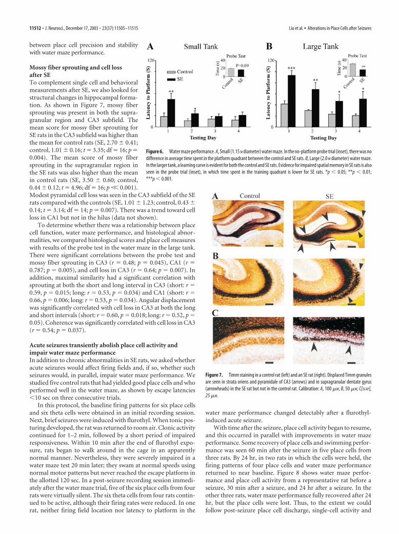
Mossy fiber sprouting and cell lossafter SETo complement single cell and behavioralmeasurements after SE, we also looked forstructural changes in hippocampal forma-tion. As shown in Figure 7, mossy fibersprouting was present in both the supra-granular region and CA3 subfield. Themean score for mossy fiber sprouting forSE rats in the CA3 subfield was higher thanthe mean for control rats (SE, 2.70 � 0.41;control, 1.01 � 0.16; t � 3.35; df � 16; p �0.004). The mean score of mossy fibersprouting in the supragranular region inthe SE rats was also higher than the meanin control rats (SE, 3.50 � 0.60; control,0.44 � 0.12; t � 4.96; df � 16; p �� 0.001).Modest pyramidal cell loss was seen in the CA3 subfield of the SErats compared with the controls (SE, 1.01 � 1.23; control, 0.43 �0.14; t � 3.14; df � 14; p � 0.007). There was a trend toward cellloss in CA1 but not in the hilus (data not shown).
To determine whether there was a relationship between placecell function, water maze performance, and histological abnor-malities, we compared histological scores and place cell measureswith results of the probe test in the water maze in the large tank.There were significant correlations between the probe test andmossy fiber sprouting in CA3 (r � 0.48; p � 0.045), CA1 (r �0.787; p � 0.005), and cell loss in CA3 (r � 0.64; p � 0.007). Inaddition, maximal similarity had a significant correlation withsprouting at both the short and long interval in CA3 (short: r �0.59, p � 0.015; long: r � 0.53, p � 0.034) and CA1 (short: r �0.66, p � 0.006; long: r � 0.53, p � 0.034). Angular displacementwas significantly correlated with cell loss in CA3 at both the longand short intervals (short: r � 0.60, p � 0.018; long: r � 0.52, p �0.05). Coherence was significantly correlated with cell loss in CA3(r � 0.54; p � 0.037).
Acute seizures transiently abolish place cell activity andimpair water maze performanceIn addition to chronic abnormalities in SE rats, we asked whetheracute seizures would affect firing fields and, if so, whether suchseizures would, in parallel, impair water maze performance. Westudied five control rats that had yielded good place cells and whoperformed well in the water maze, as shown by escape latencies�10 sec on three consecutive trials.
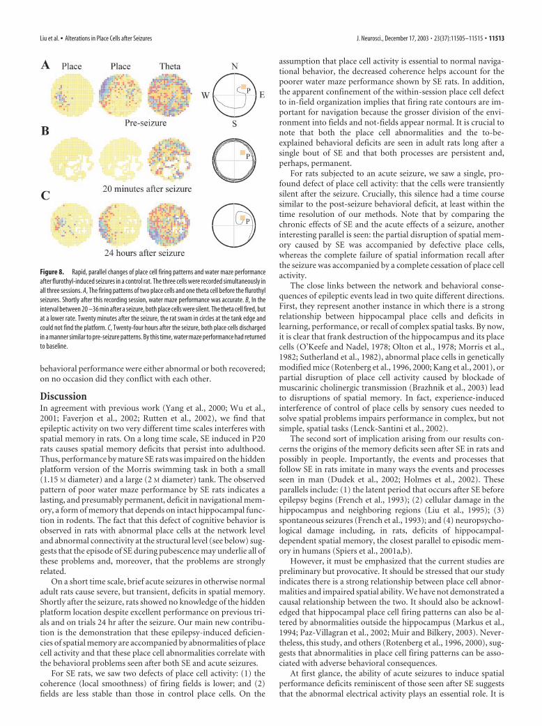
In this protocol, the baseline firing patterns for six place cellsand six theta cells were obtained in an initial recording session.Next, brief seizures were induced with flurothyl. When tonic pos-turing developed, the rat was returned to room air. Clonic activitycontinued for 1–2 min, followed by a short period of impairedresponsiveness. Within 10 min after the end of flurothyl expo-sure, rats began to walk around in the cage in an apparentlynormal manner. Nevertheless, they were severely impaired in awater maze test 20 min later; they swam at normal speeds usingnormal motor patterns but never reached the escape platform inthe allotted 120 sec. In a post-seizure recording session immedi-ately after the water maze trial, five of the six place cells from fourrats were virtually silent. The six theta cells from four rats contin-ued to be active, although their firing rates were reduced. In onerat, neither firing field location nor latency to platform in the
water maze performance changed detectably after a flurothyl-induced acute seizure.
With time after the seizure, place cell activity began to resume,and this occurred in parallel with improvements in water mazeperformance. Some recovery of place cells and swimming perfor-mance was seen 60 min after the seizure in five place cells fromthree rats. By 24 hr, in two rats in which the cells were held, thefiring patterns of four place cells and water maze performancereturned to near baseline. Figure 8 shows water maze perfor-mance and place cell activity from a representative rat before aseizure, 30 min after a seizure, and 24 hr after a seizure. In theother three rats, water maze performance fully recovered after 24hr, but the place cells were lost. Thus, to the extent we couldfollow post-seizure place cell discharge, single-cell activity and
Figure 6. Water maze performance. A, Small (1.15 M diameter) water maze. In the no-platform probe trial (inset), there was nodifference in average time spent in the platform quadrant between the control and SE rats. B, Large (2.0 M diameter) water maze.In the larger tank, a learning curve is evident for both the control and SE rats. Evidence for impaired spatial memory in SE rats is alsoseen in the probe trial (inset), in which time spent in the training quadrant is lower for SE rats. *p � 0.05; **p � 0.01;***p � 0.001.
Figure 7. Timm staining in a control rat (left) and an SE rat (right). Displaced Timm granulesare seen in strata oriens and pyramidale of CA3 (arrows) and in supragranular dentate gyrus(arrowheads) in the SE rat but not in the control rat. Calibration: A, 100 �M; B, 50 �M; C[SCAP],25 �M.
11512 • J. Neurosci., December 17, 2003 • 23(37):11505–11515 Liu et al. • Alterations in Place Cells after Seizures
behavioral performance were either abnormal or both recovered;on no occasion did they conflict with each other.
DiscussionIn agreement with previous work (Yang et al., 2000; Wu et al.,2001; Faverjon et al., 2002; Rutten et al., 2002), we find thatepileptic activity on two very different time scales interferes withspatial memory in rats. On a long time scale, SE induced in P20rats causes spatial memory deficits that persist into adulthood.Thus, performance by mature SE rats was impaired on the hiddenplatform version of the Morris swimming task in both a small(1.15 M diameter) and a large (2 M diameter) tank. The observedpattern of poor water maze performance by SE rats indicates alasting, and presumably permanent, deficit in navigational mem-ory, a form of memory that depends on intact hippocampal func-tion in rodents. The fact that this defect of cognitive behavior isobserved in rats with abnormal place cells at the network leveland abnormal connectivity at the structural level (see below) sug-gests that the episode of SE during pubescence may underlie all ofthese problems and, moreover, that the problems are stronglyrelated.
On a short time scale, brief acute seizures in otherwise normaladult rats cause severe, but transient, deficits in spatial memory.Shortly after the seizure, rats showed no knowledge of the hiddenplatform location despite excellent performance on previous tri-als and on trials 24 hr after the seizure. Our main new contribu-tion is the demonstration that these epilepsy-induced deficien-cies of spatial memory are accompanied by abnormalities of placecell activity and that these place cell abnormalities correlate withthe behavioral problems seen after both SE and acute seizures.
For SE rats, we saw two defects of place cell activity: (1) thecoherence (local smoothness) of firing fields is lower; and (2)fields are less stable than those in control place cells. On the
assumption that place cell activity is essential to normal naviga-tional behavior, the decreased coherence helps account for thepoorer water maze performance shown by SE rats. In addition,the apparent confinement of the within-session place cell defectto in-field organization implies that firing rate contours are im-portant for navigation because the grosser division of the envi-ronment into fields and not-fields appear normal. It is crucial tonote that both the place cell abnormalities and the to-be-explained behavioral deficits are seen in adult rats long after asingle bout of SE and that both processes are persistent and,perhaps, permanent.
For rats subjected to an acute seizure, we saw a single, pro-found defect of place cell activity: that the cells were transientlysilent after the seizure. Crucially, this silence had a time coursesimilar to the post-seizure behavioral deficit, at least within thetime resolution of our methods. Note that by comparing thechronic effects of SE and the acute effects of a seizure, anotherinteresting parallel is seen: the partial disruption of spatial mem-ory caused by SE was accompanied by defective place cells,whereas the complete failure of spatial information recall afterthe seizure was accompanied by a complete cessation of place cellactivity.
The close links between the network and behavioral conse-quences of epileptic events lead in two quite different directions.First, they represent another instance in which there is a strongrelationship between hippocampal place cells and deficits inlearning, performance, or recall of complex spatial tasks. By now,it is clear that frank destruction of the hippocampus and its placecells (O’Keefe and Nadel, 1978; Olton et al., 1978; Morris et al.,1982; Sutherland et al., 1982), abnormal place cells in geneticallymodified mice (Rotenberg et al., 1996, 2000; Kang et al., 2001), orpartial disruption of place cell activity caused by blockade ofmuscarinic cholinergic transmission (Brazhnik et al., 2003) leadto disruptions of spatial memory. In fact, experience-inducedinterference of control of place cells by sensory cues needed tosolve spatial problems impairs performance in complex, but notsimple, spatial tasks (Lenck-Santini et al., 2002).
The second sort of implication arising from our results con-cerns the origins of the memory deficits seen after SE in rats andpossibly in people. Importantly, the events and processes thatfollow SE in rats imitate in many ways the events and processesseen in man (Dudek et al., 2002; Holmes et al., 2002). Theseparallels include: (1) the latent period that occurs after SE beforeepilepsy begins (French et al., 1993); (2) cellular damage in thehippocampus and neighboring regions (Liu et al., 1995); (3)spontaneous seizures (French et al., 1993); and (4) neuropsycho-logical damage including, in rats, deficits of hippocampal-dependent spatial memory, the closest parallel to episodic mem-ory in humans (Spiers et al., 2001a,b).
However, it must be emphasized that the current studies arepreliminary but provocative. It should be stressed that our studyindicates there is a strong relationship between place cell abnor-malities and impaired spatial ability. We have not demonstrated acausal relationship between the two. It should also be acknowl-edged that hippocampal place cell firing patterns can also be al-tered by abnormalities outside the hippocampus (Markus et al.,1994; Paz-Villagran et al., 2002; Muir and Bilkery, 2003). Never-theless, this study, and others (Rotenberg et al., 1996, 2000), sug-gests that abnormalities in place cell firing patterns can be asso-ciated with adverse behavioral consequences.
At first glance, the ability of acute seizures to induce spatialperformance deficits reminiscent of those seen after SE suggeststhat the abnormal electrical activity plays an essential role. It is
Figure 8. Rapid, parallel changes of place cell firing patterns and water maze performanceafter flurothyl-induced seizures in a control rat. The three cells were recorded simultaneously inall three sessions. A, The firing patterns of two place cells and one theta cell before the flurothylseizures. Shortly after this recording session, water maze performance was accurate. B, In theinterval between 20 –36 min after a seizure, both place cells were silent. The theta cell fired, butat a lower rate. Twenty minutes after the seizure, the rat swam in circles at the tank edge andcould not find the platform. C, Twenty-four hours after the seizure, both place cells dischargedin a manner similar to pre-seizure patterns. By this time, water maze performance had returnedto baseline.
Liu et al. • Alterations in Place Cells after Seizures J. Neurosci., December 17, 2003 • 23(37):11505–11515 • 11513
here, however, that the ability to monitor place cell activity isshown to be of great advantage in elucidating relationships be-tween gross abnormal electrical activity and memory problems.Assume momentarily that preceding seizure activity is responsi-ble for poor performance in the water maze after SE as well asacute seizures. On this basis, we would expect that place celldischarge frequency in SE rats would be lower than in normalrats. But our place cell recordings indicate that discharge rate isno different in SE rats, despite the observation of other place celldefects that certainly can explain the performance deficits. Inaddition, we studied place cells in a subset of rats after SE that alsounderwent continuous EEG monitoring. The place cell abnor-malities reported here were not associated with any seizures be-fore or during the place cell recordings. Based on this evidence,we believe that permanent structural or functional rearrange-ments of hippocampal circuitry induced by SE are responsible forboth the place cell and spatial memory deficits.
A fundamental question is why SE in prepubescent rats shouldlead to abnormal place cells when they become adults. SE ratsresults in a myriad of changes in the hippocampus, including celldeath (Ben-Ari, 2001), increased neurogenesis (Sankar et al.,2001), changes in glutamate receptor distribution (Mathern etal., 1998; Mikuni et al., 1999) and glutamate subunit configura-tion (Friedman et al., 1997; Friedman, 1998), and synaptic reor-ganization with sprouting of mossy fibers (Sutula et al., 1988;Wuarin and Dudek, 2001). It is known that excessive mossy fibersprouting leads to abnormal synaptic connections that have beenassociated with performance in visual–spatial memory tasks(Crusio et al., 1987, 1993; de Rogalski Landrot et al., 2001;Holmes et al., 1998). Although it is tempting to implicate the cellloss and synaptic reorganization seen here to the impaired placecell firing patterns and deficits in spatial memory, it is importantto remember that correlation of two phenomena does not neces-sarily indicate causation. Teasing out the mechanisms responsi-ble for the cognitive impairment after SE will likely be complex.However, place cell firing patterns provides a powerful tool in theelucidation of those factors responsible for SE-induced cognitiveimpairment
ReferencesAicardi J, Chevrie JJ (1970) Convulsive status epilepticus in infants and chil-
dren. Epilepsia 11:187–197.Aicardi J, Chevrie JJ (1983) Consequences of status epilepticus in infants
and children. In: Advances in neurology, Vol 34, Status epilepticus: mech-anisms of brain damage and treatment (Delgado-Escueta AV, WasterlainCG, Treiman DM, Porter RJ, eds), pp 115–125. New York: Raven.
Ben-Ari Y (2001) Cell death and synaptic reorganizations produced by sei-zures. Epilepsia 42 [Suppl 3]:5–7.
Brazhnik ES, Muller RU, Fox SE (2003) Muscarinic blockade slows and de-grades the location-specific firing of hippocampal pyramidal cells. J Neu-rosci 23:611– 621.
Cilio MR, Sogawa Y, Cha BH, Liu X, Huang LT, Holmes GL (2003) Long-term effects of status epilepticus in the immature brain are age- andmodel-specific. Epilepsia 44:518 –528.
Crusio WE, Schwegler H, Lipp H-P (1987) Radial-maze performance andstructural variation of the hippocampus in mice: a correlation with mossyfiber distribution. Brain Res 425:182–185.
Crusio WE, Schwegler H, Brust I (1993) Covariations between hippocam-pal mossy fibers and working and reference memory in spatial and non-spatial radial maze tasks in mice. Eur J Neurosci 5:1413–1420.
de Rogalski Landrot I, Minokoshi M, Silveira DC, Cha BH, Holmes GL(2001) Recurrent neonatal seizures: relationship of pathology to the elec-troencephalogram and cognition. Dev Brain Res 129:27–38.
DeLorenzo RJ, Towne AR, Pellock JM, Ko D (1992) Status epilepticus inchildren, adults, and the elderly. Epilepsia 33 [Suppl 4]:S15–S25.
Dudek FE, Hellier JL, Williams PA, Dudek FE, Hellier JL, Williams PA (2002)
The course of cellular alterations associated with the development ofspontaneous seizures after status epilepticus. Prog Brain Res 135:53– 65.
Faverjon S, Silveira DC, Fu DD, Cha BH, Akman C, Hu Y, Holmes GL (2002)Beneficial effects of enriched environment following status epilepticus inimmature rats. Neurology 59:1356 –1364.
Fox SE, Ranck Jr JB (1975) Localization and anatomical identification oftheta and complex spike cells in dorsal hippocampal formation of rats.Exp Neurol 49:299 –313.
Fox SE, Ranck Jr JB (1981) Electrophysiological characteristics of hippoca-mapal complex-spike cells and theta cells. Exp Brain Res 41:399 – 410.
French JA, Williamson PD, Thadani VM, Darcey TM, Mattson RH, SpencerSS, Spencer DD (1993) Characteristics of medial temporal lobe epilepsy:I. Results of history and physical examination. Ann Neurol 34:774 –780.
Friedman LK (1998) Selective reduction of gluR2 protein in adult hip-pocampal CA3 neurons following status epilepticus but prior to cell loss.Hippocampus 8:511–525.
Friedman LK, Sperber EF, Moshe SL, Bennett MV, Zukin RS (1997) Devel-opmental regulation of glutamate and GABAA receptor gene expressionin rat hippocampus following kainate-induced status epilepticus. DevNeurosci 19:529 –542.
Hermann BP, Seidenberg M, Bell B (2002) The neurodevelopmental impactof childhood onset temporal lobe epilepsy on brain structure and func-tion and the risk of progressive cognitive effects. Prog Brain Res135:429 – 438.
Holmes GL, Gaiarsa J-L, Chevassus-Au-Louis N, Ben-Ari Y (1998) Conse-quences of neonatal seizures in the rat: morphological and behavioraleffects. Ann Neurol 44:845– 857.
Holmes GL, Sarkisian M, Ben-Ari Y, Chevassus-Au-Louis N (1999) Mossyfiber sprouting following recurrent seizures during early development inrats. J Comp Neurol 404:537–553.
Holmes GL, Khazipov R, Ben Ari Y (2002) Seizure-induced damage in thedeveloping human: relevance of experimental models. Prog Brain Res135:321–334.
Huang L, Cilio MR, Silveira DC, McCabe BK, Sogawa Y, Stafstrom CE,Holmes GL (1999) Long-term effects of neonatal seizures: a behavioral,electrophysiological, and histolological study. Brain Res Dev Brain Res118:99 –107.
Jambaque I, Dellatolas G, Dulac O, Ponsot G, Signoret JL (1993) Verbal andvisual memory impairment in children with epilepsy. Neuropsychologia31:1321–1337.
Jeltsch H, Bertrand F, Lazarus C, Cassel JC (2001) Cognitive performancesand locomotor activity following dentate granule cell damage in rats: roleof lesion extent and type of memory tested. Neurobiol Learn Mem76:81–105.
Kang H, Sun LD, Atkins CM, Soderling TR, Wilson MA, Tonegawa S (2001)An important role of neural activity-dependent CaMKIV signaling in theconsolidation of long-term memory. Cell 106:771–783.
Krumholz A, Sung GY, Fisher RS, Barry E, Bergey GK, Grattan LM (1995)Complex partial status epilepticus accompanied by serious morbidity andmortality. Neurology 45:1499 –1504.
Kubie JL (1984) A driveable bundle of microwires for collecting single-unitdata from freely-moving rats. Physiol Behav 32:115–118.
Lenck-Santini P-P, Muller RU, Save E, Poucet B (2002) Relationships be-tween place cell firing fields and navigational decisions by rats. J Neurosci22:9035–9047.
Liu Z, Mikati M, Holmes GL (1995) Mesial temporal sclerosis: pathogenesisand significance. Pediatr Neurol 12:5–16.
Markus EJ, Barnes CA, McNaughton BL, Gladden VL, Skaggs WE (1994)Spatial information content and reliability of hippocampal CA1 neurons:effects of visual input. Hippocampus 4:410 – 421.
Mathern GW, Pretorius JK, Mendoza D, Lozada A, Kornblum HI (1998)Hippocampal AMPA and NMDA mRNA levels correlate with aberrantfascia dentata mossy fiber sprouting in the pilocarpine model of sponta-neous limbic epilepsy. J Neurosci Res 54:734 –753.
Mikati MA, Holmes GL, Chronopoulos A, Hyde P, Thurber S, Gatt A, Liu Z,Werner S, Stafstrom CE (1994) Phenobarbital therapy modifies seizurerelated brain injury in the developing brain. Ann Neurol 36:425– 433.
Mikuni N, Babb TL, Christi W (1999) Increased NR1-NR2A/B coassemblyas a mechanism for rat chronic hippocampal epilepsy. Neruosci Lett267:165–168.
Morris RG, Garrud P, Rawlins JN, O’Keefe J (1982) Place navigation im-paired in rats with hippocampal lesions. Nature 297:681– 683.
11514 • J. Neurosci., December 17, 2003 • 23(37):11505–11515 Liu et al. • Alterations in Place Cells after Seizures

Muir GM, Bilkey DK (2003) Theta- and movement velocity-related firing ofhippocampal neurons is disrupted by lesions centered on the perirhinalcortex. Hippocampus 13:93–108.
Muller RU, Kubie JL (1987) The effects of changes in the environment onthe spatial firing patterns of hippocampal complex-spike cells. J Neurosci7:1951–1968.
Muller RU, Kubie JL, Ranck Jr JB (1987) Spatial firing patterns of hip-pocampal complex-spike cells in a fixed environment. J Neurosci7:1935–1950.
O’Keefe J, Nadel L (1978) The hippocampus as a cognitive map. Oxford:Clarendon.
Olton DS, Walker JA, Gage FH (1978) Hippocampal connections and spa-tial discrimination. Brain Res 139:295–308.
Paxinos G, Watson C (1998) The rat brain in stereotaxic coordinates, Ed 4.San Diego: Academic.
Paz-Villagran V, Lenck-Santini PP, Save E, Poucet B (2002) Properties ofplace cell firing after damage to the visual cortex. Eur J Neurosci16:771–776.
Ranck Jr JB (1973) Studies on single neurons in dorsal hippocampal forma-tion and septum in unrestrained rats. Exp Neurol 41:461–531.
Rotenberg A, Mayford M, Hawkins RD, Kandel ER, Muller RU (1996) Miceexpressing activated CaMKII lack low frequency LTP and do not formstable place cells in the CA1 region of the hippocampus. Cell87:1351–1361.
Rotenberg A, Abel T, Hawkins RD, Kandel ER, Muller RU (2000) Parallelinstabilities of long-term potentiation, place cells, and learning caused bydecreased protein kinase A activity. J Neurosci 20:8096 – 8102.
Rutten A, van Albada M, Silveira DC, Cha BH, Liu X, Hu YN, Cilio MR,Holmes GL (2002) Memory impairment following status epilepticus inimmature rats: time- course and environmental effects. Eur J Neurosci501–513.
Sahin M, Menache CC, Holmes GL, Riviello JJ (2001) Outcome of severerefractory status epilepticus in children. Epilepsia 42:1461–1467.
Sankar R, Shin D, Liu H, Katsumori H, Wasterlain CG (2001) Granule cellneurogenesis after status epilepticus in the immature rat brain. Epilepsia41 [Suppl 6]:S53–S56.
Schmid R, Tandon P, Stafstrom CE, Holmes GL (1999) Effects of neonatalseizures on subsequent seizure-induced brain injury. Neurology53:1754 –1761.
Shinnar S, Pellock JM, Moshe SL, Maytal J, O’Dell C, Driscoll SM, AlemanyM, Newstein D, DeLorenzo RJ (1997) In whom does status epilepticusoccur: age-related differences in children. Epilepsia 38:907–914.
Sogawa Y, Monokoshi M, Silveira DC, Cha BH, Cilio MR, McCabe BK, Liu X,Hu Y, Holmes GL (2001) Timing of cognitive deficits following neona-
tal seizures: relationship to histological changes in the hippocampus.Brain Res Dev Brain Res 131:73– 83.
Spiers HJ, Burgess N, Hartley T, Vargha-Khadem F, O’Keefe J (2001a) Bi-lateral hippocampal pathology impairs topographical and episodic mem-ory but not visual pattern matching. Hippocampus 11:715–725.
Spiers HJ, Burgess N, Maguire EA , Baxendale SA, Hartley T, Thompson PJ,O’Keefe J (2001b) Unilateral temporal lobectomy patients show lateral-ized topographical and episodic memory deficits in a virtual town. Brain124:2476 –2489.
Squire LR (1992) Memory and the hippocampus: a synthesis from findingswith rats, monkeys, and humans. Psychol Rev 99:195–231.
Stefan H, Pauli E (2002) Progressive cognitive decline in epilepsy: an indi-cation of ongoing plasticity. Prog Brain Res 135:409 – 417.
Sutherland RJ, Whishaw IQ, Kolb BA (1982) A behavioral analysis of spatiallocalization following electrolytic, kainate- or colchicine-induced damageto hippocampal formation in the rat. Behav Brain Res 7:133–153.
Sutula T, He X-X, Cavazos J, Scott G (1988) Synaptic reorganization in thehippocampus induced by abnormal functional activity. Science239:1147–1150.
Thompson LT, Best PJ (1989) Place cells and silent cells in the hippocampusof freely behaving rats. J Neurosci 9:2382–2390.
Thompson LT, Best PJ (1990) Long-term stability of the place-field activityof single units recorded from the dorsal hippocampus of freely behavingrats. Brain Res 509:299 –308.
van Esch A, Ramlal IR, van Steensel-Moll HA, Steyerberg EW, Derksen-Lubsen G (1996) Outcome after febrile status epilepticus. Dev MedChild Neurol 38:19 –24.
Villeneuve N, Ben-Ari Y, Holmes GL, Gaiarsa JL (2000) Neonatal seizuresinduced persistent changes in intrinsic properties of CA1 rat hippocampalcells. Ann Neurol 47:729 –738.
Working Group on Status Epilepticus (1993) Treatment of convulsive sta-tus epilepticus. Recommendations of the Epilepsy Foundation of Ameri-ca’s Working Group on Status Epilepticus. JAMA 270:854 – 859.
Wu CL, Huang LT, Liou CW, Wang TJ, Tung YR, Hsu HY, Lai MC (2001)Lithium-pilocarpine-induced status epilepticus in immature rats result inlong-term deficits in spatial learning and hippocampal cell loss. NeurosciLett 312:113–117.
Wuarin JP, Dudek FE (2001) Excitatory synaptic input to granule cells in-creases with time after kainate treatment. J Neurophysiol 85:1067–1077.
Yang Y, Liu Z, Cermak JM, Tandon P, Sarkisian MR, Stafstrom CE, Neill JC,Blusztajn JK, Holmes GL (2000) Protective effects of prenatal cholinesupplementation on seizure- induced memory impairment. J Neurosci20:RC109.
Liu et al. • Alterations in Place Cells after Seizures J. Neurosci., December 17, 2003 • 23(37):11505–11515 • 11515