Se2017 Program and Abstract Book
349
Se2017 Program and Abstract Book
-
Upload
khangminh22 -
Category
Documents
-
view
1 -
download
0
Transcript of Se2017 Program and Abstract Book

Se2017
Program and Abstract Book

Se2017 – 200 Years of Selenium Research Stockholm, Sweden, 13-‐17 August 2017
– ii –
Conference venue
Aula Medica The Berzelius and Scheele laboratories, with lecture halls Samuelsson, Vesalius and Retzius, as well as Poster and Exhibition room

Se2017 – 200 Years of Selenium Research Stockholm, Sweden, 13-‐17 August 2017
– iii –
Welcome!
The Se2017 Conference celebrates 200 Years of Selenium Research and is held at Karolinska Institutet in Stockholm, Sweden, on August 13-17, 2017. The conference covers all major fields of current selenium research, with a special focus on biology, medicine, biomedicine and the environment. The event is officially composed of two serial symposia; for the first time held in parallel at the same site and having overlapping plenary sessions, poster sessions and social events. The two symposia are The 11th International Symposium on Selenium in Biology and Medicine and The 5th International Conference on Selenium in the Environment and Human Health.
Please consult the conference website for further information and last-minute changes: www.Se2017.se
Welcome to Se2017!
Elias Arnér, MD PhD Chair, Se2017
Se2017 – 200 Years of Selenium Research Stockholm, Sweden, 13-‐17 August 2017
– iv –
Acknowledgements
Co-‐organizer
Gold sponsors
Se2017 – 200 Years of Selenium Research Stockholm, Sweden, 13-‐17 August 2017
– v –
Silver sponsors
Additional contributions
The Packer/Wentz Endowment
The Berzelius Society

Se2017 – 200 Years of Selenium Research Stockholm, Sweden, 13-‐17 August 2017
– vi –
Organizing committees Chair: Elias Arnér, Karolinska Institutet, Sweden
Executive Committee: Elias Arnér, Karolinska Institutet, Sweden Gary Bañuelos, USDA, USA Arne Holmgren, Karolinska Institutet, Sweden Zhi-‐Qing Lin, Southern Illinois University, Illinois, USA
Local Organizing Committee, Karolinska Institutet, Sweden: Elias Arnér Arne Holmgren Aristi Fernandes Jan Trofast
Co-‐Chair: Xuebin Yin, University of Science and Technology of China, China
International Scientific Advisory Committee: Björn Åkesson, Lund University, Sweden Marla Berry, University of Hawaii, USA Ohad Birk, Beer Sheva University, Israel Mikael Björnstedt, Karolinska Institutet, Sweden Martin Broadley, University of Nottingham, UK Raymond Burk, Vanderbilt University, USA Joel Caton, North Dakota State University, USA Allan Chilimba, Ministry of Agriculture and Food Security, Malawi Gerald Combs, USDA, USA Karaj S. Dhillon, Punjab Agricultural University, India Gijs DuLaing, Ghent University, Belgium Milton Ferreira Moraes, Federal University of Mato Grosso, Brazil Vadim Gladyshev, Harvard University, USA Luiz Roberto Guimarães Guilherme, Federal University of Lavras, Brazil Dolph Hatfield, NIH, USA John Hesketh, University of Newcastle, United Kingdom Kaixun Huang, Huazhong University of Science and Technology, China Anna Kipp, German Institute of Human Nutrition, DIfE, Potsdam, Germany Alain Krol, U-‐Strasbourg, CNRS, France Josef Köhrle, Charité-‐Universitätsmedizin Berlin, Germany Byeong Jae Lee, Seoul National University, South Korea Xingen Lei, Cornell University, USA Graham Lyons, The University of Adelaide, Australia Matilde Maiorino, University of Padova, Italy Steve McGrath, Rothamsted Research, UK Bernhard Michalke, Helmholtz München, Germany Margret Rayman, University of Surrey, United Kingdom Andre Rodrigues dos Reis, Sao Paulo State University, Brazil Michael Rother, Technical University of Dresden, Germany Lutz Schomburg, Charité-‐Universitätsmedizin Berlin, Germany Ulrich Schweizer, University of Bonn, Germany Dieter Söll, Yale, USA Roger Sunde, University of Madison, USA Anatoly Skalny, University of Orenburg, Russia

Se2017 – 200 Years of Selenium Research Stockholm, Sweden, 13-‐17 August 2017
– vii –
Instructions for speakers and chairs The allotted time for all speakers is including questions. All speakers are kindly asked to plan accordingly and Chairs are kindly asked to be stringent in keeping the time frames in their sessions. For presentations and short biographies of invited speakers and Chairs, see http://se2017.se/confirmed-‐speakers-‐and-‐chairs/.
Oral presentations – format and technical restrictions The presentation should be prepared as a PowerPoint presentation in a 16:9 format (due to the format of the screen).
We will only use PC computers (no Mac!), Windows and PowerPoint software, so please provide file(s) compatible with this setup. We recommend that you also bring a PDF file of your presentation. Please do not bring your own computer!
If any videos will be included in your presentation, please hand these in as separate files when you hand in your presentation. Do not include them in the PowerPoint presentation.
We kindly ask you to hand in your presentation no later than the break before your session and to be present in the lecture hall at a minimum of 10 minutes before your lecture. If your lecture is in the first session of the day, please provide your presentation no later than 30 minutes before the session begins.
All presentations must be given in English.
Instructions for posters Posters should be displayed in portrait (=standing) orientation. The poster board area is 110 cm wide and 200 cm
high. The material on the poster board is felt. Pins will be provided. Each poster has been given a number and should
be fixed on the board marked with the same number.
Poster mounting and dismounting All posters shall be on display throughout the whole meeting. You will be able to put up your poster during the following hours: Sunday 13 August at 15:30 to 17:00. Monday 14 August from 08:00. Please note that your poster must be mounted before 10.30 on Monday 14 August.
We kindly ask you to take down your poster on Wednesday, August 16 after the last coffee break, i.e. 16.30. If you wish, you may leave your poster to the person in charge of the poster exhibition on Wednesday and retrieve it on Thursday in Aula Medica. Language The poster should be prepared in the English language.

Se2017 – 200 Years of Selenium Research Stockholm, Sweden, 13-‐17 August 2017
– viii –
Practical information Conference office: Academic Conferences Universities in cooperation: Karolinska Institutet, Swedish University of Agricultural Sciences and Uppsala University Phone: +46 18 67 10 03 E-‐mail: [email protected] for questions.
Official language The language of the conference is English. Conference venue The Se2017 conference will be held at Karolinska Institutet Campus Solna, in Aula Medica, in two lecture halls of the Berzelius Laboratory, Gustaf Retzius and Andreas Vesalius, and in one lecture hall of the Scheele laboratory, Samuelsson.
Addresses Aula Medica Nobels väg 6 171 65 Solna
Berzelius Laboratory Berzelius väg 3 171 65 Solna
Scheele Laboratory Tomtebodavägen 6 171 65 Solna
Badges The participant name badge will be provided at the registration desk. All participants are requested to wear the badge throughout the conference. Only badge holders will be admitted to the sessions. On the badge you will also see what social events you have registered for.
Meals Lunches and refreshments are included in the registration fee.
Time zone Sweden is in the Central European Time zone.
Business hours & shopping Shops are typically open between 10.00 and 18.00 hrs on weekdays and from 10.00 to 15.00 on Saturdays. Shops in the city center have extended opening hours, some even on Sundays between 12.00 and 16.00 hrs.
Transportation between Arlanda Airport and Stockholm A high-‐speed train called Arlanda Express runs non-‐stop between Stockholm and Arlanda in 20 minutes. If you are travelling from Stockholm Central Station to Arlanda Airport, you can buy your ticket at the train station’s information desks or using the Arlanda Express self-‐service machines. Price: Adult one way 280 SEK, round trip 540 SEK (Prices as per June 2017).
Smoking policy Sweden has a non-‐smoking policy, i.e. smoking is prohibited in public buildings, public transport, taxis, buses and trains.
Tourist information Stockholm Visitor Center Kulturhuset, Sergels Torg 3 103 27 Stockholm E-‐mail: [email protected] Tel: 08-‐508 28 508
Public transportation Stockholm has a well-‐developed public transport system. For more information please visit www.sl.se/en.

Se2017 – 200 Years of Selenium Research Stockholm, Sweden, 13-‐17 August 2017
– ix –
Transportation to the venue Metro From the metro station T-‐Centralen, located below Stockholm Central Station, you can take train 17, 18, and 19 to St. Eriksplan. Change to bus no. 3 (towards Karolinska sjukhuset), 73 (towards Karolinska Institutet), 77 (towards Karolinska sjukhuset), or take a 10-‐minute walk to the venue.
Bus Bus lines 3 (towards Karolinska sjukhuset), 67 (towards Frösundavik), 73 (towards Karolinska Institutet) and 77 (towards Karolinska sjukhuset) run between central Stockholm and the east side of Karolinska Institutet, close to Aula Medica. The bus stop closest to the venue is called Karolinska Institutet Östra.
Bus line 69 runs between central Stockholm and the west side of Karolinska Institutet, close to the Berzelius and Scheele Laboratories. You can get on bus 69 by the Central Station or at Norra Bantorget and you will get off at bus stop Karolinska Institutet Västra.
Taxi The organising committee recommend the following taxi companies: Taxi Stockholm (+46 8-‐15 00 00) Taxi Kurir (+46 771-‐86 00 00) Sverigetaxi (+46 20-‐20 20 20)
WiFi at the venue Username: KI-‐Guest Password: Stockholm17
Force majeure The organisers are not liable for any claims for damages and/or losses if the entire conference has to be cancelled due to a force majeure incident.
Disclaimer The organisers are not liable for damages and/or losses of any kind which may be incurred by the conference delegates or by any other individuals accompanying them, both during the official activities as well as going to/from the conference. Delegates are responsible for their own safety and belongings.

Se2017 – 200 Years of Selenium Research Program at a Glance
– x –
Sunday 13 August 2017 Aula Medica 15.30 – 17.00 Registration
17:00 - 20:00 Inaugurating Plenary Session
20:00 – ca. 22 Get-together
Monday 14 August 2017 Samuelsson Vesalius Retzius 08:00 – 09:00 Time to mount posters in the poster and exhibition room
09:00 – 10:30 1.1. Inorganic selenium chemistry
2.1. Metabolism of selenium in living cells 3.1. Selenium supplementation for animal and livestock health 1.2. Imaging of selenium and analytical
methodologies
10:30 – 11:00 Coffee break
11:00 – 12:30
1.3. Local geological selenium sources and global cycling
2.2. Molecular mechanisms of selenium toxicity 3.2. Epidemiology of selenium related health
and disease 1.4. Relationships of selenium between soils, water, and vegetation (Session I)
2.3. Molecular consequences of selenium deficiency
12:30 – 14:00 Poster Session with lunch and exhibition
14:00 – 16:00 1.4. Relationships of selenium between soils, water, and vegetation (Session II)
2.4. Selenoprotein synthesis pathways (Session I)
3.3. Nutritional selenium intervention studies in human 3.4. Selenium based medical therapeutics (Session I)
16:00 – 16:30 Coffee break
16:30 – 19:00
1.4. Relationships of selenium between soils, water, and vegetation (Session III)
2.4. Selenoprotein synthesis pathways (Session II) 3.4. Selenium based medical therapeutics
(Sessions II and III) 1.5. Excessive selenium accumulation from natural or anthropogenic sources and remediation technologies
2.5. Selenoprotein genetics
19:00 – ca. 21 Poster session with dinner and exhibition
Se2017 – 200 Years of Selenium Research Program at a Glance
– xi –
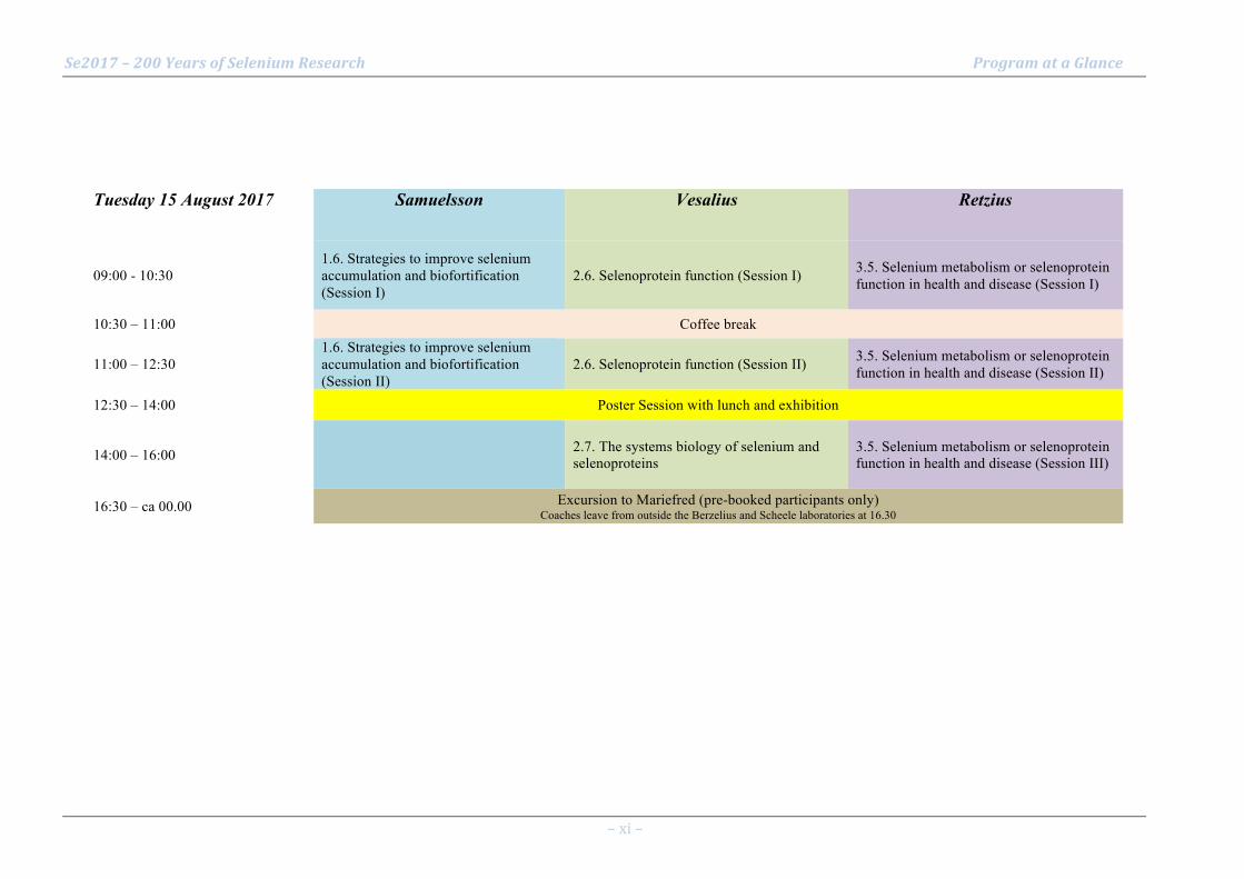
Tuesday 15 August 2017 Samuelsson Vesalius Retzius
09:00 - 10:30 1.6. Strategies to improve selenium accumulation and biofortification (Session I)
2.6. Selenoprotein function (Session I) 3.5. Selenium metabolism or selenoprotein function in health and disease (Session I)
10:30 – 11:00 Coffee break
11:00 – 12:30 1.6. Strategies to improve selenium accumulation and biofortification (Session II)
2.6. Selenoprotein function (Session II) 3.5. Selenium metabolism or selenoprotein function in health and disease (Session II)
12:30 – 14:00 Poster Session with lunch and exhibition
14:00 – 16:00 2.7. The systems biology of selenium and selenoproteins
3.5. Selenium metabolism or selenoprotein function in health and disease (Session III)
16:30 – ca 00.00 Excursion to Mariefred (pre-booked participants only) Coaches leave from outside the Berzelius and Scheele laboratories at 16.30
Se2017 – 200 Years of Selenium Research Program at a Glance
– xii –
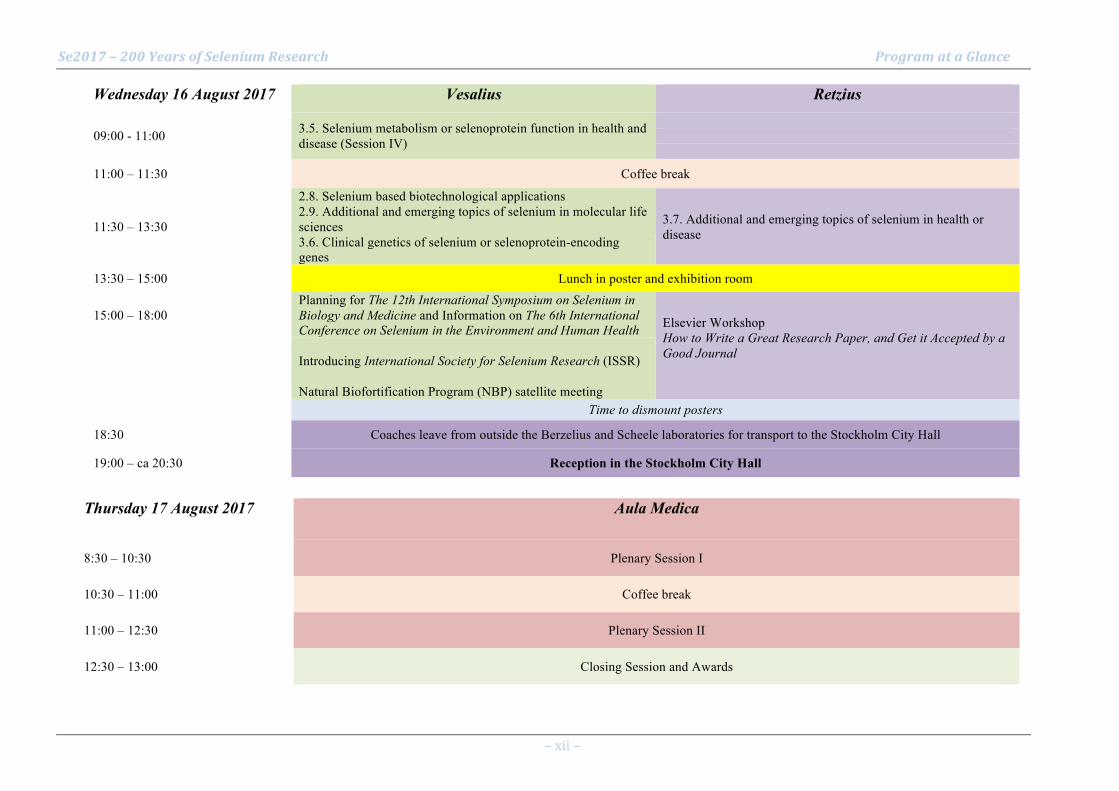
Wednesday 16 August 2017 Vesalius Retzius
09:00 - 11:00 3.5. Selenium metabolism or selenoprotein function in health and disease (Session IV)
11:00 – 11:30 Coffee break
11:30 – 13:30
2.8. Selenium based biotechnological applications 2.9. Additional and emerging topics of selenium in molecular life sciences 3.7. Additional and emerging topics of selenium in health or
disease 3.6. Clinical genetics of selenium or selenoprotein-encoding genes
13:30 – 15:00 Lunch in poster and exhibition room
15:00 – 18:00 Planning for The 12th International Symposium on Selenium in Biology and Medicine and Information on The 6th International Conference on Selenium in the Environment and Human Health Elsevier Workshop
How to Write a Great Research Paper, and Get it Accepted by a Good Journal
Introducing International Society for Selenium Research (ISSR) Natural Biofortification Program (NBP) satellite meeting
Time to dismount posters
18:30 Coaches leave from outside the Berzelius and Scheele laboratories for transport to the Stockholm City Hall
19:00 – ca 20:30 Reception in the Stockholm City Hall
Thursday 17 August 2017 Aula Medica
8:30 – 10:30 Plenary Session I
10:30 – 11:00 Coffee break
11:00 – 12:30 Plenary Session II
12:30 – 13:00 Closing Session and Awards

Se2017 – 200 Years of Selenium Research Detailed Program – Oral Presentations
– xiii –
Sunday 13 August 2017
Aula Medica
15.30 – 17.00 Registration
17:00 - 20:00 Inaugurating Plenary Session, Aula Medica Chair: Elias Arnér
17:00 – 17:10 Welcome address Elias Arnér and Gary Bañuelos
17:10 – 17:30 Berzelius and his discovery of Selenium Jan Trofast
17:30 - 18:00 O1 - The global cycle of selenium Lenny Winkel
18:00 - 18:30 O2 - Selenium metabolism in plants Philip John White
18:30 - 19:00 Thressa C. Stadtman Memorial Lecture O3 - Selenium utilization in diverse animals Vadim N. Gladyshev
19:00 - 19:30 O4 - Phenotypes and molecular pathogenesis of disorders of human selenoprotein synthesis Krishna Chatterjee
19:30 – 20:00 Performance – “Probing the mind of Berzelius” 1+1=3 (Stephen Whitmarsh and Jean-Louis Huhta)
20.00 – ca. 22 Get-together
Se2017 – 200 Years of Selenium Research Detailed Program – Oral Presentations
– xiv –
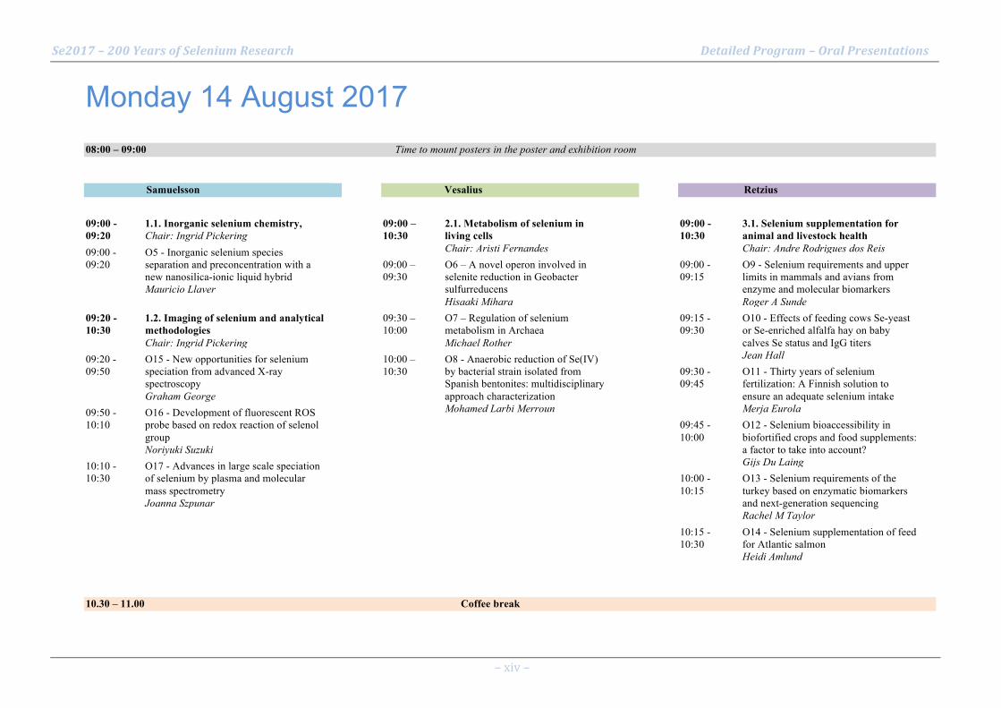
Monday 14 August 2017
08:00 – 09:00 Time to mount posters in the poster and exhibition room
Samuelsson 09:00 - 09:20
1.1. Inorganic selenium chemistry, Chair: Ingrid Pickering
09:00 - 09:20
O5 - Inorganic selenium species separation and preconcentration with a new nanosilica-ionic liquid hybrid Mauricio Llaver
09:20 - 10:30
1.2. Imaging of selenium and analytical methodologies Chair: Ingrid Pickering
09:20 - 09:50
O15 - New opportunities for selenium speciation from advanced X-ray spectroscopy Graham George
09:50 - 10:10
O16 - Development of fluorescent ROS probe based on redox reaction of selenol group Noriyuki Suzuki
10:10 - 10:30
O17 - Advances in large scale speciation of selenium by plasma and molecular mass spectrometry Joanna Szpunar
Vesalius 09:00 – 10:30
2.1. Metabolism of selenium in living cells Chair: Aristi Fernandes
09:00 – 09:30
O6 – A novel operon involved in selenite reduction in Geobacter sulfurreducens Hisaaki Mihara
09:30 – 10:00
O7 – Regulation of selenium metabolism in Archaea Michael Rother
10:00 – 10:30
O8 - Anaerobic reduction of Se(IV) by bacterial strain isolated from Spanish bentonites: multidisciplinary approach characterization Mohamed Larbi Merroun
Retzius 09:00 - 10:30
3.1. Selenium supplementation for animal and livestock health Chair: Andre Rodrigues dos Reis
09:00 - 09:15
O9 - Selenium requirements and upper limits in mammals and avians from enzyme and molecular biomarkers Roger A Sunde
09:15 - 09:30
O10 - Effects of feeding cows Se-yeast or Se-enriched alfalfa hay on baby calves Se status and IgG titers Jean Hall
09:30 - 09:45
O11 - Thirty years of selenium fertilization: A Finnish solution to ensure an adequate selenium intake Merja Eurola
09:45 - 10:00
O12 - Selenium bioaccessibility in biofortified crops and food supplements: a factor to take into account? Gijs Du Laing
10:00 - 10:15
O13 - Selenium requirements of the turkey based on enzymatic biomarkers and next-generation sequencing Rachel M Taylor
10:15 - 10:30
O14 - Selenium supplementation of feed for Atlantic salmon Heidi Amlund
10.30 – 11.00 Coffee break
Se2017 – 200 Years of Selenium Research Detailed Program – Oral Presentations
– xv –
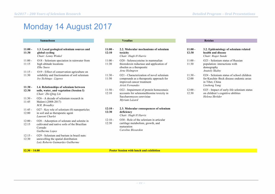
Monday 14 August 2017 Samuelsson 11:00 - 11:30
1.3. Local geological selenium sources and global cycling Chair: Lenny Winkel
11:00 - 11:15
O18 - Selenium speciation in rainwater from high altitude locations Elke Suess
11:15 - 11:30
O19 - Effect of conservation agriculture on solubility and fractionation of soil selenium Ivy Sichinga - Ligowe
11:30 - 12:30
1.4. Relationships of selenium between soils, water, and vegetation (Session I) Chair: Zhi-Qing Lin
11:30 - 11:45
O26 - A decade of selenium research in Malawi (2008-2017) M.R. Broadley
11:45 - 12:00
O27 - Key role of selenium (0) nanoparticles in soil and as therapeutic agent Laurent Charlet
12:00 - 12:15
O28 - Adsorption of selenate and selenite in cultivated and native soils of the Brazilian Cerrado Guilherme Lopes
12:15 - 12:30
O29 - Selenium and barium in brazil nuts: unravelling the spatial distribution Luiz Roberto Guimarães Guilherme
Vesalius 11:00 - 12:10
2.2. Molecular mechanisms of selenium toxicity Chair: Hugh H Harris
11:00 - 11:30
O20 - Selenocysteine in mammalian thioredoxin reductase and application of ebselen as a therapeutic Arne Holmgren
11:30 - 11:50
O21 - Characterization of novel selenium compounds as a therapeutic approach for improved cancer treatment Aristi Fernandes
11:50 - 12:10
O22 - Impairment of protein homeostasis accounts for selenomethionine toxicity in Saccharomyces cerevisiae Myriam Lazard
12:10 - 12:30
2.3. Molecular consequences of selenium deficiency Chair: Hugh H Harris
12:10 - 12:30
O30 - Role of the selenium in articular cartilage metabolism, growth, and maturation Caroline Bissardon
Retzius 11:00 - 12:30
3.2. Epidemiology of selenium related health and disease Chair: Roger Sunde
11:00 - 11:30
O23 - Selenium status of Russian population: interactions with demography Anatoly Skalny
11:30 - 12:00
O24 - Selenium status of school children for Kaschin–Beck disease endemic areas in Tibet, China Linsheng Yang
12:00 - 12:30
O25 - Impact of early-life selenium status on children’s cognitive abilities Helena Skröder
12.30 – 14.00 Poster Session with lunch and exhibition

Se2017 – 200 Years of Selenium Research Detailed Program – Oral Presentations
– xvi –
Monday 14 August 2017 Samuelsson 14:00 - 16:00
1.4. Relationships of selenium between soils, water, and vegetation (Session II) Chair: Milton Ferreira Moraes
14:00 - 14:20
O31 - Functional analysis of plant hyperaccumulator genes SpSultr1;2 and SpATPS2 via microbial expression Marinus Pilon
14:20 - 14:40
O32 - Influence of sulfate on selenium uptake in hyperaccumulator stanleya pinnata and non-accumulators Michela Schiavon
14:40 - 15:00
O33 - Selenolanthionine is the main selenium species of the hyperaccumulator plant Cardamine violifolia Eszter Borbála Both
15:00 - 15:20
O34 - Two selenium hyperaccumulators and their influence on their plant communities Ray Reynolds
15:20 - 15:40
O35 - The effect of different selenium fertilizers on garlic Hongyu Zhang
15:40 - 16:00
O36 - Biofortification of maize and beans with selenium in central Kenya highlands Gijs Du Laing
Vesalius
14:00 - 16:00
2.4. Selenoprotein synthesis pathways (Session I) Chair: Michael Rother
14:00 - 14:20
O37 - Selenophosphate synthetase 1 regulates cellular redox state and cell defense system Byeong Jae Lee
14:20 - 14:40
O38 - Translation regulation of human selenoproteins Laurent Chavatte
14:40 - 15:00
O39 - Structural and mechanistic insights into the mechanism of decoding of the Sec UGA codon in humans Miljan Simonovic
15:00 - 15:20
O40 - Identification of determinants regulating processive Sec incorporation in Selenoprotein P (SELENOP) Paul Copeland
15:20 - 15:40
O41 - Codon-specific roles for cis-acting elements during translation of selenoprotein P Michael Howard
15:40 - 16:00
O42 - Coding region determinants regulate selenoprotein P (SELENOP) expression in cells Sumangala Shetty
Retzius
14:00 - 15:00
3.3. Nutritional selenium intervention studies in human Chair: Helmut Sies
14:00 - 14:30
O43 - Effect of long-term selenium supplementation on mortality: results from a multiple-dose, RCT Margaret Rayman
14:30 - 14:45
O44 - Selenium supplementation blocked the crosstalk among pathogenic pathways in Alzheimer's disease mice Qiong Liu
14:45 - 15:00
O45 - Selenium, coenzyme Q10 and cardiovascular health – results from a 4-year-intervention in elderly Urban Alehagen
15:00 - 16:00
3.4. Selenium based medical therapeutics (Session I) Chair: Josef Köhrle
15:00 - 15:20
O46 - Antibacterial redox selenium coatings, covalent small molecules and antibody drug conjugates (ADCs) Julian Spallholz
15:20 - 15:40
O47 - Selenium in the treatment of cancer Mikael Björnstedt
15:40 - 15:50
O48 - Selective targeting of redox dysregulation of cancer cells by redox-active selenium compounds Sougat Misra
15:50 - 16:00
O49 - Selenium: A potentially powerful tool to design potent anticancer molecules – Discovery of Se-Aspirins Arun Sharma
16.00 – 16.30 Coffee break

Se2017 – 200 Years of Selenium Research Detailed Program – Oral Presentations
– xvii –
Monday 14 August 2017 Samuelsson 16:30 - 17:50
1.4. Relationships of selenium between soils, water, and vegetation (Session III) Chair: Luiz Roberto Guimarães Guilherme
16:30 - 16:50
O50 - Selenium distribution and its correlation to geochemical factors in East China intertidal zone Zhengyu BAO
16:50 - 17:10
O51 - Distribution and translocation of selenium from soil to highland barley in Kashin-Beck disease area Jing Wang
17:10 - 17:30
O52 - The status of selenium in Iraqi Kurdistan and feasibility of Se bio fortification using 77-Se Abdolbaset Karim
17:30 - 17:50
O53 - Geochemistry of selenium in Gilgit-Baltistan (North East Pakistan) Saeed Ahmad
Vesalius 16:30 - 17:50
2.4. Selenoprotein synthesis pathways (Session II) Chair: Michael Rother
16:30 - 16:50
O54 - Selenocysteine chemistry and total chemical synthesis applied for accessing human selenoproteins Norman Metanis
16:50 - 17:10
O55 - Redefining UAG for selenocysteine widens the scope of recombinant selenoprotein production in E.coli Qing Cheng
17:10 - 17:30
O56 - Delivery of selenide to selenophosphate synthetase for selenoprotein biosynthesis in bacteria Ryuta Tobe
17:30 - 17:50
O57 - Reconstitution of processive selenoprotein P synthesis in the wheat germ lysate system Mark Pinkerton
Retzius 16:30 - 17:50
3.4. Selenium based medical therapeutics (Session II) Chair: Björn Åkesson
16:30 - 16:50
O58 - Selenium in radiation oncology – 10 years of experiences in Germany Ralph Muecke
16:50 - 17:00
O59 - Selenium in the treatment of radiation-associated secondary lymphedema - An update Oliver Micke
17:00 - 17:10
O60 - Selective targeting of leukemia stem cells by selenium K. Sandeep Prabhu
17:10 - 17:20
O61 - Methylselenol suppressed the metastatic potential of B16F10 melanoma by reducing integrin expression An-Sik Chung
17:20 - 17:30
O62 - Selenium enhances auranofin-mediated Nrf2 induction in lung epithelial cells Trent Tipple
17:30 - 17:40
O63 - Preclinical chemopreventive efficacy of a novel hybrid p-XSC-aspirin compound in a lung cancer model Daniel Plano
17:40 - 17:50
O64 - Selenofolate is a promising novel agent in targeted chemotherapy Antje Zickler
17.50 – 18.00 Short break

Se2017 – 200 Years of Selenium Research Detailed Program – Oral Presentations
– xviii –
Monday 14 August 2017 Samuelsson 18:00 - 19:00
1.5. Excessive selenium accumulation from natural or anthropogenic sources and remediation technologies Chair: Gary Bañuelos
18:00 - 18:15
O65 - Environmental remediation of selenium contamination in water and soil Zhi-Qing Lin
18:15 - 18:30
O66 - Integrated passive biological selenium treatment system: Results of a 1-year pilot study James Bays
18:30 - 18:45
O67 - Industrial selenium pollution: challenges to treat flue gas desulfurization effluents Lucian Staicu
18:45 - 19:00
O68 - Role of antioxidant defense system and mitochondrial activity in selenium toxicity tolerance in whea Sucheta Sharma
Vesalius 18:00 - 19:00
2.5. Selenoprotein genetics Chair: Arne Holmgren
18:00 - 18:20
O73 - The significance of selenoproteins for human health revealed by inborn errors of metabolism Ulrich Schweizer
18:20 - 18:40
O74 - Mouse models lacking the selenoprotein thioredoxin reductase-1 Edward E Schmidt
18:40 - 19:00
O75 - Mutated selenocysteine synthase creates a Sedaghatian-type spondylometaphyseal dysplasia mouse model N Fradejas-Villar
Retzius 18:00 - 19:00
3.4. Selenium based medical therapeutics (Session III) Chair: Carmen Sanmartín
18:00 - 18:15
O69 - Insufficient documentation for clinical efficacy of selenium supplementation in chronic autoimmune thyroiditis Kristian Hillert Winther
18:15 - 18:30
O70 - Dihydroxy-1-selenolane protects cells from radiation-induced mitotic death: role of GPx Amit Kunwar
18:30 - 18:45
O71 - Inhibition of metalloenzymes, i.e., angiotensin-converting enzyme and tyrosinase, by selenol-metal ion interaction of selenoneine Takuya Seko
18:45 - 19:00
O72 - Selenium reduces macrophage and B-cell responses in vitro and suppresses germinal center B-cell responses in vivo: A potential for therapy in lupus Raghu Sinha
19.00 – ca. 21 Poster session with dinner and exhibition

Se2017 – 200 Years of Selenium Research Detailed Program – Oral Presentations
– xix –
Tuesday 15 August 2017 Samuelsson 09:00 - 10:30
1.6. Strategies to improve selenium accumulation and biofortification (Session I) Chair: Zhi-Qing Lin
09:00 - 09:20
O76 - Agronomic strategies affect the efficacy and quality of selenium biofortification Gary Bañuelos
09:20 - 09:40
O77 - Improving selenium supply in food systems Graham Lyons
09:40 - 10:00
O78 - Agronomic Biofortification with Selenium in Intercropping Systems would address Low Selenium Intake Allan Chilimba
10:00 - 10:15
O79 - To bio or not to bio? Strategies for agronomic biofortification with Se in tropical agroecosystems Luiz Roberto Guimarães Guilherme
10:15 - 10:30
O80 - Soil and foliar application of selenium in upland rice aiming agronomic biofortification Andre Rodrigues dos Reis
Vesalius 09:00 - 10:30
2.6. Selenoprotein function (Session I) Chair: Leopold Flohé
09:00 - 09:20
O81 - A model for Glutathione peroxidase 4-catalyzed reduction of lipid hydroperoxides in membranes Matilde Maiorino
09:20 - 09:40
O83 - The key ferroptosis regulator GPX4: cellular mechanisms and in vivo relevance Marcus Conrad
09:40 - 10:00
O84 - GPx4 depleted cell death involves different cell death pathway from ferroptosis Hirotaka Imai
10:00 - 10:30
O82 - Selenium versus sulfur: reversibility of chemical reactions and resistance to permanent oxidation in proteins and nucleic acid Robert J. Hondal
Retzius
09:00 - 10:30
3.5. Selenium metabolism or selenoprotein function in health and disease (Session I) Chair: Bernhard Michalke
09:00 - 09:30
O88 - Health Benefit Values (HBV) reliably indicate effects of seafood consumption Nicholas Ralston
09:30 - 09:50
O85 - Selenoproteins expressed in intestinal stem cells and cancer stem cells Anna Kipp
09:50 - 10:10
O89 - Molecular and Metabolic Mechanisms for Steatosis and Obesity Induced by Overexpression of Glutathione Peroxidase-1 in Mice Xingen Lei
10:10 - 10:30
O86 - Roles of the thioredoxin and glutathione systems in reduction of inorganic- and Cys-polysulfide spec Péter Nagy
10.30 – 11.00 Coffee break

Se2017 – 200 Years of Selenium Research Detailed Program – Oral Presentations
– xx –
Tuesday 15 August 2017 Samuelsson 11:00 - 12:30
1.6. Strategies to improve selenium accumulation and biofortification (Session II) Chair: Andre Rodrigues dos Reis
11:00 - 11:20
O90 - Selenium use efficiency by wheat cultivars Milton Ferreira Moraes
11:20 - 11:40
O91 - Effects of selenium biofortification on mineral nutrients in grains of wheat and oat Tao Li
11:40 - 12:00
O92 - Characterization on rhyzosphere bacteria from a selenium-hyperaccumulator Cardamine hupingshanesis Linxi Yuan
12:00 - 12:15
O93 - Assessment and biofortification of wheat grain selenium in staple food production regions of China Sen Wang
12:15 - 12:30
O94 - Selenium supplemented kale and kohlrabi sprouts as possible ingredients for potent functional food. Paweł Zagrodzki
Vesalius 11:00 - 12:30
2.6. Selenoprotein function (Session II) Chair: Ulrich Schweizer
11:00 - 11:20
O95 - Selenoproteins in hypothalamic energy homeostasis Matthew Pitts
11:20 - 11:40
O96 - Role of hypothalamic selenoprotein M in leptin signaling and calcium regulation Ting Gong
11:40 - 12:00
O97 - Exploring Selenoprotein N structure and function Alain Lescure
12:00 - 12:15
O98 - Disruption of cancer cell redox homeostasis promoted by S-nitrosylation of thioredoxin reductase Moran Benhar
12:15 - 12:30
O99 - Influence of small intestinal thioredoxin and thioredoxin reductase on intestinal permeability Liangwei Zhong
Retzius 11:00 - 12:30
3.5. Selenium metabolism or selenoprotein function in health and disease (Session II) Chair: Anna Kipp
11:00 - 11:30
O100 - The intricate role of selenoproteins in stress erythropoiesis Chang Liao
11:30 - 12:00
O101 - Selenoproteins restrict the replication of Francisella tularensis in macrophages. Girish Kirimanjeswara
12:00 - 12:30
O102 - Se-dietary matrices can upregulate the anti-inflammatory responses in RAW macrophages Tejo Prakash Nagaraja
12.30 – 14.00 Poster Session with lunch and exhibition

Se2017 – 200 Years of Selenium Research Detailed Program – Oral Presentations
– xxi –
Tuesday 15 August 2017 Vesalius 14:00 - 16:00
2.7. The systems biology of selenium and selenoproteins Chair: Vadim Gladyshev
14:00 - 14:20
O103 - Selenium in human and vertebrate evolution Sergi Castellano
14:20 - 14:40
O104 - Evolution of selenoproteins across the tree of life Marco Mariotti
14:40 - 14:50
O105 - The human selenomicrobiome Didac Santesmasses
14:50 - 15:00
O106 - Selenoprotein extinction in Drosophila occurred concomitantly to genome catastrophes Didac Santesmasses
15:00 - 15:20
O107 - Metabolomics of selenium Sun Hee Yim
15:20 - 15:40
O108 - Selenium-encoded chemical proteomics Chu Wang
15:40 - 16:00
O109 - Characterization of Atlantic salmon (Salmo salar) selenoproteins using bioinformatics and hyphenated Veronika Sele
Retzius 14:00 - 16:00
3.5. Selenium metabolism or selenoprotein function in health and disease (Session III) Chair: Lutz Schomburg
14:00 - 14:20
O110 - The cellular location of selenoproteins in human prostatic tissue and their role in prostate cancer. Alan Diamond
14:20 - 14:40
O111 - Dietary selenium deprivation oppositely impacts longevity and healthspan in telomere dysfunctional mice Wen-Hsing Cheng
14:40 - 15:00
O112 - Selenium and cataract Kaixun Huang
15:00 - 15:20
O113 - Role of selenoprotein P in Alzheimer's disease Xiubo Du
15:20 - 15:40
O114 - Role of selenoprotein P in function of pancreatic β cell: Improving effects of neutralizing antibody Yoshiro Saito
15:40 - 15:50
O115 - Tissue-specific pools of selenoprotein P differentially modify colitis-associated carcinogenesis Sarah Short
15:50 - 16:00
O116 - Selenoprotein P in cord serum: wide disparity between ELISA and HPLC Margaret Rayman
16.30 – ca. 00.00 Excursion to Mariefred (pre-booked participants only) Coaches leave from outside the Berzelius and Scheele laboratories at 16.30

Se2017 – 200 Years of Selenium Research Detailed Program – Oral Presentations
– xxii –
Wednesday 16 August 2017
Vesalius
09:00 - 11:00 3.5. Selenium metabolism or selenoprotein function in health and disease (Session IV)
Chair: Ed Schmidt 09:00 - 09:20 O117 - Expression and activity of enzymes of selenium metabolism in the selenoprotein P knockout mouse
Marla J. Berry 09:20 - 09:40 O118 - The Role of Selenoprotein K in Progression and Metastasis of Melanoma
Peter Hoffmann 09:40 - 10:00 O119 - Targeting thioredoxin reductase 1 as a basis for anticancer therapy
Elias Arnér 10:00 - 10:15 O120 - Redox regulation of protein kinase C by selenium and selenoprotein thioredoxin reductase influences the
cancer-preventive efficacy of selenium Rayudu Gopalakrishna
10:15 - 10:30 O121 - Thioredoxin reductase 2 inhibition by tamoxifen-like metallocifens drives Jurkat cells to apoptosis Valeria Scalcon
10:30 - 10:45 O122 - Dietary selenium and the 15 kDa selenoprotein influence initiation/promotion of colon carcinogenesis Petra Tsuji
10:45 - 11:00 O123 - Selenoprotein T is a novel neuroprotective antioxidant enzyme in Parkinson’s disease Youssef Anouar
11.00 – 11.30 Coffee break

Se2017 – 200 Years of Selenium Research Detailed Program – Oral Presentations
– xxiii –
Wednesday 16 August 2017 Vesalius
11:30 - 11:50 2.8. Selenium based biotechnological applications Chair: Elias Arnér
11:30 - 11:50 O124 - Selenium atom-specific functionalization of nucleic acids for structure and function studies Zhen Huang
11:50 - 13:15 2.9. Additional and emerging topics of selenium in molecular life sciences Chair: Elias Arnér
11:50 - 12:20 O126 - Selenocysteine and the genetic code Dieter Söll
12:20 - 12:40 O127 - Cysteine polysulfidation governed by cysteinyl-tRNA synthetases (CARSs) Takaaki Akaike
12:40 - 13:00 O128 - Direct observation of methylmercury and auranofin binding to selenocysteine in thioredoxin reductase Ingrid Pickering
13:00 - 13:15 O129 - X-ray fluorescence imaging and X-ray absorption spectroscopy combined yield insight on mammaliam selenium biochemistry Hugh Harris
13:15 - 13:30 3.6. Clinical genetics of selenium or selenoprotein-encoding genes Chair: Elias Arnér
13:15 - 13:30 O125 - Interrelationships among SELENOF genotype, cellular localization, serum selenium and race in prostate cancer Dede Ekoue
Retzius 11:30 - 13:30 3.7. Additional and emerging topics of selenium in health or
disease Chair: Regina Brigelius-Flohé
11:30 - 11:50 O130 - Se-speciation investigations at neural barrier (NB) Bernhard Michalke
11:50 - 12:10 O131 - Selenium-binding protein 1 in serum may signify a heart at risk Eike Kuehn
12:10 - 12:30 O132 - Of dogs and men: Review of translational impact of dog studies on selenium and prostate cancer risk David Waters
12:30 - 12:45 O133 - High selenium induces endothelial dysfunction via endoplasmic reticulum stress Matshediso Zachariah
12:45 - 13:00 O134 - Selenium and sex: competition between brain and testes for selenium results in male-specific consequences in mice and men Marla Berry
13:00 - 13:15 O135 - Ceramide analog S14 causes a coordinate downregulation of selenoproteins in a murine psoriasis model Jack L. Arbiser
13:15 - 13:30 O136 - Maternal nutrition and transcript abundance of selenium related genes in fetal bovine hepatic tissues at d 50 of gestation J. S. Caton
13.30 – 15.00 Lunch in poster and exhibition room

Se2017 – 200 Years of Selenium Research Detailed Program – Oral Presentations
– xxiv –
Wednesday 16 August 2017 Vesalius 15:00 - 16:30 Planning for The 12th International Symposium on Selenium in Biology
and Medicine and Information on The 6th International Conference on Selenium in the Environment and Human Health Chairs: Elias Arnér and Gary Bañuelos – Decision of venue and date for The 12th International Symposium on
Selenium in Biology and Medicine. – Presentation by Dongli Liang on The 6th International Conference on
Selenium in the Environment and Human Health, to be held in Yangling/Xi'an, China, 2019.
Introducing International Society for Selenium Research (ISSR)
16:30 - 18:00 Natural Biofortification Program (NBP) satellite meeting
Chairs: Gary Bañuelos and Zhi-Qing Lin 16:30 - 16:50 O137 - A ten-year study and practice of functional agriculture in China
Xuebin Yin 16:50 -17:00 Introduction of NBP and open proposal call to participate
Linxi Yuan 17:00 -17:10 Does selenium intake relate to human longevity: A case study in Shitai,
Anhui, China Zedong Long
17:10 -17:20 Selenium products produced from natural Se-rich areas Serun Group (Gold Sponsor)
17:20 -17:30 Leading functional agriculture practices in China Setek Co. Ltd. (Gold Sponsor)
17:30 -18:00 Concluding words Zhi-Qing Lin
Retzius 15.00 - ca. 18:00 Elsevier Workshop
O144 – How to write a great research paper, and get it accepted by a good journal Anthony Newman
18.00 – 18.30 Time to dismount posters
18:30 Coaches leave at 18:30 from outside the Berzelius and Scheele laboratories for transport to the Stockholm City Hall
19:00 – ca 20:30 Reception in the Stockholm City Hall, by invitation of the City and the Council of Stockholm.

Se2017 – 200 Years of Selenium Research Detailed Program – Oral Presentations
– xxv –
Thursday 17 August 2017
Aula Medica
08:30 - 10:30 Plenary Session I, Aula Medica
Chair: Arne Holmgren 08:30 - 09:00 O87 - Nutritional aspects of selenium in human beings
Raymond F Burk 09:00 - 09:30 O139 - Selenium from ocean fish provides protection against mercury toxicity
Nicholas Ralston 09:30 - 10:00 O138 - From atom to field: how and why plants hyperaccumulate selenium, and how this affects ecosystems
Elizabeth Pilon-Smits 10:00 - 10:30 O140 - Selenium at the redox interface of the genome and exposome
Dean Jones
10.30 – 11.00 Coffee break
11:00 - 12:30 Plenary Session II, Aula Medica
Chair: Raymond Burk 11:00 - 11:30 O141 - Enzymology and biological functions of glutathione peroxidases
Regina Brigelius-Flohé 11:30 - 12:00 O142 - Selenium versus Sulfur in GSH Peroxidases
Fulvio Ursini 12:00 - 12:30 O143 - Nrf2 improves leptin and insulin resistance provoked by selenocysteine-trna knockout in hypothalamus
Masayuki Yamamoto
12:30 - 13:00 Closing Session and Awards, Aula Medica Chair: Elias Arnér and Gary Bañuelos

Se2017 – 200 Years of Selenium Research Detailed Program – Poster Presentations
– xxvi –
Monday 14 August (Afternoon posters) Presenting authors of posters listed below are kindly asked to be present at the respective poster during the first 30 min of the session.
12:30 - 14:00 Poster Session with lunch and exhibition, Poster and Exhibition Room
P1 - In vitro Generation of Superoxide by Selenofolate in MDA-MB-468 Breast Cancer Cells Soni Khandelwal
P2 - A preliminary study of selenium species in natural selenium-enriched garlic Shaozhan Chen
P3 - Catalytic redox activity of selenium compounds generating superoxide assessed by chemiluminescence Soni Khandelwal
P4 - Quantification of selenoprotein P in human serum using isotope dilution analysis Christian Deitrich
P5 - Relevancy of using Se speciation for geographical authentication purpose: example of red wines Veronique Vacchina
P6 - Selenium Bicentennial: Two Hundred Years of Selenium Discovery Mikael Bjornstedt
P7 - Simultaneous determination of selenium and sulfur species in biological samples Nina Kroepfl
P8 - The fractions and distribution of soil selenium in Heilongjiang province and its' impacting factors Fengqin Chi
P9 - Zoning pollution-free and selenium-rich land resources with geochemistry Tao Yu
P10 - Selenium, Nitrogen and Carbon remobilization study following litter decomposition during 10 months Maryse Castrec-Rouelle
P11 - The Precipitation Impact on the Selenium Speciation in Surface Soil Dacheng Wang
P12 - Effect of pH, iron plaque and phosphorus on selenium uptake by rice seedling Ju Min
P13 - Analysis of Se (Ⅳ) and Se(Ⅵ) absorption kinetics of different genotypes of Nicotiana tabacum L. Shuxin Tu
P14 - Agronomic biofortification of carrots with selenium in oxidic soil Ediu Carlos Silva Junior
P15 - Effects of different selenium application on selenium accumulation in Lentinula edode Dongli Liang
P16 - Apples: a suitable target for selenium biofortification? Diemo Daum
P17 - Optimising fertiliser formulations for selenium biofortification of wheat grain Chandnee Ramkissoon
P18 - Effects of foliar spraying selenite or selenate at different stage on the selenium uptake and distri Xinwei Liu
P19 - Se and its antagonists Hg, As, Cd in hair of Taiwanese and Russian residents Margarita Skalnaya
P20 - Selenium and mercury interactions in the apex predators from the Gulf of Trieste (northern Adriatic) Jadran Faganeli
P21 - Environmental Selenium Influences Fish Methylmercury Bioaccumulation and Risks Nicholas Ralston
P22 - Study of geochemical characteristics and influencing factors of soil selenium in the typical soil Zhongfang Yang
P23 - Interaction of selenium and cadmium in soil-corn system in natural selenium and cadmium rich area Zezhou Zhang
Se2017 – 200 Years of Selenium Research Detailed Program – Poster Presentations
– xxvii –
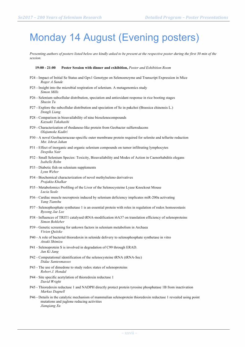
Monday 14 August (Evening posters) Presenting authors of posters listed below are kindly asked to be present at the respective poster during the first 30 min of the session.
19:00 - 21:00 Poster Session with dinner and exhibition, Poster and Exhibition Room
P24 - Impact of Initial Se Status and Gpx1 Genotype on Selenoenzyme and Transcript Expression in Mice Roger A Sunde
P25 - Insight into the microbial respiration of selenium. A metagenomics study Simon Mills
P26 - Selenium subcellular distribution, speciation and antioxidant response in rice booting stages Shuxin Tu
P27 - Explore the subcellular distribution and speciation of Se in pakchoi (Brassica chinensis L.) Dongli Liang
P28 - Comparison in bioavailability of nine bioselenocompounds Kazuaki Takahashi
P29 - Characterization of rhodanese-like protein from Geobacter sulfurreducens Olajumoke Kadiri
P30 - A novel Geobacteraceae-specific outer membrane protein required for selenite and tellurite reduction Mst. Ishrat Jahan
P31 - Effect of inorganic and organic selenium compounds on tumor infiltrating lymphocytes Deepika Nair
P32 - Small Selenium Species: Toxicity, Bioavailability and Modes of Action in Caenorhabditis elegans Isabelle Rohn
P33 - Diabetic fish on selenium supplements Lynn Weber
P34 - Biochemical characterization of novel methylseleno derivatives Prajakta Khalkar
P35 - Metabolomics Profiling of the Liver of the Selenocysteine Lyase Knockout Mouse Lucia Seale
P36 - Cardiac muscle necroptosis induced by selenium deficiency implicates miR-200a activating Yang Tianshu
P37 - Selenophosphate synthetase 1 is an essential protein with roles in regulation of redox homoeostasis Byeong Jae Lee
P38 - Influences of TRIT1 catalysed tRNA-modification i6A37 on translation efficiency of selenoproteins Simon Bohleber
P39 - Genetic screening for unkown factors in selenium metabolism in Archaea Vivien Quitzke
P40 - A role of bacterial thioredoxin in selenide delivery to selenophosphate synthetase in vitro Atsuki Shimizu
P41 - Selenoprotein S is involved in degradation of C99 through ERAD. Jun Ki Jang
P42 - Computational identification of the selenocysteine tRNA (tRNA-Sec) Didac Santesmasses
P43 - The use of dimedone to study redox states of selenoproteins Robert J. Hondal
P44 - Site specific acetylation of thioredoxin reductase 1 David Wright
P45 - Thioredoxin reductase 1 and NADPH directly protect protein tyrosine phosphatase 1B from inactivation Markus Dagnell
P46 - Details in the catalytic mechanism of mammalian selenoprotein thioredoxin reductase 1 revealed using point mutations and juglone reducing activities Jianqiang Xu

Se2017 – 200 Years of Selenium Research Detailed Program – Poster Presentations
– xxviii –
P47 - Probing the role of TRP14 (TXNDC17) in cellular signaling pathways Belén Espinosa Fernández
P48 - Apatone inhibited GPx activity and triggered AIF-mediated cell death pathway in cancer cells Xiaoyuan Ren
P49 - The significance of selenoprotein P expression in pancreatic beta-cell line MIN6 Shohei Nakao
P50 - Interaction between selenoprotein W and 14-3-3 is regulated by oxidative stress. Kwan Young Ko
P51 - Immortalized human prostate cell line RWPE-1 as a model to study selenoprotein regulation and function in normal prostatic tissue Lenny Hong
P52 - Glutathione depletion in HEK293T adresses cytosolic GPx4 to the membrane Ana-Marija Vuckovic
P53 - Synthesis and efficacy of conjugated selenium ADC monoclonal antibodies in vitro Soni Khandelwal
P54 - Antifungal Activity of Selenium Nanoparticles Synthesized by B. subtilis Against P. syringae Miao Li
P55 - Exosome-mediated methylmercury detoxification accelerated by selenium compound, selenoneine in aquatic organism Shintaro Imamura
P56 - Studying selenoproteome regulation using selenium stable isotope labeling Laurent Chavatte
Se2017 – 200 Years of Selenium Research Detailed Program – Poster Presentations
– xxix –
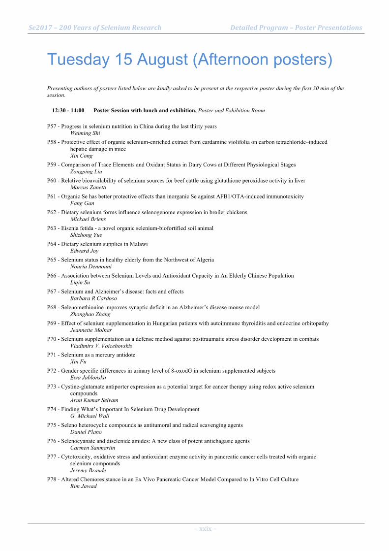
Tuesday 15 August (Afternoon posters) Presenting authors of posters listed below are kindly asked to be present at the respective poster during the first 30 min of the session.
12:30 - 14:00 Poster Session with lunch and exhibition, Poster and Exhibition Room
P57 - Progress in selenium nutrition in China during the last thirty years Weiming Shi
P58 - Protective effect of organic selenium-enriched extract from cardamine violifolia on carbon tetrachloride–induced hepatic damage in mice Xin Cong
P59 - Comparison of Trace Elements and Oxidant Status in Dairy Cows at Different Physiological Stages Zongping Liu
P60 - Relative bioavailability of selenium sources for beef cattle using glutathione peroxidase activity in liver Marcus Zanetti
P61 - Organic Se has better protective effects than inorganic Se against AFB1/OTA-induced immunotoxicity Fang Gan
P62 - Dietary selenium forms influence selenogenome expression in broiler chickens Mickael Briens
P63 - Eisenia fetida - a novel organic selenium-biofortified soil animal Shizhong Yue
P64 - Dietary selenium supplies in Malawi Edward Joy
P65 - Selenium status in healthy elderly from the Northwest of Algeria Nouria Dennouni
P66 - Association between Selenium Levels and Antioxidant Capacity in An Elderly Chinese Population Liqin Su
P67 - Selenium and Alzheimer’s disease: facts and effects Barbara R Cardoso
P68 - Selenomethionine improves synaptic deficit in an Alzheimer’s disease mouse model Zhonghao Zhang
P69 - Effect of selenium supplementation in Hungarian patients with autoimmune thyroiditis and endocrine orbitopathy Jeannette Molnar
P70 - Selenium supplementation as a defense method against posttraumatic stress disorder development in combats Vladimirs V. Voicehovskis
P71 - Selenium as a mercury antidote Xin Fu
P72 - Gender specific differences in urinary level of 8-oxodG in selenium supplemented subjects Ewa Jablonska
P73 - Cystine-glutamate antiporter expression as a potential target for cancer therapy using redox active selenium compounds Arun Kumar Selvam
P74 - Finding What’s Important In Selenium Drug Development G. Michael Wall
P75 - Seleno heterocyclic compounds as antitumoral and radical scavenging agents Daniel Plano
P76 - Selenocyanate and diselenide amides: A new class of potent antichagasic agents Carmen Sanmartin
P77 - Cytotoxicity, oxidative stress and antioxidant enzyme activity in pancreatic cancer cells treated with organic selenium compounds Jeremy Braude
P78 - Altered Chemoresistance in an Ex Vivo Pancreatic Cancer Model Compared to In Vitro Cell Culture Rim Jawad

Se2017 – 200 Years of Selenium Research Detailed Program – Poster Presentations
– xxx –
P79 - Novel selenocyanate and diselenide phosphoramides as potent anticarcinogenic agents Carmen Sanmartin
P80 - Inducing Change in Selenium Drug Development Eric Lynch
P81 - Metabolic syndrome induced oxidative stress and skin premature ageing beyond topical use of seleno-L-methionine. Julija Voicehovska
P82 - Whole Blood Selenium Levels and Selenium Supplementation in Patients treated in a Family Practice Ralph Muecke
P83 - Influences of selenium status on thyroid response of children after iodine repletion Dawd Gashu
P84 - Thiol-dependent redox systems as antimicrobial drug targets Jun Lu
P85 - Novel acylselenoureas as antiproliferative agents: design, synthesis and biological evaluation Daniel Plano
P86 - Role of selenium nanoparticles to dampen the metastatic potential of aggressive cancers Caroline Bissardon
P87 - Redox-active Selenium as an Anticancer Agent: A Critical Review Boguslaw Lipinski
P88 - New insights into the development and biological evaluation of novel methylselenol precursors Nuria Díaz Argelich
P89 - Genetic interactions in human methylation of selenium and arsenic Helena Skröder
P90 - Organic selenium supplementation increases mercury excretion and decreases oxidative damage in long-term mercury-exposed residents from Wanshan, China Xin Fu
P91 - Positive association of apolipoprotein E allel ε4 with plasma selenium in Croatian pregnant females Ingrid Falnoga
P92 - Preliminary toxicology evaluation of selenite cataract model Hongjie Chen
P93 - The powerful ameliorating effect of selenium against deltamethrin-induced oxidative stress in lactating rats and their suckling pups Sameeh Mansour
P94 - Nutritional availability of selenium derived from raw or roast beef Munehiro Yoshida
P95 - The inhibitory effect of selenium nanoparticles on atherosclerosis and the underlying mechanism Hongmei Liu
P96 - Effects of Selenium on Protein related-High Density Lipoprotein and Apolipoprotein B-100 Expression in Human Primary Hepatocytes. MIrasari Putri
P97 - Thoracic aortic degeneration and dilatation: a newly-recognised complication of human selenoprotein Erik Schoenmakers
P98 - The combined use of selen organic compounds with bioslastelin at toxic hepatitis Daulet Sharipov
P99 - In vivo effects of repeated thyronamine (T0AM) administration in male C57BL/6J mice Carolin S. Hoefig
P100 - Dynamic regulation of glutathione peroxidase 4 (GPX4) upon renal ischemia/reperfusion injury (IRI) Tobias Seibt
P101 - Assessment of Trace Elements and Systemic Oxidant Status in Dairy Cows during the Perinatal Period Hongyan Zhao
P102 - MsrA Protects Hepatocytes against Acetaminophen-Induced Toxicity via TXNRD1 Regulation Hwa-Young Kim
P103 - Knockdown of Sep15 modulates the expression of Pax6 induced by glucose in HLE cells Xiaohuan Li
P104 - Roles of the 15kD selenoprotein in lens epithelial differentiation Xiaoxiang Zheng
P105 - Role of the 15kDa selenoprotein in colorectal inflammation Kristin Peters

Se2017 – 200 Years of Selenium Research Detailed Program – Poster Presentations
– xxxi –
P106 - Low selenoprotein P status in patients with traumatic spinal cord injury Julian Seelig
P107 - Redox regulated transcription factors in 3D spheroids enriched for cancer stem cells Katarina Johansson
P108 - Differential Impact of Two Secisbp2 Mutations on Selenoprotein Expression in Liver and Brain Wenchao Zhao
P109 - A suppressive effect of selenium on amyloid-β plaque deposition in Tg2576 transgenic mice brain Sakura Yoshida
P110 - Homozygous mutation p.P190L in TXNRD1 is associated with genetic generalized epilepsy Noelia Fradejas-Villar
P111 - Comparative analysis of selenium status in common neuropsychiatric disorders in children Anastasiya Skalnaya
P112 - Does selenium intake relate to longevity: Case study in Shitai, Anhui, China ZeDong Long
P113 - Influence of statins in selenium status and inflammatory profile considering creatine kinase levels Lígia Moriguchi Watanabe
P114 - The role of selenium in an in vitro invasive model of pancreatic cancer Ali Coyle
P115 - The role of environmental metal ions in human health and diseases Young-Mi Go
P116 - Selenoproteins regulate B cell functions by targeting B cell receptor (BCR)-mediated antigen present Girish Kirimanjeswara
P117 - Effect of anti-rheumatic treatment on selenium levels in rheumatoid arthritis , psoriatic arthritis Jan Olav Aaseth
P118 - Selenium, prostate cancer, and U-shaped thinking: A paradigm shift in public health research David Waters
P119 - Development of a point-of-care test for selenoprotein P Waldemar Minich
P120 - Age-dependent protective effect of Selenium against UVA irradiation in primary human keratinocytes a Walid Rachidi
P121 - Research and practice on the standard “Selenium-enriched agricultural products” Zhangmin Wang
P122 - Micro-spectroscopic investigation of selenium speciation in Se-rich rocks Hai-Bo Qin
P123 - Selenium-enriched yeast inhibits Aβ generation and modulates autophagy in the 3xTg AD mouse Guo-Li Song
P124 - Metallomic quantifying metals in metalloproteins: The levels of Hg and Se binding to serum selenoproteins Yu-Feng Li

Se2017 – 200 Years of Selenium Research Abstracts
– 1 –
Abstracts for Oral Presentations

Se2017 – 200 Years of Selenium Research Abstracts
– 2 –
O1 - The global cycle of selenium Inaugurating plenary session Keywords: global biogeochemical cycle, atmosphere, soils Lenny Winkel1 1 ETH Zurich/Eawag Introduction: The distribution and chemical speciation of selenium in the environment is of key importance in both environmental health issues and selenium status of crops and thus dietary intakes. However, there is a lack in understanding of the biogeochemical selenium cycle and especially the role of the atmosphere in this cycle. The atmosphere is a global transient reservoir of selenium that can function as a source of selenium to terrestrial environments, and thus food chains, via wet and dry deposition. Method: This talk will give new insights into the atmospheric sources, sinks and fluxes of selenium and how these are linked. Biogenic sources of atmospheric selenium will be discussed as well as atmospheric pathways and deposition onto the Earth’s surface. Result: Climate influences selenium distributions in soils, both indirectly (by affecting retention and mobility of Se in soils) and directly (atmospheric sources), and therefore, we further investigated broad-scale relationships between climatic factors, soil properties, and soil selenium concentrations. In this talk it will be shown how climate and soil parameters can be used to predict selenium concentrations in areas where these have not been systematically analysed. Predictions of current soil selenium concentrations and future changes in these concentrations due to climatic changes indicate that soil Se distributions are dynamic. Discussion: As changes in spatial selenium distributions may ultimately affect nutritional quality of food crops and thus human and animal health, such broad-scale predictions will help in the prevention of future health hazards related to unsafe levels of Se in soils.

Se2017 – 200 Years of Selenium Research Abstracts
– 3 –
O2 - Selenium Metabolism in Plants Inaugurating plenary session Keywords: genetics, hyperaccumulator, metabolism, nutrition, toxicity Philip John White1 1 The James Hutton Institute, Dundee, United Kingdom Introduction: Selenium (Se) is not an essential element for flowering plants (angiosperms), although there is evidence that Se can benefit their growth and survival under some circumstances. However, excess Se accumulation is toxic to most angiosperms, presumably because the indiscriminate incorporation of selenocysteine and selenomethionine into proteins impairs their function. Method: There are differences between angiosperm species in their ability to tolerate Se in their tissues, which are related to their Se metabolism. These differences have shaped the ecology of seleniferous soils. Result: This lecture will first describe the proteins involved in Se uptake, translocation and metabolism in angiosperms, their location within the plant and their regulation. It will then identify differences in metabolic pathways between angiosperm species that might account for variation in their ability to tolerate large tissue Se concentrations and the genetics that underpins these differences. It will emphasise that angiosperm species that hyperaccumulate Se generally exhibit constitutive expression of genes encoding Se transporters and enzymes involved in primary Se assimilation, the biosynthesis of non-toxic Se metabolites, and Se volatilisation. Discussion: Finally, strategies for exploiting differences in the abilities of plants to accumulate Se in their tissues for either the phytoremediation of Se-contaminated soils or Se-biofoertification of edible crops for the nutrition of animals and humans will be discussed. Selected references White PJ (2016) Selenium accumulation by plants. Annals of Botany 117, 217-235.
Se2017 – 200 Years of Selenium Research Abstracts
– 4 –
O3 - Selenium Utilization in Diverse Animals Inaugurating plenary session Thressa C. Stadtman Memorial Lecture Keywords: selenium utilization, evolution Vadim N. Gladyshev1 1 Brigham and Women’s Hospital, Harvard Medical School, Boston, MA 02115, USA Introduction: The Gladyshev lab works in the areas of selenium and redox biology as applied to aging and cancer. Selenium is a trace element that exhibits both beneficial and toxic effects in human health. Importance of this micronutrient in the diet is primarily due to the fact that selenium is used in selenoproteins in the form of selenocysteine. Method: In this presentation, discussion will be focused on functions and evolution of selenium and selenoproteins. Comparative and functional genomics methods allow assessing its use at the levels of proteins, cells, organs and entire organisms. Result: Selenoproteins with known functions are oxidoreductases, and the tight link between selenium and redox biology offers an opportunity to better understand protein function and use this information to examine questions central to the thiol-based redox control of cellular processes. Discussion: We use high throughput approaches, including genome sequencing, transcriptomics, metabolomics and ionomics, to better understand the systemic role of selenium in diverse animals and other organisms.
Se2017 – 200 Years of Selenium Research Abstracts
– 5 –
O4 - Phenotypes and molecular pathogenesis of disorders of human selenoprotein synthesis Inaugurating plenary session Keywords: SECISBP2; TRU-TCA1-1; SEPSECS Krishna Chatterjee1 1 Wellcome-MRC Institute of Metabolic Science, University of Cambridge, Cambridge, UK Introduction: Mutations in three human genes (SECISBP2, TRU-TCA1-1, SEPSECS) within the selenocysteine (Sec) biosynthesis and incorporation pathway, resulting in impaired synthesis of selenoproteins, have been described. Method: We review the genetic architecture, molecular pathogenesis and clinical phenotypes of these different disorders. Result: Nine families, harbouring compound heterozygous or homozygous SECISBP2 defects, have been described to date. Many mutations generate aminoterminally truncated mutant proteins with reduced Sec incorporation function, mediating global selenoprotein deficiency. Growth retardation in childhood, abnormal thyroid function tests due to lack of Sec-containing thyroid deiodinase enzymes and low plasma selenium levels reflecting deficiencies of SEPP1 and GPx3, are universal features of cases. Muscular dystrophy and male infertility reflect loss of tissue-specific selenoproteins. Cutaneous photosensitivity, increased fat mass with preserved insulin sensitivity and hearing loss may be manifestations of elevated cellular ROS, secondary to lack of antioxidant selenoenzymes. Degeneration and dilatation of the thoracic aorta is a newly-recognised adult phenotype. A single child with a homozygous TRU-TCA1-1 mutation, altering expression and base modification of tRNA[Ser]Sec, has been described. Compared to SECISBP2 defect cases, synthesis of some selenoproteins (TXRNDs, GPx4) is preserved, with some clinical features (e.g. growth retardation, abnormal thyroid hormone and selenium levels) being similar. Ten families with compound heterozygous or homozygous SEPSECS defects, associated with progressive cerebellocerebral atrophy, mental retardation, spasticity and seizures, have been recorded. Discussion: SECISBP2 and TRU-TCA1-1 defects mediate a multisystem disorder with a thyroid signature, due to lack of tissue-specific selenoproteins, antioxidant selenoenzymes and thyroid deiodinase enzymes. SEPSECS defects mediate a predominantly neurological phenotype. Selected references Schoenmakers E, Schoenmakers N, Chatterjee K. Mutations in humans that adversely affect the selenoprotein synthesis pathway. In Selenium. Molecular biology and its role in human health, 4thedition (eds. Hatfield DL, Schweizer U, Tsuji PA, Gladyshev VN) (2016) 523-538.

Se2017 – 200 Years of Selenium Research Abstracts
– 6 –
O5 - Inorganic selenium species separation and preconcentration with a new nanosilica-ionic liquid hybrid 1. Selenium chemistry and geochemistry 1.1. Inorganic selenium chemistry Keywords: Preconcentration, Nanomaterial, Ionic Liquid, ETAAS Mauricio Llaver1 Rodolfo G. Wuilloud1 1 Laboratorio QUIANID, FCEN, Universidad Nacional de Cuyo, Mendoza, Argentina / CONICET Argentina Introduction: The functionalization of porous silica nanoparticles with the ionic liquid (IL) 3-methyl-1-dodecylimidazolium bromide and its application as a sorption material for the development of a novel separation and preconcentration method based on dispersive micro solid phase extraction (D-µ-SPE) technique for Se speciation is presented in this work. Method: The hybrid material was obtained by direct contact of 1 mg of powdered nanosilica with an IL aqueous solution. The process was characterized by FT-IR and UV spectroscopies, as well as electronic microscopies. The D-µ-SPE method started with the selective formation of a complex between Se(IV) and APDC1 at pH 4, followed by the extraction from 5 ml of aqueous sample with the IL-functionalized nanosilica under vortex stirring. After a centrifugation step, the material was dried and eluted with 100 µL of an ethyl acetate-Triton X-114 solution prior to Se determination by ETAAS. An identical procedure was applied for total Se determination with a pre-reduction step and Se(VI) was calculated by difference between total Se and Se(IV). Result: Several factors affecting the functionalization, extraction and elution steps were evaluated. Thus, a retention efficiency of 100% and an enhancement factor of 45 were obtained, yielding LODs in the ppt range. The proposed method was successfully applied for the determination inorganic Se species in drinking and natural waters. Discussion: Considering the low mass of sorbent material and the negligible amount of organic solvent used, the proposed method can be considered not only as an outstanding analytical tool, but also as a “green”2 alternative to less environment-friendly methods.3,4 Selected references 1 Martinis E.M., Escudero L.B., Berton P., Monasterio R.P., Filippini, M.F., Wuilloud, R.G., Talanta, 85(4), 2011, 2182.
2 Sanderson, K., Nature, 469, 2011, 18.
3 Wake, B.D., Bowie A.R., Butler E.C.V., Haddad, P.R., Trends in Analytical Chemistry, 23(7), 2014, 497.
4 Pyrzyńska, K; Drzewicz, P; Trojanowicz, M., Analytica Chimica Acta, 363, 1998, 141.

Se2017 – 200 Years of Selenium Research Abstracts
– 7 –
O6 - A novel operon involved in selenite reduction in Geobacter sulfurreducens 2. Selenium in the molecular life sciences 2.1 Metabolism of selenium in living cells Keywords: selenite, porin, heme, rhodanese Hisaaki Mihara1 Miki Jinno1, Nana Shimamoto1, Yuuki Matsuzaki1, Mst. Ishrat Jahan1, Yoshinobu Yamane1, Ryuta Tobe1, Olajumokei Kadiri1 1 College of Life Sciences, Ritsumeikan University, Kusatsu, Shiga 525-8577, Japan Introduction: Geobacter sulfurreducens is an obligate anaerobic Gram-negative bacterium, which grows by respiratory reduction of various metal compounds. The genome of this bacterium contains 111 ORFs coding for c-type cytochromes and 10 ORFs for selenoproteins. In the previous study, we identified a novel mutiheme-containing selenoprotein (MHSEP), which carries five hemes and one selenocysteine residue per subunit in G. sulfurreducens. MHSEP is encoded by an ORF located within an operon-like gene cluster containing 10 genes (gsu2940-gsu2930), which codes for a rhodanese-like protein (GSU2940), a porin-like protein (GSU2939), c-type and b-type cytochromes (GSU2935-GSU2930). However, little is known about the function of those encoded proteins. Method: Gene-disrupted strains were constructed for mhsep, gsu2939, gsu2940, and gsu2935-gsu2930, and their phenotypes were examined. Each of MHSEP, porin-like protein, and rhodanese-like protein was recombinantly produced, purified to homogeneity, and characterized. Result: Phenotype analysis of the gene-disrupted mutant strains showed that mhsep, gsu2939, and gsu2935-gsu2930 are important for the reduction of selenite and tellurite by G. sulfurreducens. We found that purified MHSEP exhibited selenite-reducing activity in vitro, suggesting that it is a novel-type of selenite reductase. We also found that GSU2939 is a Geobacteraceae-specific novel porin that plays a role in the reduction of selenite and tellurite probably as a porin-cytochrome protein complex. In addition, GSU2940 showed a rhodanese activity, though it has only a slight sequence identity with already characterized rhodaneses. Discussion: Our results suggest that MHSEP, GSU2940, and GSU2935-GSU2930 may form a novel porin-cytochrome protein complex to function in the electron transfer during dissimilatory selenite/tellurite reduction.

Se2017 – 200 Years of Selenium Research Abstracts
– 8 –
O7 - Regulation of selenium metabolism in Archaea 2. Selenium in the molecular life sciences 2.1 Metabolism of selenium in living cells Keywords: Archaea, Methanococcus, transcription, regulation Michael Rother1 1 TU Dresden, Institute of Microbiology, Dresden, Germany Introduction: The only known Archaea that synthesize selenocysteine (sec)-containing proteins (selenoproteins) are members of the Methanococcales and Methanopyrales. The model archaeon Methanococcus maripaludis employs several selenoproteins in its primary energy metabolism, methanogenesis. Upon selenium deprivation, or when the pathway for selenoprotein synthesis is disrupted, they are replaced by cysteine-containing isoforms, thus allowing for selenium-independent growth. Selenium-responsive transcriptional regulation of the encoding genes appears to be the underlying principle, but the mechanism and the factors involved are largely unknown. Method: Here, we will present our results of characterizing HrsM, a transcription factor involved in selenium-dependent gene regulation of M. maripaludis JJ. Discussion: A possible scenario of how selenium is sensed in the environment, of how this information is transduced in the cell, thus ultimately effecting antagonistic transcriptional regulation of the selenoprotein genes and their cysteine-encoding isogenes will be discussed.

Se2017 – 200 Years of Selenium Research Abstracts
– 9 –
O8 - Anaerobic reduction of Se(IV) by bacterial strain isolated from Spanish bentonites: multidisciplinary approach characterization 2. Selenium in the molecular life sciences 2.1 Metabolism of selenium in living cells Keywords: Se(IV), Se nanoparticles, bacterial reduction, microscopy, spectroscopy Mohamed Larbi Merroun1 Miguel Angel Ruiz-Fresneda1, Jaime Bolivar Gomez1 1 University of Granada, Department of Microbiology,Granada, Spain Introduction: The long-term disposal of radioactive wastes in a deep geological repository (DGR) is the international accepted solution for the treatment of these residues. Selenium79 is a long-lived radionuclide contained in high-level radioactive wastes. Se exist in four oxidation states i.e. +VI, +IV, 0, and –II in nature. The speciation of Se depends on different factors including oxidation state, Eh, pH, microbial processes, etc. Elucidation of interactions mechanisms of Se with bacteria isolated from different barriers of DGR (e.g. bentonites) is crucial to understand the role of microbial processes in the safety case of this disposal system. Method: The present study aimed to investigate the reduction of Se(IV) by Stenotrophomonas bentonitica BII-R7 isolated from Spanish bentonites under anaerobic relevant conditions for deep repositories. Therefore, we used a multidisciplinary approach combining microscopy (STEM/HAADF, HRTEM, etc.), spectroscopy (UV-Vis, Infrared, XRD, etc.) microbiology, biochemistry, etc. Result: Using acetate as electron donor, XRD analysis have clearly demonstrated that the cells of the studied strain are able to reduce Se(IV) to Se(0) forming Se nanoparticles (SeNPs). Using a combination of STEM-HAADF and SEM techniques allowed for the first time to identify two different sized Se NPs (30 and 100 nm), located at the cell surface, the extracellular space and intracellularly. In addition, flow cytometry was used to evaluate the toxicity of Se(IV) on cell viability and metabolic activity. Discussion: The results obtained indicated the role of bacteria isolated from bentonites in affecting the speciation of selenium under deep geological repository relevant conditions.

Se2017 – 200 Years of Selenium Research Abstracts
– 10 –
O9 - Selenium Requirements and Upper Limits in Mammals and Avians From Enzyme and Molecular Biomarkers 3. Selenium in animal and human health and disease 3.1 Selenium supplementation for animal and livestock health Keywords: biomarkers, glutathione peroxidase, requirements, transcripts, upper limits Roger A Sunde1 1 Univ of Wisconsin Introduction: To gain insights into nutrient biomarkers and dietary requirements, Se biomarker levels and requirements were compared in weanling rats and mice, 2-day-old lambs, and day-old turkeys and chickens in response to multiple graded levels of dietary Se provided as Na2SeO3 Method: When fed truly Se-deficient diets (<0.007 µg Se/g), liver Gpx1 (glutathione peroxidase) activity falls in all species to <4% of Se-adequate levels, plasma Gpx3 activity falls to <3% in all species except mice, and liver Gpx4 activity falls to <10% in avians but only to ~50% of Se-adequate levels in rodents. Robust biomarkers should be specific for the nutrient, fall dramatically in deficiency, and reach well-defined plateaus. Result: Se-response curves for these biomarkers reach well-defined plateaus with increasing Se supplementation in all species, collectively indicating minimum Se requirements of 0.06-0.10 µg Se/g for rats, mice, and lambs, but 0.10-0.13 µg Se/g for chicks and 0.23-0.33 µg Se/g for turkeys. In contrast, increasing dietary Se does not result in well-defined plateaus for erythrocyte Gpx1 activity and liver Se in most species. Se-response curves for Gpx1 mRNA for rodents and avians have well-defined plateaus and similar breakpoints. Gpx4 mRNA, in contrast, is not significantly regulated by dietary Se in rodents, but Gpx4 mRNA in avians decreases in Se deficiency to ~35% of Se-adequate plateau levels. Discussion: Notably, no selenoprotein activities nor transcripts are robust biomarkers for supernutritional Se status; small clusters of non-selenoprotein transcripts, identified by microarray and RNA Seq, are identifying potential high-Se biomarkers and pathways associated with response to high Se. Selected references
1. Barnes KM, Evenson JK, Raines AM, Sunde RA 2009 Transcript analysis of the selenoproteome indicates that dietary selenium requirements in rats based on selenium-regulated selenoprotein mRNA levels are uniformly less than those based on glutathione peroxidase activity. J. Nutr 139: 199-206. PMID: 19106321
2. Taylor RM, Sunde RA 2016 Selenoprotein transcript level and enzyme activity as biomarkers for selenium status and selenium requirements of turkeys (Meleagris gallopavo). PLoS. ONE. 11: e0151665. PMID: 27008545
3. Li JL, Sunde RA 2016 Selenoprotein transcript level and enzyme activity as biomarkers for selenium status and selenium requirements of chickens (Gallus gallus). PLoS. ONE. 11: e0152392. PMID: 27045754
4. Sunde RA, Li JL, Taylor RM 2016 Insights for setting of nutrient requirements, gleaned by comparison of selenium status biomarkers in turkeys and chickens versus rats, mice, and lambs. Adv. Nutr. 7: 1129-1138. PMID: 28140330

Se2017 – 200 Years of Selenium Research Abstracts
– 11 –
O10 - Effects of feeding cows Se-yeast or Se-enriched alfalfa hay on baby calves Se status and IgG titers 3. Selenium in animal and human health and disease 3.1 Selenium supplementation for animal and livestock health Keywords: cows, calves, IgG, Se-yeast, agronomic biofortification Jean Hall1 Gerd Bobe1 1 Oregon State University, Corvallis, OR, USA Introduction: Selenium (Se) is an essential trace mineral important for immune function and overall health of cattle. Two methods of Se-delivery to pregnant cows are organic Se-yeast supplementation and agronomic Se biofortification, whereby the Se content of hay is increased through the use of Se-containing fertilizer amendments (Hall et al., 2013a and 2013b). Our objective was to evaluate the effect of these two Se-delivery methods in cows on passive transfer of IgG to calves. Method: Se-Yeast Supplementation: During the last 8-wk before calving, dairy cows were fed either 0 (n=17) or 105 mg Se-yeast once weekly (n=20), in addition to Na-selenite at 0.3 mg Se/kg DM in their ration (Hall et al., 2014). The Se-yeast dosage was calculated to provide 15 mg of Se/d (5× the maximal FDA-permitted level). After birth, calves were fed pooled colostrum from control or supranutritional Se-yeast supplemented cows. Concentrations of whole-blood (WB)-Se and serum-IgG were measured at birth, 48-h, and 14-d of age. Agronomic Biofortification: During the last 8-wk before calving, beef cows were fed alfalfa hay fertilized with 0 (calculated Se intake: 8.3 mg Se/head/d; n=15), 45.0 (27.6 mg Se/head/d; n=15), or 89.9g Se/ha (57.5 mg Se/head/d; n=15). Concentrations of colostrum-Se and IgG1 were measured at birth, and concentrations of calf WB-Se and serum-IgG1 were measured at birth and 12, 24, 36, and 48-h of age. Result: Both Se supplementation strategies for cows during the dry period were effective for maximizing WB-Se and serum-IgG concentrations in calves. Discussion: The more economical alternative is Se-agronomic biofortification. Selected references
Hall, J.A., Bobe, G., Hunter, J.K.. et al. 2013a. Effect of feeding selenium-fertilized alfalfa hay on performance of weaned beef calves. PLoS One 8(3):e58188.
Hall, J.A., Bobe, G., Vorachek, W.R. et al. 2013b. Effects of feeding selenium-enriched alfalfa hay on immunity and health of weaned beef calves. Biol Trace Elem Res 156: 96-110.
Hall, J.A., Bobe, G., Vorachek, W.R. et al. 2014. Effect of supranutritional maternal and colostral selenium supplementation on passive absorption of immunoglobulin G in selenium-replete dairy calves. J Dairy Sci 97: 4379-91.

Se2017 – 200 Years of Selenium Research Abstracts
– 12 –
O11 - Thirty years of selenium fertilization: A Finnish solution to ensure an adequate selenium intake 3. Selenium in animal and human health and disease 3.1 Selenium supplementation for animal and livestock health Keywords: fertilization, food, feed, serum Merja Eurola1 Georg Alfthan2, Päivi Ekholm3, Iris Erlund2, Veli Hietaniemi1, Katja Korkalainen4, Titta Suoniitty4, Eija-Riitta Venäläinen4, Kari Ylivainio1 1 Natural Resources Institute Finland 2 National Institute of Health and Wellfare 3 University of Helsinki 4 Finnish Food Safety Authority Introduction: The average selenium (Se) intake in Finland was one of the lowest in the world in 1970’s due to the low Se content of Finnish agricultural products (Koivistoinen 1980). This was considered to pose a risk to human and animal health. Soils in Finland are not exceptionally poor in Se, but the soil and climatic conditions depress Se uptake by plants. Method: To improve Se uptake, fertilizers have been supplemented with sodium selenate since 1985. Result: The Se supplemented fertilization has increased Se concentration in wheat flour to about 0.14 mg kg-1 dry matter, 14 times higher than prior to Se fertilization, whereas levels in beef and pork have increased 3 and 1.5 times. The average daily Se intake has been about 0.08 mg/day in recent years which meet well the recommendations and is considered adequate and safe level. Excessive Se intake from any type of diet is not probable. In spite of Se fertilization, the amount of soluble Se in soil has not increased. In Finnish conditions Se is quickly transformed in less soluble forms and annual Se fertilization is necessary to maintain the sufficient Se levels in crops. The average serum Se content has been at the level of 1.4 µmol l-1 during the 2000s which is among the highest in Europe. Discussion: A systematic follow-up study was established in 1984. It enables an adequate revision of Se levels in fertilizers when needed. Se fertilization has been an effective, safe and economical way to improve the Se situation in Finland. Selected references
Koivistoinen, P. (ed.) 1980. Mineral element composition of Finnish foods: N, K, Ca, Mg, P, S, Fe, Cu, Mn, Mo, Co, Ni, Cr, F, Se, Si, Rb, Al, B, Br, Hg, As, Cd, Pb and ash. Acta Agriculturae Scandinavica, suppl 22.

Se2017 – 200 Years of Selenium Research Abstracts
– 13 –
O12 - Selenium bioaccessibility in biofortified crops and food supplements: a factor to take into account? 3. Selenium in animal and human health and disease 3.1 Selenium supplementation for animal and livestock health Keywords: digestion, bioavailability, bioaccessibility, gastrointestinal tract, intestine Mehrnoosh Babaahmadi1 André Rodrigues dos Reis2, Tom Van de Wiele1, Gijs Du Laing1 1 Ghent University, Gent, Belgium 2 Universidade Estadual Paulista, Sao Paulo, Brazil Introduction: The amount of Se released in the gastrointestinal tract may affect its bioavailability. Therefore, we assessed Se bioaccessibility in different Se-enriched food supplements and crop samples, including an extensive set of Se-rich rice and leek samples. Method: The human gastro-intestinal digestive process was simulated in vitro by conducting sequential batch extractions mimicking stomach, small intestine and colon conditions. Bioaccessibility in each extraction step was calculated as the ratio (%) between Se released into the liquid phase and total Se added through the food crop or supplement sample in the first step (stomach). The food crop samples originated from field experiments and samplings in Brazil, China and Belgium. Result: The bioaccessibility of Se was highest in the small intestine. It is shown to be highly variable (10-60% in stomach and 20-90% in small intestine), depending on the properties of the food supplements and growth conditions of the crops . A lower Se bioaccessibility in the colon is observed (Sun et al., 2017). Some food supplements show a very low Se bioaccessibility throughout the entire intestine (Lavu et al., 2017). Discussion: Lower bioaccessibilities may result from formation of elemental Se particles at higher supplementation doses during production of the foodstuffs. Part of the Se is transferred with remaining food matrix from small intestine to colon. The lower Se bioaccessibility in the colon may be attributed to its accumulation by gut microorganisms. To what extent Se is essential to protect the colonic environment and whether Se deficiency plays a role in colonic diseases should still be further investigated. Selected references
Lavu, R.V.S., Van de Wiele, T., Pratti, V.L., Tack, F.M.G., Du Laing, G. (2016). Selenium bioaccessibility in stomach, small intestine and colon: Comparison between pure Se compounds, Se-enriched food crops and food supplements. Food Chem., 197, 382–387.
Sun, G.-X., Van de Wiele, T., Alava, P., Tack, F.M.G., Du Laing, G. (2017). Bioaccessibility of selenium from cooked rice as determined in a simulator of the human intestinal tract (SHIME). J. Sci. Food Agric., in press.

Se2017 – 200 Years of Selenium Research Abstracts
– 14 –
O13 - Selenium Requirements of the Turkey Based on Enzymatic Biomarkers and Next-generation Sequencing 3. Selenium in animal and human health and disease 3.1 Selenium supplementation for animal and livestock health Keywords: glutathione peroxidase, next-generation sequencing, requirement, transcript, turkey Rachel M Taylor1 Roger A Sunde1 1 Univ of Wisconsin, Madison WI USA 53706 Introduction: The current National Research Council selenium (Se) requirement for the turkey is 0.2 µg Se/g diet. Method: To determine dietary Se requirements using selenoenzyme activities, and to assess the potential of transcript biomarkers from next-generation sequencing (NGS) of the liver transcriptome, we fed day-old male poults a Se-deficient torula diet (<0.007 µg Se/g) supplemented with graded levels of Se (0-1 µg/g), measured selenoenzyme activities in liver, kidney and gizzard, and conducted NGS. Result: Se deficiency decreased activity of the glutathione peroxidases (GPX1, GPX4) to <16% of Se-adequate values in liver, kidney and gizzard. Se supplementation resulted in well-defined GPX plateaus, and minimum Se requirements based on liver and kidney GPX1 are 0.30–0.33 µg Se/g diet. Se deficiency significantly altered 16 of 14,288 NGS transcripts expressed in the liver, including downregulating the selenoproteins DIO1, GPX1, GPX4, SEPP1, SELU (FAM213A) and TXNRD1. Se-response curves for regulated selenoprotein transcripts decreased in Se deficiency to 20-51% of the Se-adequate plateau values. For selenoprotein transcripts, minimum Se requirements as determined by qPCR were 0.05–0.09 µg Se/g for liver, 0.13-0.19 µg Se/g diet for kidney and 0.06–0.15 µg Se/g for gizzard. In NGS, 5 µg Se/g altered only 67 transcripts (<0.5% transcriptome), unlike the rat where 4% of the transcriptome was significantly affected by 5 µg Se/g diet. Discussion: The minimum Se requirement of the turkey poult is at least 0.3 µg Se/g diet. Minimal changes in the liver NGS transcriptome when turkeys are fed 5 µg Se/g diet indicate that the turkey is resistant to Se toxicity. Selected references
1. Taylor RM, Sunde RA. Selenoprotein transcript level and enzyme activity as biomarkers for selenium status and selenium requirements in the turkey (Meleagris gallopavo). PLOS ONE. 2016;11: e0151665. doi:10.1371/journal.pone.0151665
2. Sunde RA, Sunde GR, Sunde CM, Sunde ML, Evenson JK. Cloning, sequencing and expression of selenoprotein transcripts in the turkey (Meleagris gallopavo). PLOS ONE. 2015;10: e0129801. doi:10.1371/journal.pone.0129801
3. Raines AM, Sunde RA. Selenium toxicity but not deficient or super-nutritional selenium status vastly alters the transcriptome in rodents. BMC Genomics. 2011;12: 26. doi:10.1186/1471-2164-12-26
Se2017 – 200 Years of Selenium Research Abstracts
– 15 –
O14 - Selenium supplementation of feed for Atlantic salmon 3. Selenium in animal and human health and disease 3.1 Selenium supplementation for animal and livestock health Keywords: feed legislation, fish, speciation, toxicity, transfer Veronika Sele1 Robin Ørnsrud1, Josef D. Rasinger1, Jens J. Sloth2, Marc H. G. Berntssen1, Heidi Amlund1 1 National Institute of Nutrition and Seafood Research, Bergen, Norway 2 National Food Institute, Technical University of Denmark, Kgs. Lyngby, Denmark Introduction: Feed formulations for farmed marine fish are changing. Plant ingredients are increasingly replacing the traditional marine feed ingredients fishmeal and fish oil. This leads to lower levels of selenium in the feeds, and it may be necessary to add selenium to meet the dietary requirement of farmed fish. The supplementation of selenium is regulated through the European feed legislation setting maximum levels for total selenium in feed and, in some cases, maximum levels for the supplementation. The legislation is based on risk assessments, which often lack studies of selenium supplementation to feeds for fish. Method: The present work investigated the transfer of dietary selenium to fish and possible effects in Atlantic salmon (Salmo salar). Salmon were fed experimental diets supplemented with inorganic selenium (as sodium selenite) or organic selenium (as selenised yeast) at low (1-2 mg Se kg-1 feed) and high (15 mg Se kg-1 feed) levels for 12 weeks. Muscle samples were analysed for total selenium and selenium species, while possible toxic effects were studied in liver samples. Result: At the high supplementation level, both organic and inorganic selenium accumulated in muscle, and oxidative stress was observed in the fish. Differences between inorganic and organic selenium were observed at the low inclusion level, where selenium accumulated in fish fed organic selenium, but not in fish fed inorganic selenium. Interestingly, the accumulation of organic selenium did not lead to oxidative stress. Discussion: An overview of the study will be given, and the implications for feed and food safety will be discussed.
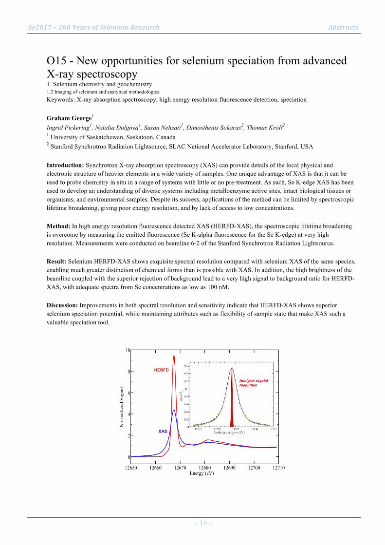
Se2017 – 200 Years of Selenium Research Abstracts
– 16 –
O15 - New opportunities for selenium speciation from advanced X-ray spectroscopy 1. Selenium chemistry and geochemistry 1.2 Imaging of selenium and analytical methodologies Keywords: X-ray absorption spectroscopy, high energy resolution fluorescence detection, speciation Graham George1 Ingrid Pickering1, Natalia Dolgova1, Susan Nehzati1, Dimosthenis Sokaras2, Thomas Kroll2 1 University of Saskatchewan, Saskatoon, Canada 2 Stanford Synchrotron Radiation Lightsource, SLAC National Accelerator Laboratory, Stanford, USA Introduction: Synchrotron X-ray absorption spectroscopy (XAS) can provide details of the local physical and electronic structure of heavier elements in a wide variety of samples. One unique advantage of XAS is that it can be used to probe chemistry in situ in a range of systems with little or no pre-treatment. As such, Se K-edge XAS has been used to develop an understanding of diverse systems including metalloenzyme active sites, intact biological tissues or organisms, and environmental samples. Despite its success, applications of the method can be limited by spectroscopic lifetime broadening, giving poor energy resolution, and by lack of access to low concentrations. Method: In high energy resolution fluorescence detected XAS (HERFD-XAS), the spectroscopic lifetime broadening is overcome by measuring the emitted fluorescence (Se K-alpha fluorescence for the Se K-edge) at very high resolution. Measurements were conducted on beamline 6-2 of the Stanford Synchrotron Radiation Lightsource. Result: Selenium HERFD-XAS shows exquisite spectral resolution compared with selenium XAS of the same species, enabling much greater distinction of chemical forms than is possible with XAS. In addition, the high brightness of the beamline coupled with the superior rejection of background lead to a very high signal to background ratio for HERFD-XAS, with adequate spectra from Se concentrations as low as 100 nM. Discussion: Improvements in both spectral resolution and sensitivity indicate that HERFD-XAS shows superior selenium speciation potential, while maintaining attributes such as flexibility of sample state that make XAS such a valuable speciation tool.
Se2017 – 200 Years of Selenium Research Abstracts
– 17 –
O16 - Development of Fluorescent ROS Probe Based on Redox Reaction of Selenol group 1. Selenium chemistry and geochemistry 1.2 Imaging of selenium and analytical methodologies Keywords: reactive oxygen species, fluorescent probe Noriyuki Suzuki1 Hiroki Watanabe1, Tomohiro Doura1, Yasumitsu Ogra1 1 Graduate School of Pharmaceutical Sciences, Chiba University, Chiba, Japan Introduction: Reactive oxygen species (ROS) are continuously generated in biological systems, and cause damage to lipids, proteins and DNA. On the other hand, ROS also act as signaling molecules in the maintenance of physiological functions. In order to investigate the behavior of ROS in vivo, we used selenol group as a sensor of ROS in our fluorescent probe. Selenium can exist in various oxidation states in biological systems. By using selenium as a ROS-sensing motif in a fluorescent probe, it enables us to observe reversible fluorescence that relevant to biological ROS signals. Method: We have designed and synthesized “SeMF”, based on the intramolecular spiro-cyclization of selenol group. Because of high acidity of the selenol group, SeMF is expected to form a spiro ring in reductive condition due to the high nucleophilicity of selenide, and the fluorescence of SeMF is completely quenched under the condition. When SeMF is oxidized by ROS, the spiro ring form is converted to the quinonoid form that shows the strong fluorescence. Result: SeMF was specifically oxidized to fluorescent form by hypochlorite, while the fluorescence was quickly reduced in the presence of glutathione to regenerate the reduced form. This redox-induced reversible fluorescence response of SeMF allowed us to detect the intracellular hypochlorite of HepG2 cells. Discussion: This fluorescent probe is expected to be useful for monitoring the dynamics of ROS production continuously in vivo.

Se2017 – 200 Years of Selenium Research Abstracts
– 18 –
O17 - Advances in large scale speciation of selenium by plasma and molecular mass spectrometry 1. Selenium chemistry and geochemistry 1.2 Imaging of selenium and analytical methodologies Keywords: speciation, mass spectrometry, electrospray MS, ICP MS, coupled techniques Joanna Szpunar1 1 IPREM, CNRS, UMR 5254, Hélioparc, Pau, France Introduction: Distinguishing between different selenium chemical forms present in biological systems presents a particular challenge for analytical chemists because - besides having several possible oxidation states in its inorganic form - selenium can be incorporated into tens of known (and probably many more not discovered yet) low molecular weight (below 1000 Da) selenometabolites, many SeCys- and SeMet-containing proteins, selenosugars, selenolipids and, the last but not least, present as selenium nanoparticles. Each of these forms has a distinct biological function and as such should be individually detected and determined in the presence of a rich biological matrix. A particular importance has been attributed to the selenium speciation in food sources and its metabolic pathways leading to synthesis of physiologically important selenoproteins and low molecular weight selenometabolites. Method: Developments in selenium speciation analysis by coupled techniques combining separation methods with parallel elemental and molecular mass spectrometry have resulted in identification and quantification of a growing number of selenium species opening a way to fully comprehensive species-selective characterization of biological samples. Discussion: The presentation will address the remaining problems in the field and propose the solutions for (1) the quantitative assessment of selenium incorporation into proteins with a focus on the preservation of the stability of SeCys residues by carefully optimized derivatization, (2) the structural elucidation of selenoproteins isoforms, (3) the speciation of high molecular selenium species in organic media and (4) quantification of nanoparticulate selenium fraction in biological samples. The role of developments in both mass spectrometry instrumentation and custom designed sample preparation methods will be highlighted.
Se2017 – 200 Years of Selenium Research Abstracts
– 19 –
O18 - Selenium speciation in rainwater from high altitude locations 1. Selenium chemistry and geochemistry 1.3 Local geological selenium sources and global cycling Keywords: selenium, precipitation, speciation, rainwater, HPLC-ICP-MS Elke Suess1 Lenny H. E. Winkel1, Jeroen E. Sonke2 1 ETH Zurich/Eawag, Institute of Biogeochemistry and Pollutant Dynamics, Zurich, Switzerland 2 Observatoire Midi-Pyrénées, CNRS-GET, Université Toulouse III - Paul Sabatier, Toulouse, France Introduction: Atmospheric transport and wet deposition play a major role in supplying selenium (Se) and other essential (micro)nutrients to terrestrial environments, including agricultural soils. To investigate the temporal variation in total Se concentrations and speciation in rainfall, we collected and analyzed weekly precipitation over 14 and 5 months at two high-altitude locations in Europe, i.e., Jungfraujoch (JFJ, Alps, Switzerland) and Pic du Midi (PDM, Pyrenees, France), respectively. Both sites are often in the free troposphere, which enables investigation of long range atmospheric transport. Method: We analyzed total concentrations and speciation of Se as well as of sulfur (S), iodine (I) and bromine (Br) in precipitation using (HPLC)-ICP-QQQ, dissolved organic carbon (DOC) concentrations (Shimadzu TOC-L CSH) and isotopic carbon signatures of DOC (δ13C, IRMS). Result: Selenium showed generally higher concentrations at PDM (0.6 nM) compared to JFJ (0.3 nM). At both locations, mainly inorganic and anionic species of Se, S, I, and Br were found, without clear seasonal differences. In the productive period (April-September) we found significant positive correlations between Se and biogenic non-sea-salt sulfate (for both sites) and for Se with δ13C-values (only for JFJ). Discussion: The results indicate that Se in rainfall may have major contributions from marine biogenic sources at both locations. Interestingly, at the more continental location (JFJ), Se, S, I, and Br concentrations were highest when δ13C-signatures were less negative, indicating that marine biogenic sources bring in highest Se (and S, I, and Br) concentrations to this region. Currently, analysis of air mass sources is underway to test this hypothesis.

Se2017 – 200 Years of Selenium Research Abstracts
– 20 –
O19 - Effect of conservation agriculture on solubility and fractionation of soil selenium 1. Selenium chemistry and geochemistry 1.3 Local geological selenium sources and global cycling Keywords: Minimum tillage, Mineralization Ivy Sichinga - Ligowe1 Patson Nalivata1, Scott Young2, Louise Ander3, Allan Chilimba4, Liz Bailey2, Vernon Kabambe1 1 Lilongwe university of Agriculture and Natural Resources, Bunda Campus, Lilongwe, Malawi 2 University of Nottingham, School of Biosciences, Sutton Bonington Campus, Loughborough, UK 3 Centre for Environmental Geochemistry, British Geological Survey, Nottingham, UK 4 Department of Agricultural Research Services, Lilongwe, Malawi Introduction: The benefits of Conservation Agriculture (CA) to soil fertility have been widely reported (Wu et al., 2005; Chivenge et al., 2006; Ngwira et al., 2012). However, the effects of CA on the nutritional quality of maize still present a significant knowledge gap. This study is part of an ongoing investigation into the trace element dynamics within a CA trial in Malawi. Method: Soil and plant samples from a 9 year long-term CA trial in Malawi were analyzed for selenium and other parameters. Soil selenium data (‘total’ (HF digest), ‘exchangeable’ (phosphate-extractable) and ‘soluble’ (in 0.01 M KNO3) from the trial plots are presented here. Result: The total selenium concentration in topsoil (0 - 20 cm) was greater in tilled treatments, as compared to the CA minimum-tilled plots whereas, by contrast, soluble and phosphate-exchangeable Se were greater in minimum tilled plots. Discussion: Minimum tillage is thought to increase mineralization and solubility of some nutrients in soil (Wolf and Snyder, 2003). This may include selenium, which is known to cycle between organic and inorganic forms. Thus both the adsorbed phosphate-exchangeable Se pool and the soluble soil Se were significantly greater in plots receiving minimum tillage across all crop treatments. Selenium solubility may be enhanced under conservation agriculture cropping systems. Current evidence suggests that dietary Se is at sub-optimum levels in Malawi (Hurst et al., 2012) and thus CA systems may present benefits beyond improved soil management practices. Selected references
Hurst R., Siyame E.W.P., Young S.D., Chilimba A.D.C., Joy E.J.M., Black C.R., Ander E.L., Watts M.J., Chilima B., Gondwe J., Kang'ombe D., Stein A.J., Fairweather-Tait S.J., Gibson R.S., Kalimbira A., Broadley M.R. 2013. Soil-type influences human selenium status and underlies widespread selenium deficiency risks in Malawi. S.R., 3, 1425, 1-6.
Ngwira, A. R., Sleutel, S. and De Neve, S. 2012. Soil carbon dynamics as influenced by tillage and crop residue management in loamy sand and sandy loam soils under smallholder farmers’ conditions in Malawi.N.C.A.J. 92:315-328.

Se2017 – 200 Years of Selenium Research Abstracts
– 21 –
O20 - Selenocysteine in mammalian thioredoxin reductase and application of ebselen as a therapeutic 2. Selenium in the molecular life sciences 2.2 Molecular mechanisms of selenium toxicity Keywords: thioredoxin reductase, ebselen, bacteria, silver Arne Holmgren1 Jun Lu1, Lili Zou1, Jun Wang2, Xiaoyuan Ren1, Yu Gao1, Lanlan Zhang1, Martin Rottenberg1 1 Karolinska Institute, Stockholm, Sweden 2 Three Gorges University, 443000 Yichang, China Introduction: A thioredoxin system consisting of thioredoxin (Trx), NADPH and thioredoxin reductase (TrxR) is present in all living cells with many functions (1) as a disulfide reductase for reductive enzymes like peroxiredoxins or ribonucleotide reductase (RNR). Most cells also have a glutaredoxin system comprising glutathione (GSH), NADPH, and glutathione reductase plus glutaredoxin (Grx) acting as an efficient electron donor for RNR in DNA synthesis. Mammalian TrxRs are three large dimeric isoenzymes with an essential selenocysteine (Sec) residue in a C-terminal active site. In contrast TrxR from yeast, plants and bacteria have a small TrxR with cysteine residues in the active site and a different structure and mechanism. Auranofin a gold-containg FDA- approved drug targeting the essential Sec residue in TrxR1 in the cytosol and TrxR2 in mitochondria causes cancer cell death. Ebselen is a selenazol drug with GSH peroxidase and antioxidant activity, a superfast oxidant of reduced Trx and a substrate of mammalian TrxR (2) but in contrast a competitive inhibibitor of bacterial TrxR (3). Method: Here I will discuss therapeutic ebselen use. Result: Ebselen has been used as a new antibiotic principle and can effectively kill bacteria like Staphylococus aureus and MRSA or Helicobacter pylori lacking GSH and Grx (3). Recent unpublished data from our laboratory show strong synergistic effects with silver, which enables targeting multi-resistant Gram-negative bacteria like E.coli (4). Silver was a strong inhibitor of both thioredoxin and thioredoxin reductase. Discussion: In combination with ebselen, silver became selectively toxic to Gram-negative bacteria as shown by mouse peritonitis model experiments (4). Selected references
1. A. Holmgren, J. Lu, Thioredoxin and thioredoxin reductase: current research with special reference to human disease. Biochem. Biophys. Res. Commun. 2010, 396, 120.
2. L.Zhao, H. Masayasu,A. Holmgren. Ebselen: a substrate for human thioredoxin reductase strongly stimulating its hydroperoxide reductase activity and a superfast thioredoxin oxidant. 2002, Proc. Natl. Acad. Sci. USA, 99, 8579.
3. J.Lu, A. Vlamis-Gardikas, K.A. Kandasamy, R. Zhao, T.N. Gustafsson, L. Engstrand, L., S.Hoffner, L. Engman, A. Holmgren: Inhibition of bacterial thioredoxin reductase: an antibiotic mechanism targeting bacteria lacking glutathione. 2013, FASEB J, 27, 1304.
4. Lili Zou, Jun Lu, Jun Wang, Xiaoyuan Ren, Lanlan Zhang , Yu Gao, Martin E. Rottenberg and Arne Holmgren : A synergistic antibacterial effect of silver and ebselen against multidrug-resistant Gram-negative bacterial infections. Submitted 2017.
Se2017 – 200 Years of Selenium Research Abstracts
– 22 –
O21 - Characterization of novel selenium compounds as a therapeutic approach for improved cancer treatment 2. Selenium in the molecular life sciences 2.2 Molecular mechanisms of selenium toxicity Keywords: cytotoxicity, programmed cell death, anticancer agents Aristi Fernandes1 Carmen Sanmartín2, Valentina Gandin3 1 Division of Biochemistry, Department of Medical Biochemistry and Biophysics (MBB), Karolinska Insti 2 Department of Organic and Pharmaceutical Chemistry, University of Navarra, Irunlarrea 1, E-31008 Pam 3 Department of Pharmaceutical and Pharmacological Sciences, University of Padova, Via Marzolo 5, 3513 Introduction: It is well established that tumor cells in general are particularly vulnerable to oxidative stress. Increased ROS production to preferentially target cancer cells has hence emerged as an attractive strategy for new cancer therapeutics (1). Based on this knowledge, redox active selenium (Se) compounds, acting as prooxidants , have gained substantial attention with promising chemotherapeutic potential in both solid and hematological malignancies (2). Novel compounds are however warranted to increase the efficacy and specificity and to minimize the systemic effects. The better pharmacological and toxicological profiles recently disclosed by some organic Se compounds has fostered our research towards the development of this type of Se-based drugs. Method: Following the synthesis of novel organic Se compounds, we have biochemically characterized and cell biologically evaluated these compounds based on their, cytotoxicity profile, cell death pathways, efficacy and specificity index in in vitro and ex vivo models. Result: Several of the novel organic Se compounds synthesized have shown superior effects compared to naturally occurring Se metabolites or compared to analogues lacking the selenium moiety. Discussion: The novel Se compounds examined has provided valuable insights into their molecular mode of action that will help in the development of new efficient Se containing chemotherapeutic agents. Selected references
1. Gorrini C, Harris IS, & Mak TW (2013) Modulation of oxidative stress as an anticancer strategy. Nat Rev Drug Discov 12(12):931-947.
2. Fernandes AP & Gandin V (2015) Selenium compounds as therapeutic agents in cancer. Biochimica et biophysica acta 1850(8):1642-1660.

Se2017 – 200 Years of Selenium Research Abstracts
– 23 –
O22 - Impairment of protein homeostasis accounts for selenomethionine toxicity in Saccharomyces cerevisiae 2. Selenium in the molecular life sciences 2.2 Molecular mechanisms of selenium toxicity Keywords: Selenomethionine, selenocysteine, toxicity, protein aggregation, yeast Myriam Lazard1 Cosmin Saveanu2, Roxane Lestini3, Marc Dauplais1, Laurence Decourty2, Alain Jacquier2, Sylvain Blanquet1, Pierre Plateau1 1 Laboratoire de Biochimie, Ecole polytechnique, CNRS, Université Paris-Saclay, 91128 Palaiseau cedex, 2 Institut Pasteur, Unité de Génétique des Interactions Macromoléculaires, CNRS-UMR3525, Paris, Franc 3 Laboratoire d'Optique et Biosciences, Ecole Polytechnique, Université Paris-Saclay, CNRS UMR7645-IN Introduction: Selenium supplementation in human diet has been proposed to exert beneficial effects against several diseases, including cancer (1). As a result, selenomethionine (SeMet) has become a popular dietary supplement. However, excessive intake is toxic. The origin of this toxicity is not yet understood, although it may help understanding the basis of SeMet beneficial properties. The high level of conservation of its genes and metabolic pathways with those of higher organisms makes yeast an attractive model organism to study the molecular basis of selenium toxicity. Method: To identify biological pathways involved in SeMet toxicity, we performed a genome-wide screen in a yeast deletion collection. Fluorescence microscopy using a GFP-tagged Hsp104 marker was used to monitor protein aggregation in vivo. Selenium incorporation into proteins was analyzed by mass spectrometry. Result: Genes involved in protein degradation and synthesis were enriched in the sensitive and resistant datasets, suggesting that SeMet causes a proteotoxic stress. Accumulation of protein aggregates was observed upon SeMet exposure. Reduction of translation rates was accompanied by a reduction of protein aggregation and SeMet toxicity. Protein aggregation and toxicity were supressed in a ∆cys3 mutant unable to synthetize selenocysteine. Lastly, SeCys incorporation in a reporter polypeptide was observed in cells exposed to SeMet (2,3). Discussion: These results suggest that SeMet toxicity is caused by protein aggregation resulting from the metabolization of SeMet to selenocysteine followed by translational incorporation in the place of cysteine. The hypothesis that selenocysteine misincorporation disrupts protein homeostasis is reinforced by studies in higher eukaryotes (4). Selected references
(1) Rayman, M. P. (2005) Selenium in cancer prevention: a review of the evidence and mechanism of action. Proc. Nutr. Soc. 64, 527-542.
(2) Lazard, M. et al. (2015) Trans-sulfuration pathway seleno-amino acids are mediators of selenomethionine toxicity in Saccharomyces cerevisiae. J. Biol Chem. 290, 10741-50
(3) Plateau, P. et al. (2017) Exposure to selenomethionine causes selenocysteine misincorporation and protein aggregation in Saccharomyces cerevisiae. Sci. Reports in press.
(4) Wallenberg, M. et al. Selenium induces a multi-targeted cell death process in addition to ROS formation. J. Cell. Mol. Med. 18, 671-684 (2014).

Se2017 – 200 Years of Selenium Research Abstracts
– 24 –
O23 - Selenium status of Russian population: interactions with demography 3. Selenium in animal and human health and disease 3.2 Epidemiology of selenium related health and disease Keywords: hair, population, demography, mortality Anatoly Skalny1 Margarita Skalnaya2, Andrei Grabeklis3, Alexey Tinkov4 1 RUDN University, Moscow, Russia; Trace Element Institute for UNESCO, Lyon, France 2 RUDN University, Moscow, Russia 3 RUDN University, Moscow, Russia; Yaroslavl State University, Yaroslavl, Russia 4 RUDN University, Moscow, Russia; Orenburg State Medical University, Orenburg, Russia Introduction: It is interesting to study how Se status has an impact on population health and demography. Method: The objective was to assess Se status of the Russian population using ICP-MS analysis of hair Se content from more than 60,000 adults and 15,000 children. Result: The median (25-75 percentile) hair Se content (µg/g) in adult men and women was 0.439 (0.393-0.484) and 0.343 (0.305-0.383), whereas in boys and girls the respective values were 0.397 (0.340-0.443) and 0.388 (0.327-0.445). The lowest hair Se values in adults were detected in the Orenburg region (South Urals), whereas the highest – in the Kabardino-Balkar Republic (North Caucasus). In children, the lowest Se levels were detected in Kaliningrad region, whereas the highest – in Chechen Republic (North Caucasus) and Republic of Mordovia (Volga region). The IUPAC calculated reference values for hair Se levels in adult Russian population were 0.093 - 0.482 µg/g. Hair Se levels significantly correlated to demographic indices. In particular, despite the absence of significant association with birth rate (p=0.602) and total morbidity (p=0.312), hair Se inversely correlated with mortality (p=0.022) and was positively associated with increased life span (p<0.001) in adults. In children, a significant inverse relationship between hair Se and total morbidity (p=0.002) was observed. Discussion: It is notable that the observed associations were more tight after adjustment for population levels of toxic trace elements (especially, Hg), being indicative of the outcome of Hg-Se antagonism. The present findings demonstrate that modulation of Se status of the population is expected to be beneficial to improve demography and population health. Selected references
Skalny AV et al. (2016). Selenium Antagonism with Mercury and Arsenic: From Chemistry to Population Health and Demography. In: Selenium (pp. 401-412). Springer International Publishing.
Skalny AV et al. (2015). Hair concentration of essential trace elements in adult non-exposed Russian population. Environ. Monit. Assess. 187(11):677.
Bjørklund G. et al. (2017). Molecular interaction between mercury and selenium in neurotoxicity. Coord. Chem. Rev. 332:30-37.

Se2017 – 200 Years of Selenium Research Abstracts
– 25 –
O24 - Selenium status of School Children for Kaschin–Beck Disease Endemic Areas in Tibet, China 3. Selenium in animal and human health and disease 3.2 Epidemiology of selenium related health and disease Keywords: Children, Kaschin–Beck disease, Tibet Hairong Li1 Linsheng Yang1, Wuyi Wang1, Jing Wang1, Zhuo Chen1, Ya'nan Guo1 1 Institute of Geographical Sciences and Natural Resources Research, CAS, Beijing, China Introduction: Selenium (Se) deficiency is an important environmental factor for the etiology of Kaschin–Beck disease (KBD). Although KBD is presently controlled in most regions of China, it is still active in Tibet. The study aims to explore selenium status of Se in school children in KBD areas in Tibet, China. Method: Hair samples of 221 school children were collected in KBD areas of Lhasa, Shigatse and Chamdo during 2013-2015. The Se contents in the hair samples were measured by inductive coupled plasma mass spectrometry. Result: The mean Se level in children’s hair was 0.252 ± 0.082 µg/g, which was 0.77 times higher than that of reported in 2003 (0.142 µg/g) in KBD areas in Tibet. The average hair Se level in boys (0.270 ± 0.088 µg/g) was significantly higher than that in girls (0.237 ± 0.075 µg/g) in the studied areas (t = 2.971,P < 0.01). The detectable X-ray of children aged 7-12 in KBD areas in Tibet was declined from 37.86% in 2000 to less than 3% since 2010. Discussion: The rising trend of Se level in children was in good agreement with the decline of KBD, which likely as a result of high Se content staple food substitution with the enforcement of Free Education Policy and Nutrition Improvement Plan in Tibet. However, 22.17% of students were still deemed to be in Se deficiency (hair Se <0.20 µg/g), which indicated that Se status of school children was also partly affected by low Se environment in KBD areas in Tibet. Selected references
Zhaxi S, Gong H, Gesang D et al (2003) Selenium test results analysis in Tibet Kaschin-Beck Disease areas. Chin J Ctrl Endem Dis 18(3):177 (In Chinese)
Zhao S, Gong H, Guo M et al (2016) Analysis on prevalence trend of Kaschin-Beck disease in Tibet from 2000 to 2015. Chin J Ctrl Endem Dis 31(8):841-844 (In Chinese)
Tan J (1990) Chemico-geography of some life elements and endemic diseases with an emphasis on China. Environmental life elements and health. Science Press, Beijing, pp 145-157

Se2017 – 200 Years of Selenium Research Abstracts
– 26 –
O25 - Impact of early-life selenium status on children’s cognitive abilities 3. Selenium in animal and human health and disease 3.2 Epidemiology of selenium related health and disease Keywords: child, cognition, neurodevelopment, pregnancy, hair selenium Helena Skröder1 Maria Kippler1, Fahmida Tofail2, Marie Vahter1 1 Karolinska Institutet, Institute of Environmental Medicine, Stockholm, Sweden 2 International Center for Diarrhoeal Disease Research, Dhaka, Bangladesh Introduction: We recently discovered that mothers’ selenium status in pregnancy was positively associated with their children’s cognitive function at 1.5 years. The present aim was to assess whether this association persisted in later childhood, and if the children’s selenium status was of importance. Method: This cohort study, including 1405 children, was nested in a population-based, randomized food and micronutrient supplementation trial in pregnancy in rural Bangladesh. Selenium in maternal blood (erythrocyte fraction, Ery-Se) at gestational week 14, and in children’s hair at 10 years, was measured using inductively coupled plasma mass spectrometry. Children’s cognitive function at 5 and 10 years was assessed using the Wechsler Pre-school and Primary Scale of Intelligence (3rd edition) and the Wechsler Intelligence Scale for Children (4th edition), respectively. Result: Multivariable-adjusted linear regression analyses revealed a persistent association between prenatal selenium status and cognitive function at 5 and 10 years. A 0.5 µg/g Hb increase in maternal Ery-Se (median: 0.45 µg/g Hb) was associated with an increase in full developmental score corresponding to 0.2 and 0.3 SD at 5 and 10 years, respectively. Children’s concurrent selenium status was also positively associated with cognitive function at 10 years, possibly through interactions with thyroid hormones. Moreover, there were indications of inverse associations at the highest hair selenium concentrations (>0.7 mg/kg), although very few children (~2%) had such high concentrations. Further adjustments for other nutritional factors (maternal BMI, erythrocyte zinc, urinary iodine, hemoglobin) did not affect the associations. Discussion: Adequate selenium status during early life appears to be beneficial for children’s cognitive function.

Se2017 – 200 Years of Selenium Research Abstracts
– 27 –
O26 - A decade of selenium research in Malawi (2008-2017) 1. Selenium chemistry and geochemistry 1.4 Relationships of selenium between soils, water, and vegetation Keywords: Dietary deficiency, fertilizers, food systems, geochemistry, geography, soil E.L. Ander1 E.H. Bailey1, M.R. Broadley1, B. Chilima1, A.D.C. Chilimba1, E.J.M. Joy1, R.M. V. Kabambe1, A.A. Kalimbira1, D.B. Kumssa1, I.S. Ligowe1, P. Nalivata1, F.P. Phiri1, M.J. Watts1, S.D. Young1 1 Malawi Selenium Research Consortium Introduction: Our research has shown that selenium (Se) deficiency is widespread in Malawi. We have been supported by many individuals and groups, including the International Selenium Society, to whom we have presented updates at all meetings since 2009. This paper summarizes our past and current research. Method: (1) Surveying soils and food crops (Chilimba et al. 2011; Joy et al. 2015a); (2) studying human Se intakes and status (Hurst et al. 2013; Siyame et al. 2013; Gibson et al. 2015); (3) analyzing food systems (Joy et al. 2014); (4) linking food and household survey data (~12,000 households; Joy et al. 2015b); (5) studying soil geochemistry and crop responses to Se fertilizers (Chilimba et al., 2012a,b; Ligowe et al., unpubl.); (6) assessing population blood/urine biomarkers of Se status (n=~3000, Phiri et al., unpubl.); (7) evaluating the potential of minor crops to address Se deficiency (Kumssa et al., 2017). Result: Human Se deficiency in Malawi is widespread, and influenced strongly by geography, including long-range variation due to soil-type and proximity to Lake Malawi, and short-range variation due to age, gender, urbanization and wealth. Discussion: Research is ongoing to provide evidence to support potential policy responses to address this potential health burden, and to determine the wider regional scale of dietary Se deficiency in sub Saharan Africa. Selected references
Chilimba, A.D.C. et al. (2012a). Agronomic biofortification of maize with selenium (Se) in Malawi. Field Crops Research, 125, 118–128. http://bit.ly/10CdKjy
Chilimba, A.D.C. et al. (2012b). Assessing residual availability of selenium applied to maize crops. Field Crops Research, 134, 11–18. http://bit.ly/X445Q0
Chilimba, A.D.C. et al. (2011). Maize grain and soil surveys reveal suboptimal dietary selenium intake is widespread in Malawi. Scientific Reports, 1, 72. http://bit.ly/ZjK3Th
Gibson, R.S. et al. (2015). Dietary iron intakes based on food composition data may underestimate the contribution of potentially exchangeable contaminant iron from soil. Journal of Food Composition and Analysis, 40, 19-23. http://bit.ly/1j2MMkn
Hurst, R. et al. (2013). Soil-type influences human selenium status and underlies widespread selenium deficiency risks in Malawi. Scientific Reports, 3, 1425. http://bit.ly/10Cd5P5.
Joy, E.J.M. et al. (2014). Dietary mineral supplies in Africa. Physiologia Plantarum, 151, 208-229. http://bit.ly/1f1klJJ
Joy, E.J.M. et al. (2015a). Soil type influences crop mineral composition in Malawi. Science of the Total Environment, 505, 587-595. http://bit.ly/1PPEHHv
Joy, E.J.M. et al. (2015b). Dietary mineral supplies in Malawi: spatial and socioeconomic assessment. BMC Nutrition, 1, 42. http://bit.ly/1SJVhQo
Kumssa, D.B. et al. (2017). Variation in the mineral element concentration of Moringa oleifera Lam. and M. stenopetala (Bak. f.) Cuf.: Role in human nutrition. PLOS ONE, 12, e0175503. http://bit.ly/2oVBnqe
Siyame, E.W.P. et al. (2013). A high prevalence of zinc- but not iron-deficiency among women in rural Malawi: a cross-sectional study. International Journal for Vitamin and Nutrition Research, 83, 176-187. http://bit.ly/1Oa6zuL

Se2017 – 200 Years of Selenium Research Abstracts
– 28 –
O27 - Key role of Selenium (0) nanoparticles in soil and as therapeutic agent 1. Selenium chemistry and geochemistry 1.4 Relationships of selenium between soils, water, and vegetation Keywords: Nanoparticle reactivity, soil, cancer cells, XANES Laurent Charlet1 Caroline Bissardon2, Sylvain Bohic2, Steve Conlan3, Celine Pallud4 1 ISTerre University of Grenoble Alps, Grenoble, France 2 Inserm GIN U836 & Nanoimaging ESRF Beamline ID16 , Grenoble, France 3 Centre for NanoHealth, Swansea University Medical School, Swansea University, Wales, UK 4 ESPM, UC Berkeley, Berkeley, CA, USA Introduction: Metallic selenium nanoparticles (SeNP) form in hydromorphic soils. via Se reduction and retention within soil aggregates, while SeNP dissolution may lead to food fortification via a Se slow delivery systems to plants. Conversely, selenium SeNP have been shown to be a promising therapy for specific ovarian and prostate cancers and a mechanistic understanding of Se methylation within cancer cell may enhance the development of cancer therapy. Method: Mineral specific investigations were conducted with selenite ion reacted in anoxic conditions with ferrous iron in presence of soil minerals. The coupling between physical and biogeochemical processes in soil aggregates is investigated by a flow-through reactor system and a 3D reactive transport model. Soil and cancer cells are investigated by synchrotron X-ray absorption near edge spectromicroscopy (XANES) linear combination fitting (LCF). Result: Selenite ions react with aqueous Fe2+ ion (a major ion in hydromorphic soils) in presence of various soil minerals (clays1, calcite2), or with Fe2+ rich minerals (siderite magnetite, pyrite)3,4 to form elemental SeNP and/or FeSe NP species. Within soil aggregates, a radial redox gradient is observed. The reactive transport model details the interplay between intra-aggregate Se diffusion and sorption/reduction. Discussion: In soil, Se reduction increased towards the core of aggregates, and in clay rich soils slow Se delivery system to plants cis investigated. Conversely Se-NPs dissolution in cancer cells were shown5,6,7 to significantly inhibate the cell growth, to impact the regulation of the cell cycle e.g. with tubuline depolymerization8, to stimulate apoptosis and to inhibit tumor cell migration and invasion in vitro. Selected references 1Charlet et al., GCA 2007 2 Chakraborty et al., EST 2010 3Charlet et al., ES&T 2012 4Scheinost and Charlet., ES&T 2008 5Schrauzer et al. 2000. Cell Mol. Life Sci. 6Zeng et aL, 2009, J. of Nutrition, 1613 7Lu and Jiang, 2005, Antioxidant and signaling, Vol 7, No11&12 8Gao et al (2014) Biomaterials 35 8854e8866
Se2017 – 200 Years of Selenium Research Abstracts
– 29 –
O28 - Adsorption of selenate and selenite in cultivated and native soils of the Brazilian Cerrado 1. Selenium chemistry and geochemistry 1.4 Relationships of selenium between soils, water, and vegetation Keywords: selenium sorption, oxidic soils, soil fertility management Guilherme Lopes1 Anderson Araujo1, Josimar Lessa1, Liniker Ferreira1, Gabrielly Silva1, Luiz Guilherme1 1 Federal University of Lavras, Lavras, MG, Brazil Introduction: The availability of selenium (Se) for plants - and as a result for humans/animals - is influenced by several factors, such as the chemical species of Se (e.g., selenate or selenite) and soil characteristics, as well as the contents of other ions, especially anions, soil texture, pH, organic matter content, among others (Yasin et al., 2015). Method: This study evaluated the adsorption of selenate, Se(VI) and selenite, Se(IV) in cultivated and uncultivated soils of the Brazilian Cerrado biome. Adsorption tests were performed adding both Se species as sodium salts (selenate or selenite) at 500 µg L-1, using 15 mmol L-1 NaCl as the electrolyte solution (pH=5.5). Result: The results showed that selenite was much more adsorbed on the studied soils compared with selenate. Moreover, uncultivated soils adsorbed higher amounts of both Se forms, which can be attributed to the presence of competing anions (e.g., phosphate and sulphate) added through soil fertility management in cultivated soils. Still, adsorption of selenate seems to be more affected by these competitive anions, when compared with selenite. Discussion: The higher adsorption of Se(IV) compared with Se(VI) indicates that selenate is the key Se species to be applied in Se-poor soils to increase Se levels in plants (biofortification) and, as a results, in animals and humans (Boldrin et al., 2012; Ramos et al., 2011). Moreover, adding selenate in fertilizers containing other anions to compete with Se for sorption sites should be considered a good strategy for studies concerning agronomic biofortification with Se. (Sponsored by FAPEMIG, CNPq, and CAPES). Selected references
BOLDRIN, P. F. et al. 2012. Se (VI) e Se (IV) na produção e biofortificação agronômica com selênio em arroz. Pesq. Agropec. Bras. 47: 831-837.
RAMOS, S. J. et al. 2011. Selenato e selenito na produção, nutrição mineral e biofortificação com selênio em cultivares de alface. R. Bras. Ci. Solo. 35: 1347-1355.
YASIN, M. et al. 2015. Microbial-enhanced selenium and iron biofortification of wheat (triticum aestivum L.) applications in phytoremediation and biofortification. Int. J. Phytorem. 17: 341-347.
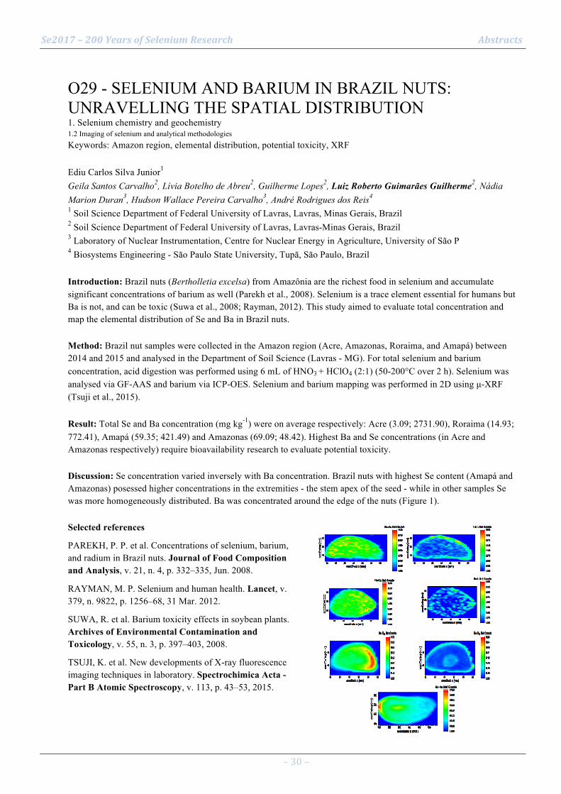
Se2017 – 200 Years of Selenium Research Abstracts
– 30 –
O29 - SELENIUM AND BARIUM IN BRAZIL NUTS: UNRAVELLING THE SPATIAL DISTRIBUTION 1. Selenium chemistry and geochemistry 1.2 Imaging of selenium and analytical methodologies Keywords: Amazon region, elemental distribution, potential toxicity, XRF Ediu Carlos Silva Junior1 Geila Santos Carvalho2, Lívia Botelho de Abreu2, Guilherme Lopes2, Luiz Roberto Guimarães Guilherme2, Nádia Marion Duran3, Hudson Wallace Pereira Carvalho3, André Rodrigues dos Reis4 1 Soil Science Department of Federal University of Lavras, Lavras, Minas Gerais, Brazil 2 Soil Science Department of Federal University of Lavras, Lavras-Minas Gerais, Brazil 3 Laboratory of Nuclear Instrumentation, Centre for Nuclear Energy in Agriculture, University of São P 4 Biosystems Engineering - São Paulo State University, Tupã, São Paulo, Brazil Introduction: Brazil nuts (Bertholletia excelsa) from Amazônia are the richest food in selenium and accumulate significant concentrations of barium as well (Parekh et al., 2008). Selenium is a trace element essential for humans but Ba is not, and can be toxic (Suwa et al., 2008; Rayman, 2012). This study aimed to evaluate total concentration and map the elemental distribution of Se and Ba in Brazil nuts. Method: Brazil nut samples were collected in the Amazon region (Acre, Amazonas, Roraima, and Amapá) between 2014 and 2015 and analysed in the Department of Soil Science (Lavras - MG). For total selenium and barium concentration, acid digestion was performed using 6 mL of HNO3 + HClO4 (2:1) (50-200°C over 2 h). Selenium was analysed via GF-AAS and barium via ICP-OES. Selenium and barium mapping was performed in 2D using µ-XRF (Tsuji et al., 2015). Result: Total Se and Ba concentration (mg kg-1) were on average respectively: Acre (3.09; 2731.90), Roraima (14.93; 772.41), Amapá (59.35; 421.49) and Amazonas (69.09; 48.42). Highest Ba and Se concentrations (in Acre and Amazonas respectively) require bioavailability research to evaluate potential toxicity. Discussion: Se concentration varied inversely with Ba concentration. Brazil nuts with highest Se content (Amapá and Amazonas) posessed higher concentrations in the extremities - the stem apex of the seed - while in other samples Se was more homogeneously distributed. Ba was concentrated around the edge of the nuts (Figure 1). Selected references
PAREKH, P. P. et al. Concentrations of selenium, barium, and radium in Brazil nuts. Journal of Food Composition and Analysis, v. 21, n. 4, p. 332–335, Jun. 2008.
RAYMAN, M. P. Selenium and human health. Lancet, v. 379, n. 9822, p. 1256–68, 31 Mar. 2012.
SUWA, R. et al. Barium toxicity effects in soybean plants. Archives of Environmental Contamination and Toxicology, v. 55, n. 3, p. 397–403, 2008.
TSUJI, K. et al. New developments of X-ray fluorescence imaging techniques in laboratory. Spectrochimica Acta - Part B Atomic Spectroscopy, v. 113, p. 43–53, 2015.
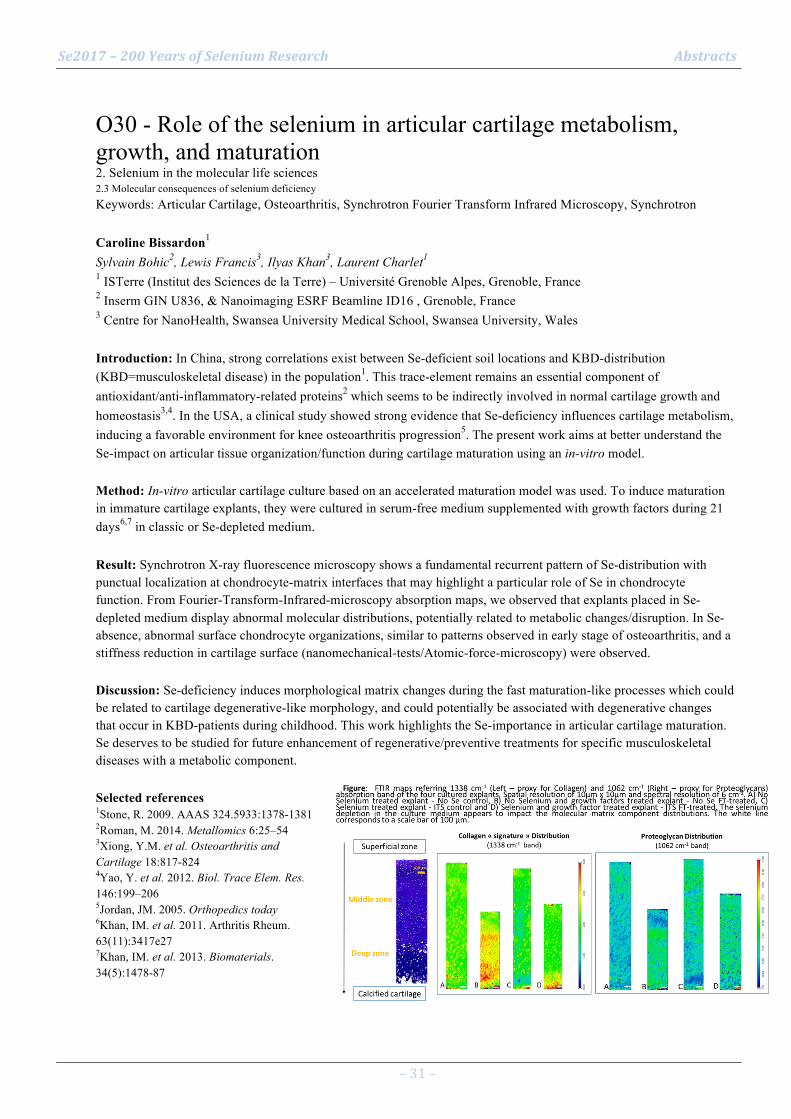
Se2017 – 200 Years of Selenium Research Abstracts
– 31 –
O30 - Role of the selenium in articular cartilage metabolism, growth, and maturation 2. Selenium in the molecular life sciences 2.3 Molecular consequences of selenium deficiency Keywords: Articular Cartilage, Osteoarthritis, Synchrotron Fourier Transform Infrared Microscopy, Synchrotron Caroline Bissardon1 Sylvain Bohic2, Lewis Francis3, Ilyas Khan3, Laurent Charlet1 1 ISTerre (Institut des Sciences de la Terre) – Université Grenoble Alpes, Grenoble, France 2 Inserm GIN U836, & Nanoimaging ESRF Beamline ID16 , Grenoble, France 3 Centre for NanoHealth, Swansea University Medical School, Swansea University, Wales Introduction: In China, strong correlations exist between Se-deficient soil locations and KBD-distribution (KBD=musculoskeletal disease) in the population1. This trace-element remains an essential component of antioxidant/anti-inflammatory-related proteins2 which seems to be indirectly involved in normal cartilage growth and homeostasis3,4. In the USA, a clinical study showed strong evidence that Se-deficiency influences cartilage metabolism, inducing a favorable environment for knee osteoarthritis progression5. The present work aims at better understand the Se-impact on articular tissue organization/function during cartilage maturation using an in-vitro model. Method: In-vitro articular cartilage culture based on an accelerated maturation model was used. To induce maturation in immature cartilage explants, they were cultured in serum-free medium supplemented with growth factors during 21 days6,7 in classic or Se-depleted medium. Result: Synchrotron X-ray fluorescence microscopy shows a fundamental recurrent pattern of Se-distribution with punctual localization at chondrocyte-matrix interfaces that may highlight a particular role of Se in chondrocyte function. From Fourier-Transform-Infrared-microscopy absorption maps, we observed that explants placed in Se-depleted medium display abnormal molecular distributions, potentially related to metabolic changes/disruption. In Se-absence, abnormal surface chondrocyte organizations, similar to patterns observed in early stage of osteoarthritis, and a stiffness reduction in cartilage surface (nanomechanical-tests/Atomic-force-microscopy) were observed. Discussion: Se-deficiency induces morphological matrix changes during the fast maturation-like processes which could be related to cartilage degenerative-like morphology, and could potentially be associated with degenerative changes that occur in KBD-patients during childhood. This work highlights the Se-importance in articular cartilage maturation. Se deserves to be studied for future enhancement of regenerative/preventive treatments for specific musculoskeletal diseases with a metabolic component. Selected references 1Stone, R. 2009. AAAS 324.5933:1378-1381 2Roman, M. 2014. Metallomics 6:25–54 3Xiong, Y.M. et al. Osteoarthritis and Cartilage 18:817-824 4Yao, Y. et al. 2012. Biol. Trace Elem. Res. 146:199–206 5Jordan, JM. 2005. Orthopedics today 6Khan, IM. et al. 2011. Arthritis Rheum. 63(11):3417e27 7Khan, IM. et al. 2013. Biomaterials. 34(5):1478-87
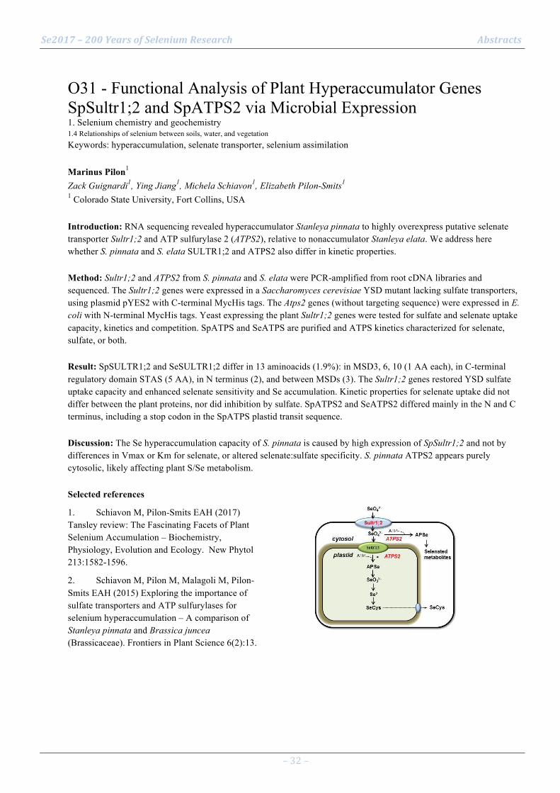
Se2017 – 200 Years of Selenium Research Abstracts
– 32 –
O31 - Functional Analysis of Plant Hyperaccumulator Genes SpSultr1;2 and SpATPS2 via Microbial Expression 1. Selenium chemistry and geochemistry 1.4 Relationships of selenium between soils, water, and vegetation Keywords: hyperaccumulation, selenate transporter, selenium assimilation Marinus Pilon1 Zack Guignardi1, Ying Jiang1, Michela Schiavon1, Elizabeth Pilon-Smits1 1 Colorado State University, Fort Collins, USA Introduction: RNA sequencing revealed hyperaccumulator Stanleya pinnata to highly overexpress putative selenate transporter Sultr1;2 and ATP sulfurylase 2 (ATPS2), relative to nonaccumulator Stanleya elata. We address here whether S. pinnata and S. elata SULTR1;2 and ATPS2 also differ in kinetic properties. Method: Sultr1;2 and ATPS2 from S. pinnata and S. elata were PCR-amplified from root cDNA libraries and sequenced. The Sultr1;2 genes were expressed in a Saccharomyces cerevisiae YSD mutant lacking sulfate transporters, using plasmid pYES2 with C-terminal MycHis tags. The Atps2 genes (without targeting sequence) were expressed in E. coli with N-terminal MycHis tags. Yeast expressing the plant Sultr1;2 genes were tested for sulfate and selenate uptake capacity, kinetics and competition. SpATPS and SeATPS are purified and ATPS kinetics characterized for selenate, sulfate, or both. Result: SpSULTR1;2 and SeSULTR1;2 differ in 13 aminoacids (1.9%): in MSD3, 6, 10 (1 AA each), in C-terminal regulatory domain STAS (5 AA), in N terminus (2), and between MSDs (3). The Sultr1;2 genes restored YSD sulfate uptake capacity and enhanced selenate sensitivity and Se accumulation. Kinetic properties for selenate uptake did not differ between the plant proteins, nor did inhibition by sulfate. SpATPS2 and SeATPS2 differed mainly in the N and C terminus, including a stop codon in the SpATPS plastid transit sequence. Discussion: The Se hyperaccumulation capacity of S. pinnata is caused by high expression of SpSultr1;2 and not by differences in Vmax or Km for selenate, or altered selenate:sulfate specificity. S. pinnata ATPS2 appears purely cytosolic, likely affecting plant S/Se metabolism. Selected references
1. Schiavon M, Pilon-Smits EAH (2017) Tansley review: The Fascinating Facets of Plant Selenium Accumulation – Biochemistry, Physiology, Evolution and Ecology. New Phytol 213:1582-1596.
2. Schiavon M, Pilon M, Malagoli M, Pilon-Smits EAH (2015) Exploring the importance of sulfate transporters and ATP sulfurylases for selenium hyperaccumulation – A comparison of Stanleya pinnata and Brassica juncea (Brassicaceae). Frontiers in Plant Science 6(2):13.

Se2017 – 200 Years of Selenium Research Abstracts
– 33 –
O32 - Influence of Sulfate on Selenium Uptake in Hyperaccumulator Stanleya pinnata and Non-Accumulators 1. Selenium chemistry and geochemistry 1.4 Relationships of selenium between soils, water, and vegetation Keywords: hyperaccumulation, sulfur, uptake, gene expression Michela Schiavon1 Ali El Mehdawi1, Marinus Pilon1, Zack Guignardi1, Ying Jiang2, Elizabeth Pilon-Smits1 1 Colorado State University, Fort Collins, USA 2 China Agricultural University, Beijing, China Introduction: Some plant species called hyperaccumulators contain selenium (Se) up to 0.1- 1.5% of their dry biomass. These plants display high Se to sulfur (S) ratios in tissues. Our hypothesis is that they may possess specific mechanisms for Se uptake. Method: In our study we used Stanleya pinnata (Brassicaceae) as the model hyperaccumulator, and S. elata and Brassica juncea as non-hyperaccumulator species. We studied the capacity of plants to absorb Se either over a long period or in the short-term using different selenate and sulfate concentration. Distinct sulfate pre-treatments were tested for their effect on Se uptake, and gene expression of sulfate transporters was assayed under varying ambient Se/S ratios. Result: S. pinnata exhibited higher capacity to absorb selenate compared to non-hyperaccumulators in both long and short-term assays, particularly under high S ambient concentration. In the long-term period, high sulfate reduced Se uptake in all species, but the effect was more pronounced in non-hyperaccumulators. The inhibitory effect of high sulfate on Se uptake was also observed in the short-term. In this case, Se uptake was totally repressed in S. elata by high sulfate, while it was moderately decreased in S pinnata and B. juncea. S pinnata plants had maximum values of root Se/S ratio and constitutive up-regulation of several sulfate transporter genes. Discussion: These findings suggest that selenate uptake in hyperaccumulators is less sensitive to the inhibition exerted by sulfate. Hyperaccumulation may be due to high expression of sulfate transporters and/or higher specificity of these transporters for selenate over sulfate. Selected references
1. El Kassis, E, Cathala, N, Rouached H, Fourcroy P, Berthomieu P, Terry N, et al. (2007). Characterization of a selenate-resistant Arabidopsis mutant. Root growth as a potential target for selenate toxicity. Plant Physiol. 143: 1231–1241.
2. Schiavon M, Pilon M, Malagoli M, Pilon-Smits E.A.H. (2015). Exploring the importance of sulfate transporters and ATP sulphurylases for selenium hyperaccumulation—a comparison of Stanleya pinnata and Brassica juncea (Brassicaceae). Fron. Plant Sci. 6: 2.
3. Schiavon M, Pilon-Smits EAH (2017) Tansley review: The Fascinating Facets of Plant Selenium Accumulation – Biochemistry, Physiology, Evolution and Ecology. New Phytol 213:1582-1596.
4. White PJ, Bowen HC, Parmaguru P, Fritz M, Spracklen WP, Spiby RE, et al. (2004). Interactions between selenium and sulphur nutrition in Arabidopsis thaliana. J. Exp. Bot. 55: 1927–1937.

Se2017 – 200 Years of Selenium Research Abstracts
– 34 –
O33 - Selenolanthionine is the main selenium species of the hyperaccumulator plant Cardamine violifolia 1. Selenium chemistry and geochemistry 1.4 Relationships of selenium between soils, water, and vegetation Keywords: speciation, HPLC-ICP-MS, HPLC-ESI-QTOF-MS, synthesis, water soluble Eszter Borbála Both1 Shuxun Shao2, Jiqian Xiang3, Zsuzsanna Jókainé Szatura1, Hongqing Yin3, Yafeng Liu2, Anna Magyar4, Mihály Dernovics1 1 Szent István University, Dept. Appl. Chemistry; Budapest, Hungary 2 State Key Lab. Ore Deposit Geochem., Inst. Geochem., Chinese Academy of Sciences, Guiyang, China 3 Enshi Autonomous Prefecture Academy of Agriculture Sciences, Enshi, Hubei Province, China 4 MTA-ELTE Research Group of Peptide Chemistry, Hungarian Academy of Sciences, Budapest, Hungary Introduction: Selenium hyperaccumulation in plants involve different selenium species to be stored in a chemical form serving for the elimination of excess selenium from plant metabolism to protect plant homeostasis. Organic forms, volatile species and inorganic forms are all concerned in this process. Method: The study aimed at the identification of the main selenium species of the selenium hyperaccumulator plant Cardamine violifolia. This plant is from the family of Brassicaceae and it is common in the highly seleniferous region of Enshi, China. A sample of this plant, containing 3.6 g Se / kg d.w. was prepared with several methods including ultrasonic assisted water extraction, proteolytic digestion, sodium sulphite and carbon disulphide extraction. The extracted selenium species were identified and quantified (when the relevant standard was available) with orthogonal chromatographic purification followed by either LC-ESI-QTOF-MS or LC-ICP-MS set-ups. Result: The Cardamine violifolia sample did not contain in considerable amount any of the organic selenium species that are often formed in hyperaccumulator plants and the inorganic selenium content (mostly in the form of elemental selenium) accounted only for less than 20% of total Se content. The most abundant selenium compound, accounting for more than 50% of total Se showed the accurate mass, elemental composition and chromatographic behaviour of selenolanthionine, a selenium species that has never been unambiguously identified before from any selenium containing sample. The identification process was completed with chemical synthesis as well. Discussion: Finding selenolanthionine as the main selenium species in a hyperaccumulating plant unearths a new way of metabolism in the selenium hyperaccumulation process.

Se2017 – 200 Years of Selenium Research Abstracts
– 35 –
O34 - Two selenium hyperaccumulators and their influence on their plant communities 1. Selenium chemistry and geochemistry 1.4 Relationships of selenium between soils, water, and vegetation Keywords: hyperaccumulator, plants, soil selenium Ray Reynolds1 Elizabeth Pilon-Smits1 1 Colorado State University, Fort collins Colorado, United States Introduction: Selenium hyperaccumulators are capable of concentrating and tolerating tissue Se levels as high as 15,000 mg/kg dry weight (DW), while most plants show toxicity from tissue concentrations in the range of 20-100 mg/kg DW. Through root and shoot turnover, hyperaccumulators change distribution, form and concentration of soil Se. How does this affect their plant communities? Method: We used transect methods to survey the plant communities of the hyperaccumulators Stanleya pinnata (Brassicaceae) and Astragalus bisulcatus (Fabaceae), as well as communities near, but without, hyperaccumulators. Both sites were also surveyed for soil and plant Se concentrations. Result: In areas with Se hyperaccumulators, canopy cover was lower and % bare ground higher, yet plant species richness was higher. These hyperaccumulator effects depend on spatial scale and are much less evident at larger scale. Some plant species show positive or negative co-occurrence with hyperaccumulators. Several species that positively co-occurred showed extreme Se tolerance and contained more Se when growing next to hyperaccumulators (sometimes >1000 mg/kg DW). Discussion: Most plants are sensitive to tissue Se concentrations from 20 to 100 mg/kg (DW). Perennial hyperaccumulators S. pinnata and A. bisulcatus appear to have a local effect on plant communities, as they over time concentrate, transform and redistribute Se through root and shoot turnover. Their influence appears most pronounced for nearby plant communities, and may exert selection pressure, Se-tolerant populations becoming more abundant and sensitive less abundant. They may also select for alleles involved in Se tolerance and thus the seed pool in the greater community. Selected references
El-Mehdawi AF, Pilon-Smits EAH (2012) Ecological Aspects of Plant Selenium Hyperaccumulation. Plant Biol 14: 1-10.
El Mehdawi, A.F., Quinn, C.F. & Pilon-Smits, E.A.H., 2011. Effects of selenium hyperaccumulation on plant-plant interactions: Evidence for elemental allelopathy? New Phytologist, 191(1), pp.120–131.

Se2017 – 200 Years of Selenium Research Abstracts
– 36 –
O35 - The effect of different Selenium fertilizers on garlic 1. Selenium chemistry and geochemistry 1.4 Relationships of selenium between soils, water, and vegetation Keywords: Se-enriched slate powder, Cultivation, Cadmium Hongyu Zhang1 Zhengyu Bao2, Changhua Wei2, Anqing Gu1, Huan Tian1 1 School of Earth Sciences, China University of Geosciences, Wuhan, China 2 Faculty of Materials Science and Chemistry, China University of Geosciences, Wuhan, China Introduction: Selenium (Se) is an essential element for human and crops are believed to be the main sources of Se. Thus, understanding the effect of different Se fertilizers on vegetables is the premise of Se agro-enhancement. In this study, the effect of different Se fertilizers on garlic and the relationship between Se and Cd were studied. Method: Garlic was grown in soils with different dose of activated Se-enriched slate powder as fertilizer[1]in 2015 and with sodium selenite in 2014. Total contents of cadmium (Cd) and Se in the garlic were determined by ICP-MS and HG-AFS, respectively. Result: 1. Both the activated Se-rich slate and sodium selenite were effective. The highest Se concentration of the garlic with the Se-fertilizer was about 4-times higher than the blank group (Se0). When the dose (Se) is less than 70 mg/m2 (Se70), Se-enriched slate powder was more efficient, and when the dose (Se) is larger, sodium selenite was more valid (Fig.1 a). 2. Se and Cd concentration of garlic treated by activated Se-enriched slate powder showed a synergistic effect when the dose (Se) is less than 35mg/m2(Se35) and antagonistic effect when the dose (Se) is larger than 35mg/m2(Se35) (Fig.1). When treated by sodium selenite, this trend still presented. But Cd concentration of garlic treated with Se-enriched slate powder was always lower than that of garlic treated with sodium selenite (Fig.1 b). Discussion: Se-enriched mineral powder has the ability to detoxify the garlic grown under Cd stress, which may be used to remediation of Cd poisoning soil. Selected references [1] Tian, H., Bao, Z., Wei, C., Zhang, H., Wu, S., & Gu, A., et al. (2016). Improved selenium bioavailability of selenium-enriched slate via calcination with a ca-based sorbent. Journal of Geochemical Exploration, 169, 73-79.

Se2017 – 200 Years of Selenium Research Abstracts
– 37 –
O36 - Biofortification of maize and beans with selenium in central Kenya highlands 1. Selenium chemistry and geochemistry 1.4 Relationships of selenium between soils, water, and vegetation Keywords: Selenium deficiency, biofortification Peter Biu Ngigi1 Gijs Du Laing1, Carl Lachat1, Peter Wafula Masinde1 1 Ghent University Introduction: Dietary Se supply for Kenya is estimated between 23-35 µg capita-1 d-1 and risk of dietary Se deficiency between 91-100 % (1). Narrow food choices exacerbates the suboptimal Se intake further increasing the deficiency risk (2). Selenium interacts with zinc (Zn), and iodine (I) which are common mineral deficiencies in Sub-Saharan Africa (3). Selenium status assessment in Kenya identified four regions with low soil Se concentration; 0.227, 0.233, 0.249, 0.323, mg Se kg-1. Staple foods from these regions recorded lowest Se concentration; maize at 0.035, and beans 0.040 mg Se kg-1. Method: Biofortification trials were conducted to determine crops response: Soil application dosages for Se, Zn, and I were 0, 5, 10, 20 g ha-1; 0, 25, 50 kg ha-1; and 0, 5, 10 g ha-1 respectively. A factorial design was used for foliar application: 4*Se (0, 5, 10, 20 g ha-1), 3*Zn (0, 2, 4 kg ha-1), 3*I (0, 5, 10 g ha-1), 2*replicates. Result: Se concentration in maize and beans gradually increased with increased Se fertilizer dosage. Crops response was best for foliar application. Beans responded significantly better compared to maize. Application of DAP did not improve Se uptake by crops. A combined fertilizer application for Se, Zn, and I increased Se uptake in maize and beans for both soil and foliar applications Discussion: The above results will determine the necessary Se application dosage for a human intervention trial in 2017 and whether Se deficiency can be addressed alongside other mineral deficiencies through agronomic biofortification Selected references
1. Joy EJM, Anderb EL, Younga SD, et. al. 2014. Dietary mineral supplies in Africa. Physiologia Plantarum, 151: 208-229
2. Chilimba AD, Young SD, Black CR, et al. 2011. Maize grain and soil surveys reveal suboptimal dietary selenium intake is widespread in Malawi. Scientific Reports, 1 : 72 |DOI: 10.1038/srep00072
3. Eybl V, Kotyzova D, Koutensky J. 1999. On the interaction of selenium with zinc in experiments with mice. Interdisciplinarni Cesko-Slovenska Toxikologicka Konference, Prednasky
4. Broadley MR, White PJ, Bryson RJ, et al. 2006. Biofortification of UK food crops with selenium. Proc Nutr Soc, 65: 169-181
5. Lyons GH, Stangoulis JCR, Graham RD. 2004. Exploiting micronutrient interaction to optimize biofortification programs: The case for inclusion of selenium and iodine in the Harvest-Plus program. Nutrition Reviews, 62: 247-252

Se2017 – 200 Years of Selenium Research Abstracts
– 38 –
O37 - Selenophosphate synthetase 1 regulates cellular redox state and cell defense system 2. Selenium in the molecular life sciences 2.4 Selenoprotein synthesis pathways Keywords: selenophosphate synthetase, reactive oxygen species, cell growth, cancer Byeong Jae Lee1 1 Department of Biological Sciences, Seoul National University Introduction: Selenophosphate synthetase (SPS) was initially detected in bacteria and was shown to synthesize selenophosphate, the active selenium donor. However, higher eukaryotes including mammals and Drosophila have two SPS paralogues, which are designated SPS1 and SPS2. Between these two isoforms, only SPS2 has the catalytic activity of selenophosphate synthesis. Interestingly, SPS1 has been known to play an essential role in the cell, although it does not contain selenophosphate synthesis activity. Method: SPS1 deficiency was induced by targeting the removal of SPS1 mRNA or its gene either by RNAi technology or by gene knockout technology. Cell growth and morpholgy were observed. Reactive oxygen species (ROS) levels were measured. Differentially expressed genes were identified from microarray analysis and pathways that could be regulated by SPS1 were predicted and validated by employing various molecular and biochemical analyses. Result: Induction of SPS1 deficiency in Drosophila, mouse and human cell resulted in common phenotypic changes: the inhibition of cell growth/proliferation and accumulation of reactive oxygen species (ROS), specifically and the growth/proliferation inhibition was caused by ROS. Therefore, it seems that the primary function of SPS1 is to regulated intracellular ROS levels. SPS1 deficiency also activated genes participating in cell defense. However, the mechanisms to control ROS scavenging cell defense system are different from cell to cell. Discussion: Our observations suggest that SPS1 regulates intracellular redox state and the ROS levels affect cell viability and/or cell defense system. Selected references
1. Shim, M.S., Kim, J.Y., Lee, K.H., Jung, H.K., Carlson, B.A., Xu, X-M., Hatfield, D.L. and Lee, B.J. (2011) l(2)01810 is a novel type of glutamate transporter that is responsible for megamitochondrial formation. Biochem. J. 439: 277–286
2. Lee, K.H., Shim, M.S., Kim, J.Y., Jung, H.K., Lee, E., Carlson, B.A., Xu, X-M., Park, J.M., Hatfield, D.L., Park, T. and Lee, B.J. (2011) Drosophila selenophosphate synthetase 1 regulates vitamin B6 metabolism: prediction and confirmation. BMC Genomics 12:426.
3. Tobe, R., Carlson, B.A., Huh, J.H., Castro, N.P., Xu, X.M., Tsuji, P.A., Lee, S.G., Bang, J., Na, J.W., Kong, Y.Y., Beaglehole, D., Southon, E., Seifried, H., Tessarollo, L., Salomon, D.S., Schweizer, U., Gladyshev, V.N., Hatfield, D.L., Lee, B.J. (2016, Jul) Selenophosphate synthetase 1 is an essential protein with roles in regulation of redox homoeostasis in mammals. Biochem J. 473(14): 2141-54.

Se2017 – 200 Years of Selenium Research Abstracts
– 39 –
O38 - Translation regulation of human selenoproteins 2. Selenium in the molecular life sciences 2.4 Selenoprotein synthesis pathways Keywords: Selenoprotein, glutathione peroxidase, oxidative stress, senescence Laurent Chavatte1 1 CIRI (Inserm U1111, CNRS UMR5308, ENS de Lyon, UCB Lyon-1) Introduction: Selenium is incorporated as a rare aminoacid, selenocysteine (Sec), into an essential family of antioxidant enzymes, the selenoproteins. Selenocysteine is co-translationally inserted using a UGA codon, normally used to stop protein synthesis. Several cis- and trans-acting components of the selenocysteine insertion machinery have yet been identified, including the stem-loop-stem-loop structure in the 3’UTR of selenoprotein mRNAs referred to as SECIS element. Method: Our goal is to evidence whether the SECIS element modulates the efficiency of UGA-selenocysteine recoding in response to various stimuli. This elongation step is indeed supposed to be limiting for selenoprotein expression. We used a battery of luciferase based reporter constructs, containing an in frame UGA codon, to evaluate how selenocysteine insertion is regulated in response to selenium variation, oxidative stress, aging and viral infection. Result: The regulation of the selenoproteome has been characterized in response to oxidative stress, cellular senescence and selenium bioavailability. We confirmed that most of the regulation of the selenoproteome occurs at the level of translation, and more precisely at the UGA-selenocysteine stage. Discussion: Our work support a translational control of selenoprotein expression at the level of selenocysteine insertion. We confirm that the SECIS element, together with its interacting partner, plays a key role in the translational control of the selenoproteome Selected references
Anne-Laure Bulteau and Laurent Chavatte (2015) Update on selenoprotein biosynthesis, Antioxidants and Redox Signaling, 23, 775-94.
Zahia Touat-Hamici, Yona Legrain, Anne-Laure Bulteau and Laurent Chavatte (2014) Selective Up-Regulation of Human Selenoproteins in Response to Oxidative Stress, The Journal of Biological Chemistry, 289, 14750-14761
Yona Legrain, Zahia Touat-Hamici and Laurent Chavatte (2014) Interplay between Selenium Levels, Selenoprotein Expression, and Replicative Senescence in WI-38 Human Fibroblasts, The Journal of Biological Chemistry, 289, 6299-6310
Lynda Latrèche, Stéphane Duhieu, Zahia Touat, Olivier Jean-Jean and Laurent Chavatte (2012) The differential expression of glutahione peroxidase 1 and 4 depends on the nature of the SECIS element, RNA Biology, 9, 681 - 690.
Lynda Latrèche, Olivier Jean-Jean, Donna M. Driscoll and Laurent Chavatte (2009) Novel structural determinants in human SECIS elements modulate the translational recoding of UGA as selenocysteine, Nucleic Acids Research, 7, 5868-5880.

Se2017 – 200 Years of Selenium Research Abstracts
– 40 –
O39 - Structural and mechanistic insights into the mechanism of decoding of the Sec UGA codon in humans 2. Selenium in the molecular life sciences 2.4 Selenoprotein synthesis pathways Keywords: selenocysteine, selenoprotein, tRNASec, eEFSec, UGA Malgorzata Dobosz-Bartoszek1 Mark H. Pinkerton2, Dieter Söll3, Paul R. Copeland2, Miljan Simonovic1 1 Dept. Biochem. & Mol. Genet., University of Illinois at Chicago, Chicago, IL, USA 2 Dept. Biochem. & Mol. Biol., Rutgers-Robert Wood Johnson Medical School, Piscataway, NJ, USA 3 Depts. Mol. Biophys. & Biochem., and Chem., Yale University, New Haven, CT, USA Introduction: Selenocysteine (Sec) is encoded by an in-frame UGA codon across kingdoms. Instead of operating as the translational stop codon, the Sec UGA codon mediates the co-translational incorporation of Sec into nascent selenoproteins. This process requires a specialized translation elongation factor, eEFSec in eukaryotes and SelB in prokaryotes. The mechanism by which eEFSec facilitates decoding of Sec UGA and selenoprotein synthesis is not well understood. Method: In order to advance our understanding of mechanisms governing co-translational incorporation of Sec, we characterized the major functional states of human eEFSec using X-ray crystallography, small-angle X-ray scattering, molecular modeling, and in vitro binding and functional assays. Result: Our structural results revealed that four domains of human eEFSec fold into a chalice-like structure (Fig. 1A). Domains 1-3 resemble EF-Tu and the C-terminal domain 4 adopts the OB-fold structure. The binding assays showed that eEFSec has similar binding affinities towards GDP, GTP, and GTP analogs. Further, the activity assays established that the ability of eEFSec to promote decoding is sensitive to mutations in the GTPase site and the Sec-binding pocket. Surprisingly, unlike in eEF1A and EF-Tu, the guanine nucleotide exchange did not induce a canonical conformational change in domain 1 of eEFSec, but instead it caused a swing of domain 4 (Fig. 1B). Discussion: We propose that eEFSec employs a non-canonical mechanism for the release of Sec-tRNASec during decoding on the ribosome. Given its dependence on the dynamics of domain 4, we suggest that the eEFSec-based mechanism may be similar to that of the universal initiation protein factor IF2/eIF5B. Selected references Dobosz-Bartoszek M, Pinkerton MH, Otwinowski Z, Chakravarthy S, Söll D, Copeland PR, Simonović M (2016) Crystal structures of the human elongation factor eEFSec suggest a non-canonical mechanism for selenocysteine incorporation, Nature Commun 7, 12941 doi: 10.1038/ncomms12941. PMC5059743.
Se2017 – 200 Years of Selenium Research Abstracts
– 41 –
O40 - Identification of determinants regulating processive Sec incorporation in Selenoprotein P (SELENOP) 2. Selenium in the molecular life sciences 2.4 Selenoprotein synthesis pathways Keywords: SECIS, Processivity, Efficiency, SELENOP Paul Copeland1 Sumangala Shetty1, Ryan Sturts2, Vidhi Chitalia2 1 Rutgers University_RWJMS, New Jersey, USA 2 Rutgers University, New Jersey, USA Introduction: Selenoproten P (SELENOP), is an interesting selenoprotein because it contains multiple selenocysteine (Sec) residues and two SECIS elements in its 3‘-untranslated region (UTR). Significant progress has been made toward deciphering the mechanism of single Sec incorporation, but processive Sec incorporation still poses a conundrum. Interestingly, the SELENOP 3‘UTR is highly conserved, especially SECIS-1. Prior work showed that SECIS-1 but not SECIS-2 is capable of processive Sec incorporation. We hypothesize that the conserved SECIS-1 may either contain sequence determinants that specifically dictates processivity or recruit ribonucleoprotein (RNP) complexes to modulate processive Sec incorporation in cells. Method: We used biochemical and molecular biology approaches such as deletion mutagenesis, chimeras, qRTPCR, affinity chromatography and CRISPR gene editing in cultured cells. Result: In this study, we compared SELENOP SECIS-1 and 2 to a non- SELENOP SECIS from Gpx4 for their ability to perform processive Sec incorporation. We found that both GPX4 and SELENOP SECIS-1 are capable of processive Sec incorporation in vitro while SECIS-2 terminated at second UGA. However in cells, GPX4 SECIS is defective for processivity despite sufficient RNA abundance, strongly suggesting a role for processivity regulation by cellular factors. We are currently investigating RNP complexes recruited by SECIS-1 and identifying sequence determinants within SECIS-1 that dictate processivity. Discussion: SELENOP serves as the major source of selenium into the brain and solely for testes. Understanding the determinants of processive Sec incorporation will allow us to modulate its expression. This study provides new insights into basic questions regarding processive Sec incorporation in SELENOP and its mechanistic aspects. Selected references Shetty SP, Shah R, Copeland PR. Regulation of selenocysteine incorporation into the selenium transport protein, selenoprotein P. J Biol Chem. 2014 Sep 5;289(36):25317-26.

Se2017 – 200 Years of Selenium Research Abstracts
– 42 –
O41 - Codon-specific roles for cis-acting elements during translation of selenoprotein P 2. Selenium in the molecular life sciences 2.4 Selenoprotein synthesis pathways Keywords: Recoding, Ribosome profiling, RNA structure, SECIS Marco Mariotti1 Janinah Baclaocos2, Lisa Baird3, Sen Wu4, John Atkins2, Michael Howard3 1 Brigham and Women's Hospital, Harvard Medical School, Boston MA USA 2 Biochemistry and Cell Biology, University College Cork, Cork, Ireland 3 Human Genetics, University of Utah, Salt Lake City, UT USA 4 College of Biological Sciences, China Agricultural University, Beijing, China Introduction: Gene-specific expansion of the genetic code allows for UGA codons to specify the amino acid selenocysteine (Sec). A striking example of UGA recoding occurs during translation of the mRNA encoding for the selenium transport protein, selenoprotein P, which in vertebrates may contain up to 22 UGA codons. Method: Here we present data from phylogenetic analyses, reporter assays, and ribosome profiling to support distinct codon-specific roles for multiple cis-acting elements located within the coding sequence and 3’ UTR of the mRNA for selenoprotein P. Result: The data supports a model originally presented by Stoycheva, Tujebajeva, Harney and Berry (2006) wherein the 3’ UTR proximal selenocysteine insertion sequence (SECIS1) supports Sec incorporation at all UGA codons downstream of the first UGA, whereas the distal SECIS element (SECIS2) effects Sec incorporation at the first UGA. Further, we describe increased probability of nucleotide pairing across the first and last ~20% of the coding sequence, identify a cis-acting element near the start codon that overlaps the signal peptide sequence and impacts translation initiation, and a secondary structure downstream of the first UGA codon that impacts recoding of that codon. Additional, discreet structures of unknown function are found in proximity of the region containing multiple UGA codons. Discussion: Selenoprotein P has evolved multiple cis-acting elements in both the coding sequence and 3’ UTR that coordinately act to facilitate and regulate multiple occurrences of UGA recoding. Selenoprotein P provides a unique example of how cis-acting elements may alter ribosome decoding during translation of different regions within the same mRNA. Selected references Stoytcheva Z et al., (2006) Mol Cell Biol, Vol 26: 9177- 9184.

Se2017 – 200 Years of Selenium Research Abstracts
– 43 –
O42 - Coding region determinants regulate Selenoprotein P (SELENOP) expression in cells 2. Selenium in the molecular life sciences 2.4 Selenoprotein synthesis pathways Keywords: SELENOP, processivity, RNA stability, ribonucleoprotein complexes Sumangala Shetty1 Vidhi Chitalia2, Paul Copeland1 1 Rutgers University_RWJMS, New Jersey, USA 2 Rutgers University, New Jersey, USA Introduction: Selenoproteins incorporate the 21st amino acid, selenocysteine (Sec) in response to a stop codon. Under selenium depleting conditions, when selenoprotein translation is dramatically inhibited, most selenoprotein transcripts are predicted targets for mRNA surveillance or decay pathways but many are resistant. Selenoprotein P (SELENOP) is one of 6 selenoprotein transcripts that display resistance to mRNA decay during low selenium. This study focusses on identifying determinants and mechanism that SELENOP employs to resist cellular RNA decay. Method: We used several biochemical and molecular biology approaches such as deletion mutagenesis, chimeras and CRISPR gene editing in cultured cells. Result: We focussed on the SELENOP coding region since 1) its highly conserved among vertebrates and 2) SECIS- deleted mutants do not effect RNA levels. In-frame coding region deletions within SELENOP N-terminal sequences reduced RNA abundance in cells by 10 to 35-fold even with adequate selenium. Similarly, fusing SELENOP native N-terminus sequence with an artificial Sec-containing C-terminus also increased RNA abundance. We are currently investigating the role of coding region ribonucleoprotein (RNP) complexes including SBP2 and key RNA regulation factors in SELENOP expression in selenium inadequate conditions. Discussion: Upon selenium starvation, differential selenoprotein regulation occurs in a tissue specific manner. This allows production of essential selenoproteins critical for cell viability. Our study provides new mechanistic insight into how selenoproteins maintain hierarchy during limiting selenium. Selected references
Fradejas-Villar N, Seeher S, Anderson CB, Doengi M, Carlson BA, Hatfield DL, Schweizer U, HowardMT. The RNA-binding protein Secisbp2 differentially modulates UGA codon reassignment and RNA decay. Nucleic Acids Res. 2016 Dec 11.
Sunde RA, Raines AM.Selenium regulation of the selenoprotein and nonselenoprotein transcriptomes in rodents. Adv Nutr. 2011 Mar;2(2):138-50. doi: 10.3945/an.110.000240. Review.
Weiss SL, Evenson JK, Thompson KM, Sunde RA. Dietary selenium regulation of glutathione peroxidase mRNA and other selenium-depen- dent parameters in male rats. J Nutr Biochem. 1997;8:85–91
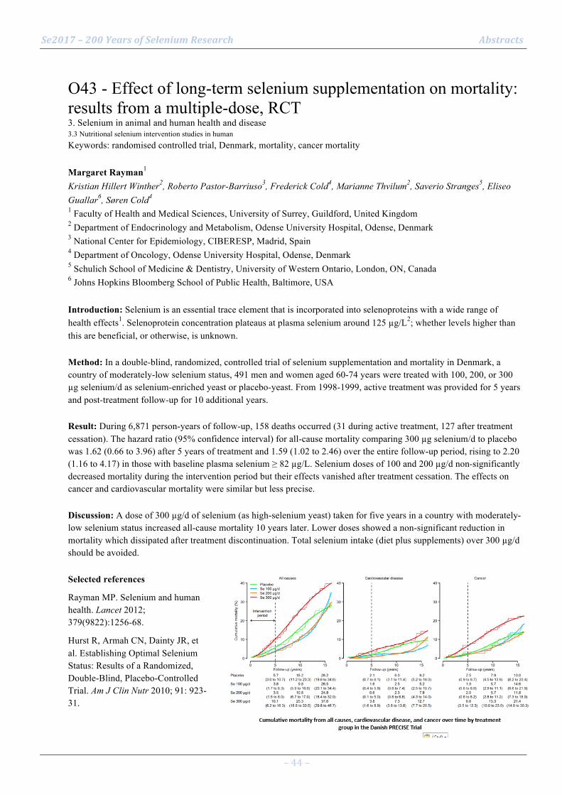
Se2017 – 200 Years of Selenium Research Abstracts
– 44 –
O43 - Effect of long-term selenium supplementation on mortality: results from a multiple-dose, RCT 3. Selenium in animal and human health and disease 3.3 Nutritional selenium intervention studies in human Keywords: randomised controlled trial, Denmark, mortality, cancer mortality Margaret Rayman1 Kristian Hillert Winther2, Roberto Pastor-Barriuso3, Frederick Cold4, Marianne Thvilum2, Saverio Stranges5, Eliseo Guallar6, Søren Cold4 1 Faculty of Health and Medical Sciences, University of Surrey, Guildford, United Kingdom 2 Department of Endocrinology and Metabolism, Odense University Hospital, Odense, Denmark 3 National Center for Epidemiology, CIBERESP, Madrid, Spain 4 Department of Oncology, Odense University Hospital, Odense, Denmark 5 Schulich School of Medicine & Dentistry, University of Western Ontario, London, ON, Canada 6 Johns Hopkins Bloomberg School of Public Health, Baltimore, USA Introduction: Selenium is an essential trace element that is incorporated into selenoproteins with a wide range of health effects1. Selenoprotein concentration plateaus at plasma selenium around 125 µg/L2; whether levels higher than this are beneficial, or otherwise, is unknown. Method: In a double-blind, randomized, controlled trial of selenium supplementation and mortality in Denmark, a country of moderately-low selenium status, 491 men and women aged 60-74 years were treated with 100, 200, or 300 µg selenium/d as selenium-enriched yeast or placebo-yeast. From 1998-1999, active treatment was provided for 5 years and post-treatment follow-up for 10 additional years. Result: During 6,871 person-years of follow-up, 158 deaths occurred (31 during active treatment, 127 after treatment cessation). The hazard ratio (95% confidence interval) for all-cause mortality comparing 300 µg selenium/d to placebo was 1.62 (0.66 to 3.96) after 5 years of treatment and 1.59 (1.02 to 2.46) over the entire follow-up period, rising to 2.20 (1.16 to 4.17) in those with baseline plasma selenium ≥ 82 µg/L. Selenium doses of 100 and 200 µg/d non-significantly decreased mortality during the intervention period but their effects vanished after treatment cessation. The effects on cancer and cardiovascular mortality were similar but less precise. Discussion: A dose of 300 µg/d of selenium (as high-selenium yeast) taken for five years in a country with moderately-low selenium status increased all-cause mortality 10 years later. Lower doses showed a non-significant reduction in mortality which dissipated after treatment discontinuation. Total selenium intake (diet plus supplements) over 300 µg/d should be avoided. Selected references
Rayman MP. Selenium and human health. Lancet 2012; 379(9822):1256-68.
Hurst R, Armah CN, Dainty JR, et al. Establishing Optimal Selenium Status: Results of a Randomized, Double-Blind, Placebo-Controlled Trial. Am J Clin Nutr 2010; 91: 923-31.
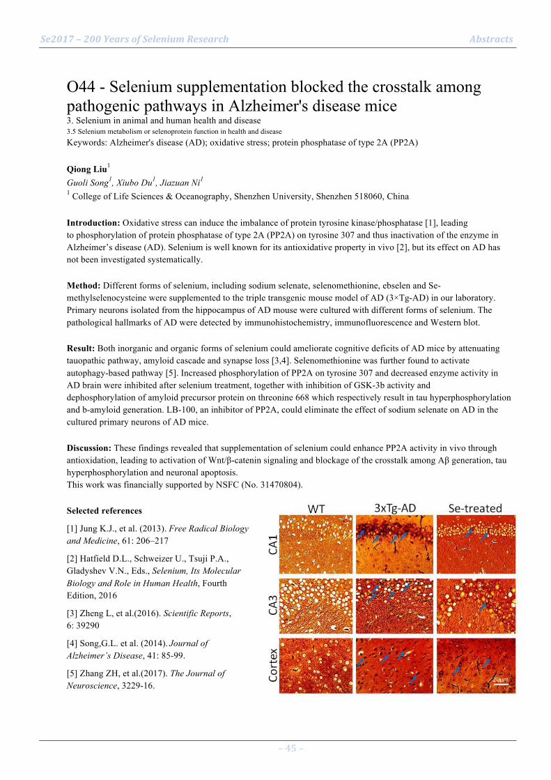
Se2017 – 200 Years of Selenium Research Abstracts
– 45 –
O44 - Selenium supplementation blocked the crosstalk among pathogenic pathways in Alzheimer's disease mice 3. Selenium in animal and human health and disease 3.5 Selenium metabolism or selenoprotein function in health and disease Keywords: Alzheimer's disease (AD); oxidative stress; protein phosphatase of type 2A (PP2A) Qiong Liu1 Guoli Song1, Xiubo Du1, Jiazuan Ni1 1 College of Life Sciences & Oceanography, Shenzhen University, Shenzhen 518060, China Introduction: Oxidative stress can induce the imbalance of protein tyrosine kinase/phosphatase [1], leading to phosphorylation of protein phosphatase of type 2A (PP2A) on tyrosine 307 and thus inactivation of the enzyme in Alzheimer’s disease (AD). Selenium is well known for its antioxidative property in vivo [2], but its effect on AD has not been investigated systematically. Method: Different forms of selenium, including sodium selenate, selenomethionine, ebselen and Se-methylselenocysteine were supplemented to the triple transgenic mouse model of AD (3×Tg-AD) in our laboratory. Primary neurons isolated from the hippocampus of AD mouse were cultured with different forms of selenium. The pathological hallmarks of AD were detected by immunohistochemistry, immunofluorescence and Western blot. Result: Both inorganic and organic forms of selenium could ameliorate cognitive deficits of AD mice by attenuating tauopathic pathway, amyloid cascade and synapse loss [3,4]. Selenomethionine was further found to activate autophagy-based pathway [5]. Increased phosphorylation of PP2A on tyrosine 307 and decreased enzyme activity in AD brain were inhibited after selenium treatment, together with inhibition of GSK-3b activity and dephosphorylation of amyloid precursor protein on threonine 668 which respectively result in tau hyperphosphorylation and b-amyloid generation. LB-100, an inhibitor of PP2A, could eliminate the effect of sodium selenate on AD in the cultured primary neurons of AD mice. Discussion: These findings revealed that supplementation of selenium could enhance PP2A activity in vivo through antioxidation, leading to activation of Wnt/β-catenin signaling and blockage of the crosstalk among Aβ generation, tau hyperphosphorylation and neuronal apoptosis. This work was financially supported by NSFC (No. 31470804). Selected references
[1] Jung K.J., et al. (2013). Free Radical Biology and Medicine, 61: 206–217
[2] Hatfield D.L., Schweizer U., Tsuji P.A., Gladyshev V.N., Eds., Selenium, Its Molecular Biology and Role in Human Health, Fourth Edition, 2016
[3] Zheng L, et al.(2016). Scientific Reports, 6: 39290
[4] Song,G.L. et al. (2014). Journal of Alzheimer’s Disease, 41: 85-99.
[5] Zhang ZH, et al.(2017). The Journal of Neuroscience, 3229-16.

Se2017 – 200 Years of Selenium Research Abstracts
– 46 –
O45 - Selenium, coenzyme Q10 and cardiovascular health – results from a 4-year-intervention in elderly 3. Selenium in animal and human health and disease 3.3 Nutritional selenium intervention studies in human Keywords: Coenzyme Q10, deficiency, supplementation, health effects Urban Alehagen1 Jan Aaseth2, Jan Alexander3, Peter Johansson4 1 Division of Cardiovascular Medicine, Linköping University, Linköping, Sweden 2 Research Department, Innlandet Hospital, and Hedmark University College, Elverum, Norway 3 Norwegian Institute of Public Health, Oslo, and Norwegian University of Life Sciences, Ås, Norway 4 4Department of Social and Welfare studies, Linköping University, Linköping, Sweden Introduction: Selenium is essential for the cell. However, in Europe selenium soil content is low. Coenzyme Q10 is an important anti-oxidant, and the two are interrelated to get optimal cellular function. Method: 443 healthy elderly participants were randomized to recieve supplementation with selenium and coenzyme Q10 or placebo during 4 years. Blood samples, echocardiography, and quality-of-life registrations were obtained. Underlying mechanisms were traced by use of laboratory biomarkers. Result: A follow-up period of 5.2 years showed reduced cardiovascular mortality, better cardiac function 1 and improved physical and emotional quality-of-life. Specific biomarkers (sP-selectin, hs-CRP) showed less inflammatory activity 2, less oxidative stress (copeptin, MR-proADM) 3, increased levels of IGF-1, and less fibrosis. MicroRNA analysis demonstrated significant difference in expression, presumed to constitute an underlying mechanism for other observations. Discussion: We propose a model illustrating relations between observed laboratory effects and clinical endpoints (Figure). It is seen that a suboptimal selenium status increases cardiovascular risk 4, which was attenuated by the supplementation. The intervention influenced the expression of microRNA, the level of inflammatory activity, oxidative stress, IGF-1 levels and fibrosis activity. Apparently, the intervention improved cardiomyocyte function reflected by better cardiac function and less NT-proBNP biomarker concentration. We also found a reduced cardiovascular mortality after 10 years although the intervention was limited to 4 years 5. The proposed model illustrates the complex relationship between some of the effects resulting in the positive clinical endpoints. Selected references 1. Alehagen U, Johansson P, Bjornstedt M, Rosen A and Dahlstrom U. Cardiovascular mortality and N-terminal-proBNP reduced after combined selenium and coenzyme Q10 supplementation: a 5-year prospective randomized double-blind placebo-controlled trial among elderly Swedish citizens. Int J Cardiol. 2013;167:1860-6. 2. Alehagen U, Lindahl TL, Aaseth J, Svensson E and Johansson P. Levels of sP-selectin and hs-CRP Decrease with Dietary Intervention with Selenium and Coenzyme Q10 Combined: A Secondary Analysis of a Randomized Clinical Trial. PLoS One. 2015;10:e0137680. 3. Alehagen U, Aaseth J and Johansson P. Less increase of copeptin and MR-proADM due to intervention with selenium and coenzyme Q10 combined: Results from a 4-year prospective randomized double-blind placebo-controlled trial among elderly Swedish citizens. Biofactors. 2015;41:443-52. 4. Alehagen U, Johansson P, Bjornstedt M, Rosen A, Post C and Aaseth J. Relatively high mortality risk in elderly Swedish subjects with low selenium status. Eur J Clin Nutr. 2016;70:91-6. 5. Alehagen U, Aaseth J and Johansson P. Reduced Cardiovascular Mortality 10 Years after Supplementation with Selenium and Coenzyme Q10 for Four Years: Follow-Up Results of a Prospective Randomized Double-Blind Placebo-Controlled Trial in Elderly Citizens. PLoS One. 2015;10:e0141641.

Se2017 – 200 Years of Selenium Research Abstracts
– 47 –
O46 - Antibacterial redox selenium coatings, covalent small molecules and antibody drug conjugates (ADCs) 3. Selenium in animal and human health and disease 3.4 Selenium based medical therapeutics Keywords: Selenium Toxicity, Selenium Coatings, Selenium Antibodies Julian Spallholz1 1 United States Introduction: Selenium (Se) has a 100+ year history as an anti-cancer element both before and after its nutritional requirement was discovered by Schwarz (1957). In the 1960’s debate ensued over the experimental reports of selenium’s anti-cancer attributes in animals and humans. The famous 1996 Clark study of human cancer with Se-yeast and failure of the SELECT Trial (Lu, 2016) have seemingly dashed hope of dietary selenium supplementation ever having an anti-cancer future. With understanding selenium redox chemistry, selenides (Spallholz, 2015) and isoselenocyanates (Crampsie, 2012) can be covalently “targeted” and delivered to cells offering a renewed assessment of selenium’s role as a therapeutic treatment, especially for cancer. Method: Selenium toxicity research really begins in the 1930’s among horses and sheep on the Great Plains of the USA by Moxon (1937). The first chemical equation of WHY selenium is toxic, superoxide generation, was by Seko (1989) for selenite (Figure1). Understanding this chemistry by organic selenium compounds permits them to be incorporated into polymers, covalently attached to “targeting” small molecules, and recently clinical antibodies, Herceptin® and Avastin®. Result: Attachment of redox selenium to proteins is accomplished with a Se-modified Bolton-Hunter reagent, as developed in the 1970’s for 125I-radioimmunoassays (Lane, 2011). This chemistry permits antibodies to deliver redox selenium, generate superoxide inducing oxidative stress and cell death. Using redox probes, Se-antibody treated cancer cells are observed to generate intracellular superoxide and H2O2. Discussion: Redox selenium and its applied “targeted” therapeutic applications require a renewed assessment of its anti-cancer and medical applications against viruses and antibiotic-resistant bacterial infections. Selected references Clark, LC., Combs, GF Jr., Turnbull, BW. , Slate, EH ., Chalker, DK., Chow, J., Davis, LS., Glover, RA., Graham, GF;, Gross, EG;, Krongrad, A., Lesher, JL Jr., Park, HK., Sanders, BB Jr., Smith, CL., and Taylor JR. (1996) Effects of selenium supplementation for cancer prevention in patients with carcinoma of the skin. A randomized controlled trial. Nutritional Prevention of Cancer Study Group. JAMA; 276:1957-1963.
Crampsie, MA., Pandey MK., Desai D., Spallholz, JE., Amin, S., Sharma AK. (2012) Phenylalkyl isoselenocyanates vs phenylalkyl isothiocyanates: Thiol reactivity and its implications. Chemico-Biol. Interact.; 200:28–37.
Lane, DJR., and Richardson, DR. (2011) William Hunter and radio iodination: revolutions in the labelling of proteins with radionuclides of iodine Biochem. J.; 4:1-4.
Lu,J., Zhang, J., Jiang, C., Deng, Y., and Ozten, N. (2016) Cancer chemoprevention research with selenium in the post-SELECT era: Promises and challenges Nutr Cancer; 68:1-17
Moxon, A L. (1937) Alkali Disease or Selenium Poisoning. Agricultural Experiment Station Bulletins. Paper 311. South Dakota State University.
Schwarz, K., and Foltz, CM. (1957) Selenium as an integral part of factor 3 against dietary necrotic liver degeneration. J. Am. Chem. Soc; 79:3292–3293
Seko, Y., Saito, Y., Kitahara, J., and Imura N. (1989) Active oxygen generation by the reaction of selenite with reduced glutathione in vitro. In: Wendel A., Editor. Selenium in Biology and Medicine. Springer Berlin Heidelberg; Tübingen, Germany; pp. 70–73.
Spallholz, JE. (2015) Redox Cycling and Toxicity of Selenium Compounds, A History; CRC Press in “Diversity of Selenium Functions in Health and Disease” R. Brigelius-Flohe and H. Sies, Eds. , Taylor & Francis, Publisher; pp. 83-108.
Se2017 – 200 Years of Selenium Research Abstracts
– 48 –
O47 - Selenium in the treatment of cancer 3. Selenium in animal and human health and disease 3.4 Selenium based medical therapeutics Keywords: Redox-active selenium compounds, ex vivo, Pharmacokinetics, Chemotherapy, Clinical trials Mikael Björnstedt1 Sougat Misra1, Ola Brodin2, Per Stål3, Clara Lenneby-Helleday2, Antje Zickler1, Rim Jawad1, Arun Kumar Selvam1, Olof Breuer4, Lutz Schomburg5, Bente Gammelgaard6 1 Department of Laboratory Medicine, Division of Pathology, Karolinska Institutet, Stockholm, Sweden 2 Department of Oncology-Pathology, Karolinska Institutet, Stockholm, Sweden 3 Department of Medicine, Karolinska Institutet, Stockholm, Sweden 4 Department of Laboratory Medicine, Karolinska Institutet, Stockholm, Sweden 5 Institute for Experimental Endocrinology, Charité-Universitätsmedizin Berlin, Berlin, Germany 6 Department of Pharmacy, University of Copenhagen, Copenhagen, Denmark Introduction: The prognosis for visceral malignancies is poor, especially for carcinoma of the lung, pancreas and liver due to resistance to chemotherapy. Over several decades, a plethora of preclinical investigations have demonstrated high degree of tumor-specific cytotoxic effects of selenium compounds, especially to therapy-resistant tumor cells [1]. Herein, we have undertaken translational approaches to establish the efficacy of selected selenium-based candidate chemotherapeutics into clinical practice. Method: The efficiency of the potent candidate drugs are evaluated both in vitro and in vivo. In vitro studies span the spectrum of cell lines, isolated primary tumor cells and ex vivo tumor tissues in cultures. In parallel, we perform clinical trials with selected selenium compounds on cancer patients. Result: Serum samples from the SECAR study on cancer patients [2] have been extensively analysed for selenium compounds. Selenite was detected in serum and selenosugar was the major organic metabolite. Pharmacokinetic analyses indicated that plasma selenite concentrations increased in a linear dose-dependent manner. An unprecedented increase in plasma SEPP level was recorded. In another study, we documented favourable anti-cancer effects of an organic selenium compound in liver cancer models. A novel target specific drug release principle might serve as a promising therapeutic intervention in liver cancer and a phase I clinical trial is underway. Discussion: Implementation of selenium-based candidate chemotherapeutics in clinical practice underscores the absolute requirement of both safety and efficacy evaluations. To this end, our founding clinical studies can greatly accelerate the transition of selenium compounds from experimental drugs to clinically relevant chemotherapeutics. Selected references
[1] Misra, S., Boylan, M., Selvam, A., Spallholz, J. E., and Björnstedt, M. (2015) Redox-active selenium compounds--from toxicity and cell death to cancer treatment, Nutrients 7, 3536-3556.
[2] Brodin, O., Eksborg, S., Wallenberg, M., Asker-Hagelberg, C., Larsen, E. H., Mohlkert, D., Lenneby-Helleday, C., Jacobsson, H., Linder, S., Misra, S., and Björnstedt, M. (2015) Pharmacokinetics and Toxicity of Sodium Selenite in the Treatment of Patients with Carcinoma in a Phase I Clinical Trial: The SECAR Study, Nutrients 7, 4978-4994.

Se2017 – 200 Years of Selenium Research Abstracts
– 49 –
O48 - Selective targeting of redox dysregulation of cancer cells by redox-active selenium compounds 3. Selenium in animal and human health and disease 3.4 Selenium based medical therapeutics Keywords: Redox balance, redox-active selenium compounds, thiols, animal model, leukemia Sougat Misra1 Arun Kumar Selvam1, Mikael Björnstedt1 1 Department of Laboratory Medicine, Division of Pathology, Karolinska Institutet, Stockholm, Sweden Introduction: Altered redox homeostasis is an acquired physiological adaptation of proliferating malignant cells. An increased cellular proliferation necessitates high energy supplies which culminate into elevated reactive oxygen species (ROS) production. Malignant cells counterbalance the cytotoxic effects of elevated ROS levels by augmenting activities of ROS-detoxifying pathways. However, such adaptive mechanisms compromise the cellular tolerance to further increase in intracellular ROS levels. Herein, we investigated whether further manipulations of cellular redox balance could inflict selective cytotoxic effects on malignant cells by two redox-active selenium compounds implicated in ROS generation. Method: Sixteen different cancer cell lines and isolated primary normal cells were used for screening purposes. We employed cell viability assays, gene and protein expression analyses, knock-down and overexpression of target genes to investigate the underlying mechanisms of cytotoxicity. Result: The selected selenium compounds were cytotoxic to all the tested cancer cell lines, however, with varying efficacy. Small molecules-induced specific modulations of cellular redox homeostasis resulted in dramatic potentiation (up to 70-fold) of the cytotoxic effects of these selenium compounds, independent of the origins or oncogene status of these cells. No such potentiation of cytotoxic effects was observed in normal cells under identical test conditions. Gene and protein expression analyses indicated involvement of key redox-regulatory enzymes in the observed cytotoxic effects on cancer cells. Animal studies are underway to investigate the in vivo efficacy of these compounds in experimental cancer models. Discussion: Specific modulations of key redox-regulatory pathways of cancer cells can greatly potentiate the cytotoxicity of redox-active selenium compounds with promising cancer chemotherapeutics applications.

Se2017 – 200 Years of Selenium Research Abstracts
– 50 –
O49 - Selenium: A potentially powerful tool to design potent anticancer molecules-Discovery of Se-Aspirins 3. Selenium in animal and human health and disease 3.4 Selenium based medical therapeutics Keywords: Se-Aspirin, cancer, apoptosis, NF-κB, ROS Arun Sharma1 Deepkamal Karelia1, Daniel Plano2, Julian Spallholz3, Carmen Sanmartín2, Junxuan Lu1, Shantu Amin1 1 Department of Pharmacology, Penn State College of Medicine, Hershey, PA 17033, USA 2 University of Navarra, Department of Organic and Pharmaceutical Chemistry, 31008 Pamplona, Spain 3 Department of Nutrition, Texas Tech University, Lubbock, Texas 79430, USA Introduction: We have shown that rational incorporation of Se into small molecules can substantially enhance their anti-cancer efficacy1-5. Recently, through extensive SAR studies based on Se incorporation into NSAIDs, we identified a Se-aspirin hybrid molecule, which was >300 times more potent than aspirin in inhibiting viability of various cancer cells5. Method: Further SAR studies and investigations directed towards efficacy, toxicity, and drug-likeness has now led to the identification of a novel Se-Aspirin molecule AS-10 as the most efficacious compound. Result: AS-10 (47 mg/kg, i.p) inhibited subcutaneous colon tumor growth by ~70% and was lethal to a variety of cancer cells including pancreatic cancer (PC) for which no effective therapy currently exists. AS-10 inhibited PC cell growth (IC50 2.5-5 µM), induced G1/G2 cell cycle arrest which was associated with increase of cell cycle inhibitory proteins p21 and p27, and induced apoptosis as evidenced by caspase 3/7 activity, PARP cleavage and Annexin V staining. AS-10 inhibited NF-κB DNA binding activity as well as NF-κB translocation to the nuclei upon stimulation by TNFα. Notably, AS-10 potentiated cytotoxic activity of gemcitabine in PC cells. Furthermore, in LNCaP prostate cancer cells, AS-10 decreased protein level of AR and its best known target PSA, and led to increased caspase-mediated apoptosis and expression of p53-DNA damage response proteins such as p21 and p-H2A.X. AS-10 induced ROS in cancer cells as likely primary biochemical event. Discussion: AS-10 may represent a promising intervention and therapeutic agent for pancreatic, prostate, and colon cancers and other cancer types through multiple targets. Selected references
Sharma AK, Sharma A, Desai D, Madhunapantula SV, Huh SJ, Robertson GP, Amin S. J Med Chem. 2008, 51:7820.
Sharma A, Sharma AK, Madhunapantula SV, Desai D, Huh SJ, Mosca P, Amin S, Robertson GP. Clin Cancer Res. 2009,15:1674.
Sharma AK, Kline CL, Berg A, Amin S, Irby RB. Clin Cancer Res. 2011,17: 4474.
Allen JE, Gallant JN, Dicker DT, Amin S, Irby RB, Sharma AK, El-Deiry WS. PLoS One. 2013;8(3): e59380.
Plano D, Karelia DN, Pandey MK, Spallholz JE, Amin S, Sharma AK. J Med Chem. 2016, 59:1946.

Se2017 – 200 Years of Selenium Research Abstracts
– 51 –
O50 - Selenium Distribution and its Correlation to Geochemical Factors in East China Intertidal Zone 1. Selenium chemistry and geochemistry 1.4 Relationships of selenium between soils, water, and vegetation Keywords: Selenium distribution, Intertidal zone, East China Zhengyu BAO1 Ming Zhang2, Guoguang Chen2 1 Zhejiang Research Institute of China University of Geosciences, Hangzhou, China 2 Nanjing Center of China Geological Survey, Nanjing, China Introduction: The east China intertidal zone (ITZ) has a total area of 11056.48 km2
,about half of the ITZ in china. In this paper, we are focused on the distribution of selenium (Se) in the east China ITZ and its correlation with other geochemical factors. Method: 1906 soil/mud samples had been taken by China Geological Survey from the east China ITZ, which includes, from north to south, 1199 samples from Jiangsu Province, 102 from Shanghai municipal city, 252 from Zhejiang Province and 353 from Fujian Province, and 54 geochemical indexes had been measured. Result: 1. Se concentration in the east China ITZ was quite low, with a range of 0.01-0.50 mg/kg and a mean value of 0.1-0.16 mg/kg, Jiangsu Province being the lowest, with a range of 0.01-0.20 mg/kg and a mean value of 0.06 mg/kg, reflecting that East China terrestrial landmass is severely selenium deficient. 2. Se concentrations in ITZ soils are found to be positively correlated to the content of organic carbon (OrgC), aluminum and iron, but negatively correlated to pH values (Fig. 1). Discussion: Absorption by organic materials, clay minerals and iron oxides seems to be the mechanism of Se accumulation in the ITZ soils and the low contents of OrgC, aluminum and iron and high pH values in East China ITZ soils may another reason to explain its low concentration of Se.
Se2017 – 200 Years of Selenium Research Abstracts
– 52 –
O51 - Distribution and translocation of selenium from soil to highland barley in Kashin-Beck disease area 1. Selenium chemistry and geochemistry 1.4 Relationships of selenium between soils, water, and vegetation Keywords: Highland barley; Translocation; Kashin-Beck disease Jing Wang1 Hairong Li2, Linsheng Yang2 1 University of Chinese Academy of Sciences; University of Nottingham 2 University of Chinese Academy of Sciences; Institute of Geographical Sciences and Natural Resources Research, Chinese Academy of Sciences Introduction: Kashin-Beck disease (KBD), which is still active in the Tibetan Plateau, is considered to be relevant to insufficient selenium intake. Highland barley as the most popular staple food in Tibetan Plateau KBD areas is one of the dominant selenium sources for local people. Method: In this study, samples of intact highland barley plant and corresponding topsoil were collected from both non-KBD and KBD endemic areas of Songpan County. Total selenium content in plant and soil samples and fractions of soil selenium were determined and analysed. Result: The results showed that selenium levels in different fractions of highland barley were lower in KBD areas than non-KBD areas (grain P = 0.090; straw P = 0.131; root P = 0.001), while no significant difference was observed in their corresponding soil (P = 0.993). The comparison of selenium transfer factors indicated that the restricting step for selenium translocation was from soil to root. Water-soluble, exchangeable and fulvic acid-bound selenium fractions in soil were the key fractions dominating in this transfer process. Stronger positive correlation was observed between tetravalent selenium speciation and plant selenium, suggesting the closer affinity to selenite for highland barley. Additionally, selenium transfer from soil to root significantly increased as the soil pH increased (P = 0.007) and the soil organic matter content decreased (P = 0.019). Discussion: The information obtained may have considerable significance for understanding the patterns of selenium transfer from soil to grain in low-selenium areas, providing a theoretical and experimental basis for proposing effective agricultural measures to increase crops selenium in KBD endemic areas. Selected references Li HF, McGrath SP, Zhao FJ. Selenium uptake, translocation and speciation in wheat supplied with selenate or selenite. New Phytol, 2008, 178, 92-102. Stroud JL, Broadley MR, Foot I, et al. Soil factors affecting selenium concentration in wheat grain and the fate and speciation of Se fertilizers. Plant Soil, 2010, 332, 19-30. Sun GX, Liu X, Williams PN, et al. Distribution and translocation of selenium from soil to grain and its speciation in paddy rice (Oryza sativa L.). Environ Sci Technol, 2010, 44, 6706-6711. Sun LY, Li Q, Meng FG, et al. T-2 toxin contamination in grains and selenium concentration in drinking water and grains in Kaschin-Beck disease endemic areas of Qinghai Province. Biol Trace Elem Res, 2012, 150(1-3): 371-375. Tan JA, Zhu WY, Wang WY, et al. Selenium in soil and endemic diseases in China. Sci Total Environ, 2002, 284, 227-235. Temmerman LD, Waegeneers N, Thiry C, et al. Selenium content of Belgian cultivated soils and its uptake by field crops and vegetables. Sci Total Environ, 2014, 468-469, 77-82. Wang J, Li HR, Li YH, et al. Speciation, Distribution and Bioavailability of Soil Selenium in the Tibetan Plateau Kashin-Beck Disease Area — a Case Study in Songpan County, Sichuan Province, China. Biol Trace Elem Res, 2013, 156, 367-375. Zhang BJ, Yang LS, Wang WY, et al. Environmental Selenium in the Kaschin-Beck disease area, Tibetan Plateau, China. Environ Geochem Health, 2011, 33, 95-501. Zhao CY, Ren JH, Xue CZ, et al. Study on the relationship between soil selenium and plant selenium uptake. Plant and Soil, 2005, 227, 197-206.
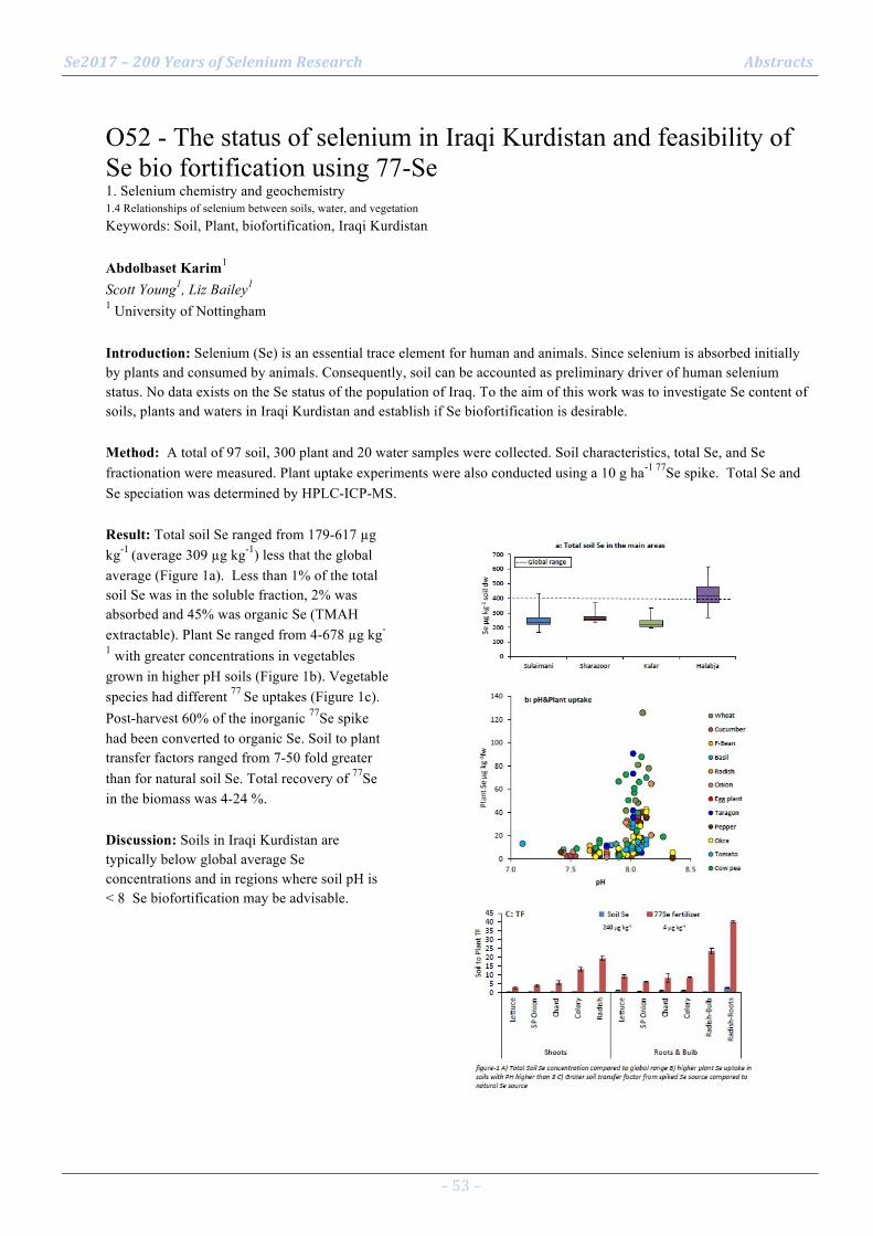
Se2017 – 200 Years of Selenium Research Abstracts
– 53 –
O52 - The status of selenium in Iraqi Kurdistan and feasibility of Se bio fortification using 77-Se 1. Selenium chemistry and geochemistry 1.4 Relationships of selenium between soils, water, and vegetation Keywords: Soil, Plant, biofortification, Iraqi Kurdistan Abdolbaset Karim1 Scott Young1, Liz Bailey1 1 University of Nottingham Introduction: Selenium (Se) is an essential trace element for human and animals. Since selenium is absorbed initially by plants and consumed by animals. Consequently, soil can be accounted as preliminary driver of human selenium status. No data exists on the Se status of the population of Iraq. To the aim of this work was to investigate Se content of soils, plants and waters in Iraqi Kurdistan and establish if Se biofortification is desirable. Method: A total of 97 soil, 300 plant and 20 water samples were collected. Soil characteristics, total Se, and Se fractionation were measured. Plant uptake experiments were also conducted using a 10 g ha-1 77Se spike. Total Se and Se speciation was determined by HPLC-ICP-MS. Result: Total soil Se ranged from 179-617 µg kg-1 (average 309 µg kg-1) less that the global average (Figure 1a). Less than 1% of the total soil Se was in the soluble fraction, 2% was absorbed and 45% was organic Se (TMAH extractable). Plant Se ranged from 4-678 µg kg-
1 with greater concentrations in vegetables grown in higher pH soils (Figure 1b). Vegetable species had different 77 Se uptakes (Figure 1c). Post-harvest 60% of the inorganic 77Se spike had been converted to organic Se. Soil to plant transfer factors ranged from 7-50 fold greater than for natural soil Se. Total recovery of 77Se in the biomass was 4-24 %. Discussion: Soils in Iraqi Kurdistan are typically below global average Se concentrations and in regions where soil pH is < 8 Se biofortification may be advisable.

Se2017 – 200 Years of Selenium Research Abstracts
– 54 –
O53 - Geochemistry of selenium in Gilgit-Baltistan (North East Pakistan) 1. Selenium chemistry and geochemistry 1.4 Relationships of selenium between soils, water, and vegetation Keywords: soil, plants Saeed Ahmad1 Elizabeth H Bailey1, Michael J Watts2, Scott D Young3 1 School of Biosciences, University of Nottingham, Nottingham, United Kingdom 2 British Geological Survey, Keyworth, Nottingham, United Kingdom 3 School of Biosciences, University of Nottingham, United Kingdom Introduction: Selenium (Se) is an important micronutrient for human and animal health. It is essential for normal functioning of the thyroid gland and thyroid hormone synthesis. Method: Samples of soil and wheat grain were collected from Gilgit-Baltistan (N. Pakistan). Samples were digested in nitric acid and extracted in 10% TMAH prior to analysis for Se concentrations by ICP-MS. Result: Soils were alkaline, with pH values c. 8.0, and low organic carbon contents (0.12% - 0.59%). Average total soil Se concentration was 290 µg kg-1, 25% less than the global average (400 µg kg-1). Discussion: Only 5% of the soil Se was extractable by TMAH suggesting that the majority of the Se is not associated with soil organic matter, or as surface-adsorbed selenite or selenate, but exists in a poorly available form. No relationship was observed between soil organic carbon and total soil selenium concentrations but soil pH was negatively correlated with TMAH-extractable Se. Selenium concentration in wheat grain was exceptionally low (1 - 95 µg kg-1). Considering that wheat consumption in the region (~350 g) provides 75% of calorie intake, it is likely that the population does not receive sufficient Se from their diet in the absence of supplementation. Further investigation of locally grown foods, including vegetables, fruit, meat and dairy products, is required to fully audit dietary Se sources. We also plan to analyse human biomarkers for Se. From this data we will assess whether sufficient dietary Se is available or whether supplementation through Se biofortification of the staple crop (wheat) is required. Selected references
Köhrle, J., 1999. The trace element selenium and the thyroid gland. Biochimie, 81, 527-533.
Stewart, A.G., 1990. Drifting continents and endemic goitre in northern Pakistan. BMJ, 300, 1507-1512.
Zia, M.H., Watts, M.J., Gardner, A. and Chenery, S.R., 2014. Iodine status of soils, grain crops, and irrigation waters in Pakistan. Environmental Earth Sciences, 73, 7995-8008.
Zimmermann, M.B. and Köhrle, J., 2002. The impact of iron and selenium deficiencies on iodine and thyroid metabolism: biochemistry and relevance to public health. Thyroid, 12, 867-878.

Se2017 – 200 Years of Selenium Research Abstracts
– 55 –
O54 - Selenocysteine Chemistry and Total Chemical Synthesis Applied for Accessing Human Selenoproteins 2. Selenium in the molecular life sciences 2.4 Selenoprotein synthesis pathways Keywords: Chemical protein synthesis, selenocysteine, selenopeptide, selenoprotein, native chemical ligation Norman Metanis1 1 The Hebrew University of Jerusalem, Institute of Chemistry, Jerusalem 91904, Israel Introduction: Once considered a toxic element, selenium is now known as an essential element for life. For example, in humans it is incorporated in proteins known as selenoproteins containing the 21st encoded amino acid selenocysteine (Sec). The differences between selenium and sulfur in their redox potentials, pKas, and nucleophilicities and electrophilicities give selenium interesting chemistry. Method: We use chemical protein synthesis as our main technique to access selenoproteins. This technology is based on two main methods, solid-phase peptide synthesis (SPPS) and chemical ligation reactions; most famous is native chemical ligation (NCL). Result: Sec can be used as a tool for chemical protein synthesis, and allowing for site selective modifications. It can be also incorporated into protein sequences to enhance oxidative protein folding. Discussion: This lecture will discuss our recent studies on chemical protein synthesis using Sec and selective deselenization reactions, which convert Sec into Ala or Ser. These advances in chemical protein synthesis bring us closer to accessing naturally occurring selenoproteins, especially human selenoproteins that still await functional characterization. Selected references
Dery, L.; Reddy, P.; Dery, S.; Mousa, R.; Ktorza, O.; Talhami, A. and Metanis, N. Chem. Sci., DOI: 10.1039/c6sc04123j (2017).
Reddy, P.; Dery, S. and Metanis, N. Angew. Chem. Int. Ed., 55, 992–995 (2016).
Dery, S.; Reddy, P.; Dery, L.; Mousa, R.; Dardashti Notis, R. and Metanis, N. Chem. Sci., 6, 6207-6212 (2015).
Metanis, N. and Hilvert D. Sci., 6, 322-325 (2015).
Metanis, N. and Hilvert D. Open. Chem. Biol., 22, 27-34 (2014).
Metanis N. and Hilvert D. Chem. Int. Ed., 51, 5585-5588 (2012)

Se2017 – 200 Years of Selenium Research Abstracts
– 56 –
O55 - Redefining UAG for selenocysteine widens the scope of recombinant selenoprotein production in E.coli 2. Selenium in the molecular life sciences 2.4 Selenoprotein synthesis pathways Keywords: Recombinant selenoprotein, E coli, Genetic code, Thioredoxin reductase, Glutathione peroxidase Qing Cheng1 Elias Arnér1 1 Biochemistry Division, MBB, Karolinska Institutet. Stockholm, Sweden Introduction: Selenoproteins, containing one or several Sec residues, are found in all three domains of life (1). It is important to characterize selenoprotein functions in order to understand their roles in life, but scarce availability of selenoproteins has remained a major bottleneck for such research. Here we illustrate a surprising flexibility of selenoprotein synthesis, which opens up a powerful method for selenoprotein research using recombinant selenoproteins (2). Method: In nature, Sec is co-translationally inserted at predefined UGA opal codons by specific translation machineries (3). Here we found that Sec can be equally efficiently incorporated at predefined UAG amber codons, thereby competing with RF1 rather than RF2. By utilizing this system within a RF1-depleted E. coli strain C321.∆A (4) we could produce a number of selenoproteins with unsurpassed purity and yield. Result: We have here produced several form of mammalian TrxR isoenzymes with much higher specific activity compared to previous methods (5). We believe this new routine could be widely employed for production of a wide variety of selenoproteins carrying penultimate or ultimate Sec residues, as in TrxRs. Interestingly, we also found that a SECIS element was no longer absolutely required in our system. This also allowed production of selenoproteins with internal Sec, such as human GPx1, and we could confirm a previously proposed catalytic tetrad in the enzyme using directed mutagenesis (6). Discussion: We conclude that our new methodology (2) should constitute a powerful resource enabling many forthcoming studies of recombinant selenoproteins, considering its versatility and comparable ease of use. Selected references
1. G. V. Kryukov et al. Science 300, 1439-1443 (2003).
2. Q. Cheng, E. S. J. Arnér. J Biol Chem, [Epub ahead of print] (2017).
3. M. Thanbichler, A. Böck. EMBO J 21, 6925-6934 (2002).
4. M. J. Lajoie et al. Science 342, 357-360 (2013).
5. E. S. J. Arnér et al. J. Mol. Biol. 292, 1003-1016 (1999).
6. S. C. Tosatto et al. Antioxidants and Redox Signalling 10, 1515-1526 (2008).
Se2017 – 200 Years of Selenium Research Abstracts
– 57 –
O56 - Delivery of selenide to selenophosphate synthetase for selenoprotein biosynthesis in bacteria 2. Selenium in the molecular life sciences 2.4 Selenoprotein synthesis pathways Keywords: selenophosphate, thioredoxin, bacteria, selenoprotein biosynthesis, selenide Ryuta Tobe1 Atsuki Shimizu1, Satoru Hagita1, Takashi Tamura2, Takukya Ogawa3, Kaito Kiriyama1, Tatsuo Kurihara3, Tejo N. Prakash4, Hisaaki Mihara1 1 College of Life Sciences, Ritsumeikan University, Kusatsu, Shiga 525-8577, Japan 2 Graduate School of Life and Environmental Science, Okayama University, Okayama 700-8530, Japan 3 Institute for Chemical Research, Kyoto University, Uji, Kyoto 611-0011, Japan 4 School of Energy and Environment, Thapar University, Patiala 147004, India Introduction: Selenophosphate synthesized by selenophosphate synthetase (SelD) from selenide (HSe-) and ATP is essential for selenoprotein biosynthesis. In mammals, glutathione (GSH) and thioredoxin reductase (TrxR) have been proposed to serve as a HSe- donor for selenophosphate synthesis. However, little is known about the HSe- donor to SelD in bacteria. Here, we investigated the mechanism of HSe- generation and HSe- supply to SelD in the selenoprotein biosynthesis in bacteria. Method: To identify the genes/proteins that affect selenoprotein biosynthesis in E. coli, 57 candidate genes potentially participating in selenite reduction, such as those encoding nitrate reductase, glutaredoxin, and GSH-related enzymes, were selected. The mutant strains defective in each of the 57 candidate genes were tested for the ability to synthesize selenoprotein by measuring formate dehydrogenase (FDH) activity. Result: A significant decrease of FDH activity was seen in a thioredoxin (TrxA)-deficient mutant, whereas the deficiency of genes related to GSH and other reductases did not affect the FDH activity. To clarify the function of TrxA in selenite reduction, the purified recombinant TrxA of Pseudomonas sp. F2a was analyzed. We found that TrxA exhibited selenite-reducing activity coupled with a TrxR reaction and that HSe- generated by TrxA was efficiently supplied to SelD for selenophosphate synthesis. Discussion: Our data provide the first evidence for a pivotal role of TrxA in selenoprotein biosynthesis in bacteria.
Se2017 – 200 Years of Selenium Research Abstracts
– 58 –
O57 - Reconstitution of processive selenoprotein P synthesis in the wheat germ lysate system 2. Selenium in the molecular life sciences 2.4 Selenoprotein synthesis pathways Keywords: Selenoprotein P, SECIS Binding Protein 2, wheat germ lysate, in vitro, translation Mark Pinkerton1 Sumangala Shetty1, Paul Copeland1 1 Rutgers University, Graduate School of Biomedical Sciences, Rutgers Piscataway, NJ Introduction: A UGA stop codon is recoded to accommodate the incorporation of the 21st amino acid selenocysteine (Sec). For UGA to be recoded a specialized set of cis and trans factors are required and consist of: an mRNA with an in frame UGA codon, a selenocysteine insertion sequence (SECIS) in the 3’ untranslated region, a SECIS binding protein 2 (SBP2), a specific translation elongation factor (eEFSec), and a selenocysteine tRNA. The N-terminus of SBP2 is believed to have no direct role in Sec incorporation because the C-terminus of SBP2 is sufficient for the incorporation of Sec into selenoproteins that have one Sec codon. Having recently developed an in vitro translation system in wheat germ requiring the addition of all Sec-incorporation factors, we found that translation of SELENOP, which contains 10 Sec codons, is defective in that only early termination products are made. This contrasts with mammalian systems where full length protein is predominantly found. This observation, combined with the fact that neither SELENOP nor the N-terminus of SBP2 are found in invertebrates, points to a specialized function for the N-terminus of SBP2 related to SELENOP synthesis. Method: To validate the relationship between the N-terminus of SBP2 and SELENOP, C-terminal and full length SBP2 were compared for in vitro translation of SELENOP. Result: We found that the production of full length SELENOP is dependent on the presence of the SBP2 N-terminus. Discussion: Our results suggest that the N-terminus of SBP2 was appended during evolution to allow for processive incorporation of multiple Sec residues into SELENOP. Selected references
Gupta N., DeMong L. W., Banda S., Copeland P. R. Reconstitution of selenocysteine incorporation reveals intrinsic regulation by SECIS elements. The Journal of molecular biology. 2013;425(14):2415-2422.
Shetty S.P, Shah R., Copeland P.R. Regulation of Selenocysteine Incorporation into the Selenium Transport Protein, Selenoprotein P. The Journal of Biological Chemistry. 2014;289(36):25317-25326.

Se2017 – 200 Years of Selenium Research Abstracts
– 59 –
O58 - Selenium in Radiation Oncology – 10 years of Experiences in Germany 3. Selenium in animal and human health and disease 3.4 Selenium based medical therapeutics Keywords: Selenium Supplementation, Radiation Oncology, Tumor Patients, Lymphedema, Radioprotection Ralph Muecke1 Oliver Micke2, Lutz Schomburg3, Jens Buentzel4, Klaus Kisters5, Irenaeus A. Adamietz6 1 Radiotherapy RheinMainNahe, Bad Kreuznach, Germany 2 Radiotherapy and Radiation Oncology, Franziskus Hospital, Bielefeld, Germany 3 Institute for Experimental Endocrinology, Charité Berlin, Germany 4 Department of Otolaryngology, Südharz Hospital Nordhausen, Germany 5 Department of Internal Medicine, St. Anna Hospital, Herne, Germany 6 Radiotherapy and Radiation Oncology, Marien Hospital Herne, Ruhr University Bochum, Germany Introduction: Se supplementation in radiation oncology is a controversial issue. We summarize the major results obtained by the German Working Group Trace Elements and Electrolytes in Oncology (AKTE). Method: We analyzed blood and tissue Se-levels of tumor patients (n=170) before and after Se-supplementation. Result: The majority of tumor patients (carcinomas of the uterus, head and neck, lung, rectal or prostate cancer) showed a relative Se deficiency in whole blood and serum. In prostate cancer, tissue Se levels were lower in the compartment surrounding the carcinoma in comparison to patients with benign prostatic hyperplasia. Se supplementation successfully corrected Se-deficiency and decreased radiotherapy-induced diarrhea in a randomized study of radiotherapy patients with carcinomas of the uterus. Survival data imply that Se supplementation did not interfere with radiation success. Limited effects of supplemental Se in the prevention of ageusia (loss of taste) and dysphagia due to radiotherapy were noted in a second randomized trial in head and neck cancer. In comparison, positive effects of Se supplementation were detected on radiation-associated secondary lymphedema in patients with limb edemas as well as in the head and neck region, including endolaryngeal edema. No adverse effects of supplemental Se were noted. Discussion: Se supplementation yielded promising results concerning radioprotection and edema prevention in tumor patients and should be considered as a meaningful adjuvant treatment option in subjects with a low Se status. Selected references Muecke R, Schomburg L, Glatzel M, et al. Multicenter, phase III trial comparing selenium supplementation with observation in gynaecologic radiation oncology. Int J Radiat Oncol Biol Phys. 2010; 70: 828-835. Muecke R, Micke O, Schomburg L et al. Multicenter, phase III trial comparing selenium supplementation with observation in gynecologic radiation oncology: follow-up analysis of the survival data 6 years after cessation of randomization. Integr Cancer Ther. 2014; 13: 463-7.

Se2017 – 200 Years of Selenium Research Abstracts
– 60 –
O59 - Selenium in the treatment of radiation-associated secondary lymphedema - An Update 3. Selenium in animal and human health and disease 3.4 Selenium based medical therapeutics Keywords: lymphedema, radiation, oncology Oliver Micke1 Jens Buentzel2, Klaus Kisters3, Lutz Schomburg4, Ralph Mücke5 1 Franziskus Hospital, Department of Radiotherapy and Radiation Oncology, Bielefeld, Germany 2 Südharz Hospital, Department of Otolaryngology, Nordhausen, Germany 3 St. Anna Hospital, Department of Internal Medicine, Herne, Germany 4 Charité Berlin, Institute for Experimental Endocrinology, Berlin Germany 5 Radiotherapy RheinMainNahe, Bad Kreuznach, Germany Introduction: Lymphedema can be observed after surgery and/or radiotherapy or as a result of tumor compression. The antiedematous effect of selenium is well-known, but not very good defined. The aim of this explorative study was to further evaluate the impact of selenium in the treatment of lymphedema after radiotherapy. Method: Between June 1996 and December 2016, 32 patients with edema of the arm and 46 patients with edema of the head-and-neck region were treated with selenium for therapy-related lymphedema. Of these 46 patients, a total of 30 had interstitial endolaryngeal edema. All patients received sodium selenite (500 µg per day) over 4 to 6 weeks. Result: Self-assessment using a visual analog scale showed a reduction of 4.3 points when comparing pre- and posttreatment values. Of 30 patients with endolaryngeal edema, 20 underwent no tracheostomy, 7 underwent a temporary tracheostomy, and only 3 underwent a permanent tracheostomy. Overall, 28 of 32 patients with arm edema showed a circumference reduction of the edematous limb and improvement in the Skin-Fold Index by 23.6 points. An improvement of one stage or more was shown by the Földi or the Miller score (n = 28) in 22 (Földi score) and in 24 (Miller score) patients. Side effects of treatment were not observed. Discussion: Treatment with sodium selenite is well tolerated, cost-effective and easy to deliver. Additionally, our results suggest that sodium selenite has a positive effect on secondary lymphedema after radiotherapy. Selected references
Bruns F, Micke O, Bremer M. Current status of selenium and other treatments for secondary lymphedema. J Support Oncol. 2003;1:121-130
Bruns F, Büntzel J, Mücke R, Schönekaes K, Kisters K, Micke O. Selenium in the treatment of head and neck lymphedema. Med Princ Pract. 2004;13:185-190
Kasseroller R. Sodium selenite as prophylaxis against erysipelas in secondary lymphedema. Anticancer Res. 1998;18:2227-2230
Micke O, Bruns F, Mücke R, Schäfer U, Glatzel M, DeVries AF, Schönekaes K, Kisters K, Büntzel J. Selenium in the treatment of radiation-associated secondary lymphedema. Int J Radiat Oncol Biol Phys. 2003;56:40-49
Micke O, Schomburg L, Buentzel J, Kisters K, Muecke R. Selenium in oncology: from chemistry to clinics. Molecules. 2009;14:3975-3988
Pfister C, Dawzcynski H, Schingale FJ. Sodium selenite and cancer related lymphedema: Biological and pharmacological effects. J Trace Elem Med Biol. 2016;37:111-116
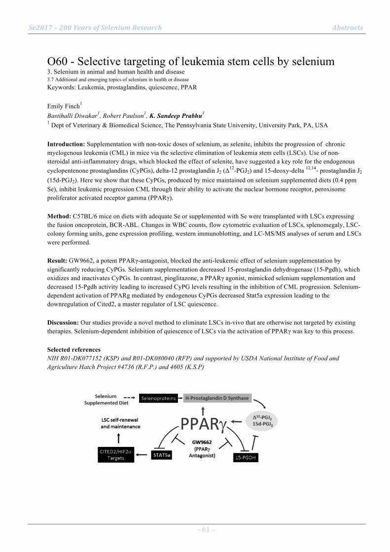
Se2017 – 200 Years of Selenium Research Abstracts
– 61 –
O60 - Selective targeting of leukemia stem cells by selenium 3. Selenium in animal and human health and disease 3.7 Additional and emerging topics of selenium in health or disease Keywords: Leukemia, prostaglandins, quiescence, PPAR Emily Finch1 Bastihalli Diwakar1, Robert Paulson1, K. Sandeep Prabhu1 1 Dept of Veterinary & Biomedical Science, The Pennsylvania State University, University Park, PA, USA Introduction: Supplementation with non-toxic doses of selenium, as selenite, inhibits the progression of chronic myelogenous leukemia (CML) in mice via the selective elimination of leukemia stem cells (LSCs). Use of non-steroidal anti-inflammatory drugs, which blocked the effect of selenite, have suggested a key role for the endogenous cyclopentenone prostaglandins (CyPGs), delta-12 prostaglandin J2 (Δ
12-PGJ2) and 15-deoxy-delta 12,14- prostaglandin J2 (15d-PGJ2). Here we show that these CyPGs, produced by mice maintained on selenium supplemented diets (0.4 ppm Se), inhibit leukemic progression CML through their ability to activate the nuclear hormone receptor, peroxisome proliferator activated receptor gamma (PPARγ). Method: C57BL/6 mice on diets with adequate Se or supplemented with Se were transplanted with LSCs expressing the fusion oncoprotein, BCR-ABL. Changes in WBC counts, flow cytometric evaluation of LSCs, splenomegaly, LSC-colony forming units, gene expression profiling, western immunoblotting, and LC-MS/MS analyses of serum and LSCs were performed. Result: GW9662, a potent PPARγ-antagonist, blocked the anti-leukemic effect of selenium supplementation by significantly reducing CyPGs. Selenium supplementation decreased 15-prostaglandin dehydrogenase (15-Pgdh), which oxidizes and inactivates CyPGs. In contrast, pioglitazone, a PPARγ agonist, mimicked selenium supplementation and decreased 15-Pgdh activity leading to increased CyPG levels resulting in the inhibition of CML progression. Selenium-dependent activation of PPARg mediated by endogenous CyPGs decreased Stat5a expression leading to the downregulation of Cited2, a master regulator of LSC quiescence. Discussion: Our studies provide a novel method to eliminate LSCs in-vivo that are otherwise not targeted by existing therapies. Selenium-dependent inhibition of quiescence of LSCs via the activation of PPARγ was key to this process. Selected references NIH R01-DK077152 (KSP) and R01-DK080040 (RFP) and supported by USDA National Institute of Food and Agriculture Hatch Project #4736 (R.F.P.) and 4605 (K.S.P)

Se2017 – 200 Years of Selenium Research Abstracts
– 62 –
O61 - Methylselenol suppressed the metastatic potential of B16F10 melanoma by reducing integrin expression 3. Selenium in animal and human health and disease 3.7 Additional and emerging topics of selenium in health or disease Keywords: methylselenol, metastasis, B16F10 melanoma, integrin expression An-Sik Chung1 Aeyung Kim2 1 Department of Biological Science, KAIST, Daejeon, Republic of Korea 2 Korean Medicine Application Center, Korea Institute of Oriental Medicine, Daegu, Republic of Korea Introduction: Our previous studies have shown that selenite inhibits the invasion of HT1080 fibrosarcoma cells by reduced expression of matrix metalloproteinase (MMP)-‐2 and -‐9 (1). Methylselenol induces apoptosis of B16F10 melanoma cells (2) and suppresses the invasive ability of the cells by reducing integrin expression (3). Method: Methylselenol can be produced by selenomethionine plus methioninase (SeMet-‐METase) in a cell culture system and by Se-‐methylselenocysteine (Se-‐SMC) in an animal model. The expression of integrins was analyzed by flow cytometry. The enzymatic activities of MMP-‐2 and -‐9 were assayed by gelatin zymography. Four–week old female C57BL/6J mice were fed a Se-‐MSC-‐supplemented diet or water. After four weeks, B16F10 melanoma cells were injected intravenously via the tail vein. On day 20 after the tumor cell injection, the pulmonary metastasis were macroscopically observed, and survival was checked 40 days later. Result: Treatment with SeMet-METase induced a substantial decrease in the expression of integrins, and fibronectin receptors in B16F10 melanoma cells. Moreover, this compound suppressed gelatinase activity and invasive ability in the cell culture system. Mice given a mash diet or water supplemented with Se-MSC almost completely diminished the pulmonary metastasis of the melanoma cells and enhanced survival compared with the control group. Collectively, our results indicates that methylselenol contributes to blocking the metastasis of B16F10 melanoma cells by the suppression of integrin expression Discussion: The results suggest that Se-MSC can be used for cancer treatment as nutritional adjuvant. More studies are required to use Se-MSC as nutritional adjuvant for determination of an adequate amount without toxicity Selected references Yoon SO, Kim MM, Chung AS. Inhibitory effect of selenite on invasion of HT1080 tumor cells. J. Biol. Chem. 276: 20085-92, 2001 Kim A, Park JM, Jung JY, Chung AS. Methyselenol generated from selenomethionine down regulates integrin expression and induces caspase mediated apoptosis of B16F10 melanoma cells. J. Cell Physiol. 212:386-400, 2007, Kim A, Jung JY, Son M, Lee SH, Lim JS, Chung AS. Long exposure of nontoxic concentrations of methylselenol suppresses the invasive potential of B16F10 melanoma. Oncol. Reports 20: 557-565, 2008
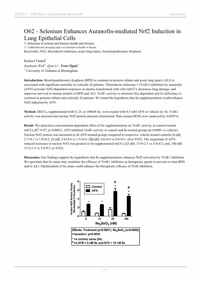
Se2017 – 200 Years of Selenium Research Abstracts
– 63 –
O62 - Selenium Enhances Auranofin-mediated Nrf2 Induction in Lung Epithelial Cells 3. Selenium in animal and human health and disease 3.7 Additional and emerging topics of selenium in health or disease Keywords: Nrf2, thioredoxin reductase, acute lung injury, bronchopulmonary dysplasia Rachael Tindell1 Stephanie Wall1, Qian Li1, Trent Tipple1 1 University of Alabama at Birmingham Introduction: Bronchopulmonary dysplasia (BPD) is common in preterm infants and acute lung injury (ALI) is associated with significant mortality in critically ill patients. Thioredoxin reductase-1 (TrxR1) inhibition by auranofin (AFN) activates Nrf2-dependent responses in murine transformed club cells (mtCC), decreases lung damage, and improves survival in murine models of BPD and ALI. TrxR1 activity is selenium (Se) dependent and Se deficiency is common in preterm infants and critically ill patients. We tested the hypothesis that Se supplementation would enhance Nrf2 induction by AFN. Method: MtCCs, supplemented with 0, 25, or 100nM Se, were treated with 0.5 mM AFN or vehicle for 1h. TrxR1 activity was assessed and nuclear Nrf2 protein amounts determined. Data (mean±SEM) were analyzed by ANOVA. Result: We detected a concentration-dependent effect of Se supplementation on TrxR1 activity in control-treated mtCCs (R2=0.97, p<0.0001). AFN inhibited TrxR1 activity in control and Se-treated groups (p<0.0001 vs vehicle). Nuclear Nrf2 protein was increased in all AFN-treated groups compared to respective vehicle-treated controls (0 nM: 2.7±0.1 vs 1.0±0.2; 25 nM: 5.6±0.6 vs 1.5±0.4; 100 nM: 4.6±0.4 vs 0.6±0.1; all p<0.05). The magnitude of AFN-induced increases in nuclear Nrf2 was greatest in Se-supplemented mtCCs (25 nM: 13.9±2.3 vs 5.8±0.2; and, 100 nM: 13.5±1.5 vs 5.8±0.2, p=0.02). Discussion: Our findings support the hypothesis that Se supplementation enhances Nrf2 activation by TrxR1 inhibition. We speculate that Se status may modulate the efficacy of TrxR1 inhibitors as therapeutic agents to prevent or treat BPD and/or ALI. Optimization of Se status could enhance the therapeutic efficacy of TrxR inhibition.

Se2017 – 200 Years of Selenium Research Abstracts
– 64 –
O63 - Preclinical chemopreventive efficacy of a novel hybrid p-XSC-aspirin compound in a lung cancer model 3. Selenium in animal and human health and disease 3.4 Selenium based medical therapeutics Keywords: Chemoprevention, lung cancer, aspirin, hybrid compounds Daniel Plano1 Cesar Aliaga1, Timothy Cooper1, Arthur Berg1, Shantu Amin1, Carmen Sanmartín2, Arun K. Sharma1 1 Department of Pharmacology; Penn State Cancer Institute, CH72; Penn State College of Medicine, USA 2 University of Navarra, Department of Organic and Pharmaceutical Chemistry, 31008 Pamplona, Spain Introduction: 1,4-Phenylenebis(methylene)selenocyanate (p-XSC) has been shown to inhibit tobacco carcinogen NNK induced lung cancer development in several animal models[1-2]. We hypothesized that p-XS-Asp would cleave in vivo to release the active p-XSeH, not releasing undesired HCN but the aspirin (Figure 1), thus making the compound less toxic and more potent than p-XSC or aspirin alone. Method: Chemopreventive efficacy of dietary p-XS-Asp was assessed using a NNK-induced A/J mouse model. Se content in plasma and tissues were carried out by atomic absorption spectroscopy. DNA adducts in lung tissues were performed by MS/MS. Result: Orally fed mice by gavage showed higher bioavailability for p-XS-Asp compared with p-XSC along with greater inhibition of DNA adducts in lung and liver. MTD study reflected lower toxicity for the hybrid derivative (MTD values between 74.5 and 111.7 versus 39.8 mg/Kg). p-XS-Asp groups at doses of 15 ppm and 7.5 ppm Se showed a significantly marked decrease in the percentage of lung cancer incidence in vivo with only 50% and 87% of tumor incidence, as compared to p-XSC (79% and 100%), respectively. NNK-control showed a 100% tumor incidence at both the doses. Likewise, the multiplicity for p-XS-Asp was 0.87 and 1.93 tumors/mouse as compared with NNK-control (11.53) and p-XSC (1.66 and 4.10 tumors/mouse, respectively) at the two doses tested. Notably, blood and tissue analyses showed no systemic toxicity for the p-XS-Asp fed group. Discussion: In conclusion, p-XS-Asp is less toxic while being more effective chemopreventive compound than p-XSC and is a promising candidate to future clinical evaluation. Selected references
[1] El-Bayoumy, K.; Upadhyaya, P.; Desai, D. H.; Amin, S.; Hoffmann, D.; Wynder, E. L. Anticancer Res 1996, 16, 2709-2712.
[2] Prokopczyk, B.; Rosa, J. G.; Desai, D.; Amin, S.; Sohn, O. S.; Fiala, E. S.; El-Bayoumy, K. Cancer Lett 2000, 161, 35-46.

Se2017 – 200 Years of Selenium Research Abstracts
– 65 –
O64 - Selenofolate is a Promising Novel Agent in Targeted Chemotherapy 3. Selenium in animal and human health and disease 3.4 Selenium based medical therapeutics Keywords: Selenofolate, Folate Receptor Alpha (FOLR1), Ovarian Cancer Antje Zickler1 Sougat Misra1, Gilbert Kirsch2, Julian Spallholz3, Mikael Björnstedt1 1 Karolinska Institutet, Department of Laboratory Medicine, Division of Pathology, Stockholm, Sweden 2 UMR CNRS 7565 SRSMC, Université de Lorraine, Metz, France 3 Department of Nutritional Sciences, Texas Tech University, Lubbock, TX, USA Introduction: Selenium compounds at high concentrations have shown growth-modulating properties with high specificity for tumor cells. Thus, they have been reported to strongly improve the efficiency of chemotherapeutics. Folate receptor alpha (FOLR1) is a membrane-bound transport protein with high affinity for folate. FOLR1 expression levels are highly upregulated in cancer cells of epithelial origin and positively correlate with tumor progression. To date, the appealing concept of therapeutic FOLR1 pathway intervention is explored extensively, as FOLR1 expression is largely absent in normal tissue. Hence, selective targeting of FOLR1 presents a unique and promising option in chemotherapy. Method: Here we used a synthetic selenofolate to investigate its effect on a FOLR1 high-expressing ovarian adenocarcinoma cell line, IGROV-1 cells, compared to a FOLR1 negative malignant melanoma cell line, A-375 cells. We tested the half maximal inhibitory concentration (IC50) of selenofolate by a luminescence-based viability assay. Result: Surprisingly, selenofolate was more cytotoxic to the FOLR1 negative A-375 cells than to the FOLR1 high-expressing IGROV-1 cells, suggesting a FOLR1-independent pathway for cellularPselenofolate uptake. Furthermore, siRNA-mediated knockdown of FOLR1 expression did not impact the growth of IGROV-1 cells despite alterations in specific mitogenic gene expression. Discussion: Our results indicate that the role of FOLR1 in malignant cells is still poorly understood and that the transport of folates might not be its foremost function. Currently, we investigate the cytotoxicity of selenofolate in combination with other drugs interfering with the folate metabolism, such as methotrexate. Our work will uncover the potential use of selenofolate as a supporting agent in chemotherapy.

Se2017 – 200 Years of Selenium Research Abstracts
– 66 –
O65 - Environmental Remediation of Selenium Contamination in Water and Soil 1. Selenium chemistry and geochemistry 1.5 Excessive selenium accumulation from natural or anthropogenic sources and remediation technologies Keywords: Treatment wetlands, Phytoremediation, Accumulation, Volatilization, Plant and microbial interaction Zhi-Qing Lin1 1 Environmental Sciences Program, Southern Illinois University, Edwardsville, Illinois 62026, USA Introduction: Selenium (Se) is essential to humans and animals, but becomes toxic to wildlife at high concentrations in the environment. Previous studies demonstrated that plants and plant-associated microbes can be applied to remove excessive Se from contaminated waters and soils. For example, the vegetated constructed wetlands were capable of significantly reducing Se from the inflow waste water. Biogenic volatilization of Se represents an environmentally sound remediation strategy to remove Se from the contaminated environment. Method: The selected Se remediation technologies include constructed treatment wetlands, different soil-plant systems, and the algal Se reduction pond. This presentation focused on the plant-soil microbial interactions and their effects on Se bioaccumulation and chemical transformation. Different environmental conditions and chemical characteristics of Se pollutants (including nanoscale elemental Se) have been evaluated for the processes of Se immobilization, bioaccumulation and volatilization. Result: The constructed treatment wetland was able to remove about 70% of the total Se mass in selenate-contaminated water. About 55% of the Se was immobilized in sediment, 5% of the Se accumulated in plants, and 10% of the Se volatilized to the atmosphere. The chemical transformation of selenate to elemental Se is the mechanistic process for the Se removal in treatment wetlands or algal retention ponds. Recent studies showed that microbial-derived nanoscale elemental Se could become bioavailable and further volatilized in soil-plant systems. Discussion: The transport and fate of Se removal was discussed, and using Se-laden plant materials from phytoremediation for producing Se-biofortified edible mushrooms has been demonstrated. Selected references
Bañuelos, G.S. Lin, Z.Q. 2007. Environmental Pollution. 150(3):306-12
Huang, J.C. Suárez, M. Yang, S.I. Lin, Z.Q. Terry, N. 2013. Environmental Science and Technology. 47: 10518−10525.
Lin, ZQ, Haddad, S, Hong, J, Morrissy, J, Bañuelos, GS, Zhang, LY. 2014. In: Bañuelos, G.S., Z.-Q. Lin, and X.B. Yin (eds.). Selenium in the Environment and Human Health. CRC Press, Boca Raton, Florida. 124-126.
Lin, Z.Q. Terry, N. 2003. Environmental Science and Technology. 3: 606-615.
Lin, Z.Q. Terry, N. Gao, S. Hussein, H. Ye, Z.H. 2010. International Journal of Phytoremediation. 12(3): 255-267.

Se2017 – 200 Years of Selenium Research Abstracts
– 67 –
O66 - Integrated Passive Biological Selenium Treatment System: Results of a 1-year Pilot Study 1. Selenium chemistry and geochemistry 1.5 Excessive selenium accumulation from natural or anthropogenic sources and remediation technologies Keywords: passive treatment, biological reduction, pilot study James Bays1 Chelsea Ransom2, Robert Thomas3, Sarah Foster4, Patrick Mulhern5, Luis Tovar6 1 CH2M, Tampa, FL, USA 2 CH2M, San Francisco, CA, USA 3 CH2M, Atlanta, GA, USA 4 CH2M, Denver, CO, USA 5 Mulhern MRE, Parker CO, USA 6 Mulhern MRE, Parker, CO, USA Introduction: Passive biological systems comprised of anaerobic organic media reactors create conditions supportive of dissimilatory reduction, adsorption and volatilization of Se at a lower cost compared to active biological treatment systems (Bays et al, 2013; Bays et al. 2014). Integrated passive biological treatment systems (BTS) combine anaerobic treatment with aerobic polishing to meet multiple regulatory water quality criteria. This paper summarizes a one-year BTS study in Centennial CO using membrane concentrate from the Joint Water Purification Plant (JWPP) for the Cottonwood Water & Sanitation District (CWSD). Method: CH2M piloted a BTS at the JWPP comprised of two parallel trains. Water in each train flowed passively through passive vertical downflow anaerobic biochemical reactors (BCRs) comprised of wood chips, sawdust, straw, horse manure, and limestone chips for primary removal of Se, followed by a sequence of organic and inorganic media cells configured for removal of metals, phosphorus, and nitrogen. The BTS was operated and monitored under the direction of CH2M in three phases from January through December 2016. Samples were collected weekly and laboratory analyses performed using US EPA methods. Result: Inflow Se averaged 68, 47 and 37 µg/L and outflow Se averaged 4, 3.4, and 3.3 µg/L for Phases 1-3, e.g. Train 1 (Figure 1). Substrates sequestered Se as predominantly adsorbed selenite, elemental and organic Se. Phosphorus and other pollutants met discharge criteria. Discussion: The BTS pilot effectively met the 30-day average discharge criterion of 4.6 µg Se/L, as well as criteria for phosphorus and other parameters, demonstrating the benefit of passive anaerobic and aerobic treatment. Selected references Bays, J.S., J. Tudini, B. T. Thomas, D. Evans, and T. Harrison, 2013. Advances in the Use of Passive Wetland Systems for Selenium Treatment of Mine-Impacted Water. Proc. SME Annual Meeting, Feb. 24 - 27, 2013, Denver, CO. Bays, J.S., R.C. Thomas, D. Evans. 2014. Integrated Biochemical Reactor Wetland Systems for Passive Treatment of Selenium. Pp. 454-363 in Proc. 14th Intl. Conf. on Wetland Systems for Pollution Control, 12-16 October 2014, Shanghai, China. Conference organized by Tongji University and Chongqing University. Prof. Qi Zhou and Prof. Jun Zhai (Eds).
Se2017 – 200 Years of Selenium Research Abstracts
– 68 –
O67 - Industrial selenium pollution: challenges to treat Flue Gas Desulfurization effluents 1. Selenium chemistry and geochemistry 1.5 Excessive selenium accumulation from natural or anthropogenic sources and remediation technologies Keywords: FGD, Industrial pollution, Ion exchange, Desulfurization Lucian Staicu1 1 University Politehnica of Bucharest, Chemistry Department, Bucharest, Romania Introduction: Selenium (Se) pollution has been linked to numerous episodes of severe aquatic ecosystem deterioration [1]. Flue Gas Desulfurization (FGD) effluents produced by coal-fired power facilities are arguably the most complex and difficult to treat [2]. In this paper, a real FGD effluent was characterized and the Se removal potential was investigated for the first time by chemical precipitation/adsorption using a commercial ion exchange resin. Method: The effluent was characterized for Se and metal content, major anions and other parameters [3]. A commercial ion exchange resin (gel type II hybrid strong base anion exchange resin impregnated with iron oxide) was employed as the adsorbent. Sulfate was precipitated (desulfurization) using BaCl2. The adsorption experiments were performed in the batch mode. Result: In the studied FGD, sulfate, a known competitor of Se, was found to be present almost 3 orders of magnitude in excess of Se (1.1 g L-1 vs 1.2 mg L-1). The resin performed poorly (~3% Se removal) against the raw FGD effluent. Desulfurization followed by ion exchange treatment led to a significant increase in Se removal (~80%). However, complete desulfurization using equimolar BaCl2 could not be achieved due to the presence of bicarbonate that acts as a sulfate competitor for Ba. In addition to Se and SO4
2-, several toxic metals were efficiently removed (Cd: 91%; Cr: 100%; Zn: 99%) by the combined treatment. Discussion: Due to the complexity of FGD effluents, this study underscores the necessity of a multistage treatment process as a high-performance Se treatment platform. Selected references
[1] Chapman PM et al. (2010) Ecological assessment of selenium in the aquatic environments. CRC Press.
[2] Staicu LC et al. (2017) Industrial selenium pollution: wastewater and physical-chemical treatment technologies. In: “Bioremediation of selenium contaminated wastewaters”, Ed. van Hullebusch ED, Springer (in press).
[3] Staicu LC et al. (2017) Chemosphere 117, 111-119.

Se2017 – 200 Years of Selenium Research Abstracts
– 69 –
O68 - Role of antioxidant defense system and mitochondrial activity in selenium toxicity tolerance in whea 1. Selenium chemistry and geochemistry 1.5 Excessive selenium accumulation from natural or anthropogenic sources and remediation technologies Keywords: Oxidative stress, Wheat, Selenate, Selenite Sucheta Sharma1 Manpreet Kaur1 1 Department of Biochemistry, Punjab Agricultural University, Ludhiana, India Introduction: Selenium (Se) induced oxidative stress as well as synthesis of non-specific selenoproteins has been attributed to its toxicity in plants. Crops grown on Se-rich soil accumulate Se to the levels, considered highly toxic to plants and for animal and human consumption. This study reveals the effect of selenium on its uptake, leaf physiology, antioxidant defense system, isoenzymic patterns and mitochondrial activity in wheat. Method: Selenium treatment-induced changes in growth, pigments, mitochondrial activity, antioxidant enzymes, antioxidants and other related metabolites were studied by standard procedures reported in the literature at tillering and ear-initiation stages. Result: Selenate-treated wheat plants accumulated higher Se concentration in leaves than control and selenite treatment. Selenium-treated plants exhibited lower Se tolerance index, reduced growth, impaired photosynthesis and mitochondrial activities, high levels of hydrogen peroxide and lower reactive oxygen species scavenging activities leading to a range of antioxidant responses that varied depending on the form and doses of Se used. There was presence of two extra catalase, one superoxide dismutase, two peroxidase and one glutathione reductase isozyme in different Se-treatments. Discussion: Wheat plants could adapt to applied selenite concentrations by developing antioxidant defense system but selenate-treated plants exhibited toxicity tolerance upto 2 mg kg-1 and died at high concentrations due to damage to tissue development and function. The higher Se uptake in selenate as compared to selenite treatment was due to its mobile nature. Significant increase in hydrogen peroxide and antioxidant enzymes activities in leaves was due to the presence of Se-induced oxidative stress in wheat plants. Selected references
Van-Hoewyk, D. 2013. A tale of two toxicities: malformed selenoproteins and oxidative stress both contribute to selenium stress in plants. Ann. Bot.163, 1-8.
Sharma, S., Gupta, R. and Singh, D. 2016. Variation in Se tolerance, accumulation, and growth parameters of different wheat cultivars. Comm. Soil Sci. Plant Anal. 47, 203-12.

Se2017 – 200 Years of Selenium Research Abstracts
– 70 –
O69 - Insufficient documentation for clinical efficacy of selenium supplementation in chronic autoimmune thyroiditis 3. Selenium in animal and human health and disease 3.4 Selenium based medical therapeutics Keywords: selenium supplementation, chronic autoimmune thyroiditis, thyroid, meta-analysis Kristian Hillert Winther1 Johanna Wichman1, Steen Bonnema1, Laszlo Hegedüs1 1 Department of Endocrinology and Metabolism, Odense University Hospital, Denmark Introduction: Selenium supplementation effectively reduces thyroid autoantibody concentrations in chronic autoimmune thyroiditis (AIT), but its effects on clinically relevant outcomes are unclear. The purpose of this systematic review and meta-analysis was to investigate clinically relevant effects of selenium supplementation in patients with AIT. Method: Controlled trials in adults (≥ 18 years) with AIT, comparing selenium with or without levothyroxine substitution (LT4), versus placebo and/or LT4, were eligible for inclusion. Identified outcomes were serum thyrotropin (TSH) levels in LT4-untreated patients, thyroid ultrasound (US) and health-related quality of life (HRQL). Eleven publications, covering nine controlled trials, were included in the systematic review. Random effects model meta-analyses were performed in weighted mean difference (WMD) for TSH, US and HRQL. Quality of evidence was assessed per outcome, using GRADE. Result: Meta-analyses showed no change in TSH, or improvements in HRQL or thyroid echogenicity (US), between LT4-untreated patients assigned to selenium supplementation or placebo. Three trials found some improvement in wellbeing in patients receiving LT4, but could not be synthesized in a meta-analysis. The quality of evidence ranged from very low to low for TSH as well as US outcomes, and low to moderate for HRQL, and was generally downgraded due to small sample sizes. Discussion: We found no effect of selenium supplementation on TSH, HRQL or thyroid US, in LT4-untreated individuals, and sporadic evaluation of clinically relevant outcomes in LT4-treated patients. Future well-powered RCTs, evaluating e.g. disease progression or HRQL, are warranted before determining the relevance of selenium supplementation in AIT.

Se2017 – 200 Years of Selenium Research Abstracts
– 71 –
O70 - Dihydroxy-1-selenolane protects cells from radiation-induced mitotic death: role of GPx 3. Selenium in animal and human health and disease 3.4 Selenium based medical therapeutics Keywords: Organoselenium, radioprotector, glutathione peroxidase, DNA repair Amit Kunwar1 Prachi Verma1, M. Iwaoka2, K.I. Priyadarsini3 1 Radiation & Photochemistry Division, Bhabha Atomic Research Centre, Trombay, Mumbai – 400 085, India 2 Department of Chemistry, School of Science, Tokai University, Hiratsuka-shi, Kanagawa 259-1292, Japan 3 Chemistry Division, Bhabha Atomic Research Centre, Trombay, Mumbai – 400 085, India Introduction: In recent times, researchers are exploring synthetic organoselenium compounds as novel therapeutic agents. Method: Dihydroxy-1-selenolane (DHS) an organoselenium compound [1] was evaluated for radio-protective effect in cellular models employing cellular, biochemical and molecular assays. Result: DHS treatment at 25µM for 16 hours significantly protected CHO cells of epithelial origin from radiation (4-11 Gy)-induced cell death and this effect was associated with increase in glutathione peroxidase (GPx) both at activity and mRNA level. The addition of mercaptosuccinic acid, a pharmacological inhibitor of GPx abrogated the radio-protective activity of DHS (fig. 1). On contrary increasing the GPx level in CHO cells through treatment with DHS-C6, a lipophilc conjugate known to increase the cellular uptake of DHS and there by GPx level, showed significantly higher protection (fig. 1). Surprisingly both DHS and DHS-C6 although increased the GPx level in spleen lymphocytes (radiosensitive cells) did not protect them from the radiation-induced apoptosis as monitored by propidium iodide (PI) and DNA fragmentation assays. Since CHO cells undergo radiation-induced cell death through mitotic catastrophe mediated through chromosomal aberration and G2/M arrest, it was anticipated that radio-protective effect of DHS might be related to its effect on DNA damage/repair and cell cycle arrest. Supporting this assumption, our results revealed that treatment with DHS and/or DHS-C6 in CHO cells led to faster repair of DNA following radiation exposure and subsequently inhibited the G2/M arrest. The GPx inhibitor, mercaptosuccinic acid reversed these effects. Discussion: Taken together it is confirmed that DHS-induced GPx level protects cells from mitotic death but not from apoptosis. Selected references A. Kunwar, P. Verma, H.N. Bhilwade, M. Iwaoka, K.I. Priyadarsini (2016) Dihydroxyselenolane (DHS) supplementation improves survival following whole-body irradiation (WBI) by suppressing tissue-specific inflammatory responses. Mutation Research 2016, 807: 33-46.
Se2017 – 200 Years of Selenium Research Abstracts
– 72 –
O71 - Inhibition of metalloenzymes, i.e., angiotensin-converting enzyme and tyrosinase, by selenol-metal ion interaction of selenoneine 3. Selenium in animal and human health and disease 3.4 Selenium based medical therapeutics Keywords: selenoneine, ACE, tyrosinase Takuya Seko1 Kenji Ishihara1, Shintaro Imamura1, Yumiko Yamashita1, Michiaki Yamashita2 1 National Research Institute of Fisheries Science, Yokohama, Japan 2 National Fisheries University, Shimonoseki, Japan Introduction: Selenoneine is a novel selenium containing compound discovered in the blood of bluefin tuna. Since selenoneine has strong antioxidant activity and detoxification ability of methyl mercury, it is expected to be used for functional foods as antioxidant and selenium supplements. This study characterized its biological activities against hypertension and skin melanism. Method: Inhibition of angiotensin converting enzyme (ACE) and tyrosinase was assayed in the presence of selenoneine. Melanin generation was measured in mice melanoma cells and human melanoma cells. Result: Selenoneine inhibited activities of ACE and tyrosinase. The Km values of ACE were 1.396 ± 0.103 mM with selenoneine and 0.724 ± 0.051 mM without selenoneine. Lineweaver-Burk plots for inhibition of ACE activities by selenoneine indicated a competitive inhibition. Melanin generation was also inhibited by selenoneine in enzyme activity assay and bioassay with human cultured keratinocytes. On the other hand, selenite and ergothioneine (thiol-analog of selenoneine) did not inhibit these enzyme activities and melanin generation. Discussion: ACE and tyrosinase are metalloenzymes. ACE has a zinc ion and tyrosinase has two copper ions in their active centers. Since seelenoneine inhibited metalloenzymes, selenol group in selenoneine may interact with metal ions situated active sites. Therefore, selenoneine has a novel biological functions in metalloenzyme inhibition, relating to prevention of hyper-tension by ACE inhibition and reduction of melanin production by tyrosinase inhibition.
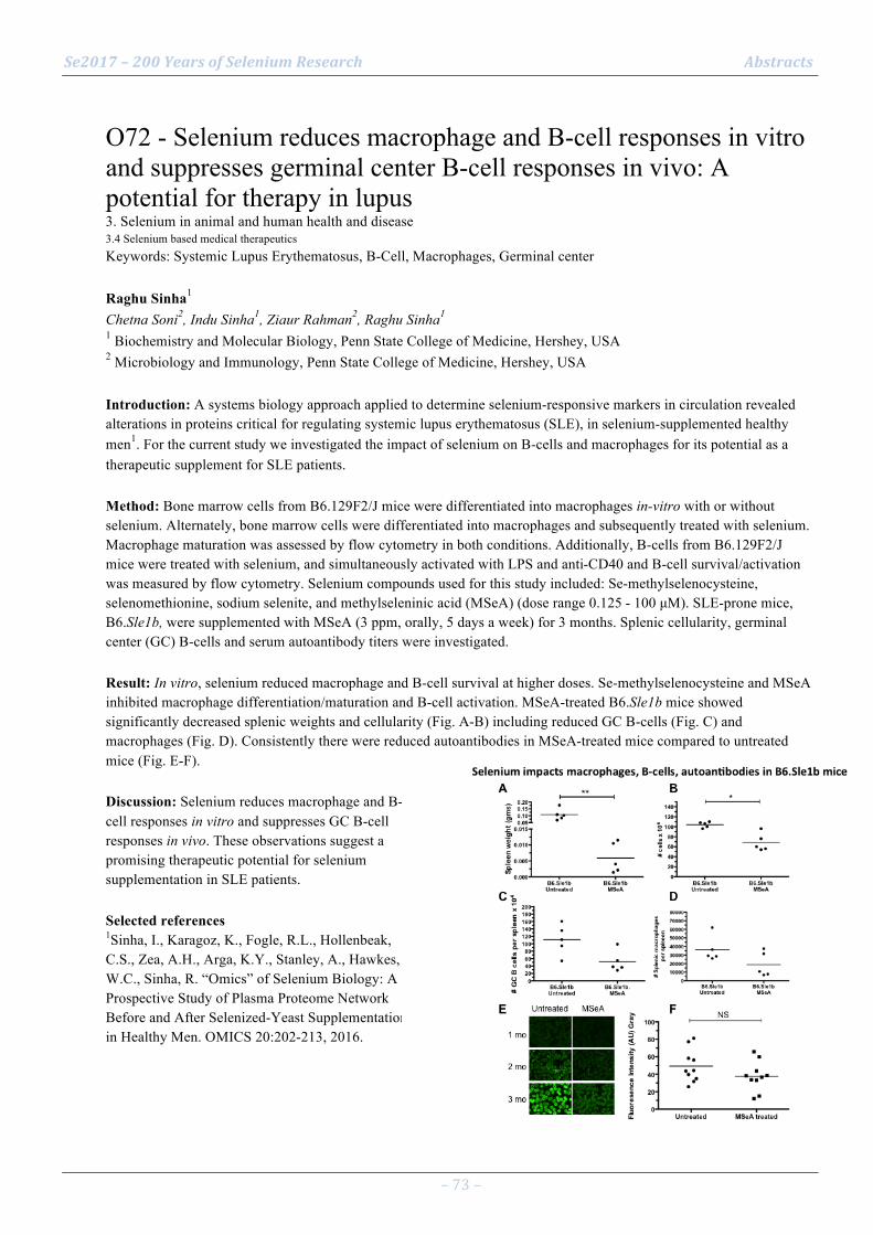
Se2017 – 200 Years of Selenium Research Abstracts
– 73 –
O72 - Selenium reduces macrophage and B-cell responses in vitro and suppresses germinal center B-cell responses in vivo: A potential for therapy in lupus 3. Selenium in animal and human health and disease 3.4 Selenium based medical therapeutics Keywords: Systemic Lupus Erythematosus, B-Cell, Macrophages, Germinal center Raghu Sinha1 Chetna Soni2, Indu Sinha1, Ziaur Rahman2, Raghu Sinha1 1 Biochemistry and Molecular Biology, Penn State College of Medicine, Hershey, USA 2 Microbiology and Immunology, Penn State College of Medicine, Hershey, USA Introduction: A systems biology approach applied to determine selenium-responsive markers in circulation revealed alterations in proteins critical for regulating systemic lupus erythematosus (SLE), in selenium-supplemented healthy men1. For the current study we investigated the impact of selenium on B-cells and macrophages for its potential as a therapeutic supplement for SLE patients. Method: Bone marrow cells from B6.129F2/J mice were differentiated into macrophages in-vitro with or without selenium. Alternately, bone marrow cells were differentiated into macrophages and subsequently treated with selenium. Macrophage maturation was assessed by flow cytometry in both conditions. Additionally, B-cells from B6.129F2/J mice were treated with selenium, and simultaneously activated with LPS and anti-CD40 and B-cell survival/activation was measured by flow cytometry. Selenium compounds used for this study included: Se-methylselenocysteine, selenomethionine, sodium selenite, and methylseleninic acid (MSeA) (dose range 0.125 - 100 µM). SLE-prone mice, B6.Sle1b, were supplemented with MSeA (3 ppm, orally, 5 days a week) for 3 months. Splenic cellularity, germinal center (GC) B-cells and serum autoantibody titers were investigated. Result: In vitro, selenium reduced macrophage and B-cell survival at higher doses. Se-methylselenocysteine and MSeA inhibited macrophage differentiation/maturation and B-cell activation. MSeA-treated B6.Sle1b mice showed significantly decreased splenic weights and cellularity (Fig. A-B) including reduced GC B-cells (Fig. C) and macrophages (Fig. D). Consistently there were reduced autoantibodies in MSeA-treated mice compared to untreated mice (Fig. E-F). Discussion: Selenium reduces macrophage and B-cell responses in vitro and suppresses GC B-cell responses in vivo. These observations suggest a promising therapeutic potential for selenium supplementation in SLE patients. Selected references 1Sinha, I., Karagoz, K., Fogle, R.L., Hollenbeak, C.S., Zea, A.H., Arga, K.Y., Stanley, A., Hawkes, W.C., Sinha, R. “Omics” of Selenium Biology: A Prospective Study of Plasma Proteome Network Before and After Selenized-Yeast Supplementation in Healthy Men. OMICS 20:202-213, 2016.
Se2017 – 200 Years of Selenium Research Abstracts
– 74 –
O73 - The significance of selenoproteins for human health revealed by inborn errors of metabolism 2. Selenium in the molecular life sciences 2.5 Selenoprotein genetics Keywords: TXNRD; neurodegeneration; PCH2; familial glucocorticoid deficiency; Sedaghatian Ulrich Schweizer1 1 Rheinische Friedrich-Wilhelms-Universität Bonn, Institut für Biochemie und Molekularbiologie Introduction: The significance of the 25 genes encoding selenoproteins for human health is increasingly recognized through the identification of patients with inborn errors in selenoprotein biosynthetic factors or in individual selenoproteins. Mutations in selenoprotein N lead a spectrum of disorders nowadays called SEPN1(selenon)-related myopathy. Mutations in glutathione peroxidase 4 cause respiratory failure and bone defects (Sedaghatian syndrome). Patients carrying mutations in thioredoxin reductase 2 (TXNRD2) suffer from familial glucocorticoid deficiency. Method: Result: Recently, we have identified patients with hypomorphic mutations in TXNRD1 who suffer from familial epilepsy. As with mutations in TXNRD2, modifier genes may modulate the phenotype. There are also inborn errors of selenoprotein biosynthesis that affect many selenoproteins. For example, pathogenic mutations in selenocysteine synthase cause neurodevelopmental and neurodegenerative disorders. Mostly endocrine phenotypes result from mutations in SECIS-binding protein 2 and tRNA[Ser]Sec, which will be covered in another lecture. Mutations in the latter two genes involve impaired metabolism and action of thyroid hormones leading to delayed bone growth and maturation. In addition, mutations in SECISBP2 sometimes affect nervous system development, muscle, inner ear, skin, and immune system function underlining the significance of selenoproteins for many organ systems. Discussion: Mouse models have been generated for several of these mutations and will be compared to human phenotypes where possible. Selected references
1. Why 21? The significance of selenoproteins for human health revealed by inborn errors of metabolism. Schweizer U, Fradejas-Villar N. FASEB J. 2016 30(11):3669-3681. Review.
2. Homozygous mutation in TXNRD1 is associated with genetic generalized epilepsy. Alexei P. Kudin, Gregor Baron, Gábor Zsurka, Kevin G. Hampel, Christian E. Elger, Alexander Grote, Yvonne Weber, Holger Lerche, Holger Thiele, Peter Nürnberg, Herbert Schulz, Ann-Kathrin Ruppert, Thomas Sander, Qing Cheng, Elias SJ Arnér, Lutz Schomburg, Sandra Seeher, Noelia Fradejas-Villar, Ulrich Schweizer, Wolfram S. Kunz Free Radical Biology and Medicine 2017 106:270–277

Se2017 – 200 Years of Selenium Research Abstracts
– 75 –
O74 - Mouse models lacking the selenoprotein thioredoxin reductase-1 2. Selenium in the molecular life sciences 2.5 Selenoprotein genetics Keywords: thioredoxin, glutathione, ribonucleotide reductase, NADPH Edward E Schmidt1 Justin R Prigge1, Lucia Coppo2, Sebastin S Martin2, Fernando Ogata3, Collin Miller1, Jean A Kundert1, Åse Mattsson2, Alix E Herr1, Matthew P Taylor1, Tomas Gustafsson4, Arne Holmgren2, Elias S J Arnér2 1 Montana State University, Bozeman, USA 2 Karolinska Institutet, Stockholm, Sweden 3 Universidade Federal de São Paulo, São Paulo, Brazil 4 Umeå University, Umeå, Sweden Introduction: Oxidation of energetic nutrients sustains high intracellular NADPH/NADP+ ratios. NADPH-dependent reduction of thioredoxin-1 (Trx1)-disulfide and glutathione-disulfide by the selenoprotein thioredoxin reductase-1 (TrxR1) and glutathione reductase (GR), respectively, fuels ribonucleotide reductase (RNR) and cytosolic antioxidant systems. Method: Here we investigated mouse models with liver-specific co-disruptions of the genes encoding TrxR1, Trx1, and GR. Result: Mouse livers lacking both TrxR1 and GR sustained reduced glutathione (GSH) pools using an NADPH-independent methionine-consuming system1. Liver-specific co-disruption of Trx1, TrxR1, and GR, paradoxically, causes a 100-fold increase in hepatocyte proliferation, indicating that, in the absence of Trx1, methionine-fueled GSH/glutaredoxin supports mammalian S phase RNR. Discussion: In the triple-null livers, like in many cancers, RNR places a critical yet relatively low-volume demand on cytosolic reducing power, thereby favoring high hepatocyte turnover over sustained hepatocyte integrity. Selected references 1 Eriksson, S., Prigge, J.R., Talago, E.A., Arner, E.S.J., and Schmidt, E.E. (2015). Dietary methionine can sustain cytosolic redox homeostasis in the mouse liver. Nature Communications 6, 6479.
Se2017 – 200 Years of Selenium Research Abstracts
– 76 –
O75 - Mutated selenocysteine synthase creates a Sedaghatian-type spondylometaphyseal dysplasia mouse model 2. Selenium in the molecular life sciences 2.5 Selenoprotein genetics Keywords: SepSecS, pontocerebellar hypoplasia, PLP N Fradejas-Villar1 U Reuter1, V Stein2, U Schweizer1 1 IBMB, Universität Bonn, Bonn, Germany 2 Institut für Physiologie II, Universität Bonn, Bonn, Germany Introduction: Mutations in SEPSECS (O-Phosphoseryl-tRNA:selenocysteinyl-tRNA synthase) have been found in different human populations. These mutations are the cause of a severe early-onset neurodegenerative disease, termed now pontocerebellar hypoplasia type 2D (PCH2D). SEPSECS is a PLP (pyridoxal phosphate) dependent-enzyme which catalyzes the final step of selenocysteine (Sec) biosynthesis using selephosphate as selenium donor. Sec is mostly found in the active site of selenoproteins, which are essential for brain function. PCH2D patients suffer from severe spasticity, profound mental retardation and progressive microcephaly. One of the SEPSECS mutations found in Sephardic Jewish patients of Iraqi ancestry consists on a substitution of a highly conserved tyrosine at position 334 with cysteine (Y334C). This mutation was suggested to affect the binding of PLP cofactor, required for the enzyme catalysis. Method: We generated mice carrying the Y334C mutation, using a targeting vector created by recombineering. Result: Unlike human PCH2D patients, homozygous mice (SepsecsY334C/Y334C) died the day after birth, did not show a milk spot, developed cyanosis, and showed abnormal electrocardiograms. Analysis of tissue expression revealed that selenoproteins, including Gpx4, were mainly reduced in brain, especially in neurons, but not in liver. In primary culture, PLP supplementation did not rescue neuronal selenoprotein levels. Discussion: The SepsecsY334C/Y334C mouse phenotype resembles Sedaghatian-type spondylometaphyseal dysplasia (SSMD), a neonatal lethal disease caused by truncating mutations in GPX4. This disease is characterized by cardio-respiratory insufficiency and neurological anomalies.
Se2017 – 200 Years of Selenium Research Abstracts
– 77 –
O76 - Agronomic strategies affect the efficacy and quality of selenium biofortification 1. Selenium chemistry and geochemistry 1.6 Strategies to improve selenium accumulation and biofortification Keywords: biofortification, selenoamino acids, vegetables Gary Bañuelos1 1 USDA-ARS, San Joaquin Valley Agricultural Sciences Center, 9611 South Riverbend, Parlier, CA, USA Introduction: The Se content of food is highly dependent on agronomic strategies, soil Se bioavailability, and the ability of plants to take up and accumulate Se as selenoamino acids. The dietary intake of Se is strongly influenced by the consumption of food products containing Se. To mitigate a low intake of Se, biofortification strategies such as applying foliar Se, amending soils with Se fertilizers or cropping in Se rich soil, have all been used to increase Se accumulation in crops. Method: In three multi-year field studies, the following biofortification strategies have been evaluated: 1) foliar application of selenate, selenite, or selenomethionine; 2) soil application of selenate, selenite, or selenomethionine; and 3) growing crops in soils naturally rich in Se. Result: Results indicated that with foliar application of Se, total Se content in Allium cepa ranged from 0.03 (control) to a high of 0.93 mg/kg DM with selenate treatment and greatest percentages (%) of selenoamino acids were ɣ-glu-MeSeCys (67), MeSeCys (15) and SeMet (13). With soil applications of Se, analyses for Se in onion are in progress. In soil naturally rich in Se, total Se in five different Brassica vegetables ranged from 4.8 to a high of 17.2 mg/kg DM in Brassica oleracea var. capitata f. rubra where the greatest percentages (%) of selenoamino acids were SeMet (22), MeSeCys (16), and ɣ-glu-MeSeCys (13). Discussion: Selenium biofortification occurred with all three sources of Se, however, Se accumulation was greatest but more variable when Brassica vegetables were grown in soils naturally rich in Se. Selected references
Bañuelos, G.S., Z.Q. Lin and M. Broadley (2017). Selenium biofortification. In: E.A.H Pilon-Smits, L.H.E. Winkel, Z.Q. Lin (eds.), Selenium in Plants-Molecular, Physiological, Ecological and Evoluationary Aspects. Springer International Publishing Switzerland. ISBN: 978-3-319-56248-3

Se2017 – 200 Years of Selenium Research Abstracts
– 78 –
O77 - Improving selenium supply in food systems 1. Selenium chemistry and geochemistry 1.6 Strategies to improve selenium accumulation and biofortification Keywords: Biofortification, food system, selenium deficiency, Moringa Graham Lyons1 1 School of Agriculture, Food & Wine, University of Adelaide, Urrbrae, South Australia Introduction: Selenium intakes are suboptimal in many countries. Strategies to increase Se supply include process fortification, biofortification, increasing dietary diversity and individual supplementation. Under the HarvestPlus food system program, agronomic biofortification trials of Se and other micronutrients were conducted in China, Colombia and Australia, and orange sweet potatoes were researched and promoted in Melanesia. Method: Potential improvements to food system Se delivery are presented, derived from studies by the author and colleagues from 2001-2015. Result: Recovery of Se in edible crop parts was less than 15% of soil-applied Se. Foliar Se is usually more efficient, especially when applied with urea. Selenium-biofortified flour has been marketed in Australia since 2005. In Tibet, where Se deficiency is widespread and where vast grasslands are unfertilised while cropland is often overfertilised, the most efficient method to increase Se may be to add with iodine in salt. This would exploit Se and iodine thyroid synergy, reach a high proportion of people and livestock and minimise wastage. Genotype x environment studies of nutritious leafy vegetables in Pacific countries found that Moringa oleifera (drumstick tree) had Se leaf levels 12-fold higher than other plants on the same soil, including low-Se soils. In sub/tropical regions with low-Se soils, consumption of drumstick leaves/leaf powder could increase Se status along with sulphur, vitamins and protein at minimal cost. Discussion: Improving the efficiency of biofortification, exploiting synergies with other micronutrients and employing natural Se biofortifiers could improve Se supply in food systems.

Se2017 – 200 Years of Selenium Research Abstracts
– 79 –
O78 - Agronomic Biofortification with Selenium in Intercropping Systems would address Low Selenium Intake 1. Selenium chemistry and geochemistry 1.6 Strategies to improve selenium accumulation and biofortification Keywords: Bio-fortification, inter-cropping systems, selenium intake ALLAN CHILIMBA1 Scott Young2, Martin Broadley2, Edward Joy2 1 AGRISO Consultants and Ngolojere Investments 2 University of Nottingham, UK Introduction: Selenium (Se) is an essential human micronutrient with critical roles in immune functioning and antioxidant defense (Fairweather-Tait et al. 2011). There is widespread dietary Se deficiency in Sub-Saharan Africa (SSA) (Chilimba et al. 2011; Joy et al. 2014). Sole crops of maize were used in bio-fortification (Chilimba et al. 2012) but smallholder farmers intercrop maize with legumes. Therefore, agronomic bio-fortification with Se in intercrops was conducted to determine efficiencies of Se uptake in intercrops or sole crops. Method: The experiment was conducted at two sites consisted monocrop maize, monocrop groundnuts, monocrop soybean, intercrop maize/groundnuts and intercrop maize/soybean with two rates of Se, 0 and 10 g ha-1 using sodium selenate and laid out in a randomized complete block design with three replicates. Grain samples were analyzed for selenium (78Se) by ICP-MS (X-SeriesII, Thermo Fisher Scientific Inc., Waltham, MA, USA). Result: Se application increased Se concentration and soybean gave the highest followed by groundnuts and maize (Figure 1). Se application in intercrops significantly increased Se recovery to 7.2 % compared to monocrops (Figure 2) and Selenium intake also increased (Table 1). Discussion: The results indicate that Se recovery in mono-cropped maize is inefficient and applied Se is leached as selenate, adsorbed as selenite or immobilized into organic forms. Legumes are efficient in uptake of the applied Se compared to maize hence higher Se recovery. The Se intake range between 66 and 162 µg cap-1 d-1 which is optimum (Fairweather-Tait et al., 2011). Selected references Chilimba, A.D.C., Young, S.D., Black, C.R., Rogerson, K.B., Ander, E.L., Watts, M., Lammel, J., & Broadley, M.R. 2011. Maize grain and soil surveys reveal suboptimal dietary selenium intake is widespread in Malawi. Scientific Reports: 1, 72. Chilimba, A.D.C. Young, S.D., Black, C.R. Meacham, M.C., Lammel, J., & Broadley M.R. (2012). Agronomic biofortification of maize with selenium (Se) in Malawi. Field Crops Research, 125, 118–128. Fairweather-Tait, S.J., Bao, Y., Broadley, M.R., Collings, R., Ford, D., Hesketh, J., & Hurst, R. 2011. Selenium in human health and disease. Antioxidants and Redox Signaling: 14: 1337-1383. Joy, E.J.M., Ander, E.L., Young, S.D., Black, C.R., Watts, M.J., Chilimba, A.D.C., Chilima, B., Siyame, E.W.P., Kalimbira, A.A., Hurst, R., Fairweather-Tait, S.J., Stein, A.J., Gibson, R.S., White, P.J., Broadley, M.R. (2014). Dietary mineral supplies in Africa. Physiol Plantarum 151:208–229. doi: 10.1111/ppl.12144.

Se2017 – 200 Years of Selenium Research Abstracts
– 80 –
O79 - To bio or not to bio? Strategies for agronomic biofortification with Se in tropical agroecosystems 1. Selenium chemistry and geochemistry 1.6 Strategies to improve selenium accumulation and biofortification Keywords: Se-fertilizers, soil and foliar application, Se sorption, Oxisols Luiz Roberto Guimarães Guilherme1 Guilherme Lopes1, Josimar Henrique de Lima Lessa1, Anderson Mendes Araújo1, Ana Paula Branco Corguinha1, Fabio Aurélio Dias Martins2, Ediu Carlos Silva Júnior1, Paulo Fernandes Boldrin3, André Rodrigues Reis4, Márcio José Santana5, Daniel Rufino Amaral5, Valdeci Orioli Júnior5, Tulio Silva Lara6 1 Federal University of Lavras, Lavras, Brazil 2 Agricultural Research Company of Minas Gerais Epamig, Patos de Minas, Brazil 3 UniRV University of Rio Verde, Rio Verde, Brazil 4 São Paulo State University, Tupã, Brazil 5 Federal Institute of Triângulo Mineiro, Uberaba, Brazil 6 Federal University of Western Pará, Santarém, Brazil Introduction: The addition of selenium (Se) in agroecosystems is important to increase Se levels in food crops, especially in Se-poor regions, e.g., Brazil. The potential of different plant species and agronomic strategies for Se biofortification have been addressed in several studies worldwide. Soil application of Se-containing fertilizers seems to be an effective strategy in soils from temperate regions, yet such approach may result in poor efficiency of the Se source in oxidic soils, where anionic forms of Se are strongly adsorbed. Under these circumstances, the application of Se together with other selected elements/nutrients may be considered in order to increase the efficiency of plants for taking up soil-Se. Method: This work will discuss different strategies that can be used for increasing Se content in crops of great social/economic relevance in Brazil, through adoption of different agronomic biofortification practices. For that, we are setting a Research Network (BioSeBrazil) of greenhouse/field experiments with food and feed crops in different soils to test different methods (soil and/or foliar) and sources (inorganic and/or organic Se) for Se biofortification. Result: Our efforts for addressing the best soil application approach focus on the use of Se together with P-fertilizers (at planting time), and/or through split nitrogen (N) fertilizer applications, using different N sources, as this play an important role in a nutrient management strategy as well as Se acquisition by plants. Discussion: The rationale for adding Se with P and/or N fertilizers will be discussed, with a focus on the factors affecting Se availability in tropical (oxidic) soils (Figure 1).
Se2017 – 200 Years of Selenium Research Abstracts
– 81 –
O80 - Soil and foliar application of selenium in upland rice aiming agronomic biofortification 1. Selenium chemistry and geochemistry 1.6 Strategies to improve selenium accumulation and biofortification Keywords: rice, agronomic biofortification, foliar application, soil application Andre Rodrigues dos Reis1 Heitor Pontes Gestal Reis1, José Mateus Kondo Santini1 1 São Paulo State University Introduction: Agronomic biofortification is a good strategy to increase selenium in plants. This study aimed to evaluate the levels of Se and application forms on rice growth, grain yield, and Se accumulation in seeds. Method: The experiment was performed under field condition located at Selviria city, Mato do Grosso do Sul State, Brazil. The experimental design was a completely randomized 2 × 6 factorial scheme as follows: 2 forms of Se application (soil and foliar) and 6 levels of Se (0; 10; 20; 40; 80; e 160 g ha-1). The rice seeds were harvested and prepared for chemical analysis. The Se analysis was performed as described in Chilimba et al. (2011). Result: The total Se concentration ranged from 0.01 to 0.54 mg kg-1 in the leaves and 0.03 to 0.36 mg kg-1 in the seeds when applied at the level of 20 g ha-1 via soil application. On the other hand, the Se level 20 g ha-1 applied through foliar application the Se concentration in seeds ranged from 0.01 to 1.65 mg kg-1. Discussion: In this study, the calculated daily intake of Se from the biofortified rice grains ranged from 2.05 to 24.7 µg day-1, which represent an increase from 3.72% to 44.9% of the daily requirement of Se per day when applied 20 g ha-1 through soil application. Despite that the recommendation for daily Se intake for adults is 55 µg day-1, the present study show relevant information on agronomic biofortification to increase Se content in edible parts, which can benefits human health. Selected references Chilimba ADC, Young SD, Black CR, Rogerson KB, Ander EL, Watts MJ, Lammel J, Broadley, MR. Maize grain and soil surveys reveal suboptimal dietary selenium intake is widespread in Malawi. Scientific Reports. 2011; 1(72):1-9. doi: 10.1038/srep00072

Se2017 – 200 Years of Selenium Research Abstracts
– 82 –
O81 - A model for Glutathione peroxidase 4-catalyzed reduction of lipid hydroperoxides in membranes 2. Selenium in the molecular life sciences 2.6 Selenoprotein function Keywords: PHGPx, SPR analysis, Molecular Dynamics, Phospholipid hydroperoxide. Matilde Maiorino1 Giorgio Cozza1, Monica Rossetto1, Antonella Roveri1, Stefano Toppo1, Ana-Marija Vučković1, Mattia Zaccarin1, Lucio Zennaro1, Fulvio Ursini1 1 Department of Molecular Medicine, Viale G. Colombo, 3, University of Padova, 35121-Padova, Italy Introduction: Reduction of PLOOH by GPx4/GSH is vital. Although details of the reaction have been unraveled, what is still unknown is how the enzyme interacts with membrane and how this impacts on catalytic cycle. Method: SPR analysis to evaluate the affinity of GPx4 for different bilayers; in silico analysis of the docking and dynamics (MD) of the interaction during the catalytic cycle. Result: SPR analysis indicates cardiolipin as the phospholipid with the maximal affinity to GPx4. This complies with the notion that GPx4 contains a cationic surface adjacent to the catalytic center. Consistently, KCl increases the kD of the GPx4 - CL complex. MD simulation shows that a hydroperoxide group posed in the core of the membrane moves up and is stabilized at the lipid-water interface. Docking analysis, validated by MD simulation, indicates two seemingly oscillating interaction patterns of the GPx4 cationic surface with the anionic head of CL. These interactions precisely address water-exposed 13-OOH or 9-OOH groups of tetra-linoleoyl CL toward catalytic redox center. However, in this pose the catalytic site would be inaccessible to the reducing substrate but the redox chemistry inside the complex and the anionic nature of GSH contribute to a displacement. This permits the last reaction of the cycle, when the enzyme is regenerated and GSSG released. Discussion: GPx4 binds to the membrane by electrostatic interactions precisely addressing the redox center toward the phospholipid hydroperoxide, and GSH affects the binding. From this set of data, a model describing how the peroxidase jumps on the membrane seeking for hydroperoxide, is produced.

Se2017 – 200 Years of Selenium Research Abstracts
– 83 –
O82 - Selenium versus sulfur: reversibility of chemical reactions and resistance to permanent oxidation in proteins and nucleic acid 2. Selenium in the molecular life sciences 2.6 Selenoprotein function Keywords: selenocysteine, cysteine, selenouridine, thiouridine, reversibility, resistance to oxidation Robert J. Hondal1 1 Department of Biochemistry, University of Vermont, U.S.A. Introduction: The central question in the field of selenium biochemistry is: “What chemical advantage does selenium confer to a protein, tRNA, or other biomolecule relative to the more abundant element sulfur?” Method: One possible answer to this question is that chemical reactions with selenium are more readily reversible than those of sulfur. This is especially true for the oxidation of selenium relative to sulfur as Se-oxides are more rapidly reduced compared to S-oxides. Result: Here, the evidence that selenium confers resistance to irreversible oxidation in selenium-containing enzymes is reviewed. Discussion: New evidence will also be presented that resistance to permanent oxidation is extended to selenouridine-containing tRNAs.

Se2017 – 200 Years of Selenium Research Abstracts
– 84 –
O83 - The key ferroptosis regulator GPX4: cellular mechanisms and in vivo relevance 2. Selenium in the molecular life sciences 2.6 Selenoprotein function Keywords: ACSL4, cell death, ferroptosis, PHGPx Marcus Conrad1 1 Helmholtz Zentrum München, Institute of Developmental Genetics, 85764 Neuherberg - Munich, Germany Introduction: The selenoenzyme glutathione peroxidase 4 (GPX4) has emerged as the key regulator of ferroptosis [1,2], a recently recognized form of regulated necrotic cell death [3]. Ferroptosis is clearly distinct from other paradigms of regulated cell death, such as apoptosis and necroptosis, and is characterized by an iron-dependent lipid peroxidation. Initially being recognized as a cell death modality highly relevant for a subset of cancer entities, ferroptosis has been implicated in a variety of degenerative disease contexts including tissue ischemia/reperfusion injury and neurodegeneration. Method: CRISPR/Cas9-mediated genome-wide screening efforts and expression analysis of ferroptosis-resistant cell lines have recently identified acyl-CoA synthetase long-chain family member 4 (ACSL4) as a novel player in the ferroptotic process downstream of GPX4 deletion/inhibition [4]. Result: Using an interdisciplinary approach combining redox global phospholipidomics, reverse genetics, bioinformatics and systems biology, 15-hydroperoxy-arachidonoyl- and 15-hydroperoxy-adrenoyl residues esterified in phosphatidylethanolamines were identified as proximate signals in the ferroptotic death program [5]. Genetic ACSL4 disruption and pharmacological inhibition of ACSL4 prevented the accumulation of these death signals in cells and conferred an unprecedented resistance to ferroptosis induction triggered by GPX4 inactivation. Discussion: Despite its outstanding importance for ferroptosis, little is known about the cellular and in vivo mechanisms that regulate GPX4 stability and potential implications in various disease contexts, which shall be discussed here. Selected references
[1] Friedmann Angeli et al. (2014) Inactivation of the ferroptosis regulator Gpx4 triggers acute renal failure in mice. Nat Cell Biol 16, 1180-1191
[2] Yang et al. (2014) Regulation of Ferroptotic Cancer Cell Death by GPX4. Cell 156, 317-331
[3] Dixon et al. (2012) Ferroptosis: an iron-dependent form of nonapoptotic cell death. Cell 149, 1060-1072
[4] Doll et al. (2017) ACSL4 dictates ferroptosis sensitivity by shaping cellular lipid composition. Nat Chem Biol 13, 91-98
[5] Kagan et al. (2017) Oxidized arachidonic and adrenic PEs navigate cells to ferroptosis. Nat Chem Biol 13, 81-90

Se2017 – 200 Years of Selenium Research Abstracts
– 85 –
O84 - GPx4 depleted cell death involves different cell death pathway from ferroptosis 2. Selenium in the molecular life sciences 2.6 Selenoprotein function Keywords: GPx4, ferroptosis, lipid peroxidation, vitamin E, DFO Hirotaka Imai1 1 School of Pharmaceutical Sciences, Kitasato University Introduction: GPx4 is an intracellular antioxidant selenoprotein that directly reduces peroxidized phospholipids produced in cell membranes. We previously showed that disruption of GPx4 gene in embryo, testis and retina of mice induces cell death in normal tissues. Administration of vitamin E in testis GPx4 KO mice suppressed the cell death of spermatogenic cells, indicating that suppression of lipid peroxidation by GPx4 or vitamin E is required for the fate of the normal cells. On the other hand, anti-mutated Ras cancer drug erastin and RSL3 induce the iron dependent novel cell death (Ferroptosis). Since erastin and RSL3 indirectly or directly could suppress the activity of GPx4, GPx4 is a regulator of ferroptosis by suppression of lipid peroxidation. However, we found that tamoxifen inducible GPx4 gene disruption enhanced lipid peroxidation until 26hr, resulting in cell death at 48~72hr in our established MEF cells, on the other hand, erastin or RSL3 could induce cell death until 24hr. The differences of cell death mechanism between ferroptosis and GPx4 depleted cell death remained to be clarified. Method: We analyzed this differences of cell death mechanism by several strategies using our established tamoxifen inducible GPx4 gene disruption MEF cells without 15-LOX expression. Result: We found that DFO, an iron chelator, could suppress lipid peroxidation and ferroptosis but not GPx4 depleted cell death. Several our identified Lipo genes that could suppress GPx4 depleted cell death, could not inhibit ferroptosis. Discussion: These results indicated that GPx4 gene disruption induced cell death involves different cell death pathway from ferroptosis by anti cancer drug. Selected references Lipid peroxidation-dependent cell death regulated by GPx4 and ferroptosis Imai, H et al. Curr Top Microbiol Immunol 2017 Feb16 doi:10.1007/82_2016_508
Se2017 – 200 Years of Selenium Research Abstracts
– 86 –
O85 - Selenoproteins expressed in intestinal stem cells and cancer stem cells 3. Selenium in animal and human health and disease 3.5. Selenium metabolism or selenoprotein function in health and disease Keywords: GPX2, SELENOH, cancer, stem cells, intestine Anna Kipp1 Martin Bertz2, Stefanie Deubel2 1 University of Jena, Germany 2 German Institute of human Nutrition, Potsdam-Rehbruecke, Germany Introduction: Both healthy and cancer stem cells are highly susceptible to changes in the cellular redox status. Because of that, they up-regulate antioxidant enzymes to protect them from damage and to modulate their signaling pathways. We aimed to identify selenoproteins relevant for stem cell maintenance and to study their role during intestinal differentiation or tumor development, respectively. Method: We compared selenoprotein expression profiles of intestinal epithelial cells of the crypt base to those of differentiated epithelial cells. The identified selenoproteins were studied by means of a shRNA-mediated knockdown in cancer cell lines, by xenograft models, or by the characterization of knockout mice. Result: Both glutathione peroxidase 2 (GPX2) and selenoprotein H (SELENOH) were highly expressed in undifferentiated intestinal epithelial cells. In colorectal cancer cells, a stable knockdown of either GPX2 or SELENOH resulted in less differentiation and stem cell maintenance. Also in the healthy intestine of GPX2 knockout mice and in 3D cultures with loss of GPX2 expression, differentiation into the different lineages was found to be disturbed. However, while GPX2 knockdown cells almost completely lost their competence to form colonies or tumor xenografts, SELENOH knockdown cells grow better under both circumstances. These effects appear to be modulated by different redox-sensitive pathways being affected by loss of either of these selenoproteins. Discussion: Selenoprotein expression and thus selenium supply both are involved in the maintenance of stem cell physiology. However, the specific role of individual selenoproteins further needs to be clarified in the future to more efficiently counteract processes such as tumorigenesis.

Se2017 – 200 Years of Selenium Research Abstracts
– 87 –
O86 - Roles of the thioredoxin and glutathione systems in reduction of inorganic- and Cys-polysulfide spec 2. Selenium in the molecular life sciences 3.5 Selenium metabolism or selenoprotein function in health and disease Keywords: Thioredoxin reductase, thioredoxin, hydrogen sulfide, polysulfide protein persulfide Péter Nagy1 Éva Dóka1, Tomoaki Ida2, Akira Nishimura3, Takeru Sonobe3, Risa Kudo3, Adrienn Bíró1, David Heppner4, Albert van der Vliet4, Qing Cheng5, Justin Prigge6, Jon Fukuto7, Edward Schmidt6, Takaaki Akaike3, Elias Arnér5 1 Department of Molecular Immunology and Toxicology, National Institute of Oncology, Ráth György utca 2 Department of Environmental Health Sciences and Molecular Toxicology, Tohoku University Graduate Sc 3 Department of Environmental Health Sciences and Molecular Toxicology, Tohoku University Graduate Sch 4 Department of Pathology and Laboratory Medicine, University of Vermont, Burlington, VT 05405, USA 5 Division of Biochemistry, Department of Medical Biochemistry and Biophysics, Karolinska Institutet, 6 Department of Microbiology and Immunology, Montana State University, Cooley Hall, PO Box 173520, Boz 7 Department of Chemistry, Sonoma State University, Rohnert Park, CA 94928, USA Introduction: The pivotal roles of Reactive Sulfur Species, in particular inorganic- and Cys-polysulfides, as mediators of thiol-based redox signaling and protecting agents against oxidative stress is an emerging field of study. Production of these sulfur species is highly regulated in vivo and their biological actions in most cases is related to redox reactions on regulatory or functional protein Cys residues.1,2 Method: We will discuss the underlying molecular mechanisms of the reductions of inorganic- and Cys-polysulfides species in a biological context. Result: We found that the thioredoxin and glutathione systems are key players in the biological functions of endogenous polysulfide species via orchestrating their reductions to H2S and the corresponding cysteine thiol derivatives.3 We developed novel methods to detect polysulfide species in cells and tissue samples. With the aid of these new methods we collected substantial evidence for the roles of the different components of the thioredoxin and glutathione sytems in i) maintaining sulfane sulfur homeostasis, ii) regulation of redox signaling and iii) Cys protecting functions of polysulfides under oxidative stress. Discussion: Our results shed light on novel pathways in the protecting and signaling functions of the NADPH driven thioredoxin and glutathione reducing machineries. Polysulfide reduction by Thioredoxin reductase, and/or by its concerted actions with other components of the thioredoxin system is critically Se-dependent. A number of proposed models will be shown for different biological scenarios. Selected references
1) Nagy P. Methods in enzymology 554, 3-29 2) Ono K. et al. Free Radical Biology and Medicine 77, 82-94 3) Dóka É. et al. Science Advances 2 (1), e1500968

Se2017 – 200 Years of Selenium Research Abstracts
– 88 –
O87 - Nutritional aspects of selenium in human beings Plenary Session I Keywords: cellular and whole-body selenoprotein hierarchies, selenoprotein P, selenium nutritional status Raymond F Burk1 Kristina E. Hill1 1 Vanderbilt University School of Medicine Introduction: Se deficiency causes a variety of diseases in animals but is known only as a cause of Keshan Disease in humans. Thus, Se status in humans might affect other diseases as well. Method: Because most work on Se homeostasis has been carried out in animals, animal research will be covered and, then, those findings will be discussed in humans. Result: Regulation of Se metabolism is controlled by its availability. When supply is more than is needed for selenoproteins, excess Se is excreted. There are several cellular mechanisms that regulate Se use for synthesis of individual selenoproteins. Some selenoproteins are favored over others for Se incorporation, creating a cellular hierarchy. Selenoprotein P (SELENO-P) is higher in the liver cellular hierarchy than Gpx1, which contains 25% of mouse Se. The liver secretes SELENO-P into the plasma to supply other tissues with Se. SELENO-P binds to the endocytic receptor apoER2. ApoER2 varies among tissues, creating an organ hierarchy for Se (in SELENO-P) uptake. Discussion: : Together, the two hierarchies create a whole-body selenoprotein hierarchy. This is how liver Se can maintain selenoproteins in other parts of the body. Human plasma contains most of its Se in two selenoproteins: Gpx3, which originates in the kidney and SELENO-P, which originates in the liver. Plasma also contains unregulated selenomethionine in all its methionine-containing proteins. Thus, plasma Se does not accurately mirror the regulated Se pool. Plasma Gpx3 mirrors kidney Se and SELENO-P mirrors liver Se. Plasma SELENO-P appears to be the most accurate indicator of whole-body Se.
Se2017 – 200 Years of Selenium Research Abstracts
– 89 –
O88 - Health Benefit Values (HBV) Reliably Indicate Effects of Seafood Consumption 3. Selenium in animal and human health and disease 3.5 Selenium metabolism or selenoprotein function in health and disease Keywords: Seafood, mercury, neurodevelopment Nicholas Ralston1 Laura Raymond2 1 University of North Dakota 2 Translational Medicine Independent Research Consultants Introduction: Methylmercury (MeHg) affinities for selenium (Se) are ~106 greater than its affinity for sulfur. Thus, MeHg inhibits the Se-dependent enzymes required for healthy brain development and functions. This appears to explain why laboratory and epidemiological studies find adverse effects of MeHg are associated with exposures to amounts in substantial molar excess of Se. Future risk assessments need to apply the Health Benefit Value (HBV) that considers MeHg in relation to Se, and thus reliably predicts benefits or risks associated with maternal seafood intakes. Method: We conducted a study of Hg and Se contents of over 14,000 ocean and freshwater fish samples collected from all over North America. The updated equation for calculating the HBV (Ralston et al., 2016) reflects the positive yeild of dietary Se biologically available to consumers or the negative effects of MeHg in molar excess of Se. Result: Calculation of the HBVs of seafoods consumed in New Zealand (typical varieties of ocean fish as well as great white sharks) and the Faroe Islands (pilot whale meats and cod) indicated these populations were exposed to substantial amounts of MeHg in excess of Se (negative HBV seafoods). Discussion: Because it considers the beneficial effects associated with improved dietary intakes of Se, the HBV equation correctly predicts the adverse effects of pilot whale or shark meats as well as beneficial outcomes now known to be associated with consumption of typical varieties of ocean fish. Meanwhile, all other epidemiological studies involved seafoods with positive HBV's, consistent with their uniform findings of benefits. Selected references Ralston NVC, Ralston CR, Raymond LJ (2016) Selenium Health Benefit Values: Updated Criteria for Mercury Risk Assessments. Biological Trace Element Research. 171:262-269.
Se2017 – 200 Years of Selenium Research Abstracts
– 90 –
O89 - Molecular and Metabolic Mechanisms for Steatosis and Obesity Induced by Overexpression of Glutathione Peroxidase-1 in Mice 3. Selenium in animal and human health and disease 3.5 Selenium metabolism or selenoprotein function in health and disease Keywords: Calorie, ER stress, Glutathione peroxidase, Metabolic syndrome Xingen Lei1 Zhixiong Pan1 1 Cornell University, Ithaca, NY, USA Introduction: Mice overexpressing Se-dependent glutathione peroxidase-1 (OE) developed fatty liver and obesity. This study was to reveal molecular dysregulation of tissue lipid metabolism and body energy expenditure shift leading to the disorder in these mice. Method: Male OE and the wild-type (WT) mice were fed an Se-adequate Torula yeast-sucrose diet and were tested for energy metabolism at ages of 3 wk and 2, 6, 9, and 12 mo. Plasma, liver, and adipose tissue were collected at those ages for histology and assays of lipid profiles and functional expression of 20 key factors involved in lipid metabolism and ER stress. Result: Obesity started to appear at 4 mo of age, whereas steatosis developed between 6 and 12 mo of age in the OE mice as shown by liver histology and total triglyceride accumulation. The OE mice displayed elevated levels of O2 consumption and CO2 production, but decreased activity after 6 mo of age than the WT mice. Compared with the WT, the OE mice showed upregulation (P < 0.05, 2-3 fold) of FASN, ChREBP and SREBP1c, but down-regulation (P < 0.05) of PPARβ, CPT1α, and ATGL in the liver and adipose tissue. The OE mice also had elevated expression (P < 0.01) of ER stress-related biomarkers (IRE1 and XBP1) and selenoproteins (K and S) in both tissues over the WT. Discussion: Development of obesity and steatosis in the OE mice were associated with a stimulation of ER stress-related selenoproteins and enhanced triglyceride synthesis, along with decreased fatty acid oxidation, lipolysis, and activity.

Se2017 – 200 Years of Selenium Research Abstracts
– 91 –
O90 - Selenium use efficiency by wheat cultivars 1. Selenium chemistry and geochemistry 1.6 Strategies to improve selenium accumulation and biofortification Keywords: Agronomic biofortification, micronutrients, food quality, plant nutrition Milton Ferreira Moraes1 Caio Ricardo dos Santos Domingues2, João Augusto Lopes Pascoalino2, André Rodrigues dos Reis3 1 Federal University of Mato Grosso, Barra do Garças, MT, Brazil 2 Federal University of Parana, Curitiba, PR, Brazil 3 Sao Paulo State University, Tupa, Sp, Brazil Introduction: Agronomic biofortification is considered one of the best ways to raise the levels of nutrients and vitamins in foods, because of its practicality, speed and low cost. Method: An experiment was conducted with 8 wheat cultivars, previously selected in field for contrasts in the accumulation of selenium (Se) in the grains. A completely randomized experimental design was used, with application of 0.5 mg dm-3 of Se and without Se application. Each experimental unit was represented by a 3 kg-1 pot of soil, with three replicates. Result: There was a positive effect of applying Se on the concentration and accumulation of Se in grains of all cultivars, but without significant difference in the concentration. Cultivars BRS Guamirim and Mentana presented the highest average Se concentration (2.8 mg kg-1), followed by Abalone and BRS Parrudo (2.6 mg kg-1), Londrina (2.5 mg kg-1), CD 150 and Embrapa 21 (2.1 mg kg-1) and BRS 210 (2.0 mg kg-1). In the treatments without Se application, it was not possible to detect Se in the grains. Discussion: Grain yield did not vary among the cultivars, regardless of Se application. There was small variation in the concentrations of macronutrients and micronutrients. Higher concentrations of Se and micronutrients were found in the diagnostic leaves than in the grains. Se addition in the soil was efficient to enrich the Se in the grains, proving to be an effective and practical way to biofortify wheat cultivars. There was no genetic variability in the accumulation of Se in the wheat cultivars.
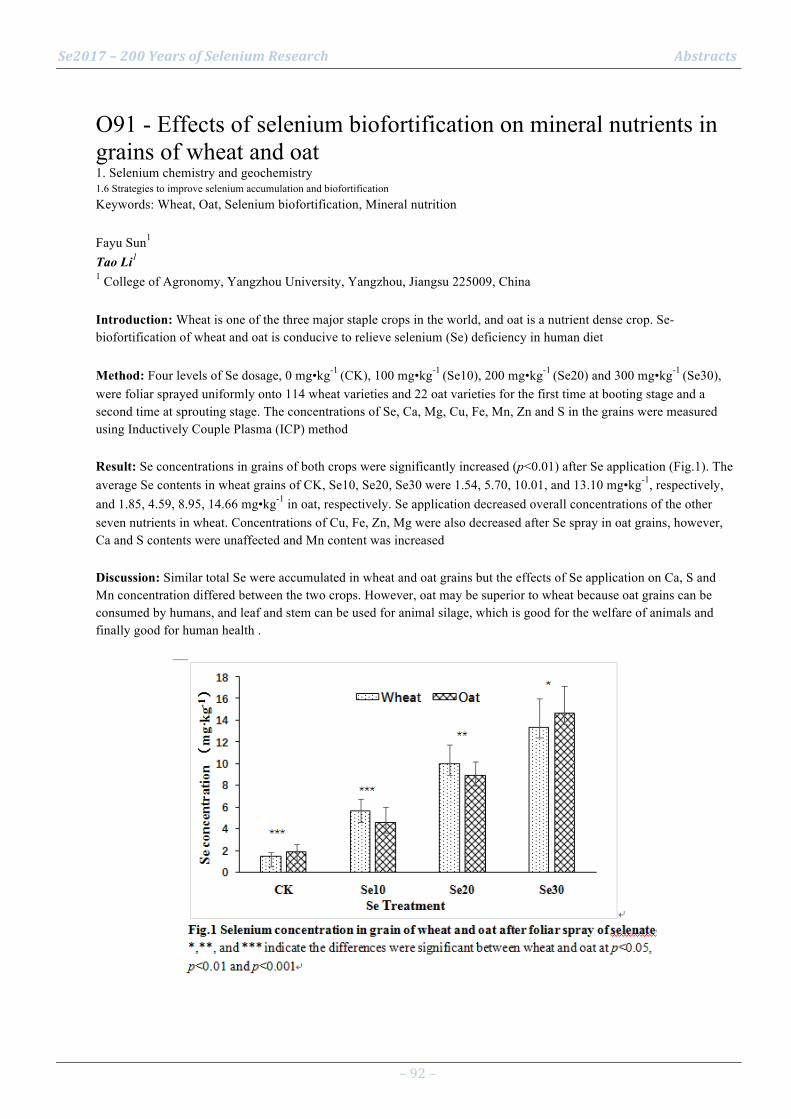
Se2017 – 200 Years of Selenium Research Abstracts
– 92 –
O91 - Effects of selenium biofortification on mineral nutrients in grains of wheat and oat 1. Selenium chemistry and geochemistry 1.6 Strategies to improve selenium accumulation and biofortification Keywords: Wheat, Oat, Selenium biofortification, Mineral nutrition Fayu Sun1 Tao Li1 1 College of Agronomy, Yangzhou University, Yangzhou, Jiangsu 225009, China Introduction: Wheat is one of the three major staple crops in the world, and oat is a nutrient dense crop. Se-biofortification of wheat and oat is conducive to relieve selenium (Se) deficiency in human diet Method: Four levels of Se dosage, 0 mg•kg-1 (CK), 100 mg•kg-1 (Se10), 200 mg•kg-1 (Se20) and 300 mg•kg-1 (Se30), were foliar sprayed uniformly onto 114 wheat varieties and 22 oat varieties for the first time at booting stage and a second time at sprouting stage. The concentrations of Se, Ca, Mg, Cu, Fe, Mn, Zn and S in the grains were measured using Inductively Couple Plasma (ICP) method Result: Se concentrations in grains of both crops were significantly increased (p<0.01) after Se application (Fig.1). The average Se contents in wheat grains of CK, Se10, Se20, Se30 were 1.54, 5.70, 10.01, and 13.10 mg•kg-1, respectively, and 1.85, 4.59, 8.95, 14.66 mg•kg-1 in oat, respectively. Se application decreased overall concentrations of the other seven nutrients in wheat. Concentrations of Cu, Fe, Zn, Mg were also decreased after Se spray in oat grains, however, Ca and S contents were unaffected and Mn content was increased Discussion: Similar total Se were accumulated in wheat and oat grains but the effects of Se application on Ca, S and Mn concentration differed between the two crops. However, oat may be superior to wheat because oat grains can be consumed by humans, and leaf and stem can be used for animal silage, which is good for the welfare of animals and finally good for human health .
Se2017 – 200 Years of Selenium Research Abstracts
– 93 –
O92 - Characterization on rhyzosphere bacteria from a selenium-hyperaccumulator Cardamine hupingshanesis 1. Selenium chemistry and geochemistry 1.6 Strategies to improve selenium accumulation and biofortification Keywords: Enshi, China; Cardamine hupingshanesis; Rhyzosphere Linxi Yuan1 Ying Zhang2, Ru Zhang3 1 Jiangsu Bio-Engineering Research Centre of Selenium, Suzhou 215123, Jiangus, China 2 University of Science and Technology of China, Hefei 230026, Anhui, China 3 Anhui Agricultural University, Hefei 230026, Anhui, China Introduction: In the Se-mine drainage area from Enshi, China, a novel Se hyperaccumulating plant, Cardamine hupingshanesis, was discovered in 2013, and the plant could accumulate Se up to 4414 mg/kg in the root dominated by selenocystine (SeCys2) (Yuan et al., 2013). Previous studies by Pilon-Smits and her team revealed that there were 24 Se-tolerant fungus strains in the rhizosphere of Stanleya Pinnata, a typical Se-hyperaccumulator, and those strains could survive in a medium with Se up to 10 mg/L (Wangeline et al., 2011) Method: High-performance 16S rRNA analysis was performed on rhyzosphere bacteria from C. hupingshanesis to demonstrate their roles on Se-hyperaccumulation by C. hupingshanesis. Result: Compared with the Se non-accumulating plants in study site, there have much higher contents of microorganisms in the rhyzosphere of C. hupingshanesis, and it was predominant by alpha-proteobacteria class (15-22 %), beta-proteobacteria class (10-16 %), actinobacteria class (10-18 %), acidobacteria class (8-15 %), delta-proteobacteria class (5-16 %). Moreover, some special microorganism with nitrospira class (2-5 %), gemmatimonadetes class (2-5 %), verrucomicrobiae class (2-4 %), planctomycetacia class (1-2 %) and others (opitutae class, sphingobacteria class, bacilli class, clostridia class) (3-4 %). Discussion: The diversity of rhyzosphere microorganisms from C. hupingshanesis was plenty and some of them could be Se-tolerant to relate with Se transformation from soil to plant roots
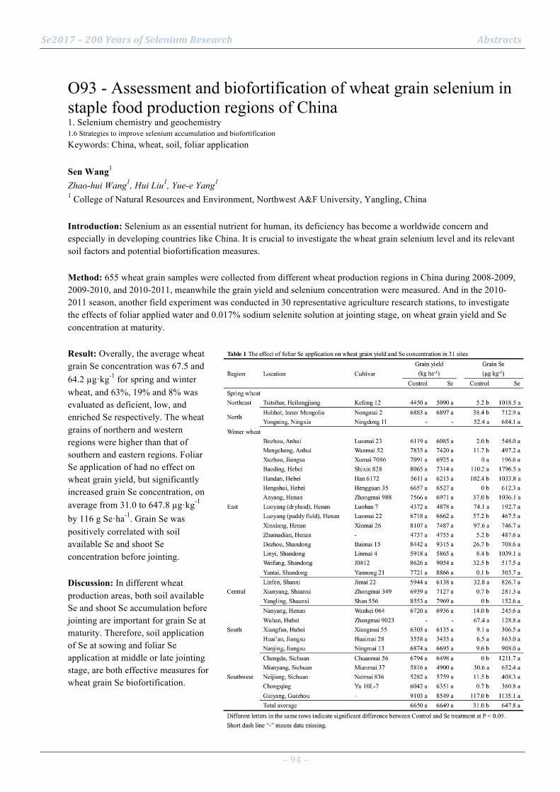
Se2017 – 200 Years of Selenium Research Abstracts
– 94 –
O93 - Assessment and biofortification of wheat grain selenium in staple food production regions of China 1. Selenium chemistry and geochemistry 1.6 Strategies to improve selenium accumulation and biofortification Keywords: China, wheat, soil, foliar application Sen Wang1 Zhao-hui Wang1, Hui Liu1, Yue-e Yang1 1 College of Natural Resources and Environment, Northwest A&F University, Yangling, China Introduction: Selenium as an essential nutrient for human, its deficiency has become a worldwide concern and especially in developing countries like China. It is crucial to investigate the wheat grain selenium level and its relevant soil factors and potential biofortification measures. Method: 655 wheat grain samples were collected from different wheat production regions in China during 2008-2009, 2009-2010, and 2010-2011, meanwhile the grain yield and selenium concentration were measured. And in the 2010-2011 season, another field experiment was conducted in 30 representative agriculture research stations, to investigate the effects of foliar applied water and 0.017% sodium selenite solution at jointing stage, on wheat grain yield and Se concentration at maturity. Result: Overally, the average wheat grain Se concentration was 67.5 and 64.2 µg·kg-1 for spring and winter wheat, and 63%, 19% and 8% was evaluated as deficient, low, and enriched Se respectively. The wheat grains of northern and western regions were higher than that of southern and eastern regions. Foliar Se application of had no effect on wheat grain yield, but significantly increased grain Se concentration, on average from 31.0 to 647.8 µg·kg-1 by 116 g Se·ha-1. Grain Se was positively correlated with soil available Se and shoot Se concentration before jointing. Discussion: In different wheat production areas, both soil available Se and shoot Se accumulation before jointing are important for grain Se at maturity. Therefore, soil application of Se at sowing and foliar Se application at middle or late jointing stage, are both effective measures for wheat grain Se biofortification.

Se2017 – 200 Years of Selenium Research Abstracts
– 95 –
O94 - Selenium supplemented kale and kohlrabi sprouts as possible ingredients for potent functional food. 1. Selenium chemistry and geochemistry 1.6 Strategies to improve selenium accumulation and biofortification Keywords: sprouts supplemented with selenium, antioxidant activity, phenolic compounds Paweł Zagrodzki1 Paweł Paśko2, Agnieszka Galanty3, Renata Wietecha-Posłuszny4, Małgorzata Tyszka-Czochara5, Pol Rubió6, Henryk Bartoń2, Maria Fołta2, Ewelina Prochownik2, Shela Gorinstein7 1 1) Institute of Nuclear Physics PAS; 2) Medical College, Jagiellonian University, Krakow, Poland. 2 Department of Food Chemistry and Nutrition, Medical College, Jagiellonian University, Krakow, Poland 3 Department of Pharmacognosy, Medical College, Jagiellonian University, Krakow, Poland 4 Department of Analytical Chemistry, Faculty of Chemistry, Jagiellonian University, Krakow, Poland 5 Department of Radioligands, Medical College, Jagiellonian University, Krakow, Poland 6 Faculty of Pharmacy and Food Science, The University of Barcelona, Barcelona, Spain 7 Institute for Drug Research, School of Pharmacy, Hebrew University, Jerusalem, Israel Introduction: The objective of this project was to examine biochemical properties of kale and kohlrabi sprouts supplemented with different doses of selenium. Method: The kale and kohlrabi sprouts were grown in EQMM-Easy-Green-Microfarm for 5-7 days, and watered with tap water or selenium supplemented water (10 mg/L, 15 mg/L, 30 mg/L). We determined in sprouts: selenium concentration (AFS-230 spectrometer); phenolic acids (Dionex HPLC), antioxidant activity (DPPH, FRAP) and total phenolic compounds (TP) (Synergy-2 reader). Result: Selenium was in the range 11-16 mg/g. Highest concentrations of sinapic acid were in kale sprouts supplemented with 15 or 30 mg/L Se (120.0±1.8 and 175.7±1.4 mg/100 g d.w., respectively). Chlorogenic acid occurred solely in kale sprouts and the highest concentration (10.3±0.1 mg/100 g d.w.) was for samples supplemented with 30 mg/L Se. Izo-chlorogenic acid was quantitatively determined only in samples supplemented with selenium, being one order of magnitude higher in kale than in kohlrabi sprouts (2349.8±448.1 vs. 383.1±116.9 mg/100 g d.w.), showing dose dependent positive relationship with selenium. The differences in TP followed the pattern of izo-chlorogenic acid. FRAP and DPPH showed slight increasing tendency following increase of selenium concentration, irrespectively of sprout species. Discussion: We measured the antioxidant potential of selenium-supplemented kale and kohlrabi sprouts and identified some candidates (among phenolic acids) for most biologically active compounds they contained, and determined the influence of selenium on their synthesis. Next stage of the study will include the assessment of the impact of the examined sprouts on the viability, proliferation and metabolic activity of human normal and cancer cells. Selected references
1. Pasko P., Gdula-Argasinska J., Podporska-Carroll J., et al. (2015). Influence of selenium supplementation on fatty acids profile and biological activity of four edible amaranth sprouts as new kind of functional food. J. Food Sci. Technol., 52, 4724-4736.
2. Tyszka-Czochara M., Pasko P., Zagrodzki P., et al. (2016). Selenium supplementation of amaranth sprouts influences betacyanin content and improves anti-inflammatory properties via NFΚB in murine RAW 264.7 macrophages. Biol. Trace Elem. Res., 169, 320-330.

Se2017 – 200 Years of Selenium Research Abstracts
– 96 –
O95 - Selenoproteins in hypothalamic energy homeostasis 2. Selenium in the molecular life sciences 2.6 Selenoprotein function Keywords: SELENOM, hypothalamus, leptin, energy metabolism Matthew Pitts1 Marla Berry2, Ting Gong2 1 University of Hawaii at Manoa 2 University of Hawaii Introduction: Leptin is a hormone secreted from adipose tissue that is a key regulator of energy metabolism. Upon binding to its cognate receptor in the hypothalamus, leptin activates cell signaling pathways that serve to decrease appetite and stimulate energy expenditure. One of the cardinal features of obesity is leptin resistance, in which increasing leptin levels fail to reduce feeding behavior and stimulate energy metabolism. The exact mechanism underlying hypothalamic leptin resistance is not entirely clear, but ER stress, inflammation, and impaired leptin transport across the blood-brain barrier have been implicated as contributing factors. Given the fact that selenoproteins are known to mitigate ER stress and inflammation, hypothalamic selenoproteins may protect against leptin resistance. Moreover, knockout studies in mice have shown that several selenoproteins (DIO2, GPX1, SELENOM, SELENOP, SELENOT) significantly impact energy homeostasis. Method: This lecture will discuss experimental evidence supporting the beneficial influence of hypothalamic selenoproteins on global energy metabolism. Result: Recent studies have shown that constitutive deletion of SELENOM and hypothalamic-specific knockout of selenocysteine-tRNA (Trsp) both lead to the development of obesity and hypothalamic leptin resistance in mice. Moreover, preliminary findings indicate that SELENOM promotes leptin signaling and Ca2+ homeostasis in an Agrp-expressing hypothalamic cell line (mHypoE-44). Discussion: These findings strongly suggest that selenoproteins promote leptin signaling in the hypothalamus.
Se2017 – 200 Years of Selenium Research Abstracts
– 97 –
O96 - Role of hypothalamic selenoprotein M in leptin signaling and calcium regulation 2. Selenium in the molecular life sciences 2.6 Selenoprotein function Keywords: selenoprotein M, leptin signaling, endoplasmic reticulum stress, calcium regulation, Hypothalamus Ting Gong1 Marla Berry1, Matthew Pitts1 1 University of Hawaii at Manoa, Department of Cell & Molecular Biology, Honolulu, USA Introduction: The prevalence of obesity is growing rapidly and has become an indisputable problem worldwide. One hallmark of obesity is hypothalamic leptin resistance, of which ER stress is a major contributing factor. Selenoprotein M (SELENOM) is an ER-resident thiol-disulfide oxidoreductase that is highly expressed in the brain. Our group has reported that deletion of SELENOM in mice results in elevated body weight, adiposity, and serum leptin levels, symptoms that are indicative of leptin resistance [1]. Moreover, SELENOM is highly expressed in the arcuate and paraventricular nuclei of the hypothalamus, two key regions involved in neural control of energy metabolism. In this study, we investigated the function of hypothalamic SELENOM using an immortalized hypothalamic cell line (mHypoE-44) that expresses agouti-related peptide, neuropeptide Y, and the leptin receptor. Method: Responses to leptin stimulation and ER stress were assessed by Western blotting in mHypoE-44 cells where SELENOM expression was manipulated. Further studies utilized Ca2+ imaging to assess changes in cytosolic Ca2+ flux in response to both leptin and ER stress. Result: Our studies show that SELENOM knockdown significantly attenuates leptin signaling in mHypoE-44 neurons, while overexpression promotes leptin-stimulated increase in pSTAT3, suggesting that SELENOM promotes leptin signaling in hypothalamus. Ca2+ imaging studies showed that SELENOM-depletion augmented cytosolic Ca2+ elevations in response to thapsigargin-induced ER stress, but mitigated leptin-induced reductions of cytosolic Ca2+ levels. Discussion: These results provide clear evidence that SELENOM has an important role in leptin signaling, ER stress, and Ca2+ regulation in hypothalamic neurons. This study may provide future therapeutic applications for the obese populations. Selected references
Pitts MW, Reeves MA, Hashimoto AC, Ogawa A, Kremer P, et al. (2013) Deletion of selenoprotein M leads to obesity without cognitive deficits. J Biol Chem 288: 26121-26134.

Se2017 – 200 Years of Selenium Research Abstracts
– 98 –
O97 - Exploring Selenoprotein N structure and function 2. Selenium in the molecular life sciences 2.6 Selenoprotein function Keywords: Selenoprotein N, endoplasmic reticulum, membrane protein topology, structure function analysis Alain Lescure1 Mickaël Briens2, Vanessa Dacleu-Siewe1, Melanie Braye-Thami1, Luc Thomès1, Mireille Baltzinger1 1 University of Strasbourg, CNRS, Architecture and Reactivity of RNA, Strasbourg, France 2 Adisseo, Commnetry, France Introduction: The Selenoprotein N (SelenoN) is a membrane glycoprotein localized in the endoplasmic reticulum (ER) and it is ubiquitously expressed in all tissues. However, mutations in the SELENON gene, which encodes SelenoN, leads to a selective set of muscular diseases (SELENON-related myopathies), which suggests that, despite its widespread expression, SelenoN is particularly important for molecular processes relevant to muscle development and maintenance. At the molecular level, the presence of a thioredoxin reductase like motif in the core of the protein indicates a redox function, but its catalytic activity, including its substrate(s) and co-factor(s), remains unknown. Method: We are conducting structural and molecular studies to address the SelenoN activity and to analyze this activity in the context of cellular metabolic or signaling pathways, such as ER oxidative stress control or calcium homeostasis. Result: We characterized the topology of SelenoN within the ER membrane, and showed the importance of its post-translational modifications. In parallel, we have developed eukaryotic expression systems for the production and purification of recombinant protein to be used in crystallographic studies and in vitro enzymatic assays. At the cellular level, we have screened for conditions controlling the transcription of the SELENON gene, and using SelenoN depleted models, we showed that the unfolded protein response, one important activity within the ER, is not affected by SelenoN loss of function. Discussion: A better understanding of the precise SelenoN enzymatic activity is fundamental to deciphering the pathogenesis of SELENON-related myopathies.
Se2017 – 200 Years of Selenium Research Abstracts
– 99 –
O98 - Disruption of cancer cell redox homeostasis promoted by S-nitrosylation of thioredoxin reductase 2. Selenium in the molecular life sciences 2.6 Selenoprotein function Keywords: Nitrosylation; Thioredoxin reductase; Redox regulation; Cell death Moran Benhar1 Rotem Engelman1, Tamar Ziv1, Elias Arnér2 1 Technion, Haifa, Israel 2 Karolinska Institutet, Stockholm, Sweden Introduction: Nitric oxide (NO)-induced signaling and cytotoxic responses are mediated in part by S-nitrosylation of protein cysteine residues, to form S-nitrosothiols (SNOs). Protein denitrosylation is primarily carried out by glutathione (GSH), thioredoxin (Trx), and their associated redox systems. SNO reduction by GSH and Trx is increasingly implicated in the regulation of NO/SNO-mediated cellular signaling and in protection from SNO-related cellular stress. Method: We used biochemical and cellular approaches to study the effects of NO/SNO donors on recombinant and endogenous Trx reductase (TrxR). Result: NO/SNO donating agents rapidly and effectively inhibited the activity of recombinant rat TrxR1. In particular, the NADPH-reduced TrxR1 was partially and reversibly inhibited upon exposure to low micromolar concentrations of S-nitrosocysteine and markedly and continuously inhibited at higher doses. Further analyses indicated that its active site selenocysteine residue renders TrxR1 highly susceptible to nitrosylation-mediated inhibition, and revealed both thiol and selenol modifications at the two redox active centers of the enzyme. Studies in HeLa cancer cells further confirmed the susceptibility of TrxR to inhibitory nitrosylation. Notably, depletion of cellular glutathione with L-buthionine-sulfoximine (BSO) synergized with nitrosating agents in promoting rapid and sustained nitrosylation and inactivation of TrxR. These events were accompanied by significant oxidation of Trx1, activation of multiple cellular stress pathways, and induction of cell death. Discussion: Our findings uncover novel aspects of the interplay between NO and the Trx and GSH redox systems. The observations suggest the utility of BSO/NO combination therapy for inhibition of tumor growth through the disruption of cancer cell NO-redox homeostasis.
Se2017 – 200 Years of Selenium Research Abstracts
– 100 –
O99 - Influence of small intestinal thioredoxin and thioredoxin reductase on intestinal permeability 2. Selenium in the molecular life sciences 2.6 Selenoprotein function Keywords: small intestine, thioredoxin, thioredoxin reductase Jinglin Li1 Xiaolin Zhang2, Fei Ye2, Liangwei Zhong1 1 Medical School, University of Chinese Academy of Sciences, Beijing, China 2 Institute of Materia Medica, Chinese Academy of Medical Sciences & Perking Union Medical College, Beijing, China Introduction: Obesity is associated with increased intestinal permeability, but the underlying mechanism remains largely unclear. Here we show that the development of high-fat diet-induced obesity in mice was accompanied by a decrease in small intestinal thioredoxin (Trx) and its reductase (TrxR). Method: Although Trx and TrxR are components of Trx system that participates in many cellular processes, the relationship between Trx system and intestinal permeability remains unknown. Thus, we made two model intestinal epithelial cells with increased or decreased activity of Trx system via selenium supplementation or Trx-knocking down. Result: Comparison of these model cells showed distinct patterns. Na+-K+ATPase α-subunit is a crucial determinant in regulating intestinal permeability. In the cells with lower activity of Trx system, Na+-K+ATPase α-subunit expression was lower, but its phosphorylation level was higher, consisting with a decrease in Na+-K+ATPase activity. In the same cells, the increased activity of protein kinase Cα and decreased activity of protein phosphatase-2A were in favor of Na+-K+ATPase phosphorylation. In addition, the cells with decreased activity of Trx system had higher level of reactive oxygen species, abnormal shape, lower level of Zonula Occludens-1 and increased transepithelial transport. Discussion: The observed differences reveal a previous unrecognized role of Trx/TrxR in regulating intestinal permeability.

Se2017 – 200 Years of Selenium Research Abstracts
– 101 –
O100 - The intricate role of selenoproteins in stress erythropoiesis 3. Selenium in animal and human health and disease 3.5 Selenium metabolism or selenoprotein function in health and disease Keywords: Erythropoiesis, proerythroblasts, anemia, erythroblastic islands, selenoproteins Chang Liao1 Ross Hardison2, Mary Kennett1, Bradley Carlson3, Robert Paulson1, K. Sandeep Prabhu1 1 Dept of Veterinary & Biomedical Science, The Pennsylvania State University, University Park, PA, USA 2 Dept of Biochemistry & Mol Biology, The Pennsylvania State University, University Park, PA, USA 3 Molecular Biology of Selenium Section, MCGP, National Cancer Institute, Bethesda, MD, USA Introduction: Se, in the form of selenoproteins, functions as a cellular redox gatekeeper to modulate a wide array of physiological processes. Stress erythropoiesis is a systemic response to hypoxia stress that involves the rapid generation of new erythrocytes. Our data show that Se and selenoproteins impacts stress erythropoiesis at several levels. Method: Acute anemia model was induced by phenylhydrazine administration. Microarray was applied for transcriptomic analysis. Flow cytometry was used for phenotypic study. Result: Se deficiency severely impairs stress erythropoiesis and compromises the response to acute anemia in mice. Se deficient animals fail to expand stress erythroid progenitors in the spleen and exhibit a defect in the differentiation of proerythroblasts to basophilic erythroblasts during terminal maturation. Molecular analysis showed that Se deficiency alters the progression of terminal erythroid differentiation leading to augmented cleavage of transcription factor GATA1 and defects in enucleation. Transcriptomic analysis revealed significant differences in proerythroblasts as a function of Se status. In particular, the expression of selenoprotein W (SELENOW) was decreased in Se-deficient cells suggesting that it may play a role in the terminal differentiation of stress erythroid progenitors. Se deficiency also decreased development of erythroblastic islands, which form the erythropoietic niche. To further demonstrate a role for selenoproteins in stress erythropoiesis, mice mutant for tRNA[Ser]Sec (Trsp) exhibited impaired stress erythropoiesis and delayed recovery from anemia. Discussion: Our data suggest a fundamental role for selenoproteins in supporting effective stress erythropoiesis and that adequacy of Se is required throughout the recovery process in the erythropoietic niche. Selected references NIH R01-DK077152 (KSP) and R01-DK080040 (RFP) and supported by USDA National Institute of Food and Agriculture Hatch Project #4736 (R.F.P.) and 4605 (K.S.P)

Se2017 – 200 Years of Selenium Research Abstracts
– 102 –
O101 - Selenoproteins restrict the replication of Francisella tularensis in macrophages. 3. Selenium in animal and human health and disease 3.5 Selenium metabolism or selenoprotein function in health and disease Keywords: bacteria, infection, selenoprotein, autophagy Rachel Markley1 David Williamson1, Bhuvana Katkere1, Kalyan Dewan1, David Place1, Sarah Sumner1, Bradley Carlson2, K Sandeep Prabhu1, Girish Kirimanjeswara1 1 The Pennsylvania State University, University park, PA 16802, USA 2 NIH, Bethesda, MD, USA Introduction: The micronutrient selenium (Se) is known to regulate immune functions via selenoproteins. However, the mechanisms by which selenoproteins regulate immune functions during an acute infection are not clear. Therefore, we investigated the role of macrophage (Mac) selenoproteins during an acute bacterial infection. Method: Francisella tularensis (Ft.), the causative agent of tularemia, is a gram-negative intracellular bacterium. Since Ft. infects and replicates primarily in Macs, we measured the bacterial growth in Macs derived from TrspM mice that are unable to synthesize selenoproteins. The disease progression was monitored in wild-type (WT) and TrspM mice to determine the influence selenoproteins in an acute infection. Since Ft. growth is closely associated with autophagy, we investigated the kinetics of autophagy in infected cells by western blot. Result: Macs maintained on deficient level of Se had a ~50 fold higher bacterial numbers than the Macs maintained on adequate or supplemented levels of Se. Similarly, TrspM Macs had a significantly higher growth of Ft. compared to WT Macs. Moreover, TrspM mice were more susceptible to Ft. infection and harbored significantly higher levels of bacteria in their livers and spleens as compared to WT mice. Our data also indicate that selenoproteins inhibit autophagy in Macs, which may limit the availability of nutrients to the bacteria. Discussion: These data indicate that Mac selenoproteins are essential for restricting bacterial growth and promoting host survival. Our studies demonstrate that dietary Se affect the outcome of an infection by influencing host immune response, and reveal potential novel targets for antibacterial therapies.

Se2017 – 200 Years of Selenium Research Abstracts
– 103 –
O102 - Se-dietary matrices can upregulate the anti-inflammatory responses in RAW macrophages 3. Selenium in animal and human health and disease 3.5 Selenium metabolism or selenoprotein function in health and disease Keywords: wheat, rMETase, inflammation, LPS Noorpreet Dhanjal1 Siddharth Sharma1, Sandeep Prabhu Kumble2, Tejo Prakash Nagaraja3 1 Department of Biotechnology, Thapar University, Patiala, India 2 Department of Veterinary & Biomedical Sciences,The Pennsylvania State University, State College, USA 3 School of Energy and Environment, Thapar University, Patiala, India Introduction: Dietary Se supplementation downregulates expression of proinflammatory genes. However, the extent of Se bioavailability from dietary matrices such as Se-rich wheat, and its impact on inflammation is not known. Method: We tested the ability of Se-rich wheat extracts to impact the pathways of inflammation and pro-resolution in RAW264.7 macrophages stimulated with bacterial endotoxin lipopolysaccharide (LPS). Cells were supplemented with Se in the form of sodium selenite (SS), seleniferous wheat extract (SeW) and seleniferous wheat extract treated with rMETase (SeW+rMET) in three increasing concentrations (500nM>100nM>50nM). The expression of GPx-1, COX-2, HPGDS, mPGES-1, iNOS were examined in qPCR or western blot analyses. Result: Cells supplemented with 500 nM SS and SeW+rMET showed 9.0 and 8.0 fold increase in GPx-1 expression, respectively, as compared to SeW treated cells (1.1 fold) at 4h of LPS incubation. rMETase pretreatment increased the bio-availability of Se from wheat matrices and showed effects equivalent to SS supplementation. 500nM SeW+rMET downregulated the LPS-induced expression of Cox-2, mPges-1 and iNos up to 4.0, 12.0 and 10.0 fold compared to Se deficient cells, respectively, at both mRNA and protein levels. SeW+rMET treatment significantly upregulated (4.4 fold) the expression of HPGDS, which produces anti-inflammatory and pro-resolving eicosanoid 15-deoxy-prostaglandin J2. Discussion: Our studies show that treatment of seleniferous wheat extracts with rMetase increases the bioavailability of Se to downregulate pathways of inflammation, while increasing the pro-resolution responses to effectively mitigate inflammation. These results highlight the potential application of selenium supplementation through dietary matrices to alleviate diseases where inflammation contributes to the underlying pathology. Selected references Funding 38(1341)/12/EMRII (NTP); NIH DK 077152; CA 162665 (KSP)

Se2017 – 200 Years of Selenium Research Abstracts
– 104 –
O103 - Selenium in human and vertebrate evolution 2. Selenium in the molecular life sciences 2.7 The systems biology of selenium and selenoproteins Keywords: selenium deficiency, adequacy, toxicity, adaptation Sergi Castellano1 1 Max Planck Institute for Evolutionary Anthropology, Leipzig, Germany Introduction: Selenium, a sparse element on earth, is an essential micronutrient in the diet of humans and other vertebrates and its intake depends on its content in the soils and waters across the world. Selenium is required due to its function in selenoproteins, which contain the amino acid selenocysteine (the 21st amino acid in the genetic code) as one of their constituent residues. Selenocysteine is analogous to the amino acid cysteine, which uses the abundant element sulfur in place of selenium. Despite the irregular and often scant distribution of selenium in the world, the distinct biochemical properties of selenium have made the substitution of selenocysteine for cysteine rare in vertebrate proteins. Still, vertebrate species have lived in environments with different amounts of selenium and may have adapted in different ways to it. Method: Using comparative and population genomics approaches, we investigate whether selenoprotein genes and genes involved in the metabolism of selenium have genetic signatures compatible with adaptive evolution. Result: Fishes have genetic signatures compatible with adaptation to the variable but generally abundant selenium in waters throughout the world, whereas humans have genetic signatures compatible with adaptation to the varying but generally scarce selenium in lands across the world. In both cases, the adaptive signatures are shared among genes that use (selenoproteins) or regulate selenium, suggesting that it is the overall metabolism and homeostasis of selenium that adapts to its worldwide variation. Discussion: Dietary selenium has thus distinctly shaped the evolution of vertebrates.
Se2017 – 200 Years of Selenium Research Abstracts
– 105 –
O104 - Evolution of selenoproteins across the tree of life 2. Selenium in the molecular life sciences 2.7 The systems biology of selenium and selenoproteins Keywords: Evolution, genomics, SECIS, SPS, Archaea Marco Mariotti1 Didac Santesmasses2, Alexei V. Lobanov1, Bruno Manta1, Andreu Bofill2, Montserrat Corominas3, Toni Gabaldón2, Roderic Guigó2, Vadim N. Gladyshev1 1 Brigham and Women’s Hospital, Harvard Medical School, Boston, MA, USA 2 Centre for Genomic Regulation (CRG), Barcelona, Spain 3 Department of Genetics, Microbiology and Statistics, School of Biology, University of Barcelona Introduction: Rapid technological advances have brought an unprecedented wealth of sequences from organisms across the whole tree of life. Through the lens of computational genomics, these resources can provide insights about biological processes at evolutionary timescales. When applied to the biology of selenium, these studies can outline the evolution of selenoprotein genes, elucidate the origin of the selenocysteine pathway, and delineate new roles for its components. Method: In the last few years we developed several tools for the computational identification of selenoproteins, SECIS elements and Sec machinery genes [1-3]. We applied these methods for evolutionary studies at diverse scales, from a relatively recent history [4] to the origin of Life itself [5,6]. Result: Through the study of SPS proteins (selenophosphate synthetases), we produced the first portrait of selenium utilization traits encompassing all sequenced organisms at once [5]. We uncovered a peculiar process of gene duplication/subfunctionalization explaining the multiple appearances of SPS1 proteins in metazoans. We also discovered selenoproteins in Lokiarchaeota, an archaeal lineage related to the first eukaryotes [6], which shed light on the origin of the eukaryotic Sec encoding system. Despite having typical archaeal selenoproteins, Lokiarchaeota possess SECIS elements strikingly similar to those of eukaryotes, but exhibit no obvious SBP2 partner. We traced back this proto-eukaryotic structure to the archaeal SECIS of one specific selenoprotein, which shows its core key motif in methanogenic archaea. Discussion: Our studies provide broad insights on selenium utilization and selenoprotein function, and frame them in the context of evolutionary processes at large. Selected references 1. Mariotti M, Guigó R. 2010. Selenoprofiles: profile-based scanning of eukaryotic genome sequences for selenoprotein genes. Bioinformatics 26 (21), 2656-2663. 2. Mariotti M, Lobanov AV, Guigó R, Gladyshev VN. 2013. SECISearch3 and Seblastian: new tools for prediction of SECIS elements and selenoproteins. Nucleic acids research 41 (15), e149-e149. 3. Santesmasses D, Mariotti M, Guigó R. 2017. Computational identification of the selenocysteine tRNA (tRNASec) in genomes. PLOS Computational Biology 13 (2), e1005383 4. Mariotti M, Ridge PG, Zhang Y, Lobanov AV, Pringle TH, et al. 2012. Composition and evolution of the vertebrate and mammalian selenoproteomes. PLoS One 7 (3), e33066. 5. Mariotti M, Santesmasses D, Capella-Gutierrez S, Mateo A, Arnan C, et al. 2015. Evolution of selenophosphate synthetases: emergence and relocation of function through independent duplications and recurrent subfunctionalization. Genome Research 25 (9), 1256–1267. 6. Mariotti M, Lobanov AV, Manta B, Santesmasses S, Bofill A, et al. 2016. Lokiarchaeota marks the transition between the archaeal and eukaryotic selenocysteine encoding systems. Mol Biol Evol 33 (9): 2441-2453.

Se2017 – 200 Years of Selenium Research Abstracts
– 106 –
O105 - The human selenomicrobiome 2. Selenium in the molecular life sciences 2.7 The systems biology of selenium and selenoproteins Keywords: human microbiome, selenium utilization, selenoprotein prediction, bSECIS Didac Santesmasses1 Marco Mariotti2, Alexis Grimaldi1, Roderic Guigó1 1 Centre for Genomic Regulation (CRG), Barcelona, Spain 2 Brigham and Women’s Hospital, Harvard Medical School, Boston, MA USA Introduction: The human microbiota comprises bacteria, archaea, viruses, and microbial eukaryotes that play fundamental roles in human health and disease. Selenium is an essential trace element for humans and for organisms inhabiting our body. For the first time, we characterized the composition and distribution of selenoproteins and other selenium utilization forms in the human microbiome. Method: We analyzed whole metagenome assemblies from the Human Microbiome Project (HMP)[1]. The HMP provides a catalog of the microbial communities across five body areas from a large cohort of healthy adult individuals. By applying computational methods developed in our group, we identified and quantified selenoproteins and other genetic markers of selenium utilization. Result: We found that selenoprotein genes are commonly present in the bacterial microbiota throughout the entire human body. Selenoproteins are remarkably abundant in the oral cavity, and are depleted in stool samples, where the usage of selenium as cofactor is surprisingly more spread than selenocysteine. By applying our new method bSeblastian, we also identified novel bacterial selenoproteins. Discussion: The effect of selenium in human health cannot be fully understood without taking into account the human microbiome. Our results provide a profile of selenium utilization by the microbiota inhabiting our body. Selenium availability might be important for the regulation, not only of the selenoproteins encoded in our genome, but also for those encoded in our microbiome. Selected references
The Human Microbiome Project Consortium (2012) Structure, function and diversity of the healthy human microbiome. Nature 486:207–14. doi: 10.1038/nature11234

Se2017 – 200 Years of Selenium Research Abstracts
– 107 –
O106 - Selenoprotein extinction in Drosophila occurred concomitantly to genome catastrophes 2. Selenium in the molecular life sciences 2.7 The systems biology of selenium and selenoproteins Keywords: Drosophila, selenoprotein loss, genome sequencing, comparative genomics, evolution Didac Santesmasses1 Marco Mariotti2, Daeyeon Kim1, Marina Ruiz-Romero1, Salvador Capella-Gutierrez1, Sílvia Pérez-Lluch1, Anna Vlasova1, Miquel Calvo3, Toni Gabaldón1, Marta Morey3, Montserrat Corominas3, Roderic Guigó1, Matthieu Louis1 1 Centre for Genomic Regulation (CRG), Barcelona, Spain 2 Brigham and Women’s Hospital, Harvard Medical School, Boston, MA USA 3 Department of Genetics, Microbiology and Statistics, University of Barcelona, Barcelona, Spain Introduction: Drosophila willistoni was the first animal discovered to lack selenoproteins [1]. The three selenoproteins of D. melanogaster were either lost or converted to cysteine homologs in D. willistoni, as the selenoprotein synthesis machinery degenerated. Other selenoproteinless animals have been identified [2,3,4,5], but little is known about the evolutionary path leading to the complete loss of selenocysteine. Method: We sequenced eight Drosophila genomes from the saltans group, the sister lineage of D. willistoni, and performed RNAseq in ten Drosophila species. Using a multispecies design, we further conducted various functional experiments at diverse levels, ranging from biochemical to behavioural assays. Result: From the newly sequenced genomes, we identified three additional selenoprotein extinction events in Drosophila. Although independent, these events happened in a single lineage: saltans/willistoni. Interestingly, various other genomic features set saltans/willistoni apart from the rest of Drosophila (most remarkably their lower GC content). Based on comparative transcriptomics and other observations, we hypothesized that saltans/willistoni underwent an ancestral event of adaptation to hypoxia, setting in motion a “genome catastrophe” that triggered, among other effects, the eventual loss of selenoproteins. We show various lines of evidence supporting our hypothesis, including assays in which saltans/willistoni species exhibit phenotypes similar to a D. melanogaster strain artificially adapted to hypoxic conditions [6]. Discussion: By high-throughput sequencing and comparative analyses, we delineated the evolutionary events leading to selenocysteine losses in Drosophila, and we propose a comprehensive explanation involving major adaptive changes occurred at the root of the saltans/willistoni lineage. Selected references Clark AG, Eisen MB, Smith DR, et al (2007) Evolution of genes and genomes on the Drosophila phylogeny. Nature 450:203–18. doi: 10.1038/nature06341 Chapple CE, Guigó R (2008) Relaxation of selective constraints causes independent selenoprotein extinction in insect genomes. PLoS One 3:e2968. doi: 10.1371/journal.pone.0002968 Lobanov A V, Hatfield DL, Gladyshev VN (2008) Selenoproteinless animals: selenophosphate synthetase SPS1 functions in a pathway unrelated to selenocysteine biosynthesis. Protein Sci 17:176–82. doi: 10.1110/ps.073261508 Mariotti M, Santesmasses D, Capella-Gutierrez S, et al (2015) Evolution of selenophosphate synthetases: emergence and relocation of function through independent duplications and recurrent subfunctionalization. Genome Res 25:1256–67. doi: 10.1101/gr.190538.115 Otero L, Romanelli-Cedrez L, Turanov AA, et al (2014) Adjustments, extinction, and remains of selenocysteine incorporation machinery in the nematode lineage. RNA 20:1023–34. doi: 10.1261/rna.043877.113 Perkins G, Hsiao Y, Yin S, et al (2012) Ultrastructural modifications in the mitochondria of hypoxia-adapted Drosophila melanogaster. PLoS One 7:e45344. doi: 10.1371/journal.pone.0045344
Se2017 – 200 Years of Selenium Research Abstracts
– 108 –
O107 - Metabolomics of selenium 2. Selenium in the molecular life sciences 2.9 Additional and emerging topics of selenium in molecular life science Keywords: selenoproteins, metabolite profiling, lifespan, miRNA Sun Hee Yim1 Clary Clish2, Vadim Gladyshev1 1 BWH/Harvard Medical School 2 Broad Institute of MIT and Harvard Introduction: Selenium is an essential trace element in mammals due to its presence in 25 selenoproteins in the form of selenocysteine residue. Both selenium deficiency, which leads to compromised selenoprotein functions, and high levels of selenium, resulting in selenium toxicity, have been associated with oxidative stress and adverse health effects, although molecular mechanisms are not fully understood. Method: Here, we subjected mice to diets spanning selenium deficiency and toxicity and examined global changes in metabolism using metabolite profiling and miRNA assays. Long-term effects of dietary selenium were also investigated. Result: Most significant changes in metabolite patterns were associated with selenium deficiency, which altered redox homeostasis, reduced the levels of free amino acids, and affected miRNA expression. Dietary selenium more strongly affected liver metabolism than brain metabolism. Discussion: These changes, along with the activation of metabolic pathways associated with xenobiotic metabolism and redox homeostasis, were consistent with a pro-longevity state induced by selenium deficiency. Indeed, both male and female mice subjected to selenium deficiency for their entire adult life, while dramatically reducing selenoprotein expression, did not shorten lifespan. Thus, selenium deficiency, while associated with adverse health effects, is compensated for by pro-longevity mechanisms supporting normal lifespan of mice.

Se2017 – 200 Years of Selenium Research Abstracts
– 109 –
O108 - Selenium-encoded chemical proteomics 2. Selenium in the molecular life sciences 2.7 The systems biology of selenium and selenoproteins Keywords: chemical proteomics, selenoprotein, activity-based probes, selenocysteine Jinjun Gao1 Chu Wang1 1 Peking University, China Introduction: Selenium is one of the indispensable trace elements for human health and its dominant form in human body is selenocysteine, which serves as critical active-site residues in selenoproteins in regulating redox balance. Selenium excess and deficiency are both implicated with severe diseases. However, it remains challenging to profile selenoproteins by traditional shotgun proteomics tools due to its low abundance and versatile activity states. Method: We have developed a computational program to detect the characteristic isotope envelope of selenium-containing peptides from complex proteomic data and use these information to guide the proteomic analysis in a targeted mode. With a selenium-encoded enrichment tag, this method can be seamlessly combined with current chemical proteomic technologies using activity-based or metabolic analog probes to profiling sites of post-translational modifications. Result: We showed that the targeted analysis can dramatically improve the sensitivity of detecting the sites of post-translational modification or probe adduction, as well as to improve identification of natural selenoproteins in cellular and tissue proteomes. The method could also be applied to aid target analysis of selenium-containing metabolites. Discussion: Our selenium-encoded chemical proteomic strategy will be a novel tool and great resource for in-depth proteomic analysis of selenoproteins and other selenium-containing biomolecules, which will aid functional studies of selenoproteins and their implications in human health.
Se2017 – 200 Years of Selenium Research Abstracts
– 110 –
O109 - Characterization of Atlantic salmon (Salmo salar) selenoproteins using bioinformatics and hyphenated 2. Selenium in the molecular life sciences 2.7 The systems biology of selenium and selenoproteins Keywords: selenoproteins, Atlantic salmon, mass spectrometry, bioinformatics, hyphenated techniques Veronika Sele1 Magali Perez2, Andrea Raab3, Joerg Feldmann3, Eva Krupp3, Jens J. Sloth4, Heidi Amlund1, Robin Ørnsurd1, Marc H.G. Berntssen1, Josef D. Rasinger1 1 1. National Institute of Nutrition and Seafood Research, PO Box 2029 Nordnes, 5817 Bergen, Norway 2 2. University of Aberdeen, Department of Chemistry, Old Aberdeen, AB, 24 3UE, Scotland, UK 3 2. University of Aberdeen, Department of Chemistry, Old Aberdeen, AB, 24 3UE, Scotland, UK. 4 3. National Food Institute, Technical University of Denmark, 2800 Kgs. Lyngby, Denmark Introduction: Selenium is an essential element, important for the function of several selenoproteins. The number of selenoproteins may vary between different animal species. For example, compared to vertebrates, teleost fish such as Atlantic salmon (Salmon salar) feature a high number of selenoproteins. The biological functions of many of these selenoproteins are yet to be elucidated. With the recently published Atlantic salmon genome (Lien et al. 2016) it has become possible to predict selenoproteins using bioinformatics tools. However, a pure in silico description of the Atlantic salmon selenoproteome is challenging. The selenoproteins’ key amino acid, selenocysteine, is coded for by messenger RNA (mRNA) comprising the exact same nucleic acid triplet (TGA) that signals the termination of translation into proteins. This may lead to selenproteins being missannotated or completely missed. Therefore, the validation of the theoretical prediction through analytical data is essential. Method: Recently, hyphenated analytical techniques, which employ laser ablation coupled to inductively coupled plasma mass spectrometry (LA-ICP-MS) and high performance liquid chromatography (HPLC) coupled to both ICP-MS and high-resolution tandem mass spectrometry (HR-MS) to analyse selenoproteins in human cells (Bianga et al. 2014). In the present study, a similar methodological approach for the analysis of the salmon liver selenoproteome was implemented. Result: By combining the theoretical genome derived bioinformatics data with the results from the analytical techniques, we expect to obtain a phenotypically anchored description of the salmon selenoproteome. Discussion: These findings will ultimately contribute to the advancement of our understanding of selenium biology in marine organisms Selected references
Bianga, J., Z. Touat-Hamici, et al. (2014). "Speciation analysis for trace levels of selenoproteins in cultured human cells." Journal of Proteomics 108: 316-324.
Lien, S., B. F. Koop, et al. (2016). "The Atlantic salmon genome provides insights into rediploidization." Nature 533(7602): 200-+.
Se2017 – 200 Years of Selenium Research Abstracts
– 111 –
O110 - The cellular location of selenoproteins in human prostatic tissue and their role in prostate cancer. 3. Selenium in animal and human health and disease 3.5 Selenium metabolism or selenoprotein function in health and disease Keywords: prostate, cancer, SELENOF, GPX1, SBP1 Dede Ekoue1 Emmanuel Ansong1, Wancai Yang2, Peter Gann1, Andre Kajdacsy-Balla1, Alan Diamond1 1 University of Illinois, Chicago, United States 2 Jining Medical University, Jining, China and University of Illinois, Chicago, United States Introduction: Two selenocysteine-containing proteins, SELONOF and GPX1, and a non-selenocysteine-containing protein that binds selenium, SBP1, have been implicated in cancer etiology. Given human epidemiological data indicating an inverse association between selenium intake and prostate cancer risk, these proteins were imaged in human cell lines and prostate tissue obtained from men who underwent radical prostatectomy. Method: Specificity of anti-SELENOF, -GPX1 and -SBP1 antibodies was verified by western blot analysis as well as by peptide blocking. Proteins were visualized by either confocal microscopy of selected cell lines or immunohistochemistry of human prostate tissue microarrays obtained from the Cooperative Prostate Cancer Tissue Resource. Result: SBP1 was localized to the nucleus and cytoplasm in prostatic tissues, with the nuclear/cytoplasm ratio being inversely associated with tumor grade, while lower levels of SBP1 were associated with recurrence (OR 0.38, 95% CI 0.20-0.72)1. SELENOF was localized to the endoplasmic reticulum in breast and prostate cancer cell lines, but principally localized to the outer cell membrane in benign prostate tissue, as well as in immortalized or primary human prostate cultured cells. There was dramatically lower levels in benign tissue vs. tumor (p=1x10⁻⁷). GPX1 was primarily localized to the cytoplasm and mitochondria in all cells examined, although a small of number of cells exhibited nuclear localization that occasionally segregated asymmetrically in dividing cells Discussion: These data indicate that cellular localization and abundance should be considered when trying to understand the role these proteins play in prostate cancer. Selected references 1Ansong et al. PLoS One 10:e 0126285, 2015

Se2017 – 200 Years of Selenium Research Abstracts
– 112 –
O111 - Dietary Selenium Deprivation Oppositely Impacts Longevity and Healthspan in Telomere Dysfunctional Mice 3. Selenium in animal and human health and disease 3.5 Selenium metabolism or selenoprotein function in health and disease Keywords: Aging, Genome stability, metabolism, hormesis, selenoprotein Wen-Hsing Cheng1 1 Mississippi State University Introduction: Selenium (Se), a trace metalloid essential for life, plays nutritional and physiological roles mainly through selenoproteins. While telomere attrition provokes DNA damage response and replicative senescence, such an aging process is restricted in mice due to their comparatively long telomeres. Method: To circumvent this limitation and employ a mouse model with humanized telomeres, weanling third generation telomerase RNA component knockout mice carrying short telomeres were fed a Se-deficient, Torula-yeast basal diet or the diet supplemented with 0.15 ppm Se as sodium selenate throughout their life. Result: Dietary Se deprivation delayed wound healing and accelerated a wide range of age-related degenerations including osteoporosis, grey hair, alopecia, cataract, and hyperglycemia. Surprisingly, long-term dietary Se deprivation paradoxically promoted longevity in both sexes. Plasma microRNA profiling revealed a circulating signature of Se deprivation and aging, and subsequent ontological analyses predicted dominant changes in metabolism. Consistent with this, dietary Se deprivation accelerated age-dependent declines in glucose tolerance, glucose-stimulated insulin production and insulin sensitivity, as well as DNA damage and senescence responses in the pancreas. Selenotranscriptomic and metagenomic analyses identified key selenoprotein mRNAs and gut bacteria in the response to dietary Se deprivation in males and females at old age. Discussion: These results suggest a novel model of aging with conceptual advances, whereby Se at low or nutritional levels of intake may be considered a hormetic chemical and decouple healthspan and longevity. Selected references Wu et al., Aging Cell, 2017, 16:125-135; Wu et al., J. Biol. Chem., 2014, 289:34378-34388.

Se2017 – 200 Years of Selenium Research Abstracts
– 113 –
O112 - Selenium and Cataract 3. Selenium in animal and human health and disease 3.5 Selenium metabolism or selenoprotein function in health and disease Keywords: SelR, Sep15, cataract, endoplasmic reticulum stress, oxidative stress Kaixun Huang1 Na Yin1, Jie Dai1, Jun Zhou1, Hongmei Liu1 1 Hubei Key Laboratory of Bioinorganic Chemistry & Materia Medica, HUST, Wuhan, China Introduction: Much of selenium’s beneficial influence on health is attributed to its presence in 25 selenoproteins. Previous reports showed that Sep15 knockout mice developed a prominent nuclear cataract; selenium supplementation could slow the development of naphthalene cataract. However, the mechanism remains unclear. Cataract is the leading cause of blindness worldwide—it accounts for approximately 42% of all blindness. More than 17 million people are blind because of cataract. Approximately 25% of the population over 65 and about 50% over 80 have serious loss of vision because of cataract. Meanwhile, Cataract is one of major complications of both type 1 and 2 diabetes, a severe metabolic disorder characterized by hyperglycemia. Although the precise mechanism of cataract formation is not well understood, lens epithelial cell apoptosis might be a common cellular basis for initiation of noncongenital cataract formation in humans and animals and oxidative stress is believed to play a major role in cataract formation. Result: This presentation will review the research progress of selenium and cataract, especially, the roles of SelR and Sep15 in cytoprotection against oxidative stress and endoplasmic reticulum stress-induced lens epithelial cell damage and apoptosis in human lens epithelial cells and rats, the effects of SelR or Sep15 gene knockdown on the FGF/MAPK and FGF/PI3K signaling pathways, and selenium supplementation on redox and endoplasmic reticulum homeostasis. Discussion: Our results imply that these selenoproteins play a role in protecting lens epithelial cells against differentiation dysfunction and selenium supplementation might maintain redox and endoplasmic reticulum homeostasis via increased expression of selenoproteins. Selected references (1) Kasaikina MV, Hatfield DL, Gladyshev VN et al. Roles of the 15-kDa selenoprotein (Sep15) in redox homeostasis and cataract development revealed by the analysis of Sep 15 knockout mice. J Biol Chem 286 (2011) 33203–33212 (2) Dai J, Zhou J, Liu HM, Huang KX*. Selenite and ebselen supplementation attenuates d-galactose-induced oxidative stress and increases expression of SELR and SEP15 in rat lens, J Biol Inorg Chem 21 (2016) 1037–1046 (3) Yin N, Zheng XX, Zhou J, Liu HM, Huang KX*. Knockdown of 15�kDa selenoprotein (Sep15) increases hLE cells’ susceptibility to tunicamycin�induced apoptosis. J Biol Inorg Chem (2015) 20: 1307–1317 (4) Jia Y, Zhou J, Liu HM, Huang KX*. Effect of Methionine sulfoxide reductase B1 (SelR) gene silencing on peroxynitrite-induced F-actin disruption in human lens epithelial cells. Biochem Biophy Res Comm. 443 (2014) 876–81 (5) Chen HJ, Tian WQ, Huang KX*. Effect of blood-retinal barrier development on formation of selenite nuclear cataract in rat. Toxicology Letters. 216 (2013) 181-188 (6) Li Y, Jia Y, Zhou J, Huang KX*. Effect of methionine sulfoxide reductase B1 silencing on high-glucose-induced apoptosis of human lens epithelial cells. Life Sciences. 92 (2013) 193-201 (7) Jia Y, Li Y, Du SQ, Huang KX*. Involvement of MsrB1 in the regulation of redox balance and inhibition of peroxynitrite-induced apoptosis in human lens epithelial cells. Exp Eye Res 100 (2012) 7–16. (8) Li Y, Zhang W, Li PW, Huang KX*. Effect of streptozocin-induced diabetes mellitus on expression of methionine sulfoxide reductases and accumulation of their substrates in mouse lenses. Exp Eye Res. 92 (2011) 401–407
Se2017 – 200 Years of Selenium Research Abstracts
– 114 –
O113 - Role of selenoprotein P in Alzheimer's disease 3. Selenium in animal and human health and disease 3.7 Additional and emerging topics of selenium in health or disease Keywords: Selenoprotein P (SeP), Alzheimer's disease (AD), metal, Aβ, tau Xiubo Du1 1 Shenzhen University, Shenzhen, China Introduction: Dyshomeostasis of metal ions clearly occurs in AD brains. Previously, we reported SeP inhibited metal-induced aggregation and neurotoxicity of both Aβ and tau by its excellent metal binding properties in vitro.1-3 In this study, we evaluated the potential of SeP in the treatment of cognitive dysfunction of triple transgenic AD mice. Method: Brain-targeted SelP-H gene was delivered via recombinant adeno-associated virus serotype 9 (rAAV9). Cognitive ability and activity of the AD mice were examined by Morris Water Maze test and Open Field test. Concentration and distribution of metal ions in the brains were determined by ICP-MS and SR-XRF. Protein levels of SeP, Aβ, tau, p-tau, GSK3β, PP2A, APP, BACE1 and synaptic proteins were measured by western blot. Result: Overexpression of SeP-H: 1) significantly improved the cognitive ability and activity of AD-mice; 2) decreased Aβ deposition in the brains but did not alter expression levels of APP and Bace-1; 3) inhibited tau hyperposphorylation and NFT formation by decreasing GSK3β but promoting PP2A expression; 4) mitigated the decrease of synaptic proteins; 5) changed the concentration and distribution of metal ions in the mouse brains. Discussion: Our study suggested that SeP ameliorates Aβand tau pathology by regulating metal homeostasis in the brains and finally improved cognitive decline of AD model mouse. Selected references (1) Du, X.,et al. Inorg Chem 2014, 53, 1672. (2) Du, X.,et al.Inorg Chem 2014, 53, 11221. (3) Du, X.,et al.Metallomics 2013, 5, 861.

Se2017 – 200 Years of Selenium Research Abstracts
– 115 –
O114 - Role of selenoprotein P in function of pancreatic β cell: Improving effects of neutralizing antibody 3. Selenium in animal and human health and disease 3.5 Selenium metabolism or selenoprotein function in health and disease Keywords: Selenoprotein P, diabetes, hepatokine, neutralizing antibody, insulin secretion Yoshiro Saito1 1 Dep Med Life Sys, Doshisha Univ, Kyotanabe, Japan Introduction: Selenoprotein P (SeP), a selenium (Se)-supply protein, is synthesized mainly by the liver and functions as a Se-supply protein to maintain Se levels in peripheral tissue. SeP is identified as a hepatokine, causing insulin resistance in type 2 diabetes. Thus, the suppression of Se-supply activity of excess SeP might improve insulin resistance and glucose metabolism. Method: We established neutralizing antibodies (Abs) with activity against Se-supply of SeP by using purified human SeP protein and cultured cells. Human SeP protein was administrated to induce glucose intolerance and insulin resistance in treated mice. Result: Anti-human SeP monoclonal Ab AE2 was identified as having strong neutralizing activity. Administration of AE2 to mice significantly improved glucose intolerance and insulin resistance that were induced by human SeP administration. Furthermore, excess SeP administration significantly decreased in pancreas insulin levels and high glucose-induced insulin secretion, which were improved by AE2 administration. The impaired effects of excess SeP on insulin secretion and protective effects of AE2 were also observed in isolated islets and a MIN6 cell model of pancreatic β cells. Neutralizing Ab of mouse SeP improved glucose intolerance and insulin secretion in a mouse model of diabetes, such as KKAy mice and mice fed a high-fat, high-sucrose diet. Discussion: The present study demonstrated that administration of SeP-neutralizing Ab can improve glucose metabolism, insulin resistance, and insulin secretion in vivo and established a molecular basis for the development of therapeutic agents that target SeP for treatment of type 2 diabetes. Selected references
H Misu, et al. A Liver-Derived Secretory Protein, Selenoprotein P, Causes Insulin Resistance. Cell Metabolism, 12, 483-495, 2010
M Tanaka, et al. Development of a sol particle homogeneous immunoassay for measuring full-length selenoprotein P in human serum. J Clin Lab Anal, 30, 114-122, 2016

Se2017 – 200 Years of Selenium Research Abstracts
– 116 –
O115 - Tissue-specific pools of Selenoprotein P differentially modify colitis-associated carcinogenesis 3. Selenium in animal and human health and disease 3.5 Selenium metabolism or selenoprotein function in health and disease Keywords: Selenoprotein P, colitis-associated carcinoma, intestine, cancer Sarah Short1 Vishruth Reddy1, Caitlyn Barrett2, Anne Powell3, Amy Motley1, Kay Washington4, Kristina Hill1, Raymond Burk1, Christopher Williams1 1 Department of Medicine, Vanderbilt University Medical Center, Nashville USA 2 Center for Cancer Genomics, National Cancer Institute, Bethesda USA 3 Department of Biology, University of Oregon, Eugene USA 4 Department of Pathology, Vanderbilt University Medical Center, Nashville USA Introduction: Patients with inflammatory bowel disease demonstrate nutritional Se deficiencies and are at increased risk for colon cancer due to heightened inflammation and oxidative stress1. Previously, we determined that global loss of the selenium-containing protein, Selenoprotein P (SELENOP), exacerbates experimental colitis-associated cancer (CAC)2. However, because SELENOP loss altered both immune and epithelial cellular function, we sought to delineate tissue-specific contributions of SELENOP to intestinal inflammatory carcinogenesis. Method: Conditional Selenop floxed mice3 were crossed with Albumin-Cre, Villin-CreER, or LysM-Cre lines to delete Selenop in the liver, intestinal epithelium, or myeloid lineage, respectively. Resulting cohorts were placed on an azoxymethane/dextran sodium sulfate (AOM/DSS) experimental CAC protocol. Result: Although liver-specific Selenop deletion decreased levels of colonic Se, it failed to modify AOM/DSS-mediated tumorigenesis. Conversely, complete loss in the intestinal epithelium increased tumor incidence, number, size, degree of dysplasia, and DNA damage, while no effect was observed in heterozygous mice. However, much like the global Selenop model, hemizygosity in the myeloid population increased tumor number and degree of dysplasia, while complete loss resulted in smaller, less aggressive tumors than both WT and heterozygous cohorts. Discussion: Despite the vast majority of total body SELENOP being produced by the liver3, these results implicate locally-expressed SELENOP as the primary mediator of experimental CAC. As epithelial-specific loss increased DNA damage and apoptosis, this pool of SELENOP likely attenuates tumor initiation through local antioxidant mechanisms. However, myeloid-specific loss closely recapitulated the results observed with the global SELENOP model, suggesting that these SELENOP pools are the largest contributor to inflammatory-carcinogenesis. Selected references Castro Aguilar-Tablada T et al. Ulcerative colitis and Crohn's disease are associated with decreased serum selenium concentrations and increased cardiovascular risk. Nutrients 2016;8(12):780.
Barrett CW et al. Selenoprotein P influences colitis-induced tumorigenesis by mediating stemness and oxidative damage. J Clin Invest 2015;125(7):2646-2660.
Hill KE et al. Production of selenoprotein P (Sepp1) by hepatocytes is central to selenium homeostasis. J Biol Chem 2012;287(48):40414–24.

Se2017 – 200 Years of Selenium Research Abstracts
– 117 –
O116 - Selenoprotein P in cord serum: wide disparity between ELISA and HPLC 3. Selenium in animal and human health and disease 3.5 Selenium metabolism or selenoprotein function in health and disease Keywords: SELENOP, delivery, cord blood. Inés Velasco1 Eduardo García-Fuentes2, Tamara García-Barrera3, José Luís Gómez-Ariza3, Margaret Rayman4 1 Pediatrics, Obstetrics & Gynecology Unit, Hospital de Riotinto, Minas de Riotinto, Huelva, Spain. 2 Digestive Unit, Institute of Biomedical Investigation of Málaga, (IBIMA). Málaga, Spain. 3 Department of Chemistry and Materials Science, University of Huelva, Huelva, Spain. 4 Department of Nutritional Sciences,University of Surrey, UK. Introduction: Selenoprotein P (SELENOP) is the largest fraction of selenium (Se) in blood1. The maternal-neonatal transfer of SELENOP at the time of birth has not been thoroughly elucidated2. Our aim was to determine total Se and SELENOP in healthy women and their newborns at delivery. Method: A cross-sectional study included 83 healthy mother-baby couples. Total Se and SELENOP were measured in maternal serum and umbilical-cord serum by an in-series three-dimensional size exclusion/affinity high-performance/anion exchange liquid chromatography (3D/SE-AF-AEC-HPLC). SELENOP concentrations were also measured by ELISA. Result: Total Se was significantly higher in maternal serum compared to cord serum (68.9±15.2 and 56.1±14.6 µg/L respectively; p<0.01). SELENOP concentration determined by HPLC significantly correlated between mothers and newborns, although they also show significant differences (42.5±9.5 vs 28.1±7.7 µg/L in maternal serum and cord serum respectively, p<0.01). SELENOP concentration determined by ELISA was significantly different in maternal and cord serum (38.27±12.34 µg/L and 1.93±0.98 µg/L respectively, p<0.01). Although SELENOP concentration in maternal serum measured by HPLC and ELISA were not significantly different, there was a significant difference between these two methods of determination in cord serum (28.06 ± 7.69 µg/L by HPLC and 1.93 ± 0.98 µg/L by ELISA, p<0.01). There was no correlation between SELENOP concentrations measured by HPLC and ELISA in either maternal or cord serum3. Discussion: Total Se and SELENOP concentrations are higher in mother than newborns. SELENOP concentration determined by ELISA in cord serum does not reflect the real Se status in newborns. Circulating SELENOP in newborns needs further investigation. Selected references
1. Burk RF, Hill KE. Selenoprotein P-expression, functions, and roles in mammals. Biochim Biophys Acta. 2009 Nov;1790(11):1441-7.
2. Burk RF et al. Maternal-fetal transfer of selenium in the mouse. FASEB J. 2013 Aug;27(8):3249-56.
3. Hybsier S et al. Sex-specific and inter-individual differences in biomarkers of selenium status identified by a calibrated ELISA for selenoprotein P. Redox Biol. 2017 Apr;11:403-414.

Se2017 – 200 Years of Selenium Research Abstracts
– 118 –
O117 - Expression and activity of enzymes of selenium metabolism in the selenoprotein P knockout mouse 3. Selenium in animal and human health and disease 3.5 Selenium metabolism or selenoprotein function in health and disease Keywords: Sepp1, Scly, CTH, CBS, TrxR1 Herena Y. Ha1 Lucia A. Seale1, Ann C. Hashimoto1, Marla J. Berry1 1 University of Hawaii at Manoa Introduction: Selenoprotein P (Sepp1) is produced primarily in the liver and transports selenium (Se) to other tissues. Male mice lacking Sepp1 (Sepp1 KO) have decreased total hepatic selenium. We hypothesized that absence of Sepp1 activates the transsulfuration pathway whereby selenocompounds are decomposed by either selenocysteine lyase (Scly), cystathionine γ-lyase (CTH), cystathionine β-synthase (CBS), or thioredoxin reductase 1 (TrxR1) to provide selenide for selenoprotein production. To further understand the role of Sepp1 in hepatic Se metabolism, we evaluated the expression and activity of several enzymes involved in selenium metabolism in the Sepp1 KO mouse model. Method: Juvenile male Sepp1 KO and C57/J wild type (WT) mice on an adequate Se diet were probed for Scly, CTH, CSB, and TrxR1 expression in the testes, lung, kidney, and liver. Scly activity was measured in the same tissues. Result: Sepp1 KO mice exhibit a decrease in liver mRNA expression of Scly, however, there was no significant difference in Scly protein expression in the testes, lung, kidney, and liver. Expression of CTH and CBS in the liver does not differ significantly in Sepp1 KO versus WT mice, while there was a ~40% decrease in TrxR1 protein expression. Discussion: The transsulfuration pathway is not activated in the liver of the Sepp1 KO mice through Scly, CTH, and CBS, while TrxR1 upregulation suggests selenite is more efficiently utilized.

Se2017 – 200 Years of Selenium Research Abstracts
– 119 –
O118 - The Role of Selenoprotein K in Progression and Metastasis of Melanoma 3. Selenium in animal and human health and disease 3.5 Selenium metabolism or selenoprotein function in health and disease Keywords: cancer, melanoma, migration, metastasis, calcium Peter Hoffmann1 Michael Marciel1, FuKun Hoffmann1 1 John A. Burns School of Medicine, University of Hawaii, Honolulu, Hawaii, USA Introduction: Selenoprotein K (SELENOK) has been implicated in calcium flux in immune cells during activation and migration, and we hypothesized that it also plays a critical role in the progression of primary and secondary melanoma tumors. Method: We developed an in vitro human melanoma cell model as well as a mouse model of spontaneous melanoma. For the first studies, we used CRISPR/Cas9 to generate a SELENOK-null clone in the NCI-60 validated human melanoma cell line, SK-Mel28. For in vivo studies, we utilized a spontaneous melanoma transgenic (Tg) mouse model involving the overexpression of the glutamate receptor 1 (Grm1) in melanocytes. We generated littermate controlled experiments consisting of Grm1/SELENOK+/+, Grm1/SELENOK+/-, and Grm1/SELENOK-/- mice that could be compared for the development of primary and secondary tumors. Result: The SELENOK-null SK-Mel28 cells were compared to wt control SK-Mel28 cells for proliferation, migration, and calcium flux. All three functions were impaired in the SELENOK-null cells compared to controls. Results showed for both males and females the Grm1/SELENOK-/- mice had significantly lower levels of tumors compared to Grm1/SELENOK+/+ and Grm1/SELENOK+/- littermate controls, which both were similar to each other. Also, the Grm1/SELENOK-/- mice had significantly lower levels of metastasis to draining lymph nodes compared to Grm1/SELENOK+/+ and Grm1/SELENOK+/- littermate controls, which both were similar to each other. Discussion: These data suggest that SELENOK is crucial for melanoma progression and this selenoprotein may serve as a therapeutic target for treating melanoma in humans.
Se2017 – 200 Years of Selenium Research Abstracts
– 120 –
O119 - Targeting thioredoxin reductase 1 as a basis for anticancer therapy 3. Selenium in animal and human health and disease 3.5 Selenium metabolism or selenoprotein function in health and disease Keywords: thioredoxin reductase, TrxR1, TXNRD1, cancer, SecTRAPs Elias Arnér1 1 Division of Biochemistry, Department of Medical Biochemistry and Biophysics, Karolinska Institutet Introduction: Many studies have implicated that the thioredoxin system may be important in cancer progression, but the molecular mechanisms underpinning this relation are highly intricate and complex. Becuase thioredoxin reductases are easily targeted by electrophilic anticancer agents it is possible that the inhibition of these enzymes can often be a correlative finding rather than a mechanistic basis for anticancer efficacy. Method: We have studied the targeting of cytosolic thioredoxin reductase (TrxR1, TXNRD1) in pure form, cellular context and in mouse models with the overriding aim of asking whether the enzyme may be a bona fide anticancer target or not. This presentation will present some of our results from these studies. Result: Using newly developed small molecule inhibitors of TrxR1 with hitherto unsurpassed selectivity in TrxR1 over TrxR2 inhibition, our results suggest that TrxR1 targeting, and the conversion of the enzyme to SecTRAPs (Selenium compromised thioredoxin reductase-derived apoptotic proteins) can indeed serve as a mechanistic basis for anticancer therapy. Discussion: Targeting of TrxR1 with small molecules can provide anticancer efficacy without overt toxic side effects, at least in mouse models. Challenges for the furhter development of this prinicple of therapy include complete assessment of toxicology profiles, combinatory effects with classical chemotherapeutic agents, additional SAR analyses and development towards trials in human subjects. Selected references
Eriksson, S.E., Prast-Nielsen, S., Flaberg, E., Szekely, L., Arnér, E.S.J. (2009) High levels of thioredoxin reductase 1 modulate drug-specific cytotoxic efficacy. Free Radic Biol Med. 47, 1661-1671
Prast-Nielsen, S., Dexheimer, T.S., Schultz, L., Stafford, W.C., Cheng, Q., Xu, J., Jadhav, A., Arnér, E.S.J., Simeonov, A. (2011) Inhibition of thioredoxin reductase 1 by porphyrins and other small molecules identified by a high-throughput screening assay. Free Radic Biol Med. 50, 1114-1123
Schmidt, E.E. and Arnér, E.S.J. (2016) Thioredoxin Reductase 1 as an Anticancer Drug Target, in Selenium: its molecular biology and role in human health, Fourth Edition (Hatfield, D. et al, Ed.), Springer Science + Business Media, Norwell, Chapter 16.
Stafford, W.C., Peng, X., Hägg Olofsson, M., Zhang, X., Luci, D.K., Lu, L., Cheng, Q. Trésaugues, L.,Dexheimer, T.S., Coussens, N.P., Augsten, M., Martinsson Ahlzén,H.-S., Orwar, O., Östman, A., Stone-Elander, S., Maloney, D.J., Jadhav, A., Simeonov, A., Linder, S. and Arnér, E.S.J. (2017) Specific irreversible inhibition of cytosolic thioredoxin reductase 1 as a mechanistic basis for anticancer therapy. Manuscript in preparation
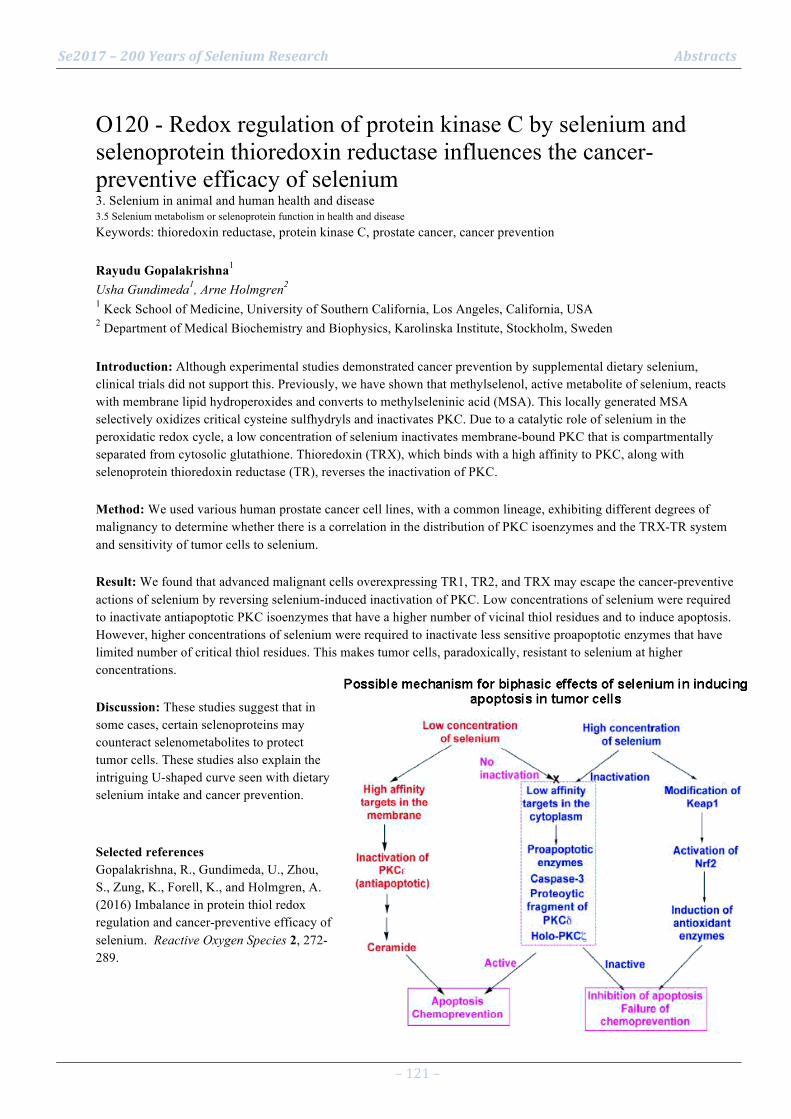
Se2017 – 200 Years of Selenium Research Abstracts
– 121 –
O120 - Redox regulation of protein kinase C by selenium and selenoprotein thioredoxin reductase influences the cancer-preventive efficacy of selenium 3. Selenium in animal and human health and disease 3.5 Selenium metabolism or selenoprotein function in health and disease Keywords: thioredoxin reductase, protein kinase C, prostate cancer, cancer prevention Rayudu Gopalakrishna1 Usha Gundimeda1, Arne Holmgren2 1 Keck School of Medicine, University of Southern California, Los Angeles, California, USA 2 Department of Medical Biochemistry and Biophysics, Karolinska Institute, Stockholm, Sweden Introduction: Although experimental studies demonstrated cancer prevention by supplemental dietary selenium, clinical trials did not support this. Previously, we have shown that methylselenol, active metabolite of selenium, reacts with membrane lipid hydroperoxides and converts to methylseleninic acid (MSA). This locally generated MSA selectively oxidizes critical cysteine sulfhydryls and inactivates PKC. Due to a catalytic role of selenium in the peroxidatic redox cycle, a low concentration of selenium inactivates membrane-bound PKC that is compartmentally separated from cytosolic glutathione. Thioredoxin (TRX), which binds with a high affinity to PKC, along with selenoprotein thioredoxin reductase (TR), reverses the inactivation of PKC. Method: We used various human prostate cancer cell lines, with a common lineage, exhibiting different degrees of malignancy to determine whether there is a correlation in the distribution of PKC isoenzymes and the TRX-TR system and sensitivity of tumor cells to selenium. Result: We found that advanced malignant cells overexpressing TR1, TR2, and TRX may escape the cancer-preventive actions of selenium by reversing selenium-induced inactivation of PKC. Low concentrations of selenium were required to inactivate antiapoptotic PKC isoenzymes that have a higher number of vicinal thiol residues and to induce apoptosis. However, higher concentrations of selenium were required to inactivate less sensitive proapoptotic enzymes that have limited number of critical thiol residues. This makes tumor cells, paradoxically, resistant to selenium at higher concentrations. Discussion: These studies suggest that in some cases, certain selenoproteins may counteract selenometabolites to protect tumor cells. These studies also explain the intriguing U-shaped curve seen with dietary selenium intake and cancer prevention. Selected references Gopalakrishna, R., Gundimeda, U., Zhou, S., Zung, K., Forell, K., and Holmgren, A. (2016) Imbalance in protein thiol redox regulation and cancer-preventive efficacy of selenium. Reactive Oxygen Species 2, 272-289.
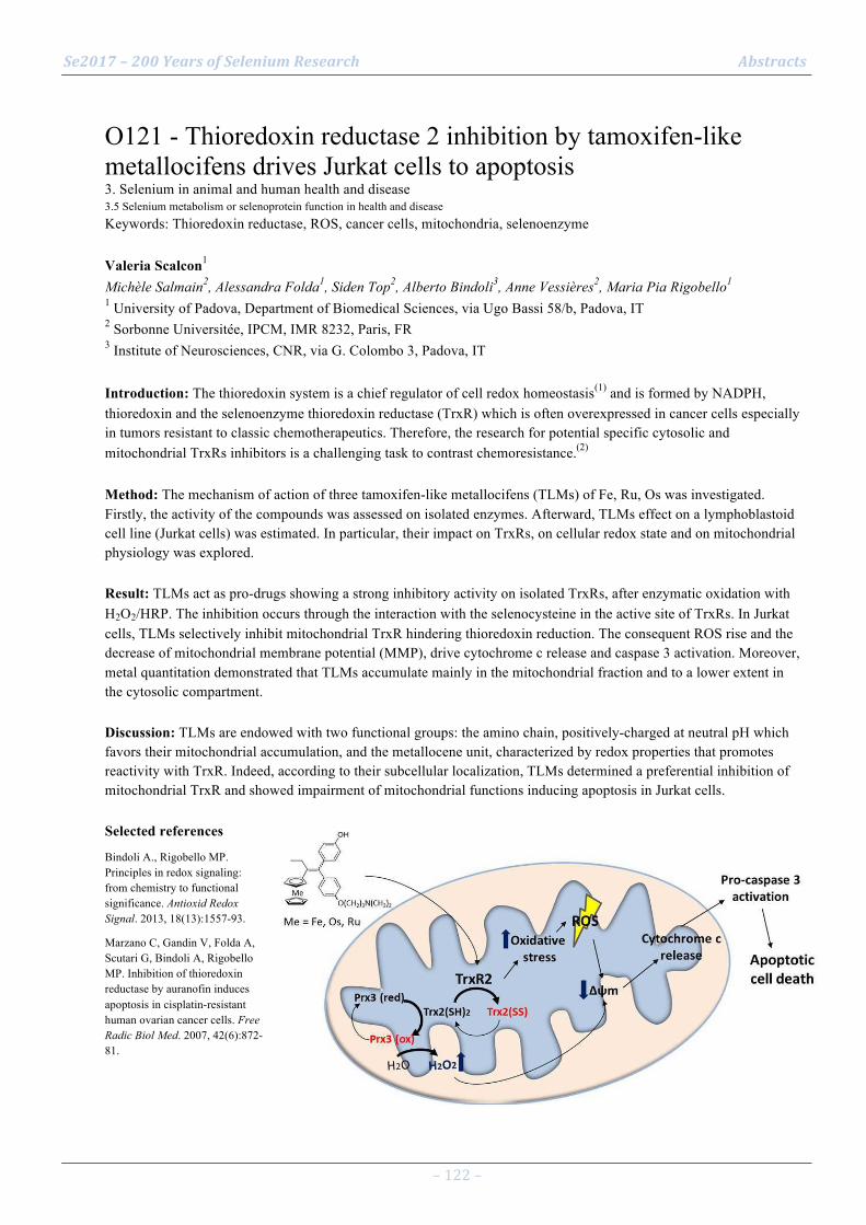
Se2017 – 200 Years of Selenium Research Abstracts
– 122 –
O121 - Thioredoxin reductase 2 inhibition by tamoxifen-like metallocifens drives Jurkat cells to apoptosis 3. Selenium in animal and human health and disease 3.5 Selenium metabolism or selenoprotein function in health and disease Keywords: Thioredoxin reductase, ROS, cancer cells, mitochondria, selenoenzyme Valeria Scalcon1 Michèle Salmain2, Alessandra Folda1, Siden Top2, Alberto Bindoli3, Anne Vessières2, Maria Pia Rigobello1 1 University of Padova, Department of Biomedical Sciences, via Ugo Bassi 58/b, Padova, IT 2 Sorbonne Universitée, IPCM, IMR 8232, Paris, FR 3 Institute of Neurosciences, CNR, via G. Colombo 3, Padova, IT Introduction: The thioredoxin system is a chief regulator of cell redox homeostasis(1) and is formed by NADPH, thioredoxin and the selenoenzyme thioredoxin reductase (TrxR) which is often overexpressed in cancer cells especially in tumors resistant to classic chemotherapeutics. Therefore, the research for potential specific cytosolic and mitochondrial TrxRs inhibitors is a challenging task to contrast chemoresistance.(2) Method: The mechanism of action of three tamoxifen-like metallocifens (TLMs) of Fe, Ru, Os was investigated. Firstly, the activity of the compounds was assessed on isolated enzymes. Afterward, TLMs effect on a lymphoblastoid cell line (Jurkat cells) was estimated. In particular, their impact on TrxRs, on cellular redox state and on mitochondrial physiology was explored. Result: TLMs act as pro-drugs showing a strong inhibitory activity on isolated TrxRs, after enzymatic oxidation with H2O2/HRP. The inhibition occurs through the interaction with the selenocysteine in the active site of TrxRs. In Jurkat cells, TLMs selectively inhibit mitochondrial TrxR hindering thioredoxin reduction. The consequent ROS rise and the decrease of mitochondrial membrane potential (MMP), drive cytochrome c release and caspase 3 activation. Moreover, metal quantitation demonstrated that TLMs accumulate mainly in the mitochondrial fraction and to a lower extent in the cytosolic compartment. Discussion: TLMs are endowed with two functional groups: the amino chain, positively-charged at neutral pH which favors their mitochondrial accumulation, and the metallocene unit, characterized by redox properties that promotes reactivity with TrxR. Indeed, according to their subcellular localization, TLMs determined a preferential inhibition of mitochondrial TrxR and showed impairment of mitochondrial functions inducing apoptosis in Jurkat cells. Selected references Bindoli A., Rigobello MP. Principles in redox signaling: from chemistry to functional significance. Antioxid Redox Signal. 2013, 18(13):1557-93. Marzano C, Gandin V, Folda A, Scutari G, Bindoli A, Rigobello MP. Inhibition of thioredoxin reductase by auranofin induces apoptosis in cisplatin-resistant human ovarian cancer cells. Free Radic Biol Med. 2007, 42(6):872-81.

Se2017 – 200 Years of Selenium Research Abstracts
– 123 –
O122 - Dietary selenium and the 15 kDa selenoprotein influence initiation/promotion of colon carcinogenesis 3. Selenium in animal and human health and disease 3.5 Selenium metabolism or selenoprotein function in health and disease Keywords: colon cancer, intestinal microbiome, 15 kDa selenprotein Jessica Canter1 Bradley Carlson2, Derek Margulies1, Angelica Patterson1, Vadim Gladyshev3, Yunkai Yu4, Liang Cao4, Cindy Davis5, Dolph Hatfield2, Petra Tsuji1 1 Dept. of Biological Sciences, Towson University, Towson, MD, USA 2 Molecular Biology of Selenium Section, NIH, Bethesda, MD, USA 3 Brigham & Women's Hospital, Harvard Medical School, Boston, MA, USA 4 Molecular Targets Core, NIH, Bethesda, MD, USA 5 Office of Dietary Supplements, NIH, Bethesda, MD, USA Introduction: The 15kDa selenoprotein (SELENOF) plays roles in cancer prevention and promotion. Previously, SELENOF knockout (KO) mice were shown to be resistant to formation of chemically-induced pre-neoplastic lesions [1]. Method: In our current inflammatory tumorigenesis study, KO mice and littermate controls were maintained on diets with deficient (0.02 ppm) or adequate (0.1 ppm) dietary selenium (Se), and injected with azoxymethane (AOM) or saline, and treated with dextran sulfate sodium (DSS) or water. Result: Formation of pre-neoplastic lesions was dramatically reduced in KO mice compared to controls. Additionally, fewer KO mice developed tumors when Se-deficient, but formed a similar number of tumors as controls when Se-replete. Thus, KO mice may be protected against cancer initiation, but not against tumor formation, and dietary Se may modify the outcome. Further analyses suggest that CYP2E1, the hepatic enzyme responsible for activating AOM to its mutagenic form, has lower expression in untreated KO mice than controls under Se-deficient conditions, whereas no differences were detected in AOM/DSS-treated mice. Because intestinal microbiota are suspected to affect colon tumorigenesis, bacterial DNA was isolated from mouse feces. Subsequent amplification of the 16S rRNA gene and sequencing with Illumina MiSeq revealed an increased abundance of Bacteriodetes in KO mice fed Se-deficient diets, and a substantial increase in Verrucomicrobia, especially the mucin-degrading species Akkermansia muciniphila, in Se-adequate KO mice. Discussion: Levels of both SELENOF and dietary selenium have substantial effects on colon tumorigenesis and inflammation, which may be modulated by the intestinal microbiome. Their contribution to the regulation of colorectal cancer will be further examined. Selected references [1] Tsuji et al. PlosONE 2012

Se2017 – 200 Years of Selenium Research Abstracts
– 124 –
O123 - Selenoprotein T is a novel neuroprotective antioxidant enzyme in Parkinson’s disease 3. Selenium in animal and human health and disease 3.5 Selenium metabolism or selenoprotein function in health and disease Keywords: Selenoprotein, neuroprotection, neurodegeneration, dopamine, transgenic mice Youssef Anouar1 Abdallah Hamieh1, Matthieu Castex1, Ifat Alsharif1, Anthony Falluel-Morel1, Isabelle Lihrmann1, Loubna Boukhzar1 1 INSERM U1239, University of Rouen Normandy, Rouen, France Introduction: Oxidative stress is central to the pathogenesis of Parkinson’s disease (PD), but the mechanisms involved in the control of this stress in dopaminergic cells are not fully understood. There is increasing evidence that selenoproteins play a central role in the control of redox homeostasis and cell defense, but the precise contribution of members of this family of proteins during the course of neurodegenerative diseases is still elusive. Method: We used the SH-SY5Y dopaminergic cell model and brain-specific selenoprotein T (SelT)-deficient mice to demonstrate the importance of this protein in neuroprotection. Result: In the SH-SY5Y cell model of dopaminergic neurons, both silencing and overexpression of SelT affected oxidative stress and cell survival. Treatment with PD-inducing neurotoxins such as MPTP or rotenone triggered SelT expression in the nigrostriatal pathway of wild-type mice, but provoked rapid and severe parkinsonian-like motor defects in conditional brain SelT-deficient mice. This motor impairment was associated with marked oxidative stress and neurodegeneration, and decreased tyrosine hydroxylase activity and dopamine levels in the nigrostriatal system. Finally, in PD patients, we report that SelT is tremendously increased in the caudate putamen tissue. Discussion: These results reveal the activity of a novel selenoprotein enzyme that protects dopaminergic neurons against oxidative stress and prevents early and severe movement impairment in animal models of PD. Our findings indicate that selenoproteins such as SelT play a crucial role in the protection of dopaminergic neurons against oxidative stress and cell death, providing insight into the molecular underpinnings of this stress in PD. Selected references Boukhzar, L., Hamieh, A., Castex, M., Chargraoui, A., Bonnet, J.J., Arabo, A., Errami, M., Falluel-Morel, A., Lihrmann, I. and Anouar, Y.: Selenoprotein T exerts an essential oxidoreductase activity that protects dopaminergic neurons in mouse models of Parkinson’s disease. Antioxid. Redox Signal. 24:557-74 (2016).
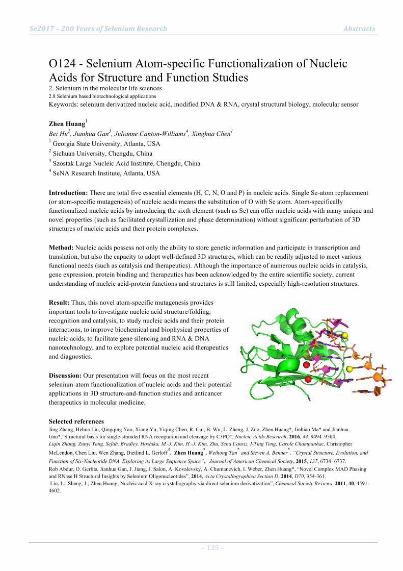
Se2017 – 200 Years of Selenium Research Abstracts
– 125 –
O124 - Selenium Atom-specific Functionalization of Nucleic Acids for Structure and Function Studies 2. Selenium in the molecular life sciences 2.8 Selenium based biotechnological applications Keywords: selenium derivatized nucleic acid, modified DNA & RNA, crystal structural biology, molecular sensor Zhen Huang1 Bei Hu2, Jianhua Gan3, Julianne Canton-Williams4, Xinghua Chen1 1 Georgia State University, Atlanta, USA 2 Sichuan University, Chengdu, China 3 Szostak Large Nucleic Acid Institute, Chengdu, China 4 SeNA Research Institute, Atlanta, USA Introduction: There are total five essential elements (H, C, N, O and P) in nucleic acids. Single Se-atom replacement (or atom-specific mutagenesis) of nucleic acids means the substitution of O with Se atom. Atom-specifically functionalized nucleic acids by introducing the sixth element (such as Se) can offer nucleic acids with many unique and novel properties (such as facilitated crystallization and phase determination) without significant perturbation of 3D structures of nucleic acids and their protein complexes. Method: Nucleic acids possess not only the ability to store genetic information and participate in transcription and translation, but also the capacity to adopt well-defined 3D structures, which can be readily adjusted to meet various functional needs (such as catalysis and therapeutics). Although the importance of numerous nucleic acids in catalysis, gene expression, protein binding and therapeutics has been acknowledged by the entire scientific society, current understanding of nucleic acid-protein functions and structures is still limited, especially high-resolution structures. Result: Thus, this novel atom-specific mutagenesis provides important tools to investigate nucleic acid structure/folding, recognition and catalysis, to study nucleic acids and their protein interactions, to improve biochemical and biophysical properties of nucleic acids, to facilitate gene silencing and RNA & DNA nanotechnology, and to explore potential nucleic acid therapeutics and diagnostics. Discussion: Our presentation will focus on the most recent selenium-atom functionalization of nucleic acids and their potential applications in 3D structure-and-function studies and anticancer therapeutics in molecular medicine. Selected references Jing Zhang, Hehua Liu, Qingqing Yao, Xiang Yu, Yiqing Chen, R. Cui, B. Wu, L. Zheng, J. Zuo, Zhen Huang*, Jinbiao Ma* and Jianhua Gan*,”Structural basis for single-stranded RNA recognition and cleavage by C3PO”, Nucleic Acids Research, 2016, 44, 9494–9504. Liqin Zhang, Zunyi Yang, Sefah, Bradley, Hoshika, M.-J. Kim, H.-J. Kim, Zhu, Sena Cansiz, I-Ting Teng, Carole Champanhac, Christopher
McLendon, Chen Liu, Wen Zhang, Dietlind L. Gerloff3, Zhen Huang*, Weihong Tan* and Steven A. Benner*, “Crystal Structure, Evolution, and Function of Six-Nucleotide DNA. Exploring its Large Sequence Space”,Journal of American Chemical Society, 2015, 137, 6734−6737. Rob Abdur, O. Gerlits, Jianhua Gan, J. Jiang, J. Salon, A. Kovalevsky, A. Chumanevich, I. Weber, Zhen Huang*, “Novel Complex MAD Phasing and RNase H Structural Insights by Selenium Oligonucleotides”, 2014, Acta Crystallographica Section D, 2014, D70, 354-361. Lin, L.; Sheng, J.; Zhen Huang, Nucleic acid X-ray crystallography via direct selenium derivatization”, Chemical Society Reviews, 2011, 40, 4591-4602.

Se2017 – 200 Years of Selenium Research Abstracts
– 126 –
O125 - Interrelationships among SELENOF genotype, cellular localization, serum selenium and race in prostate cancer 3. Selenium in animal and human health and disease 3.6 Clinical genetics of selenium or selenoprotein-encoding genes Keywords: prostate, cancer, SELENOF, genetics, race Dede Ekoue1 Emmanuel Ansong1, Andre Kajdacsy-Balla1, Li Liu1, Larisa Nonn1, Peter Gann1, Vincent Freeman1, Alan Diamond1 1 University of Illinois at Chicago, Chicago, United States Introduction: Polymorphisms in the endoplasmic reticulum (ER) resident selenoprotein SELENOF are associated with prostate cancer mortality and are 5-fold more prevalent in African Americans, who are also at greater risk of dying of the disease. A role of SELENOF in prostate cancer was investigated in human prostate tissues and cell lines. Method: Plasma selenium levels were quantified by atomic absorption spectroscopy. DNA from 129 men who had undergone prostatectomy were genotyped for SELENOF and the interactions between SELENOF genotype, serum selenium, and race were assessed. Levels and cellular location of SELENOF were analyzed in cultured cells by western blotting and in human tissue microarrays by immunohistochemistry scored by computer-assisted image analysis. Result: SELENOF predominated in the outer cell membranes (OCM) of cells in benign glands and in primary benign human epithelial cells, but not in human cancer-derived cell lines and tumors tissue. Protein levels were significantly lower, and generally absent in the OCM in cancer compared to benign prostatic tissue (p=1x10⁻⁷). SELENOF levels were lower in tumor cells from African Americans (n=33) compared to Caucasians (n=295) (p<0.02). The SELENOF allele that was more frequent in African Americans was associated with both higher prostate specific antigen levels (p<0.05) and Gleason score. African American men in this cohort had significantly lower mean serum selenium levels (115.24 ±3.04 ng/mL) than Caucasians (139.18 ±2.41 ng/mL, p<0.01). Discussion: SELENOF may have a non-ER function in normal prostate epithelium. Genotype and selenium levels may contribute to the prostate cancer racial disparities observed in African Americans.

Se2017 – 200 Years of Selenium Research Abstracts
– 127 –
O126 - Selenocysteine and the Genetic Code 2. Selenium in the molecular life sciences 2.9 Additional and emerging topics of selenium in molecular life science Keywords: genetic code, selenoprotein synthesis, synthetic biology, recoding Dieter Söll1 1 Department of Molecular Biophysics & Biochemistry, Yale University, New Haven, CT 06511, USA Introduction: At the time of its elucidation the genetic code was suggested to be universal in all organisms, and the result of a ‘frozen accident’ unable to evolve further (1). Method: Today we know 22 natural amino acids (2): selenocysteine, and pyrrolysine are directly inserted into growing poly-peptides during translation. Result: The incorporation of selenocysteine directed by UGA requires the action of specific RNA and protein elements, a fact that has restricted engineer-ing of selenoproteins. Based on the realization that protein plasticity is a feature of living cells (3), man-made expansion of the genetic code based on orthogonal transla-tion systems (OTSs) is an active research field (4-6). Discussion: Although the design of in vivo specific and highly active OTS systems is still not ideal (7,8), the increasing number of successful recoding strategies promises a bright future for genetic code expansion (9-12). Selected references
1. Crick (1968) J Mol Biol 38, 367.
2.Ambrogelly et al (2007) Nat Chem Biol 3, 29.
3. Ruan et al (2008) Proc Natl Acad Sci USA 105, 16502.
4. Liu, Schultz (2010) Annu Rev Biochem 79, 413.
5. Park et al (2011) Science 333, 1151.
6. Yang et al (2016) Science 354, 623.
7. O’Donoghue et al (2013) Nat Chem Biol 9, 594.
8. Guo et al (2014) Proc Natl Acad Sci USA 111, 16724.
9. Bröcker et al (2013) Angew Chem Int Ed Engl 53, 319.
10. Miller et al (2015) FEBS Lett 589, 2194.
11. Hoesl et al (2015) Angew Chem Int Ed Engl 54, 10030.
12. Mukai et al (2016) Angew Chem Int Ed Engl 55, 5337.

Se2017 – 200 Years of Selenium Research Abstracts
– 128 –
O127 - Cysteine polysulfidation governed by cysteinyl-tRNA synthetases (CARSs) 2. Selenium in the molecular life sciences 2.9 Additional and emerging topics of selenium in molecular life science Keywords: Cysteine persulfide, CARS, cysteine persulfide synthase, CPERS, bioenergeticss Takaaki Akaike1 Tomoaki Ida1, Masanobu Morita1, Akira Nishimura1, Tetsuro Matsunaga1, Martin Feelisch2, Péter Nagy3, Jon M. Fukuto4, Hozumi Motohashi5 1 Department of Environmental Health Sciences and Molecular Toxicology, Tohoku University Graduate School of Medicine, Sendai 980-8575, Japan 2 Clinical and Experimental Sciences, Faculty of Medicine, University of Southampton, Southampton SO16 6YD, United Kingdom 3 Department of Molecular Immunology and Toxicology, National Institute of Oncology, 1122 Budapest, Hungary 4 Department of Chemistry, Sonoma State University, Rohnert Park, CA 94928 5 Department of Gene Expression Regulation, Institute of Development, Aging and Cancer, Tohoku University, Sendai 980-8575, Japan Introduction: Cysteine hydropersulfide (CysSSH) occurs in abundant quantities in various organisms including mammals, yet little is known about its biosynthesis and physiological function(s). Moreover, extensive persulfide formation is apparent in Cys-containing proteins in Escherichia coli and mammalian cells, and believed to be the result of post-translational processes involving hydrogen sulfide-related chemistry. Method: Prokaryotic and mammalian cysteinyl-tRNA synthetases (CARSs) were investigated for their potential CysSSH production both in vitro and in vivo. Result: Here, we demonstrate effective CysSSH synthesis from the substrate L-cysteine, with the synthesis being catalyzed by prokaryotic and mammalian CARSs. Targeted disruption of the genes encoding mitochondrial CARSs in mice and human cells revealed the major role of CARSs in endogenous CysSSH production, which suggests that CARSs serve as the principal cysteine persulfide synthase (CPERS) in vivo. CARSs also catalyze co-translational protein polysulfidation and are involved in the regulation of mitochondrial biogenesis and bioenergetics. Preliminary results furthermore suggest that the selenium-dependent mammalian thioredoxin system may be involved in regulation of CysSSH homeostasis. Discussion: Investigating translation-coupled persulfide production may thus lead to evolution of the central dogma of molecular biology, unravel mechanisms of aberrant redox-regulation, and suggest novel treatment options for mitochondrial dysfunction. Selected references Nishida M, et al. Redox regulation of electrophilic signaling by reactive persulfides in cardiac cells. Free Radic. Biol. Med. 2017. pii: S0891-
5849(17)30033-3. doi: 10.1016/j.freeradbiomed.2017.01.024. Kunikata H., et al. Metabolomic profiling of reactive persulfides and polysulfides in the aqueous and vitreous humors. Sci. Rep., 2017 Feb 7;7:41984.
doi: 10.1038/srep41984. Jung M., et al. Protein polysulfidation-dependent persulfide dioxygenase activity of ethylmalonic encephalopathy protein 1. Biochem. Biophys. Res.
Commun. 480:180-186, 2016. Ida T., et al. Reactive cysteine persulfides and S-polythiolation regulate oxidative stress and redox signaling. Proc. Natl. Acad. Sci. USA, 111: 7606-
7611, 2014. Nishida M., et al. Hydrogen sulfide anion regulates redox signaling via electrophile sulfhydration. Nature Chem. Biol., 8, 714-724, 2012.
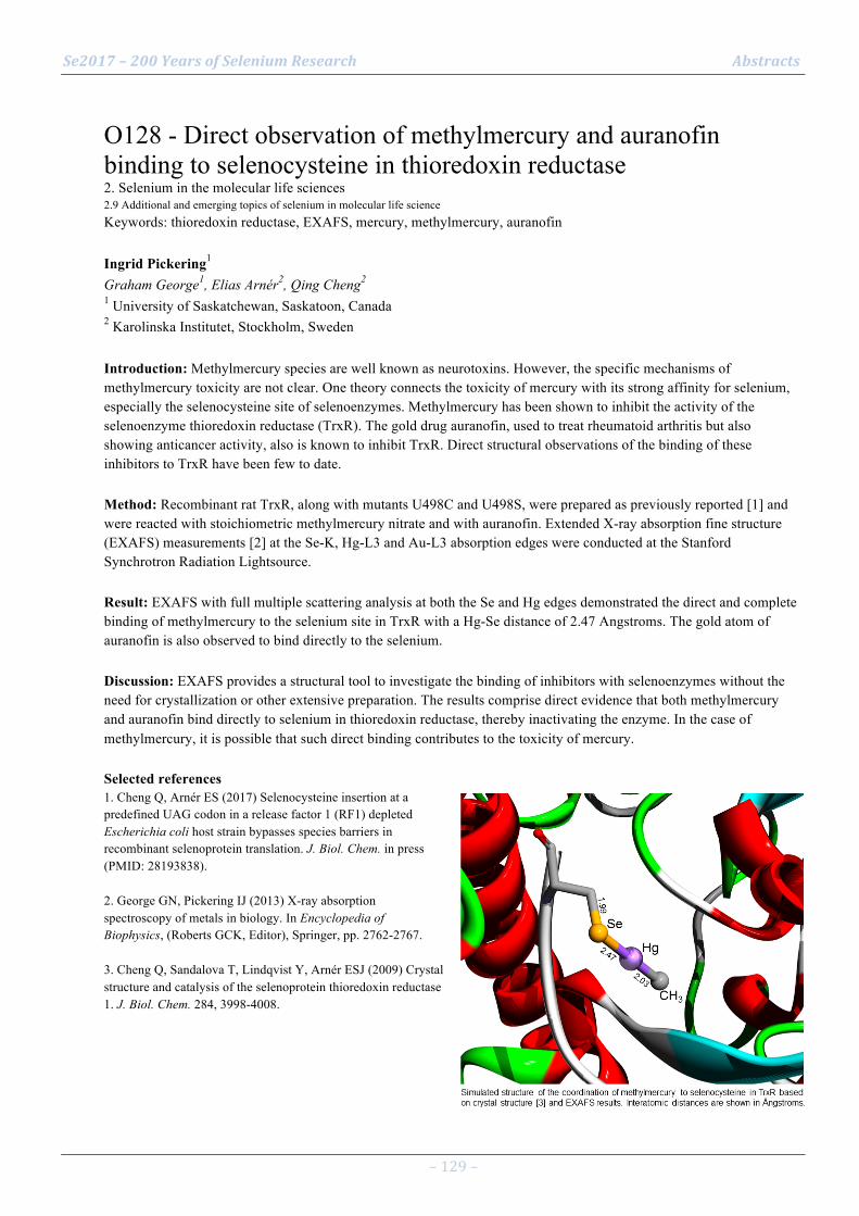
Se2017 – 200 Years of Selenium Research Abstracts
– 129 –
O128 - Direct observation of methylmercury and auranofin binding to selenocysteine in thioredoxin reductase 2. Selenium in the molecular life sciences 2.9 Additional and emerging topics of selenium in molecular life science Keywords: thioredoxin reductase, EXAFS, mercury, methylmercury, auranofin Ingrid Pickering1 Graham George1, Elias Arnér2, Qing Cheng2 1 University of Saskatchewan, Saskatoon, Canada 2 Karolinska Institutet, Stockholm, Sweden Introduction: Methylmercury species are well known as neurotoxins. However, the specific mechanisms of methylmercury toxicity are not clear. One theory connects the toxicity of mercury with its strong affinity for selenium, especially the selenocysteine site of selenoenzymes. Methylmercury has been shown to inhibit the activity of the selenoenzyme thioredoxin reductase (TrxR). The gold drug auranofin, used to treat rheumatoid arthritis but also showing anticancer activity, also is known to inhibit TrxR. Direct structural observations of the binding of these inhibitors to TrxR have been few to date. Method: Recombinant rat TrxR, along with mutants U498C and U498S, were prepared as previously reported [1] and were reacted with stoichiometric methylmercury nitrate and with auranofin. Extended X-ray absorption fine structure (EXAFS) measurements [2] at the Se-K, Hg-L3 and Au-L3 absorption edges were conducted at the Stanford Synchrotron Radiation Lightsource. Result: EXAFS with full multiple scattering analysis at both the Se and Hg edges demonstrated the direct and complete binding of methylmercury to the selenium site in TrxR with a Hg-Se distance of 2.47 Angstroms. The gold atom of auranofin is also observed to bind directly to the selenium. Discussion: EXAFS provides a structural tool to investigate the binding of inhibitors with selenoenzymes without the need for crystallization or other extensive preparation. The results comprise direct evidence that both methylmercury and auranofin bind directly to selenium in thioredoxin reductase, thereby inactivating the enzyme. In the case of methylmercury, it is possible that such direct binding contributes to the toxicity of mercury. Selected references 1. Cheng Q, Arnér ES (2017) Selenocysteine insertion at a predefined UAG codon in a release factor 1 (RF1) depleted Escherichia coli host strain bypasses species barriers in recombinant selenoprotein translation. J. Biol. Chem. in press (PMID: 28193838). 2. George GN, Pickering IJ (2013) X-ray absorption spectroscopy of metals in biology. In Encyclopedia of Biophysics, (Roberts GCK, Editor), Springer, pp. 2762-2767. 3. Cheng Q, Sandalova T, Lindqvist Y, Arnér ESJ (2009) Crystal structure and catalysis of the selenoprotein thioredoxin reductase 1. J. Biol. Chem. 284, 3998-4008.
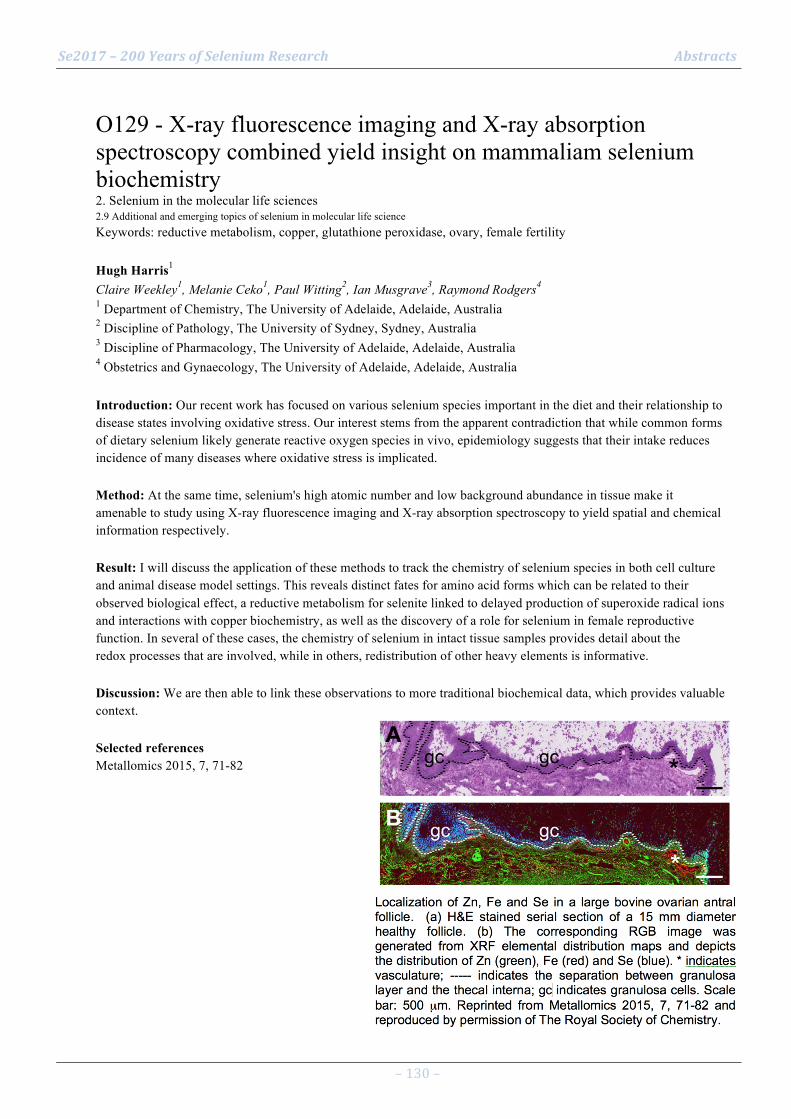
Se2017 – 200 Years of Selenium Research Abstracts
– 130 –
O129 - X-ray fluorescence imaging and X-ray absorption spectroscopy combined yield insight on mammaliam selenium biochemistry 2. Selenium in the molecular life sciences 2.9 Additional and emerging topics of selenium in molecular life science Keywords: reductive metabolism, copper, glutathione peroxidase, ovary, female fertility Hugh Harris1 Claire Weekley1, Melanie Ceko1, Paul Witting2, Ian Musgrave3, Raymond Rodgers4 1 Department of Chemistry, The University of Adelaide, Adelaide, Australia 2 Discipline of Pathology, The University of Sydney, Sydney, Australia 3 Discipline of Pharmacology, The University of Adelaide, Adelaide, Australia 4 Obstetrics and Gynaecology, The University of Adelaide, Adelaide, Australia Introduction: Our recent work has focused on various selenium species important in the diet and their relationship to disease states involving oxidative stress. Our interest stems from the apparent contradiction that while common forms of dietary selenium likely generate reactive oxygen species in vivo, epidemiology suggests that their intake reduces incidence of many diseases where oxidative stress is implicated. Method: At the same time, selenium's high atomic number and low background abundance in tissue make it amenable to study using X-ray fluorescence imaging and X-ray absorption spectroscopy to yield spatial and chemical information respectively. Result: I will discuss the application of these methods to track the chemistry of selenium species in both cell culture and animal disease model settings. This reveals distinct fates for amino acid forms which can be related to their observed biological effect, a reductive metabolism for selenite linked to delayed production of superoxide radical ions and interactions with copper biochemistry, as well as the discovery of a role for selenium in female reproductive function. In several of these cases, the chemistry of selenium in intact tissue samples provides detail about the redox processes that are involved, while in others, redistribution of other heavy elements is informative. Discussion: We are then able to link these observations to more traditional biochemical data, which provides valuable context. Selected references Metallomics 2015, 7, 71-82

Se2017 – 200 Years of Selenium Research Abstracts
– 131 –
O130 - Se-speciation investigations at neural barrier (NB) 3. Selenium in animal and human health and disease 3.7 Additional and emerging topics of selenium in health or disease Keywords: Selenium-speciation, HPLC-(CE)-ICP-DRC-MS, neural barrier, cerebrospinal fluid, ALS Bernhard Michalke1 Nikolay Solovyev2, Marco Vinceti3 1 Helmholtz Center Munich , Analytical BioGeoChemistry, 85764 Neuherberg, Germany 2 St. Petersburg State University, St. Petersburg, Russia 3 CREAGEN Research Center, University of Modena and Reggio Emilia, Modena, italy Introduction: Se-speciation helps for deeper insight into Se-metabolism and transport, important at NB or under neurological diseases. Analytical set-up of hyphenated speciation techniques with 2-D-identification of Se-compounds is described. Then applications at NB in paired serum and cerebrospinal fluid (CSF) samples are reported, finishing with comparison of Se-speciation of neurologically diseased persons vs. controls. Method: Serum and CSF samples were subject to speciation analysis by HPLC-ICP-DRC-MS. For improved species identification Se-species were analysed serially after HPLC by CE-ICP-DRC-MS (2D approach). Result: Paired samples had 58.39 (serum) or 0.86 (CSF , each µg Se/L). Prominent Se-species were selenoprotein-P (SePP), glutathione-peroxidase (GPx), thioredoxinreductase (TrxR), Se(IV) and Se-albumin (Se-HSA). Relationships between Se-species from serum and CSF allowed evaluating Se-species passage across NB: SePP-serum
correlated with total Se-serum when > 65 µg/L. SePP-CSF appeared independent from SePP-serum. For anti-oxidative Se-enzymes higher correlation factors (r2) were calculated: GPx-serum/GPx-CSF: r2=0.3837 and TrxR-serum/TrxR-CSF: r2=0.6293. No correlation for inorganic Se-compounds was found proving limited representativeness of their circulating levels beyond NB[1]. Se-species-ratios (CSF/serum) were 21.4*10-3 (TrxR) or 8.3*10-3 (GPx), being significantly elevated compared to NB permeability factor 3.8*10-3 (HSA). In a hospital-referred cases-control we investigated Se-species in CSF of patients with amyotrophic lateral sclerosis, compared to reference neurological patients. We found an excess concentration of inorganic Se(IV) and reduced levels of organic Se-compounds among ALS patients[2]. Discussion: ROS-protecting enzymes GPx and TrxR seem to be shuttled across NB to brain/CSF. In ALS etiology overexposure of inorganic Se(IV) together with decreased organic Se-species may be involved in ALS etiology. Selected references [1] N. Solovyev, A. Berthele, B. Michalke; Selenium Speciation in paired serum and cerebrospinal fluid samples. Anal. Bioanal Chem, (2013) 405:1875–1884. [2] M.Vinceti, N.Solovyev, J. Mandrioli, C.M. Crespi , F. Bonvicini, E. Arcolin, E.Georgoulopoulou, B.Michalke; Cerebrospinal fluid of newly diagnosed amyotrophic lateral sclerosis patients exhibits abnormal levels of selenium species including elevated selenite. NeuroToxicology 38 (2013) 25–32.
Se2017 – 200 Years of Selenium Research Abstracts
– 132 –
O131 - Selenium-Binding Protein 1 in serum may signify a heart at risk 3. Selenium in animal and human health and disease 3.7 Additional and emerging topics of selenium in health or disease Keywords: SELENBP1, diagnostics, sandwich assay, serum, heart, acute myocardial infarction Eike Kuehn1 Andreas Goetzenich2, Anna Slagman3, Ellen Kuehn-Heid1, Christian Schwiebert1, Waldemar Minich1, Sandra Kraemer2, Martin Moeckel3, Christian Stoppe2, Lutz Schomburg1 1 Charité - Universitätsmedizin Berlin, Institut für Experimentelle Endokrinologie, Berlin, Germany 2 RWTH Aachen, Abteilung für Anästhesie und Intensivmedizin, Aachen, Germany 3 Charité - Universitätsmedizin Berlin, Notfallmedizin / Rettungsstellen und Chest Pain Units, Berlin, Germany Introduction: Selenium-Binding Protein 1 (SELENBP1) is an intracellular protein with unknown function. The objective of this study was to test for its potential presence in serum and its relation to cardiovascular disease. Method: Monoclonal antibodies were generated and a sandwich luminometric assay for SELENBP1 was established. The assay has a limit of detection (LOD) of 10 µg/L. Serum samples of 77 patients with a cardio pulmonary bypass (CPB) during cardiac surgery as well as 113 patients with suspected acute myocardial infarction (AMI) were analyzed. Result: Prior to surgery, serum SELENBP1 was below detection limit in all patients. Serum SELENBP1 increased during surgery in 97% of patients to detectable concentrations until 15 min after weaning from the CPB. SELENBP1 concentrations correlated positively with duration of cardioplegia (rho = 0.56, p = 0.0000004). In the patients with suspected AMI, 69% experienced an AMI or a major adverse cardiac event (MACE) until discharge. A time-resolved analysis indicated rising serum concentrations of SELENBP1 (to > 10 ng/ml) in 41% of patients. A significant rise in serum SELENBP1 was associated with a 78% risk of suffering an AMI or MACE. Discussion: The physiological role of SELENBP1 remains unclear, but its appearance in serum may provide novel diagnostic or prognostic insights in cardiac disease. These findings and the newly established sandwich assay may help to better pinpoint the physiological functions of SELENBP1 and to elucidate the molecular mechanisms leading to its disease-related release into serum. Selected references Supported by Charité – Universitätsmedizin Berlin.
Se2017 – 200 Years of Selenium Research Abstracts
– 133 –
O132 - Of dogs and men: Review of translational impact of dog studies on selenium and prostate cancer risk 3. Selenium in animal and human health and disease 3.7 Additional and emerging topics of selenium in health or disease Keywords: Cancer prevention, Dose-dependent effects, Prostate cancer, Preclinical models, Human health Emily Chiang1 David Waters2 1 Center for Exceptional Longevity Studies, Gerald P. Murphy Cancer Foundation, W. Lafayette, IN, USA 2 Purdue University Center on Aging and the Life Course, West Lafayette, IN, USA Introduction: Dogs, like men, naturally develop prostate cancer during aging. Therefore, it provides an appropriate context to study the effects of selenium on the aging prostate prior to cancer diagnosis. Method: In this presentation, we summarize the translational significance of research progress gained from a decade of dog studies on selenium and prostate cancer risk. Result: In a randomized feeding trial in elderly beagles (equivalent to 65 year-old men), we showed daily supplementation with selenomethionine or selenium-yeast significantly reduced prostatic DNA damage. Our discovery of a U-shaped dose-response between toenail selenium and prostatic DNA damage in dogs remarkably paralleled data from men in North America and the Netherlands. Later, this U-curve provided a plausible explanation for the alarming increase in prostate cancers in men with highest baseline selenium that received supplementation in SELECT. Next, this U-curve guided the discovery of a non-antioxidant, anti-carcinogenic mechanism of organic selenium: “homeostatic housecleaning”, the preferential triggering of apoptosis in DNA damaged cells. Finally, we catalogued form-dependent effects of selenium on intraprostatic phenotypes (prostatic androgen levels, cell proliferation, apoptosis and DNA damage), complementing human studies in which analyses are often limited to markers in serum. Discussion: Over the last decade, we have demonstrated that dogs have advanced our understanding of selenium’s in vivo chemopreventive actions. Dog studies can contribute important insights into dose-dependent and form-dependent effects – aspects of selenium biology that will have to be further elucidated if the burgeoning science of selenium is to be translated into effective strategies for prevention of human disease. Selected references
Waters DJ, et al. (2003) J Natl Cancer Inst 95:237-41.
Waters DJ, et al. (2005) Carcinogenesis 26:1256-62.
Chiang EC, et al. (2009) Dose Response 8: 285-300.
Waters DJ, et al. (2012) Nutrients 4:1650-63.
Chiang EC., et al. (2013) Biofactors 39:575-88.

Se2017 – 200 Years of Selenium Research Abstracts
– 134 –
O133 - High selenium induces endothelial dysfunction via endoplasmic reticulum stress 3. Selenium in animal and human health and disease 3.7 Additional and emerging topics of selenium in health or disease Keywords: ER stress, endoplasmic reticulum stress, endothelial dysfunction. Matshediso Zachariah1 Hatem Maamoun1, Abdelali Agouni2, Lisi Meira1, Margaret P. Rayman1 1 University of Surrey 2 Qatar University Introduction: Selenium is associated with insulin resistance and may affect endothelial function, increasing the risk of type II diabetes and associated cardiovascular-disease [1]. However, the molecular mechanisms involved are not clear. The endoplasmic reticulum (ER) stress response is a mechanism involved in high selenium-induced apoptosis in some cancer cells [2] and, also in the pathogenesis of insulin resistance and endothelial dysfunction [3]. Thus, we hypothesised that high selenium status causes ED through ER stress. Method: Endothelial cells (HUVECs) and EA.hy926 cell lines were treated with selenite (0.5-10 µM) for 24 hours in the presence or absence of the ER chemical chaperone, 4-phenylbutryic acid (PBA). ER stress markers were investigated using qPCR and western blot technique. Endothelial function was assessed by the Griess assay, flow cytometry, Matrigel® and colorimetric assays. Data were expressed as S.E.M (p<0.05) vs. control. Result: High-selenium concentration (5-10 µM) compared to physiological concentration (0.5–2.0 µM) enhanced mRNA expression of ER-stress markers: activating transcription factor-4 (ATF4), CAAA/enhanced-binding homologous protein (CHOP) and X-binding box-1 (XBP-1). In addition, high selenite concentration reduced NO production and angiogenic capacity in endothelial cells. Moreover, high selenite treatment significantly (p<0.05) increased ROS production and induced apoptosis through caspase-3/7 activity. Interestingly, PBA completely reversed all the effects of high selenite on endothelial function, indicating the involvement of the ER-stress response. Discussion: High-selenium treatment caused endothelial dysfunction through the activation of the ER-stress response. This highlights the importance of monitoring the risk of cardiovascular disease in cancer patients supplemented with high selenium as part of their therapy. Selected references 1. Streinbrenner H, Pinto A, Sics H (2011) High selenium intake and increased diabetes risk: experimental evidence for interplay between selenium and carbohydrate metabolism. J Clini Biochem Nutr 48: 40-5. 2. Wu Y, Zhang H, Dong Y, Park YM, Ip C (2005) Endoplasmic reticulum stress signal mediators are targets of selenium action. Cancer Res 65:9073-9. 3. Ozcan U, Cao Q, Yilmaz E, Lee AH, Iwakoshi NN, Ozdelen E, Tuncman G, Gorgun C, Glimcher LH, Hotamisligil GS (2004) Endoplasmic reticulum stress links obesity, insulin action and type 2 diabetes. Science 306: 457-61.

Se2017 – 200 Years of Selenium Research Abstracts
– 135 –
O134 - Selenium and sex: competition between brain and testes for selenium results in male-specific consequences in mice and men 3. Selenium in animal and human health and disease 3.7 Additional and emerging topics of selenium in health or disease Keywords: selenocysteine lyase, neurological deficits, metabolic syndrome, gender Marla Berry1 Matthew Pitts1, Ann Hashimoto1, Lucia Seale1, Ashley Ogawa-Wong1, Daniel Torres1, China Byrns1, Christopher Williams2 1 Department of Cell and Molecular Biology, John A. Burns School of Medicine, University of Hawaii, Honolulu, Hawaii, USA 2 2Department of Medicine and Cancer Biology, Vanderbilt University School of Medicine, Nashville, Tennessee, USA Introduction: Selenium (Se) is essential for normal neurological development and male fertility in mammals, setting the stage for competition between brain and testes when Se is limiting or its distribution is perturbed. Se supplementation studies in humans, and genetic knockout (KO) of selenium transport and recycling proteins in mice, unveiled the surprising findings that males exhibit more severe consequences than females when selenium homeostasis is altered. In one study, Se supplementation in humans resulted in an increased risk of type 2 diabetes (T2D) in men with high baseline Se. However, other studies found no correlation between increased Se and T2D susceptibility, and the discrepancies in these human trials are controversial. Method: Mice with KO of the Se recycling enzyme, selenocysteine lyase (Scly) or double KO of Scly and the Se transport protein, Selenoprotein P (Sepp1) were characterized. Result: KO mice exhibit metabolic syndrome that is more pronounced in males, whereas DKO resulted in low viability and neurological deficits including audiogenic seizures, also more pronounced in males. Pre-pubescent castration reversed the double KO neurological phenotype and partially attenuated the Scly KO metabolic phenotype. Assessment of brain and testes Se levels in these mice suggests that competition for available Se between these two tissues is a significant contributor to the observed phenotypes. Discussion: Testosterone replacement studies are underway to investigate whether there are also hormone-dependent mechanisms at play. These studies unveil novel insights into the complex mechanisms involved in Se distribution and utilization, and may be informative regarding sex specific Se requirements in humans.
Se2017 – 200 Years of Selenium Research Abstracts
– 136 –
O135 - Ceramide analog S14 causes a coordinate downregulation of selenoproteins in a murine psoriasis model 3. Selenium in animal and human health and disease 3.7 Additional and emerging topics of selenium in health or disease Keywords: Psoriasis, Selenoproteins, S14, Ceramide Jack L. Arbiser1 Kellie Michaels2, Ron Nowark2, Yuliya Skabytska3, Michael Y. Bonner1, Tilo Biedermann Biedermann3, Shikha Rao1, Nabiha Yusuf4, Linda Gilbert1, Isabella Karlsson Karlsson1, Yi Fritz2, Nicole L. Ward2 1 Departments of Dermatology, Emory University School of Medicine, Atlanta, GA, 30322 Veterans Affair 2 Case Western Reserve University, Cleveland, OH 44106 3 Department of Dermatology and Allergology Technische Universität München, Germany 4 Department of Dermatology, University of Alabama School of Medicine, Department of Dermatology and S Introduction: Psoriasis is a common disorder affecting 1-‐2 percent of the world population. Currently, the role of selenoproteins in the pathogenesis of psoriasis is not well understood. Method: We have previously demonstrated that ceramide analogs based upon the ant venom alkaloid, solenopsin, have anti-inflammatory effects on a well-established murine model, the KC-Tie-2 model of psoriasis. In an effort to better understand the role of selenoproteins and other proteins involved in glutathione metabolism, we compared vehicle treated skin versus S14 treated skin by gene array analysis. S14 is a 14 carbon chain containing solenopsin analog, which has potent anti-psoriatic activity in this model. Result: Surprisingly, a coordinate downregulation of selenium containing and glutathione metabolizing proteins was observed in treated mice compared with controls. Notably, these include glutathione peroxidase 1 (Gpx1), superoxide dismutase 1 and 2, glutathione peroxidase 4 (Gpx4), and thioredoxin domain containing proteins 15 and 17. Discussion: Gpx4 is a major regulator of ferroptosis, an iron mediated mechanism of cell death involving oxidation of unsaturated fatty acids. While psoriasis has traditionally been thought to be mediated by excess reactive oxygen, our data suggests that novel upregulation of reactive oxygen species by the S14 ceramide derivative may cause therapeutic benefit by causing ferroptosis and perhaps induction of regulatory T cells by reactive oxygen. Selected references Arbiser JL, Kau T, Konar M, Narra K, Ramchandran R, Summers SA, Vlahos CJ, Ye K, Perry BN, Matter W, Fischl A, Cook J, Silver PA, Bain J, Cohen P, Whitmire D, Furness S, Govindarajan B, Bowen JP. Solenopsin, the alkaloidal component of the fire ant (Solenopsis invicta), is a naturally occurring inhibitor of phosphatidylinositol-3-kinase signaling and angiogenesis. Blood. 2007 Jan 15;109(2):560-5. Karlsson I, Zhou X, Thomas R, Smith AT, Bonner MY, Bakshi P, Banga AK, Bowen JP, Qabaja G, Ford SL, Ballard MD, Petersen KS, Li X, Chen G, Ogretmen B, Zhang J, Watkins EB, Arnold RS, Arbiser JL. Solenopsin A and analogs exhibit ceramide-like biological activity. Vasc Cell. 2015 May 8;7:5. doi: 10.1186/s13221-015-0030-2. Park J, Kaufmann GF, Bowen JP, Arbiser JL, Janda KD. Solenopsin A, a venom alkaloid from the fire ant Solenopsis invicta, inhibits quorum-sensing signaling in Pseudomonas aeruginosa. J Infect Dis. 2008 Oct 15;198(8):1198-201. doi: 10.1086/591916. Wolfram JA, Diaconu D, Hatala DA, Rastegar J, Knutsen DA, Lowther A, Askew D, Gilliam AC, McCormick TS, Ward NL. Keratinocyte but not endothelial cell-specific overexpression of Tie2 leads to the development of psoriasis. Am J Pathol. 2009 Apr;174(4):1443-58. doi: 10.2353/ajpath.2009.080858.
Se2017 – 200 Years of Selenium Research Abstracts
– 137 –
O136 - Maternal nutrition and transcript abundance of selenium related genes in fetal bovine hepatic tissues at d 50 of gestation 3. Selenium in animal and human health and disease 3.7 Additional and emerging topics of selenium in health or disease Keywords: developmental programming, fetus, nutrition, RNA-Seq M. S. Crouse1 A. K. Ward1, K. J. McLean2, C. R. Dahlen1, P. P. Borowicz1, L. P. Reynolds1, J. S. Caton1 1 Department of Animal Sciences, North Dakota State University, Fargo 58108 2 Department of Animal and Food Sciences, University of Kentucky, Lexington, 40546 Introduction: We hypothesized that maternal nutrient restriction during the first 50 d of gestation would affect transcript abundance of selenoprotein and glutathione related genes in developing bovine fetuses. Method: Fourteen Angus-cross heifers were estrus synchronized and assigned at breeding to one of two dietary treatments (CON- 100% of nutrient requirements to gain 0.45 kg/d; RES- 60% of CON). At d 50 of gestation, heifers were ovariohysterectomized, and fetal livers were collected. Analysis by RNA-seq was conducted on the Illumina HiSeq 2500 platform using 50-bp paired-end reads at a depth of 2 × 10.4M reads/sample. Transcriptome analysis was performed using the Tuxedo Suite, and ontological analysis with DAVID 6.8. Significance was set at (P < 0.01; q ≤ 0.19). Result: For fetal liver, a total of 80 selenium-related genes were detected and grouped into glutathione metabolism (n = 42), glutathione peroxidase (n = 11), and selenoprotein metabolism (n = 27). Of the glutathione metabolism genes, 2 (GSTA2 and GSR) were upregulated in CON, and 2 (ETHE1 and GGT5) were upregulated in RES. For glutathione peroxidase, 1 gene (LTC4S) was upregulated in RES compared with CON. In the selenoprotein metabolism, 1 gene (TRNAU1AP) was upregulated in RES compared with CON. Discussion: These data suggest that by d 50 of gestation, a large number of genes associated with selenoproteins, glutathione metabolism, and glutathione peroxidase are being transcribed in the bovine fetal liver; of these, 6 genes were responsive to maternal nutritional treatment.

Se2017 – 200 Years of Selenium Research Abstracts
– 138 –
O137 - A ten-year study and practice of functional agriculture in China Natural Biofortification Program (NBP) satellite meeting Keywords: Functional agriculture, China, 10 years Xuebin Yin1 Zhangmin Wang1, Linxi Yuan1, Fei Li2, Xiaoqi Lu2, Zedong Long1 1 Advanced Lab for Functional Agriculture, Suzhou Institute for Advanced Study, USTC, Suzhou, China 2 Suzhou Setek Co., Ltd., Suzhou, China Introduction: Functional agriculture was firstly put forward by Qiguo Zhao, academician of Chinese Academy of Sciences, in “Agricultural Science and Technology in China: A road map to 2050" in 2008[1], and development of selenium-biofortification agriculture is an important part of functional agriculture. Dr. Xuebin Yin’s team from University of Science and Technology of China (USTC) firstly conducted systematic studies and industrialization practices of functional agriculture for 10 years[2]. Method: Hundred to million-ha-trials were conducted in more than 30 different crop varieties in different regions in China. Crop uptake models and fertilizer application specifications were developed. Result: Functional agriculture developed a new agriculture 3.0 era following high yield agriculture and green agriculture (Fig. 1). Studies on the stability of selenium content in selenium-biofortified agricultural products collected from the demonstration bases in the past years showed that the relative standard deviation can be controlled within 30% in the same plot and the same variety (Table 1).More than 20 product technical standards and 5 local planting standards had been formulated, and 1 industrial standard will be issued in June, 2017. Discussion: Since functional agriculture products had the biofortified trace mineral elements, it will be sold at a higher price and be beneficial to consumers. Several members of the National People's Congress proposed to develop the functional agricultural industry as a new way to reform China's agricultural supply side. Thus, functional agricultural were expressed in “Document No. 1 of China Central Government in 2017”, which highly encouraged functional agricultural industries. Selected references
[1] QG Zhao. 2011. Agricultural Science and Technology in China: A road map to 2050. Science Press and Springer, Beijing and Berlin, 100-126.
[2] QG Zhao, Xuebin Yin. 2016. Functional Agriculture. Science Press, Beijing.

Se2017 – 200 Years of Selenium Research Abstracts
– 139 –
O138 - From Atom to Field: How and Why Plants Hyperaccumulate Selenium, and How this Affects Ecosystems Plenary session I Keywords: hyperaccumulation, plants, ecology, evolution, transcriptomics Elizabeth Pilon-Smits1 Jennifer Cappa1, Jiameng Wang1, Jason Reynolds1, Ali El Mehdawi1, Michela Schiavon1 1 Colorado State University, Fort Collins, USA Introduction: Hyperaccumulators can sequester Se to 1.5% of dry weight from seleniferous soil. We address the questions why and how these plants accumulate so much Se, and how hyperaccumulation affects the local ecosystem. Method: Stanleya pinnata (Brassicaceae) is our model hyperaccumulator, with S. elata and Brassica juncea as non-hyperaccumulator reference species. We study the plants under controlled lab conditions as well as in natural habitats and from atomic to field level. We employ x-ray microprobe analysis (XANES, XRF), RNA-sequencing, biochemical analysis, whole-plant physiology, plant-microbe, -plant, -herbivore and -pollinator interactions and field surveys. Result: Unique properties of hyperaccumulators are their preferential uptake of selenate over sulfate, the quantitative Se assimilation to methyl-selenocysteine and its S-independent transport and sequestration. The adaptive significance of hyperaccumulation appears ecological: protection from herbivores and pathogens and inhibition of neighboring plants. No evolutionary constraint of hyperaccumulation was found. Hyperaccumulators live in symbiosis with a variety of Se-tolerant ecological partners: herbivores, pollinators, neighboring vegetation, and a hyperaccumulator-specific rhizomicrobiome. The molecular mechanisms of Se hyperaccumulation in S. pinnata include constitutive high expression of selenate/sulfate transporters and sulfate/selenate assimilation genes, which may be linked to constitutively elevated levels of stress/defense hormones jasmonic acid, salicylic acid and ethylene. Stanleya pinnata also showed higher transcript levels of genes involved in oxidative stress resistance, likely upregulated by the same hormones. This may contribute to Se tolerance. Discussion: These findings shed light on why and how Se hyperaccumulation evolved, and give insight into the profound ecological implications of hyperaccumulation at the ecosystem level. Selected references 1. Schiavon M, Pilon-Smits EAH (2017) Tansley review: The Fascinating Facets of Plant Selenium Accumulation – Biochemistry, Physiology, Evolution and Ecology. New Phytol 213:1582-1596. 2. Cappa JJ, Yetter C, Fakra SF, Cappa PJ, DeTar R, Landes C, Pilon-Smits EAH, Simmons MP (2015) Evolution of selenium hyperaccumulation in Stanleya (Brassicaceae) as inferred from phylogeny, physiology and x-ray microprobe analysis. New Phytol 205: 583-595. 3. El-Mehdawi AF, Pilon-Smits EAH (2012) Ecological Aspects of Plant Selenium Hyperaccumulation. Plant Biol 14: 1-10.
Se2017 – 200 Years of Selenium Research Abstracts
– 140 –
O139 - Selenium from ocean fish provides protection against mercury toxicity Plenary session I Keywords: Mercury, Fish Nicholas Ralston1 Laura Raymond2 1 University of North Dakota 2 Translational Medicine Independent Research Consultants Introduction: Since methylmercury is a highly specific irreversible inhibitor of selenoenzymes, this study examined if selenium from bigeye tuna, swordfish, and mako shark provides biologically active selenium to offset the loss that occurs through mercury-dependent sequestration of intracellular selenium. Method: Se depleted rats were fed 10 ppm methylmercury in diets augmented with selenium from fish protein isolates from bigeye tuna (3.34 ± 0.28 µmol Se/kg), swordfish (2.17 ± 0.01 µmol Se/kg) or mako shark (2.19 ± 0.12 µmol Se/kg) for a final mercury concentration of 41.34 ± 5.50, 50.03 ± 0.43, and 45.14 ± 1.57 µmol Hg/kg respectively. Toxic effects of high MeHg exposures was assessed by growth inhibition and hind limb crossing scoring. Blood, livers, and brains were analyzed for Hg and Se concentrations. Result: The only group that developed hind limb crossing were those fed low selenium, high MeHg diets. No effects were observed in any rats on low MeHg diets nor in any other group provided high MeHg diets. Neurotoxicity and growth inhibition was independent of total mercury present in brain, since the rats with the highest mercury bioaccumulations did not show toxic effects on either parameter. Discussion: This study establishes that the selenium present in proteins of three representative varieties of ocean fish is highly bioavailable and fully effective in preventing onset of signs and symptoms of MeHg toxicity and that neurotoxicity and growth inhibition was independent of total mercury present in brain.

Se2017 – 200 Years of Selenium Research Abstracts
– 141 –
O140 - Selenium at the redox interface of the genome and exposome Plenary session I Keywords: Redox metabolic networks, integrated metabolomics and trascriptomics study, lipid metabolism Dean Jones1 Xin Hu1, Karan Uppal1, Young-Mi Go1 1 Division of Pulmonary Medicine, Department of Medicine, Emory University, Atlanta, GA, USA Introduction: A critical redox interface exists between a living organism and its environment due to the relatively reducing internal steady state relative to the oxidizing external steady state. Selenium, a redox-active element from the environment, is used biologically to maintain the internal redox steady state but also poses a threat to the redox network structures when present in excess. We performed an integrated metabolomics and transcriptomics study of Se excess in mice to explore the central network responses to variation in this essential nutrient. Method: C57BL6 male mice were treated with sodium selenate (4 mg Na2SeO4/L, 16 weeks) and liver extracts were analyzed by high-resolution metabolomics and transcriptomics with xMWAS for data integration. Result: Mice with excess Se had increased body weight and more reduced liver GSH/GSSG redox potential. Metabolomics studies showed significantly decreased levels of bile acids and increased acyl carnitines, with pathway effects linked to b-oxidation. Gene set enrichment of transcriptome data showed significant effects on cholesterol homeostasis, pancreatic b-cell signaling and fatty acid metabolism. Integrated analysis showed central hubs associated with fatty acid metabolism and glucose homeostasis. Discussion: This global analysis of the liver metabolome and transcriptome shows that excess Se causes disruption of central fatty acid and glucose metabolism. The results emphasize that the definition of adequacy and excess Se intake may be inseparable from central redox metabolic networks controlling energy balance.
Se2017 – 200 Years of Selenium Research Abstracts
– 142 –
O141 - Enzymology and biological functions of glutathione peroxidases Plenary session II Keywords: glutathione peroxidases, hydroperoxides, thiols, redox regulation, programmed cell death Regina Brigelius-Flohé1 1 German Institute of Human Nutrition Potsdam-Rehbruecke Introduction: Glutathione peroxidases (GPxs) constitute a diversified family of enzymes, five of which are selenoproteins in humans. They all reduce hydroperoxides by thiols via a common ping-pong mechanism. Method: Structure and function of the selenium-containing GPxs are reviewed based on published and ongoing research. Result: GPxs are homotetrameric (GPx1-3 and 6) or monomeric (GPx4) proteins. Discrete structural differences between the subfamilies imply peculiar specificities for both, the oxidizing and reducing substrates. The name-giving glutathione peroxidase activity dominates in GPx1, while GPx4 is least specific even accepting protein thiols and efficiently reducing hydroperoxides of membrane-integrated complex lipids. The individual GPxs also differ in localization and transcriptional control, which further enables them to adopt distinct biological roles. GPx1 is essential for detoxification of H2O2 and other water-soluble hydroperoxides, but also regulates insulin signaling and, like others, dampens apoptosis and inflammation. GPx2 is implicated in regulating the balance between proliferation and apoptosis of the gastrointestinal epithelium and plays a Janus-faced role in colon carcinogenesis. The extracellular GPx3 inhibits platelet-dependent thrombosis. The roles of GPx4 vary with the expression forms. The cytosolic one is of vital importance, probably by counteracting COX/LOX-driven forms of programmed cell death. The nuclear form contributes to chromatin compaction and the mitochondrial one is pivotal to male fertility. GPx6 is restricted to the olfactory bulb and is speculated to be involved in related sensory functions. Discussion: Due to structural diversification, the GPx subfamilies adopted surprising roles far beyond the mere detoxification of hydroperoxides such as redox regulation and differentiation. Selected references
R. Brigelius-Flohé, L. Flohé (2017) Selenium and redox signaling. ABB 617, 49-59.
R. Brigelius-Flohé, M. Maiorino (2013) Glutathione peroxidases. BBA 1830, 3289-3303.
R. Brigelius-Flohé, H. Sies (Eds): Diversity of selenium functions in health and disease. CRC Press, Boca Raton 2016.
L. Flohé (2016) The impact of thiol peroxidases on redox signaling. FRR 50, 126-142.
L. Orian et al. (2015) Selenocysteine oxidation in glutathione peroxidase catalysis: an MS-supported quantum mechanics study. FRBM 87, 1-14.

Se2017 – 200 Years of Selenium Research Abstracts
– 143 –
O142 - Selenium versus Sulfur in GSH Peroxidases Plenary session II Keywords: GPx, molecular dynamics, MS, DFT-QM Fulvio Ursini1 1 Department of Molecular Medicine, University of Padova, Italy Introduction: The family of GPx is spread in nature, encompassing enzymes in prokaryotes, animals and plants. While the majority of enzymes use S in the redox center, in vertebrates Se substitutes for S in all tetrameric GPxs and the monomeric GPx4. Almost all S-GPx, but none of Se-GPx use a Cr to stabilize the oxidized intermediate and then must use Trx, instead of GSH, as substrate at lower reduction potential. The emerging questions are therefore: Which is the actual advantage of substituting Se for S? Which is the mechanism of the reaction? How is the oxidized intermediate stabilized in enzymes missing the Cr? Method: Steady-state kinetic analysis of enzymatic reaction of different enzymes –native or heterologously expressed- integrated with quantum-mechanics studies of the single steps of the catalytic cycle and MS analysis of intact proteins and tryptic fragments. Result: The advantage of Se on GPx is due to: 1) use energetically cheaper GSH instead of Trx; 2) enzymatic activity and rate constant for individual steps much higher in Se-enzymes; 3) energetic barrier for the formation of the transition state leading to a charge separated intermediate lower for Se-enzymes; 4) evolution, in the presence of limiting reducing substrate, of oxidized species (S-OH and Se-OH) to SO-OH and a selenenylamide (Se-N); 5) regeneration of the reduced form of the enzyme only from Se-N; 6) formation of Dehydroalanine from Se-N by beta-cleavage only under severe denaturing conditions Discussion: Se accounts for a faster reaction and a more stable oxidized form of the enzyme.
Se2017 – 200 Years of Selenium Research Abstracts
– 144 –
O143 - Nrf2 Improves Leptin and Insulin Resistance Provoked by Selenocysteine-tRNA Knockout in Hypothalamus Plenary session II Keywords: Nrf2, leptin, insulin resistance, Trsp, hypothalamus Masayuki Yamamoto1 1 Tohoku University Tohoku Medical Megabank Organization Introduction: The relationship between loss of hypothalamic function and onset of diabetes mellitus remains elusive. Method: We generated a targeted oxidative-stress murine model utilizing conditional knockout of selenocysteine-tRNA (Trsp) using rat insulin promoter-driven-Cre (RIP-Cre). Result: These Trsp-knockout (TrspRIPKO) mice exhibit deletion of Trsp in both hypothalamic cells and pancreatic b-cells leading to increased hypothalamic oxidative stress and severe insulin resistance. Leptin signals were suppressed and numbers of proopiomelanocortin (POMC) positive neurons in the hypothalamus were decreased, resulting in development of general leptin and insulin resistance, obesity and diabetes mellitus. In contrast, a Trsp-knockout mouse (TrspIns1KO) expressing Cre specifically in pancreatic b-cells, but not in the hypothalamus, did not display such insulin and leptin resistance, demonstrating a critical role of the hypothalamus in the onset of diabetes mellitus. Discussion: Nrf2 regulates antioxidant gene expression. Conditional Keap1 knockout-driven increase in Nrf2 signaling suppressed the hypothalamic oxidative stress and improved insulin and leptin resistance in TrspRIPKO mice. Our results thus demonstrate that Nrf2 induction by conditional Keap1 knockout prevents oxidative damage of POMC-positive neurons and ameliorates the metabolic abnormalities.
Se2017 – 200 Years of Selenium Research Abstracts
– 145 –
O144 - How to Write a Great Research Paper, and Get it Accepted by a Good Journal Elsevier Workshop Keywords: Scientific Publishing, Paper authorship, Ethics, Journal Anthony Newman1 1 Senior Publisher, Life Sciences Department, Elsevier, Amsterdam, The Netherlands ([email protected])
Introduction: Knowing the best way of structuring your paper when writing it, and the most appropriate journal to send it to, really helps in getting your paper accepted. Also understanding how editors and publishers think and what they expect, and knowing how the peer review process works, is invaluable insight into the publishing process. Method: Workshop with a publisher with discussions on key issues in writing and publishing research articles. Result: After attending this free 2 to 2.5 hour workshop, one in the Elsevier Publishing Connect Workshop series, participants will have a clear idea of the steps needed to be taken before starting to write a paper. They will also be able to plan writing manuscripts using the logical step sequence – not the sequence in which the paper will be read. Authors are also made aware of what aspects of their papers Editors, Reviewers, and Publishers look at critically, and to ensure that in taking care of these areas, their papers are much more likely to be accepted. Dealing with referees’ comments and the art of polite rebuttal are also described such that these can be used to improve the submitted paper suitably. Sensitive areas such as publishing ethics, plagiarism, duplicate publishing, etc are also clearly explained such that participants have a clear understanding of what their responsibilities are, what is allowed, and what is not permitted. Discussion: These insights into the publishing process will enable the participants to be more confident as an author in the world of science publishing, and so should help them get their papers published more easily.

Se2017 – 200 Years of Selenium Research Abstracts
– 146 –
Poster Abstracts

Se2017 – 200 Years of Selenium Research Abstracts
– 147 –
P1 - In vitro Generation of Superoxide by Selenofolate in MDA-MB-468 Breast Cancer Cells 1. Selenium chemistry and geochemistry 1.1 Inorganic selenium chemistry Keywords: Selenofolate, Triple Negative Breast Cancer Cell Soni Khandelwal1 Gilbert Kirsch2, Lauren Gollahon1, Mallory Boylan1, Julian E. Spallholz1 1 Texas Tech University, Lubbock, Texas, USA 2 Universite de Lorraine, Metz, France Introduction: Prior studies have shown an association between generation of reactive oxygen species and some selenium compounds. Redox activity of selenium compounds is now recognized as beneficial in averting development of drug resistance, impacting the efficacy of chemotherapeutics. Several selenium compounds have been shown to be cytotoxic against cancer cells. Folate, a water-soluble vitamin that is required for DNA and RNA synthesis, and the receptors of which are upregulated in ovarian, breast, kidney and other cancer cells, was conjugated to selenium to determine if this combination produced anticancer effects. Method: In this study, synthesized Selenofolate and the unconjugated Folate control were assayed using Lucigenin Chemiluminescence (CL) for quantifying generation of superoxide from Glutathione (GSH) oxidation. At different GSH concentrations, total chemiluminescence generated by Selenofolate was markedly increased over Folate alone which did not generate detectable superoxide. Generation of superoxide by Selenofolate was confirmed to occur intracellularly by treating triple negative breast cancer (TNBC) cells, MDA-MB-468, with Dihydroethidium (DHE), a probe that emits a red florescence indicating the presence of superoxide. Result: Results were analyzed by fluorescence microscopy and quantitated using a fluorometric microplate reader at an excitation of 520 nm and emission of 610 nm. A difference in superoxide generation was observed with Selenofolate treatment over controls. Discussion: These data suggest that selenium conjugation may present a strategy for the rational drug design of new anticancer pharmaceuticals. Selected references
Liu, L., ... & Cai, J. (2013). Pathway of cytotoxicity induced by folic acid modified selenium nanoparticles in MCF-7 cells.Applied microbiology and biotechnology,97(3), 1051-1062.
O’Shannessy, D. J., Somers, E. B., Maltzman, J., Smale, R., & Fu, Y. S. (2012). Folate receptor alpha (FRA) expression in breast cancer: identification of a new molecular subtype and association with triple negative disease.SpringerPlus,1(1), 1-9.
Rakha, E. A., El�Sayed, M. E., Green, A. R., Lee, A. H., Robertson, J. F., & Ellis, I. O. (2007). Prognostic markers in triple�negative breast cancer. cancer, 109(1), 25-32.
Sudimack, J., & Lee, R. J. (2000). Targeted drug delivery via the folate receptor.Advanced drug delivery reviews,41(2), 147-162.
Spallholz, J. E. (1994). On the nature of selenium toxicity and carcinostatic activity.Free Radical Biology and Medicine,17(1), 45-64.
Chaudiere, J., Courtin, O., & Leclaire, J. (1992). Glutathione oxidase activity of selenocystamine: a mechanistic study.Archives of biochemistry and biophysics,296(1), 328-336

Se2017 – 200 Years of Selenium Research Abstracts
– 148 –
P2 - A preliminary study of selenium species in natural selenium-enriched garlic 1. Selenium chemistry and geochemistry 1.2 Imaging of selenium and analytical methodologies Keywords: selenium species, Se-enriched foods, garlic Shaozhan Chen1 Xiong He1, Liping Liu1, Nina Zhang1 1 Beijing Center for Disease Control and Prevention, Beijing 100013, China Introduction: Selenium is one of essential trace elements for hu-man health, which plays an important role in protect-ing cell membrane from the oxidative damage of ox-ygen-free radicals as an antioxidant (Dumont et al., 2006). The appearance of Se-enriched foods become an effective way for providing selenium for humans (Hu et al., 2002). However, the toxicity and bioavailability of selenium mainly depends on its chemical form present (Rayman. 2008). Selenium species have been characterized by HPLC-ICP-MS in some studies (Mcsheehy et al., 2000). Method: HPLC-ICP-MS was applied for analysis of selenium species including SeCys2, Se(IV) MeSeCys, SeMet and Se(VI) in natural Se-enriched garlic. 0.2 g of garlic sample was put into a centrifuge tube and 5 mL ultrapure water was added. After sonication extraction at 80 °C for 4 h, the solution was centrifuged. The supernatant was filtered through a 0.45 µm filter membrane. Total selenium content was determined by ICP-MS after microwave assisted acid digestion. Result: The content of total Se was 5.76 mg/kg in test sample. The contents of SeCys2, MeSeCys, SeMet and Se(VI) were 0.15, 2.53, 0.24 and 0.81 mg/kg, respectively. MeSeCys was found to be the main Se species, which accounted for 43.8% of total Se. The extraction efficiency of this method was around 65%. Two unknown selenium compounds were detected and shown in Figure 1. Discussion: This method shows good extraction efficiency in garlic samples. MeSeCys was found to be the main Se species in garlic. In order to identify the unknown selenium compounds, more coupling techniques are needed. Selected references Dumont, E., Vanhaecke, F., & Cornelis, R. 2006. Selenium
speciation from food source to metabolites: a critical review. Analytical and Bioanalytical Chemistry, 385(7), 1304-1323.
Hu, Q., Chen, L., Xu, J., Zhang, Y., & Pan, G. 2002. Determination of selenium concentration in rice and the effect of foliar application of se-enriched fertiliser or sodium selenite on the selenium content of rice. Journal of the Science of Food & Agriculture, 82(8), 869–872.
Rayman, M. P. 2008. Food-chain selenium and human health: emphasis on intake. British Journal of Nutrition, 100(2), 238.
Mcsheehy, S., Yang, W. J., Pannier, F., Szpunar, J., Auger, J., & Potingautier, M. 2000. Speciation analysis of selenium in garlic by two-dimensional high-performance liquid chromatography with parallel inductively coupled plasma mass spectrometric and electrospray tandem mass spectrometric detection. Analytica Chimica Acta, 421(2), 147-153.
Se2017 – 200 Years of Selenium Research Abstracts
– 149 –
P3 - Catalytic redox activity of selenium compounds generating superoxide assessed by chemiluminescence 1. Selenium chemistry and geochemistry 1.2 Imaging of selenium and analytical methodologies Keywords: Redox Chemistry, Superoxide, Cell detection in vitro, Chemiluminescence Soni Khandelwal1 Maria Del Mar Garcia-Hernandez1, Lauren Gollahon1, Mallory Boylan1, Julian E. Spallholz1 1 Texas Tech University, Lubbock, TX Introduction: Selenium (Se) compounds vary greatly in their equimolar systemic toxicity in both in vivo and in vitro models, with selenite usually cited as being most toxic. Universal selenium toxicity i.e., selenite (Seko, 1989); diselenides (Chaudière, 1992); isoselenocyanates (Crampsie, 2012), associated with the initial catalytic generation of the superoxide anion (O2
-), remains under appreciated. Here we show the varying catalytic activities of diverse selenium compounds generating superoxide in vitro. Selenite, selenodiglutathione, absorbed diselenides on polymers, diselenides in solution, and selenomethionine. Method: The catalytic Se generation of superoxide from thiol oxidation was monitored in vitro by reduction of oxidized Cytochrome c (Lavender, 1973), Methylene Blue (Rhead, 1974), or Lucigenin (Owusu-Ansah, 2008). In vitro, superoxide is Se-generated from reduced glutathione, cysteine, and dithiothreitol. The RSe- radical was oxidized by Lucigenin; (9,9′-Bis(N-methylacridinium nitrate) producing an associated photon of light upon reduction; i.e., Chemiluminescence (CL). Result: The CL measured is proportional to the amount of Se-generated superoxide. In culture, normal cells and selenium-treated cells intracellularly generated superoxide which was detected by the oxidation of Dihydroethidium (DHE, 2,7-Diamino-10-ethyl-9-phenyl-9,10-dihydrophenanthridine). Upon oxidation, DHE intercalates into the DNA, resulting in a bright red fluorescence that can be photographed and/or quantitated at 580 nm (Georgiou, 2008). Discussion: Selenite and other catalytic Se compounds generate superoxide as measured by CL or DHE. This superoxide is transposed into H2O2 possibly attributing to the dose dependency of selenium toxicity. Selected references Bindokas, VP., Jordan, J., Lee, CC. and Miller, RJ. (1996) Superoxide production in rat hippocampal neurons: selective imaging
with hydroethidine. J Neurosci; 168, 1324-1336. Chaudière, J., Courtin, O. and Leclaire, J. (1992) Glutathione oxidase activity of selenocystamine: a mechanistic study. Arch
Biochem Biophys. ;296:328-336. Crampsie, MA., Pandey, MK., Desai, D., Spallholz, JE., Amin, S, and Sharma, AK. (2012) Phenylalkyl isoselenocyanates vs
phenylalkyl isothiocyanates: thiol reactivity and its implications. Chem Biol Interact.; 200:28-37. Georgiou, CD. , Papapostolou, I. and Grintzalis, K. (2008) Superoxide radical detection in cells, tissues, organisms (animals, plants,
insects, microorganisms) and soils. Nat Protoc.; 3:1679-1692. Levander, OA, Morris, VC, and Higgs DJ. (1973) Selenium as a catalyst for the reduction of cytochrome c by glutathione.
Biochemistry.; 12:4591-4595. Owusu-Ansah, E. ,Yavari., A. and Banerjee, U. (2008) A protocol for in vivo detection of reactive oxygen species. Protocol
Exchange doi:10.1038/nprot.2008.23 Rhead, WJ. and Schrauzer, GN. (1974) The selenium catalyzed reduction of methylene blue by thiols. Bioinorg Chem. ; 3:225-242. .Seko, Y., Saito, Y., Kitahara, J., and Imura, N. (1989) Active oxygen generation by the reaction of selenite with reduced glutathione
in vitro. In: Wendel A., Editor. Selenium in Biology and Medicine. Springer: Berlin Heidelberg; Tübingen, Germany; pp. 70–73.
Se2017 – 200 Years of Selenium Research Abstracts
– 150 –
P4 - Quantification of selenoprotein P in human serum using isotope dilution analysis 1. Selenium chemistry and geochemistry 1.2 Imaging of selenium and analytical methodologies Keywords: Selenoprotein P, Quantification, ICP-MS, Se-peptides, Isotope Dilution Christian Deitrich1 Susana Cuello-Nuñez1, Diana Kmiotek1, Frank Attila Torma1, Maria Estela Del Castillo Busto1, Paola Fisicaro2, Heidi Goenaga-Infante1 1 LGC, Teddington, UK 2 LNE, Paris, France Introduction: Measurement results which are accurate, species-specific and comparable between laboratories are a requirement within the clinical community. A novel absolute quantification method for Selenoprotein P (SEPP1), an important bio-marker for human nutrition and disease(1-2) was developed and validated. Method: The approach is based on the use of elemental species-specific double isotope dilution mass spectrometry (SSIDA) in combination with HPLC-ICP-MS/MS for the determination of Se in SEPP1 at the peptide level in a complex serum matrix at clinical concentrations. Naturally and isotopically enriched Se-peptides, were synthesised and fully characterised. Two reference materials, BCR-637 and SRM1950, for which literature data and a reference value for SEPP1 are available,(3-4) were used to assess the method accuracy. Result: The method enabled the accurate quantification of SEPP1 in 200 µl serum without the need of clean-up or pre-concentration. The mass fraction of Se in SEPP1 in SRM1950 was determined as 60.6 µg kg-1 with a combined relative measurement uncertainty of 5.35%. Different isotope ratio pairs and peptide calibrants provided confirmatory values. Results are in good agreement with a different SSIDA approach based on the quantification of the entire protein. (5-6) Discussion: This is the first systematic approach for the quantification of selenoprotein (SEPP1), which is suitable for reference measurements under relevant clinical conditions. This quantification approach of selenium associated with proteins will be invaluable for the certification of reference materials and the provision of reference values to clinical measurements and clinical trials. Future work will be the extension of the current approach to other selenoproteins relevant to health. Selected references (1) Rayman, M. P., 2012. Selenium and human health, Lancet, 379, 1256-1268. (2) Thomson, C. D., 1998. Selenium speciation in human body fluids, Analyst, 123, 827-831. (3) Jitaru, P.; Roman, M.; Barbante, C.; Vaskin-Reimann, S.; Ficicaro, P., 2010. Challenges in the accurate speciation analysis of selenium in humans: first report on indicative levels of selenoproteins in a serum certified reference material for total selenium (BCR-637), Accred. Qual. Assur., 15, 343-350. (4) Baillihaut, G.; Kilpartrick, L. E.; Davis, W. C., 2011. Detection, Identification, and Quantification of Selenoproteins in a Candidate Human Plasma Standard Reference Material, Anal. Chem., 83, (22), 8667-8674. (5) Deitrich, C. L.; Cuello-Nuñez, S.; Kmiotek, D.; Torma, F. A.; Estela del Castillo Busto, M.; Fisicaro, P.; Goenaga-Infante., H., 2016. Accurate Quantification of Selenoprotein P (SEPP1) in Plasma Using Isotopically Enriched Seleno-peptides and Species-Specific Isotope Dilution with HPLC Coupled to ICP-MS/MS. Anal.Chem., 88 (12), 6357–6365. (6) Estela del Castillo Busto, M.; Oster, C.; Cuello-Nuñez, S.; Deitrich, C. L.; Raab, A.; Konopka, A.; Lehmann, W. D.; Goenaga-Infante, H.; Fisicaro, P., 2016. Accurate quantification of selenoproteins in human plasma/serum by isotope dilution ICP-MS: focus on selenoprotein P, J. Anal. At. Spec., 31, 1904-1912.
Se2017 – 200 Years of Selenium Research Abstracts
– 151 –
P5 - Relevancy of using Se speciation for geographical authentication purpose : example of red wines 1. Selenium chemistry and geochemistry 1.2 Imaging of selenium and analytical methodologies Keywords: speciation, red wines, geographical authentication Veronique Vacchina1 Bernard Medina2, Olivier Donard3, Fabienne Seby1 1 ADERA/UT2A, Pau, France 2 Société des Experts Chimistes de France, Paris, France 3 IPREM, Pau, France Introduction: Se is an element naturally present in the soils. It can therefore be recovered in the products coming from plants which have grown on these soils. It’s the case for example of wine. Obviously, determining the Se speciation in these wines brings more speciation on its bioavailability, toxicity,… But the objective of our work was to check whether the Se speciation could be linked to the geographical origin of the wines and therefore to evaluate the relevancy of using Se speciation for wines authentication. Method: Se speciation was performed by HPLC – ICP MS, a sensitive analytical tool able to fit with the levels of Se encountered in the red wines, after proteolytic digestion. Result: The presentation will first introduce the development and the validation of the analytical methodology allowing Se speciation in red wines. Then the application of the method developed to the analysis of a set of red wines samples from different geographical origins will be presented. Finally the statistical treatment of these data will be set out to conclude on the relevancy of using Se speciation to allow the geographical origin discrimination of these red wines. Discussion: Even if an incomplete discrimination can be achieved, Se speciation appears to be more discriminant than the total Se content.
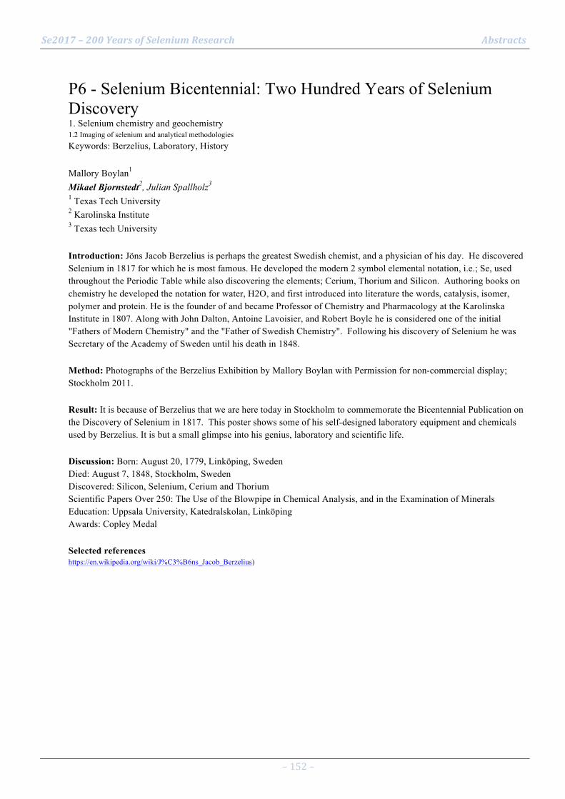
Se2017 – 200 Years of Selenium Research Abstracts
– 152 –
P6 - Selenium Bicentennial: Two Hundred Years of Selenium Discovery 1. Selenium chemistry and geochemistry 1.2 Imaging of selenium and analytical methodologies Keywords: Berzelius, Laboratory, History Mallory Boylan1 Mikael Bjornstedt2, Julian Spallholz3 1 Texas Tech University 2 Karolinska Institute 3 Texas tech University Introduction: Jöns Jacob Berzelius is perhaps the greatest Swedish chemist, and a physician of his day. He discovered Selenium in 1817 for which he is most famous. He developed the modern 2 symbol elemental notation, i.e.; Se, used throughout the Periodic Table while also discovering the elements; Cerium, Thorium and Silicon. Authoring books on chemistry he developed the notation for water, H2O, and first introduced into literature the words, catalysis, isomer, polymer and protein. He is the founder of and became Professor of Chemistry and Pharmacology at the Karolinska Institute in 1807. Along with John Dalton, Antoine Lavoisier, and Robert Boyle he is considered one of the initial "Fathers of Modern Chemistry" and the "Father of Swedish Chemistry". Following his discovery of Selenium he was Secretary of the Academy of Sweden until his death in 1848. Method: Photographs of the Berzelius Exhibition by Mallory Boylan with Permission for non-commercial display; Stockholm 2011. Result: It is because of Berzelius that we are here today in Stockholm to commemorate the Bicentennial Publication on the Discovery of Selenium in 1817. This poster shows some of his self-designed laboratory equipment and chemicals used by Berzelius. It is but a small glimpse into his genius, laboratory and scientific life. Discussion: Born: August 20, 1779, Linköping, Sweden Died: August 7, 1848, Stockholm, Sweden Discovered: Silicon, Selenium, Cerium and Thorium Scientific Papers Over 250: The Use of the Blowpipe in Chemical Analysis, and in the Examination of Minerals Education: Uppsala University, Katedralskolan, Linköping Awards: Copley Medal Selected references https://en.wikipedia.org/wiki/J%C3%B6ns_Jacob_Berzelius)

Se2017 – 200 Years of Selenium Research Abstracts
– 153 –
P7 - Simultaneous determination of selenium and sulfur species in biological samples 1. Selenium chemistry and geochemistry 1.2 Imaging of selenium and analytical methodologies Keywords: sulfur, HPLC, mass spectrometry, biological samples Nina Kroepfl1 Kevin A. Francesconi1, Tanja Schwerdtle2, Doris Kuehnelt1 1 University of Graz, Institute of Chemistry, Graz, Austria 2 University of Potsdam, Institute of Nutritional Science, Nuthetal, Germany Introduction: The micronutrient selenium and the macronutrient sulfur are essential for life and show strong similarity in respect to their chemical properties and naturally occurring compounds. To further elucidate similarities and differences of selenium and sulfur, simultaneous determination of their species in biological samples is of interest. While HPLC/ICPMS is the most common technique for selenium speciation analysis in biological samples, polyatomic interferences on the main sulfur isotope hamper the determination of sulfur species using this technique. Method: Simultaneous determination of selenium and sulfur species in biological samples was performed by HPLC coupled to ICP-triple quadrupole-MS (ICP-QQQ-MS) with oxygen as a reaction gas. Result: We present a method for the simultaneous determination of selenium and sulfur species in biological samples by HPLC/ICP-QQQ-MS. The oxygen reaction mode is used for the detection of selenium and sulfur as SeO+ and SO+, respectively, thereby overcoming interferences typically hindering the ICPMS detection of these two elements. When employing reversed-phase HPLC as the separation step before ICP-QQQ-MS, limits of detection in the low to sub µg per liter range were obtained, for example, for selenoneine and its sulfur analogue ergothioneine, which have both been ascribed antioxidative properties and are, hence expected to be of considerable health relevance. Discussion: HPLC/ICP-QQQ-MS offers the possibility of the simultaneous detection of selenium and sulfur species in biological samples. This approach is a promising tool for further investigation of similarities and differences of these two elements.

Se2017 – 200 Years of Selenium Research Abstracts
– 154 –
P8 - The fractions and distribution of soil selenium in Heilongjiang province and its' impacting factors 1. Selenium chemistry and geochemistry 1.3 Local geological selenium sources and global cycling Keywords: Soil selenium;Fractions;Physical and chemical properties;Heilongjiang province Fengqin Chi1 Enjun Kuang1, Jiuming Zhang1, Zixuan Li2 1 Soil Fertilizer and Environment Resources Institute, Heilongjiang Academy of Agriculture Sciences 2 College of Resources and Environment, Northeast Agricultural University Introduction: The contents, fractions, distribution of soil selenium (Se) and its' influencing factors in different soil types in Heilongjiang Province(China) were surveyed and analyzed. Method: It was investigated 19 soil samples were selected by above five natural geographical areas were collected by 0-20cm, including black soil, albic soil, chernozem, dark brown soil, volcanic ash soil, meadow soil, aeolian sandy soil and saline-alkali soil for the Se fractions analysis and the measurement of physical and chemical properties. Result: The topography of terrain in Heilongjiang Province is vast and complex, and the geochemical conditions are obviously different, which leads to a great change in the total Se content in this study, ranging from 0.0689 to 0.4628 mg kg-1, was generally low. Residual Se and organic Se were the main Se fractions in low Se soils, followed by acid soluble Se. The water soluble Se and exchangeable Se contents which plant could uptake was very low, which may be main reason resulting in low Se bioavailability content in soils in Heilongjiang. There was a significant correlation among soil acidic Se, organic Se and residual Se and total Se content in soil, but the correlation between water soluble and exchangeable selenium was not significant. Discussion: The personal correlation analysis showed that the contents of metal oxides, clay and sand were the main factors affecting soil Se capacity, which restricted the distribution of total Se in soil. pH, soil organic carbon (SOC) and other factors could not be ignored in impacting the content of Se.

Se2017 – 200 Years of Selenium Research Abstracts
– 155 –
P9 - Zoning pollution-free and selenium-rich land resources with geochemistry 1. Selenium chemistry and geochemistry 1.3 Local geological selenium sources and global cycling Keywords: cadmium, safe utilization Tao Yu1 Zhongfang Yang1, Qingye Hou1, Xueqi Xia1 1 China University of Geosciences, Beijing Introduction: It is difficult to maintain a safe level of selenium (Se) intake in adults, owing mainly to the relatively narrow gap between Se deficiency and excessive Se dose.Thus it’s meaningful for zoning safe and Se-rich soils. Method: A typical Se-rich area covering 50 000 ha in Enshi (located in Hubei, China) was investigated. Hundreds of samples as rhizosphere soils, corns, irrigation water, soil profiles, rocks, fertilizers and atmospheric depositions were collected. All of the samples were well prepared and analyzed with recommended methods. Result: It was inferred that the status of Se in crops showed the strong dependence on the total Se in soils that were similar to the status of Cd. The average total Se and Cd concentrations in corn topsoil were 1.88 and 2.12 µg•g−1, respectively. Higher Se and Cd concentrations in soil were distributed with the Permian or Triassic strata which were stretched with the black shale. The geochemical model of the Se cycle model implied that parent rocks were the main source of soil Se contents. Discussion: The average total Se and Cd concentrations in corn were 0.39 and 0.24 µg•g−1, respectively. In the study area, the concentrations of Se and Cd in corns were highly correlated with Se, Cd, Hg, Cu, Zn, K and Ca in soils. Considering the safe intake level of Se and Cd by corns, the models of Se and Cd concentrations in crop-soil system were built by multiple regression analysis. With these, the areas where were favorable to utilizing safe Se-rich crops were zoned. Selected references
1. Yang, Z., Yu, T. and Hou, Q., et al. (2014). Geochemical evaluation of land quality in China and its applications. Journal of Geochemical Exploration 139: 122-135.
2. Yu, T., Yang, Z. and Lv, Y., et al. (2014). The origin and geochemical cycle of soil selenium in a Se-rich area of China. Journal of Geochemical Exploration 139: 97-108.
3. Levander QA. (1997).Selenium requirements as discussed in the 1996 joint FAO/IAWA/WHO expert consultation on trace elements in human nutrition.Biomedical and Environmental Sciences10:214-219.
4.Navarro-Alarcón M, López-Martínez MC. (2000).Essentiality of selenium in the human body: relationship with different diseases.Science of The Total Environment 249:347–371.
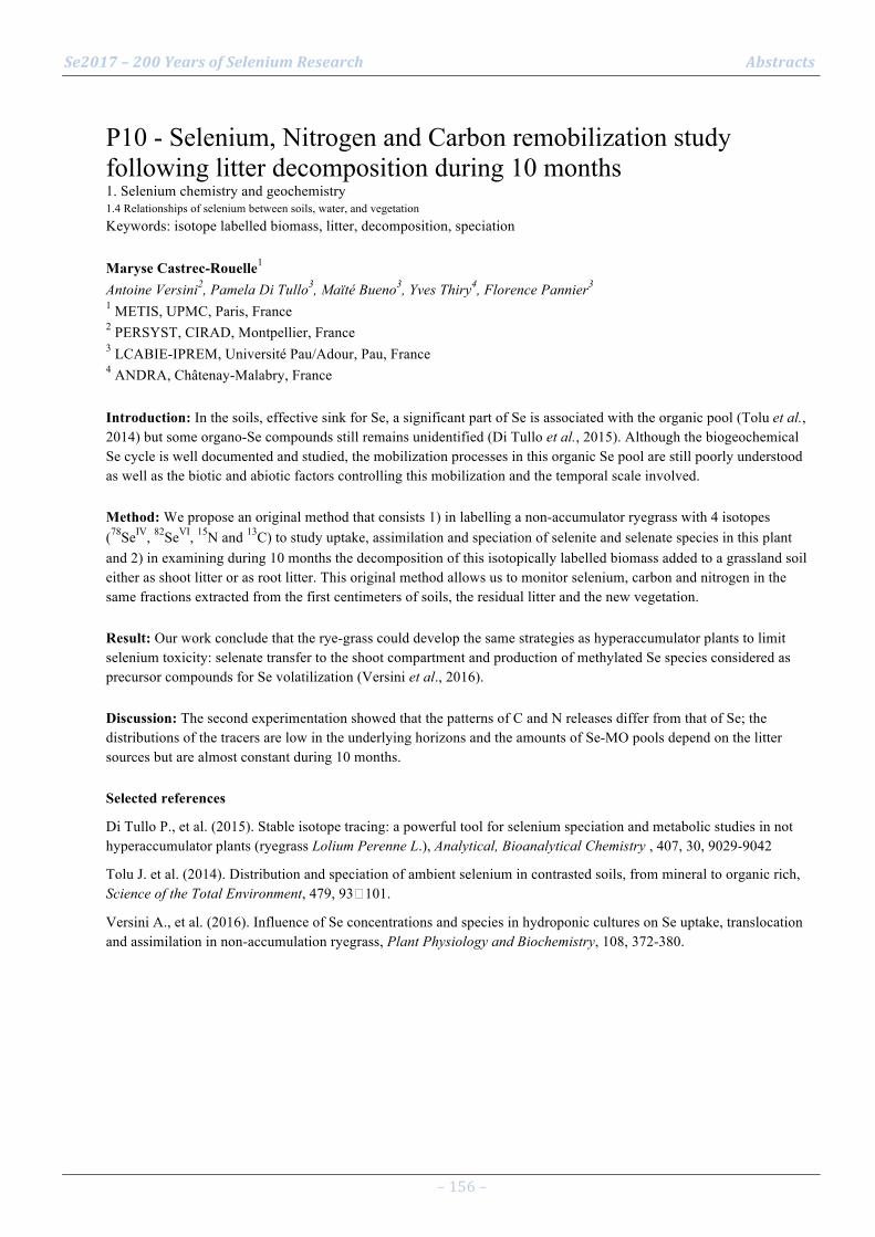
Se2017 – 200 Years of Selenium Research Abstracts
– 156 –
P10 - Selenium, Nitrogen and Carbon remobilization study following litter decomposition during 10 months 1. Selenium chemistry and geochemistry 1.4 Relationships of selenium between soils, water, and vegetation Keywords: isotope labelled biomass, litter, decomposition, speciation Maryse Castrec-Rouelle1 Antoine Versini2, Pamela Di Tullo3, Maïté Bueno3, Yves Thiry4, Florence Pannier3 1 METIS, UPMC, Paris, France 2 PERSYST, CIRAD, Montpellier, France 3 LCABIE-IPREM, Université Pau/Adour, Pau, France 4 ANDRA, Châtenay-Malabry, France Introduction: In the soils, effective sink for Se, a significant part of Se is associated with the organic pool (Tolu et al., 2014) but some organo-Se compounds still remains unidentified (Di Tullo et al., 2015). Although the biogeochemical Se cycle is well documented and studied, the mobilization processes in this organic Se pool are still poorly understood as well as the biotic and abiotic factors controlling this mobilization and the temporal scale involved. Method: We propose an original method that consists 1) in labelling a non-accumulator ryegrass with 4 isotopes (78SeIV, 82SeVI, 15N and 13C) to study uptake, assimilation and speciation of selenite and selenate species in this plant and 2) in examining during 10 months the decomposition of this isotopically labelled biomass added to a grassland soil either as shoot litter or as root litter. This original method allows us to monitor selenium, carbon and nitrogen in the same fractions extracted from the first centimeters of soils, the residual litter and the new vegetation. Result: Our work conclude that the rye-grass could develop the same strategies as hyperaccumulator plants to limit selenium toxicity: selenate transfer to the shoot compartment and production of methylated Se species considered as precursor compounds for Se volatilization (Versini et al., 2016). Discussion: The second experimentation showed that the patterns of C and N releases differ from that of Se; the distributions of the tracers are low in the underlying horizons and the amounts of Se-MO pools depend on the litter sources but are almost constant during 10 months. Selected references
Di Tullo P., et al. (2015). Stable isotope tracing: a powerful tool for selenium speciation and metabolic studies in not hyperaccumulator plants (ryegrass Lolium Perenne L.), Analytical, Bioanalytical Chemistry , 407, 30, 9029-9042
Tolu J. et al. (2014). Distribution and speciation of ambient selenium in contrasted soils, from mineral to organic rich, Science of the Total Environment, 479, 93�101.
Versini A., et al. (2016). Influence of Se concentrations and species in hydroponic cultures on Se uptake, translocation and assimilation in non-accumulation ryegrass, Plant Physiology and Biochemistry, 108, 372-380.
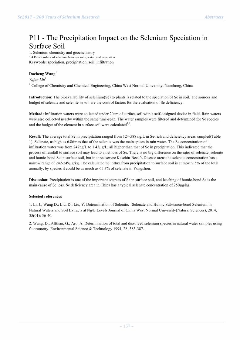
Se2017 – 200 Years of Selenium Research Abstracts
– 157 –
P11 - The Precipitation Impact on the Selenium Speciation in Surface Soil 1. Selenium chemistry and geochemistry 1.4 Relationships of selenium between soils, water, and vegetation Keywords: speciation, precipitation, soil, infiltration Dacheng Wang1 Yajun Liu1 1 College of Chemistry and Chemical Engineering, China West Normal Uinversity, Nanchong, China Introduction: The bioavailability of selenium(Se) to plants is related to the speciation of Se in soil. The sources and budget of selenate and selenite in soil are the control factors for the evaluation of Se deficiency. Method: Infiltration waters were collected under 20cm of surface soil with a self-designed devise in field. Rain waters were also collected nearby within the same time-span. The water samples were filtered and determined for Se species and the budget of the element in surface soil were calculated1,2. Result: The average total Se in precipitation ranged from 124-588 ng/L in Se-rich and deficiency areas sampled(Table 1). Selenate, as high as 6.8times that of the selenite was the main spices in rain water. The Se concentration of infiltration water was from 247ng/L to 1.43µg/L, all higher than that of Se in precipitation. This indicated that the process of rainfall to surface soil may lead to a net loss of Se. There is no big difference on the ratio of selenate, selenite and humic-bond Se in surface soil, but in three severe Kaschin-Beck’s Disease areas the selenate concentration has a narrow range of 242-249µg/kg. The calculated Se influx from precipitation to surface soil is at most 9.5% of the total annually, by species it could be as much as 65.3% of selenate in Yongshou. Discussion: Precipitation is one of the important sources of Se in surface soil, and leaching of humic-bond Se is the main cause of Se loss. Se deficiency area in China has a typical selenate concentration of 250µg/kg. Selected references
1. Li, J., Wang D.; Liu, D.; Liu, Y. Determination of Selenite,Selenate and Humic Substance-bond Selenium in Natural Waters and Soil Extracts at Ng/L Levels Journal of China West Normal University(Natural Sciences), 2014, 35(01): 36-40.
2. Wang, D.; Alfthan, G.; Aro, A. Determination of total and dissolved selenium species in natural water samples using fluorometry. Environmental Science & Technology 1994, 28: 383-387.

Se2017 – 200 Years of Selenium Research Abstracts
– 158 –
P12 - Effect of pH, iron plaque and phosphorus on selenium uptake by rice seedling 1. Selenium chemistry and geochemistry 1.4 Relationships of selenium between soils, water, and vegetation Keywords: pH levels, iron, phosphorus, root, shoot Ju Min1 Shuhui Yu2, Xinbin Zhou2, Weiming Shi1 1 Institute of Soil Science, Chinese Academy of Sciences, Nanjing, China 2 College of Resource and Environment,Southwest University,Chongqing,China Introduction: Plants are the dominant source of Se for animals and humans. Rice is the staple food for people in many parts of the word. Rice plays a crucial role in the food supply in China, and it is also an efficient way to provide Se to consumers. Thus, Se uptake by rice plants is a significant component of the food chain for satisfying human nutrition requirements. The aim of the current study was to explore the effect of iron plaque and phosphorus on selenium uptake by rice seedling in response to different pH levels. Method: A hydroponic experiment was carried out. Result: The results showed that the content of selenium in shoots and roots of rice seedling was significantly affected by pH level. The highest content of selenium in the shoots and roots were at pH 5 and pH 3, respectively. The selenium uptake by rice seedling shoots was inhibited by iron plaque, but this effect on roots was not significantly. Phosphorus significantly promoted the selenium uptake by rice seedling shoots. When pH changed from 3 to 8, the distribution rate of selenium was increased at first and then decreased in the shoots, while the opposite tendency in the roots. Discussion: Rice absorption and transport of selenium affected by pH, iron plaque and phosphorus composite. In the rice cultivation, should be paid attention to the comprehensive adjustment of soil pH, phosphorus and iron fertilization conditions, to increase the selenium accumulation in rice. Selected references
Li S M, Bañuelos G S, Min J and Shi W M*. Effect of continuous application of inorganic nitrogen fertilizer on selenium concentration in vegetables grown in the Taihu Lake region of China. Plant Soil 2015, 393:351-360.
Zhou X B, Yu S H, Lai F. Mechanisms of differences in selenium absorption and transport between rice plants different in cultivar (In Chinese). Acta Pedologica Sinica, 2014, 51(3): 594-599.
Zhou X B, Shi W M, Yang L Z. Study on mechanisms of selenium accumulation in rice grains (In Chinese) . Plant Nutrition and Fertilizer Science, 2008, 14(3): 503-507.
Zhou X B, Shi W M, Zhang L H. Iron plaque outside roots affects selenite uptake by rice seedlings ( Oryza sativa L. ) grown in solution culture. Plant and Soil, 2007, 290(1/2): 17-28.
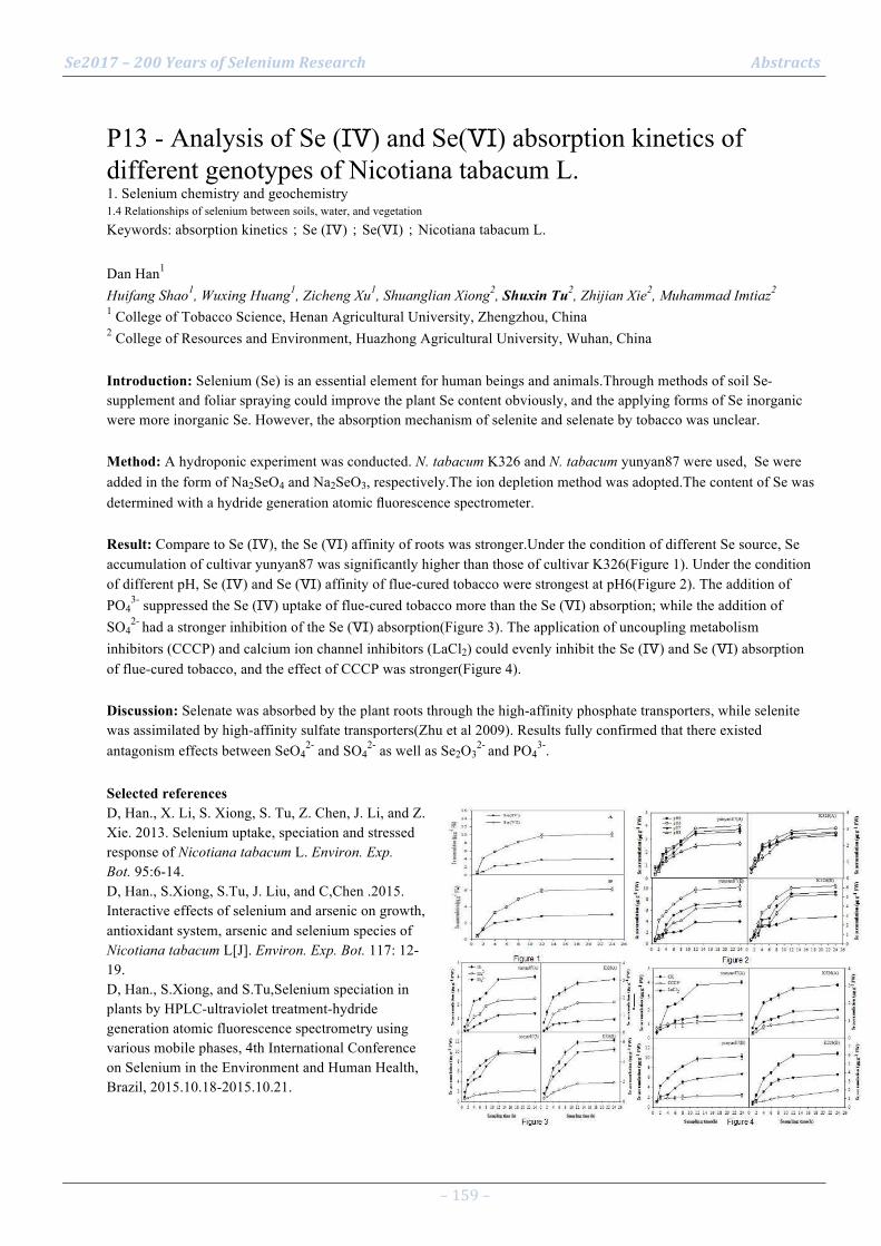
Se2017 – 200 Years of Selenium Research Abstracts
– 159 –
P13 - Analysis of Se (Ⅳ) and Se(Ⅵ) absorption kinetics of different genotypes of Nicotiana tabacum L. 1. Selenium chemistry and geochemistry 1.4 Relationships of selenium between soils, water, and vegetation Keywords: absorption kinetics;Se (Ⅳ);Se(Ⅵ);Nicotiana tabacum L. Dan Han1 Huifang Shao1, Wuxing Huang1, Zicheng Xu1, Shuanglian Xiong2, Shuxin Tu2, Zhijian Xie2, Muhammad Imtiaz2 1 College of Tobacco Science, Henan Agricultural University, Zhengzhou, China 2 College of Resources and Environment, Huazhong Agricultural University, Wuhan, China Introduction: Selenium (Se) is an essential element for human beings and animals.Through methods of soil Se-supplement and foliar spraying could improve the plant Se content obviously, and the applying forms of Se inorganic were more inorganic Se. However, the absorption mechanism of selenite and selenate by tobacco was unclear. Method: A hydroponic experiment was conducted. N. tabacum K326 and N. tabacum yunyan87 were used, Se were added in the form of Na2SeO4 and Na2SeO3, respectively.The ion depletion method was adopted.The content of Se was determined with a hydride generation atomic fluorescence spectrometer. Result: Compare to Se (Ⅳ), the Se (Ⅵ) affinity of roots was stronger.Under the condition of different Se source, Se accumulation of cultivar yunyan87 was significantly higher than those of cultivar K326(Figure 1). Under the condition of different pH, Se (Ⅳ) and Se (Ⅵ) affinity of flue-cured tobacco were strongest at pH6(Figure 2). The addition of PO4
3- suppressed the Se (Ⅳ) uptake of flue-cured tobacco more than the Se (Ⅵ) absorption; while the addition of SO4
2- had a stronger inhibition of the Se (Ⅵ) absorption(Figure 3). The application of uncoupling metabolism inhibitors (CCCP) and calcium ion channel inhibitors (LaCl2) could evenly inhibit the Se (Ⅳ) and Se (Ⅵ) absorption of flue-cured tobacco, and the effect of CCCP was stronger(Figure 4). Discussion: Selenate was absorbed by the plant roots through the high-affinity phosphate transporters, while selenite was assimilated by high-affinity sulfate transporters(Zhu et al 2009). Results fully confirmed that there existed antagonism effects between SeO4
2- and SO42- as well as Se2O3
2- and PO43-.
Selected references D, Han., X. Li, S. Xiong, S. Tu, Z. Chen, J. Li, and Z. Xie. 2013. Selenium uptake, speciation and stressed response of Nicotiana tabacum L. Environ. Exp. Bot. 95:6-14. D, Han., S.Xiong, S.Tu, J. Liu, and C,Chen .2015. Interactive effects of selenium and arsenic on growth, antioxidant system, arsenic and selenium species of Nicotiana tabacum L[J]. Environ. Exp. Bot. 117: 12-19. D, Han., S.Xiong, and S.Tu,Selenium speciation in plants by HPLC-ultraviolet treatment-hydride generation atomic fluorescence spectrometry using various mobile phases, 4th International Conference on Selenium in the Environment and Human Health, Brazil, 2015.10.18-2015.10.21.

Se2017 – 200 Years of Selenium Research Abstracts
– 160 –
P14 - Agronomic biofortification of carrots with selenium in oxidic soil 1. Selenium chemistry and geochemistry 1.6 Strategies to improve selenium accumulation and biofortification Keywords: tuber, selenate, Oxisol, greenhouse, nutrition Ediu Carlos Silva Junior1 Josimar Henrique Lessa1, André Baldansi Andrade1, Mateus Olímpyo Tavares de Ávila1, Maria Lígia de Sousa Silva1, Valdemar Faquin1, Luiz Roberto Guimarães Guilherme1 1 Department of Soil Science, Federal University of Lavras, Minas Gerais, Brazil Introduction: Selenium is an important element linked to physiological processes in plants, microorganisms, animals, and humans (Rayman, 2012). The addition of selenium to crops via soil application of Se i.e. agronomic biofortification is recommended to overcome dietary deficiencies (Chilimba et al., 2012; Bañuelos et al., 2015; Winkel et al., 2015). This work tested agronomic biofortification strategies to increase Se to nutritionally ideal levels inside the edible parts of carrots growing in oxidic soils. Method: The experiment was conducted in a greenhouse in Lavras - MG, Brazil. The plots comprised one carrot plant (Daucus carota L.) per pot, filled with 1.6 kg of a clayey-textured Oxisol. The experimental design was completely randomized using seven doses of Se as Na2SeO4: 0.0; 0.1; 0.2; 0.4; 0.8; 1.6 and 3.2 mg kg-1. Total selenium content in carrots was determined after acid digestion with HNO3 + HClO4 (ratio 2:1, 50-200°C over 2 h) followed by analysis with GFAAS. Result: Plants were highly responsive to Se application as selenate in the soil up to the highest dose (3.2 mg kg-1), which resulted in a total Se accumulation of 76.45 mg kg-1 in tubers and 178.89 mg kg-1 by dry weight (DW) in leaves. Discussion: Selenium content was higher in the leaves compared to the storage roots for all doses. The results showed that Se-enriched carrots can be obtained via agronomic biofortification. Consumption of 100 g f.w. of carrots fertilized with 0.4 mg kg-1 Na2SeO4 can supply 100% (80.37 µg) of the Recommended Daily Allowance (RDA) for Se. Selected references
BAÑUELOS, G. S. et al. Selenium biofortification of broccoli and carrots grown in soil amended with Se-enriched hyperaccumulator Stanleya pinnata. Food chemistry, v. 166, p. 603–8, 1 Jan. 2015.
CHILIMBA, A. D. C. et al. Agronomic biofortification of maize with selenium (Se) in Malawi. Field Crops Research, v. 125, p. 118–128, 2012.
RAYMAN, M. P. Selenium and human health. Lancet, v. 379, n. 9822, p. 1256–68, 31 Mar. 2012.
WINKEL, L. H. E. et al. Selenium cycling across soil-plant-atmosphere interfaces: A critical review. Nutrients, v. 7, n. 6, p. 4199–4239, 2015.

Se2017 – 200 Years of Selenium Research Abstracts
– 161 –
P15 - Effects of different selenium application on selenium accumulation in Lentinula edode 1. Selenium chemistry and geochemistry 1.6 Strategies to improve selenium accumulation and biofortification Keywords: Lentinula edodes; selenium biofortification Fei Zhou1 Dongli Liang1, Wenxiao Yang1, Yuexia Miao1 1 College of Natural Resources and Environment, Northwest A&F University, Yangling, Shaanxi, China Introduction: Many edible fungi have been demonstrated to absorb inorganic Se from the substrate and convert it into more efficient forms (Maseko et al., 2013), but little study is conducted on the effect of exogenous Se species on the growth and enrichment of Se in Lentinula edodes. Therefore the difference of selenite, selenate and Se-enriched yeast on Se accumulation was studied to explore the absorption of Se in Lentinula edodes. Method: Na2SeO3, Na2SeO4 and Se-enriched yeast (Se content: 510 mg/kg) were injected into base material for mushroom one week before fruiting. The mature fruiting bodies were harvested at 7, 14, 21, 28, 49and 70 days after injection, respectively. All samples were collected, oven-dried at 55�, weighed and ground. After digestion with HNO3-HClO4 (4:1, v/v) and reduction in 6 mol/L HCl, Se concentrations were determined by HG-AFS. Result: All treatments didn’t cause visible impact for the growth of L.edodes. The concentration in L.edodes fruit bodies, which was applied with Se, considerably increased and it was proportional to the amount of Se added to the substrate (Fig. 1). Se contnets of mushrooms increased firstly and then decreased with the increase of harvest times. The highest content was occured in 21(selenite) and 49 (selenate and Se-yeast) days after injection, respectively.When the same concentration was added into the substrate, Se contents of mushrooms were in the order of Na2SeO3 > Na2SeO4 > Se-yeast. Discussion: L.edodes is a good Se accumulator. Among investigated Se species sodium selenite is the best sources in Se biofortification of L.edodes. Selected references
Maseko T, Callahan D L, Dunshea F R, et al. Chemical characterisation and speciation of organic selenium in cultivated selenium-enriched Agaricus bisporus[J]. Food Chemistry, 2013, 141(4):3681.

Se2017 – 200 Years of Selenium Research Abstracts
– 162 –
P16 - Apples: a suitable target for selenium biofortification? 1. Selenium chemistry and geochemistry 1.6 Strategies to improve selenium accumulation and biofortification Keywords: biofortification, fruits, foliar spray, health claims, consumer acceptance Diemo Daum1 Christoph Budke1, Lena Wortmann1, Ulrich Enneking1 1 Osnabrueck University of Applied Sciences, Osnabrueck, Germany Introduction: Apples are the most important fruit crop grown in Germany. As with most food of plant origin, the selenium (Se) content of apples is relatively low and thus the fruits contribute little to human Se intake. The objective of this study was to investigate whether apples are suitable for Se biofortification programs, both from an agronomical and a consumer point of view. Method: Apple trees were sprayed with different doses of sodium selenite and sodium selenate, respectively. The Se content in fruits was analyzed by graphite furnace AAS. The consumer survey, including 384 respondents, was performed online and based on a questionnaire with 32 items. Result: The targeted selenium content in apple fruits (10 – 20 µg Se/100 g fm) was achieved by a single foliar spray of 100 – 150 g Se/ha both with selenite and selenate. At this enrichment level of Se, the use of nutritional and health claims is approved according to regulations (EC) No 1924/2006 and (EU) No 432/2012, respectively. About 52% of all consumers questioned expect selenium in food to have a positive impact on human health and 56% would appreciate apples offered with the claim “rich in selenium”. Among the health claims, the statement “Se contributes to the normal functioning of the immune system” was most attractive for those questioned (76%). Discussion: With regard to the promising results of our preliminary spray trials, and the predominantly positive consumer perception of Se enriched fruit produce, it seems to be worthwhile to develop an appropriate Se biofortification technique for apples.

Se2017 – 200 Years of Selenium Research Abstracts
– 163 –
P17 - Optimising fertiliser formulations for selenium biofortification of wheat grain 1. Selenium chemistry and geochemistry 1.6 Strategies to improve selenium accumulation and biofortification Keywords: biofortification, macronutrient carrier, application method, wheat Chandnee Ramkissoon1 Dr Mike McLaughlin1, Dr Fien Degryse1, Dr Scott Young2 1 University of Adelaide 2 University of Nottingham Introduction: Currently, about 15% of the global population is at risk of health problems due to inadequate consumption of the essential micronutrient selenium (Se). Although biofortification of staple crops through soil or foliar application of Se has proven to be very successful, few studies have investigated crop response to the application of different Se-enriched macronutrient fertiliser matrices, which could reduce labour and application costs. Method: Hence, pot trials were set up to determine crop uptake of Se by different macronutrient carriers and methods of application in 3 soils with contrasting properties. Four macronutrient fertilisers – urea, di-ammonium phosphate, muriate of potash and sulfate of ammonia – were enriched with Se by mixing the macronutrient powder with sodium selenate solution and pressing the mixture into 4 cm pellets, prior to cutting them into tablets. The granules were then soil applied just before planting the seedlings. For the foliar fertilisation, Se was applied to leaves with a micropipette, as sodium selenate with either urea or urea ammonium nitrate at ear emergence. In both experiments, Se was supplied at a constant rate of 3.33 µg kg-1 soil (a rate equivalent to 10 g ha-1). Wheat was grown under controlled conditions until maturity. After 12 weeks, grains were harvested and analysed for Se concentrations by ICPMS. Result: Uptake data, which show differences in Se accumulation due to macronutrient carrier and/or application method, will be presented at the conference. Discussion: To our current knowledge, this is the first study undertaken to determine the optimal fertiliser matrix to supplement wheat with Se.
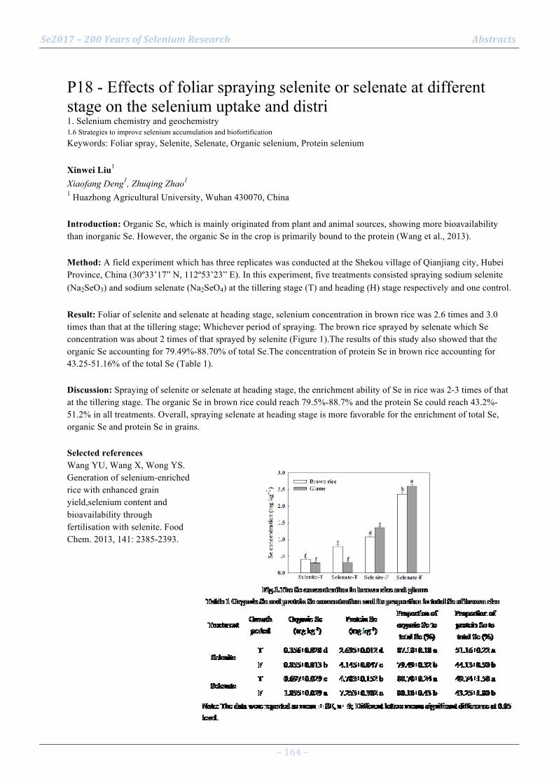
Se2017 – 200 Years of Selenium Research Abstracts
– 164 –
P18 - Effects of foliar spraying selenite or selenate at different stage on the selenium uptake and distri 1. Selenium chemistry and geochemistry 1.6 Strategies to improve selenium accumulation and biofortification Keywords: Foliar spray, Selenite, Selenate, Organic selenium, Protein selenium Xinwei Liu1 Xiaofang Deng1, Zhuqing Zhao1 1 Huazhong Agricultural University, Wuhan 430070, China Introduction: Organic Se, which is mainly originated from plant and animal sources, showing more bioavailability than inorganic Se. However, the organic Se in the crop is primarily bound to the protein (Wang et al., 2013). Method: A field experiment which has three replicates was conducted at the Shekou village of Qianjiang city, Hubei Province, China (30º33’17” N, 112º53’23” E). In this experiment, five treatments consisted spraying sodium selenite (Na2SeO3) and sodium selenate (Na2SeO4) at the tillering stage (T) and heading (H) stage respectively and one control. Result: Foliar of selenite and selenate at heading stage, selenium concentration in brown rice was 2.6 times and 3.0 times than that at the tillering stage; Whichever period of spraying. The brown rice sprayed by selenate which Se concentration was about 2 times of that sprayed by selenite (Figure 1).The results of this study also showed that the organic Se accounting for 79.49%-88.70% of total Se.The concentration of protein Se in brown rice accounting for 43.25-51.16% of the total Se (Table 1). Discussion: Spraying of selenite or selenate at heading stage, the enrichment ability of Se in rice was 2-3 times of that at the tillering stage. The organic Se in brown rice could reach 79.5%-88.7% and the protein Se could reach 43.2%-51.2% in all treatments. Overall, spraying selenate at heading stage is more favorable for the enrichment of total Se, organic Se and protein Se in grains. Selected references Wang YU, Wang X, Wong YS. Generation of selenium-enriched rice with enhanced grain yield,selenium content and bioavailability through fertilisation with selenite. Food Chem. 2013, 141: 2385-2393.
Se2017 – 200 Years of Selenium Research Abstracts
– 165 –
P19 - Se and its antagonists Hg, As, Cd in hair of Taiwanese and Russian residents 1. Selenium chemistry and geochemistry 1.7 Selenium interactions with other elements in the environment Keywords: Key words: Hg, As, Cd, Russia, Taiwan, hair Cheng-Chi Wu1 Pai-Tsang Huang2, Oksana Skalnaya3, Irina Zhegalova4, Igor Grabeklis5, Margarita Skalnaya6 1 Neomedi Clinic, Taipei R.O.C. 2 Wan Fang Medical Center, Taipei R.O.C. 3 National Taiwan University, Business Administration, Taipei R.O.C. 4 First Moscow State Medical University; RUDN - People’s Friendship University of Russia, Moscow, Russia 5 Department of radioecology and ecotoxicology, Lomonosov Moscow State University; RUDN - People’s Friendship University of Russia, Moscow, Russia 6 RUDN – People’s Friendship University of Russia; ANO Center for Biotic Medicine, Moscow, Russia Introduction: An antagonism between Se and Hg, As, Cd is well-known, and Se administration has the protective effects against intoxications by these trace elements. Hair levels of Se, Hg, As, Cd were studied in residents of territories differ in climate and life patterns. Method: Totally 178 Taiwanese and 41389 Russians (39162 Moscow, 1972 Novosibirsk, 255 Sakhalin) 20-50 y/o males and females were investigated. Hair samples were analyzed by ICP-MS. Result: For Se in men there was no significant difference for Russian vs Taiwanese (median 0.44 vs 0.50 µg/g, p > 0.1), while Russian women had a difference vs Taiwanese (0.33 vs 0.43 µg/g, p < 0.05). Hair As for women and men in Moscow (0.021, 0.046) was significantly lower vs Taiwan (0.037, 0.065). Hair Hg for women and men was lower in Moscow (0.55, 0.60), Novosibirsk (0.40, 0.43) and Sakhalin for men only (1.16) vs Taiwan (0.84, 1.71). Hair Cd for women and men was higher in all Russian cities (0.0105, 0.0140) vs Taiwan (0.0065, 0.0057). Discussion: Generally, Sakhalin residents were similar to Taiwanese in hair As (males and females), Hg (females), Se (males) due to geographical and nutritional similarity while continental Russians had higher Cd and lower Se. There were no distinct correlations between Se and other elements in hair, while most subjects had their concentrations within normal ranges.
Se2017 – 200 Years of Selenium Research Abstracts
– 166 –
P20 - Selenium and mercury interactions in the apex predators from the Gulf of Trieste (northern Adriatic) 1. Selenium chemistry and geochemistry 1.7 Selenium interactions with other elements in the environment Keywords: Mercury, Fish, Rays, Northern Adriatic Jadran Faganeli1 Ingrid Falnoga2, Darja Mazej2, Lovrenc Lipej1, Katja Klun1, Milena Horvat2 1 Marine Biological Station, National Institute of Biology, Fornace 41 6330 Piran, Slovenia 2 Dept. Environmental Sciences, Jozef Stefan Institute, Jamova 39 1000 Ljubljana, Slovenia Introduction: It was suggested that environmental Se moderates the bioaccumulation and toxicity of Hg in marine organisms. Method: Interactions between Se and Hg were studied in waters, plankton, and benthic (Pteromylaeus bovinus, Myliobatis aquila) and pelagic (Dasyiatis violacea, Dasyiatis pastinaca) rays, as apex predators, in the Gulf of Trieste (northern Adriatic Sea), one of the most severely Hg polluted area in the Mediterranean and worldwide due to the input of Hg from the former mine in Idrija (NW Slovenia) by the Soča/Isonzo River. Result: Seawater contains 0.32-2.13 µg/L dissolved Se. Dissolved Hg ranges between 0.18-4.9 ng/L with higher concentrations in area near the river discharge. In net-zooplankton (>200 µm), Se ranged between 2.2-3.8 µg/g (d.w.) and Hg between 130-370 ng/g. Se in ray species varied between 1.0-4.41 µg/g (d.w.) in male and female muscle. Pelagic species contained higher Se in muscle. Slightly lower levels were in liver ranging between 0.87-2.54 µg/g (d.w.) in both genders. The highest Hg (mostly as MeHg) levels were in muscle of D. violacea (1.17-4.40 µg/g d.w.), followed by P. bovinus (0.13-3.05 µg/g d.w.), and D. pastinaca (0.07-0.47 µg/g d.w.). Discussion: In benthic ray species, a parallel increase of Se and Hg in muscle was found, so greater Hg bioaccumulation resulted in Se coaccumulation. Hg/Se ratios (molar) in muscle of pelagic nad benthic species were ≤1 while in zooplankton and liver were <0.01 and <0.7, respectively. Low Hg contents in muscle and liver of rays corresponded to low Hg/Se ratios increasing in muscle to 1 at about 6 µg/g (d.w.).

Se2017 – 200 Years of Selenium Research Abstracts
– 167 –
P21 - Environmental Selenium Influences Fish Methylmercury Bioaccumulation and Risks 1. Selenium chemistry and geochemistry 1.7 Selenium interactions with other elements in the environment Keywords: Mercury, Freshwater Fish Nicholas Ralston1 Laura Raymond2 1 University of North Dakota 2 Translational Medicine Independent Research Consultants Introduction: Environmental mercury can either bioaccumulate within an aquatic food chain as the readily absorbed and highly toxic form methylmercury (MeHg) or biologically retire as insoluble and inert forms such as mercury sulfide (HgS) and mercury selenide (HgSe). Method: Selenium analyses were performed on freshwater fish collected from across North America states. Fish included piscivores and non-piscivores. Result: The HBV of all fish was inversely associated with size and age. While the HBV of more than 95% of piscivores was positive, the incidence of large predatory species with negative HBVs was highly correlated with their occurrence in fresh water bodies with poor environmental Se availability. While Hg accumulation of non-piscivores was also inversely associated with environmental Se, none were found to have negative HBVs. Discussion: MeHg bioaccumulation was found to be inversely related to environmental Se availability. Since MeHg toxicity is proportional to both its absolute as well as relative amounts (in relation to Se), consumption of such fish is likely to be associated with far greater risks than is currently recognized.
Se2017 – 200 Years of Selenium Research Abstracts
– 168 –
P22 - Study of geochemical characteristics and influencing factors of soil selenium in the typical soil 1. Selenium chemistry and geochemistry 1.7 Selenium interactions with other elements in the environment Keywords: profiles, influencing factors Zhongfang Yang1 Qingye Hou1, Qiong Yang1, Qiubei Gu1, Tao Yu1 1 China University of Geosciences, Beijing Introduction: Selenium was discovered by a Swedish chemist, in 1817. After two hundred years, people are increasingly concerned about this marvelous element. Many Se-rich soils have been investigated around China, but the mechanism of the migration and transformation of selenium in soil is still hot issues in the field of geochemistry. Method: The typical soil profiles of carbonate rock and terrigenous clastic rock in Wuming County of Guangxi were chosen as the investigative objects.Chemical elements, pH and total organic carbon(TOC) were analyzed by recommended methods. Result: The results showed that the soil selenium content was inherited from the parent materials, with average values of 0.55 mg/kg and 1.43 mg/kg respectively; and the selenium content of the carbonate rock was enriched significantly in the surface soil, but relatively stable in the terrigenous clastic rock. The total water-soluble selenium content of the carbonate rock was higher than that in the terrigenous clastic rock, with arithmetic average values of 3.48 µg/kg and 1.81 µg/kg respectively, and they were both given priority to with selenate, followed by selenite and humic acid combined with selenium. Discussion: Different factors which may control the contents of selenium were considered. It was infered that the contents and chemical speciations of selenium in soil profiles originated from the carbonate rock were mainly dominated by the total organic carbon (TOC) contents and pH values, while of which originated from the terrigenous clastic rock were controlled by the pH values, the contents of TOC, aluminum oxide , iron oxide and soil texture. Selected references
1. Yu, T., Yang, Z. and Lv, Y., et al. (2014). The origin and geochemical cycle of soil selenium in a Se-rich area of China. Journal of Geochemical Exploration 139: 97-108.
2. Lv, Y., Yu, T. and Yang, Z., et al. (2014). Constraint on selenium bioavailability caused by its geochemical behavior in typical Kaschin–Beck disease areas in Aba, Sichuan Province of China. Science of The Total Environment 493: 737-749.
3. Weng, L., Vega, F. A. and Supriatin, S., et al. (2011). Speciation of Se and DOC in Soil Solution and Their Relation to Se Bioavailability. Environmental Science & Technology 45 (1): 262-267.
4. Sun, G., Meharg, A. A. and Li, G., et al. (2016). Distribution of soil selenium in China is potentially controlled by deposition and volatilization? Scientific Reports 6: 20953.

Se2017 – 200 Years of Selenium Research Abstracts
– 169 –
P23 - Interaction of selenium and cadmium in soil-corn system in natural selenium and cadmium rich area 1. Selenium chemistry and geochemistry 1.7 Selenium interactions with other elements in the environment Keywords: cadmium, corn, bioaccumulation factors, interaction Zezhou Zhang1 Linxi Yuan2 1 State Key Laboratory of Biogeology and Environmental Geology, China University of Geosciences, Wuhan 2 Jiangsu Bio-Engineering Centre on Selenium, Suzhou, Jiangsu, China Introduction: A large number of studies have confirmed that selenium-cadmium (Se-Cd) interaction is a widespread phenomenon. However, little attention has been paid to plants especially in natural Se-Cd rich area. To investigate the influence of Se on the uptake and translocation of Cd in the soil-corn system, the levels of Se and Cd in different parts of corn plants and corresponding soils of root zones collected from a Se-Cd rich area were determined. Method: Se and Cd speciations in soil were treated by sequential extraction protocol [1, 2]. All treated samples were acid-digested and analyzed for total Se by hydride generation atomic fluorescence spectrometry (HG-AFS), and Cd by atomic absorption spectroscopy (AAS) Result: Molar ratios of bioavailable forms of Se and Cd (Se:Cd) in the soil and the bioaccumulation factors (BAFs) of Cd were used to elucidate interaction effects of Se-Cd between soil and corn. A reduction in translocation of Cd in the aerial parts with increasing Se levels in the soils was observed. However, the levels of bioavailable forms of Se in the soils were positively correlated with the BAFs of Cd in the roots when molar ratios of bioavailable forms of Se and Cd (Se:Cd) in the soil over ten. Discussion: The pronounced role of Se in protecting corn plants from Cd bioaccumulation and its potential use for reducing Cd concentration in different tissues of corn plants, and the root may act as an effective buffer or barrier for the suppression of Cd absorption or translocation to the aerial tissues [3-6]. Selected references [1] Honma T, Ohba H, Makino T, et al. Relationship between Cadmium Fractions Obtained by Sequential Extraction of Soil and the Soil Properties in Contaminated and Uncontaminated Paddy Soils[J]. Journal Of Chemistry,2015, 2015:1-9 [2] Kulp T R, Pratt L M. Speciation and weathering of selenium in Upper Cretaceous chalk and shale from South Dakota and Wyoming, USA[J]. Geochimica Et Cosmochimica Acta, 2004, 68(18): 3687-3701. [3] Zhang H, Feng X, Zhu J, et al. Selenium in Soil Inhibits Mercury Uptake and Trans location in Rice (Oryza sativa L.)[J]. Environmental Science & Technology, 2012, 46(18): 10040-10046. [4] Schutzendubel A, Schwanz P, Teichmann T, et al. Cadmium-induced changes in antioxidative systems, hydrogen peroxide content, and differentiation in Scots pine roots[J]. Plant Physiology, 2001, 127(3): 887-898. [5] Zembala M, Filek M, Walas S, et al. Effect of selenium on macro- and microelement distribution and physiological parameters of rape and wheat seedlings exposed to cadmium stress[J]. Plant And Soil, 2010, 329(1-2): 457-468. [6] Lin L, Zhou W, Dai H, et al. Selenium reduces cadmium uptake and mitigates cadmium toxicity in rice[J]. Journal Of Hazardous Materials, 2012, 235:343-351.

Se2017 – 200 Years of Selenium Research Abstracts
– 170 –
P24 - Impact of Initial Se Status and Gpx1 Genotype on Selenoenzyme and Transcript Expression in Mice 2. Selenium in the molecular life sciences 2.1 Metabolism of selenium in living cells Keywords: glutathione peroxidase, metabolism, mice, transcript Roger A Sunde1 Andrew B Blink1, Edward T Zemaitis II1, Julia A Lawinger1 1 Univ of Wisconsin, Madison WI USA 53706 Introduction: Key components of Se metabolism in intact animals include relative rates of Se incorporation into selenoproteins, underlying regulation of selenoprotein transcripts, and Se status itself. At steady state, just 0.1 and 0.05 µg Se/g diet are required to raise liver glutathione peroxidase-1 (Gpx1) and plasma Gpx3 activities, respectively, to plateau levels in wildtype mice. Knockout of Gpx1 both reduces body Se stores but also blocks flux of Se into Gpx1. Method: Here, we studied the short-term availability (flux) of Se in male Gpx1 wildtype (Gpx1+/+), heterozygote (Gpx1+/-t), and knockout (Gpx1-/-) mice, that had been fed a basal Se-deficient diet for 17 wk after weaning, and then were repleted with 0, 0.05, 0.1, 0.2, and 0.3 µg Se/g diet as Na2SeO3 for only 7 days. Result: Se response curves for liver Gpx1 activity were sigmoidal, reaching the plateau at 0.15 µg Se/g for Gpx1+/+ mice but at 0.20 µg Se/g diet for Gpx1+/- mice, and with no effect on liver Gpx1 activity in Gpx1-/- mice (as expected). In contrast, plasma Gpx3 activity increased only in mice fed 0.3 g Se/g diet for all genotypes, and there was little effect on RBC Gpx1 activity regardless of genotype or Se supplementation. Discussion: These results clearly show that the flux of Se metabolism is altered, at least in the short-term, when initially Se-deficient mice are Se repleted, and this is further modified by Gpx1 genotype. Ongoing analysis of selenoprotein transcripts indicates that differential and/or delayed upregulation of Gpx1 transcripts underlies the observed shifts in selenoezyme activity.

Se2017 – 200 Years of Selenium Research Abstracts
– 171 –
P25 - Insight into the microbial respiration of selenium. A metagenomics study 2. Selenium in the molecular life sciences 2.1 Metabolism of selenium in living cells Keywords: Biogenic Selenium, Metagenomics, Microbial Selenium Respiration, Nanoparticles, 16S rRNA Sequencing Simon Mills1 Lucian Staciu2, Gavin Collins1 1 Microbial Commnities Laboratory, Microbiology, National University of Ireland Galway 2 Blaise Pascal University and Institut de Chimie de Clermont-Ferrand Introduction: Selenium oxyanions are effective electron acceptors, and essential trace elements, in microbial respiration [1]. Selenium cycling is highly dependant on microbial communities. A deeper understanding of microbially-mediated selenium cycling is required to exploit selenium microbiology and develop new environmental biotechnologies. This study investigated the phylogeny of microbial populations underpinning selenium reduction in different environments, including soils and waste-conversion biofilms, to expand our understanding of the microbial ecology of selenium cycling. Method: Batch incubations of loam soil; Atlantic peatland soil; and both intact and physically-homogenised, anaerobic sludge granules were used to enrich for selenite- and selenate-reducers by serially sub-culturing enrichments to fresh media. Reductants were lactate:acetate (16mM:4mM) or H2:CO2 (80:20v/v). Population dynamics and community succession were monitored by sequencing 16S rRNA genes in temporal sub-cultures, and to identify key selenium-reducing taxa. Metagenomics were used to comprehensively characterise highly enriched selenium-reducing cultures. 16S rRNA and functional mRNA targets were probed in cross-sections (10um-thick) of intact sludge granules (diameter 0.5-2mm) using fluorescence in situ hybridisations (FISH). Result: The formation of elemental selenium nanoparticles, including on the surface of spherical sludge granules, indicated selenate and selenite reduction, even in autotrophic enrichments fed only with H2 and CO2. Sequencing indicated distinct taxa associated with selenium metabolism in each of the soil and sludge enrichments. FISH revealed highly organised distributions of selenium cyclers across the architecture of sludge granule biofilms. Discussion: The results indicate widely distributed potential for selenium-reduction across an expansive phylogeny in previously unexposed samples. Selected references
[1] Nancharaiah, Y. V, & Lens, P. N. L. (2015). Ecology and Biotechnology of Selenium-Respiring Bacteria, 79(1), 61–80
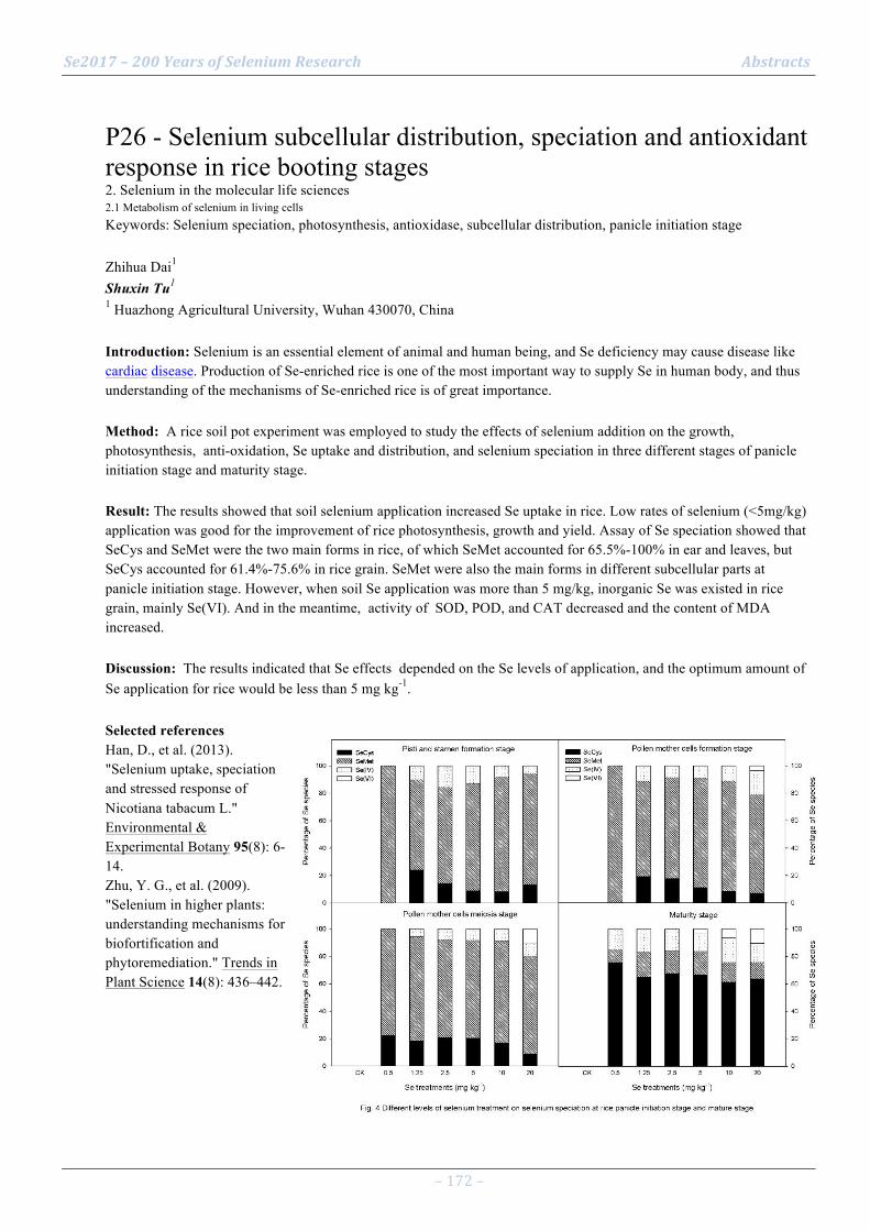
Se2017 – 200 Years of Selenium Research Abstracts
– 172 –
P26 - Selenium subcellular distribution, speciation and antioxidant response in rice booting stages 2. Selenium in the molecular life sciences 2.1 Metabolism of selenium in living cells Keywords: Selenium speciation, photosynthesis, antioxidase, subcellular distribution, panicle initiation stage Zhihua Dai1 Shuxin Tu1 1 Huazhong Agricultural University, Wuhan 430070, China Introduction: Selenium is an essential element of animal and human being, and Se deficiency may cause disease like cardiac disease. Production of Se-enriched rice is one of the most important way to supply Se in human body, and thus understanding of the mechanisms of Se-enriched rice is of great importance. Method: A rice soil pot experiment was employed to study the effects of selenium addition on the growth, photosynthesis, anti-oxidation, Se uptake and distribution, and selenium speciation in three different stages of panicle initiation stage and maturity stage. Result: The results showed that soil selenium application increased Se uptake in rice. Low rates of selenium (<5mg/kg) application was good for the improvement of rice photosynthesis, growth and yield. Assay of Se speciation showed that SeCys and SeMet were the two main forms in rice, of which SeMet accounted for 65.5%-100% in ear and leaves, but SeCys accounted for 61.4%-75.6% in rice grain. SeMet were also the main forms in different subcellular parts at panicle initiation stage. However, when soil Se application was more than 5 mg/kg, inorganic Se was existed in rice grain, mainly Se(VI). And in the meantime, activity of SOD, POD, and CAT decreased and the content of MDA increased. Discussion: The results indicated that Se effects depended on the Se levels of application, and the optimum amount of Se application for rice would be less than 5 mg kg-1. Selected references Han, D., et al. (2013). "Selenium uptake, speciation and stressed response of Nicotiana tabacum L." Environmental & Experimental Botany 95(8): 6-14. Zhu, Y. G., et al. (2009). "Selenium in higher plants: understanding mechanisms for biofortification and phytoremediation." Trends in Plant Science 14(8): 436–442.
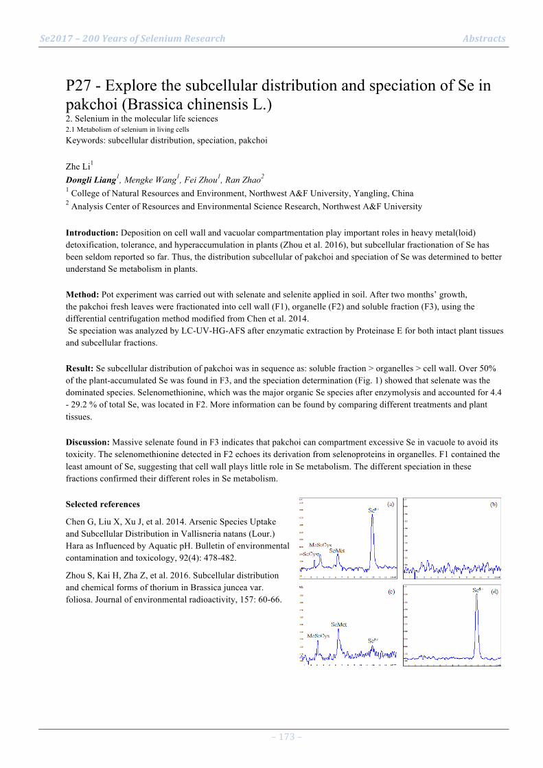
Se2017 – 200 Years of Selenium Research Abstracts
– 173 –
P27 - Explore the subcellular distribution and speciation of Se in pakchoi (Brassica chinensis L.) 2. Selenium in the molecular life sciences 2.1 Metabolism of selenium in living cells Keywords: subcellular distribution, speciation, pakchoi Zhe Li1 Dongli Liang1, Mengke Wang1, Fei Zhou1, Ran Zhao2 1 College of Natural Resources and Environment, Northwest A&F University, Yangling, China 2 Analysis Center of Resources and Environmental Science Research, Northwest A&F University Introduction: Deposition on cell wall and vacuolar compartmentation play important roles in heavy metal(loid) detoxification, tolerance, and hyperaccumulation in plants (Zhou et al. 2016), but subcellular fractionation of Se has been seldom reported so far. Thus, the distribution subcellular of pakchoi and speciation of Se was determined to better understand Se metabolism in plants. Method: Pot experiment was carried out with selenate and selenite applied in soil. After two months’ growth, the pakchoi fresh leaves were fractionated into cell wall (F1), organelle (F2) and soluble fraction (F3), using the differential centrifugation method modified from Chen et al. 2014. Se speciation was analyzed by LC-UV-HG-AFS after enzymatic extraction by Proteinase E for both intact plant tissues and subcellular fractions. Result: Se subcellular distribution of pakchoi was in sequence as: soluble fraction > organelles > cell wall. Over 50% of the plant-accumulated Se was found in F3, and the speciation determination (Fig. 1) showed that selenate was the dominated species. Selenomethionine, which was the major organic Se species after enzymolysis and accounted for 4.4 - 29.2 % of total Se, was located in F2. More information can be found by comparing different treatments and plant tissues. Discussion: Massive selenate found in F3 indicates that pakchoi can compartment excessive Se in vacuole to avoid its toxicity. The selenomethionine detected in F2 echoes its derivation from selenoproteins in organelles. F1 contained the least amount of Se, suggesting that cell wall plays little role in Se metabolism. The different speciation in these fractions confirmed their different roles in Se metabolism. Selected references
Chen G, Liu X, Xu J, et al. 2014. Arsenic Species Uptake and Subcellular Distribution in Vallisneria natans (Lour.) Hara as Influenced by Aquatic pH. Bulletin of environmental contamination and toxicology, 92(4): 478-482.
Zhou S, Kai H, Zha Z, et al. 2016. Subcellular distribution and chemical forms of thorium in Brassica juncea var. foliosa. Journal of environmental radioactivity, 157: 60-66.
Se2017 – 200 Years of Selenium Research Abstracts
– 174 –
P28 - Comparison in bioavailability of nine bioselenocompounds 2. Selenium in the molecular life sciences 2.1 Metabolism of selenium in living cells Keywords: bioavailability, Caco-2, ICP-MS, speciation, selenosugar Kazuaki Takahashi1 Noriyuki Suzuki1, Yasumitsu Ogra1 1 Graduate School of Pharmaceutical Sciences, Chiba University, Chiba, Japan Introduction: Biological effects of selenium (Se) are dependent on its chemical form. In this study, we evaluated the bioavailability of nine naturally occurring Se compounds, or the so-called bioselenocompounds, in vivo and in vitro. Method: The bioselenocompounds such as selenite, selenate and selenocyanate (SeCN-), L-selenomethionin (SeMet), Se-methylseleno-L-cysteine (MeSeCys), L-selenohomolanthionine (SeHLan), selenocystine (SeCys2), Se-methylseleno-N-acetylgalactosamine (SeSug1) and trimethylselenonium ion (TMSe+) were used. Caco-2 cells were used for in vitro membrane permeability study, and in vivo bioavailability was evaluated by the recovery of serum selenoproteins in Se-deficiency rats. Result: In in vitro experiments, SeMet and MeSeCys were more efficiently permeated through Caco-2 monolayer than other bioselenocompounds. Although SeHLan and SeCys2 was other selenoamino acids, SeHLan and SeCys2 were less efficiently transported than SeMet and MeSeCys. Contrary to the results of in vitro study, Quantitative methods indicated no significant differences in bioavailability among the bioselenocompounds except TMSe+ in vivo.(1) These results indicate that animals can equally assimilate both inorganic and organic bioselenocompounds except TMSe+, which is one of the urinary Se metabolites. Discussion: The discrepancy between in vitro and in vivo results is interesting and important for the consideration of the nutritional availability of bioselenocompounds. We offer two explanations for the discrepancy. First, although monomeric selenoamino acids such as SeMet and MeSeCys are more rapidly absorbed in the gastrointestinal tract than other Se species, methylated Se in the selenoamino acids is less efficiently used for the synthesis of selenoproteins than non-methylated Se species. Second, the bioselenocompounds are metabolized/decomposed before absorption in the gastrointestinal tract. Selected references
(1) Takahashi, K.; Suzuki, N.; Ogra, Y. Bioavailability Comparison of Nine Bioselenocompounds In Vitro and In Vivo. Int. J. Mol. Sci. 2017, 18, 506.
Se2017 – 200 Years of Selenium Research Abstracts
– 175 –
P29 - Characterization of rhodanese-like protein from Geobacter sulfurreducens 2. Selenium in the molecular life sciences 2.1 Metabolism of selenium in living cells Keywords: rhodanese, selenoprotein, selenite, Geobacter Olajumoke Kadiri1 Yoshinobu Yamane1, Mst. Ishrat Jahan1, Ryuta Tobe1, Hisaaki Mihara1 1 College of Life Sciences, Ritsumeikan University, Kusatsu, Shiga 525-8577, Japan Introduction: Geobacter sulfurreducens is a metal-reducing anaerobic bacterium. The novel selenoprotein MHSEP involved in selenite/tellurite reduction was found in our previous research. The mhsep gene forms an operon-like gene cluster (gsu2940-2930) on the genome. The gsu2940 gene encodes a protein with a sequence similarity to a sulfurtransferase rhodanese, which catalyzes the detoxification of toxic substances by the relay of sulfur atom through its active site Cys residue. In this study, GSU2940 was produced in E. coli, purified, and characterized. Method: gsu2940 was cloned into pColdI and expressed in E. coli BL21(DE3). GSU2940 was purified as a His-tag fusion protein. A portion of gsu2940 was replaced with a kanamycin-resistance gene to produce the gsu2940-disrupted mutant strain. Result: The purified GSU2940 exhibited a rhodanese activity with the optimum pH at 7.5 and the optimum temperature at 55°C. Thermal stability (Tm) of GSU2940 was determined to be 40°C. Furthermore, the rhodanese activity in the gsu2940-disrupted strain, was remarkably decreased as compared with that in the wild-type strain, suggesting that GSU2940 is the dominant sulfurtransferase in G. sulfurreducens. Discussion: Although GSU2940 exhibited a rhodanese activity, it shows only a slight sequence identity with already-known rhodaneses. GSU2940 is proposed to cooperate with MHSEP and other proteins encoded by the same gene cluster.

Se2017 – 200 Years of Selenium Research Abstracts
– 176 –
P30 - A novel Geobacteraceae-specific outer membrane protein required for selenite and tellurite reduction 2. Selenium in the molecular life sciences 2.1 Metabolism of selenium in living cells Keywords: Porin, selenoprotein, selenite and tellurite reduction, porin-cytochrome complex, Geobacter Mst. Ishrat Jahan1 Ryuta Tobe1, Tatsuo Kurihara2, Jun Kawamoto2, Hisaaki Mihara1 1 College of Life Sciences, Ritsumeikan University, Kusatsu, Shiga 525-8577, Japan 2 Institute for Chemical Research, Kyoto University, Uji, Kyoto 611-0011, Japan Introduction: A putative outer membrane channel protein, GSU2939, was found in a metal-reducing bacterium, Geobacter sulfurreducens. The gsu2939 gene is part of an operon together with 10 other genes including the multi-heme-containing selenoprotein, but the function of GSU2939 remains unexplored. GSU2939 showed only less than 20% homology with phosphate selective porins belonging to the porin P_O family, suggesting that GSU2939 is a Geobacteraceae-specific novel type of porin. Herein, we investigated the characteristics of the putative outer membrane protein GSU2939. Method: Expression level of gsu2939 was examined under various growth conditions. A gsu2939-deficient mutant strain was constructed to investigate its phenotype. Result: The Geobacteraceae-specific putative outer membrane porin, GSU2939, showed impermeability in β-lactam antibiotic transportation. Investigation on the expression levels of gsu2939 at different growth phases in the wild-type cells showed that GSU2930 is a log-phase specific protein. The expression of gsu2939 was neither affected by phosphate-starving conditions nor osmotic-stressed conditions. However, in the presence of selenite and tellurite, a gsu2939-deficient mutant showed more susceptibility than the wild-type strain, suggesting that the protein may not function as a typical outer membrane channel. Furthermore, the gsu2939-deficient mutant showed lower reducing ability for ferric citrate, selenite, and tellurite as an electron acceptor, compared with the wild type. Discussion: Our data suggest that GSU2939 may function as a porin-cytochrome protein complex together with the multi-heme-containing selenoprotein and c-type cytochrome proteins encoded in the same gene cluster and facilitate a trans-outer membrane electron transfer during extracellular reduction of ferric citrate, selenite, and tellurite.

Se2017 – 200 Years of Selenium Research Abstracts
– 177 –
P31 - Effect of inorganic and organic selenium compounds on tumor infiltrating lymphocytes 2. Selenium in the molecular life sciences 2.2 Molecular mechanisms of selenium toxicity Keywords: ovarian cancer, tumor infiltrating lymphocytes Deepika Nair1 Emelie Rådestad2, Nuria Diaz-Argelich3, Johanna Ungerstedt4, Michael Uhlin2, Aristi Fernandes1 1 Department of Medical Biochemistry and Biophysics, Karolinska Institutet, Stockholm, Sweden 2 Department of Oncology and Pathology, Karolinska Institutet, Stockholm, Sweden 3 Department of Organic and Pharmaceutical Chemistry, University of Navarra, Pamplona, Spain. 4 Department of Medicine Huddinge, Karolinska Institutet, Stockholm, Sweden Introduction: Ovarian cancer is characterized by its unique immunosuppressive tumor microenvironment which alters many cellular processes including immune surveillance (1). This impaired functionality of tumour infiltrating lymphocytes (TILs) can lead to defective cytotoxic activity, diminished secretion of lymphokines and failure to proliferate in response to stimulation. Selenium (Se) has known anticancerous effects with many proposed mechanisms including enhanced immune surveillance (2). Little is however known about the different effects between various Se compounds and their specific effect on immune cells, which we aimed to study herein. Method: Immune cells were isolated using density gradient centrifugation and were further selected by magnetic beads. Se cytotoxicity was then compared between tumour and immune cells. Following activation of immune cells and preincubation of organic or inorganic selenium compounds with tumour cells, lysis assay was performed. ELISA, qPCR and flow cytometry based analyses were used to analyse markers of the immune response. Result: Tumour cells were found to be more sensitive to the cytotoxic effects exerted by the Se compounds compared to the immune cells from both healthy donors and TILs from ovarian cancer patients. Our preliminary experiments have also shown effects of Se on immune cell activation, with changes in expression of central genes. Discussion: In conclusion, Se compounds have the potential to act as anticarcinogenic agents in ovarian cancer at concentrations that do not kill or inhibit the immune cells, and concurrently facilitate the immune response. Selected references
(1) Worzfeld T. et al. The Unique Molecular and Cellular Microenvironment of Ovarian Cancer. Frontiers in Oncology, 2017;7:24.
(2) Fernandes AP., Gandin V. Selenium compounds as therapeutic agents in cancer. Biochim Biophys Acta, 2015;1850:8.
Se2017 – 200 Years of Selenium Research Abstracts
– 178 –
P32 - Small Selenium Species: Toxicity, Bioavailability and Modes of Action in Caenorhabditis elegans 2. Selenium in the molecular life sciences 2.2 Molecular mechanisms of selenium toxicity Keywords: selenite, selenomethionine, Se-methylselenocysteine, Caenorhabditis elegans, ICP-QQQ-MS Isabelle Rohn1 Stefanie Raschke1, Talke Marschall1, Michael Aschner2, Doris Kuehnelt3, Tanja Schwerdtle1, Julia Bornhorst1 1 University of Potsdam, Institute of Nutritional Science, Potsdam, Germany 2 Albert Einstein College of Medicine, Department of Molecular Pharmacology, New York, USA 3 University of Graz, Institute of Chemistry, Analytical Chemistry, Graz, Austria Introduction: Small selenium species play a major role in selenium metabolism, but their either toxic or protective potential, bioavailability and metabolic transformations are not fully understood. The roundworm Caenorhabditis elegans is an emerging model in toxicological research and enables to study these endpoints in a whole metabolizing organism. The simplicity of the roundworm having only one selenoprotein (TrxR-1) might give deeper insight into selenium metabolism. Method: Toxicity of selenite, selenomethionine and Se-methylselenocysteine was assessed by lethality and development assays. Total selenium quantification and selenium speciation analysis were performed by (HPLC)-ICP-QQQ-MS based methods. Reactive oxygen and nitrogen species (RONS) and TrxR activity were measured using fluorimetric and colorimetric assays. Result: Selenite exerted strongest toxic effects (LD50 = 12 mM after 30 min incubation in the first larval stage) and caused a developmental delay of worm larvae. In contrast, selenomethionine and Se-methylselenocysteine were only slightly toxic (survival rate decreased by 20% at 50 mM). Total selenium quantification revealed that all three species were bioavailable, with an inverse relationship between toxicity and bioavailability. Speciation analysis indicated that selenomethionine and Se-methylselenocysteine are partially metabolized to other small selenium species. Protection against RONS induction was observed at a low dose range (1 – 100 µM) for all three selenium compounds. Discussion: Our findings indicate that selenomethionine and Se-methylselenocysteine seem to be utilized by Caenorhabditis elegans and undergo, at least in part, metabolisation processes. Independently, all three selenium species shared a protective potential against oxidative stress, while the underlying mechanism remains to be clarified.

Se2017 – 200 Years of Selenium Research Abstracts
– 179 –
P33 - Diabetic fish on selenium supplements 2. Selenium in the molecular life sciences 2.2 Molecular mechanisms of selenium toxicity Keywords: selenomethionine, toxicology, fish, metabolism, cardiac function David Janz1 Jith Thomas2, Connor Pettem2, Lynn Weber3 1 Toxicology Centre, University of Saskatchewan, Saskatoon, Canada 2 Toxicology Graduate Program, University of Saskatchewan, Saskatoon, Canada 3 Veterinary Biomedical Sciences, University of Saskatchewan, Saskatoon, Canada Introduction: A variety of anthropogenic activities increase loading of selenium into aquatic ecosystems, which poses an extreme toxicological hazard to fishes. Although many studies have reported developmental toxicities in larval fishes, fewer studies have investigated sublethal toxicological effects that may occur following dietary selenium exposure in adult fishes. Method: Adult zebrafish were exposed to dietary selenomethionine (SeMet) at Se-normal levels (1.3 µg Se/g food, dry mass) and supraphysiological levels (3.4, 9.8, or 27.5 µg/g) for 90 days. Swimming performance, O2 consumption and metabolic rates were determined using a swim tunnel respirometer. Cardiac function was assessed using high resolution (30 µm) ultrasound biomicroscopy. Energy stores (triglycerides and glycogen) and mRNA transcript abundance of selected genes were determined. Result: Compared to controls, zebrafish exposed to elevated dietary SeMet exhibited impaired swimming performance (lower fatigue velocity, or Ucrit). This was associated with elevated basal metabolic rate and reduced aerobic scope, indicating impaired aerobic capacity. Triglycerides (the primary fuel for aerobic swimming) were elevated in a dose-dependent manner, which was associated with altered transcript abundance of several genes involved in lipid homeostasis. Ultrasonography revealed decreased cardiac output, which was associated with increased echodensity at the atrial-ventricular junction and reduced mRNA expression of the collagenase, MMP2. Discussion: These results suggest significant ecophysiological effects that may impair the fitness of fishes exposed to elevated dietary Se in contaminated ecosystems. From a comparative biomedical viewpoint, these results highlight the utility of zebrafish as a model to investigate mechanisms of metabolic, energetic, and cardiovascular toxicities caused by excessive dietary Se exposure. Selected references Janz, D.M. 2012. Selenium. Pp. 327-374 In: C.W. Wood, A.P. Farrell and C.J. Brauner (Eds.) Fish Physiology Vol 31A, Homeostasis and Toxicology of Essential Metals. Elsevier, San Diego, CA.
Janz, D.M., D.K. DeForest, M.L. Brooks, P.M. Chapman, G. Gilron, D. Hoff, W.D. Hopkins, D.O. McIntyre, C.A. Mebane, V.P. Palace, J.P. Skorupa and M. Wayland. 2010. Selenium toxicity to aquatic organisms. Pp. 141-231 In: P.M. Chapman, W.J. Adams, M.L. Brooks, C.G. Delos, S.N. Luoma, W.A. Maher, H.M. Ohlendorf, T.S. Presser and D.P. Shaw (Eds.) Ecological Assessment of Selenium in the Aquatic Environment. CRC Press, Boca Raton, FL.
Thomas, J.K., S. Wiseman, J.P. Giesy and D.M. Janz. 2013. Effects of chronic selenomethionine exposure on repeat swimming performance, aerobic metabolism and methionine catabolism in adult zebrafish (Danio rerio). Aquatic Toxicology 130-131, 112-122.
Thomas, J.K. and D.M. Janz. 2015. Developmental and persistent toxicities of maternally deposited selenomethionine in zebrafish (Danio rerio). Environmental Science and Technology 49, 10182-10189.
Thomas, J.K. and D.M. Janz. 2016. Embryo microinjection of selenomethionine reduces hatchability and modifies oxidant responsive gene expression in zebrafish. Nature Scientific Reports 6, doi:10.1038/srep26520.
Se2017 – 200 Years of Selenium Research Abstracts
– 180 –
P34 - Biochemical characterization of novel methylseleno derivatives 2. Selenium in the molecular life sciences 2.2 Molecular mechanisms of selenium toxicity Keywords: methylselenol, pancreatic cancer cells, chemotherapeutic drugs. Prajakta Khalkar1 Nuria Diaz Argelich2, Daniel Plano3, Carmen Sanmartin Grijalba2, Aristi Fernandes1 1 Department of Medical Biochemistry and Biophysics (MBB), Karolinska Institute, Stockholm, Sweden 2 Department of Organic and Pharmaceutical Chemistry, University of Navarra, Pamplona, Spain 3 Oncology and Hematology Section, Navarra Institute for Health Research, Pamplona, Spain Introduction: Redox active selenium (Se) compounds at subtoxic doses, act as pro-oxidants and are highly cytotoxic to tumor cells and are foreseen as promising candidates as chemotherapeutic agents (1,2). Methylselenol, a key metabolite of Se compounds, is known to be involved in apoptosis and anti-mitogenic processes (2). However, the high reactivity of methylselenol limits its use as a therapeutic drug. In the present study, we aim to test the effects of novel compounds containing a methylselenol moiety. Method: These novel compounds were tested and characterized in a pancreatic cancer cell line (Panc-1). Methylseleninic acid (MSA), a penultimate precursor of methylselenol, was used as a positive control (3). Experiments were performed in 2D cultures and 3D spheroids using viability assays, clonogenic assay, immunofluorescent (IF) staining and western blotting against a panel of cell death markers. Expression of cellular adhesion markers was analyzed using flow cytometry and IF staining. Result: Our compounds induced cell death, while analogues lacking the methylselenol moiety were inert. Clonogenic assay revealed loss of colony formation ability of live cells post treatment. Cell death was associated with loss of cellular adhesion and plasma membrane integrity, and was observed with a change in expression of different cellular adhesion markers like integrin beta 1 and cadherins. Discussion: The novel Se compounds induce cytotoxicity in Panc-1 cells, with one compound being more efficient than MSA. Both MSA and our new compounds display similar mechanism of action, hence suggesting that the key player in inducing cellular death is the methylselenol moeity. Selected references
Fernandes, A. P., and Gandin, V. (2015) Selenium compounds as therapeutic agents in cancer, Biochimica et biophysica acta 1850, 1642-1660.
Weekley, C. M. and H. H. Harris (2013) Which form is that? The importance of selenium speciation and metabolism in the prevention and treatment of disease. Chem Soc Rev 42(23): 8870-94.
Wang, Z., C. Jiang, et al. (2001). Antimitogenic and proapoptotic activities of methylseleninic acid in vascular endothelial cells and associated effects on PI3K-AKT, ERK, JNK and p38 MAPK signaling. Cancer Res 61(19): 7171-8.

Se2017 – 200 Years of Selenium Research Abstracts
– 181 –
P35 - Metabolomics Profiling of the Liver of the Selenocysteine Lyase Knockout Mouse 2. Selenium in the molecular life sciences 2.3 Molecular consequences of selenium deficiency Keywords: selenocysteine lyase, metabolomics, glycine Lucia Seale1 Herena Ha1, Ashley Ogawa-Wong1, Ann Hashimoto1, Wei Jia2, Guoxiang Xie2, Marla Berry1 1 John A. Burns School of Medicine, University of Hawaii at Manoa, Honolulu, HI, USA 2 UH Cancer Center, University of Hawaii at Manoa, Honolulu, HI, USA Introduction: The role of the micronutrient selenium in metabolic disorders remains controversial, with epidemiological studies pointing to either a protective or a deleterious correlation. In cells, selenium is processed into selenocysteine (Sec), an amino acid present at the core of selenoproteins. Selenoproteins help to cope with oxidative stress, a hallmark of disorders affecting energy metabolism. The enzyme Sec lyase (Scly) decomposes Sec into alanine and selenide, the latter being reutilized in selenoprotein translation. We previously demonstrated that mice lacking Scly (Scly-/-) develop all characteristics of a metabolic syndrome (MS) phenotype1, including insulin resistance, obesity, glucose intolerance and fatty liver. We thus investigated the role of Scly in hepatic energy metabolism. Method: We performed metabolomics profiling analysis in the livers of Scly-/- mice fed a selenium adequate and selenium deficient diets to unveil metabolic pathways potentially involved in the Scly-/- metabolic phenotype. Result: Our preliminary analysis revealed enriched metabolites for amino acid metabolism, specifically glycine, serine, threonine, and creatine, in Scly-/- livers. We further detected elevated expression of dimethylglycine dehydrogenase, a key enzyme in glycine metabolism, in Scly-/- mice. Moreover, the transsulfuration pathway that degrades selenomethionine, and cysteine and methionine metabolism, were affected in a sex-specific manner in the Scly-/- mouse. Discussion: Our results point to an additional role of Scly in the regulation of glucogenic amino acid metabolism. Ultimately, these results will help to improve our knowledge of the complex relationship between dietary selenium and metabolic disorders. Selected references
1. Seale LA, Hashimoto AC, Kurokawa S, Gilman CL, Seyedali A, Bellinger FP, Raman AV, Berry MJ. (2012) Disruption of the selenocysteine lyase-mediated selenium recycling pathway leads to metabolic syndrome in mice. Molecular and Cellular Biology 32 (20):4141-54. PMID: 22890841.

Se2017 – 200 Years of Selenium Research Abstracts
– 182 –
P36 - Cardiac muscle necroptosis induced by selenium deficiency implicates miR-200a activating 2. Selenium in the molecular life sciences 2.3 Molecular consequences of selenium deficiency Keywords: Necroptosis; Myocardial cells; MiR-200a; Chick; RNF11 Xu Shiwen1 Yang Tianshu1, Liu Tianqi1, Zhao Xia1, Zhang Ziwei1 1 College of Veterinary Medicine, Northeast Agricultural University, Harbin 150030, People’s Republic Introduction: Necroptosis have characterized new forms of the paradigm of cell death. At the outset, we found necroptosis may play a key role in heart disease[1], and additionally with the selenium deficiency. However, the related of necroptosis with selenium deficiency in heart disease are still largely unknown. Method: We choosed miR-200a for our study and explored the role of miR-200a in selenium-deficient myocardial necroptosis by dual luciferase reporter assay and RT-PCR. And then we detected endogenous expression of miR-200a by RT-PCR method and transfected miR-200a mimic and inhibitor in myocardial cells to examine the expression of necroptosis related genes and inflammation related genes by quantitative real-time PCR and Western bloting. We observed ultrastructure in myocardial induced by selenium deficiency and transfected myocardial cells, meantime, we performed AO/EB staining and flow cytometry. Result: The mRNA expression of the inflammation related genes and necroptosis related genes in vivo and in vitro showed significant differences, miR-200a mimic and selenium deficiency cardiac muscle revealed inflammation and necroptosis. According to the results of electron microscope observation and AO / EB staining and flow cytometry, we confirmed that selenium deficiency induced myocardial necroptosis in chicken possible in part by activiting miR-200a and inhibiting RNF11 (Fig1). Discussion: Based on the biological function identified target genes RNF11, we hypothesized that mir-200a suppress RNF11 to induce necroptosis in selenium deficiency cardiac muscle and may drive the dysfunction of heart. Selected references
[1] Cokkinos D V. The Mechanisms and Modalities of Cell Death[M]// Introduction to Translational Cardiovascular Research. Springer International Publishing, 2015:253-277.

Se2017 – 200 Years of Selenium Research Abstracts
– 183 –
P37 - Selenophosphate synthetase 1 is an essential protein with roles in regulation of redox homoeostasis 2. Selenium in the molecular life sciences 2.4 Selenoprotein synthesis pathways Keywords: cancer, reactive oxygen species, redox regulation, selenophosphate synthetase 1 Byeong Jae Lee1 Ryuta Tobe2, Bradley Carlson2, Jang Hoe Huh1, Nadia Castro3, Xue-Ming Xu2, Petra Tsuji4, Sang-Goo Lee5, Jeyoung Bang1, Ji-Woon Na1, Young-Yun Kong1, Daniel Beaglehole2, Eileen Southon6, Harold Seifried7, Lino Tessarollo8, David Salomon3, Ulrich Schweizer9, Vadim Gladyshev5, Dolph Hatfield2 1 Department of Biological Sciences, Seoul National University 2 Molecular Biology of Selenium, Mouse Cancer Genetics Program,Center for Cancer Research, National Institutes of Health, U.S.A. 3 Tumor Growth Factor Section, Mouse Cancer Genetics Program,Center for Cancer Research, National Institutes of Health, U.S.A. 4 Department of Biological Sciences, Towson University, Towson, MD 21252, U.S.A. 5 Division of Genetics, Brigham & Women’s Hospital, Harvard Medical School, Boston, MA 02115, U.S.A. 6 Basic Science Program, SAIC-Frederick, NCI-Frederick, Frederick, MD 21702, U.S.A. 7 Nutritional Science Research Group, National Cancer Institute, Rockville, MD 20892, U.S.A 8 Neural Development Section, Mouse Cancer Genetics Program,Center for Cancer Research, National Institutes of Health, U.S.A. 9 Institute for Biochemistry and Molecular Biology, Rheinische Friedrich-Wilhelms-University Bonn, Germany Introduction: Selenophosphate synthetase (SPS) was initially detected in bacteria and was shown to synthesize selenophosphate, the active selenium donor. However, mammals have two SPS paralogues, which are designated SPS1 and SPS2. Although it is known that SPS2 catalyses the synthesis of selenophosphate, the function of SPS1 remains largely unclear. Method: To examine the role of SPS1 in mammals, a Sps1-knockout mouse line was generated, and microarray analysis was performed to pinpoint the genes affected by SPS1-deficiency. Several assays such as cell growth rate assay, Sps1-knockdown/rescue vector overexpression assay in cell line, and reactive oxygen species (ROS) detection assay were also performed to validate alterations in gene expression and their phenotypic influences due to the absence of SPS1. Result: SPS1 tKO mice revealed some differences from WT mice in size of embryo and organ morphology. Loss of Sps1 in the liver showed differences in stress-related selenoproteins and cellular level of iron and manganese. And gene expression analysis of Sps1 suggested linkage between Sps1, GSH system, and cancer properties. Loss of Sps1 in F9 cells showed reduced expression level of Glrx1, Gsto1, and SelW. And loss of Sps1 in F9 cells also showed different level of hydrogen peroxide due to down-regulation of Glrx1 and also showed reverse cancer characteristics. Loss of Sps1 in drosophila cells showed decreased level of PLP. Discussion: Our observations suggest that SPS1 influences levels of hydrogen peroxide and hydrogen peroxide-related selenoproteins, regulates redox homeostasis-related gene expression, and reverses cancer characteristics in cancer cell line. Selected references 1. Carlson, B.A., Tobe, R., Tsuji, P.A., Yoo, M.H., Feigenbaum, L., Tessarollo, L., Lee, B.J., Schweizer, U., Gladyshev, V.N. and Hatfield, D.L. (2015) Selenocysteine tRNA[Ser]Sec: The central component of selenoprotein biosynthesis. In Selenium functions in health and disease (Brigelius-Flohe, R. and Sies, H., eds), pp. 55–82, CRC Press; Boca Raton 2. Shim, M.S., Kim, J.Y., Jung, H.K., Lee, K.H., Xu, X.M., Carlson, B.A., Kim, K.W., Kim, I.Y., Hatfield, D.L. and Lee, B.J. (2009) Elevation of glutamine level by selenophosphate synthetase 1 knockdown induces megamitochondrial formation in Drosophila cells. J. Biol. Chem. 284, 32881–32894 CrossRef PubMed3. Bang, J., Jang, M., Huh, J.H., Na, J.W., Shim, M., Carlson, B.A., Tobe, R., Tsuji, P.A., Gladyshev, V.N., Hatfield, D.L. and Lee, B.J. (2014) Deficiency of the 15-kDa selenoprotein led to cytoskeleton remodeling and non-apoptotic membrane blebbing through a RhoA/ROCK pathway. Biochem. Biophys. Res. Commun. 456, 884–890

Se2017 – 200 Years of Selenium Research Abstracts
– 184 –
P38 - Influences of TRIT1 catalysed tRNA-modification i6A37 on translation efficiency of selenoproteins 2. Selenium in the molecular life sciences 2.4 Selenoprotein synthesis pathways Keywords: TRIT1, tRNA modification, i6A, ribosome profiling, translation efficiency Simon Bohleber1 Noelia Fradejas Villar1, Ulrich Schweizer1 1 IBMB, Universität Bonn, 53115 Bonn, Germany Introduction: tRNA modifications in the anticodon loop are crucial for translation efficiency, fidelity and mRNA stability. The enzyme tRNA-isopentenyltransferase 1 (TRIT1) modifies adenosines at position 37 in tRNAs with an isopentenyl moiety at N6 (i6A37). We want to focus on the defined group of selenoproteins, which are depending on one specific tRNA, the tRNA[Ser]Sec. It is known that selenoprotein expression is reduced, when tRNA[Ser]Sec lacks i6A37
[1][2]. The i6A37 modification is found in cytosolic and in mitochondrial tRNAs. A patient with homozygous TRIT1 p.Arg323Gln point mutation, which causes mitochondrial disorder, was identified [3]. Method: Western blot analysis of selenoproteins and 75Se labeling was performed on patient fibroblasts. In order to find out whether the mutation in TRIT1 alters interaction with tRNA[Ser]Sec, we have set up an in vitro assay for tRNA isopentenylation reaction. We will examine direct influences of i6A37 on mRNA abundance using RNA-seq data derived from liver samples of our newly generated Trit1-deficient mice. Following, translation efficiency at single codon resolution will be measured by focusing on i6A37 depended codons with ribosome profiling in cytosol and mitochondria [4]. Result: Patient fibroblasts showed no reduced selenoprotein expression. The point mutated TRIT1 enzyme seems to have a decreased activity compared to wildtype TRIT1 in vitro. First experiments on mitochondria derived from our Trit1-deficient mice suggested, that the membrane potential of mitochondria seems to be affected. Brain specific Trit1-deficient mice have microcephaly and showed seizures. Discussion: Ribosomal profiling will reveal how individual codons are affected by lack of i6A in tRNAs. Selected references
[1] Warner G. et al, J. Biol. Chem. 275, 28110-28119, 2014
[2] Fradejas N. et al, Biochem. J. 450, 427-432, 2013
[3] Yarham JW. et al, PloS Genet. 10(6), e1004424, 2014
[4] Ingolia N. et al, Science 324, 218-223, 2009

Se2017 – 200 Years of Selenium Research Abstracts
– 185 –
P39 - Genetic screening for unkown factors in selenium metabolism in Archaea 2. Selenium in the molecular life sciences 2.4 Selenoprotein synthesis pathways Keywords: selenocysteine, Archaea, Methanococcus, regulation, random mutagenesis Vivien Quitzke1 Michael Rother2 1 TU Dresden, Mikrobielle Diversität, Dresden, Germany 2 TU Dresden, Mirkobielle Diversität, Dresden, Germany Introduction: Selenocysteine (sec) -containing proteins (selenoproteins) are found in all three domains of life Archaea, Bacteria, and Eukarya. Most selenoproteins of the archaeal model organism Methanococcus maripaludis are involved in its primary metabolism, hydrogenotrophic methanogenesis. Interestingly, cysteine (cys) -containing isoforms of all selenoproteins (with the exception of formate dehydrogenase), are formed when selenium is scarce or when the path of sec synthesis is disrupted. While formate-dependent growth is impaired under such conditions, the organism can still grow on H2 + CO2. We searched for unknown factors involved in selenium metabolism of M. maripaludis, like those sensing the selenium status of the cell or those transducing the information to effect antagonistic regulation of the selenoprotein genes and their cys- encoding isogenes. Method: A transcriptional fusion comprising the promoter of frcA (encoding a subunit of the selenium- free, F420-reducing, hydrogenase Frc), PfrcA, and bla (encoding beta-lactamase from E. coli) was placed on the chromosome of M. maripaludis. In order to identify factors involved in selenium metabolism, the reporter strain was subjected to random mutagenesis. Result: For more than 6,000 transposon mutants, the Bla activity and the ability to grow with formate was assessed. For those mutants exhibiting the desired phenotype, the transposon insertion site on the chromosome was mapped. Among other factors, a putative membrane protein was identified, which might serve as a sensor of the exogenous selenium supply. Discussion: Characterizing new factors involved in the selenium metabolism of M. maripaludis, will aid the understanding of how Archaea cope with fluctuating supply of this important trace element.

Se2017 – 200 Years of Selenium Research Abstracts
– 186 –
P40 - A role of bacterial thioredoxin in selenide delivery to selenophosphate synthetase in vitro 2. Selenium in the molecular life sciences 2.4 Selenoprotein synthesis pathways Keywords: selenoprotein biosynthesis, bacteria, selenophosphate, thioredoxin, selenide Atsuki Shimizu1 Ryuta Tobe1, Satoru Hagita1, Takashi Tamura2, Tatsuo Kurihara3, Takuya Ogawa3, Tejo N. Prakash4, Hisaaki Mihara1 1 College of Life Sciences, Ritsumeikan University, Kusatsu, Shiga 525-8577, Japan 2 Graduate School of Life and Environmental Science, Okayama University, Okayama 700-8530, Japan 3 Institute for Chemical Research, Kyoto University, Uji, Kyoto 611-0011, Japan 4 School of Energy and Environment, Thapar University, Patiala 147004, India Introduction: In bacteria, a key selenium intermediate for selenoprotein biosynthesis, selenophosphate, is synthesized by selenophosphate synthetase (SelD), which has been shown to use selenide (HSe-) and ATP as in vitro substrates. However, little is known about the metabolic pathway for HSe- generation and the mechanism of specific supply of HSe- to SelD. In this study, we investigated whether purified TrxA could reduce selenite to provide the HSe- substrate to SelD. Method: The trxA, selD, and trxB (encoding thioredoxin reductase) genes of Pseudomonas sp. F2a were cloned and expressed as His-tag fusion proteins in E. coli. To elucidate the function of a Trx motif (WCxxC), two mutant TrxAs, C33A and C36A, were generated. Using the purified TrxAs, a selenite reduction assay and a SelD-coupled assay were performed. Result: The wild-type TrxA showed a selenite reduction activity in the presence of TrxB, whereas significant decreases in the activity were observed in the mutants C33A and C36A compared to the wild-type TrxA. In the SelD-coupled assay, di-thiol reductants, TrxA and DTT, reduced selenite to efficiently provide HSe- to SelD. In contrast, mono-thiol reductants, β-mercaptoethanol and glutathione, did not facilitate the catalytic reaction of SelD. Discussion: Together with our previous results, TrxA may play a pivotal role in selenoprotein biosynthesis as a HSe-
supplier to SelD in bacteria.

Se2017 – 200 Years of Selenium Research Abstracts
– 187 –
P41 - Selenoprotein S is involved in degradation of C99 through ERAD. 2. Selenium in the molecular life sciences 2.6 Selenoprotein function Keywords: Alzheimer's disease, Amyloid beta, C99, ERAD, Selenoprotein S Jun Ki Jang1 Jea Hwang Lee1, Kwan Young Ko1, Ick Young Kim1 1 Division of Life Sciences, Korea University, Seoul, Republic of Korea Introduction: In amyloidogenic pathway, C99 is produced from Amyloid beta precursor protein (APP) by β-secretase (BACE1) in the endoplasmic reticulum (ER) membrane. C99 is cleaved to amyloid β which is known to cause Alzheimer’s disease by its aggregation. Selenoprotein S (SelS) is an ER-resident selenoprotein involved in endoplasmic reticulum-associated degradation (ERAD). ERAD has been known as one of the processes to clear C99, but it remains unclear if SelS is required for regulation of C99 through ERAD. Method: We used γ-secretase inhibitor (DAPT) and mutant C99 construct, because C99 is rapidly cleaved to Aβ. To investigate whether C99 is regulated by SelS, we used SelS knock-downed N2a cells. To identify that C99 is the substrate of SelS-dependent ERAD, we performed cycloheximide chase assay, immunoprecipitation, and ubiquitination assay. Result: While the SelS was increased by ER stress, C99 level was decreased. However, there was no change of C99 level in SelS knock-down cells, despite the induction of ER stress. C99 was proved to be the substrate of SelS-dependent ERAD by confirming that the interaction of C99 with p97 (VCP) did not occur in SelS knock-down cells. Moreover, the ubiquitination of C99 was decreased in SelS knock-down cells. We also found that the extracellular level of Aβ1-42 was increased by SelS knock-down. Discussion: Triggering the C99 degradation before Aβ formation is a potential therapeutic target for preventing the disease. We demonstrated that SelS is involved in C99 clearance through ERAD, resulting in reduction of Aβ production. Selected references
[1] H.A. Bustamante, A. Rivera-Dictter, V.A. Cavieres, V.C. Munoz, A. Gonzalez, Y. Lin, G.A. Mardones, P.V. Burgos, Turnover of C99 is controlled by a crosstalk between ERAD and ubiquitin-independent lysosomal degradation in human neuroglioma cells, PLoS One 8 (2013) e83096 [2] J.H. Lee, K.J. Park, J.K. Jang, Y.H. Jeon, K.Y. Ko, J.H. Kwon, S.R. Lee, I.Y. Kim, Selenoprotein S-dependent Selenoprotein K Binding to p97(VCP) Protein Is Essential for Endoplasmic Reticulum-associated Degradation, J Biol Chem 290 (2015) 29941-29952.

Se2017 – 200 Years of Selenium Research Abstracts
– 188 –
P42 - Computational identification of the selenocysteine tRNA (tRNA-Sec) 2. Selenium in the molecular life sciences 2.4 Selenoprotein synthesis pathways Keywords: tRNA-Sec Didac Santesmasses1 Marco Mariotti2, Roderic Guigó1 1 Centre for Genomic Regulation (CRG), Barcelona, Spain 2 Brigham and Women’s Hospital, Harvard Medical School, Boston, MA USA Introduction: The selenocysteine tRNA (tRNASec) drives the recoding of UGA codons from stop signal to Sec in selenoprotein transcripts. Although found in organisms from the three domains of life, Sec is not universal. Many organisms are completely devoid of selenoprotein genes and lack the ability to synthesize Sec. Since tRNASec is a key component in selenoprotein biosynthesis, its efficient identification in genomes is instrumental to characterize the utilization of Sec across lineages. Available tRNA prediction methods fail to accurately predict tRNASec due to its unusual structure. Method: We present Secmarker [1], a method based on manually curated covariance models capturing the specific tRNASec structure in archaea, bacteria and eukaryotes. We exploited the non-universality of Sec to build a proper benchmark set for tRNASec predictions, which is not possible for other tRNAs. We show that Secmarker greatly improves the accuracy of previously existing methods. Result: The analysis of a large set of fully sequenced genomes revealed new insights in the biology of tRNASec, led to the discovery of a novel bacterial selenoprotein family, and shed additional light on the phylogenetic distribution of selenoprotein containing genomes. Secmarker is freely accessible for download, or online analysis through a web server at http://secmarker.crg.cat. Discussion: We describe here the development and validation of Secmarker, a tool to predict tRNASec in genomes. Selected references
Santesmasses D, Mariotti M, Guigó R (2017) Computational identification of the selenocysteine tRNA (tRNASec) in genomes. PLOS Comput Biol 13:e1005383. doi: 10.1371/journal.pcbi.1005383
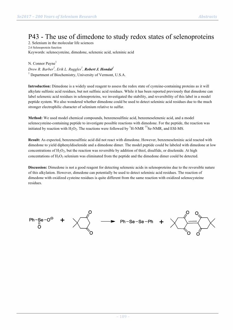
Se2017 – 200 Years of Selenium Research Abstracts
– 189 –
P43 - The use of dimedone to study redox states of selenoproteins 2. Selenium in the molecular life sciences 2.6 Selenoprotein function Keywords: selenocysteine, dimedone, selenenic acid, seleninic acid N. Connor Payne1 Drew R. Barber1, Erik L. Ruggles1, Robert J. Hondal1 1 Department of Biochemistry, University of Vermont, U.S.A. Introduction: Dimedone is a widely used reagent to assess the redox state of cysteine-containing proteins as it will alkylate sulfenic acid residues, but not sulfinic acid residues. While it has been reported previously that dimedone can label selenenic acid residues in selenoproteins, we investigated the stability, and reversibility of this label in a model peptide system. We also wondered whether dimedone could be used to detect seleninic acid residues due to the much stronger electrophilic character of selenium relative to sulfur. Method: We used model chemical compounds, benzenesulfinic acid, benzeneselenenic acid, and a model selenocysteine-containing peptide to investigate possible reactions with dimedone. For the peptide, the reaction was initiated by reaction with H2O2. The reactions were followed by 1H-NMR 77Se-NMR, and ESI-MS. Result: As expected, benzenesulfinic acid did not react with dimedone. However, benzeneseleninic acid reacted with dimedone to yield diphenyldiselenide and a dimedone dimer. The model peptide could be labeled with dimedone at low concentrations of H2O2, but the reaction was reversible by addition of thiol, disulfide, or diselenide. At high concentations of H2O2 selenium was eliminated from the peptide and the dimedone dimer could be detected. Discussion: Dimedone is not a good reagent for detecting selenenic acids in selenoproteins due to the reversible nature of this alkylation. However, dimedone can potentially be used to detect seleninic acid residues. The reaction of dimedone with oxidized cysteine residues is quite different from the same reaction with oxidized selenocysteine residues.

Se2017 – 200 Years of Selenium Research Abstracts
– 190 –
P44 - Site specific acetylation of thioredoxin reductase 1 2. Selenium in the molecular life sciences 2.6 Selenoprotein function Keywords: orthogonal translation, post-translational modification, redox, selenocysteine, thioredoxin reductase David Wright1 Zaid Altaany2, Yumin Bi1, Zaccary Alperstein1, Patrick O'Donoghue1 1 University of Western Ontario, London, Ontario 2 Faculty of Medicine, Yarmouk University, Irbid, Jordan. Introduction: Thioredoxin reductase 1 (TrxR1) is an essential human selenoprotein involved in ROS defense. Overactive TrxR1 is a diagnostic marker for some cancers (1), and a drug target for some drug resistant cancers (2). TrxR1 acetylation sites have been experimentally characterized, but the effect of acetylation at specific sites on the activity of TrxR1 has not been determined. TrxR1 exists as a catalytically active dimer, but can form low activity tetramers or higher order multimers (3). The acetylation sites occur on the surface of the dimer, at the dimer dimer interface in the tetramer (3). Method: We employed genetic code expansion to produce site specifically acetylated TrxR1 in E. coli. This was accomplished by reassigning UAG stop codons to acetyl-Lysine (acK) using an orthongonal tRNA, tRNA acK synthetase pair, as well as using E. coli's endogenous selenocysteine insertion system to recode a single UGA codon to selenocysteine. Result: We found that site specific acetylation of TrxR1 increased TrxR1 activity on DTNB. Acetylated TrxR1 variants eluted in later fractions from a size exclusion column under native conditions compared to unacetylated TrxR1, indicating a shift towards lower molecular weight oligomers. The effect of acetylation on TrxR1 activity was reversed following incubation of acetylated TrxR1 with histone deacetylase 3. Discussion: We discovered that acetylation increases TrxR1 activity, likely by destabalizing low activity tetramers and promoting the formation of catalytically active dimers (Figure 1). This provides new insight into how TrxR1 activity is regulated in the cell, and could be relevant to diseases involving TrxR1. Selected references
Selenius, A. K. Rundlof, E. Olm, A. P. Fernandes, M. Bjornstedt, Antioxidants & redox signaling 2010, 12, 867-880.
Wang, Z. Yang, J. Fu, H. Yin, K. Xiong, Q. Tan, H. Jin, J. Li, T. Wang, W. Tang, J. Yin, G. Cai, M. Liu, S. Kehr, K. Becker, H. Zeng, Free Radic Biol Med 2012, 52, 898-908.
Xu, S. E. Eriksson, M. Cebula, T. Sandalova, E. Hedstrom, I. Pader, Q. Cheng, C. R. Myers, W. E. Antholine, P. Nagy, U. Hellman, G. Selivanova, Y. Lindqvist, E. S. Arner, Cell death & disease 2015, 6, e1616.
Se2017 – 200 Years of Selenium Research Abstracts
– 191 –
P45 - Thioredoxin reductase 1 and NADPH directly protect protein tyrosine phosphatase 1B from inactivation 2. Selenium in the molecular life sciences 2.6 Selenoprotein function Keywords: PTP1B, TrxR1, Prx2, redox regulation Markus Dagnell1 Paul E. Pace2, Qing Cheng1, Jeroen Frijhoff3, Arne Östman4, Elias S.J. Arnér1, Mark B Hampton2, Christine C. Winterbourn2 1 Karolinska Institutet, Department of Medical Biochemistry and Biophysics, Stockholm, Sweden 2 University of Otago, Department of Pathology, Christchurch, New Zealand 3 Maastricht University, Cardiovascular Research Institute, Maastricht, the Netherlands 4 Karolinska Institutet, Department of Oncology and Pathology, Stockholm, Sweden Introduction: Protein tyrosine phosphatases (PTPs) are regulated by oxidation of their active site cysteine during growth factor signaling (1). Recent work by us has identified thioredoxin reductase (TrxR1), in combination with thioredoxin (Trx1) as activators of PTP1B both in vitro as well as during cellular signaling (2). How PTP1B can be oxidized in the presence of peroxiredoxins and the thioredoxin system is however insufficiently unknown. Here we have characterized the influence of different components of the Trx system on hydrogen peroxide-mediated PTP inactivation. Method: Recombinant PTP1B, TrxR1, Trx1 and Prx2 were used to mimic cellular growth factor dependent PTP regulation in vitro under different conditions of oxidative bursts, PTP activity was assesed using a chromogenic substrate (pNPP). Result: We found that H2O2 cannot efficiently inactivate PTP1B in the presence of complete TrxR1 / Trx1 / Prx system. Our results are against a transfer of oxidative equivalents from Trx1 or Prx2 to PTP1B upon burst of H2O2. We found that TrxR1 and NADPH (without Trx) was able to protect reduced PTP1B if present during exposure to hydrogen peroxide. The protective effect was dependent on TrxR1 concentration, required an intact selenocysteine residue, and was blocked by the TrxR1 inhibitor auranofin. Discussion: TrxR1 was shown to prevent H2O2 dependent PTP1B inactivation. We found that Trx or Prx2 also protect PTP1B from inactivation (3). Mechanisms of PTP1B inactivation by H2O2 still need to be characterized. Selected references
Frijhoff J, Dagnell M, Godfrey R, & Ostman A (2014) Regulation of protein tyrosine phosphatase oxidation in cell adhesion and migration. Antioxid Redox Signal 20(13):1994-2010.
Dagnell M, et al. (2013) Selective activation of oxidized PTP1B by the thioredoxin system modulates PDGF-beta receptor tyrosine kinase signaling. Proc Natl Acad Sci U S A 110(33):13398-13403.
Dagnell et al. "Manuscript in preparation" Thioredoxin reductase 1 and NADPH directly protect protein tyrosine phosphatase 1B from inactivation during exposure to H2O2

Se2017 – 200 Years of Selenium Research Abstracts
– 192 –
P46 - Details in the catalytic mechanism of mammalian selenoprotein thioredoxin reductase 1 revealed using point mutations and juglone reducing activities 2. Selenium in the molecular life sciences 2.6 Selenoprotein function Keywords: Redox Cycling, Superoxide, NADPH Oxidase Activity, Electron Transfer, Thioredoxin Reductase, Juglone Jianqiang Xu1 Qing Cheng2, Elias Arnér2 1 School of Life Science and Medicine, Dalian University of Technology, Panjin 124221, China 2 Department of Medical Biochemistry and Biophysics, Karolinska Institutet, SE-17177, Stockholm, Sweden Introduction: The mammalian selenoprotein thioredoxin reductase 1 (TrxR1) is a key enzyme in redox regulation, antioxidant defense, and cellular growth. TrxR1 catalyzes efficient reduction of juglone (5-hydroxy-1,4-naphthoquinone; walnut toxin), however, the details in the catalytic mechanism of TrxR1-mediated reduction of juglone and most other substrates, remain unclear. Method: Using a number of TrxR1 mutant variants, we here found that a sole Cys residue at the C-term tail of TrxR1 is required for high-efficiency juglone-coupled NADPH oxidase activity of Sec-deficient enzyme, occurring with mixed one- and two-electron reactions producing superoxide. The activity also utilizes the FAD and the N-terminal redox active disulfide/dithiol motif of TrxR1. Result: If a sole Cys residue at the C-term tail of TrxR1, in the absence of Sec, was moved further towards the C-term end of the protein compared to its natural position at residue 497, juglone reduction was, surprisingly, further increased. Four residues of Sec-deficient TrxR1 were found to be easily arylated by juglone, including the Cys residue at position 497. Four residues of Sec-deficient TrxR1 were found to be easily arylated by juglone, including the Cys residue at position 497. Discussion: Based upon our observations, we suggest a model for involvement of the juglone-arylated C-term motif of TrxR1 to explain its high activity with juglone. This study thus provides novel insights into the catalytic mechanisms of TrxR1. One-electron juglone reduction by TrxR1 producing superoxide should furthermore contribute to the well-known prooxidant cytotoxicity of juglone. Selected references This study was supported by Grants from the Swedish Cancer Society, the Swedish Research Council (Medicine) and the National Natural Science Foundation of China (31670767). We also thank the support from the Chinese Fundamental Research Funds for the Central Universities. [1] Arnér, E. S. J. Focus on mammalian thioredoxin reductases--important selenoproteins with versatile functions. Biochimica et Biophysica Acta 1790:495-526; 2009. [2] Arnér, E. S. J. Selenoproteins-What unique properties can arise with selenocysteine in place of cysteine? Exp Cell Res 316:1296-1303; 2010. [3] Xu, J.; Arnér, E. S. J. Pyrroloquinoline quinone modulates the kinetic parameters of the mammalian selenoprotein thioredoxin reductase 1 and is an inhibitor of glutathione reductase. Biochemical Pharmacology 83:815-820; 2012. [4] Cheng, Q.; Antholine, W. E.; Myers, J. M.; Kalyanaraman, B.; Arnér, E. S. J.; Myers, C. R. The selenium-independent inherent pro-oxidant NADPH oxidase activity of mammalian thioredoxin reductase and its selenium-dependent direct peroxidase activities. The Journal of Biological Chemistry 285:21708-21723; 2010. [5] Anestal, K.; Prast-Nielsen, S.; Cenas, N.; Arnér, E. S. J. Cell death by SecTRAPs: thioredoxin reductase as a prooxidant killer of cells. PLoS One 3:e1846; 2008. [6] Anestal, K.; Arnér, E. S. J. Rapid induction of cell death by selenium-compromised thioredoxin reductase 1 but not by the fully active enzyme containing selenocysteine. The Journal of Biological Chemistry 278:15966-15972; 2003.
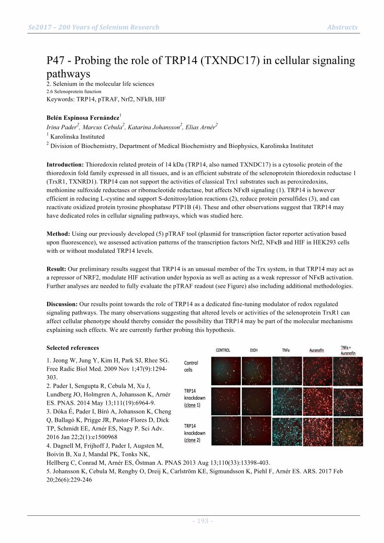
Se2017 – 200 Years of Selenium Research Abstracts
– 193 –
P47 - Probing the role of TRP14 (TXNDC17) in cellular signaling pathways 2. Selenium in the molecular life sciences 2.6 Selenoprotein function Keywords: TRP14, pTRAF, Nrf2, NFkB, HIF Belén Espinosa Fernández1 Irina Pader2, Marcus Cebula2, Katarina Johansson2, Elias Arnér2 1 Karolinska Instituted 2 Division of Biochemistry, Department of Medical Biochemistry and Biophysics, Karolinska Institutet Introduction: Thioredoxin related protein of 14 kDa (TRP14, also named TXNDC17) is a cytosolic protein of the thioredoxin fold family expressed in all tissues, and is an efficient substrate of the selenoprotein thioredoxin reductase 1 (TrxR1, TXNRD1). TRP14 can not support the activities of classical Trx1 substrates such as peroxiredoxins, methionine sulfoxide reductases or ribonucleotide reductase, but affects NFκB signaling (1). TRP14 is however efficient in reducing L-cystine and support S-denitrosylation reactions (2), reduce protein persulfides (3), and can reactivate oxidized protein tyrosine phosphatase PTP1B (4). These and other observations suggest that TRP14 may have dedicated roles in cellular signaling pathways, which was studied here. Method: Using our previously developed (5) pTRAF tool (plasmid for transcription factor reporter activation based upon fluorescence), we assessed activation patterns of the transcription factors Nrf2, NFκB and HIF in HEK293 cells with or without modulated TRP14 levels. Result: Our preliminary results suggest that TRP14 is an unusual member of the Trx system, in that TRP14 may act as a repressor of NRF2, modulate HIF activation under hypoxia as well as acting as a weak repressor of NFκB activation. Further analyses are needed to fully evaluate the pTRAF readout (see Figure) also including additional methodologies. Discussion: Our results point towards the role of TRP14 as a dedicated fine-tuning modulator of redox regulated signaling pathways. The many observations suggesting that altered levels or activities of the selenoprotein TrxR1 can affect cellular phenotype should thereby consider the possibility that TRP14 may be part of the molecular mechanisms explaining such effects. We are currently further probing this hypothesis. Selected references
1. Jeong W, Jung Y, Kim H, Park SJ, Rhee SG. Free Radic Biol Med. 2009 Nov 1;47(9):1294-303. 2. Pader I, Sengupta R, Cebula M, Xu J, Lundberg JO, Holmgren A, Johansson K, Arnér ES. PNAS. 2014 May 13;111(19):6964-9. 3. Dóka É, Pader I, Bíró A, Johansson K, Cheng Q, Ballagó K, Prigge JR, Pastor-Flores D, Dick TP, Schmidt EE, Arnér ES, Nagy P. Sci Adv. 2016 Jan 22;2(1):e1500968 4. Dagnell M, Frijhoff J, Pader I, Augsten M, Boivin B, Xu J, Mandal PK, Tonks NK, Hellberg C, Conrad M, Arnér ES, Östman A. PNAS 2013 Aug 13;110(33):13398-403. 5. Johansson K, Cebula M, Rengby O, Dreij K, Carlström KE, Sigmundsson K, Piehl F, Arnér ES. ARS. 2017 Feb 20;26(6):229-246
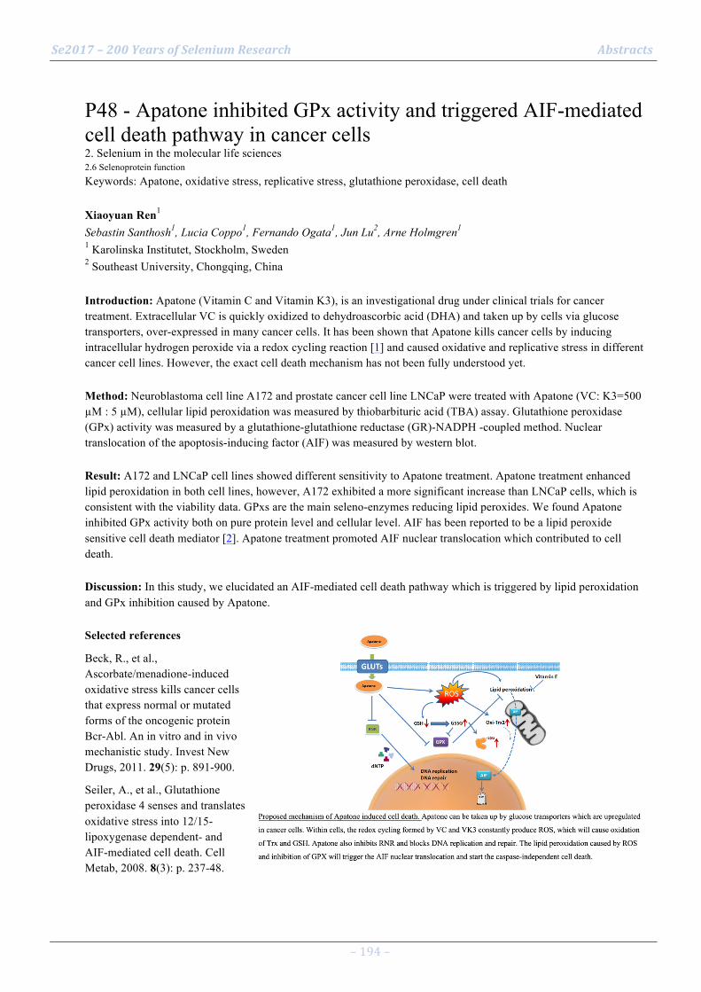
Se2017 – 200 Years of Selenium Research Abstracts
– 194 –
P48 - Apatone inhibited GPx activity and triggered AIF-mediated cell death pathway in cancer cells 2. Selenium in the molecular life sciences 2.6 Selenoprotein function Keywords: Apatone, oxidative stress, replicative stress, glutathione peroxidase, cell death Xiaoyuan Ren1 Sebastin Santhosh1, Lucia Coppo1, Fernando Ogata1, Jun Lu2, Arne Holmgren1 1 Karolinska Institutet, Stockholm, Sweden 2 Southeast University, Chongqing, China Introduction: Apatone (Vitamin C and Vitamin K3), is an investigational drug under clinical trials for cancer treatment. Extracellular VC is quickly oxidized to dehydroascorbic acid (DHA) and taken up by cells via glucose transporters, over-expressed in many cancer cells. It has been shown that Apatone kills cancer cells by inducing intracellular hydrogen peroxide via a redox cycling reaction [1] and caused oxidative and replicative stress in different cancer cell lines. However, the exact cell death mechanism has not been fully understood yet. Method: Neuroblastoma cell line A172 and prostate cancer cell line LNCaP were treated with Apatone (VC: K3=500 µM : 5 µM), cellular lipid peroxidation was measured by thiobarbituric acid (TBA) assay. Glutathione peroxidase (GPx) activity was measured by a glutathione-glutathione reductase (GR)-NADPH -coupled method. Nuclear translocation of the apoptosis-inducing factor (AIF) was measured by western blot. Result: A172 and LNCaP cell lines showed different sensitivity to Apatone treatment. Apatone treatment enhanced lipid peroxidation in both cell lines, however, A172 exhibited a more significant increase than LNCaP cells, which is consistent with the viability data. GPxs are the main seleno-enzymes reducing lipid peroxides. We found Apatone inhibited GPx activity both on pure protein level and cellular level. AIF has been reported to be a lipid peroxide sensitive cell death mediator [2]. Apatone treatment promoted AIF nuclear translocation which contributed to cell death. Discussion: In this study, we elucidated an AIF-mediated cell death pathway which is triggered by lipid peroxidation and GPx inhibition caused by Apatone. Selected references
Beck, R., et al., Ascorbate/menadione-induced oxidative stress kills cancer cells that express normal or mutated forms of the oncogenic protein Bcr-Abl. An in vitro and in vivo mechanistic study. Invest New Drugs, 2011. 29(5): p. 891-900.
Seiler, A., et al., Glutathione peroxidase 4 senses and translates oxidative stress into 12/15-lipoxygenase dependent- and AIF-mediated cell death. Cell Metab, 2008. 8(3): p. 237-48.
Se2017 – 200 Years of Selenium Research Abstracts
– 195 –
P49 - The significance of selenoprotein P expression in pancreatic beta-cell line MIN6 2. Selenium in the molecular life sciences 2.6 Selenoprotein function Keywords: Selenoprotein P, Antioxidant, MIN6, Insulin, Cell death Shohei Nakao1 Yuichiro Mita1, Noriko Noguchi1, Yoshiro Saito1 1 Faculty of Life and Medical Sciences, Doshisha University, Kyotanabe, Kyoto, Japan Introduction: Selenoprotein P (SeP), a selenocysteine (Sec) -containing protein, is mainly synthesized in the liver and functions as a selenium (Se) -supply protein to maintain Se levels in several tissues1). Se is essential for the synthesis of antioxidantive selenoproteins, such as glutathione peroxidases (GPxs) and thioredoxin reductases (TRs). Hence, SeP contributes to cellular antioxidative system via the maintenance of selenoproteins. Several studies have reported that pancreatic beta-cells are vulnerable to oxidative stress and antioxidant system is essential for beta-cells2). Recently, we found that MIN6 cells express SeP. In the present study, we investigated the significance of SeP expression in MIN6 cells. Method: We established SeP-deficient MIN6 cells by using siRNA. We used western blotting to evaluate expression levels of proteins such as insulin and selenoproteins (GPx1, GPx4 and TR1). Real-time PCR was used to measure these mRNA expression levels. WST-8 assay was used to evaluate cell viability. Result: SeP-siRNA significantly decreased SeP mRNA levels in MIN6 cells. Knockdown of SeP resulted in significant reduction in protein levels of selenoproteins and insulin in MIN6 cells. The decrease in cell viability was also observed by WST-8 assay. Addition of Sec to SeP-deficient MIN6 cells recovered these protein levels and cell viability. Alpha-Tocopherol, a classical lipophilic antioxidant, also recovered cell viability. Discussion: This report provides the significance of SeP expression in MIN6 cells for maintaining levels of selenoprotein, insulin and cell viability. The effect of alpha-tocopherol and possible cell death signaling will be discussed. Selected references
1) Y. Saito et al., Characterization of selenoprotein P as a selenium supply protein, Eur J Biochem/FEBS 269, 5746-5751 (2002)
2) Y. Kajimoto et al., Role of oxidative stress in pancreatic beta-cell dysfunction, Ann N Y Acad Sci 1011, 168-76 (2004)

Se2017 – 200 Years of Selenium Research Abstracts
– 196 –
P50 - Interaction between selenoprotein W and 14-3-3 is regulated by oxidative stress. 2. Selenium in the molecular life sciences 2.6 Selenoprotein function Keywords: SelW, 14-3-3, Oxidative stress Kwan Young Ko1 Yeong Ha Jeon1, Minju Ham1, Yunjung Jin1, Ick Young Kim1 1 Division of Life Sciences, Korea University, Seoul, Republic of Korea Introduction: Mouse Selenoprotein W (SelW) contains a selenocysteine (sec) and four cysteines. N-terminal of SelW contains a CXXU motif, corresponding to the thioredoxin active site CXXC motif. Thus SelW has been none seen as a putative antioxidant protein that interaction with other redox regulators. We have previously reported that the binding of 14-3-3 protein to its target proteins, including CDC25B, Rictor and TAZ, is inhibited by the interaction of 14-3-3 protein with SelW. In this study, we sought to determine the binding site of SelW to understand the regulatory mechanism of the interaction between SelW and 14-3-3 and its biological effects. Method: The interaction between SelW and 14-3-3 was determined by both immunoprecipitation assay and GST-pull-down assay using various purified GST-SelW mutants. To understand the reguratory mechanism, the interaction was also determined in oxidative stress condition. Cell cycle rescue assay was performed to examine the biological effect of the interaction using SelW-knockdown MCF7 cells. Result: A mutant SelW in which Cys (U13C) is changed to Ser (U13S) was unable to interact with 14-3-3 protein and thus did not inhibit the interaction of 14-3-3 to other target proteins. However, other Cys mutants of SelW (C10S, C33S and C37S) normally interacted with 14-3-3 protein. Also, the interaction of SelW to 14-3-3 protein was enhanced by oxidative stress. We also found that SelW (U13S) was not able to rescue the effect of SelW depletion. Discussion: Thus, SelW may have a regulatory function in redox cell signaling by interacting with 14-3-3 protein. Selected references
[1] Y.H. Jeon, Y.H. Park, J.H. Kwon, J.H. Lee, I.Y. Kim, Inhibition of 14-3-3 binding to Rictor of mTORC2 for Akt phosphorylation at Ser473 is regulated by selenoprotein W, Biochim. Biophys. Acta 1833 (2013) 2135–2142.
[2] Y.H. Jeon, Y.H. Park, J.H. Lee, J.H. Hong, I.Y. Kim, Selenoprotein W enhances skeletal muscle differentiation by inhibiting TAZ binding to 14-3-3 protein, Biochim. Biophys. Acta 1843 (2014) 1356–1364.

Se2017 – 200 Years of Selenium Research Abstracts
– 197 –
P51 - Immortalized human prostate cell line RWPE-1 as a model to study selenoprotein regulation and function in normal prostatic tissue 2. Selenium in the molecular life sciences 2.6 Selenoprotein function Keywords: selenoproteins, GPX1, SELENOF, SBP1, RWPE1 Lenny Hong1 Dede Ekoue1, Alan Diamond1 1 University of Illinois at Chicago, Chicago, IL USA Introduction: While epidemiology has indicted a likely impact of selenium status on prostate cancer risk, the role of selenoproteins in this disease remains to be determined. Many studies have investigated selenoprotein levels and function in prostate cancer cell lines, but little is known about the function of several of these proteins in normal prostate epithelia. Towards this goal, studies have focused on an immortalized but not transformed human prostate cell line, RWPE-1. Method: GPX1, SELENOF and SBP1 levels were determined by western blotting with specific antibodies with and without treatment with 100 nM sodium selenite. The cellular localization of these proteins in RWPE-1 cells was evaluated by confocal microscopy and compared to that seen in human prostate cancer cell lines and primary prostate epithelia cells using the same techniques, as well as immunohistochemistry of human tissue. Result: SELENOF was predominantly located in the outer membrane of RWPE-1 cells as was observed for primary cells and benign human prostate tissue. Unlike what is typically seen in prostate cancer cell lines, the levels of GPX1 and SELENOF were either unchanged or minimally changed by the addition of selenium to the culture media, while SBP1 was undetectable in these cells. Discussion: Selenoprotein location and levels in RWPE-1 cells closely resemble that seen in human tissues. These cells will therefore be a useful model for further research on the function of selenoproteins in normal prostatic tissue biology and the progression to cancer.
Se2017 – 200 Years of Selenium Research Abstracts
– 198 –
P52 - Glutathione depletion in HEK293T adresses cytosolic GPx4 to the membrane 2. Selenium in the molecular life sciences 2.6 Selenoprotein function Keywords: PHGPx, glutathione, ferroptosis Ana-Marija Vuckovic1 Lucia La Pira1, Antonella Roveri1, Valentina Bosello Travain1, Matilde Maiorino1, Fulvio Ursini1, Giorgio Cozza1 1 University of Padova, Department of Molecular Medicine Introduction: Due to its important role in catalysing reactions inhibiting lipid peroxidation1,2,glutathione peroxidase (GPx4) stands out as the key regulator of the cell death pathway named ferroptosis3. In order to study the molecular mechanism of GPx4 inactivation by ferroptosis inducing agents, a massive depletion of its reducing substrate glutathione was produced using a thiol alkylating agent, diethyl maleate (DEM), together with an inhibitor of GSH synthesis, buthionine sulfoximine (BSO). Method: For this study the HEK293T cell line was selected. The cell pellet was lysed, sonicated and centrifuged. Supernatants were used for protein quantification, GPx4 enzymatic activity test and Tietze assay. After SDS-Page, Western blot analysis was performed using an anti-GPx4 antibody. Result: Treatment of HEK293T cells with BSO and DEM yields a decline in the total glutathione content in cytosol. Furthermore, a marked decrease of GPx4 specific activity is observed already after 3h, which is accompanied with a decline of GPx4 protein, as seen by Western blot. As the treatment with proteosomal inhibitor MG132 fails to prevent this phenomenon, the GPx4 content in BSO/DEM treated cells was analysed in the residual pellet after the centrifugation. Results show that, while disappearing from cytosol, GPx4 appears in the membrane fraction. Discussion: Thus, upon GSH depletion, GPx4 is addressed to the membranous fraction of the cells. This result complies with both, SPR analysis and in silico study indicating that the affinity of GPx4 for the polar head of membrane phospholipids is increased when GSH is absent. Selected references
Thomas, J. P., Maiorino, M., Ursini, F. & Girotti, A. W. Protective action of phospholipid hydroperoxide glutathione peroxidase against membrane-damaging lipid peroxidation. In situ reduction of phospholipid and cholesterol hydroperoxides. JBC 265, 454–461 (1990).
Ursini, F., Maiorino, M. & Gregolin, C. The selenoenzyme phospholipid hydroperoxide glutathione peroxidase. Biochim Biophys Acta 839(1), 62–70 (1985).
Yang, W. S. et al. Regulation of Ferroptotic Cancer Cell Death by GPX4. Cell 156, 317–331 (2014).

Se2017 – 200 Years of Selenium Research Abstracts
– 199 –
P53 - Synthesis and efficacy of conjugated selenium ADC monoclonal antibodies in vitro 2. Selenium in the molecular life sciences 2.8 Selenium based biotechnological applications Keywords: Monoclonal Antibodies, Triple Negative Breast Cancer Cell, Treatment Soni Khandelwal1 Lauren Gollahon1, Mallory Boylan2, Maria Del Mar Gracia-Hernandez1, Julian Spallholz1 1 Texas Tech University, Lubbock, Texas, USA 2 Texas Tech University,Lubbock, Texas, USA Introduction: Monoclonal antibodies (mAbs) are known for their high specificity and affinity to target antigens. Yet sometimes mAbs lack therapeutic activity; primary resistance (Pohlmann, 2009; Wilkins, 2010) hence the efficacy of mAbs may be improved by drug conjugation (ADCs). Method: We describe here the attributes of two selenium (Se) monoclonal ADCs; Se-Herceptin and Se-Avastin. Redox selenium (Se) was covalently attached to commercially available and clinically employed Her2+ and VEGF mAbs, Herceptin® and Avastin®. Selenium attachment to the mAbs is made using a Se-modified Bolton-Hunter reagent (Lane, 2011; Bapat, 2015). The Se-Bolton-Hunter reagent was dissolved in tetrahydrofuran and incubated with Herceptin® or Avastin® in a sodium borate buffer at 4oC for 72 hr and then exhaustively dialyzed against PBS. Control Herceptin® and Avastin® mAbs were treated identically but without selenium attachment; Figure. Result: After dialysis, Se-ADCs were quantitated using ICP-MS. Chemiluminescence activity of the Se-ADCs was determined for the detection of superoxide generation following the procedures of Chen (Chen, 2008). Protein concentrations of the Se-ADCs were determined by a BCA assay. All mAbs were subjected to PAGE gel electrophoresis under reducing and non-reducing conditions and stained with Coomassie Blue R250. Discussion: Human Triple Negative Breast Cancer (TNBC) cell lines MDA-MB-468 and MDA-MB-231 ex vivo were subjected to time and dose responses to Herceptin®, Avastin®, and their corresponding Se-ADCs. MDA-MB-468 and MDA-MB-231 cells at all time and concentrations were unaffected by Herceptin® or Avastin® treatments, whereas cytolysis of these TNBC lines were highly responsive to the time and concentrations of the redox (Stewart, 1999) Se-ADC treatments. Selected references
Bapat, P. (2015). Cytotoxic effects of selenium conjugated trastuzumab on HER2+ breast cancer cell lines (Doctoral dissertation; Texas Tech University).
Chen, J. J., Boylan, L. M., Wu, C. K., & Spallholz, J. E. (2007). Oxidation of glutathione and superoxide generation by inorganic and organic selenium compounds. Biofactors, 31:55-66.
Lane, D. J., & Richardson, D. R. (2011). William Hunter and radioiodination: revolutions in the labelling of proteins with radionuclides of iodine. Biochem J, 1-4.
Stewart, M. S., Spallholz, J. E., Neldner, K. H., & Pence, B. C. (1999). Selenium compounds have disparate abilities to impose oxidative stress and induce apoptosis. Free Radical Biology and Medicine, 26: 42-48.
Wilken, J. A., & Maihle, N. J. (2010). Primary trastuzumab resistance: new tricks for an old drug. Annals of the New York Academy of Sciences, 1210: 53-65.

Se2017 – 200 Years of Selenium Research Abstracts
– 200 –
P54 - Antifungal Activity of Selenium Nanoparticles Synthesized by B. subtilis Against P. syringae 2. Selenium in the molecular life sciences 2.8 Selenium based biotechnological applications Keywords: Antifungal Activity, Nano Selenium , Biosynthesis, Bacillus subtilis, Pseudomonas syringae Miao Li1 1 Anhui Agriculture University, Hefei, China Introduction: The low toxicity of Se nanoparticles (NPs) and properties such as antibacterial, antiviral, and antioxidant activities have attracted research attention. Physicochemical techniques, biological methods, and the synthesis of nanostructures with bacterial and fungal strains as well as several plant extracts offer novel, clean, non-toxic, and eco-friendly methods for the production of Se NPs. In the present study, biogenic Se NPs were purified from the whole-cell lysate of Bacillus subtilis Bs-217 and characterized. Then, the antibacterial efficacy of these novel NPs against Pseudomonas syringae pv. actinidiae was studied. Method: Se NPs were prepared by reducing Se+4 ions with the B. subtilis Bs-217, which was previously identified with a 16S ribosomal DNA method. The antibacterial activities of Se NPs against P. syringae pv. actinidiae were determined by using the MIC method. Result: Se NPs were successfully synthesized with B. subtilis Bs-217 as evidenced by a significant change in the cultivation medium from colorless to insoluble orange-red, indicating the presence of elemental Se (Se0), after 24 hours. The antibacterial activities of the Se NPs against P. syringae pv. actinidiae is shown in Table 1. The measured MICs for P. syringae pv. actinidiae (60 µg/mL) showed that the biogenic Se NPs had good antibacterial activity. Discussion: Use of NPs against pathogenic bacteria is yet another novel approach. The results of the present study suggest that nanoscale biogenic elemental Se has antibacterial activity against P. syringae pv. actinidiae. The mechanism of this antibacterial effect is unknown and merits further in vivo and in vitro studies. Selected references
Alghuthaymi, M.A., Almoammar, H., Rai, M., Said-Galiev, E., & Abd-Elsalam, K.A. 2015. Myconanoparticles: synthesis and their role in phytopathogens management. Biotechnology & Biotechnological Equipment 29(2): 221-236.

Se2017 – 200 Years of Selenium Research Abstracts
– 201 –
P55 - Exosome-mediated methylmercury detoxification accelerated by selenium compound, selenoneine in aquatic organism 2. Selenium in the molecular life sciences 2.9 Additional and emerging topics of selenium in molecular life science Keywords: exosome, methylmercury, selenoneine, zebrafish Shintaro Imamura1 Tomomi Kato1, Takuya Seko1, Kenji Ishihara1, Yumiko Yamashita1, Hajime Shiba2, Takeshi Yabu2, Ståle Ellingsen3, Heidi Amlund3, Anne-Katrine L. Haldorsen3, Michiaki Yamashita4 1 National Research Institute of Fisheries Science, Yokohama, Japan 2 Nihon University, Fujisawa, Japan 3 National Institute of Nutrition and Seafood Research, Bergen, Norway 4 National Fisheries University, Shimonoseki, Japan Introduction: Exosomes are extracellular small granule vesicles produced by cells, and might mediate a signal transduction mechanisms to transfer the information between cells and organisms. They contain various molecular constituents, such as proteins, mRNA and miRNA, and transfer molecules from cells through membrane vesicle trafficking. We found exosome-mediated methylmercury (MeHg) excretion pathway accelerated by 2-selenyl-trimethyl-histidine, selenoneine, in zebrafish embryos. Here, we report evidence that a selenoneine accelerates the excretion of MeHg though exosomal secretion, and dynamics of secreted exosomes following MeHg exposure in zebrafish models. Method: The embryos (8 hpf) were microinjected with MeHg-Cys (10−200 µg Hg/ml) into yolk sac, and cultured for 16 h. Exosomes were purified by ultracentrifugation at 100,000g for 1-3 h. Exosomal markers (protein content, AChE activity and CD63 content) were measured. To visualize the production of exosomes in vivo, cd63-GFP expressing zebrafish lines were generated. Result: Exosomes were released by MeHg exposure in a dose dependent manner. Scanning probe and electron microscopic observation revealed that the exosomes were vesicles of 20-50 nm in diameter. Hg (0.05-0.1 ng Hg/embryo) was contained in the exosomes. Selenoneine (3 µM) treatment accelerated exosomal secretion and Hg excretion. Exosomal secretion pathway was accelerated by selenoneine incorporated into cells and played an essential role for methylmercury detoxification. In cd63-GFP transgenic zebrafish lines, the fluorescent signals was observed in central nervous system, yolk sac and somite, and enhanced signals were detected by MeHg injection and selenoneine treatment. Discussion: Therefore, induced production of exosomes was visualized in living animals, and indicated that selenoneine involve in MeHg detoxification.
Se2017 – 200 Years of Selenium Research Abstracts
– 202 –
P56 - Studying selenoproteome regulation using selenium stable isotope labeling 2. Selenium in the molecular life sciences 2.9 Additional and emerging topics of selenium in molecular life science Keywords: Stable isotope, ICP-MS, selenoproteins Jordan Sonet1 Anne-Laure Bulteau2, Laurent Chavatte3 1 LCABIE, PAU, FRANCE 2 IGFL, LYON, FRANCE 3 CIRI (Inserm U1111, CNRS UMR5308, ENS de Lyon, UCB Lyon-1), LYON, FRANCE Introduction: Selenium (Se) is an essential trace element, which is incorporated as a rare aminoacid, selenocysteine, in twenty five selenoproteins, to constitute the selenoproteome. Selenoprotein family is one of the most important bioactive form of selenium in human health. To understand the function and regulation of human selenoproteome, which is expressed at a trace levels, it appears critical to develop innovative strategies. The use of isotopically enriched selenium also allows cellular labelling and tracing of selenoproteins and other seleno-coupounds. Method: Selenium has a particular isotopic profile with six stable isotope (74Se, 76Se, 77Se, 78Se, 80Se and 82Se) used as a signature in our analysis with ICP-MS or ESI-MS/MS. When selenium is added as pure isotope, its incorporation into selenoproteins can be followed by ICP-MS. Result: We are able to (i) follow the selective labeling of the cellular selenocompounds with non radioactive Se-enriched isotopes (76Se, 77Se) and (ii) detect the Se-isotopic ratio of the selenoproteins using size-exclusion chromatography followed by inductively coupled plasma mass spectrometry detection (SEC ICP-MS). The reliability of our strategy is further confirmed by western blots with distinct selenoprotein-specific antibodies. Dose response and kinetic experiments of selenium supplementation or exchange have been used to study the hierarchy of selenoproteome regulation. Discussion: Here, we describe the use of isotopically-enriched forms of Se as an alternative stragegy to radioactive 75Se, for the labeling and tracing of selenoproteins in cultured cell lines. Selected references
Anne-Laure Bulteau and Laurent Chavatte (2015) Update on selenoprotein biosynthesis, Antioxidants and Redox Signaling, 23, 775-94.
Jordan Sonet, Anne-Laure Bulteau and Laurent Chavatte (2016) Selenium and selenoproteins in human health and diseases. Book on Metallomics: Analytical Techniques and Speciation Methods.
Juliusz Bianga, Zahia Touat-Hamici, Katarzyna Bierla, Sandra Mounicou, Joanna Szpunar, Laurent Chavatte, Ryszard Lobinski (2014) Speciation analysis for trace levels of selenoproteins in cultured human cells, Journal of Proteomics, 108, 316-24.

Se2017 – 200 Years of Selenium Research Abstracts
– 203 –
P57 - Progress in selenium nutrition in China during the last thirty years 3. Selenium in animal and human health and disease 3.1 Selenium supplementation for animal and livestock health Keywords: soil, china, human intake Weiming Shi1 Ju Min1, Fei Yu1, Xuebin Yin2, Xiaoqi Lu2 1 Institute of Soil Science, Chinese Academy of Sciences, Nanjing, China 2 Suzhou Institute of Advanced Study, University of Science and Technology of China, Suzhou, China Introduction: From the perspective of soil - plant nutrition science, this paper summarized the progress and achievements of Se study in agriculture science in China during the last 30 years. Method: The literatures from China Academic Journal Network Publishing Database were analyzed using bibliometrics method. Result: We presented temporal and spatial variations of Se in agricultural soils and its availability, temporal and spatial variations of Se in main crops, the role of Se on crop growth and its influence factors, and development of Se-rich agricultural products. In view of the two sides of Se (nutrition and toxicity), it is put forward that Se effective state and critical threshold research needs to be strengthened to meet the safe use in soil-crop-human body system, and the development of Se related industry standards and quality supervision system should be accelerated to meet the development of Se-enrich products. In this regard, Chinese authorities have made a draft standard of Se-rich agricultural products, and are promoting it as a national standard. Discussion: Interestingly, although China has made rapid development in its economy during the recent thirty years, the resident daily selenium intake showed decreasing trend. Thus, appropriate supplementary selenium is very necessary. Selected references
Li S., Bañuelos G.S., Wu L.H. and Shi W.M*. The Changing Selenium Nutritional Status of Chinese Residents. Nutrients 2014, 6, 1103-1114.
Li S., Bañuelos G.S., Min J. and Shi W.M*. Effect of continuous application of inorganic nitrogen fertilizer on selenium concentration in vegetables grown in the Taihu Lake region of China. Plant Soil 2015, 393:351-360.
Zhang L.H., Yu F.Y., Shi W.M.*, et al. Physiological characteristics of selenite uptake by maize roots in response to different pH levels. J. Plant Nutr. Soil Sci. 2010, 173, 417-422.
Zhou X.B., Shi W.M* and Zhang L.H. Iron plaque outside roots affects selenite uptake by rice seedlings (Oryza sativa L.) grown in solution culture. Plant Soil 2007, 290:17-28.

Se2017 – 200 Years of Selenium Research Abstracts
– 204 –
P58 - Protective effect of organic selenium-enriched extract from cardamine violifolia on carbon tetrachloride–induced hepatic damage in mice 3. Selenium in animal and human health and disease 3.1 Selenium supplementation for animal and livestock health Keywords: Cardamine violifolia, Organic selenium-enriched extract, Liver protection Xin Cong1 Shujun Liu2, Junhua Wu3 1 Associate Professor, Research and Development Center of Serun Healthy Industry Group, Nanjing, China 2 Research and Development Center of Serun Healthy Industry Group, Nanjing, China 3 Associate Professor, Medical School of Nanjing University, Nanjing, China Introduction: Cardamine violifolia is a kind of wild plant in western China and found to have strong selenium-enriching ability in World Selenium Capital-Enshi, China. We has extracted a kind of organic selenium-enriched extract containing 4800 µg/g of selenium. The aim of this study was to evaluate the protective effect of organic selenium-enriched extract on carbon tetrachloride-induced hepatic injury in mice. Method: The mice of each group were given different doses of the extract (calculated in selenium: 60, 180, 540 µg / kg·d) or sodium selenite (calculated in selenium: 180µg / kg·d) for 15 days. Then the mice were injected intraperitoneally with CCl4 (0.3% in olive oil, 10 ml/ kg of olive oil solution) . After 24 hours, the mice were sacrificed to detect liver injury, oxidation and inflammation related index. Result: The medium (180 µg / kg·d) and high dose (540 µg / kg·d) of the extract significantly alleviated the pathological injuries in liver and significantly decreased the release of ALT and AST from the liver compared with the CCL4 group. Compared with the CCL4 group, the activities of SOD and GSH-Px were elevated, and TNF-α, iNOS and COX-2 were reduced in livers from mice treated with the extract. We will further separate some pure selenium compounds to determine the active ingredient. Discussion: The results suggest that the pre-supplementation of organic selenium-enriched extract from cardamine violifolia alleviate CCl4-induced acute liver injury, which is likely due to the improvement of antioxidant enzymes and down-regulation of pro-inflammatory mediators. Cardamine violifolia may be a potential healthy organice selenium source. Selected references
[1] Fan J, Wang R, Xiang B K, et al. Study of selenium application on absorption and transformation of selenium in Cardamine violifolia[J]. Soil & Fertilizer Sciences in China, 2014, 34(1):63-68.
[2] Cheng P. Relation of the Nutritive Ingredients and Selenium Content in Different Growth Period of Cardamine violifolia[J]. Journal of Anhui Agricultural Sciences, 2006.
[3] Dong D, Xu L, Yin L, et al. Naringin prevents carbon tetrachloride-induced acute liver injury in mice[J]. Journal of Functional Foods, 2015, 12:179-191.
[4] Lee C H, Park S W, Kim Y S, et al. Protective mechanism of glycyrrhizin on acute liver injury induced by carbon tetrachloride in mice[J]. Biological & Pharmaceutical Bulletin, 2007, 30(10):1898.

Se2017 – 200 Years of Selenium Research Abstracts
– 205 –
P59 - Comparison of Trace Elements and Oxidant Status in Dairy Cows at Different Physiological Stages 3. Selenium in animal and human health and disease 3.7 Additional and emerging topics of selenium in health or disease Keywords: Cows, Trace elements, Oxidant Status, Different Physiological Stages Ruilong Song1 Hongyan Zhao2, Hui Zou1, Jianhong Gu1, Yan Yuan1, Xuezhong Liu1, Jiaqiao Zhu1, Jianchun Bian1, Zongping Liu1 1 College of Veterinary Medicine,Yangzhou University, Yangzhou, China 2 Testing Center,Yangzhou University, Yangzhou, China Introduction: Hardly shall we find specific directions for trace element additives, preventing diseases caused by oxidative stress in cows at different physiological stages for dairy husbandry. The aim of the study is to compare the serum concentrations of trace elements and oxidative status in dairy cows at different physiological stages. Method: 20 calves (3-4 month old), replacement heifers, prenatal cows (30 days before delivery), postnatal cows (30 days after delivery) and late-lactation cows were randomly selected into 5 groups. Serum concentrations of selenium, zinc, iron, copper and manganese of each group were determined by ICP-MS. The parameters including glutathione peroxidase (GSH-Px), superoxide dismutase (SOD), catalase (CAT) activity and malondialdehyde (MDA) content were measured by commercial ELISA kits. Result: The results showed that except for zinc, other trace element levels were significant lower in calf group (P<0.05 or P<0.01) than others. There is no significant difference (P>0.05) between replacement heifers and adult cows. The level of selenium was significant lower (P<0.05 or P<0.01) in groups of prenatal and late-lactation cows. Parameters indicative of oxidative status was not related to ages, however the levels of GSH-Px, SOD, CAT activity and MDA were significant higher (P<0.05 or P<0.01) in prenatal and postnatal cows. However the mechanism of the difference of oxidative status in dairy cows at different physiological stages has been understood poorly. Discussion: The results above confirmed that the concentrations of trace elements and oxicative status in calves were the lowest, and imbalance of oxidative state was associated with late pregnancy and early lactation. Selected references [1] Gong J, Xiao M. Selenium and Antioxidant Status in Dairy Cows at Different Stages of Lactation[J]. Biological Trace Element Research, 2016, 171(1):89-93. [2] Teixeira AG, Lima FS, Bicalho ML, Kussler A, Lima SF, Felippe MJ, Bicalho RC. Effect of an injectable trace mineral supplement containing selenium, copper, zinc, and manganese on immunity, health, and growth of dairy calves[J]. Journal of Dairy Science, 2014, 97(7):4216-26.

Se2017 – 200 Years of Selenium Research Abstracts
– 206 –
P60 - Relative bioavailability of selenium sources for beef cattle using glutathione peroxidase activity in liver 3. Selenium in animal and human health and disease 3.1 Selenium supplementation for animal and livestock health Keywords: beef cattle, feedlot, liver, selenium yeast, sodium selenite Janaina S. Silva1 Silvana M. P. Pugine1, Mariza P. Melo1, Marcus Zanetti1 1 University of São Paulo, Pirassununga, Brazil Introduction: Most of Selenium used in animal nutrition is in inorganic form, but organic sources have better bioavailability. To compare the bioavailability of organic and inorganic selenium sources using glutathione peroxidase activity (GPx) in beef cattle liver. Method: It was used 45 Nellore cattle with 24 months of age and 392 kg live weight, in a feedlot study during 84 days, in individual pens. The animals (nine per treatment) were submitted to one of the five diets: control diet without additional selenium supplementation; control + 0.3 mg Se kg-1 DM in the form of sodium selenite or selenium yeast; control + 0.9 mg Se kg-1 DM in the form of sodium selenite or selenium yeast. The control diet was deficient and had 0.065 mg of Se/kg of DM. Diets had a roughage: concentrate ratio of 30:70, based on corn silage, corn grain and soybean meal. At the end of the experiment the animals were slaughtered and liver samples were taken for glutathione peroxidase activity according Paglia & Valentine (1967). The bioavailability was calculated by technique of slope ratio assay (AMMERMAN et al., 1995). Linear regression was performed with the Proc GLM of SAS to characterize the glutathione peroxidase activity. Result: The difference between the slopes was significant (P<0.0001), being that estimation with standard errors were: 37.6 ± 16.7 for the sodium selenite and 146.0 ± 16.7 for the yeast source. Discussion: The relative bioavailability estimated by GPx for the yeast selenium in relation to the sodium selenite (100) was 3.89 or 389%. Selected references
AMMERMAN, C.B.; BAKER, D.H.; LEWIS, A.J. Bioavailability of nutrients for animals. Academic Press, San Diego, 1995. 441p.
Paglia, D. E. & Valentine, W. N. (1967). Studies on the quantitative and qualitative Characterization of erythrocyte glutathione peroxidase. Journal of Laboratory and Clinical Medicine 70, 158-169.
Se2017 – 200 Years of Selenium Research Abstracts
– 207 –
P61 - Organic Se has better protective effects than inorganic Se against AFB1/OTA-induced immunotoxicity 3. Selenium in animal and human health and disease 3.1 Selenium supplementation for animal and livestock health Keywords: Selenomethionine, Sodium selenite, Aflatoxin B1, Ochratoxin A, GPx1 Fang Gan1 Haibin Xu1, Shu Hao1, Kehe Huang1 1 Institute of Nutritional and Metabolic Disorders in Domestic Animals and Fowls, Nanjing Agricultural Introduction: Aflatoxin B1(AFB1)and ochratoxin A (OTA) frequently contaminate a wide variety of food and feeds and decrease immune function of humans and animals. Whether Se alleviates the immune toxicity induced by these mycotoxins and the underlying mechanism remain unclear. Method: Advanced technologies such as flowcytometry, western blotting, confocal laser, and RNA interference were used in the study. Result: Our present study demonstrated that AFB1 or OTA induced immunotoxicity in porcine primary splenocytes or porcine alveolar macrophages (PAMs) cell line 3D4/21 respectively in a dose-dependent manner, as demonstrated by decreasing cell viability, and increasing LDH activity, Annexin V-binding, Bcl-2/Bax mRNA ratio, and pro-inflammatory cytokines expression. AFB1 or OTA increased intracellular ROS levels. NAC, a free radical scavenger, alleviated the AFB1 or OTA-induced ROS production and cell toxicity. Further, organic Se (selenomethionine, Se-Met) and inorganic Se(sodium selenite, SS)are supplemented to study their effects on toxicity of AFB1 or OTA. The results showed that both Se sources have significant effects on alleviating AFB1 or OTA -induced immunotoxicity in porcine primary splenocytes or PAMs. Se-Met and SS supplementation significantly improved GPx1, SelS and TR1 expression. However, Se-Met supplementation has a more dramatic effect on alleviating the toxicity of AFB1 and OTA, and up-regulating of GPx1 expressions than SS supplementation. Knockdown of GPx1 by GPx1-specific siRNA diminished the protective effects of SeMet and SS against AFB1 or OTA -induced immunotoxicity. Discussion: These results suggest that SeMet and SS could alleviats AFB1 and OTA -induced immunotoxicity by improving GPx1 expression, in which organic Se is better than inorganic Se.

Se2017 – 200 Years of Selenium Research Abstracts
– 208 –
P62 - Dietary selenium forms influence selenogenome expression in broiler chickens 3. Selenium in animal and human health and disease 3.1 Selenium supplementation for animal and livestock health Keywords: broiler chicken, dietary selenium, hydroxy-selenomethionine Mickael Briens1 Yves Mercier1 1 Adisseo France S.A.S., 10 Place du Général de Gaulle, 92160 Antony, France Introduction: Poultry diets can be supplemented with either mineral or organic Se forms (Briens et al., 2013). Thus, this study aimed to compare sodium selenite and hydroxy-selenomethionine on chicken selenoprotein gene expression. Method: Chickens were fed from 1 to 21 days old three dietary treatments. The dietary treatments correspond to a standard diet either unsupplemented in Se: negative control (NC); NC supplemented with: sodium selenite (SS-0.2) and NC supplemented with hydroxy-selenomethionine (SO-0.2), both at 0.2 mg Se/kg feed. On day 21, eight birds per treatment were selected for breast muscle and liver collection for total RNA extraction and RT q-PCR measures of the 26 chicken selenoprotein genes. Result: The three dietary treatments were analyzed for endogenous Se content and revealed the following concentrations of Se, NC: 0.13 mg/kg; SS-0.2: 0.32; SO-0.2: 0.32. In the muscle, Selenom expression was significantly increased in Se supplemented groups compared to NC by >1.3 fold. Only SO-0.2 treatment significantly increased gene expression of Gpx1, Gpx3, Txnrd1, Txnrd2, compared to SS-0.2 (1.15 to 1.35 fold). In the liver, only SO-0.2 treatment significantly increased gene expression of Dio2, Dio3, Gpx1, Selenop, compared to SS-0.2 (1.31 to 1.61 fold). Discussion: Those results indicate that not only the selenium level in the diet but also the chemical form of Se can influence selenoprotein gene expression. Several genes with known redox functions were upregulated when hydroxy-selenomethionine was given. Further studies are needed to measure the benefits of those enhanced selenoprotein expressions on the antioxidant capacities of the animals and their capacity to fight oxidative stress conditions. Selected references
Briens, M., Mercier, Y., Rouffineau, F., Vacchina, V., and Geraert, P.-A. (2013). Comparative study of a new organic selenium source v. seleno-yeast and mineral selenium sources on muscle selenium enrichment and selenium digestibility in broiler chickens. Br. J. Nutr. 110, 617–624.

Se2017 – 200 Years of Selenium Research Abstracts
– 209 –
P63 - Eisenia fetida - a novel organic selenium-biofortified soil animal 3. Selenium in animal and human health and disease 3.1 Selenium supplementation for animal and livestock health Keywords: Eisenia fetida, organic selenium, speciation, biofortification Shizhong Yue1 Yuhui Qiao1 1 College of Resources and Environmental Sciences, China Agricultural University, Beijing, China Introduction: Selenium (Se) is a crucial element for human health [1]. Fungi, vegetables and cereals are typical Se-biofortified materials. We here recommend an organic Se-biofortified animal - Eisenia fetida, which could enrich kinds of metals efficiently [2] including Se. This study will provide the foundation for further application of the Se-biofortification strategy. Method: Eisenia foetida were put in the mixed-matrix of cow manure and mushroom residue (m:m=1:1, Se (Na2SeO3) concentrations were 0 and 40 mg/kg) and bred 45 days. All the simples were acid-digested (HNO3:HClO4=8:2, v:v), Se concentration and speciation were determined by AFS and HPLC-UV-HG-AFS respectively. Result: Se Concentrations are shown in Table 1. In the experimental group, the value reached up to 133 mg/kg, which was about 21 times more than the control group (5.97 mg/kg). For Se speciation (Table 2.), the proportion of SeMet compound was 54.67% in the experimental group, SeCys2 and SeMeCys accounted for 42.23%, the percentage of Se4+ was 1.61%, while an unknown organic Se form was 1.59%, no Se6+ found. Over all, organic Se accounted for 98.39% of total selenium. This study indicated that Eisenia foetida is a good material for Se-biofortification. Discussion: Yeast (3000 mg Se /kg) is probably the best Se enriched material [3], but Eisenia foetida may be a good terrestrial animal for Se-biofortification owing to the Se concentration and organic Se conversion efficiency are extremely high. Se biological effects are highly dependent on organic Se [4], therefore, Eisenia foetida would be an excellent source of Se supplement. Selected references [1]. Rayman, M.P., The use of high-selenium yeast to raise selenium status: how does it measure up? British Journal of Nutrition, 2004. 92(04): p. 557. [2]. Nahmani, J., M.E. Hodson and S. Black, A review of studies performed to assess metal uptake by earthworms. Environmental Pollution, 2007. 145(2): p. 402-424. [3]. Pedrero, Z. and Y. Madrid, Novel approaches for selenium speciation in foodstuffs and biological specimens: A review. Analytica Chimica Acta, 2009. 634(2): p. 135-152. [4]. Mazej, D., J. Osvald and V. Stibilj, Selenium species in leaves of chicory, dandelion, lamb’s lettuce and parsley. Food Chemistry, 2008. 107(1): p. 75-83.

Se2017 – 200 Years of Selenium Research Abstracts
– 210 –
P64 - Dietary selenium supplies in Malawi 3. Selenium in animal and human health and disease 3.2 Epidemiology of selenium related health and disease Keywords: Malawi; household survey; spatial; socioeconomic Edward Joy1 Diriba Kumssa2, Martin Broadley2, Michael Watts3, Scott Young2, Allan Chilimba4, Louise Ander3 1 Faculty of Epidemiology and Population Health, London School of Hygiene & Tropical Medicine, UK 2 School of Biosciences, University of Nottingham, Sutton Bonington Campus, UK 3 Centre for Environmental Geochemistry, British Geological Survey, Keyworth, UK 4 Department of Agricultural Research, Ministry of Agriculture, Malawi Introduction: Dietary micronutrient deficiencies (MNDs) including selenium (Se) are widespread in Malawi causing a large disease burden. Nationally-representative information on the prevalence of dietary deficiencies, with appropriate spatial resolution and stratification by socioeconomic factors, can inform national policies to address MNDs. Method: This study combined food supply data from the Third Integrated Household Survey of Malawi with locally-generated food crop composition data to derive estimates of dietary Se supply in Malawi. Result: We estimated that 74% of households in Malawi have inadequate dietary Se supplies. Hidden hunger is likely to be widespread: among households with adequate energy supplies, 56% had inadequate Se supplies. Soil factors are a major underlying factor, with inadequate dietary Se supplies for >80% of rural households living on low-pH soils but 55% living on calcareous soils. Inadequate dietary Se supplies occur alongside inadequate supplies of Ca and Zn in >80% of the poorest rural households living on low-pH soils. An agronomic biofortification strategy could reduce the prevalence of inadequate dietary Se supplies from 82% to 14% of households living in areas with low-pH soils, including from 95% to 21% for the poorest subset of those households, at a cost per alleviated case of dietary Se deficiency of ~ US$ 0.36 year−1. Discussion: Dietary consumption data from household surveys in conjunction with high resolution crop and geochemical data can provide useful insights into the prevalence and underlying causes of dietary mineral deficiencies, allowing disaggregation by spatial and socioeconomic criteria. Furthermore, impacts of potential interventions can be quantified. Selected references
Chilimba ADC, Young SD, Black CR, Rogerson KB, Ander EL, Watts MJ, Lammel J, Broadley MR (2011) Maize grain and soil surveys reveal suboptimal dietary selenium intake is widespread in Malawi. Sci Rep 23, 1–72
Joy EJM, Broadley MR, Young SD, Black CR, Chilimba AD, Ander EL, Barlow TS, Watts MJ (2015) Soil type influences crop mineral composition in Malawi. Sci Total Environ 505, 587–595
Joy EJM, Kumssa DB, Broadley MR, Watts MJ, Young SD, Chilimba ADC, Ander EL (2015) Dietary mineral supplies in Malawi: spatial and socioeconomic assessment. BMC Nutr 1, 42.

Se2017 – 200 Years of Selenium Research Abstracts
– 211 –
P65 - Selenium status in healthy elderly from the Northwest of Algeria 3. Selenium in animal and human health and disease 3.2 Epidemiology of selenium related health and disease Keywords: plasma, dietary intake, elderly, Algeria Nouria Dennouni1 Majda Dali-Sahi1, Yahia Harek1 1 Université Abou Bekr Belkaid Introduction: Selenium is a key component of several major metabolic pathways, including thyroid hormone metabolism, antioxidant defense systems, and immune function [1]. Selenium inadequacy is common in older people [1]. The observation that Se status is low in older individuals suggests that requirements may increase with age [1]. The purpose of this study is to determine plasma Se level and dietary seleium intake of 80 healthy elderly subjects living in Tlemcen city situated in Northwest of Algeria. This group is over 60 years. Method: Selenium was measured using differential pulse cathodic stripping voltammetry. Data of selenium intake were collected by weighted 3-days diet records. The results are compared by t test with healthy individuals who are between 20-50 years of age. Result: The mean of plasma selenium concentrations is 68.10±20.10 µg/L. This concentration doesn’t vary in relation to the gender of the subject (67.60±20.42 µg/L in women versus 69.70±19.30 µg/L in men, P>0.05) but is significantly lower than Algerians 20-50 years old (78.65±20.80 µg/L, P<0.05). The mean of Se dietary intake is 45.21±42µg/day. It is not significantly different from that of younger individuals (48.12±12 µg/day). Discussion: Similar results were observed in men who took part in the National Health and Nutrition Examination Survey (NHANES III) carried out in the United States [2]. Selenium level observed in healthy elderly of Northwest Algeria is lower than that of younger subjects. All groups consume less Se than the U.S. Recommended Dietary Allowance values [3]. Selected references
Méplan C. Journal of Trace Elements in Medicine and Biology. 25S (2011) S11–S16
http://www.ars.usda.gov/ba/bhnrc/fsrg.
Shrauzer GN et Surai PF. Critical Reviews in Biotechnology. 29 (2009) 2-9.

Se2017 – 200 Years of Selenium Research Abstracts
– 212 –
P66 - Association between Selenium Levels and Antioxidant Capacity in An Elderly Chinese Population 3. Selenium in animal and human health and disease 3.2 Epidemiology of selenium related health and disease Keywords: Selenoprotein; Oxidative stress; Elderly population Liqin Su1 Yinlong Jin1, Xiaochen Wang1, Sujuan Gao2 1 Institute for Environmental Health and Related Product Safety, China CDC, Beijing, China 2 Indiana University School of Medicine, Indianapolis, USA Introduction: Selenium (Se) has been hypothesized to prevent geriatric diseases for the antioxidant properties of selenoproteins. However, selenoprotein levels and the antioxidant capacity of elderly population were rarely measured in published studies. This study aims to explore the association between Se levels and the antioxidant capacity of an elderly Chinese population with life-long natural selenium exposure. Method: A cross-sectional study of 433 elderly people aged 65 and older (72.4±5.3a) from four rural counties in Enshi with diverse environmental Se levels was conducted. Whole blood total Se, serum Selenoprotein P1 (SEPP1) and Malondialchehych (MDA) concentration, serum Glutathione peroxidase (GPx) and Superoxide dismutase (SOD) activity were measured. ANCOVA models and logistic regression models were used to examine the relationships. Result: The mean measurements for the whole blood Se, SEPP1, GPx, SOD, MDA were 0.159µg/L, 60.67pg/ml, 211.36µmol/L, 50.68 U/ml, 4.24 nmol/ml, respectively. Significant associations between whole blood Se and SOD, whole blood Se and SEPP1were observed adjusting for age, gender, education, alcohol consumption and smoking. Although not statistically significant, a negative association between SEPP1and MDA was observed. The mean SEPP1 and GPx were higher and MDA was lower in higher blood Se quintile groups. No significant difference in SOD was observed among five blood Se quintile groups. Discussion: Higher whole blood Se levels were associated with higher selenoprotein levels and lower lipid peroxidation levels in the elderly population. Selenoproteins should be measured in future studies concerning on Se and geriatric diseases. Selected references [1] Steinbrenner H, Speckmann B, Klotz LO. Selenoproteins: Antioxidant selenoenzymes and beyond. Arch Biochem Biophys. 2016, 595:113-119. [2] Du X, Wang C, Liu Q. Potential Roles of Selenium and Selenoproteins in the Prevention of Alzheimer's Disease. Curr Top Med Chem. 2016, 16(8):835-848. [3] Xia Y, Hill KE, Li P, Xu J, Zhou D, Motley AK, Wang L, Byrne DW, Burk RF. Optimization of selenoprotein P and other plasma selenium biomarkers for the assessment of the selenium nutritional requirement: a placebo-controlled, double-blind study of selenomethionine supplementation in selenium-deficient Chinese subjects. Am J Clin Nutr. 2010, 92(3):525-531.

Se2017 – 200 Years of Selenium Research Abstracts
– 213 –
P67 - Selenium and Alzheimer’s disease: facts and effects 3. Selenium in animal and human health and disease 3.3 Nutritional selenium intervention studies in human Keywords: Alzheimer's disease, neurodegeneration, cognitive decline Barbara R Cardoso1 Blaine R Roberts2, Dominic J Hare2, Ashley I Bush2, Silvia M F Cozzolino1 1 Department of Food and Experimental Nutrition, University of Sao Paulo, Brazil 2 The Florey Institute of Neuroscience and Mental Health, The University of Melbourne, Australia Introduction: Deficient status of antioxidants has been associated with cognitive decline and risk for dementia. Method: We first aimed to associate selenium status and cognitive performance in Alzheimer’s disease (AD) and mildly cognitive impaired (MCI) people in two different populations (Brazilian and Australian). Moreover, we conduct two independent pilot trials that used different selenium sources for 6 months aiming to improve selenium status and cognition: i) a Brazilian population with MCI received one Brazil nut, the richest selenium food source, daily (~288 µg Se/d); ii) an Australian AD group received 10 mg of sodium selenate. In both studies, a control (non-treated) group was included. Result: More than 90% of Brazilians were selenium deficient, while in Australian group only one individual did not present sufficient selenium status. AD Brazilian patients presented lower plasma and erythrocyte selenium levels when compared with healthy controls, and in the Australian population only erythrocytes were statistically lower in AD group. Such discrepancy between these populations raises the discussion about the best biomarkers for selenium. MCI elderly who received Brazil nuts improved cognition performance measured by constructional praxis and verbal fluency tests. Moreover, AD patients treated with sodium selenate presented higher selenium concentration in CSF, which was associated with better performance on Mini Mental State Exam. Discussion: The discrepancy between Brazilian and Australian populations regarding selenium status raises the discussion about the best biomarkers for selenium. Additionally, our data shows the relevance of selenium as strategy to slow the onset of dementia, and encourages larger studies that consider baseline selenium status. Selected references
Cardoso, B. R., Ong, T. P., Jacob-Filho, W., Jaluul, O., Freitas, M. I., and Cozzolino, S. M. (2010) Nutritional status of selenium in Alzheimer's disease patients, Br J Nutr 103, 803-806.
Rita Cardoso, B., Silva Bandeira, V., Jacob-Filho, W., and Franciscato Cozzolino, S. M. (2014) Selenium status in elderly: relation to cognitive decline, J Trace Elem Med Biol 28, 422-426.
Rita Cardoso, B., Apolinario, D., da Silva Bandeira, V., Busse, A. L., Magaldi, R. M., Jacob-Filho, W., and Cozzolino, S. M. (2016) Effects of Brazil nut consumption on selenium status and cognitive performance in older adults with mild cognitive impairment: a randomized controlled pilot trial, Eur J Nutr 55, 107-116.
Cardoso, B. R., Hare, D. J., Bush, A. I., Li, Q. X., Fowler, C. J., Masters, C. L., Martins, R. N., Ganio, K., Lothian, A., Mukherjee, S., Kapp, E. A., and Roberts, B. R. (2017) Selenium Levels in Serum, Red Blood Cells, and Cerebrospinal Fluid of Alzheimer's Disease Patients: A Report from the Australian Imaging, Biomarker & Lifestyle Flagship Study of Ageing (AIBL), Journal of Alzheimer's disease : JAD 57, 183-193
Se2017 – 200 Years of Selenium Research Abstracts
– 214 –
P68 - Selenomethionine improves synaptic deficit in an Alzheimer’s disease mouse model 3. Selenium in animal and human health and disease 3.1 Selenium supplementation for animal and livestock health Keywords: Alzheimer’s disease, Synaptic dysfunction, Synaptic plasticity, Dendritic spine Zhonghao Zhang1 Jiazuan Ni1, Guoli Song1 1 Shenzhen University Introduction: Alzheimer’s disease (AD) is the most common type of dementia in the aged people. Synaptic dysfunction induced by the accumulation of extracellular senile plaques (SPs) and intracellular neurofibrillary tangles (NFTs) is believed to be the basis for memory loss in early stages of AD. Studies have showed that the increased basal intracellular Ca2+ concentration can block the neuronal signaling and impair the generation and maturation of dendrite spines which directly regulate the synaptic plasticity, thereby leading to the decline in learning and memory in AD. Selenomethionine (Se-Met), a major form of dietary selenium (Se) with powerful antioxidant capacity, reduced Aβ deposition and tau hyperphosphorylation, improved synaptic proteins levels and ameliorated cognitive deficits in the triple transgenic AD mice.However, the specific mechanism underlying the synapse- or memory- improving effects of Se-Met remains unknown. Method: In the present study, a 3xTg-AD mouse model with YFP labeled neurons was used to dynamically investigate the effect of Se-Met on dendritic spine formation in vivo. Result: We found 3-month treatment with Se-Met lead to a rapid outgrowth of dendritic protrusions and an increase in spine generation in AD mice brain. Meanwhile, Se-Met significantly attenuated the basal intracellular Ca2+ concentrations and stimulated long-term potentiation (LTP) between perforant path and DG circuit. Se-Met treatment also decreased the levels of NMDAR, Fyn and Src and activated ERKs and CREB. Discussion: Our results demonstrated for the first time that Se-Met improved the synaptic function and neurons signal transmission, and maintained the stability of dendritic spines in AD mice. Selected references
[1] Bhatt DH, Zhang S, Gan WB. Dendritic spine dynamics. Annu Rev Physiol. 2009,71: 261-282.
[2] Jiang X, Chai GS, Wang ZH, Hu Y, Li XG, Ma ZW, Wang Q, Wang JZ, Liu GP. CaMKII-dependent dendrite ramification and spine generation promote spatial training-induced memory improvement in a rat model of sporadic Alzheimer's disease. Neurobiol Aging. 2015,36(2): 867-876.
[3] Mota SI, Ferreira IL, Rego AC. Dysfunctional synapse in Alzheimer's disease - A focus on NMDA receptors. Neuropharmacology. 2014,76 Pt A: 16-26.

Se2017 – 200 Years of Selenium Research Abstracts
– 215 –
P69 - Effect of selenium supplementation in Hungarian patients with autoimmune thyroiditis and endocrine orbitopathy 3. Selenium in animal and human health and disease 3.3 Nutritional selenium intervention studies in human Keywords: autoimmune thyroiditis, obitopathy Jeannette Molnar1 Csaba Balazs1 1 Budai Endocrine Centre Introduction: In the last decade selenium (Se) supplementation has been adopted as an adjuvant treatment for patients with autoimmune thyroiditis (AIT) or in patients with mild endocrine orbitopathy. The dosage and length of treatment with Se is still a matter of debate. The aim of this small study was to follow up patients, to evaluate the effects of treatment with Se, and to register possible side effects. Method: Altogether 150 patients with autoimmune thyroiditis and 20 patients with orbitopathy participated in the study. The patients received 100 or 200 µg organic Se daily depending on the activity of the autoimmune process. The relevant disease markers were determined before the beginning of treatment and at regular check-up appointments. Result: Patients with orbitopathy benefitted from treatment with Se as symptoms and clinical activity scores improved significantly. The effect of Se treatment in patients with autoimmune thyroiditis was less evident. Elevated thyroglobulin antibody levels generally decreased significantly. Anti-thyroid peroxidase antibodies decreased in less than 40% of patients. In case of treatment with selenium-enriched yeast, serum Se concentrations exceeded the reference range in less than the recommended treatment time of six months. No side effects were registered. The patients did not develop type 2 diabetes mellitus during treatment. Discussion: Treatment with Se is effective in Hungarian patients with endocrine orbitopathy. In patients with autoimmune thyroiditis, Se may contribute to a decrease in thyroglobulin antibody titers but elevated anti-thyroid peroxidase antibodies may not improve at all, the cause of which remains unclear and should prompt further investigations.
Se2017 – 200 Years of Selenium Research Abstracts
– 216 –
P70 - Selenium supplementation as a defense method against posttraumatic stress disorder development in combats 3. Selenium in animal and human health and disease 3.3 Nutritional selenium intervention studies in human Keywords: Posttraumatic Stress Disorder, Oxidative Stress, Antioxidants, Combats Vladimirs V. Voicehovskis1 Andrejs Skesters1, Gunta Ancane1, Julija G. Voicehovska1, Alise Silova1, Tarass Ivascenko1, Janis Micans2, Normunds Vaivads2, Nikolajs Voicehovskis1 1 Riga Stradins University, Latvia 2 National Armed Forces, Latvia Introduction: The combats of Contingent of the International Operations (CIO) are a subject of various extreme factors action, which can cause Posttraumatic Stress Disorder (PTSD). Oxidative stress (OS) level considerably changes because of reactive oxygen species excess accumulation. Neuronal membranes phospholipids are especially vulnerable to damage, the injury leads to the receptor-mediated signal transduction and information processing disorders. Selenium (Se) is recognized to be a potentially protective factor because of scavenging endogenous and environmental oxidant sources. Study aim: to assess PTSD, OS and Se levels, their correlation in CIO, defense by Se ordination efficacy. Method: Prospective study. Totally 143 participants in patients and control groups: Latvian CIO, regular personnel, males, Europeans, average age of 27.4, before and after Peace Support Mission (PSM) in Afghanistan examined. Questionnaires PCL-M, the valid Latvian language version were used for PTSD prevalence rate (PR) evaluation. Neuronal tissue specific OS indicator - Intensity of lipid peroxidation - Malondialdehyde (MDA) and Se in plasma (SeP) were determined. Patient group received 200 mcg of Se per day during mission. Result: Before PSM: response rate (RR) 0.98, PR 0.04, MDA level 2.56 µM, SeP 85.25 µg/L. After PSM - patients: RR 0.94, PR 0.05, MDA 2.41 µM, SeP 103.24 µg/L. After PSM - controls: RR 0.92, PR 0.09, MDA 3.18 µM, SeP 88.79 µg/L. There is positive correlation between increase of SeP, OS and PTSD in CIO. Discussion: PTSD PR after PSM reduced for 46.03% in patients compared with controls. Se ordination is the effective defense method against both OS and PTSD development.

Se2017 – 200 Years of Selenium Research Abstracts
– 217 –
P71 - Selenium as a mercury antidote 3. Selenium in animal and human health and disease 3.3 Nutritional selenium intervention studies in human Keywords: SelenoPrecise, Mercury, heavy metals YuFeng Li1 Christian Dan Sindberg2, Xin Fu2, Jorgen Dam2 1 Institute of High Energy Physics, Chinese Academy of Sciences, Beijing, China 2 Pharma Nord, Sadelmagervej 30-32, 7100 Vejle, Denmark Introduction: There is a complex biochemical releationship between selenium and mercury and other heavy metals. Because the bonding is stronger than a similar sulfur bond, selenium seems to be able to extract mercury – including the methylmercury ion - from protein structures in the cell (A-402). Selenium binds to mercury in an inert selenid complex, that prevents the heavy metal damage and the normal toxicity symptoms are not observed (A-402). In addition, selenium in adequate amounts is metabolized to about 35 different selenoenzymes that protect different cellular structures or vital biological transformations (A-6149). If the diet contains inadequate amounts of selenium, the detoxification only operates incomplete. This would increase the risk of chronic disease by weakening the immune response further (A-402). Method: A well-controlled, human study examined this relationship in 103 persons exposed to long-term heavy metal pollution in the Wanshan mercury mining area in China. Half of the persons received 100 mcg selenium yeast (SelenoPrecise, Pharma Nord, Denmark) per day for 3 months, while the remaining half received placebo. Result: Compared to placebo, urinary excretion rate of mercury increased significantly by about 150% after 30 days until the 90 days end of the study period. Similarly biochemical markers supported the main observation of a mercury reduction in the body (A-9287). Discussion: The current evidence shows that increased intakes of selenium relative to mercury have at least three beneficial effects; binding bio-active mercury in an inert complex, increasing the protection from toxicity through selenoenzymes, and increasing the urinary excretion rate of mercury. Selected references
A-402 Schrauzer GN: Quecksilber-Detoxifikation durch Selen: Ein Beitrag zurL; Dtsch.Zschr.f Biologische Zahnmedizin 1989, 162-164 A-4621 Bjoerkman L, Palm B, Nylander M, Nordberg M: Mercury and selenium distribution in human kidney cortex.; Biol Trace Elem Res 1994, 40, 255-65 A-6149 Rayman M: The importance of selenium to human health; Lancet 2000, 356, 233-241 A-9287 Li YF, Dong Z, CHEN C, LI B, et al.: Organic selenium supplementation increases mercury excretion and decreasesoxidative damage in long-term mercury-exposed residents from Wanshan, China; Environ Sci Technol 2012 46(20), 11313-8
Se2017 – 200 Years of Selenium Research Abstracts
– 218 –
P72 - Gender specific differences in urinary level of 8-oxodG in selenium supplemented subjects 3. Selenium in animal and human health and disease 3.3 Nutritional selenium intervention studies in human Keywords: selenium supplementation, gender, 8-oxodG Ewa Jablonska1 Edyta Reszka1, Jolanta Gromadzinska1, Dorota Bledzka-Boruta1, Karolina Mikolajewska1, Edyta Wieczorek1, Magdalena B. Krol1, Katarzyna Socha2, Maria H. Borawska2, Wojciech Wasowicz1 1 Nofer Institute of Occupational Medicine, Lodz, Poland 2 Medical University of Bialystok, Poland Introduction: It is supposed that biological effects of selenium (Se) supplementation as well as its association with risk of cancer, are sex-specific. However, this issue has not been widely investigated. In this study we aimed to analyze gender differences in the urinary level of 8-oxo-7,8-dihydro-2’-deoxyguanosine (8-oxodG) in the Se supplemented individuals. 8-oxodG is a predominant form of oxidative lesion in DNA and its urinary level is a common biomarker of oxidative stress and carcinogenesis. Method: 52 individuals (26 males and 26 females) were supplemented with 200 mcg of Se/day (as Se yeast) for six weeks. Blood and urine were collected at baseline, after 2 weeks/6 weeks of supplementation and 4 weeks after washout. Urinary concentration of 8-oxodG was analyzed using high-performance liquid chromatography with tandem mass detection (HPLC-MS/MS). We determined also two markers of Se status: plasma Se concentration (using atomic absorption spectroscopy) and glutathione peroxidase 1 (GPx1) activity in erythrocytes (by method of Paglia and Valentine). Differences with respect to time and sex were analyzed using two-way repeated measures ANOVA. Result: At baseline, males and females were not different in terms of plasma Se and urinary 8-oxodG, however females had significantly higher baseline GPx1 activity as compared to males. Significant sex-specific differences were observed upon Se supplementation in 8-oxodG concentration (p for time*sex interaction = 0.008). The marker was increased by 69% in males and decreased by 27% in females after 6 weeks of supplementation. Discussion: This study supports the modifying role of sex in determining oxidative stress response to Se supplementation.

Se2017 – 200 Years of Selenium Research Abstracts
– 219 –
P73 - Cystine-glutamate antiporter expression as a potential target for cancer therapy using redox active selenium compounds 3. Selenium in animal and human health and disease 3.4 Selenium based medical therapeutics Keywords: Drug resistance, Cancer chemotherapy, Selenium compounds, Cystine-glutamate antiporter Arun Kumar Selvam1 Mikael Björnstedt1, Sougat Misra1 1 Department of Laboratory Medicine, Division of Pathology,Stockholm, Sweden Introduction: One of the major drawbacks in cancer treatment is the development of resistance against cytostatic drugs. Chemotherapy resistance has been positively correlated with overexpression of the antiporter system xCT, cystine-glutamate transporter (gene name: SLC7A11). This transporter plays a key role in protecting cells against oxidative stress via uptake of cystine. However, xCT overexpression is associated with enhanced sensitivity towards certain selenium compounds. In the present study, we aim at investigating the involvement of xCT antiporter in mediating cytotoxicity of selected redox-active selenium compounds. Method: We used multiple cancer cell lines in which ectopic overexpression and siRNA-mediated knock-down of xCT were performed and validated. Additionally, we adopted chemical inhibition approach to test the functional roles of this transporter. Western blot and qPCR were used to quantitate the expression of xCT. Result: The basal levels of xCT expression in the tested cancer cell lines correlated well with the sensitivity towards the tested selenium compounds. When we used siRNA targeted against xCT, cytotoxic effects of these selenium compounds were markedly inhibited. Conversely, ectopic overexpression of xCT resulted in dramatic increase in cytotoxicity, especially in those cells that were resistant to the tested selenium compounds. Small molecule-mediated inhibition of xCT transporter activity resulted in loss of sensitivity towards these selenium compounds. Discussion: Our results demonstrate the functional role of xCT in mediating cytotoxicity of the studied selenium compounds. These findings suggest the potential cancer chemotherapeutic applications of selected selenium compounds, specifically in xCT over-expressing tumors.
Se2017 – 200 Years of Selenium Research Abstracts
– 220 –
P74 - Finding What’s Important In Selenium Drug Development 3. Selenium in animal and human health and disease 3.4 Selenium based medical therapeutics Keywords: ebselen, LC-ICP-MS, ototoxicity, hearing, Meniere's G. Michael Wall1 James LaGasse1, Jonathan Kil1, Eric Lynch1 1 Sound Pharmaceuticals, Inc., Seattle, USA Introduction: Ebselen, a seleno-organic drug substance (DS), is being evaluated under four investigational new drug (IND) applications: prevention of noise-induced hearing loss, treatment of Meniere’s disease, and prevention of chemotherapy- or aminoglycoside-induced ototoxicity. Comprising approximately 29% of ebselen by weight, Se may also exist in various inorganic species, e.g., selenate and selenite, considered impurities in DS and drug product (DP) (ebselen, 200 mg, SPI-1005 capsules). Tentative acceptance criteria and a novel method of control were justified to the U.S. Food and Drug Administration (FDA) in the INDs. Method: Pharmaceutical regulatory guidance (ICH Q3D) addresses elemental impurities in general, however, analytical methodology and limits must be justified by Sponsor to FDA. A validated liquid chromatography-inductively coupled plasma-mass spectrometry (LC-ICP-MS) method was developed and validated to quantitate Se species in DS and DP. Result: An LC-ICP-MS method was developed (Limit of detection (LOD)=0.2 ppm) to quantitate Se in DS and DP. Levels of inorganic Se (up to 1.3 ppm selenite and no selenate in several lots of DS; up to 0.7 µg selenite/capsule in early lots, but no selenate or selenite in recent lots of DP) were low or below the LOD, with no significant changes in DP over 18 months stability storage (as per ICH). Inorganic Se < 10 ppm in DS ensures < 2 µg/SPI-1005 capsule. Discussion: Control strategy and tentative acceptance criterion of inorganic Se < 2 µg/SPI-1005 capsule were agreed upon with FDA. A novel LC-ICP-MS method was developed, validated and found sufficient for this clinical purpose.

Se2017 – 200 Years of Selenium Research Abstracts
– 221 –
P75 - Seleno heterocyclic compounds as antitumoral and radical scavenging agents 3. Selenium in animal and human health and disease 3.4 Selenium based medical therapeutics Keywords: Heterocycle, cancer, radical scavenging Daniel Plano1 Ana Carolina Ruberte1, Pablo Garnica1, Ignacio Encío2, Carmen Sanmartín1 1 University of Navarra, Department of Organic and Pharmaceutical Chemistry, 31008 Pamplona, Spain 2 Department of Health Sciences, Public University of Navarra, 31008 Pamplona, Spain Introduction: Adequate levels of selenium (Se) have been associated with low overall mortality. Accordingly, heterocyclic compounds with Se atom in endo position, such as ebselen (EBS), have achieved very promising activity against cancer and excellent pharmacological profile, currently being under Phase II clinical trial for the evaluation of its therapeutic safety and efficacy (1). We decided to make structural modifications over this scaffold with the aim of improving its characteristics (2). Method: Twenty-eight novel benzo[c][1,2,5]selenodiazole-5-carboxamide derivatives were designed and synthesized. Cytotoxicity of these structures was tested in vitro against a panel of seven human cancer and non-malignant cell lines (PC-3, HT-29, CCRF-CEM, HTB-54, MCF-7, 184B5 and BEAS-2B) by the MTT assay. For MCF-7 cells, the apoptotic status and cell cycle analysis of the cells were based on the TUNEL technique. Likewise, their radical scavenging activity was determined using the DPPH assay. Result: The GI50 values for every tested product were lower than EBS. The cytotoxic activity of compound 8 was remarkable in MCF-7 cells. However, the induction of cell death was independent of the apoptotic process and do not either effect the cell cycle progression. The radical scavenging capacity of compounds 1, 2, 9 and 10 was greater than EBS. Discussion: The toxicity of 8 on the nontumoral line 184B5 was low and it exhibited promising antiproliferative values in MCF-7 cells. Four compounds showed potent radical scavenging activity. Selected references
Eric Lynch JK. Development of Ebselen, a Glutathione Peroxidase Mimic, for the Prevention and Treatment of Noise-Induced Hearing Loss. Semin Hear. 2009;30(1):47-55.
2. Plano D, Moreno E, Font M, Encio I, Palop JA, Sanmartin C. Synthesis and in vitro anticancer activities of some selenadiazole derivatives. Arch Pharm (Weinheim). 2010;343(11-12):680-91.
Se2017 – 200 Years of Selenium Research Abstracts
– 222 –
P76 - Selenocyanate and diselenide amides: A new class of potent antichagasic agents 3. Selenium in animal and human health and disease 3.4 Selenium based medical therapeutics Keywords: Selenocyanate, diselenide, Chagas Carmen Sanmartin1 Mikel Etxebeste-Mitxeltorena1, Ruben Martín-Escolano2, Manuel Sánchez-Moreno2, Socorro Espuelas3, Daniel Plano1 1 University of Navarra, Organic and Pharmaceutical Chemistry Department, Pamplona, Spain 2 University of Granada, Parasitology Department, Granada, Spain 3 University of Navarra, ISTUN, Pamplona, Spain Introduction: American trypanosomiasis or Chagas disease is an infectious and neglected tropical disease caused by different parasite species of Trypanosoma cruzi. Over six million people are infected around the world. The chemotherapy drugs used nowadays are not effective enough for the treatment of chronic patients. Therefore, there is an urgent need to develop more effective, safe and affordable drugs. (1, 2) Method: In this work, 28 novel amides containing selenocyanate and diselenide moieties have been synthesized. Compounds have been tested in vitro in three different epimastigotes strains (SN3, Thulawen and Arequipa) and in Vero cells afterwards in order to determine their effectiveness and toxicity. After these preliminary studies and considering the results obtained, two compounds where selected as the most promising structures. The lead compounds were tested in both amastigote and tripomastigote forms in three strains. Result: On the basis of their potent activity and high selectivity index (<40), these two compounds were selected for further in vivo evaluation Selected references
1-Paucar R, Moreno-Viguri, E, Pérez-Silanes, S. (2016),Challenges in Chagas Disease Drug Discovery: A review. Curr Med Chem. 23(28):3154-3170 2-Beltran-Hortelano I, Pérez-Silanes S, Galiano S. (2017) Trypanothione Reductase and Superoxide Dismutase as Current Drug Targets for Trypanosoma cruzi: An overview of compounds with activity against Chagas disease. Curr Med Chem. 24, 1-73.

Se2017 – 200 Years of Selenium Research Abstracts
– 223 –
P77 - Cytotoxicity, oxidative stress and antioxidant enzyme activity in pancreatic cancer cells treated with organic selenium compounds 3. Selenium in animal and human health and disease 3.4 Selenium based medical therapeutics Keywords: pancreatic cancer, k-ras, reactive oxygen species, antioxidant enzyme activity, organic selenium Jeremy Braude1 Sofia Parrasia1, Aristi Fernandes2, Valentina Gandin1 1 Department of Pharmaceutical Sciences, The University of Padua, Padua, Italy 2 Department of Medical Biochemistry and Biophysics, Karolinska Institutet, Stockholm, Sweden Introduction: Pancreatic cancer (PC) has a poor prognosis and limited chemotherapeutic efficacy. Recently, a key role for K-ras mutation has been identified in PC carcinogenesis, ultimately favouring glycolysis and altering the redox environment. Although cancer cells adapt to regulate oxidative stress, this capacity is considered to be maximized. Since supranutritional selenium is a potent pro-oxidant, targeting antioxidant pathways with organic selenium (Seleno-L-cystine, methylseleninic acid and Se-(methyl)selenocysteine) is a promising therapeutic approach. Method: All experiments were performed in parallel in PSN1 (K-ras mutated PC) and BxPC3(K-ras wildtype PC) cells. Cell viability was assayed by both MTT and SRB. Cellular selenium uptake was examined by GF-AAS. ROS production and mitochondrial membrane potential assays were performed. Antioxidant enzyme activity was determined in vitro and in cell lysates to measure the direct effects of and cellular responses to selenium treatment, respectively. Result: As determined by MTT, all compounds displayed IC50 values >10x lower in PSN1 than in BxPC3 cells, correlating with cellular selenium uptake. Conversely, comparable IC50 values across cell lines were determined by SRB. Both ROS production and mitochondrial membrane depolarisation was higher in PSN1 than in BxPC3 cells. Congruently, the induction of antioxidant enzyme activity was substantially higher in BxPC3 than PSN1 cells. Discussion: The higher susceptibility of PSN1 than BxPC3 cells is likely explained by their already maximised antioxidant defense, rendering them incapable of combating selenium induced stress. The different redox environments across cells is partly related to K-ras mutation, which is an important consideration given the high prevalence of K-ras mutation in PC. Selected references
Selenium compounds as therapeutic agents in cancer. Fernandes AP & Gandin V (2015). Biochim Biophys Acta 1850(8):1642-60.
Se2017 – 200 Years of Selenium Research Abstracts
– 224 –
P78 - Altered Chemoresistance in an Ex Vivo Pancreatic Cancer Model Compared to In Vitro Cell Culture 3. Selenium in animal and human health and disease 3.4 Selenium based medical therapeutics Keywords: pancreas, cancer, ex vivo, selenocompompounds, spheroid Rim Jawad1 Sougat Misra1, Mikael Björnstedt1 1 Karolinska Institutet, Divison of Pathology, Department of Laboratory Medicine Introduction: In vitro cell culture models are well-established and versatile tools in drug testing. However, their limitations make experimental results difficult to translate to humans. Thus, the development of new experimental models mimicking an in vivo setting is of greatest importance within translational research. In this study, we set out to compare toxicity of selenium compounds and chemotherapeutics in a 2D, 3D and an ex vivo organotypic model of pancreas cancer. Method: Monolayer culture of PANC-1, a human pancreatic carcinoma of ductal origin, was used as the cognate 2D model. The formation of 3D spheroids was aided by the addition of methylcellulose in the media of the PANC-1 cells. Pancreatic tumor tissues were sliced using a vibrating microtome and cultured for up to 7 days. Drug-induced changes in viability of the cells and tissue were measured and compared. Result: The 3D cell culture model and the ex vivo pancreatic tumor tissue model showed decreased sensitivity to various selenium compounds as well as to conventional chemotherapeutic drugs when compared to the 2D model. Discussion: Consideration of the complex environment of a cancerous tissue is essential to implement successful drug efficacy screening. Development and the use of an ex vivo model of pancreatic tumor tissue would allow for better drug screening as it closely resembles the in vivo tissue architecture, with increased chemoresistance compared to customary in vitro monolayer cell culture models.
Se2017 – 200 Years of Selenium Research Abstracts
– 225 –
P79 - Novel selenocyanate and diselenide phosphoramides as potent anticarcinogenic agents 3. Selenium in animal and human health and disease 3.4 Selenium based medical therapeutics Keywords: Selenocyanate, diselenide, cancer Carmen Sanmartin1 Mikel Etxebeste-Mitxeltorena1, Socorro Espuelas2, Daniel Plano1 1 University of Navarra, Organic and Pharmaceutical Chemistry Department, Pamplona, Spain 2 University of Navarra, ISTUN, Pamplona, Spain Introduction: Cancer is one of the leading causes of death worldwide and the efficacy of current chemotherapeutic agents is limited by the development of tumor resistance and the appearance of undesired side effects (1, 2). In the last years, our group has demonstrated the potent activity of selenium containing compounds against this illness. (3) Method: In this work, twelve novel phosphoramides containing selenocyanate and diselenide moieties have been synthesized. Compounds were screened at two concentrations against three human cancer cells lines (MCF-7, HT-29 and CCRF-CEM). Compounds showing GI50 values below 10 µM, where tested to evaluate dose response activity against five human cancer cells lines (MCF-7, HT-29, PC-3, HTB54 and CRF-CEM) and a normal human mammary and lung epithelial cell line (184B5 and BEAS-2B) in order to determine their activity and selectivity against malignant cells. Result: Four compounds showed GI50 values below 10 µM in more than one cancer cell line and two of them exhibited a selectivity index higher than 6. On the basis of their potent activity and high selectivity index, one of the compounds was selected for further biological evaluation of cycle arrest in MCF-7 and CCRF-CEM cell lines and apoptosis mechanism. Selected references
(1) L.A. Torre, F. Bray, R.L. Siegel, J. Ferlay, J. Lortet-Tieulent, A. Jemal. (2015) Global cancer statistics, CA Cancer. J. Clin. 65, 87-108.
(2) C.E. DeSantis, C.C. Lin, A.B. Mariotto, R.L. Siegel, K.D. Stein, J.L. Kramer, R. Alteri, A.S. Robbins, A. Jemal. (2014) Cancer treatment and survivorship statistics, CA Cancer. J. Clin. 64 252-271.
(3) B. Romano, D. Plano, I. Encío, J.A. Palop, C. Sanmartín. (2015) In vitro radical scavenging and cytotoxic activities of novel hybrid selenocarbamates, Bioorg. Med. Chem. 23 (8)1716-27

Se2017 – 200 Years of Selenium Research Abstracts
– 226 –
P80 - Inducing Change in Selenium Drug Development 3. Selenium in animal and human health and disease 3.4 Selenium based medical therapeutics Keywords: cochlea, ebselen, hearing, microarray, selenoprotein Eric Lynch1 James LaGasse1, G. Michael Wall1, Rende Gu1, Jonathan Kil1 1 Sound Pharmaceuticals, Inc. Introduction: Ebselen, an organo-selenium drug, mimics activity of selenoprotein isoforms of Glutathione Peroxidase (GPx) and increase cochlear levels of selenoprotein GPx1, is being evaluated under four investigational new drug (IND) applications, for prevention of sensorineural hearing loss, treatment of Meniere’s disease and prevention of chemotherapy- or aminoglycoside-induced ototoxicity. To understand effects on gene expression in the ear, we performed micro-array analysis with an emphasis on expression changes for known selenoproteins. Method: Three-month-old F-344 rats with normal hearing by ABR, were noise exposed to octave band noise (OBN) at 115 dB for 4 hours which caused temporary and permanent hearing loss. Ebselen (8mg/kg) or vehicle was orally delivered for 3 or 14-days, beginning 1 day pre-noise. Cochlear RNA was extracted at 1-hr, 1-day, 1-week and 1-month post-noise. Total RNA was labeled and hybridized to Affymetrix microarrays. Data were analyzed using Genesifter software. Three cochlea for each time and condition were analyzed for temporal and conditional change in gene expression following noise exposure in the presence or absence of the electrophilic Nrf2 inducer ebselen (Sakurai, et al., 2006, Kil, et al., 2007). Result: Spiral ligament and stria vascularis were most affected structures within noise-exposed cochlea and had highest level of GPx1 protein. Changes in mRNA expression at 1 hour post-noise returned to near baseline within 24 hours in vehicle- or ebselen-treated rats exposed to OBN. Discussion: Microarray analysis of rat cochlear RNA provided a sensitive measure of environmentally-induced changes in gene expression and can be used to provide new insights into functional mechanisms of otoprotective agents. Selected references
Ebselen Sakurai T, Kanayama M, Shibata T, Itoh K, Kobayashi A, Yamamoto M, Uchida K. Chem Res Toxicol. 2006 Sep;19(9):1196-204. Erratum in: Chem Res Toxicol. 2006 Nov;19(11):1557. PMID: 16978024
Ebselen Kil J, Pierce C, Tran H, Gu R, Lynch ED. Hear Res. 2007 Apr;226(1-2):44-51. PMID: 17030476
Se2017 – 200 Years of Selenium Research Abstracts
– 227 –
P81 - Metabolic syndrome induced oxidative stress and skin premature ageing beyond topical use of seleno-L-methionine. 3. Selenium in animal and human health and disease 3.4 Selenium based medical therapeutics Keywords: oxidative stress, seleno-L-methionine, skin ageing, metabolic syndrome Julija Voicehovska1 Jana Janovska1, Andrejs Skesters1, Alise Silova1, Sergejs Babikovs1, Vladimirs Voicehovskis1, Irēna Daberte1, Dagmāra Sprudža1, Olga Zubova1, Jānis Ķīsis1 1 Riga Stradins university Introduction: Metabolic syndrome (MetS) is a combination of changes linked to disruption of metabolism, it also features a latent inflammation that activates peroxidation. Accumulation of lipid peroxidation and oxidative lipoprotein and lipid layer damage lead to morphologic changes of skin structural components. Method: The prospective controlled study of random, age- and sex-stratified population sample was conducted during 2015-2016 at the Department of Internal Medicine, Riga Stradiņš university, Latvia. Detailed examination of the skin for the presence of MetS associated skin changes was performed. Serum MDA and SOD were determined. A specific cream composition, which contains seleno-L-methionine was being applied in thin layer 2 times per day (in the morning and in the evening) on clean facial skin, neck and decollete areas. Clinical evaluation was repeated in 14, 30 and 60 days from the start of cream use. Result: Statistical analysis (SPSS 20.0 for Windows) showed a direct linear relationship (Positive Pearson correlation, Chi-square test, p <0.05) between skin alterations and intensity of MDA and SOD. In 60 days improvement of the barrier function of the skin was ascertained. Discussion: Topical use of a specific cream composition, which contains seleno-L-methionine improves turgor and hydration of the skin, restores elasticity and reduces the phenomenon of pigmentation, inflammatory papilloma. Selected references
Zhou, Shi-Sheng et al. “The Skin Function: A Factor of Anti-Metabolic Syndrome.” Diabetology & Metabolic Syndrome 4 (2012): 15. PMC. Web. 25 Aug. 2015. Aouacheri O, Saka S, Krim M, Messaadia A, Maidi I. The investigation of the oxidative stress-related parameters in type 2 diabetes mellitus. Can J Diabetes. 2015 Feb;39(1):44-9.

Se2017 – 200 Years of Selenium Research Abstracts
– 228 –
P82 - Whole Blood Selenium Levels and Selenium Supplementation in Patients treated in a Family Practice 3. Selenium in animal and human health and disease 3.4 Selenium based medical therapeutics Keywords: Whole Blood Selenium Levels, Selenium Supplementation, Family Practice Ralph Muecke1 Knut Waldschock2, Lutz Schomburg3, Oliver Micke4, Jens Buentzel5, Klaus Kisters6, Jutta Huebner7 1 RadiotherapyRheinMainNahe, Bad Kreuznach, Germany 2 Familiy Practice, Bergstraße 7, 15983 Golßen, Germany 3 Institute for Experimental Endocrinology, Charité Berlin, Germany 4 Radiotherapy and Radiation Oncology, Franziskus Hospital Bielefeld, Germany 5 Department of Otolaryngology, Südharz-Hospital Nordhausen, Germany 6 Department of Internal Medicine, St. Anna Hospital, Herne, Germany 7 German Cancer Society Introduction: Se supply varies widely in Germany. Therefore, a laboratory study was carried out in patients treated in a family practice in Brandenburg (Germany). We examined whether there is a general Se deficiency in this area. Specifically, whether Se concentrations differ with age, sex or cancer. Moreover, we tested the effects of a Se supplementation on whole blood Se levels (WBSL). Method: WBSL were analyzed in 871 patients (496 female, 395 male, median age: 67 years) in the time-period of 2006 to 2013. 143 of them (78 female, 65 male) were cancer patients in the aftercare situation. 317 patients (76 with tumors, 241 without tumors) received continuous Se supplementation with sodium selenite (300 µg per day). WBSL were compared by student's T-test for paired and independent samples. Result: The initial WBSL of all patients was 97.2 +/- 20.7 µg/l (mean +/- SD). WBSL did not differ with regard to age and sex, but cancer patients had the lowest WBSL (Table 1). Se-supplementation increased mean WBSL both in cancer patients (to 128.5 µg/l) and in patients without tumors (to 119.52 µg/l) (p <0.001). Discussion: Tumor patients displayed significantly lower WBSL than non-tumor patients, indicating some negative effect of tumors on Se intake, absorption or metabolism. Significant influences of age or sex were not observed. As selenite supplementation was efficiently improving the WBSL to concentrations considered to provide health benefits and protecting from Se-deficiency associated health risk, we consider this measure as a meaningful adjuvant therapeutic option in subjects with proven low WBSL.

Se2017 – 200 Years of Selenium Research Abstracts
– 229 –
P83 - Influences of selenium status on thyroid response of children after iodine repletion 3. Selenium in animal and human health and disease 3.5 Selenium metabolism or selenoprotein function in health and disease Keywords: thyroid metabolism, children, iodine deficiency Dawd Gashu1 Barbara Stoecker2, Karim Bougma3, Frances Aboud4, Grace Marquis3 1 Center for Food Science and Nutrition, Addis Ababa University, Addis Ababa, Ethiopia 2 Department of Nutritional Sciences, Oklahoma State University, Stillwater, OK, USA 3 School of Dietetics and Human Nutrition, McGill University, Sainte Anne-de-Bellevue, QC, Canada 4 Department of Psychology, McGill University, Montreal, QC, Canada Introduction: Selenium is integral part of selenoproteins important for thyroid metabolism. The influence of selenium status on thyroid response of previously iodine deficient Ethiopian children, 15 months after iodized salt became available, was investigated. Method: A total of 624 children at baseline and 555 children at endline participated in the study. Inductively Coupled Plasma Mass Spectrometer for serum selenium and a clinical analyzer for thyroid markers analysis in serum-triiodothyronine (T3), thyroxin (T4), thyroid stimulating hormone (TSH), and thyroglobulin were used. Pearson correlation to study relationships between serum selenium and thyroid markers, and independent t-test to compare thyroid markers by selenium status were used. Result: The mean age of children at baseline was 56.9±1.8 mo (54-60). There was a significant increase in median urinary iodine at endline (167.1µg/l vs 9.3µg/l; p<0.001). Selenium deficiency was prevalent in 57.8% of children. The participants had median serum selenium concentration of 61.4µg/l at baseline and 70.6µg/l at end line (p=0.07). Selenium concentration was significantly correlated (r= -0.22, p <0.01) with T4 at baseline and with T3(r=0.38, p<0.001), T4(r=0.15, p<0.001) and TSH(r=-0.205, p<0.001) at endline. Compared to selenium adequate children, children with selenium deficiency had higher T4 (p<0.001) at baseline but lower T4 (p<0.001) and T3 (p<0.001) concentration at endline. At endline, compared to children with normal selenium status, selenium deficient children had higher TSH concentration (p=0.002). At endline, a significant increase in serum T3 in selenium adequate (p=0.004) but not in selenium deficient children (p=0.208) was observed. Discussion: Selenium deficiency lowers T3 concentration in iodine replete children.

Se2017 – 200 Years of Selenium Research Abstracts
– 230 –
P84 - Thiol-dependent redox systems as antimicrobial drug targets 3. Selenium in animal and human health and disease 3.4 Selenium based medical therapeutics Keywords: thiol, selenoprotein, bacteria, parasite, thioredoxin Jun Lu1 Lili Zou2, Xiaoyuan Ren2, Martin E. Rottenberg3, Arne Holmgren2 1 School of Pharmaceutical Sciences, Southwest University, 400715 Chongqing, China 2 Division of Biochemistry, Department of Medical Biochemistry and Biophysics, Karolinska Institutet 3 Department of Microbiology, Tumour and Cell Biology, Karolinska Institutet, SE-171 77 Stockholm, Sw Introduction: It is urgent to discover new antibiotics and antibiotic principles because of the occurrence multi-drug resistant bacteria and parasite. The difference between the mammalian host and the bacteria or parasite in thiol-dependent antioxidant enzymes emerges as the selective target. Mammalian cells remove ROS very efficiently with various selenoproteins in the thioredoxin and glutathione system (thioredoxin reductases and glutathione peroxidases). In contrast, bacteria and parasites do not possess selenoproteins in their thiol-dependent redox systems. In particular, some infection pathogens exert specific thiol-dependent antioxidant activities. For example, GSH is absent in pathogens such as H. pylori, S. aureus or M. tuberculosis and thus thioredoxin system become an essential unique thiol-dependent pathway for these bacteria. Thioredoxin reductase and glutathione reductase are absent in Trypanosoma brucei and the pathogen employ special trypanothione pathway for the thiol dependent electron transfer. These properties give us an opportunity to treat these pathogen-caused human diseases by targeting microbial thiol-dependent redox system. Method: We have found that ebselen an organoselenium compound and its sulfur analog ebsulfur have been shown to be promising antimicrobial drug candidates for this antimicrobial concept. Result: Ebselen acts as a specific inhibitor of bacterial TrxR, whereas it is substrate for the corresponding human enzyme. Bacteria which are bacteria lacking GSH, are susceptible to ebselen. Ebsulfur formed a complex with TryR and caused oxidation and inactivation of the enzyme. Inhibition of TryR produced more intracellular ROS and enhanced by the presence of ROS. Discussion: These results display a novel antimicrobial strategy to fight with multidrug resistant bacteria and parasites.

Se2017 – 200 Years of Selenium Research Abstracts
– 231 –
P85 - Novel acylselenoureas as antiproliferative agents: design, synthesis and biological evaluation 3. Selenium in animal and human health and disease 3.4 Selenium based medical therapeutics Keywords: acylselenoureas, cancer, synthesis Daniel Plano1 Pablo Garnica1, Ana Carolina Ruberte1, Ignacio Encío2, Carmen Sanmartín1 1 University of Navarra, Department of Organic and Pharmaceutical Chemistry, 31008 Pamplona, Spain 2 Department of Health Sciences, Public University of Navarra, 31008 Pamplona, Spain Introduction: Cancer can be categorized as a serious clinical problem as it affects millions of patients worldwide. Only in the United States of America statistics show death rate by cancer will reach the alarming ratio of 1,600 deaths per day1. Our recent reports have proven potent antitumor activity of several moieties containing selenium such as selenocyanate, diselenide and selenourea2. We have now designed a series of compounds containing both selenourea and diselenide moieties. Structure endings vary from substituted aromatic rings to heterocyclic scaffolds. Method: The in vitro effects on cell viability was assessed using a colorimetric MTT assay at 72 h in six human cancer cell lines. In order to determine their selectivity index (SI) the assay was also performed on two cell lines derived from non-malignant cells. As a preliminary approach to the mechanism of action, cell cycle distribution and cell death status was studied by flow cytometry by the TUNEL technique. Result: Fifteen new derivatives were synthesized. Seven compounds exhibited GI--50 values under 10 µM in CCRF-CEM, MCF-7 and PC-3. Six structures are highly selective for breast adenocarcinoma with SI values up to 120-fold higher than those exhibited by doxorubicin. Discussion: Regarding affection to cell cycle distribution, arrest in G-2/M was induced by the leader structures in a time and dose dependent manner. Both derivatives increased the percentages of subdiploid cells in a dose- and time-dependent manner in the TUNEL assay. Selected references
1. Siegel, R. L.; Miller, K. D.; Jemal, A. Cancer statistics, 2016. CA Cancer J Clin 2016, 66, 7-30.
2. Alcolea, V.; Plano, D.; Karelia, D. N.; Palop, J. A.; Amin, S.; Sanmartin, C.; Sharma, A. K. Novel seleno- and thio-urea derivatives with potent in vitro activities against several cancer cell lines. Eur J Med Chem 2016, 113, 134-44.

Se2017 – 200 Years of Selenium Research Abstracts
– 232 –
P86 - Role of selenium nanoparticles to dampen the metastatic potential of aggressive cancers 3. Selenium in animal and human health and disease 3.4 Selenium based medical therapeutics Keywords: Ovarian and prostate cancers cells, Nanoparticles, AFM, Synchrotron X-ray fluorescence microscopy Caroline Bissardon1 Laurent Charlet2, Lewis Francis3, Steve Conlan3, Sylvain Bohic1 1 Inserm GIN U836, & Nanoimaging ESRF Beamline ID16 , Grenoble, France 2 ISTerre (Institut des Sciences de la Terre) – Université Grenoble Alpes, Grenoble, France 3 Centre for NanoHealth, Swansea University Medical School, Swansea University, Wales Introduction: Selenium is a trace element that is reported to be efficient to prevent transformation of normal cells to malignant cells and could be an effective chemopreventive and chemotherapeutic agent1. Se-bioavailability and toxicity (related to its chemical species) limit Se-use as a chemotherapeutic agent. It appears that Se-nanoparticles (Se-NPs) present interesting characteristics such as a higher bioavailability, an excellent low toxicity (towards non-cancerous cells) and anti-proliferative properties2,3. Se-NPs might have potential applications for the management of human cancers and could be used as potential therapeutic-agent and drug-carrier. Method: Specific human cell lines (OV-90 and TOV-112D (ovarian), PC3 and LNCap clone FGC (Prostate)) with different metastatic potentials are exposed to different types of Se-NPs (either coated with bovine-serum-albumin or chitosan). Cells are cultured in RPMI medium and further exposed or not to ~ 25 µg/mL BSA/chitosan stabilized Se-NPs (~40nm-diameter) for 24h to 48h. Result: Using synchrotron X-ray spectroscopic methods (X-ray fluorescence nanoimaging/HERFD-XAS) and optical-fluorescence microscopy, detailed quantitative information are collected on Se-NPs intracellular distributions and on their chemical form to unveil targeted their intracellular compartmentalization4 and biotransformation. These information are vital to investigate the in-situ cellular and matrix-biomechanical effects of Se-NPs on cancer cells and understand how Se-NPs could lead to significant alterations to the physicochemical forces that drive proliferation, differentiation, migration and biomechanics of studied cancer cells5,6. Discussion: Information on bio-nanomechanical properties of internalized Se-NPs will provide important understanding regarding chemopreventive effects of these nanoparticles. This is major point for designing drugs to inhibit invasion and metastasis, from which the majority of cancer mortalities arise. Selected references 1Schrauzer et al. 2000. Cell Mol. Life Sci. 2Wang H, et al. 2007. Free Radic Biol Med.42:1524–1533 3Zhang J, et al. 2008. Toxicol Sci.101:22–31 4Lewis DJ., et al. 2010. Nanomedicine 5:1547-1557 5Kong L, et al. 2011. Biomaterials 32:6515-6522. 6Vekariya KK, et al. 2012 Nanomedicine 8:1125-1132.

Se2017 – 200 Years of Selenium Research Abstracts
– 233 –
P87 - Redox-active Selenium as an Anticancer Agent: A Critical Review 3. Selenium in animal and human health and disease 3.4 Selenium based medical therapeutics Keywords: Fibrinogen, selenite, sulfhydryl groups, tumors Boguslaw Lipinski1 Marek Kieliszek2 1 Harvard Medical School 2 Life Sciences University Introduction: Selenium (Se) occurs in the Earth soil in various organic and inorganic forms, however their distribution is very uneven, and in certain cases can result in a severe Se deficiency resulting in an increased incidence of cancer. Consequently, several organic and inorganic selenium compounds were developed and tested as possible anticancer agents. However, most recent data indicate that the most biologically active is an inorganic sodium selenite with four-valent Se, and not that with six-valent (selenate). This difference in their biological activities is due to their physicochemical properties. Method: It was documented that the vicinal sulfhydryl groups of tumor protein membranes can react with fibrinogen molecules converting them into an insoluble polymer that coats tumors cells and presents them as ‘self’ to the innate cellular immune system. Result: As the result, macrophages of the lymphatic system do not recognize neoplastic cells as ‘foreign bodies’ and spare them from the immune destruction. It is argued that redox forms of selenium oxidize proteins sulfhydryl groups expressed on the surface of tumor cells and thus inhibit the modification of fibrinogen molecules. Discussion: The formation of a protective coat around the cancer cells can explain the failure of various immunotherapies to eliminate completely tumors from human bodies. In conclusion, redox-active forms of selenium can be used as inexpensive drugs of choice in the cancer treatment and prevention.

Se2017 – 200 Years of Selenium Research Abstracts
– 234 –
P88 - New insights into the development and biological evaluation of novel methylselenol precursors 3. Selenium in animal and human health and disease 3.4 Selenium based medical therapeutics Keywords: methylselenol, cytotoxicity, cell death, cell cycle arrest Nuria Díaz Argelich1 Daniel Plano1, Aristi P Fernandes2, Ignacio Encío3, Juan Antonio Palop1, Carmen Sanmartín1 1 Department of Organic and Pharmaceutical Chemistry, University of Navarra. Pamplona, Spain 2 Division of Biochemistry, MBB, Karolinska Institutet, Stockholm, Sweden 3 Departamento de Ciencias de la Salud, Universidad Pública de Navarra, Pamplona, Spain Introduction: Selenium compounds can be potential therapeutic agents in cancer. Importantly, speciation, dose and cell status determine the biological effects, which are exerted by their metabolites. Methylselenol is one of the key executors of Se antitumor activity but given its high reactivity and volatility, the synthesis of precursors is required. Herein we developed a new series of mono- or bifunctionalized methylselenoesters where the core of the molecule modulates the release of methylselenol in aqueous environment. Method: The compounds were synthesized by reaction of sodium hydrogen selenide with an acyl chloride and consequent methylation. Their ability to release methylselenol was tested by Ellman´s assay and their radical scavenging activity was evaluated by the DPPH and ABTS assays. Cytotoxicity was assessed by the MTT assay in a panel of 6 cancer cell lines. Two compounds were selected to evaluate their interaction with redox-related enzymes and their effect on cell cycle and cell death by flow cytometry. Result: The compounds presented different rates of methylselenol release without significantly affecting cytotoxicity. Only one compound had radical scavenging activity. Most of the compounds had GI50 values below 10 µM at 72h in most cell lines. The two analyzed compounds were substrates for thioredoxin reductase but not for the glutathione/glutaredoxin system. Both of them induced G2/M arrest in MCF7 cell line and cell death was partially executed by caspases. Discussion: Even though further characterization is required, we provide new insights into the development of methylselenol precursors with good cytotoxic activity.
Se2017 – 200 Years of Selenium Research Abstracts
– 235 –
P89 - Genetic interactions in human methylation of selenium and arsenic 3. Selenium in animal and human health and disease 3.5 Selenium metabolism or selenoprotein function in health and disease Keywords: biotransformation, methylation, AS3MT, INMT, pregnancy Helena Skröder1 Doris Kuehnelt2, Maria Kippler1, Karin Broberg1, Marie Vahter1 1 Karolinska Institutet, Institute of Environmental Medicine, Stockholm, Sweden 2 University of Graz, Institute of Chemistry, NAWI Graz, Graz, Austria Introduction: Toxicological interactions between selenium and arsenic have been proposed, but how the elements interact in humans is unclear. We investigated potential interactions in the methylation of selenium and arsenic. Method: For 226 pregnant women in a rural Bangladeshi area with elevated arsenic exposure through drinking water, we measured urinary selenium (U-Se) and arsenic (U-As) by ICPMS. Mono- and dimethylated arsenic metabolites (MMA and DMA) and trimethyl selenonium ion (TMSe) in urine were measured by HPLC-vapor generation-ICPMS. Methylation efficiency was assessed by relative amounts (%) of metabolites in urine. Genotyping for the main arsenic and selenium methyltransferases, AS3MT and INMT, was performed with TaqMan probes or Sequenom. Result: In this sub-cohort with poor selenium status (~60% with plasma selenium <60 µg/L), the median concentrations of U-Se and U-As was 7.1 and 80 µg/L, respectively. Multiple linear regression analyses for %TMSe showed positive associations with %MMA and U-As, but inverse with U-Se. The associations were strongest in women predisposed to produce TMSe. There was an interaction for two of the assessed haplotypes of AS3MT and INMT genotype for their impact on %MMA and %TMSe. In particular, %MMA was only influenced by AS3MT among women with INMT GG genotype (TMSe non-producers). When aligning INMT and AS3MT sequences we found no strong evidence for homology. Discussion: Our study indicates interactions between methylation of the essential element selenium and the highly toxic arsenic, although the methyltransferases INMT and AS3MT showed no sequence similarity. Importantly, INMT genotype appeared to impact the influence of AS3MT on arsenic methylation.
Se2017 – 200 Years of Selenium Research Abstracts
– 236 –
P90 - Organic selenium supplementation increases mercury excretion and decreases oxidative damage in long-term mercury-exposed residents from Wanshan, China 3. Selenium in animal and human health and disease 3.5 Selenium metabolism or selenoprotein function in health and disease Keywords: SelenoPrecise, Mercury, excretion, malondialdehyde, 8-hydroxy-2-deoxyguanosine YuFeng Li1 ZeQin Dong2, ChunYing Chen3, Bai Li4, YuXi Gao5, LiYa Qu2, TianChen Wang6, Xin Fu7, YuLiang Zhao4, ZhiFang Chai4 1 Uppsala University, Uppsala SE-75105, Sweden 2 GuiZhou Institute of Environmental Science and Designing, GuiYang, China 3 National Center for Nanoscience and Technology, Beijing, China 4 Institute of High Energy Physics, Chinese Academy of Sciences, Beijing, China 5 Institute of High Energy Physics, Chinese Academy of Sciences, Beijing, China. 6 The Third Hospital of Peking University, Beijing, China 7 Uppsala University, Uppsala, Sweden Introduction: There are about 15 000 residents around WanShan Hg mine site, China. Due to a long history of extensive mercury mining and smelting activities, local residents are suffering from elevated mercury exposure. The current objective was to study the effects of oral supplementation with selenium-enriched yeast (SelenoPrecise, Pharma Nord, Denmark) in these long-term mercury-exposed populations. Method: One hundred and three volunteers from Wanshan area were recruited and 53 of them were supplemented with 100 µg of organic selenium daily as selenium-enriched yeast while 50 of them were supplemented with the nonselenium-enriched yeast for 3 months. The effects of selenium supplementation on urinary mercury, selenium, and oxidative stress-related biomarkers including malondialdehyde and 8-hydroxy-2-deoxyguanosine were assessed. Result: This 90-day selenium supplementation trial indicated that organic selenium supplementation could increase mercury excretion and decrease urinary malondialdehyde (MDA) and 8-hydroxy-2-deoxyguanosine (8-OHdG) levels in local residents. Discussion: The significantly increased Hg excretion (approximately 1.5-2.5-fold) was seen after 30 days of Se supplementation and it continued to increase even at the end of the supplementation (day 90). The increased Hg excretion can be ascribed to the redistribution of Hg from the body deposit to the kidney and further be excreted through urine. The decreased urinary MDA and 8-OHdG levels indicate that the restored function of the selenoenzymes which lead to the reduced lipid peroxidation and DNA damage.

Se2017 – 200 Years of Selenium Research Abstracts
– 237 –
P91 - Positive association of apolipoprotein E allel ε4 with plasma selenium in Croatian pregnant females 3. Selenium in animal and human health and disease 3.5 Selenium metabolism or selenoprotein function in health and disease Keywords: selenium status, apolipoprotein E, APOE polymorphism, metal(loid)s Ajda Trdin1 Janja Snoj Tratnik1, Anja Stajnko1, Janja Marc2, Darja Mazej1, Igor Prpić3, Zdravko Špirić4, Milena Horvat1, Ingrid Falnoga1 1 Jožef Stefan Institute, Ljubljana, Slovenia 2 Faculty of Pharmacy, University of Ljubljana, Ljubljana, Slovenia 3 University Hospital Centre Rijeka, Rijeka, Croatia 4 Oikon Ltd, Zagreb, Croatia Introduction: Relation between apolipoprotein E gene (APOE) polymorphisms (allel ε4 carriers versus non carriers) and metal(loid)s (Hg, As, Pb, Cd, Se) in mothers and their newborns exposed to seafood metal(loid)s was investigated. Apo E is a lipid binding glycoprotein involved in lipid and neuronal metabolism, having antioxidative, metal-binding and immunomodulatory/anti-inflammatory effects. Its three isoforms are encoded by alleles ε2, ε3 and ε4, respectively. Allel ε4 is believed to be beneficial early in life and detrimental in age related diseases3. ε4 carriers could be more susceptible to oxidative stress and metal toxicity. Method: Archived DNA from Croatian pregnant females (n=222, aged 19-44 years, 3rd trimester) and newborns (n=176) was genotyped by TaqMan® SNP assay for rs429358 and rs7412 (Applied Biosystems, USA). Results were compared with metal(loid)s in maternal urine, milk, hair, blood, plasma, cord blood, cord plasma, and child urine. Statistics: STATA; projects: EU-PHIME1,2, EU-HEALS. Result: We identified 17% (n=37) and 20% (n=35) carriers of allele ε4 among mothers and newborns, respectively. Mothers with allele ε4 had higher geometrical means of: i) blood selenium, mercury and arsenic; ii) plasma selenium, iii) hair mercury and iv) cord blood mercury. For selenium only, the associations persisted after taking into account the influence of possible confounders (Table 1). Drawbacks of the study were small number of ε4 carriers and low levels of metals (< 4.5 ngHg/g, < 4 ngAs/g , < 14 ngPb/g, < 1ng Cd/g - maternal & cord blood). Discussion: Superior selenium status found in healthy pregnant females carriyng allel ε4 could be linked to the ‘APOE ε4 beneficial effects early in life’3. Selected references
1. Miklavčič, A., Casetta, A., Snoj Tratnik, J., Mazej, D., Krsnik, M., Mariuz, M., Sofianou, K., Špirić, Z., Barbone, F., Horvat, M., 2013. Environ. Res. 120, 7–17.
2. Valent, F., Horvat, M., Sofianou-Katsoulis, A., Spiric, Z., Mazej, D., Little, D., Prasouli, A., Mariuz, M., Tamburlini, G., Nakou, S., Barbone, F., 2013. J. Epidemiol. 23, 146–152.
3. Tuminello, E. R., Duke Han, S, 2011. International Journal of Alzheimer’s Disease. vol. 2011, 1-12. Article ID 726197; doi:10.4061/2011/726197
Se2017 – 200 Years of Selenium Research Abstracts
– 238 –
P92 - Preliminary toxicology evaluation of selenite cataract model 3. Selenium in animal and human health and disease 3.5 Selenium metabolism or selenoprotein function in health and disease Keywords: Selenite cataract, Toxicology, Oxidative damage, Model evaluation, Drug screening Hongjie Chen1 Kaixun Huang2 1 School of Environmental and Biological Engineering, Wuhan Technology and Business University, Wuhan 430065, P. R. China 2 School of Chemistry and Chemical Engineering, Huazhong University of Science and Technology, Wuhan 430074, P. R. China Introduction: Selenite cataract, an experimental animal model for simulating human senile cataract, is widely used in investigating mechanism of cataract formation, as well as screening anti-cataract drugs, because it is quick and convenient. As screening anti-cataract drugs, the biological function of candidate drugs have to be affected inevitably by physiological state of the model animal. However, there is no report on the effect of high dosage sodium selenite on the other major organs. Method: In order to investigate this important topic, we measured the changes of antioxidant status of some tissues in selenite cataract rat, such as liver, kidney and brain. Result: The results show that, comparing with the control group, injecting sodium selenite resulted in slight decrease of mRNA expression level of GPx1 and MsrA in liver and brain, but increase in kidney; the change trend of GPx activity is also like this. However, mRNA expression level of MsrB1 in liver, kidney and brain all increased. After injection of sodium selenite, MDA content increased significantly in rat liver, remained unchanged in brain, but reduced slightly in kidney. The result of immunohistochemistry analysis displayed the similar change trend. Discussion: These results indicate significant oxidative damage in the liver of selenite cataract rat; the balance of redox state in liver and kidney has been broken by high dosage sodium selenite. So in the future research, this important change of redox balance should be taken into account when screening anti-cataract drugs by this animal model. Selected references
[1]Kruk J, Kubasik-Kladna K, Aboul-Enein HY. The Role Oxidative Stress in the Pathogenesis of Eye Diseases: Current Status and a Dual Role of Physical Activity. Mini Rev Med Chem. 2015,16(3): 241-257.
[2]Tinaztepe ÖE, Ay M, Eser E. Nuclear and Mitochondrial DNA of Age-Related Cataract Patients Are Susceptible to Oxidative Damage. Curr Eye Res. 2016, 21: 1-6.
[3]Dartt DA, Besharse J C, Dana R (Eds). Encyclopedia of the eye. Oxford: Academic Press, 2010, 76-81.

Se2017 – 200 Years of Selenium Research Abstracts
– 239 –
P93 - The powerful ameliorating effect of selenium against deltamethrin-induced oxidative stress in lactating rats and their suckling pups 3. Selenium in animal and human health and disease 3.1 Selenium supplementation for animal and livestock health Keywords: Lactating rats, Offspring, Deltamethrin, Oxidative stress, Amelioration index Sameeh Mansour1 Reham Mohamed1, Amina Ali1 1 National Research Centre, Dokki, Giza, Cairo Introduction: Selenium (Se) has shown its protective effects against toxic hazards of methomyl-induced hepato-renal dysfunction in experimental animals [1, 2]. This was referred to its role as an antioxidant agent. Here, we evaluate the ameliorative effect of Se against deltamethrin-induced oxidative stress to lactating rats and their suckling pups. Method: Deltamethrin (DEL) at doses equivalent to 1/10 and 1/100 LD50 were administered to dams, either with or without Se (6.66 µg/kg bwt), from postnatal day 1 (PN1) until day 20 (PN20) after delivery. One more group was administered Se only, while the sixth one served as water control. On the day 21, both dams and their suckling pups were sacrificed. Result: Compared with control results, animals treated with DEL recorded high elevation in the activity of ALT, AST, ALP, creatinine and MDA, as well as severe decline in BuChE, urea, SOD and TAC. Also, DEL caused noticeable histopathological changes in liver, kidney and ovary. The dams were affected than their pups. Co-administration of Se in conjunction with DEL normalized the hepato-renal parameters, as well as the antioxidant status especially at the lower dose, and achieved noticeable improvement at the higher one. Discussion: The efficiency of Se was revealed by estimating “Amelioration Indices” [3]; which ranged between 0.70 and 1.03 for the obtained biochemical parameters in DEL+Se treatments. It was concluded that low doses of DEL can induce oxidative stress to lactating rats and to their offspring which were not treated directly with the pesticide, but Se can alleviate such hazards to a great extent. Selected references
[1] Mansour SA, Barakat AA, Mahafrash SM, Heikal TM, and El Mahy SA. 2012. Ameliorative effect of selenium on the hepatotoxicity of methomyl, some common drugs and their combinations. ScienceJet 1 : 18, 8 pp.
[2] Mansour SA, Mohamed RI, and Ali AR. 2015. Which exposure period to selenium more protective against hepato-renal toxicity of methomyl in male rats? J. Biochem. Int. 2(3): 105-118.
[3]Mansour SA, and Gamet-Payrastre L. 2016. Ameliorative effect of vitamin E to mouse dams and their pups following exposure of mothers to chlorpyrifos during gestation and lactation periods. Toxicol. Indust. Health 32(7): 1179-1196.
Se2017 – 200 Years of Selenium Research Abstracts
– 240 –
P94 - Nutritional availability of selenium derived from raw or roast beef 3. Selenium in animal and human health and disease 3.5 Selenium metabolism or selenoprotein function in health and disease Keywords: Beef, nutritional availability, cooking with heat Munehiro Yoshida1 Ayako Yukami1, Ryota Hosomi1, Kenji Fukunaga1 1 Faculty of Chemistry, Materials and Bioengineering, Kansai University, Suita Osaka 564-8680, Japan Introduction: It is believed that beef contains selenium (Se) as selenocysteine (Sec) residue in protein. In many cases, beef is eaten after cooking with heat. Because Sec is unstable chemically, a nutritional availability of Se in beef may change with heat. In the present study, we examined the nutritional availability of Se derived from raw or roast beef. Method: Beef round was sliced to an approximately 8 mm thickness and roasted at 200 to 250˚C for 5 minutes. After lyophilization, the raw and roast beef round were powdered and defatted. Protein and Se contents in these beef powder were 88.5% and 610 ng/g, respectively. Three types of experimental diet (casein diet, raw beef diet, roast beef diet) were prepared using casein and sodium selenite, the raw beef powder or the roast beef powder as protein and Se sources. Se content of these experimental diets was 0.11 µg/g. Male 4-week-old Wistar rats were fed these three diets. After four weeks, Se content and glutathione peroxidase (GPX) activity of serum, liver and kidney were measured. Result: In Se content, rats fed the raw beef diet showed lower values in the serum and liver compared to other two groups. In the kidney Se, the highest value was observed in rats fed the roast beef diet. However, in GPX activity, rats fed the roast beef diet tend to show lower values in liver and kidney. Discussion: These results indicate that cooking with heat may change the tissue accumulation and nutritional utilization of Se derived from beef.

Se2017 – 200 Years of Selenium Research Abstracts
– 241 –
P95 - The inhibitory effect of selenium nanoparticles on atherosclerosis and the underlying mechanism 3. Selenium in animal and human health and disease 3.4 Selenium based medical therapeutics Keywords: selenium nanoparticles, atherosclerosis, oxidative stress, lipid metabolism Hongmei Liu1 1 School of Chemistry and Chemical Engineering, Huazhong University of Science and Technology, Wuhan, Introduction: Atherosclerotic cardiovascular diseases represent one of the greatest threats to human health worldwide. Selenium nanoparticles (SeNPs) will be a novel prospect for nutritional supplementation because of their lower toxicity [1]. In this work, we investigated for the first time the potential of SeNPs to protect against atherosclerosis and the underlying mechanisms. Method: SeNPs were synthesized by the wet chemical reduction method in the presence of bovine serum albumin. Apolipoprotein E-knockout (apoE-/-) mice fed with high-fat diet were randomly divided into four groups: control group (untreated with SeNPs), low dose SeNPs group (25 mg·kg-1·d-1), moderate dose SeNPs group (50 mg·kg-1·d-1), high dose SeNPs group (100 mg·kg-1·d-1). SeNPs were administrated by gavage. After 12 weeks, atherosclerotic lesions in the aortic arch were assessed by haematoxylin and eosin staining and transmission electron microscope. Serum lipid levels, serum and hepatic oxidative stress, the expressions of some selenoproteins and the key enzymes of cholesterol synthesis and fatty acid synthesis were measured as well. Result: Administration of SeNPs at different doses significantly decreased serum lipid levels and atherosclerotic lesions in the aortic arch of mice. Furthermore, SeNPs significantly counteracted serum and hepatic oxidative stress. The expressions of some selenoproteins having antioxidant capacity were increased significantly in SeNPs-treated mice. In addition, SeNPs were able to inhibit the expressions of HMG-CoA reductase, fatty acid synthetase, acetyl CoA carboxylase and stearoyl-CoA desaturase-1. Discussion: This study suggested that SeNPs could alleviate hyperlipidemia and atherosclerotic lesions in apoE-/- mice, possibly by regulating oxidative stress and lipid metabolism. Selected references
[1] Skalickova S, Milosavljevic V, Cihalova K, Horky P, Richtera L, Adam V. Selenium nanoparticles as a nutritional supplement. Nutrition 2017;33:83-90
Se2017 – 200 Years of Selenium Research Abstracts
– 242 –
P96 - Effects of Selenium on Protein related-High Density Lipoprotein and Apolipoprotein B-100 Expression in Human Primary Hepatocytes. 3. Selenium in animal and human health and disease 3.5 Selenium metabolism or selenoprotein function in health and disease Keywords: Apolipoprotein B-100, Glutathione Peroxidase, High Density Lipoprotein, Sodium Selenite MIrasari Putri1 Chiho Yamazaki2, Satomi Kameo2, Tatsuya Iso3, Nugraha Sutadipura1, Sadiah Achmad1, Mas Rizky A. A. Syamsunarno4, Masahiko Kurabayashi3, Hiroshi Koyama2 1 Department of Biochemistry, Faculty of Medicine, Universitas Islam Bandung, Bandung, Indonesia. 2 Department of Public Health, Gunma University Graduate School of Medicine, Gunma, Japan. 3 Department of Medicine and Biological Science, Gunma University Graduate School of Medicine, Gunma, 4 Department of Biochemistry, Faculty of Medicine, Universitas Padjadjaran, Jatinangor, Indonesia. Introduction: Selenium dependent enzyme, Glutathione Peroxidase 1 (GPx1) is correlated with the activity of nuclear factor-kB (Nf-kB). NF-kB is a major transcription factor that regulates the expression of various genes, such as lipid transport-related genes, including apolipoprotein. Its activation can be triggered by reactive oxygen species (ROS). The purpose of this study is to investigate the effect of selenium supplementation on the preβ-HDL formation and apolipoprotein B-100 (apoB-100) expressions in human primary hepatocyte (Hc cells) under a basal state condition. Method: The Hc cells, representing a healthy human liver, were cultured in a medium supplemented with 0–10 µM sodium selenite. The effects of sodium selenite supplementation on several target proteins and genes expressions were measured by western blot analysis and real-time PCR, respectively. Result: After treatment with sodium selenite, protein and gene expressions of GPx-1 and apolipoprotein A-I (apoA-I) were upregulated significantly (p<0.05). There were also upregulation of mRNA expressions of selenoprotein P and apoB-100 significantly (p<0.05). The optimum dose of sodium selenite supplementation on protein and mRNA expressions on the same dose (50 nM) and higher concentrations reduced the effect. Discussion: Sodium selenite supplementation increases the expression of ApoA-1 and apoB-100 in Hc cells at low doses under basal state condition. Selected references
Brenneisen P, Steinbrenner H, Sies H. Selenium, oxidative stress, and health aspects. Mol Aspects Med. 2005;26(4-5):256-67.
Li Q, et., al. GPx-1 gene delivery modulates NFkappaB activation following diverse environmental injuries through a specific subunit of the IKK complex. Antioxidants & redox signaling. 2001;3(3):415-32.

Se2017 – 200 Years of Selenium Research Abstracts
– 243 –
P97 - Thoracic aortic degeneration and dilatation: a newly-recognised complication of human selenoprotein 3. Selenium in animal and human health and disease 3.5 Selenium metabolism or selenoprotein function in health and disease Keywords: Human selenoprotein deficiency, aortic dilatation, ROS W. Edward Visser1 Erik Schoenmakers2, Carla Moran2, Maura Agostini2, Greta Lyons2, Theo J. Visser1, Krishna Chatterjee2 1 Department of Internal Medicine and Rotterdam Thyroid Centre, Erasmus University Medical Centre, Rot 2 University of Cambridge Metabolic Research Laboratories, Institute of Metabolic Science, Addenbrooke Introduction: 12 individuals from 9 families with selenoprotein deficiency due to homozygous or compound heterozygous mutations in SECISBP2 have been described. Here, we describe a new, potentially life-threatening, vascular phenotype associated with this disorder. Method: Clinical and imaging surveillance has documented progressive aortic dilatation in three patients (P1, P2, P3). Histological and biochemical studies were undertaken in aortic tissue and vascular smooth muscle cells from P1 following surgery. Result: Patient P1 (male, age 43yrs), with abnormal thyroid function (1) and compound heterozygous SECISBP2 mutations (K438X, c.1312A>T and c.1894-3C>G), developed ascending aortic dilatation (8cm) age 23yrs, requiring aortic arch and valve replacement; recurrent distal thoracic aortic dilatation age 43yrs, necessitated further surgery. Histological studies show cystic degeneration of the medial aortic layer. Cultured medial smooth muscle cells exhibit reduced selenoprotein expression, raised ROS and increased membrane lipid peroxidation. P2 (male, age 44yrs), with known, compound heterozygous SECISBP2 mutations (2), has a high-arched palate, joint hypermobility, digital vasospasm (Raynaud’s) and varicose veins. Progressive aortic dilatation (5cm) and valvular regurgitation mandate surgery. P3 (male, age 19yrs), with a homozygous SECISBP2 defect (3), has progressive dilatation (Z score >3) of aortic root and sinotubular junction. Discussion: We have documented progressive thoracic aortic dilatation, requiring surgery in two cases, in three adult, selenoprotein-deficient patients. Clinical features (P2) and aortic histology (P1) resemble other aortopathy-associated syndromes (e.g. Marfan’s, Loeys-Dietz, Ehlers-Danlos). Aortopathy may not have developed as yet (e.g. childhood patients) or been ascertained in other cases. How selenoprotein deficiency mediates this vascular abnormality remains to be elucidated. Selected references
1) Jansen M, et al. 1982. Hyperthyroxinaemia due to decreased peripheral triiodothyronine production. Lancet. 2(8303):849-51. 2) Schoenmakers E, et al. 2010. Mutations in the selenocysteine insertion sequence-binding protein 2 gene lead to a multisystem selenoprotein deficiency disorder in humans. J Clin Invest. 120(12):4220–4235. 3) Di Cosmo C, et al. 2009. Clinical and molecular characterization of a novel selenocysteine insertion sequence-binding protein 2 (SBP2) gene mutation (R128X). J Clin Endocrinol Metab. 94(10):4003-9.

Se2017 – 200 Years of Selenium Research Abstracts
– 244 –
P98 - The combined use of selen organic compounds with bioslastelin at toxic hepatitis 3. Selenium in animal and human health and disease 3.4 Selenium based medical therapeutics Keywords: bioslastelin, toxic hepatitis, cytochrome P450, cytochrome b5, antioxidant system Daulet Sharipov1 1 Kazakh National Medical University named after S.D. Asfendiyarov, Almaty, Kazakhstan Introduction: Nowadays despite the many positive attributes of herbal drugs, great attention is paid on the treatment of a disease by the combination of drugs. The aim of this work is to study the effect of new synthetic compound of selenium in combination with bioslastelin on the quantitative content of cytochromes P450, b5 and antioxidant system of the body on the background of toxic hepatitis (TH). Method: The study was performed on outbred rats of both sexes. Piperidine selenophosphate (PSP) is newly synthesized selenoorganic compound. Its (LD50) was 300 mg / kg. TH of chemical genesis was modeled by intragastric injection water suspension of yellow phosphorus. Result: The combined use of PSP and bioslastelin at TH significantly improves the studied parameters of antioxidant system of the body and quantitative content of cytochromes P450, b5. Thus the total therapeutic effects of binary composition of PSP and bioslastelin have significantly better therapeutic effect than applied individually. This demonstrates the synergy of their action in a joint application. Discussion: To sum up, the potentiation of therapeutic effects of the selenium organic compound and liquorice root is explained by their effect on various links of antioxidant system and ability of bioslastelin to bond cytochrome P450, which makes it possible to justify binary combination as a more advanced and recommend using for the treatment and prevention of hepatitis and toxic poisoning.
Se2017 – 200 Years of Selenium Research Abstracts
– 245 –
P99 - In vivo effects of repeated thyronamine (T0AM) administration in male C57BL/6J mice 3. Selenium in animal and human health and disease 3.5 Selenium metabolism or selenoprotein function in health and disease Keywords: thyroid hormone metabolism, selenoprotein function Lisbeth Harder1 Nancy Schanze2, Assel Sarsenbayeva2, Franziska Kugel2, Josef Köhrle2, Lutz Schomburg2, Jens Mittag1, Carolin S. Hoefig2 1 Universität Lübeck, Lübeck, Germany 2 Charité Universitätsklinikum Berlin, Berlin, Germany Introduction: Thyronamines are decarboxylated and deiodinated metabolites of thyroid hormone (TH). Of all possible thyronamine variants, only 3-iodothyronamine (3-T1AM) and thyronamine (T0AM) have been detected in vivo. While intensive research is done on the (patho-)physiological action of 3-T1AM, the role of T0AM is studied less-intensively. Method: Therefore, we determined whether a single pharmacological dose (50 mg/kg, i.p.) or repeated administration (5 mg/kg/day, i.p., for 7 days) of T0AM affects metabolism, cardiovascular function or thermoregulation in C57BL/6J male mice. Result: A single injection of T0AM had no effect on heart rate, temperature or activity as assessed by radio telemetry. Likewise, daily administration of T0AM did not alter body weight, food or water intake, heart rate, blood pressure, brown adipose tissue thermogenesis or body temperature, and no significant differences in hepatic glycogen content or mRNA expression of genes involved in cardiovascular function or metabolic control were determined. However, the TH - responsive genes Spot14 and selenocysteine t-RNA synthase were significantly upregulated, whereas hepatic deiodinase 1 and serum total TH concentrations were unaffected albeit hepatic T0AM was significantly increased. Since selenium is important for proper TH function, we have analyzed its concentration in liver, serum and kidney using total reflection X-ray analysis, but no significant changes were observed. Discussion: In summary, our data demonstrate that T0AM elicits no obvious metabolic, cardiovascular, or thermoregulatory activities in mice. As T0AM does also not interfere with TH metabolism, we conclude that the deiodination of 3-T1AM to T0AM constitutes an efficient inactivation mechanism, terminating the actions of the more powerful precursor.
Se2017 – 200 Years of Selenium Research Abstracts
– 246 –
P100 - Dynamic regulation of glutathione peroxidase 4 (GPX4) upon renal ischemia/reperfusion injury (IRI) 3. Selenium in animal and human health and disease 3.5 Selenium metabolism or selenoprotein function in health and disease Keywords: Glutathione peroxidase 4, ischemia/reperfusion injury, acute tubular necrosis, Ferroptosis Tobias Seibt1 Marc Weidenbusch1, Johannes Bauernschmitt1, Bettina Proneth2, Christopher Horst Lillig3, Eva-Maria Hanschmann4, Hans-Joachim Anders1, Marcus Conrad2 1 Klinikum der Universität München, Medizinische Klinik und Poliklinik IV, Department of Nephrology, Munich, Germany 2 Institute of Developmental Genetics, Helmholtz Zentrum München, Neuherberg, Germany 3 Institute of Medical Biochemistry and Molecular Biology, Ernst-Moritz-Arndt-Universität Greifswald, Germany 4 Klinikum der Heinrich-Heine-Universität Düsseldorf, Klinik für Neurologie, Germany Introduction: Among the known 24 (25) mammalian selenoproteins, GPX4 has emerged as one of the key selenocysteine containing enzymes being essential for early embryonic development in mice. Moreover, GPX4 has been recently described as a key regulator of ferroptotic cell death, a novel form of lipid peroxidation driven, caspase-independent, non-apoptotic cell death. Conditional ablation of Gpx4 in adult mice was shown to cause lethal acute kidney failure approximately 14 days after induction. Histopathologically, acute tubular necrosis (ATN) is the main cause of death sharing great similarities with human kidneys suffering from IRI. Method: Kidneys of mice have been collected after transient unilateral renal artery clamping (marginally sublethal) followed by different periods of reperfusion to induce ATN. Immunohistochemistry as well as time course immunoblots of whole tissue lysates of the affected kidneys were performed and compared to its contralateral, non-clamped counterparts to evaluate GPX4 expression. Result: We observed an early and massive downregulation of GPX4 protein levels in response to reperfusion compared to contralateral kidneys. The decrease in GPX4 protein was even more distinctive when focusing on the ischemic sensitive outer stripe of the outer medulla (OSOM) of the respective kidney. Discussion: For the first time we provide evidence for the loss of murine GPX4 upon renal IRI. Further studies will reveal whether downregulation of GPX4 as well as ATN can be pharmacologically ameliorated. Additionally, we will evaluate renal GPX4 expression of adult deceased-donor kidney transplant recipients suffering ATN after transplantation. Selected references
Dixon SJ, Lemberg KM, Lamprecht MR, Skouta R, Zaitsev EM, Gleason CE, Patel DN, Bauer AJ, Cantley AM, Yang WS et al. . 2012. Ferroptosis: an iron-dependent form of nonapoptotic cell death. Cell 149(5):1060-1072.
Friedmann Angeli JP, Schneider M, Proneth B, Tyurina YY, Tyurin VA, Hammond VJ, Herbach N, Aichler M, Walch A, Eggenhofer E et al. . 2014. Inactivation of the ferroptosis regulator Gpx4 triggers acute renal failure in mice. Nature cell biology 16(12):1180-1191.
Seiler A, Schneider M, Forster H, Roth S, Wirth EK, Culmsee C, Plesnila N, Kremmer E, Radmark O, Wurst W et al. . 2008. Glutathione peroxidase 4 senses and translates oxidative stress into 12/15-lipoxygenase dependent- and AIF-mediated cell death. Cell metabolism 8(3):237-248.
Yang WS, SriRamaratnam R, Welsch ME, Shimada K, Skouta R, Viswanathan VS, Cheah JH, Clemons PA, Shamji AF, Clish CB et al. . 2014. Regulation of ferroptotic cancer cell death by GPX4. Cell 156(1-2):317-331.
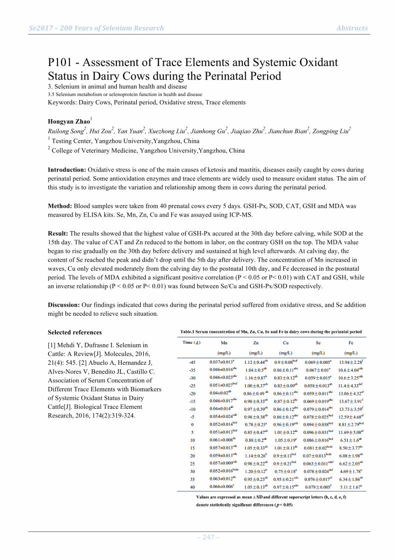
Se2017 – 200 Years of Selenium Research Abstracts
– 247 –
P101 - Assessment of Trace Elements and Systemic Oxidant Status in Dairy Cows during the Perinatal Period 3. Selenium in animal and human health and disease 3.5 Selenium metabolism or selenoprotein function in health and disease Keywords: Dairy Cows, Perinatal period, Oxidative stress, Trace elements Hongyan Zhao1 Ruilong Song2, Hui Zou2, Yan Yuan2, Xuezhong Liu2, Jianhong Gu2, Jiaqiao Zhu2, Jianchun Bian2, Zongping Liu2 1 Testing Center, Yangzhou University,Yangzhou, China 2 College of Veterinary Medicine, Yangzhou University,Yangzhou, China Introduction: Oxidative stress is one of the main causes of ketosis and mastitis, diseases easily caught by cows during perinatal period. Some antioxidation enzymes and trace elements are widely used to measure oxidant status. The aim of this study is to investigate the variation and relationship among them in cows during the perinatal period. Method: Blood samples were taken from 40 prenatal cows every 5 days. GSH-Px, SOD, CAT, GSH and MDA was measured by ELISA kits. Se, Mn, Zn, Cu and Fe was assayed using ICP-MS. Result: The results showed that the highest value of GSH-Px accured at the 30th day before calving, while SOD at the 15th day. The value of CAT and Zn reduced to the bottom in labor, on the contrary GSH on the top. The MDA value began to rise gradually on the 30th day before delivery and sustained at high level afterwards. At calving day, the content of Se reached the peak and didn’t drop until the 5th day after delivery. The concentration of Mn increased in waves, Cu only elevated moderately from the calving day to the postnatal 10th day, and Fe decreased in the postnatal period. The levels of MDA exhibited a significant positive correlation (P < 0.05 or P< 0.01) with CAT and GSH, while an inverse relationship (P < 0.05 or P< 0.01) was found between Se/Cu and GSH-Px/SOD respectively. Discussion: Our findings indicated that cows during the perinatal period suffered from oxidative stress, and Se addition might be needed to relieve such situation. Selected references
[1] Mehdi Y, Dufrasne I. Selenium in Cattle: A Review[J]. Molecules, 2016, 21(4): 545. [2] Abuelo A, Hernandez J, Alves-Nores V, Benedito JL, Castillo C. Association of Serum Concentration of Different Trace Elements with Biomarkers of Systemic Oxidant Status in Dairy Cattle[J]. Biological Trace Element Research, 2016, 174(2):319-324.

Se2017 – 200 Years of Selenium Research Abstracts
– 248 –
P102 - MsrA Protects Hepatocytes against Acetaminophen-Induced Toxicity via TXNRD1 Regulation 3. Selenium in animal and human health and disease 3.5 Selenium metabolism or selenoprotein function in health and disease Keywords: Methionine sulfoxide, Acetaminophen, Hepatotoxicity, Thioredoxin reductase Mahendra Singh1 Geun-Hee Kwak1, Ki Young Kim1, Hwa-Young Kim1 1 Yeungnam University College of Medicine, Daegu, South Korea Introduction: Thioredoxin reductase 1 (TXNRD1) is associated with susceptibility to acetaminophen (APAP)-induced liver damage. Methionine sulfoxide reductase A (MsrA) is an antioxidant and protein repair enzyme that specifically catalyzes the reduction of methionine S-sulfoxide residues. We have previously shown that MsrA deficiency exacerbates acute liver injury induced by APAP. Method: We used primary hepatocytes isolated from MsrA gene-deleted (MsrA–/–) and wild-type (MsrA+/+) mice to investigate the underlying mechanism of the protective effect of MsrA against APAP-induced hepatotoxicity. Result: MsrA–/– hepatocytes showed higher susceptibility to APAP-induced cytotoxicity than MsrA+/+ cells, consistent with our previous in vivo results. MsrA deficiency increased APAP-induced glutathione depletion and reactive oxygen species production. APAP treatment increased Nrf2 activation more profoundly in MsrA–/– than in MsrA+/+ hepatocytes. Basal TXNRD1 levels were significantly higher in MsrA–/– than in MsrA+/+ hepatocytes, while TXNRD1 depletion in both MsrA–/– and MsrA+/+ cells resulted in increased resistance to APAP-induced cytotoxicity. In addition, APAP treatment significantly increased TXNRD1 expression in MsrA–/– hepatocytes, while no significant change was observed in MsrA+/+ cells. Overexpression of MsrA reduced APAP-induced cytotoxicity and TXNRD1 expression levels in APAP-treated MsrA–/– hepatocytes. Discussion: Collectively, our results suggest that MsrA protects hepatocytes from APAP-induced cytotoxicity through the modulation of TXNRD1 expression.
Se2017 – 200 Years of Selenium Research Abstracts
– 249 –
P103 - Knockdown of Sep15 modulates the expression of Pax6 induced by glucose in HLE cells 3. Selenium in animal and human health and disease 3.5 Selenium metabolism or selenoprotein function in health and disease Keywords: Sep15; Pax6; cataract; lens epithelial cells Xiaohuan Li1 Kaixun Huang1 1 Huazhong University of Science and Technology,Wuhan,China Introduction: The 15-kDa selenoprotein (Sep15) is a thioredoxin-like selenoprotein, residenting in the lumen of endoplasmic reticulum and implicated in the quality control of glycoprotein folding through its interaction with UDP-glucose:glycoprotein glucosyltransferase. Although Sep15 KO mice developed a prominent nuclear cataract, the exact function of this protein is not known. Transcription factor Pax6 is highly expression in human lens epithelial (HLE) cells and essential for development of the eye system. The expression level of Pax6 changes with the process of lens fiber differentiation. When epithelial cells differentiate into primary lens fiber cells, the expression of Pax6 will be inhibited. However, effect of Sep15 knockdown on Pax6 remains largely unknown in lens fiber cells differentiation. Method: Short interfering RNAs (siRNA) was used to knockdown the expression of Sep15 in human lens epithelial cells (SRA01/04). Expressions levels of Sep15, Pax6, αA-crystallin, p-ERK, p-AKT, β-catenin, sfrp2, six3 and cyclin D1 were measured by Western blotting or RT-PCR. Result: In the present study, effects of knockdown of Sep15 or glucose on expression of Pax6 and related signal pathway were explored. Our results showed that knockdown of Sep15 or 30mM glucose treated alone resulted in the high expression of Pax6, however, combination showed the expression of Pax6 decreased significantly. Expressions of αA-crystallin, cyclin D1, p-ERK , p-AKT and other signal molecules also changed significantly. Discussion: Our results demonstrate that knockdown of Sep15 impacts expression of Pax6 and related signal molecules, suggesting that Sep15 might play a role in regulating lens fiber cell differentiation. Selected references
Shaham O, Smith A N, Robinson M L, et al. Pax6 is essential for lens fiber cell differentiation[J]. Development, 2009, 136(15):2567-78.
Kasaikina M V, Fomenko D E, Labunskyy V M, et al. Roles of the 15-kDa selenoprotein (Sep15) in redox homeostasis and cataract development revealed by the analysis of Sep 15 knockout mice.[J]. 2011, 286(38):33203-33212.
Labunskyy V M, Hatfield D L, Gladyshev V N. The Sep15 protein family: Roles in disulfide bond formation and quality control in the endoplasmic reticulum[J]. Iubmb Life, 2007, 59(1):1-5.
Korotkov K V, Kumaraswamy E, Zhou Y, et al. Association between the 15-kDa selenoprotein and UDP-glucose:glycoprotein glucosyltransferase in the endoplasmic reticulum of mammalian cells[J]. Journal of Biological Chemistry, 2001, 276(18):15330-6.
Yang Y, Stopka T, Golestaneh N, et al. Regulation of alphaA-crystallin via Pax6, c-Maf, CREB and a broad domain of lens-specific chromatin[J]. Embo Journal, 2006, 25(10):2107–2118.

Se2017 – 200 Years of Selenium Research Abstracts
– 250 –
P104 - Roles of the 15kD selenoprotein in lens epithelial differentiation 3. Selenium in animal and human health and disease 3.5 Selenium metabolism or selenoprotein function in health and disease Keywords: 15kD selenoprotein, lens, cataract Xiaoxiang Zheng1 Jun Zhou1, Hongmei Liu1, Kaixun Huang1 1 School of Chemistry&Chemical Engineering, Huazhong University of Science&Technology,Wuhan,China Introduction: Selenium is an essential micronutrient in mammals mainly presented in the form of selenoprotein. The 15kD selenoprotein (Sep15) is an endoplasmic reticulum-resident selenoprotein while its precise function still remains elusive. It was reported that Sep15 knockout mice developed nuclear cataracts at an early age, indicated the essential role of Sep15 in lens development. So we speculate that Sep15 might participate in regulation of differentiation in lens epithelial cells based on its structure and function. Method: Effects of Sep15 knockdown by RNAi on some differentiation biomarkers were studied in bFGF-induced human lens epithelial cells (HLEC). Sep15 knockout mice were established by CRISPR/Cas9. Westernblot, RT-PCR and immunohistochemical were used to measure expression levels of differentiation biomarkers or some signaling molecules in HLEC and mice lens. HE stain and TEM were used to detect the pathology phenotypes of mice lens. Result: Several biomarkers of lens cell differentiation such as β-crystallin and N-cadherin were significantly influenced by Sep15 knockdown in bFGF induced HLEC. That was also verified in lens of Sep15 knockout mice. The difference of lens lesion including some parameters between knockout and wildtype mice increased with age of mouse. Discussion: The current results show that lens epithelial cell differentiation was influenced by Sep15 knockdown or knockout, suggesting that Sep15 might participate in regulating differentiation of lens epithelial cell. Further researches are needed to observe the lens abnormalities and more parameters in mice of different ages. We hope this work could contribute to reveal the relationship between Sep15 and cataract formation. Selected references
Yin N, Zheng X, Zhou J, et al. Knockdown of 15-kDa selenoprotein (Sep15) increases hLE cells’ susceptibility to tunicamycin-induced apoptosis. JBIC Journal of Biological Inorganic Chemistry, 2015, 20(8): 1307-1317.
Labunskyy V M, Hatfield D L, Gladyshev V N. The Sep15 protein family: Roles in disulfide bond formation and quality control in the endoplasmic reticulum. Iubmb Life, 2007, 59(1):1-5.
Kasaikina M V, Fomenko D E, Labunskyy V M, et al. Roles of the 15-kDa selenoprotein (Sep15) in redox homeostasis and cataract development revealed by the analysis of Sep 15 knockout mice[J]. Journal of Biological Chemistry, 2011, 286(38): 33203-33212.
Menko A S. Lens epithelial cell differentiation. Experimental eye research, 2002, 75(5): 485-490.
Se2017 – 200 Years of Selenium Research Abstracts
– 251 –
P105 - Role of the 15kDa selenoprotein in colorectal inflammation 3. Selenium in animal and human health and disease 3.5 Selenium metabolism or selenoprotein function in health and disease Keywords: inflammation, colitis, 15kDa selenoprotein, gut microbiome Kristin Peters1 Bradley Carlson2, Jessica Canter1, Ryuta Tobe3, Harold Seifried4, Yunkai Yu5, Liang Cao5, Vadim Gladyshev6, Cindy Davis7, Dolph Hatfield2, Petra Tsuji1 1 Dept. of Biological Sciences, Towson University, Towson, MD, USA 2 Molecular Biology of Selenium Section, NIH, Bethesda, MD, USA 3 Dept. of Biotechnology, Ritsumeikan University, Shiga, Japan 4 Division of Cancer Prevention, NIH, Rockville, MD, USA 5 Molecular Targets Core, NIH, Bethesda, MD, USA 6 Brigham & Women's Hospital, Harvard Medical School, Boston, MA, USA 7 Office of Dietary Supplements, NIH, Bethesda, MD, USA Introduction: Previously, systemic knockout (KO) of the 15 kDa selenoprotein (SELENOF) protected mice against the formation of chemically-induced pre-neoplastic lesions and suggested modulation of inflammatory pathways [1]. We hypothesized that SELENOF expression is inversely correlated with a protective pro-inflammatory response in a colitis-associated mouse model. Method: The potential role of SELENOF in inflammation was examined by assessing gene expression of KO mice and littermate controls (WT) that were either treated with 2% dextran sulfate sodium (DSS) to induce colitis or given water (control). Result: Microarray analyses of colonic mucosa suggested that lack of SELENOF expression itself changed gene expression more significantly than DSS-induced inflammation. Changes in colonic gene expression in SELENOF-KO mice, as validated with qPCR, included up-regulation of inflammation-related genes, such as the interferon (IFN)-γ-regulated guanylate-binding protein 1 (p=0.004), compared to WT mice regardless of treatment. Because intestinal microbiota are known to influence inflammation of the host, bacterial DNA was isolated from feces of both WT and KO mice. Subsequent amplification of the 16S rRNA gene and sequencing with Illumina MiSeq revealed increased abundance of Proteobacteria, particularly Enterobacteriaceae, in untreated WT compared to KO mice. Discussion: We evaluated the potential influence of SELENOF on the composition of the intestinal microbiome. Our preliminary analyses indicate that SELENOF may influence the expression of inflammatory genes in the colon. Its contribution to the regulation of colitis, which may be modulated by the microbiome, will be further examined. Selected references
[1] Tsuji et al. PlosONE 2012

Se2017 – 200 Years of Selenium Research Abstracts
– 252 –
P106 - Low selenoprotein P status in patients with traumatic spinal cord injury 3. Selenium in animal and human health and disease 3.5 Selenium metabolism or selenoprotein function in health and disease Keywords: SELENBP1, diagnostics, sandwich assay, serum, neuron, paraplegia Julian Seelig1 Raban Heller2, Qian Sun1, Bahram Biglari3, Arash Moghaddam-Alvandi4, Lutz Schomburg1 1 Charité - Universitätsmedizin Berlin, Institut für Experimentelle Endokrinologie, Berlin, Germany 2 Universitätsklinikum Heidelberg. Department für Orthopädie, Unfallchirurgie und Paraplegiologie, Germany 3 Department of Paraplegiology, Berufsgenossenschaftliche Unfallklinik Ludwigshafen, Ludwigshafen, Germany 4 Klinikum Aschaffenburg-Alzenau, Abt. Unfall-Chirurgie, Handchirurgie, Orthopädie, Germany Introduction: Traumatic spinal cord injury is associated with neuronal loss and bears the risk for paraplegia. As selenium is an essential factor for neuronal development and protects from neuron degeneration, and traumatic injury constitutes a major inflammatory stimulus potentially down-regulating selenium metabolism, we aimed to monitor selenium status upon traumatic spinal cord injury during remission or non-remission by a time-resolved analysis. Method: Selenoprotein P was analyzed by sandwich ELISA. Total serum selenium concentrations were measured by total reflection X-ray fluorescence. Concentrations of selenium-binding protein 1 were determined by a newly developed sandwich assay. Result: A set of 20 subjects with traumatic spinal cord injury were analyzed, 10 of which went into remission and 10 developed paraplegia. In addition, 10 subjects with an unrelated injury were analyzed. Selenoprotein P showed a wide spectrum of concentrations within a given group, as well as between the three patient groups (mean±SD; 4.7±1.9; 4.9+1.6; 4.6± 1.4 mg/L). Especially in one group of patients, selenium binding protein 1 was detectable in the early time points analyzed, decreasing with time. De-blinding of the patient characteristics for clinical associations has not yet taken place. Discussion: Selenoprotein P is a cell-death inhibitory factor for neurons, which may be of high clinical importance in traumatic spine injuries. Some patients displayed extremely low selenoprotein P concentrations (< 1 mg/L) at early time points post injury, which may impair regenerative processes, causing excessive neuronal loss and may warrant selenium supplementation as promising adjuvant therapeutic measure in order to reduce paraplegia risk. Selected references Supported by Charité – Universitätsmedizin Berlin.
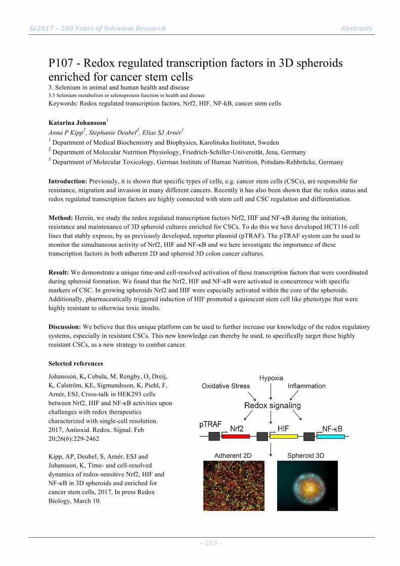
Se2017 – 200 Years of Selenium Research Abstracts
– 253 –
P107 - Redox regulated transcription factors in 3D spheroids enriched for cancer stem cells 3. Selenium in animal and human health and disease 3.5 Selenium metabolism or selenoprotein function in health and disease Keywords: Redox regulated transcription factors, Nrf2, HIF, NF-kB, cancer stem cells Katarina Johansson1 Anna P Kipp2, Stephanie Deubel3, Elias SJ Arnér1 1 Department of Medical Biochemistry and Biophysics, Karolinska Institutet, Sweden 2 Department of Molecular Nutrition Physiology, Friedrich-Schiller-Universität, Jena, Germany 3 Department of Molecular Toxicology, German Institute of Human Nutrition, Potsdam-Rehbrücke, Germany Introduction: Previously, it is shown that specific types of cells, e.g. cancer stem cells (CSCs), are responsible for resistance, migration and invasion in many different cancers. Recently it has also been shown that the redox status and redox regulated transcription factors are highly connected with stem cell and CSC regulation and differentiation. Method: Herein, we study the redox regulated transcription factors Nrf2, HIF and NF-κB during the initiation, resistance and maintenance of 3D spheroid cultures enriched for CSCs. To do this we have developed HCT116 cell lines that stably express, by us previously developed, reporter plasmid (pTRAF). The pTRAF system can be used to monitor the simultaneous activity of Nrf2, HIF and NF-κB and we here investigate the importance of these transcription factors in both adherent 2D and spheroid 3D colon cancer cultures. Result: We demonstrate a unique time-and cell-resolved activation of these transcription factors that were coordinated during spheroid formation. We found that the Nrf2, HIF and NF-κB were activated in concurrence with specific markers of CSC. In growing spheroids Nrf2 and HIF were especially activated within the core of the spheroids. Additionally, pharmaceutically triggered induction of HIF promoted a quiescent stem cell like phenotype that were highly resistant to otherwise toxic insults. Discussion: We believe that this unique platform can be used to further increase our knowledge of the redox regulatory systems, especially in resistant CSCs. This new knowledge can thereby be used, to specifically target these highly resistant CSCs, as a new strategy to combat cancer. Selected references
Johansson, K, Cebula, M, Rengby, O, Dreij, K, Calström, KE, Sigmundsson, K, Piehl, F, Arnér, ESJ, Cross-talk in HEK293 cells between Nrf2, HIF and NF-κB activities upon challenges with redox therapeutics characterized with single-cell resolution. 2017, Antioxid. Redox. Signal. Feb 20;26(6):229-2462 Kipp, AP, Deubel, S, Arnér, ESJ and Johansson, K, Time- and cell-resolved dynamics of redox-sensitive Nrf2, HIF and NF-κB in 3D spheroids and enriched for cancer stem cells, 2017, In press Redox Biology, March 10.

Se2017 – 200 Years of Selenium Research Abstracts
– 254 –
P108 - Differential Impact of Two Secisbp2 Mutations on Selenoprotein Expression in Liver and Brain 3. Selenium in animal and human health and disease 3.5 Selenium metabolism or selenoprotein function in health and disease Keywords: Secisbp2, missense mutation, R543Q, C696R, selenoprotein Wenchao Zhao1 Simon Bohleber1, Mike Howard2, Noelia Fradejas-Villa1, Ulrich Schweizer1 1 IBMB, Rheinische Friedrich-Wilhelms-Universität, Bonn, Germany 2 Department of Genetics, University of Salt Lake City, UT, USA Introduction: Secisbp2 (SECIS-binding protein 2) interacts with SECIS (selenocysteine insertion sequence) element, located in the 3′-untranslated region of eukaryotic selenoprotein mRNAs. Secisbp2 is essential for the incorporation of selenocysteine, the 21st amino acid, into protein [1]. Recently, several SECISBP2 mutations were reported in human patients [2,3,4]. These mutations lead to reduced selenoprotein expression in many organs. Most patients suffer from thyroid hormone resistance, some also show neurological and muscular impairments [4,5]. Method: We have generated, liver and brain-specific Secisbp2 missense mutations in mice (C696R [2], R543Q [3]), in addition to Secisbp2 knockout mice which we have already published [6,7]. We also have employed ribosomal profiling to analyze in detail the impact of the R543Q mutation on selenoprotein expression in cerebral cortex. Result: Our data show that both mutations significantly reduce the levels of several selenoproteins in liver – indistinguishable from the knockout mice. In neuron-specific transgenic mice, selenoprotein expression is significantly reduced in C696R mutant mice resembling Secicbp2 knockout mice. In contrast, the R543Q mutation is milder and does not lead to overt neurological dysfunction in mice in line with mild reduction of several selenoproteins. Discussion: Our findings indicate that the distinct phenotypes in two mutations are caused by differential impact on selenoprotein expression. Next we will find out how two mutations affect the translation status of selenoprotein biosynthesis. Selected references
[1] Fradejas-Villar N, Nucleic Acids Research, 2016, 35(10):3214-3222.
[2] Schoenmakers E, Journal of Clinical Investigation, 2010, 120(12):4220.
[3] Dumitrescu A M, Nature Genetics, 2005, 37(11):1247.
[4] Dumitrescu A M, Antioxidants & Redox Signaling, 2009, 12(7):905-920.
[5] Azevedo M F, Journal of Clinical Endocrinology & Metabolism, 2010, 95(8):4066.
[6] Seeher S, Biochemical Journal, 2014, 462(1):67-75.
[7] Seeher S, Antioxidants & Redox Signaling, 2014, 21(6):835-49.

Se2017 – 200 Years of Selenium Research Abstracts
– 255 –
P109 - A suppressive effect of selenium on amyloid-β plaque deposition in Tg2576 transgenic mice brain 3. Selenium in animal and human health and disease 3.5 Selenium metabolism or selenoprotein function in health and disease Keywords: Alzheimer's disease, selenium deficiency, GPx Sakura Yoshida1 Mamoru Haratake2, Eriko Hori1, Ryunosuke Kadotomi1, Miho Iwataka1, Wataru Uehara1, Takeshi Fuchigami1, Morio Nakayama1 1 Graduate School of Biomedical Sciences, Nagasaki University, 1-14, Bunkyo-machi, Nagasaki, Japan 2 Faculty of Pharmaceutical Sciences, Sojo University, 4-22-1, Ikeda, Kumamoto, Japan Introduction: Alzheimer’s disease (AD) is a chronic neurodegenerative disease and characterized by deposition of amyloid-b (Ab) peptide in the brain. Reactive oxygen species (ROS) are thought to be associated with the onset and/or progression of AD. Selenium-dependent glutathione peroxidases (GPxs) play a critical role in the brain in the extinction of ROS. The selenium concentration in the brain is kept higher than those of other organs/tissues even when dietary selenium is limited, which suggests the importance of this element in the brain. In this paper, we investigated the effect of dietary selenium on the Ab plaque deposition in the Tg2576 transgenic mice brain. Method: Female 16-week-old Tg2576 mice (Taconic Farms, Inc.) were fed diets with different selenium-status for 17-month. Selenium concentrations were fluorometrically determined with 2,3-diaminonaphthalene after acid digestion. GPx activity was determined by measuring a decrease in the absorbance of NADPH at 340 nm. Ab plaques in brain sections were fluorescently detected using amyloid-specific thioflavin T. Result: Selenium concentration and GPx activity in the brain of the selenium-deficient diet-fed mice was lower than those in the selenium-adequate conventional diet-fed mice. The number of Ab plaques in the brain sections of the selenium-deficient diet-fed mice was larger than those in the selenium-adequate conventional diet-fed mice. A similar trend was also observed between the selenium-deficient and the selenomethionine (SeMet)-supplemented diet-fed mice. Discussion: The Ab plaque deposition in the brain of the Tg2576 mice appears to associate with its GPx activity.

Se2017 – 200 Years of Selenium Research Abstracts
– 256 –
P110 - Homozygous mutation p.P190L in TXNRD1 is associated with genetic generalized epilepsy 3. Selenium in animal and human health and disease 3.6 Clinical genetics of selenium or selenoprotein-encoding genes Keywords: Generalized seizures, Epilepsy, Oxidative stress, TXNRD1 Noelia Fradejas-Villar1 A Kudin2, G Baron2, Q Cheng3, ES Arnér3, W Kunz2, U Schweizer1 1 IBMB, University of Bonn, Bonn, Germany 2 Department of Epileptology and Life&Brain, University of Bonn, Bonn, Germany. 3 Dept. Medical Biochemistry and Biophysics (MBB), Karolinska Institutet, Stockholm, Sweden Introduction: Growing evidence points to oxidative stress is connected to epilepsy pathogenesis. Increased production of ROS has been reported during seizures. Loss-of-function mutations in brain selenoproteins lead to epileptic phenotypes in mice and humans. Method: Here, we report a homozygous mutation in TXNRD1 (thioredoxin reductase 1) in a family with genetic generalized epilepsy identified via whole exome sequencing. Result: The TXNRD1 mutation p.Pro190Leu affecting a highly conserved amino acid residue co-segregates with the phenotype within the family. TXNRD1 activity was determined in subcellular fractions from a skeletal muscle biopsy and skin fibroblasts of the index patient. As result of the mutation, the activity of TXNRD1 was reduced in the patient's fibroblasts and skeletal muscle (to 34±3% and 16±8% of controls, respectively). Expression of the mutated protein was decreased as judged by 75Se labeling and Western blot analysis. We detected reduced 75Se-labeling of the enzyme (41±3% of controls) in patient fibroblasts. An in-depth in vitro kinetic analysis of the recombinant enzyme indicated 30–40% lowered kcat/Se values. Moreover, the patient fibroblasts were less resistant to a hydrogen peroxide challenge than controls. Discussion: Therefore, a reduced activity of the enzyme in the patient's tissue samples is explained by (i) lower enzyme turnover and (ii) reduced abundance of the mutated enzyme. Our data agree with a potential role of insufficient ROS detoxification in epilepsy. They show that findings in genetic models deficient in cerebral selenoprotein expression are relevant to human disease.
Se2017 – 200 Years of Selenium Research Abstracts
– 257 –
P111 - Comparative analysis of selenium status in common neuropsychiatric disorders in children 3. Selenium in animal and human health and disease 3.7 Additional and emerging topics of selenium in health or disease Keywords: autism, cerebral palsy, attention deficit hyperactivity disorder, epilepsy Anastasiya Skalnaya1 Alexey Tinkov2, Andrew Grabeklis3, Anatoly Skalny3 1 Lomonosov Moscow State University, Department of Medicine, Moscow, Russia 2 Orenburg State Medical Academy, Orenburg, Russia 3 RUDN – People’s Friendship University of Russia, Moscow, Russia Introduction: Selenium plays a significant role in various metabolic processes through its structural role in selenoproteins. At the same time, both selenium deficiency and excess was shown to be associated with increased oxidative stress and different pathologies, including neurodegenerative diseases. Method: Therefore, the objective was to assess the level of Se in hair and serum of children (aged 0-5 and 5-15 years) with autism spectrum disorders (ASD), cerebral palsy (CP), communication disorders (CD), attention deficit hyperactivity disorder (ADHD), and epilepsy (Ep) in comparison with the control ones using ICP-DRC-MS. Result: The obtained data demonstrate that children (< 5 y.o.) with Ep > ADHD > ASD > CD were characterized by significantly (p < 0.001) increased hair Se levels, whereas no significant difference was detected between the control and CP group values. More profound difference was detected in the older children (5-15 y.o.) due to significantly increased hair Se levels in all examined pathologies (p < 0.001). It is notable that serum Se levels were more stable in both groups of children. In a group of < 5 y.o. children hair Se levels decreased in the following order Ep > CP > ADHD > CD > ASD, whereas the difference from the control levels was significant only in Ep and CP groups (p < 0.05). In older children serum Se levels did not differ significantly from the control levels. Discussion: So, Se metabolism is significantly deranged in children with common neuropsychiatric diseases. The observed difference in hair and serum Se levels may be indicative of increased Se excretion and changes in Se species. Selected references
Skalny, A. V., Simashkova, N. V., Klyushnik, T. P., Grabeklis, A. R., Bjørklund, G., Skalnaya, M. G., ... & Tinkov, A. A. (2017). Hair toxic and essential trace elements in children with autism spectrum disorder. Metabolic Brain Disease, 32(1), 195-202.
Skalny, A. V., Simashkova, N. V., Klyushnik, T. P., Grabeklis, A. R., Radysh, I. V., Skalnaya, M. G., & Tinkov, A. A. (2016). Analysis of Hair Trace Elements in Children with Autism Spectrum Disorders and Communication Disorders. Biological Trace Element Research, doi: 10.1007/s12011-016-0878-x
Skalny, A. V., Simashkova, N. V., Klyushnik, T. P., Grabeklis, A. R., Radysh, I. V., Skalnaya, M. G., ... & Tinkov, A. A. (2016). Assessment of serum trace elements and electrolytes in children with childhood and atypical autism. Journal of Trace Elements in Medicine and Biology, doi: 10.1016/j.jtemb.2016.09.009
El-Ansary A, Bjorklund G, Tinkov AA, Skalny AV, Al Dera HS (2017) Relationship between selenium, lead, and mercury in red blood cells of Saudi autistic children. Metabolic Brain Disease, doi: 10.1007/s11011-017-9996-1

Se2017 – 200 Years of Selenium Research Abstracts
– 258 –
P112 - Does selenium intake relate to longevity: Case study in Shitai, Anhui, China 3. Selenium in animal and human health and disease 3.7 Additional and emerging topics of selenium in health or disease Keywords: Selenium intake, Human longevity, Shitai ZeDong Long1 Zhangmin Wang2, Xiuxia Li2, Linxi Yuan2, Xuebin Yin1 1 School of Earth and Space Sciences, University of Science and Technology of China , Hefei ,China 2 Jiangsu Bio-Engineering Research Centre of Selenium, Suzhou 215123, China Introduction: Mccann found that age-related diseases were prospectively associated with modest Selenium(Se) deficiency and genetic dysfunction of nonessential selenoproteins [1]. A viewpoint that Se at low levels may be considered a hormetic chemical and decouple healthspan and longevity, was observed in a mice experiment [2]. Does Se play a vital role in human longevity? The understanding of its mechanism is far from being completed on human beings. Method: In order to exclude the impact of climate, socio-economy, lifestyle and other variable factors, two different villages, Dashan as a longevity village and Kushan, 10 kilometers apart, as a common village, were selected in Shitai, Anhui, China. Soil, rice, hair and plasma samples were collected to determine Se concentrations. All treated samples were acid-digested and analyzed for total Se by atomic fluorescence spectrometry (AFS). Result: As shown in Table 1, soil, rice, hair and plasma Se concentrations in Dashan were significant higher than those in Kushan (P<0.05) Discussion: In this study, residents in Dashan have longer lifespan than those in Kushan (unpublished data). Except for the Se contents in soil, these two villages have similar conditions. Since the residents only eat foods locally, Se could be intaken by human beings via soil to rice, which is reflected by the Se contents in hair and plasma. To elucidate the mechanism of Se on human health, more investigations about human health indicators, such as GPX, SOD, will be carried out in future. Selected references
[1] J.C. Mccann, B.N. Ames, Adaptive dysfunction of selenoproteins from the perspective of the triage theory: why modest selenium deficiency may increase risk of diseases of aging, Faseb Journal 25(6) (2011) 1793-814.
[2] Wu R T, Cao L, Mattson E, et al. Opposing impacts on healthspan and longevity by limiting dietary selenium in telomere dysfunctional mice[J]. Aging Cell, 2016.

Se2017 – 200 Years of Selenium Research Abstracts
– 259 –
P113 - Influence of statins in selenium status and inflammatory profile considering creatine kinase levels 3. Selenium in animal and human health and disease 3.7 Additional and emerging topics of selenium in health or disease Keywords: statin, inflammatory profile Lígia Moriguchi Watanabe1 Lívia Fernandes Lima1, Fernando Barbosa Júnior2, Alceu Afonso Jordão Júnior1, Anderson Marliere Navarro1 1 Faculty of Medicine of Ribeirão Preto, University of São Paulo – Ribeirão Preto-SP, Brazil 2 Faculty of Pharmaceutical Sciences, University of São Paulo – Ribeirão Preto-SP, Brazil Introduction: Statins are the most frequently used medication for the treatment of hypercholesterolemia and prevention of cardiovascular events. However, their use has been associated with alterations in bioavailability of selenium, an essential micronutrient that plays a role in anti-inflammatory and antioxidant processes. The aim of this study was to assess the influence of the use of statins in selenium status and inflammatory profile in patients with normal or increased creatine kinase (CK) concentration. Method: Individuals, using statin for less than a year, were classified as: Group 1 (G1): n=24, with changes in the CK levels, and Group 2 (G2): n=31, without changes in CK levels. Erythrocyte selenium, C-reactive protein (CRP), Interleukin 6 (IL-6), tumor necrosis factor alpha (TNF-a) and CK were evaluated. Result: The average age of participants was 56.5±14,4 years. The sample consisted of 35 males (63,6%) and 20 females (36,4%). Comparison of CK, erythrocyte selenium, CRP, IL-6 and TNF-a between study groups are displayed in Table 1. Discussion: Our study demonstrated that individuals using statin presented a deficiency in erythrocyte selenium. The higher levels of IL-6 in both groups indicate a pro-inflammatory profile that did not seem related with CK changes but could be associated with the deficiency of selenium. Interestingly, the concentrations of TNF-a were increased in both groups and was higher in G1 (increased CK levels) indicating a relative risk for cardiovascular events. Finally, more studies are required to further address the effects (or side effects) of statins in selenium status and metabolism. Selected references aSTEFANOWICZ, FA, et al. Erythrocyte selenium concentration as a marker of selenium status. Clin Nutri. 32(5):837-42, 2013. bVOLP, ACP et al. Inflammation biomarkers capacity in predicting the metabolic syndrome. Arq Bras Endrocrinol Metab. 52(3): 537-549, 2008. cRAUCHHAUS, M et al. Inflammatory cytokines and the possible immunological role for lipoproteins in chronic heart failure. International Journal of Cardiology. 76: 125–133, 2000.
Se2017 – 200 Years of Selenium Research Abstracts
– 260 –
P114 - The role of selenium in an in vitro invasive model of pancreatic cancer 3. Selenium in animal and human health and disease 3.7 Additional and emerging topics of selenium in health or disease Keywords: pancreatic cancer invasion Ali Coyle1 Joanne Keenan1, Finbarr O Sullivan1, Karina Horgan2, Martin Clynes1 1 National Institute of Cellular Biotechnology, Dublin City University 2 Alltech Ireland Introduction: Pancreatic cancer patients have been found to have lower levels of serum selenium compared to healthy controls, and higher levels of serum selenium have been correlated with longer survival of patients with pancreatic cancer1. These findings suggest an important role for selenium intake and potential therapeutic supplementation in pancreatic cancer. However, less is known about the role selenium may play in pancreatic cell invasion and motility. This work focuses on the effect of pre-treatment and co-treatment with an organic and an inorganic selenium source on motility and invasion in pancreatic cancer cells in vitro. Method: A panel of pancreatic cell lines with varying invasive profiles were tested for their sensitivity to two selenium compounds using acid phosphatase as an end-point to toxicity assays. Pancreatic cancer cells were pre-treated or co-treated with two selenium compounds before being set up for in vitro invasion assays using the Boyden chamber method. Motility (without matrigel) and invasion (with matrigel) was assessed by crystal violet staining of migrated or invaded cells. Result: Selenium tolerance varied greatly in the panel of pancreatic cell lines. Pre-treatment and co-treatment with the organic and inorganic compounds gave differential effects on the migrating and invading capacity of the cells. Discussion: This work will aid in our understanding of any potential role for selenium in pancreatic cancer metastasis, and whether that role is affected by time or by the chemical form of selenium used. Selected references
1. Lener, M. R. et al. Serum Concentrations of Selenium and Copper in Patients Diagnosed with Pancreatic Cancer. Cancer Res. Treat. 48, 1056–64 (2016).
Se2017 – 200 Years of Selenium Research Abstracts
– 261 –
P115 - The role of environmental metal ions in human health and diseases 3. Selenium in animal and human health and disease 3.7 Additional and emerging topics of selenium in health or disease Keywords: Excess selenium, cadmium toxicity, metal interactions, lipid metabolism, liver disease Young-Mi Go1 Xin Hu1, Jushua Chandler1, Dean Jones1 1 Department of Medicine, Emory University, Atlanta, USA Introduction: Multiple metal ions are present in the human diet, with some being required as essential nutrients and all causing toxicity at high intakes. Low level environmental cadmium (Cd) occurs in human diet and accumulates in vivo. Selenium (Se) is an essential micronutrient, and a component of 25 selenoproteins responsible for physiological functions. However, excess Se increases the risk of diseases, therefore, determinination of adequate Se amount for human health is critical. Impacts of metals and their interaction on cells and organs need to be addressed. Method: C57BL6 male mice were treated with Se (4 mg Na2SeO4/L, 16 weeks), Cd (3.3 mg CdCl2/L, 16 weeks) and mixture. Metals, redox states, metabolites and phenotypic markers for lung and liver functions were analyzed. Result: Urinary Se was 4-6 folds higher in Se-treated group while no changes were observed in lung and liver. Cd levels were elevated in urine, lung and liver in Cd mice. Se-treated mice significant increased body mass than control or Cd groups. Cd oxidized Prx3 in lung but not in liver, and co-treatment showed that Se inhibited Prx3 oxidation. Liver metabolomics of Se-treated mice showed that altered metabolome was significantly associated with weight gain, and accompanied by decreased levels of bile acids and acyl carnitines. Discussion: Excess Se disrupts lipids homeostasis. Our study showed that mice treated with Se caused higher Cd accumulation in lung and liver, suggesting that excess Se could elevate Cd toxicity. The results suggest that presence of multiple metal ions in diet could affect human health potentiating metal toxicity.
Se2017 – 200 Years of Selenium Research Abstracts
– 262 –
P116 - Selenoproteins regulate B cell functions by targeting B cell receptor (BCR)-mediated antigen present 3. Selenium in animal and human health and disease 3.5 Selenium metabolism or selenoprotein function in health and disease Keywords: selenoprotein, B cell, ROS, antigen processing Bhuvana Katkere1 Rachel Markley1, David Williamson1, Ashley Shay1, Bradley Carlson2, K Sandeep Prabhu1, Girish Kirimanjeswara1 1 The Pennsylvania University, University Park, PA 16802 USA 2 NIH, Bethesda, MD USA Introduction: The role selenoproteins in B cells remains poorly understood. We sought to determine the role of selenoproteins in BCR-mediated effects because BCR-mediated signaling is critical in B cell development, activation, and functions and is highly sensitive to ROS. Method: BCR endocytosis, calcium signaling, antigen trafficking, and antigen processing were measured in B cells that were maintained at various concentrations of sodium selenite (Na2Se3, 0-500 nmol) or B cells isolated from the mice maintained on Se-deficient, -adequate or -supplemented diets. The effect of ROS on BCR-mediated functions was determined by treating the cells with N-acetyl Cysteine (NAC). The role of selenoproteins were determined by generating mice that lack selenoproteins in B cells. Result: The kinetics of BCR endocytsosis, Ca signaling, antigen trafficking and processing were significantly increased in B cells treated with >200 nmol Na2Se3 compared to cells treated with 0-100 nmol. Similarly, BCR-mediated signaling and antigen processing were significantly increased in B cells from Se-supplemented compared to that of Se-deficient or -adequate mice. The ROS levels were significantly higher in B cells maintained at deficient or adequate levels of Se. However, upon treatment with the NAC, the BCR-mediated functions were significantly enhanced in Se-deficient and Se-adequate B cells. It was further determined that BCR-mediated effects were regulated by selenoproteins. Discussion: This study demonstrates that selenoprteins regulate BCR-mediated effects by controlling ROS levels in the cells. Interestingly, the functions of B cells were significantly enhanced by supplementing with higher levels of Se. Thus, selenoproteins could be the new targets for optimizing humoral immunity.
Se2017 – 200 Years of Selenium Research Abstracts
– 263 –
P117 - Effect of anti-rheumatic treatment on selenium levels in rheumatoid arthritis , psoriatic arthritis 3. Selenium in animal and human health and disease 3.7 Additional and emerging topics of selenium in health or disease Keywords: Rheumatoid arthritis, Psoriatic arthritis, Ankylosing arthritis, inflammation Gia Deyab1 Ingrid Hokstad2, Jan Olav Aaseth3, Jon Elling Whist1, Milada Cvancarova Småstuen4, Stefan Agewall5, Torstein Lyberg6, Gunnbjørg Hjeltnes7, Ivana Hollan8 1 Department of Medical Biochemistry, Innlandet Hospital Trust, Norway 2 Lillehammer Hospital for Rheumatic Diseases, Norway 3 Innlandet University College, Norway 4 Institution of health care - Health science PhD programme, Oslo and Akershus University College, Nor 5 Oslo University Hospital, Ullevål , Norway 6 Department of Medical Biochemistry, Oslo University Hospital, Ullevål, Norway 7 Department of Medicine, Innlandet Hospital Trust, Norway 8 Harvard Medical School, Boston, USA Introduction: The reason for increased cardiovascular (CV) risk in inflammatory rheumatic diseases (IRDs) is unclear. Selenium deficiency has been proposed to contribute to development of CV disease. Although the reference range is 50-120 µg/L, there are indications that levels <80 µg/L might be insufficient [1]. Our aim was to measure serum selenium (s-Se) levels in IRD patients, and to evaluate the effect of anti-rheumatic treatment on s-Se levels. Method: We examined samples from 130 patients with IRDs (64 with Rheumatoid arthritis (RA), 40 with Psoriasis arthritis (PsA) and 26 with Ankylosing spondylitis (AS)) starting with methotrexate monotherapy or anti-tumor necrosis factor treatment (anti-TNF) with or without methotrexate. s-Se and serologic inflammatory biomarkers were measured. The patients were evaluated at baseline, and after 6 weeks and 6 months of therapy. Result: The baseline median s-Se level in the total IRD group was 72µg/L. The s-Se levels increased in all groups (total, RA, PsA and AS) after therapy, but the differences from baseline were not significant. Changes in s-Se were significantly negatively related to changes in C-reactive protein and erythrocyte sedimentation rate. s-Se increased at 6 weeks (p=0.012) and 6 months (p=0.038) in patients treated with methotrexate. Anti-TNF was not associated with any significant s-Se changes. Discussion: IRD patients appeared to have suboptimal s-Se levels, but the levels significantly increased with methotrexate treatment. Further studies are needed to determine if methotrexate and other anti-rheumatic drugs might reduce CV risk, and to determine if Se supplementation might reduce CV risk in IRD [2]. Selected references
Hurst, R., C.N. Armah, J.R. Dainty, D.J. Hart, B. Teucher, A.J. Goldson, et al., Establishing optimal selenium status: results of a randomized, double-blind, placebo-controlled trial. Am J Clin Nutr, 2010. 91(4): p. 923-31.
Alehagen, U., J. Alexander, and J. Aaseth, Supplementation with Selenium and Coenzyme Q10 Reduces Cardiovascular Mortality in Elderly with Low Selenium Status. A Secondary Analysis of a Randomised Clinical Trial. PLoS One, 2016. 11(7): p. e0157541.
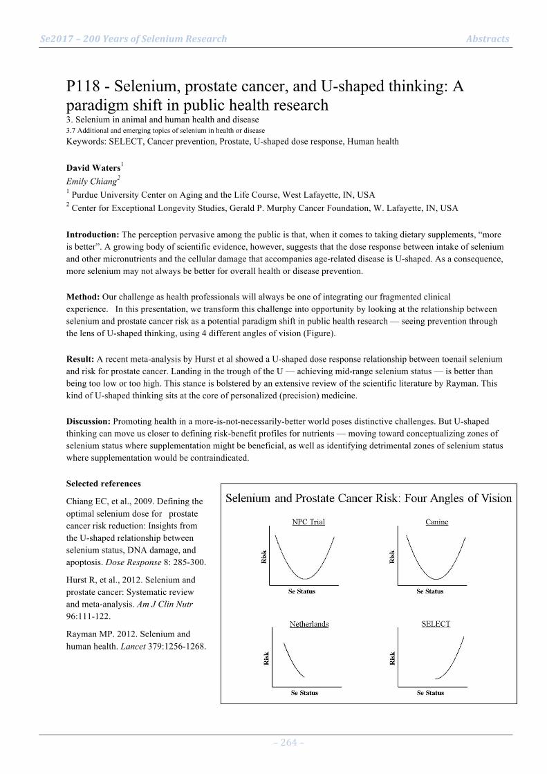
Se2017 – 200 Years of Selenium Research Abstracts
– 264 –
P118 - Selenium, prostate cancer, and U-shaped thinking: A paradigm shift in public health research 3. Selenium in animal and human health and disease 3.7 Additional and emerging topics of selenium in health or disease Keywords: SELECT, Cancer prevention, Prostate, U-shaped dose response, Human health David Waters1 Emily Chiang2 1 Purdue University Center on Aging and the Life Course, West Lafayette, IN, USA 2 Center for Exceptional Longevity Studies, Gerald P. Murphy Cancer Foundation, W. Lafayette, IN, USA Introduction: The perception pervasive among the public is that, when it comes to taking dietary supplements, “more is better”. A growing body of scientific evidence, however, suggests that the dose response between intake of selenium and other micronutrients and the cellular damage that accompanies age-related disease is U-shaped. As a consequence, more selenium may not always be better for overall health or disease prevention. Method: Our challenge as health professionals will always be one of integrating our fragmented clinical experience. In this presentation, we transform this challenge into opportunity by looking at the relationship between selenium and prostate cancer risk as a potential paradigm shift in public health research — seeing prevention through the lens of U-shaped thinking, using 4 different angles of vision (Figure). Result: A recent meta-analysis by Hurst et al showed a U-shaped dose response relationship between toenail selenium and risk for prostate cancer. Landing in the trough of the U — achieving mid-range selenium status — is better than being too low or too high. This stance is bolstered by an extensive review of the scientific literature by Rayman. This kind of U-shaped thinking sits at the core of personalized (precision) medicine. Discussion: Promoting health in a more-is-not-necessarily-better world poses distinctive challenges. But U-shaped thinking can move us closer to defining risk-benefit profiles for nutrients — moving toward conceptualizing zones of selenium status where supplementation might be beneficial, as well as identifying detrimental zones of selenium status where supplementation would be contraindicated. Selected references
Chiang EC, et al., 2009. Defining the optimal selenium dose for prostate cancer risk reduction: Insights from the U-shaped relationship between selenium status, DNA damage, and apoptosis. Dose Response 8: 285-300.
Hurst R, et al., 2012. Selenium and prostate cancer: Systematic review and meta-analysis. Am J Clin Nutr 96:111-122.
Rayman MP. 2012. Selenium and human health. Lancet 379:1256-1268.

Se2017 – 200 Years of Selenium Research Abstracts
– 265 –
P119 - Development of a point-of-care test for selenoprotein P 3. Selenium in animal and human health and disease 3.7 Additional and emerging topics of selenium in health or disease Keywords: POCT, lateral-flow, sandwich, ELISA, blood, bedside test Waldemar Minich1 Torsten Schulz2, Petra Seemann3, Lutz Schomburg1 1 Charité - Universitätsmedizin Berlin, Institut für Experimentelle Endokrinologie, Berlin, Germany 2 InVivo BioTech Services GmbH, Hennigsdorf, Germany 3 Charité - Universitätsmedizin Berlin, Berlin-Brandenburg Center for Regenerative Therapies, Berlin, Germany Introduction: Selenoprotein P (SELENOP) constitutes a relevant and reliable biomarker of selenium intake and status in humans. Both genetic and analytical studies provided evidence linking SELENOP genotype and expression levels with human disease, including cancer, autoimmune or inflammatory diseases. Concentrations of SELENOP can be determined by different analytical methods, yet a point-of-care test (POCT) for bedside analysis is missing. Method: Two SELENOP-specific monoclonal antibodies (mAb) have been generated in mice, intensively characterized and used to establish a reliable sandwich ELISA (1). One of these mAb was labeled by colloidal-gold and the other was immobilized as catcher line on a lateral flow matrix. Different labeling methods, mAb concentrations and ways of storage and pre-dilution of human serum samples are compared for optimizing signal generation. Result: Taking advantage of the same mAb used for the sandwich ELISA, a reliable detection of SELENOP from human serum samples by a POCT is feasible. Serial dilutions of a standard sample yielded signal intensities correlating to the SELENOP amount in a concentration range of 10 – 500 ng/ml. Inter-assay POCT coefficients of variation were not consistently below 30%, indicating that further optimization steps are necessary. Discussion: A SELENOP-specific POCT will enable the determination of SELENOP concentrations from drops of blood at the bed side or in field studies in a non-invasive way without the need for lab equipment or a cooling chain. Unfortunately, the requirements for a highly reliable systems are not yet fully reached, and some parameters need to be further optimized before it can be used in clinical studies. Selected references
(1) Hybsier S, Schulz T, Wu Z, Demuth I, Minich WB, Renko K, Rijntjes E, Köhrle J, Strasburger CJ, Steinhagen-Thiessen E, Schomburg L. Sex-specific and inter-individual differences in biomarkers of selenium status identified by a calibrated ELISA for selenoprotein P. Redox Biol. 2017; 11:403-414.

Se2017 – 200 Years of Selenium Research Abstracts
– 266 –
P120 - Age-dependent protective effect of Selenium against UVA irradiation in primary human keratinocytes a 3. Selenium in animal and human health and disease 3.7 Additional and emerging topics of selenium in health or disease Keywords: Skin, DNA repair, aging Walid Rachidi1 1 Université Grenoble Alpes Introduction: Few studies have focused on the protective role of selenium (Se) on skin aging and photoaging although selenoproteins are essential for keratinocyte function and skin development. Method: Our study aimed at studying the effect of selenium supplementation on primary human keratinocytes obtained from normal skin biopsies of two age-groups of donors elderly (60-70 years old) or young (20-30 years old), which could reflect different genetic background and physiological conditions, at baseline or after exposure to UVA irradiations. Moreover, using our multiplexed DNA repair assay, we assessed the DNA repair signature in primary keratinocytes coming from the age-groups of donors with or without Se supplementation Result: Low doses of Se were very potent protector against UVA-induced cytotoxicity on young keratinocytes, whereas aged keratinocytes require four times more of Se than young keratinocytes in order to be protected from UVA-induced cytotoxicity. Moreover, we showed a drastic fall in DNA repair capacities on old keratinocytes versus young one at basal level and Se supplementation enhances significantly DNA repair of 8oxoG, only on keratinocytes isolated from young donors. Discussion: These original data strongly suggest an increased vulnerability of aged keratinocytes to oxidative damages with age and should be taken into account regarding Se needs in elderly. Strengthening of DNA repair activities by selenium may represent a new strategy to fight against aging and skin photo-aging. These results will highlight the protective mechanism of selenium, and therefore, could identify new targets for UVA exposure protection. Selected references
1. De Rosa V, Erkekoğlu P, Forestier A, Favier A, Hincal F, Diamond AM, Douki T, Rachidi W. Low doses of selenium specifically stimulate the repair of oxidative DNA damage in LNCaP prostate cancer cells. Free Radic Res. 2012 Feb;46(2):105-16. 2. Bera S, De Rosa V, Rachidi W, Diamond AM. Does a role for selenium in DNA damage repair explain apparent controversies in its use in chemoprevention? Mutagenesis. 2013 Mar;28(2):127-34. 3. Sengupta A, Lichti UF, Carlson BA, Ryscavage AO, Gladyshev VN, Yuspa SH, Hatfield DL. Selenoproteins are essential for proper keratinocyte function and skin development. PLoS One. 2010 Aug 18;5(8)

Se2017 – 200 Years of Selenium Research Abstracts
– 267 –
P121 - Research and practice on the standard “Selenium-enriched agricultural products” 3. Selenium in animal and human health and disease 3.7 Additional and emerging topics of selenium in health or disease Keywords: standardization, China, agricultural product Zhangmin Wang1 Yuanyuan Zhu1, Zedong Long1, Zezhou Zhang1, Linxi Yuan1, Xuebin Yin1 1 Suzhou Setek Co., Ltd., Suzhou, Jiangsu, P.R. China Introduction: Selenium-enriched agricultural products has no corresponding national standards or industrial standards in China. Only a minority provinces in China established relevant local standards about selenium-enriched food, while organic selenium contents were not provided yet. Method: From 2013 to 2015, selenium content and Se amino acid content in 401 agricultural products samples collected from 17 counties from 6 provinces of China were determined. The setting values of this standard were mainly considered from the following aspects: daily selenium intake should be between 60 and 250 g/d recommended by Chinese Nutrition Society (2013 Edition), and the maximum dietary intake does not exceed 400 g/d. Reference to the existing selenium standards, this national standard “Selenium-enriched agricultural products” was established for supply and marketing cooperatives in China. Result: This standard requests total selenium and organic selenium contents in 8 kinds of selenium-enriched agricultural products, including cereals, beans, potatoes, vegetables, edible fungus, meat, eggs, and teas. Discussion: This standard “Selenium-enriched agricultural products” was compared with the existing standards of selenium-enriched agricultural products, such as Hubei provincial standard "Label of Selenium-enriched food " (DB42/211-2002), Ankang local standard "Selenium content classification standard for selenium-enriched food" (DB6124.01-2010), Shanxi provincial standard “Selenium content standard for selenium-enriched food and related products" (DB61/T556-2012), Guangxi provincial standard "Selenium content classification standard for selenium-enriched agricultural products" (DB45/T1061-2014), and Hubei provincial standard for food safety "Selenium content of organic-selenium-enriched foods" (DBS42/002-2014), the data of the samples collected in this standard was basically the same as the above standards.

Se2017 – 200 Years of Selenium Research Abstracts
– 268 –
P122 - Micro-spectroscopic investigation of selenium speciation in Se-rich rocks 1. Selenium chemistry and geochemistry 1.3 Local geological selenium sources and global cycling Keywords: distribution, speciation, μ-‐XRF-‐XAFS Hai-Bo Qin1 Jian-Ming Zhu2 1 Institute of Geochemistry, Chinese Academy of Sciences, Guiyang, China 2 China University of Geosciences, Beijing, China Introduction: Selenium (Se) speciation in rocks is critically important for understanding the solubility, mobility, bioavailability, and toxicity of Se in the environment. However, Se speciation analysis in rocks using X-ray absorption fine structure (XAFS) technique is oftentimes limited due to its relatively low Se content. Method: In this study, the distribution and chemical speciation of Se in Se-rich rocks from seleniferous areas were investigated using the µ-XRF-XAFS techniques. Result: The µ-XRF result showed that Se distribution in rocks is highly heterogeneous, and elevated intensity of Se was generally found in the areas with higher intensity of Fe. Se K-edge µ-XAFS spectra for Se hotspots in weathered rocks were very similar to that of Se(IV), but reduced Se species was dominant in relatively fresh rocks. Discussion: These results suggest that Fe-containing minerals are likely to be the main host phases of Fe in Se-rich rocks. Se in rocks can be transformed from Se(-II) to Se(IV) during its weathering process, resulting in the release of Se into soil and aqueous environment.
Se2017 – 200 Years of Selenium Research Abstracts
– 269 –
P123 - Selenium-enriched yeast inhibits Aβ generation and modulates autophagy in the 3xTg AD mouse 3. Selenium in animal and human health and disease 3.1 Selenium supplementation for animal and livestock health Keywords: Selenium-‐enriched yeast, Alzheimer's disease, Aβ, autophagy Guo-Li Song1 1 College of Life Sciences and Oceanography, Shenzhen University Introduction: As a progressive, degenerative brain disease that destroys memory and thinking skills in aged people, Alzheimer's disease (AD) is pathologically featured by numerous amyloid plaques surrounded by dystrophic neurites, profound synaptic loss, and neurofibrillary tangles. The amyloid plaques are mainly composed of amyloid β-peptide (Aβ), a primary factor contributing to the pathogenesis of AD. Elimination or reducing the level of Aβ is considered an important strategy for AD treatment. The therapeutic effect of selenium has been confirmed in a number of experimental models of neurodegenerative diseases. Selenium-enriched yeast (Se-yeast) is a common used selenium-containing nutritional supplement. Method: The objective of our study was to investigate the effect and underlying mechanisms of Se-yeast on Aβ pathology in a 4-month-old triple transgenic mouse model of AD (3×Tg AD mice). Result: Administration of Se-yeast attenuated Aβ deposition in the brain of AD mice and decreased the ratio of LC3Ⅱ/LC3Ⅰ. Se-yeast treatment also decreased the amyloid protein precursor (APP) level, downregulated the activity of AMP-activated protein kinase (AMPK) and upregulated the activities of Akt, mTOR, and p70S6K. Furthermore, concomitant with the increased turnover of Aβ and APP, the level of p62 also significantly decreased and cathepsin D level increased in Se-yeast-treated AD mice. Discussion: These results suggest that besides decreasing the generation of Aβ, Se-yeast also inhibited the initiation of autophagy through modulating the AMPK/AKT-mTOR-p70S6K signaling pathway and enhanced autophagic clearance, thus reduced the burden of Aβ accumulation in the brains of AD mice. Our results further highlight the potential therapeutic effects of Se-yeast on AD.
Se2017 – 200 Years of Selenium Research Abstracts
– 270 –
P124 - Metallomic quantifying metals in metalloproteins: The levels of Hg and Se binding to serum selenoproteins 1. Selenium chemistry and geochemistry 1.7 Selenium interactions with other elements in the environment Keywords: metallomics, methylmercury, selenoproteins, rats, isotope dilution Yu-Feng Li1 Yang Liu1, Jiating Zhao1, Bai Li1, Yuxi Gao1 1 Institute of High Energy Physics, Chinese Academy of Sciences Introduction: It has been known for decades that Se could antagonize the toxicity of Hg, both in inorganic (Hg2+) and organic (i.e. methylmercury, MeHg) forms in different organisms when co-exposed. It is not known if such kind of complex will also form when selenite meet methylmercury (MeHg) in blood stream. Besides, it is not known how much Hg and Se in serum were bound to selenoproteins. Method: Selenite was supplemented for 30 days in methylmercury-poisoned rats. Blood samples were collected and the contents of Se and Hg in serum were measured using ICP-MS. AF coupled to ICP-MS was used to find out the Hg, Se binding selenoproteins in serum samples. Post-column isotope dilution analysis using both enriched 78Se and 199Hg as spike was applied to quantify the levels of Hg and Se binding to selenoproteins. Result: It was found that 93.6% of total Se and 73% of Hg were bound to SelP in serum after Se treatment. Our results showed that Se treatment can reduce the level of Hg in serum, and increase the proportion of Hg bound to SelP, indicating that interactions between Hg/Se in serum related to accumulation and detoxification processes. Discussion: These results provide information about elements distribution and interactions and reveal the potential of metallomic approaches in such kind of studies. This quantitative methodology to the same protein could be very useful for investigating the interaction between Se and other heavy metals in living organisms to understanding the role of these elements in health and disease. Selected references 1 O. A. Levander, Anu Rev Nutr, 1987, 7, 227-50. 2 R. Read, T. Bellew, J. G. Yang, K. E. Hill, I. S. Palmer, R. F. Burk, J Biol Chem, 1990, 265,17899-905. 3 D. P. H. Behne, D. K. A. Rothlein, Trace Elements in Man and Animals, 2000, 29-34. 4 D. L. Hatfield, M. J. Berry, V. N. Gladyshev. D.L. Hatfield (Ed.), Selenium, Kluwer Academic Publishers, Hingham, Maine: 2001, 205-217. 5 M. Ramauge, S. Pallud, A. Esfandiari, J. Gavaret, A. Lennon, M. Pierre, F. Courtin, Endocrinology ,1996, 137, 3021-5. 6 M. Roy, L. Kiremidjian-Schumacher, H. I. Wishe, M. W. Cohen, G. Stotzky, Proc Soc Exp Biol Med ,1995, 209, 369-75. 7 M. P. Rayman, Lancet, 2000, 356, 233-41. 8 D. Taylor, C. Dalton, A. Hall, M. N. Woodroofe, P. H. Gardiner, Br J Biomed Sci, 2009, 66,107-16. 9 C. H. Hill, Fed Proc ,1975, 34, 2096-100. 10 D. J. McNear, S. E. Afton, J. A. Caruso, Metallomics, 2012, 4, 267-76. 11 J. Zhao, Y. Gao, Y. F. Li, Y. Hu, X. Peng, Y. Dong, B. Li, C. Chen, Z. Chai, Environ Res,2013, 125, 75-81. 12 J. Zhao, Y. Li, Y. Li, Y. Gao, B. Li, Y. Hu, Y. Zhao, Z. Chai, Metallomics, 2014, 6, 1951-7,. 13 Y. Li, J. Zhao, Y. Li, H. Li, J. Zhang, B. Li, Y. Gao, C. Chen, M. Luo, R. Huang, J. Li, Plant Soil , 2015, 391, 195-205. 14 J. Gailer, G. N. George, I. J. Pickering, S. Madden, R. C. Prince, E. Y. Yu, M. B. Denton, H. S.Younis, H. V. Aposhian, Chem Res Toxicol, 2000, 13, 1135-42. 15 S. Yoneda, K. T. Suzuki, Biochem Biophys Res Commun, 1997, 231, 7-11, 16 G. Bjorklund, Biometals , 2015, 28. 605-14. 17 A. Naganuma, N. Imura, Res Commun Chem Pathol Pharmacol ,1980, 27, 163-73. 18 A. Naganuma, Y. Kojima, N. Imura, Res Commun Chem Pathol Pharmacol ,1980, 30, 301-16. 19 L. Hinojosa Reyes, J. M. Marchante Gay ì n, J. I. Garc ì a Alonso, A. Sanz-Medel, J. Anal. At.Spectrom. 2003, 18, 11-16. 20 M. Xu, L. Yang, Q. Wang, J. Anal. At. Spectrom , 2008, 23, 1545. 21 J. Szpunar, Analyst , 2000, 125, 963-88. 22 M. Resano, P. Marzo, J. P ì rez-Arantegui, M. Aramend ì a, C. Cloquet, F. Vanhaecke, J. Anal.At. Spectrom. 2008, 1182-1191. 23 P. Rodr ì guez-Gonz ì lez, J. M. Marchante-Gay ì n, J. I. G. ì. Alonso, A. Sanz-Medel,Spectrochimica Acta Part B: Atomic Spectroscopy , 2005, 60, 151-207. 24 P. Jitaru, G. Cozzi, A. Gambaro, P. Cescon, C. Barbante, Anal Bioanal Chem , 2008, 391. 661-9. 25 Li. Y, Hu. L, Li. B, Huang. X, Larsen E, Gao. Y, Chai. Z, C. C, J. Anal. At. Spectrom., 2011, 26,224-229. 26 Y. L. Jing. H, Z. J. Shen Y, Z. W. Bai. LI, X. Q. Ni. LI, Mountain Agric Bio, 2014, 32, 55-60

Se2017 – 200 Years of Selenium Research Authors and Chairs
Authors and Chairs

Se2017 – 200 Years of Selenium Research Authors and Chairs
A
Aaseth, Jan O45
Aaseth, Jan Olav P117 Aboud, Frances P83
Achmad, Sadiah P96
Adamietz, Irenaeus A. O58
Afonso Jordão Júnior, Alceu P113
Agewall, Stefan P117 Agostini, Maura P97
Agouni, Abdelali O133
Ahmad, Saeed O53
Akaike, Takaaki O86, O127
Alehagen, Urban O45 Alexander, Jan O45
Alfthan, Georg O11
Ali, Amina P93
Aliaga, Cesar O63
Alperstein, Zaccary P44 Alsharif, Ifat O123
Altaany, Zaid P44
Amaral, Daniel Rufino O79
Amin, Shantu O63, O49
Amlund, Heidi O109, O14, P55
Ancane, Gunta P70 Ander, E.L. O26
Ander, Louise O19, P64
Anders, Hans-Joachim P100
Anouar, Youssef O123
Ansong, Emmanuel O110, O125 Araujo, Anderson O28
Araújo, Anderson Mendes O79
Arbiser, Jack L. O135
Arnér, Elias S. J. O119, O86, O55, O74, O98, O128, P45, P46, P47, P107, P110, Chair IP, 2.8, 2.9, 3.6
Aschner, Michael P32
Atkins, John O41
Attila Torma, Frank P4

Se2017 – 200 Years of Selenium Research Authors and Chairs
B
Babaahmadi, Mehrnoosh O12
Babikovs, Sergejs P81 Baclaocos, Janinah O41
Bailey, E.H. O26
Bailey, Elizabeth H O53
Bailey, Liz O52, O19
Baird, Lisa O41 Balazs, Csaba P69
Baldansi Andrade, André P14
Baltzinger, Mireille O97
Bang, Jeyoung P37
Bañuelos, Gary O76, Chair IP, 1.5, NBP Bao, Zhengyu O35, O50
Barber, Drew R. P43
Barbosa Júnior, Fernando P113
Baron, G P110
Barrett, Caitlyn O115 Bartoń, Henryk O94
Bauernschmitt, Johannes P100
Bays, James O66
Beaglehole, Daniel P37
Benhar, Moran O98 Berg, Arthur O63
Berntssen, Marc H. G. O14, O109
Berry, Marla J. O96, O95, P35, O117, O134
Bertz, Martin O85
Bi, Yumin P44
Bian, Jianchun P101, P59
Biedermann, Tilo Biedermann O135
Biglari, Bahram P106
Bindoli, Alberto O121
Bissardon, Caroline O27, O30, P86
Björnstedt, Mikael O64, P6, P78, O47, P73, O48 Blanquet, Sylvain O22
Bledzka-Boruta, Dorota P72
Blink, Andrew B P24

Se2017 – 200 Years of Selenium Research Authors and Chairs
Bobe, Gerd O10 Bofill, Andreu O104
Bohic, Sylvain O27, O30, P86
Bohleber, Simon P38, P108
Boldrin, Paulo Fernandes O79
Bolivar Gomez, Jaime O8 Bonnema, Steen O69
Bonner, Michael Y. O135
Borawska, Maria H. P72
Bornhorst, Julia P32
Borowicz, P. P. O136 Bosello Travain, Valentina P52
Botelho de Abreu, Lívia O29
Both, Eszter Borbála O33
Bougma, Karim P83
Boukhzar, Loubna O123 Boylan, Mallory P6, P53, P3, P1
Braude, Jeremy P77
Braye-Thami, Melanie O97
Breuer, Olof O47
Briens, Mickaël P62, O97 Brigelius-Flohé, Regina O141, Chair 3.7
Broadley, M.R. O26
Broadley, Martin P64, O78
Broberg, Karin P89
Brodin, Ola O47 Budke, Christoph P16
Bueno, Maïté P10
Buentzel, Jens O59, O58, P82
Bulteau, Anne-Laure P56
Burk, Raymond F O87, O115, Chair PS (II)
Byrns, China O134 Bíró, Adrienn O86
C
Calvo, Miquel O106
Canter, Jessica O122, P105
Canton-Williams, Julianne O124

Se2017 – 200 Years of Selenium Research Authors and Chairs
Cao, Liang P105, O122 Capella-Gutierrez, Salvador O106
Cappa, Jennifer O138
Carlson, Bradley O100, P105, O122, P37, P116, O101
Castellano, Sergi O103
Castex, Matthieu O123 Castrec-ROUELLE, Maryse P10
Castro, Nadia P37
Caton, J. S. O136
Cebula, Marcus P47
Ceko, Melanie O129 Chai, ZhiFang P90
Chandler, Jushua P115
Charlet, Laurent O27, O30, P86
Chatterjee, Krishna O4, P97
Chavatte, Laurent O38, P56 Chen, ChunYing P90
Chen, Guoguang O50
Chen, Hongjie P92
Chen, Shaozhan P2
Chen, Xinghua O124 Chen, Zhuo O24
Cheng, Q P110
Cheng, Qing O86, O55, P46, P45, O128
Cheng, Wen-Hsing O111
Chi, Fengqin P8 Chiang, Emily P118, O132
Chilima, B. O26
Chilimba, A.D.C. O26
Chilimba, Allan O19, P64, O78
Chitalia, Vidhi O40, O42
Chung, An-Sik O61 Clish, Clary O107
Clynes, Martin P114
Cold, Frederick O43
Cold, Søren O43
Collins, Gavin P25 Cong, Xin P58

Se2017 – 200 Years of Selenium Research Authors and Chairs
Conlan, Steve O27, P86 Connor Payne, N. P43
Conrad, Marcus P100, O83
Cooper, Timothy O63
Copeland, Paul R. O39, O40, O42, O57
Coppo, Lucia O74, P48 Corguinha, Ana Paula Branco O79
Corominas, Montserrat O104, O106
Coyle, Ali P114
Cozza, Giorgio P52, O81
Crouse, M. S. O136 Cuello-Nuñez, Susana P4
Cvancarova Småstuen, Milada P117
D
Daberte, Irēna P81
Dacleu-Siewe, Vanessa O97
Dagnell, Markus P45
Dahlen, C. R. O136 Dai, Jie O112
Dai, Zhihua P26
Dali-Sahi, Majda P65
Dam, Jorgen P71
Dan Sindberg, Christian P71 Daum, Diemo P16
Dauplais, Marc O22
Davis, Cindy P105, O122
de Sousa Silva, Maria Lígia P14
Decourty, Laurence O22
Degryse, Dr Fien P17
Deitrich, Christian P4
Deng, Xiaofang P18
Dennouni, Nouria P65
Dernovics, Mihály O33
Deubel, Stefanie O85 Deubel, Stephanie P107
Dewan, Kalyan O101
Deyab, Gia P117

Se2017 – 200 Years of Selenium Research Authors and Chairs
Dhanjal, Noorpreet O102 Di Tullo, Pamela P10
Diamond, Alan O110, O125, P51
Díaz Argelich, Nuria P31, P34, P88
Diwakar, Bastihalli O60
Dobosz-Bartoszek, Malgorzata O39 Dóka, Éva O86
Dolgova, Natalia O15
Domingues, Caio Ricardo dos Santos O90
DONARD, OLIVIER P5
Dong, ZeQin P90 Doura, Tomohiro O16
Du Laing, Gijs O36, O12
Du, Xiubo O44, O113
E
Ekholm, Päivi O11
Ekoue, Dede O110, O125, P51
El Mehdawi, Ali O32, O138 Ellingsen, Ståle P55
Encío, Ignacio P88, P75, P85
Engelman, Rotem O98
Enneking, Ulrich P16
Erlund, Iris O11 Espinosa Fernández, Belén P47
Espuelas, Socorro P76, P79
Estela Del Castillo Busto, Maria P4
Etxebeste-Mitxeltorena, Mikel P76, P79
Eurola, Merja O11
F
Faganeli, Jadran P20
Falluel-Morel, Anthony O123
Falnoga, Ingrid P20, P91
Faquin, Valdemar P14
Feelisch, Martin O127
Feldmann, Joerg O109
Fernandes Lima, Lívia P113 Fernandes, Aristi P P34, P31, P77, P88, O21, Chair 2.1

Se2017 – 200 Years of Selenium Research Authors and Chairs
Ferreira, Liniker O28 Finch, Emily O60
Fisicaro, Paola P4
Flohé, Leopold Chair 2.6 (I)
Folda, Alessandra O121
Foster, Sarah O66 Fołta, Maria O94
Fradejas-Villar, Noelia O75, P38, P108, P110
Francesconi, Kevin A. P7
Francis, Lewis O30, P86
Freeman, Vincent O125 Frijhoff, Jeroen P45
Fritz, Yi O135
Fu, Xin P71, P90
Fuchigami, Takeshi P109
Fukunaga, Kenji P94 Fukuto, Jon O86
Fukuto, Jon M. O127
G
Gabaldón, Toni O104, O106
Galanty, Agnieszka O94
Gammelgaard, Bente O47
Gan, Fang P61 Gan, Jianhua O124
Gandin, Valentina P77, O21
Gann, Peter O110, O125
Gao, Jinjun O108
Gao, Sujuan P66
Gao, Yu O20
Gao, YuXi P90, P124
Garcia-Hernandez, Maria Del Mar P3
García-Barrera, Tamara O116
García-Fuentes, Eduardo O116
Garnica, Pablo P75, P85 Gashu, Dawd P83
George, Graham O15, O128
Gilbert, Linda O135
Se2017 – 200 Years of Selenium Research Authors and Chairs
Gladyshev, Vadim N. O3, O104, O107, O122, P37, P105, Chair 2.7 Go, Young-Mi P115, O140
Goenaga-Infante, Heidi P4
Goetzenich, Andreas O131
Gollahon, Lauren P1, P53, P3
Gong, Ting O96, O95 Gopalakrishna, Rayudu O120
Gorinstein, Shela O94
Grabeklis, Andrei O23
Grabeklis, Andrew P111
Grabeklis, Igor P19 Gracia-Hernandez, Maria Del Mar P53
Grimaldi, Alexis O105
Gromadzinska, Jolanta P72
Gu, Anqing O35
Gu, Jianhong P101, P59 Gu, Qiubei P22
Gu, Rende P80
Guallar, Eliseo O43
Guignardi, Zack O31, O32
Guigó, Roderic P42, O104, O105, O106 Guilherme, Luiz O28
Guilherme, Luiz Roberto Guimarães O29, P14, O79, Chair 1.4 (III)
Gundimeda, Usha O120
Guo, Ya'nan O24
Gustafsson, Tomas O74 Gómez-Ariza, José Luís O116
H
Ha, Herena P35
Ha, Herena Y. O117
Hagita, Satoru O56, P40
Haldorsen, Anne-Katrine L. P55
Hall, Jean O10
Ham, Minju P50 Hamieh, Abdallah O123
Hampton, Mark B P45
Han, Dan P13

Se2017 – 200 Years of Selenium Research Authors and Chairs
Hanschmann, Eva-Maria P100 Hao, Shu P61
Haratake, Mamoru P109
Harder, Lisbeth P99
Hardison, Ross O100
Harek, Yahia P65 Harris, Hugh O129, Chair 2.2 & 2.3
Hashimoto, Ann P35, O134
Hashimoto, Ann C. O117
Hatfield, Dolph P105, O122, P37
He, Xiong P2 Hegedüs, Laszlo O69
Heller, Raban P106
Henrique Lessa, Josimar P14
Heppner, David O86
Herr, Alix E O74 Hietaniemi, Veli O11
Hill, Kristina O115
Hill, Kristina E. O87
Hillert Winther, Kristian O43
Hjeltnes, Gunnbjørg P117 Hoefig, Carolin S. P99
Hoffmann, FuKun O118
Hoffmann, Peter O118
Hokstad, Ingrid P117
Hollan, Ivana P117 Holmgren, Arne O120, P84, O20, O74, P48, Chair 2.5 & PS (I)
Hondal, Robert J. P43, O82
Hong, Lenny P51
Horgan, Karina P114
Hori, Eriko P109
Horvat, Milena P20, P91 Hosomi, Ryota P94
Hou, Qingye P9, P22
Howard, Michael O41
Howard, Mike P108
Hu, Bei O124 Hu, Xin P115, O140

Se2017 – 200 Years of Selenium Research Authors and Chairs
Huang, Kaixun O112, P103, P104, P92 Huang, Kehe P61
Huang, Pai-Tsang P19
Huang, Wuxing P13
Huang, Zhen O124
Huebner, Jutta P82 Huh, Jang Hoe P37
I
I Bush, Ashley P67
Ida, Tomoaki O86, O127
Imai, Hirotaka O84
Imamura, Shintaro O71, P55
Imtiaz, Muhammad P13 Ishihara, Kenji O71, P55
Iso, Tatsuya P96
Ivascenko, Tarass P70
Iwaoka, M. O70
Iwataka, Miho P109
J
J Hare, Dominic P67 Jablonska, Ewa P72
Jacquier, Alain O22
Jahan, Mst. Ishrat P30, O6, P29
Jang, Jun Ki P41
Janovska, Jana P81 Janz, David P33
Jawad, Rim P78, O47
Jeon, Yeong Ha P50
Jia, Wei P35
Jiang, Ying O31, O32
Jin, Yinlong P66
Jin, Yunjung P50
Jinno, Miki O6
Johansson, Katarina P107, P47
Johansson, Peter O45
Jones, Dean P115, O140 Joy, E.J.M. O26
Se2017 – 200 Years of Selenium Research Authors and Chairs
Joy, Edward P64, O78 Jókainé Szatura, Zsuzsanna O33
K
Kabambe, R.M. V. O26
Kabambe, Vernon O19
Kadiri, Olajumoke P29
Kadiri, Olajumokei O6
Kadotomi, Ryunosuke P109 Kajdacsy-Balla, Andre O110, O125
Kalimbira, A.A. O26
Kameo, Satomi P96
Karelia, Deepkamal O49
Karim, Abdolbaset O52 Karlsson, Isabella Karlsson O135
Katkere, Bhuvana P116, O101
Kato, Tomomi P55
Kaur, Manpreet O68
Kawamoto, Jun P30 Keenan, Joanne P114
Kennett, Mary O100
Khalkar, Prajakta P34
Khan, Ilyas O30
Khandelwal, Soni P1, P53, P3 Kieliszek, Marek P87
Kil, Jonathan P74, P80
Kim, Aeyung O61
Kim, Daeyeon O106
Kim, Hwa-Young P102
Kim, Ick Young P41, P50
Kim, Ki Young P102
Kipp, Anna P107, O85, Chair 3.5 (II)
Kippler, Maria O25, P89
Kirimanjeswara, Girish P116, O101
Kiriyama, Kaito O56 Kirsch, Gilbert O64, P1
Ķīsis, Jānis P81
Kisters, Klaus O59, O58, P82
Se2017 – 200 Years of Selenium Research Authors and Chairs
Klun, Katja P20 Kmiotek, Diana P4
Ko, Kwan Young P41, P50
Kong, Young-Yun P37
Korkalainen, Katja O11
Koyama, Hiroshi P96 Kraemer, Sandra O131
Kroepfl, Nina P7
Krol, Magdalena B. P72
Kroll, Thomas O15
Krupp, Eva O109 Kuang, Enjun P8
Kudin, A P110
Kudo, Risa O86
Kuehn, Eike O131
Kuehn-Heid, Ellen O131 Kuehnelt, Doris P32, P89, P7
Kugel, Franziska P99
Kumble, Sandeep Prabhu O102
Kumssa, D.B. O26
Kumssa, Diriba P64 Kundert, Jean A O74
Kunwar, Amit O70
Kunz, W P110
Kurabayashi, Masahiko P96
Kurihara, Tatsuo O56, P30, P40 Kwak, Geun-Hee P102
Köhrle, Josef P99, Chair 3.4 (I)
L
La Pira, Lucia P52
Lachat, Carl O36
LaGasse, James P74, P80
Lara, Tulio Silva O79
Lawinger, Julia A P24 Lazard, Myriam O22
Lee, Byeong Jae O37, P37
Lee, Jea Hwang P41

Se2017 – 200 Years of Selenium Research Authors and Chairs
Lee, Sang-Goo P37 Lei, Xingen O89
Lenneby-Helleday, Clara O47
Lescure, Alain O97
Lessa, Josimar O28
Lessa, Josimar Henrique de Lima O79 Lestini, Roxane O22
Li, Bai P90, P124
Li, Fei O137
Li, Hairong O51, O24
Li, Jinglin O99 Li, Miao P54
Li, Qian O62
Li, Tao O91
Li, Xiaohuan P103
Li, Xiuxia P112 Li, YuFeng P71, P90, P124
Li, Zhe P27
Li, Zixuan P8
Liang, Dongli P27, P15
Liao, Chang O100 Ligowe, I.S. O26
Lihrmann, Isabelle O123
Lillig, Christopher Horst P100
Lin, Zhi-Qing O65, Chair 1.4 (I), 1.6, NBP
Lipej, Lovrenc P20 Lipinski, Boguslaw P87
Liu, Hongmei P95, O112, P104
Liu, Hui O93
Liu, Li O125
Liu, Liping P2
Liu, Qiong O44 Liu, Shujun P58
Liu, Xinwei P18
Liu, Xuezhong P59, P101
Liu, Yafeng O33
Liu, Yajun P11 Liu, Yang P124
Se2017 – 200 Years of Selenium Research Authors and Chairs
Liu, Zongping P101, P59 Llaver, Mauricio O5
Lobanov, Alexei V. O104
Long, ZeDong P112, P121, O137
Lopes, Guilherme O79, O28, O29
Louis, Matthieu O106 Lu, Jun P84, O20, P48
Lu, Junxuan O49
Lu, Xiaoqi P57, O137
Lyberg, Torstein P117
Lynch, Eric P74, P80 Lyons, Graham O77
Lyons, Greta P97
M
M F Cozzolino, Silvia P67
M. P. Pugine, Silvana P60
Maamoun, Hatem O133
Magyar, Anna O33 Maiorino, Matilde P52, O81
Mansour, Sameeh P93
Manta, Bruno O104
Marc, Janja P91
Marciel, Michael O118 Margulies, Derek O122
Marion Duran, Nádia O29
Mariotti, Marco O41, P42, O105, O106, O104
Markley, Rachel P116, O101
Marliere Navarro, Anderson P113
Marquis, Grace P83
Marschall, Talke P32
Martin, Sebastin S O74
Martins, Fabio Aurélio Dias O79
Martín-Escolano, Ruben P76
Matsunaga, Tetsuro O127 Matsuzaki, Yuuki O6
Mattsson, Åse O74
Mazej, Darja P20, P91

Se2017 – 200 Years of Selenium Research Authors and Chairs
McLaughlin, Dr Mike P17 McLean, K. J. O136
Medina, Bernard P5
Meira, Lisi O133
Mercier, Yves P62
Merroun, Mohamed Larbi O8 Metanis, Norman O54
Miao, Yuexia P15
Micans, Janis P70
Michaels, Kellie O135
Michalke, Bernhard O130, Chair 3.5 (I) Micke, Oliver O59, O58, P82
Mihara, Hisaaki O56, P29, P30, P40, O6
Mikolajewska, Karolina P72
Miller, Collin O74
Mills, Simon P25 Min, Ju P57, P12
Minich, Waldemar O131, P119
Misra, Sougat O64, P78, O47, P73, O48
Mita, Yuichiro P49
Mittag, Jens P99 Moeckel, Martin O131
Moghaddam-Alvandi, Arash P106
Mohamed, Reham P93
Molnar, Jeannette P69
Moraes, Milton Ferreira O90, Chair 1.4 (II) Moran, Carla P97
Morey, Marta O106
Moriguchi Watanabe, Lígia P113
Morita, Masanobu O127
Motley, Amy O115
Motohashi, Hozumi O127 Muecke, Ralph O58, P82
Mulhern, Patrick O66
Musgrave, Ian O129
Mücke, Ralph O59
N
Se2017 – 200 Years of Selenium Research Authors and Chairs
Na, Ji-Woon P37 Nagaraja, Tejo Prakash O102
Nagy, Péter O86, O127
Nair, Deepika P31
Nakao, Shohei P49
Nakayama, Morio P109 Nalivata, P. O26
Nalivata, Patson O19
Nehzati, Susan O15
Newman, Anthony O144
Ngigi, Peter Biu O36 Ni, Jiazuan O44, P68
Nishimura, Akira O86, O127
Noguchi, Noriko P49
Nonn, Larisa O125
Nowark, Ron O135
O
O Sullivan, Finbarr P114 O'Donoghue, Patrick P44
Ogata, Fernando P48, O74
Ogawa, Takukya O56
Ogawa, Takuya P40
Ogawa-Wong, Ashley P35, O134 Ogra, Yasumitsu O16, P28
Orioli Júnior, Valdeci O79
P
P. Melo, Mariza P60
Pace, Paul E. P45
Pader, Irina P47
Pallud, Celine O27
Palop, Juan Antonio P88
Pan, Zhixiong O89
Pannier, Florence P10
Parrasia, Sofia P77
Pascoalino, João Augusto Lopes O90
Pastor-Barriuso, Roberto O43 Patterson, Angelica O122
Se2017 – 200 Years of Selenium Research Authors and Chairs
Paulson, Robert O60, O100 Paśko, Paweł O94
Perez, Magali O109
Peters, Kristin P105
Pettem, Connor P33
Phiri, F.P. O26 Pickering, Ingrid O15, O128, Chair 1.1 & 1.2
Pilon, Marinus O31, O32
Pilon-Smits, Elizabeth O138, O31, O32, O34
Pinkerton, Mark O57
Pinkerton, Mark H. O39 Pitts, Matthew O96, O95, O134
Place, David O101
Plano, Daniel P88, P34, P76, P79, P75, P85, O63, O49
Plateau, Pierre O22
Powell, Anne O115 Prabhu, K Sandeep P116, O101
Prabhu, K. Sandeep O60, O100
Prakash, Tejo N. O56, P40
Prigge, Justin O86
Prigge, Justin R O74 Priyadarsini, K.I. O70
Prochownik, Ewelina O94
Proneth, Bettina P100
Prpić, Igor P91
Putri, MIrasari P96 Pérez-Lluch, Sílvia O106
Q
Qiao, Yuhui P63
Qin, Hai-Bo P122
Qu, LiYa P90
Quitzke, Vivien P39
R
R Cardoso, Barbara P67
R Roberts, Blaine P67
Raab, Andrea O109 Rachidi, Walid P120

Se2017 – 200 Years of Selenium Research Authors and Chairs
Rahman, Ziaur O72 Ralston, Nicholas P21, O88, O139
Ramkissoon, Chandnee P17
Ransom, Chelsea O66
Rao, Shikha O135
Raschke, Stefanie P32 Rasinger, Josef D. O109, O14
Rayman, Margaret O116, O43
Rayman, Margaret P. O133
Raymond, Laura P21, O88, O139
Reddy, Vishruth O115 Reis, André Rodrigues dos O12, O29, O79, O80, O90, Chair 3.1 & 1.6 (II)
Reis, Heitor Pontes Gestal O80
Ren, Xiaoyuan P84, O20, P48
Reszka, Edyta P72
Reuter, U O75 Reynolds, Jason O138
Reynolds, L. P. O136
Reynolds, Ray O34
Rigobello, Maria Pia O121
Rodgers, Raymond O129 Rohn, Isabelle P32
Rossetto, Monica O81
Rother, Michael P39, O7, Chair 2.4 (I & II)
Rottenberg, Martin O20
Rottenberg, Martin E. P84 Roveri, Antonella P52, O81
Ruberte, Ana Carolina P75, P85
Rubió, Pol O94
Ruggles, Erik L. P43
Ruiz-Fresneda, Miguel Angel O8
Ruiz-Romero, Marina O106 Rådestad, Emelie P31
S
S. Silva, Janaina P60
Saito, Yoshiro P49, O114
Salmain, Michèle O121

Se2017 – 200 Years of Selenium Research Authors and Chairs
Salomon, David P37 Sanmartin Grijalba, Carmen P34
Sanmartín, Carmen P76, P79, P88, O21, O49, O63, P75, P85, Chair 3.4 (III)
Santana, Márcio José O79
Santesmasses, Didac P42, O105, O106, O104 Santhosh, Sebastin P48
Santini, José Mateus Kondo O80
Santos Carvalho, Geila O29
Sarsenbayeva, Assel P99
Saveanu, Cosmin O22 Scalcon, Valeria O121
Schanze, Nancy P99
Schiavon, Michela O31, O138, O32
Schmidt, Edward E O86, O74, Chair 3.5 (IV)
Schoenmakers, Erik P97
Schomburg, Lutz P99, P106, P119, O131, O59, O47, O58, P82, Chair 3.5 (III)
Schulz, Torsten P119
Schweizer, U P110, O75
Schweizer, Ulrich P38, P108, P37, O73, Chair 2.6 (II)
Schwerdtle, Tanja P32, P7 Schwiebert, Christian O131
Seale, Lucia P35, O134
Seale, Lucia A. O117
Seby, Fabienne P5
Seelig, Julian P106 Seemann, Petra P119
Seibt, Tobias P100
Seifried, Harold P105, P37
Seko, Takuya O71, P55
Sele, Veronika O109, O14
Selvam, Arun Kumar O47, O48, P73 Shao, Huifang P13
Shao, Shuxun O33
Sharipov, Daulet P98
Sharma, Arun O49
Sharma, Arun K. O63 Sharma, Siddharth O102
Se2017 – 200 Years of Selenium Research Authors and Chairs
Sharma, Sucheta O68 Shay, Ashley P116
Shetty, Sumangala O57, O42, O40
Shi, Weiming P57, P12
Shiba, Hajime P55
Shimamoto, Nana O6 Shimizu, Atsuki O56, P40
Shiwen, Xu P36
Short, Sarah O115
Sichinga - Ligowe, Ivy O19
Sies, Helmut Chair 3.3
Silova, Alise P70, P81
Silva Junior, Ediu Carlos O29, O79, P14
Silva, Gabrielly O28
Simonovic, Miljan O39
Singh, Mahendra P102 Sinha, Indu O72
Sinha, Raghu O72, O72
Skabytska, Yuliya O135
Skalnaya, Anastasiya P111
Skalnaya, Margarita O23, P19 Skalnaya, Oksana P19
Skalny, Anatoly O23, P111
Skesters, Andrejs P70, P81
Skröder, Helena O25, P89
Slagman, Anna O131 Sloth, Jens J. O109, O14
Snoj Tratnik, Janja P91
Socha, Katarzyna P72
Sokaras, Dimosthenis O15
Solovyev, Nikolay O130
Sonet, Jordan P56 Song, Guo-Li O44, P68, P123
Song, Ruilong P101, P59
Soni, Chetna O72
Sonke, Jeroen E. O18
Sonobe, Takeru O86 Southon, Eileen P37

Se2017 – 200 Years of Selenium Research Authors and Chairs
Spallholz, Julian P6, O46, P53, O64, O49 Spallholz, Julian E. P3, P1
Špirić, Zdravko P91
Sprudža, Dagmāra P81
Staciu, Lucian P25
Staicu, Lucian O67 Stajnko, Anja P91
Stein, V O75
Stoecker, Barbara P83
Stoppe, Christian O131
Stranges, Saverio O43 Sturts, Ryan O40
Stål, Per O47
Su, Liqin P66
Suess, Elke O18
Sumner, Sarah O101 Sun, Fayu O91
Sun, Qian P106
Sunde, Roger A O9, P24, O13, Chair 3.2
Suoniitty, Titta O11
Sutadipura, Nugraha P96 Suzuki, Noriyuki P28, O16
Syamsunarno, Mas Rizky A. A. P96
Szpunar, Joanna O17
Sánchez-Moreno, Manuel P76
Söll, Dieter O39, O126
T
Takahashi, Kazuaki P28
Tamura, Takashi O56, P40
Tavares de Ávila, Mateus Olímpyo P14
Taylor, Matthew P O74
Taylor, Rachel M O13
Tessarollo, Lino P37
Thiry, Yves P10 Thomas, Jith P33
Thomas, Robert O66
Thomès, Luc O97
Se2017 – 200 Years of Selenium Research Authors and Chairs
Thvilum, Marianne O43 Tian, Huan O35
Tianqi, Liu P36
Tianshu, Yang P36
Tindell, Rachael O62
Tinkov, Alexey O23, P111 Tipple, Trent O62
Tobe, Ryuta P105, O56, O6, P29, P30, P40, P37
Tofail, Fahmida O25
Top, Siden O121
Toppo, Stefano O81 Torres, Daniel O134
Tovar, Luis O66
Trdin, Ajda P91
Tsuji, Petra P105, O122, P37
Tu, Shuxin P13, P26 Tyszka-Czochara, Małgorzata O94
U
Uehara, Wataru P109
Uhlin, Michael P31
Ungerstedt, Johanna P31
Uppal, Karan O140
Ursini, Fulvio O142, P52, O81
V
VACCHINA, VERONIQUE P5 Vahter, Marie O25, P89
Vaivads, Normunds P70
Van de Wiele, Tom O12
van der Vliet, Albert O86
Velasco, Inés O116
Venäläinen, Eija-Riitta O11
Verma, Prachi O70
Versini, Antoine P10
Vessières, Anne O121
Vinceti, Marco O130
Visser, Theo J. P97 Visser, W. Edward P97
Se2017 – 200 Years of Selenium Research Authors and Chairs
Vlasova, Anna O106 Voicehovska, Julija P81
Voicehovska, Julija G. P70
Voicehovskis, Nikolajs P70
Voicehovskis, Vladimirs P81
Voicehovskis, Vladimirs V. P70 Vuckovic, Ana-Marija P52
Vučković, Ana-Marija O81
W
Wafula Masinde, Peter O36
Waldschock, Knut P82
Wall, G. Michael P74, P80
Wall, Stephanie O62 Wallace Pereira Carvalho, Hudson O29
Wang, Chu O108
Wang, Dacheng P11
Wang, Jiameng O138
Wang, Jing O51, O24 Wang, Jun O20
Wang, Mengke P27
Wang, Sen O93
Wang, TianChen P90
Wang, Wuyi O24 Wang, Xiaochen P66
Wang, Zhangmin P112, P121, O137
Wang, Zhao-hui O93
Ward, A. K. O136
Ward, Nicole L. O135
Washington, Kay O115
Wasowicz, Wojciech P72
Watanabe, Hiroki O16
Waters, David P118, O132
Watts, M.J. O26
Watts, Michael P64 Watts, Michael J O53
Weber, Lynn P33
Weekley, Claire O129

Se2017 – 200 Years of Selenium Research Authors and Chairs
Wei, Changhua O35 Weidenbusch, Marc P100
Whist, Jon Elling P117
White, Philip John O2
Wichman, Johanna O69
Wieczorek, Edyta P72 Wietecha-Posłuszny, Renata O94
Williams, Christopher O115, O134
Williamson, David P116, O101
Winkel, Lenny O1, O18, Chair 1.3
Winterbourn, Christine C. P45 Winther, Kristian Hillert O69
Witting, Paul O129
Wortmann, Lena P16
Wright, David P44
Wu, Cheng-Chi P19 Wu, Junhua P58
Wu, Sen O41
Wuilloud, Rodolfo G. O5
X
Xia, Xueqi P9
Xia, Zhao P36
Xiang, Jiqian O33 Xie, Guoxiang P35
Xie, Zhijian P13
Xiong, Shuanglian P13
Xu, Haibin P61
Xu, Jianqiang P46
Xu, Xue-Ming P37
Xu, Zicheng P13
Y
Yabu, Takeshi P55
Yamamoto, Masayuki O143
Yamane, Yoshinobu O6, P29
Yamashita, Michiaki O71, P55
Yamashita, Yumiko O71, P55 Yamazaki, Chiho P96

Se2017 – 200 Years of Selenium Research Authors and Chairs
Yang, Linsheng O51, O24 Yang, Qiong P22
Yang, Wancai O110
Yang, Wenxiao P15
Yang, Yue-e O93
Yang, Zhongfang P22, P9 Ye, Fei O99
Yim, Sun Hee O107
Yin, Hongqing O33
Yin, Na O112
Yin, Xuebin P112, P57, P121, O137 Ylivainio, Kari O11
Yoshida, Munehiro P94
Yoshida, Sakura P109
Young, Dr Scott P17
Young, S.D. O26 Young, Scott O52, O19, P64, O78
Young, Scott D O53
Yu, Fei P57
Yu, Shuhui P12
Yu, Tao P22, P9 Yu, Yunkai P105, O122
Yuan, Linxi P112, P121, O92, O137, P23
Yuan, Yan P101, P59
Yue, Shizhong P63
Yukami, Ayako P94 Yusuf, Nabiha O135
Z
Zaccarin, Mattia O81
Zachariah, Matshediso O133
Zagrodzki, Paweł O94
Zanetti, Marcus P60
Zemaitis II, Edward T P24
Zennaro, Lucio O81 Zhang, Hongyu O35
Zhang, Jiuming P8
Zhang, Lanlan O20
Se2017 – 200 Years of Selenium Research Authors and Chairs
Zhang, Ming O50 Zhang, Nina P2
Zhang, Ru O92
Zhang, Xiaolin O99
Zhang, Ying O92
Zhang, Zezhou P121, P23 Zhang, Zhonghao P68
Zhao, Hongyan P101, P59
Zhao, Jiating P124
Zhao, Ran P27
Zhao, Wenchao P108 Zhao, YuLiang P90
Zhao, Zhuqing P18
Zhegalova, Irina P19
Zheng, Xiaoxiang P104
Zhong, Liangwei O99 Zhou, Fei P27, P15
Zhou, Jun O112, P104
Zhou, Xinbin P12
Zhu, Jian-Ming P122
Zhu, Jiaqiao P101, P59 Zhu, Yuanyuan P121
Zickler, Antje O64, O47
Ziv, Tamar O98
Ziwei, Zhang P36
Zou, Hui P101, P59 Zou, Lili P84, O20
Zubova, Olga P81
Å
Åkesson, Björn Chair 3.4 (II)
Ö
Östman, Arne P45
Ø
Ørnsrud, Robin O14
Ørnsurd, Robin O109

Se2017 – 200 Years of Selenium Research Keywords
Keywords

Se2017 – 200 Years of Selenium Research Keywords
1
10 years · 138 14-3-3 · 196 15 kDa selenprotein · 123 15kD selenoprotein · 250 15kDa selenoprotein · 251 16S rRNA Sequencing · 171
8
8-‐hydroxy-‐2-‐deoxyguanosine · 236 8-‐oxodG · 218
A
absorption kinetics · 159 Accumulation · 66 ACE · 72 Acetaminophen · 248 ACSL4 · 84 activity-‐based probes · 109 acute lung injury · 63 acute myocardial infarction · 132 acute tubular necrosis · 246 acylselenoureas · 231 adaptation · 104 adequacy · 104 Aflatoxin B1 · 207 AFM · 232 Aging · 112, 266 agricultural product · 267 agronomic biofortification · 11, 81, 91 Algeria · 211 ALS · 131 Alzheimer's disease · 114, 187, 213, 255, 271 Alzheimer's disease (AD) · 45 Alzheimer’s disease · 214 Amazon region · 30 Amelioration index · 239 Amyloid beta · 187 anemia · 101 animal model · 49 Ankylosing arthritis · 263 anticancer agents · 22 Antifungal Activity · 200 antigen processing · 262 Antioxidant · 195 antioxidant activity · 95 antioxidant enzyme activity · 223
antioxidant system · 244 Antioxidants · 216 antioxidase · 172 aortic dilatation · 243 Apatone · 194 APOE polymorphism · 237 Apolipoprotein B-‐100 · 242 apolipoprotein E · 237 apoptosis · 50 application method · 163 Archaea · 8, 105, 185 Articular Cartilage · 31 As · 165 AS3MT · 235 aspirin · 64 atherosclerosis · 241 Atlantic salmon · 110 atmosphere · 2 attention deficit hyperactivity disorder · 257 auranofin · 129 autism · 257 autoimmune thyroiditis · 215 autophagy · 102, 271 Aβ · 114, 271
B
B cell · 262 B16F10 melanoma · 62 Bacillus subtilis · 200 bacteria · 21 bacteria · 57, 102, 186, 230 bacterial reduction · 9 B-‐Cell · 73 bedside test · 265 Beef · 240 beef cattle · 206 Berzelius · 152 bioaccumulation factors · 169 bioavailability · 174 bioenergeticss · 128 Bio-‐fortification · 79 biofortification · 37, 53, 77, 78, 162, 163, 209 Biogenic Selenium · 171 bioinformatics · 110 biological reduction · 67 biological samples · 153 bioslastelin · 244 Biosynthesis · 200 biotransformation · 235 blood · 265 broiler chicken · 208
bronchopulmonary dysplasia · 63 bSECIS · 106
C
C696R · 254 C99 · 187 Caco-2 · 174 Cadmium · 36, 155, 169 cadmium toxicity · 261 Caenorhabditis elegans · 178 calcium · 119 calcium regulation · 97 Calorie · 90 cancer · 38, 50, 86, 111, 116, 119, 120, 126, 183, 221, 224, 225, 231 cancer cells · 28, 122 Cancer chemotherapy · 219 cancer mortality · 44 cancer prevention · 121, 133, 264 cancer stem cells · 253 Cardamine hupingshanesis · 93 Cardamine violifolia · 204 cardiac function · 179 CARS · 128 cataract · 113, 249, 250 CBS · 118 Cd · 165 cell cycle arrest · 234 cell death · 84, 99, 194, 195, 234 Cell detection in vitro · 149 cell growth · 38 cellular and whole-‐body selenoprotein hierarchies · 88 Ceramide · 136 cerebral palsy · 257 cerebrospinal fluid · 131 Chagas · 222 Chemical protein synthesis · 55 chemical proteomics · 109 Chemiluminescence · 149 Chemoprevention · 64 chemotherapeutic drugs · 180 Chemotherapy · 48 Chick · 182 child · 26 Children · 25, 229 China · 93, 94, 138, 203, 267 chronic autoimmune thyroiditis · 70 Clinical trials · 48 cochlea · 226 Coenzyme Q10 · 46 cognition · 26
Se2017 – 200 Years of Selenium Research Keywords
cognitive decline · 213 colitis · 251 colitis-‐associated carcinoma · 116 colon cancer · 123 Combats · 216 comparative genomics · 107 consumer acceptance · 162 cooking with heat · 240 copper · 130 cord blood · 117 corn · 169 coupled techniques · 18 cows · 11, 205 CPERS · 128 crystal structural biology · 125 CTH · 118 Cultivation · 36 cysteine · 83 Cysteine persulfide · 128 cysteine persulfide synthase · 128 Cystine-‐glutamate antiporter · 219 cytochrome b5 · 244 cytochrome P450 · 244 cytotoxicity · 22, 234
D
Dairy Cows · 247 decomposition · 156 deficiency · 46 delivery · 117 Deltamethrin · 239 demography · 24 Dendritic spine · 214 Denmark · 44 Desulfurization · 68 developmental programming · 137 DFO · 85 DFT-QM · 143 diabetes · 115 diagnostics · 132, 252 Dietary deficiency · 27 dietary intake · 211 dietary selenium · 208 Different Physiological Stages · 205 dimedone · 189 diselenide · 222, 225 distribution · 270 DNA repair · 71, 266 dopamine · 124 Dose-‐dependent effects · 133 Drosophila · 107 Drug resistance · 219 Drug screening · 238
E
E coli · 56 East China · 51 ebselen · 21, 220, 226 ecology · 139 eEFSec · 40 Efficiency · 41 Eisenia fetida · 209 elderly · 211 Elderly population · 212 Electron Transfer · 192 electrospray MS · 18 elemental distribution · 30 ELISA · 265 endoplasmic reticulum · 98 endoplasmic reticulum stress · 97, 113, 134 endothelial dysfunction · 134 energy metabolism · 96 Enshi · 93 Epilepsy · 256, 257 ER stress · 90, 134 ERAD · 187 erythroblastic islands · 101 Erythropoiesis · 101 ETAAS · 6 Ethics · 145 evolution · 4, 105, 107, 139 ex vivo · 48, 224 EXAFS · 129 Excess selenium · 261 excretion · 236 exosome · 201
F
familial glucocorticoid deficiency · 74 Family Practice · 228 feed legislation · 15 feedlot · 206 female fertility · 130 ferroptosis · 84, 85, 198, 246 fertilization · 12 fertilizers · 27 fetus · 137 FGD · 68 Fibrinogen · 233 fish · 15, 140, 166, 179 fluorescent probe · 17 Folate Receptor Alpha (FOLR1) · 65 foliar application · 81, 94 foliar spray · 162, 164 food · 12, 13
food quality · 91 food system · 78 food systems · 27 Fractions · 154 Freshwater Fish · 167 fruits · 162 Functional agriculture · 138
G
garlic · 148 gastrointestinal tract · 13 gender · 135, 218 gene expression · 33 Generalized seizures · 256 Genetic code · 56, 127 genetics · 126 genome sequencing · 107 Genome stability · 112 genomics · 105 Geobacter · 175, 176 geochemistry · 27 geographical authentication · 151 geography · 27 Germinal center · 73 global biogeochemical cycle · 2 glutathione · 75, 198 glutathione peroxidase · 10, 14, 39, 56, 71, 90, 130, 170, 194, 242 Glutathione peroxidase 4 · 246 glutathione peroxidases · 142 glycine · 181 GPx · 143, 255 GPX1 · 111, 197, 207 GPX2 · 86 GPx4 · 85 greenhouse · 160 gut microbiome · 251
H
hair · 24, 165 hair selenium · 26 health claims · 162 health effects · 46 hearing · 220, 226 heart · 132 heavy metals · 217 Heilongjiang province · 154 hepatokine · 115 Hepatotoxicity · 248 Heterocycle · 221 Hg · 165 HIF · 193, 253 High Density Lipoprotein · 242

Se2017 – 200 Years of Selenium Research Keywords
high energy resolution fluorescence detection · 16 Highland barley · 52 History · 152 hormesis · 112 household survey · 210 HPLC · 153 HPLC-‐(CE)-‐ICP-‐DRC-‐MS · 131 HPLC-‐ESI-‐QTOF-‐MS · 34 HPLC-‐ICP-‐MS · 19, 34 Human health · 133, 264 human intake · 203 Human longevity · 258 human microbiome · 106 Human selenoprotein deficiency · 243 hybrid compounds · 64 hydrogen sulfide · 87 hydroperoxides · 142 hydroxy-‐selenomethionine · 208 hyperaccumulation · 32, 33, 139 hyperaccumulator · 35 hyphenated techniques · 110 hypothalamus · 96, 97, 144
I
i6A · 184 ICP MS · 18 ICP-MS · 150, 174, 202 ICP-‐QQQ-‐MS · 178 in vitro · 58 Industrial pollution · 68 infection · 102 infiltration · 157 inflammation · 103, 251, 263 inflammatory profile · 259 influencing factors · 168 INMT · 235 Insulin · 195 insulin resistance · 144 insulin secretion · 115 integrated metabolomics and trascriptomics study · 141 integrin expression · 62 interaction · 169 inter-‐cropping systems · 79 Intertidal zone · 51 intestinal microbiome · 123 intestine · 13, 86, 116 iodine deficiency · 229 Ion exchange · 68 Ionic Liquid · 6 Iraqi Kurdistan · 53 iron · 158 ischemia/reperfusion injury · 246
Isotope Dilution · 150, 272 isotope labelled biomass · 156
J
Journal · 145 Juglone · 192
K
Kaschin–Beck disease · 25 Kashin-‐Beck disease · 52 k-ras · 223
L
Laboratory · 152 Lactating rats · 239 lateral-‐flow · 265 LC-‐ICP-‐MS · 220 lens · 250 lens epithelial cells · 249 Lentinula edodes · 161 leptin · 96, 144 leptin signaling · 97 leukemia · 49, 61 lifespan · 108 lipid metabolism · 141, 241, 261 lipid peroxidation · 85 litter · 156 liver · 206 liver disease · 261 Liver protection · 204 LPS · 103 lung cancer · 64 Lymphedema · 59, 60
M
μ-‐XRF-‐XAFS · 270 macronutrient carrier · 163 Macrophages · 73 Malawi · 210 malondialdehyde · 236 mass spectrometry · 18, 110, 153 melanoma · 119 membrane protein topology · 98 Meniere's · 220 mercury · 89, 129, 140, 166, 167, 217, 236 meta-‐analysis · 70 Metabolic syndrome · 90, 135, 227 metabolism · 112, 170, 179
metabolite profiling · 108 metabolomics · 181 Metagenomics · 171 metal · 114 metal interactions · 261 metal(loid)s · 237 metallomics · 270 metastasis · 62, 119 Methanococcus · 8, 9, 185 Methionine sulfoxide · 248 methylation · 235 methylmercury · 129, 201, 272 methylselenol · 62, 180, 234 mice · 170 microarray · 226 Microbial Selenium Respiration · 171 micronutrients · 91 microscopy · 9 migration · 119 MIN6 · 195 Mineral nutrition · 92 Minimum tillage · 20 MiR-‐200a · 182 miRNA · 108 missense mutation · 254 mitochondria · 122 Model evaluation · 238 modified DNA & RNA · 125 Molecular Dynamics · 82, 143 molecular sensor · 125 Monoclonal Antibodies · 199 Moringa · 78 mortality · 24, 44 MS · 143 Myocardial cells · 182
N
NADPH · 75 NADPH Oxidase Activity · 192 Nano Selenium · 200 Nanomaterial · 6 Nanoparticle reactivity · 28 Nanoparticles · 171, 232 native chemical ligation · 55 Necroptosis · 182 neural barrier · 131 neurodegeneration · 74, 124, 213 neurodevelopment · 26, 89 neurological deficits · 135 neuron · 252 neuroprotection · 124 neutralizing antibody · 115 next-‐generation sequencing · 14 NF-kB · 253

Se2017 – 200 Years of Selenium Research Keywords
NFkB · 193 NF-κB · 50 Nicotiana tabacum L. · 159 Nitrosylation · 99 Northern Adriatic · 166 Nrf2 · 63, 144, 193, 253 nutrition · 137, 160 nutritional availability · 240
O
Oat · 92 obitopathy · 215 Ochratoxin A · 207 Offspring · 239 oncology · 60 Organic selenium · 164, 209, 223 Organic selenium-‐enriched extract · 204 Organoselenium · 71 orthogonal translation · 190 ototoxicity · 220 Ovarian and prostate cancers cells · 232 Ovarian Cancer · 65, 177 ovary · 130 Oxidant Status · 205 Oxidative damage · 238 oxidative stress · 39, 45, 69, 113, 194, 196, 212, 216, 227, 239, 241, 247, 256 oxidic soils · 29 Oxisol · 160 Oxisols · 80
P
pakchoi · 173 pancreas · 224 pancreatic cancer · 223 pancreatic cancer cells · 180 pancreatic cancer invasion · 260 panicle initiation stage · 172 Paper authorship · 145 paraplegia · 252 parasite · 230 passive treatment · 67 Pax6 · 249 PCH2 · 74 Perinatal period · 247 pH levels · 158 Pharmacokinetics · 48 phenolic compounds · 95 PHGPx · 82, 84, 198 Phospholipid hydroperoxide · 82
phosphorus · 158 photosynthesis · 172 Physical and chemical properties · 154 Phytoremediation · 66 pilot study · 67 Plant · 53 Plant and microbial interaction · 66 plant nutrition · 91 plants · 35, 54, 139 plasma · 211 PLP · 76 POCT · 265 polysulfide protein persulfide · 87 pontocerebellar hypoplasia · 76 population · 24 porin · 7, 176 porin-‐cytochrome complex · 176 post-‐translational modification · 190 Posttraumatic Stress Disorder · 216 potential toxicity · 30 PPAR · 61 precipitation · 157 Preclinical models · 133 Preconcentration · 6 pregnancy · 26, 235 Processivity · 41, 43 proerythroblasts · 101 profiles · 168 programmed cell death · 22, 142 prostaglandins · 61 prostate · 111, 126, 264 prostate cancer · 121, 133 protein aggregation · 23 protein kinase C · 121 protein phosphatase of type 2A (PP2A) · 45 Protein selenium · 164 Prx2 · 191 Pseudomonas syringae · 200 Psoriasis · 136 Psoriatic arthritis · 263 PTP1B · 191 pTRAF · 193
Q
Quantification · 150 quiescence · 61
R
R543Q · 254
race · 126 radiation · 60 Radiation Oncology · 59 radical scavenging · 221 Radioprotection · 59 radioprotector · 71 random mutagenesis · 185 randomised controlled trial · 44 rats · 270 Rays · 166 reactive oxygen species · 17, 38, 183, 223 Recoding · 42, 127 Recombinant selenoprotein · 56 red wines · 151 redox · 190 Redox balance · 49 Redox Chemistry · 149 Redox Cycling · 192 Redox metabolic networks · 141 Redox regulated transcription factors · 253 Redox regulation · 99, 142, 183, 191 Redox-‐active selenium compounds · 48, 49 reductive metabolism · 130 regulation · 185 replicative stress · 194 requirements · 10 resistance to oxidation · 83 reversibility · 83 Rheumatoid arthritis · 263 rhodanese · 175 Rhyzosphere · 93 ribonucleoprotein complexes · 43 ribonucleotide reductase · 75 Ribosome profiling · 42, 184 rice · 81 rMETase · 103 RNA stability · 43 RNA structure · 42 RNA-‐Seq · 137 RNF11 · 182 root · 158 ROS · 50, 122, 243, 262 Russia · 165 RWPE1 · 197
S
S14 · 136 safe utilization · 155 sandwich · 265 sandwich assay · 132, 252 SBP1 · 111, 197 Scientific Publishing · 145

Se2017 – 200 Years of Selenium Research Keywords
Scly · 118 Se (Ⅳ) · 159 Se nanoparticles · 9 Se sorption · 80 Se(Ⅵ) · 159 Seafood · 89 Se-‐Aspirin · 50 SECIS · 41, 42, 105 SECIS Binding Protein 2 · 58 SECISBP2 · 5, 254 SecTRAPs · 120 Sedaghatian · 74 Se-‐enriched foods · 148 Se-‐enriched slate powder · 36 Se-‐fertilizers · 80 SELECT · 264 Selenate · 69, 160, 164 selenate transporter · 32 SELENBP1 · 132, 252 selenenic acid · 189 selenide · 57, 186 seleninic acid · 189 selenite · 7, 69, 164, 175, 178, 233 selenite and tellurite reduction · 176 Selenite cataract · 238 selenium · 19 Selenium Antibodies · 47 selenium assimilation · 32 Selenium biofortification · 92, 161 Selenium Coatings · 47 Selenium compounds · 219 Selenium deficiency · 37, 78, 104, 255 selenium derivatized nucleic acid · 125 Selenium distribution · 51 selenium intake · 79, 258 selenium nanoparticles · 241 selenium nutritional status · 88 selenium sorption · 29 Selenium speciation · 172 selenium species · 148 selenium status · 237 Selenium Supplementation · 59, 70, 218, 228 Selenium Toxicity · 47 selenium utilization · 4, 106 selenium yeast · 206 Selenium-‐enriched yeast · 271 Selenium-‐speciation · 131 selenoamino acids · 77 selenocompompounds · 224 Selenocyanate · 222, 225 selenocysteine · 23, 40, 55, 83, 109, 185, 189, 190 selenocysteine lyase · 135, 181
selenoenzyme · 122 SELENOF · 111, 126, 197 Selenofolate · 65, 147 SELENOH · 86 seleno-‐L-‐methionine · 227 SELENOM · 96 Selenomethionine · 23, 178, 179, 207 selenoneine · 72, 201 SELENOP · 41, 43, 117 selenopeptide · 55 selenophosphate · 57, 186 selenophosphate synthetase · 38 selenophosphate synthetase 1 · 183 SelenoPrecise · 217, 236 Selenoprotein · 39, 40, 55, 102, 109, 112, 124, 175, 176, 212, 226, 230, 254, 262 selenoprotein biosynthesis · 57, 186 selenoprotein function · 245 selenoprotein loss · 107 selenoprotein M · 97 Selenoprotein N · 98 Selenoprotein P · 58, 88, 114, 115, 116, 150, 195 selenoprotein prediction · 106 Selenoprotein S · 187 selenoprotein synthesis · 127 selenoproteins · 101, 108, 110, 136, 197, 202, 272 selenosugar · 174 selenouridine · 83 SelR · 113 SelW · 196 Se-‐methylselenocysteine · 178 senescence · 39 Sep15 · 113, 249 Se-‐peptides · 150 Sepp1 · 118 SEPSECS · 5, 76 serum · 132, 252 Se-‐yeast · 11 Shitai · 258 shoot · 158 silver · 21 Skin · 266 skin ageing · 227 small intestine · 100 socioeconomic · 210 sodium selenite · 206, 207, 242 soil · 27, 28, 53, 54, 94, 157, 203 soil and foliar application · 80 soil application · 81 soil fertility management · 29 soil selenium · 35, 154
soils · 2, 3 spatial · 210 speciation · 16, 18, 34, 151, 156, 157, 173, 174, 209, 270 spectroscopy · 9, 10 spheroid · 224 SPR analysis · 82 sprouts supplemented with selenium · 95 SPS · 105 Stable isotope · 202 standardization · 267 statin · 259 stem cells · 86 structure function analysis · 98 subcellular distribution · 172, 173 sulfhydryl groups · 233 sulfur · 33, 153 Superoxide · 192 supplementation · 46 Synaptic dysfunction · 214 Synaptic plasticity · 214 Synchrotron · 31 Synchrotron Fourier Transform Infrared Microscopy · 31 Synchrotron X-‐ray fluorescence microscopy · 232 synthesis · 34, 231 synthetic biology · 127 Systemic Lupus Erythematosus · 73
T
Taiwan · 165 tau · 114 thiol · 230 thiols · 49, 142 thioredoxin · 57, 75, 87, 100, 186, 230 thioredoxin reductase · 21, 56, 63, 87, 99, 100, 120, 121, 122, 129, 190, 192, 248 thiouridine · 83 thyroid · 70 thyroid hormone metabolism · 245 thyroid metabolism · 229 Tibet · 25 toxic hepatitis · 244 toxicity · 23, 104 toxicology · 179, 238 Trace elements · 205, 247 transcript · 170 transcriptomics · 139 transfer · 15 transgenic mice · 124
Se2017 – 200 Years of Selenium Research Keywords
translation · 58 translation efficiency · 184 Translocation · 52 Treatment · 199 Treatment wetlands · 66 Triple Negative Breast Cancer Cell · 147, 199 TRIT1 · 184 tRNA modification · 184 tRNA-‐Sec · 188 tRNASec · 40 TRP14 · 193 Trsp · 144 TRU-‐TCA1-‐1 · 5 TrxR1 · 118, 120, 191 tuber · 160 tumor infiltrating lymphocytes · 177 Tumor Patients · 59 tumors · 233 TXNRD · 74 TXNRD1 · 120, 256
tyrosinase · 72
U
UGA · 40 upper limits · 10 uptake · 33 U-‐shaped dose response · 264
V
vegetables · 77 vitamin E · 85 Volatilization · 66
W
water soluble · 34 Wheat · 69, 92, 94, 103, 163
wheat germ lysate · 58 Whole Blood Selenium Levels · 228
X
XANES · 28 X-‐ray absorption spectroscopy · 16 XRF · 30 XRF-‐XAFS · 270
Y
yeast · 23
Z
zebrafish · 201

Se2017 – 200 Years of Selenium Research Notes
Notes

Se2017 – 200 Years of Selenium Research Notes

Se2017 – 200 Years of Selenium Research Notes

Se2017 – 200 Years of Selenium Research Notes

Se2017 – 200 Years of Selenium Research Notes

Se2017 – 200 Years of Selenium Research Notes

Se2017 – 200 Years of Selenium Research Notes

Se2017 – 200 Years of Selenium Research Notes

Se2017 – 200 Years of Selenium Research Notes

Se2017 – 200 Years of Selenium Research Notes

Se2017 – 200 Years of Selenium Research Notes

Se2017 – 200 Years of Selenium Research Notes

Se2017 – 200 Years of Selenium Research Notes

Se2017 – 200 Years of Selenium Research Notes