A \"DropChip\" Cell Array for DNA and siRNA Transfection Combined with Drug Screening
Upload
khangminh22Category
view
0download
0
marine drugs
Review
Screening Marine Natural Products for New DrugLeads against Trypanosomatids and Malaria
María Álvarez-Bardón 1 , Yolanda Pérez-Pertejo 1, César Ordóñez 1,Daniel Sepúlveda-Crespo 1 , Nestor M. Carballeira 2, Babu L. Tekwani 3,Sankaranarayanan Murugesan 4, Maria Martinez-Valladares 5 , Carlos García-Estrada 6,Rosa M. Reguera 1 and Rafael Balaña-Fouce 1,*
1 Department of Biomedical Sciences; University of León, 24071 León, Spain; [email protected] (M.Á.-B.);[email protected] (Y.P.-P.); [email protected] (C.O.); [email protected] (D.S.-C.);[email protected] (R.M.R.)
2 Department of Chemistry, University of Puerto Rico, Río Piedras 00925-2537, San Juan, Puerto Rico;[email protected]
3 Department of Infectious Diseases, Division of Drug Discovery, Southern Research, Birmingham, AL 35205,USA; [email protected]
4 Department of Pharmacy, Birla Institute of Technology and Science, Pilani Campus, Vidya Vihar,Pilani 333031, India; [email protected]
5 Department of Animal Health, Instituto de Ganadería de Montaña (CSIC-Universidad de León), Grulleros,24346 León, Spain; [email protected]
6 INBIOTEC (Instituto de Biotecnología de León), Avda. Real 1-Parque Científico de León, 24006 León, Spain;[email protected]
* Correspondence: [email protected]; Tel.: +34-987291590
Received: 29 February 2020; Accepted: 25 March 2020; Published: 31 March 2020�����������������
Abstract: Neglected Tropical Diseases (NTD) represent a serious threat to humans, especially for thoseliving in poor or developing countries. Almost one-sixth of the world population is at risk of sufferingfrom these diseases and many thousands die because of NTDs, to which we should add the sanitary,labor and social issues that hinder the economic development of these countries. Protozoan-bornediseases are responsible for more than one million deaths every year. Visceral leishmaniasis, Chagasdisease or sleeping sickness are among the most lethal NTDs. Despite not being considered an NTDby the World Health Organization (WHO), malaria must be added to this sinister group. Malaria,caused by the apicomplexan parasite Plasmodium falciparum, is responsible for thousands of deathseach year. The treatment of this disease has been losing effectiveness year after year. Many of themedicines currently in use are obsolete due to their gradual loss of efficacy, their intrinsic toxicity andthe emergence of drug resistance or a lack of adherence to treatment. Therefore, there is an urgent andglobal need for new drugs. Despite this, the scant interest shown by most of the stakeholders involvedin the pharmaceutical industry makes our present therapeutic arsenal scarce, and until recently, thesearch for new drugs has not been seriously addressed. The sources of new drugs for these andother pathologies include natural products, synthetic molecules or repurposing drugs. The mostfrequent sources of natural products are microorganisms, e.g., bacteria, fungi, yeasts, algae and plants,which are able to synthesize many drugs that are currently in use (e.g. antimicrobials, antitumor,immunosuppressants, etc.). The marine environment is another well-established source of bioactivenatural products, with recent applications against parasites, bacteria and other pathogens whichaffect humans and animals. Drug discovery techniques have rapidly advanced since the beginningof the millennium. The combination of novel techniques that include the genetic modification ofpathogens, bioimaging and robotics has given rise to the standardization of High-PerformanceScreening platforms in the discovery of drugs. These advancements have accelerated the discoveryof new chemical entities with antiparasitic effects. This review presents critical updates regarding
Mar. Drugs 2020, 18, 187; doi:10.3390/md18040187 www.mdpi.com/journal/marinedrugs

Mar. Drugs 2020, 18, 187 2 of 42
the use of High-Throughput Screening (HTS) in the discovery of drugs for NTDs transmitted byprotozoa, including malaria, and its application in the discovery of new drugs of marine origin.
Keywords: neglected tropical diseases; trypanosomatids; malaria; high-throughput screening;phenotypic screening; target-based screening; marine pharmacology; chloroquine derivatives
1. Introduction
The marine environment, by being so diverse, is a unique place to find novel biomaterials.In this world, there is severe competition for survival, as well as environmental pressure. Thisunique, but reachable biodiversity utilizes unique metabolites for either defense, attack or signaling.These metabolites, many without counterparts in the terrestrial world, continue to hold potentialfor new human applications, mostly in the field of pharmaceuticals and novel pharmacophores [1].Some marine organisms from which bioactive metabolites can be isolated include sponges, tunicates,corals and mollusks. In addition, microorganisms associated with marine organisms such as theactinomycetes, fungi, dinoflagellates, cyanobacteria and even noncultivable symbionts are alsopromising sources of interesting bioactive metabolites [2]. Amongst the several marine-derivedcompounds already on the market, we find drugs to treat cancer, viral infections, neuropathic painand even hypertriglyceridemia [3]. Marine-derived anticancer products include Adcetris®, Halaven®,Yondelis®, and Cytosar-u® [4]. Adcetris® (Brentuximab vedotin) is an antibody-drug conjugate activeagainst CD30-positive cancer cells such as those associated with classical Hodgkin lymphoma [5].The key component of Adcetris®is a peptide, originally isolated from the marine mollusk Dolabellaauricularia, related to the dolastatins, which displays high antitumor activity [6]. Halichondrin B, isolatedfrom the sponge Halichondria okadai, inspired the development of Halaven® (Eribulin mesylate) [7],marketed by Eisai in Japan and also approved by the US Food and Drug Administration (FDA).It is recommended for the treatment of patients with breast cancer and liposarcoma by inhibitingthe microtubule assembly [8]. Yondelis® (Trabectedin), a drug developed by PharmaMar SA basedon Ecteinascidin 743, a marine natural product isolated from the tunicate Ecteinascidia turbinata, hasbeen approved for use in Europe, Russia and South Korea for the treatment of advanced soft-tissuesarcoma [9]. The US-FDA has approved this drug to treat two types of soft tissue sarcomas thatmetastasize to different parts of the body and cannot be treated by standard surgery [10]. Cytosar-u®
(cytarabine) was developed to treat different forms of leukemia, including myeloid and meningealleukemia. The original natural product from which cytarabine was modeled is spongothymidine,which was isolated from the sponge Cryptotheca crypta [4,11]. However, despite these promisingdrugs, no marine-based drugs have been developed for NTDs and malaria. This review providescritical updates on High-Throughput Screening (HTS) techniques for NTDs transmitted by majortrypanosomatids and malaria, and their possible application to the discovery of new drugs frommarine sources.
2. Trypanosomatids-Borne NTDs and Malaria
Parasitic protozoa of the family trypanosomatidae are responsible for a number of deadly NTDs,mostly in low-income communities of Africa, Asia and South America [12]. The prevalence andincidence of these diseases can be alleviated through the implementation of appropriate public healthand hygiene measures, but the pharmacological approach is the only useful tool that can control themwhen an epidemic breaks out. However, the figures published by the WHO for NTDs and malariaare alarming; one sixth of the global population is affected by at least one of these diseases, especiallyin some areas of Africa and Asia [13–15]. NTDs and malaria are deadly when left untreated, andtheir impact should not only be measured in terms of epidemiological data, but also in terms of the

Mar. Drugs 2020, 18, 187 3 of 42
devastation produced in families, whose incomes can be seriously compromised on account of illness,lost work days due to disability or the high cost of treatment [16].
2.1. Human African Trypanosomiasis
Human African Trypanosomiasis (HAT) is a zoonosis caused by T. brucei and transmitted bythe tsetse fly in central and sub-Saharan African countries [17–19]. The prevalence of this diseaseis showing a clear decrease, partly due to the control efforts made over the past 20 years. Statisticsfor the last 10 years (period 2008–2016) revealed that the number of HAT cases (both gambiense androdhesiense) reported and registered was ca. 55,000, with the incidence in 2018 being just 977 [20].These figures are the lowest since systematic statistics of this disease were established 80 years ago,and clearly improve the estimations made in the WHO roadmap in 2012 [21]. However, 57 millionpeople still remain at risk of contracting the disease in 36 countries, (the Democratic Republic of theCongo is home to about 70% of this population). The estimated Disability Adjusted Life Year (DALY)for HAT in 2010 was 560,000, which represents a 72% decrease in comparison to the 1990 statistics [22].
The severity of HAT depends on the subspecies of the parasite involved in the infection. T. bruceigambiense causes chronic infection and is responsible for 98% of cases. The disease may go undetectedfor months or years, before neurological symptoms appear in the advanced stages [23]. However,T. brucei rhodesiense causes acute infection in only 2% of cases [24]. Both forms of the disease can be fatalwhen left untreated; however, self-healing has been described in patients affected by the gambienseform [25].
2.2. American Trypanosomiasis or Chagas Disease
Chagas disease is also a zoonotic disease caused by T. cruzi and transmitted by the feces oftriatomine kissing bugs [26–28]. Chagas disease is limited to the South American subcontinent, but isan emerging disease in USA [29] and Europe [30]—mainly in the southern countries—due to migratoryflows over the past 20 years. More than 8 million people (down from 30 million in 1990) suffer from thisdisease worldwide and nearly 10,000 people die each year from complications related to the disease.Annual DALYs due to American trypanosomiasis are estimated to be 236,100, which represents onlya 3% reduction over the period 2005–2015 [22]. The disease is curable if treatment begins withinthe early stages of the infection. However, the chronic phase of the disease, developed by 30% ofinfected persons, is responsible for cardiac [31] and digestive [32] disorders that can be fatal, even whentreated. Although the transmission vector is absent from the European continent, Chagas disease is anemerging disease in southern European countries due to infected migrants from endemic areas of LatinAmerica [33]. Contact with infected blood during blood transfusions [34] or organ transplants [35]and vertical transmission from pregnant women are common ways of acquiring the disease in bothendemic and nonendemic areas [36].
2.3. Leishmaniasis
Leishmaniasis is a complex of diseases produced by parasites of the genus Leishmania andtransmitted by phlebotomine sand flies. Leishmaniasis includes at least three forms of diseasepresentation and one relapse form [37–39]. The cutaneous form of the disease is mainly produced byL. major and L. tropica in the Old World and by L. braziliensis in the New World. It is the less severe formof the disease, but is responsible for sores and scars that can be disfiguring [37–40]. The mucocutaneouspresentation, caused by the L. amazonensis complex in the New World, is a more severe and stigmatizingform of the disease. The infection usually progresses from a simple sore at the bite site to the completedestruction of the mucous membranes of the mouth and nose [37–39,41]. Finally, visceral leishmaniasisis the most severe presentation of the disease. It is produced by L. donovani and L. infantum in theOld World and by L. chagasi (infantum) in the New World [37–39,42]. Swelling of the liver and spleen,together with renal dysfunction, may lead to death without treatment. Visceral leishmaniasis mayevolve into a postkala azar dermal leishmaniasis (PKDL), a rare skin form of the disease that occurs

Mar. Drugs 2020, 18, 187 4 of 42
after the failure of classical antimony therapy [43]. The current incidence of the disease is 50,000 to90,000 new cases of visceral leishmaniasis and 700,000 to 1 million of the different forms of cutaneousleishmaniasis, with a total of 26,000 to 65,000 deaths due exclusively to the visceral presentation [44,45].The estimated DALYs in 2015 were almost 1.4 million, with a 38% increase in 2005–2015, mainly (>99%)due to visceral leishmaniasis [22]. Several eradication campaigns have been carried out in the Indiansubcontinent, reducing the incidence of the disease in India, Nepal and Bangladesh by more than50% [46]. However, the emergence of resistance to first-line drugs because of inappropriate applicationor undue treatment interruption has created resistant strains of the parasites [47].
2.4. Malaria or Paludism
Malaria is a major health problem in developing countries. Malaria occurs due to infection by aspecies of apicomplexan endoparasites of the genus Plasmodium, which are transmitted by Anopheles sp.mosquitoes [15]. According to the latest WHO report, approximately 3.4 billion people were living inmalaria risk areas in 2012, with approximately 70% of malaria cases coming from 11 endemic countries,i.e., 10 in Africa and India. WHO estimated that 219 million people were affected worldwide, with theinfamous figure of 435,000 malaria deaths in 2017 [48]. Between 2000 and 2017, the worldwide annualincidence of malaria declined by 36%, and the annual death rate declined by 60% [49]. However, thecontinuous decrease in the number of malaria cases in the Indian subcontinent contrasts with the highincidence in endemic African countries [50]. DALYs lost due to malaria were close to 56 million in 2015,which represents a 38.3% reduction since 2005. P. falciparum was responsible for 99% of malaria casesin the African continent and in Asia, while P. vivax accounts for approximately 9% of malaria casesworldwide, with the latter being the dominant species outside Africa. Like other diseases, childrenunder the age of 5 are the most vulnerable population group and represented 61% of malaria deaths in2017 [48].
3. Prevention and Lack of Vaccines Against NTDs
One of the most discouraging facts for the prevention of these diseases is the lack of effectivevaccines that can prevent outbreaks in the human population. Most of these diseases induce immunityand, therefore, the challenge with live, attenuated or part of the parasites is an acceptable hypothesis toprevent future infections. In the 1980s, during the Iran–Iraq war, a massive preventive immunizationcampaign against cutaneous leishmaniasis (leishmanization) was carried out, in which Iran vaccinatedits army with an attenuated strain of L. major with good results and few side effects. [51]. Some vaccineshave been licensed to prevent L. infantum-canine leishmaniasis [52]. However, an effective and safevaccine against visceral leishmaniasis in humans is still not available. Several prototype vaccines havebeen tested in human communities with only moderate or medium results. The first malaria vaccinethat reached clinical phase III was RTS,S (Mosquirix™), which was developed by GlaxoSmithKline(GSK) and Walter Reed Army Institute of Research (WRAIR) (USA) from the circumsporozoiteantigen obtained from attenuated P. falciparum sporozoites. The results obtained with more than15,000 participants younger than 17 months of age (6,500 from 6 to 12 weeks of age) from sevensub-Saharan countries between 2009 and 2014 were modest (around 40% efficacy), but improved thehealth conditions and economic development of the regions where the program was implemented [53].In spite of these results, the WHO recommended implementation studies with this vaccine in a 4-doseschedule for children 5–17 months old in several endemic African countries [54].
4. The Pharmacological Approach
Most of the drugs used against NTDs are outdated and associated with adverse side effects and/ortreatment failures and relapses due to drug resistance [55,56]. However, the importance of thesemedicines to save lives is such that most of them are included in the WHO Essential Medicines List(Figures 1–4).

Mar. Drugs 2020, 18, 187 5 of 42
4.1. Treatment of African Trypanosomiasis
At present, there are four drugs globally approved for the treatment of HAT: pentamidine,melarsoprol, eflornithine and nifurtimox, the latter only in combination with eflornithine (NECT) [57].Fexinidazole, the first all-oral treatment for sleeping sickness, has been recently approved in DemocraticRepublic of Congo for the treatment of HAT (vide infra) (Figure 1).
1
Figure 1. Drugs in clinical use against Human African Trypanosomiasis (HAT).
Figure 4. Drugs in clinical use against malaria.
Figure 1. Drugs in clinical use against Human African Trypanosomiasis (HAT).
Pentamidine (1) is an aromatic diamidine synthesized in the 1950s that is prescribed for the earlynonneurological stage of sleeping sickness caused by T. brucei gambiense in West Africa [58], but not forintermediate or late stages [59]. The cationic nature of terminal amidine groups enables the interactionof pentamidine with anionic groups of DNA at the minor groove, thus inhibiting DNA synthesis [60].In addition, pentamidine inhibits S-adenosylmethionine decarboxylase, and as a consequence, causesgrowth arrest [61]. Due to its cationic nature, pentamidine uses membrane cell transporters, suchas AqP2 aquaglyceroporine, for its internalization. Mutations in this transporter are at the origin ofthe mixed arsenical-pentamidine coresistances observed in the field [62]. Pentamidine has low oralbioavailability and should be administered by intramuscular injection at 4 mg/kg body weight at 24 hintervals for one week [57,63] (Table 1). Pentamidine is generally well tolerated, but it may cause painat the site of injection, vomiting, hypotension, tachycardia and skin irritation [58].
Suramin (2) is a polysulfonated napthylamine-based drug that is effective against early stages ofrhodesiensis HAT [64,65]. Suramin is poorly absorbed by the oral route and has to be intravenouslyadministered at a dose of 5 mg/kg body weight on day one, followed by 10 mg/kg body weight on day 3and 20 mg/kg body weight on days 5, 11, 17, 23 and 30 [57]. The strong binding of suramin to plasmaticproteins provides this molecule with an estimated half-life of 44–54 days [66], and justifies the lowpenetration into the central nervous system cerebrospinal fluid [67]. Suramin enters into the parasitecompartment through receptor-mediated endocytosis [68] and disturbs the trypanosomal enzymesof glycosomes, thus preventing the formation of procyclic forms [69]. Major adverse side-effects forsuramin include hypersensitivity, nephropathy, peripheral neuropathy and bone marrow toxicity [70].
Melarsoprol (3) is an arsenite-based drug used for the treatment of second-stage of gambiensesleeping sickness when the patient develops neurological symptoms [57]. The drug is highly effectivewhen administered in a 10-day intravenous infusion program consisting of 2.2 mg/kg body weight/day(more than 93.9% cure rate) [57,71]. Melarsoprol is a prodrug that has to be metabolized to anactive molecule of As3+ (melarsen-oxide), the metabolite found in CNS [72]. As3+ is the only activespecies able to inhibit the pathways for scavenging free radicals in trypanosomatids [73]. Anothermechanism of action of melarsoprol is the inhibition of the adenosine (P2 carrier) [74,75] and aquaporin(AqP2) transporters of the parasite [76]. Mutations at the AqP2 locus are responsible for resistance to

Mar. Drugs 2020, 18, 187 6 of 42
melarsoprol and also for cross-resistance to pentamidine [77]. Arsenical encephalopathy is one of thelate-syndromes found after chronic treatment of HAT with melarsoprol and other As-based drugs [78].
Eflornithine (4) or difluoromethylornithine (DFMO) is a fluorinated analogue of ornithine thatwas repurposed early from its use against cancer. Eflornithine is an irreversible inhibitor of ornithinedecarboxylase (ODC), the key enzyme of putrescine biosynthesis. This drug was originally usedagainst the late-CNS-stage of gambiense HAT, but not against rodhesiense HAT [79]. The seminalwork by Bacchi’s group (Pace University, New York, USA) showed the curative effect of the drug onmurine in vivo infections with T. brucei brucei by irreversible inhibition of parasite ODC [79–81].
Eflornithine is the most advanced target-based drug used against NTDs, but its pharmacokineticlimitations reduce its trypanostatic/cydal potential. Firstly, DFMO is poorly bioavailable and requiresintravenous administration. Secondly, bulky doses of 100 to 150 (children) mg/kg body weight every6 h up to a total of 56 doses administered during 14 days are necessary to obtain a 99% cure rateagainst gambiense HAT, and may lead to noncompliance [57]. The long durations of drug treatmentsare necessary due to its short blood half-life (estimated to be 1.5 to 5 h). Another reason is thelow rate of drug penetration through the blood-brain barrier to kill the parasites [82–85]. Adversedrug-effects include diarrhea, dizziness, headaches and seizures. More severe symptoms includeanemia, leukopenia and thrombocytopenia [86].
The most advanced therapy against gambiense HAT is NECT, a combination of eflornithine withthe trypanocidal nitroheterocyclic-class drug nifurtimox [87,88]. NECT cuts the treatment duration toalmost 50% with only 14 total infusions. In this case, nifurtimox should be administered orally in threedaily doses for 10 days. In a multicenter randomized study conducted in the Democratic Republic ofthe Congo, NECT achieved a more than 97% cure rate [89].
Fexinidazole (5), a 5-nitroimidazole derivative, is a DNA synthesis inhibitor developed by Sanofiin collaboration with the Drugs for Neglected Diseases initiative (DNDi), for the oral treatment ofHAT [90]. Fexinidazole is the first oral-drug treatment for both the early and late stages of thedisease [91,92].
4.2. Treatment of American Trypanosomiasis
The two drugs currently used against both the acute and chronic phases of Americantrypanosomiasis, namely, benznidazole and nifurtimox, belong to the large group of nitroheterocycliccompounds [93,94] (Figure 2).
Mar. Drugs 2020, 18, x FOR PEER REVIEW 6 of 41
Eflornithine (4) or difluoromethylornithine (DFMO) is a fluorinated analogue of ornithine that was repurposed early from its use against cancer. Eflornithine is an irreversible inhibitor of ornithine decarboxylase (ODC), the key enzyme of putrescine biosynthesis. This drug was originally used against the late-CNS-stage of gambiense HAT, but not against rodhesiense HAT [79]. The seminal work by Bacchi’s group (Pace University, New York, USA) showed the curative effect of the drug on murine in vivo infections with T. brucei brucei by irreversible inhibition of parasite ODC [79–81].
Eflornithine is the most advanced target-based drug used against NTDs, but its pharmacokinetic limitations reduce its trypanostatic/cydal potential. Firstly, DFMO is poorly bioavailable and requires intravenous administration. Secondly, bulky doses of 100 to 150 (children) mg/kg body weight every 6 h up to a total of 56 doses administered during 14 days are necessary to obtain a 99% cure rate against gambiense HAT, and may lead to noncompliance [57]. The long durations of drug treatments are necessary due to its short blood half-life (estimated to be 1.5 to 5 h). Another reason is the low rate of drug penetration through the blood-brain barrier to kill the parasites [82–85]. Adverse drug-effects include diarrhea, dizziness, headaches and seizures. More severe symptoms include anemia, leukopenia and thrombocytopenia [86].
The most advanced therapy against gambiense HAT is NECT, a combination of eflornithine with the trypanocidal nitroheterocyclic-class drug nifurtimox [87,88]. NECT cuts the treatment duration to almost 50% with only 14 total infusions. In this case, nifurtimox should be administered orally in three daily doses for 10 days. In a multicenter randomized study conducted in the Democratic Republic of the Congo, NECT achieved a more than 97% cure rate [89].
Fexinidazole (5), a 5-nitroimidazole derivative, is a DNA synthesis inhibitor developed by Sanofi in collaboration with the Drugs for Neglected Diseases initiative (DNDi), for the oral treatment of HAT [90]. Fexinidazole is the first oral-drug treatment for both the early and late stages of the disease [91,92].
4.2. Treatment of American Trypanosomiasis
The two drugs currently used against both the acute and chronic phases of American trypanosomiasis, namely, benznidazole and nifurtimox, belong to the large group of nitroheterocyclic compounds [93,94] (Figure 2).
Figure 2. Drugs in clinical use against American trypanosomiasis (Chagas disease).
Benznidazole (6) is a pro-drug with nitroimidazole structure, which was originally developed for the treatment of HAT. The recommended dose of benznidazole is 5–7 mg/kg body weight orally, divided into 2–3 daily doses for 60 days in adults and 5–10 mg/kg body weight orally, divided into 2–3 daily doses for 60 days for children up to 12 years old [95]. The mechanism of action of nitroheterocyclic compounds is not clear. Activation by a specific type I (oxygen sensitive) nitroreductase (NTR-1), which is absent in the host cells, generates a series of reactive intermediates responsible for the trypanocydal effect, which may also be the cause of their mutagenic risk to the host [96,97]. Benznidazole benefits are higher during acute stages of the disease in adults and for children and young adults with chronic intermediate Chagas disease [98]. This is explained by its full oral bioavailability, with plasma half-lives ranging from 11 to 13 h in adults [99]. Since the half live is shorter in children, dose adjustment is required [100]. Congenital transmission of Chagas disease is one of the most important challenges in both endemic and nonendemic countries. Benznidazole
6 7 Benznidazole N ifurtim ox
Figure 2. Drugs in clinical use against American trypanosomiasis (Chagas disease).
Benznidazole (6) is a pro-drug with nitroimidazole structure, which was originally developed forthe treatment of HAT. The recommended dose of benznidazole is 5–7 mg/kg body weight orally, dividedinto 2–3 daily doses for 60 days in adults and 5–10 mg/kg body weight orally, divided into 2–3 dailydoses for 60 days for children up to 12 years old [95]. The mechanism of action of nitroheterocycliccompounds is not clear. Activation by a specific type I (oxygen sensitive) nitroreductase (NTR-1), whichis absent in the host cells, generates a series of reactive intermediates responsible for the trypanocydaleffect, which may also be the cause of their mutagenic risk to the host [96,97]. Benznidazole benefitsare higher during acute stages of the disease in adults and for children and young adults with chronicintermediate Chagas disease [98]. This is explained by its full oral bioavailability, with plasma half-livesranging from 11 to 13 h in adults [99]. Since the half live is shorter in children, dose adjustment is

Mar. Drugs 2020, 18, 187 7 of 42
required [100]. Congenital transmission of Chagas disease is one of the most important challengesin both endemic and nonendemic countries. Benznidazole prevents congenital transmission whenadministered to women of childbearing age, which can be an important strategy to prevent the diseasein newborns [101]. Benznidazole tolerance is satisfactory, since no serious side effects have beendescribed in treated patients. The side effects include allergy, dermopathy, nausea and vomiting. Lessfrequent are polyneuropathy and bone marrow depression [102].
Nifurtimox (7) is a nitrofurazone developed as second-line option for the treatment of Americantrypanosomiasis [103]. Several clinical studies have shown that nifurtimox administered to adultsat 8–10 mg/kg body weight orally in 3 daily doses for 90 days or at 15–20 mg/kg body weight orallyor as 4 doses divided daily for 90 days to children achieved a cure rate of 80–90% [104]. Similar toother nitroheterocycles, reduction of the nitro group by specific NTR-2 promotes the accumulationof nitrogen free radicals that cause cell death [105]. Nifurtimox is extensively metabolized after oraladministration with an elimination half-life ranging from 2.0 to 5.4 h [106]. Adverse side effects tonifurtimox are frequent and include anorexia, vomiting, gastric pain, insomnia, headache, myalgia andconvulsions [107].
Despite their long therapeutic use, nitroheterocycle-based compounds have inspired new drugsentering clinical trials against NTDs. The 5-nitroimidazole fexinidazole has strong trypanocidalactivity and is in phase III clinical development for HAT, supported by the DNDi [92,108]. For its part,DNDI-0690, a substituted nitroimidazooxazine based on the structure of the antitubercular (S)-PA-824,has been selected to enter phase I trials in the treatment of visceral leishmaniasis [109].
4.3. Treatment of Leishmaniasis
Since leishmaniasis is a complex of several diseases, different treatments are currently in use.These include different drugs, administration guidelines or combinations of drugs, depending on thegeographical area and the presentation of the disease [110]. Five drugs are being used as monotherapyor in combination (Figure 3). However, despite the good results shown over the years, serious problemsof resistance, lack of efficacy and toxicity recommend the introduction of new drugs [111,112].
Pentavalent antimonials. Similar to HAT, the first approaches to fighting leishmaniasis during thecolonial era were based on organometallic derivatives of As and Sb [113]. Meglumine antimoniate(Glucantime) (8) and sodium stibogluconate (Pentostam) (9) are SbV-based drugs that are being usedas monotherapy or in combination with amphotericin B (AmpB) or miltefosine [114]. SbV derivativesare actually prodrugs that must be reduced by parasite reductases to the active SbIII species [115]. BothGlucantime and Pentostam are poorly absorbed orally, and therefore, intramuscular or subcutaneousadministration is recommended. In the Indian subcontinent and Africa, Pentostam is administered at aregime of 20–30 mg/kg body weight/day for 25–30 days [116]. The mechanism of action of pentavalentantimonials is not fully known. The active SbIII species inhibit several enzymes of energy metabolism,cause the oxidation of fatty acids and induce thiol redox imbalance in leishmania amastigotes [113].The excessive use of these drugs has resulted in the emergence of resistant Leishmania sp. strains.In addition, high levels of arsenic in drinking water in some regions of the Indian subcontinent may beresponsible for natural resistance to these drugs [117]. The interruption of SbV treatment can causerelapses and the development of PKDL [118]. Antimony-based medicines are prescribed to youth over15 years and adults up to 40 years of age. Cardiotoxicity increases in people below and above theseages. Arthralgia, myalgia, hepatotoxicity, pancreatitis and nephrotoxicity are other common toxicadverse effects of antimony-based drugs [119].

Mar. Drugs 2020, 18, 187 8 of 42
1
8 9
11
G lucantim e Pentostam
M iltefosine
Parom om ycin
12
10 A m photericin B
OH
Figure 3. Drugs in clinical use against leishmaniasis.
Amphotericin B (AmpB) (10) is a polyene macrolide antifungal produced by streptomycetes.Several formulations of AmpB are marketed for antifungal and antiparasitic purposes. The deoxycholatesalt (Fungizone®) is currently being replaced by lipid formulations, such as AmBisome®, whichincreases the bioavailability of the drug, reduces the dose and decreases the nephrotoxic adverseeffects [120]. The mechanism of action of AmpB is based on the binding to specific sterols (ergosterol)of the external cell membrane of fungi and parasites, which produces watery pores that cause the lossof the electrochemical gradient and cell death [121].
The poor oral bioavailability of AmpB formulations require their administration by slowintravenous infusion. In India, intravenous infusion of deoxycholate amphotericin at 1 mg/kg bodyweight/day is almost 100% effective [116]. However, this treatment is tedious, requires hospitalization,and there is a risk of noncompliance [122]. However, the administration of a single injection ofAmBisome® at 10 mg/kg body weight produces a remarkable 96% cure rate, and is becoming the goldstandard in India, where resistance to SbV is frequent [123]. A serious problem of AmpB-based drugsis their poor stability in the extreme climatic conditions of the endemic countries of East Africa [124].In addition, full AmBisome® treatment is unacceptably expensive for the target endemic countries.The registration and marketing of new lipid formulations of AmpB by local companies has loweredthe price of the drug in India. AmpB is not completely safe, and adverse effects such as nephrotoxicity,hypokalemia and myocarditis are common [125].
Miltefosine. The alkylphospholipid miltefosine (11) was originally developed as an antitumoral,but quickly showed its utility as antileishmanial drug. The remarkable water-solubility of miltefosinedue to its zwiterionic nature permits oral administration with high plasma bioavailability [126].Several metabolic processes, including sterol and fatty acid synthesis and, eventually, the induction ofprogrammed cell death of the parasite, are the proposed mechanisms of action of miltefosine [127].In addition to these effects on the parasite, miltefosine is also an immunomodulator that promotes

Mar. Drugs 2020, 18, 187 9 of 42
IL-12-dependent T helper response in the host, leading to parasite clearance [128]. Treatment withmitefosine at a dose of 50-100 mg/kg body weight/day for a total of 28 days [129,130] yields a cure rateof almost 95%. In addition to its good oral bioavailability, a second advantage of miltefosine is its longhalf-life (more than 1 week), that results into a 5- to 10-fold increase in plasma levels in steady-stateconditions [129]. However, allometric administration in children is needed to adjust its efficacy [131].Miltefosine is a safe medicine. Common adverse effects are intestinal cramps, vomiting, diarrhea andanorexia. Miltefosine is not recommended during pregnancy due to teratogenic issues [132].
Paromomycin (12) is an aminoglycoside antibiotic produced by streptomycetes that is effectiveagainst visceral leishmaniasis when combined with miltefosine. Intramuscular injection ofparomomycin at 11 mg/kg body weight/day for 21 days as monotherapy has been shown to reduceparasite burden in visceral leishmaniasis cases in India [133], but not in East Africa, where it hasto be administered along with antimony-based drugs or miltefosine. The painful intramuscularinjections, nephrotoxicity and ototoxicity are amongst the common adverse effects described forthis drug [134]. Pentamidine (1) is still in use in human immunodeficiency virus (HIV)-visceralleishmaniasis co-infections in some African countries, and in cases of cutaneous and mucocutaneousleishmaniasis in South America [135].
Several combinations of AmBisome + paromomycin and AmBisome + miltefosine, tested inAsia [136] and Pentostam + paromomycin in East Africa, reduce time of treatment and improve thecompliance of the patients [137].
4.4. Treatment of Malaria
The treatment of malaria is determined by both the etiology (species of the genus Plasmodiumresponsible for the disease) and severity of the disease (complicated or uncomplicated). Preventivetreatments for travelers visiting endemic countries are also important factors in the selection of thetype of treatment of the disease [15,138].
Artemisinin-based drugs (13-15) (Figure 4) are a family of compounds derived from naturalproducts isolated from the plant Artemisia annua. These compounds were identified and characterizedwithin Research Project 523, developed in China during the Cultural Revolution, with the aim oferadicating malaria using traditional Chinese medicine [139]. Artemisinins are sesquiterpene lactonesthat provides the scaffold for the development of semisynthetic derivatives, such as artemether (14)and artesunate (15), which are currently in use against both complicated and uncomplicated forms offalciparum malaria [15]. Both artemether and artesunate are prodrugs, which are transformed intothe active dihydroartemisinin form [140]. Artemisinin (13) contains a 1,2,4 trioxane ring, whichis bioactivated by Fe2+, resulting in reactive oxygen radicals that destroy the intraerithrocyticschizonts [141,142]. Artesunate is the drug of choice against severe falciparum malaria. Intramuscularor intravenous injections of artesunate at 4 mg/kg body weight twice on the first day, followed by3 days’ treatment with artemisinin-based combined therapies (ACT), have significantly reduced malariafatalities in Asia and Africa compared to traditional quinine treatment [143].
Artemether, given by intramuscular injection at 3.2 mg/kg body weight, followed by 1.6 mg/kgbody weight daily, confers smaller benefits than artesunate, and is used as second-choice drug or whenartesunate is not available [143]. For uncomplicated falciparum malaria, ACTs are recommended,namely, artemether-lumefantine or artesunate-mefloquine, amodiaquine or artesunate-mefloquine,amodiaquine or alternatively, sulfadoxime-pyrimethamine combinations [15,144]. In this case, theartemisinin derivative is given for three days within the fixed dose combination of the alternativedrugs, preferably as oral tablets [15]. ACTs are rapidly and orally effective, with cure rates higher than90% and with fairly affordable prices for endemic countries [145]. Artemisinins do not have seriousadverse effects. Neurological and reproductive toxicity effects have been described only at highernonclinical doses [146].

Mar. Drugs 2020, 18, 187 10 of 42
1
Figure 1. Drugs in clinical use against Human African Trypanosomiasis (HAT).
Figure 4. Drugs in clinical use against malaria.
Figure 4. Drugs in clinical use against malaria.
Quinine and quinidine were the first compounds with a quinoline structure isolated from thebark of the cinchona tree in the early 19th century. These compounds were used as antimalarialsuntil 2006. The antimalarial mechanism of action of quinoline-based compounds is attributed to theinhibition of hemozoin biocrystallization in the blood schizont stage of the parasite [147,148]. Quinine(16) and its semisynthetic derivative, chloroquine (17) (Figure 4), have been the most widely usedantimalarial drugs to date, but the WHO has recommended that their use as a first-line antimalarial andas monotherapies should be discontinued as a result of increasing resistance rates since the 1980s [149].However, the quinoline-scaffold has served as the inspiration for many others antimalarial drugs stillin use, mostly in combination therapies against uncomplicated malaria presentations (Figure 5).
1
Figure 5. Drugs in clinical use against malaria.
Figure 6. Active metabolites isolated from marine macroalgae.
Figure 5. Drugs in clinical use against malaria.

Mar. Drugs 2020, 18, 187 11 of 42
Mefloquine (18) is used in combination with artesunate for uncomplicated falciparum malaria,chloroquine resistance and also for the prevention of malaria in travelers [150]. Mefloquine is orallyadministered in tablets at a dose of 5-11 mg/kg body weight/day for 3 days along with artesunatefor the blood stage of the disease [143]. Similar, to other quinolines, mefloquine also has veryhigh bioavailability with a long half-life, i.e., from 2 to 5 weeks [151]. Common side effects inboth adults and children include nausea, vomiting, diarrhea, headaches and cutaneous rash [152].Neurological adverse effects are rare and include hallucinations, anxiety and depression, whichprevent its use in patients with psychiatric disorders [153]. Next, 4-aminoquinoline, amodiaquine(19) is used in combination with artesunate for the treatment of uncomplicated malaria, but not as apreventive drug [154]. Similar to chloroquine, amodiaquine is fully metabolized by CYP450-2C8, butits de-ethylated metabolite still retains high antimalarial activity [155]. Amodiaquine is administeredorally against P. falciparum-susceptible strains at 7.5-15 mg/kg body weight/day for 3 days alongwith artesunate [143]. Hepatitis and agranulocytosis were seen in patients taking amodiaquine forprophylaxis, which has led to its recommended discontinuation for this indication. Other adverseeffects include headaches, trouble seeing, seizures and cardiac arrest. [152].
Antimalarials derived from the 8-aminoquinoline scaffold (Figure 5), tafenoquine (20), andprimaquine (21), are recommended in combination with other antimalarials for the prevention ofrelapse of P. vivax and P. ovale infections, and by themselves as primary prophylaxis for travelers visitingendemic areas with high incidence of P. vivax [15,143,156]. Unlike 4-aminoquinoline antimalarials,8-aminoquinolines kill the dormant hypnozoite liver stage, which is responsible for relapses, evenwhen the blood stages are fully cleared [157]. Tafenoquine, administered at a single oral dose of300 mg [158] in combination with the schizonticide, has a considerable advantage over primaquine,which is used at a dose of 0.25 mg/kg body weight/day for a 14-day course [143,159]. These drugsare contraindicated in patients with genetic deficiency of glucose 6-phosphate dehydrogenase dueto severe hemolytic anemia [160]. Unlike primaquine, tafenoquine has been linked to transient mildelevation of liver serum enzymes during therapy [152].
Sulfadoxine (22) and pyrimethamine (23) were introduced in combination therapy as antimalarials,and they are currently administered along with artesunate in the treatment of uncomplicated,chloroquine-resistant falciparum malaria [15,143,161]. The synergy between both drugs is dueto the fact that both inhibit folic acid synthesis by competing with dihydropteroate synthetase,which is an enzyme necessary for the conversion of p-aminobenzoic acid to folic acid (sulfadoxine),and dihydrofolate reductase (pyrimethamine), thereby blocking the biosynthesis of purines andpyrimidines in the parasite [162]. The combination of both drugs is greatly synergistic, with good oralbioavailability and long half-lives. A single dose of 25 mg/kg body weight sulfadoxine + 1.25 mg/kgbody weight pyrimethamine is administered, along with intramuscular injections of artesunate tocure sensitive forms of uncomplicated falciparum malaria [19,143]. Preventive administration ofsulfadoxine/pyrimethamine combination is an effective therapy to reduce the cases of malaria duringpregnancy in Africa, although supplementation with folic acid is recommended [163]. Adverse effectsinclude diarrhea, rash, itching, headache and hair loss. Its use is not recommended for patients withliver or kidney diseases [152].
Mar. Drugs 2020, 18, 187 12 of 42
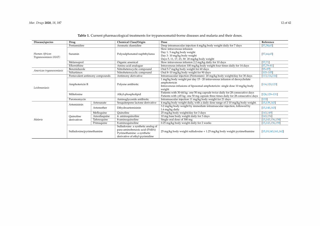
Table 1. Current pharmacological treatments for trypanosomatid-borne diseases and malaria and their doses.
Disease/species Drug Chemical Class/Origin Dose Reference
Human AfricanTrypanosomiasis (HAT)
Pentamidine Aromatic diamidine Deep intramuscular injection 4 mg/kg body weight daily for 7 days [57,58,63]
Suramin Polysulphonated naphthylurea
Slow intravenous infusionDay 1: 5 mg/kg body weightDay 3: 10 mg/kg body weightDays 5, 11, 17, 23, 30: 20 mg/kg body weight
[57,64,65]
Melarsoprol Organic arsenical Slow intravenous infusion 2.2 mg/kg daily for 10 days [57,71]Eflornithine Amino acid analogue Intravenous infusion 100 mg/kg body weight four times daily for 14 days [57,79–81]
American trypanosomiasis Benznidazole Nitroheterocyclic compound Oral 5-7 mg/kg body weight for 60 days [95–97]Nifurtimox Nitroheterocyclic compound Oral 8–10 mg/kg body weight for 90 days [103–105]
Leishmaniasis
Pentavalent antimony compounds Antimony derivative Intramuscular injection (Pentostam): 20 mg/kg body weight/day for 30 days [113,114,116]
Amphotericin B Polyene antibiotic
1 mg/kg body weight per day 15 - 20 intravenous infusion of deoxycholateamphotericinIntravenous infusions of liposomal amphotericin: single dose 10 mg/kg bodyweight
[116,120,123]
Miltefosine Alkyl-phospholipid Patients with 30-44 kg: one 50 mg capsule twice daily for 28 consecutive daysPatients with ≥45 kg: one 50 mg capsule three times daily for 28 consecutive days [126,129–131]
Paromomycin Aminoglycoside antibiotic Intramuscular injection 11 mg/kg body weight for 21 days [133]
Malaria
ArtemisininArtesunate Sesquiterpene lactone derivative 4 mg/kg body weight daily, with a daily dose range of 2-10 mg/kg body weight. [15,139,143]
Artemether Dihydroartemisinin 3.2 mg/kg body weight by immediate intramuscular injection, followed by1.6 mg/kg daily [15,140,143]
Quinolinederivatives
Mefloquine Quinoline 25 mg/kg body weight/day for 3 days [143,149]Amodiaquine 4- aminoquinoline 10 mg base body weight daily for 3 days [143,154]Tafenoquine 8-aminoquinoline Single oral dose of 300 mg [15,143,156,158]Primaquine 8-aminoquinoline 0.25 mg/kg body weight daily for 2 weeks [15,143,156,159]
Sulfadoxine/pyrimethamine
Sulfadoxine: a synthetic analog ofpara-aminobenzoic acid (PABA)Pyrimethamine: a syntheticderivative of ethyl-pyrimidine
25 mg/kg body weight sulfadoxine + 1.25 mg/kg body weight pyrimethamine [15,19,143,161,162]

Mar. Drugs 2020, 18, 187 13 of 42
5. Current tools for Drug Screening
Over the past two decades, as a result of the initiative of international stakeholders led by nonprofitresearch and development organizations, the search for new drugs against NTDs has taken a giant stepforward. DNDi, Medicines for Malaria Venture (MMV), NGOs, academic and institutional centers,and private-public-partnerships with pharmaceutical companies such as GSK, Tres Cantos, Madrid(Spain) or Novartis GNF, San Diego (USA) have opened their robotic facilities and huge libraries ofcompounds to external researchers. These partnerships have resulted in significant successes in thediscovery of new drugs against NTDs and malaria.
The use of HTS strategies for the identification of biologically active natural products is less frequentcompared to that of synthetic compounds. More than 60% of small molecules approved between 1981and 2014 against cancer were developed from a natural product or one of its pharmacophores [164].Only 1% of the papers published in this same period used HTS technology [165]. Finally, this proportionbecomes almost nonexistent when we talk about natural products of marine origin.
As a starting point, a high level of structural diversity is necessary from both small molecules andbiological extracts which can be obtained by sampling the diverse marine taxonomy or by harvestingthem in unexploited ecological niches. To give a few examples, it is worth mentioning the effort madeby public institutions such as the National Cancer Institute of the United States (NCI) USA under theProgram for the Discovery of Natural Products Discovery (NPNPD), which owns one of the largestand most diverse collections of natural extracts and natural products in the world [166]. This libraryis freely available in 384-well plates and is HTS-eligible for the research community. On the otherhand, Fundación Medina (Granada, Spain) has a library of more than 130,000 extracts and semipurifiedfractions, which is currently the largest chemical space of natural products obtained from a largesampling of diverse geographical locations [167]. From the side of private enterprises, Pharmamar(Madrid, Spain), which is responsible for the development and commercialization of the antitumordrug trabectedin, has the world’s largest collection of natural products from marine origin, withapproximately 200,000 samples of macroorganisms and microorganisms.
Empirical and nonempirical target-based screenings are current drug discovery strategies toidentify new antiparasitic hits for further preclinical and clinical studies. There are pros and cons thatcan determine the choice of the screening method when establishing a new drug discovery campaignagainst NTDs [168]. On the one hand, phenotypic screening assays are unbiased and more relevantthan target-based screenings, since neither lead to prejudices in the selection of compounds related totheir mechanism of action, nor are accessibility issues present for the compound to the specific target.However, with these results, it is difficult to obtain conclusions regarding quantitative structure-activityrelationships (QSAR), and complicated deconvolution studies are required to determine the possiblemechanisms of action of the selected lead compounds to improve their effectiveness in a rationalway [169]. On the other hand, target-based screens are more rational when a robust target is available.It is easier to improve the new compounds based on computational 3D docking studies. However, therelevance of this approach is usually lower, because the compounds are usually modified by the hostor pathogen cells, and they barely reach the target in the active form [170]. In conclusion, althoughboth are valid paradigms, historically, target-based screening has been the method of choice to identifybest-in-class drugs, whereas phenotypic screens have served to identify first-in-class drugs [171].Once the most promising hits are identified, new simulation-based computational analyses [172]and mammalian cell toxicity tests [173] are performed with a threefold objective: (i) to avoid toxiccompounds, (ii) to perform QSAR analysis in order to introduce chemical modifications that improvethe pharmacokinetic and pharmacodynamic properties of the compounds, and (iii) to avoid compoundswhich require complex synthesis at the pilot or industrial plant scales.
5.1. Phenotypic vs. Target-Based Screening in Trypanosomatids
The low throughput of the previously employed whole cell-based phenotypic models, togetherwith the revolution in genomics and 3D-assisted prediction models, have led to support for the

Mar. Drugs 2020, 18, 187 14 of 42
"target-first-then-phenotypic" strategy in drug discovery for many years [171]. If a validated drugtarget and a good chemical scaffold to selectively interact with the target are available, a rationalstructure-based drug discovery approach may be adapted for the synthesis of thousands of newcompounds in the search for drug candidates. In general terms, target-based drug screening is easierto implement and provides continuous feedback from which to introduce rational modifications in themolecule that improve the interactions with its target through in-silico 3D predictions [174]. In thecase of trypanosomes and malaria, a pharmacological target must have a number of characteristics,namely, (i) the target should be essential for the survival of the pathogen; (ii) should be druggable; (iii)should be structurally different from the heterologous form occurring in the host; and (iv) should bedifferently expressed in the parasite with respect to the host [175].
ODC, the key enzyme of polyamine biosynthesis, is the only consolidated target in T. brucei,although it is not in T. cruzi, Leishmania parasites and Plasmodium spp. [176]. The irreversible inhibitionof this enzyme, differences in the rate of turnover and structural differences with the host protein, andthe differential expression of the ODC-encoding gene, make ODC the ideal target for the developmenteflornithine, a drug for the treatment of HAT [177]. Sterol 14α-demethylase, a CYP monooxygenasethat catalyzes the removal of the 14α-methyl group from eburicol [178], is another example of selectivetarget of drug discovery for Chagas disease [179]. Two triazole antifungals, posaconazole and E1224(a ravuconazole pro-drug), were developed against this target and submitted to Chagas disease clinicaltrials. Posaconazole and E1224 showed a transient suppressive effect on parasite clearance, but bothfailed during the follow-up [180,181]. Similarly, cyclin-dependent Cdc2-related kinase 12 (CRK12) wasproposed as a potential druggable target in kinetoplastids [182]. The pyrazolopyrimidine GSK-3186899identified in target-based screening showed excellent in vitro and in vivo antileishmanial effect whenadministered orally to mice infected with L. donovani. The in vivo efficacy, novel mechanism ofaction and safety profile of GSK-3186899 supported the advancement of this compound for definitivephase I clinical trials [183]. N-myristoyltransferase, an enzyme responsible for posttranslationalprotein modification in fungi and protozoa but not in mammals, was identified as a robust target inT. brucei [184]. It was consolidated after HTS of a library with 62,000 compounds [185], which yieldedthe optimized lead DDD85646. This is a pyrazole sulfonamide with good results in experimentalinfections of T. b. brucei and T. b. rodhesiense in mice [186].
Other targets that were genetically validated in kinetoplastids are the enzymes of both folateand unconjugated pteridines [187]. These include pteridine reductase 1 (PTR1) and the bifunctionaldihydrofolate reductase-thymidylate synthase (DHFR-TS) [188]. Both enzymes have good draggabilityand have been assayed under HTS technologies in T. brucei [189] and L. major [190]. Trypanothione isthe key molecule used by trypanosomatids for modulating oxidative stress in place of glutathione.Trypanothione redox balance is regulated by two enzymes, trypanothione synthase and trypanothionereductase, which are absent in the host and have been chemically and genetically validated as druggabletargets [191,192]. Numerous efforts made to synthesize new compounds to target trypanothionereductase in trypanosomatids have delivered novel chemical scaffolds capable of inhibiting thisenzyme [193]. Unfortunately, none of these compounds have shown good profiles as drug candidatesfor further development [194]. DNA topoisomerase IB, the enzyme involved in the relaxation of nuclearDNA from trypanosomatids, differs from the host’s counterpart in structure and gene expression [195].Genetic disruption of any of the two subunits of DNA topoisomerase IB results in a nonviablephenotype that proves potential draggability of this essential enzyme [196]. Camptothecin-likecompounds irreversibly target this enzyme and have shown antileishmanial effects in vivo [197].
Since the most of the lead compounds selected through target-based screening frequentlylack activity against the whole cell or in whole organism assays, improved higher performancephenotype-based screens have been introduced in recent decades [174,198]. The goals to be addressedin a phenotypic screening for trypanosomatids are: (i) to find the drug that selectively kills the parasiteat the lowest concentration; (ii) to kill the most relevant parasitic form, which is responsible for thepathological outcome in the host, and (iii) to find the safest concentrations for the host’s cells [168,199].
Mar. Drugs 2020, 18, 187 15 of 42
To achieve the first goal, the introduction of bioimaging techniques has accelerated the discovery ofnew potential hits based on phenotypic screening [199,200]. The use of genetically modified pathogensthat express reporter genes encoding colored, fluorescent or luminescent proteins, along with highcontent screening (HCS) readouts, have facilitated the early discovery of drugs under the phenotypicscreening paradigm [201,202]. Genes encoding potential reporter proteins were initially identified inmarine organisms and could be easily cloned in suitable vectors for steady expression in the pathogens,preferably using genomic integration strategies [200,202]. The choice of these reporters will dependon the preferable or adequate readout, although some pros and cons should be considered [200].Color-based readouts lack the sensitivity of those using fluorescence or luminescence ones. In addition,they are only useful for free-living forms of the parasites, unless HCS systems are available to avoidmisinterpretation of results [203,204]. Vital dyes like MTT or Alamar Blue, or transgenic parasitesexpressing intracellular reporter genes like lacZ (encoding beta-galactosidase) have been used totest T. brucei bloodstream parasites, T. cruzi trypomastigotes and axenic Leishmania amastigotes [205].However, these techniques are currently outdated. In order to improve sensitivity and detection ofparasites in cocultures with mammalian cells, genetically modified parasites expressing fluorescentproteins are preferred. The advantages of fluorescence emission over colorimetric readings includeincreased sensitivity, no dye requirement and the possibility of scaling up to certain preclinical modelsusing charge-coupled devices (CCD) with the reporters [206,207]. Genes encoding fluorescent proteins,which emit in the range of UV to near-infrared spectra, are extensively used to create transgenicstrains of kinetoplastids [174,200]. Either episomal or integrative transgenic strains of T. brucei, T. cruziand several species of Leishmania have been used in phenotypic HTS drug discovery campaigns.The use of stable-transfected (genome integrated) pathogen strains over episomally-transfected ispreferred because selection drug-pressure is not (always) necessary to avoid the loss of the plasmidvector [208]. Gene integration into the 18s ribosomal (SSU) loci of trypanosomatids warrants genomicstability and does not affect the viability and virulence of the pathogen [209]. Transfected strains withluciferase-encoding genes from either firefly or marine animals are widely used in drug discoverycampaigns due to their greater sensitivity and poor autofluorescence-noise. However, specific luciferasesubstrates, i.e., luciferin or coelenterazine, significantly increase the cost of these assays. [210,211].Despite these drawbacks, the radiance emitted by the luciferase-transfected strains in the presence ofthe dye, and the poor autofluorescence background of the tissues, which do not have to be light-excitedfor visualization, make it possible to acquire in vivo images in real time of internal infections usingCCD cameras [212–214].
The second goal of phenotypic screening recommends the use of the most pathogenically relevantform of the parasite. In the case of HAT, T. brucei bloodstream forms are easy to grow under in vitroconditions which may closely resemble the blood environment where the parasite lives [215]. However,this is not easy to reproduce with other trypanosomatids. T. cruzi has an early epimastigote form in thebloodstream that rapidly invades different host cells and transforms into intracellular amastigotes [216].In the case of Leishmania spp., the most relevant pathological form, the amastigote, lives inside thephagolysosomes of the host macrophages. For many years, cell-based screens on free-living forms(promastigotes and trypomastigotes) or axenic amastigotes (a nonnatural extracellular form createdunder laboratory conditions) were unable to detect active compounds and identified a large number offalse-positives [217]. A step forward in the chain of translatability was represented by the phenotypicplatforms based on cultures of mammalian cells infected in vitro with the pathogen. Immortalizedcell lines like Vero, 3T3 fibroblasts or LS cells, as well as primary cardiomyocytes, were used toevaluate the intracellular development of T. cruzi amastigotes [205,216]. Similarly, murine (J744.2 orRAW-transformed monocytes) or human macrophages (THP-1 transformed monocytes) are suitablemodels to study the intracellular form of Leishmania spp [218,219]. However, despite these intracellularcocultures being closer to real-life experimental models and doing away with the need to use ofartificial cell stages like axenic amastigotes, some technical problems remain. They include the lowerthroughput of the platform, the tumoral origin of most of host the cell lines, which can interfere

Mar. Drugs 2020, 18, 187 16 of 42
with the results, and the artificial method of infection, including washing steps, which are far fromnatural conditions.
An alternative method that works in Leishmania is the use of primary cultures of splenic explantsobtained from infected rodents ex vivo [220]. This method was first used to screen a library of10,000 compounds using hamster splenocytes infected with L. infantum by Melby’s group (Universityof Texas, USA) [221]. It was subsequently used with lymph node cells of mice infected with a strain ofL. major that had been previously transformed with the luciferase-encoding gene [222]. This methodwas later adapted to a murine model of chronic visceral leishmaniasis using a near-infrared fluorescentL. infantum strain [206]. A remarkable advantage of using naturally infected host cells is that it canavoid some handling problems, such as the washing and removal steps of noninternalized parasitesthat often disturb the readings of experimental infections in vitro. In addition, ex vivo splenic explantsare 3D-primary cocultures that include other components of the host immune system which maycontribute to the clearance of parasite burden [223]. The translatability of this coculture is currentlyunder evaluation. This model may improve the chain of translatability and thus accelerate the drugdiscovery process for leishmaniasis.
DNDis, MMV and the Tb Alliance in collaboration with the Bill and Melinda Gates Foundationrecommend a number of general and specific hit-to-lead criteria for screening against NTDs, malariaand tuberculosis. An initial screen of potency at a single concentration of 10 µM has been recommended,which is considered as an inclusive criterion. The compounds with the best inhibition rates (70–100%)may be screened in a second round to determine their IC50 value. The cytotoxicity of the leadcompounds is assessed in mammalian cell lines to determine Selective Indexes (SI). The compoundswith SI>10 are recommended for early preclinical evaluation using in vivo models of the disease.Complementary in vitro and in vivo assays, including in vivo exposure after oral administration,in vivo efficacy resulting in >70% pathogen reduction, early safety assessment including in vitrocardiotoxicity, AMES test for genotoxicity and tolerability studies are also necessary to describe aputative lead compound. Several rounds of lead expansion and optimization are required to find apromising drug candidate for advanced preclinical and clinical evaluation [220,224,225].
5.2. Phenotypic vs. Target-Based Screening in Malaria
Similar to most of the new drug discovery programs, new antimalarial drug discovery platformshave also relied on whole cell pathogen culture-based phenotypic screenings, as well as on moleculartarget-based HTS approaches [226]. It is remarkable that most of the antimalarial lead compoundsidentified through parasite culture-based phenotypic screening have shown better rates of successcompared to target-based screening regarding their further advancement to lead-optimization andpreclinical development pathways [227,228]. However, target-based screening models are still relevantfor structure-activity analyses and for the optimization of new antimalarial drug leads [229,230].The utility of target-based antimalarial screens has been further enhanced by engineering thetarget action to functional phenotypic cell-based models [231]. For instance, the electron transportchain [232], the protein kinases PfCLK3 [233] and Pfnek-1 [234], the β5 sites of Pf20S proteasome [235]and the mitochondrial enzyme, dihydroorotate dehydrogenase [236,237], have been identified aspotential targets for the discovery of novel antimalarial inhibitors. Remarkably, the dihydroorotatedehydrogenase inhibitor, DSM265 [238], has recently demonstrated its efficacy in patients withP. falciparum and P. vivax malaria infections.
The main strategy for the discovery of antimalarial drugs under the phenotypic-based paradigmis related to the creation of a drug which prevents development and proliferation during the lifecycle of malaria parasites. However, the complexity of multiple cellular, physiological and molecularparasite stages represents a challenge in optimizing the therapeutic development of a new drug. Themajority of the antimalarial drug discovery and screening programs were developed to cure malariaby acting against the intraerythrocytic asexual blood stages of the parasite [239], which are primarilyresponsible for severe pathogenesis of the disease and deaths due to malaria infections [240]. However,

Mar. Drugs 2020, 18, 187 17 of 42
platforms designed against either sexual (gametocytes) or hepatic stages (hypnozoites) to prevent orto kill dormant stages of malarial infections are already under development. Several HTS methodshave been employed to screen compound libraries against blood stages of the malaria parasite [241].These methods include DNA-binding fluorescent dyes [242], the parasite lactate dehydrogenase assaymethods [243] or the use of transgenic parasites expressing luciferase-reporter cassettes [244–247].The SYBR® green fluorescence-based screening with blood stage P. falciparum cultures has been thehallmark of antimalarial drug discovery for more than a decade [248]. SYBR® green-based assayshave been further optimized for antimalarial screening under low levels of parasitemia against clinicalfield isolates of P. falciparum for surveys of drug resistance [249,250] in infections with mixed strainsand for the evaluation of antimalarial drug combinations [251]. The flow-cytometric adaptation ofSYBR® green assays can determine the efficacy of drugs against specific life-cycle stages of the malariaparasite [252], and also during in vivo screening in a P. berghei mouse model of malaria [253,254].SYBR® Green assay is a faster, less expensive and more reproducible approach than other traditionaltechniques, and the most widely used in vitro screening approach with the aim of achieving newadaptable and optimized versions [255].
Transgenic P. falciparum cell lines with stable high-level firefly luciferase expression have beenemployed for high throughput antimalarial screening [256,257]. The stable luciferase-expressingcell lines of P. berghei have also been employed for noninvasive whole mouse imaging and in vivoantimalarial screening [247,257]. A simple one-step technique based on RNA dye growth inhibitionand high-content imaging assay has been developed for antimalarial HTS [258]. The high content livecell imaging platform with an RNA sensitive dye and imaging at timed intervals has been employed toscreen a library of marine extracts against P. falciparum [259].
Antimalarial drug discovery programs have been further expanded to screen the compoundlibraries against sexual gametocytes, [260,261]. The sexual gametocytes are responsible for thetransmission of malaria [262], while malaria infection in the mammalian host is established initiallyin the hepatocytes [263]. Several screenings with gametocytes and different throughputs have beendeveloped. They include mainly colorimetric methods [264,265] and genetically modified parasiteswith a fluorescent/bioluminescent protein labeling and high-content imaging [262,266–271]. RobustHTS campaigns have identified several new gametocidal antimalarial drug leads with utility in theprevention of malaria transmission [272–276]. Recently, the efficacy of this technique has been usefulin the identification of active compounds against gametocytes that had not been identified in asexualblood-stage assays [272]. In addition to asexual blood-stage parasites and sexual gametocytes, HTSbased on liver-stage screens have been developed for antimalarial drug discovery [277–279]. Thesescreening models, which identify the antimalarial drugs that are active against dormant P. vivax andP. ovale hyponozites, have utility for the radical cure and prevention of malaria relapse [280]. A fewHTS assay models have been reported for the screening of compound libraries against Plasmodiumliver stages [279–283]. Remarkably, a recently study published by Antonova-Koch and coworkers,who performed an HTS with more than 500,000 compounds against malaria liver stages usingluciferase-expressing P. berghei [284], revealed 58 mitochondrial inhibitors and further chemotypes,although no mechanism of action was identified.
6. Marine Based Compounds for NTDs and Malaria
Marine-derived compounds already on the market include drugs to treat cancer, viruses,neuropathic pain and even hypertriglyceridemia, but none for the treatment of NTDs or malaria.However, many natural marine products have been reported to show antiprotozoal activity. In addition,marine organisms are a complementary source of chemical entities that can serve as inspiration for thesynthesis of novel drugs in the treatment of tropical diseases [285]. The following paragraphs includesome relevant examples of secondary metabolites of marine origin that are currently being evaluatedas potential drugs for the treatment of NTDs and malaria [286].

Mar. Drugs 2020, 18, 187 18 of 42
6.1. Algae-Derived Compounds
Benthic marine algae include red (Phyllum Rhodophyta), brown (Phyllum Heterokontophyta,Class Phaeophyceae) and green (Phyllum Chlorophyta) algae. Numerous extracts from marine algaehave been evaluated for their antiprotozoal effect. However, only the compounds isolated andidentified from these extracts are taken into account in the current review. These compounds includediterpenes, halogenated triterpenes, sulfated polysaccharides, acetogenins, polyphenols and others(Figure 6). A recent review estimated that 151 extracts from up to 30,000 macroalgae species identifiedworldwide have proven antileishmanial activity [287]. From these, 48 extracts were obtained frombrown Phaeophyceae macroalgae, 80 from Rhodophyceae and 23 from green Chlorophytes. Only 12 ofthese species where further studied to identify bioactive antileishmanial compounds. More than 50% ofantileishmanial compounds were major secondary metabolites from brown Dictyotaceae seaweeds withditerpene structure. Due to the relevance of the intracellular amastigote form as the most suitable modelfor drug-screening, only the results obtained using this parasite stage are presented. The most active ofthese compounds was the diterpene (4R,9S,14S)-4α-acetoxy-9β,14α-dihydroxydolast-1(15),7-diene, anelectron chain transport uncoupler (24), isolated from Canistrocarpus cervicornis.
1
Figure 6. Active metabolites isolated from marine macroalgae.
The IC50 value for L. amazonensis intracellular amastigotes was 4.0 µg/mL, with an interesting SIof 93 in mouse J774 macrophages [288]. Dolabelladienetriol (25) is a diterpene isolated from Dictyotapfaffii that inhibited the growth of L. amazonensis intracellular amastigotes with a modest IC50 value of43.9 µM and SI>2 on murine macrophages. Dolabelladienetriol modulates macrophage activity byinhibiting NO, TGF-β and TNF-α production, which may explain its antileishmanial activity [289,290].Bioassay-guided fractionation of extracts from the brown alga Bifurcaria bifurcata revealed diterpeneelaganolone (26) (6E,10E,14E)-16-hydroxy-2,6,10,14-tetramethyl-hexadeca-2,6,10,14-tetraen-4-one, withmild trypanocidal activity against T. brucei rhodesiense (IC50 = 45 µM and SI 4.0) [291]. Meroditerpenoids,such as (3S)-tetraprenyltoluquinol (1a/1b) (27) isolated from extracts of the brown alga Cystoseirabaccata inhibited the growth of L. infantum intracellular amastigotes with an IC50 value of 25.0 µMand a SI of 5 on murine macrophages. Mechanistic experiments showed that this compound inducedcytoplasmic vacuolization and the presence of coiled multilamellar structures in mitochondria, whichproduced intense disruption of the mitochondrial membrane potential [292]. Atomaric acid (28) isanother meroditerpenoid, isolated from extracts of the Caribbean-sea alga Stypopodium zonale. Itshowed an IC50 value of 20.2 µM against intracellular amastigotes of L. amazonensis and SI value of>8.4. The generation of free radicals may be partially responsible for its antiprotozoal activity [293].The triterpene fucosterol (29) (Figure 7), isolated from the brown macroalga Lessonia vadosa, was activeagainst intracellular amastigotes of both L. amazonensis and L. infantum (IC50 values of 7.89 µM and10.30 µM, respectively) and exhibited relatively low cytotoxicity (CC50 >100 µM) [294].

Mar. Drugs 2020, 18, 187 19 of 42
1
Figure 7. Active metabolites isolated from marine macroalgae.
Figure 7. Active metabolites isolated from marine macroalgae.
The weak effect exerted by this compound on promastigotes may indicate that the antileishmanialactivity of fucosterol is somewhat dependent on macrophage function [294]. Recently, a large numberof triterpene polyether compounds with significant structural and pharmacological diversity wereidentified in the red alga Laurencia viridis. Dehydrothyrsiferol (30), a natural oxasqualenoid, has similartrypanocidal (T. cruzi) activity to the reference drug benznidazole (6). However, the SI value was notas safe as that of benznidazole (SI = 3 vs. 56), the clinically used drug. In addition, dehydrothyrsiferolshowed an IC50 of 2.16 µM against L. amazonensis amastigotes, but again, the SI was much lower thanthat shown by the reference drug miltefosine (11). However, the semisynthetic derivative resultingfrom the introduction of an iodine atom in these series led to the identification of 28-iodosaiyacenolsA and B, which exhibited notable antileishmanial activity and turned out to be nontoxic against theJ774 line of murine macrophages [295]. Sesquiterpene derivatives elatol (31), obtusol (32) and thetriquinane derivative silphiperfol-5-en-3-ol (33) (Figure 8), isolated from the red macroalga Laurenciadendroidea, were tested against L. amazonensis. Unlike silphiperfol-5-en-3-ol (IC50 = 48.7 µg/mL), elatoland obtusol were strongly active against intracellular amastigotes (IC50 = 4.5 µg/mL and 3.9 µg/mL,respectively). None of these compounds significantly activated NO synthesis by infected macrophages,which suggests that their antileishmanial activity is likely to be exerted on the parasites, rather thanthrough macrophage activation [296,297].
1
Figure 8. Active metabolites isolated from marine macroalgae.
Figure 9. Active metabolites isolated from sponges.
Figure 8. Active metabolites isolated from marine macroalgae.
The antileishmanial activity of sulfated polysaccharide fucoidan (34), isolated from the brown algaLaminaria japonica (Figure 8), was tested in both in vitro and in vivo models of L. donovani. Fucoidanwas able to kill more than 90% L. donovani intracellular amastigotes at 50.6 µg/mL. The effectiveness ofthis compound for the clearance of parasite burden in liver and spleen was complete in in vivo modelsof antimony-susceptible and -resistant strains of L. donovani. Fucoidan induced a strong Th-1 responsein the host by increasing the production of NO, cytokines and free radicals in infected macrophages.Unfortunately, at a dose of 200 mg/kg body weight/day three times daily, this compound showed highhemorrhagic risk and poor bioavailability [298].
6.2. Sponge-Derived Compounds
Sponges produce many different kinds of chemical substances which are active against severalpathogens, including virus, bacteria and protozoa [299,300]. However, despite the considerable numberof new bioactive compounds that have constantly been isolated from sponges, only a small numberhas reached the market.
The most promising antiprotozoal compounds isolated from sponges are manzamines (Figure 9).Manzamines are eight-membered β-carboline alkaloids that have served as structural core for a

Mar. Drugs 2020, 18, 187 20 of 42
large number of biologically relevant semisynthetic compounds. The isolation, characterization andanticancer effect of manzamine A (35) from sponges of the Haliclona genus was reported by Sakai andcoworkers in 1986 [301]. The first antimalarial effects of manzamine A and 8-hydroxymanzamineA (36) against asexual erythrocytic stages of P. berghei were reported by Ang and coworkers in2000 [302]. These authors showed that a single intraperitoneal injection of 100 µmol/kg body weightof manzamine A and 8-hydroxymanzamine-A to P. berghei-infected mice prolonged survival timefor more than 60 days. In addition, oral administration of manzamine A at a similar dose alsoproduced significant reductions in parasitemia, which indicates good pharmacokinetic propertiesof these alkaloids [303]. Natural manzamine derivatives showed moderate antileishmanial activityagainst L. donovani promastigotes. However, the semisynthetic derivatives 8-methoxymanzamineA (37) and 8-acetoxymanzamine A (38) are prodrugs that showed improved antileishmanial andantimalarial potencies with low cytotoxicity [304].
1
Figure 8. Active metabolites isolated from marine macroalgae.
Figure 9. Active metabolites isolated from sponges.
Figure 9. Active metabolites isolated from sponges.
Many other β-carboline alkaloids have been synthesized since then, but they have hardly increasedthe antiprotozoal potency of the parent compounds [305,306]. Recently, zamamidines A-C (39-41)(Figure 10), other manzamine alkaloids isolated from Amphimedon sp. sponges, have shown inhibitoryactivities against T. brucei brucei (IC50 values ranging from 0.27 mg/mL to 1.05 mg/mL) and P. falciparum(IC50 values ranging from 0.58 mg/mL to 12.20 mg/mL), with the C form (41) being the most activecompound of the series [307]. The mechanism of action of manzamine alkaloids is not fully understood,but some authors describe β-carboline alkaloids as micromolar inhibitors of glycogen synthase 3(GSK-3) from malaria parasites [308] and DNA topoisomerase inhibitors through intercalating intoDNA base pairs [309].
1
Figure 10. Active metabolites isolated from sponges.
Figure 11. Active metabolites isolated from sponges.
Figure 10. Active metabolites isolated from sponges.
Isonitrile-, isothiocyanate- and formamide-containing sesquiterpenoid metabolites were firstisolated from extracts of the sponge Axinella cannabina (Figure 11). Axisonitrile-3 (42), unlikeaxisothiocyanate-3 (43), was found to possess a potent antimalarial activity both on chloroquine-sensitive and chloroquine-resistant P. falciparum strains in the nanomolar range. In addition, it showed

Mar. Drugs 2020, 18, 187 21 of 42
poor cytotoxicity, which points to the relevance of the isonitrile group [310]. A chemical analysis ofthe sponge Cymbastela hooperi (Axinellidae) provided other diterpene isonitriles, which showed evenhigher antimalarial effect and moderate toxicity [311]. Docking studies of these compounds withhuman hemoglobin indicated that the inhibitors directly interacted with the heme group, thus forminga coordination complex with the iron center and inhibiting the transformation of the heme group intoβ-hematin and then hemozoin [312].
1
Figure 10. Active metabolites isolated from sponges.
Figure 11. Active metabolites isolated from sponges.
Figure 11. Active metabolites isolated from sponges.
The diterpenes membranolides G (44) and H (45) (Figure 12), isolated from the sponge Dendrillamembranosa, also displayed antileishmanial activity against L. donovani axenic amastigotes with IC50
values of 0.8 and 1.1. µM, respectively. The latter examples attest to the fact that marine biota canprovide novel compounds with antiprotozoal activities if properly assayed [313]. The extracts of marinesponges from the genera Spongia and Ircinia contain broad-spectrum antiprotozoal meroterpenes, lineartriterpenoid and squalene, with good inhibitory effects on major trypanosomatids and P. falciparum. Themeroterpene dorisenone D (46) could become a promising antiplasmodial lead compound. In addition,the diterpene 11β-acetoxyspongi-12-en-16-one (47) exhibited strong antileishmanial potential againstL. donovani, but with low SI [314] (Figure 12).
1
Figure 10. Active metabolites isolated from sponges.
Figure 11. Active metabolites isolated from sponges.
Figure 12. Active metabolites isolated from sponges.
Figure 12. Active metabolites isolated from sponges.
The steroidal glycosides pandaroside E (48), G (49) and H (50) and their methyl estersisolated from extracts of the Caribbean sponge Pandaros acanthifolium strongly inhibited the growthof T. brucei rhodesiense (bloodstream forms), T. cruzi (intracellular amastigotes in L6 rat skeletalmyoblasts), L. donovani (axenic amastigotes) and P. falciparum at low and submicromolar concentrations(Figure 13) [315].
1
Figure 13. Active metabolites isolated from sponges.
Figure 14. Natural (51,52,56,57) and synthetic (53-55) long-chain unsaturated fatty acids.
Figure 13. Active metabolites isolated from sponges.

Mar. Drugs 2020, 18, 187 22 of 42
The antileishmanial and antimalarial activities of naturally occurring long-chain fatty acids havebeen recognized for many years [316]. However, an important observation made by Carballeira’sgroup in both P. falciparum and Leishmania spp. was that fatty acids should contain a high degree ofunsaturation to display antiprotozoal activity (Figure 14) [317]. The naturally occurring acetylenicfatty acids 6-heptadecynoic (51) and 6-icosynoic (52) acids displayed good antileishmanial activity,with 6-icosynoic acid being the most effective in the series.
1
Figure 13. Active metabolites isolated from sponges.
Figure 14. Natural (51,52,56,57) and synthetic (53-55) long-chain unsaturated fatty acids.
Figure 14. Natural (51,52,56,57) and synthetic (53-55) long-chain unsaturated fatty acids.
The saturated fatty acids n-heptadecanoic acid and n-eicosanoic acid were not effective towardsL. donovani, which indicates that double or triple bonds are necessary for activity. Interestingly,6-icosynoic acid (52) reversibly inhibited the leishmania DNA topoisomerase IB enzyme (LTopIB), withan IC50 between 36-49 µM. [318]. LTopIB was described as a potential target in Trypanosomatids due toits anomalous heterodimeric structure and differential expression in the parasite [319]. Therefore, otherunsaturated fatty acids were synthesized to target it [320–322]. Within this series, the 2-octadecynoicacid (2-ODA) (53), 2-hexadecynoic acid (2-HDA) (54) and 2-tetradecynoic acid (2-TDA) (55) werestudied on the three major Trypanosomatids. The best effect was achieved on L. donovani amastigotes(IC50 ranging from 11.0 to 24.7 µM), whereas the worst results were obtained against T. cruzi (IC50
between 62.4 and 80.0 µM) and T. brucei rhodesiense (IC50 between 64.5 and 255.4 µM) (trend: 2-ODA> 2HDA > 2TDA) [323]. More recently, a series of α-methoxylated ∆5,9 fatty acids (56,57) from thesponges Asteropus niger and Erylus goffrilleri showed antileishmanial activity against intracellularamastigotes of L. infantum. These compounds inhibited LTopIB by means of a mechanism differentfrom that of camptothecin, the reference inhibitor of TopIB enzymes [324,325]. The series of C16isomeric acetylenic fatty acids was tested in vitro against the blood stage of P. falciparum and the liverstage of P. yoelii, and 2-HDA was found to be the most effective fatty acid (IC50 = 41.2 µM) with nocytotoxic effects on mammalian cells [326].
The 2-alkynoic fatty acids inhibit the so-called type II fatty acid synthase (FASII), an exclusiveeight-enzyme complex involved in fatty acids synthesis in plasmodium parasites. Also, 2-ODA (53)and 2-HDA (54) showed good inhibition of three enzymes of the complex (IC50 1-2 µM) by binding toa different site from that of the substrate or the cofactor [327].

Mar. Drugs 2020, 18, 187 23 of 42
6.3. Metabolites Derived from Other Invertebrates
Cnidarians produce or accumulate a diverse array of biologically active secondary metabolitesincluding steroids, terpenes, acetogenins, alkaloids and polyphenolics (Figure 15). They areexcellent candidates for development as pharmacological probes, especially for the treatment ofNTDs [328]. To identify inhibitors of T. brucei growth, a phenotypic HTS based on resazurin stainingwas carried out with a library consisting of 861 purified natural products and subfractionatedextracts (including 433 extracts and 428 pure compounds) from soft corals and echinoderms livingin Vietnamese seas (genus Lobophytum, Sinularia, Astropecten and Diadema). Several compoundsisolated from these extracts, like laevigatol B (57), (24S)-ergost-4-ene-3-one (58), astropectenol A(59) and cholest-8-ene-3β,5α,6β,7α-tetraol (60), showed T. brucei antrypanosomal activity and alsoexhibited significant inhibitory effects on T. cruzi. By using a high content analysis Operetta device,all these compounds showed anti-inflammatory or anticancer properties in previous studies [329].Interestingly, in order to increase the antiprotozoal spectrum of these compounds, 34 of the activecembranoid diterpenes selected from previous work were tested against L. donovani and P. falciparum.From these compounds, lobocrasol A (61) and lobocrasol C (62) were strongly and selectively activeagainst L. donovani. Only one compound, laevigatol A (63), displayed antiplasmodial activity with anIC50 < 5.0 µM, and can therefore be considered moderately active. None of the compounds displayedsignificant cytotoxicity [330].
1
Figure 15. Active metabolites isolated from marine invertebrates other than sponges.
Figure 15. Active metabolites isolated from marine invertebrates other than sponges.
The steroid 18-acetoxipregna-1,4,20-trien-3-one (64), isolated from the cnidarian Carijoa riisei, wasassayed against L. chagasi and T. cruzi parasites. While the trypomastigotes of T. cruzi were susceptible,with a moderate IC50 value of 50.5 µg/mL, L. chagasi promastigotes were much more susceptible, withan IC50 value of 5.5 µg/mL. Nevertheless, the usefulness of this compound is compromised due to itshigh cytotoxicity on macrophage cells [331].

Mar. Drugs 2020, 18, 187 24 of 42
7. Conclusions and Future Perspectives
NTDs represent one of the biggest challenges for a changing humanity, in which urbanconcentration and global warming favor the transmission of these diseases and threaten the mostdisadvantaged sectors of the population. Furthermore, migratory fluxes increase the risks in regions ofthe planet that are currently not endemic. In addition to these problems, there is a lack of prophylactictreatments in the form of effective vaccines, which means that prevention falls to public health policies,the latter being often ineffective in countries threatened by more urgent problems. However, itis clear that pharmacological treatment is the only effective solution after the onset of symptoms.Throughout this review, it has become evident that all existing treatments for NTDs in the 21st centuryare over 40 years old. Although they have saved hundreds of thousands of lives, many have lost theirinitial efficacy and generated resistance in certain regions where they are currently not effective. In aglobalized world, the existence of these diseases has become ethically intolerable, and a multitude ofinitiatives have emerged for their total eradication. Supranational entities such as the DNDi, the Bill andMelinda Gates Foundation (Seattle, WA, USA), private companies, NGOs and public administrations,among others, signed the London Declaration on NTDs on 30 January 2012 in London. This initiativelaunched a collaborative disease eradication program inspired by the WHO 2020 road map. Sincethen, the efforts made have been remarkable, but only a handful of alternative treatments, many ofthem consisting of a combination of existing drugs or optimized treatments for certain sectors of thepopulation, have improved the previous situation. However, new drugs that are more effective thanexisting ones have not emerged. It should be emphasized that existing treatments, in addition tocausing resistance, must be administered parenterally. This reduces adherence to prolonged treatmentsand, in many cases, involves patient hospitalization. In addition, and more importantly, they are alsoresponsible for significant adverse reactions.
For this reason, the screening of the current chemical space in optimized in vitro systems hasbecome the most promising option for these diseases. This strategy, which has been successfullyapplied to other important diseases, such as cancer, cardiovascular and neurological diseases, amongstothers, comes late for NTDs. In order to find new drug candidates that fill the gap in preclinicalmodels of the disease, the development of accurate in vitro models and access to the libraries ofhundreds of thousands of compounds of large companies and institutions are necessary. In addition,the introduction of omics techniques has shed light into parasite biology and their interaction with thehost. An example is the limitation of long-term culture of P. vivax, where single-cell omics is bringingnew perspectives to solve complex problems. Metabolomics that lie at the end of the omics cascade hasbeen used to reveal the mechanism of action of some antiparasitic drugs, such as beznidazole againstT. cruzi or the natural resistance to antileishmania drugs of the clinical isolates of L. donovani.
The existence of relevant in vitro models of NTDs for application to HTS platforms is a relativelyrecent event. The application of bioimaging systems to phenotypic screens has improved thetranslatability to preclinical and clinical reality, and has served to identify a plethora of new chemicalentities with potential antimalarial or antitrypanosomal effect (including Leishmania). However,despite the richness and diversity of compounds of marine origin, most of the chemicals from marineenvironments with pharmacological use for NTDs, including malaria, have been identified usinglow-performance techniques in in vitro, and sometimes, in in vivo models.
Accessing the chemical diversity of natural products is a major challenge for many modern drugdiscovery programs. This is largely due to the willingness to examine libraries of pure compounds,which raises problems regarding the availability and supply of compounds. New programs and/orfoundations with important natural product repositories have made their libraries of natural products,extracts and facilities available to academic researchers. This circumstance and the collaboration withsupranational institutions may help in the identification of new compounds with better pharmacologicaland toxicological profiles for the treatment of these diseases. From these collaborations, new moleculesagainst NTDs and malaria are expected to emerge in the current decade. They will be added to existingtreatments for the eradication of some of the diseases covered in this review.

Mar. Drugs 2020, 18, 187 25 of 42
Author Contributions: M.Á.-B., Y.P.-P., R.M.R. and R.B.-F., prepared the section of platforms of drug discovery intrypanosomatids; C.O., D.S.-C., M.M.-V. and C.G.-E., prepared introduction and current treatments of NTD; B.L.T.,S.M. and R.M.R., prepared the sections of malaria and platforms of drug discovery in malaria; N.M.C., B.L.T., S.M.and R.B.-F., prepared sections of introduction and marine pharmacology. All authors have read and agreed to thepublished version of the manuscript
Funding: Financial support from the Ministerio de Economía y Competitividad (MINECO, AEI, FEDER, UE)[MINECO: AGL2016-79813-C2-1R and SAF2017-83575-R], the Junta de Castilla y León co-financed by FEDER, UE[LE020P17] to RBF and DBT, New Delhi [BT/IN/Spain/39/SM/2017-18] to MS are gratefully acknowledged. MAB(LE051-18) and DSC (LE020P17) are supported by a scholarship from the Junta de Castilla y León co-financed byFSE. MMV is supported by the Spanish “Ramon y Cajal” Programme Ministry of Economy and Competitiveness(Ministerio de Economia y Competitividad; MMV, RYC-2015-18368). We thank University of Leon for providingfunding to cover publication expenses.
Conflicts of Interest: The authors declare no conflict of interest.
Abbreviations
NTDs Neglected Tropical Diseases;WHO World Health Organization;HTS High-Throughput Screening;FDA Food and Drug and Administration;HAT Human African Trypanosomiasis;DALY Disability Adjusted Life Year;NECT Nifurtimox-Eflornithine Combination Therapy;CNS Central Nervous System;ODC Ornithine Decarboxylase;NTR-1 type 1 nitroreductase;PKDL Post-Kala-azar Dermal Leishmaniasis;AmpB Amphotericin B;ACT Artemisinin-based Combined Therapies;DNDi Drugs for Neglected Diseases Initiative;MMV Medicine for Malaria Venture;NGOs Non-Governmental Organizations;NCI National Cancer Institute;NPNPD Program for the Discovery of Natural Products;QSAR Quantitative Structure-Activity Relationship;CRK12 Cdc2-related kinase 12;PTR1 Pteridine Reductase 1;DHFR-TS bifunctional Dihydrofolate Reductase-Thymidylate Synthase;HCS High Content Screening;CCD charge-coupled device;SI Selective Index;LTopIB Leishmania DNA topoisomerase IB.
References
1. Lindequist, U. Marine-derived pharmaceuticals—Challenges and opportunities. Biomol. Ther. 2016, 24,561–571. [CrossRef] [PubMed]
2. Faulkner, D.J.; Harper, M.K.; Haygood, M.G.; Salomon, C.E.; Schmidt, E.W. Symbiotic bacteria in sponges:Sources of bioactive substances. In Drugs from the Sea; Fusetani, N., Ed.; Karger: Basel, Switzerland, 2000;pp. 107–119.
3. Blunt, J.W.; Copp, B.R.; Keyzers, R.A.; Munro, M.H.; Prinsep, M.R. Marine natural products. Nat. Prod. Rep.2016, 33, 382–431. [CrossRef] [PubMed]
4. Pereira, R.B.; Evdokimov, N.M.; Lefranc, F.; Valentão, P.; Kornienko, A.; Pereira, D.M.; Andrade, P.B.;Gomes, N.G.M. Marine-derived anticancer agents: Clinical benefits, innovative mechanisms, and new targets.Mar. Drugs 2019, 17, 329. [CrossRef] [PubMed]

Mar. Drugs 2020, 18, 187 26 of 42
5. Scott, L.J. Brentuximab Vedotin: A review in CD30-positive Hodgkin lymphoma. Drugs 2017, 77, 435–445.[CrossRef] [PubMed]
6. Senter, P.D.; Sievers, E.L. The discovery and development of brentuximab vedotin for use in relapsed Hodgkinlymphoma and systemic anaplastic large cell lymphoma. Nat. Biotechnol. 2012, 30, 631–637. [CrossRef]
7. Aseyev, O.; Ribeiro, J.M.; Cardoso, F. Review on the clinical use of eribulin mesylate for the treatment ofbreast cancer. Expert Opin. Pharmacother. 2016, 17, 589–600. [CrossRef]
8. Schöffski, P.; Chawla, S.; Maki, R.G.; Italiano, A.; Gelderblom, H.; Choy, E.; Grignani, G.; Camargo, V.; Bauer, S.;Rha, S.Y.; et al. Eribulin versus dacarbazine in previously treated patients with advanced liposarcomaor leiomyosarcoma: A randomised, open–label, multicentre, phase 3 trial. Lancet 2016, 387, 1629–1637.[CrossRef]
9. Rinerhart, K.L.; Fregeau, N.L.; Stroh, J.G.; Keifer, P.; Sun, F.; Li, H.; Martin, D.G. Ecteinascidins 729, 743, 745,759A, 759B, and 770: Potent antitumour agents from the Caribbean tunicate Ecteinascidia turbinata. J. Org.Chem. 1990, 55, 4512–4515. [CrossRef]
10. Demetri, G.D.; von Mehren, M.; Jones, R.L.; Hensley, M.L.; Schuetze, S.M.; Staddon, A.; Milhem, M.; Elias, A.;Ganjoo, K.; Tawbi, H.; et al. Efficacy and safety of trabectedin or dacarbazine for metastatic liposarcoma orleiomyosarcoma after failure of conventional chemotherapy: Results of a phase III randomized multicenterclinical trial. J. Clin. Oncol. 2016, 34, 786–793. [CrossRef]
11. Schwartsmann, G.; Brondani da Rocha, A.; Berlinck, R.G.; Jimeno, J. Marine organisms as a source of newanticancer agents. Lancet Oncol. 2001, 2, 221–225. [CrossRef]
12. Molyneux, D.H.; Savioli, L.; Engels, D. Neglected tropical diseases: Progress towards addressing the chronicpandemic. Lancet 2017, 389, 312–325. [CrossRef]
13. Mitra, A.K.; Mawson, A.R. Neglected tropical diseases: Epidemiology and global burden. Trop. Med. Infect.Dis. 2017, 2, 36. [CrossRef] [PubMed]
14. Qian, M.B.; Zhou, X.N. Global burden on neglected tropical diseases. Lancet 2016, 16, P1113–P1114. [CrossRef]15. White, N.J.; Pukrittayakamee, S.; Hien, T.T.; Faiz, M.A.; Mokuolu, O.A.; Dondorp, A.M. Malaria. Lancet 2014,
383, 723–735. [CrossRef]16. Klohe, K.; Amuasi, J.; Moriku Kaducu, J.; Haavardsson, I.; Bogatyreva, E.; Husøy Onarheim, K.; Harrison, W.;
Kristensen, F.; Prazeres da Costa, C.; Winkler, A.S. The 2017 Oslo conference report on neglected tropicaldiseases and emerging/re-emerging infectious diseases—Focus on populations underserved. Infect. Dis.Poverty 2019, 8, 40. [CrossRef] [PubMed]
17. Büscher, P.; Cecchi, G.; Jamonneau, V.; Priotto, G. Human African trypanosomiasis. Lancet 2017, 390,2397–2409. [CrossRef]
18. Dunn, N.; Wang, S.; Adigun, R. African Trypanosomiasis (Sleeping Sickness); StatPearls Publishing: TreasureIsland, FL, USA, 2019.
19. Bottieau, E.; Clerinx, J. Human African Trypanosomiasis: Progress and stagnation. Infect. Dis. Clin. NorthAm. 2019, 3, 61–77. [CrossRef]
20. Franco, J.R.; Cecchi, G.; Priotto, G.; Paone, M.; Diarra, A.; Grout, L.; Simarro, P.P.; Zhao, W.; Argaw, D.Monitoring the elimination of human African trypanosomiasis: Update to 2016. PLoS Negl. Trop. Dis. 2018,12, e0006890. [CrossRef]
21. World Health Organization. Control and Surveillance of Human African Trypanosomiasis: Report of aWHO Expert Committee (World Health Organization). 2013. Available online: http://www.who.int/iris/handle/10665/95732 (accessed on 1 December 2019).
22. GBD 2015 DALYs and HALE Collaborators. Global, Regional, and National Disability-Adjusted Life-Years(DALYs) for 315 Diseases and Injuries and Healthy Life Expectancy (HALE), 1990–2015: A SystematicAnalysis for the Global Burden of Disease Study 2015. Lancet 2016, 388, 1603–1658. [CrossRef]
23. Checchi, F.; Filipe, J.A.; Barrett, M.P.; Chandramohan, D. The natural progression of Gambiense sleepingsickness: What is the evidence? PLoS Negl. Trop. Dis. 2008, 2, e303. [CrossRef]
24. Kennedy, P.G. Clinical features, diagnosis, and treatment of human African trypanosomiasis (sleepingsickness). Lancet Neurol. 2013, 12, 186–194. [CrossRef]
25. Jamonneau, V.; Ilboudo, H.; Kaboré, J.; Kaba, D.; Koffi, M.; Solano, P.; Garcia, A.; Courtin, D.; Laveissière, C.;Lingue, K.; et al. Untreated human infections by Trypanosoma brucei gambiense are not 100% fatal. PLoS Negl.Trop. Dis. 2012, 6, e1691. [CrossRef] [PubMed]
26. Pérez-Molina, J.A.; Molina, I. Chagas disease. Lancet 2018, 391, 82–94. [CrossRef]

Mar. Drugs 2020, 18, 187 27 of 42
27. Echeverria, L.E.; Morillo, C.A. American Trypanosomiasis (Chagas Disease). Infect. Dis. Clin. North Am.2019, 33, 119–134. [CrossRef]
28. Bern, C. Chagas’ Disease. N. Engl. J. Med. 2015, 373, 456–466. [CrossRef]29. Gunter, S.M.; Ronca, S.E.; Sandoval, M.; Coffman, K.; Leining, L.; Gorchakov, R.; Murray, K.O.; Nolan, M.S.
Chagas disease infection prevalence and vector exposure in a high-risk population of Texas hunters. Am. J.Trop. Med. Hyg. 2020, 102, 294–297. [CrossRef]
30. Monge-Maillo, B.; López-Vélez, R. Challenges in the management of Chagas disease in Latin-Americanmigrants in Europe. Clin. Microbiol. Infect. 2017, 23, 290–295. [CrossRef]
31. Muñoz-Saravia, S.G.; Haberland, A.; Wallukat, G.; Schimke, I. Chronic Chagas’ heart disease: A disease onits way to becoming a worldwide health problem: Epidemiology, etiopathology, treatment, pathogenesisand laboratory medicine. Heart Fail. Rev. 2012, 17, 45–64. [CrossRef]
32. Jabari, S.; de Oliveira, E.C.; Brehmer, A.; da Silveira, A.B. Chagasic megacolon: Enteric neurons and relatedstructures. Histochem. Cell. Biol. 2014, 142, 235–244. [CrossRef]
33. Antinori, S.; Corbellino, M. Chagas disease in Europe: A long way to go. Eur. J. Intern. Med. 2018, 48,e29–e30. [CrossRef]
34. Angheben, A.; Boix, L.; Buonfrate, D.; Gobbi, F.; Bisoffi, Z.; Pupella, S.; Gandini, G.; Aprili, G. Chagas diseaseand transfusion medicine: A perspective from non-endemic countries. Blood Transfus. 2015, 13, 540–550.[CrossRef] [PubMed]
35. Huprikar, S.; Bosserman, E.; Patel, G.; Moore, A.; Pinney, S.; Anyanwu, A.; Neofytos, D.; Ketterer, D.;Striker, R.; Silveira, F.; et al. Donor-derived Trypanosoma cruzi infection in solid organ recipients in the UnitedStates, 2001–2011. Am. J. Transplant. 2013, 13, 2418–2425. [CrossRef] [PubMed]
36. Carlier, Y.; Sosa-Estani, S.; Luquetti, A.O.; Buekens, P. Congenital Chagas disease: An update. Mem. Inst.Oswaldo Cruz 2015, 110, 363–368. [CrossRef] [PubMed]
37. Torres-Guerrero, E.; Quintanilla-Cedillo, M.R.; Esmenjaud, J.R.; Arenasa, R. Leishmaniasis: A review.F1000Res 2017, 6, 750. [CrossRef] [PubMed]
38. Burza, S.; Croft, S.L.; Boelaert, M. Leishmaniasis. Lancet 2018, 392, 951–970. [CrossRef]39. Colmenares, M.; Kar, S.; Goldsmith-Pestana, K.; McMahon-Pratt, D. Mechanisms of pathogenesis: Differences
amongst Leishmania species. Trans. R. Soc. Trop. Med. Hyg. 2002, 96, 3–7. [CrossRef]40. Reithinger, R.; Dujardin, J.C.; Louzir, H.; Pirmez, C.; Alexander, B.; Brooker, S. Cutaneous leishmaniasis.
Lancet Infect. Dis. 2007, 7, 581–596. [CrossRef]41. Handler, M.Z.; Patel, P.A.; Kapila, R.; Al-Qubati, Y.; Schwartz, R.A. Cutaneous and mucocutaneous
leishmaniasis: Clinical perspectives. J. Am. Acad. Dermatol. 2015, 73, 897–908. [CrossRef]42. Van Griensven, J.; Diro, E. Visceral leishmaniasis. Infect. Dis. Clin. North Am. 2012, 26, 309–322. [CrossRef]43. Zijlstra, E.E.; Musa, A.M.; Khalil, E.A.; el-Hassan, I.M.; el-Hassan, A.M. Post-kala-azar dermal leishmaniasis.
Lancet Infect. Dis. 2003, 3, 87–98. [CrossRef]44. Alvar, J.; Velez, I.D.; Bern, C.; Herrero, M.; Desjeux, P.; Cano, J.; Jannin, J.; den Boer, M. WHO Leishmaniasis
Control Team. Leishmaniasis worldwide and global estimates of its incidence. PLoS ONE 2012, 7, e35671.[CrossRef] [PubMed]
45. World Health Organization. Leishmaniasis. Geneva: World Health Organization. Available online:http://www.who.int/mediacentre/factsheets/fs375/en/ (accessed on 9 January 2019).
46. Singh, O.P.; Hasker, E.; Boelaert, M.; Sundar, S. Elimination of visceral leishmaniasis on the Indian subcontinent.Lancet Infect. Dis. 2016, 16, e304–e309. [CrossRef]
47. Hendrickx, S.; Guerin, P.J.; Caljon, G.; Croft, S.L.; Maes, L. Evaluating drug resistance in visceral leishmaniasis:The challenges. Parasitology 2018, 145, 453–463. [CrossRef] [PubMed]
48. World Health Organization. World Malaria Report. 2012. Available online: http://www.who.int/malaria/
publications/world_malaria_report_2012/wmr2012_full_report.pdf (accessed on 11 December 2019).49. Feachem, R.G.A.; Chen, I.; Akbari, O.; Bertozzi-Villa, A.; Bhatt, S.; Binka, F.; Boni, M.F.; Buckee, C.; Dieleman, J.;
Dondorp, A.; et al. Malaria eradication within a generation: Ambitious, achievable, and necessary. Lancet2019, 394, 1056–1112. [CrossRef]
50. African Leaders Malaria Alliance. About ALMA. 2016. Available online: http://alma2030.org/about (accessedon 17 September 2018).
51. Mohebali, M.; Nadim, A.; Khamesipour, A. An overview of leishmanization experience: A successful controlmeasure and a tool to evaluate candidate vaccines. Acta Trop. 2019, 200, 105173. [CrossRef]

Mar. Drugs 2020, 18, 187 28 of 42
52. Reguera, R.M.; Morán, M.; Pérez-Pertejo, Y.; García-Estrada, C.; Balaña-Fouce, R. Current status on preventionand treatment of canine leishmaniasis. Vet. Parasitol. 2016, 227, 98–114. [CrossRef]
53. Vandoolaeghe, P.; Schuerman, L. The RTS,S/AS01 malaria vaccine in children 5 to 17 months of age at firstvaccination. Expert Rev. Vaccin. 2016, 15, 1481–1493. [CrossRef]
54. Laurens, M.B. RTS,S/AS01 vaccine (Mosquirix™): An overview. Hum. Vaccin. Immunother. 2019, 22, 1–10.[CrossRef]
55. Goupil, L.S.; McKerrow, J.H. Introduction: Drug discovery and development for neglected diseases. Chem. Rev.2014, 114, 11131–11137. [CrossRef]
56. Njoroge, M.; Njuguna, N.M.; Mutai, P.; Ongarora, D.S.; Smith, P.W.; Chibale, K. Recent approaches tochemical discovery and development against malaria and the neglected tropical diseases human Africantrypanosomiasis and schistosomiasis. Chem. Rev. 2014, 114, 11138–11163. [CrossRef]
57. Hedley, L.; Fink, D.; Sparkes, D.; Chiodini, P.L. African sleeping sickness. Br. J. Hosp. Med. 2016, 77,C157–C160. [CrossRef] [PubMed]
58. Doua, F.; Miezan, T.W.; Sanon Singaro, J.R.; Boa Yapo, F.; Baltz, T. The efficacy of pentamidine in the treatmentof early-late stage Trypanosoma brucei gambiense trypanosomiasis. Am. J. Trop. Med. Hyg. 1996, 55, 586–588.[CrossRef] [PubMed]
59. Lejon, V.; Legros, D.; Savignoni, A.; Etchegorry, M.G.; Mbulamberi, D.; Buscher, P. Neuro-inflammatory riskfactors for treatment failure in “early second stage” sleeping sickness patients treated with pentamidine.J. Neuroimmunol. 2003, 144, 132–138. [CrossRef] [PubMed]
60. Yang, G.; Choi, G.; No, J.H. Antileishmanial mechanism of diamidines involves targeting kinetoplasts.Antimicrob. Agents Chemother. 2016, 60, 6828–6836. [CrossRef]
61. Reguera, R.M.; Redondo, C.M.; Pérez-Pertejo, Y.; Balaña-Fouce, R. S-Adenosyl-methionine in protozoanparasites: Functions, synthesis and regulation. Mol. Biochem. Parasitol. 2007, 152, 1–10. [CrossRef]
62. Song, J.; Baker, N.; Rothert, M.; Henke, B.; Jeacock, L.; Horn, D.; Beitz, E. Pentamidine is not a permeantbut a nanomolar inhibitor of the Trypanosoma brucei aquaglyceroporin-2. PLoS Pathog. 2016, 12, e1005436.[CrossRef]
63. Lutje, V.; Seixas, J.; Kennedy, A. Chemotherapy for second-stage Human African trypanosomiasis. CochraneDatabase Syst. Rev. 2013, D006201. [CrossRef]
64. Barrett, M.P.; Boykin, D.W.; Brun, R.; Tidwell, R.R. Human African trypanosomiasis: Pharmacologicalre-engagement with a neglected disease. Br. J. Pharmacol. 2007, 152, 1155–1171. [CrossRef] [PubMed]
65. Burri, C. Chemotherapy against human African trypanosomiasis: Is there a road to success? Parasitology2010, 137, 1987–1994. [CrossRef]
66. Collins, J.M.; Klecker, R.W., Jr.; Yarchoan, R.; Lane, H.C.; Fauci, A.S.; Redfield, R.R.; Broder, S.; Myers, C.E.Clinical pharmacokinetics of suramin in patients with HTLV-III/LAV infection. J. Clin. Pharmacol. 1986, 26,22–26.
67. Schultzberg, M.; Ambatsis, M.; Samuelsson, E.B.; Kristensson, K.; Meirvenne, N. Spread of Trypanosomabrucei to the nervous system: Early attack on circumventricular organs and sensory ganglia. J. Neurosci. Res.1988, 21, 56–61. [CrossRef] [PubMed]
68. Pal, A.; Hall, B.S.; Field, M.C. Evidence for a non-LDL-mediated entry route for the trypanocidal drugsuramin in Trypanosoma brucei. Mol. Biochem. Parasitol. 2002, 122, 217–221. [CrossRef]
69. Wierenga, R.K.; Swinkels, B.; Michels, P.A.; Osinga, K.; Misset, O.; Van Beeumen, J.; Gibson, W.C.; Postma, J.P.;Borst, P.; Opperdoes, F.R. Common elements on the surface of glycolytic enzymes from Trypanosoma bruceimay serve as topogenic signals for import into glycosomes. EMBO J. 1987, 6, 215–221. [CrossRef] [PubMed]
70. Burri, C.; Brun, R. Human African trypanosomiasis. In Manson’s Tropical Diseases; Chapter 76; Cook, G.,Zumla, A., Eds.; Saunders: London, ON, Canada, 2008; pp. 1307–1325.
71. Schmid, C.; Richer, M.; Bilenge, C.M.; Josenando, T.; Chappuis, F.; Manthelot, C.R.; Nangouma, A.; Doua, F.;Asumu, P.N.; Simarro, P.P.; et al. Effectiveness of a 10-day melarsoprol schedule for the treatment of late-stagehuman African trypanosomiasis: Confirmation from a multinational study (IMPAMEL II). J. Infect. Dis. 2005,191, 1922–1931. [CrossRef] [PubMed]
72. Fairlamb, A.H.; Horn, D. Melarsoprol resistance in African Trypanosomiasis. Trends Parasitol. 2018, 34,481–492. [CrossRef] [PubMed]
73. Fairlamb, A.H.; Henderson, G.B.; Cerami, A. Trypanothione is the primary target for arsenical drugs againstAfrican trypanosomes. Proc. Natl. Acad. Sci. USA 1989, 86, 2607–2611. [CrossRef]

Mar. Drugs 2020, 18, 187 29 of 42
74. Maser, P.; Sütterlin, C.; Kralli, A.; Kaminsky, R. A nucleoside transporter from Trypanosoma brucei involved indrug resistance. Science 1999, 285, 242–244.
75. Matovu, E.; Stewart, M.L.; Geiser, F.; Brun, R.; Mäser, P.; Wallace, L.J.; Burchmore, R.J.; Enyaru, J.C.;Barrett, M.P.; Kaminsky, R.; et al. Mechanisms of arsenical and diamidine uptake and resistance inTrypanosoma brucei. Eukaryot. Cell 2003, 2, 1003–1008. [CrossRef]
76. Graf, F.E.; Ludin, P.; Wenzler, T.; Kaiser, M.; Brun, R.; Pyana, P.P.; Büscher, P.; de Koning, H.P.; Horn, D.;Mäser, P. Aquaporin 2 mutations in Trypanosoma brucei gambiense field isolates correlate with decreasedsusceptibility to pentamidine and melarsoprol. PLoS Negl. Trop. Dis. 2013, 7, e2475. [CrossRef]
77. Graf, F.E.; Baker, N.; Munday, J.C.; de Koning, H.P.; Horn, D.; Mäser, P. Chimerization at the AQP2-AQP3locus is the genetic basis of melarsoprol-pentamidine cross-resistance in clinical Trypanosoma brucei gambienseisolates. Int. J. Parasitol. Drugs Drug Resist. 2015, 5, 65–68. [CrossRef]
78. Blum, J.; Nkunku, S.; Burri, C. Clinical description of encephalopathic syndromes and risk factors for theiroccurrence and outcome during melarsoprol treatment of human African trypanosomiasis. Trop. Med. Int.Health 2001, 6, 390–400. [CrossRef] [PubMed]
79. Burri, C.; Brun, R. Eflornithine for the treatment of human African trypanosomiasis. Parasitol. Res. 2003, 90,S49–S52. [CrossRef] [PubMed]
80. Bacchi, C.J.; Nathan, H.C.; Hutner, S.H.; McCann, P.P.; Sjoerdsma, A. Polyamine metabolism: A potentialtherapeutic target in trypanosomes. Science 1980, 210, 332–334. [CrossRef]
81. Bacchi, C.J.; Yarlett, N. Polyamine metabolism as chemotherapeutic target in protozoan parasites. Mini Rev.Med. Chem. 2002, 2, 553–563. [CrossRef] [PubMed]
82. Haegele, K.D.; Alken, R.G.; Grove, J.; Schechter, P.J.; Koch-Weser, J. Kinetics of alpha-difluoromethylornithine:An irreversible inhibitor of ornithine decarboxylase. Clin. Pharmacol. Ther. 1981, 30, 210–217. [CrossRef][PubMed]
83. LoGiudice, N.; Le, L.; Abuan, I.; Leizorek, Y.; Roberts, S.C. Alpha-difluoromethylornithine, an irreversibleinhibitor of polyamine biosynthesis, as a therapeutic strategy against hyperproliferative and infectiousdiseases. Med. Sci. 2018, 6, 12. [CrossRef]
84. Sanderson, L.; Dogruel, M.; Rodgers, J.; Bradley, B.; Thomas, S.A. The blood–brain barrier significantly limitseflornithine entry into Trypanosoma brucei brucei infected mouse brain. J. Neurochem. 2008, 107, 1136–1146.
85. Pegg, A.E. Regulation of ornithine decarboxylase. J. Biol. Chem. 2006, 281, 14529–14532. [CrossRef]86. Milord, F.; Pépin, J.; Loko, L.; Ethier, L.; Mpia, B. Efficacy and toxicity of eflornithine for treatment of
Trypanosoma brucei gambiense sleeping sickness. Lancet 1992, 340, 652–655. [CrossRef]87. Baker, C.H.; Welburn, S.C. The long wait for a new drug for Human African Trypanosomiasis. Trends
Parasitol. 2018, 34, 818–827. [CrossRef]88. Yun, O.; Priotto, G.; Tong, J.; Flevaud, L.; Chappuis, F. NECT is next: Implementing the new drug combination
therapy for Trypanosoma brucei gambiense sleeping sickness. PLoS Negl. Trop. Dis. 2010, 4, e720. [CrossRef][PubMed]
89. Priotto, G.; Kasparian, S.; Mutombo, W.; Ngouama, D.; Ghorashian, S.; Arnold, U.; Ghabri, S.; Baudin, E.;Buard, V.; Kazadi-Kyanza, S.; et al. Nifurtimox-eflornithine combination therapy for second-stage AfricanTrypanosoma brucei gambiense trypanosomiasis: A multicentre, randomised, phase III, non-inferiority trial.Lancet 2009, 374, 56–64. [CrossRef]
90. Deeks, E.D. Fexinidazole: First global approval. Drugs 2019, 79, 215–220. [CrossRef] [PubMed]91. Pollastri, M.P. Fexinidazole: A new drug for African sleeping sickness on the horizon. Trends Parasitol. 2018,
34, 178–179. [CrossRef]92. Mesu, V.K.B.K.; Kalonji, W.M.; Bardonneau, C.; Mordt, O.V.; Blesson, S.; Simon, F.; Delhomme, S.; Bernhard, S.;
Kuziena, W.; Lubaki, J.F.; et al. Oral fexinidazole for late-stage African Trypanosoma brucei gambiensetrypanosomiasis: A pivotal multicentre, randomised, non-inferiority trial. Lancet 2018, 391, 144–154.[CrossRef]
93. Kratz, J.M. Drug discovery for Chagas disease: A viewpoint. Acta Trop. 2019, 198, 105–107. [CrossRef]94. Ribeiro, V.; Dias, N.; Paiva, T.; Hagström-Bex, L.; Nitz, N.; Pratesi, R.; Hecht, M. Current trends in the
pharmacological management of Chagas disease. Int. J. Parasitol. Drugs Drug. Resist. 2019, 12, 7–17.[CrossRef]
95. Kratz, J.M.; Garcia Bournissen, F.; Forsyth, C.J.; Sosa-Estani, S. Clinical and pharmacological profile ofbenznidazole for treatment of Chagas disease. Expert Rev. Clin. Pharmacol. 2018, 11, 943–957. [CrossRef]

Mar. Drugs 2020, 18, 187 30 of 42
96. Patterson, S.; Wyllie, S. Nitro drugs for the treatment of trypanosomatid diseases: Past, present, and futureprospects. Trends Parasitol. 2014, 30, 289–298. [CrossRef]
97. Wilkinson, S.R. Trypanocidal activity of nitroaromatic prodrugs: Current treatments and future perspectives.Curr. Top. Med. Chem. 2011, 11, 2072–2084. [CrossRef] [PubMed]
98. Caldas, I.S.; Santos, E.G.; Novaes, R.D. An evaluation of benznidazole as a Chagas disease therapeutic.Expert Opin. Pharmacother. 2019, 20, 1797–1807. [CrossRef] [PubMed]
99. Molina, I.; Salvador, F.; Sánchez-Montalvá, A.; Artaza, M.A.; Moreno, R.; Perin, L.; Esquisabel, A.; Pinto, L.;Pedraz, J.L. Pharmacokinetics of benznidazole in healthy volunteers and implications in future clinical trials.Antimicrob. Agents Chemother. 2017, 61, e01912-16. [CrossRef] [PubMed]
100. Altcheh, J.; Moscatelli, G.; Mastrantonio, G.; Moroni, S.; Giglio, N.; Marson, M.E.; Ballering, G.; Bisio, M.;Koren, G.; García-Bournissen, F. Population pharmacokinetic study of benznidazole in pediatric Chagasdisease suggests efficacy despite lower plasma concentrations than in adults. PLoS Negl. Trop. Dis. 2014, 8,e2907. [CrossRef]
101. Álvarez, M.G.; Vigliano, C.; Lococo, B.; Bertocchi, G.; Viotti, R. Prevention of congenital Chagas disease byBenznidazole treatment in reproductive-age women. An observational study. Acta Trop. 2017, 174, 149–152.[CrossRef] [PubMed]
102. Pinazo, M.J.; Guerrero, L.; Posada, E.; Rodríguez, E.; Soy, D.; Gascon, J. Benznidazole-related adverse drugreactions and their relationship to serum drug concentrations in patients with chronic Chagas disease.Antimicrob. Agents Chemother. 2013, 57, 390–395. [CrossRef] [PubMed]
103. Bermudez, J.; Davies, C.; Simonazzia, A.; Real, J.P.; Palma, S. Current drug therapy and pharmaceuticalchallenges for Chagas disease. Acta Tropica 2016, 156, 1–16. [CrossRef]
104. Sales, P.A., Jr.; Molina, I.; Fonseca Murta, S.M.; Sánchez-Montalvá, A.; Salvador, F.; Corrêa-Oliveira, R.;Martins Carneiro, C. Experimental and clinical treatment of Chagas disease: A review. Am. J. Trop. Med.Hyg. 2017, 97, 1289–1303. [CrossRef]
105. Wilkinson, S.R.; Taylor, M.C.; Horn, D.; Kelly, J.M.; Cheeseman, I. A mechanism for cross-resistance tonifurtimox and benznidazole in trypanosomes. Proc. Natl. Acad. Sci. USA 2008, 105, 5022–5027. [CrossRef]
106. González-Martin, G.; Thambo, S.; Paulos, C.; Vásquez, I.; Paredes, J. The pharmacokinetics of nifurtimox inchronic renal failure. Eur. J. Clin. Pharmacol. 1992, 42, 671–673. [CrossRef]
107. Castro, J.A.; De Mecca, M.M.; Bartel, L.C. Toxic side effects of drugs used to treat CD (Americantrypanosomiasis). Hum. Exp. Toxicol. 2006, 25, 471–479. [CrossRef]
108. Kaiser, M.; Bray, M.A.; Cal, M.; Bourdin Trunz, B.; Torreele, E.; Brun, R. Antitrypanosomal activity offexinidazole, a new oral nitroimidazole drug candidate for treatment of sleeping sickness. Antimicrob. AgentsChemother. 2011, 55, 5602–5608. [CrossRef] [PubMed]
109. Thompson, A.M.; O’Connor, P.D.; Blaser, A.; Yardley, V.; Maes, L.; Gupta, S.; Launay, D.; Martin, D.;Franzblau, S.G.; Wan, B.; et al. Repositioning antitubercular 6-Nitro-2,3-dihydroimidazo[2,1-b][1,3]oxazolesfor Neglected Tropical Diseases: Structure-activity studies on a preclinical candidate for visceral leishmaniasis.J. Med. Chem. 2016, 59, 2530–2550. [CrossRef] [PubMed]
110. Alves, F.; Bilbe, G.; Blesson, S.; Goyal, V.; Monnerat, S.; Mowbray, C.; Muthoni Ouattara, G.; Pécoul, B.;Rijal, S.; Rode, J.; et al. Recent development of visceral leishmaniasis treatments: Successes, pitfalls, andperspectives. Clin. Microbiol. Rev. 2018, 31, e00048-18. [CrossRef] [PubMed]
111. Reguera, R.M.; Gutiérrez-Corbo, C.; Ordóñez, C.; Pérez-Pertejo, M.Y.; Balaña-Fouce, R. Current and promisingnovel drug candidates against visceral leishmaniasis. Pure Appl. Chem. 2019, 91, 1385–1404. [CrossRef]
112. Caridha, D.; Vesely, B.; van Bocxlaer, K.; Arana, B.; Mowbray, C.E.; Rafati, S.; Uliana, S.; Reguera, R.;Kreishman-Deitrick, M.; Sciotti, R.; et al. Route map for the discovery and pre-clinical development of newdrugs and treatments for cutaneous leishmaniasis. Int. J. Parasitol. Drugs Drug. Resist. 2019, 11, 106–117.[CrossRef]
113. Frézard, F.; Demicheli, C.; Ribeiro, R.R. Pentavalent antimonials: New perspectives for old drugs. Molecules2009, 14, 2317–2336. [CrossRef]
114. Salih, M.A.M.; Fakiola, M.; Lyons, P.A.; Younis, B.M.; Musa, A.M.; Elhassan, A.M.; Anderson, D.; Syn, G.;Ibrahim, M.E.; Blackwell, J.M.; et al. Expression profiling of Sudanese visceral leishmaniasis patients pre-and post-treatment with sodium stibogluconate. Parasite Immunol. 2017, 39, e12431. [CrossRef]
115. Shaked-Mishan, P.; Ulrich, N.; Ephros, M.; Zilberstein, D. Novel Intracellular SbV reducing activity correlateswith antimony susceptibility in Leishmania donovani. J. Biol. Chem. 2001, 276, 3971–3976. [CrossRef]

Mar. Drugs 2020, 18, 187 31 of 42
116. Sundar, S.; Chakravarty, J. Leishmaniasis: An update of current pharmacotherapy. Expert Opin. Pharmacother.2012, 14, 53–63. [CrossRef]
117. Perry, M.; Wyllie, S.; Prajapati, V.; Menten, J.; Raab, A.; Feldmann, J.; Chakraborti, D.; Sundar, S.; Boelaert, M.;Picado, A.; et al. Arsenic, antimony, and Leishmania: Has arsenic contamination of drinking water in Indialed to treatment- resistant kala-azar? Lancet 2015, 385, S80. [CrossRef]
118. Mukhopadhyay, D.; Dalton, J.E.; Kaye, P.M.; Chatterjee, M. Post kala-azar dermal leishmaniasis:An unresolved mystery. Trends Parasitol. 2014, 30, 65–74. [CrossRef] [PubMed]
119. Sundar, S.; Chakravarty, J. Antimony toxicity. Int. J. Environ. Res. Public Health 2010, 7, 4267–4277. [CrossRef][PubMed]
120. Loo, A.S.; Muhsin, S.A.; Walsh, T.J. Toxicokinetic and mechanistic basis for the safety and tolerability ofliposomal amphotericin B. Expert Opin. Drug Saf. 2013, 12, 881–895. [CrossRef] [PubMed]
121. Brajtburg, J.; Powderly, W.G.; Kobayashi, G.S.; Medoff, G. Amphotericin B: Current understanding ofmechanisms of action. Antimicrob. Agents Chemother. 1990, 34, 183–188. [CrossRef]
122. Stone, N.R.H.; Bicanic, T.; Salim, R.; Hope, W. Liposomal Amphotericin B (AmBisome®): A review of thepharmacokinetics, pharmacodynamics, clinical experience and future directions. Drugs 2016, 76, 485–500.[CrossRef] [PubMed]
123. Sundar, S.; Chakravarty, J.; Agarwal, D.; Rai, M.; Murray, H.W. Single-dose liposomal amphotericin B forvisceral leishmaniasis in India. N. Engl. J. Med. 2010, 362, 504–512. [CrossRef]
124. Jensen, G.M. The care and feeding of a commercial liposomal product: Liposomal amphotericin B(AmBisome®). J. Liposome Res. 2017, 27, 173–179. [CrossRef]
125. Deray, G. Amphotericin B nephrotoxicity. J. Antimicrob. Chemother. 2002, 49, 37–41. [CrossRef]126. Jha, T.K.; Sundar, S.; Thakur, C.P.; Bachmann, P.; Karbwang, J.; Fischer, C.; Voss, A.; Berman, J. Oral miltefosine
for Indian visceral leishmaniasis. N. Engl. J. Med. 1999, 341, 1795–1800. [CrossRef]127. Verma, N.K.; Dey, C.S. Possible mechanism of miltefosine-mediated death of Leishmania donovani. Antimicrob.
Agents Chemother. 2004, 48, 3010–3015. [CrossRef]128. Ghosh, M.; Roy, K.; Roy, S. Immunomodulatory effects of antileishmanial drugs. J. Antimicrob. Chemother.
2013, 68, 2834–2838. [CrossRef] [PubMed]129. Dorlo, T.P.; Balasegaram, M.; Beijnen, J.H.; de Vries, P.J. Miltefosine: A review of its pharmacology and
therapeutic efficacy in the treatment of leishmaniasis. J. Antimicrob. Chemother. 2012, 67, 2576–2597.[CrossRef] [PubMed]
130. Sundar, S.; Singh, A. Chemotherapeutics of visceral leishmaniasis: Present and future developments.Parasitology 2018, 145, 481–489. [CrossRef]
131. Mbui, J.; Olobo, J.; Omollo, R.; Solomos, A.; Kip, A.E.; Kirigi, G.; Sagaki, P.; Kimutai, R.; Were, L.; Omollo, T.;et al. Pharmacokinetics, safety, and efficacy of an allometric miltefosine regimen for the treatment of visceralleishmaniasis in eastern African children: An open-label, phase ii clinical trial. Clin. Infect. Dis. 2019, 68,1530–1538. [CrossRef] [PubMed]
132. Sundar, S.; Olliaro, P.L. Miltefosine in the treatment of leishmaniasis: Clinical evidence for informed clinicalrisk management. Ther. Clin. Risk Manag. 2007, 3, 733–740.
133. Jamil, K.M.; Haque, R.; Rahman, R.; Faiz, M.A.; Bhuiyan, A.T.; Kumar, A.; Hassan, S.M.; Kelly, H.; Dhalaria, P.;Kochhar, S.; et al. Effectiveness study of paromomycin im injection (PMIM) for the treatment of VisceralLeishmaniasis (VL) in Bangladesh. PLoS Negl. Trop. Dis. 2015, 9, e0004118. [CrossRef]
134. Sundar, S.; Jha, T.K.; Thakur, C.P.; Sinha, P.K.; Bhattacharya, S.K. Injectable paromomycin for Visceralleishmaniasis in India. N. Engl. J. Med. 2007, 356, 2571–2581. [CrossRef]
135. Diro, E.; Ritmeijer, K.; Boelaert, M.; Alves, F.; Mohammed, R.; Abongomera, C.; Ravinetto, R.; De Crop, M.;Fikre, H.; Adera, C.; et al. Long-term clinical outcomes in visceral leishmaniasis/human immunodeficiencyvirus-coinfected patients during and after pentamidine secondary prophylaxis in Ethiopia: A single-armclinical trial. Clin. Infect. Dis. 2018, 66, 444–451. [CrossRef]
136. Rahman, R.; Goyal, V.; Haque, R.; Jamil, K.; Faiz, A.; Samad, R.; Ellis, S.; Balasegaram, M.; Boer, M.D.;Rijal, S.; et al. Safety and efficacy of short course combination regimens with AmBisome, miltefosine andparomomycin for the treatment of visceral leishmaniasis (VL) in Bangladesh. PLoS Negl. Trop. Dis. 2017, 11,e0005635. [CrossRef]

Mar. Drugs 2020, 18, 187 32 of 42
137. Melaku, Y.; Collin, S.M.; Keus, K.; Gatluak, F.; Ritmeijer, K.; Davidson, R.N. Treatment of kala-azar in southernSudan using a 17-day regimen of sodium stibogluconate combined with paromomycin: A retrospectivecomparison with 30-day sodium stibogluconate monotherapy. Am. J. Trop. Med. Hyg. 2007, 77, 89–94.[CrossRef]
138. Tse, E.G.; Korsik, M.; Todd, M.H. The past, present and future of anti-malarial medicines. Malar. J. 2019, 18,93. [CrossRef]
139. Su, X.-Z.; Miller, L.H. The discovery of artemisinin and the Nobel Prize in Physiology or Medicine. Sci. ChinaLife Sci. 2015, 58, 1175–1179. [CrossRef]
140. Morris, C.A.; Duparc, S.; Borghini-Fuhrer, I.; Jung, D.; Shin, C.S.; Fleckenstein, L. Review of the clinicalpharmacokinetics of artesunate and its active metabolite dihydroartemisinin following intravenous,intramuscular, oral or rectal administration. Malar. J. 2011, 10, 263. [CrossRef]
141. Pandey, A.V.; Tekwani, B.L.; Singh, R.L.; Chauhan, V.S. Artemisinin, an endoperoxide antimalarial, disruptsthe hemoglobin catabolism and heme detoxification systems in malarial parasite. J. Biol. Chem. 1999, 274,19383–19388. [CrossRef]
142. Meshnick, S.R. Artemisinin: Mechanisms of action, resistance and toxicity. Int. J. Parasitol. 2002, 32,1655–1660. [CrossRef]
143. WHO. Guidelines for the Treatment of Malaria. Third Edition April. 2015. Available online: https://www.who.int/malaria/publications/atoz/9789241549127/en/ (accessed on 10 November 2019).
144. International Artemisinin Study Group. Artesunate combinations for treatment of malaria: Meta-analysis.Lancet 2004, 363, 9–17. [CrossRef]
145. Sinclair, D.; Zani, B.; Donegan, S.; Olliaro, P.; Garner, P. Artemisinin-based combination therapy for treatinguncomplicated malaria. Cochrane Database Syst. Rev. 2009, 8, CD007483. [CrossRef]
146. Medhi, B.; Patyar, S.; Rao, R.S.; Byrav, P.; Prakash, A. Pharmacokinetic and toxicological profile of artemisinincompounds: An update. Pharmacology 2009, 84, 323–332. [CrossRef]
147. Sullivan, D.J., Jr.; Gluzman, I.Y.; Goldberg, D.E. Plasmodium hemozoin formation mediated by histidine-richproteins. Science 1996, 271, 219–222. [CrossRef]
148. Tekwani, B.L.; Walker, L.A. Targeting the hemozoin synthesis pathway for new antimalarial drug discovery:Technologies for in vitro β-hematin formation assay. Comb. Chem. High Throughput Screen. 2005, 8, 63–79.[CrossRef]
149. Noubiap, J.J.N. Shifting from quinine to artesunate as first-line treatment of severe malaria in children andadults: Saving more lives. J. Infect. Public Health 2014, 7, 407–412. [CrossRef]
150. Ashley, E.A.; Phyo, A.P. Drugs in development for malaria. Drugs 2018, 78, 861–879. [CrossRef]151. Karbwang, J.; White, N.J. Clinical pharmacokinetics of mefloquine. Clin. Pharmacokinet. 1990, 19, 264–279.
[CrossRef]152. Alkadi, H.O. Antimalarial drug toxicity: A review. Chemotherapy 2007, 53, 385–391. [CrossRef]153. Palmer, K.J.M.; Holliday, S.M.; Brogden, R.N. Mefloquine. A review of its antimalarial activity,
pharmacokinetic properties and therapeutic efficacy. Drugs 1993, 45, 430–475. [CrossRef]154. Olliaro, P.; Nevill, C.; LeBras, J.; Ringwald, P.; Mussano, P.; Garner, P.; Brasseur, P. Systematic review of
amodiaquine treatment in uncomplicated malaria. Lancet 1996, 348, 1196–1201. [CrossRef]155. Backman, J.T.; Filppula, A.M.; Niemi, M.; Neuvonen, P.J. Role of cytochrome P450 2C8 in drug metabolism
and interactions. Pharm. Rev. 2015, 68, 168–241. [CrossRef]156. Ebstie, Y.A.; Abay, S.M.; Tadesse, W.T.; Ejigu, D.A. Tafenoquine and its potential in the treatment and relapse
prevention of Plasmodium vivax malaria: The evidence to date. Drug Des. Dev. Ther. 2016, 10, 2387–2399.[CrossRef]
157. Imwong, M.; Snounou, G.; Pukrittayakamee, S.; Tanomsing, N.; Kim, J.R.; Nandy, A.; Guthmann, J.P.;Nosten, F.; Carlton, J.; Looareesuwan, S.; et al. Relapses of Plasmodium vivax infection usually result fromactivation of heterologous hypnozoites. J. Infect. Dis. 2007, 195, 927–933. [CrossRef]
158. Baird, J.K. Tafenoquine for travelers’ malaria: Evidence, rationale and recommendations. J. Travel. Med. 2018,25, 110. [CrossRef]
159. Rishikesh, K.; Saravu, K. Primaquine treatment and relapse in Plasmodium vivax malaria. Pathog. Glob. Health2016, 110, 1–8. [CrossRef]

Mar. Drugs 2020, 18, 187 33 of 42
160. Ramos Júnior, W.M.; Sardinha, J.F.; Costa, M.R.; Santana, M.S.; Alecrim, M.G.; Lacerda, M.V. Clinical aspectsof hemolysis in patients with Plasmodium vivax malaria treated with primaquine, in the Brazilian Amazon.Braz. J. Infect. Dis. 2010, 14, 410–412. [CrossRef]
161. Adam, I.; Ibrahim, Y.; Gasim, G.I. Efficacy and safety of artemisinin-based combination therapy foruncomplicated Plasmodium falciparum malaria in Sudan: A systematic review and meta-analysis. Malar. J.2018, 17, 110. [CrossRef]
162. Müller, I.B.; Hyde, J.E. Folate metabolism in human malaria parasites—75 years on. Mol. Biochem. Parasitol.2013, 188, 63–77. [CrossRef]
163. Peters, P.J.; Thigpen, M.C.; Parise, M.E.; Newman, R.D. Safety and toxicity of sulfadoxine/pyrimethamine:Implications for malaria prevention in pregnancy using intermittent preventive treatment. Drug Saf. 2007,30, 481–501. [CrossRef]
164. Xiao, Z.; Morris-Natschke, S.L.; Lee, K.H. Strategies for the optimization of natural leads to anticancer drugsor drug candidates. Med. Res. Rev. 2016, 36, 32–91. [CrossRef]
165. Henrich, C.; Beutler, J.A. Matching the power of high throughput screening to the chemical diversity ofnatural products. Nat. Prod. Rep. 2013, 30, 1284–1298. [CrossRef]
166. Thornburg, C.C.; Britt, J.R.; Evans, J.R.; Akee, R.K.; Whitt, J.A.; Trinh, S.K.; Harris, M.J.; Thompson, J.R.;Ewing, T.L.; Shipley, S.M.; et al. NCI Program for natural product discovery: A publicly-accessible library ofnatural product fractions for high-throughput screening. ACS Chem. Biol. 2018, 13, 2484–2497. [CrossRef]
167. Annang, F.; Pérez-Moreno, G.; García-Hernández, R.; Cordon-Obras, C.; Martín, J.; Tormo, J.R.; Rodríguez, L.;de Pedro, N.; Gómez-Pérez, V.; Valente, M.; et al. High-throughput screening platform for naturalproduct-based drug discovery against 3 neglected tropical diseases: Human African trypanosomiasis,leishmaniasis, and Chagas disease. J. Biomol. Screen. 2015, 20, 82–91. [CrossRef]
168. Reguera, R.M.; Calvo-Álvarez, E.; Alvarez-Velilla, R.; Balaña-Fouce, R. Target-based vs. phenotypic screeningsin Leishmania drug discovery: A marriage of convenience or a dialogue of the deaf? Int. J. Parasitol. DrugsDrug Resist. 2014, 4, 355–357. [CrossRef]
169. Gilbert, I.H. Drug discovery for neglected diseases: Molecular target-based and phenotypic approaches.J. Med. Chem. 2013, 56, 7719–7726. [CrossRef]
170. Moffat, J.G.; Vincent, F.; Lee, J.A.; Eder, J.; Prunotto, M. Opportunities and challenges in phenotypic drugdiscovery: An industry perspective. Nat. Rev. Drug Discov. 2017, 16, 531–543. [CrossRef]
171. Swinney, D.C. Phenotypic vs. target-based drug discovery for first-in-class medicines. Clin. Pharmacol. Ther.2013, 93, 299–301. [CrossRef]
172. Varela, J.N.; Lammoglia Cobo, M.F.; Pawar, S.V.; Yadav, V.G. Cheminformatic analysis of antimalarialchemical space illuminates therapeutic mechanisms and offers strategies for therapy development. J. Chem.Inf. Model. 2017, 57, 2119–2131. [CrossRef]
173. Szymanski, P.; Markowicz, M.; Mikiciuk-Olasik, E. Adaptation of High-Throughput Screening in drugdiscovery—Toxicological screening tests. Int. J. Mol. Sci. 2012, 13, 427–452. [CrossRef]
174. Zulfiqar, B.; Shelper, T.B.; Avery, V.M. Leishmaniasis drug discovery: Recent progress and challenges in assaydevelopment. Drug Discov.Today 2017, 22, 1516–1531. [CrossRef]
175. Frearson, J.A.; Wyatt, P.G.; Gilbert, I.H.; Fairlamb, A.H. Target assessment for antiparasitic drug discovery.Trends Parasitol. 2007, 23, 589–595. [CrossRef]
176. Roberts, S.; Ullman, B. Parasite polyamines as pharmaceutical targets. Curr. Pharm. Des. 2017, 23, 3325–3341.[CrossRef]
177. Persson, L.; Jeppsson, A.; Nasizadeh, S. Turnover of trypanosomal ornithine decarboxylases. Biochem. Soc.Trans. 2003, 31, 411–414. [CrossRef]
178. Lepesheva, G.I.; Villalta, F.; Waterman, M.R. Targeting Trypanosoma cruzi sterol 14α-demethylase (CYP51).Chagas disease, Part, A. Adv. Parasitol. 2011, 75, 65–87.
179. Bucknera, F.S.; Urbina, J.A. Recent developments in sterol 14-demethylase inhibitors for Chagas disease.Int. J. Parasitol. Drugs Drug Resist. 2012, 2, 236–242. [CrossRef] [PubMed]
180. Molina, I.; Gómez i Prat, J.; Salvador, F.; Treviño, B.; Sulleiro, E.; Serre, N.; Pou, D.; Roure, S.; Cabezos, J.;Valerio, L.; et al. Randomized trial of posaconazole and benznidazole for chronic Chagas’ disease. N. Engl. J.Med. 2014, 370, 1899–1908. [CrossRef] [PubMed]

Mar. Drugs 2020, 18, 187 34 of 42
181. Torrico, F.; Gascon, J.; Ortiz, L.; Alonso-Vega, C.; Pinazo, M.J.; Schijman, A.; Almeida, I.C.; Alves, F.;Strub-Wourgaft, N.; Ribeiro, I.; et al. Treatment of adult chronic indeterminate Chagas disease withbenznidazole and three E1224 dosing regimens: A proof-of-concept, randomised, placebo-controlled trial.Lancet Infect. Dis. 2018, 18, 419–430. [CrossRef]
182. Cleghorn, L.A.; Woodland, A.; Collie, I.T.; Torrie, L.S.; Norcross, N.; Luksch, T.; Mpamhanga, C.; Walker, R.G.;Mottram, J.C.; Brenk, R.; et al. Identification of inhibitors of the Leishmania cdc2-related protein kinase CRK3.ChemMedChem 2011, 6, 2214–2224. [CrossRef] [PubMed]
183. Wyllie, S.; Thomas, M.; Patterson, S.; Crouch, S.; De Rycker, M.; Lowe, R.; Gresham, S.; Urbaniak, M.D.;Otto, T.D.; Stojanovski, L.; et al. Cyclin-dependent kinase 12 is a drug target for visceral leishmaniasis. Nature2018, 560, 192–197. [CrossRef] [PubMed]
184. Price, H.P.; Menon, M.R.; Panethymitaki, C.; Goulding, D.; McKean, P.G.; Smith, D.F. Myristoyl-CoA: ProteinN-myristoyltransferase, an essential enzyme and potential drug target in kinetoplastid parasites. J. Biol.Chem. 2003, 278, 7206–7214. [CrossRef]
185. Brand, S.; Cleghorn, L.A.; McElroy, S.P.; Robinson, D.A.; Smith, V.C.; Hallyburton, I.; Harrison, J.R.;Norcross, N.R.; Spinks, D.; Bayliss, T.; et al. Discovery of a novel class of orally active trypanocidalN-myristoyltransferase inhibitors. J. Med. Chem. 2012, 55, 140–152. [CrossRef]
186. Spinks, D.; Smith, V.; Thompson, S.; Robinson, D.A.; Luksch, T.; Smith, A.; Torrie, L.S.; McElroy, S.;Stojanovski, L.; Norval, S.; et al. Development of small-molecule Trypanosoma brucei N-myristoyltransferaseinhibitors: Discovery and optimisation of a novel binding mode. ChemMedChem 2015, 10, 1821–1836.[CrossRef]
187. Vickers, T.J.; Beverley, S.M. Folate metabolic pathways in Leishmania. Essays Biochem. 2011, 51, 63–80.188. Sienkiewicz, N.; Jaroslawski, S.; Wyllie, S.; Fairlamb, A.H. Chemical and genetic validation of dihydrofolate
reductase-thymidylate synthase as a drug target in African trypanosomes. Mol. Microbiol. 2008, 69, 520–533.[CrossRef]
189. Moraes, C.B.; Witt, G.; Kuzikov, M.; Ellinger, B.; Calogeropoulou, T.; Prousis, K.C.; Mangani, S.; Di Pisa, F.;Landi, G.; Iacono, L.D.; et al. Accelerating drug discovery efforts for trypanosomatidic infections using anintegrated transnational academic drug discovery platform. SLAS Discov. 2019, 24, 346–361. [CrossRef][PubMed]
190. Das Neves, G.M.; Kagami, L.P.; Gonçalves, I.L.; Eifler-Lima, V.L. Targeting pteridine reductase 1 anddihydrofolate reductase: The old is a new trend for leishmaniasis drug discovery. Future Med. Chem. 2019,11, 2107–2130. [CrossRef] [PubMed]
191. Wyllie, S.; Oza, S.L.; Patterson, S.; Spinks, D.; Thompson, S.; Fairlamb, A.H. Dissecting the essentiality of thebifunctional trypanothione synthetase-amidase in Trypanosoma brucei using chemical and genetic methods.Mol. Microbiol. 2009, 74, 529–540. [CrossRef] [PubMed]
192. Tiwari, N.; Tanwar, N.; Munde, M. Molecular insights into trypanothione reductase-inhibitor interaction:A structure-based review. Archiv. Pharm. 2018, 351, 1700373. [CrossRef] [PubMed]
193. Vázquez, K.; Paulino, M.; Salas, C.O.; Zarate-Ramos, J.J.; Vera, B.; Rivera, G. Trypanothione reductase:A target for the development of anti-Trypanosoma cruzi drugs. Mini Rev. Med. Chem. 2017, 17, 939–946.[CrossRef]
194. Santos Mendonça, A.A.; Morais Coelho, C.; Paranho Veloso, M.; Santana Caldas, I.; Vilela Gonçalves, R.;Teixeira, A.L.; Silva de Miranda, A.; Dias Novaes, R. Relevance of trypanothione reductase inhibitors onTrypanosoma cruzi infection: A systematic review, meta-analysis, and in silico integrated approach. Oxid.Med. Cell. Longev. 2018, 2018, 8676578.
195. Balaña-Fouce, R.; Redondo, C.M.; Pérez-Pertejo, Y.; Díaz-González, R.; Reguera, R.M. Targeting atypicaltrypanosomatid DNA topoisomerase I. Drug Discov. Today 2006, 15-16, 733–740.
196. Bakshi, R.P.; Shapiro, T.A. RNA interference of Trypanosoma brucei topoisomerase IB: Both subunits areessential. Mol. Biochem. Parasitol. 2004, 136, 249–255. [CrossRef]
197. Balaña-Fouce, R.; Prada, C.F.; Requena, J.M.; Cushman, M.; Pommier, Y.; Álvarez-Velilla, R.;Escudero-Martínez, J.M.; Calvo-Álvarez, E.; Pérez-Pertejo, Y.; Reguera, R.M. Indotecan (LMP400) andAM13-55: Two novel indenoisoquinolines show potential for treating visceral leishmaniasis. Antimicrob.Agents Chemother. 2012, 56, 5264–5270. [CrossRef]
198. Chatelain, E. Chagas disease drug discovery: Toward a new era. J. Biomol. Screen. 2015, 20, 22–35. [CrossRef]

Mar. Drugs 2020, 18, 187 35 of 42
199. Freitas-Junior, L.H.; Chatelain, E.; Kim, H.A.; Siqueira-Neto, J.L. Visceral leishmaniasis treatment: Whatdo we have, what do we need and how to deliver it? Int. J. Parasitol. Drugs Drug Resist. 2012, 2, 11–19.[CrossRef] [PubMed]
200. Calvo-Álvarez, E.; Álvarez-Velilla, R.; Fernández-Prada, C.; Balaña-Fouce, R.; Reguera, R.M. Trypanosomatidssee the light: Recent advances in bioimaging research. Drug Discov. Today 2015, 20, 114–121. [CrossRef][PubMed]
201. Andreu, N.; Zelmer, A.; Wiles, S. Noninvasive biophotonic imaging for studies of infectious disease. FEMSMicrobiol. Rev. 2011, 35, 360–394. [CrossRef] [PubMed]
202. Lang, T.; Lecoeur, H.; Prina, E. Imaging Leishmania development in their host cells. Trends Parasitol. 2009, 25,464–473. [CrossRef] [PubMed]
203. Yang, G.; Lee, N.; Ioset, J.R.; No, J.H. Evaluation of parameters impacting drug susceptibility in intracellularTrypanosoma cruzi assay protocols. SLAS Discov. 2017, 22, 125–134. [CrossRef] [PubMed]
204. Dube, A.; Gupta, R.; Sing, N. Reporter genes facilitating discovery of drugs targeting protozoan parasites.Trends Parasitol. 2009, 25, 432–439. [CrossRef]
205. Peña, I.; Manzano, M.P.; Cantizani, J.; Kessler, A.; Alonso-Padilla, J.; Bardera, A.I.; Alvarez, E.; Colmenarejo, G.;Cotillo, I.; Roquero, I.; et al. New compound sets identified from high throughput phenotypic screeningagainst three kinetoplastid parasites: An open resource. Sci. Rep. 2015, 5, 8771.
206. Calvo-Álvarez, E.; Stamatakis, K.; Punzón, C.; Álvarez-Velilla, R.; Tejería, A.; Escudero-Martínez, J.M.;Pérez-Pertejo, Y.; Fresno, M.; Balaña-Fouce, R.; Reguera, R.M. Infrared fluorescent imaging as a potent toolfor in vitro, ex vivo and in vivo models of visceral leishmaniasis. PLoS Negl. Trop. Dis. 2015, 9, e0003666.[CrossRef]
207. Calvo-Alvarez, E.; Cren-Travaillé, C.; Crouzols, A.; Rotureau, B. A new chimeric triple reporter fusion proteinas a tool for in vitro and in vivo multimodal imaging to monitor the development of African trypanosomesand Leishmania parasites. Infect. Genet. Evol. 2018, 63, 391–403. [CrossRef]
208. Roy, G.; Dumas, C.; Sereno, D.; Wu, Y.; Singh, A.K.; Tremblay, M.J.; Ouellette, M.; Olivier, M.; Papadopoulou, B.Episomal and stable expression of the luciferase reporter gene for quantifying Leishmania spp. infections inmacrophages and in animal models. Mol. Biochem. Parasitol. 2000, 110, 195–206. [CrossRef]
209. Miβlitz, A.; Mottram, J.C.; Overath, P.; Aebischer, T. Targeted integration into a rRNA locus results in uniformand high-level expression of transgenes in Leishmania amastigotes. Mol. Biochem. Parasitol. 2000, 107, 251–261.[CrossRef]
210. Choy, G.; O’Connor, S.; Diehn, F.E.; Costouros, N.; Alexander, H.R.; Choyke, P.; Libutti, S.K. Comparison ofnoninvasive fluorescent and bioluminescent small animal optical imaging. Biotechniques 2003, 35, 1022–1026.[CrossRef] [PubMed]
211. Ashutosh; Gupta, S.; Ramesh; Sundar, S.; Goyal, N. Use of Leishmania donovani field isolates expressingthe luciferase reporter gene in in vitro drug screening. Antimicrob. Agents Chemother. 2005, 49, 3776–3783.[CrossRef] [PubMed]
212. Goyard, S.; Dutra, P.L.; Deolindo, P.; Autheman, D.; D’Archivio, S.; Minoprio, P. In vivo imaging oftrypanosomes for a better assessment of host–parasite relationships and drug efficacy. Parasitol. Int. 2014, 63,260–268. [CrossRef] [PubMed]
213. Henriques, C.; Henriques-Pons, A.; Meuser-Batista, M.; Ribeiro, A.S.; de Souza, W. In vivo imaging of miceinfected with bioluminescent Trypanosoma cruzi unveils novel sites of infection. Parasit. Vectors 2014, 7, 89.[CrossRef] [PubMed]
214. Álvarez-Velilla, R.; Gutiérrez-Corbo, M.C.; Punzón, C.; Pérez-Pertejo, M.Y.; Balaña-Fouce, R.; Fresno, M.;Reguera, R.M. A chronic bioluminescent model of experimental visceral leishmaniasis for accelerating drugdiscovery. PLoS Negl. Trop. Dis. 2019, 13, e0007133. [CrossRef]
215. Giroud, C.; Ottones, F.; Coustou, V.; Dacheux, D.; Biteau, N.; Miezan, B.; Van Reet, N.; Carrington, M.;Doua, F.; Baltz, T. Murine models for Trypanosoma brucei gambiense disease progression—From silent tochronic infections and early brain tropism. PLoS Negl. Trop. Dis. 2009, 3, e509. [CrossRef]
216. Kessler, R.L.; Gradia, D.F.; de Cássia Pontello Rampazzo, R.; Lourenço, É.E.; Fidêncio, N.J.; Manhaes, L.;Probst, C.M.; Ávila, A.R.; Fragoso, S.P. Stage-regulated GFP expression in Trypanosoma cruzi: Applicationsfrom host-parasite interactions to drug screening. PLoS ONE 2013, 8, e67441. [CrossRef]

Mar. Drugs 2020, 18, 187 36 of 42
217. Pescher, P.; Blisnick, T.; Bastin, P.; Späth, G.F. Quantitative proteome profiling informs on phenotypic traitsthat adapt Leishmania donovani for axenic and intracellular proliferation. Cell. Microbiol. 2011, 13, 978–991.[CrossRef]
218. Singh, N.; Gupta, R.; Jaiswal, A.K.; Sundar, S.; Dube, A. Transgenic Leishmania donovani clinical isolatesexpressing green fluorescent protein constitutively for rapid and reliable ex vivo drug screening. J. Antimicrob.Chemother. 2009, 64, 370–374. [CrossRef]
219. Dagley, M.J.; Saunders, E.C.; Simpson, K.J.; McConville, M.J. High-content assay for measuring intracellulargrowth of Leishmania in human macrophages. Assay Drug Dev. Technol. 2015, 13, 389–401. [CrossRef][PubMed]
220. Balaña-Fouce, R.; Pérez Pertejo, M.Y.; Domínguez-Asenjo, B.; Gutiérrez-Corbo, C.; Reguera, R.M. Walkinga tightrope: Drug discovery in visceral leishmaniasis. Drug Discov. Today 2019, 24, 1209–1216. [CrossRef][PubMed]
221. Osorio, Y.; Travi, B.L.; Renslo, A.R.; Peniche, A.G.; Melby, P.C. Identification of small molecule lead compoundsfor visceral leishmaniasis using a novel ex vivo splenic explant model system. PLoS Negl. Trop. Dis. 2011, 5,e962. [CrossRef] [PubMed]
222. Peniche, A.G.; Osorio, Y.; Renslo, A.R.; Frantz, D.E.; Melby, P.C.; Travi, B.L. Development of an ex vivo lymphnode explant model for identification of novel molecules active against Leishmania major. Antimicrob. AgentsChemother. 2014, 58, 78–87. [CrossRef]
223. Ettinger, N.A.; Wilson, M.E. Macrophage and T-cell gene expression in a model of early infection with theprotozoan Leishmania chagasi. PLoS Negl. Trop. Dis. 2008, 2, e252. [CrossRef]
224. Katsuno, K.; Burrows, J.N.; Duncan, K.; Hooft van Huijsduijnen, R.; Kaneko, T.; Kita, K.; Mowbray, C.E.;Schmatz, D.; Warner, P.; Slingsby, B.T. Hit and lead criteria in drug discovery for infectious diseases of thedeveloping world. Nat. Rev. Drug Discov. 2015, 14, 751–758. [CrossRef]
225. Field, M.C.; Horn, D.; Fairlamb, A.H.; Ferguson, M.; Gray, D.W.; Read, K.; De Rycker, M.; Torrie, L.S.;Wyatt, P.G.; Wyllie, S.; et al. Anti-trypanosomatid drug discovery: An ongoing challenge and a continuingneed. Nat. Rev. Microbiol. 2017, 15, 217–231. [CrossRef]
226. Hovlid, M.L.; Winzeler, E.A. Phenotypic screens in antimalarial drug discovery. Trends Parasitol. 2016, 32,697–707. [CrossRef]
227. Cully, M. Trial watch: Next-generation antimalarial from phenotypic screen shows clinical promise. Nat. Rev.Drug Discov. 2014, 13, 717. [CrossRef]
228. Guiguemde, W.A.; Shelat, A.A.; Garcia-Bustos, J.F.; Diagana, T.T.; Gamo, F.J.; Guy, R.K. Global phenotypicscreening for antimalarials. Chem. Biol. 2012, 19, 116–129. [CrossRef]
229. Linzke, M.; Yan, S.L.R.; Tárnok, A.; Ulrich, H.; Groves, M.R.; Wrenger, C. Live and let dye: Visualizing thecellular compartments of the malaria parasite Plasmodium falciparum. Cytometry A 2019. [CrossRef] [PubMed]
230. Vu, H.; Pedro, L.; Mak, T.; McCormick, B.; Rowley, J.; Liu, M.; Di Capua, A.; Williams-Noonan, B.; Pham, N.B.;Pouwer, R.; et al. Fragment-Based Screening of a natural product library against 62 potential malaria drugtargets employing native mass spectrometry. ACS Infect. Dis. 2018, 4, 431–444. [CrossRef] [PubMed]
231. Frame, I.J.; Deniskin, R.; Rinderspacher, A.; Katz, F.; Deng, S.X.; Moir, R.D.; Adjalley, S.H.; Coburn-Flynn, O.;Fidock, D.A.; Willis, I.M.; et al. Yeast-based high-throughput screen identifies Plasmodium falciparumequilibrative nucleoside transporter 1 inhibitors that kill malaria parasites. ACS Chem. Biol. 2015, 10, 775–783.[CrossRef] [PubMed]
232. Gómez-Lorenzo, M.G.; Rodríguez-Alejandre, A.; Moliner-Cubel, S.; Martínez-Hoyos, M.; Bahamontes-Rosa, N.;González Del Rio, R.; Ródenas, C.; Fuente, J.; Lavandera, J.L.; García-Bustos, J.F.; et al. Functional screening ofselective mitochondrial inhibitors of Plasmodium. Int. J. Parasitol. Drugs Drug Resist. 2018, 8, 295–303. [CrossRef]
233. Alam, M.M.; Sanchez-Azqueta, A.; Janha, O.; Flannery, E.L.; Mahindra, A.; Mapesa, K.; Char, A.B.;Sriranganadane, D.; Brancucci, N.M.B.; Antonova-Koch, Y.; et al. Validation of the protein kinase PfCLK3 asa multistage cross-species malarial drug target. Science 2019, 365, eaau1682. [CrossRef]
234. Laurent, D.; Jullian, V.; Parenty, A.; Knibiehler, M.; Dorin, D.; Schmitt, S.; Lozach, O.; Lebouvier, N.; Frostin, M.;Alby, F.; et al. Antimalarial potential of xestoquinone, a protein kinase inhibitor isolated from a Vanuatumarine sponge Xestospongia sp. Bioorg. Med. Chem. 2006, 14, 4477–4482. [CrossRef]
235. Xie, S.C.; Gillett, D.L.; Spillman, N.J.; Tsu, C.; Luth, M.R.; Ottilie, S.; Duffy, S.; Gould, A.E.; Hales, P.;Seager, B.A.; et al. Target validation and identification of novel boronate inhibitors of the Plasmodiumfalciparum proteasome. J. Med. Chem. 2018, 61, 10053–10066. [CrossRef]

Mar. Drugs 2020, 18, 187 37 of 42
236. Hoelz, L.V.; Calil, F.A.; Nonato, M.C.; Pinheiro, L.; Boechat, N. Plasmodium falciparum dihydroorotatedehydrogenase: A drug target against malaria. Future Med. Chem. 2018, 10, 1853–1874. [CrossRef]
237. Baldwin, J.; Michnoff, C.H.; Malmquist, N.A.; White, J.; Roth, M.G.; Rathod, P.K.; Phillips, M.A.High-throughput screening for potent and selective inhibitors of Plasmodium falciparum dihydroorotatedehydrogenase. J. Biol. Chem. 2005, 280, 21847–21853. [CrossRef]
238. Llanos-Cuentas, A.; Casapia, M.; Chuquiyauri, R.; Hinojosa, J.C.; Kerr, N.; Rosario, M.; Toovey, S.; Arch, R.H.;Phillips, M.A.; Rozenberg, F.D.; et al. Antimalarial activity of single-dose DSM265, a novel plasmodiumdihydroorotate dehydrogenase inhibitor, in patients with uncomplicated Plasmodium falciparum or Plasmodiumvivax malaria infection: A proof-of-concept, open-label, phase 2a study. Lancet Infect. Dis. 2018, 18, 874–883.[CrossRef]
239. Hooft van Huijsduijnen, R.; Wells, T.N. The antimalarial pipeline. Curr. Opin. Pharmacol. 2018, 42, 1–6.[CrossRef] [PubMed]
240. Miller, L.H.; Ackerman, H.C.; Su, X.Z.; Wellems, T.E. Malaria biology and disease pathogenesis: Insights fornew treatments. Nat. Med. 2013, 19, 156–167. [CrossRef] [PubMed]
241. Sinha, S.; Sarma, P.; Sehgal, R.; Medhi, B. Development in assay methods for in vitro antimalarial drugefficacy testing: A systematic review. Front. Pharmacol. 2017, 8, 754. [CrossRef] [PubMed]
242. Joanny, F.; Held, J.; Mordmüller, B. In vitro activity of fluorescent dyes against asexual blood stages ofPlasmodium falciparum. Antimicrob. Agents Chemother. 2012, 56, 5982–5985. [CrossRef] [PubMed]
243. Nambati, E.A.; Kiarie, W.C.; Kimani, F.; Kimotho, J.H.; Otinga, M.S.; Too, E.; Kaniaru, S.; Limson, J.; Bulimo, W.Unclear association between levels of Plasmodium falciparum lactate dehydrogenase (PfLDH) in saliva ofmalaria patients and blood parasitaemia: Diagnostic implications? Malar. J. 2018, 17, 9. [CrossRef]
244. Hasenkamp, S.; Sidaway, A.; Devine, O.; Roye, R.; Horrocks, P. Evaluation of bioluminescence-based assaysof anti-malarial drug activity. Malar. J. 2013, 12, 58. [CrossRef]
245. Swann, J.; Corey, V.; Scherer, C.A.; Kato, N.; Comer, E.; Maetani, M.; Antonova-Koch, Y.; Reimer, C.;Gagaring, K.; Ibanez, M.; et al. High-throughput luciferase-based assay for the discovery of therapeutics thatprevent malaria. ACS Infect. Dis. 2016, 2, 281–293. [CrossRef]
246. Cui, L.; Miao, J.; Wang, J.; Li, Q.; Cui, L. Plasmodium falciparum: Development of a transgenic line for screeningantimalarials using firefly luciferase as the reporter. Exp. Parasitol. 2008, 120, 80–87. [CrossRef]
247. Caridha, D.; Hickman, M.; Xie, L.; Ngundam, F.; Milner, E.; Schenk, A.; Butler, K.; Nugent, D.; Lee, P.;Roncal, N.; et al. Updating the modified Thompson test by using whole-body bioluminescence imagingto replace traditional efficacy testing in experimental models of murine malaria. Malar. J. 2019, 18, 38.[CrossRef]
248. Leidenberger, M.; Voigtländer, C.; Simon, N.; Kappes, B. SYBR® Green I-based fluorescence assay to assesscell viability of malaria parasites for routine use in compound screening. Methods Mol. Biol. 2017, 1601,97–110.
249. Rason, M.A.; Randriantsoa, T.; Andrianantenaina, H.; Ratsimbasoa, A.; Menard, D. Performance andreliability of the SYBR Green I based assay for the routine monitoring of susceptibility of Plasmodiumfalciparum clinical isolates. Trans. R. Soc. Trop. Med. Hyg. 2008, 102, 346–351. [CrossRef] [PubMed]
250. Cheruiyot, A.C.; Auschwitz, J.M.; Lee, P.J.; Yeda, R.A.; Okello, C.O.; Leed, S.E.; Talwar, M.; Murthy, T.;Gaona, H.W.; Hickman, M.R.; et al. Assessment of the worldwide antimalarial resistance networkstandardized procedure for in vitro malaria drug sensitivity testing using SYBR green assay for fieldsamples with various initial parasitemia levels. Antimicrob. Agents Chemother. 2016, 60, 2417–2424. [CrossRef][PubMed]
251. Co, E.M.; Dennull, R.A.; Reinbold, D.D.; Waters, N.C.; Johnson, J.D. Assessment of malaria in vitro drugcombination screening and mixed-strain infections using the malaria SYBR green I-based fluorescence assay.Antimicrob. Agents Chemother. 2009, 53, 2557–2563. [CrossRef] [PubMed]
252. Karl, S.; Wong, R.P.; St Pierre, T.G.; Davis, T.M. A comparative study of a flow-cytometry-based assessmentof in vitro Plasmodium falciparum drug sensitivity. Malar. J. 2009, 8, 294. [CrossRef] [PubMed]
253. Arias, M.H.; Deharo, E.; Valentin, A.; Garavito, G. Adaptation and optimization of a fluorescence-basedassay for in vivo antimalarial drug screening. Parasitol. Res. 2017, 116, 1955–1962. [CrossRef]
254. Somsak, V.; Srichairatanakool, S.; Yuthavong, Y.; Kamchonwongpaisan, S.; Uthaipibull, C. Flow cytometricenumeration of Plasmodium berghei-infected red blood cells stained with SYBR green I. Acta Trop. 2012, 122,113–118. [CrossRef]

Mar. Drugs 2020, 18, 187 38 of 42
255. Bennett, T.N.; Paguio, M.; Gligorijevic, B.; Seudieu, C.; Kosar, A.D.; Davidson, E.; Roepe, P.D. Novel, rapid,and inexpensive cell-based quantification of antimalarial drug efficacy. Antimicrob. Agents Chemother. 2004,48, 1807–1810. [CrossRef]
256. Che, P.; Cui, L.; Kutsch, O.; Cui, L.; Li, Q. Validating a firefly luciferase-based high-throughput screeningassay for antimalarial drug discovery. Assay Drug Dev. Technol. 2012, 10, 61–68. [CrossRef]
257. Lucumi, E.; Darling, C.; Jo, H.; Napper, A.D.; Chandramohanadas, R.; Fisher, N.; Shone, A.E.; Jing, H.;Ward, S.A.; Biagini, G.A.; et al. Discovery of potent small-molecule inhibitors of multidrug-resistantPlasmodium falciparum using a novel miniaturized high-throughput luciferase-based assay. Antimicrob. AgentsChemother. 2010, 54, 3597–3604. [CrossRef]
258. Cervantes, S.; Prudhomme, J.; Carter, D.; Gopi, K.G.; Li, Q.; Chang, Y.T.; Le Roch, K.G. High-content live cellimaging with RNA probes: Advancements in high-throughput antimalarial drug discovery. BMC Cell Biol.2009, 10, 45. [CrossRef]
259. Cervantes, S.; Stout, P.E.; Prudhomme, J.; Engel, S.; Bruton, M.; Cervantes, M.; Carter, D.; Tae-Chang, Y.;Hay, M.E.; Aalbersberg, W.; et al. High content live cell imaging for the discovery of new antimalarial marinenatural products. BMC Infect. Dis. 2012, 12, 1. [CrossRef] [PubMed]
260. Kato, N.; March, S.; Bhatia, S.N.; Marti, M. Phenotypic screening of small molecules with antimalarial activityfor three different parasitic life stages. Methods Mol. Biol. 2018, 1787, 41–52. [PubMed]
261. Poonam;Gupta, Y.; Gupta, N.; Singh, S.; Wu, L.; Chhikara, B.S.; Rawat, M.; Rathi, B. Multistage inhibitors ofthe malaria parasite: Emerging hope for chemoprotection and malaria eradication. Med. Res. Rev. 2018, 38,1511–1535. [CrossRef] [PubMed]
262. Lucantoni, L.; Fidock, D.A.; Avery, V.M. Luciferase-based, high-throughput assay for screening and profilingtransmission-blocking compounds against Plasmodium falciparum gametocytes. Antimicrob. Agents Chemother.2016, 60, 2097–2107. [CrossRef] [PubMed]
263. Delves, M.J.; Angrisano, F.; Blagborough, A.M. Antimalarial transmission-blocking interventions: Past,present, and future. Trends Parasitol. 2018, 34, 735–746. [CrossRef] [PubMed]
264. D’Alessandro, S.; Silvestrini, F.; Dechering, K.; Corbett, Y.; Parapini, S.; Timmerman, M.; Galastri, L.;Basilico, N.; Sauerwein, R.; Alano, P.; et al. A Plasmodium falciparum screening assay for anti-gametocytedrugs based on parasite lactate dehydrogenase detection. J. Antimicrob. Chemother. 2013, 68, 2048–2058.[CrossRef] [PubMed]
265. Tanaka, T.Q.; Dehdashti, S.J.; Nguyen, D.T.; McKew, J.C.; Zheng, W.; Williamson, K.C. A quantitative highthroughput assay for identifying gametocytocidal compounds. Mol. Biochem. Parasitol. 2013, 188, 20–25.[CrossRef]
266. Spicer, T.P.; Gardiner, D.L.; Schoenen, F.J.; Roy, S.; Griffin, P.R.; Chase, P.; Scampavia, L.; Hodder, P.;Trenholme, K.R. Identification of antimalarial inhibitors using late-stage gametocytes in a phenotypiclive/dead assay. SLAS Discov. 2019, 24, 38–46. [CrossRef]
267. Delves, M.J.; Miguel-Blanco, C.; Matthews, H.; Molina, I.; Ruecker, A.; Yahiya, S.; Straschil, U.; Abraham, M.;León, M.L.; Fischer, O.J.; et al. A high throughput screen for next-generation leads targeting malaria parasitetransmission. Nat. Commun. 2018, 9, 3805. [CrossRef]
268. Lucantoni, L.; Silvestrini, F.; Signore, M.; Siciliano, G.; Eldering, M.; Dechering, K.J.; Avery, V.M.; Alano, P.A simple and predictive phenotypic High Content Imaging assay for Plasmodium falciparum maturegametocytes to identify malaria transmission blocking compounds. Sci. Rep. 2015, 5, 16414. [CrossRef]
269. Lucantoni, L.; Duffy, S.; Adjalley, S.H.; Fidock, D.A.; Avery, V.M. Identification of MMV malaria box inhibitorsof Plasmodium falciparum early-stage gametocytes using a luciferase-based high-throughput assay. Antimicrob.Agents Chemother. 2013, 57, 6050–6062. [CrossRef]
270. Plouffe, D.M.; Wree, M.; Du, A.Y.; Meister, S.; Li, F.; Patra, K.; Lubar, A.; Okitsu, S.L.; Flannery, E.L.; Kato, N.;et al. High-throughput assay and discovery of small molecules that interrupt malaria transmission. Cell HostMicrobe 2016, 19, 114–126. [CrossRef] [PubMed]
271. D’Alessandro, S.; Camarda, G.; Corbett, Y.; Siciliano, G.; Parapini, S.; Cevenini, L.; Michelini, E.; Roda, A.;Leroy, D.; Taramelli, D.; et al. A chemical susceptibility profile of the Plasmodium falciparum transmissionstages by complementary cell-based gametocyte assays. J. Antimicrob. Chemother. 2016, 71, 1148–1158.[CrossRef] [PubMed]

Mar. Drugs 2020, 18, 187 39 of 42
272. Buchholz, K.; Burke, T.A.; Williamson, K.C.; Wiegand, R.C.; Wirth, D.F.; Marti, M. A high-throughput screentargeting malaria transmission stages opens new avenues for drug development. J. Infect. Dis. 2011, 203,1445–1453. [CrossRef] [PubMed]
273. Lucantoni, L.; Avery, V. Whole-cell in vitro screening for gametocytocidal compounds. Future Med. Chem.2012, 4, 2337–2360. [CrossRef] [PubMed]
274. Miguel-Blanco, C.; Lelièvre, J.; Delves, M.J.; Bardera, A.I.; Presa, J.L.; López-Barragán, M.J.; Ruecker, A.;Marques, S.; Sinden, R.E.; Herreros, E. Imaging-based high-throughput screening assay to identify newmolecules with transmission-blocking potential against Plasmodium falciparum female gamete formation.Antimicrob. Agents Chemother 2015, 59, 3298–3305. [CrossRef]
275. Sanders, N.G.; Sullivan, D.J.; Mlambo, G.; Dimopoulos, G.; Tripathi, A.K. Gametocytocidal screen identifiesnovel chemical classes with Plasmodium falciparum transmission blocking activity. PLoS ONE 2014, 9, e105817.[CrossRef]
276. Sun, W.; Huang, X.; Li, H.; Tawa, G.; Fisher, E.; Tanaka, T.Q.; Shinn, P.; Huang, W.; Williamson, K.C.; Zheng, W.Novel lead structures with both Plasmodium falciparum gametocytocidal and asexual blood stage activityidentified from high throughput compound screening. Malar. J. 2017, 16, 147. [CrossRef]
277. Da Cruz, F.P.; Martin, C.; Buchholz, K.; Lafuente-Monasterio, M.J.; Rodrigues, T.; Sönnichsen, B.; Moreira, R.;Gamo, F.J.; Marti, M.; Mota, M.M.; et al. Drug screen targeted at Plasmodium liver stages identifies a potentmultistage antimalarial drug. J. Infect. Dis. 2012, 205, 1278–1286. [CrossRef]
278. Kåhrström, C.T. Techniques & applications: A new tool for liver-stage malaria. Nat. Rev. Microbiol. 2013,11, 596.
279. Roth, A.; Maher, S.P.; Conway, A.J.; Ubalee, R.; Chaumeau, V.; Andolina, C.; Kaba, S.A.; Vantaux, A.;Bakowski, M.A.; Thomson-Luque, R.; et al. A comprehensive model for assessment of liver stage therapiestargeting Plasmodium vivax and Plasmodium falciparum. Nat. Commun. 2018, 9, 1837. [CrossRef] [PubMed]
280. Campo, B.; Vandal, O.; Wesche, D.L.; Burrows, J.N. Killing the hypnozoite—Drug discovery approaches toprevent relapse in Plasmodium vivax. Pathog. Glob. Health 2015, 109, 107–122. [CrossRef] [PubMed]
281. Gego, A.; Silvie, O.; Franetich, J.F.; Farhati, K.; Hannoun, L.; Luty, A.J.; Sauerwein, R.W.; Boucheix, C.;Rubinstein, E.; Mazier, D. New approach for high-throughput screening of drug activity on Plasmodium liverstages. Antimicrob. Agents Chemother. 2006, 50, 1586–1589. [CrossRef] [PubMed]
282. March, S.; Ng, S.; Velmurugan, S.; Galstian, A.; Shan, J.; Logan, D.J.; Carpenter, A.E.; Thomas, D.; Sim, B.K.;Mota, M.M.; et al. A microscale human liver platform that supports the hepatic stages of Plasmodiumfalciparum and vivax. Cell Host Microbe 2013, 14, 104–115. [CrossRef] [PubMed]
283. Chattopadhyay, R.; Velmurugan, S.; Chakiath, C.; Andrews Donkor, L.; Milhous, W.; Barnwell, J.W.;Collins, W.E.; Hoffman, S.L. Establishment of an in vitro assay for assessing the effects of drugs on the liverstages of Plasmodium vivax malaria. PLoS ONE 2010, 5, e14275. [CrossRef] [PubMed]
284. Antonova-Koch, Y.; Meister, S.; Abraham, M.; Luth, M.R.; Ottilie, S.; Lukens, A.K.; Sakata-Kato, T.;Vanaerschot, M.; Owen, E.; Jado, J.C.; et al. Open-source discovery of chemical leads for next-generationchemoprotective antimalarials. Science 2018, 362, eaat9446. [CrossRef]
285. Lage, O.M.; Ramos, M.C.; Calisto, R.; Almeida, E.; Vasconcelos, V.; Vicente, F. Current screening methodologiesin drug discovery for selected human diseases. Mar. Drugs 2018, 16, 279. [CrossRef]
286. Torres, F.A.E.; Passalacqua, T.G.; Velásquez, A.M.A.; de Souza, R.A.; Colepicolo, P.; Graminha, M.A.S. Newdrugs with antiprotozoal activity from marine algae: A review. Rev. Bras. Farmacol. 2014, 24, 265–276.[CrossRef]
287. Tchokouaha Yamthe, L.R.; Appiah-Opong, R.; Tsouh Fokou, P.V.; Tsabang, N.; Fekam Boyom, F.; Nyarko, A.K.;Wilson, M.D. Marine algae as source of novel antileishmanial drugs: A review. Mar. Drugs 2017, 15, 323.[CrossRef]
288. Dos Santos, A.O.; Britta, E.A.; Bianco, E.M.; Ueda-Nakamura, T.; Filho, B.P.; Pereira, R.C.; Nakamura, C.V.4-Acetoxydolastane diterpene from the Brazilian brown alga Canistrocarpus cervicornis as antileishmanialagent. Mar. Drugs 2011, 9, 2369–2383. [CrossRef]
289. Soares, D.C.; Calegari-Silva, T.C.; Lopes, U.G.; Teixeira, V.L.; de Palmer Paixão, I.C.; Cirne-Santos, C.;Bou-Habib, D.C.; Saraiva, E.M. Dolabelladienetriol, a compound from Dictyota pfaffii algae, inhibits theinfection by Leishmania amazonensis. PLoS Negl. Trop. Dis. 2012, 6, e1787. [CrossRef] [PubMed]

Mar. Drugs 2020, 18, 187 40 of 42
290. Chiboub, O.; Sifaoui, I.; Lorenzo-Morales, J.; Abderrabba, M.; Mejri, M.; Fernández, J.J.; Piñero, J.E.;Díaz-Marrero, A.R. Spiralyde A, an antikinetoplastid dolabellane from the brown alga Dictyota spiralis. Mar.Drugs 2019, 17, 192. [CrossRef] [PubMed]
291. Gallé, J.B.; Attioua, B.; Kaiser, M.; Rusig, A.M.; Lobstein, A.; Vonthron-Sénécheau, C. Eleganolone, a diterpenefrom the French marine alga Bifurcaria bifurcata inhibits growth of the human pathogens Trypanosoma bruceiand Plasmodium falciparum. Mar. Drugs 2013, 11, 599–610. [CrossRef] [PubMed]
292. Bruno de Sousa, C.; Gangadhar, K.N.; Morais, T.R.; Conserva, G.A.; Vizetto-Duarte, C.; Pereira, H.;Laurenti, M.D.; Campino, L.; Levy, D.; Uemi, M.; et al. Antileishmanial activity of meroditerpenoids fromthe macroalgae Cystoseira baccata. Exp. Parasitol. 2017, 174, 1–9. [CrossRef]
293. Soares, D.C.; Szlachta, M.M.; Teixeira, V.L.; Soares, A.R.; Saraiva, E.M. The brown alga Stypopodium zonale(Dictyotaceae): A potential source of anti-leishmania drugs. Mar. Drugs 2016, 14, 163. [CrossRef] [PubMed]
294. Becerra, M.; Boutefnouchet, S.; Córdoba, O.; Pinto Vitorino, G.; Brehu, L.; Lamour, I.; Laimay, F.; Efstathiou, A.;Smirlis, D.; Michel, S.; et al. Antileishmanial activity of fucosterol recovered from Lessonia vadosa Searles(Lessoniaceae) by SFE, PSE and CPC. Phytochem. Lett. 2015, 11, 418–423. [CrossRef]
295. Díaz-Marrero, A.R.; López-Arencibia, A.; Bethencout-Estrella, C.J.; Cen-Pacheco, F.; Sifaoui, I.; HernándezCreus, A.; Duque-Ramírez, M.C.; Souto, M.L.; Hernández Daranas, A.; Lorenzo-Morales, J.; et al.Antiprotozoal activities of marine polyether triterpenoids. Bioorg. Chem. 2019, 92, 103276. [CrossRef]
296. Dos Santos, A.O.; Veiga-Santos, P.; Ueda-Nakamura, T.; Filho, B.P.; Sudatti, D.B.; Bianco, E.M.; Pereira, R.C.;Nakamura, C.V. Effect of elatol, isolated from red seaweed Laurencia dendroidea, on Leishmania amazonensis.Mar. Drugs. 2010, 8, 2733–2743. [CrossRef]
297. Da Silva Machado, F.L.; Pacienza-Lima, W.; Rossi-Bergmann, B.; de Souza Gestinari, L.M.; Fujii, M.T.; Camposde Paula, J.; Costa, S.S.; Lopes, N.P.; Kaiser, C.R.; Soares, A.R. Antileishmanial sesquiterpenes from theBrazilian red alga Laurencia dendroidea. Planta Med. 2011, 77, 733–735. [CrossRef]
298. Kar, S.; Sharma, G.; Das, P.K. Fucoidan cures infection with both antimony-susceptible and -resistant strainsof Leishmania donovani through Th1 response and macrophage-derived oxidants. J. Antimicrob. Chemother.2011, 66, 618–625. [CrossRef]
299. Anjum, K.; Abbas, S.Q.; Shah, S.A.; Akhter, N.; Batool, S.; Hassan, S.S. Marine sponges as a drug treasure.Biomol. Ther. 2016, 24, 347–362. [CrossRef] [PubMed]
300. Cheng, C.; MacIntyre, L.; Abdelmohsen, U.R.; Horn, H.; Polymenakou, P.N.; Edrada-Ebel, R.; Hentschel, U.Biodiversity, anti-trypanosomal activity screening, and metabolomic profiling of actinomycetes isolated fromMediterranean sponges. PLoS ONE 2015, 10, e0138528. [CrossRef] [PubMed]
301. Sakai, R.; Tatsuo, H.; Jefford, C.W.; Bernardinelli, G. Manzamine A, a novel antitumor alkaloid from a sponge.J. Am. Chem. Soc. 1986, 108, 6404–6405. [CrossRef]
302. Ang, K.K.; Holmes, M.J.; Higa, T.; Hamann, M.T.; Kara, U.A. In vivo antimalarial activity of the beta-carbolinealkaloid manzamine A. Antimicrob. Agents Chemother. 2000, 44, 1645–1649. [CrossRef] [PubMed]
303. Yousaf, M.; Hammond, N.L.; Peng, J.; Wahyuono, S.; McIntosh, K.A.; Charman, W.N.; Mayer, A.M.;Hamann, M.T. New manzamine alkaloids from an Indo-Pacific sponge. Pharmacokinetics, oral availability,and the significant activity of several manzamines against HIV-I, AIDS opportunistic infections, andinflammatory diseases. J. Med. Chem. 2004, 47, 3512–3517. [CrossRef] [PubMed]
304. Shilabin, A.G.; Kasanah, N.; Tekwani, B.L.; Hamann, M.T. Kinetic studies and bioactivity of potentialmanzamine prodrugs. J. Nat. Prod. 2008, 71, 1218–1221. [CrossRef]
305. Ashok, P.; Ganguly, S.; Murugesan, S. Manzamine alkaloids: Isolation, cytotoxicity, antimalarial activity andSAR studies. Drug Discov. Today 2014, 19, 1781–1791. [CrossRef]
306. Ashok, P.; Lathiya, H.; Murugesan, S. Manzamine alkaloids as antileishmanial agents: A review. Eur. J. Med.Chem. 2015, 97, 928–936. [CrossRef]
307. Shady, N.H.; Fouad, M.A.; Kamel, M.S.; Schirmeister, T.; Abdelmohsen, U.R. Natural product repertoire ofthe Genus Amphimedon. Mar. Drugs 2019, 17, 19. [CrossRef]
308. Skropeta, D.; Pastro, N.; Zivanovic, A. Kinase Inhibitors from marine sponges. Mar. Drugs 2011, 9, 2131–2154.[CrossRef]
309. Cao, R.; Peng, W.; Chen, H.; Ma, Y.; Liu, X.; Hou, X.; Guan, H.; Xu, A. DNA binding properties of 9-substitutedharmine derivatives. Biochem. Biophys. Res. Commun. 2005, 338, 1557–1563. [CrossRef] [PubMed]
310. Fattorusso, E.; Taglialatela-Scafati, O. Marine antimalarials. Mar. Drugs 2009, 7, 130–152. [CrossRef][PubMed]

Mar. Drugs 2020, 18, 187 41 of 42
311. Wright, A.D.; McCluskey, A.; Robertson, M.J.; MacGregor, K.A.; Gordon, C.P.; Guenther, J. Anti-malarial,anti-algal, anti-tubercular, anti-bacterial, anti-photosynthetic, and anti-fouling activity of diterpene andditerpene isonitriles from the tropical marine sponge Cymbastela hooperi. Org. Biomol. Chem. 2011, 9, 400–407.[CrossRef] [PubMed]
312. Wright, A.D.; Wang, H.; Gurrath, M.; König, G.M.; Kocak, G.; Neumann, G.; Loria, P.; Foley, M.; Tilley, L.Inhibition of heme detoxification processes underlies the antimalarial activity of terpene isonitrile compoundsfrom marine sponges. J. Med. Chem. 2001, 44, 873–885. [CrossRef] [PubMed]
313. Witowski, C.G.; Maschek, A.; Vesely, B.; Kyle, D.E.; McClintock, J.; Amsler, C.D.; Baker, B.J. Characterizationof membranolides B-H from Dendrilla membranosa and their activity against leishmaniasis. Planta Med. 2014,80, PB8. [CrossRef]
314. Orhan, I.; Sener, B.; Kaiser, M.; Brun, R.; Tasdemir, D. Inhibitory activity of marine sponge-derived naturalproducts against parasitic protozoa. Mar. Drugs 2010, 8, 47–58. [CrossRef]
315. Regalado, E.L.; Tasdemir, D.; Kaiser, M.; Cachet, N.; Amade, P.; Thomas, O.P. Antiprotozoal steroidal saponinsfrom the marine sponge Pandaros acanthifolium. J. Nat. Prod. 2010, 73, 1404–1410. [CrossRef]
316. Cunningham, L.V.; Kazan, B.H.; Kuwahara, S.S. Effect of long-chain fatty acids on some trypanosomatidflagellates. J. Gen. Microbiol. 1972, 70, 491–496. [CrossRef]
317. Kumaratilake, L.M.; Robinson, B.S.; Ferrante, A.; Poulos, A. Antimalarial properties of n-3 and n-6polyunsaturated fatty acids: In vitro effects on Plasmodium falciparum and in vivo effects on P. berghei. J. Clin.Invest. 1992, 89, 961–967. [CrossRef]
318. Carballeira, N.M.; Cartagena, M.M.; Prada, C.F.; Rubio, C.F.; Balaña-Fouce, R. Total synthesis andantileishmanial activity of the natural occurring acetylenic fatty acids 6-heptadecynoic acid and 6-icosynoicacid. Lipids 2009, 44, 953–961. [CrossRef]
319. Balaña-Fouce, R.; Alvarez-Velilla, R.; Fernández-Prada, C.; García-Estrada, C.; Reguera, R.M. Trypanosomatidstopoisomerase re-visited. New structural findings and role in drug discovery. Int. J. Parasitol. Drugs Drug. Resist.2014, 4, 326–337. [CrossRef] [PubMed]
320. Carballeira, N.M.; Montano, N.; Cintrón, G.A.; Márquez, C.; Rubio, C.F.; Prada, C.F.; Balaña-Fouce, R.R. Firsttotal synthesis and antileishmanial activity of (Z)-16-methyl-11-heptadecenoic acid, a new marine fatty acidfrom the sponge Dragmaxia undata. Chem. Phys. Lipids 2011, 164, 113–117. [CrossRef] [PubMed]
321. Carballeira, N.M.; Montano, N.; Reguera, R.M.; Balaña-Fouce, R. The first total synthesis of the(±)-17-methyl-trans-4,5-methyleneoctadecanoic acid and related analogs with antileishmanial activity.Tetrahedron Lett. 2010, 51, 6153–6155. [CrossRef] [PubMed]
322. Carballeira, N.M.; Cartagena, M.; Sanabria, D.; Tasdemir, D.; Prada, C.F.; Reguera, R.M.; Balaña-Fouce, R.2-Alkynoic fatty acids inhibit topoisomerase IB from Leishmania donovani. Bioorg. Med. Chem. Lett. 2012, 22,6185–6189. [CrossRef]
323. Carballeira, N.M. Recent developments in the antiprotozoal and anticancer activities of the 2-alkynoic fattyacids. Chem. Phys. Lipids 2013, 172, 58–66. [CrossRef]
324. Carballeira, N.M.; Montano, N.; Amador, L.A.; Rodríguez, A.D.; Golovko, M.Y.; Golovko, S.A.; Reguera, R.M.;Álvarez-Velilla, R.; Balaña-Fouce, R. Novel very long-chain α-methoxylated α5,9 fatty acids from the spongeAsteropus niger are effective inhibitors of topoisomerases IB. Lipids 2016, 51, 245–256. [CrossRef]
325. Carballeira, N.M.; Montano, N.; Alvarez-Velilla, R.; Prada, C.F.; Reguera, R.M.; Balaña-Fouce, R. Synthesisof marine α-methoxylated fatty acid analogs that effectively inhibit the topoisomerase IB from Leishmaniadonovani with a mechanism different from that of camptothecin. Mar. Drugs 2013, 11, 3661–3675. [CrossRef]
326. Tasdemir, D.; Sanabria, D.; Lauinger, I.L.; Tarun, A.; Herman, R.; Perozzo, R.; Zloh, M.; Kappe, S.H.; Brun, R.;Carballeira, N.M. 2-Hexadecynoic acid inhibits plasmodial FAS-II enzymes and arrests erythrocytic and liverstage Plasmodium infections. Bioorg. Med. Chem. 2010, 18, 7475–7485. [CrossRef]
327. Carballeira, N.M.; Bwalya, A.G.; Itoe, M.A.; Andricopulo, A.D.; Cordero-Maldonado, M.L.; Kaiser, M.;Mota, M.M.; Crawford, A.D.; Guido, R.V.; Tasdemir, D. 2-Octadecynoic acid as a dual life stage inhibitorof Plasmodium infections and plasmodial FAS-II enzymes. Bioorg. Med. Chem. Lett. 2014, 24, 4151–4157.[CrossRef]
328. Mayer, A.M.S.; Guerrero, A.J.; Rodríguez, A.D.; Taglialatela-Scafati, O.; Nakamura, F.; Fusetani, N.Marine pharmacology in 2014–2015: Marine compounds with antibacterial, antidiabetic, antifungal,anti-inflammatory, antiprotozoal, antituberculosis, antiviral, and anthelmintic activities; affecting theimmune and nervous systems, and other miscellaneous mechanisms of action. Mar. Drugs 2019, 18, 273.

Mar. Drugs 2020, 18, 187 42 of 42
329. Thao, N.P.; No, J.H.; Luyen, B.T.; Yang, G.; Byun, S.Y.; Goo, J.; Kim, K.T.; Cuong, N.X.; Nam, N.H.;Van Minh, C.; et al. Secondary metabolites from Vietnamese marine invertebrates with activity againstTrypanosoma brucei and T. cruzi. Molecules 2014, 19, 7869–7880. [CrossRef] [PubMed]
330. Thao, N.P.; Luyen, B.T.; Brun, R.; Kaiser, M.; Van Kiem, P.; Van Minh, C.; Schmidt, T.J.; Kang, J.S.; Kim, Y.H.Anti-protozoal activities of cembrane-type diterpenes from Vietnamese soft corals. Molecules 2015, 20,12459–12468. [CrossRef] [PubMed]
331. Reimão, J.Q.; Migotto, A.E.; Kossuga, M.H.; Berlinck, R.G.; Tempone, A.G. Antiprotozoan activity of Brazilianmarine cnidarian extracts and of a modified steroid from the octocoral Carijoa riisei. Parasitol. Res. 2008, 103,1445–1450. [CrossRef] [PubMed]
© 2020 by the authors. Licensee MDPI, Basel, Switzerland. This article is an open accessarticle distributed under the terms and conditions of the Creative Commons Attribution(CC BY) license (http://creativecommons.org/licenses/by/4.0/).
Copyright © 2022 FDOKUMEN