samenvatting Human 2017
192
Chapter 17,18 and 20 in the textbook Chapter 2: Chemistry of life Water and life ❖ Water is the most rich molecule in living organisms. ❖ 60-70% of the total body weight. ❖ The physical and chemical properties of water make life (as we know it) possible. ❖ Electrons spend most time circling around O. o Because O has greater ability to attract electrons. o Oxygen becomes slightly negative. o Hydrogens becomes slightly positive. ❖ Water is a polar molecule o Oxygen end: negative charge ( - ). o Hydrogen end: positive charge ( + ). Hydrogen bonds ❖ = attraction of a slightly positive covalent bonded hydrogen to a slightly negative atom in the neighborhood. o Usually between hydrogen and either oxygen or nitrogen. ❖ Hydrogen bond is represented by dotted line o Because it is a relatively weak bond and can be broken easily. Properties of water ❖ First cell(s) evolved in water and all living organisms are 70-90% water. ❖ Because of hydrogen bonding water molecules cling together. Without it water would freeze at -100 degree and boil at – 91 degree making life unlikely. ❖ Hydrogen Bonding is responsible for water being a liquid at temperatures typically found on the Earth’s surface. It freezes at 0 and boils at 100 ❖ Water has a high heat capacity • A calorie is the amount of heat energy needed to raise the temperature of 1 gram of water 1 degree. (other covalently bonded liquids require input of only about half this amount of energy to rise 1 degree in temperature. • The many hydrogen bonds that link water molecules together help water absorb heat without a great change in temperature. ❖ Water has a high heat of evaporation • When water boils, it evaporates
-
Upload
khangminh22 -
Category
Documents
-
view
2 -
download
0
Transcript of samenvatting Human 2017

Chapter 17,18 and 20 in the textbook Chapter 2: Chemistry of life
Water and life ❖ Water is the most rich molecule in living organisms.
❖ 60-70% of the total body weight.
❖ The physical and chemical properties of water make life (as we know it) possible.
❖ Electrons spend most time circling around O.
o Because O has greater ability to attract electrons.
o Oxygen becomes slightly negative.
o Hydrogens becomes slightly positive.
❖ Water is a polar molecule
o Oxygen end: negative charge ( -).
o Hydrogen end: positive charge ( +).
Hydrogen bonds
❖ = attraction of a slightly positive covalent bonded hydrogen to a slightly negative atom in the neighborhood.
o Usually between hydrogen and either oxygen or nitrogen.
❖ Hydrogen bond is represented by dotted line
o Because it is a relatively weak bond and can be broken easily.
Properties of water
❖ First cell(s) evolved in water and all living organisms are 70-90% water. ❖ Because of hydrogen bonding water molecules cling together. Without it water
would freeze at -100 degree and boil at – 91 degree making life unlikely. ❖ Hydrogen Bonding is responsible for water being a liquid at temperatures typically
found on the Earth’s surface. It freezes at 0 and boils at 100 ❖ Water has a high heat capacity
• A calorie is the amount of heat energy needed to raise the temperature of 1 gram of water 1 degree. (other covalently bonded liquids require input of only about half this amount of energy to rise 1 degree in temperature.
• The many hydrogen bonds that link water molecules together help water absorb heat without a great change in temperature.
❖ Water has a high heat of evaporation • When water boils, it evaporates

• Has a high heat of evaporation because hydrogen bonds must be broken before water boils
• Gives our bodies an efficient way to release excess body heat in a hot environment (useful for cooling the body)
• Because of water’s high heat of vaporization and its ability to hold on its heat, temperatures along the coast are moderate.
❖ Water is solvent for polar molecules • Due its polarity, water facilitates chemical reactions
• It dissolves a great number of substances (especially those that are polar)
• A solution contains dissolved substances, which are then called solutes
• Molecules that can attract water are said to be hydrophilic (tend to attract other polar molecules). Molecules that cannot attract water, also known as nonpolar, are said to be hydrophobic (usually associate with other nonpolar molecules)
❖ Water molecules are cohesive and adhesive
• Cohesion refers to the ability of water molecules to cling to each other due to hydrogen bonding.
• Adhesion refers to the ability of water molecules to cling to other polar surfaces. This is a result of water’s polarity.
❖ Frozen water is less dense than liquid water (ice floats on liquid water)
• As a liquid water cools, the molecules come closer together. (water most dense at 4, but the water molecules are still moving)
• At temperatures below 4 , only vibrational movement occurs, and hydrogen bonding becomes more rigid but also more open. This means that water expands as it reaches 0 degree and freezes.
• This property of water plays an important role in many aquatic ecosystems. For example when a body of water freezes on the surface, the ice acts as an insulator to prevent the water below it from freezing. Allows aquatic organisms to survive winter.
• Ice is less dense than water, because the hydrogen bonds in ice are farther apart than the hydrogen bonds of liquid water.
Acids and bases
❖ When water molecules dissociate (break up), they release an equal number of hydrogen ions (H+) and hydroxide ions (OH-)
❖ A mole is a unit of scientific measurement for atoms, ions, and molecules.
Acidic solutions ( high H+ concentrations)
❖ Acids are substances that dissociate in water, releasing (donate) hydrogen ions (H+).
❖ In our bodies, hydrochloric acid is produces by the stomach and aids in food digestion.

Basic solutions ( Low H+ concentrations)
❖ Bases are substances that either take up hydrogen ions (H+) or release hydroxide ions (OH-)
❖ Some acids and bases are strong, meaning that they donate a large number of H+ or OH -ions. You should not taste strong acids and bases cause they are destructive to cells.
pH scale
❖ PH scale is used to indicate the acidity or basicity of a solution (measure of hydrogen (H+) concentration)
❖ pH scale ranges from 0 to 14. Neutral water has a pH of 7 (hydrogen ion and hydroxide ion concentrations are equal. pH below 7 is an acidic solution. A basic solution has a pH of greater than 7.
❖ As we move down the pH scale from pH 14 to pH 0, each unit is 10 times more acidic than the previous unit. As we move up, each unit is 10 times more basic. Therefore, pH 5 is 100 times more acidic than pH 7 and 100 times more basic than pH 3.
Buffers
❖ pH needs to be maintained within a narrow range to prevent negative consequences. pH stability is possible because the body and the environment have buffers to prevent pH changes.
❖ A buffer is a substance that tends to minimize the changes in pH that might otherwise occur when an acid or base is added to a solution.
❖ Buffers help keep the pH within normal limits, because they are chemicals or combinations of chemicals that take up excess hydrogen ions (H+) or hydroxide ions(OH-)
❖ The pH of our blood when we are healthy is always about 7.4. Blood always contains a combination of some carbonic aid and some bicarbonate ions (= most important buffer pair)
Molecules of life ❖ Four categories of organic molecules – carbohydrates, lipids, proteins and nucleic
acids. Organic refers to a molecule that contains carbon ( C ) and hydrogen (H) and is usually associated with living organisms.
❖ Organic molecule is composed of submits. When a cell constructs a macromolecule( = a molecule that contains many submits), it uses a dehydration reaction (=a type of synthesis reaction). During a dehydration reaction, a OH (hydroxyl group) and a H (hydrogen atom), the equivalent of a water molecule, are removed as the molecule forms.
❖ To break down macromolecules, the cell uses a hydrolysis reaction: in which components of water are added during the breaking of the (covalent) bond between the molecules.

Carbohydrates ❖ Made of submits called monosaccharides
❖ Made of C, H, and O in which the H and O atoms are in a 2:1 ratio
❖ Function as short- and long-term energy storage
❖ Found as simple and complex forms
Simple Carbohydrates
❖ Monosaccharide or simple sugar= If a carbohydrate is made up of just one ring and its number ofcarbon atoms is low (from five to seven)
❖ Monosaccharide= carbon + 2 hydrogen + oxogyn
• Glucose, fructose, galactose, ribose and desoxyribose
• Glucose = energy for cells
❖ Disaccharide= made by joining only two monosaccharides together by a dehydration reaction between two glucose molecules.
• Maltose, sucrose

Bovenste: Monosaccharide – 1 carbon ring as found in glucose
onderste: Disaccharide – 2 carbon rings as found in maltose
Complex Carbohydrates (Polysaccharides)
❖ Polysaccharide = thousand of monosaccharides are joined together. Are made of many carbon rings.
• Way for plants to store energy
• Macromolecules such as starch, glycogen, and cellulose are polysaccharides that contain many glucose units.
• Starch= readily stored form of glucose in plants (vb in bread, cake, potatoes)
• Glycogen= readily stored form of glucose in animals
• Glycogen ismore branched than starch
• Cellulose= found in plant cell walls. The glucose units are joined by a slightly different type of linkage than in starch or glycogen. Important because we are unable to digest foods containing this type of linkage. Largely passes through our digestive tract as fiber,or roughage.
Lipids ❖ Lipids do not dissolve in water. Low solubility is due to an absence of hydrophilic
polar groups. They contain little oxygen and consist mostly of carbon and hydrogen atoms. They contain more energy per gram than other biological molecules. Lipids include fats and oils, phospholipids and steriods. Fats store energy. Phospholipids and cholesterol are important structural components of the cell membrane. The sex hormones are steroids synthesized from cholesterol.
Fats and oils
❖ Fats • Usually animal origin • Solid at room temperature • Function for long-term energy storage, insulation from heat loss, and
cushion for organs ❖ Oils
• Usually plant origin

• Liquid at room temperature
❖ Fats and oils form when one glycerol molecule reacts with three fatty acid molecules
❖ Steriods are formed from smaller lipid monecules and funvtion as chemical messengers
❖ Emulsifiers can cause fats to mix with water. The molecules position themselves about an oil droplet, so that their polar ends project outward. The droplet disperses in water, which means emulsification has occured.
❖ In our bodies, emulsification occurs during the digestion of fatty foods. To assist in the breakdown of fats, the liver manufactures bile (= stored by the gallbladder, emulsifies the fats in food, allowing greater access by the digestive enzymes.)
❖ A fat is sometimes called a triglyceride because of its three –part structure, or te term neutral fat can be used, because the molecule is nonpolar and carries no charges.
❖ Waxes are molecules made up of one fatty acid combined with another single organic molecule, uasually an alcohol. Waxes prevent loss of moisture from body surfaces.
Saturated, Unsaturated, and Trans Fatty Acids
❖ A fatty acid is a carbon-hydrogen chain that ends with the acidic group (COOH)
❖ Saturated fatty acids
• have no double bonds between the carbon atoms.
• Solid at room temperature
• Animal fats (butter, bacon grease)
• Contribute to the disease atherosclerosis= caused by the formations of lesions on the inside of the blood vessels. This is the primary cause of cardiovascular disease (heart attack and stroke)
❖ Unsaturated fatty acids (=oils)
• have double bonds in the carbon chain wherever the number of hydrogens is less than two per carbon.
• Liquid at room temperature
❖ Even more harmful than saturated fats are the trans fats, which are created artificially from vegetable oils.
• May be partially hyrdogenated to make them semilosid. Partial hydrogenation does not saturate all bonds. It reconfigures some double bonds, but sum of the hydrogen atoms end up on different sides of the chain. This configuration makes this bonds difficult to break.
Dietary fat
❖ The recommendation for total amount of fat for a 2,000 calorie diet is 65g. ❖ A % DV (= daily value) of 5% or less is low and 20% or more is high.
Phospholipids
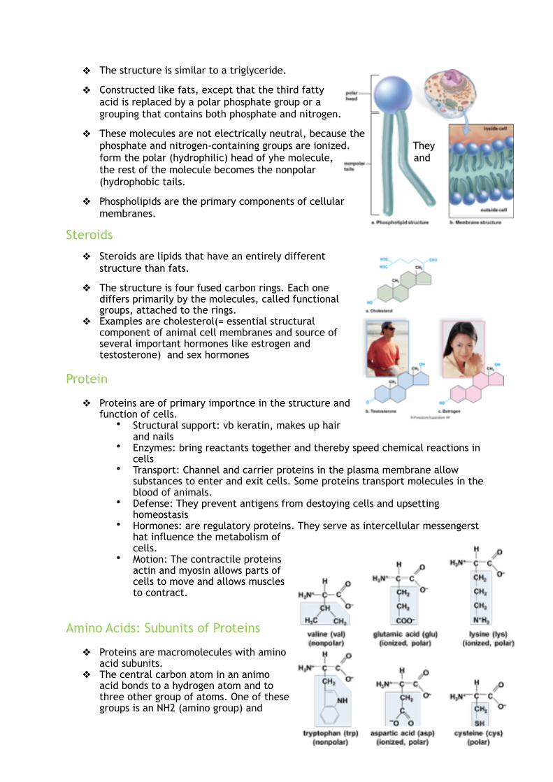
❖ The structure is similar to a triglyceride.
❖ Constructed like fats, except that the third fatty acid is replaced by a polar phosphate group or a grouping that contains both phosphate and nitrogen.
❖ These molecules are not electrically neutral, because the phosphate and nitrogen-containing groups are ionized. They form the polar (hydrophilic) head of yhe molecule, and the rest of the molecule becomes the nonpolar (hydrophobic tails.
❖ Phospholipids are the primary components of cellular membranes.
Steroids
❖ Steroids are lipids that have an entirely different structure than fats.
❖ The structure is four fused carbon rings. Each one differs primarily by the molecules, called functional groups, attached to the rings.
❖ Examples are cholesterol(= essential structural component of animal cell membranes and source of several important hormones like estrogen and testosterone) and sex hormones
Protein
❖ Proteins are of primary importnce in the structure and function of cells.
• Structural support: vb keratin, makes up hair and nails
• Enzymes: bring reactants together and thereby speed chemical reactions in cells
• Transport: Channel and carrier proteins in the plasma membrane allow substances to enter and exit cells. Some proteins transport molecules in the blood of animals.
• Defense: They prevent antigens from destoying cells and upsetting homeostasis
• Hormones: are regulatory proteins. They serve as intercellular messengerst hat influence the metabolism of cells.
• Motion: The contractile proteins actin and myosin allows parts of cells to move and allows muscles to contract.
Amino Acids: Subunits of Proteins
❖ Proteins are macromolecules with amino acid subunits.
❖ The central carbon atom in an animo acid bonds to a hydrogen atom and to three other group of atoms. One of these groups is an NH2 (amino group) and

COOH (carboxyl group, an acid), the third group is the R group. ❖ Amino acids differ according tot heir particular R group
Peptides
❖ The covalent bond between two amino acids is called a peptide bond ❖ When three or more amino acids are linked by peptide bonds, the chain that results
is called a polypeptide. ❖ The atoms associated with the peptide bond share the electrons unevenly. The
hydrogen attached to the nitrogen has a slightly positive charge,whereas the oxygen has a slightly negative charge.
Shape of Proteins
❖ Proteins cannot function unless they have a specific shape. ❖ Denaturation= permanent disruption of protein structure, leading to loss of function
• Caused by high temperature or changes in pH Levels of protein organization
❖ The structure of a protein has at least three levels of organization (but can have four levels)
• Primary structure = the linear sequence of the amino acids joined by peptide bonds
• Secondary structure = how the chain is orientated in space
o Alpha helix o Beta sheet o Random coil
• Tertiary structure: how the protein twists and folds to form a 3-dimensional shape
• Quaternary structure: how many polypeptide chains make up the protein and how they associate with each other. Vb hemoglobin
Nucleic Acids
❖ Made of nucleotide subunits
❖ Function in the cell to make proteins
❖ Include RNA and DN ❖ Nucleic acids, which are polymers of nucleotides, store information, include
instructions for life, and conduct chemical reactions ❖ DNA (deoxyribonucleic acid)
• genetic material of the cell • stores information about how to copy or replicate itself • specifies the order in which amino acids are to be joined to make a protein
❖ RNA (ribonucleic acid)

• Messenger RNA (mRNA)is a temporary copy of a gene in the DNA that specifies what the amino acid sequence will be during the process of protein synthesis.
• Transfer RNA (tRNA) is also necessary in synthesizing proteins and helps translate the sequence of nucleic acids in a gene into the correct sequence of amino acid during protein synthesis.
• Ribosomal RNA (rRNA) works as an enzyme to form the peptide bonds between amino acids in polypeptide.
❖ Not all nucleotides are made into DNA or RNA polymers. Some nucleotides are directly invloved in metabolic functions in cells.
• some are components of coenzymes = nonprotein oranic molecules that help regulate enzymatic reactions
• ATP (adenosine triphosphate) = a nucleotide that stores large amounts of energy needed for synthetic reactions and for various other energy requiring processes cells
How the structures of DNA and RNA differ
❖ These differences give DNA and RNA their unique functions in the body. Nucleotide structure
❖ Each nucleotide is a molecular complex of three types of submit molecules
• Phosphate (phosphoric acid) • Pentose (5carbon) sugar • Nitrogen-containing base
❖ Nucloetides in DNA • Contain the sugar deoxyribose • There are four different types of bases in DNA
o Adenine (A) o Thymine (T) o Guanine (G) o Cytosine (C )
❖ Nucleotides in RNA • Contain the sugar ribose
o The base Uracil (U) replaces the base thymine ❖ Adenine (A) and guanine (G) are double-ringed purines.
❖ Cytosine (C), thymine (T), and uracil (U) are single-ringed pyrimidines.
❖ These structures are called bases because their presence raises the pH of a solution
DNA and RNA structure
❖ The nucleotides link to make a polynucleotide called a strand • Has a backbone made up of phosphate-sugar-phosphate-sugar
❖ DNA is double stranded • With the the two strands twisted about each other i the form of a double
helix o The two strands are held together by hydrogen bonds between the
bases o A (adenine) pairs with T (thymine) and G (guanine) pairs with C
(cytosine)= complementary base pairing -> allows DNA to replicate in a way that ensures the sequence of bases will remain the same.
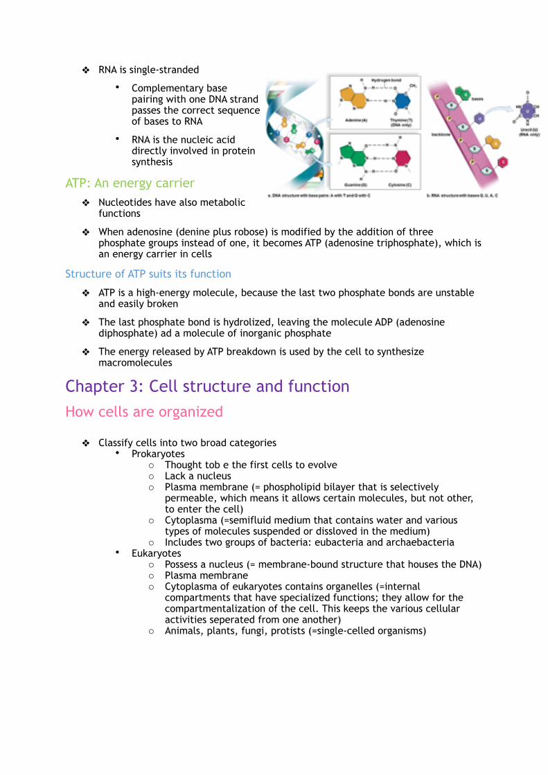
❖ RNA is single-stranded
• Complementary base pairing with one DNA strand passes the correct sequence of bases to RNA
• RNA is the nucleic acid directly involved in protein synthesis
ATP: An energy carrier ❖ Nucleotides have also metabolic
functions
❖ When adenosine (denine plus robose) is modified by the addition of three phosphate groups instead of one, it becomes ATP (adenosine triphosphate), which is an energy carrier in cells
Structure of ATP suits its function
❖ ATP is a high-energy molecule, because the last two phosphate bonds are unstable and easily broken
❖ The last phosphate bond is hydrolized, leaving the molecule ADP (adenosine diphosphate) ad a molecule of inorganic phosphate
❖ The energy released by ATP breakdown is used by the cell to synthesize macromolecules
Chapter 3: Cell structure and function How cells are organized
❖ Classify cells into two broad categories • Prokaryotes
o Thought tob e the first cells to evolve o Lack a nucleus o Plasma membrane (= phospholipid bilayer that is selectively
permeable, which means it allows certain molecules, but not other, to enter the cell)
o Cytoplasma (=semifluid medium that contains water and various types of molecules suspended or dissloved in the medium)
o Includes two groups of bacteria: eubacteria and archaebacteria • Eukaryotes
o Possess a nucleus (= membrane-bound structure that houses the DNA) o Plasma membrane o Cytoplasma of eukaryotes contains organelles (=internal
compartments that have specialized functions; they allow for the compartmentalization of the cell. This keeps the various cellular activities seperated from one another)
o Animals, plants, fungi, protists (=single-celled organisms)

Evolutionary history of the Eukaryotic cell
❖ Prokaryotic cells today are represented by the bacteria and archea
❖ Early prokaryotic organisms evolved in contained conditions thatwould be instantly lethal to life today -> they gradually adapted to earth’s environment today and some are still around
❖ Evidence widely supports the hypothesis that eukaryotic cells evolved from the archea
❖ ( zie boek p49)
The plasma membrane and how substances cross it ❖ The plasma membrane is a phospholipid bilayer with attached or embedded
proteins

• Phospholipid molecule
o Has a polar head (water soluble) and nonpolar tails (water-insoluble)
o Polar heads are hydrophilic (attracted to water), position themselves to face toward the water
o Nonpolar tails are hydrophobic (not attracted to water), they turn inward toward one another, where there is no water
• At body temperature, the phospholipid bilayer is liquid
• Fluid-mosaic model = a working description of membrane structure
o It states that the protein molecules form a shifting pattern within the fluid phospholipid bilayer
• Cholesterol lends support the membrane (making it more rigid)
• Short chains of sugar are attached to the outer surface of some protein and lipid molecules
o Called glycoproteins and glycolipids: they help mark the cell as belonging to a specific individual. V
▪ vb. they account for why people have different bloodtypes
▪ others act as chemical messengers (vb hormone)
▪ others form substances through which substances can enter cells
▪ others are carriers involved in the passage of molecules through the membrane

Plasma membrane functions
❖ The plasma membrane isolates the interior of the cell from the external environment
❖ Is selectively permeable
• It allows only certain molecules and ions to enter and exit the cytoplasm freely
o Small, lipid-soluble molecules pass easily
o Ions and large molecules cannot cross without more direct assistance
Diffusion
❖ Random movement of molecules from an area of higher concentration to an area of lower concentration, until they are equally distributed.
❖ Passive way for molecules to enter or exit a cell
❖ The net movement will be from the region of higher concentration to the region of lower concentration, until equilibrium (as many molecules of the substance will be entering as leaving the cell) is achieved
Osmosis
❖ The diffusion of water molecules
❖ Is the net movement of water across a semipermeable membrane
❖ The direction by which water will diffuse is determined by the tonicity of the solutions inside and outside the cell

• Tonicity is based on dissolved particles, called solutes, within a solution (the higher the concentration of solutes, the lower the concentration of water
• Water will diffuse from the area that has less solute (low tonicity and therefore more water) to the area with more solute.
❖ Normally body fluids are isotonic to cells. There is the same concentration of nondiffusible solutes and water on both sides of the plasma membrane. Therefore cells maintain their normal size and shape and they do not get affected
❖ Solutions that cause cells to swell or even to burst (lysis) due to an intake of water are said to be hypotonic. A hypotonic solution has a lower concentration of solute and a higher concentration of water than the cells. Bursting of red blood cells is termed hemolysis
❖ Hypertonic solutions have more solute than the inside of the cell and lead to crenation (shriveling)
❖ these changes have occurred due to osmotic pressure.
• Controls water movement in our bodies
Facilitated transport
❖ A molecule is transported across the plasma membrane from the side of higher concentration to the side of lower concentration via a protein carrier
• Passive transport (no need to expend energy
• Each protein carrier (transporter) binds only to a particular monecule
Active transport
❖ Movement of molecules from a lower to higher concentration
❖ Active transport requires a protein carrier and the use of cellular energy obtained from the breakdown of ATP
• The energy is used to carry out active transport
• Pumps = proteins that actively transport molecules across the plasma membrane
• Energy is used to move substances against their concentration gradients
• One type of pump active in all cells moves sodium ions (Na+) to the outside and potassium ions (K+) to the inside of the cell
Endocytosis and Exotysosis
❖ Endocytosis
• transports molecules or cells into the cell

• via invagination of the plasma membrane to form a endocytic vesicle
• phagocytosis: process where some white blood cells are able to take up pathogens (disease-causing agents) by endocytosis
• one form of endocytosis uses a receptor, a form of membrane protein, on the surface of the cell to concentrate specific molecules of interest for endocytosis
❖ Exocytosis
• transports molecules outside the cell
• via the fusion of a vesicle with the plasma membrane
The nucleus and endomembrane system ❖ nucleus and several organelles are involved in the production
and processing of proteins
❖ endomembrane system = a series of membrane organelles that function in the processing of materials for the cell
The nucleus
❖ prominent sructure in eukaryotic cells
❖ stores genetic information
❖ genes • are segments of DNA that contain information for the poduction of specific
proteins
❖ DNA, with RNA acting as an intermediary, specifies the proteins in a cell ❖ Proteins have many functions in cells, and they help determine a cell’s specificity ❖ Chromatin
• Is the combination of DNA molecules and proteins that make up the chromosomes, the structures that transmit genetic information from one generation to the next
• Can coil tightly to form visible chromosomes during cell division -> most of the time uncoiled
• Is surrounded by a semifluid medium called the nucleoplasm
• Dark region = the nucleolus, where ribosomal RNA (rRNA) is produced. This is also where rRNA joins with proteins to form the subunits of robosomes
• Nuclear envelope = double membrane that seperates the nucleas from the cytoplasm
• The nuclear envelope has nuclear pores of sufficient size to permit the passage of ribosomal subunits out of the nucleus and proteins into the nucleus
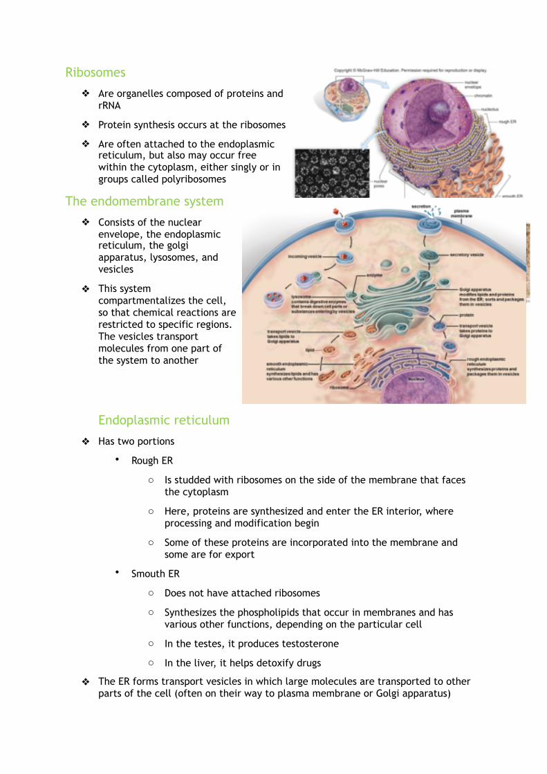
Ribosomes
❖ Are organelles composed of proteins and rRNA
❖ Protein synthesis occurs at the ribosomes ❖ Are often attached to the endoplasmic
reticulum, but also may occur free within the cytoplasm, either singly or in groups called polyribosomes
The endomembrane system
❖ Consists of the nuclear envelope, the endoplasmic reticulum, the golgi apparatus, lysosomes, and vesicles
❖ This system compartmentalizes the cell, so that chemical reactions are restricted to specific regions. The vesicles transport molecules from one part of the system to another
Endoplasmic reticulum
❖ Has two portions
• Rough ER
o Is studded with ribosomes on the side of the membrane that faces the cytoplasm
o Here, proteins are synthesized and enter the ER interior, where processing and modification begin
o Some of these proteins are incorporated into the membrane and some are for export
• Smouth ER
o Does not have attached ribosomes
o Synthesizes the phospholipids that occur in membranes and has various other functions, depending on the particular cell
o In the testes, it produces testosterone
o In the liver, it helps detoxify drugs
❖ The ER forms transport vesicles in which large molecules are transported to other parts of the cell (often on their way to plasma membrane or Golgi apparatus)

The Golgi apparatus
❖ Consist of a slighty curved saccules
❖ Here proteins and lipids received from the ER are modified
❖ The vesicles that leave the Golgi apparatus move to other parts of the cell
❖ In all, the Golgi apparatus is involved in processing, packaging, and secretion
Lysosomes
❖ Membranous sacs produces by the Golgi apparatus, contain hydrolytic enzymes
❖ Are found in all cells of the body but are particularly numerous in white blood cells that engulf disease-causing microbes.
❖ Autodigestion = parts of a cell may be broken down by the lysosomes
❖ Some human diseases are caused by the lack of a particular lysosome enzyme
• Tay-Sachs disease: occurs when an undigested substance collects in nerve cells, leading to developmental problems and death in the early childhood
The cytoskeleton, Cell Movement and Cell Junctions ❖ A series of proteins that maintain cell shape, as well as anchors and/or moves
organelles in the cell
❖ Made of 3 types of fibers
• large microtubules
o each is a cylinder that contains rows of a protein called turbulin
o regulation of microtube assembly is under the control of a microtube organizing center called centrosome
o help maintain the shape of the cell land act as tracks along which organelles move.
o During cell division, microtubules form spindle fibers, which assist in the movement of chromosomes
• thin actin filaments
o made of a protein called actin, are long, extremely thin fibers that usually occur in bundles or other groupings.
o Are onvolved in movement
• medium-sized intermediate filaments
o are intermediate in size between microtubules and actin filaments
o their structure and function according to the type of cell
Cilia and Flagella
❖ made of microtubules, used in movement
❖ Cilia are about 20x shorter than flagella

❖ Are involved in movement
❖ The ciliated cells that line our respiratory tract sweep debris trapped within muscus back up the throat. This helps keep the lungs clean.
❖ The importance of normal cilia and flagella is illustrated by the occurrence of a genetic disorder. Some individuals have an inherited genetic defect that leads to malformed microtubules in cilia and flagella. These individuals suffer from recurrent and severe respiratory infections.
Extracellular matrix (ECM)
❖ Is a meshwork of proteins and polysaccharides in close association with the cell that produced them.
❖ Collagen (resists stretching) and elastin fibers (gives the ECM resilience)are well-known structural proteins in the ECM
❖ Fibronectin a • an adhesive protein that binds to a protein in the plasma membrane called
integrin ❖ Integrins
• are integral membrane proteins that connect to fibronectin externally and to the actin cytoskeleton internally
• plays a role in signaling, permitting the ECM to influence the activities of the cytoskeleton and, therefore, the shape and activities of the cell
• amino sugars in the ECM form multiple polysaccharides, which attach to a protein and are, therefore, called proteoglycans
• proteoglycans o attach to a very long, centrally placed polysaccharide
o entire structure resists compression of the extracellular matrix
o influence the process of cell signaling by regulating the passage of molecules through the ECM to the plasma membrane, where receptors are located
Junctions between cells
❖ human tissues are known to have junctions between their cells that allow them to function in a coordinated manner
❖ 3 mean types

• Adhesion junctions
o mechanically attach adjacent cells (common in skin cells).
o Cytoskeletons of two adjacent cells are interconnected
• Tight junctions
o are connections between the plasma membrane proteins of neighboring cells that produce a zipper-like barrier (common in digestive system and kidney where fluids must be contained to a specific area).
• Gap junctions
o are communication portals between cells; they channel proteins of the plasma membrane fuse, allowing easy movement between adjacent cells.
Metabolism and the energy reactions
Metabolic pathways
❖ Metabolism includes all chemical reactions that occur in a cell.
• Products
• Reactants
❖ Often, metabolism equires pathways and is carried out bt enzymes sequentially arrenged in cells:
❖ the letters, except A and G, are products of the previous reaction and the reactants for the next reaction.
❖ A = beginning reactant
❖ G = final product
❖ Number in the pathway refer to different enzymes
❖ Each reaction in a metabolic pathway requires a specific enzyme
❖ Metabolic pathways are highly regulated by the cell. • One type of regulation = feedback inhibition
o One of the end products of the metabolic pathway interacts with an enzyme early in the pathway.
o In most cases, this feedback slows down the pathway, so that the cell does not produce more product than it needs.
Enzymes
❖ Are metabolic assistants that speed up the rate of a chemical reaction.

❖ Most enzymes are proteins.
❖ Enzymes are often named for the molecules that they work on, called substrates
❖ Enzymes are specific to what substrate they work on.
❖ Enzymes have active sites (specific region) where a substrate binds.
❖ An enzyme specificity is caused by the shape of the active site. Here the enzyme and its substrate(s) fit together in a specific way.
❖ After one reaction is complete, the product or products are released. The enzyme is ready to
be used again.
❖ Chemical reaction: E + S ! ES ! E + P
❖ Enzymes are not used up in a reaction but instead are recycled.
❖ Molecules frequently do not react with one another unless they are activated in some way. The energy that needs to be added to cause molecules to react with one another is called the energy of activation (Ea). The input of some enrgy is required to overcome the energy of activation.
❖ Enzymes lower the amount of energy of activation needed. By lowering the energy of activation, the enzyme increases the rate of the reaction.
❖ Coenzymes are nonprotein molecules that assist the activity of an enzyme and may even accept or contribute attoms to the reaction.
❖ Some enzymes are aided by nonprotein molecules called coenzymes.
Mitochondria and Cellular respiration
❖ Highly folded organelles in eukaryotic cells
❖ Convert the chemical energy of glucose products into the chemical energy of ATP molecules.
• This process is called cellular respiration: because the mitochondria use up oxygen and give off carbon dioxide
❖ Structure of mitochondria is appropriate to the task
• Inner membrane: folded tof om little shelves = cristae
• Project into the matrix = inner space filled with gel-like fluid
o Contains enzymes for breaking down glucose products
• ATP production than occurs at the cristae
• Protein complexes that aid in the conversion of energy are located in an assembly-line fashion on these membranous shelves
❖ Thought to be derived from an engulfed prokaryotic cell

❖ Mitochondria have their own genes and they reproduce themselves
Cellular respiration
❖ After blood transports glucose and oxygen to cells, cellular respiration begins
❖ It breaks down glucose to carbon dioxide and water
❖ Three pathways are invloved in the breakdown (they allow the enrgy in a glucose molecule to be slowly released, so that ATP can be gradually produced
• Glycolysis (= sugar splitting)
o 6 – carbon ( C6) molecule, is split so that the result is two 3-carbon molecules of pyruvate
o Occurs in cytoplasm, is found in most every type of cell
o Is an anaerobic pathway: because it does not require oxygen
o During glycosis, hydrogens and electrons are removed from glucose, and NADH results
• Citric acid cycle (krebs cycle)
o Each of the pyruvate molecules, after a brief modification, enters the citric acid cycle as acetyl CoA
o Is a cyclical series of enzymatic reactions that occurs in the matrix of mitochondria
o Purpose is to complete the breakdown of glucose by breaking the remaining C – C bonds
o As the reactions progress, carbon dioxide is released, a small amount of ATP (two per glucose) is produced, and the remaining hydrogen and electrons are carried away by NADH and a similar molecule called FADH2.
• Electron transport chain
o NADH molecules from glycolysis and the critic acid cycle deliver electrons to the electron transport chain
o The members are carrier proteins grouped into complexes (embedded in the cristae of mitochondrion
▪ Each carrier accepts two electrons and passes them on to the next carrier
▪ High-energy electrons enter the chain and, as they are passed fom carrier to carrier, the electrons lose energy
▪ Low-energy electrons emerge from the chain
▪ Oxygen serves as the final acceptor of the electrons at the end of the chain
▪ After oxygen receives the electrons, it combines with hydrogens and becomes water

o The presence of oxygen makes the electon transport chain aerobic. Sole purpose of oxygen is to receive electrons at the end
o The energy released as electrons pass from carrier to carrier is used for ATP production
❖ ATP-ADP Cycle
• Each cell produces ATP within its mitochondria; therefore each cell uses ATP for its own purposes
• Glucose breakdown leads to ATP buildup, and the ATP is used for the metabolic work of the cell
• Muscle cells use aTP for concentration, and nerve cells use it for conduction of nerve impulses
• ATP breakdown releases heat
Fermentation
❖ Is an anaerobic process, meaning that it does not require oxygen
❖ When oxygen is not available to cells, the electron transport chain soon becomes inoperative
• Normally NADH takes electrons to the electron transport chain and, thereby, is recycled to become NAD+
• If the system is not working duet o a lack of oxygen, NADH passes its hydrogens and electrons to pyruvate molecules:
o NADH NAD+
Pyruvate ! lactate
• This means that the critic acid cycle and the electron transport chain do not function as a part of fermentation
• When oxygen is available again, lactate can be converted back to pyruvate and metabolism can proceed as usual.
• Fermentation can give us a burst of energy for a short time, but it produces only two ATP per glucose molecule

• Fermentation results in the buildup of lactate. Lactate is toxic to cells and causes muscles to cramp and fatigue. If fermentation continues for any length of time, death follows
Chapter 4: Organization and regulation of body systems
Types of tissues ❖ Cells are composed of molecules
❖ A tissue is a collection of cells of the same type that perform a common function in the body
❖ An organ contains several types of tissues; and several organs are found in an organ system
❖ Four major types
• Connective tissue: binds and supports body parts
• Muscular tissue: moves the body and its parts
• Nervous tissue: receives sensory information and conducts nerve impulses
• Epithelial tissue: covers body surfaces and lines body cavities
Connective tissue connects and supports ❖ It binds and supports parts of the body.
❖ Diverse structure and function, but all types of connective tissue have three similar components: specialized cells, ground substance, and protein fibers.
❖ Ground substance is noncellular material that seperates the cells. It varies from solid to fluid.
❖ The fibers are of three possible types
• Collagen fibers
o Contain collagen, a protein that gives them flexibility and strength
• Reticular fibers
o Are very thin collagen fibers, highly branched proteins that form delicate supporting networks
• Elastic fibers
o Contain elastin, a protein that is not as strong as collagen but is more elastic
o Return to their original shape
❖ Inherited connective tissue disorders arise when people inherit genest hat lead to malformed fibers
❖ Ground substance and protein fibers together make up the matrix of the tissue.
❖ There are 3 main types of connective tissue: A. fibrous , B. supportive, and C. fluid.

Fibrous connective tissue
❖ There are 2 types: dense and loose, but both contain fibroblast cells with a matrix of collagen and elastic fibers.
❖ Matrix is a term that includes ground substance and fibers
❖ Loose fibrous tissue (which includes areolar and reticular connective tissue)
• is found supporting epithelium and many internal organs.
• Its presence in lungs, arteries, and the urinary bladder allows these organs to expand
• Forms a protective covering enclosing many internal organs, such as muscles, blood vessels and nerves
❖ Adipose tissue
• is a special type of loose connective tissue in which the cells enlarge and store fat
• has a little extra cellular matrix
• its cells, which are called adipocytes, are crowded, and each is filled with liquid fat
• body uses this stored fat for energy, insulation ad organ protection
• releases a hormone called leptin = regulates appetite-control centers in the brain
• primarily found beneath the skin, around the kidneys, and on the surface of the heart
❖ dense fibrous connective tissue
• contains many collagen fibers packed together
• has more specific function than loose connective tissue
• is found in tendons (which connect muscles to bones), and in ligaments (which connect bones to other bones at joints)

Supportive connective tissue
❖ cartilage and bone are the two main supportive connective tissues ❖ each provides structure, shape, protection, and leverage for movement ❖ cartilage is more flexible than bone, because it lacks mineralization of the matrix
Cartilage
❖ the cells lie in small chambers, called lacunae, separated by a solid, yet flexible matrix
❖ this matrix is formed by cells called chondroblasts and chondrocytes
❖ because this tissue lacks a direct blood supply, it often heals slowly
❖ three types of cartilage are distinguished by the type of fiber found in the matrix
• Hyaline cartilage
o Most common type
o Contains only fine collagen fibers
o Location: Nose, ends of long bones, rips, walls of respiratory passages and fetal skeleton
• Elastic cartilage
o more elastic fibers than cartilage fibers and is more flexible
o Location: Outer ear
• Fibrocartilage
o Has a matrix containing strong collagen fibers
o Found in structures that withstand tension and pressure
o Location: Disks between vertebrae, cushions in the knee joint

Bone
❖ Most rigid connective tissue
❖ Consists of an extremely hard matrix of inorganic salts, notably calcium salts
• Inorganic salts are deposited around protein fibers, epecially collagen fibers
• Inorganic salts give bone rigidity
• Protein fibers provide elasticity and strength
• Cells called osteoblasts and osteoclasts are responsible for forming the matrix in bone tissue
❖ Compact bone
• made of repeating circular units called osteons which contain the hard matrix, living cells, and blood vessels
• Location: Shafts of long bones
❖ Spongy bone
• an open latticework with irregular spaces
• Location: Ends of long bones
Fluid connective tissue
Blood
❖ Made of a fluid matrix called plasma and cellular components that are called formed elements
❖ Located in the blood vessels
❖ Transports nutrients and oxygen to interstitial fluid (bathes the body’s cells and removes carbon dioxide and other wastes
❖ Helps distribute heat and plays a role in fluid, ion, and pH balance
❖ 3 formed elements
• Red blood cells ( erythrocytes)
o Have no nucleus
o Red pigment hemoglobin
o Cells that carry oxygen
• White blood cells (leukocytes)
o Have nucleus

o Cells that fight infection
o Some are generalists
▪ Meaning that they respond to any foreign invader in the body
▪ Are phagocytic cells, because they engulf infectious agents (vb. Bacteria)
o Others are more specific and either produce antibodies or directly attack specific invading agents or infected cells in the body
• Platelets ( thrombocytes)
o Not complete cells
o Fragments of giant cells present only in bone marrow
o pieces of cells that clot blood
lymph
❖ clear fluid derived from the fluids surrounding the tissues
❖ contains white blood cells
❖ lymphatic vessels
• absorb excess tissue fluid and various dissolved solutes in the tissues.
• Transport lymph to particular vessels of the cardiovascular system
• Absorb fat molecules from the small intestine
❖ Lymph nodes, composed of fibrous connective tissue, occur along the length of lymphatic vessels. Lymph is cleansed as it passes through lymph nodes, because white blood cells congregate there. Lymph nodes enlarge when you have an infection
Muscular tissue moves the body ❖ Specialized to contract
❖ It is made of muscle fibers/cells and protein fibers called actin and myosin.
❖ There are 3 types of muscle tissue in humans
• Skeletal
o Appearance: long, cylindrical cells, multiple nuclei, striated fibers
o Location: attached to bone for movement
o Nature: voluntary movement
• Smooth muscle
o Appearance: spindle-shaped cell with one nucleus, lacks striations
o Location: walls of hollow organs and vessels
o Nature: involuntary movement
• cardiac muscle

o Appearance: branched cells with a single nucleus, striations with darker striations called intercalated disks between cells
o Location: heart
o Nature: involuntary movement
Nervous tissue communicates ❖ Consists of nerve cells, called neurons and neuroglia, the cells that support and
nourish the neurons
❖ Central component in the nervous system
❖ It allows for communication between cells through sensory input, integration of data, and motor output.
Neurons
❖ Specialized cell that has three parts
• Dendrites
o Extension that receives signals from sensory receptors or other
neurons. carry information toward the cell body.
• Cell body
o Contain most of the cell’s cytoplasm and the nucleus
• Axon

o Extension that conducts nerve impulses. carry information away from the cell body.
o Are covered by myelin
❖ Fiber is used here to refer to an axon along wit hits myelin sheath, if it has one
❖ Outside the bain and spinal cord, fibers bound by connective tissue form nerves.
❖ Nerves
• conduct signals from sensory receptord to the spinal cord and the brain, where integration, or processing, occurs.
• Conduct signals from the spinal cord and brain to muscles, gland and other organs. This triggers a characteristic response from each tissue
Neuroglia
❖ They are a collection of cells that support and nourish neurons.
❖ They outnumber neurons nine to one and take up more than half the volume of the brain
❖ Do not have long extensions (axon and dendrites) ( evidence that neurons communicate among themselves even without these extensions)
❖ Examples are
• Microglia
- Supporting neurons,engulf bacterial and cellular debris
• Astrocytes
- Provide nutrients to neurons and produce a hormone known as glial-derived neurotrophic factor (GDNF)
• oligodendrocytes,
- form the myelin sheets around fibers in the brain and spinal cord
❖ outside the brain, shawnn cells are the type of neuroglia that encircle long nerve fibers and form a myelin sheeth
Epithelial tissue protects ❖ It is a group of cells that forms a tight, continuous network.
❖ It lines body cavities, covers body surfaces, and is found in glands.
❖ It has a protective function
❖ It can also be modified to carry out secretion, absorption, excretion, and filtration

❖ Cells are anchored by a basement membrane on one side and free (exposed to the environement) on the other side.
❖ It is named after the appearance of cell layers and the shape of the cells.
❖ There is transitional epithelium that changes in appearance in response to tension.
Simple epithelia
❖ Have only a single layer of cells and are classiefied according to cell type
❖ Squamous epithelium • Composed of flattened cells
• Found lining the air sacs of lungs and walls of blood vessels. It’s shape permits exchanges of substaces in these locations
❖ Cuboidal epithelium
• Consists of a single layer of cube-shaped cells
• Frequently found in glands
• Simple cuboidal epithelium also covers the ovaries and lines kidney tubules
• When cuboidal cells are involved in absorption, they have microvilli (minute cellular extensions of the plasma membrane) -> increase he surface area of the cells
• When cuboidal cells function in active transport they contain many mitochondria
❖ Columnar epithelium
• Has cells resembling rectangular pillars or columns
• Lines the digestive tract, where microvilli expand the surface area and aid in absorbing the products of digestion.
• Ciliated columnar epithelium is found lining the uterine tubes, where it propels the egg toward the uterus.
❖ Pseudostratified epithelium
• Appears to be layered, but true layers do not exist, because each cells touches the basement membrane. Only one layer exists.
• Lining of the windpipe, or trachea is pseudostratified ciliated epithelium
❖ In some cases, columnar and pseudostratifies epithelium secrete a product. In this cas it is said to be glandular.
• Exocrine glands: glands with ducts that secrete their product onto the outer surface or into a cavity. Ducts can be simple or compound
• Endocrine glands: glands that have no ducts. They secrete hormones directly into the bloodstream.
Stratified Epithelia
❖ Have layers of cells piled one on top of the other. Only the bottom layer touches the basement membrane.

❖ Nose, mouth, esophagus, anal canal, outer portion of the cervix and vagina are lined with it.
❖ Outer layer of the skin is also stratified squamous epithelium, but the cells are reinforced by keratin, a protein that provides strength. Statified cuboidal and columnar epithelia also are found in the body.
❖ Transitional epithelium: term is used to imply changeability, because tissue changes in response to tension.
• Forms the lining of the urinary bladder, the ureters and part of the urethra. All are organs that may need to stretch.
❖ The skin is an organ comprimising all four tissue types: epithelial, connective, muscular and nervous tissue.
❖ An organ system contains many different organs that cooperate to carry out a process. The skin has several accessory organs (hair, nails, sweat glands, sebaceous glands) and, therefore, is sometimes referred to as integumentary system.
❖ Skin accounts for nearly 15 % of the weight of an average human
❖ Skin has numerous functions
• It protects underlying tissues from physical trauma, pathogen invasion and water loss
• Helps regulate body temperature. Therefore skin plays an important role in homeostasis
• Synthesizes certain chemicals that affect the rest of the body.

• Contains sensory receptors and temperature receptors. It help sus be aware of our surroundings and to communicate with others.
Regions of the skin
❖ The skin has two regions: epidermis and dermis
❖ A subcutaneous layer (hypodermis) is found between the skin and any underlying structures, such as muscle or bone
The epidermis
❖ It is the thin, outermost layer of the skin.
❖ It is made of stratified squamous epithelial tissue.
❖ New epidermal cells for the renewal of skin are derived from stem (basal) cells. The importance of these cells is observed when there is an injury to the skin. (p78)
❖ Newly generated cells become flattened and hardened as they push to the surface. Hardening takes place because the cells produce keratin, a waterproof protein.
❖ Cells in the uppermost layers are dead and become filled with keratin, thus the skin is waterproof. This prevents water loss and helps maintain homeostasis. Also prevents water from entering when the skin is immersed.
❖ Two types of specialized cells are located deep in the epidermis
• Langerhans cells are a type of white blood cells that help fight pathogens.
• Melanocytes produce melanin that lend to skin color and protection from UV light.
❖ Some cells convert cholesterol to vitamin D with the aid of UV radiation. Only a small amount of UV radiation is needed. Vitamin D leaves the skin and helps regulate both calcium and phosphorus metabolism in the body.
❖ Skin cancer
• Too much ultraviolet radiation is dangerous and can lead to skin cancer.
• Basal cell carcinoma is the most common type of cancer and most curable

• Melanoma is extremely serious
• What can you do to help prevent this?
o Stay out of the sun between 10 A.M. and 3 P.M.
o Wear protective clothing (tight weave, treated sunglasses, wide-brimmed hat).
o Use sunscreen with an SPF of at least 15 that protects from UV-A and UV-B rays.
o Do not use tanning beds.
The Dermis
❖ It is the thick, inner layer of the skin.
❖ It is made of dense fibrous connective tissue bebeath the epidermis.
❖ It contains elastic and collagen fibers. The collagen fibers are flexible but offer great resistance to overstretching. The elastic fibers maintain normal skin tension but also stretch to allow movement of underlying muscles and joints.
❖ The number of collagen and elastic fibers decreases with age and with exposure to the sun, causing the skin to become less supple and more prone to wrinkling.
❖ It contains blood vessels that nourish the skin.play a role in temperature regulation.
❖ many sensory receptors, specialized for touch, pressure, pain, hot and cold. They supply the central nervous system with information about the external environment.
❖ It contains glands.
The Subcutaneous Layer
❖ Technically speaking it is not a part of the skin.
❖ Common site for injections
❖ Is composed of loose connective tissue and adipose tissue which stores fat
❖ A well-developed subcutaneous layer gives the body a rouded appearance and provides protective padding against external assaults.
Accessory Organs of the Skin
❖ They include nails, hair, and glands (structures of epidermal origin).
❖ Nails are derived from the epidermis and offer a protective covering.
❖ Hair follicles are derived from the dermis, but hair grows from epidermal cells.
❖ Oil glands are associated with hair and produce sebum that lubricates the hair and skin and retards bacterial growth.
❖ Sweat glands are derived from the dermis and help to regulate body temperature.
Organ systems, body cavities and body membranes ❖ An organ is 2 or more tissue types working towards a particular function.

❖ An organ system is a combination of organs that work together to carry out a particular function.
Organ systems
❖ Organs work together in an rogan system, organ systems work together in the body.

Body cavities
❖ Human body is divided into two main cavities
• Ventral cavity
o Called the coelum in early development, the ventral cavity later becomes the thoracic, abdominal and pelvic cavities
• Dorsal cavity
o Cranial cavity
o Vertebral canal
Body Membranes
❖ Line cavities and the internal spaces of organs and tubes that open to the outside
❖ Are four types
• Mucous membranes
o line the digestive, respiratory, urinary, and reproductive systems
o are composed of an epithelium overlying a loose fibrous connective tissue layer
o contains specialized cells that secrete muscus
▪ protects the body from invasion by bacteria and viruses

▪ protects the walls of the stomach and small intestine from digestive juices (breaks down when a person develops an ulcer)
• Serous membranes
o line and support the lungs, heart, and abdominal cavity and its internal organs
o secrete a watery fluid that keeps the membranes lubricated
o support the internal organs and compartmentalize the large thoracic and abdominal cavities
o have specific names acoording to their location
▪ Pleura: line thoracic cavity and cover the lungs
▪ Peritoneum: lines abdominal cavity and covers its organs
▪ Pericardium: forms pericardial sac and covers the heart
• Synovial membranes
o line the cavities of freely movable joints, composed of only loose connective tissue
o secrete synovial fluid into the joint cavity. Fluid lubricates the end of the bones, so that they can move freely
• Meninges
o Membranes within the dorsal cavity
o Composed only of connective tissueand serve as a protective covering for the brain and spinal cord
Homeostasis ❖ It is the ability to maintain a relatively constant internal environment in the body.
❖ The nervous and endocrine systems are key in maintaining homeostasis.
❖ Changes from the normal tolerance limits result in illness or even death.
The internal Environment
❖ Has two parts
• Blood
o Delivers oxygen and nutrients to the tissues and carries away carbon dioxide and wastes
• Interstitial fluid
o Bathes the body’s cells
o Is the medium through which substances are exchanged between cells and blood
❖ The cooperation of body systems is required to keep these substances within the range of normalcy and interstitial fluid

The body systems and homeostasis
❖ Nervous and endocrine syspems are particulary important in coordinating the activities of all the other organ systems as they funcion to maintain homeostasis.
❖ Nervous system
• is also able to bring about rapid responses to any changes in the internal environment.
• Issues commands by electochemical signals rapidly transmitted to effector organs
❖ Endocrine system
• Brings about slower respones , but they generally have more lasting effects.
• Glands release hormones = chemical messengerst hat must travel through the blood and interstitial fluid to reach their targets.
❖ The nervous and endocrine systems together dirct numerous activities that maintain homeostasis, but all the organ systems must do their part to keep us alive and healthy
❖ Zie boek p 85 what if any component failed
Negative feedback

❖ The primary mechanism for maintaining homeostasis
❖ The output of the systemresuslts or corrects the original stimulus
❖ Has 2 components
• Sensor: detects a change in the internal environment
• control center: brings about an effect to bring conditions back to normal
❖ zie p 86-87 examples
Positive feedback
❖ A mechanism for increasing the change of the internal environment in one direction
❖ An example is the secretion of oxytocin during birth to continually increase uterine contractions
❖ Can be harmful such as when a fever is too high and continues to rise
Chapter 5: Cardiovascular System: Heart and blood vessels
Overview of the cardiovascular system ❖ Consists of
• Heart (pumps blood)
• Blood vessels (blood flows through)
Circulation perfoms exchanges
❖ Overall purpose of circulation is to service the cells of the body
❖ Cells are surrounded by interstitial fluid that is used to exchange substances between the blood and the cells
❖ Blood
• Removes waste products from that fluid
• Provides it with oxygen and the nutrients cells require to continue their existence
• Drops of carbond dioxide at the lungs and picks up oxygen
• Nutrients enter the bloodstream at the intestines and transport much needed substances to the body’s cells
• Blood is purified of it wastes at the kidneys and water and salts are retained as needed
❖ Liver
• takes up amino acids from the blood and returns needed proteins
• removes toxins and chemicals that may have entered at the intestines, and its colonies of white blood cells destroy bacteria and other pathogens
❖ Blood vessels move the blood and its contents through the body to and from all the body’s organs
Functions of the cardiovascular system
❖ Transport: oxygen, carbon dioxide and other wastes products, nutrients, and hormones
❖ Protection: cells of the immune system are transported to help protect the body from infection
❖ Regulation: maintain homeostasis of a variety of the body’s conditions
❖ The lymphatic system assists the cardiovascular system, because lymphatic vessels collect excess interstitial fluid and return it to the cardiovascular system.
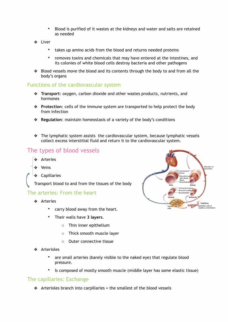
The types of blood vessels ❖ Arteries
❖ Veins
❖ Capillaries
Transport blood to and from the tissues of the body
The arteries: From the heart
❖ Arteries
• carry blood away from the heart.
• Their walls have 3 layers.
o Thin inner epithelium
o Thick smooth muscle layer
o Outer connective tissue
❖ Arterioles
• are small arteries (barely visible to the naked eye) that regulate blood pressure.
• Is composed of mostly smooth muscle (middle layer has some elastic tissue)
The capillaries: Exchange
❖ Arterioles branch into carpillaries = the smallest of the blood vessels

❖ Structure is adapted for the exchange of materials with the cells of the body
❖ Each capillary is an extremely narrow, microscopic tube with a wall composed only of endothelium (= formed by a single layer of epithelial cells with a basement membrane)
❖ Capillary beds are present in all regions of the body (so no cell far away from gas exchange)
❖ Rings of muscle called precapillary sphincters control the blood flow through a capillary bed
❖ When a capillary bed is closed, the bood moves to an area where gas exchange is needed, going directly from arteriole to venule through a pathway called an Arteriovenous shunt
The veins: to the heart
❖ Venules
• are small veins that receive blood from the capillaries and then join to form a vein.
❖ Veins
• carry blood toward the heart
• vein ofen have valves, which allow blood to flow only toward the heart when open and prevent backward flow of blood when closed. Valves are extensions of the inner wall-layer and are found in the veins that carry bloodagainst the force of gravity, especially the veins of the lower extremities
❖ Venule and vein walls have 3 layers.
• Thin inner epithelium
• Thick smooth muscle layer
• Outer connective tissue
❖ However, there is less smooth muscle in the middle layer of a vein and less connective tissue in the outer layer
❖ At any time, about 70% of the blood is in the veins

The heart is a double pump ❖ Location: midline/left inside chest cavity
❖ myocardium
• The major portion of the heart is the interior wall of tissue called the myocardium, consisting largely of cardiac muscle tissue
• Muscle fibers are branched
• Each fiber is tightly joined to neighboring fibers by structures called intercalated disks. They also incude cell junctions like:
▪ Gap junctions: are used to aid in simultaneous contractions of the cardiac fibers
▪ Desmosomes: include arrangements of protein fibers that tightly hold the membranes of adjacent cells together and prevent overstretching
❖ Pericardium (fibrous sac) encloses heart
• Protection
• Anchor
• Prevents heart overfilling with blood
❖ Pericardial cavity: space between heart and pericardium, filled with lubricant (pericardial fluid)
❖ Consists of 2 sides, right and left, separated by a septum
❖ Consists of 4 chambers: 2 atria and 2 ventricles. Each atrium has a wrinkled, earlike flap on the outer surface called an auricle.
❖ 2 sets of valves
• semilunar valves
o pulmonary semilunar valve
o aortic semilunar valve
• atrioventricular valves (AV valves)
o are suppored by strong, fibrous strings called chordae tendineae
o chordae enter the valves, preventing them from inverting when the heart contracts
o AV valve on the left side is called bicuspid valve (mitral valve) (two flaps)
o AV valve on the right side is called the tricuspid valve (three flaps)
• Valves produce the “lub” and “dub” sounds of the heartbeat
Coronary circulation: the heart’s blood supply
❖ The myocardium receives oxygen and nutrients from the coronary arteries

❖ Wastes are removed by the cardiac veins
❖ Blood that flows through the heart contributes little to either nutrient supply or waste removal
❖ Coronary arteries
• Serve the heart muscle itself
• Lie on the exterior surface of the heart, where they divide into diverse arterioles
• Coronary capillary beds joint o form venules, which converge to form the cardiac veins, which empty into the right atrium
• They can become easily clogged, leading to coronary disease
• If that happens oxygen and nutrients will not reach the muscles of the heart and this may result in a myocardial infarction or heart attack
Passage of blood through the heart
❖ We can trace the path of blood through the heart and body in the following manner
• The superior vena cava and the inferior vena cava carry oxygen-poor blood from the body veins to the right atrium
• The right atrium contracts (simultaneously with the left atrium), sending blood through an atrioventricular valve (the tricupis valve) to the right ventricle
• The right ventricle contracts, pumping blood through the pulmonary semilunar valve into the pulmonary trunk. The pulmonary trunk, which carries oxygen-poor blood, divides into two pulmonary arteries, which go to the lungs
• Pulmonary capillaries within the lungs allow gas exchange. Oxygen enters the blood; carbon dioxide waste is excreted from the blood.
• Four pulmonary veins, which carry oxygen-rich blood, enter the left atrium
• The left atrium pumps blood through an atrioventricular valve (bicuspid valve) to the left venticle
• The left ventricle contracts (at the same time as the right), sending blood through the aortic semilunar valve in the aorta.
• Arteries and arterioles supply tissue capillaries. Tissue capillaries drain into increasingly larger veins. Veins drain into the superior and inferior venae cavae, and the cycle starts again.

The heartbeat is controlled
❖ Each heartbeat is called a cardiac cycle
❖ The stages of the cardiac cycle
• a: When the atria contract, the ventricles are relaxed and filling with blood. Atrioventricular valves are open; semilunar valves are closed.
• b: When the ventricles contract, atrioventricular valves are closed, semilunar valves are open. Blood is pumped into the pulmonary trunk and aorta; this corresponds to the ‘lub’ sound.
• c: Backward flow of blood against the semilunar valves causes the ‘dub’ sound. When the heart is relaxed, both atria and ventricles fill with blood. The atrioventricular valves are open; semilunar valves are closed
• d: Aortic semilunar valve and bicuspid valve
Internal control of heartbeat
❖ The rhythmic contraction of the atria and ventricles is duet o the internal (intrinsic) conduction system of the heart.
❖ The SA (sinoatrial) node • Is located in the upper dorsal wall of the right atrium • Initiates the heartbeat and automatically sends out an excitation signal
every 0.85 second. This causes the atria to contract. • When signal impulses reach the AV node, there is a slight delay that allows
the atria to finish their contraction before the ventricles begin their contraction

• The signal for the ventricles to contract travels from the AVnode through the two branches of the AV bundle before reaching the numerous and smaller Purkinje fibers.
• The AV bundle, its branches, and the purkinje fibers work efficient because gap junctions allow electrical current to flow from cell to cell.
• The SA node is also called the pacemaker, because it regulates the heartbeat.
❖ The AV (atrioventricular) node • Is located in the base of the right atrium very near the septum
❖ If the SA node fails to work properly, the heart still beats due to signals generated by the AV node. But the beat is slower. To correct this condition, it is possible to implant an artificial, which automatically gives an electrical stimulus to the heart every 0.85 second.
External control of heartbeat
❖ The body has an external (extrinsic) way to regulate the heartbeat.
❖ A cardiac control center in the medulla oblongata ( a portion of the brain that controls internal organs) can alter the beat of the heart by way of the parasympathetic and sympathetic portions of the nervous system.
❖ Parasymathetic division
• Promotes those functions associated with a resting state.
• Decreases SA and AV nodal activity when we are inactive
•
❖ Sympathetic division
• Brings about those responses associated with fight or flight.
• Increases SA and AV nodal activity when we are active or excited

❖ The hormones epinephrine and norepinephrine, released by the adrenal medulla, also stimulates the heart.
An electrocardiogram is a record of the heartbeat
❖ It is a record of the electrical changes in the heart muscle during a cardiac cycle.
❖ The atria produce an electrical current, called the P wave, when stimulated by the SA node.
❖ The contraction of the ventricles is the QRS complex.
❖ The recovery of the ventricles is called the T wave.
❖ Looking at these electrical changes allows doctors to detect abnormalities.
• Like ventricular fibrillation
o is caused by uncoordinated, irregular electrical activity in the ventricles
o once the ventricles are fibrillating, coordinated pumping of the heart ceases and body tissue quickly becomes oxygen starved
o a strong electrical current is apllied to the chest for a short time in a process called defibrillation. In response all heart cells discharge their electricity at once. Then the SA node may be able to reestablish a coordinated beat
Features of the cardiovascular system
Pulse rate equals heart rate
❖ Rhythmic expension and recoil of an arterial wall can be felt as a pulse in any artery that runs close to the body’s surface.
❖ Normally, the pulse rate indicates the heart rate because the arterial walls pulse whenever the left ventricles contracts.
❖ Pulse rate is usually 70 beats per minute in a healthy adult, but can vary between 60 and 80 beats per minute.
Blood flow is regulated
❖ The beating of the heart is necessary to homeostasis, because it creates the pressure that propels blood in the arteries and the arterioles.
❖ Arterioles lead to the capillaries where exchange with interstitial fluid takes place, thus supplying cells with nutrients and removing waste material.
Blood pressure moves blood in arteries
❖ It is the pressure against a blood vessel wall, usually measured in an artery of the arm.

❖ The highest pressure, called the systolic pressure, is reached during blood ejection from the heart.
❖ The lowest pressure, the diastolic pressure, occurs when the ventricles relax.
❖ Average blood pressure is recorded at about 120/80 mmHg (systolic/diastolic).
❖ Reminder: this is controlled by the arterioles.
❖ High blood pressure is called hypertension and low blood pressure is called hypotension
❖ Both systolic and diastolic blood pressure decrease with distance from the left ventricle, because the total cross-sectional area of the blood vessels increases.
❖ The decrease in blood pressure causes the blood velocity to gradually decrease as it flows toward the capillaries.
Blood flow is slow in the capillaries
❖ There are many more capillaries than arterioles, and blood moves slowly through the capillaries.
❖ This is important because the slow progress allows time for the exchange of substances between the blood in the capillaries and the surrounding tissues.
❖ Any needed changes in flow rate are adjusted by the opening and closing of the precapillary sphincters.
Blood flow in veins returns blood to the heart
❖ Blood flow is under the highest pressure in the arteries but remember the thick, muscular walls.
❖ Blood flow is slower in the capillaries which is important to allow time for exchange between cells.
❖ Blood pressure is minimal in the veins and venules but blood flow increases.
❖ If blood pressure is so low in the veins, why does the blood flow increase?
• They have help.
o 1. Skeletal muscle contraction
o 2. Breathing
o 3. Valves
Two cardiovascular pathways ❖ Pulmonary circuit
• the right side of the heart that brings blood from the body to the heart and the lungs

❖ Systemic circuit
• the left side of the heart that brings blood to the entire body to deliver nutrients and rid it of wastes
❖ [Coronary circulation – blood supply to the heart (muscles)]
❖ Both circuits are important in the maintenance of homeostasis in the body.
The pulmonary circuit: exchange of gases
❖ Path of blood through the lungs:
• Blood from all regions of the body first collects in the right atrium and then passes into the right ventricle, which pumps i tinto the pulmonary trunk
• The pulmonary trunk divides into the right and left pulmonary arteries, which branch as they approach the lungs
• The arterioles take blood to the pulmonary capillaries, where carbon dioxide is given off and oxygen is picked up
• Blood then passes through the pulmonary venules, which lead to the four pulmonary veins that enter the left atrium.
❖ Blood in the pulmonary arteries is oxygen-poor blood but in the pulmonary veins is oxygen-rich, so it is not correct to say that all arteries carry blood high in oxygen and all veins carry blood low in oxygen.
The systemic circuit: exchanges with interstitial fluid
❖ aorta
• Largest artery in the systemic circuit
• receives blood from th heart
❖ the largest veins
• superior vena cava
o collects blood from the head, the chest, and the arms
• inferior vena cava
o collects blood from the lower body regions
• both enter the right atrium
• both return blood to the heart
Tracing the path of blood
❖ when tracing blood first mention the aorta and then the artery branching from the aorta
❖ then list the region of the body where the capillaries are found, followed by the vein returning the blood to the vena cava.
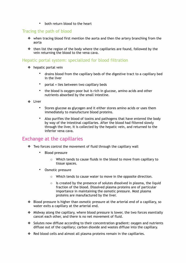
Hepatic portal system: specialized for blood filtration
❖ hepatic portal vein
• drains blood from the capillary beds of the digestive tract to a capillary bed in the liver
• portal = lies between two capillary beds
• the blood is oxygen-poor but is rich in glucose, amino acids and other nutrients absorbed by the small intestine.
❖ Liver
• Stores glucose as glycogen and it either stores amino acids or uses them immediately to manufacture blood proteins.
• Also purifies the blood of toxins and pathogens that have entered the body by way of the intestinal capillaries. After the blood had filtered slowly through the liver, it is collected by the hepatic vein, and returned to the inferior vena cava.
Exchange at the capillaries ❖ Two forces control the movement of fluid through the capillary wall
• Blood pressure
o Which tends to cause fluids in the blood to move from capillary to tissue spaces.
• Osmotic pressure
o Which tends to cause water to move in the opposite direction.
o Is created by the presence of solutes dissolved in plasma, the liquid fraction of the blood. Dissolved plasma proteins are of particular importance in maintaining the osmotic pressure. Most plasma proteins are manufactured by the liver.
❖ Blood pressure is higher than osmotic pressure at the arterial end of a capillary, so water exits a capillary at the arterial end.
❖ Midway along the capillary, where blood pressure is lower, the two forces esentially cancel each other, and there is no net movement of fluid.
❖ Solutes now diffuse according to their concentration gradient: oxygen and nutrients diffuse out of the capillary; carbon dioxide and wastes diffuse into the capillary.
❖ Red blood cells and almost all plasma proteins remain in the capillaries.

❖ The substances that leave a capillary contribute to interstitial fluid, the fluid between the body’s cells.
❖ Plasma proteins are too large to readily pass out of the capillary. Thus interstitial fluid tends to contain all components of plasma, except much lower amounts of protein.
❖ Osmotic pressure is greater than blood pressure, and fluid tends to move back.
❖ Lymphatic capillary beds lie alongside capillary beds.
❖ When lymphatic capillaries take up excess fluid it becomes lymph.
❖ Lymph returns to the cardiovascular veins (subclavian vein) in the chest.
❖ Precapillary sphincters can shut down a blood capillary, and blood then flows through the shunt.

Cardiovascular disorders ❖ Cardiovascular disease (CVD) is the most common cause of death in the Western
world.
• 31% deaths are due to CVD (heart attacks, strokes)
Disorders of the blood vessels
❖ Hypertension/high blood pressure
❖ Atherosclerosis
❖ Stroke
❖ Heart attack
❖ Aneurysm
High blood pressure
❖ Blood moves through vessels at a higher pressure than normal.
❖ Causes?
• Many risk factors! Age, life style, genetics
• Often narrowing of vessel diameter
❖ 140/90 mmHg is considered hypertension.
❖ It is a silent killer because there are few symptoms.
❖ Consequences
• Increase work load for heart and arteries
• Damage to arterial endothelium (less flexible)
• Plaque formation (narrowing), thrombosis
• Thickening of heart muscles (less flexible, less effective)
❖ Hypertension is often seen in individuals who have atherosclerosis
• Is caused by the formation of lesions, or atherosclerotic plaques, on the inside of blood vessels.
• The plaques narrow blood vessel diameter, chocking off blood and oxygen supply to the tissues.
• Plaque that is stationary is called a thrombus, and an embolus when it detaches and can move to distant sites.
• To prevent the development a diet low in saturated fat and cholesterol but rich in omega-3 polyunsaturated fatty acids.
• It is associated with a stroke, heart attack, and aneurysm.
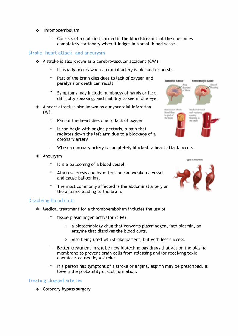
❖ Thromboembolism
• Consists of a clot first carried in the bloodstream that then becomes completely stationary when it lodges in a small blood vessel.
Stroke, heart attack, and aneurysm
❖ A stroke is also known as a cerebrovascular accident (CVA).
• It usually occurs when a cranial artery is blocked or bursts.
• Part of the brain dies dues to lack of oxygen and paralysis or death can result
• Symptoms may include numbness of hands or face, difficulty speaking, and inability to see in one eye.
❖ A heart attack is also known as a myocardial infarction (MI).
• Part of the heart dies due to lack of oxygen.
• It can begin with angina pectoris, a pain that radiates down the left arm due to a blockage of a coronary artery.
• When a coronary artery is completely blocked, a heart attack occurs
❖ Aneurysm
• It is a ballooning of a blood vessel.
• Atherosclerosis and hypertension can weaken a vessel and cause ballooning.
• The most commonly affected is the abdominal artery or the arteries leading to the brain.
Dissolving blood clots
❖ Medical treatment for a thromboembolism includes the use of
• tissue plasminogen activator (t-PA)
o a biotechnology drug that converts plasminogen, into plasmin, an enzyme that dissolves the blood clots.
o Also being used wth stroke patient, but with less success.
• Better treatment might be new biotechnology drugs that act on the plasma membrane to prevent brain cells from releasing and/or receiving toxic chemicals caused by a stroke.
• If a person has symptons of a stroke or angina, aspirin may be prescribed. It lowers the probability of clot formation.
Treating clogged arteries
❖ Coronary bypass surgery

• A surgeon takes a blood vessel (usually a vein from the leg) and stitches one end to the aorta and the other end to a coronary artery located past the point of obstruction.
❖ Gene therapy (alternative for bypass)
• Has been used instead of a coronary bypass to grow new blood vessels that will carry blood to cardiac muscle.
• The surgeon needs only to make a small incision and inject many copies of the gene that codes for vascular endothelial growth factor (VEGF) between the ribs directly into the area of the heart that most needs improved blood flow.
• VEGF encourages new blood vessels to sprout out of an artery.
• If collateral blood vessels do form, they transport blood past clogged arteries, making bypass surgery unnecessary.
• Works fora bout 60% of the patients.
❖ Stent (alternative for bypass)
• A small metal mesh cylinder that holds a coronary artery open after a blockage has been cleared.
❖ Balloon angioplasty
• a stent with an inner balloon is pushed into the blocked area.
• Whe the balloon inside is inflated, it expands, locking itself in place.
• The stent is more successful when it is coated with a drug that seeps into the artery lining and discourages cell growth.
Heart failure
❖ Heart failure
• The hart no longer pumps as it should
• Growing problem because of people who survived heart attacks are left with damaged hearts
• Heart is often oversized because it is sagging and swollen
• Solutions: Medication, surgery (LVAD), heart transplant
❖ Arrhythmia
• Failing heart can have problems such as abnormal heart rythem
• Solutions: Medication, pacemaker, cardioverter-defribrillator (ICD)
Heart transplants
❖ About 2300 heart transplants are done annually, though many thousands of people could use them.

❖ Left ventricular assist device (LVAD), implanted in the abdomen, is an alternative to a heart transplant.
• A tube passes blood from the left ventricle to the device, which pumps it on to the aorta. A cable passes from the device through the skin to an external battery, which the patient must tote around.
❖ Another option is Jarvik 2000
• A pump inverted inside the left ventricle.
❖ Total artificial heart (TAH)
• An internal battery and a controller regulate the pumping speed, and an external battery powers the device by passing electricity through the skin via externa land internal coils. A rotating centrifugal pump moves silicon hydraulic fluid between lef tand right sacs to force bood out of the heart into the pulmonary runk and the aorta.
Chapter 6: cardiovascular system: blood
Blood: an overview
Functions of blood
❖ Transportation
• oxygen, nutrients, wastes, carbon dioxide, and hormones
❖ Defense
• against invasion by pathogens and blood loss
• certain blood cells are capable of engulfing and destroying pathogens by a process called phagocytosis.
• Other white blood cells secrete antibodies into the blood. Antibody= a protein that combines with and disables specific pathogens.
• When an injury occurs, blood clots and defends against blood loss.
❖ Regulatory functions
• Regulating the body’s homeostasis.
• body temperature
• water-salt balance
o the liquid portion of blood, the plasma, contains dissolved salt and proteins. These solutes create blood’s osmotic pressure, which keeps the liquid content of blood high.
• body pH (constant pH of 7.4)
Composition of blood
❖ blood is a tissue and it contains cells and cell fragments.
❖ Collectively the cells and cell fragments are called the formed elements

❖ The cel land cell fragments are suspended in a liquid called plasma.
❖ Blood is classified as a liquid connective tissue
The formed elements
❖ are produced in red bone marrow. Red bone marrow contains pluripotent stem cells, the parent cells that divide and give rise to all the types of blood cells.
• Red blood cells/erythrocytes (RBCs)
o Two to three times smaller than white blod cells, but there are many more of them.
• White blood cells/leukocytes (WBCs)
• Platelets/thrombocytes
Plasma
❖ Liquid medium for carrying various substances in the blood.
❖ Also distributes the heat generated as a by-product of metabolism, particulary muscle contraction.
❖ It consists of 91% water and 9% salts (ions) and organic molecules.
• These salts are dissolved in plasma and, along with plama proteins, help maintain the osmotic pressure of blood.
• Salts also function as buffers that help maintain blood pH.
❖ Plasma proteins are the most abundant organic molecules.
• The liver produces the majority of the plasma proteins
• Have many functions that help maintain homeostasis
• Are able to take up and release hydrogen ions. They help keep blood pH around 7.4
• Are too large to pass through the capillary walls, thus, they remain in the blood, establishing an osmotic gradient between blood and intersitial fluid.
• Osmotic pressure = a force that prevents excessive loss of plasma from the capillaries into the interstitial fluid.
❖ Three major types of plama proteins
• Albumins
o most abundant and important for plasma’s osmotic pressure as well as transport
• Globulins –
o also important in transport
▪ α (alpha)-, β (beta)-, γ (gamma)- globuline
▪ gamma globuling (antibodies) areproduces by white blood cells called lymphocytes, not by the liver.

▪ Gamma globulins are important in fighting disease-causing pathogens.
• Fibrinogen
o important for the formation of blood clots
Red blood cells and transport of oxygen
❖ red blood cells, also known as erythrocytes, are small, biconcave disks.
❖ The presence of hemoglobin and their unique internal structure, makes them a unique cell type.
❖ Are also very abundant
How red blood cells carry oxygen
❖ Red blood cells are highly specialized for oxygen transport.
❖ Contain hemoglobin (Hb) = a pigment with a high affinity (attraction) for oxygen.
• Also responsible for the red coloration of blood.
• The globin portion is a protein that contains four highly folded polypeptide chains.
• The heme part of hemoglobin is an iron-containing group in the center of each polypeptide chain. The iron combines reversibly with oxygen. This means that heme accepts oxygen in the lungs and then lets go of it in the tissue. CO combines with the iron of heme and then will not easily let go.
❖ They have no nucleus (lose their nucleus during maturation)
❖ The biconcave shape fo RBCs gives them a greater surface area for the diffusion of gases into and out of the cell. All the internal space is used for the transport of oxygen.
❖ RBCs also lack most organelles, incuding mitochondria

❖ Anaerobically produce ATP, and they do not consume any of the oxygen they transport.
❖ When oxygen binds to heme in the lungs, hemoglobin assumes a slightly different shape and is called oxyhemoglobin. In the tissue, heme gives up this oxygen, and hemoglobin resumes its former shape, called deoxyhemoglobin. The released oxygen diffuses out of the blood into the interstitial fluid and then into cells.
How red blood cells help transport carbon dioxide
❖ After blood picks up carbon dioxide in the tissues, about 7% is dissolved in plasma.(if higher than 7% it would bubble)
❖ Hemoglobin directly transports about 23% of carbon dioxide., combining it with the globin protein.
❖ Hemoglobin- carrying co2 is termed carbaminohemoglobin. The remaining co2 is transported as the bicarbonate ion (HCO3 - )in the plasma.
❖ Carbon dioxide moves into RBCs, combinig with cellular water to form carbonic acid.
❖ An enzyme inside RBCs, called carbonic anhydrase, speeds the reaction.
❖ Carbonic acid quickly seperates, or dissociates, to form hyrdrogen ions (H+) and bicarbonate ions (HCO3-). The bicorbonate ions diffuse out of the RBCs tob e carried in the plasma. The h+ from this equation binds to globin, the protein portion of hemoglobin.
❖ Hemoglobin assists plasma proteins and salts in keeping the blood pH constant.
❖ When blood reaches the lungs,the reaction is reversed.
❖ Hydrogen ions and bicrbonate ions reunite to re-form carbonic acid.
❖ The carbonic anhydrase enzyme also speeds this reverse reaction.
❖ Carbon dioxide diffuses out of the blood and into the airways of the lungs, tob e exhaled from the body.
Red blood cells are produced in bone marrow
❖ RBC cells in the bone marrow divide and produce new cells, which differentiate into mature RBCs
❖ They acquire hemoglobin and lose their nucleus and other internal organelles. The lack of internal organelles means that RBCs are incapable of conducting many of the fuctions of other eukaryotic cells in the body.

❖ The cytoskeleton of RBCs contains proteins that provide their unique shape, thus allowing them to move through narrow capillary beds.
❖ Also duet heir lack of a nucleus, RBCs are unable to replenish important proteins and repair cellular damage.
❖ They have a lifespan of about 120 days.
❖ About 2 million RBCs are destroyed per second, and therefore an equal number must be produced to keep the red blood cell count in balance.
❖ When red blood cells are broken down, hemoglobin is released.
❖ The globin portion of hemoglobin is broken down into its component amino acids, which are recycled by the body.
❖ The majority of the iron is recovered and returned to the bone marrow for reuse, although a small amount is lost and must be replaced in the diet.
❖ The rest of the heme portion of the molecule undergoes chemical degradation and is excreted by the liver and kidneys.
❖ The body has a way to boost the number of RBCs when insuffiecient oxygen is being deliverd to the cells:
• The kidneys release a hormone called Erythropoietin (EPO), which stimulates the stem cells in bone marrow to produce more red blood cells.
• The liver and other tissues also produce EPO for the same purpose.
❖ What is blood doping
• It is any method of increasing the number of RBCs to increase athletic performance.
• It allows more efficient delivery of oxygen and reduces fatigue.
• EPO is injected into a person months prior to an athletic event.
• Overuse can lead to fatal blood thickening
Disorders involving red blood cells
❖ If the liver fails to excrete heme, it accumulates in tissues, causing a condition called jaundice
• In jaundice the skin and whites of the eyes turn yellow.
❖ Anemia
• low number of RBC and/or hemoglobin
• suffers from tired, run-down feeling
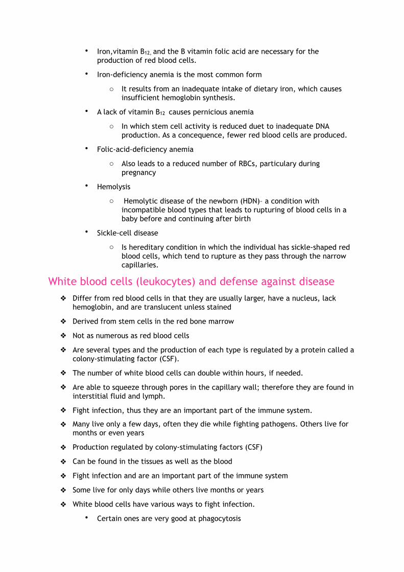
• Iron,vitamin B12, and the B vitamin folic acid are necessary for the production of red blood cells.
• Iron-deficiency anemia is the most common form
o It results from an inadequate intake of dietary iron, which causes insufficient hemoglobin synthesis.
• A lack of vitamin B12 causes pernicious anemia
o In which stem cell activity is reduced duet to inadequate DNA production. As a concequence, fewer red blood cells are produced.
• Folic-acid-deficiency anemia
o Also leads to a reduced number of RBCs, particulary during pregnancy
• Hemolysis
o Hemolytic disease of the newborn (HDN)– a condition with incompatible blood types that leads to rupturing of blood cells in a baby before and continuing after birth
• Sickle-cell disease
o Is hereditary condition in which the individual has sickle-shaped red blood cells, which tend to rupture as they pass through the narrow capillaries.
White blood cells (leukocytes) and defense against disease ❖ Differ from red blood cells in that they are usually larger, have a nucleus, lack
hemoglobin, and are translucent unless stained
❖ Derived from stem cells in the red bone marrow
❖ Not as numerous as red blood cells
❖ Are several types and the production of each type is regulated by a protein called a colony-stimulating factor (CSF).
❖ The number of white blood cells can double within hours, if needed.
❖ Are able to squeeze through pores in the capillary wall; therefore they are found in interstitial fluid and lymph.
❖ Fight infection, thus they are an important part of the immune system.
❖ Many live only a few days, often they die while fighting pathogens. Others live for months or even years
❖ Production regulated by colony-stimulating factors (CSF)
❖ Can be found in the tissues as well as the blood
❖ Fight infection and are an important part of the immune system
❖ Some live for only days while others live months or years
❖ White blood cells have various ways to fight infection.
• Certain ones are very good at phagocytosis

o During phagocytosis, a projection from the cell surounds a pathogen and engulfs it.
o A vesicle containing the pathogen is formed inside the cell.
o Lysosomes attach and empty their digestive enzymes into the vesicle.
o The enzymes break down the pathogen.
• Other with blood cells produce antibodies, proteins that combine with antigens
o An antigen is a cell or other substance foreign to the individual that invokes an immune reponse.
o The antibody-antigen pair is then marked for destruction, again by phagocytosis.
Types of white blood cells
Granular Leukocytes
❖ Have noticeable cytoplasmic granules. Granules, like lysosomes, contain various enzymes and proteins.
❖ Include:
• Neutrophils
o Account for 50-70 % of all white blood cells.
o Are the most abundant
o Have a multilobed nucleus, so they are called polymorphonuclear leukocytes or “polys”
o Granules of neutrophils are not easily stained with either acidic red dye or basic purple dye. (this accounts fort heir name)
o Are usually first responders to bacterial infection, and their intense phagocytic activity is essential to overcoming an invasion by a pathogen.
• Eosinophils
o have a bilobed nucleus
o their large, abundant granules take up eosin and become a red color. (accounts for the name)
o are involved in the protection of the body against large parasites and the phagocytosis of the allergens and proteins associated with the inflammatory response.
• Basophils
o Are the rarest, but they play an important role in the immune response.
o Have a U-shaped or lobed nucleus.

o Their granules take up the basic stain and become a dark-blue color. (accounts for their name)
o In the connective tissues, basophils and similar cells called mast cells release histamine associated with allergic reactions.
o Histamine dilates blood vessels but constricts the air tubes that lead to the lungs, which is what happens when someone has difficulty breathing during an asthma attack.
Agranular leukocytes
❖ Include the lymphocytes and the monocytes.
• Do not have granules, but do have nonlobular nuclei.
• Are sometimes called the mononuclear leukocytes.
❖ Lymphocytes
• About 25-35% of all WBCs
• Second must abundant type of WBCs
• Are responsible for specific immunity to particular pathogens and toxins
• Large nucleus that takes up most of the cytoplasm
• There are two types
o B cells
▪ Mature B cells, called plasma cells produce antibodies, the proteins that combine with target pathogens and mark them for destruction.
o T cells
▪ Some T cells directly destroy pathogens.
▪ The aids virus attacks one of several types of T cells
❖ Monocytes
• Relatively uncommon WBCs
• Largest WBC, with horseshoe-shaped nucleus
• Take residence in tissues and develop into macrophages
• In the skin, they become dendritic cells
• Macrophages and dendritic cells are active phagocytes, destroying pathogens, old cells, and cellular debris. They also stimulate other white blood cells, including mymphocytes, to defend the body.
• Macrophages use phagocytosis to engulf pathogens
Disorders involving white blood cells
❖ Some immune deficiencies can be inherited.
❖ Severe combined immunodeficiency (SCID)
• When the stem cells of white blood cells lack an enzyme called adenosine deaminase. Without this enzyme, B and T lymphocytes do not develop and the body cannot fight infections.
• Injections of the missing enzyme can be given twice weekly, but a bone marrow transplant from a compatible donor is the best way to cure the disease.
❖ Leukemia
• Cancer is duet o uncontrolled cell growth
• Means “white blood”, refers to a group of cancers that affect white blood cells in which these cells proliferate without control.
• Most of these with blood cells are abnormal or immature. Therefore, they are incapable of performing their normal defense functions.
• Each type of leukemia is named for the type of cell dividing out of control.
o Chemotherapy
o Radiation
o Bone marrow transplant (stem cell transplantation)
❖ Infectious mononucleosis
• also known as the “kissing disease” and occurs when the Epstein-Barr virus (EBV) infects lymphocytes resulting in fatigue, sore throat, and swollen lymph nodes
Platelets (thrombocytes) and blood clotting ❖ They result from fragmentation of large cells, called megakaryocytes, in the red
bone marrow.
❖ About 200 billion platelets are made per day.
❖ They function in blood clotting or coagulation.
❖ Also involved are the plasma proteins prothrombin and fibrinogen, manufactured in the liver and deposited in the blood. Vitamin K is necessary to the production of prothrombin.

❖ Hemostasis: inhibition of blood loss
Blood clotting
❖ Helps the body maintain homeostasis in the cardiovascular system by ensuring that the plasma and formed elements remain within the blood vessels.
❖ At least 12 clotting factors and calcium ions (CA2+) participate in the formation of a blood clot.
❖ When a blood vessel in the body is damaged, platelets clump at the site of the puncture and partially seal the leak.
❖ Platelets and injured tissues release a clotting factor called prothrombin activator, which converts prothrombin in the plasma to thrombin. This reaction requires calcium ions (CA2+).
❖ Thrombin, in turn, acts as an enzyme that severs two short amino acid chains from a fibrinogen molecule, one of the proteins in plasma.
❖ These activated fragments then join end to end, forming long threads of fibrin.
❖ Fibrin threads wind around the platelet plug in the damaged area of the blood vessel and provide the framework for the clot.
❖ Red blood cells trapped within the fibrin threads make the clot appear red.
❖ A fibrin clot is temporary. Once blood vessel repair starts, an enzyme called plasmin destroys the fibrin network, so that tissue cells can grow.
❖ After the blood clots, a yellowish fluid called serum escapes from the clot. If contains all the components of plasma except fibrinogen and prothrombin.
Disorders related to blood clotting
❖ Thrombocytopenia
• An insufficient number of platelets
• Is due to either low platelet production in bone marrow or increased breakdown of platelets outside the marrow.
• A number of conditions, including leukemia, ca lead to thrombocytopenia.
• It can also be rug-induced.
• Symptoms include bruising, rash, and nosebleeds or bleeding in the mouth.

• Gastrointestinal bleeding and bleeding in the brain arepossible complications.
❖ Thrombosis (obstructive clotting)
• Obstruction of blood vessels by a blood clot (thrombus)
• Sitting for long periods can also cause thrombus formation.
❖ Embolism (thromboembolism)
• Lodging of an embolus (blood clot) inside a blood vessel and blocking it
• If left untreated, blood flow to the tissues can stop completely. Heart attack or stroke can result.
❖ Some medications interfere with hemostasis
• Aspirin
• Warfarin (rat poison)
❖ Hemophilia (Bleeding disorder)
• deficiency of one or more clotting proteins
o Hemophilia A
▪ lack Factor VIII
▪ more likely to occur in boys
▪ caused by an abnormal copy of the factor VIII production gene, found on the X chromosome.
▪ Arises when a boy has an abnormal gene on his single X chromosome
▪ Girls need only one normal gene between their two X chromosomes to make the normal amounts of clotting factor VIII.
▪ In hemophilia, the slightest bump can cause bleeding into the joints. Cartilage degeneration in the joints and absorption of underlying bone can follow.
▪ The most frequent cause of death is bleeding into the brain, with accompanying neurological damage.
▪ Today, Factor VIII can be made by genetic engineering to supplement hemophiliacs
o Hemophilia B –lack of factor IX (“Royal” disease)

Blood typing and transfusions ❖ Blood transfusion:
• administration of blood directly into bloodstream of another person
• For transfusions to be done safely,blood must be typed, so that agglutination does not occur when blood from different people is mixed.
• Success depends on matching the blood type of the donor with that of the recipient
• ABO blood types (A, B, AB, and O)
• Rh (Rh-positive and Rh-negative)
ABO blood groups
❖ Only certain type of blood transfusions are safe, because the plasma membranes of red blood cells carry glycoproteins that can be antigenst to other individuals.
❖ Antigen
• a foreign (non-self) substance, often a polysaccharide or a protein, that stimulates an immune response
• ABO blood typing is based on the presence or absence of two possible antigens, called type A antigen and type B antigen. Wether these antigens are present or not depends on the inheritance of the individual.
• Type A blood has the A antigen, type B blood has the B antigen and typeAB blood has both the A and B antigens. Type O blood has neither antigen on its red blood cells.
❖ Antibody
• a protein made in response to an antigen in the body which binds specifically to that antigen
• Type A and B blood have antibodies that correspond to the opposite blood type.
o A blood has B-antibodies
o B blood has A-antibodies
o O blood has both antibodies
o AB blood lack both antibodies
• Anti-A and/or ant-B antibodies are not present at birth but they appear over the course of several months.
• Each antibody has a binding

site that will combine wit hit corresponding antigen in a tight lock-and-key fit.
Blood compatibility
❖ The antibodies in the plasma must not combine with the antigens on the surface of the red blood cells or else agglutination occurs.
• Agglutination is expected if the donor has type A blood and the recipient has type B blood and the other way around.
• Type O blood is called the universal donor, because the red blood cells of type O lack A and B antigens.
• Type AB blood is sometimes called the universal recipient blood, because the plasma lacks A and B antibodies.
• In practice there are other possible blood groups aside from ABO blood groups. Before blood can be safely transfused from one person to another, it is necessary to physically combine donor blood with recipient blood on a glass slide, then observe wether aggluination occurs = blood-type crossmatching
Rh blood groups
❖ The destination of blood type usually also includes wether the person has or does not have the Rh factor on the red blood cell.
❖ Rh-negative individuals normally do not have antibodies to the Rh factor, but they make them when exposed to the Rh factor.
❖ The Rh factors= another surface marker on RBC
❖ Rh factor positive or negative (e.g. O+, A+; AB-)
❖ Exposure to Rh+ blood can trigger antibody production in Rh- people
• hemolytic disease of the newborn (HDN) o In subsequent pregnancy with another Rh-positive baby, the anti-Rh
antibodies can cross the placenta and destroy the unborn child’s red blood cells.
o Is called HDN because hemolysis starts in the womb and continues after the baby is born.
o Due to red blood cell derstruction the baby wil be severly anemic. o Excess hemoglobin breakdown products in the blood can lead to
brain damage and intellectual disability, or even death o How prevented?
▪ The Rh problem is prevented by giving Rh-negative women an Rh immonuglobin injection no later than 72 hours after giving birth to an Rh-positive child.

▪ This injection contains ant-Rh antibodies tahta attack any of the baby’s red blood cells in the mother’s blood before these cells can stimulate her immune system to produce her own antibodies.
▪ This treatment is called RhoGAM. It does not harm the newborn’s red blood cells; however, the treatment is not beneficial if the women has already begun to produce antibodies.
▪ This will have to be repeated if an Rh- mother has another Rh+ baby in case she has later pregnancies.
Homeostasis ❖ Organ systems of the body interact with the cardiovascular system to maintain
homeostasis.
❖ Body’s internal environment contains not only blood but also interstitial fluid.
❖ Interstitial fluid orginates from blood plasma but contains very little plasma protein. It is absorbed by lumphatic capillaries, after which it is referred to as lymph.
❖ The lymph courses through the lymphatic vessels, eventually returning to the venous system.
• Thus the cardiovascular and lymphatic systems are intimately linked.
❖ Homeostasis is possible only if the cardiovascular system delivers oxygen from the lungs, as wel as nutrients from the digestve system, to the interstitial fluid surrounding cells. The cardiovascular system also removes metabolic wastes, delivering waste to excretory organs.
❖ The three components of the muscular system make essential contributions to blood movement.
• Cardiac muscle contractions circulate blood throughout the body. Contraction or relaxtion of the smooth muscle in blood vessel walls changes vessel diameter and helps maintain the correct blood pressure.
• Skeletal muscle contraction compresses both the cardiovascular and lymphatic veins. Lymph returns to cardiovascular veins, and blood in the cardiovascular drains back to the heart. The circulation of interstitial fluid and blood is then complete.
❖ The skeletal and endocrine systems are vital to cardiovascular homeostasis.
• Red bone marrow produces blood cells. Without the needed blood cells, the person becomes anemic and lacks an immune response.
• Bones contribute calcium ions ( CA2+) to the process of blood clotting.
• Both blood cell production and bone calcium release are regulated by hormones.
❖ Urinary system
• Besides producing and excreting urine, the kidneys help regulate the acid-base and water-salt balances of the blood and interstititial fluid.

• Erythropoietin, a hormone produced by the kidneys, stimulates red blood cell production.
• The urinary system joins the muscular, skeletal and endocrine systems to maintain the internal environment.
Chapter 7: The lymphatic and immune systems
The lymphatic system ❖ Consists of lymphatic vessels and the lymphatic organs.
❖ Four main functions that contribute to homeostasis
• Lymphatic capillaries absorb excess tissue fluid and return it to the bloodstream.
• Lymphatic capillaries (lacteals) in the small intestine absorb fats associated with proteins.
• The lymphatic system works in the production, maintenance, and distribution of lymphocytes in the body.
• t helps in defense against pathogens.
Lymphatic vessels
❖ One-way valve system that carries fluid called lymph
❖ Made of capillaries, vessels, and ducts
❖ Function to return tissue fluid (which includes water, solutes, and cell

products) to the bloodstream
❖ Transports lipids from digestive system to circulation
❖ Pickup bacteria for destruction in lymph nodes
❖ Larger vessels are similar in structure to veins and even have valves
Lymphatic organs
❖ Primary lymphatic organs
• Red bone marrow
• Thymus
❖ Secondary lymphatic organs
• Lymph nodes
• Spleen
The primary lymphatic organs
❖ Red bone marrow
• blood cell production.
• In a child,most bones have red bone marrow. In adults, it is selected (sternum, vertebrae,ribs, part of the pelvic girdle and the upper ends of the humerus and femur)
• Produces various types of white blood cells (neutrophils, eosinophils, basophils, lymphocytes and monocytes)
• Lymphocytes are either B cells or T cells. B cells mature in the bone marrow, but T cells mature in the thymus.
• Any B cell that reacts with cells of the body is removed in the bone marrow and does not enter the circulation. This ensures that the B cells do not harm normal cells of the body.
❖ Thymus
• Located in the thoracic cavity between the trachea and the sternum, superior to the heart.
• bi-lobed gland
• It is largest in children and shrinks as we age.
• Has two functions
o It produces thymic hormones, such as thymosin, thought to aid in the maturation of T lymphocytes. Thymosin may also have other funcions in immunity.

o
• Immature T lymphocytes move from the bone marrow through the bloodstream to the thymus where they mature and 95% will stay.
Secondary lymphatic organs
❖ Lymph nodes
• Small, bean-shaped structures found along the lymphatic vessels
• Filter lymph
• Connective tissue forms a capsule and divides a lymph node into compartements. Each compartment contains a sinus that increases in size toward the center of the node.
• Filled with B cells, T cells, and macrophages (which engulf pathogens and debris)
• Lymph nodes are named for their location.
• Lymphatic nodules: are contractions of lymphoid tissue not surrounded by a capsule.
• Tonsils: are patches of lymphoid tissue located in a ring about the pharynx. Perform the same functions as lymph nodes. They are the first to encounter pathogens and antigens that enter the body by way of the nose and mouth.
• Peyer patches: are located in the intestinal wall and tissues within the appendix, a small extension of the large intestine, and encounter pathogens that enter the body by way of the intestinal tract.
• Common in the neck, armpit, and groin regions
❖ Spleen
• Located in the upper left region of the abdominal cavity
• Filters blood
• Connective tissue divides the spleen into regions known as
o White pulp
▪o Red pulp
▪ Surrounds venous sinuses (cavities)
▪ Is involved in filtering the blood
▪ Althought the spleen’s functions are replaced by other organs, a person without a spleen is often slightly more susceptible to infections and may have to receive antibiotic therapy indefinitely.

Innate immune defenses ❖ We are constantly exposed to microbes
such as viruses, bacteria and fungi
❖ Immunity is the capability of killing or removing foreign substances, pathogens and cancer cells from the body.
❖ Adaptive immunity
• Is initiated and amplified by exposure
❖ Innate immunity
• Innate immune defenses (fully functional without previous exposure to these invaders) include physical and chemical barriers; the inflammatory response and protectiveproteins such as complement and interferons.
• Innate defenses occur immediately or very short after infection occurs.
• With innate immunity, there is no rcognition that an intruder has attacked before, ad therefore no immunological “memory” of the attacker is present.
Physical and chemical barriers to entry
❖ Built-in barriers that serve as the first line of defense against an infection by pathogens.
❖ Physical barriers
• The skin is an effective physical barrier.
• Tears, saliva, and urine physically flush out microbes.
• Mucous membranes line the respiratory, digestive, reproductive, and urinary tracts.
• Resident bacteria/normal flora that inhabit the body use available nutrients and space thus preventing pathogens from taking up residence.
❖ Chemical barriers
• Secretions of sebaceous (oil) glands of the skin. These secretions contain chemicals that weaken or kill certain bacteria on the skin.
• lysosome
o Perspiration, saliva and tears contain an antibacterial enzyme called lysozyme. Saliva also helps wash microbes off the teeth and tongue, and tears wash the eyes. Similary, as urine is voided from the body, it flushes bacteria from the urinary tract.
• The acid pH of the stomach and vagina
o inhibits growth or kills many types of bacteria. Now we know that ulcers are caused by the bacterium Helicobacter pylori. Similary, the acidity of the vagina and its thick walls discourage the presence of pathogens.
Inflammatory response
❖ second line of defense against invasion by a pathogen.
❖ Inflammation employs mainly neutrophils and macrophages to surround and kill pathogens trying to get a foothold inside the body. Protective proteins are also involved.
❖ Five hallmark symptoms are redness, heat, swelling, pain, and loss of function.
• Are due to capillary changes in the damaged area, and all protect the body.
• Chemical mediators, such as histamine, released by damaged tissue cells and mast cells, cause the capillaries to dilate and become more permeable.
• Excess blood flow due to enlarged capillaries causes the skin to redden and become warm.
• Increased temperature in an inflamed area tends to inhibit growth of some pathogens. Increased blood flow bring white blood cells to the area.
• Increased permeability of capillaries allows fluids and proteins, including blood-clotting factors to escape into the tissues. Clot formation in the injured area prevents blood loss.
• The excess fluid in the area presses on nerve endings, causing the pain associated with swelling.
• Together these events summon white blood cells to the area.
• As soon as the white blood cells arive, they move out of the bloodstream into the surrounding tissue.
• The neutrophils are first and actively phagocytize debris, dead cells, and bacteria they encounter.
• The many neutrophils attracted to the area can usually localize any infection and keep it from spreading.
• If neutrophils die off in great quantities, they become a yellow-white substance called pus.
• Injury not serious:healing process short-lived. Nearby cells secrete chemical factors to ensure the growth (and repair) of blood vessels and new cells to fill in the damaged area.

• If neutrophils are overwhelmed, they call for reinforcements by secreting chemical mediators called cytokines (attract more white blood cells, including monocytes to the area.
• Monocytes become macrophages, even more powerful phagocytes than neutrophils. They can enlist the help of lymphocytes to carry out specific defense mechanisms.
• Inflammatory chemicals may cause collateral damage to the body, in addition to killing the invaders.
• Should an inflammation persist, anti-inflammatory medications, such as aspirin, ibuprofen or cortisone, can minimize the effects of various chemical mediators.
Protective proteins
❖ Complement (system)
• Composed of a number of blood plasma proteins designed by the letter C and a number.
• These proteins “complement” certain immune responses
• Involved in the inflammatory response by binding to mast cells, causing them to release histamine
• Attract phagocytes to pathogens by binding them
• Form a membrane attack complex that makes holes in some bacteria and viruses, causing them to burst
❖ Interferons: communication molecules
• Proteins produced by virus-infected cells sent out to warn neighboring healthy cells
o Infected cells: apoptosis
o Uninfected cells: destroy viral RNA and reduce protein synthesis
o Activate immune cells

• Bind to receptors of non-infected cells, causing the mto prepare for possible attack by producing substances that interfere with viral replication.
• Are used to treat viral infections, such as hepatitis C.
Adaptive immune defenses ❖ Third line of defense
❖ When innate defenses have failed to prevent an infection, adaptive defenses come into play.
❖ Helps protect us against specific pathogens when nonspecific defenses fail
❖ Helps protect us against cancer
❖ Depends on the action of B and T cells (lymphocytes)
How adaptive defenses work
❖ Adaptive defenses respond to large molecules, normally protein structures, called antigens that the immune system recognizes as foreign to the body.
❖ Adaptive defenses primarily depend on the action of lymphocytes, which differentiate as either B cells or T cells.
• B cells and T cells are capable of recognizing antigens because they have specific antigen receptors.
• These antigen receptors are plasma membrane proteins whose shape allows the mto combine with particular antigens.
• Each lymphocyte has only one type of receptor. Millions of specific B cells and/or T cells are formed, increasing the likelihood that at least one will recognize any possible antigen.
• B cells differentiate into other types of cells, and so do T cells
• The two major pathways of adaptive immunity:
o APC activates T cell (@ TCR) => clonal expansion
▪ Helper T cells secrete cytokines that control the immune response
➢ Activate B cell clonal expansion
o Cytotoxic T cells destroy nonself antigen-bearing cells
B cells and atibody-mediated immunity
❖ The receptor on a B cell is called a B-cell receptor (BCR).
❖ The clonal selection model states that an antigen selects, then binds to the BCR of only one type of B cell. Then this B cell produces multiple copies of itself. The

resulting group of identical cells is called a clone.Similary, an antigen can bind to a T-cell receptor (TCR), and this T cell will clone.
❖ Characteristics of B cells
• Antibody-mediated immunity against pathogens
• Produced and mature in bone marrow
• Directly recognize antigen (@ BCR) and then undergo clonal selection
• Cytokines stimulate B cells to clone (copy/multiply)
• Clonal expansion produces antibody-secreting plasma cells as well as memory B cells
• Plasma cells undergo apoptosis after infection
❖ B cells become plasma cells and memory B cells
• Each B cell has a specific BCR represented by shape.
• Only the B cell with a BCR that has a shape that fits the antigen undergoes clonal expension. During clonal expension, cytokines secreted by helper T (TH) cells stimulate B cells to clone.
• Most of the cloned B cells become plasma cells, which circulate in the blood and lymph.
o Plasma cells are larger than regular B cells, because they have extensive rough endoplasmic reticulum. This is for the mass production and secretion of antibodies to a specific antigen.
o Antibodies identical to the BCR of the activated B cell are secreted from the plasma B cell.
• Some cloned B cells, become memory cells, the means by which long-term immunity is possible.
• If the same antigen enters the system again, memory B cells quickly divide and transform into plasma cells.
• The correct type of antibody can quickly be produced by these plasma cells.
• Apoptosis
o Once the threat of an infection has passed, the development of new plasma cells ceases, and those present undergo apoptosis = process of programmed cell death. It involves a cascade of specific cellular events leading to the death and the removal of the cell remnants from the body as a waste product.
• Antibody-mediated immunity
o = defense by B cells or humoral immunity
o Activated B cells become plasma cells that produce antibodies.
o Colectively, plasma cells probably produce as many as 2 million different antibodies.
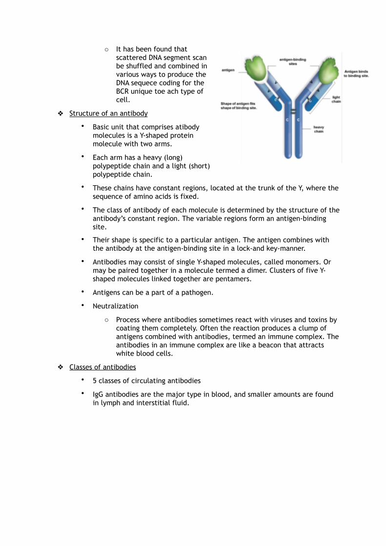
o It has been found that scattered DNA segment scan be shuffled and combined in various ways to produce the DNA sequece coding for the BCR unique toe ach type of cell.
❖ Structure of an antibody
• Basic unit that comprises atibody molecules is a Y-shaped protein molecule with two arms.
• Each arm has a heavy (long) polypeptide chain and a light (short) polypeptide chain.
• These chains have constant regions, located at the trunk of the Y, where the sequence of amino acids is fixed.
• The class of antibody of each molecule is determined by the structure of the antibody’s constant region. The variable regions form an antigen-binding site.
• Their shape is specific to a particular antigen. The antigen combines with the antibody at the antigen-binding site in a lock-and key-manner.
• Antibodies may consist of single Y-shaped molecules, called monomers. Or may be paired together in a molecule termed a dimer. Clusters of five Y-shaped molecules linked together are pentamers.
• Antigens can be a part of a pathogen.
• Neutralization
o Process where antibodies sometimes react with viruses and toxins by coating them completely. Often the reaction produces a clump of antigens combined with antibodies, termed an immune complex. The antibodies in an immune complex are like a beacon that attracts white blood cells.
❖ Classes of antibodies
• 5 classes of circulating antibodies
• IgG antibodies are the major type in blood, and smaller amounts are found in lymph and interstitial fluid.

❖Monoclonal antibodies
•Every plasma cellderived from the same B cell secretes antibodies against a specific region.
•Are monoclonal antibodies because all of them are the same type and because they are produces by plasma cells derived from the same B
cell.
• Method of producing: in vitro (outside the body)
o B lymphocytes are removed from an animal and are exposed to a particular antigen.
o The resulting plasma cells are fused with myeloma cells (immortal cells)
o The fused cells are called hybrid, because they result form the fusion of two different cells and –oma because one of the cells is a cancer cell.
• At present, monoclonal antibodies are being used for quick and certain diagnosis of various conditions.
T cells and cell-mediated immunity
❖ Cell-mediated immunity is named for the action of T cells that directly attack diseased cells and cancer cells
❖ Other T cells release cytokines that stimulate both nonspecific and specific defenses
❖ How T cells recognize an antigen
• When a T cell leaves the thymus, it has a unique T-cell receptor (TCR).
• T cells are unable to recognize an antigen withpout help. The antigen must be displayes tot hem by an antigen-presenting cell (APC), such as macrophage.
• After phagocytizing a pathogen, APCs travel to a lymph node or the spleen, where T cells also congregate.
• In the meantime, the APC has broken the pathogen apart in a lysosome.

• A piece of the pathogen is then displayed in the groove of a major histocompatibility complex (MHC) protein on the cells surface.
o MHC I
o MHC II
▪ Human MHC II proteins are called human leukocyte antigens (HLAs).
▪ Are found on all of our body cells.
▪ Three general groups of HLAs
➢ HLA-A
➢ HLA-B
➢ HLA-DR
➔ Each with a number of protein variations
▪ Each person has a unique combination of HLAs.
▪ One exception are the HLAs of identical twins.
o MHC antigens are self proteins, because they mark the cell as belonging to a particular individual.
o Comparison od the three classes of HLAs must always be carried out before a transplant attempted. The greater the number of proteins that match, the more likely the transplant will be succesfull.
• When an antigen-presenting cell links a foreign antigen to the self protein on its plasma membrane, it carries out an important safeguard for the rest of the body.The T cell tob e activated can compare the antigen and self protein side by side.
• The activated T cel land all thedaughter cells, which it will form, can rcognize foreign from self. These T cells go on to destroy cells carrying foreign antigens, while leaving normal body cells unharmed.
❖ Clonal expansion
• The T cells have specific TCRs, represented by their different shape.
• A macrophage is presenting an antigen to a T cell that has the specific TCR that will combine with this particular antigen (represented by a green circle)
• The T cell is activated and undergoes clonal expansion. Many copies of the activated T cell are produced during clonal expansion.
• A subgroup of T cells recognizes APCs that display an antigen within the groove of an MHC I protein.
o These T cells will activate and become cytotoxic T (TC) cells.
• A subgroup of T cells recognizes APCs that display an antigen within the groove of an MHC II protein.
o These T cells will activate and become helper T (TH) cells.

• As the illness disappears, the immune reaction wanes.
• Activated T cells become susceptible to apoptosis. Apoptosis contributes to homeostasis by regulating the number of cells present in an organ, in this case, the immune system.
• When apoptosis does not occur as it should, the potential exists for an autoimmune response or for T-cell cancers.
• Cytotoxic T (TC) cells
o have vacuoles containing granzymes and perforins.
o After a TC cell binds to a virus-infected cell or tumor it releases perforin molecules,which punch holes in the plasma membrane, forming a pore
o TC cells then deliver granzymes into the pore. These cause the cell to undergo apoptosis.
o Once TC cells have released the perforins and granzymes, they move on to the next target cell.
o CT cells are responsible for cell-mediated immunity.
• Helper T (TH) cells
o Regulate immunity by secreting cytokines, the chemicals that enhance the response of all types of immune cells, B cells cannot be activatedwithout T-cell help.
o The human immunodeficiency virus (HIV), which causes aids, infects helper T cells and other cells of the immune system. The virus thus inactivates the immune response and makes HIV-infected individuals susceptible to the opportunistic infection that eventually kill them.
• Memory T cells
o Few of the clonally expanded Tcells are memory cells
o They remain in the body and can jump-start an immune reaction to an antigen previously present in the body.
Acquired immunity ❖ Immunity is the ability to combat diseases and cancer.
❖ It can be brought about naturally through an infection or artificially through medical intervention.
❖ There are 2 types of immunity
• Active immunity
o The individual alone produces antibodies against an antigen

• Passive immunity
o The individual is given prepared antibodies via an injection.
Active immunity
❖ Antibodies made against a particular antigen.
❖ Natural infection or through immunization involving vaccines.
❖ Primary immune response:
• First encounter with antigen => recognition + clonal expansion of B and T cells; production of memory cells
• Lag time for AB production
❖ Secondary immune response:
• Recognition and clonal expansion
• Fast and increased AB production (memory cells)
❖ This type of immunity is usually long-lasting.
❖ It depends on memory B and T cells.
Passive immunity
❖ An individual is given antibodies against a particular antigen.
❖ This type of immunity is short-lived.
❖ This can happen naturally as antibodies are passed across the placenta or during breastfeeding, or artificially via an injection of antibodies.
Cytokines and immunity
❖ Are signaling molecules produced by T lymphocytes, macrophages and other cells.
❖ Regulate white blood cell formation and/or function, so there being investegated for a possible adjunct therapy for cancer and aids.
❖ Interferon (produced by virus-infected cells) and interleukins ( produced by various white blood cells) have been used for as immunotherapeutic drugs. These are used particulary to enhance the ability of the individial’s T cells to fight cancer.
❖ Most cancer cells carry an altered protein on their cell surface, so they should be attacked and destroyed by cytotoxic T cells.
❖ Whenever cancer develops, it is possible that cytotoxic T cells have not been activated. In that case, cytokines might awaken the immune system and lead to the destruction of the cancer.
❖ In one technique
• Researchers withdrew T cells from the patient and presented cancer cell antigenst o the isolated T cells.
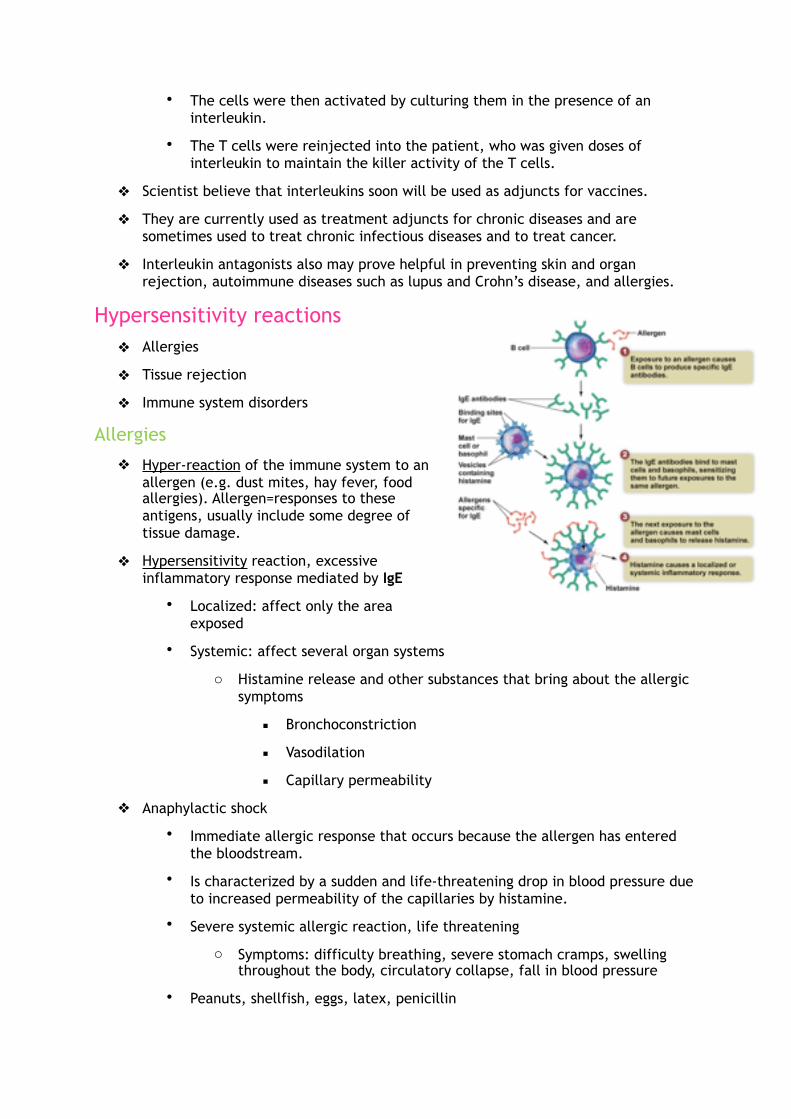
• The cells were then activated by culturing them in the presence of an interleukin.
• The T cells were reinjected into the patient, who was given doses of interleukin to maintain the killer activity of the T cells.
❖ Scientist believe that interleukins soon will be used as adjuncts for vaccines.
❖ They are currently used as treatment adjuncts for chronic diseases and are sometimes used to treat chronic infectious diseases and to treat cancer.
❖ Interleukin antagonists also may prove helpful in preventing skin and organ rejection, autoimmune diseases such as lupus and Crohn’s disease, and allergies.
Hypersensitivity reactions ❖ Allergies
❖ Tissue rejection
❖ Immune system disorders
Allergies
❖ Hyper-reaction of the immune system to an allergen (e.g. dust mites, hay fever, food allergies). Allergen=responses to these antigens, usually include some degree of tissue damage.
❖ Hypersensitivity reaction, excessive inflammatory response mediated by IgE
• Localized: affect only the area exposed
• Systemic: affect several organ systems
o Histamine release and other substances that bring about the allergic symptoms
▪ Bronchoconstriction
▪ Vasodilation
▪ Capillary permeability
❖ Anaphylactic shock
• Immediate allergic response that occurs because the allergen has entered the bloodstream.
• Is characterized by a sudden and life-threatening drop in blood pressure due to increased permeability of the capillaries by histamine.
• Severe systemic allergic reaction, life threatening
o Symptoms: difficulty breathing, severe stomach cramps, swelling throughout the body, circulatory collapse, fall in blood pressure
• Peanuts, shellfish, eggs, latex, penicillin
• Emergency: Epinephrine injection (counters shock)
❖ A delayed allergic response
• Is initiated by memory T cells a the site of allergen contact in the body.
• The allergic response is regulated by the cytokines secreted by both T cells and macrophages.
• Examples: skin test for tuberculosis,contact dermetitis
Other immune problems
❖ Rejection of transplanted tissue results because the recipient’s immune system recognizes that the transplanted tissue is not “self”. Cytotoxic T cells respond by attacking the cells of the transplanted tissue.
❖ Organ rejection can be controlled by carefully selecting the organ to be transplated and administering immunosuppressive drugs.
• It is the best if the transplated organ has the same type of MHC antigens as those of the recipient, because cytotoxic T cells recognize foreign MHC antigens.
• Two well-known immunosuppressive drugs
o Cyclosporine
o Tacrolimus
Act by inhibiting the production of certain T-cell cytokines
❖ Xenotransplantation
• Is the use of animal organs instead of human organs in transplant patients.
• Scientist have chosen to use the pig.
• Genetic engineering can make the pig organs less antigenic. The ultimate goal is to make pig organs as widely accepted as the type O blood.
• An alternative to xenotransplantation exists because tissue engineering is making organs in the laboratory.
❖ Immunodeficiency diseases
• A disease in which the immune system is compromised and thus unable to defend the body against disease
o Severe combined immunodeficency (SCID)
▪ Both antibody- and cell-mediated immunity are lacking or inadequate.
▪ Without treatment even common infections can be fatal.
▪ Bone marrow transplants and gene therapy have been successful in SCID patients.
o Acquired immunodeficiency syndrome (aids)
▪ Acquired immune deficiencies can be caused by infections, chemical exposure or radiation

▪ Aids is a result of an infection with the human immunodeficiency virus (HIV).
▪ As a result of a weakened immune system, aids patients show a greater susceptibility to infections and have a higher risk of cancer.
❖ Autoimmune disease
• When cytotoxic T cells or antibodies mistakenly attack the body’s own cells
• Exact cause is not known, although it appears to involve both genetic and environmental factors.
o People with certain HLA antigens are more susceptible
o Women are more likely than men to develop an autoimmune disease
• Sometimes the autoimmune disease follows an infection.
o Rheumatic fever
▪ Antibodies induces by a streptococcal (bacterial) infection of the throat also react with heart muscle.
▪ Rhis causes an inflammatory response, with damage to the heart muscle and valves
o Rheumatoid arthritis
▪ An autoimmune disease in which the joints are chronically inflamed
▪ It is thought that antigen-antibody complexes, complement, neutrophils, activated T cells, and macrophages are all involved in the destruction of cartilage in the joints.
o Systemic lupus erythematosus (SLE)
▪ Commonly just called lupus, has various symptoms including a facial rash, fever, and joint pain.
▪ In these patients, damage to the central nervous system, heart and kidneys can be fatal.
▪ SLE patients produce high levels of anti-DNA antibodies.
▪ All human cells (except red blood cells) contain DNA, so the symptoms of lupus interfere with tissues throughout the body.
o Myasthenia gravis
▪ Develops when antibodies attach to and interfere with the function of neuromuscular junctions.
▪ The severe muscle weakness, eventually resulting in death from respiratory failure.
o Multiple sclerosis (MS)

▪ T cells attack the myelin sheath covering nerve fibers, causing CNS dysfunction, double vision and muscular weakness.
• Treatments for autoimmune diseases usually involve drugs designed to decrease the immune response.
Chapter 8: Biology of infectious diseases
Bacteria and viruses ❖ Microbes are very abundant, both in the environment and as inhabitants of our
bodies.
• Microbiome: 10x more bacterial cells than own cells
❖ We use microbes to make many foods and we even use them to make drugs.
❖ Microbes are important as decomposers to recycle nutrients.
❖ Some microbes cause disease in humans; we call these pathogens.
❖ Microbes are microscopic organisms and particles that include
• bacteria,
• viruses,
• protists
• prions,
• and other organisms such as fungi (candida albicans), multicellular parasites (worms)…
❖ Our body has multiple lines of defenses against bacteria and viruses,including:
• Barriers to entry, such as the skin and mucous membranes of body cavities, prevent pathogens from gaining entrance into the body.
• First responders, such as the phagocytic white blood cells, prevent an infection after an invasion has occurred duet o a pathogen getting past a barrier and into the body.
• Acquired defenses overcome an infection by killing the disease-causing agent that has entered the body. Acquired defenses also protect us against cancer.
Bacteria
❖ Bacteria are single-celled, prokaryotic organisms that do not have a nucleus.
❖ 3 common shapes
• Coccus (sphere-shaped)
• Bacillus (rod-shaped)

• Spirillum (curved, sometimes spiral-shaped)
❖ They almost all have a cell wall that contains a unique amino disaccharide.
❖ The cell wall of some bacteria is surrounded by a capsule that has a thick, gelatinous concsistency.
• Capsuels often allow bacteria to stick to surfaces, such as teeth.
• They also prevent phagocytic white blood cells from taking them up and destroying them.
❖ Motile bacteria usually have long, very thin appendages called flagella.
• The flagella rotate 360° and cause the bacterium to move backward.
❖ Some bacteria have fimbriae
• stiff fibers that allow the bacteria to adhere to surfaces such as host cells.
• Allow a bacterium to cling to and gain access to the body.
❖ Pilus
• An elongated, hollow appendage used to tranfer DNA from one cell to another.
• Genes that allow bacteria to be resistant to antibiotics can be passed between bacterial cells through a pilus by a process called conjugation.
❖ Bacteria are independent cells capable of performing many diverse functions.
❖ Bacteria have DNA in a single chromosome.
❖ They have ribosomes.
❖ Some bacteria have accessory rings of DNA called plasmids.
• Genes that allow bacteria to be resistant to antibiotics are often located in a plasmid.
❖ they reproduce by a process called binary fission
• The single, circular chromosome attached to the plasma membrane is copied.
• Then the chromosomes are seperated as the cell enlarges.
• The newly formed plasma membrane and cell wall separate the cell into two cells.
• Bacteria can reproduce rapidly under favorable conditions, with some species doubling their number every 20 minutes.
❖ Strep throat, tuberculosis, gangrene, gonorrhea and syphilis are well-known bacterial diseases.

❖ Some bacteria release molecules called toxins that inhibit cellular metabolism.
Viruses
❖ Bridge the gap between the living and the nonliving.
❖ Viruses are small, nonliving obligate parasites.
❖ They reproduce inside of a host cell.
❖ They are acellular (not composed of cells) and they lack the metabolic machinery needed to acquire and use nutrients.
❖ they are cellular parasites
❖ viruses cause diseases such as colds, flu, measles, chickenpox, polio, rabies, AIDS, genital warts and genital herpes.
❖ All viruses have an outer protein coat called a capsid and nucleic acid (RNA or DNA) inside.
❖ A virus carries the genetic information needed to reproduce itself. The viral genetic material need not be double-stranded DNA, or even DNA. Indeed some viruses have RNA as their genetic material.
❖ A virus may also contain various enzymes that help it reproduce.
❖ Some viruses have an envelope.
❖ Viruses are microscopic pirates, commandeering the metabolic machinery of a host cell.
❖ Viruses are specific to which cell type they will attach to and enter.
❖ Once the virus is attached the viral genetic material (DNA or RNA) enters the cell.
❖ Inside the cell, the nucleic acid codes for the protein units in the capsid.
❖ In addition, the virus may have genes for special enzymes needed for the virus to reproduce and exit from the host cell.
❖ In largemeasure, however, a virus relies on the host’s enzymes and ribosomes for its own reproduction.
Prions
❖ Prions are infectious protein particles.
❖ They cause degenerative disease of the nervous system.
• Bovine spongiform encephalitis (BSE, “mad cow disease”)
• Creutzfeld-Jacob disease (CJD)
• These infections are apparently transmitted by ingestion of brain and nerve tissues from infected animals.

• Prion proteins are believed to play the role of a “housekeeper” in the brains of healthy individuals.
• Normal proteins change their shape (protein folding).
Infectious diseases and human health ❖ Epidemiology: the study of diseases in populations
❖ Infectious diseases: diseases caused by pathogens
• Bacteria
• Viruses
• Fungi
• Parasites
• Protozoans
• Prions
❖ Epidemic: more cases of the disease than expected in a certain area for a certain period
❖ Outbreak: the epidemic is confined to a local area
❖ Pandemic: a global epidemic
❖ Determination of Health Risk
• Transmissibility
o How easily it is passed from person to person
• Mode of transmission
o Respiratory, fecal–oral, body fluids
• Virulence
o How much damage is caused by the infection
HIV/aids
❖ Acquired immunodeficiency syndrome (aids) is caused by a virus known as the human immunodeficiency virus (HIV)
❖ Two main types of HIV
• HIV-1
o Is the more widesread, virulent form of HIV.
• HIV-2
o Corresponds to a type of immunodeficiency virus found in the green monkey, which lives in western Africa.

❖ HIV viruses were originally found only in nonhuman primates for meat. They could have mutated to HIV after humans ate nonhuman primates for meat.
❖ HIV can infect cells with particular surface receptors.
❖ HIV infects and destroys cells of the immune system, particularly helper T cells and macrophages. As the umner of helper T cells declines, the body’s ability to fight an infection also declines. As a result the person becomes ill with various diseases.
❖ aids is the advanced stage of HIV infection, in which a person develops one or more of a number of opportunistic infections.
• Opportunistic infection = one that has the opportunity to occur only because the immune system is severely weakened.
Origin of and prevalence of HIV
❖ Origins in Africa: HIV was originally found in nonhuman primates and may have mutated.
❖ Exact dates of the first human cases are still being investigated.
❖ The first documented death in the US was in 1969.
❖ HIV was found to be the cause of AIDS in 1983-1984.
Phases of an HIV infection
❖ HIV occurs as several subtypes.
• HIV-1C: is prominent in Africa
• HIV-1B: causes most infections in the united states.
❖ The following description of the phases of HIV infection pertains to an HIV-1B infection.
❖ The helper T cells and macrophages infected by HIV are called CD4 cells, because they display a molecule called CD4 on their surface.
❖ With the destruction of the CD4 cells, the immune system is significantly impaired.
❖ In the US, one of the most common causes of aids deaths is Pneumocystis jiroveci pneumonia (PCP)
❖ Category A, Acute Phase:
• Asymptomatic (no apparent symptoms) but highly infectious
• CD4 count above 500 cells/mm3, which is suffiecient for the immune system to function normally. A normal CD4 T-cell count is at least 800 cells/mm3.
• Today, investigators are able to track not only the blood level of CD4 cells but also the viral load. Viral load = number of HIV particles in the blood.
o At the start of an HIV-1B infection, the virus replicates ferociously, and the killing of the CD4 T cells is evident, because the blood level of these cells drops dramatically.
o During the first few weeks of infection, some people (1-2%) develop flulike symptoms that may last an average of two weeks.

o After this, a person may remain symptom free for years.
o Average of 25 days before there are detectable levels of HIV antibodies in body fluids. After time, the body responds to the infection by increased activity of immune cells, and the HIV blood test becomes positive.
o During this phase, the number of CD4 T cells is greater than the viral load.
o The body is staying ahead of the hordes of viruses enetring the blood by producing as many as 1 to 2 billion new helper T lymphocytes each day. This is called the “kitchen-sink model” for CD4 T-cell loss.
o As long as the body can produce enough new CD4 T cells to keep pace with the destruction of these cells by HIV and by cytotoxic T cells, the person has a healthy immune system that can deal with the infection.
❖ Category B, Chronic Phase:
• Has one or more of a variety of symptoms related to an impaired immune system
• CD4 count between 200-499 cells/mm3
• Symptoms include yeast infections of the mouth or vagina.
• Cervical dysplasia (precancerous abnormal growth), prolonged diarrhea, thick sores on the tongue, and shingles. Swollen lymph nodes, unexplained persistent or recurrent fevers, fatigue, coughs, and diarrhea are often seen as well.
• During this chronic stage of infection, thenumber of HIV particles is on the rise.
❖ Category C, AIDS:
• CD4 count has fallen below 200 cells/mm3 or the person has developed oner or more of the 25 AIDS-defining illnesses.
• Has one or more of the opportunistic infections that eventually cause death. Recall that an opportunistic illness occurs only when the immune system is weakened.These diseases include the following:
o Pneumocystis jiroveci pneumonia: a fungal infection of the lungs.
o Mycobacterium tuberculosis: a bacterial infection usually of lymph nodes or ungs but may be spread to other organs.
o Toxoplasmic encephalitis: a protozoan parasitic infection, often seen in the brains of aids patients.
o Kaposi sarcoma: an unusual cancer of the blood vessels, which gives rise to reddish-purple, coin-sized spots and lesions on the skin.
o Invasive cervical cancer: a cancer of the cervix, which spreads to nearby tissue.
• Once one or more of these opportunistic infections have occurred, the person will remain in category C.

• Newly developed drugs can treat opportunistic diseases. Still, most aids patients are repeatedly hospitalized duet o weight loss, constant fatigue and multiple infections.
• Death usually follows in 2-4 years.
HIV structure
❖ HIV consists of:
• Two single strands of RNA (its nucleic acid genome)
• Various proteins
• An envelope, which it acquires from its host cell.
❖ The virus genetic material is protected by a series of coats:
• Nucleocapsid
• Capsid
• matrix
❖ within the matrix there are 3 very important enzymes
• Reverse transcriptase
o Catalyzes reverse transcription, the conversion of the viral RNA to viral DNA.
• Integrase
o Catalyzes the integration of viral DNA into the DNAof the host cell.
• Protease
o Catalyzes the breakdown of the newly synthesized viral polypeptides into functional viral proteins.
❖ Embedded in HIV’s envelope are protein spikes referred to as gp120s. These spikes must be present for HIV to gain entry into its targets immune cells.

❖ The genome for HIV consists of RNA instead of DNA, which classifies HIV as retrovirus.
❖ A retrovirus must use reverse transcription to covert its rNA into viral DNA. Then, it can insert its genome into the host’s genome (DNA).
HIV life cycle
❖ The events that occur in the reproductive cycle of an HIV virus are as follows:
• Attachment
o During attachment, HIV binds to the plasma membrane of its target cell. The spike located on the surface of hIV, gp120, binds to a CD4 receptor on the surface of a helper T cell or macrophage.
• Fusion
o After attachment occurs, HIV fuses with the plasma membrane and the virus enters the cell.
• Entry
o During a process called uncoating, the capsid and protein coats are removed, relaesing RNA and viral proteins into the cytoplasm of the host cell.
• Reverse tanscription
o This event in the reproductive cycle is unique to retroviruses. During this phase, an enzyme called reverse transcriptase catalyzes the conversion of HIV’s single stranded RNA into double-stranded viral DNA. Udually in cells, DNA is transcribed into RNA. Retroviruses can do the opposite only because they have a unique enzyme from which they take heir name
• Integration
o The newly synthesized viral DNA, along with the viral enzyme integrase, migrates into the nucleus of the host cell. Then, with the help of integrase, the host cell’s DNA is spliced. Double-stranded viral DNA is then integrated into the host cell’s DNA (chromosome). Once viral DNA has integrated into the host cell’s DNA, HIV is referred to as a provirus, meaning it is now a part of the cells’s genetic material. HIV is usually transmitted to another person by means of cells that contain proviruses. Also, proviruses serve as a latent reservoir for HIV during drug treatment. Even if drug therapy results in an undetectable viral load, investigators know that there are still proviruses inside infected lymphocytes.
• Biosynthesis and cleavage
o When the provirus is activated, perhaps by a new and different infection, the normal cell machinery directs the production of more viral RNA. Some of this RNA becomes the genetic material for new viral particles. The rest of the viral RNA brings about the synthesis of very long polypeptides. These polypeptides have to be cut up into smaller pieces. This cutting process, called cleavage, is catalyzed by the HIV protease enzyme.

• Assmebly
o Capsid proteins, viral enzymes and RNA can now be assembled to form new viral particles.
• Budding
o During budding, the virus gets its envelope and envelope marker coded for by the viral genetic material. The envelope is actually host plama membrane.

❖ The cycle of an HIV virus includes transmission to an new host.
• Body secretions, such as semen from an infected male, conatin proviruses inside CD4 T cells.
• When this semen is discharged into the vagina, rectum or mouth, infected CD4 T cells migrate through the organ’s lining and enter the body.
• The receptive partner in analrectal intercourse appears tob e most at risk, because the lining of the rectum is apparently prone to penetration.
• CD4 macrophages present in tissues are believed to be the first infected when proviruses enter the body. When these macrophages move to the lymph nodes, HIV begins to infect CD4 T cells.
• Hiv can hide out in local lymph nodes for some time, but eventually the lymph nodes degenerate. Large numbers of HIV particles can then enter the bloodstream.
• Now the viral load begins to increase; when it exceeds the CD4 T- cell count, the individual progresses to the final phase of an HIV infection.
Transmission and prevention of HIV
❖ Transmission is through sexual contact, dirty needles, a blood transfusion, or to a baby from the mother.
❖ Globally, heterosexual sex is the most common mode of transmission.
❖ Blood, semen, vaginal fluid and breast milk are the body fluids known to have the highest concentration of HIV.
❖ HIV is not passed through casual contact. Casual kissing, hugging or shaking hands doesn’t spread the virus.
❖ Prevention is through abstinence, sex with only one uninfected partner, and proper, consistent use of condoms.
HIV testing and treatment
❖ HIV tests detect antibodies, not the virus itself.
❖ Most people develop antibodies within 2-8 weeks of infection but it can take 3-6 months, so consider this when being tested!
❖ Treatments:
• Drug therapy: Highly active antiretroviral therapy (HAART) uses a combination of drugs to inhibit HIV replication.
• Vaccines: Scientists have studied more than 50 different preventive vaccines and 30 therapeutic vaccines.
❖ Science Focus: AIDS vaccines (box)
• Difficulties in vaccine development:
o No vaccine has ever proven to be 100% effective at blocking a virus from entry into cells.
o HIV viruses are genetically different locally and globally.

o Vaccines may only provide short-term protection from infection.
o There are concerns that the vaccine may increase the chances of getting the disease or even cause the disease.
o HIV inserts its genetic material into human cells and hides from the immune system.
o There is no ideal animal model for testing besides humans themselves.
Influenza
❖ Common name is the flu, and each year it affects 5-20% of Americans and causes an estimated 36000 fatalities.
❖ Is a viral infection that causes runny nose, cough, chills, fever, head and body aches and nausea.
❖ You can catch influenza by inhaling virus-laden droplets that have been coughed or sneezed into the air by an infected person, or by contact with contaminated objects, such as door handles or bedding.
❖ The viruses then attch to and infect cells of the respiratory tract.
Influenza viruses
❖ The influenza virus has an H (hemagglutinin) spike and an N (neuraminidase) spike.
❖ H spike
• Allows the virus to bind to its receptor.
❖ N spike
• Attacks host plasma membranes in a way that allows mature viruses to exit the cell.
❖ Both H spikes and N spikes have variations in their shapes
• 16 types of H and 9 types of N spikes are known.
❖ Furthermore, each type of spike can occur in different varieties called subtypes.
❖ Many of the influenza viruses are assigned specific codes based on the type of spike.
❖ Our immune system can recognize only the particular variety of H spikes and N spikes it has been exposed to in the past by infection or immunization.
❖ When a new influenza virus arises, one for which there is little or no immunity in the human population,a flu pandemic (global outbreak) may occur.
Possible bird flu pandemic of the future
❖ Currently, the H5N1 subtype of influenza virus is of great concern because of its potential to reach pandemic proportions.
❖ An H5N1 is common in wild birds and can readily infect domestic poultry, which is why it is referred to as avian influenza or bird flu.

❖ An H5N1 virus has infected waterfowl for some time without causing serious illness.
❖ A more pathogenic version of H5N1 appeared about a decade ago in china and promptly started to cause widespread and severe illness in domestic chickens.
❖ Scientist are still trying to determine what made H5N1 become so lethal, first to chicken and then to humans.
❖ Why can bird flu H5N1 infect humans?
• Because the virus can attach to both a bird flu receptor and a human flu receptor.
• Close contact between domestic poultry and humans is necessary for this to happen.
• The concern is that with additional mutations, the H5N1 virus could become capable of sustained human-to-human transmission and then spread around the world.
❖ How could H5N1 become better at spreading within the human population?
• At this time, bird flu H5N1 infects mostly the lungs.
• Most human flu viruses infect the upper respiratory tract, trachea and bronchi and can be spread by coughing.
• I a spontaneous mutation in the H spike of H5N1 enabled it to attack the upper respiratory tract, then it could be easily spread from human to human by coughing and sneezing.
• Another possibility is that a combining of spikes could occur in a person who was infected with both the bird flu and the human flu viruses.
• While additionalmutations in the virus may reduce the overall efectiveness of the vaccine, it is believed to provide a good foundation of protection against this form of influenza.
Antibiotic resistance ❖ Antibiotica
• Molecules produced by microorganisms to fight other microorganisms (bacteria)
• Penicillin (Alexander Fleming 1928)
• Interfere with:
o Protein synthesis
o Cell wall synthesis
o Damaging cell wall structure
o Metabolism
❖ Antibiotic resistance
• Penicillin was introduced in 1943.
o Four years later, resistant bacteria were noted.

• Antibiotic use does not cause humans to become resistant to the drugs; pathogens become resistant.
• Resistant strains of the TB bacterium and Staphylococcus aureus are of particular concern.
o Nosocomial infections
• The best way to fight antibiotic resistance is by using antibiotics wisely.

Chapter 9: Digestive system and nutrition
Overview of digestion ❖ The organs of the digestive system are located within a tube called the
gastrointestinal (GI) tract.
❖ The purpose of digestion is to hydrolyze, or break down using wtaer, these macromolecules to their subunit molecules.
❖ The subunit molecules, mainly monosaccharides, amino acids, fatty acids and glycerol, can cross plasma membranes using facilitated and acive transport.
❖ Our food also contains water, salts, vitamins and minerals that help the body function normally.
❖ The nutrients made available by digestive processes are carried by the blood to our cells.
❖ The following processes are necessary to the digestive process:
• Ingestion
o intake of food via the mouth
• Digestion
o Breakdown of larger pieces of food into smaller pieces that can be acted on by the digestive enzymes.
o mechanically or chemically breaking down foods into their subunits
o mechanical digestion
▪ occurs primarily in the mouth by chewing and by wavelike contractions of the smooth muscles in the stomach.
o Chemical digestion
▪ Digestive enzymes hydrolyze our food’s macromolecules into absorbable subunits.
▪ The digestive enzymes all have pH ranges at which they are most effective.
▪ Begins in the mouth, continues in the stomach and is completed in the small inetstine.
• Movement
o food must be moved along the GI tract in order to fulfill all functions, normally by contractions of smooth muscle tissue called peristalsis
• Absorption
o movement of nutrients across the GI tract wall to be delivered to cells via the blood
• Elimination
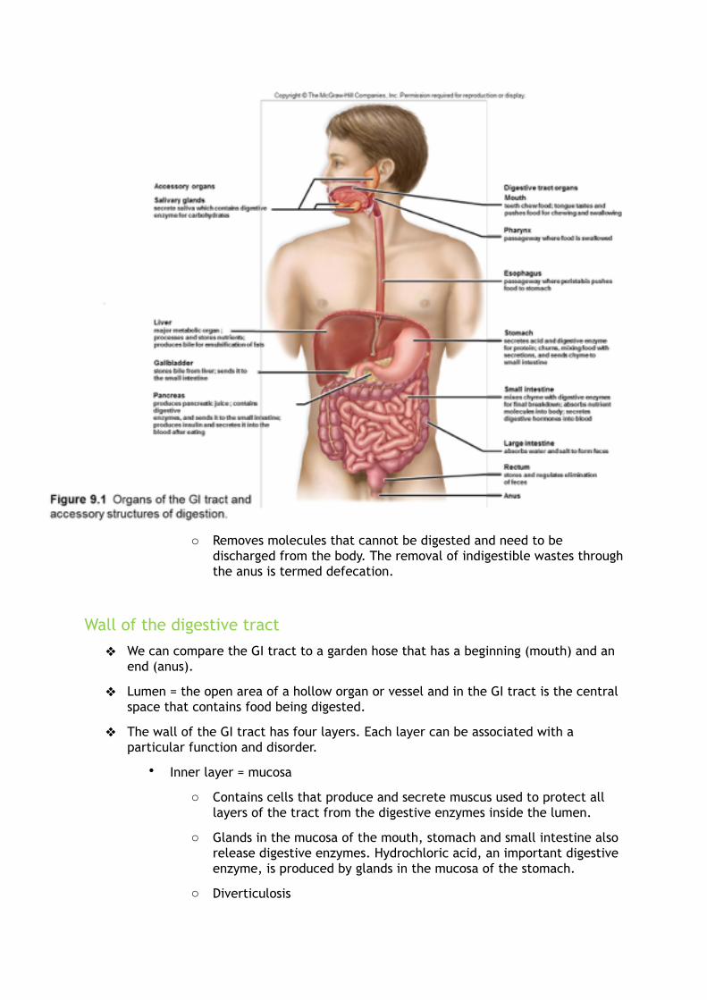
o Removes molecules that cannot be digested and need to be discharged from the body. The removal of indigestible wastes through the anus is termed defecation.
Wall of the digestive tract
❖ We can compare the GI tract to a garden hose that has a beginning (mouth) and an end (anus).
❖ Lumen = the open area of a hollow organ or vessel and in the GI tract is the central space that contains food being digested.
❖ The wall of the GI tract has four layers. Each layer can be associated with a particular function and disorder.
• Inner layer = mucosa
o Contains cells that produce and secrete muscus used to protect all layers of the tract from the digestive enzymes inside the lumen.
o Glands in the mucosa of the mouth, stomach and small intestine also release digestive enzymes. Hydrochloric acid, an important digestive enzyme, is produced by glands in the mucosa of the stomach.
o Diverticulosis

▪ Is a condition in which portions of the mucosa of any part of the GI tract, but primarily the large intestine, have pushed through the other layers and formed pouches , where food can collect.
▪ The pouches can be likened to an inner tube that pokes through weak places in a tire.
▪ When the pouches become infected or inflamed, the condition is called diverticulitis. This happens in 10-25% of people with diverticulosis.
• Second layer = submucosa
o Is a broad band of loose connective tissue that cotains blood vessels, lymphatic vessels and nerves.
o These are the vessels that will carry the nutrients absorbed by the mucosa.
o Lymph nodes
▪ Called peyer patches, are also in the submucosa.
▪ They help protect us from disease.
o Submucosa contains blood vessels, so it can be the site of an inflammatory response that leads to inflammatory bowel disease.
▪ Chronic diarrhea, abdominal pain, fever and weight loss are symptoms of IBD.
• Third layer = muscularis
o Contains two layers of smooth muscle.
▪ Inner circular, layer: encircles the tract
▪ Outer, longitudinal layer: lies in the same direction as the tract
o The contraction of these muscles, under nervous and hormonal control, accounts for peristalsis and subsequent movement of digested food from the esophagus to the anus.
o The muscularis can be associated with irritable bowel syndrome (IBS), in which contractions of the wall cause abdominal pain, constipation, and/or diarrhea.
▪ The underlying cause of IBS is not known, although some suggest that because this arrea is under nervous system control, stress may be an underlying cause.
• Fourth layer = serosa (serous membrane layer)
o Secretes a lubricating fluid
o Is a part of the peritoneum, the internal lining of the abdominal cavity.

The mouth, pharynx and esophagus ❖ Are in the first part of the GI tract
The Mouth
❖ Tonsils at the back of the mouth are lymphatic tissues, important in fighting disease.
❖ The mouth contains teeth that begin the mechanical breakdown of food.
❖ The tongue is covered in taste buds and also assists in the mechanical breakdown and movement of food.
❖ The tongue forms a bolus (mass of chewed food) and moves it toward the pharynx.
❖ 3 salivary glands produce saliva
• All three glands connect to the oral cavity via ducts.
• Saliva moistens food, making it easier to chew and swallow.
o Mucin: mucuslike protein holding food particles for easy swallowing
o Salivary amylase: cleaving carbohydrates into smaller pieces
o Bicarbonate (HCO3 -): maintains the pH of the mouth between 6.5 and 7.5,
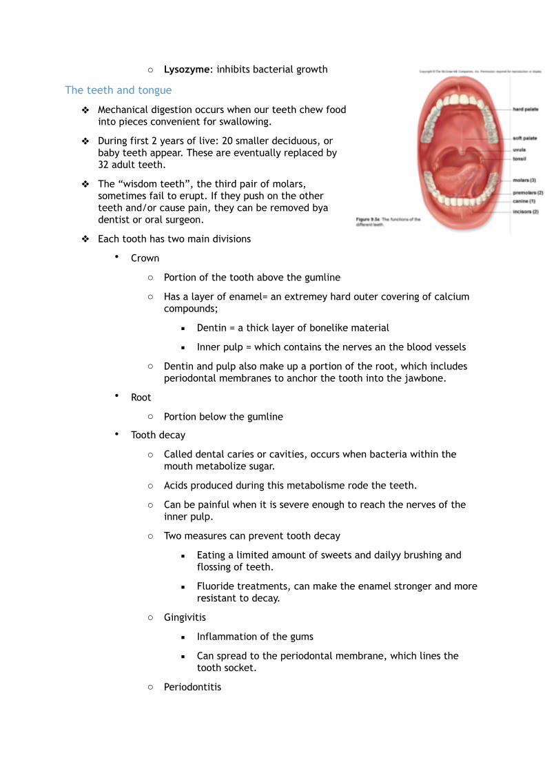
o Lysozyme: inhibits bacterial growth
The teeth and tongue
❖ Mechanical digestion occurs when our teeth chew food into pieces convenient for swallowing.
❖ During first 2 years of live: 20 smaller deciduous, or baby teeth appear. These are eventually replaced by 32 adult teeth.
❖ The “wisdom teeth”, the third pair of molars, sometimes fail to erupt. If they push on the other teeth and/or cause pain, they can be removed bya dentist or oral surgeon.
❖ Each tooth has two main divisions
• Crown
o Portion of the tooth above the gumline
o Has a layer of enamel= an extremey hard outer covering of calcium compounds;
▪ Dentin = a thick layer of bonelike material
▪ Inner pulp = which contains the nerves an the blood vessels
o Dentin and pulp also make up a portion of the root, which includes periodontal membranes to anchor the tooth into the jawbone.
• Root
o Portion below the gumline
• Tooth decay
o Called dental caries or cavities, occurs when bacteria within the mouth metabolize sugar.
o Acids produced during this metabolisme rode the teeth.
o Can be painful when it is severe enough to reach the nerves of the inner pulp.
o Two measures can prevent tooth decay
▪ Eating a limited amount of sweets and dailyy brushing and flossing of teeth.
▪ Fluoride treatments, can make the enamel stronger and more resistant to decay.
o Gingivitis
▪ Inflammation of the gums
▪ Can spread to the periodontal membrane, which lines the tooth socket.
o Periodontitis

▪ Characterized by a loss of bone and loosening of the teeth.
▪ Extensive dental work may be required or teeth will be completely lost.
▪ Stimulation of the gums in an manner advised by a dentist is helpful in controlling this condition. Medications are also available.
❖ The tongue
• Covered by a muscous membrane, which contains the sensory receptors called taste buds.
• When taste buds are activatedby the presence of food, nerve impulses travel by way of nerves to the brain.
• Is composed of skeletal muscle and it assists the teeth in carrying out mechanical digestion by moving food around in the mouth.
• In preparation for swallowing, the tongue forms shewed food into a mass called a bolus, which it pushes toward the pharynx.
The pharynx and esophagus
❖ Both the mouth and the nasal passages lead to the pharynx, a hollow space at the back of the throat.
❖ In turn, the pharynx opens into both food passage (esophagus) and the air passage (trachea or windpipe).
❖ These two tubes are parallel toe ach other with the trachea anterior to (in front of) the esophagus.
Swallowing
❖ Voluntary phase
❖ Involuntary phase
• Once food or drink is pushed back far enough into the pharynx, swallowing becomes a reflex action performed involuntarily.
❖ During swallowing, food normally enters the esophagus, a muscular tube that moves food into the stomach, because other possible avenues are blocked.
❖ The soft palate moves back to close off the nasal passages, and the trachea moves up under the epiglottis to cover the glottis.
❖ The glottis is the opening to the larynx (voice box) and, therefore, the air passage.
❖ Sometimes, the epiglottis does not cover the glottis fast enough or completely enough and food or liquid can end up in the trachea instead of the esophagus. When this occurs, the muscles around

the lungs contract and force a cough that will bring the food back up the trachea and into the pharynx.
Peristalsis
❖ Pushes food through the esophagus.
❖ Peristaltic contractions continue in the stomach and intestines.
❖ Esophagus
• Sole purpose is to move the food bolus from the mouth to the stomach.
• Sphincters
•Are muscles that encircle tubes and act as valves.
•The tubes close when the sphincters contract, and they open when the sphincters relax.
•When food or saliva is swallowed, the sphincter relaxes for a moment to allow the food or saliva to enter the stomach.
•The sphincter then contracts, preventing the acidic stomach
contents from backing up into the esophagus.
• A constriction called the lower gastroesophageal sphincter marks the entrace of the esophagus to the stomach.
• Heartburn
• When the lower esophageal sphincter fails to open and allow food into the stomach, or when the sphincter is opened and food moves from the stomach back to the esophagus.
• This condition can lead to damage of the esophagus and lower esophageal sphincter
• Vomiting occurs when strong contractions of the abdominal muscles and the diaphragm (the muscle separating the thoratic and abdomial cavities) force the contents of the stomach into the esophagus and oral cavity.
❖ Lump of food (bolus) stretches a portion of the GI tract,
• In front of bolus: smooth muscle relax
• Behind bolus : muscles contract.
❖ Peristaltic wave of contraction
• mixing the contents of the stomach
• pushing the contents of the esophagus and intestines forward.
The stomach and small intestine

❖ The stomach and small intestine complete the digestion of food, which began in the mouth.
The stomach
❖ Is a thick-walled, J-shaped organ thatlies on the left sise of the body beneath the diaphragm.
❖ Is continuous with the esophagus above and theduodenum of the small intestine below.
❖ It functions to store food, start digestion of proteins, and control movement of chyme into the small intestine.
❖ Nutrients are not absorbed by the stomach. However, it does absorb alcohol.
❖ The stomach wall has the usual four layers, but two of them are modified for particular functions.
❖ Muscularis layer
• There are three layers of muscle in the muscularis layer of the stomach wall to help in mechanical digestion and allow it to stretch.
• Circular and longitudinal layers
• Oblige layer
o Also allows the stomach to stretch and to mechenicallly break down food into smaller fragments that are mixed with gastric juice.
❖ The mucosa layer has deep folds called rugae, and gastric pits that lead into gastric glands that secrete gastric juice.
❖ Gastric juice contains pepsin, an enzyme that breaks down proteins, plus HCl and mucus.
❖ HCl gives the stomach a pH of 2, which activates pepsin and helps kill bacteria found in food.
❖ A bacterium, Helicobacter pylori, lives in the mucus and can cause gastric ulcers.
❖ The stomach empties chyme into the small intestine after 2-6 hrs.
The small intestine
❖ Is named for its small diameter compared with that of the large intestine.
❖ Small intestine is very long (6m) compared with the large intestine (1.5m).
Digestion is completed in the small intestine

❖ Contains a wide range of enzymes to digest the carbohydrate and fat content of food.
❖ Most of these enzymes are secreted by the pancreas and enter via a duct at the duodenum = the first 25 cm of the small intestine.
❖ Another duct brings bile from the liver and galbladder into the duodenum
• Bile emulsifies fat
• Emulsification = a form of mechanical digestion that causes fat droplets to disperse in water.
❖ After emulsification, the lipase enzyme, produced by th pancreas, hydrolyzes fats to form the monoglycerides and fatty acids.
❖ Pancreatic amylase begins the digestion of carbohydrates.
❖ An intestinal enzyme completes the digestion of carbohydrates to glucose.
❖ Pancreatic trypsin, a protease enzyme, begins and intestinal enzymes finish the digestion of proteins to amino acids.
❖ The intestine has a slightly basic pH, because pancreatic juice contains sodium bicarbonate (NaHCO3), which neutrolizes chyme.
Nutrients are absorbed in the small intestine
❖ 95% of nutrients absorbed in small intestine
❖ The wall of the small intesine absorbs the sugar, amino acid, glycerol and fatty acid molecules that were the products of the digestive process.
❖ Mucosa of the small intestine is modified for absorption. large surface area for absorption.
• Mucosa contains fingerlike projections called villi, which give the intestinal wall a soft, velvety appearance.
• A vilus has an outer layer of columnar epithelial cells and each of these cells has thousands of microscopic extensions called microvilli.
• Microvilli
o give the villi a fuzzy border known as the brush border.
o Contain enzymes, called brush border enzymes, tha complete the digestive process.
o Greatly increase the surface area of the villus for the absorption of nutrients.
• Nutrients are absorbed into the vessels of a villus.
• Villus: contains blood capillaries and a small lymphatic capillary called a lacteal.
• Lymphatic vessels carry a fluid called lymph to the cardiovascular veins.
• Lipoprotein droplets, called chylomicrons, are formed when monoglycerides and fatty acids are rejoined in the villi epithelia cells. Chylomicrons then enter a lacteal.

• After nutrients are absorbed, they are eventually carried to all the cells of the body by the bloodstream.
Lactose intolerance
❖ Lactose is the primary sugar in milk.
❖ People who do not have the brush border enzyme lactase, cannot digest lactose.
❖ Result is lactose intolerance
• Characterized by diarrhea, gas, bloating and abdominal cramps after ingesting milk and other dairy products
❖ Lactase deficiency (‘normal’ after weaning)
❖ Genetics (lactase persistence)
❖ Congenital lactase deficiency (babies)
❖ Lactose-intolerant people can eat cheese and yogurt because the lactose in these milk products has already been digested.
❖ Gluten intolerance
❖ Celiac disease: immune system destroys villi →malabsorbtion
❖ Genetic , triggers (stress, surgery, pregnancy, infection)
❖ Common (1/150)
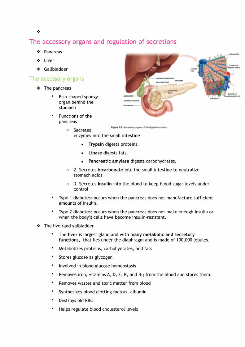
❖
The accessory organs and regulation of secretions ❖ Pancreas
❖ Liver
❖ Gallbladder
The accessory organs
❖ The pancreas
• Fish-shaped spongy organ behind the stomach
• Functions of the pancreas
o Secretes enzymes into the small intestine
▪ Trypsin digests proteins.
▪ Lipase digests fats.
▪ Pancreatic amylase digests carbohydrates.
o 2. Secretes bicarbonate into the small intestine to neutralize stomach acids
o 3. Secretes insulin into the blood to keep blood sugar levels under control
• Type 1 diabetes: occurs when the pancreas does not manufacture sufficient amounts of insulin.
• Type 2 diabetes: occurs when the pancreas does not make eneogh insulin or when the body’s cells have become insulin-resistant.
❖ The live rand galbladder
• The liver is largest gland and with many metabolic and secretory functions, that lies under the diaphragm and is made of 100,000 lobules.
• Metabolizes proteins, carbohydrates, and fats
• Stores glucose as glycogen
• Involved in blood glucose homeostasis
• Removes iron, vitamins A, D, E, K, and B12 from the blood and stores them.
• Removes wastes and toxic matter from blood
• Synthesizes blood clotting factors, albumin
• Destroys old RBC
• Helps regulate blood cholesterol levels

• It makes ~ 1lt/d of bile that is then stored in the gallbladder to be secreted into the small intestine to emulsify fats.
o Bile salts helps digest/adsorb fats
o Bile salts help excrete toxic waste products
o Gallstones form when liquid storen in the gallbladder hardens into pieces of stonelike material.
Liver disorders
❖ Hepatitis and cirrhosis are two serious diseases that affect the entire liver and hinder its ability to repair itself.
❖ Jaundice
• When a person has a liver ailment, bile pigments may leak into the blood, causing jaundice
• Is a yellowish tint to the white of the eyes and to the skin of light-pigmented persons.
• Can result from hepatitis = inflammation of the liver.
o Hepatitis A
▪ Is usually acquired from sewage-contaminated drinking water and food.
o Hepatitis B
▪ Usually spread by sexual contact, can also be spread by blood transfusions or contaminated needles.
➔ Vaccins are available for hepatitis A and B
o Hepatitis C
▪ Usually acquired by contact with infected blood and can lead to chronic hepatitis, liver cancer and death. There is no vaccine.
• Cirrhosis
o Another chronic disease of the liver.
o First the organ becomes fatty and then liver tissue is replaced by inactive, fibrous scar tissue.
o Often seen in alcoholics and obese people
❖ The liver has amazing regenerative powers and can recover if the rate of regeneration exceeds the rate of damage.
❖ During liver failure, however, there may not be enough time to let the liver heal itself and liver transplantation is usually the preferred treatment.
❖ The liver is a vital organ and its failure leads to death.
Regulation of digestive secretions

❖ The secretions of digestive juices are controlled by the nervous system and by digestive hormones.
❖ When you look at/smell food, the parasympathetic nervous system automatically stimulates gastric secretion.
❖ Gastrin
• when a person has eaten a meal particulary rich in protein, the stomach produces the hormone gastrin.
• Gastrin enters the bloodstream and soon the secretory activity of gastric glands increases.
❖ Secretin
• Secretin release is stimulated by acid, especially the HCI present in chyme.
❖ Cholecystokinin (CCK)
• Parially digested proteins and fat stimulate the release of CCK.
• Soon after these hormones enter the bloodstream, the pancreas increases its output of pancreatic juice.
o Pancreatic juice buffers the acidic chyme enterig the intestine from the stomach and helps digest food.
• CCk also causes the liver to increase its production of bile and causes the gallbladder to contract and release stored bile. The bile then aids the digestion of fats that stimulated the release of cCK.
The large intestine and defecation
❖ Is larger in diameter than the small intestine (6.5 cm compared with 2.5 cm), but is shorter in length.
❖ Cecum
• First portion of the large intestine, joining the end of the small intestine.

• Usually has a small projection called the vermiform appendix
o In humans, the appendix is thought to aid in fighting infections.
o Appendix may also contribute to the population of needed bacteria in the large intestine.
o An inflammed appendix (appendicitis) may be treated using antibiotics or removed surgically.
o Should the appendix burst, the result can be peritonitis, a life-threatening swlling and infection of the peritoneum
❖ Colon
• Ascending colon
o Which goes up the right side of the body to the level of the liver.
• Transverse colon
o Which crosses the abdominal cavity just below the liver and the stomach
• Descending colon
o Which passes down the left side of the body.
• Sigmoid colon
o Which enters the rectum
❖ Rectum
• The last 20 cm of the large intestine.
❖ Anus
• The rectum opens at the anus, where defecation, the expulsion of feces, occurs.
Functions of the large intestine
❖ absorb water to prevent dehydration and maintain homeostasis.
❖ absorb vitamins (B complex and K) produced by intestinal flora = the bacteria that inhabit the intestine and aid in keeping us healthy.
❖ form and rid the body of feces through the anus.
• Consistency of normal feces usually is:
o Three-fourths water
o One-fourth solid wastes
▪ Bacteria, dietary fiber (indigestible remains) and other indigestible materials.
o Bacterial action on indigestible materials causes the odor of feces and accountsfor the presence of gas.

o Stercobilin, which is a breakdown product of bilirubin (the yellow-orange bile pigment produced from the breakdown of hemoglobin) and oxidized iron cause he brown color of feces.
o Peristalsis occurs infrequently in the large intestine, but when it does, feces are forced into the rectum.
o Feces collect in the rectum until its apprpriate to defecate.
o At that time, stretching of the rectal wall initiates nerve impulses to the spinal cord. Shorttly thereafter, the rectal muscles contract and the anal sphincters relax. This allows the feces to exit the body through the anus.
Disorders of the colon and rectum
Diarrhea
❖ Characterized by bowel movements that are loose or watery
❖ Major causes are
• infection of the lower intestinal tract
o The intestinal wall becomes irritated and peristalsis increases when an infection occurs. The diarrhea that results rids the body of the infectious organisms.
• nervous system stimulation.
o The nervous system stimulates the intestinal wal land diarrhea results.
❖ Prolonged diarrhea can lead to dehydration because of water loss, which can lead to an imbalance of salts in the blood that can affect heart muscle contraction and potentially lead to death.
Constipation
❖ The feces are hard and dry, making it difficult for them to be expelled.
❖ Diets that lack whole-grain foods, as well as ignoring the urge to defecate, are often the causes of constipation.
❖ Adequate water and fiber intake can help regularity of defecation.
❖ The frequent use of laxatives is discouraged, because it can result in dependence on their use for normal bowel movements.
• The most natural is a bulk laxative. It produces a soft mass of cellulose in the colon.
❖ Chronic constipation is associated with the development of hemorrhoids, enlarged and inflamed blood vessels at the anus.
❖ Other contributing factors for hemorrhoid development include pregnancy, aging and anal intercourse.
Diverticulosis
❖ Is the occurence of little pouches of mucosa where food can collect.

❖ The pouches form when the mucosa pushes through weak spots in the muscularis.
❖ A frequent site is the last part of the descending colon.
Irritable bowel syndrome (IBS)
❖ Or spastic colon, is a condition in which the muscularis contracts powerfully but without its normal coordination.
❖ The symptoms are abdominal cramps ;gas; constipation; and urgent explosive stools (feces discharge)
Inflammatory bowel disease (IBD)
❖ Is a collective term for a number of inflammatory disorders.
❖ Ulcerative colitis
• Affects the large intestine and rectum and results in diarrhea, rectal bleeding, abdominal cramps and urgency in defecation.
❖ Crohn’s disease
• Is normally isolated to the small intestine but can affect any arrea of the digestive tract, including the colon and rectum.
• It is characterized by the breakdown of the lining of the affected area, resulting in ulcers.
• Ulcers are painful and cause bleeding, because they erode the submucosal layer, where there are nerves and blood vessels. This also results in the inability to absorb nutrients at the affected sites.
• Symptoms: diarrhea, weight loss, abdominal cramping, anemia, bleeding and malnutrition.
Polyps and cancer
❖ The colon is subject to the development of polyps = small growths arising from the epithelial lining.
❖ Polyps can be removed surgically.
❖ Some investigators believe that dietary fat increases the likelihood of colon cancer,because dietary fat causes an increase in bile secretion. It could be that intestinal bacteria convert bile salts to substances that promote the development of cancer.
❖ Fiber in the diet seems to inhibit the development of colon cancer, and regular elimination reduces the time that the colon wall is exposed to any cancer-promoting agents in feces.
❖ Diagnostic tool for this disorders
• Endoscopic exam called a colonoscopy.
o A flexible tube containing a camera is inserted into the GI tract, usually from the anus.
• This procedure is gradually being replaced by the PillCam, a camera you swallow.

Nutrition and weight control ❖ Obesity or being significantly overweight, has become one of the greatest health
problems in the united states.
❖ Almost 35% of adults and 18% of children, are classified as obese.
❖ Of great concern, because excess body fat is associated with a higher risk for premature death, type 2 diabetes, hypertension, cardiovascular disease, stroke, gallbladder disease, respiratory dysfunction, osteoarthritis and certain types of cancer.
❖ To reverse a trend toward obesity, eat fewer calories by making wiser food choices and be more active.
Defining obesity
❖ Obesity is defined as abnormal or excessive fat accumulation that presents a risk to health (WHO).
❖ Crude estimte is BMI (bw/height2). BMI>30 obese
❖ Calculation
• Weight (1bs)/height (in.)2 x 703
❖ General rule
• A healthy BMI = 18.5 to 24.9
• An overweight BMI = 25 to 29.9
• An obese BMI = 30 to 39.9
• A morbidly obese BMI = 40 or higher
❖ Risk factors for chronic diseases:
• Diabetes
• Cardiovascular diseases
• Cancer
❖ Weight gain: consumed calories > burned calories
• Increase in energy-dense food (high in fat and sugars)
• Decrease in physical activity
• Processed food: using sugars or fats to carry flavor
• Hidden in plain sight:
o Sodas, candy, ... Bread, tomato sauce, soup, …
Classes of nutrients
❖ Nutrients are components of food that are needed to perform physiological body functions.

❖ Nutrients provide us with energy, promote growth and development and regulate cellular metabolism.
❖ Nutrients include
• carbohydrates.
• proteins.
• lipids.
• minerals.
• vitamins.
Carbohydrates
❖ Carbohydrates are sugars (simple) or polysaccharides (complex) that are digested into the simple sugars which are an important energy source.
❖ It is necessary to include carboydrates in the diet, because the body is unable to convert fatty acids to glucose and brain cells require glucose.
❖ Grains, beans, tubers, nuts, fruits source for carbohydrates
❖ Glycemic index: food effect on person’s blood glucose levels
❖ Can carbohydrates be harmful?
• Obesity
• these foods are said to have a high glycemic index, because they quickly increase blood glucose. Chronically high insulin level may lead to Insulin resistance (type 2 diabetes
• Increased fat deposition is associated with coronary heart disease, liver ailments and several types of cancer.
• Craving for sweets (sugar dependence)
Proteins
❖ Proteins are digested into 20 different amino acids which are used to produce cellular proteins.
❖ Essential amino acids are the 8 amino acids that must be attained through diet.
❖ Complete proteins that have all essential amino acids are usually derived from animals such as meat, dairy, eggs milk products, poultry,...
❖ Non-animal sources of proteins are found in legumes, seeds, nuts and grains. However, each of these alone is an incomplete protein source because of a deficiency in at least 1 of the essential amino acids.
❖ A daily supply of essential amino acids is needed because they are not stored in the body.
❖ Can proteins be harmful?
• Dehydration may occur if protein consumption is excessive.
• High-protein diets, especially those rich in animal proteins, can also increase calcium loss in urine.
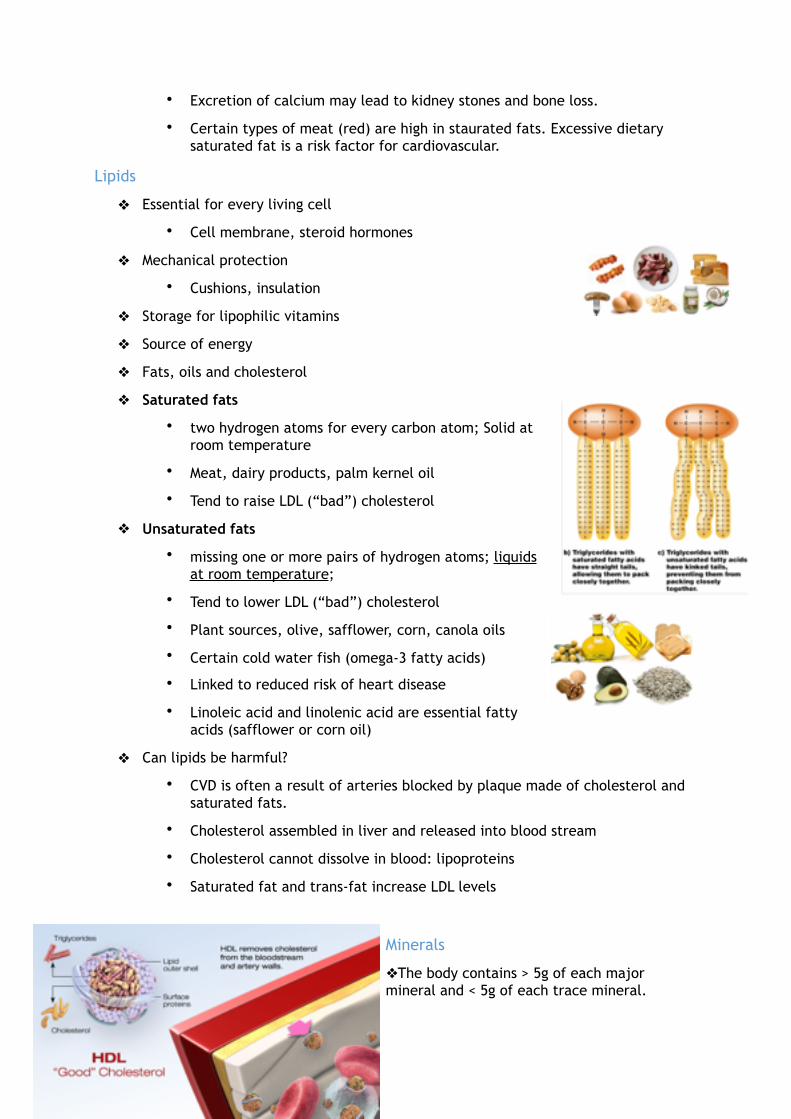
• Excretion of calcium may lead to kidney stones and bone loss.
• Certain types of meat (red) are high in staurated fats. Excessive dietary saturated fat is a risk factor for cardiovascular.
Lipids
❖ Essential for every living cell
• Cell membrane, steroid hormones
❖ Mechanical protection
• Cushions, insulation
❖ Storage for lipophilic vitamins
❖ Source of energy
❖ Fats, oils and cholesterol
❖ Saturated fats
• two hydrogen atoms for every carbon atom; Solid at room temperature
• Meat, dairy products, palm kernel oil
• Tend to raise LDL (“bad”) cholesterol
❖ Unsaturated fats
• missing one or more pairs of hydrogen atoms; liquids at room temperature;
• Tend to lower LDL (“bad”) cholesterol
• Plant sources, olive, safflower, corn, canola oils
• Certain cold water fish (omega-3 fatty acids)
• Linked to reduced risk of heart disease
• Linoleic acid and linolenic acid are essential fatty acids (safflower or corn oil)
❖ Can lipids be harmful?
• CVD is often a result of arteries blocked by plaque made of cholesterol and saturated fats.
• Cholesterol assembled in liver and released into blood stream
• Cholesterol cannot dissolve in blood: lipoproteins
• Saturated fat and trans-fat increase LDL levels
Minerals
❖The body contains > 5g of each major mineral and < 5g of each trace mineral.

❖ Major minerals make up components of cells, body fluids, and tissues (i.e., calcium).
❖ Minor minerals are components of larger molecules (i.e., iron in hemoglobin).
❖ A varied and complete diet usually provides necessary minerals
Calcium
❖ Calcium is needed to make bone and teeth, nerve impulse conduction, and muscle contraction.
❖ 1,000 mg/day are recommended to keep bones healthy early in life and 1,300 mg/day after menopausal age.
❖ Vitamin D is needed with calcium to prevent bone loss (osteoporosis). Osteoporosis develops becausebone-eating cells called osteoclasts are more active than bone-forming cells called osteoblasts.Therefore, the bones are porous and they break easily because they lack sufficient calcium.
Sodium
❖ Sodium is needed for regulating water balance, as does chloride (C1-).
❖ Plays an important role in the movement of materials across the plasma membrane as well as the conduction of a nerve impulse.
❖ 500 mg/day is the recommended amount (on average each American takes in 4,000 - 4,700 mg/day).
❖ Sodium can increase hypertension in people who already have it
❖ About 1/3 of the sodium we consume occurs naturally in foods. Another third is added during commercial processing, and we add the last third either during home cooking or at the table in the form of table salt.

Vitamins
❖ Organic compounds (not including proteins, fats, or carbohydrates) are used for metabolism but not produced in high enough quantities by the body.
❖ Vitamins are often enzyme helpers (coenzymes). Coenzymes are needed in only small amounts, because each can be used over and over. Not all vitamins are coenzymes.
❖ There is a total of 13 vitamins in two groups: fat soluble and water soluble.

Antioxidants
❖ Antioxidants are chemicals that decrease the rate of oxidation or transfer of electrons.
❖ Vitamin C, E, and A are considered antioxidants because they are thought to defend the body against free radicals that can transfer electrons and damage cells and DNA.
❖ The vitamins are common in fruits and vegetables.
Vitamin D
❖ Skin cells contain a precursor cholesterol molecule converted to vitamin D after UV exposure.
❖ Vitamin D leaves the skin and is modified first in the kidneys and then the liver until finally it becomes calcitriol. Calcitriol promotes the absorption of calcium by the intestines.
❖ The lack of vitamin D leads to rickets in children. Rickets, characterized by bowing of the legs,is caused by defective mineralization of the skeleton.
How to plan nutritious meals
❖ Basal metabolism = the number of calories a person’s body burns at rest to maintain normal body functions.
❖ The less exercise, the fewer calories needed beyond the basal metabolic rate.
❖ Maximum number of calories you can take each day is 2000 for a woman and 2500 for a man
❖ Zie guidelines MyPlate p 190-192
Eating disorders
❖ Characterized by abnormal eating habits that damage individual’s health
❖ Serious but treatable mental illness (2-4)
❖ Develop in adolescents
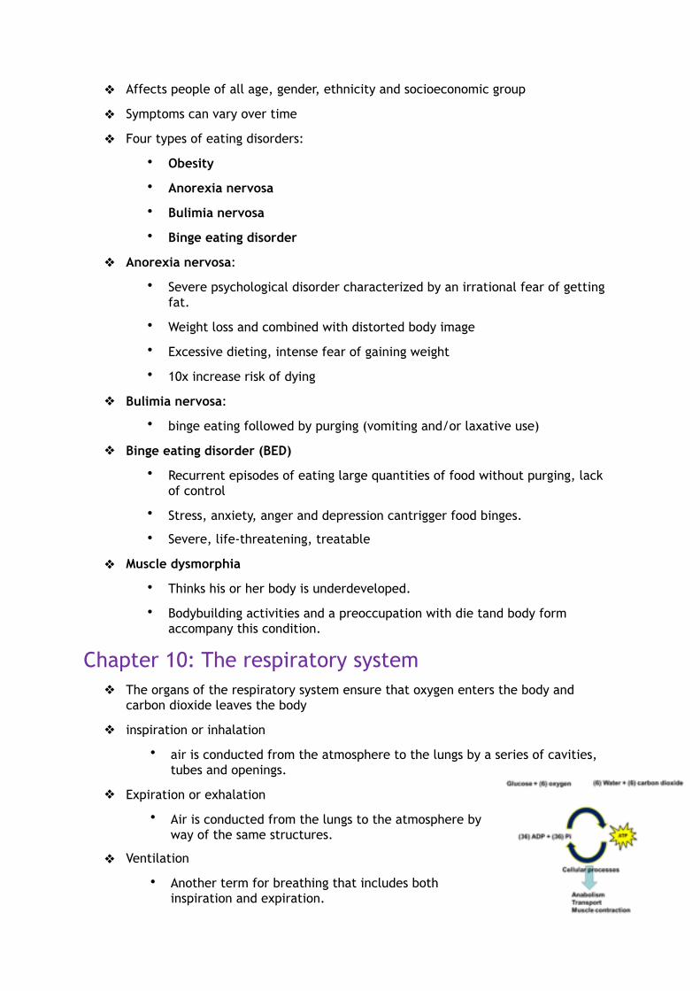
❖ Affects people of all age, gender, ethnicity and socioeconomic group
❖ Symptoms can vary over time
❖ Four types of eating disorders:
• Obesity
• Anorexia nervosa
• Bulimia nervosa
• Binge eating disorder
❖ Anorexia nervosa:
• Severe psychological disorder characterized by an irrational fear of getting fat.
• Weight loss and combined with distorted body image
• Excessive dieting, intense fear of gaining weight
• 10x increase risk of dying
❖ Bulimia nervosa:
• binge eating followed by purging (vomiting and/or laxative use)
❖ Binge eating disorder (BED)
• Recurrent episodes of eating large quantities of food without purging, lack of control
• Stress, anxiety, anger and depression cantrigger food binges.
• Severe, life-threatening, treatable
❖ Muscle dysmorphia
• Thinks his or her body is underdeveloped.
• Bodybuilding activities and a preoccupation with die tand body form accompany this condition.
Chapter 10: The respiratory system ❖ The organs of the respiratory system ensure that oxygen enters the body and
carbon dioxide leaves the body
❖ inspiration or inhalation
• air is conducted from the atmosphere to the lungs by a series of cavities, tubes and openings.
❖ Expiration or exhalation
• Air is conducted from the lungs to the atmosphere by way of the same structures.
❖ Ventilation
• Another term for breathing that includes both inspiration and expiration.

❖ Once ventilation has occurred, the respiratory system depends on the cardiovascular system to transport oxygen (O2) from the lungs to the tissues and carbon dioxide (CO2) from the tissues to the lungs.
❖ Gas exchange is necessary because the cells of the body carry out cellular respiration to make energy in the form of ATP.
❖ During cellular respiration, cells use up O2 and produce CO2. The respiratory system provides these cells with O2 and removes CO2.
The upper respiratory tract ❖The nasal cavities, pharynx and larynx are the organs of the upper respiratory tract.
The nose
❖ Opens at the nares (nostrils) that lead to the nasal cavities.
• Nasal cavities = are narrow canals separated from each other by a septum comosed of bone and cartilage.
❖ Air entering the nasal cavities encounters large, stiff hairs that act as a screening mechanism.
• The hairs filter the air and trap small particles, so that they don’t enter air passages.
❖ The rest of the nasal cavities are lined by muscous membrane.
• Muscus
o Secreted by this membrane, helps trap dus tand move it to the pharynx. Where it can be swallowed or expectorated by coughing or spitting.
• Submucosa
o Contains a large number of capillaries that help warm and moisten the incoming air.
❖ In the narrow upper recesses of the nasal cavities are ciliated cells that act as odor receptors. Nerves lead from these cells to the brain, where the impulses generated by the odor receptor are interpreted as smell.
❖ The tear (lacrimal) glands drain into the nasal cavities by way of tear ducts. The nasal cavities also connect with the sinuses (cavities) of the skull.

❖ Air in the nasal cavities passes into the nasopharynx, the upper portion of the pharynx.
❖ Connected to the nasopharyx are tubes called auditory tubes that connect to the middle ear.
❖ When air pressure inside the middle ears equalizes with the air pressure in the nasopharynx, the auditory tube openings may create a “popping” sensation.
The Pharynx
❖ Referred to as ‘throat’, is a funnel-shaped passageway that connects the nasal and oral cavities to the laryx.
❖ Pharynx had three parts
• Nasopharynx
o Where the nasal cavities open above the soft plate.
• Oropharynx
o Where the oral cavity opens
• Laryngopharynx
o Opens into the larynx
❖ Tonsils
• Form a protective ring at the junction of the oral cavity and the pharynx.
• Are comprised of lymphoid tissue and are actually a part of the immune system.
• They contain lymphocytes, which protect against invasion of inhaled foreign antigens.
• Are the primary defense during breathing, because inhaled air passes directly over the tonsils.
• In the tonsils, B and T cells respond to antigenst hat may subsequently invade internal tissues and fluids.
❖ In the pharynx, the air passage and the food passage lie parallel to each other and share an opening in the laryngopharynx. The larynx is normally open, allowing air to pass, but the esophagus is normally closed and opens only when a person swallows. If the passageway remains blocked, the Heimlich maneuver can be used to dislodge food blocking airway.
The Larynx
❖ Is a cartilaginous structure that serves as a passageway for air between the pharynx and the trachea.
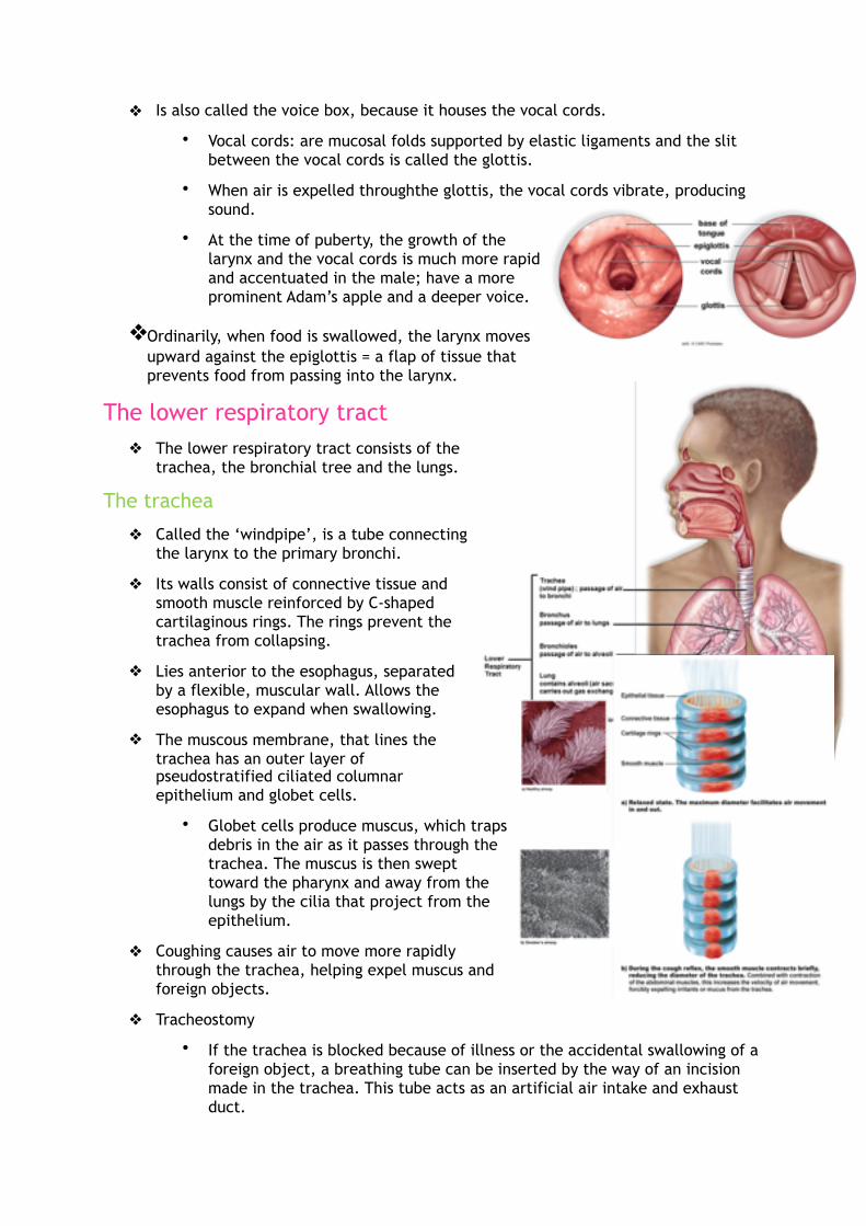
❖ Is also called the voice box, because it houses the vocal cords.
• Vocal cords: are mucosal folds supported by elastic ligaments and the slit between the vocal cords is called the glottis.
• When air is expelled throughthe glottis, the vocal cords vibrate, producing sound.
• At the time of puberty, the growth of the larynx and the vocal cords is much more rapid and accentuated in the male; have a more prominent Adam’s apple and a deeper voice.
❖Ordinarily, when food is swallowed, the larynx moves upward against the epiglottis = a flap of tissue that prevents food from passing into the larynx.
The lower respiratory tract ❖ The lower respiratory tract consists of the
trachea, the bronchial tree and the lungs. The trachea
❖ Called the ‘windpipe’, is a tube connecting the larynx to the primary bronchi.
❖ Its walls consist of connective tissue and smooth muscle reinforced by C-shaped cartilaginous rings. The rings prevent the trachea from collapsing.
❖ Lies anterior to the esophagus, separated by a flexible, muscular wall. Allows the esophagus to expand when swallowing.
❖ The muscous membrane, that lines the trachea has an outer layer of pseudostratified ciliated columnar epithelium and globet cells.
• Globet cells produce muscus, which traps debris in the air as it passes through the trachea. The muscus is then swept toward the pharynx and away from the lungs by the cilia that project from the epithelium.
❖ Coughing causes air to move more rapidly through the trachea, helping expel muscus and foreign objects.
❖ Tracheostomy
• If the trachea is blocked because of illness or the accidental swallowing of a foreign object, a breathing tube can be inserted by the way of an incision made in the trachea. This tube acts as an artificial air intake and exhaust duct.

The bronchial tree
❖ Divides into the right and left primary bronchi, which lead into the right and left lungs.
❖ Bronchi branch into a few secondary bronchi that also branch, until the branches become about 1 mm in diameter and are called bronchioles.
❖ As the bronchial tubes divide and subdivide, their walls become thinner and the small rings of cartilage are no longer present. During an asthma attack, the smooth muscle of the bronchioles contracts, causing bronchiolar constriction and characteristic wheezing.
❖ Each bronchiole leads to an elongated space enclosed by a multitude of air pockets, or sacs, called alveoli.
❖ (trachea->bronchus->bronchioles->alveolus)
Lungs
❖ Are paired, cone-shaped organs in the thoracic cavity.
❖ Center of the thoracic cavity = trachea, heart, thymus and esophagus
❖ The lungs are on each side of the trachea
• Right lung: has three lobes
• Left lung: has two lobes
❖ Each lung is further divided into lobules and each lobule has a bronchiole serving many alveoli.
❖ The lungs follow the contours of the thoracic cavity, including the diaphragm, the muscle that separates the thoracic cavity from the abdominal cavity.
❖ Each lung is enclosed by pleurae, two layers of serous membrane that produce serous fluid.
• Parietal pleura: adheres to the thoracic cavity wall
• Visceral pleura: adheres to the surface of the lung
❖ Surface tension = the tendency for water molecules to cling to one another due to hydrogen bonding between molecules. It holds the two pleural layers together.
❖ Pleurisy occurs when these layers become inflamed.
• Breathing, sneezing and coughing are quite painful because the layers rub against each other
• Causes include viral infections, tuberculosis and pneumonia.
The Alveoli
❖ There are 300 million alveoli in the lungs that greatly increase surface area.
❖ Alveoli are enveloped by blood capillaries.
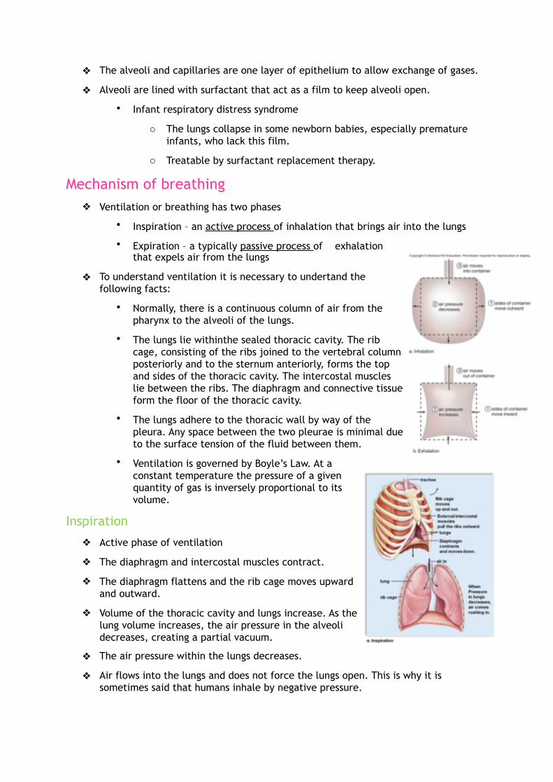
❖ The alveoli and capillaries are one layer of epithelium to allow exchange of gases.
❖ Alveoli are lined with surfactant that act as a film to keep alveoli open.
• Infant respiratory distress syndrome
o The lungs collapse in some newborn babies, especially premature infants, who lack this film.
o Treatable by surfactant replacement therapy.
Mechanism of breathing ❖ Ventilation or breathing has two phases
• Inspiration – an active process of inhalation that brings air into the lungs
• Expiration – a typically passive process of exhalation that expels air from the lungs
❖ To understand ventilation it is necessary to undertand the following facts:
• Normally, there is a continuous column of air from the pharynx to the alveoli of the lungs.
• The lungs lie withinthe sealed thoracic cavity. The rib cage, consisting of the ribs joined to the vertebral column posteriorly and to the sternum anteriorly, forms the top and sides of the thoracic cavity. The intercostal muscles lie between the ribs. The diaphragm and connective tissue form the floor of the thoracic cavity.
• The lungs adhere to the thoracic wall by way of the pleura. Any space between the two pleurae is minimal due to the surface tension of the fluid between them.
• Ventilation is governed by Boyle’s Law. At a constant temperature the pressure of a given quantity of gas is inversely proportional to its volume.
Inspiration
❖ Active phase of ventilation
❖ The diaphragm and intercostal muscles contract.
❖ The diaphragm flattens and the rib cage moves upward and outward.
❖ Volume of the thoracic cavity and lungs increase. As the lung volume increases, the air pressure in the alveoli decreases, creating a partial vacuum.
❖ The air pressure within the lungs decreases.
❖ Air flows into the lungs and does not force the lungs open. This is why it is sometimes said that humans inhale by negative pressure.

Expiration
❖ Passive phase
❖ The diaphragm and intercostal muscles relax.
❖ The diaphragm moves upward and becomes dome-shaped.
❖ The rib cage moves downward and inward.
❖ Volume of the thoracic cavity and lungs decreases.
❖ The air pressure within the lungs increases.
❖ Air flows out of the lungs.
❖ What keeps the alveoli from collapsing as a part of expiration?
• Presence of surfactant lowers the surface tension within the alveoli.
• The lungs recoil, the pressure between the pleurae decreases and this tend to make the alveoli stay open.
Maximum inspiratory effort and forced expiration
❖ Maximum inspiratory effort involves muscles of the back, chest and neck. This increases the size of the thoracic cavity to larger than normal, thus allowing maximum expension of the lungs.
❖ Expiration can also be forced
• Maximum inspiratory efforts of heavy exercise
• Sing, blow air in trumpet, blow out birthday candles.
❖ Contration of the inetrnal intercostal muscles can force the rib cage to move downward and inward.
❖ When the abdominal wall muscles contract, they push on the abdominal organs. In turn, the organs push upward against the diaphragm and the increased pressure in the thoracic cavity helps expel air.
Volumes of air exchanged during ventilation
❖ A technique has been developed that allows physicians to determine if there is a medical problem that prevents hhe lungs from filling with air on inspiration and releasing it from the body on expiration.
❖ Spirometer
• Measurements recorded when a person breathes as directed by a technician.
❖ Tidal volume
• When we are relaxed, only a small amount of air moves in and ot with each breath.
• Is only about 500 ml.
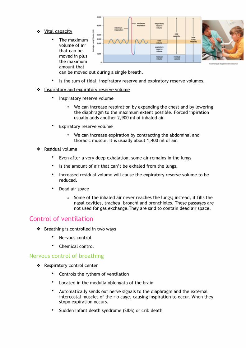
❖ Vital capacity
• The maximum volume of air that can be moved in plus the maximum amount that can be moved out during a single breath.
• Is the sum of tidal, inspiratory reserve and expiratory reserve volumes.
❖ Inspiratory and expiratory reserve volume
• Inspiratory reserve volume
o We can increase respiration by expanding the chest and by lowering the diaphragm to the maximum extent possible. Forced inpiration usually adds another 2,900 ml of inhaled air.
• Expiratory reserve volume
o We can increase expiration by contracting the abdominal and thoracic muscle. It is usually about 1,400 ml of air.
❖ Residual volume
• Even after a very deep exhalation, some air remains in the lungs
• Is the amount of air that can’t be exhaled from the lungs.
• Increased residual volume will cause the expiratory reserve volume to be reduced.
• Dead air space
o Some of the inhaled air never reaches the lungs; instead, it fills the nasal cavities, trachea, bronchi and bronchioles. These passages are not used for gas exchange.They are said to contain dead air space.
Control of ventilation ❖ Breathing is controlled in two ways
• Nervous control
• Chemical control
Nervous control of breathing
❖ Respiratory control center
• Controls the rythem of ventilation
• Located in the medulla oblongata of the brain
• Automatically sends out nerve signals to the diaphragm and the external intercostal muscles of the rib cage, causing inspiration to occur. When they stopn expiration occurs.
• Sudden infant death syndrome (SIDS) or crib death

o An infant under one year of age is put to bed seemingly healthy and sometime while sleeping, the child stops breathing.
o Precise cause not known. Most research is focussing on miscommunication between the respiratory center of the brain and the lungs and, possibly problems with the heart function.
• Its activity can be influenced by nervous input.
o We can voluntarily change our breathing pattern to accomodate activities such as speaking,…
o Following forced inspiration, stretch receptors in the airway walls respond to increased pressure. The receptors initiate inhibitory nerve impulses.
o The impulses travel from the inflated lungs to the respiratory center. This temporarily stops the respiratory center from sending out nerve signals.
o In this manner, ecessive stretching of the elastic tissue of the lungs is prevented.
❖ Cardiovascular center: heart rate
❖ Vasomotor center: blood pressure
❖ Reflex center for sneezing, vomiting, swallowing, coughing
Chemical control of breathing
❖ Chemical control
• Chemoreceptors = sensory receptors in the body that are sensitive to chemical composition of body fluids.
• Two sets of chemoreceptors sense the drop in pH (increase in protons) and can cause breathing to speed up:
o Peripheral
▪ circulatory system: Aortic and carotid bodies are sensitive to blood pH.
o Central
▪ located in the medulla oblongata
• These chemoreceptors are not strongly affected by low oxygen (O2) levels.
• They are stimulated when the carbon dioxide entering the blood is suffiecient to change blood pH.
• When the pH of blood becomes more acidic (decreases) the respiratory center increases the rate and depth of breathing.
• With increased breathing rate, more carbon dioxide is removed from the blood. The hydrogen ion concentration returns to normal and the breathing returns to normal.

• Increase of protons due to increase of carbon dioxide levels in blood.
Gas exchanges in the body ❖ Gas exchange is critical for homeostasis.
❖ Oxygen needed to produce energy must be supplied to all the cells ad carbon dioxide must be removed from the body during gas exchange.
❖ The exchange of gases is dependent on diffusion.
❖ Partial pressure is the amount of pressure each gas exerts (PCO2 or PO2).
❖ If the partial pressure of oxygen differs across a membrane, oxygen and carbon dioxide will diffuse from the area of higher to the area of lower partial pressure.
External respiration
❖ Exchange of gases between the lung alveoli and the blood capillaries by passive diffusion.
❖ PCO2 is higher in the lung capillaries than the atmospheric air, thus CO2 diffuses out of the plasma into the lungs.
❖ Most of the CO2 is carried in plasma as bicarbonate (HCO3-).
❖ The enzyme carbonic anhydrase speeds the breakdown of carbonic acid (H2CO3) in red blood cells.
❖ In the low –POco2 environment of the lungs, the reactionproceeds to the right.
❖What happens if you hyperventilate?
• Blood wil have fewer hydrogen ions and a high blood pH results.
• Breathing is inhibited: various symptoms: dizziness, continous contractions skeletal muscles.
• Breathing in a paper bag helps restore the blood pH because you’re inhaling the co2 that you hust exhaled into the bag.

❖ What happens if you hypoventilate (breathing at low rate) and this reaction does not occur?
• Hydrogen ions build up in the blood and acidosis occurs.
• Buffers may compensate for the low pH and breathing most likely increases.
• Exterme changes in blood pH affect enzyme function, which may lead to coma and death.
❖ The partial pressure pattern for O2 is just the opposite of that for CO2. ❖ Blood in the pulmonary capillaries is low in oxygen and alveolar air contains a
higher partial pressure of oxygen. Therefore, O2, diffuses into plasma and then into red blood cells in the lungs.
❖ Hemoglobin takes up this oxygen and become oxyhemoglobin (HbO2).
Internal respiration
❖The exchange of gases between the blood in the systemic capillaries outside of the
lungs and the tissue fluid.
❖ PO2 is higher in the capillaries than the tissue fluid, thus O2 diffuses out of the blood into the tissues.
❖ Carbon dioxide diffuses into the blood from the tissues because the Pco2 of interstitial fluid is higher than that of blood.
❖ After CO2 diffuses into the blood, most enters the red blood cells, where a small amount is taken up by hemoglobin, forming carbaminohemoglobin (HbCO2).
❖ The enzyme carbonic anhydrase speeds the reaction

in red blood cells.
❖ Bicarbonate ions (HCO3-) diffuse out of red blood cells and are carried in the plasma.
❖ The globin portion of hemoglobin combines with excess hydrogen ions produced by the overall reaction, and Hb becomes HHb, called reduced hemoglobin. In this way, the pH of blood remains fairly constant.
Respiration and health ❖ Upper respiratory infections (URIs) can spread from the nasal cavities to the
sinuses, middle ears and larynx. For example step throat: primary bacterial infection, caused by streptococcus pyogenes that can lead to a generalized URI and even a systematic.
Sinusitis
❖ Develops when nasal congestion blocks the tiny openings leading to the sinuses
❖ Symptoms: postnasal discharge and facial pain ( worsens when bends forward). Pain and tenderness over oower forehead and cheeks.
❖ Successful treatment depends on restoring proper drainage of the sinuses.
Otitis media
❖ Is an infection of the middle ear.
❖ Often seen in childre n who have a nasal infection.
❖ Symptoms: pain, sense of fullness, hearing loss, dizziness and fever.
❖ Solution: antibiotics, tubes placed in the eardrums of children with multiple recurrences.
Tonsillitis
❖ Ocuurs when the tonsils become inflamed and enlarged.
❖ If tonsillitis occurs frequently and enlargement makes breathing difficult, the tonsils can be removed surgically in a tonsillectomy.
Laryngitis
❖ Is an infection of the larynx with accompanying hoarseness, leading to the inability to talk in an audible voice.
❖ Usually dissapears with treatment of the URI.
Lower respiratory tract disorders
❖ Include infecions, restrictive pulmonary disorders, obstructive pulmonary disorders and lung cancer.
Lower respiratory infections
❖ Acute bronchitis
• An infection of the primary and secondary bronchi.

• Usually preceded by viral infection that has led to secondary bacterial infection.
• Most likely, a nonproductive cough has become a deep cough that expectorates muscus and perhaps pus.
❖ Pneumonia
• Is a viral or bacterial infection of the lungs on which the bronchi and alveoli fill with thick fluid.
• Is preceded by influenza.
• Symptoms: high fever and chills with headache and chest pain.
• May be localized in specific lobules of the lungs. The more lobules involved, the more serious the infection..
• Can be caused by a bacterium.
• Aids patients are subject to a particularly rare form of pneumonia caused by a fungus named Pneumocystis jiroveci.
❖ Tuberculosis
• Mycobacterium tuberculosis, waxy coat provides protection
o Infection by inhalation of droplets
o Lung scaring
o 1/3 world population has latent TB (carrier, not ill, no transmitter)
o Worlds top infectious killer (1.5 M deaths in 2013), high risk for immune-compromised people (HIV)
▪ ‘La dame aux Camelias (Dumas)’, Violetta from ‘La Traviata’
• Antibiotics for 6-9 months, multidrug-resistant TB
• Oral vaccine
❖ Cystic fibrosis (mucoviscidosis)
• Gene defect results in thick, sticky mucus from exocrine glands (lungs, liver, pancreas)
o Genetic mutation
o Belgium 1/20 carrier, ~1200 with CF, 1 baby/week born with CF
o Patient (20y) ~ 2y physical therapy, ~3h/day therapy
o Life expectancy limited: Lung transplantation

Restrictive pulmonary disorders
❖Vital capacity is reduced, because the lungs have lost their elasticity.
❖Pulmonary fibrosis
•Can be caused by inhaling particles such as silica (sad), coal dust, asbestos and fiberglass.
• A condition in which fibrous connective tissue builds up in the lungs. The lungs cannot inflate properly and are always tending toward deflation.
• Breathing asbestos is also associated with cancer.
Obstructive pulmonary disorders

❖ Air does not flow freely in the airways and the time it takes to inhale or exhale is greatly increased.
❖ Chronic obstructive pulmonary disease (COPD)
• Chronic bronchitits
o The airways are inflamed and filled with muscus.
o A cough that brings up muscus is common.
o Bronchi have undergone degenerative changes; loss of cilia and their normal cleansing action.
• Emphysema
o Is a chronic and incurable disorder in which the alveoli are distended and their walls damaged.
o Surface area available for gas exchange is reduced, so less oxygen reaches the heart ad the brain. Result can be increased workload on the heart and lack of oxygen to the brain can make the person feel depressed, sluggish and irritable.
o Often preceded by chronic bronchitis
o Victim is breathless and may have a cough
o Exercise, drug therapy, supplemental oxygen and giving up smoking may relieve sympoms and slow the progression of emphysema.
o Severe emphysema may be treated by lung transplantation or lung volume reduction surgerey (LVRS).
• Asthma
o Is a disease of the bronchi and bronchioles that is marked by wheezing, breathlessness and sometimes a cough and expectoration of muscus.
o The airways are unusually sensitive to specific irritants.
o When exposed to the irritants, the smooth muscle in the bronchioles undergoes spasms. It now appears that chemical mediators given off by immune cells in the bronchioles cause the spasms.
o Most asthma patients have some degree of bronchial inflammation that further reduces the diameter of the airways and contributes to the seriousness of an attack.
o Asthma is not curable, but treatable (special inhalers)
Lung cancer
❖ Is more prevalent in men than in women but has surpassed breast cancer as cause of death in women (because increasen number of women who smoke).
❖ First event appears tob e thickening and callusing of the cells lining the bronchi.
❖ Then cilia are lost, making it impossible to prevent dust and dirt from settling in the lungs.

❖ Following this, cells with atypical nuclei appear in the callused lining.
❖ A tumor consisting of disordered cells with atypical nuclei is considered cancer in citu (at one location).
❖ A final step occurs when some of these cells break loose and penetrate other tissues, a process called metastasis.
❖ Now the cancer has spread.The original tumor may grow until a bronchus is blocked, cutting off the supply of air tot hat lung.
❖ The entire lung than collapses, the secretions trapped in the lung spaces become infected and pneumonia or a lung abscess results.
❖ Only treatment = pneumonectomy
o Remove a lobe or the whole lung before metastasis has had time to occur.
❖ If the cancer has spread, chemotherapy and radiation are also required.
Chapter 11: urinary system
The urinary system ❖ Is the organ system of the body that plays a major role in maintaining the salt,
water and pH homeostasis of the blood
❖ Collectively, these organs carry out the process of excretion, or the removal of metabolic waste from the body. These metabolic waste materialsare the by-products of the normal activities of the cells and tissues.
❖ Defecation = a process of the digestive system. It eliminates undigested food and bacteria in the form of feces
Functions of the urinary system
❖ Carries out process of excretion
❖ Important functions that contribute to homeostasis.
Excretion of metabolic wastes
❖ Primarily of nitrogenous waste.
• Urea
o waste product of amino acid metabolism, is the primary nitrogenous end product of metabolism in humans.
o Ammonia
▪ compound that is very toxic to cells, released by the breakdown of amino acids in the liver. The liver rapidly combines the ammonia with carbon dioxide to produce urea (much less harmful).
o Normally urea levels in the blood are between 10 and 20 (mg/dl).
o Uremia

▪ Caused by elevated urea levels in the blood.
▪ A condition that causes cardiac arrhythmia, vomiting, respiratory problems and potentially death.
• Creatine
o Secreted by the kidneys
o A waste product that results from the breakdown of creatine phosphate, a high-energy phosphate reserve molecule in muscles.
• Uric acid
o Secreted by the kidneys
o Formed from the metabolic processing of nucleotides
o Rather insoluble
o If too much uric acid is present in blood, crystals form and precipitate out.
o Crystals of uric acid sometimes collect in the joints, producing a painful ailment called gout.
Maintenance of water-salt balance
❖ Principal function of the kidneys.
❖ The more salts there are in the blood, the greater the blood volume and the greater the blood volume and the greater the blood perssure.
❖ By regulating the concentration of certain ions, namely sodium (Na+) and potassium (K+), in the blood, the kidneys regulate blood pressure.
❖ Kidneys also maintain the appropriate blood level of ions such as potassium (K+), bicarbonate (HCO3-) and calcium (Ca2+).
Maintenance of acid-base balance
❖ The kidneys excrete hydrogen ions and reabsorb bicarbonate ions; this acid-base balance helps maintain a blood pH of 7.4
Secretion of Hormones
❖ Kidneys assist the endocrine system in hormone secretion.
• Release renin = an enzyme that leads to aldosterone secretion.
▪ Aldosterone = a hormone produced by the adrenal glands, which lie atop the kidneys. Is involved in regulating the water-salt balance of the blood.
• Release erythropoietin (EPO) = a hormone that regulates the production of red blood cells.
Additional functions of the kidneys
❖ Also reabsorb filtered nutrients and synthesize vitamin D.
❖ Vitamin D = a hormone that promotes calcium ion (Ca2+) absorption from the digestive tract.

Organs of the urinary system
Kidneys
❖ Are paired organs located on each side of the vertebral column at the same level as the small of the lower back.
❖ Liein depressions beneath the peritoneum, where they receive some protection from the rib cage
❖ Right kidney is positioned slightly lower than the left.
❖ Bean-shaped and reddish-brown in color.
❖ Are covered by a tough capsule of fibrous connective tissue, called a renal capsule.
❖ Masses of adipose tissue adhere to each kidney.
❖ The concave side of a kidney has a depression where a renal artery enters and a renal vein and a ureter exit the kidney. The renal artery transports blood to be filtered to the kidneys and the renal vein carries filtered blood away from the kidneys.
Ureters
❖ Conduct urine drom the kidneys to the bladder.
❖ The wall of a ureter had three layers
• Inner mucosa (muscous membrane)
• Smooth muscle layer
• Outer fibrous coat of connective tissue
❖ Peristaltic contractions cause urine to enter the bladder even if a person is lying down.
❖ Urine enters the bladder in spurts at the rate of one to five per minute.
Urinary bladder
❖ It stores urine, sphincters keep it closed.
❖ Bladder has three openings: two for the ureters and one for the urethra, which drain the bladder.
❖ Expandable wall contains a middle layer of circular fibers of smooth muscle and 2 layers of longitudinal smooth muscle.
❖ The epithelium of the mucosa becomes thinner and folds in the mucosa called rugae disspear as the bladder enlarges.
❖ Lining of transitional epithelium allows expansion of mucosa. Urinary bladder has a maximum capacity of 700 and 800 ml.
❖ After urine enters the bladder from a ureter, small fold of bladder mucosa act as valve to prevent backward flow.
❖ Two sphincters in close proximity are found where the urethra exits the bladder. The internal sphincter occurs around the opening to the urethra. It is composed of smooth muscle and is involuntarily controlled.

❖ An external sphincter is composed of skeletal muscle that can be voluntarily controlled.
❖ When the urinary bladder fills to about 250 ml with urine, stretch receptors are activated by the enlargement of the bladder.
❖ These receptors send sensory nerve signals to the spinal cord.
❖ Motor nerve impulses from the spinal cord cause the urinary bladder to contract and the sphincters to relax, so that urination (micturition) is possible.
Urethra
❖ Is a small tube that extends from the urinary bladder to an external opening.
❖ Its function is to remove urine from the body.
❖ In females
• about 4 cm long. That makes bacterial invasion of the urinary tract easier.
• The reproductive and urinary systems are not connected.
❖ In males
• about 20 cm long
• As the urethra leaves the male urinary bladder, it is encircled by the prostate gland.
• The prostate sometimes enlarges, restricting the flow of urine in the urethra.
• The urethra carries urine during urination and sperm during ejaculation.
Kidney structure ❖ Three regions of a kidney:
• Renal cortex
o An outer granulated layer that dips down in between a radially striated inner layer called the renal medulla.
• Renal medulla
o Consists of cone-shaped tissue masses called renal pyramids.
• Renal pelvis
o A central space or cavity continous with the ureter.
❖ The kidney is composed of over 1 million nephrons
• Sometimes called renal or kidney tubules
• Filter the blood and produce urine.
• Each nephron is positioned so that the urine flows into a collecting duct. The collecting ducts eventually enter the renal pelvis.

Anatomy of a nephron
❖ Each nephron has its own blood supply, including two capillary regions.
❖ From the renal artery, an afferant arteriole transports blood to the glomerus
• a knot of capillaries inside the glomerular capsule where pores produce a blood filtrate
❖ Blood leaving the glomerus is carried away by the efferent arteriole.
❖ Blood pressure is higher in the glomerus.
❖ The efferent arteriole divides and forms the peritubular capillary network, which surrounds the rest of the nephron.
❖ Blood from the efferent arteriole travels through the peritubular capillary network. Then the blood goes into a venule that carries blood into the renal vein.
Part of a nephron
❖ Glomerular capsule
• Outer layer: is composed of squamous epithelial cells.
• Inner layer: is made up of podocytes that have long, cytoplasmic extensions.
o Glomerular filtration: The podocytes cling to the capillary walls of the glomerus and leave pores that allow easy passage of small molecules from the glomerus to the inside of the glomerular capsule. This process produces a filtrate of the blood.
❖ Proximal convoluted tubule
• epithelial layer with a brush border of microvilli to allow reabsorption of filtrate components
❖ Loop of nephron
• U-shaped structure that has a descending limb to allow water to leave and an ascending limb that pushes out salt
❖ Distal convoluted tubule
• made of epithelial cells rich in mitochondria and thus is important for movement of molecules from the blood to the tubule (tubular secretion)

❖ Collecting ducts
• several nephrons share a collecting duct which serve to carry urine to the renal pelvis.
Urine formation ❖ is divided into three processes
Glomerular filtration
❖ occurs when whole blood enters the glomerulus by way of the afferent arteriole.
❖
❖ Water and small molecules move from the glomerulus to the glomerular capsule, while large molecules and formed elements remain in the glomerular blood, because they ar unable to pass through the capillary wall.
❖ In effect blood has two portions: filterable components and nonfilterable components.
❖ The nonfilterable components leave the glomerus by way of the efferent arteriole.
❖ The glomerular filtrate inside the glomerular capsule now contains the filterable blood components in approximately the same concetration as plasma.
Tubular reabsoption
❖ Occurs as molecules and ions are passively and actively reabsorbed from the nephron into the blood of the peritubular capillary network.
❖ Sodium ions (Na+) are actively transported by one of two types of transport proteins
• First the movement of sodium may be coupled to the movement of larger solutes, such as amino acids or glucose. This is called a symport, because both solutes are being moved in the same direction.
• Second mechanism involves an antiport protein, which moves Na+ ions into the cell, while transporting H+ ions out of the cell.
o This also regulates the pH balance of the blood.
❖ Nutrients such as glucose and amino acids return to the peritubular capillaries almost exclusively at the proximal convoluted tubule. This is a selective process, because only molecules recognized by carrier proteins are actively reabsorbed.
❖ In diabetes mellitus
• The live rand muscles fail to store glucose as glycogen and because of that the glucose level is above normal ad glucose appears in the urine.
❖ We have seen that the filtrate that enters the proximal convoluted tubule is divided into two portions:
• Components reabsorbed from the tubule into the blood

• Components not reabsorbed that contine to pass through the nephron tob e further processed into urine.
❖ The substances not reabsorbed become the tubular fluid, which enters the loop of the nephron.
Tubular secretion
❖ Is the second way by which substances are removed drom blood and added to the tubular fluid.
❖ In the end, urine contains substances that have undergone glomerular filtration but have not been reabsorbed and substances that have undergone tubular secretion.
❖ Tubular secretion occurs along the length of the kidney tubule.
Kidneys and homeostasis
Kidneys excrete waste molecules
❖ The liver produces urea, the primary nitrogenous end product of humans, which is excreted by the kidneys.
❖ Metabolic waste removal is absolutely necessary for maintaining homeostasis.
❖ The urine-producing kidneys are responsible for the excretion of nitrogenous wastes.
❖ They are assisted to a limited degree by the sweat glands in the skin, which excrete perspiration, a mixture of water, salt and some urea.
Water-salt balance

❖ Water is returned to the blood by the process of osmosis. During the process of reabsorption, water passes through water channels, called aquaporins, within a plasma membrane protein.
Reabsorption of salt and water from cortical portions of the nephron
❖ Most of the water (65%) that enters the glomerular capsule is reabsorbed from the nephron into the blood at the proximal convoluted tubule.
❖ Na+ is actively reabsorbed, and C1- follows passively. Aquaporing are always open and water is reabsorbed osmotically into the blood.
❖ Hormones regulate the reabsorption of sodium and water in the distal concoluted tubule.
• Aldosterone
o Hormone secreted by the adrenal glands, which sit atop the kidneys.
o promotes the excretion of K+ and the reabsorption of Na+.
• juxtaglomerular apparatus
o Where the afferent arteriole and the distal convoluted tubule touch
o When blood volume (and therefore blood pressure) falls too low for filtration to occur, the juxtaglomerular apparatus can respond to the decrease by secreting renin.
o Renin is an enzymethat ultimately leads to secretion of aldosterone by the adrenal glands.
❖ Aquaporins are not always open in the distal concoluted tubule.
❖ antidiuretic hormone (ADH) must be present
• produced by the hypothalamus and secreted by the posterior pituitary accordig to the osmolarity of the blood. If your intake of water has been low, ADH is secreted by the posterior pituitary.
• Water moves from the distal convoluted tubule and the collecting duct into the blood.
❖ Atrial natriuretic hormone (ANH)
• Is a hormone secreted by the atria of the heart when cardiac cells are stretched due to increased blood volume.
• Inhibits the secretion of renin and aldosterone.
• Its effect is to promote the excretion of sodiums ions (Na+) called natriuresis.
• By causing salt excretion, ANH causes water excretion

• If ANH is present, less water will be reabsorbed, even if ADH is also present.
Reabsorption of salt and water from medullary portions of the nephron
❖ The ability of humans to regulate the tonicity of their urine is dependent on the work of the medullary portions of the nephron ad the collecting duct.
❖ The loop of the nephron
• A long loop of the nephron is made up of a descending limb and an ascending limb.
• Salt (NaCl) passively diffuses out of the lower portion of the ascending limb.
• Any remaining salt is actively transported from the thick upper portion of the limb into the tissue of the outer medulla.
• In the end, the concentration of salt is greater in the direction of the inner medulla. Supringsingly, however, the inner medulla has an even higher concentration of solutes than expected.
• Water leaves the descending limb along its entire length via a countercurrent mechanism because of the osmotic gradient within the medulla.
• Although water is reabsorbed as soon as fluid enters the descending limb, the remaining fluid within the limb encounters an increasing osmotic concentration of solute.
• Therefore, water continues tob e reabsorbed, even to the bottom of the descending limb.
• The ascending limb does not reabsorb water, primarily because it lacks aquaporins. Its job is tot help establish the solute concentration gradient.
• Water is returned to the cardiovascular system when it is reabsorbed.
❖ The collecting duct
• Fluid within the collecting duct encounters the same osmotic gradient established by the ascending limb of the nephron.
• Therefore, water diffuses from the entire length of the collecting duct into the blood if aquaporins are open, as they are if ADH is present.
• When ADH is present, more water is reabsorbed (blood volume and pressure rise) and a decreased amount of urine results. ADH ultimately fine-tunes the tonicity of urine according to the needs of the body.
Interaction of renin, aldosterone and ANH
❖ If blood does not have the usual water-salt balance, blood volume and blood pressure are affected.
❖ Without adequate blood pressure, exchange across capillary walls cannot take place, nor is glomerular filtration possible in the kidneys.

❖ What happens if you have insufficient sodium ions (Na+) in your blood and interstitial fluid?
• This can occur due to prolonged heavy sweating. When blood Na+ concentration falls and the renin-aldosterone sequence begins.
• Then the kidneys increase Na+ reabsorption, conserving as much as possible. Subsequently, the osmolarity of the blood and the blood pressure return to normal.
• Quickly ingesting a large amount of pure water can dilute the body’s remaining Na+ and disrupt water-salt balance.
Diuretics
❖ Are chemicals that increase the flow of urine.
• Drinking alcohol causes diuresis, because it inhibits the secretion o ADH. The dehydration that follows is believed to contribute to the symptoms of a hangover.
• Caffeine is a diuretic, because it increases the glomerular filtration rate and decreases the tubular reabsorption of sodium ions (Na+)
• Diuretic drugs developed to counteract high blood pressure also decrease the tubular reabsorption of Na+. A decrease in water reabsorption and a decrease in blood volume and pressure follow.
Acid-base balance of body fluids
❖ The pH scale can be used to indicate the basicity (alkalinity) or the acidity of body fluids.
❖ Basic solution
• Has a lower hydrogen ion concentration (H+) than the neutral pH of 7.0.
❖ Acidic solution
• Has a greater (H+) than neutral pH.
❖ Normal pH for body fluids is between 7.35 and 7.45. This is the pH at which our proteins, such as cellular enzymes, function properly.
❖ Abnormal conditions that may need medical attention:
• Alkalosis
o If the blood pH rises above 7.45.
• Acidosis
o If the blood pH decreases below 7.35.
❖ The foods we eat add basic or acidic substances to the blood and so does metabolism.
❖ The pH of body fluids stays at just about 7.4 via several mechanisms, primarily acid-base buffer systems, the respiratory center and the kidneys.
Acid-base buffer systems

❖ Buffer
• Is a chemical or a combination of chemicals that can take up excess hydrogen ions (H+) or excess hydrogen ions (OH-).
• When H+ are added to blood:
o H+ + HCO3- H2CO3
• When OH- are added to blood:
o OH- + H2CO3 HCO3- + H2O
• These reactions temporarily prevent any significant change in the blood pH.
Respiratory center
❖ The respiratory center in the medulla oblongata increases the breathing rate if the hydrogen ion concentration of the blood rises.
❖ Increasing the breathing rate rids the body of hydrogen ions, because the following reaction takes place in pulmonary capillaries:
• H+ + HCO3- H2CO3 H2O + CO2
❖ In other words, when carbon dioxide is exhaled, this reaction shifts to the right and the amount of hydrogen is reduced.
The kidneys
❖ Only the kidneys can rid the body of a wide range of acidic and basic substances and, otherwise, adjust the pH.
❖ Are slower acting than the other two mechanisms, but they have a more powerful effect on pH.
❖ If the blood is acidic, hydrogen ions are excreted and bicarbonate ions are reabsorbed.
❖ If the blood is basic, hydrogen ions are not excreted and bicarbonate ions are not reabsorbed.
❖ Urine is usually acidic
❖ Amonia (NH3) provides another means of buffering and removing the hydrogen ions in urine:
• H+ + NH3 ! NH4+
• Amonia is produces in tubule cells by the deamination of amino acids.
❖ Phosphate provides another means of buffering hydrogen ions in urine.
The kidneys assist other systems
❖ Aside from producing renin, the kidneys assist the endocrine system and cardiovascular system by producing erythropoietin (EPO)
• A hormone secreted by the kidneys

❖ When blood oxygen decreases, EPO increases red blood cell synthesis by stem cells in the bone marrow.
❖ When the concentration of red blood cells increases, blood oxygen increases also.
❖ Kidney failure
• The kidneys may produce less EPO, resulting in fewer red blood cells and symptoms of fatigue.
• Epoetin (Procrit)
o Represent a form of EPO produced by genetic engineering and biotechnology.
o Increases red blood cell synthesis ad energy levels
o Often used to stimulate red bone marrow production in patients in renal failure or recovering from chemotherapy
❖ The kidneys assist the skeletal, nervous and muscular systems by helping regulate the amount of calcium ions (Ca 2+) in the blood.
• The kidneys convert vitamin D to its active form needed for Ca2+ absorption by the digestive tract and they regulate the excretion of electrolytes, including Ca2+.
❖ The kidneys also regulate the sodium (Na+) and potassium ion (K+) content of the blood. These ions, needed for nerve conduction, are necessary to the ontraction of the heart and other muscles in the body.
Kidney function disorders ❖ Diabetes, hypertension, and inherited conditions are the most common cause of
renal disease and failure such as:
❖ Urethritis
• localized infection of the urethra
❖ Cystitis
• infection in the urinary bladder
❖ Pyelonephritis
• infection of the kidneys
❖ Kidney stones
• hard granules formed in the renal pelvis due to Urinary tract infections, enlarged prostate, pH imbalances, or intake of too much calcium. Usually pass unnoticed in the urinary flow.
• If they grow to several centimeters and block the renal pelvis or ureter, a reverse pressure builds up and destroys nephrons.
• When a large kidney stone passes, strog contractions in a ureter can be excruciatingly painful.

• One of the first signs of nephron damage is albumin, white blood cells, or even red blood cells in the urine. A urinalysis can detect urine abnormalities rapidly.
❖ Uremia
• high levels of urea and other waste substances in the blood that cause a serious condition when water and salts are retained due to extensive nephron damage.
Hemodialysis
❖ Patients with renal failure can undergo hemodialysis using either
• Dialysis
o Artificial kidney machine
o The diffusion of dissolved molecules through a semipermeable natural or synthetic membrane that had pore sizes that allow only small molecules to pass through.
o In an artificial kidney machine the patients blood is passed through a membranous tube that is in contact with a dialysis solution or dialysate.
o The artificial kidney can be used either to extract substances from blood, including waste products or toxic chemicals and drugs, or to add substances to blood
o A patients needs to undergo treatment only about twice a week.
• Continuous ambulatory peritoneal dialysis (CAPD)
o uses the peritoneal membrane to filter the blood and allows a person to go about their normal life without interruption.
Replacing a kidney
❖ Kidney transplant operation • During which a functioning kidney from a donor is received. • There is a possibility of organ rejection. • Receiving a kidney from a close relative has the highest chance of success. • In the future, transplantable kidneys may be created in a laboratory.
Chapter 22: DNA biology and technology
DNA and RNA structure and function ❖ DNA (deoxyribonucleic acid) = the genetic material.
• Within information is organized as genes
o short segments of DNA that contain instructions for a specific trait.
o Are organized into structures called chromosomes

▪ The mechanism by which genetic information is transmitted, at both the cellular and organismal level, from one generation to the next.
• In eukaryotic cells, including those of humans, the majority of the DNA is located in the nucleus.
• (A small amount of DNA is also found in the mitochondria.)
• DNA must be able to do three things:
o Replicate, so that it can be transmitted to the next generation
o Store information
o Undergo change (mutation) to provide genetic variability.
Structure of DNA
❖ Double helix
❖ Composed of two strands that spiral about each other
• Each strand is a polynucleotide, because it is composed of a series of nucleotides.
• Nucleotide= a molecule comosed of three subunits
o Phosphoric acid (phosphate)
o Pentose sugar ( deoxyribose)
o Nitrogen-containing base (adenine (A), cytosine (C), guanine (G), thymine (T)
▪ Adenine and guanine are purines (two rings)
▪ Cytosine and thymine are pyrimidines (one ring)
• Looking at one strand of DNA, notice that the phosphate ad sugar molecules make up a back bone and the bases project to one side. Put the two strands together and DNA resembles a ladder.
• The sugar –phosphate backbones make up the supports of the ladder
• The rungs of the ladder are paired bases, which are held together by hydrogen bonding
o A pairs with T by forming two hydrogen bonds
o G pairs with C by forming three hydrogen bonds, or vice versa
= complementary base pairing
o Is important to the functioning of DNA.
o The two strand of DNA are antiparallel, meaning they run in opposite directions.
Replication of DNA

❖ DNA replication
• The process of copying a DNA helix.
• During the S phase of interphase during mitosis when DNA is replicated, the double-stranded structure of DNA allows each original strand to serve as a template for the formation of a complementary new strand.
• Is termed semiconservative, because each new double helix has one original strand and one new strand.
• Each original strand has produced a new strand through complementary base pairing, so there are now two DNA helices identical to each other and to the original molecule.
• At the molecular level, several enzymes and proteins participate in the semiconservative replication of the new DNa strands
1. The enzyme DNA helicase unwinds and “unzips” double-stranded DNA by breakig the weak hydrogen bonds between the paired bases.
2. New complementary DNA nucleotides, always present in the nucleus, fit into place by the process of complementary base pairing. These are positioned and joined by the enzyme DNA polymerase.
3. Because the strands of DNA are orieted in an antiparallel configuration, and the DNA polymerase may add new nucleotides onto one end of the chain, DNA synthesis occurs in opposite directions. The leading strand follows the helicase enzyme, while synthesis on the lagging strand results in the formation of short segments of DNA, called Okazaki fragments.
4. To complete replication, the enzyme ligase seals any breaks in the sugar-phosphate backbone. The DNA returns to its coiled structure.
5. The two double-helix molecules are identical to each other and to the original DNA molecule.
• Relatively fast process
• Mutation
o A replication error that persists
o A permenant change in the sequence of bases.
o Not always a bad thing; it may introduce variability by producing a new allele that alters the phenotype.

The structure and function of RNA (ribonucleic acid)
❖ Single-stranded.
❖ RNA is composed of repeating nucleotides.
❖ Sugar-phosphate is the backbone.
❖ Bases are A, C, G, and uracil (U).
❖ RNA is divided into coding and noncoding RNAs
• Coding RNA= messenger RNA (mRNA), which is translated into protein
• Noncoding RNAs are divided into
o ribosomal RNA (rRNA),
o transfer RNA (tRNA)
o small RNAs: are involved in the expression of the genest hat code form RNA and rRNA
❖ Comparing DNA and RNA
• Similarities
o They are nucleic acids.
o They are made of nucleotides.
o The have sugar-phosphate backbones.
o They are found in the nucleus.
• Differences
o DNA is double-stranded while RNA is single-stranded.
o DNA has T while RNA has U.
o RNA is also found in the cytoplasm as well as the nucleus while DNA is not.
Messenger RNA
❖ Is produced in the nucleus, where DNA serves as a template for its information.
❖ carries genetic information from DNA to the ribosomes in the cytoplasm, where protein synthesis occurs.
❖ Is a linear molecule
Ribosomal RNA
❖ Is produced using a DNA template in the nucleolus of the nucleus.
❖ Joins with specific proteins to form the large and small subunits of ribosomes.
• The subunits then leave the nucleus and either attach themselves to the endoplasmic reticulum or remain free within the cytoplasm.

• During the process of protein synthesis, the large and small ribosomal subunits combine to form a complex (the ribosome) that acts as a workbench for the manufacture of proteins.
Transfer RNA
❖ Produced in the nucleus and a portion of DNA also serves as a template for its production.
❖ Transfers amino acids to the ribosomes.
❖ At the ribosomes, the amino acids are bonded together in the correct order to form a protein.
❖ There are 20 different types of amino acids used to make proteins.
❖ Each type of tRNA carries only one type of amino acid; therefore, at least 20 different tRNA molecules must be functioning in the cell to properly make a protein.
Small RNAs
❖ Are divided into several classes:
• Small nuclear RNAs (snRNAs)
o are involved in splicing the mRNA before it is exported from the nucleus to the cytoplasm for translation.
• Small nucleolar RNAs (snoRNAs)
o Modify the ribosomal RNAs within the nucleolus of the cell.
• microRNAs (miRNAs)
o attach to mRNAs in the cytoplasm. Messenger RNAs are thus prevented from being translated unnecessarily.
• Small interfering RNAs (siRNAs)
o Also bind to mRNAs. Attachment of the siRNA prepares the mRNA for degradation.
Gene expression ❖ Also known as protein synthesis,the process of transcription makes an RNA copy of
DNA and the process of translation makes protein from the RNA.
Structure and function of proteins
❖ Proteins are composed of subunits called amino acids.
❖ Twenty different amino acids are commonly found in proteins.
❖ Proteins differ because the number and order of their amino acids differ.
❖ The sequence of amino acids determines the shape of the protein.
❖ They are synthesized at the ribosomes.
❖ Proteins are important for diverse functions in the body including hormones, enzymes, and transport.

❖ Proteins are used as structural and regulatory components of cells.
❖ They are used as enzymes to catalyze chemical reactions, neurotransmitters to aid in the function of the nervous system, antibodies for the immune system and hormones to change activities of certain cells.
❖ They can denature, causing a loss of function.
Gene expression: an overview
❖ 2 steps of gene expression
• Transcription
o A strand of mRNA forms that is complementary to the template of the DNA molecule.
o The mRNA molecule that forms (mRNA) is a transcript of a gene
o The sequence of nucleotides in DNA is copied to a sequence of nucleotides in mRNA.
• Translation
o a sequence of nucleotides (the mRNA) is translated into the sequence of amino acids (the protein)
o This is possible only if the base in DNA and mRNA code for amino acids. This code is called the genetic code.
The genetic code
❖ It is made of four kinds of bases.
❖ Bases act as a code for 20 different amino acids used in translation.
❖ Every three bases of the mRNA is called a codon; a typical codon specifies a particular amino acid in translation.
❖ One codon marks the start of translation (AUG), three codons mark the end (stop codon): defined reading frame
Transcription
❖ During transcription, a segment of the DNA serves as a template for the production of an RNA molecule.
Forming mRNA
❖ Transcription begins in the nucleus, when the enzyme RNA polymerase opens up the DNA helix, so that complementary base pairing can occur.
• Base pairing between DNA and the mRNA strand will be A-U and C-G.

❖ Then, RNA polymerase joins the RNA nucleotides and an mRNA molecule with a sequence of bases complementary to the DNA segment results.
Processing mRNA
❖ Before the transcribed mRNA leaves the nucleus for translation in the cytoplasm, it undergoes a serie of processing steps.
❖ The newly synthesized primary mRNA molecules becomes a mature mRNA molecule after processing.
❖ DNA contains parts that encode protein sequence (exons) and parts that are non-coding (introns).
❖ Primary mRNA contains bases complementary to both exons and introns, but during processing
1. One end of the mRNA is capped by addition of an altered guanin nucleotide. The other end is given a tail, by the addition of multiple adenosine nucleotides.
2. The introns are removed and the exons are joined to form a mature mRNA molecule consisting of continuous exons. This splicing of mRNA is done by a complex called a spliceosome, which is composed of both RNA and protein. Supringsingly, the RNA portion, not the protein, is functioning as the enzyme and so it is called a ribozyme.
❖ Processing brings together all the exons of a gene.
❖ mRNA is capped on both ends to prevent degradation, increase stability and help with nuclear export
Translation
❖ Transfer RNA (tRNA) molecules bring amino acids to the ribosomes, where polypeptide synthesis occurs.
❖ The ribosome consists os subuntits that will join together during translation and bind to the mRNA strand. This creates a translation complex.
❖ The ribosome contains binding sites called the A,P and E sites, where individual tRNA molecules can bind with the mRNA strand.
❖ A tRNA molecule has an are for binding onto an amino acid and a region called an anticodon.
• Anticodon = three-base sequence of RNA that will complementary base pair with the codons of mRNA.
❖ This complementary base pairing between an anticodon and a codon is how the tRNA brings the correct amino acid into the correct order instructed by the mRNA strand.
❖ Each amino acid, coded by specific codons,has a tRNA molecule with a specific anticodon that carries it to the translation complex.
❖ The tRNA molecules attach to the translation complex at the A site where each anticodon pairs with the complementary codon.
❖ Polypeptide synthesis requires three steps

• Initiation
o mRNA binds to the smaller of the two ribosomal subunits. Then the larger ribosomal subunit associates with the smaller one, forming the translation complex.
• Elongation
o adding more and more amino acids in the order of the mRNA sequence mRNA runs through the ribosome like a tape. New tRNA enter A-site, amino acids form a bond. Ribosome moves one codon, empty tRNA is ejected
• Termination
o ribosome encounters stop codon
o Translation stops; Ribosome falls apart in subunits
o Polypeptide chain released=> new protein
❖ Several ribosomes, collectively called a polyribosome, can move along one mRNA at a time.
❖ Several polypeptides of the same type can be synthesized using one mRNA molecule.
❖ This, in addition to the recycling of the translation complex, gives every cell the ability to make sufficient amounts of proteins.
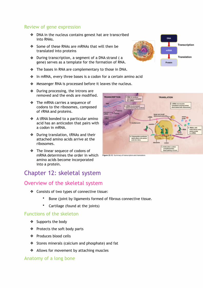
Review of gene expression
❖ DNA in the nucleus contains genest hat are transcribed into RNAs.
❖ Some of these RNAs are mRNAs that will then be translated into proteins
❖ During transcription, a segment of a DNA strand ( a gene) serves as a template for the formation of RNA.
❖ The bases in RNA are complementary to those in DNA.
❖ In mRNA, every three bases is a codon for a certain amino acid
❖ Messenger RNA is processed before it leaves the nucleus.
❖ During processing, the introns are removed and the ends are modified.
❖ The mRNA carries a sequence of codons to the ribosomes, composed of rRNA and proteins.
❖ A tRNA bonded to a particular amino acid has an anticodon that pairs with a codon in mRNA.
❖ During translation, tRNAs and their attached amino acids arrive at the ribosomes.
❖ The linear sequece of codons of mRNA determines the order in which amino acids become incorporated into a protein.
Chapter 12: skeletal system
Overview of the skeletal system ❖ Consists of two types of connective tissue:
• Bone (joint by ligaments formed of fibrous connective tissue.
• Cartilage (found at the joints)
Functions of the skeleton
❖ Supports the body
❖ Protects the soft body parts
❖ Produces blood cells
❖ Stores minerals (calcium and phosphate) and fat
❖ Allows for movement by attaching muscles
Anatomy of a long bone

❖ Bones of the body vary greatly in size and shape.
❖ Long bone
• a type of bone common in the arms and legs.
• Diaphysis
o the shaft, or main portion of the bone.
o Has a large medullary cavity, whose walls are composed of compact bone. It is lined with a thin, vascular membrane (the endosteum) and is filled with yellow bone marrow, which stores fat.
• Epiphysis
o Expanded region at the end of a long bone.
o Are seperated from the diaphyses by a small region of mature bone called the metaphysis, which contain the epiphyseal plate = a region of cartilage that allows for bone growth.
o Are composed of largely spongy bone bone thatcontins red bone marrow, where blood cells are made.
o Are coated with a thin layer of hyaline cartilage, which is also called articular cartilage, because it occurs at a joint.
• Periosteum
o Covers the long bone completely by a by a layer of fibrus connective tissue.
o This covering contains blood vessels, lymphatic vessels and nerves.
o Is continuous with ligaments and tendeons connected to a bone.
Bone
❖ Compact bone
• Is highly organized and composed of tubular units called osteons.
• In a cross section of an osteon, bone cells called osteocytes lie in a lacunae = tiny chambers arranged in concentric circles around a central canal.
• Matrix fills the space between the rows of lacunae.
• Canaliculi = tiny canals that run through the matrix. They connect the lacune with one another and with the central canal. The cells stay in contact by strands of cytoplasm that extend into the canaliculi.
• Osteacytes nearest the center of an osteon exchange nutirents and wastes with the blood vessels in the central canal. These cells then pass on nutrients and collect wastes from the other cells via gap junctions.

❖ Spongy bone
• Has an unorganized appearace.
• Contains numerous thin plates, called trabeculae, seperated by unequal spaces.
• Spongy bone is lighter than compact bone, spongy bone is still designed for strength.
• Spaces of spongy bone are often filled with red bone marrow = a specialized tissue that produces all types of blood cells.
• The osteocytes of spongy bone are irregularly placed within the trabeculae. Canaliculi bring them nutrients from the red bone marrow.
Cartilage
❖ Not as strong as bone, but more flexible
❖ Its matrix is gel-like and contains many collagenous and elastic fibers.
❖ The cells, called chondrocytes, lie within a lacunae that are irregularly grouped.
❖ Has no nerves, making it well suited for padding joints where the stresses of movement are intense.
❖ Also had no blood vessels and relies on neighboring tissues for nutrient and waste exchange. This makes it slow to heal.
❖ Three types of cartilage
• Hyaline cartilage
o Firm and somewhat flexible
o Matrix appears uniform and glassy, but actually it contains a generous supply of collagen fibers.
o Found ends of long bones, nose, ends of ribs, larynx and trachea
• Fibrocartilage
o stronger than hyaline cartilage, because the matrix contains wide rows of thick collagenous fibers.
o Is able to withstand both pressure and tension
o Found in the disks between vertebrae and in the cartilage of the knee
• Elastic cartilage
o more flexible than hyaline cartilage, because the matrix contains mostly elastin fibers.

o Found in the ear flaps and epiglottis
Fibrous connective tissue
❖ Contains rows of cells called fibroblasts, separated by bundles of collagenous fibers.
❖ This tissue makes up
• Ligaments : connect bone to bone
• Tendons: connect muscle to bone at the joint. (also called an articulation)
Bones of the axial skeleton ❖ 206 bones of the skeleton, are classified according to whether they occur in the
axial skeleton or the appendicular skeleton.
❖ Axial skeleton lies in the midline of the body and consists of the skull, hyoid bone, vertebral column and the rib cage.
The skull
The cranium
❖ Protects the brain
❖ In adults: it is composed of eight bones fitted tightly together
❖ In newborns: certain cranial bones are not completely formed. These bones are joined by membranous regions called fontanels (usually closed by the age of 16 by the process of intramembranous ossification.)
❖ Major bones:
• Fontal bone
• Parietal bones
• Occipital bone
o Foramen magnum = a large opening through which the spinal cord passes and becomes the brain stem
• Temporal bone
o Has an opnening (external auditory canal) that leads to the middle ear.
❖ Sphenoid bone
• Shaped like a bat with outstretched wings, extends across the floor of the cranium from one side to the other.

• Is the keystone of the cranial bone, because all the other bones articulate with it.
• Completes the sides of the skull and contribute to forming the orbits (eye sockets)
❖ Ethmoid bone
• Lies in front of the sphenoid, also helps form the orbits and the nasal septum.
❖ Sinuses
• Air spaces lined by muscous membrane.
• Reduce the weight of the skull and give a resonant sound to the voice.
• Are named according to the bones in which they are located.
• Major sinuses: frontal, sphenoid, ethmoid and maxillary
• Smaller set of sinuses: mastoid sinuses, drain into the middle ear.
o Mastoiditis = a condition that can lead to deafness, is an inflammation of these sinuses.
The facial bones
❖ Mandible: or lower jaw, is the only movable portion of the skull and it forms the chin.
❖ Maxillae: form the upper jaw and a portion of the eye sucket
❖ Zygomatic bones: the cheekbone prominences
❖ Nasal bones: form the bridge of the nose.
• Other bones are part of the nasal septum, whivh divides the interior of the nose into two nasal cavities.
• The lacrimal bone contains the opening for the nasolacrimal canal, which drains tears from the eyes to the nose.
❖ Certain cranial bones contribute to the face.

The hyoid bone
❖ Not a part of the skull, but part of the axial skeleton.
❖ Only bone in the body that does not articulate with another bone.
❖ Is attached to the temporal bones by muscles and ligaments and to the larynx (= voice box at the top of the trachea in the neck region) by a membrane.
❖ Anchors the tongue and serves as the site for the attachment of muscles associated with swallowing.
❖ Does not fracture easily.
The vertebral column
❖ Consists of 33 vertebrae
❖ Normally, the vertebral column has four curvatures that provide more resilience and strenghth than a straight column could provide.
❖ Scoliosis = an abnormal lateral (sideways) curvature of the spine.
❖ Kyphosis = an abnormal posterior curvature that often results in “hunchback”. An abnormal anterior curvature results in Iordosis or “swayback”.
❖ As the individual vertebrae are layered on top of one another, they form the vertebral column.
❖ The vertebral canal is in the center of the column, and the spinal cord passes through this canal.
❖ Spinal nerves control skeletal muscles contraction, among other functions.
Types of vertebrae
❖ The various vertebrae are named according tot heir location in the vertebral column.
❖ Cervical (7) vertebrae: are located in the neck.
• First cervical vertebra, called the atlas, holds up the head. It allows the head to tilt from side to side.
• Second cervical vertebra, called the axis, because it allows rotation, as when we shake the head “no”.
❖ Thoracic (12) vertebrae: have long, thin spinous processes and articular facets for the attachment of the ribs.
❖ Lumbar (5) vertebrae: have large bodies and thick processes.
❖ Sacrum = five sacral vertebrae fused together
❖ Coccyx or tailbone, is usually composed of four fused vertebrae.
Intervertebral disks
❖ Composed of fibrocartilage that act as padding.
❖ The disks prevent the vertebrae form grinding against one another.
❖ They also absorb shock caused by movement.

❖ The disks become weakened with age and can herniate and rupture
❖ Pain occurs if a disk presses against the spinal cord and/or spinal nerves. If that occus, surgical reoval of the disk may relieve the pain.
The rib cage
❖ Also called thoracic cage, is composed of the thoracic vertebrae, the ribs and their associated cartilages, and the sternum.
❖ Part of the axial skeleton
❖ Demonstrates how the skeleton is protective but also felxible.
❖ Protects the heart and lungs
The ribs
❖ Is a flattened bone that originates at the thoracic vertebrae and proceeds toward the anterior thoracic wall.
❖ There are 12 pairs of ribs.
❖ A rib articulates with the body and transverse process of its corresponding thoracic vertebra. Each rib curves outward and then forward and downward.
❖ True ribs
• Upper seven pairs of ribs
• Connect directly to the sternum by means of a long strip of hyaline cartilage called costal cartilage.
❖ False ribs
• Ribs 8 through 12
• Because their costal cartilage at the end of the ribs does not connect directly to the sternum
❖ Floating ribs
• Ribs 11 and 12
• Because they have no connection with the sterum.
The sternum
❖ Known as the breastbone
❖ Helps protect the heart and body
❖ Is composed of three bones
• Manubrium (the handle)

o Articulates with the clavicles of the appendicular skeleton.
o Costal cartilages from the first pair of ribs also join to the manubrium.
o Joins with the body of the sternum at an angle (important because it allows the ribs tob e counted)
• The body (the blade)
• Xiphoid process (the point of the blade)
o The variably shaped xiphoid process serves as an attachment site for the diaphragm, which seperates the thoracic cavity from the abdominal cavity.
Bones of the appendicular skeleton ❖ Consists of the bones within the pectoral and pelvic girdles and their attached
limbs.
❖ A pectoral (shoulder) girdle and upper limb are specialized for flexibility.
❖ The pelvic (hip) girdle and lower limbs are specialized for strength.
The pectoral girdle and upper limb
❖ The body has left and right pectoral girdles.
• Each consist of a
o Clavicle (collarbone)
▪ Extends across the top of the thorax.
▪ It articulates with (joins with) the sternum and the acromion process of the scapula, a visible bone in the back.
o Scapula (shoulder blade)
• The muscles of the arm and chest attach to the coracoid process of the scapula.
• Glenoid cavity of the scapula
o Articulates with and is much smaller than the head of the humerus. This allows the arm to move in almost any direction but reduces stability. This is the joint most apt to dislocate. Ligaments and tendons stabilize this joint.
• Rotator cuff
o Tendons that extend to the humerus from four small muscles originating on the scapula.
o Vigorous circular movements of the arm can lead to rotator cuff injuries.
❖ The components of a pectoral girdle freely follow the movements of the upper limb, which consists of the humerus in the arm and the radius and ulna in the forearm.
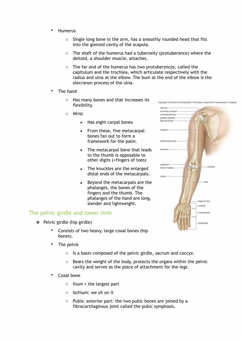
• Humerus
o Single long bone in the arm, has a smoothly rounded head that fits into the glenoid cavity of the scapula.
o The shaft of the humerus had a tuberosity (protuberance) where the deltoid, a shoulder muscle, attaches.
o The far end of the humerus has two protuberznces, called the capitulum and the trochlea, which articulate respectively with the radius and ulna at the elbow. The bum at the end of the elbow is the olecranon process of the ulna.
• The hand
o Has many bones and that increases its flexibility.
o Wrist
▪ Has eight carpal bones
▪ From these, five metacarpal bones fan out to form a framework for the palm.
▪ The metacarpal bone that leads to the thumb is opposable to other digits (=fingers of toes)
▪ The knuckles are the enlarged distal ends of the metacarpals.
▪ Beyond the metacarpals are the phalanges, the bones of the fingers and the thumb. The phalanges of the hand are long, slender and lightweight.
The pelvic girdle and lower limb
❖ Pelvic girdle (hip girdle)
• Consists of two heavy, large coxal bones (hip bones).
• The pelvis
o Is a basin composed of the pelvic girdle, sacrum and coccyx.
o Bears the weight of the body, protects the organs within the pelvic cavity and serves as the place of attachment for the legs.
• Coxal bone
o Ilium = the largest part
o Ischium: we sit on it
o Pubis: anterior part: the two pubic bones are joined by a fibrocartilaginous joint called the pubic symphasis.

• Acetabulum (hip socket) occurs where this three bones meet.
• The male pelvis is different from the female pelvis.(adaptions vaginal delivery)
• Femur (thighbone)
o Longest and strongest bone in the body
o Head of the femur articulates with the coxal bones at the acetabulum and the short neck better positions the legs for walking.
o Has two large processes: the greater and lesser trochanters, which are places of attachment fort thigh muscles, buttock muscles and hip flexors.
o At its distal end, the femur has medial and lateral condyles that articulate with the tibia of the leg.
• Tibia
o Region of the knee and the patella or kneecap.
o Patella is held in place by the quadriceps tendon, which continues as a ligament that attaches to the tibial tuberosity.
o At the distal end, the medial malleolus fo the tibia causes the inner bulge of the ankle.
• Fibula
o More slender bone in the leg.
o Has a head that articulates with the tibia and a distal lateral malleolus that forms the outer bulge of the ankle.
• Foot
o Ankle
▪ Contains seven tarsal bones.
▪ The calcaneus or heel bone is also part of the ankle.
▪ The talus and calcaneus support the weight of the body.
o Instep
▪ Has five elongated metatarsal bones.
o Five toes
▪ Bines of the toes are called phalanges
▪ In the foot, the phalages are stout and extremely sturdy.
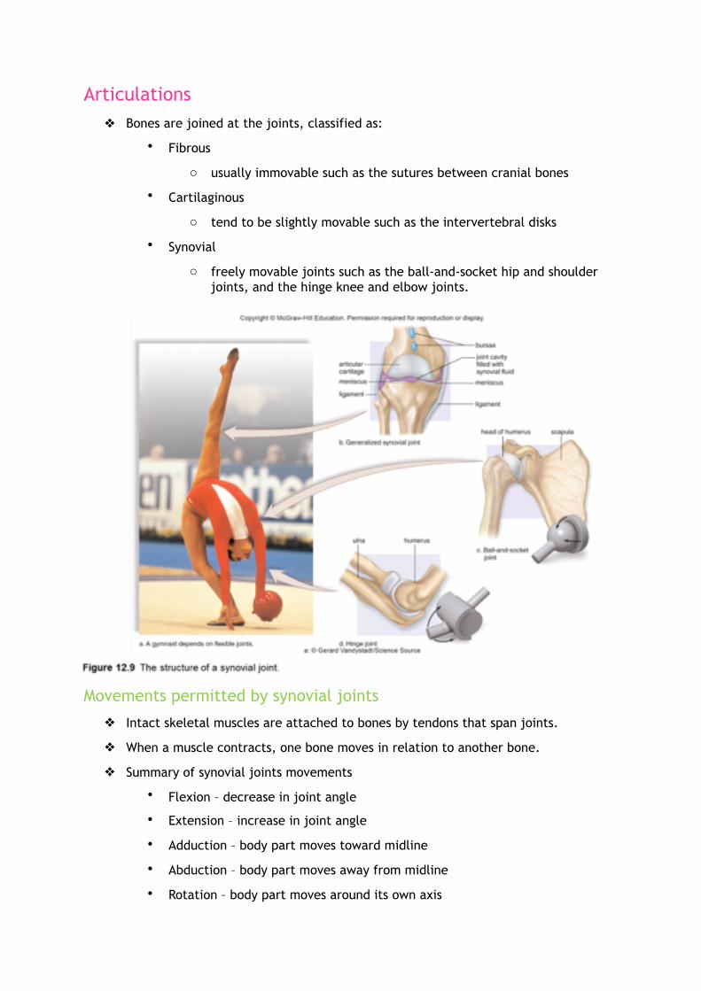
Articulations ❖ Bones are joined at the joints, classified as:
• Fibrous
o usually immovable such as the sutures between cranial bones
• Cartilaginous
o tend to be slightly movable such as the intervertebral disks
• Synovial
o freely movable joints such as the ball-and-socket hip and shoulder joints, and the hinge knee and elbow joints.
Movements permitted by synovial joints
❖ Intact skeletal muscles are attached to bones by tendons that span joints.
❖ When a muscle contracts, one bone moves in relation to another bone.
❖ Summary of synovial joints movements
• Flexion – decrease in joint angle
• Extension – increase in joint angle
• Adduction – body part moves toward midline
• Abduction – body part moves away from midline
• Rotation – body part moves around its own axis

• Supination – hand faces anterior or downward
• Pronation – hand faces posterior or downward
• Circumduction – body part moves so that a cone shape is outlined
• Inversion – sole of foot turns inward
• Eversion – sole of foot turns outward
Bone growth and homeostasis ❖ Theskeleton starts forming at about 6 weeks.
❖ Most bones grow in lengt hand width through adolescence, but some continue enlarging until about age 25.
❖ Remodeling = process in which bones can grow throughout a lifetime, because they are able to respond to stress by changing size, shape and strength.
❖ If a bone fractures, it can heal by bone repair.
❖ Several different types of cells are involved in bone growth, remodeling and repair
• Osteoblasts
o bone-forming cells
o They secrete the organic matrix of bone and promote the deposition of calcium salts into the matrix.
• Osteocytes
o mature bone cells that maintain bone structure derived from osteoblasts.
o They maintain the structure of bone.

• Osteoclasts
o bone-absorbing cells
o They break down bone and assist in returning calcium and phosphate to the blood.
• Chondrocytes
o cartilage-forming cells
Bone development and growth
❖ Ossification is the formation of bone in 2 distinct ways:
Intramembranous ossification
❖ For example flat bones, such as the bones of the skull
❖ Bones develop between sheets of fibrous connective tissue.
❖ Here, cells derived from connective tissue cells become osteoblasts located in issification centers.
❖ The osteoblasts
• secrete the organic matrix of bone.
o This matrix consists of mucopolysaccharides and collagen fibrils. Calsification occurs when calcium salts are added to the organic matrix.
• Promote calcification or ossification of the matrix.
o Ossification results in the formation of soft sheets or trabeculae of spongy bone.
o Spongy bone remains inside a flat bone. Spongy bone contains red bone marrow.
o A periosteum forms outside the spongy bone
• Osteoblasts derived from the priosteum carry out further ossification.
o Trabeculae form and fuse to become compact bone.
o The compact bone forms a bone collar that surrounds the spongy bone on the inside.
Endochondral ossification
❖ Used by most bones, which means that the bone forms within the cartilage.
❖ Bone replaces the cartilaginous (hyaline) models of the bones. Gradually, the cartilage is replaced by the calcified bone matrix that makes these bones capable of bearing weight.
❖ Inside, bone formation spreads from the center to the ends.
❖ For example the long bones, such as tibia.
1. The cartilage model

o chondrocytes lay down hyaline cartilage in the shape of the future bones. As the cartilage models cancify, the chondrocytes die off.
2. The bone collar
o osteoblasts secrete bone matrix and results in a collar made of compact bone.
3. The primary ossification center
o osteoblasts are brought interiorly by blood and lay down spongy bone.
4. The medullary cavity and secondary ossification sites
o spongy bone of the diaphysis is absorbed by osteoclasts becoming the medullary cavity and secondary ossification centers form in the epiphyses after birth.
5. The epiphyseal (growth) plate
o a cartilage band that acts as a growth plate that allows bones to lengthen.
❖ The epiphyseal plate contains four layers.
• Resting zone: where cartilage remains
• Proliferating zone: in which chondrocytes are producing new cartilage cells.
• Degenerating zone: the cartilage cells are dying off.
• Ossification zone: bone is forming: causes the length of the bone to increase.


❖ Final size of the bones
❖ When the epiphyseal plates close, bone lengthening can no longer occur.
❖ The epiphyseal plates in the arms and legs of women close at about age 16-18, in men until about age 20. Other bones may continue to grow until age 25.
❖ Hormones = chemical messengers that are produced by one part of the body and act on a different part of the body, are secreted by the endocrine glands and distributed about the body by the bloodtsream. They control the activitu of the epiphyseal plate.
Hormones affect bone growth
❖ Vitamin D is converted to a hormone that allows calcium absorption in the intestine.
• In the absence of vitamin D, children can develop rickets = a condition marled by bone deformities, including bowed long bones.
❖ Growth hormone (GH) stimulates general bone growth and the epiphyseal plates.
• However, growth hormone is somewhat ineffective if the metabolic activity of cells is not promoted.
• Thyroid hormone: promotes the metabolic activity of cells.
• Acromegaly: results from excess GH in adults following epiphyseal fusion. This condition produces excessive growth of bones in the hands and face.
❖ Sex hormones increase growth during adolescence (growth spurt)
• These hormones apparently stimulate osteoblast activity.
• Rapid growth causes epiphyseal plates to become “paved over” by the faster-growing bone tissue within 1 or 2 years of the onset of puberty.
Bone remodeling and calcium homeostasis
❖ Bone is constantly being broken downby osteoclasts and reformed by osteoblasts in the adult.
❖ The process of bone renewal, often called bone remodeling, recycles as much as 18% of bone each year. It normally keeps bones strong.
❖ Paget disease: new bone is generated at a faster-than-normal rate. This rapid remodeling produces bone that’s softer and weaker than normal bone and can cause bone pain, deformities and fractures.
❖ Remodeling allows bones to respond to stress.
• If you engage in an activity that calls upon the use of a particular bone, the bone enlarges in diameter at the region most affected by the activity.
• During this process, osteoblasts in the periosteum form compact bone around the external bone surface and osteoclasts break down bone on the internal bone surface around the medullary cavity.
• Increasing the size of the medullary cavity prevents the bones from getting too heavy and thick. Excercises such as, walking, jogging and weight lifting are recommended.

❖ Bone recycling allows the body to regulate the amount of calcium in the blood.
• The skeleton acts as a reservoir for storage of this important mineral.
• If the blood calcium rises above normal, at least some of the excess is deposited in the bones.
• If the blood calcium dips to low, calcium is removed from the bones tob ring it back up to the normal level.
❖ It regulates the calcium in the blood through hormones.
• Parathyroid hormone (PTH) increases blood calcium by accelerating bone recycling.
• Calcitonin decreases blood calcium. The reduction of estrogen in older women is ofte given as reason for the development of weak bones, called osteoporosis.
❖ Bone remodeling
• Diameter increases as bone absorption inside the shaft is matched by bone formation outside the shaft.
Bone repair
❖ Is required afte rit breaks or fractures.
❖ Fracture repair takes place over a span of several months in a series of 4 steps
1) Hematoma (6-8 hours)
• blood clot formed between broken bones.
2) Fibrocartilaginous callus (3 weeks)
• cartilaginous callus forms between broken bones. (tissue repair begins)
3) Bony callus (3-4 months)
• cartilaginous callus is turned to bone
4) Remodeling
• old bone tissue is replaced by new bone tissue
❖ A complete fracture = If the bone is broken clear through
❖ Incomplete fracture= If the bone is not separated into two parts.
❖ Simple fracture = If it does not pierce the skin.
❖ Compound fracture = I fit

does pierce the dkin.
❖ Impacted fracture = means that the broken end are wedged into each other.
❖ Spiral fracture = occurs when the break is ragged duet o twisting of a bone.
Blood cells are produced in bones
❖ The bones of your skeleton contains two types of marrow
• Yellow marrow
o Fat is stored, making it part of the body’s energy reserves.
• Red marrow
o Site of blood cell production.
o The red blood cells are the carriers of oxygen in the blood. Oxygen is necessary for the production of ATP by aerobic cellular respiration.
o White blood cells also originate in the red bone marrow. The white cells are involved in defending your body against pathogens and cancerous cells; without the, you would soon succumb to disease and die.
Chapter 13: Muscular system
Overview of the muscular system ❖ Is involved with movement and is made up of muscles.
❖ All muscles contract, or shorten. It is this activity that causes movement.
Types of muscles
❖ Three types of muscles
❖ The cells of these tissues are called muscle fibers.
• Smooth muscle
o involuntary muscle found in hollow organs and blood vessels
o Although smooth muscle is slower to contract than skeletal muscle. It can sustain prolonged contractions and does not fatigue easily.
• Cardiac muscle
o involuntary muscle found in the heart
o Its fibers are generally uniny-ucleated, striated and tubular.
o Branching allows the fibers to interlock at intercalated disks.
o The plasma membranes at intercalated disks cotain gap junctions that permit contractions to spread quickly throughout the heart wall.
o Contraction of cardiac muscle is rhythmic
• Skeletal muscle

o voluntary muscle that is attached to the skeleton
o Fibers are tubular, multinucleated and striated.
Skeletal muscles of the body
❖ Humans belong to a class of animals called the vertebrates.
❖ Vertebrate animals possess an internal vertebral column, a skeletal and jointed appendages.
❖ Our skeletal muscles are attached to the skeleton and their contraction causes the movement of bones at a joint.
Functions of skeletal muscles
❖ Support the body by allowing us to stay upright
❖ Allow for movement by attaching to the skeleton
❖ Help maintain a constant body temperature
❖ Assist in movement in the cardiovascular and lymphatic vessels
❖ Protect internal organs and stabilize joints
Basic structure of skeletal muscles
❖ Skeletal muscles are well organized.
❖ A whole muscle contains bundles of skeletal muscle fibers called fascicles.
• Within a fasicle, each fiber is surrounded by

connective tissue. The fasicle is also surrounded by connective tissue.
❖ Muscles are covered with fascia = a type of connective tissue that extends beyond the muscles and becomes its tendon.
• Tendon = connective tissue that connects muscle to bone.
• Tendons quite often extend past a joint before anchoring a muscle to a bone.
• Small, fluid-filled sacs called bursae, can often be found between tendons and bones. The bursae act as cushions, allowing ease of movement.
Skeletal muscles work in pairs
❖ In general, each muscle is concerned with the movement of only one bone.
❖ Origin = attachment of a muscle on a stationary bone
❖ Insertion = attachment of a muscle on a bone that moves. When a muscle contracts, it pulls on the tendons at its insertion and the bone moves. (for example, when the biceps brachii contracts, it raises the forearm
❖ Skeletal muscles usually function in groups.To make a particular movement, your nervous system stimulates an appropriate group of muscles.
❖ Agonist or prime mover = For any particular movement one muscle does most of the work.
❖ Synergists = assist the agonist and make its action more effective.
❖ When muscles contract, they shorten. Therefore muscles can only pull and not push. This means that muscles work in opposite pairs.
❖ Antagonist = The muscle that acts opposite to a prime mover. (for example, the biceps brachii and the triceps brachii are antagonists)
❖ Not all skeletal muscles are involved in the movement of limbs. For example, the facial muscles produce the facial expressions that tell us about the emotions and mood of a person and therefore play an important role i our interactions with other people.
Names and actions of skeletal muscles
❖ The names of the various skeletal muscles are often combinations of the following terms used to characterize muscles:
• Size = the gluteus maximus is the largest buttock muscle. The gluteus minimus is the smallest of the gluteal muscles. Other terms used to indicate size are vastus (huge), longus (long) and brevis (short).
• Shape = the deltoid is triangular (Greek letter delta is Δ). The trapezius is shaped like a trapezoid. Other terms used are latissimus (wide) and teres (round).
• Location = the frontalis overlies the frontal bone. The external oblique muscles are located outside the internal obliques. Other terms used are pectoralis (chest), gluteus (buttock), bracchi (arm) and sub (beneath).
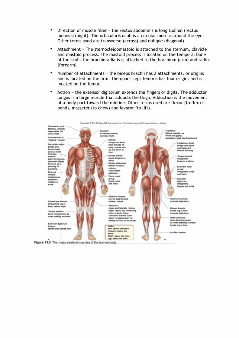
• Direction of muscle fiber = the rectus abdominis is longitudinal (rectus means straight). The orbicularis oculi is a circular muscle around the eye. Other terms used are transverse (across) and oblique (diagonal).
• Attachment = The sternocleidomastoid is attached to the sternum, clavicle and mastoid process. The mastoid process is located on the temporal bone of the skull. the brachioradialis is attached to the brachium (arm) and radius (forearm)
• Number of attachments = the biceps brachii has 2 attachments, or origins and is located on the arm. The quadriceps femoris has four origins and is located on the femur.
• Action = the extensor digitorum extends the fingers or digits. The adductor longus is a large muscle that adducts the thigh. Adduction is the movement of a body part toward the midline. Other terms used are flexor (to flex or bend), masseter (to chew) and levator (to lift).

Skeletal muscle fiber contraction ❖ Skeletal muscle tissue had alternating light and dark bands, giving it a striated
appearance. These bands are due to the arrangement of myofilaments in a muscle fiber.
Muscle fibers and how they slide
❖ Muscle fiber = a cell containing the usual cellular components, but special names have been assigned to some of these components.
• The plasma membrane is called the sarcolemma.
• The cytoplasm is called the sarcoplasm.
• The endoplasmic reticulum is called the sarcoplasmic reticulum and stores calcium.
❖ A muscle fiber also has some unique anatomical characteristics
• One feature is its T (for transverse) system. The sarcolemma forms T tubules, which penetrate, or dip down, into the cells. The transverse tubules come into contact, but do not fuse, with expanded portions of the sarcoplasmic reticulum (which are calcium storage sites, essential for muscle contraction.)
• The sarcolemma encases hundreds and sometimes even thousands of myofibrils
o Myofibrils = are the contractile portions of the muscle fibers.
• Any other organelles, such as mitochondria, are located in the sarcoplasm between the myofibrils.
• The sarcoplasm also contains glycogen, which provides stored energy for muscle contraction.
• In addition, sracoplasm includes the red pigment myoglobin, which binds oxygen until it is needed for muscle contraction.
Myofibrils and sarcomeres
❖ A muscle cell or fiber is roughly cylindrical in shape.
❖ Myofibrils
• Smaller cylinders grouped inside this larger cylinder.
• Run the entire length of the muscle fiber.

• Are composed of even smaller cylinders called myofilaments.
❖ Thus, the muscle cell is a set of small cylinders (myofilaments) assembled into larger cylinders (myofibrils) clustered within the largest cylinder (the muscle fiber).
❖ The skeletal muscle fibers have light and dark bands called striations. The sytiations of skeletal muscle fibers are formed by the placement of myofilaments within myofibrils.
❖ Two types of myofilaments
• Thick myofilaments are made up of a protein called myosin.
• Thin myofilaments are composed of a second protein termed actin.
❖ Myofibrils are further divided into sarcomeres = are the repeating units of actin and myosin found along a myofibril.
❖ A sarcomere extends between two dark vertical lines called the Z lines.
Myofilaments
❖ Thick filaments
• A thick filament is composed of several hundred molecules of the protein myosin. Each myosin molecule is shaped like a golf club.
❖ Thin filaments
• Primarily, a thin filament consists of two intertwining strands of the protein actin.
• Two other proteins, called tropomyosin and troponin, also playa role.
❖ Sliding filaments
• When muscles are stimulated, electrical signals travel across the sarcolemma and then down a T tubule.
• In turn, this signals calcium to be released from the sarcoplasmic reticulum. Now the muscle fibers contracts as the sarcomeres within the myofibrils shorten.
• When a sarcomere shortens, the actin (thins) filaments approach one another as they slide past the myosin (thick) filaments. This causes the I band to shorten, the Z line to move inward and the H band to almost or completely disappear.
• The movement of actin filaments in relation to myosin filaments is called the sliding filament model of muscle contraction.
• ATP supplies the energy for muscle contraction.
• Although the actin filaments slide past the myosin filaments, it is the myosin filaments that do the work. Myosin filaments break down ATP and their cross-bridges pull the actin filament toward the center of the sarcomere.

Control of muscle fiber contraction
❖ Nerve impulses travel down a motor neuron to a neuromuscular junction.
❖ Acetylcholine (ACh) is released from the neuron and binds to the muscle fiber.
❖ This binding stimulates the fiber causing calcium to be released from the sarcoplasmic reticulum.
❖ Released calcium combines with troponin, a molecule associated with actin.
❖ This causes the tropomyosin threads around actin to shift and expose myosin binding sites.
❖ Myosin heads bind to these sites forming cross-bridges.
❖ ATP binds to the myosin heads and is used for energy to pull the actin filaments towards the center of the sarcomere – contraction now occurs.
❖ What role does ATP play in muscle contraction and rigor mortis?
• ATP is needed to attach and detach the myosin heads from actin.
• After death, muscle cells continue to produce ATP through fermentation and muscle cells can continue to contract.
• When ATP runs out, some myosin heads are still attached and cannot detach, causing rigor mortis.
• Rigor mortis and body temperature may be used to estimate time of death.
Whole muscle contraction ❖ In order for a whole muscle, such as the biceps or triceps, to contract, the
individual muscle fibers must be activated by signals from the nervous system.

Muscles have motor units
❖ Each axon within a nerve stimulates a number of muscle fibers.
❖ A motor unit
• a nerve fiber and all of the muscle fibers it stimulates
• Operates in the principle of “all-or-none”, because all of the muscle fibers are stimulated at once.
• What varies is the number of muscle fibers within a motor unit.
❖ Muscle twitch
• When a motor unit is stimulated by infrequent electrical impulses, a single contraction occurs. This response is called a muscle twitch and lasts only a fraction of a second.
• Muscle twitch is customarily divided into three stages
o Latent period: is the time between stimulation and initiation of contraction. The events that begin muscle contraction are occurring. The neurotransmitter Ach diffuses across the synaptic cleft, causing an electrical signal to spread across the sarcolemma and down the T tubules.
o Contraction period: follows as calcium leaves the sarcoplasmic reticulum and myosin-actin cross-bridges form. The muscle shortens as its contracts. The force increases as the muscle contracts.
o Relaxation period: completes the muscle twitch. Myosin-actin cross-bridges are broken and calcium returns to the sarcoplasmic reticulum. Force diminishes as the muscle returns to its former length. (If a motor unit is given a rapid series of stimuli, it can respond to the next stimulus without relaxing completely.)
❖ Summation
• an increase in muscle contraction until the maximal sustained contraction, called tetanus, is reached.
• Tetanus continues until the muscle fatigues due to depletion of energy reserves.
• Fatigue is apparent when a muscle relaxes, even though stimulation continues.
• The tetanus of muscles cells is not the same as the infection called tetanus. The infection called tetanus is caused by the bacterium clostridium tetani.

Death occurs because the muscles, including the respiratory muscles, become fully contracted and do not relax.
❖ Recruitment
• A whole muscle typically contains many motor units. As the intensity of nervous stimulation increases, more motor units in a muscle are activated. This phenomenon is known as recruitmnt.
• Maximum contraction of a muscle would require that all motor units be undergoing tetanic contraction. This rarely happens,because they could all fatigue at the same time. Instead, some motor units are contracting maximally while others are resting, allowing sustained contraction to occur.
Muscle tone
❖ Is a continuous, partial contraction of alternate muscle fibers causing the muscle to look firm.
❖ One desirable effect of exercise is to have a good muscle tone. Muscles that have a good tone are are firm and solid, as opposed to soft and flabby.
❖ The amount of muscle tone is dependent on muscle contraction.
Energy for muscle contraction
❖ Muscles can use various fuel sources for energy and they have various ways of producing the ATP needed for muscle contraction.
Fuel sources for exercise
❖ A muscle has four possible energy sources.
• Two of these are stored in muscle (glycogen and triglycerides).
• Two are acquired from blood (glucose and fatty acids).
❖ The amount of each of these that is used depends on exercise intensity and duration.
❖ Notice that as the length of the exercise period is increased, use of muscle energy stores, such as glycogen and triglycerides, decreases and use of energy sources from the blood (glucose and fatty acids) increases.
❖ Blood glucose and plasma fatty acids are delivered to muscles by circulating blood.
❖ Submaximal exercise burns fat better that maximal exercise, for reasons we will now explore.
Sources of ATP for muscle contraction
❖ Muscle cells store limited amounts of ATP.
❖ Once stored ATP is used up, the cells have three ways to produce more ATP.
• The creatine phosphate (CP) pathway
o Most simple and rapid way,

because it consists of only one reaction.
o This reaction occurs in the midst of sliding filaments.
o CP is formed only when a muscle cell is resting and only a limited amount is stored.
o The CP pathway is used at the beginning of submaximal exercise and during short-term, high-intensity exercise that lasts less than 5 seconds.
• Fermentation
o Produces two ATP molecules from the anaerobic breakdown of glucose to lactate.
o This pathway is one most likely to begin with glycogen. Hormones provide the signal to muscle cells to break down glycogen, making glucose available as an energy source.
o Fermentation is fast-acting, but it results in the build-up of lactate. Formation of lactate is noticeable because it produces short-term muscle aches and fatigue on exercising.
o The continued intake of oxygen, called oxygen debt, is required, in part, to complete the metabolism of lactate and restore cells tot heir original energy state.
o The lactate is transported to the liver, where 20 % of it is completely broken down to carbon dioxide and water.
o The ATP gained by this respiration is then used to reconvert 80% of the lactate to glucose and then glycogen.
o In persons who train, the number of mitochondria in individual muscles increases. There is a greater reliance on these additional mitochondria to produce ATP. Muscles rely less on fermentation as a result.
• Cellular respiration
o Slowest of al three mechanisms.
o But is also the most efficient, typically producing several dozen molecules of ATP from each food molecule.
o Cellular respiration occurs in the motochondria.
o The process is aerobic and oxygen is supplied by the respiratory system.
o In addition, a protein called myoglobin in muscle cells delivers oxygen directly to the mitochondria.
o Cellular respiration can make use of glucose from the breakdown of stored muscle glycogen, glucose taken up from the blood, and/or fatty acids fromm fat digestion.
o Is more likely to supply ATP when exercise is submaximal in intensity.
Fast-twitch and slow-twitch muscle fibers
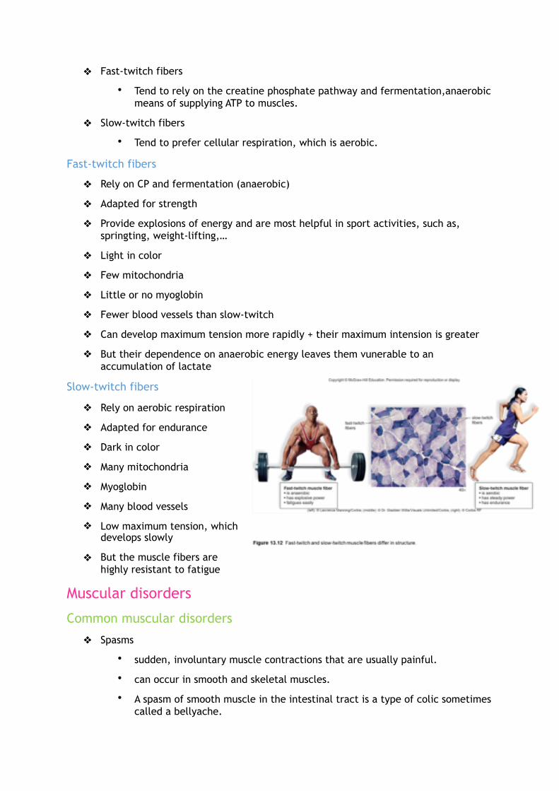
❖ Fast-twitch fibers
• Tend to rely on the creatine phosphate pathway and fermentation,anaerobic means of supplying ATP to muscles.
❖ Slow-twitch fibers
• Tend to prefer cellular respiration, which is aerobic.
Fast-twitch fibers
❖ Rely on CP and fermentation (anaerobic)
❖ Adapted for strength
❖ Provide explosions of energy and are most helpful in sport activities, such as, springting, weight-lifting,…
❖ Light in color
❖ Few mitochondria
❖ Little or no myoglobin
❖ Fewer blood vessels than slow-twitch
❖ Can develop maximum tension more rapidly + their maximum intension is greater
❖ But their dependence on anaerobic energy leaves them vunerable to an accumulation of lactate
Slow-twitch fibers
❖ Rely on aerobic respiration
❖ Adapted for endurance
❖ Dark in color
❖ Many mitochondria
❖ Myoglobin
❖ Many blood vessels
❖ Low maximum tension, which develops slowly
❖ But the muscle fibers are highly resistant to fatigue
Muscular disorders
Common muscular disorders
❖ Spasms
• sudden, involuntary muscle contractions that are usually painful.
• can occur in smooth and skeletal muscles.
• A spasm of smooth muscle in the intestinal tract is a type of colic sometimes called a bellyache.

❖ Convulsions (seizures)
• multiple spasms of skeletal muscles
❖ Cramps
• strong, painful spasms often of the leg and foot, usuallu due to strenuous activity.
❖ Facial tics
• Are spams that can be controlled volutarily, but only with great effort.
• Periodic eye blinking, head turning or grimacing.
❖ Strain
• stretching or tearing of a muscle.
❖ Sprain
• twisting of a joint involving muscles, ligaments, tendons, blood vessels, and nerves.
• When a tendon is inflamed by a sprain, tendinitis results.
• Tendinitis may irritate the bursa underlying the tendon, causing bursitis.
Muscular diseases
Myalgia and fibromyalgia
❖ Myalgia
• achy muscles due to injury or infection.
• Most common cause is either overuse or overstretchig of a muscle or a group of muscle.
• Myalgia without a traumatic history is often due to viral infections.
• May accompany myositis (inflammation of the muscles), either in response to a viral infection or as an immune system disorder.
❖ Fibromyalgia
• chronic condition whose symptoms include widespread pain, tendernessand stiffness of muscles.
• Precise cause is not known
• In some people, it occurs suddenly after an injury, while in others it has a more gradual progression.
Muscular Dystrophy
❖ Muscular dystrophy
• Is broad term applied ta a group of disorders characterized by a progressive degeneration and weakening of muscles. As muscle fibers die, fat and connectivetissue take their place.
❖ Duchenne muscular dystrophy

• Most common type
• Is inherited through a flawed gene on the X chromosome
• The lack of a protein called dystrophin causes the condition.
• When dystrophin is absent, calcium leaks into the cel land activates an enzyme that dissolves muscle fibers.
• In an attempt to treat the condition, muscles have been injected with immature muscle cells that do produce dystrophin.
Myasthenia gravis
❖ Myasthenia gravis
• autoimmune disorder that attacks the ACh receptor and weakens muscles of the face, neck, and extremities.
• Muscle contraction is impaired because the immune system mistakenly produces antibodies that destroy acetylcholine (Ach) receptors.
• In many cases, the first sign of the disease is a drooping of the eyelids and double vision.
• Treatment includes drugs that inhibit the enzyme that digests acetylcholine, so that ACh accumulates in neuromuscular junction.
❖ Amyotrophic lateral sclerosis (ALS)
• commonly known as Lou Gehrig’s disease; motor neurons degenerate and die leading to loss of voluntary muscle movement.
Muscle cancer
❖ Sarcomas
• cancers that originate in muscle, or the connective tissue associated with muscle.
• Occur in a variety of tissues, including bone, adipose and cartilage.
• Soft tissue sarcomas occur in both smooth and skeletal muscles.
• One of the more common forms of smooth muscle cancer = leiomyoma, which occurs in the uterine wall.
• Rhabdomyosarcomas are a rare form of cancer that originate in the skeletal muscle or move into the muscle from another location in the body.
• Both of these types of sarcomas may be either benign or malignant.
Homeastasis ❖ In many cases, the muscular system works closely with the skeletal system.
Both systems produce movement
❖ Movement is essential for maintaining homeostasis. The skeletal and muscular systems work together to enable body movement.

❖ This is most evidently illustrated by what happens when skeletal muscles contract and pull on the bones to which they are attached, causing movements at joints.
Both systems protect body parts
❖ Skeletal system plays an important role by protecting the soft internal organs of your body.
❖ Brain, heart, lungs, spinal cord, kidneys, liver and most of the endocrine glands are shield by the skeleton. In particular, the nervous and endocrine organs must be defended, so that they can carry out activities necessary for homeostasis.
❖ The skeletal muscles pad and protect the bones and the tendons and bursae associated with skeletal muscles reinforce and cushion the joints. Muscles of the abdominal wall offer additional protection to the soft internal organs.
Muscles help maintain body temperature
❖ When you are very cold, smooth muscle constricts inside the blood vessels supplying the skin. Thus, the amount of blood close to the surface of the body is reduced. This helps conserve heat in the body’ core, where vital organs lie.
❖ If you are cold enough you may start to shiver. Shivering is caused by involuntarily skeletal muscle contractions. This is initiated by temperature-sensitive neurons in the hypothalamus of the brain. Skeletal muscle contraction requires ATP, and using ATP generates heat.
❖ You may notice that you get goose bumps when you are cold. This is because arrector pili muscles contract. These tiny bundles of smooth muscle attached to the hair follicles cause the hairs to stand up. Goose bumps can also be a sign af fear.

Chapter 19: Patterns of chromosome inheritance
Chromosomes ❖ A human nucleus holds all the genetic material that is necessary to direct all the
functions in the body.
❖ The genetic material is arranged into chromosomes, structures that assist in transmission of genetic information from one generation to the next.
❖ The instructions in each chromosome are contained within genes, which in turn are composed of DNA.
❖ Chromosomes also contain proteins that assist in the organizational structure of the chromosome.
❖ Collectively, the DNA ad proteins are called chromatin.
❖ Humans have 46 chromosomes, which occur in 23 pairs.
• Twenty-two of these pairs are called autosomes.
• All these chromosomes are found in both males and females.
• One pair of chromosomes is called the sex chromosomes, because this pair contains the genes that control gender.
o Males have the seks chromosomes X and Y, and females have two X chromosomes.
A karyotype
❖ Any cell in the body except red blood cells, which lack a nucleus, can be a source of chromosomes for examination.

❖ In adults, it is easiest to obtain and use white blood cells separated from a blood sample for the purpose of looking at the chromosomes.
❖ For an examination of fetal chromosomes, a physician may recommend procedures such as chorionic villus sampling or amniocentesis.
❖ After a cell sample has been obtained, the cells are stimulated to divide in a culture medium.
❖ When a cell divides, chromatin condenses to form chromosomes.
❖ The nuclear envelope fragments, liberating the chromosomes.
❖ Next, a chemical is used to stop the division process when the chromosomes are most compacted and visible microscopically.
❖ Stains are apllied to the slides and the cells can be photographed with a camera attached to a microscope.
❖ Staining causes the chromosomes to have dark and light crossbands of varying widths and these can be used – in addition to size and shape – to help pair up the chromosomes.
❖ A computer is used to arrange the chromosomes in pairs.
The display is called a karyotype
❖ Picture shows karyotype of a normal male.
❖ A normal karyotype tells us a lot about a body cell.
❖ First, we should notice that a normal body cell is a diploid, meaning it has the full complement of 46 chromosomes.
❖ How does it happen that almost all the cells in your body (red blood cells and liver cells are exceptions) have 46 chromosomes?
• Mitosis = a form of cell division that begins when the fertilized egg starts dividing and ensures that every cell has 46 chromosomes.
❖ Each chromosome is composed of two identical parts, called sister chromatids.
• These chromosomes are said to be replicated or duplicated chromosomes because the two sister chromatids contain the same genes.
• Genes are the units of heredity thatcontrol the cell
• Replication of the chromosomes is possible only because each chromatid contains a DNA double helix.
❖ Centromere = a region that holds the chromatids together.
• Holds the chromatids together until a certain phase of mitosis, when the centromere splits.
• Once separated, each sister chromatid is a chromosome.

• In this way, a duplicated chromosome gives rise to two individual daughter chromosomes. When daughter chromosomes separate, the new cell gets one of each type and, therefore, a full complement of chromosomes.
The cell cycle ❖ Is an orderly process that has two parts
• Interphase
• Cell division
❖ To understand it, it is necessary to understand the structure of a cell.
• A human cell has a plasma membrane, which encloses the cytoplasm, the content of the cell outside the nucleus.
• In the cytoplasm are various organelles that carry on various functions necessary to the life of the cell.
• When a cell is not undergoing division, the chromatin (DNA and associated proteins) within a nucleus is a tangled mass of thin threads.
Interphase
❖ Most of the cell cycle is spent in interphase.
❖ This is the time when the organelles in the cell carry on their usual functions.
❖ As the cell continues through interphase, it gets ready to divide. The cell grows larger, the number of organelles doubles and the amount of chromatin doubles as DNA replication occurs.
❖ Interphase is divided into three main stages
• G1 stage
o occurs before DNA synthesis
o The cell returns to normal size and resumes its function in the body. A cell doubles its organelles (e.g., mitochondria and ribosomes) and it accumulates the materials needed for DNA synthesis.
• S stage
o Includes DNA replication
o A copy is made of all the DNA in the cell. DNA replication occurs, so each chromosome consists of two identical DNA double-helix molecules. These molecules occur in the strands called sister chromatids.
• G2 Stage
o Occurs after DNA replication
o The cell synthesizes the proteins needed for cell division, such as the protein found in microtubules. The role of microtubules in cell division is described in a later section.

❖ The amount of time the cell takes for interphase varies widely.
• Some cells, such as nerve cells and muscle cells, typically do not complete the cell cycle and are permanently arrested in G1. These cells are said to have entered a G0 stage.
• Embryonic cells spend very little time in G1 and complete the cell cycle in a few hours
Mitosis and cytokinesis
❖ Following interphase, the cell enters the cell division part of the cell cycle.
❖ Cell division has two stages
• M ( for “mitotic”) stage (mitosis)
o Is a type of nuclear division.
o Is also referred to as duplication division, because each new nucleus contains the same number and type of chromosomes as the former cell.
o During mitosis, the sister chromatids of each chromosome separate, becoming chromosomes distributed to two daughter nuclei.
o Mammalian cells usually require only about 4 hours to complete the mitotic stage.
o Mitosis is balanced by the process of apoptosis, or programmed cell death. Apoptosis occurs when cells are no longer needed or have become excessively damaged.
• Cytokinesis
o Is division of the cytoplasm.
o When cytokinesis is complete, two daughter cells are now present.
❖ The cell cycle, including interphase and cell division, occurs continuously in certain tissues.
Cell cycle control
❖ For a cell to reproduce succesfully, the cell cycle must be controlled.
❖ The cell cycle is controlled by checkpoints, which can delay the cell cycle until certain conditions are met.
❖ In addition, the cell cycle may be controlled by external factors, such as hormones and growth factors. Failure of the cell cycle control mechanisms may result in unrestricted cell growth, or cancer.
❖ Checkpoints
❖ G1 checkpoint
• Is especially significant, because if the cell cycle passes this checkpoint, the cell is committed to divide.
• If the cell does not pass this checkpoint, it can enter a holding phase called G0, during which it performs its normal functions but does not divide.
• The proper growth signals, such as certain growth factors, must be present for a cell to pass the G1 checkpoint.
• Additionally, the integrity of the cell’s DNA is also checked. If the DNA is damaged, proteins can stop the cycle at this checkpoint and place the cell in G0 phase.
• G0 phase acts as a holding phase; if the DNA can be repaired, the cell may reenter the cell cycle. If not, then internal mechanisms cause the cell to undergo apoptosis.
❖ Checkpoint G2
• The cell cycle halts momentarily until the cell verifies that DNA has replicated.
• This prevents the initiation of the M stage unless the chromosomes are duplicated.
• If DNA is damaged, as from exposure to solar (UV) radiation or X-rays, arresting the cell cycle at this checkpoint allows time for the damage tob e repaired, so that it is not passed on to daughter cells.
❖ Checkpoint during the mitotic stage
• The cycle hesitates between metaphase and anaphase to make sure the chromosomes are properly attached to the spindle and will be distributed accurately to the daughter cells.
• The cell cycle does not continue until every chromosome is rready for the nuclear division process.
❖ External control
❖ The cell cycle control system extends from the plasma membrane to particular genes in the nucleus.
❖ Some external signals, such as hormones and growth factors, can stimulate a cell to go through the cell cycle.
❖ During reception, an external signal delivers a message to a specific receptor embedded in the plasma membrane of a receiving cell. The signal is then relayed from the receptor to proteins inside the cell’s cytoplasm.
❖ The proteins form a pathway called the signal transduction pathway because they pass the signal from one to the other.
❖ The last signal activates genes in the nucleus of the cell. The expression of these genes may either stimulate or inhibit the cell cycle.
❖ Genes called proto-oncogenes stimulate the cell cycle. Genes called tumor suppressor genes inhibit the cell cycle.

Mitosis ❖ Is responsible for new cells in the developing embryo, fetus and child.
❖ Also responsible for replacement cells in an adult.
❖ Is often referred to as duplication division (zie eerder)
❖ During mitosis, the cell that divides is called the parent cel land the new cells are called daugther cells. Following mitosis, the daughter cells are practically genetically identical to the parent cell.
Overview of mitosis
❖ During S phase of the cell cycle, replication of the DNA occurs, thus duplicating the chromosomes.
❖ Each chromosomen ow contains two identical parts, called sister chromatids, held together at a centromere. They are called sister chromatids because they contain the same genes.
❖ As mitosis begins, the proteins in the chromatin assist in causing the chromosomes to become highly condensed. (Once this occurs, the chromosomes become visible under a light miscroscope)
❖ Only four chromosomes are depicted. Notice that there are two copies of each chromosome (one maternal and one paternal), so in this case the diploid (2n) number of chromosomes is four.
❖ During mitosis, the centromeres divide and the sister chromatids separate. Following separation during mitosis, each chromatid is called a chromosome.
❖ Each daughter cell gets a complete set of chromosomes and is diploid (2n).
❖ Therefore, each daughter cell receives the same number and types of chromosomes as the parent cell. Each daughter cell is genetically identical to the other and to the parent cell.

The mitotic spindle
❖ Another event of importance during mitosis is the duplication of the centrosome, the microtubule organizing center of the cell.
❖ After centrosomes duplicate, they separate and form the poles of the mitotic spindle, where they assemble the microtubules that make up the spindle fibers. The chromosomes are attached to the spidle fibers at their centromeres.
❖ An array of microtubules called an aster is also at the poles.
❖ Each centrosome contains a pair of centrioles, which consist of short cylinders of microtubules. They are absent in plant cells and their function in animals cells is not completely understood, although it is believed that they assist in the formation of the spindle that separates the chromatids during mitosis.
Phases of mitosis
❖ Stages of mitosis are continuous. One stage flows from the other with no noticeable interruption.
Prophase
❖ Several events occur that visibly indicate the cell is preparing to divide.
❖ The centrosomes outside the nucleus have duplicated and they begin moving away from one another toward opposite ends of the nucleus.
❖ Spindle fibers appear between the separating centrosomes.
❖ The nuclear envelope begins to fragment.
❖ The nucleolus, a special region of DNA, dissappears as the chromosomes coil and become condensed.
❖ The chromosomes are now clearly visible. Each is composed of two sister chromatids held together at a centromere.
Prometaphase
❖ The spindle fibers attach to the centromeres as the chromosomes continue to shorten and thicken.
❖ Chromosomes are randomly placed in the nucleus.
Metaphase
❖ The nuclear envelope is fragmented and the spindle occupies the region formerly occupied by the nucleus.
❖ The chromosomes are now at the equator (center) of the spindle.

❖ Is characterized by a fully formed spindle.
❖ The chromosomes, each with two sister chromatids, are aligned at the equator.
Anaphase
❖ At the start, the centromeres uniting the sister chromatids divide.
❖ Then the sister chromatids separate, becoming chromosomes that move toward opposite poles of the spindle.
❖ Separation of the sister chromatids ensures that each cell receives a copy of each type of chromosome and thereby has a full complement of genes.
❖ Is characterized by the diploid (2n) number of chromosomes moving toward each pole.
❖ Spindle function in anaphase
❖ Spindle brings about chromosomal movement.
❖ Two types of spindle fibers are involved in the movement of chromosomes during anaphase.
• One type extends from the poles to the equator of the spindle. There, they overlap. As mitosis proceeds, these fibers increase in length, which helps push the chromosomes apart.
• The chromosomes themselves are attached to other spindle fibers that extend from their centromeres to the poles.These fibers (composed of microtubules that can disassemble) become shorter as the chromosomes move toward the poles. Therefore, they pull the chromosomes apart.
❖ spindle fibers are composed of microtubules. Microtubules can assemble and disassemble by the addition or subtraction of tubulin (protein) subunits. This what ultimately causes the movement of the chromosomes.
Telophase
❖ Begins when the chromosomes arrive at the poles.
❖ The chromosomes become indistinct chromatin again.
❖ The spindle disappears as the nuclear envelope components reassemble in each cell.
❖ Each nucleus has a nucleolus, because each has a region of the DNA where ribosomal subunits are produced.
❖ Is characterized by the presence of two daughter nuclei.
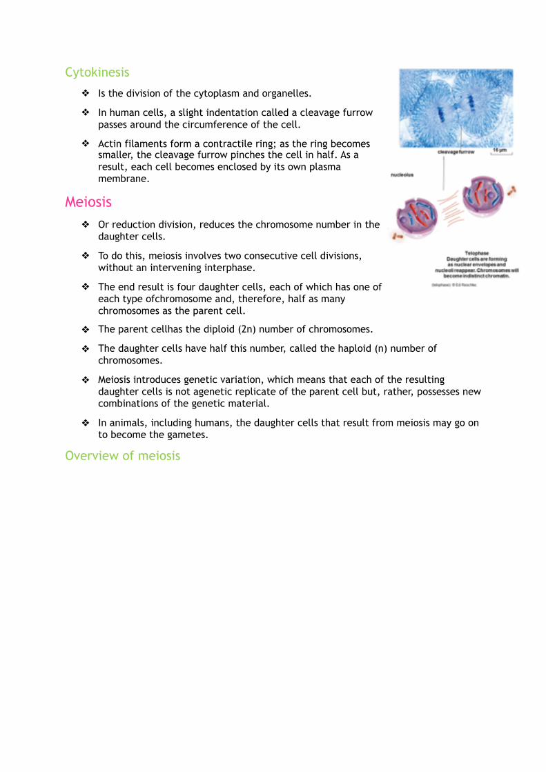
Cytokinesis
❖ Is the division of the cytoplasm and organelles.
❖ In human cells, a slight indentation called a cleavage furrow passes around the circumference of the cell.
❖ Actin filaments form a contractile ring; as the ring becomes smaller, the cleavage furrow pinches the cell in half. As a result, each cell becomes enclosed by its own plasma membrane.
Meiosis ❖ Or reduction division, reduces the chromosome number in the
daughter cells.
❖ To do this, meiosis involves two consecutive cell divisions, without an intervening interphase.
❖ The end result is four daughter cells, each of which has one of each type ofchromosome and, therefore, half as many chromosomes as the parent cell.
❖ The parent cellhas the diploid (2n) number of chromosomes.
❖ The daughter cells have half this number, called the haploid (n) number of chromosomes.
❖ Meiosis introduces genetic variation, which means that each of the resulting daughter cells is not agenetic replicate of the parent cell but, rather, possesses new combinations of the genetic material.
❖ In animals, including humans, the daughter cells that result from meiosis may go on to become the gametes.
Overview of meiosis