Safety Assessment and Pain Relief Properties of Saffron from ...
20
Citation: Ait Tastift, M.; Makbal, R.; Bourhim, T.; Omari, Z.; Isoda, H.; Gadhi, C. Safety Assessment and Pain Relief Properties of Saffron from Taliouine Region (Morocco). Molecules 2022, 27, 3339. https:// doi.org/10.3390/molecules27103339 Academic Editor: Hinanit Koltai Received: 5 March 2022 Accepted: 19 May 2022 Published: 23 May 2022 Publisher’s Note: MDPI stays neutral with regard to jurisdictional claims in published maps and institutional affil- iations. Copyright: © 2022 by the authors. Licensee MDPI, Basel, Switzerland. This article is an open access article distributed under the terms and conditions of the Creative Commons Attribution (CC BY) license (https:// creativecommons.org/licenses/by/ 4.0/). molecules Article Safety Assessment and Pain Relief Properties of Saffron from Taliouine Region (Morocco) Maroua Ait Tastift 1,2 , Rachida Makbal 1,2 , Thouria Bourhim 1,2 , Zineb Omari 1,2 , Hiroko Isoda 3,4 and Chemseddoha Gadhi 1,2,4, * 1 Laboratory of Agri-Food, Biotechnology, and Valorization of Plant Resources, Phytochemistry and Pharmacology of Medicinal Plants Unit, Faculty of Sciences Semlalia, Cadi Ayyad University, Avenue Prince Moulay Abdellah, BP 2390, Marrakesh 40000, Morocco; [email protected] (M.A.T.); [email protected] (R.M.); [email protected] (T.B.); [email protected] (Z.O.) 2 Agrobiotechnology and Bioengineering Center, CNRST-Labeled Research Unit (AgroBiotech-URL-CNRST-05 Center), Cadi Ayyad University, Marrakesh 40000, Morocco 3 Faculty of Life and Environmental Sciences, University of Tsukuba, Tennodai 1-1-1, Tsukuba City 305-8572, Ibaraki, Japan; [email protected] 4 Alliance for Research on the Mediterranean and North Africa (ARENA), University of Tsukuba, Tennodai 1-1-1, Tsukuba City 305-8572, Ibaraki, Japan * Correspondence: [email protected] Abstract: Saffron is the most expensive spice in the world. In addition to its culinary utilization, this spice is used for medicinal purposes such as in pain management. In this study, the analgesic activity of Crocus sativus stigma extract (CSSE) was evaluated in rodents and its possible physiological mechanism was elucidated. The anti-nociceptive effect of CSSE was evaluated using three animal models (hot plate, writhing, and formalin tests). The analgesic pathways involved were assessed using various analgesia-mediating receptors antagonists. The oral administration of CSSE, up to 2000 mg/kg, caused no death or changes in the behavior or in the hematological and biochemical blood parameters of treated animals nor in the histological architecture of the animals’ livers and kidneys. CSSE showed a central, dose-dependent, anti-nociceptive effect in response to thermal stimuli; and a peripheral analgesic effect in the test of contortions induced by acetic acid. The dual (central and peripheral) analgesic effect was confirmed by the formalin test. The anti-nociceptive activity of CSSE was totally or partially reversed by the co-administration of receptor antagonists, naloxone, atropine, haloperidol, yohimbine, and glibenclamide. CSSE influenced signal processing, by the modulation of the opioidergic, adrenergic, and muscarinic systems at the peripheral and central levels; and by regulation of the dopaminergic system and control of the opening of the ATP-sensitive K + channels at the spinal level. The obtained data point to a multimodal mechanism of action for CSSE: An anti-inflammatory effect and a modulation, through different physiological pathways, of the electrical signal generated by the nociceptors. Further clinical trials are required to endorse the potential utilization of Moroccan saffron as a natural painkiller. Keywords: Crocus sativus; stigma; analgesia; receptor systems; Moroccan saffron 1. Introduction Pain is a physiological process whose purpose is to warn the person of a threat to his physical integrity, hence the term nociception [1]. However, to this simple transmission of the nociceptive message from the periphery to the somato-sensitive cortical centers is added an emotional and behavioral component, which explain the complex and multifactorial nature of pain. Suffering from pain, especially chronic pain, has become a major medical and socio- economic burden worldwide. Pain reduces the quality of life and has physical, psycho- logical, and environmental impacts not only on the individual but also on society as a whole [2]. Molecules 2022, 27, 3339. https://doi.org/10.3390/molecules27103339 https://www.mdpi.com/journal/molecules
-
Upload
khangminh22 -
Category
Documents
-
view
0 -
download
0
Transcript of Safety Assessment and Pain Relief Properties of Saffron from ...
Citation: Ait Tastift, M.; Makbal, R.;
Bourhim, T.; Omari, Z.; Isoda, H.;
Gadhi, C. Safety Assessment and
Pain Relief Properties of Saffron from
Taliouine Region (Morocco).
Molecules 2022, 27, 3339. https://
doi.org/10.3390/molecules27103339
Academic Editor: Hinanit Koltai
Received: 5 March 2022
Accepted: 19 May 2022
Published: 23 May 2022
Publisher’s Note: MDPI stays neutral
with regard to jurisdictional claims in
published maps and institutional affil-
iations.
Copyright: © 2022 by the authors.
Licensee MDPI, Basel, Switzerland.
This article is an open access article
distributed under the terms and
conditions of the Creative Commons
Attribution (CC BY) license (https://
creativecommons.org/licenses/by/
4.0/).
molecules
Article
Safety Assessment and Pain Relief Properties of Saffron fromTaliouine Region (Morocco)Maroua Ait Tastift 1,2, Rachida Makbal 1,2, Thouria Bourhim 1,2 , Zineb Omari 1,2, Hiroko Isoda 3,4 andChemseddoha Gadhi 1,2,4,*
1 Laboratory of Agri-Food, Biotechnology, and Valorization of Plant Resources, Phytochemistry andPharmacology of Medicinal Plants Unit, Faculty of Sciences Semlalia, Cadi Ayyad University, Avenue PrinceMoulay Abdellah, BP 2390, Marrakesh 40000, Morocco; [email protected] (M.A.T.);[email protected] (R.M.); [email protected] (T.B.); [email protected] (Z.O.)
2 Agrobiotechnology and Bioengineering Center, CNRST-Labeled Research Unit (AgroBiotech-URL-CNRST-05Center), Cadi Ayyad University, Marrakesh 40000, Morocco
3 Faculty of Life and Environmental Sciences, University of Tsukuba, Tennodai 1-1-1, Tsukuba City 305-8572,Ibaraki, Japan; [email protected]
4 Alliance for Research on the Mediterranean and North Africa (ARENA), University of Tsukuba,Tennodai 1-1-1, Tsukuba City 305-8572, Ibaraki, Japan
* Correspondence: [email protected]
Abstract: Saffron is the most expensive spice in the world. In addition to its culinary utilization,this spice is used for medicinal purposes such as in pain management. In this study, the analgesicactivity of Crocus sativus stigma extract (CSSE) was evaluated in rodents and its possible physiologicalmechanism was elucidated. The anti-nociceptive effect of CSSE was evaluated using three animalmodels (hot plate, writhing, and formalin tests). The analgesic pathways involved were assessedusing various analgesia-mediating receptors antagonists. The oral administration of CSSE, up to2000 mg/kg, caused no death or changes in the behavior or in the hematological and biochemicalblood parameters of treated animals nor in the histological architecture of the animals’ livers andkidneys. CSSE showed a central, dose-dependent, anti-nociceptive effect in response to thermalstimuli; and a peripheral analgesic effect in the test of contortions induced by acetic acid. The dual(central and peripheral) analgesic effect was confirmed by the formalin test. The anti-nociceptiveactivity of CSSE was totally or partially reversed by the co-administration of receptor antagonists,naloxone, atropine, haloperidol, yohimbine, and glibenclamide. CSSE influenced signal processing,by the modulation of the opioidergic, adrenergic, and muscarinic systems at the peripheral andcentral levels; and by regulation of the dopaminergic system and control of the opening of theATP-sensitive K+ channels at the spinal level. The obtained data point to a multimodal mechanismof action for CSSE: An anti-inflammatory effect and a modulation, through different physiologicalpathways, of the electrical signal generated by the nociceptors. Further clinical trials are required toendorse the potential utilization of Moroccan saffron as a natural painkiller.
Keywords: Crocus sativus; stigma; analgesia; receptor systems; Moroccan saffron
1. Introduction
Pain is a physiological process whose purpose is to warn the person of a threat to hisphysical integrity, hence the term nociception [1]. However, to this simple transmission ofthe nociceptive message from the periphery to the somato-sensitive cortical centers is addedan emotional and behavioral component, which explain the complex and multifactorialnature of pain.
Suffering from pain, especially chronic pain, has become a major medical and socio-economic burden worldwide. Pain reduces the quality of life and has physical, psycho-logical, and environmental impacts not only on the individual but also on society as awhole [2].
Molecules 2022, 27, 3339. https://doi.org/10.3390/molecules27103339 https://www.mdpi.com/journal/molecules
Molecules 2022, 27, 3339 2 of 20
The intervention of central and peripheral mechanisms in pain in the majority of casesmakes the choice of an adequate pharmacological therapy very difficult. The analgesicsthat act at the central level, in spite of their great effectiveness, are not dissociated fromside effects, namely addiction, constipation, and respiratory depression [3,4]. Furthermore,analgesics that proceed at the peripheral level have undesirable effects, such as gastroin-testinal and renal lesions [5,6]. In this sense, several studieshave been devoted to discovernew natural resources with analgesic potential and low secondary effects.
Saffron, also known as Crocus sativus L. stigmas (Iridaceae), is the most expensivespice in the world. At the retail price, it costs 4000 to 6000 US dollar per kg of dried stigmasand requires the collection of 200,000 to 300,000 flowers for one kg of saffron. The flowersare manually handled to gently remove the three precious stigmas that make up saffron.The major saffron-growing countries around the world are Iran, India, Morocco, Spain,Greece, Afghanistan, and Italy [7–10]. Iran produces 90% of the saffron that exists on themarket, making it the leading producer and exporter in the world.
Morocco occupies an important place in the saffron market [11]. The country is theworld’s fourth largest producer of this expensive spice, with an annual production of6.5 tons in 2019, grown over an area of 1865 Ha, mainly in the region of Taliouine in theAnti-Atlas Mountains [12].
The chemical composition of saffron has been widely studied [8,10,13]. Stigmasmainly contain secondary metabolites belonging to the terpenoid family, in particularmonoterpenes and apocarotenoids, and to the flavonoid family, all glycosidic derivativesof kaempferol [8,11,14–16].
Among the apocarotenoids, crocetin and its glycoside esters, crocins, are the mostimportant [8] while safranal, a monoterpene aldehyde, and its glucoside derivative, picro-crocin, are the other two most important phytochemicals abundant in C. sativus stigmas.
The quality of saffron depends on its coloring potential, its taste, and its aroma,ascribable to crocin, picrocrocin, and safranal contents, respectively. Three categories ofsaffron are defined according to the saffron ISO-norm 3632-2: 2010 [17], namely category 1(the highest quality category), category 2 (intermediate), and category 3 (lowest quality).
Although saffron samples from different locations or countries have similar overallchemical composition, it has been reported that there are qualitative and/or quantitativedifferences in chemical composition between samples; differences that are related to thegeographical origin, drying process, and storage conditions [8,11,14,18–21]. Thus, it im-pacts the organoleptic properties and beneficial health effects of the saffron sample. Thedifferences concern the structure of crocin (the glycoside ester of crocetin), the compositionof the volatile fraction, and the structure of the kaempferol derivatives [8,14,20,21].
In saffron samples, different crocin derivatives exist depending on the isomer (cis/trans)of crocetin involved and on the number of glucose molecules attached to the molecule ofcrocetin [8,20,21]. Kaempferol derivatives also differ from one sample of saffron to anotherdepending on the structure of sugars moiety [8]. Nevertheless, the key difference concernsthe aromatic volatile fraction. Indeed, in addition to the drying process [11,18,20], thegeographical location affects the chemical profile of the volatile fraction of saffron [14].
Karabagias et al. [14] compared saffron from Greece, Spain, Iran, and Morocco, on thebasis of volatile compounds. They reported significant variations in the volatile compoundscontent, according to the geographical origin of the saffron, finding the following pattern:Morocco > Iran > Greece > Spain. The GC/MS analysis revealed that in all samples, safranalwas the most abundant volatile compound but its relative content differs depending onsaffron samples. Safranal represented 79.04, 71.35, 76.42, and 64.71% of saffron’s totalvolatile fraction, in Moroccan, Greek, Iranian, and Spanish samples, respectively [14].Regarding other volatile safranal derivatives, a significant difference was recorded amongsamples in relation to the geographical origin [14].
Saffron has been used as a spice, dye, and for medicinal purposes for nearly fourmillennia [7]. It is considered safe in therapeutic doses [15,22]. Different studies reportedthe non-toxic effects of saffron after acute oral administration of a dose of 2 g/kg [15,22] and
Molecules 2022, 27, 3339 3 of 20
even at doses up to 5 g/kg [8,23]. No symptoms of toxicity were recorded after repeatedadministration of low doses of saffron (28 days of administration of a dose of 100 mg/kgin the rat) [22] while repeated oral administration of higher doses (>5 g/kg) was likely tocause vomiting, uterine contractions, dizziness, etc. [24].
Various health-promoting properties have been accredited to it and its main metabo-lites, crocin, crocetin, picrocrocin, and safranal, namely anticancer, anti-Alzheimer’s, anti-Parkinson’s, improved memory and learning abilities, treatment of anxiety disorders andschizophrenia, anti-nociceptive, anti-inflammatory, hepatoprotective, antidiabetic, maculardegeneration protection, anticoagulant, skin anti-aging, skin lightening, antisolar, andmoisturizing properties [9,13,15,16,19,25–33].
In recent times, saffron has gained a special interest for its antidepressant proper-ties based on in vivo testing and randomized controlled trials [34–38]. The inhibition ofserotonin reuptake has been proposed as a possible mechanism of action of saffron andcrocin [34]. It has also been acclaimed for its anti-cancer potential [39–44]. In a very recentreport, El Midaoui et al. (2022) [45] highlighted the role of saffron in the treatment ofneuropsychiatric and age-related diseases. Saffron and its major compounds have pow-erful antioxidant and anti-inflammatory properties in vitro and in vivo [46,47]; propertiesthat would be partly responsible for the effectiveness of saffron and its metabolites in thetreatment of diseases [44,48–50].
Saffron is traditionally known to ease pain. Its analgesic activity was evaluated in vivoand in clinical trials [15,30–33]. The anti-nociceptive effect was found by Hosseinzadeh andYounesi [30] to not be significant in mice and to not involve the central nervous system,while other studies have confirmed saffron’s effectiveness [31–33], and the involvement ofcentral mechanisms [15], without, however, specifying which ones are involved.
The majority of studies have used, in their investigation, saffron from Iran for itsconvenience, Iran being the first producer and exporter of this spice in the world [24]However, despite Morocco’s place on the podium of saffron production, and even with thestrong aroma that characterizes it (high yield in volatile fraction [14]), Moroccan saffronhas not been investigated for its functionality and, therefore, could hide unexpected assets.
Thus, the aim of the present work was primarily to assess, for the first time, the anal-gesic properties of saffron from Morocco. Furthermore, the elucidation of the physiologicalmechanism involved in the analgesic activity of C. sativus stigma extract (CSSE) usingvarious antagonists of analgesia-mediating receptors was also recorded.
2. Results2.1. CSSE Had No Toxic Effect
The acute toxicity was studied using the limit dose test reported in the Food and DrugAdministration (FDA) and OECD guidelines [51,52]. The limit dose of 2000 mg/kg bodyweight is the recommended dose for acute toxicity studies to avoid the unnecessary useand killing of animals (one of the pillars of animals handling).
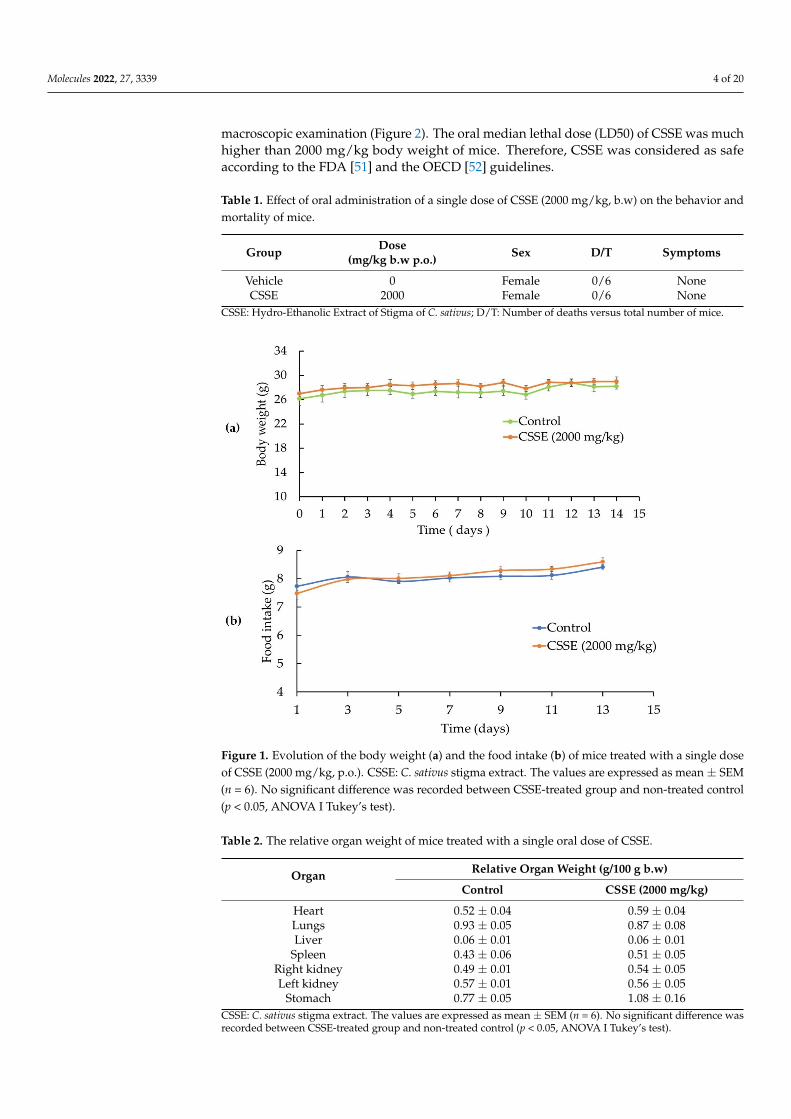
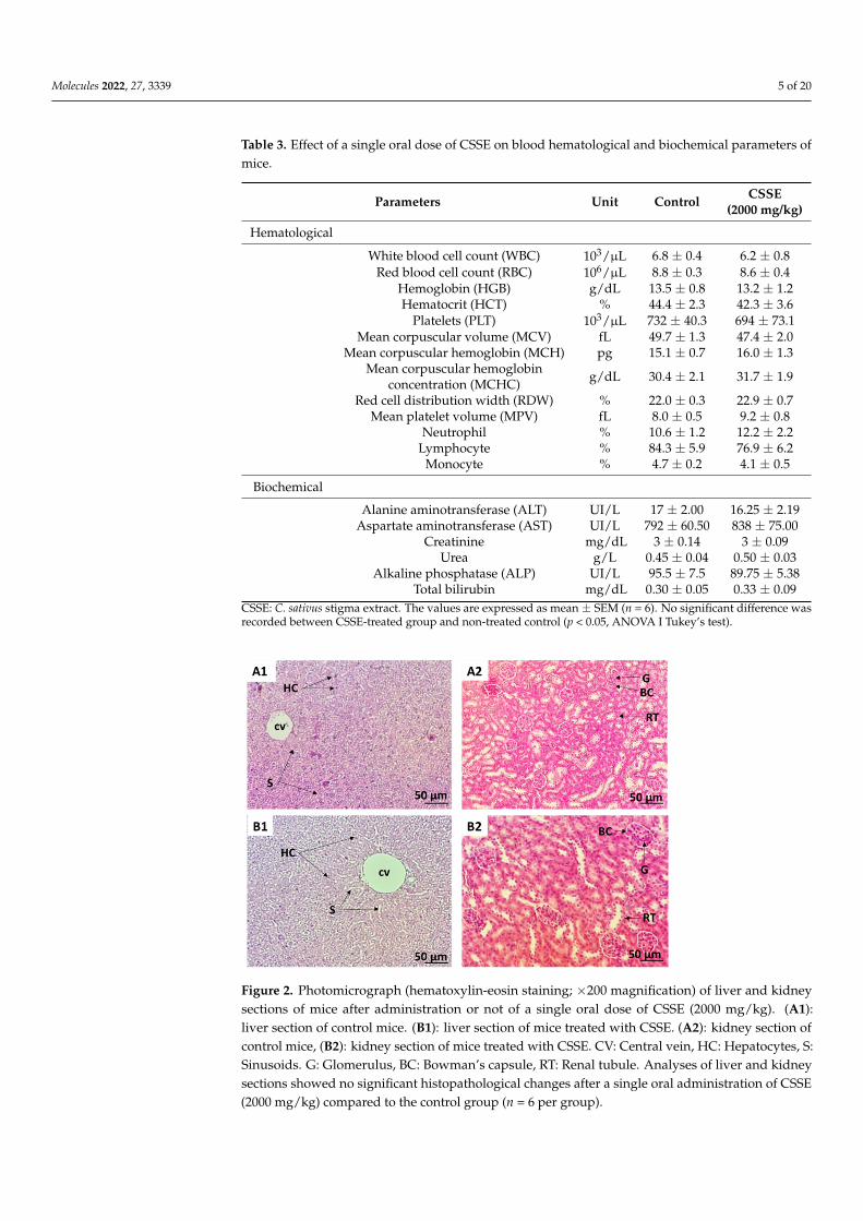
The oral administration of a single dose of CSSE (2000 mg/kg body weight) to micedid not cause any death within the fourteen days of the study. No toxicity symptoms oradverse behavioral changes were observed in CSSE treated mice (Table 1). In addition,the extract did not affect mice body weight (Figure 1a) nor the amount of food intake(Figure 1b) compared to the control. No macroscopic alterations were observed in themice’s organs during the autopsy and the relative organ weights were unaffected (Table 2).Hematological parameters showed no significant difference between the treated mice andthe control group (Table 3). Biochemical analysis of serum aspartate aminotransferase(AST) and alanine aminotransferase (ALT) showed that treatment with CSSE did not causesignificant changes in serum transaminases (Table 3). In the same way, no changes increatinine, urea, alkaline phosphatase, and total bilirubin levels were observed comparedto the control group. The data show that the biochemical parameters remained withinthe physiological range, indicating that CSSE did not affect liver and kidney function.Histopathological analysis of the liver and kidneys was performed and confirmed the
Molecules 2022, 27, 3339 4 of 20
macroscopic examination (Figure 2). The oral median lethal dose (LD50) of CSSE was muchhigher than 2000 mg/kg body weight of mice. Therefore, CSSE was considered as safeaccording to the FDA [51] and the OECD [52] guidelines.
Table 1. Effect of oral administration of a single dose of CSSE (2000 mg/kg, b.w) on the behavior andmortality of mice.
Group Dose(mg/kg b.w p.o.) Sex D/T Symptoms
Vehicle 0 Female 0/6 NoneCSSE 2000 Female 0/6 None
CSSE: Hydro-Ethanolic Extract of Stigma of C. sativus; D/T: Number of deaths versus total number of mice.
Molecules 2022, 27, x FOR PEER REVIEW 5 of 21
Mean platelet volume (MPV) fL 8.0 ± 0.5 9.2 ± 0.8 Neutrophil % 10.6 ± 1.2 12.2 ± 2.2 Lymphocyte % 84.3 ± 5.9 76.9 ± 6.2 Monocyte % 4.7 ± 0.2 4.1 ± 0.5
Biochemical Alanine aminotransferase (ALT) UI/L 17 ± 2.00 16.25 ± 2.19
Aspartate aminotransferase (AST) UI/L 792 ± 60.50 838 ± 75.00
Creatinine mg/dL 3 ± 0.14 3 ± 0.09 Urea g/l 0.45 ± 0.04 0.50 ± 0.03 Alkaline phosphatase (ALP) UI/L 95.5 ± 7.5 89.75 ± 5.38 Total bilirubin mg/dL 0.30 ± 0.05 0.33 ± 0.09
CSSE: C. sativus stigma extract. The values are expressed as mean ± SEM (n = 6). No significant difference was recorded between CSSE-treated group and non-treated control (p < 0.05, ANOVA I Tukey’s test).
Figure 1. Evolution of the body weight (a) and the food intake (b) of mice treated with a single dose of CSSE (2000 mg/kg, p.o). CSSE: C. sativus stigma extract. The values are expressed as mean ± SEM (n = 6). No significant difference was recorded between CSSE-treated group and non-treated control (p < 0.05, ANOVA I Tukey’s test).
Figure 1. Evolution of the body weight (a) and the food intake (b) of mice treated with a single doseof CSSE (2000 mg/kg, p.o.). CSSE: C. sativus stigma extract. The values are expressed as mean ± SEM(n = 6). No significant difference was recorded between CSSE-treated group and non-treated control(p < 0.05, ANOVA I Tukey’s test).
Table 2. The relative organ weight of mice treated with a single oral dose of CSSE.
Organ Relative Organ Weight (g/100 g b.w)
Control CSSE (2000 mg/kg)
Heart 0.52 ± 0.04 0.59 ± 0.04Lungs 0.93 ± 0.05 0.87 ± 0.08Liver 0.06 ± 0.01 0.06 ± 0.01
Spleen 0.43 ± 0.06 0.51 ± 0.05Right kidney 0.49 ± 0.01 0.54 ± 0.05Left kidney 0.57 ± 0.01 0.56 ± 0.05
Stomach 0.77 ± 0.05 1.08 ± 0.16CSSE: C. sativus stigma extract. The values are expressed as mean ± SEM (n = 6). No significant difference wasrecorded between CSSE-treated group and non-treated control (p < 0.05, ANOVA I Tukey’s test).
Molecules 2022, 27, 3339 5 of 20
Table 3. Effect of a single oral dose of CSSE on blood hematological and biochemical parameters ofmice.
Parameters Unit Control CSSE(2000 mg/kg)
Hematological
White blood cell count (WBC) 103/µL 6.8 ± 0.4 6.2 ± 0.8Red blood cell count (RBC) 106/µL 8.8 ± 0.3 8.6 ± 0.4
Hemoglobin (HGB) g/dL 13.5 ± 0.8 13.2 ± 1.2Hematocrit (HCT) % 44.4 ± 2.3 42.3 ± 3.6
Platelets (PLT) 103/µL 732 ± 40.3 694 ± 73.1Mean corpuscular volume (MCV) fL 49.7 ± 1.3 47.4 ± 2.0
Mean corpuscular hemoglobin (MCH) pg 15.1 ± 0.7 16.0 ± 1.3Mean corpuscular hemoglobin
concentration (MCHC) g/dL 30.4 ± 2.1 31.7 ± 1.9
Red cell distribution width (RDW) % 22.0 ± 0.3 22.9 ± 0.7Mean platelet volume (MPV) fL 8.0 ± 0.5 9.2 ± 0.8
Neutrophil % 10.6 ± 1.2 12.2 ± 2.2Lymphocyte % 84.3 ± 5.9 76.9 ± 6.2
Monocyte % 4.7 ± 0.2 4.1 ± 0.5
Biochemical
Alanine aminotransferase (ALT) UI/L 17 ± 2.00 16.25 ± 2.19Aspartate aminotransferase (AST) UI/L 792 ± 60.50 838 ± 75.00
Creatinine mg/dL 3 ± 0.14 3 ± 0.09Urea g/L 0.45 ± 0.04 0.50 ± 0.03
Alkaline phosphatase (ALP) UI/L 95.5 ± 7.5 89.75 ± 5.38Total bilirubin mg/dL 0.30 ± 0.05 0.33 ± 0.09
CSSE: C. sativus stigma extract. The values are expressed as mean ± SEM (n = 6). No significant difference wasrecorded between CSSE-treated group and non-treated control (p < 0.05, ANOVA I Tukey’s test).
Molecules 2022, 27, x FOR PEER REVIEW 6 of 21
Figure 2. Photomicrograph (hematoxylin-eosin staining; ×200 magnification) of liver and kidney sections of mice after administration or not of a single oral dose of CSSE (2000 mg/kg). (A1): liver section of control mice. (B1): liver section of mice treated with CSSE. (A2): kidney section of control mice, (B2): kidney section of mice treated with CSSE. CV: Central vein, HC: Hepatocytes, S: Sinus-oids. G: Glomerulus, BC: Bowman’s capsule, RT: Renal tubule. Analyses of liver and kidney sections showed no significant histopathological changes after a single oral administration of CSSE (2000 mg/kg) compared to the control group (n = 6 per group).
2.2. Analgesic Activity. 2.2.1. CSSE Reduced the Abdominal Contractions Induced by Intraperitoneal Injection of Acetic Acid in Mice
Acetic acid is typically used as noxious stimuli in visceral pain studies [53]. When injected intraperitoneally to mice, it induces abdominal writhing (stretching, retracting, or pressing the belly against the floor). The writhing test is based on counting these events and is used to assess the pain threshold for inflammatory agents [53].
The pretreatment with CSSE significantly decreased the writhing response in a dose-dependent manner with a high inhibition rate of writhing (71.6%) observed at 200 mg/kg; while at 50 mg/kg and 100 mg/kg, the inhibition rates induced by acetic acid were of 48.3% and 60.5%, respectively. This pain-relieving effect of CSSE was very significant, as shown by its relative activity when compared to morphine (1 mg/kg, i.p). At 200 mg/kg, CSSE exhibited an 84.7 % morphine equivalent inhibitory effect (Table 4).
Table 4. Effect of CSSE on abdominal contractions induced by intraperitoneal injection of acetic acid in mice.
Group Dose
(mg/kg, b.w) Number of Contortions
Inhibition of Pain (%)
Relative Activity Compared to Morphine (%)
Control - 34.2 ± 1.5 - - Morphine 1 5.3 ± 0.7 *** 84.5 ± 0.5 100
CSSE 50 17.7 ± 0.3 *** 48.3 ± 0.8 +++ 57.2
100 13.5 ± 0.4 *** 60.5 ± 1.0 +++ 71.6 200 9.7 ± 0.4 *** 71.6 ± 0.9 ++ 84.7
CSSE: Hydro-ethanolic extract of stigma of C. sativus; Morphine (1 mg/kg, i.p) is used as positive control. Each value represents the mean ± SEM (n = 6); *** Statistically significant (p ≤ 0.001) versus the non-treated control (ANOVA, Tukey’s test). ++ and +++ Statistically significant (p ≤ 0.01) and (p ≤ 0.001), respectively versus morphine-treated group (ANOVA, Tukey’s test).
Figure 2. Photomicrograph (hematoxylin-eosin staining; ×200 magnification) of liver and kidneysections of mice after administration or not of a single oral dose of CSSE (2000 mg/kg). (A1):liver section of control mice. (B1): liver section of mice treated with CSSE. (A2): kidney section ofcontrol mice, (B2): kidney section of mice treated with CSSE. CV: Central vein, HC: Hepatocytes, S:Sinusoids. G: Glomerulus, BC: Bowman’s capsule, RT: Renal tubule. Analyses of liver and kidneysections showed no significant histopathological changes after a single oral administration of CSSE(2000 mg/kg) compared to the control group (n = 6 per group).
Molecules 2022, 27, 3339 6 of 20
2.2. Analgesic Activity2.2.1. CSSE Reduced the Abdominal Contractions Induced by Intraperitoneal Injection ofAcetic Acid in Mice
Acetic acid is typically used as noxious stimuli in visceral pain studies [53]. Wheninjected intraperitoneally to mice, it induces abdominal writhing (stretching, retracting, orpressing the belly against the floor). The writhing test is based on counting these eventsand is used to assess the pain threshold for inflammatory agents [53].
The pretreatment with CSSE significantly decreased the writhing response in a dose-dependent manner with a high inhibition rate of writhing (71.6%) observed at 200 mg/kg;while at 50 mg/kg and 100 mg/kg, the inhibition rates induced by acetic acid were of 48.3%and 60.5%, respectively. This pain-relieving effect of CSSE was very significant, as shownby its relative activity when compared to morphine (1 mg/kg, i.p). At 200 mg/kg, CSSEexhibited an 84.7 % morphine equivalent inhibitory effect (Table 4).
Table 4. Effect of CSSE on abdominal contractions induced by intraperitoneal injection of acetic acidin mice.
Group Dose(mg/kg, b.w)
Number ofContortions
Inhibition ofPain (%)
Relative ActivityCompared to Morphine (%)
Control - 34.2 ± 1.5 - -Morphine 1 5.3 ± 0.7 *** 84.5 ± 0.5 100
CSSE50 17.7 ± 0.3 *** 48.3 ± 0.8 +++ 57.2
100 13.5 ± 0.4 *** 60.5 ± 1.0 +++ 71.6200 9.7 ± 0.4 *** 71.6 ± 0.9 ++ 84.7
CSSE: Hydro-ethanolic extract of stigma of C. sativus; Morphine (1 mg/kg, i.p) is used as positive control.Each value represents the mean ± SEM (n = 6); *** Statistically significant (p ≤ 0.001) versus the non-treatedcontrol (ANOVA, Tukey’s test). ++ and +++ Statistically significant (p ≤ 0.01) and (p ≤ 0.001), respectively versusmorphine-treated group (ANOVA, Tukey’s test).
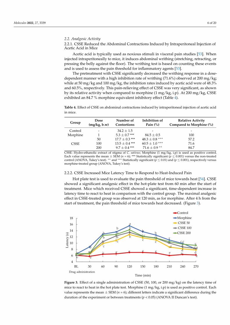
2.2.2. CSSE Increased Mice Latency Time to Respond to Heat-Induced Pain
Hot plate test is used to evaluate the pain threshold of mice towards heat [54]. CSSEshowed a significant analgesic effect in the hot-plate test from 60 min after the start oftreatment. Mice which received CSSE showed a significant, time-dependent increase inlatency time to react to heat in comparison with the control group. The maximal analgesiceffect in CSSE-treated group was observed at 120 min, as for morphine. After 4 h from thestart of treatment, the pain threshold of mice towards heat decreased. (Figure 3).
Molecules 2022, 27, x FOR PEER REVIEW 7 of 21
2.2.2. CSSE Increased Mice Latency Time to Respond to Heat-Induced Pain Hot plate test is used to evaluate the pain threshold of mice towards heat [54]. CSSE
showed a significant analgesic effect in the hot-plate test from 60 min after the start of treatment. Mice which received CSSE showed a significant, time-dependent increase in latency time to react to heat in comparison with the control group. The maximal analgesic effect in CSSE-treated group was observed at 120 min, as for morphine. After 4 h from the start of treatment, the pain threshold of mice towards heat decreased. (Figure 3).
Figure 3. Effect of a single administration of CSSE (50, 100, or 200 mg/kg) on the latency time of mice to react to heat in the hot plate test. Morphine (1 mg/kg, i.p) is used as positive control. Each valuerepresents the mean ± SEM (n = 6); different letters indicate a significant difference during the du-ration of the experiment or between treatments (p < 0.05) (ANOVA II Duncan’s test).
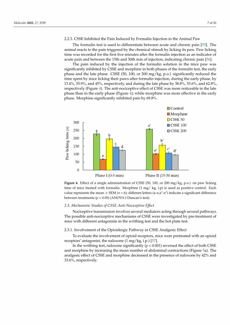
2.2.3. CSSE Inhibited the Pain Induced by Formalin Injection in the Animal Paw The formalin test is used to differentiate between acute and chronic pain [55]. The
animal reacts to the pain triggered by the chemical stimuli by licking its paw. Paw licking time was recorded for the first five minutes after the formalin injection as an indicator of acute pain and between the 15th and 30th min of injection, indicating chronic pain [56].
The pain induced by the injection of the formalin solution in the mice paw was sig-nificantly inhibited by CSSE and morphine in both phases of the formalin test, the early phase and the late phase. CSSE (50, 100, or 200 mg/kg, p.o) significantly reduced the time spent by mice licking their paws after formalin injection, during the early phase, by 13.4%, 35.9%, and 45%, respectively, and during the late phase by 38.8%, 53.6%, and 62.8%, re-spectively (Figure 4). The anti-nociceptive effect of CSSE was more noticeable in the latephase than in the early phase (Figure 4); while morphine was more effective in the early phase. Morphine significantly inhibited pain by 69.8%.
Figure 3. Effect of a single administration of CSSE (50, 100, or 200 mg/kg) on the latency time ofmice to react to heat in the hot plate test. Morphine (1 mg/kg, i.p) is used as positive control. Eachvalue represents the mean ± SEM (n = 6); different letters indicate a significant difference during theduration of the experiment or between treatments (p < 0.05) (ANOVA II Duncan’s test).
Molecules 2022, 27, 3339 7 of 20
2.2.3. CSSE Inhibited the Pain Induced by Formalin Injection in the Animal Paw
The formalin test is used to differentiate between acute and chronic pain [55]. Theanimal reacts to the pain triggered by the chemical stimuli by licking its paw. Paw lickingtime was recorded for the first five minutes after the formalin injection as an indicator ofacute pain and between the 15th and 30th min of injection, indicating chronic pain [56].
The pain induced by the injection of the formalin solution in the mice paw wassignificantly inhibited by CSSE and morphine in both phases of the formalin test, the earlyphase and the late phase. CSSE (50, 100, or 200 mg/kg, p.o.) significantly reduced thetime spent by mice licking their paws after formalin injection, during the early phase, by13.4%, 35.9%, and 45%, respectively, and during the late phase by 38.8%, 53.6%, and 62.8%,respectively (Figure 4). The anti-nociceptive effect of CSSE was more noticeable in the latephase than in the early phase (Figure 4); while morphine was more effective in the earlyphase. Morphine significantly inhibited pain by 69.8%.
Molecules 2022, 27, x FOR PEER REVIEW 8 of 21
Figure 4. Effect of a single administration of CSSE (50, 100, or 200 mg/kg, p.o) on paw licking time of mice treated with formalin. Morphine (1 mg/ kg, i.p) is used as positive control. Each value rep-resents the mean ± SEM (n = 6); different letters (a,b,c,d,e,a′,b′,c′,d′,e′) indicate a significant difference between treatments (p < 0.05) (ANOVA I Duncan’s test).
2.3. Mechanistic Studies of CSSE Anti-Nociceptive Effect Nociceptive transmission involves several mediators acting through several path-
ways. The possible anti-nociceptive mechanisms of CSSE were investigated by pre-treat-ment of mice with different antagonists in the writhing test and the hot plate test.
2.3.1. Involvement of the Opioidergic Pathway in CSSE Analgesic Effect To evaluate the involvement of opioid receptors, mice were pretreated with an opioid
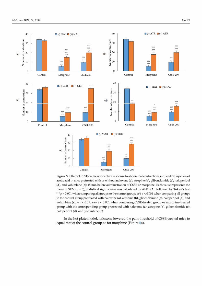
receptors’ antagonist, the naloxone (1 mg/kg, i.p.) [57]. In the writhing test, naloxone significantly (p < 0.001) reversed the effect of both CSSE
and morphine by increasing the mean number of abdominal contractions (Figure 5a). The analgesic effect of CSSE and morphine decreased in the presence of naloxone by 42% and 33.6%, respectively.
In the hot plate model, naloxone lowered the pain threshold of CSSE-treated mice to equal that of the control group as for morphine (Figure 6a).
Figure 4. Effect of a single administration of CSSE (50, 100, or 200 mg/kg, p.o.) on paw lickingtime of mice treated with formalin. Morphine (1 mg/ kg, i.p) is used as positive control. Eachvalue represents the mean ± SEM (n = 6); different letters (a–e,a′–e′) indicate a significant differencebetween treatments (p < 0.05) (ANOVA I Duncan’s test).
2.3. Mechanistic Studies of CSSE Anti-Nociceptive Effect
Nociceptive transmission involves several mediators acting through several pathways.The possible anti-nociceptive mechanisms of CSSE were investigated by pre-treatment ofmice with different antagonists in the writhing test and the hot plate test.
2.3.1. Involvement of the Opioidergic Pathway in CSSE Analgesic Effect
To evaluate the involvement of opioid receptors, mice were pretreated with an opioidreceptors’ antagonist, the naloxone (1 mg/kg, i.p.) [57].
In the writhing test, naloxone significantly (p < 0.001) reversed the effect of both CSSEand morphine by increasing the mean number of abdominal contractions (Figure 5a). Theanalgesic effect of CSSE and morphine decreased in the presence of naloxone by 42% and33.6%, respectively.
Molecules 2022, 27, 3339 8 of 20Molecules 2022, 27, x FOR PEER REVIEW 9 of 21
Figure 5. Effect of CSSE on the nociceptive response to abdominal contractions induced by injection of acetic acid in mice pretreated with or without naloxone (a), atropine (b), glibenclamide (c), haloperidol (d), and yohimbine (e); 15 min before administration of CSSE or morphine. Each value represents the mean ± SEM (n = 6); Statistical significance was calculated by ANOVA I followed by Tukey’s test. *** p < 0.001 when comparing all groups to the control group; ### p < 0.001 when com-paring all groups to the control group pretreated with naloxone (a), atropine (b), glibenclamide (c), haloperidol (d), and yohimbine (e); + p < 0.05, +++ p < 0.001 when comparing CSSE-treated group or morphine-treated group with the corresponding group pretreated with naloxone (a), atropine (b), glibenclamide (c), haloperidol (d), and yohimbine (e).
Figure 5. Effect of CSSE on the nociceptive response to abdominal contractions induced by injection ofacetic acid in mice pretreated with or without naloxone (a), atropine (b), glibenclamide (c), haloperidol(d), and yohimbine (e); 15 min before administration of CSSE or morphine. Each value represents themean ± SEM (n = 6); Statistical significance was calculated by ANOVA I followed by Tukey’s test.*** p < 0.001 when comparing all groups to the control group; ### p < 0.001 when comparing all groupsto the control group pretreated with naloxone (a), atropine (b), glibenclamide (c), haloperidol (d), andyohimbine (e); + p < 0.05, +++ p < 0.001 when comparing CSSE-treated group or morphine-treatedgroup with the corresponding group pretreated with naloxone (a), atropine (b), glibenclamide (c),haloperidol (d), and yohimbine (e).
In the hot plate model, naloxone lowered the pain threshold of CSSE-treated mice toequal that of the control group as for morphine (Figure 6a).
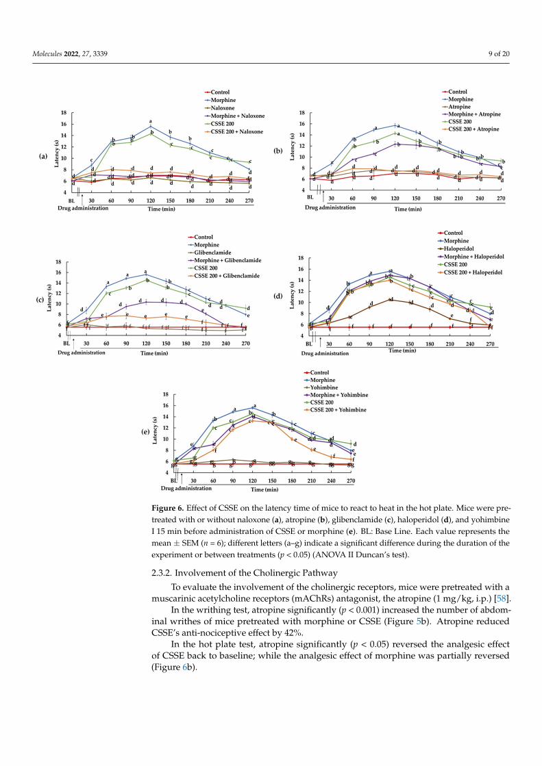
Molecules 2022, 27, 3339 9 of 20Molecules 2022, 27, x FOR PEER REVIEW 10 of 21
Figure 6. Effect of CSSE on the latency time of mice to react to heat in the hot plate. Mice were pretreated with or without naloxone (a), atropine (b), glibenclamide (c), haloperidol (d), and yohim-bine I 15 min before administration of CSSE or morphine (e). BL: Base Line. Each value represents the mean ± SEM (n = 6); different letters (a,b,c,d,e,f,g) indicate a significant difference during the duration of the experiment or between treatments (p < 0.05) (ANOVA II Duncan’s test).
2.3.2. Involvement of the Cholinergic Pathway To evaluate the involvement of the cholinergic receptors, mice were pretreated with
a muscarinic acetylcholine receptors (mAChRs) antagonist, the atropine (1 mg/kg, i.p.) [58].
In the writhing test, atropine significantly (p < 0.001) increased the number of ab-dominal writhes of mice pretreated with morphine or CSSE (Figure 5b). Atropine reduced CSSE's anti-nociceptive effect by 42%.
In the hot plate test, atropine significantly (p < 0.05) reversed the analgesic effect of CSSE back to baseline; while the analgesic effect of morphine was partially reversed (Fig-ure 6b).
Figure 6. Effect of CSSE on the latency time of mice to react to heat in the hot plate. Mice were pre-treated with or without naloxone (a), atropine (b), glibenclamide (c), haloperidol (d), and yohimbineI 15 min before administration of CSSE or morphine (e). BL: Base Line. Each value represents themean ± SEM (n = 6); different letters (a–g) indicate a significant difference during the duration of theexperiment or between treatments (p < 0.05) (ANOVA II Duncan’s test).
2.3.2. Involvement of the Cholinergic Pathway
To evaluate the involvement of the cholinergic receptors, mice were pretreated with amuscarinic acetylcholine receptors (mAChRs) antagonist, the atropine (1 mg/kg, i.p.) [58].
In the writhing test, atropine significantly (p < 0.001) increased the number of abdom-inal writhes of mice pretreated with morphine or CSSE (Figure 5b). Atropine reducedCSSE’s anti-nociceptive effect by 42%.
In the hot plate test, atropine significantly (p < 0.05) reversed the analgesic effectof CSSE back to baseline; while the analgesic effect of morphine was partially reversed(Figure 6b).
Molecules 2022, 27, 3339 10 of 20
2.3.3. Involvement of ATP Channel Blocker Pathway in CSSE Analgesic Effect
Another pathway potentially involved in pain generation is the depolarization of thecells. Glibenclamide is an inhibitor of ATP-sensitive K+ channels [59–61].
In the writhing test, the administration of glibenclamide (10 mg/kg, p.o.) to CSSE-pretreated mice totally abolished CSSE’s analgesic effect. The mean number of abdominalwrithes produced by acetic acid increased by 100% in this group to be similar to the control(Figure 5c). Meanwhile, glibenclamide did not significantly change the mean number ofabdominal writhes of mice treated with morphine. Morphine’s analgesic effect reduced onlyby 14.5% in the presence of glibenclamide (Figure 5c). In the hot plate test, glibenclamidepartially reversed the analgesic effect of morphine and CSSE (Figure 6c).
2.3.4. Involvement of Dopaminergic Pathway in CSSE Analgesic Effect
The active mechanism of haloperidol is to block the postsynaptic dopamine (D2)receptors [62]. It was therefore used to assess if CSSE acts through this pathway.
In the writhing test, the analgesic effect of morphine and CSSE decreased in thepresence of haloperidol by only 14% and 24.4%, respectively (Figure 5d). In the hot platetest, haloperidol did not reverse the analgesic effect of CSSE and only slightly reversed thatof morphine (Figure 6d). In contrast, haloperidol alone exerted a significant analgesic effectcompared to the control with a pain inhibitory activity of 43.9% (Figure 6d).
2.3.5. Involvement of the Adrenergic Pathway in the Mode of Action of CSSE
Yohimbine blocks presynaptic alpha-2 adrenergic receptors [63].Yohimbine significantly (p < 0.001) increased the mean number of acetic acid-induced
writhing in groups pretreated with CSSE or morphine. The analgesic effect of CSSEwas greatly reduced (77%) in the presence of yohimbine as well as for morphine (47.5%)(Figure 5e). In the hot plate test, yohimbine very slightly lowered the pain threshold ofmice treated with CSSE or morphine (Figure 6e).
2.4. Chemical Characterization of Moroccan Saffron According to ISO 3632 Guidelines
The quality of saffron is standardized by the norm for saffron ISO 3632 2:2010 [17]. Thecharacterization of saffron samples was assessed by measuring the amount of picrocrocin(flavor strength), safranal (aroma strength), and crocin (coloring strength), and by themoisture content (expressed as percentage). The results (Table 5) showed a high content ofcrocin (5.6 times the required minimal limit for the first quality category), a high content ofpicrocrocin (7.5 times the minimal limit for the first quality category), and a high content ofsafranal, which ranges into the limits established by the norms for the first quality category(min. 20; max. 50). The moisture content (10.3 ± 0.24%) was lower than the maximum limitrequired for all categories (12%). Therefore, and according to the limits established by theISO 3632 2:2010 [17], this saffron is classified in the first category, which refers to saffron ofa high quality.
Table 5. Chemical characterization of Moroccan saffron according to saffron norm ISO 3632 2:2010 [17].
StrengthIndicator In:
A1%1cm
MeasureSaffronSamples
ISO 3632Category 1
QualityConformity
PigmentsPicrocrocin Flavor λ 257 525.5 ± 0.4 min. 70 First Quality Category
Crocin Coloring λ 440 1126.3 ± 0.5 min. 200 First Quality CategorySafranal Aroma λ 330 32.4 ± 0.2 min. 20; max. 50 First Quality Category
Moisture content - - 10.3 ± 0.24% max 12% Conform
3. Discussion
Saffron is a spice traditionally known for its health benefits [16]. It is considered safein therapeutic doses [15,22] but becomes toxic in high doses [24]. In the present study, thesafety of Moroccan saffron was evaluated in order to further assess its analgesic activity at
Molecules 2022, 27, 3339 11 of 20
doses without toxic symptoms. CSSE has been shown to be safe up to a dose of 2 g/kg. Nodeath or signs of toxicity were recorded in the treated group, which is consistent with theliterature reports on saffron [15,22]. Therefore, it was possible to perform pharmacologicaltests in the range of safe doses and to assess the pain-relieving potential of CSSE.
The anti-nociceptive activity of CSSE was evaluated in this study using three tests: thehot plate test, writhing test, and formalin test.
The writhing test corresponds to the counting of abdominal stretching induced bythe injection of acetic acid into the peritoneal cavity of mice [53]. Acetic acid, a chemicalirritant, induces inflammation and is responsible for the release of chemical mediatorssuch as serotonin, histamine, bradykinin, substance P, and prostaglandins (PGE2α) [64–66].These chemical mediators stimulate peripheral chemo-sensitive nociceptors, induce anincrease in vascular permeability [65], and result in abdominal writhing [66]. Therefore,because the pretreatment with CSSE significantly decreased the writhing response in adose-dependent manner, CSSE most likely has an anti-inflammatory effect whereby itmodulates nociception. This suggestion was backed up by the results of the formalin test.CSSE significantly reduced, in a dose-dependent manner, the time mice spent licking theirpaws after formalin injection.
The injection of the formalin solution into the mouse’ paw causes a biphasic re-sponse [55,67]. The early neurogenic pain phase is initiated immediately after formalininjection and is characterized by stimulation of the C fibers and release of the substanceP and bradykinin. The second phase is due to the local inflammatory pain caused by theproduction of serotonin, histamine, and prostaglandins [68,69]. The results revealed thatCSSE has acted effectively in both phases of the formalin test with a more noticeable effectin the late inflammatory phase than in the early neurogenic pain phase (Figure 4). Thus,CSSE acts largely in the management of pain through the control of inflammation.
Several studies have indeed confirmed the anti-inflammatory activity ofsaffron [15,28,45,46] and ascribed it to the activity of its metabolites, in particular crocetin,the active form of crocin. Crocetin acted through its modulatory effect on redox balance [46],regulatory activity on inducible nitric synthase (iNOS) expression, and through the inhibi-tion of cyclooxygenase-1 and cyclooxygenase-2, and prostaglandins production [45].
CSSE also controls pain through central neurogenic pain management, as confirmed,in addition to the formalin test, by the hot plate test. The hot plate test is a central modelthat has a selectivity for opioid-derived analgesics [70].
CSSE showed a time-dependent anti-nociceptive effect by increasing the pain thresholdof mice towards heat. This result was similar to that reported by Khan et al. [15] but doesnot corroborate the conclusions of Hosseinzadeh and Younesi [30], who reported that theaqueous-ethanolic and aqueous extracts of a saffron from Iran have no significant effectson latency time, hence no central analgesic effect. This discrepancy may be related to thedifference in the chemical composition, which in turn is related to the geographical andenvironmental conditions [14].
To understand how CSSE modulates the electric signal triggered by the nociceptors,it is essential to specify which endogenous pathways have been involved and at whichlevel (spinal and/or supraspinal). To answer the question of the level of signal integrationand control, the acetic acid-induced writhing test was used to confirm the spinal controllevel and the hot plate test for the supraspinal one [54,71]. As the signal integrationmechanism involves the release of various neurotransmitters, receptor antagonists of theseneurotransmitters were used to verify their involvement in CSSE-induced analgesia.
Naloxone (non-selective opioid receptors antagonist [57]), atropine (non-selective mus-carinic receptor antagonist [58]), glibenclamide (ATP-sensitive K+ channels blocker [59–61]),haloperidol (dopamine receptors antagonist [62]), and yohimbine (a selective α2-adrenoceptorantagonist [63]) were used to substantiate the involvement of the opioidergic, cholinergic,ATP-sensitive K+ channels, and dopaminergic and adrenergic systems, respectively.
Naloxone totally reversed the analgesic effect of CSSE in the hot plate test and partiallyreversed it in the writhing test. This presumes the involvement of opioid receptors [72] in
Molecules 2022, 27, 3339 12 of 20
the analgesic effects of CSSE, mainly at the supraspinal level, but also at spinal level, evenin the peripheral endings of the primary afferent fibers, because opioids µ-receptors arealso located in the periphery [72]. A similar scheme was found with atropine, a muscarinicreceptor antagonist. This suggests that CSSE may also involve the cholinergic pathwayin its analgesic activity at the supraspinal and spinal levels. In particular, this is becausethe activation of the muscarinic receptors in the dorsal horn of the spinal cord has beenreported to contribute to the analgesic effect by releasing inhibitory interneurons, reducingnociceptive transmission [73].
The implication of the adrenergic system in the analgesic activity of CSSE was con-firmed as well. Yohimbine, a selective α2-adrenoceptor antagonist, helped to reduce theanalgesic activity of CSSE, mainly at the spinal level, with a 77% decrease in analgesiain the acetic acid-induced writhing test. The supraspinal level of action of CSSE on theadrenergic system was moderate, as confirmed by the slight decrease in CSSE activityin the hot plate test in the presence of yohimbine. These findings corroborate what wasreported concerning the involvement of the adrenergic system in nociception at the spinaland supraspinal levels, mediated through activation of α- adrenoceptors and descendinginhibitory pathways [74,75]. They also corroborate what was reported concerning the stim-ulatory effect of saffron’s metabolite crocetin on ß2-adrenoceptors [76], which contribute tocrocetin antinociception activity.
Among the neurotransmitters, the dopaminergic system also plays a role in paincontrol [77]. Dopamine receptors are expressed in primary nociceptors as well as in thespinal neurons located in different laminae of the dorsal horn of the spinal cord, suggestingthat dopamine may modulate pain signals by acting on both presynaptic and postsynaptictargets [78]. Decreased levels of dopamine have been associated with painful symptoms [77].At the physiological level, haloperidol reduces dopamine availability by blocking thedopamine D2 receptors [79]. Haloperidol reduced CSSE-induced antinociception in thewrithing test, which suggests that CSSE modulated the dopamine system at the spinal levelthrough activation of the D2 receptors.
At the supraspinal level, haloperidol did not modify the central anti-nociceptive effectof CSSE (Figure 6d), which implies that CSSE analgesic activity in the brain certainly didnot involve D2 receptors, although it may still act on the dopamine system but throughother mechanisms. This assumption was supported by previous reports on saffron’s effecton the brain [80,81]. It has been reported that brain dopamine concentration is increased bysaffron [80]. In line with this report, Monchaux De Oliveira et al. [81] recently establishedthat the saffron-induced improvement of depressive-like behavior was associated with themodulation of monoaminergic neurotransmission, in particular changes in serotonergic anddopaminergic neurotransmission. Saffron modulated the dopamine neurotransmission inthe frontal cortex by reducing its catabolism and in the striatum by significantly increasinglocal dopamine levels [81].
The generation of many pain signals in the human nervous system is mediated byion channels [79]. Open ATP-sensitive potassium channels produce analgesia by reducingneuronal excitability and by inhibiting the release of various neurotransmitters in the spinalcord [79]. Glibenclamide, a sulfonylurea, is an oral hypoglycemic drug that acts throughthe inhibition of the ATP-sensitive K+ channels in the pancreatic cells, which leads to thedepolarization of the cells and insulin secretion [82].
At the spinal level, in the writhing test, the analgesic effect of CSSE was fully reversedby glibenclamide, suggesting that the pain-relieving activity of CSSE was certainly due tothe opening of the ATP-sensitive K+ channels. At the supraspinal level, CSSE increased thepain threshold, but to a lesser extent, through the opening of the K+ channels, because theglibenclamide only partially reversed the analgesic effect of CSSE in the hot plate test.
Over all, the mechanism behind the analgesic effect of CSSE is multimodal. CSSEreduced nociception, through its anti-inflammatory action, demonstrated by the formalintest and by the modulation of the electrical signal generated by the nociceptors. CSSEinfluenced signal processing, both peripherally and centrally, by the modulation of the
Molecules 2022, 27, 3339 13 of 20
opioidergic, adrenergic, and muscarinic systems; and at the spinal level, by the modulationof the dopaminergic system and the opening of the ATP-sensitive K+ channels.
4. Materials and Methods4.1. Plant Material
The stigmas of C. sativus were purchased in November 2018 from local producers inSktana, Taliouine region (South of Morocco). The stigmas were authenticated by Prof. A.Ouhammou, a taxonomist at Cadi Ayyad University. A voucher specimen (MARK11120)has been deposited in the regional herbarium MARK, Faculty of Sciences Semlalia, Univer-sity Cadi Ayyad, Marrakech, Morocco.
4.2. Standards and Reagents
Acetic acid and ethanol were purchased from VWR International (Rosny-sous-Bois,France). Formalin, atropine, naloxone, and urethane were purchased from Sigma-Aldrich(St. Louis, MO, USA). Glibenclamide was bought from Promopharma (Had Soualem,Morocco). Haloperidol was procured from Pharma5 (Casablanca, Morocco). Yohimbinewas purchased from Micro Ingredients (Montclair, CA, USA). Morphine was providedby Sothema (Casablanca, Morocco). Hematoxylin and eosin were purchased from Sigma-Aldrich (Merck KGaA, Darmstadt, Germany).
4.3. Animals
The in vivo study was carried out using adult Swiss albino mice (12 weeks) housedin cages under standard conditions (25 ± 2 ◦C, 12/12 h light/dark cycle) with free accessto water and food. The animals were divided into groups with 6 mice per group. Theywere acclimatized for one week prior to the experiment and fasted overnight before eachtest. All studies were authorized by the Ethical Committee for Animal Care of the Facultyof Sciences Semlalia, University Cadi Ayyad, Marrakech, Morocco, in accordance withthe European decree, related to the ethical evaluation and authorization of projects usinganimals for experimental procedures, 1 February 2013, NOR: AGRG1238767A. All micewere handled by the 3R principles of laboratory animal care and use.
4.4. Preparation of CSSE Extract
Saffron stigmas were grounded to powder just prior to extract preparation. Stigmaspowder (0.5 g) was extracted by 5 mL of 70% ethanol, with mechanical stirring (150 Hz/min)for 48 h at room temperature and in the dark. The extract was then centrifuged for 10 minat 3000 rpm/min. A second extraction was carried out on the marc by macerating themfor 48 h in 5 mL of 70% ethanol. After centrifugation, the same operation was repeatedwith maceration times of an additional 48 h in order to exhaust the plant material. Thesupernatants from the various extractions were combined and then evaporated to drynessunder reduced pressure at 45 ◦C.
4.5. Acute Toxicity Test
The acute toxicity was studied by limit dose test [51,52]. Albino mice were divided intotwo groups (n = 6 per group). Control group received distilled water and test group wastreated orally with CSSE (2000 mg/kg in H2O). Observation of the behavior of the animalswas carried out every 30 min for 4 h on the first day and once a day for 14 days in order torecord any signs or symptoms of intoxication, namely the modifications of the autonomousactivity, piloerection, respiratory rhythm, presence of hemorrhage, diarrhea, and death.Food intake and body weight were also recorded daily. At the end of the observation period,animals were anesthetized with urethane (1 g/kg, i.p.). Blood was collected in a tube andcentrifuged at 3000 rpm at 4 ◦C for 10 min to obtain the serum for biochemical analysis.Animals were then sacrificed, and the organs were removed, weighed, and stored in theformaldehyde solution for histopathological analysis [51,52]. Biochemical analysis of serumsamples was performed using an automatic chemistry analyzer (Cobas Integra 400 plus
Molecules 2022, 27, 3339 14 of 20
analyzer, Roche). The biochemical parameters measured were alanine aminotransferase(ALT), aspartate aminotransferase (AST), creatinine, urea, alkaline phosphatase (ALP), andtotal bilirubin. Hematological analysis was performed using an automatic hematologicalanalyzer (ABX MICROS 60-OT). The hematological parameters analyzed were white bloodcell count (WBC), red blood cell count (RBC), platelets (PLT), red cell distribution width(RDW), hemoglobin (HGB), hematocrit (Hct), mean corpuscular volume (MCV), meancorpuscular hemoglobin (MCH), mean corpuscular hemoglobin concentration (MCHC),red cell distribution width (RDW), mean platelet volume (MPV), neutrophile, lymphocyte,and monocyte.
Finally, the acute toxicity of CSSE was evaluated by the determination of the medianlethal dose (LD50), the evaluation of signs of intoxication at behavioral and biological levels,and the target organs. The relative organ weight of each animal was calculated as follows:
relative organ weight = absolute organ weight (g) ×(body weight of mice on sacrifice day (g))−1 × 100
Liver and kidney samples from each treatment group were subjected to histopathologicexamination. Organs were fixed in 10% formalin, then tissues were dehydrated and em-bedded in paraffin blocks. Sections of 4 µm thickness were stained with hematoxylin-eosin(H&E). Then, the slides were examined under a light microscope at 200×magnification.
4.6. Analgesic Activity4.6.1. Acetic Acid-Induced Writhing Test
The analgesic activity of CSSE was deduced by the decrease in the frequency ofwrithing induced by acetic acid injection [53]. Mice (n = 6/group) were treated with10 mL/kg, b.w, (p.o.) of distillated water (control), 50, 100, or 200 mg/kg (p.o.) of CSSE,or 1 mg/mL (i.p.) of morphine. The choice of these doses was based on the literaturereview. After 30 min of administration of the extracts, the animals were intraperitoneallyinjected with 0.6% of acetic acid. Then, the number of writhes produced in the mice within15 min was counted for 20 min [56]. The analgesic effect was measured by calculating thereduction in the mean number of abdominal writhing for each group as compared to thecontrol. The inhibition rate was calculated by applying the following formula:
Inhibition rate = 100 × (mean number of writhes of control group—mean number ofwrithes of drug treatment group) × (mean number of writhes of control group)−1
4.6.2. Hot Plate Test
The analgesic activity of CSSE was tested by hot plate test, as described by Laughlinet al. [83]. Swiss albino mice were divided into 5 groups (n = 6 per group). Group (I) wasassigned as a control and received 10 mL/kg, b.w of distillated water. Groups (II, III, andIV) were administered CSSE orally at doses 50, 100, and 200 mg/kg, respectively. Group(V) was given i.p. 1 mg/mL of morphine. The hot plate was maintained at 55 ± 1 ◦C for amaximum time of 15 s to prevent the animals from being burnt. The latency time (time forwhich mouse remains on the hot plate (55 ± 0.1 ◦C) without licking or flicking of hind limbor jumping) in seconds was determined before and after the administration of the extractevery 30 min for 4 h.
4.6.3. Formalin-Induced Paw-Licking Test
The formalin-induced nociceptive behavior was performed as described by Kimet al. [84]. Mice were divided into 5 groups (n = 6 per group). Three groups orally receivedCSSE at different doses (50, 100, or 200 mg/kg); one group assigned as positive groupreceived morphine (1 mg/kg, i.p.), and the control group was administered distilled water(10 mL/kg, p.o.). After 30 min of oral administration of the test substances, 20 µL of 1%formalin in saline solution was injected subcutaneously into the hind paw of the mouse.The animal was immediately placed in an observation cage for 30 min. The nociceptive
Molecules 2022, 27, 3339 15 of 20
response was assessed by quantifying the licking time of the paw during the early phase(0–5 min) and the late phase (20–30 min) after injection.
4.7. Mechanistic Studies
Nociceptive transmission involves several mediators acting through several pathways.The possible anti-nociceptive mechanisms of C. sativus stigma were investigated by pre-treatment of mice with different antagonists in the writhing test and the hot plate test [85].The doses of antagonists were chosen based on the previous literature data. The dose(200 mg/kg) for CSSE was chosen on the basis of its high analgesic efficacy.
4.7.1. The Examination of the Effect of CSSE on Opioidergic System
The possible involvement of CSSE analgesic action on opioidergic pathway wasinvestigated using the hot plate test and the writhing test. Seventy-two mice were dividedinto groups with six animals each. The first 6 groups were separated as follow; groups 1 and2 received an oral administration of distilled water (10 mL/kg, b.w) and CSSE (200 mg/kgb.w), respectively. In parallel, groups 3 and 4 were injected by morphine (1 mg/kg, i.p)and naloxone (1 mg/kg, i.p, a non-selective opioid receptor antagonist [57]). Meanwhile,groups 5 and 6, for which naloxone was administrated 15 min earlier, were treated withmorphine (1 mg/kg, i.p) and CSSE (200 mg/kg, p.o.), respectively. After 30 min, the hotplate test was performed. The same treatment groups were used with the remaining sixgroups for the torsion test.
4.7.2. The Inspection of CSSE Nociceptive Activity on Cholinergic System
To inspect the possible analgesic action of CSSE on the cholinergic system, 12 groups ofmice were divided so that they had 6 mice per group, and then the mice were used for thehot plate and writhing test. The first 6 groups were divided as follow: the groups 1, 2, and 3received distilled water (10 mL/kg, p.o.), CSSE (200 mg/kg, p.o.), and morphine (1 mg/kg,i.p), respectively. The atropine (5 mg/kg, ip, a muscarinic receptor antagonist [58]) waspre-injected to groups 4, 5, and 6. After 15 min, groups 4, 5, and 6 received distilled water(10 mL/kg, p.o.), morphine (1 mg/kg, i.p), and CSSE (200 mg/kg, p.o.), respectively. After30 min of treatment, the hot plate test was carried out for all mice. The same treatmentgroups were used with the remaining six groups for writhing test.
4.7.3. The Investigation of the CSSE Effect on K+-ATP Channel Blocker Pathway
The mechanism action of CSSE on the ATP-sensitive potassium channel blockerssystem was examined by acetic acid-induced writhing test. In this study, the glibenclamidewas used as a K+ ATP channel blocker [59–61]. Twelve groups with six mice each weretreated. The groups 1, 2, and 3 received distilled water (10 mL/kg, p.o.), CSSE (200 mg/kg,p.o.), and morphine (1 mg/kg, i.p), respectively. The groups 4, 5, and 6 were injectedwith glibenclamide (10 mg/kg, p.o.) 15 min prior to the administration of distilled water(10 mL/kg, p.o.), morphine (1 mg/kg, i.p), and CSSE (200 mg/kg, p.o.), respectively. After1 h from the previous treatments, acetic acid-induced writhing test was proceeded for allgroups. For another 6 groups of 6 mice each, the same treatments were applied and the hotplate test carried out.
4.7.4. The Study of CSSE Analgesic Effect on Dopaminergic Pathway
To elucidate the possible contribution of CSSE on dopaminergic pathway, haloperidolwas used as a dopamine receptor antagonist [62]. For this purpose, 12 groups were used.The groups 1, 2, 3, and 4 were given distilled water (10 mL/kg, p.o.), CSSE (200 mg/kg,p.o.), morphine (1 mg/kg, p.i), and haloperidol (1 mg/kg, p.o.), respectively. The groups 5and 6 were pre-treated with haloperidol (1 mg/kg, p.o.) 15 min prior to the administrationof CSSE (200 mg/kg, p.o.) and morphine (1mg/kg, i.p), respectively. The acetic acid-induced writhing test was realized for the first six groups of animals after 1 h of mentionedtreatments and the others were used for the hot plate test with the same treatments
Molecules 2022, 27, 3339 16 of 20
4.7.5. The Evaluation of CSSE Analgesic Effect on Adrenergic System
To assess the potential involvement of CSSE on the adrenergic system, yohimbine wasused as an α2 adrenergic antagonist [63]. A total of 12 groups of animals (6 mice each) wereused for the writhing and hot plate test. The groups 1, 2, 3, and 4 were administrated withdistilled water (10 mL/kg, p.o.), CSSE (200 mg/kg, p.o.), morphine (1 mg/kg, i.p), andyohimbine (1 mg/kg, i.p), respectively. The groups 5 and 6 were pre-injected 15 min earlierwith yohimbine (1 mg/kg, ip), then they received CSSE (200 mg/kg, p.o.) and morphine(1 mg/kg, i.p), respectively. After 1 h, all animals were subjected to acetic acid-inducedwrithing test. The other groups were treated with the same treatments in the hot plate test.
4.8. Chemical Characterization of Moroccan Saffron According to ISO 3632 Guidelines
The norm ISO 3632-2 for saffron [17] describes methods suitable for testing the spicesaffron, which is obtained from the flowers of the saffron crocus (C. sativus L.). It refersto the method for the determination of the main characteristics of saffron (picocrocine,safranal, and crocine) by spectrometric method.
Three repetitions of 0.1 g of stigma powder were macerated in 200 mL of distilledwater with mechanical stirring (150 Hz/min) for 1 h in the dark at room temperature. Atotal of 10 mL of the solution was poured into a 100 mL flask, adding distilled water untilsorted. After dilution, the solution was filtered. The absorbance of the various componentsof saffron was measured by a spectrophotometer UV-VIS at the following wavelengths;257 nm (picrocrocin), 330 nm (safranal), and 440 nm (crocin). The results were expressedaccording to the following formula:
A1% 1 cm (λmax) = (D × 10,000)/(m × (100 − wMV) (1)
with:D: the specific absorbance;m: the mass in g of the test part;WMV: moisture content of the sample in %.
4.9. Statistical Analyses
The data were accessed as mean ± SEM. Statistical analysis was performed using one-way and two-way analysis of variance (ANOVA) using SigmaPlot 11.0 software (WPCubedGmbH, Munich, Germany). Post hoc tests were carried out using Tukey test for one-way orDuncan test for two-way ANOVA. The results were considered to be significant at p ≤ 0.05.
5. Conclusions
The present study proved that CSSE was safe up to 2000 mg/kg. It highlighted for thefirst time the analgesic potential of Moroccan saffron and displayed its dual effect (centraland peripheral), unlike Iranian saffron. For the first time, this study sheds light on thepossible physiological mechanism involved in the analgesic activity of saffron in general.This study implicated the activation of the opioidergic, cholinergic, dopaminergic, andadrenergic receptors, as well as the opening of ATP-sensitive K+-channels. The obtaineddata point to a multimodal mechanism of action: (i) an anti-inflammatory action and (ii)a modulation, through different physiological pathways, of the electric signal triggeredby the nociceptors. Thus, the present study scientifically validates the traditional use ofC. sativus stigma in the treatment of pain. However, further experimental studies focusingon the elucidation of the molecular mechanism behind the analgesic effect of CSSE arerequired; as well as clinical trials to obtain reliable data.
Author Contributions: Conceptualization, C.G. and H.I.; methodology, C.G. and M.A.T.; fundingacquisition, C.G. and H.I.; investigation, M.A.T., R.M. and Z.O.; supervision, C.G.; writing—originaldraft, M.A.T.; writing—review and editing, M.A.T., T.B. and C.G. All authors have read and agreed tothe published version of the manuscript.
Molecules 2022, 27, 3339 17 of 20
Funding: This research was funded by the Japan International Cooperation Agency (JICA)—JapanScience and Technology Agency (JST)’s Science and Technology Research Partnership for SustainableDevelopment (SATREPS) project entitled, “Valorization of Bioresources Based on Scientific Evidencein Semi- and Arid Land for Creation of New Industry”; and by Ministry of Higher Education,Scientific Research and Executive Training (MHESRET) of the Kingdom of Morocco.
Institutional Review Board Statement: Not applicable.
Informed Consent Statement: Not applicable.
Data Availability Statement: The data are available from the corresponding author on reasonablerequest.
Acknowledgments: Authors are grateful and wish to acknowledge the support of Dar AzaafaraneTaliouine, Morocco.
Conflicts of Interest: The authors declare that they have no conflict of interest. The funders had norole in the design of the study; in the collection, analyses, or interpretation of data; in the writing ofthe manuscript, or in the decision to publish the results.
Sample Availability: Samples are available from the first author (M.A.T.).
References1. Le Bars, D.; Gozariu, M.; Cadden, S.W. Animal models of nociception. Pharmacol. Rev. 2001, 53, 597–652.2. Sessle, B. Unrelieved pain: A crisis. Pain Res. Manag. 2011, 16, 416–420. [CrossRef]3. Droney, J.M.; Gretton, S.K.; Sato, H.; Ross, J.R.; Branford, R.; Welsh, K.I.; Cookson, W.; Riley, J. Analgesia central side-effects: Two
separate dimensions of morphine response. Br. J. Clin. Pharmacol. 2013, 75, 1340–1350. [CrossRef]4. Vella-Brincat, J.; Macleod, A.D. Adverse effects of opioids on the central nervous systems of palliative care patients. J. Pain Palliat.
Care Pharmacother. 2007, 21, 15–25. [CrossRef]5. Carter, G.T.; Duong, V.; Ho, S.; Ngo, K.C.; Greer, C.L.; Weeks, D.L. Side effects of commonly prescribed analgesic medications.
Phys. Med. Rehabil. Clin. 2014, 25, 457–470. [CrossRef]6. Sostres, C.; Gargallo, C.J.; Arroyo, M.T.; Lanas, A. Adverse effects of non-steroidal anti-inflammatory drugs (NSAIDs, aspirin and
coxibs) on upper gastrointestinal tract. Bailliere’s Best Pract. Res. Clin. Gastroenterol. 2010, 24, 121–132. [CrossRef]7. Mousavi, S.Z.; Bathaie, S.Z. Historical uses of saffron: Identifying potential new avenues for modern research. Avicenna J.
Phytomed. 2011, 1, 57–66.8. Mykhailenko, O.; Kovalyov, V.; Goryacha, O.; Ivanauskas, L.; Georgiyants, V. Biologically active compounds and pharmacological
activities of species of the genus Crocus: A review. Phytochemistry 2019, 162, 56–89. [CrossRef]9. Cardone, L.; Castronuovo, D.; Perniola, M.; Cicco, N.; Candido, V. Saffron (Crocus sativus L.), the king of spices: An overview. Sci.
Hortic. 2020, 272, 109560. [CrossRef]10. Kothari, D.; Thakur, R.; Kumar, R. Saffron (Crocus sativus L.): Gold of the spices—A comprehensive review. Hortic. Environ.
Biotechnol. 2021, 62, 661–677. [CrossRef]11. Lage, M.; Melai, B.; Cioni, P.L.; Flamini, G.; Gaboun, F.; Bakhy, K.; Zouahri, A.; Pistelli, L. Phytochemical composition of Moroccan
saffron accessions by headspace solid phase microextraction. Am. J. Essent. Oil. 2015, 2, 1–7.12. Ministry of Agriculture, Fisheries, Rural Development, Water and Forests, Kingdom of Morocco: Saffron Sector. Available online:
https://www.agriculture.gov.ma/fr/filiere/safran (accessed on 28 January 2022).13. Bagur, M.J.; Salinas, G.L.A.; Jiménez-Monreal, A.M.; Chaouqi, S.; Llorens, S.; Martínez-Tomé, M.; Alonso, G.L. Saffron: An old
medicinal plant and a potential novel functional food. Molecules 2018, 23, 30. [CrossRef] [PubMed]14. Karabagias, I.K.; Koutsoumpou, M.; Liakou, V.; Kontakos, S.; Kontominas, M.G. Characterization and geographical discrimination
of saffron from Greece, Spain, Iran, and Morocco based on volatile and bioactivity markers, using chemometrics. Eur. Food Res.Technol. 2017, 243, 1577–1591. [CrossRef]
15. Khan, A.; Muhamad, N.A.; Ismail, H.; Nasir, A.; Khalil, A.A.K.; Anwar, Y.; Khan, Z.; Ali, A.; Taha, R.M.; Al-Shara, B.; et al.Potential nutraceutical benefits of in vivo grown saffron (Crocus sativus L.) as analgesic, anti-inflammatory, anticoagulant, andantidepressant in mice. Plants 2020, 9, 1414. [CrossRef]
16. Maggi, M.A.; Bisti, S.; Picco, C. Saffron: Chemical composition and neuroprotective activity. Molecules 2020, 25, 5618. [CrossRef]17. International Organization for Standardization ISO 3632-2. Épices—Safran (Crocus sativus L.).—Partie 2: Méthodes D’essai; Interna-
tional Organization for Standardization: Geneva, Switzerland, 2010. Available online: https://www.iso.org/fr/standard/44526.html (accessed on 20 December 2019).
18. Chaouqi, S.; Moratalla-López, N.; Lage, M.; Lorenzo, C.; Alonso, G.L.; Guedira, T. Effect of drying and storage process onMoroccan saffron quality. Food Biosci. 2018, 22, 146–153. [CrossRef]
19. Mzabri, I.; Addi, M.; Berrichi, A. Traditional and modern uses of saffron (Crocus sativus). Cosmetics 2019, 6, 63. [CrossRef]
Molecules 2022, 27, 3339 18 of 20
20. Cid-Pérez, T.S.; Nevárez-Moorillón, G.V.; Ochoa-Velasco, C.E.; Navarro-Cruz, A.R.; Hernández-Carranza, P.; Avila-Sosa, R. Therelation between drying conditions and the development of volatile compounds in saffron (Crocus sativus). Molecules 2021, 26,6954. [CrossRef]
21. Tabtabaei, S.; D’Archivio, A.A.; Maggi, M.A.; Brutus, M.; Bajracharya, D.H.; Konakbayeva, D.; Soleimani, A.; Brim, H.; Ashktorab,H. Geographical classification of Iranian and Italian saffron sources based on HPLC analysis and UV–Vis spectra of aqueousextracts. Eur. Food Res. Technol. 2019, 245, 2435–2446. [CrossRef]
22. Bharate, S.S.; Kumar, V.; Singh, G.; Singh, A.; Gupta, M.; Singh, D.; Kumar, A.; Vishwakarma, R.A.; Bharate, S.B. Preclinicaldevelopment of Crocus sativus-based botanical lead IIIM-141 for Alzheimer’s disease: Chemical standardization, efficacy,formulation development, pharmacokinetics, and safety pharmacology. ACS Omega 2018, 3, 9572–9585. [CrossRef]
23. Ramadan, A.; Soliman, G.; Mahmoud, S.S.; Nofal, S.M.; Abdel-Rahman, R.F. Evaluation of the safety and antioxidant activities ofCrocus sativus and Propolis ethanolic extracts. J. Saudi Chem. Soc. 2012, 16, 13–21. [CrossRef]
24. Schmidt, M.; Betti, G.; Hensel, A. Saffron in phytotherapy: Pharmacology and clinical uses. Wien. Med. Wochenschr. 2007, 157,315–319. [CrossRef] [PubMed]
25. Giannoulaki, P.; Kotzakioulafi, E.; Chourdakis, M.; Hatzitolios, A.; Didangelos, T. Impact of Crocus sativus L. on metabolic profilein patients with diabetes mellitus or metabolic syndrome: A systematic review. Nutrients 2020, 12, 1424. [CrossRef]
26. Rahim, V.B.; Khammar, M.T.; Rakhshandeh, H.; Samzadeh-Kermani, A.; Hosseini, A.; Askari, V.R. Crocin protects cardiomyocytesagainst LPS-Induced inflammation. Pharmacol. Rep. 2019, 71, 1228–1234. [CrossRef] [PubMed]
27. Pitsikas, N. Constituents of saffron (Crocus sativus L.) as potential candidates for the treatment of anxiety disorders and schizophre-nia. Molecules 2016, 21, 303. [CrossRef]
28. Zandi, N.; Pazoki, B.; Roudsari, N.M.; Lashgari, N.; Jamshidi, V.; Momtaz, S.; Abdolghaffari, A.H.; Akhondzadeh, S. Prospects ofsaffron and its derivatives in Alzheimer’s disease. Arch. Iran Med. 2021, 24, 233–252. [CrossRef]
29. Ahmad, A.S.; Ansari, M.A.; Ahmad, M.; Saleem, S.; Yousuf, S.; Hoda, M.N.; Islam, F. Neuroprotection by crocetin in a hemi-parkinsonian rat model. Pharmacol. Biochem. Behav. 2005, 81, 805–813. [CrossRef]
30. Hosseinzadeh, H.; Younesi, H.M. Antinociceptive and anti-inflammatory effects of Crocus sativus L. stigma and petal extracts inmice. BMC Pharmacol. Toxicol. 2002, 2, 7.
31. Azhari, S.; Ahmadi, S.; Rakhshandeh, H.; Jafarzadeh, H.; Mazlom, S.R. Evaluation of the effect of oral saffron capsules on painintensity during the active phase of labor. Iran. J. Obstet. Gynecol. Infertil. 2014, 17, 1–10.
32. Safakhah, H.A.; Taghavi, T.; Rashidy-Pour, A.; Vafaei, A.A.; Sokhanvar, M.; Mohebbi, N.; Rezaei-Tavirani, M. Effects of saffron(Crocus sativus L.) stigma extract and its active constituent crocin on neuropathic pain responses in a rat model of chronicconstriction injury. Iran. J. Pharm. Sci. 2016, 15, 253.
33. Mohammadierad, R.; Mohammad-Alizadeh-Charandabi, S.; Mirghafourvand, M.; Fazil, F. Effect of saffron with or without datesugar on intensity of pain and anxiety during labor in primiparous females: A randomized, controlled trial. Iran. Red CrescentMed. J. 2018, 20, 1–8. [CrossRef]
34. Hausenblas, H.A.; Saha, D.; Dubyak, P.J.; Anton, S.D. Saffron (Crocus sativus L.) and major depressive disorder: A meta-analysisof randomized clinical trials. J. Integr. Med. 2013, 11, 377–383. [CrossRef] [PubMed]
35. Shafiee, M.; Arekhi, S.; Omranzadeh, A.; Sahebkar, A. Saffron in the treatment of depression, anxiety and other mental disorders:Current evidence and potential mechanisms of action. J. Affect. Disord. 2018, 227, 330–337. [CrossRef]
36. Tóth, B.; Hegyi, P.; Lantos, T.; Szakács, Z.; Kerémi, B.; Varga, G.; Tenk, J.; Pétervári, E.; Balaskó, M.; Rumbus, Z.; et al. The efficacyof saffron in the treatment of mild to moderate depression: A meta-analysis. Planta Med. 2018, 85, 24–31. [CrossRef]
37. Jelodar, G.; Javid, Z.; Sahraian, A.; Jelodar, S. Saffron improved depression and reduced homocysteine level in patients with majordepression: A randomized, double-blind study. Avicenna J. Phytomed. 2018, 8, 43–50. [PubMed]
38. Kashani, L.; Esalatmanesh, S.; Eftekhari, F.; Salimi, S.; Foroughifar, T.; Etesam, F.; Safiaghdam, H.; Moazen-Zadeh, E.;Akhondzadeh, S. Efficacy of Crocus sativus (saffron) in treatment of major depressive disorder associated with post-menopausalhot flashes: A double-blind, randomized, placebo-controlled trial. Arch. Gynecol. Obstet. 2018, 297, 717–724. [CrossRef]
39. Lambrianidou, A.; Koutsougianni, F.; Papapostolou, I.; Dimas, K. Recent advances on the anticancer properties of saffron (Crocussativus L.) and its major constituents. Molecules 2021, 26, 86. [CrossRef]
40. Gezici, S. Comparative anticancer activity analysis of saffron extracts and a principle component, crocetin for prevention andtreatment of human malignancies. J. Food Sci. Technol. 2019, 56, 5435–5443. [CrossRef]
41. Khorasanchia, Z.; Shafiee, M.; Kermanshahi, F.; Khazaei, M.; Ryzhikov, M.; Parizadeh, M.R.; Kermanshahi, B.; Ferns, G.A.; Avan,A.; Hassanian, S.M. Crocus sativus a natural food coloring and flavoring has potent anti-tumor properties. Phytomedicine 2018, 43,21–27. [CrossRef]
42. Naeimi, M.; Shafiee, M.; Kermanshahi, F.; Khorasanchi, Z.; Khazaei, M.; Ryzhikov, M.; Avan, A.; Gorji, N.; Hassanian, S.M. Saffron(Crocus sativus) in the treatment of gastrointestinal cancers: Current findings and potential mechanisms of action. J. Cell Biochem.2019, 120, 16330–16339. [CrossRef]
43. Shakeri, M.; Tayer, A.H.; Shakeri, H.; Jahromi, A.S.; Moradzadeh, M.; Hojjat-Farsangi, M. Toxicity of saffron extracts on cancerand normal cells: A review article. Asian Pac. J. Cancer Prev. 2020, 21, 1867–1875. [CrossRef] [PubMed]
44. Sharma, D.; Gupta, S. Anticancer activity of Crocus sativus: A review. IJRAR 2018, 5, 851–854.
Molecules 2022, 27, 3339 19 of 20
45. El Midaoui, A.; Ghzaiel, I.; Vervandier-Fasseur, D.; Ksila, M.; Zarrouk, A.; Nury, T.; Khallouki, F.; El Hessni, A.; Ibrahimi, S.O.;Latruffe, N.; et al. Saffron (Crocus sativus L.): A source of nutrients for health and for the treatment of neuropsychiatric andage-related diseases. Nutrients 2022, 14, 597. [CrossRef]
46. Wen, Y.-L.; He, Z.; Hou, D.-X.; Qin, S. Crocetin exerts its anti-inflammatory property in LPS-induced RAW264.7 cells potentiallyvia modulation on the crosstalk between MEK1/JNK/NF-κB/iNOS pathway and Nrf2/HO-1 pathway. Oxid. Med. Cell. Longev.2021, 2021, 6631929. [CrossRef]
47. Akbari-Fakhrabadi, M.; Najafi, M.; Mortazavian, S.; Rasouli, M.; Memari, A.H.; Shidfar, F. Effect of saffron. (Crocus sativus L.) andendurance training on mitochondrial biogenesis, endurance capacity, inflammation, antioxidant, and metabolic biomarkers inWistar rats. J. Food Biochem. 2019, 43, e12946. [CrossRef] [PubMed]
48. Akowuah, G.A.; Htar, T.T. Therapeutic properties of saffron and its chemical constituents. J. Nat. Prod. 2014, 7, 5–13.49. Siddiqui, M.J.; Saleh, M.S.M.; Basharuddin, S.N.B.; Zamri, S.H.; Mohd Najib, M.H.; Che Ibrahim, M.Z.; Mohd Noor, N.A.; Binti
Mazha, H.N.; Hassan, N.M.; Khatib, A. Saffron (Crocus sativus L.): As an antidepressant. J. Pharm. Bioallied Sci. 2018, 10, 173–180.[CrossRef]
50. D’Onofrio, G.; Nabavi, S.M.; Sancarlo, D.; Greco, A.; Pieretti, S. Crocus sativus L. (Saffron) in Alzheimer’s disease treatment:Bioactive effects on cognitive impairment. Curr. Neuropharm. 2021, 19, 1606–1616. [CrossRef]
51. Food and Drug Administration (FDA). LD50 Test Policy. Fed. Regist. 1988, 53, 39650–39651.52. Organization for Economic Co-operation and Development (OECD). OECD Guidelines for the Testing of Chemicals, Section 4/Test No.
423: Acute Oral Toxicity—Acute Toxic Class Method; Editions OCDE: Paris, France, 2002; pp. 1–14. [CrossRef]53. Ribeiro, R.A.; Vale, M.L.; Thomazzi, S.M.; Paschoalato, A.B.; Poole, S.; Ferreira, S.H.; Cunha, F.Q. Involvement of resident
macrophages and mast cells in the writhing nociceptive response induced by zymosan and acetic acid in mice. Eur. J. Pharm.2000, 387, 111–118. [CrossRef]
54. Deuis, J.R.; Dvorakova, L.S.; Vetter, I. Methods used to evaluate pain behaviors in rodents. Front. Mol. Neurosci 2017, 10, 284.[CrossRef] [PubMed]
55. Hunskaar, S.; Hole, K. The formalin test in mice: Dissociation between inflammatory and non-inflammatory pain. Pain 1987, 30,103–114. [CrossRef]
56. Deng, J.; Han, J.; Chen, J.; Zhang, Y.; Huang, Q.; Wang, Y.; Xiaoxiao, Q.; Zhongqiu, L.; Elaine Lai-Han, L.; Dawei, W.; et al.Comparison of analgesic activities of aconitine in different mice pain models. PLoS ONE 2021, 16, e0249276. [CrossRef] [PubMed]
57. Costa, F.V.; Canzian, J.; Stefanello, F.V.; Kalueff, A.V.; Rosemberg, D.B. Naloxone prolongs abdominal constriction writhing-likebehavior in a zebrafish-based pain model. Neurosci. Lett. 2019, 708, 134336. [CrossRef]
58. Giorno, T.B.S.; Santos, C.H.C.D.; Carvalho, M.G.D.; Silva, V.C.D.; Sousa, P.T.D.; Fernandes, P.D.; Boylan, F. Study on theantinociceptive activity and mechanism of action of isolated saponins from siolmatra brasiliensis (Cogn.) Baill. Molecules 2019, 24,4584. [CrossRef]
59. León-Reyes, M.R.; Castañeda-Hernández, G.; Ortiz, M.I. Pharmacokinetic of diclofenac in the presence and absence of gliben-clamide in the rat. J. Pharm. Pharm. Sci. 2009, 12, 280–287. [CrossRef]
60. Foster, M.N.; Coetzee, W.A. KATP Channels in the Cardiovascular System. Physiol. Rev. 2016, 96, 177–252. [CrossRef]61. Xia, P.; Liu, L.; Hu, X.; Yu, D.; Dong, S.; Du, J.; Xu, Y.; Liu, B.; Yang, Y.; Gao, F.; et al. KATP Channel Blocker Glibenclamide Prevents
Radiation-Induced Lung Injury and Inhibits Radiation-Induced Apoptosis of Vascular Endothelial Cells by Increased Ca2+ Influxand Subsequent PKC Activation. Radiat. Res. 2020, 193, 171–185. [CrossRef]
62. Becker, A.; Grecksch, G.; Zernig, G.; Ladstaetter, E.; Hiemke, C.; Schmitt, U. Haloperidol and risperidone have specific effects onaltered pain sensitivity in the ketamine model of schizophrenia. Psychopharmacology 2009, 202, 579–587. [CrossRef]
63. Vo, L.; Hood, S.; Drummond, P.D. Involvement of opioid receptors and α2-adrenoceptors in inhibitory pain modulation processes:A double-blind placebo-controlled crossover study. J. Pain 2016, 17, 1164–1173. [CrossRef]
64. Garcia, M.D.; Fernandez, M.A.; Alvarez, A.; Saenz, M.T. Antinociceptive and anti-inflammatory effect of the aqueous extract fromleaves of Pimenta racemosa varozua (Mirtaceae). J. Ethnopharmacol. 2004, 91, 69–73. [CrossRef] [PubMed]
65. Raj, P.P. Pain Medicine: A Comprehensive Review, 2nd ed.; Mosby Publications: St. Louis, MI, USA; Philadelphia, PA, USA, 2003;p. 448.
66. Bley, K.R.; Hunter, J.C.; Eglen, R.M.; Smith, J.A. The role of IP prostanoid receptors in inflammatory pain. Trends Pharmacol. Sci.1998, 19, 141–147. [CrossRef]
67. Chau, T.T. Analgesic testing in animal’s models. In Pharmacological Methods in the Control of Inflammation; Alan R. Liss, Inc.:New York, NY, USA, 1989; pp. 195–212.
68. Tsai, H.Y.; Lin, Y.T.; Tsai, C.H.; Chen, Y.F. Effects of paeoniflorin on the formalin-induced nociceptive behavior in mice. J.Ethnopharmacol. 2001, 75, 267–271. [CrossRef]
69. Santa-Cecília, F.V.; Vilela, F.C.; da Rocha, C.Q.; Dias, D.F.; Cavalcante, G.P.; Freitas, L.A.; Giusti-Paiva, A. Anti-inflammatory andantinociceptive effects of Garcinia brasiliensis. J. Ethnopharmacol. 2011, 133, 467–473. [CrossRef]
70. Hajhashemi, V.; Ghannadi, A.; Sedighifar, S. Analgesic and anti-inflammatory properties of the hydroalcoholic, polyphenolic andboiled extracts of Stachys lavandulifolia. Res. Pharm. Sci. 2007, 1, 92–98.
71. Muhammad, N. In-Vivo Models for Management of Pain. Pharmacol. Pharm. 2014, 5, 42226. [CrossRef]72. Kieffer, B.L. Opioid receptors: From genes to mice. J. Pain 2000, 1, 45–50. [CrossRef]
Molecules 2022, 27, 3339 20 of 20
73. Wess, J.; Duttaroy, A.; Gomeza, J.; Zhang, W.; Yamada, M.; Felder, C.C.; Bernardini, N.; Reeh, P.W. Muscarinic receptor subtypesmediating central and peripheral antinociception studied with muscarinic receptor knockout mice: A review. Life Sci. 2003, 72,2047–2954. [CrossRef]
74. Millan, M.J. Descending control of pain. Prog. Neurobiol. 2002, 66, 355–474. [CrossRef]75. Roczniak, W.; Wrobel, J.; Dolczak, L.; Nowak, P. Influence of central noradrenergic system lesion on the serotoninergic 5-HT3
receptor mediated analgesia in rats. Adv. Clin. Exp. Med. 2013, 22, 629–638.76. Erfanparast, A.; Tamaddonfard, E.; Henareh-Chareh, F. Central H2 histaminergic and alpha-2 adrenergic receptors involvement
in crocetin-induced antinociception in orofacial formalin pain in rat. Vet. Res. Forum 2020, 11, 229–234. [PubMed]77. Wood, P.B. Role of central dopamine in pain and analgesia. Expert Rev. Neurotherap. 2008, 8, 781–797. [CrossRef] [PubMed]78. Reinig, S.; Driever, W.; Arrenberg, A.B. The descending diencephalic dopamine system is tuned to sensory stimuli. Curr. Biol.
2017, 27, 318–333. [CrossRef] [PubMed]79. Zakaria, Z.A.; Abdul Rahim, M.H.; Roosli, R.A.; Mohd Sani, M.H.; Omar, M.H.; Tohid, S.F.M.; Othman, F.; Ching, S.M.; Abdul
Kadir, A. Antinociceptive activity of methanolic extract of Clinacanthus nutans leaves: Possible mechanisms of action involved.Pain Res. Manag. 2018, 2018, 9536406. [CrossRef]
80. Ettehadi, H.; Mojabi, S.N.; Ranjbaran, M.; Shams, J.; Sahraei, H.; Hedayati, M.; Asefi, F. Aqueous extract of saffron (Crocus sativus)increases brain dopamine and glutamate concentrations in rats. J. Behav. Brain Sci. 2013, 3, 315–319. [CrossRef]
81. Monchaux De Oliveira, C.; Pourtau, L.; Vancassel, S.; Pouchieu, C.; Capuron, L.; Gaudout, D.; Castanon, N. Saffron extract-induced improvement of depressive-like behavior in mice is associated with modulation of monoaminergic neurotransmission.Nutrients 2021, 13, 904. [CrossRef]
82. Fuhlendorff, J.; Rorsman, P.; Kofod, H.; Brand, C.L.; Rolin, B.; MacKay, P.; Shymko, R.; Carr, R.D. Stimulation of insulin release byrepaglinide and glibenclamide involves both common and distinct processes. Diabetes 1998, 47, 345–351. [CrossRef]
83. Laughlin, T.M.; Tram, K.V.; Wilcox, G.L.; Birnbaum, A.K. Comparison of antiepileptic drugs tiagabine, lamotrigine, and gabapentinin mouse models of acute, prolonged, and chronic nociception. J. Pharmacol. Exp. Ther. 2002, 302, 1168–1175. [CrossRef]
84. Kim, H.W.; Kwon, Y.B.; Roh, D.H.; Yoon, S.Y.; Han, H.J.; Kim, K.W.; Lee, J.H. Intrathecal treatment with σ1 receptor antagonistsreduces formalin-induced phosphorylation of NMDA receptor subunit 1 and the second phase of formalin test in mice. Br. J.Pharmacol. 2006, 148, 490–498. [CrossRef]
85. Rangel, R.A.; Marinho, B.G.; Fernandes, P.D.; De Moura, R.S.; Lessa, M.A. Pharmacological mechanisms involved in theantinociceptive effects of dexmedetomidine in mice. Fundam. Clin. Pharmacol. 2012, 28, 104–113. [CrossRef]