sacrococcygeal teratomata in infancy - a report of six cases ...
12
SACROCOCCYGEAL TERATOMATA IN INFANCY A REPORT OF SIX CASES * WILLIAM RTIER, M.D. AND WILLIS J. POTTS, M.D. CHICAGO, ILL. FROM THE DEPARTMENT OF SURGERY, THE CHILDREN'S MEMORIAL IHOSPITAL, CHICAGO, ILLINOIS TERATOMAS AND TUMORS containing structures of trigeminal origini, duplicating with varying degrees of developmenit alniost anly tisstue or orgall in the body. They tend to occuir along the long axis of tlle l)ody, especially at its poles. Teratonias of the sacrococcvgeal region; are rel-atively li0o1iimoii; less than 100 have beeni reported in the literature. In the past 30 years at the Clhildren's Memorial Hospital, seveni sticlh tumiiors have been observed. One of these cases, conitaining a well forme(d scapula, was reported by Dr. A. H. AMontgonmery in 1922.1 The remiiainlilng six cases are now presented. CASE REPORTS Case 1-History. J. S., a white girl, 11 months of age, was admitted to the Chil- dren's Memorial Hospital January 5, 1935. At birth a soft tumor was noted at the base of the spine extending into the buttocks, on the right more than on the left. This mass had never been tender or inflamed but gradually doubled in size. There was no weakness or loss of sensation in the lower extremities. Examination. Physical examination was essentially negative except for the tumor mass over the lower sacral region extending into the buttocks. It was soft, irregular in contour, and about the size of a grapefruit. The skin over it was tense wvith bluish discoloration in the midline. The lower extremities were normal. The blood count and urinalysis were normal. Roentgen-ray examination revealed no abnormality in the spinle and no bone was seen in the tumor mass. Operation. Under ether anesthesia a transverse incision was made across the tumor. exposing an encapsulated mass. This was dissected free and removed. The defect in the pelvic floor was repaired by suture of the levator ani muscles and the wound closed in layers without drainage. A transfusion of 250 cc. of whole blood was given immediately following surgery. The wound healed well and the patient was discharged on the twenty- first postoperative day. Pathologic Report. The specimen consisted of an encapsulated oval mass, weighing 280 Gms. On the surface was a coiled piece of intestine 20 cm. long with its own mesentery. It contained material having the gross appearance of meconium. The central portion consisted of fat fibrous tissue and small areas of cartilage. One section suggested corpus luteum or adrenal cortical tissue. Microscopic sections revealed fatty and fibrous tissue with scattered structures resenm- bling adrenal glands, fallopian tube, gastric mucosa, renal pelvis and a fully developed portion of small bowel wall. A diagnosis of nonmalignant teratoma was made. Course. One year after surgery a mass wras noticed in the right buttock at the operative site. This gradually increased in size. Four roentgeni-ray treatments were given. On March 13, 1936, the patient was readmnitted to the hospital. Examinationl revealed only the mass described above. The following day the scar and an orange-sized cystic mass were removed. It was rather firmly adherenit to the rectum. Another trans- ftusion had to be given. * Submitted for publication, September 1947. 89
-
Upload
khangminh22 -
Category
Documents
-
view
0 -
download
0
Transcript of sacrococcygeal teratomata in infancy - a report of six cases ...

SACROCOCCYGEAL TERATOMATA IN INFANCYA REPORT OF SIX CASES *
WILLIAM RTIER, M.D. AND WILLIS J. POTTS, M.D.CHICAGO, ILL.
FROM THE DEPARTMENT OF SURGERY, THE CHILDREN'S MEMORIAL IHOSPITAL, CHICAGO, ILLINOIS
TERATOMAS AND TUMORS containing structures of trigeminal origini,duplicating with varying degrees of developmenit alniost anly tisstue or orgallin the body. They tend to occuir along the long axis of tlle l)ody, especiallyat its poles. Teratonias of the sacrococcvgeal region; are rel-atively li0o1iimoii;less than 100 have beeni reported in the literature.
In the past 30 years at the Clhildren's Memorial Hospital, seveni sticlhtumiiors have been observed. One of these cases, conitaining a well forme(dscapula, was reported by Dr. A. H. AMontgonmery in 1922.1 The remiiainlilng sixcases are now presented.
CASE REPORTSCase 1-History. J. S., a white girl, 11 months of age, was admitted to the Chil-
dren's Memorial Hospital January 5, 1935. At birth a soft tumor was noted at the baseof the spine extending into the buttocks, on the right more than on the left. This masshad never been tender or inflamed but gradually doubled in size. There was no weaknessor loss of sensation in the lower extremities.
Examination. Physical examination was essentially negative except for the tumormass over the lower sacral region extending into the buttocks. It was soft, irregular incontour, and about the size of a grapefruit. The skin over it was tense wvith bluishdiscoloration in the midline. The lower extremities were normal. The blood count andurinalysis were normal. Roentgen-ray examination revealed no abnormality in the spinleand no bone was seen in the tumor mass.
Operation. Under ether anesthesia a transverse incision was made across the tumor.exposing an encapsulated mass. This was dissected free and removed. The defect in thepelvic floor was repaired by suture of the levator ani muscles and the wound closed inlayers without drainage. A transfusion of 250 cc. of whole blood was given immediatelyfollowing surgery. The wound healed well and the patient was discharged on the twenty-first postoperative day.
Pathologic Report. The specimen consisted of an encapsulated oval mass, weighing280 Gms. On the surface was a coiled piece of intestine 20 cm. long with its ownmesentery. It contained material having the gross appearance of meconium. The centralportion consisted of fat fibrous tissue and small areas of cartilage. One section suggestedcorpus luteum or adrenal cortical tissue.
Microscopic sections revealed fatty and fibrous tissue with scattered structures resenm-bling adrenal glands, fallopian tube, gastric mucosa, renal pelvis and a fully developedportion of small bowel wall.
A diagnosis of nonmalignant teratoma was made.Course. One year after surgery a mass wras noticed in the right buttock at the
operative site. This gradually increased in size. Four roentgeni-ray treatments weregiven. On March 13, 1936, the patient was readmnitted to the hospital. Examinationlrevealed only the mass described above. The following day the scar and an orange-sizedcystic mass were removed. It was rather firmly adherenit to the rectum. Another trans-ftusion had to be given.
* Submitted for publication, September 1947.
89

RIKER AND, POTTS Annals of SurgeryJ u 1
y. 1 9 4 S
Pathologic Report. The specimen was a firm, 5 x6.5 cm., poorly encapsulated mass,containing cysts, firm fibromyxomatous tissue and areas of pink, softer tissue. One areawas soft and bright yellow and another contained cartilage.
The microscopic sections showed columnar cells with a tendency to acinar formation,proliferating fibroblasts, smooth muscle and embryonal fat. The diagnosis was recurrentteratoma with malignant changes.
On June 20, 1936, the tumor had recurred in the same area and was removed again.The patient's family cannot be contacted to discover the outcome of this case but it ispresumed that she died of recurrence.
Case 2.-Histor3. C. Z., an 11-day-old white female was admitted March 4, 1937.At delivery a mass had been noticed in the region of the left hip and back. It had slightlyincreased in size. The bowels moved frequently and on two occasions blood was passed.
FIG 1 Case ona,dm..ission to hospital I
Examiniiationt There -%vas a large cystic miass in the sacrococygeal area, push-ing thlerectum forard aiid displacing the anus to the left (Fig. 1) The cyst transilluminatedand seemed to contain a ballotable mass. Roenitgen -ray examinationi revealed areas ofincreased dei sity in the mass but nio spina bifida. (Fig. 2.) To rule out meningocele, 30 cc.
3f turbid strawcolored fluid were aspirated from the tumor. There was no sinking of thefontanelle but following aspirationl hard masses could be felt in the tumor.
Operationi. On March 12, 1937, a skin incision was made alonig the upper border ofthe mass anid the skin flap reflected downiwards. The cyst contained yellow fluid and twosmiall hard miasses. It was dissected free from its attachment in the region of the sacrum
behiind the rectum. Tissue in this regioni was white, hard and gristle-like. The woundwvas closed in layers and drained.
Pathologic Report. The specimen consisted of a cyst wall and two hard masses oftissue.
Microscopic sections revealed brainytissue, cartilage, boie, fatty areolar tissue, fibrousreticulum and sebaceous glains. One section resembled the floor of the lateral ventricle ofthe brain. Another rex ealed epithelial structures simulating renal pelvis and tubules.
Course. The wound healed and the child was dischtarged o the thirty-first post-operative day. Roentgen-ray th9erapy was given.
90
V "-m- 128Xtimilber I SACRIOOCCYGEAL TERATOAIA
On December 16, 1937, the child was readmitted for constipation followed by diar-rhea, abdominal distention and vomiting of two days' duration. Examination revealedthe bladder above the umbilicus. In addition, rectal examination revealed a large, soft,non-tender mass lying in the hollow of the sacrum, pushing the rectum forward. Therewvas a small mass above the operative scar in the sacral region.
FIG. 2.-X-ray of same patienit (Case 2) showinig areas of calcifi-cation in the soft tissue mass.
91
RIKER AND) POTTS Annals of SurgervJ u 1 y. 1 9 4 S
Bladder drainage was instituted. Cystograms and initravenious pyelograms revealeda large bladder with some hydro-ureter and clubbing of the minor calyces. The urinecontained pus, staphylococcus aureus and E. coli. An abscess on the buttock drainedspontaneously after the patient had run a febrile course for two months.
Three months later another abscess formed on the back and was drained. Theurinary condition had markedly improved. A mild secondary anemia developed. Anothercourse of roentgen-ray therapy was given. Sinuses about the sacral region continuedto drain.
The chiild died at home in 1942, presumably because of the tuimor.
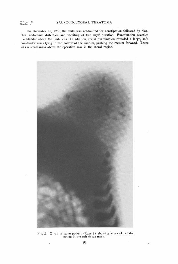
FIG. 3.-Cross section of tumor removed from Case 3, showing thetypical cystic nature.
Case 3-History. P. T., a white girl, age eight months, entered the Children'sMemorial Hospital on October 2, 1942, with the history of having had a lobulated, cysticgrowth in the left buttock since birth. This mass had grown larger and become moresuperficial, extending into the right btuttock. Aside from frequency in stools since birth,the history was otherwise niegative.
Examninationt. The examintationi i-evealed a multilocular, cystic tumor mass cov-ere(d with tense skin occupying the left buttock and medial one-third of the right buttockposterior to the rectum. A hemanigioma of the left calf was present. The hemoglobinwas eight grams; red blood cell count was 3,600,000; white blood cell count was 16,300.The urine was negative.
92

Volunw 12.S SACROCOCCYGEAI1 TERATOMANUmber 1
Operationt. The day followinlg adnission a large mul1tilocular cyst was removedfrom the sacrococcygeal region. The clhild wx-as given I 1() cc. of whole blood in tfleoperating room. The w-ound heale( well and the child was discharged the seventh1mostoperative day.
Patliologic RI-port. Ilie specimen consisted( of aii irregular lobulated imiass 11 .S x4 cmii. anid weighed 280 Gmn. Some portions were cystic; other portionis were solid. Thecapsule was 2 imm. thick. The largest cyst was 4.5 x 4 x 2 cm. and conitained thini, yellowfiuid(. The microscopic sectionis revealed smooth muscle, cartilage, bone, adipose, tissuie,s(juanmous epitheliuni anid nervous tissue. One section revealed glanldular tissue withpapillary features. The epithelium was pleomorphic with irregular nuclei and disorderlyatypical mitosis.
A diagnosis of teratoma with an area of emiibryonial carcinioma was made (Fig. 3.)Couirse. On Janiuary 10, 1943, the patient was readmitted with the hiistory of recur-
rence of the tumor above the healed inlcisioIn thlree weeks pireviously. This mass hadincreased in size and the patienat had lost four pounds of weight. Examination revealeda firm, fixed bluish lump the size of a walnut above the incisioni over the sacrum. Thiswas resected and the wound healed well.
Pathologic Report. The specimens consisted of two yellowislh gray pieces of tissue1 V x 1 X2 X 1 2". The miscroscopic section revealed cartilage, adipose and fibrous tissueand glandular tissue with less mitosis than the tumor showed previously.
No trace of this patienit can be found. It is probable that she also died of recurrenceof the tumor.
Case 4-History. J. C. was a full term white baby girl. One week after a normaldelivery a small tumor had beeni felt in the left buttock. It grew rapidly in size and atthe age of two weeks the child developed urinary retentioni and was taken to anotherhospital. Catheterization did not relieve the abdominal distention, so a laparotomy wasperformed. It was reported that rupture of the bladder and peritonitis were found. Thebladder was repaired and drained through a urethral catheter. A small biopsy was takenfrom a large mass lying in the pelvis behind the bladder. Microscopic sections revealed"nerve tissue having the appearance of brain tissue." Two weeks later the mass in tllebuttock was removed. The child made a fairly good recovery anid at the age of twomonths was admitted to the Children's Memorial Hospital oni May 12, 1941.
Examiniationi. The infant was poorly nourished. Her abdomen was distended. A hardmass could be palpated rising out of the pelvis and lying posterior to the rectum. A small,indurated, healed wound was present on the left buttock tinder which could be felt alarge firm mass, extending into the lhollow of- the sacrum. There was an indwellingurethral catheter. Intravenous pyelograms disclosed an enlarged bladder and hydro-ureters. The hemoglobin was 85 per cent; the red blood cell count was 4,300,000; theurine contained four plus albumin, numerous white blood cells and an occasional redblood cell.
Course. Roentgen-ray therapy was given but produced no decrease in the tumor size.Microscopic examination of a biopsy of the mass in the right buttock revealed dense
fibrolus tissue containing cystic spaces lined with squamous, low cuboidal and tall columnarel)ithelium. In some areas there were papillary projections. There was evidence ofreaction to irradiation, the bladder function returned to normal and the child was dis-charged in good condition, although the tumor on the buttock was gradually increasingin size.
On September 12, 1941, the child was readmitted with the acute intestinal obstructionand died 24 hours later.
Postmnortem Findings. The examination was limited to the reopening of the surgicalincisions. There was a large, multiloculatedc cystic tumor rising retroperitoneally in theihdomen, 2 cm. above the umbilical level and extending downi between the rectumn and
93

RIKER ANiD POTTS Annals of Surgery
sacrutiii illto the buttock-. It was attaclhe(l by firm fibrous adhesions to the coccyx. C(ltsectionis revealed cystic cavities varying markedly in size anid in the thickness of theirwvalls. Scattered amoing the cysts vere several white, granular, homogenous nodules andareas of soft, pinkish gray tissue. In addition there was bilateral hydronephrosis, hydro-ureter and ascites.
Microscopic sections showed cystic spaces lined with epithelium similar to those foundon biopsy. Some areas resembled gastric mucosa and one section appeared to be pancreatic
G.4.-Case5.onadmissin... . .. . ....
FIG. 4.-Case 5. on admission to hosDital.
FIG. 5.-Case 5, three months postoperatively.
94
'V'oltnie 128 SACROCOCCYGEAL TERATOAIANumvber I
tissue. There were large aggregates of atypical ganiglioni cells atnd nerve fibers in colla-genous connective tissue. Peri-aortic nodes showed no metastasis. A diagnosis of teratomawithout evidence of malignant changes was made.
Case 5-History. M. was a white female, one day old, weighing seven pounds, twoounces, admitted to the Children's Memorial Hospital on June 1, 1946. At birth a largetumor attached over the sacrum was noted (Fig. 4).
Examination. The infant was normal except for a large, irregular tumor lying overthe lower sacral region. It was covered with skin except for a small ulcerated area.Urinalysis and blood counit were normal. Roentgen-ray examination revealed no bonypathology in the spine but the tumor mass 8 x 12 cm. could be visualized containing areasof calcification. The diagnosis of sacrococcygeal teratoma was made.-~~~.
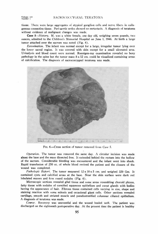
FIG. 6.-Cross section of tuimor removed from Case 5.
Operation. The tumor was removed the samne day. A circular incision was madeabout the base and the mass dissected free. It extended behind the rectum into the hollowof the sacrum. Considerable bleeding was encountered and the infant went into shock.Rapid transfusion of 250 cc. of whole blood revived the patient and the closure of thewound was completed.
Pathologic Report. The tumor measured 12 x 10 x 5 cm. and weighed 320 Gm. Itcontained cysts and calcified areas at the base. Near the skin surface were dark redlobulated masses and firm round nodules (Fig. 6).
Microscopic sections revealed glial tissue and some areas resembling choroid plexus,fatty tissue with niodules of cornified squamous epithelium and sweat glands with bodieshaving the appearanice of hair. Fibrous tissue contained cells varying in size, shape andstaining reaction with some mitosis and occasional giant cells. Other sections revealedcartilage, smooth and striated muscle and pseudostratified columnar ciliated epithelium.A diagnosis of teratoma was made.
Course. Recovery was uneventful and the wound healed well. The patient wasdischarged on the eighteenth postoperative day. At the present time the patient is healthy
95

lliKER AND POTTS Annals of Slurger'J. yt1 . 1 !) -l s
anid sliows nio r-ecurrenice of the tumiior (Fig. 5).Case 6-History. J. T., a t3V2 month old white female was a(dmitte(l to the hospital
October 26, 1944, for con.stipationi due to a congellital anal stricture and a hardi mass illthe right buttock presenit for three (lays.
Examinationt. There was ani anial stricture anid a rectovaginal fistula. The rightbuttock was swollen, hard and tender anld one week later pus drained spontaneously fromthe intergluteal fold. Two months later the abscess reformed anid was incised.
Course. A draining silnus remained in the sacrococcygeal area. Frequent rectal dila-tationis were necessary. On July 26, 1945, a colostomy was performed. A year later thechild was readmitted.
Operationt. On July 31, 1946, an inlcisioIn was made in the sacral region anid a cysticstructure was found imbedded in dense scar tissue. This extended downi toward thesacrum and was dissected free. Attached to it was a second, smaller cyst which waslikewise removed. Some clear fluid escaped whiclh resembled spinal fluid. The woundwas closed in layers without drainage.
Pathologic Report. The specimen conisisted of a dumbbell-shaped piece of tissue5 x 2 cm. containing a cyst with a smooth lining and filled with milky fluid. Micro-scopically the cyst was lined witlh stratified squamous epithelium surrounded by densefibrous tissue. Other areas showed columniar epithelium, loose myxomatous connectivetissue and nerves. One area conitained ganglion cells imbedded in a fibrillar networkresembling neurologial tissue. A diagnosis of a benign teratoid tumor was made. Becauseno structures of entodermal origin could be found, the tumor more closely resembled adermoid cyst.
Course. The wound healed well and there has been no recurrence to date. Therectovaginal fistula is to be repaired soon.
DISCUSSION
Incidence.-Calbet2 stated that congenital tumors of the sacrococcygeal re-gions occur once in 34,582 births. Teratomas represent only a very small per-centage of these tumors.
The tumors are thought to be invariably present at birth and about 90per cent of them are recognized at that time. Very few remain unnoticed untiladult life. A large percentage of the patients are stillborn or die shortly afterbirth. Because of their size, these tumors may cause dystocia.3
Chaffin4 reports that 75 per cent of the patients with these tumors arefemales. In our series all six of the cases were female.
In the past 30 years there have been a little over 600 tumors examinedpathologically at the Children's Memorial Hospital. Seven of these weresacrococcygeal teratomas, giving an incidence of approximately one per centof all tumors in this hospital.
Origin.-The origin of those tumors is the only really controversial aspectof teratology. Two main theories have been advanced.
1. Parthenogenic development of the individual's primitive germ cellsmight give rise to these tumors as is suggested by the experimental work ofBosarus5 with stimulation of unfertilized frog ova. These germ cells migratingfrom the primitive streak of the genital ridge could lodge in the sacrococcygealregion. Similarly, in early embryonic life a blastomere resulting from seg-mentation might begin its own development remaining attached to the mainembryo and becoming incorporated in it. If the former occurred, the teratoma
Volume 128Niiniber 1 SACROCOCCYGEAL TERATOMA
Uotil(l rel)resent aii offspriing wllile in tile latter case it would be a twin. It iseasy to believe that sacrococcygeal teratoma are rudimentary organ massesrepresenting an ill-developed pygopagus twin.
2. Other authorities believe that these tumors are not fetal implants butarise primarily from cells already present in the sacrococcygeal region innormal embryonic development such as the postanal gut and the neurentericcanal. The close approximation of nervous, intestinal, bony and connectivetissue elements in this region is supposed to explain the development of tumorscontaining structures from all three germ layers.2
Chaffin suggests that temporary adhesions between the fetal rump and theamnion may cause formation of teratomas.
Pathology.-Teratomas are usually lobulated tissue masses with cystic andsolid areas and may contain tissue or malformed organs representing anystructure in the body. The most common contents are epithelial lined cysts,connective tissue, ganglion cells and other nervous elements, intestinal mucosa,bone and cartilage. Infrequent tissues are liver, pancreas, kidney, testicle,ovary and chorionic epithelium. Malignant changes will be discussed below.
Clinical Features.-Teratomas of this region always have their origin re-trorectally and are attached to the coccyx or sacrum.6 From there they usuallygrow downward and posteriorly into the buttocks, displacing the anus forwardand to one side. They may also extend upward behind the rectum into theabdominal cavity.
The tumors vary in size from a scarcely noticeable lump to a huge massinterfering with bodily function. They are covered with skin but often haveulcerations or fistulae. They may be cystic or solid but usually contain areasof both. Bone may be felt within the tumors and occasionally peristalsis canbe observed. Upon stimulation, muscular twitching may be present.
Aside from local examination, it is important to examine the abdomen forevidence of extension of the mass producing urinary or bowel obstruction.Rectal examination will reveal the extent of pelvic growth and the degree offixation to the rectum. Roentgen-ray is helpful in showing the presence ofbone or teeth and in determining spinal development. Aspiration of cysticareas in the tumor is of doubtful help and often leads to infection and fistulaformation. Other congenital anomalies are frequently present.
Differential Diagnosis.-Other causes of a mass in the sacral region maybe meningocele, hernia, abscess, chordoma, dermoid, lipoma, myoma, angioma,myeloma, sarcoma, tuberculosis of sacrum or rectal carcinoma. The last fewwould be extremely rare in infants.
An accurate diagnosis is necessary in order to determine the propertreatment.
DeVeer and Browder7 have emphasized several points to differentiate ateratoma from a meningocele. The latter is smaller and does not increase inproportion to the infant growth as does a teratoma. A meningocele is coveredby a transluicent membrane whereas a teratoma is covered with skin. The
97

RIKER AND POTTS Annals of Surgern.1 uil . 1 9 4 8
rectumii is inot often displaced by a meningocele but it is by a teratoma. Thereis usually evidence of commnunication between a meniingocele and the spinalcanal suclh as expansioni of the tumior with couglhing anld crying or bulgingof thle fontanelle upon compression of the tumor. Neurologic signs in the lowerextremities are mlore often associated with a meningocele. A roentgenogramof the spine showing spina bifida, also suggests meningocele.
The possibility of the tumiior being a rar-e type of herniia may niecessitatea barium mlieal stu(ly to demiionistrate coiitinuity of the bowel in the al)(lbdoiienand in the tumor.
Coniplications.-l. Malignancy-Sacrococcygeal teratomiias (levelop iiialig-nant changes in about 15 per cent of the cases. In 1942 Lisco8 collecte(d 12cases of malignancy from the 72 teratomias reported ul) to that time. In otirown series, cases I and III contained definite malignant elements and develope(dlocal recurrences of the tumors.
Usually only one tissue of the teratoma becomes malignant. This resemiiblesa sarcoma as to the method of spread andl ten(lency to local recturrence.9Patlhologically, however, the malignant areas usually are either papillary car-cinomas or neoplasms of neural origin.
Rapid growth of a teratoma does not necessarily indicate miialignancy be-cause growth of the tumor usually parallels that of the clhild. The presence ofwell-formed bony structures, muscular twitching or bowel witlh peristalticactivity indicates a benign tumor.10
2. Ulceration, iinfection and fistula formation mayav develop as occurred iiicase II and case VI.
3. Obstruction of the rectumil or lower urinary tract may occur if the tumorexpands in the hollow of the sacrum. This has been noted frequently inmledical literature"11 12, 13 and occurred in case II and IV.
4. Very rarely hypersection of some endocrine gland in the teratoma mayproduce generalized effects. Rhoden13 reported a case of precocious sexualdevelopment which he thought was due to the secretion of the adrenal corticaltissue in the teratomiia.
5. The position anid large size of somlie teratomas may become a nuisanceand discomfort. A case reported by Brinles4 hlad a very large tumor containingrudimentary armii and(l halnld bones that miiade sitting or walking quite difficult.
Treatment.-It is agreed that complete excisioni of tfle tumor as early aspossible is the treatmiient of clhoice. More anid more cases of successful removalof teratomas have beeni reported in the past few years. Operation in the firstyear of life gives better restults as to recurrences and mortality than if surgeryis postponed until later life.4 Postoperative radiation tlherapy mlay also begiven especially if pathological examiiinationi reveals miialignant elements.
Pearse9 emliplhasizes that the posterior approach is the best because: 1 ) com-plete remiioval of the tumiior with resection of the coccyx, if necessary, is moreeasily accomplislhed; 2) the peritoneal cavity is avoided; 3) hemorrlhage is
98

Volume 128Number 1 SACROCOCCYGEAL TERATOMA
controlled nmore readily; 4) local recurrences in the scar are easily seen andtreated. All the cases operated tupon at thlis hospital have been approachedposteriorly.
It is interesting to note the degree of shock that usually accompanies theremoval of these tumors. Some authors mention that shock seemed out of pro-portion to the blood loss.10 12, 13 The reasons for this may be several. A largevascular bed is being remiioved. The region from wlhich the tumor is removedis niormiially quite vascuilar anid hiemiiostasis is difficult. Also, the degree of bloodloss in infants is dlece)tive. A few blood-damiipened sponges may represent alarge fraction of tlhe baby's blood volume.
It will be noted that in several of our cases a transfusion was necessaryduring or imme(liatelv after the operation. In case V the infant would havesuccumbed were it not for rapid replacement of blood loss. In this case atransfusion of 250 cc. of blood was given. Considering the weight of thepatient, this represents almost a comiiplete replacement of the total blood volumleof the infant.
It has been our practice in all stuclh miiajor procedures to insert a cannulainto the long saphenous vein at the anikle and attach to it intravenous tubingand a three-way stopcock to which is connected a 50 cc. syringe and a Salvarsanflask with a Murphy drip tube. During the operation physiologic salinesolution is allowed to drip slowly to keep the system open. If necessary.citrated whole blood can be poured into the flask and(l ptumiiped into the patientsvein quite rapidly by means of the syringe.
SUMMARY1. Six previously unreported cases of sacrococcvgeal teratomas are pre-
sented. All of these were females below the age of one year. Removal of thetumor was attempted in five cases. Malignancy an(d recurrence occurred intwo cases and probably a third. One child died as the result of vressure of thetumor causing urinary and bowel obstruction. Two cases are living and wellone year after surgery with no evidence of recurrence.
2. Some clinical aspects of these tunmors are reviewe(l with emphasis ontr-eatment by early and complete surgical excision via the posterior route.
3. A well-known but not widely used method of giving a rapid, often life-saving, transfusion during operation is described. In the removal of thesetumors as well as many other major operations on infants the survival of thechild may depend on the ability to replace rapidly a serious blood loss.
REFERENCES1Montgomery, A. H.: Sacral Teratoma Containing an Embryonic Scapula. J.A.M.A.,
78: 416, 1922.2 Hundling, H. W.: Ventral Tumors of the Sacrum. Surg., Gynec., & Obst., 38: 518,
1924.'Neal, M. P., and J. B. Carlisle: Congenital Sacroccygeal Tumors. South. Med. Jour.,
36: 677-678, 1943.'Chaffin, L.: Clinical Aspects of Sacroccygeal Teratomas. Surg., Gynec., & Obst., 69:
337, 1939.
99

RIKER AN-D POTTS Annals of Surgery
MacCallutm, W. G.: Teratomata. Textbooks of Pathology, Philadelphia, WV. B. Saui-(lers Co., 1936.
Hansmaiiim, G. H., andl C. J. lierne: Sacrococcygeal Teratomas. Arcli. Surg., 25: 1090,1932.
7 DeVeer, J. A., and J. Browder: Sacrococcygeal Teratoma. Ann. Surg., 105: 408, 1937.8Lisco, H.: Malignant Tumors Developing in Sacrococcygeal Teratomata. Ann. Surg.,
115: 378, 1942.9 Pearse, H.: Removal of Ventral Tumors of the Sacrum by the Posterior Route. Surg.,
Gynec., & Obst., 33: 164, 1921.10 McKnight, H. A.: Sacrococcygeal Teratomata in the Newborn. Am. J. Surg., 46:
387, 1939.11 Stewart, J. D., Alter and Craig: Sacrococcygeal Teratoma with Malignant Degen-
eration in Childhood. Surg., Gynec., & Obst., 50: 85, 1930.12 Renner, R. R., and E. Goodsit: Sacrococcygeal Teratomata-Report of a Case of
Double Tumor in a Newborn Infant. Am. J. Cancer, 24: 617, 1935.13Rhoden, A. E.: Precocious Sexual and Somatic Development in a Male Infant with
a Presacral Teratoma containing Androgen Producing Tissue. J. Clin. Endocrin.,4: 185, 1944.
14Brines, R. J.: A Large Teratoma Containing Rudimentary Arm Bones and a Hand.J.A.M.A., 103: 338, 1934.
15 Morris, K. A.: Sacrococcygeal Teratoma-Report of Case. Am. J. Surg., 33: 285, 1936.
100