s41467-017-01801-z.pdf - Nature
8
ARTICLE A new stem sarcopterygian illuminates patterns of character evolution in early bony fishes Jing Lu 1 , Sam Giles 2 , Matt Friedman 2,3 & Min Zhu 1 Discoveries of putative stem sarcopterygians from the late Silurian and Early Devonian of South China have increased our knowledge of the initial diversification of osteichthyans while also highlighting incongruities in character evolution in this major jawed vertebrate group. Character-rich endocrania are incompletely preserved for early bony fishes, limiting a detailed understanding of complex internal morphology and evolutionary changes in the cranium. Here we report a new sarcopterygian (Ptyctolepis brachynotus gen. et sp. nov.) from the Pragian (Early Devonian) of South China, which preserves a unique example of a completely ossified otoccipital division of the braincase in a stem lobe-finned fish. The hyomandibular facets are paired but lie dorsal to the jugular canal, representing a hitherto unobserved combination of derived and primitive character states. This new taxon prompts a reassess- ment of early osteichthyan interrelationships, including the phylogenetic placement of psarolepids, which might branch from the osteichthyan—rather than sarcopterygian—stem. DOI: 10.1038/s41467-017-01801-z OPEN 1 Key Laboratory of Vertebrate Evolution and Human Origins of Chinese Academy of Sciences, Institute of Vertebrate Paleontology and Paleoanthropology, Chinese Academy of Sciences, Beijing, 100044, China. 2 Department of Earth Sciences, University of Oxford, South Parks Road, Oxford, OX1 3AN, UK. 3 Museum of Paleontology and Department of Earth and Environmental Sciences, University of Michigan, 1109 Geddes Ave, Ann Arbor, MI 48109, USA. Correspondence and requests for materials should be addressed to J.L. (email: [email protected]) or to M.Z. (email: [email protected]) NATURE COMMUNICATIONS | 8: 1932 | DOI: 10.1038/s41467-017-01801-z | www.nature.com/naturecommunications 1 1234567890
-
Upload
khangminh22 -
Category
Documents
-
view
0 -
download
0
Transcript of s41467-017-01801-z.pdf - Nature

ARTICLE
A new stem sarcopterygian illuminates patterns ofcharacter evolution in early bony fishesJing Lu1, Sam Giles2, Matt Friedman 2,3 & Min Zhu 1
Discoveries of putative stem sarcopterygians from the late Silurian and Early Devonian of
South China have increased our knowledge of the initial diversification of osteichthyans while
also highlighting incongruities in character evolution in this major jawed vertebrate group.
Character-rich endocrania are incompletely preserved for early bony fishes, limiting a detailed
understanding of complex internal morphology and evolutionary changes in the cranium.
Here we report a new sarcopterygian (Ptyctolepis brachynotus gen. et sp. nov.) from the
Pragian (Early Devonian) of South China, which preserves a unique example of a completely
ossified otoccipital division of the braincase in a stem lobe-finned fish. The hyomandibular
facets are paired but lie dorsal to the jugular canal, representing a hitherto unobserved
combination of derived and primitive character states. This new taxon prompts a reassess-
ment of early osteichthyan interrelationships, including the phylogenetic placement of
psarolepids, which might branch from the osteichthyan—rather than sarcopterygian—stem.
DOI: 10.1038/s41467-017-01801-z OPEN
1 Key Laboratory of Vertebrate Evolution and Human Origins of Chinese Academy of Sciences, Institute of Vertebrate Paleontology and Paleoanthropology,Chinese Academy of Sciences, Beijing, 100044, China. 2 Department of Earth Sciences, University of Oxford, South Parks Road, Oxford, OX1 3AN, UK.3Museum of Paleontology and Department of Earth and Environmental Sciences, University of Michigan, 1109 Geddes Ave, Ann Arbor, MI 48109, USA.Correspondence and requests for materials should be addressed to J.L. (email: [email protected]) or to M.Z. (email: [email protected])
NATURE COMMUNICATIONS |8: 1932 |DOI: 10.1038/s41467-017-01801-z |www.nature.com/naturecommunications 1
1234
5678
90

Recent discoveries of osteichthyans (bony vertebrates com-prising bony fishes and tetrapods) from the Ludfordian(Ludlow, late Silurian; ~425 million years ago, mya) to
Lochkovian (Early Devonian; ~415 mya) have highlighted thesignificance of South China in understanding the diversificationof lobe-finned fishes1–5, as well as actinopterygians6 andgnathostomes7–9 more generally. Chief among these are thepsarolepids (sensu ref. 10), a clade of apparent stem sarcopter-ygians known from articulated (Guiyu1 and Sparalepis10), dis-sociated postcranial and cranial (Psarolepis5) and cranial only(Achoania4) remains. Despite early phylogenetic ambiguities thatincluded associations with porolepiforms11 and the osteichthyanstem5,12, placement of psarolepids as stem lobe-fins—and thuscrown bony fishes—has become one of the dominant motifs ofsystematic analyses of early vertebrates1–3,5,6,8,10,12–17. Thisfamily of results posits a well-populated sarcopterygian stempeppered with incongruous character transformations. However,a steady stream of discoveries revealing unexpectedly plesio-morphic aspects of psarolepid anatomy, including the presence of'placoderm'-like pelvic fin girdles and generalized dental histol-ogy18,19, have amplified apparent complexities in earlyosteichthyan evolution. Association of these taxa with the sar-copterygian stem nevertheless persists.
While most recent work on the early diversification of bonyfishes has centered on material from deposits flanking theSilurian–Devonian boundary, including those yielding
psarolepids, slightly younger strata have also provided importantnew fossils. Key among these is the Posongchong Formation ofSouth China, which contains an abundance of osteichthyansincluding a diverse array of crown sarcopterygians, such as theearliest tetrapodomorphs20, anatomically modern coelacanths21,and early onychodonts22,23. Significantly, these fossils are all wellpreserved with character-rich endocrania.
Here we report a new sarcopterygian from the PosongchongFormation (~409 mya, Pragian, Early Devonian) of Yunnan,China. This taxon is represented by a single well preserved andcompletely ossified otoccipital division of the skull measuring4.4 cm in width, 1.8 cm in length, and 2.7 cm in height, whichsuggests an individual considerably larger than other knownmembers of the co-occurring sarcopterygian fauna (e.g. Tungse-nia20 and Euporosteus yunnanensis21). This region of the brain-case is poorly known in sarcopterygians of Pragian age or older,with the only complete examples found in Styloichthys3 andrepresentatives of Dipnomorpha (Youngolepis24 and Powich-thys25), all of which are interpreted as crown sarcopterygians. Thenew taxon combines an anteroposteriorly short postparietalshield with vermiform ornament, while the braincase lacks ves-tibular fontanels and shows an unprecedented condition of thehyomandibular facet. The intact braincase permits reconstructionof a full endocast of the otic region, representing the secondoldest complete osteichthyan example after Youngolepis24. Basedon these and other observations, we include this new taxon in a
a
b
c
d
e
c.ju
ppa
mp
pr.ad
pp
gr.ju
nc
hyv
fm
gr.ju
c.Xoatm
ncpr.pam
gr.a.dl
gr.a.dl
hyd
hyv
gr.ju
nc
pr.pam
ppa
pr.pam
sup.art
c.a.dl
hydd.ot.n
f.occ.lat hyd
pr.ad
Fig. 1 The skull of Ptyctolepis brachynotus gen. et sp. nov. (IVPP V23386). High-resolution CT rendering of specimen in dorsal (a), right lateral (b), ventral(c), and posterior (e) views and (d) interpretive drawing in ventral view. c.a.dl canal for lateral dorsal aorta, c.X canal for vagus nerve, d.ot.n dorsal branchof the otic lateral line nerve, fm foramen magnum, f.occ.lat lateral occipital fissure, gr.a.dl groove for lateral dorsal aorta, gr.ju groove for jugular canal, hyddorsal hyoid articular area, hyv ventral hyoid articular area, mp middle pit-line, nc notochordal canal, oatm attachment area for trunk musculature, pdfposterior dorsal fontanel, pp posterior pit-line, ppa postparietal, pr.ad antero-dorsal process, pr.pam parampullary process, sup.art articular area forsuprapharyngobranchial. Scale bar, 5 mm
ARTICLE NATURE COMMUNICATIONS | DOI: 10.1038/s41467-017-01801-z
2 NATURE COMMUNICATIONS |8: 1932 |DOI: 10.1038/s41467-017-01801-z |www.nature.com/naturecommunications
revised cladistic analysis to infer its phylogenetic position andevaluate its impact on the pattern of branching and characterevolution deep within osteichthyan phylogeny.
ResultsSystematic paleontology.
Osteichthyes Huxley, 188026
Sarcopterygii Romer, 195527
Ptyctolepis brachynotus gen. et sp. nov.
Etymology. Generic name referring to vermiculate-ridgeornamentation on the skull roof, from Greek ptyktos (fold)and lepidos (scale). Specific name from Greek brachyno(shorten) and otos (ear, otic region), meaning the short oticregion.Holotype. IVPP V23386, a complete posterior cranial portionof the skull, Institute of Vertebrate Paleontology andPaleoanthropology (IVPP), Chinese Academy of Sciences(CAS), Beijing, China.Locality and horizon. Outcrop near the Qingmen Reservoir inthe suburb of Zhaotong, northeastern Yunnan. The fossilhorizon belongs to the Posongchong Formation, which mainlycomprises yellowish sandstones. In addition to lingulidbrachiopods and plant remains, the associated biota includesan abundance of galeaspid agnathans28–31, 'placoderms'32, andosteichthyans20–23,31,33,34. Specimens from this horizon arethree-dimensionally preserved with little distortion. The age ofthe Posongchong Formation is considered to be late Pragian,
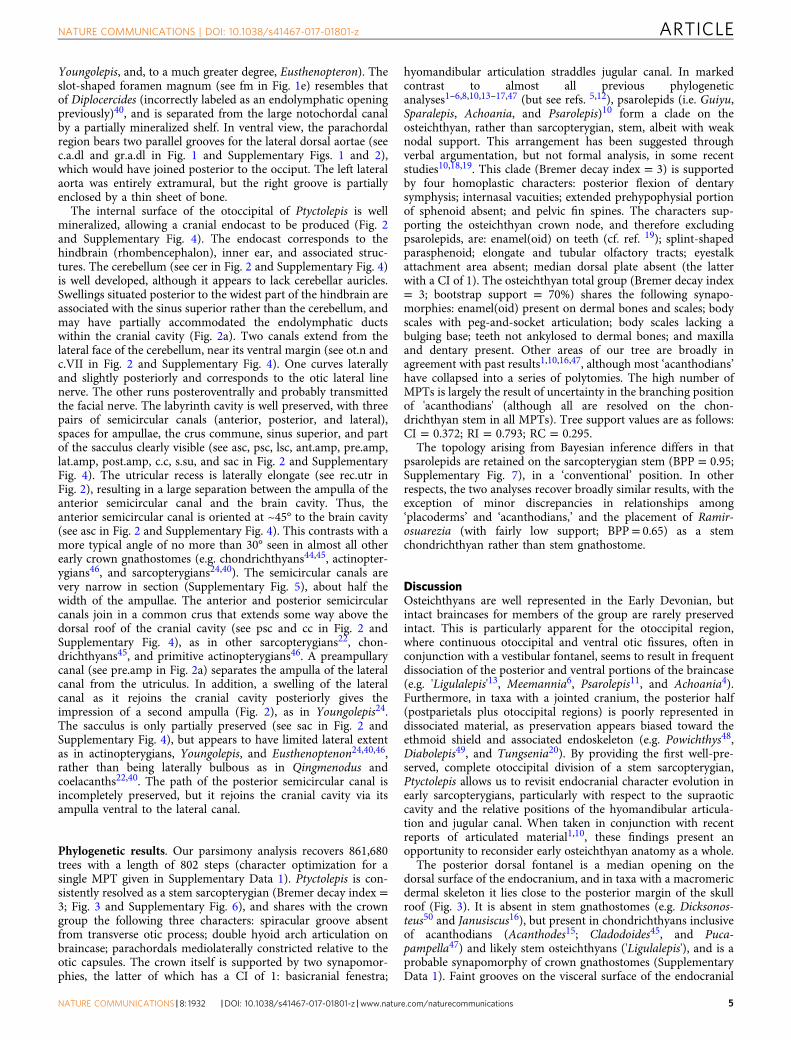
mainly based on the correlation of marine invertebrates andconodonts from the overlying Pojiao Formation35,36.Diagnosis. A sarcopterygian characterized by the uniquecombination of: laterally broad and anteroposteriorly shortpostparietal shield, very large notochordal canal, pairedhyomandibular facets dorsal to jugular canal, basicranialfenestra and vestibular fontanel both absent, middle andposterior pit lines lying close to midline of skull.
Description. The skull roof is represented by the posterior cranialportion (otoccipital shield/postparietal shield; Fig. 1 and Supple-mentary Figs. 1 and 2). The shield is much wider than long, with awidth:length ratio (~2.5) much higher than that of Psarolepis(~1.8) or Guiyu (~1.3). The anterior margin of the postparietals(see ppa in Fig. 1a) is straight, suggesting that a dermal cranialjoint was well developed. The ornament comprises short, vermi-form ridges (Fig. 1 and Supplementary Figs. 1 and 3) most similarto those of Guiyu1 or 'Ligulalepis'13, rather than the smooth,porous ornament seen in Psarolepis37 and Meemannia6. However,CT data show small pores open between the ridges (Supplemen-tary Fig. 3). The midline suture between the postparietals is clear,with the left bone shifted slightly over the right (Fig. 1a andSupplementary Fig. 1). The middle and posterior pit lines aresituated posteriorly and meet at the midline (see mp and pp inFig. 1a), as in Janusiscus16, Guiyu, Dialipina38, and 'Ligulalepis,'but unlike the more widely spaced pit lines of Meemannia andPsarolepis. There is no indication of the anterior pit-line on thepostparietals, which therefore must have been borne on theparietals. The main lateral line canal extends anteroposteriorly
c.cc.X
s.su
lat.amp
post.amp
a
c
b
d
rec.utr
rec.utr
lsc
psc
psc
psc
asc
asc
ant.amp
ot.n
pre.amp
cer
c.VII
c.c
post.amp
ot.n
f.occ.lat
c.X
f.occ.lat
pdf cer
lsc
sac
lat.amp
sac
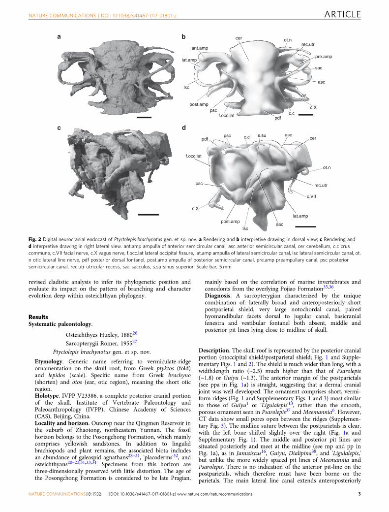
Fig. 2 Digital neurocranial endocast of Ptyctolepis brachynotus gen. et sp. nov. a Rendering and b interpretive drawing in dorsal view; c Rendering andd interpretive drawing in right lateral view. ant.amp ampulla of anterior semicircular canal, asc anterior semicircular canal, cer cerebellum, c.c cruscommune, c.VII facial nerve, c.X vagus nerve, f.occ.lat lateral occipital fissure, lat.amp ampulla of lateral semicircular canal, lsc lateral semicircular canal, ot.n otic lateral line nerve, pdf posterior dorsal fontanel, post.amp ampulla of posterior semicircular canal, pre.amp preampullary canal, psc posteriorsemicircular canal, rec.utr utricular recess, sac sacculus, s.su sinus superior. Scale bar, 5 mm
NATURE COMMUNICATIONS | DOI: 10.1038/s41467-017-01801-z ARTICLE
NATURE COMMUNICATIONS |8: 1932 |DOI: 10.1038/s41467-017-01801-z |www.nature.com/naturecommunications 3

near the lateral margin of the postparietal (see soc inSupplementary Fig. 2a). Sutures between the postparietal andmore lateral bones (i.e. tabular and supratemporal) cannot not betraced.
The otoccipital division of the braincase is well ossified (Fig. 1and Supplementary Figs. 1 and 2). The most conspicuous featureof the neurocranium is the large notochordal opening, which isfully two-thirds of the height of the occipital region (see nc inFig. 1e and Supplementary Fig. 2b).
There is no well-developed otic shelf of the kind seen incoelacanths, onychodonts, and tetrapodomorph fishes23,39,40, butthere is a modest ridge that extends along the ventral margin ofthe jugular groove in a corresponding position (Fig. 1c–d). Thedorsal margin of the jugular groove is marked by a suprajugularridge, similar to that in Youngolepis24. As in coelacanths,onychodonts, and Psarolepis11,39,41, the trigeminal nerve doesnot pass through the otoccipital. A small antero-dorsal process,similar to that of Psarolepis11 is present on the right side (see pr.ad in Fig. 1 and Supplementary Figs. 1 and 2), but missing fromthe damaged left side. Posteriorly, the transverse otic process (cf.Cheirolepis42, sometimes referred to as the lateral commissure; fora discussion of terminology of lateral processes of the braincase in
early gnathostomes, see ref. 16) is pierced by the jugular canal andbears two facets for the hyomandibula (see hyd and hyv in Fig. 1and Supplementary Figs. 1 and 2), as in other sarcopterygians(e.g. Qingmenodus22, Youngolepis24, and Styloichthys3). Thelaterally facing dorsal facet is larger, and is separated from theventrally directed lower facet by a thin ridge of bone. Thiscontrasts with the single hyomandibular facet present inPsarolepis11, 'Ligulalepis'13, and actinopterygians43. Unlike othertaxa with paired facets, both articular areas lie dorsal to thejugular vein rather than straddling it. The otoccipital fissure,through which the vagus nerve exits, is well developed (see c.Xand f.occ.lat in Fig. 1e), and a basicranial fenestra is absent, as inStyloichthys3 and Youngolepis24. Ptyctolepis lacks a vestibularfontanel, like Styloichthys, Qingmenodus, and coelacanths14.
An opening on the posterodorsal surface of the occiput (see pdfin Fig. 1e), continuous with the foramen magnum but set off fromit by a constriction, corresponds to the posterior dorsal fontanel.The articular area for the first suprapharyngobranchial is borneon a stout post-otic process at the level of the foramen magnum(see sup.art in Fig. 1e). A depression dorsal to the post-oticprocess likely served as an attachment area for trunk musculature(see otam in Fig. 1), but is not divided into distinct regions (cf.
Chondrichthyes
Meemannia
Sparalepis
Onychodonts
Coelacanths
Dipnomorpha
Tetrapodomorpha
Guiyu
Cheirolepis
‘Ligulalepis ’
Dialipina
Other Actinopterygii
DevonianSilurian
Early Middle Late
359 (Ma)419430
Ptyctolepis
Psarolepis
Achoania
Janusiscus
Jugular canal
Hyomandibular facet
Stem osteichthyans
Sarcopterygians
Actinopterygians
Posterior dorsal fontanelle
Skull roof
Supraotic cavity
a dc
ba
b
c
d
Fig. 3 Simplified phylogeny showing braincase evolution in osteichthyans. a Single hyomandibular facet dorsal to the jugular vein, as shown in 'Ligulalepis'13.b Double hyomandibular facet dorsal to the jugular vein, as shown in Ptyctolepis. c Double hyomandibular facet straddling the jugular vein, as shown inQingmenodus22. d Double hyomandibular facet dorsal to the jugular vein; supraotic cavity present, as shown in Eusthenopteron40. Not drawn to scale
ARTICLE NATURE COMMUNICATIONS | DOI: 10.1038/s41467-017-01801-z
4 NATURE COMMUNICATIONS |8: 1932 |DOI: 10.1038/s41467-017-01801-z |www.nature.com/naturecommunications
Youngolepis, and, to a much greater degree, Eusthenopteron). Theslot-shaped foramen magnum (see fm in Fig. 1e) resembles thatof Diplocercides (incorrectly labeled as an endolymphatic openingpreviously)40, and is separated from the large notochordal canalby a partially mineralized shelf. In ventral view, the parachordalregion bears two parallel grooves for the lateral dorsal aortae (seec.a.dl and gr.a.dl in Fig. 1 and Supplementary Figs. 1 and 2),which would have joined posterior to the occiput. The left lateralaorta was entirely extramural, but the right groove is partiallyenclosed by a thin sheet of bone.
The internal surface of the otoccipital of Ptyctolepis is wellmineralized, allowing a cranial endocast to be produced (Fig. 2and Supplementary Fig. 4). The endocast corresponds to thehindbrain (rhombencephalon), inner ear, and associated struc-tures. The cerebellum (see cer in Fig. 2 and Supplementary Fig. 4)is well developed, although it appears to lack cerebellar auricles.Swellings situated posterior to the widest part of the hindbrain areassociated with the sinus superior rather than the cerebellum, andmay have partially accommodated the endolymphatic ductswithin the cranial cavity (Fig. 2a). Two canals extend from thelateral face of the cerebellum, near its ventral margin (see ot.n andc.VII in Fig. 2 and Supplementary Fig. 4). One curves laterallyand slightly posteriorly and corresponds to the otic lateral linenerve. The other runs posteroventrally and probably transmittedthe facial nerve. The labyrinth cavity is well preserved, with threepairs of semicircular canals (anterior, posterior, and lateral),spaces for ampullae, the crus commune, sinus superior, and partof the sacculus clearly visible (see asc, psc, lsc, ant.amp, pre.amp,lat.amp, post.amp, c.c, s.su, and sac in Fig. 2 and SupplementaryFig. 4). The utricular recess is laterally elongate (see rec.utr inFig. 2), resulting in a large separation between the ampulla of theanterior semicircular canal and the brain cavity. Thus, theanterior semicircular canal is oriented at ~45° to the brain cavity(see asc in Fig. 2 and Supplementary Fig. 4). This contrasts with amore typical angle of no more than 30° seen in almost all otherearly crown gnathostomes (e.g. chondrichthyans44,45, actinopter-ygians46, and sarcopterygians24,40). The semicircular canals arevery narrow in section (Supplementary Fig. 5), about half thewidth of the ampullae. The anterior and posterior semicircularcanals join in a common crus that extends some way above thedorsal roof of the cranial cavity (see psc and cc in Fig. 2 andSupplementary Fig. 4), as in other sarcopterygians22, chon-drichthyans45, and primitive actinopterygians46. A preampullarycanal (see pre.amp in Fig. 2a) separates the ampulla of the lateralcanal from the utriculus. In addition, a swelling of the lateralcanal as it rejoins the cranial cavity posteriorly gives theimpression of a second ampulla (Fig. 2), as in Youngolepis24.The sacculus is only partially preserved (see sac in Fig. 2 andSupplementary Fig. 4), but appears to have limited lateral extentas in actinopterygians, Youngolepis, and Eusthenoptenon24,40,46,rather than being laterally bulbous as in Qingmenodus andcoelacanths22,40. The path of the posterior semicircular canal isincompletely preserved, but it rejoins the cranial cavity via itsampulla ventral to the lateral canal.
Phylogenetic results. Our parsimony analysis recovers 861,680trees with a length of 802 steps (character optimization for asingle MPT given in Supplementary Data 1). Ptyctolepis is con-sistently resolved as a stem sarcopterygian (Bremer decay index =3; Fig. 3 and Supplementary Fig. 6), and shares with the crowngroup the following three characters: spiracular groove absentfrom transverse otic process; double hyoid arch articulation onbraincase; parachordals mediolaterally constricted relative to theotic capsules. The crown itself is supported by two synapomor-phies, the latter of which has a CI of 1: basicranial fenestra;
hyomandibular articulation straddles jugular canal. In markedcontrast to almost all previous phylogeneticanalyses1–6,8,10,13–17,47 (but see refs. 5,12), psarolepids (i.e. Guiyu,Sparalepis, Achoania, and Psarolepis)10 form a clade on theosteichthyan, rather than sarcopterygian, stem, albeit with weaknodal support. This arrangement has been suggested throughverbal argumentation, but not formal analysis, in some recentstudies10,18,19. This clade (Bremer decay index = 3) is supportedby four homoplastic characters: posterior flexion of dentarysymphysis; internasal vacuities; extended prehypophysial portionof sphenoid absent; and pelvic fin spines. The characters sup-porting the osteichthyan crown node, and therefore excludingpsarolepids, are: enamel(oid) on teeth (cf. ref. 19); splint-shapedparasphenoid; elongate and tubular olfactory tracts; eyestalkattachment area absent; median dorsal plate absent (the latterwith a CI of 1). The osteichthyan total group (Bremer decay index= 3; bootstrap support = 70%) shares the following synapo-morphies: enamel(oid) present on dermal bones and scales; bodyscales with peg-and-socket articulation; body scales lacking abulging base; teeth not ankylosed to dermal bones; and maxillaand dentary present. Other areas of our tree are broadly inagreement with past results1,10,16,47, although most ‘acanthodians’have collapsed into a series of polytomies. The high number ofMPTs is largely the result of uncertainty in the branching positionof 'acanthodians' (although all are resolved on the chon-drichthyan stem in all MPTs). Tree support values are as follows:CI = 0.372; RI = 0.793; RC = 0.295.
The topology arising from Bayesian inference differs in thatpsarolepids are retained on the sarcopterygian stem (BPP = 0.95;Supplementary Fig. 7), in a ‘conventional’ position. In otherrespects, the two analyses recover broadly similar results, with theexception of minor discrepancies in relationships among‘placoderms’ and ‘acanthodians,’ and the placement of Ramir-osuarezia (with fairly low support; BPP= 0.65) as a stemchondrichthyan rather than stem gnathostome.
DiscussionOsteichthyans are well represented in the Early Devonian, butintact braincases for members of the group are rarely preservedintact. This is particularly apparent for the otoccipital region,where continuous otoccipital and ventral otic fissures, often inconjunction with a vestibular fontanel, seems to result in frequentdissociation of the posterior and ventral portions of the braincase(e.g. 'Ligulalepis'13, Meemannia6, Psarolepis11, and Achoania4).Furthermore, in taxa with a jointed cranium, the posterior half(postparietals plus otoccipital regions) is poorly represented indissociated material, as preservation appears biased toward theethmoid shield and associated endoskeleton (e.g. Powichthys48,Diabolepis49, and Tungsenia20). By providing the first well-pre-served, complete otoccipital division of a stem sarcopterygian,Ptyctolepis allows us to revisit endocranial character evolution inearly sarcopterygians, particularly with respect to the supraoticcavity and the relative positions of the hyomandibular articula-tion and jugular canal. When taken in conjunction with recentreports of articulated material1,10, these findings present anopportunity to reconsider early osteichthyan anatomy as a whole.
The posterior dorsal fontanel is a median opening on thedorsal surface of the endocranium, and in taxa with a macromericdermal skeleton it lies close to the posterior margin of the skullroof (Fig. 3). It is absent in stem gnathostomes (e.g. Dicksonos-teus50 and Janusiscus16), but present in chondrichthyans inclusiveof acanthodians (Acanthodes15; Cladodoides45, and Puca-pampella47) and likely stem osteichthyans ('Ligulalepis'), and is aprobable synapomorphy of crown gnathostomes (SupplementaryData 1). Faint grooves on the visceral surface of the endocranial
NATURE COMMUNICATIONS | DOI: 10.1038/s41467-017-01801-z ARTICLE
NATURE COMMUNICATIONS |8: 1932 |DOI: 10.1038/s41467-017-01801-z |www.nature.com/naturecommunications 5

roof in 'Ligulalepis' lead from the sinus superior to the posteriordorsal fontanel51, suggesting that the endolympatic ducts weretransmitted within the dorsal part of the cranial cavity (Fig. 3a).Unlike in chondrichthyans45, the posterior dorsal fontanel isconfluent with the otoccipital fissure. The primitive osteichthyancondition, with a semicircular posterior dorsal fontanel that iscontinuous with the fissure, and with endolymphatic duststransmitted through the cranial cavity, is also characteristic ofearly sarcopterygians based on our new evidence from Ptyctolepis(Fig. 3b). The fontanel becomes progressively larger in acti-nopterygians, eventually extending from the otoccipital fissure tothe level of the sinus superior (e.g. Mimipiscis46), but the condi-tion in crown sarcopterygians is somewhat different. In Young-olepis, an opening corresponding to the posterior dorsal fontanel(although its homology has previously been left equivocal24,40)leads into the cranial cavity and is confluent with the fissure, as inother early osteichthyans. In addition, this is joined by a deepridge on the roof of the cranial cavity that continues anteriorlyand terminates as a blind-ending canal anterior to the sinussuperior. Posteriorly, it extends toward the rear of the braincase,meeting its fellow at the midline just anterior to the posteriordorsal fontanel, with which it is continuous. This structure isreferred to as the supraotic cavity, and assumed to house theendolymphatic sac. This is elaborated further in taxa such asEusthenopteron (Fig. 3d), Gogonasus, and lungfishes, where thesupraotic cavity is largely or entirely separate from the cranialcavity after leaving the sinus superior40,52, and is not continuouswith the otoccipital fissure. A structure apparently correspondingto the posterior dorsal fontanel is also present, but does notappear to connect to the otoccipital fissure. No such divisionbetween the supraotic and cranial cavities is present in Psarolepis,onychodonts, coelacanths, or Ptyctolepis. Comparison with out-groups such as actinopterygians (e.g. Mimipiscis46) and chon-drichthyans (e.g. Cobelodus45) suggests that this is the primitivecondition.
The structure of the hyomandibular facet in osteichthyans fallsbroadly into two categories: examples where a single facet islocated dorsal to the course of the jugular vein, and exampleswhere paired facets straddle the level of the jugular canal (Fig. 3).The first of these conditions appears primitive, and is found in thestem gnathostome Janusiscus, the probable stem osteichthyan‘Ligulalepis’ (Fig. 3a), actinopterygians, and Psarolepis. The pre-sence of paired facets bridging the jugular vein is classicallyconsidered a character of crown sarcopterygians (Fig. 3c–d),although in some members of this group there is a single facetthought to represent the coalescence of primitively pairedarticular areas (e.g. lungfishes). Ptyctolepis (Fig. 3b) presents acombination of these two contrasting arrangements. As in pri-mitive osteichthyans, the hyomandibula articulates with the oticcapsule dorsal to the jugular vein. However, it shows division ofthe facet into separate dorsal and ventral components, as incrown sarcopterygians. The placement of Ptyctolepis in ourphylogenetic analysis as a member of the sarcopterygian stemlineage suggests that division of the hyomandibular facet pre-ceded the ventral extension of the hyoid articulation across thejugular vein canal.
The unusual character combinations of psarolepids have beenclear since their initial discovery and description5,11, with morerecent discoveries of articulated material - only serving to mag-nify their mosaic bodyplans. Central among the issues raised bypsarolepids are features present in the group and osteichthyanoutgroups, but absent in other definitive crown bony fishes: aneyestalk attachment area (the putative eyestalk in Styloichthys isresolved as non-homologous in our analysis), a median dorsalplate, dorsal-fin spines, pectoral-fin spines, pelvic fin spines, anddermal pelvic girdles. Previous phylogenetic consensus—
anchored by the presence of classic sarcopterygian features like anintracranial joint and cosmine53—regarded psarolepids as stemlobe-finned fishes, demanding one of two possible evolutionaryhistories for each of these characters: retention of the primitivecondition in psarolepids with parallel loss in actinopterygiansand other sarcopterygians, or absence in the common ancestorof sarcopterygians and actinopterygians with a reversal inpsarolepids. While the integrity of the intracranial joint asa sarcopterygian feature remained intact, other evidence for alobe-finned—or even crown osteichthyan—affinity of psarolepidshas eroded with further scrutiny. First is the report that unlikeactinopterygians and sarcopterygians, psarolepid teeth lackenamel54. Second is the discovery that many of the individualtraits that characterize the complex tissue cosmine are presentin probable (Meemannia) and definitive (Cheirolepis) acti-nopterygians6, meaning that dermal bone histology of psarolepidsis not compelling evidence of sarcopterygian affinity. Theseand other observations of primitive aspects of psarolepidanatomy have triggered a steady stream of discussion that thesetaxa might be stem osteichthyans rather than early sarcopter-ygians10,18,19, contrary to the apparent consensus arising fromformal analyses.
Placement of psarolepids as stem osteichthyans provides apotential solution to the unusually high number of generalizedfeatures present in this group. Median dorsal plates, for examplewould now represent a symplesiomorphy inherited from stemgnathostomes and lost at the osteichthyan crown node, whiletooth enamel is a crown osteichthyan synapomorphy. Someissues, however, remain. Tooth whorls are present in chon-drichthyans (inclusive of 'acanthodians'), psarolepids, onycho-donts, Gavinia, Styloichthys, and porolepiforms, but due toabsences at nodes preceding or subtending the gnathostome (i.e.Entelognathus), osteichthyan (actinopterygians), and sarcopter-ygian (some coelacanths and dipnomorphs) total groups, statesfor this character cannot be optimized deep in osteichthyanphylogeny. Multiple independent appearances are considered asparsimonious as a single appearance below the gnathostomecrown node and several losses. Similarly, many characters per-taining to fin spines cannot be optimized as the condition isunknown in proximate stem gnathostome and stem osteichthyantaxa; fin spines are known to be absent only in the anatomicallypeculiar and poorly understood Dialipina38.
In addition to these ambiguities resulting from missing data, astem osteichthyan placement of psarolepids implies a morecomplicated evolutionary history for the intracranial joint thanpreviously suspected. Rather than a synapomorphy of sarcop-terygians, lost independently in tetrapods and lungfishes53, itrepresents either convergence between psarolepids and sarcop-terygians, or a character of psarolepids and crown osteichthyans,subsequently lost in actinopterygians and multiple sarcopterygianlineages. However, these conclusions remain tentative at best, asour Bayesian trees resolve psarolepids in the conventional stemsarcopterygian position with high nodal support; BPP= 0.95).Indeed, although the psarolepids are placed as stem osteichthyansin all most parsimonious trees, there is no significant difference interms of tree length between this solution and one that placesthese taxa in a more conventional position on the sarcopterygianstem (Templeton test; p= 0.8474). We anticipate thatfurther discoveries of early osteichthyan material from theSilurian of China, and additional study of existing fossils, will helpto clarify the evolutionary histories of these characters, as well asproviding critical tests of competing hypotheses for the placementof psarolepids. More definitive resolution of this problem willhave consequences not only for our understanding of characterevolution among early osteichthyans, but also the timescale ofvertebrate diversification, as the psarolepid Guiyu has become a
ARTICLE NATURE COMMUNICATIONS | DOI: 10.1038/s41467-017-01801-z
6 NATURE COMMUNICATIONS |8: 1932 |DOI: 10.1038/s41467-017-01801-z |www.nature.com/naturecommunications

key fossil marker in molecular clock studies55. In the light ofpresent ambiguities, we regard the placement of these taxa asuncertain, but limited to either the osteichthyan or sarcopterygianstem.
MethodsHigh-resolution computed tomography. The holotype of Ptyctolepis brachynotusgen. et sp. nov. (IVPP V23386) was scanned at the Institute of VertebratePaleontology and Paleoanthropology (IVPP), Chinese Academy of Sciences (CAS),Beijing, China, using 225 kV microCT (developed by the Institute of High EnergyPhysics, CAS). The specimen was scanned with a beam energy of 130 kV and a fluxof 100 mA at a detector resolution of 27.4 µm per pixel, using a 1440° rotation witha step size of 0.25° and an unfiltered aluminum reflection target. A total of 1440transmission images were reconstructed in a 2048 × 2048 matrix of 1536 slices.Scan data were analyzed using Mimics v.18.01 (http://biomedical.materialise.com/mimics; Materialize) and imaged in Blender (blender.org).
Phylogenetic data set assembly and analyses. Our data set is modified from ref.6 (which is largely based on ref. 16). It has been expanded by the addition of fourtaxa (Ptyctolepis, Achoania, Qingmenodus, and Sparalepis) and nine characters(both novel and taken from the literature10,56, giving a total of 278 characters and94 taxa (see Supplementary Table 1 and Supplementary Note 1. Data matrix givenin Supplementary Data 2). Codings for some taxa have been updated in light ofrecent publications (e.g. Doliodus57; Romundina58; and Psarolepis19) and to correctprevious miscodes. We have also reformulated character 186, relating to the pre-sence of median dorsals, to reflect that dermal plates are coded as inapplicable(rather than absent) in 'acanthodians' and chondrichthyans. An equally weightedparsimony analysis (with 500 random addition sequences, five trees held at eachstep, maxtrees set to automatically increase, nchuck = 10,000, chuckscore = 1) wasperformed in PAUP* 4.0a15059, with the outgroup constrained as [Galeaspida[Osteostraci[ingroup]]]. Bootstrap values were calculated in PAUP using 500replicates of a heuristic search, with five trees held at each step, rearrlimit =50,000,000, limitperrep = yes, nchuck = 10,000, chuckscore= 1. Bremer decayvalues were also calculated in PAUP. Bayesian analysis was run under the Mkvmodel in MrBayes v.3.2.660, until the standard deviation of split frequenciesreached less than 0.01, indicating convergence had been reached. The first half ofeach run was discarded as burnin.
Nomenclatural acts. This published work and the nomenclatural acts it containshave been registered in ZooBank, the proposed online registration system for theInternational Code of Zoological Nomenclature (ICZN). The ZooBank LSIDs (LifeScience Identifiers) can be resolved and the associated information viewed throughany standard web browser by appending the LSID to the prefix “http://zoobank.org/.” The LSIDs for this publication are: urn:lsid:zoobank.org:pub:C1D7AD35-DEC1-4F6D-B0C8-D94DEB89F710 (article); urn:lsid:zoobank.org:act:5CBD2D52-0E50-441E-8BD4-74D9204E3570 (genus); and urn:lsid:zoobank.org:pub:C1D7AD35-DEC1-4F6D-B0C8-D94DEB89F710 (species).
Data availability. The CT data that support the findings of this study, as well as 3Dsurface files of described material, are available in figshare61 with the identifierhttps://doi.org/10.6084/m9.figshare.5458165. All other data files are included in theSupplementary Information.
Received: 22 June 2017 Accepted: 16 October 2017
References1. Zhu, M. et al. The oldest articulated osteichthyan reveals mosaic gnathostome
characters. Nature 458, 469–474 (2009).2. Zhu, M., Yu, X.-B., Wang, W., Zhao, W.-J. & Jia, L.-T. A primitive fish provides
key characters bearing on deep osteichthyan phylogeny. Nature 441, 77–80(2006).
3. Zhu, M. & Yu, X.-B. A primitive fish close to the common ancestor of tetrapodsand lungfish. Nature 418, 767–770 (2002).
4. Zhu, M., Yu, X.-B. & Ahlberg, P. E. A primitive sarcopterygian fish with aneyestalk. Nature 410, 81–84 (2001).
5. Zhu, M., Yu, X.-B. & Janvier, P. A primitive fossil fish sheds light on the originof bony fishes. Nature 397, 607–610 (1999).
6. Lu, J., Giles, S., Friedman, M., den Blaauwen, J. L. & Zhu, M. The oldestactinopterygian highlights the cryptic early history of the hyperdiverse ray-finned fishes. Curr. Biol. 26, 1602–1608 (2016).
7. Zhu, M. et al. A Silurian maxillate placoderm illuminates jaw evolution. Science354, 334–336 (2016).
8. Zhu, M. et al. A Silurian placoderm with osteichthyan-like marginal jaw bones.Nature 502, 188–193 (2013).
9. Friedman, M. & Brazeau, M. D. A jaw-dropping fossil fish. Nature 502,175–177 (2013).
10. Choo, B. et al. A new osteichthyan from the late Silurian of Yunnan, China.PLoS ONE 12, e0170929 (2017).
11. Yu, X.-B. A new porolepiform-like fish, Psarolepis romeri, gen. et sp. nov.(Sarcopterygii, Osteichthyes) from the Lower Devonian of Yunnan, China. J.Vertebr. Paleontol. 18, 261–274 (1998).
12. Zhu, M. & Schultze, H.-P. in Major Events in Early Vertebrate Evolution:Palaeontology, Phylogeny, Genetics and Development (ed Ahlberg, P. E.)289–314 (Taylor & Francis, London, 2001).
13. Basden, A. M., Young, G. C., Coates, M. I. & Ritchie, A. The most primitiveosteichthyan braincase? Nature 403, 185–188 (2000).
14. Friedman, M. Styloichthys as the oldest coelacanth: implications for earlyosteichthyan interrelationships. J. Syst. Palaeontol. 5, 289–343 (2007).
15. Davis, S. P., Finarelli, J. A. & Coates, M. I. Acanthodes and shark-like conditionsin the last common ancestor of modern gnathostomes. Nature 486, 247–250(2012).
16. Giles, S., Friedman, M. & Brazeau, M. D. Osteichthyan-like cranial conditionsin an Early Devonian stem gnathostome. Nature 520, 82–85 (2015).
17. Choo, B., Zhu, M., Zhao, W.-J., Jia, L.-T. & Zhu, Y.-A. The largest Silurianvertebrate and its palaeoecological implications. Sci. Rep. 4, 5242 (2014).
18. Zhu, M. et al. Fossil fishes from China provide first evidence of dermal pelvicgirdles in osteichthyans. PLoS ONE 7, e35103 (2012).
19. Qu, Q.-M., Haitina, T., Zhu, M. & Ahlberg, P. E. New genomic and fossil datailluminate the origin of enamel. Nature 526, 108–111 (2015).
20. Lu, J. et al. The earliest known stem-tetrapod from the Lower Devonian ofChina. Nat. Commun. 3, 1160 (2012).
21. Zhu, M. et al. Earliest known coelacanth skull extends the range of anatomicallymodern coelacanths to the Early Devonian. Nat. Commun. 3, 772 (2012).
22. Lu, J. et al. A Devonian predatory fish provides insights into the early evolutionof modern sarcopterygians. Sci. Adv. 2, e1600154 (2016).
23. Lu, J. & Zhu, M. An onychodont fish (Osteichthyes, Sarcopterygii) from theEarly Devonian of China, and the evolution of the Onychodontiformes. Proc.Biol.Sci. 277, 293–299 (2010).
24. Chang, M.-M. The Braincase of Youngolepis, a Lower Devonian Crossopterygianfrom Yunnan, South-western China. Thesis, Stockholm Univ. (1982).
25. Jessen, H. L. Lower Devonian Porolepiformes from the Canadian Arctic withspecial reference to Powichthys thorsteinssoni Jessen. Palaeontogr. Abt. A 167,180–214 (1980).
26. Huxley, T. H. On the application of the laws of evolution to the arrangement ofthe Vertebrata, and more particularly of the Mammalia. Proc. Zool. Soc. Lond.43, 649–662 (1880).
27. Romer, A. S. Herpetichthyes, Amphibioidei, Choanichthyes or Sarcopterygii?Nature 176, 126 (1955).
28. Liu, Y.-H. Lower Devonian agnathans of Yunnan and Sichuan. Vertebr.PalAsiat. 13, 202–216 (1975).
29. Pan, J. & Wang, S.-T. New discoveries of polybranchiaspids from YunnanProvince. Vertebr. PalAsiat. 19, 113–121 (1981).
30. Wang, J.-Q. & Zhu, M. Zhaotongaspis janvieri gen. et sp. nov., a galeaspid fromEarly Devonian of Zhaotong, northeastern Yunnan. Vertebr. PalAsiat. 32,231–243 (1994).
31. Wang, J.-Q., Fan, J.-H. & Zhu, M. Early vertebrate fossils from the earlyDevonian of Zhaotong District, northeastern Yunnan. Vertebr. PalAsiat. 34,1–17 (1996).
32. Dupret, V. A new wuttagoonaspid (Placodermi, Arthrodira) from the lowerDevonian of Yunnan (South China): origin, dispersal, and paleobiogeographicsignificance. J. Vertebr. Paleontol. 28, 12–20 (2008).
33. Zhu, M. & Janvier, P. Un onychodontide (Vertebrata, Sarcopterygii) duDévonien inférieur de Chine. Compt. Rend. Sci. 319, 951–956 (1994).
34. Lu, J. & Zhu, M. An early Devonian (Pragian) sarcopterygian from zhaotong,Yunnan, China. Vertebr. PalAsiat. 46, 161–170 (2008).
35. Liao, W.-H., Xu, H.-K. & Wang, C.-Y. in Symposium on the Devonian System ofSouth China (ed Inst. Geol. Min. Res.) 193–213 (Geological Press, 1978).
36. Hao, S.-G., Wang, D.-M. & Wang, Q. A new species of Estinnophyton from theLower Devonian Posongchong Formation, Yunnan, China; its phylogeneticand palaeophytogeographical significance. Bot. J. Linn. Soc. 146, 201–216(2004).
37. Qu, Q.-M., Zhu, M. & Wang, W. Scales and dermal skeletal histology of anearly bony fish Psarolepis romeri and their bearing on the evolution of rhombicscales and hard tissues. PLoS ONE 8, e61485 (2013).
38. Schultze, H.-P. & Cumbaa, S. L. in Major Events in Early Vertebrate Evolution:Palaeontology, Phylogeny, Genetics and Development (ed Ahlberg, P. E.) 315–-332 (Taylor & Francis, London, 2001).
39. Forey, P. L. History of the Coelacanth Fishes (Chapman & Hall, London, 1998).40. Jarvik, E. Basic Structure and Evolution of Vertebrates, Vol. 1 (Academic Press,
London, 1980).
NATURE COMMUNICATIONS | DOI: 10.1038/s41467-017-01801-z ARTICLE
NATURE COMMUNICATIONS |8: 1932 |DOI: 10.1038/s41467-017-01801-z |www.nature.com/naturecommunications 7

41. Andrews, S. M., Long, J. A., Ahlberg, P. E., Barwick, R. & Campbell, K. S. W.The structure of the sarcopterygian Onychodus jandemarrai n. sp. from Gogo,Western Australia: with a functional interpretation of the skeleton. Trans. R.Soc. Edinb. Earth Sci. 96, 197–307 (2005).
42. Giles, S. et al. Endoskeletal structure in Cheirolepis (Osteichthyes,Actinopterygii), an early ray-finned fish. Palaeontology 58, 849–870 (2015).
43. Gardiner, B. G. The relationships of the palaeoniscid fishes, a review based onnew specimens of Mimia and Moythomasia from the Upper Devonian ofWestern Australia. Bull. Br. Mus. Nat. Hist. Geol. 37, 173–428 (1984).
44. Schaeffer, B. The xenacanth shark neurocranium, with comments onelasmobranch monophyly. Bull. Am. Mus. Nat. Hist. 169, 1–66 (1981).
45. Maisey, J. G. The braincase in Paleozoic symmoriiform and cladoselachiansharks. Bull. Am. Mus. Nat. Hist. 307, 1–122 (2007).
46. Giles, S. & Friedman, M. Virtual reconstruction of endocast anatomy in early ray-finned fishes (Osteichthyes, Actinopterygii). J. Paleontol. 88, 636–651 (2014).
47. Brazeau, M. D. The braincase and jaws of a Devonian ‘acanthodian’ andmodern gnathostome origins. Nature 457, 305–308 (2009).
48. Clément, G. & Ahlberg, P. E. in Morphology, Phylogeny and Paleobiogeographyof Fossil Fishes (eds Elliott, D. K., Maisey, J. G., Yu, X.-B. & Miao, D.-S.)363–377 (Verlag Dr. Friedrich Pfeil, Munich, 2010).
49. Chang, M.-M. Diabolepis and its bearing on the relationships betweenporolepiforms and dipnoans. Bull. Mus. Natl Hist. Nat. Paris 4è Sér. 17,235–268 (1995).
50. Goujet, D. F. Les Poissons Placodermes du Spitsberg. Arthrodires Dolichothoracide la Formation de Wood Bay (Dévonien inférieur) (CNRS, Paris, 1984).
51. Basden, A. M. & Young, G. C. A primitive actinopterygian neurocranium fromthe Early Devonian of southeastern Australia. J. Vertebr. Paleontol. 21, 754–766(2001).
52. Holland, T. The endocranial anatomy of Gogonasus andrewsae Long, 1985revealed through micro CT-scanning. Earth Environ. Sci. Trans. R. Soc. Edinb.105, 9–34 (2014).
53. Friedman, M. & Brazeau, M. D. A reappraisal of the origin and basal radiationof the Osteichthyes. J. Vertebr. Paleontol. 30, 36–56 (2010).
54. Qu, Q.-M., Sanchez, S., Zhu, M., Blom, H. & Ahlberg, P. E. The origin of novelfeatures by changes in developmental mechanisms: ontogeny and three-dimensional microanatomy of polyodontode scales of two early osteichthyans.Biol. Rev. 92, 1189–1212 (2017).
55. Benton, M. J. et al. Constraints on the timescale of animal evolutionary history.Palaeontol. Electron. 18, 1–107 (2015).
56. Brazeau, M. D. & de Winter, V. The hyoid arch and braincase anatomy ofAcanthodes support chondrichthyan affinity of ‘acanthodians’. Proc. R. Soc. B282, 20152210 (2015).
57. Maisey, J. G. et al. Pectoral morphology in Doliodus: bridging the ‘acanthodian’-chondrichthyan divide. Am. Mus. Novitat. 3875, 1–15 (2017).
58. Dupret, V., Sanchez, S., Goujet, D. F. & Ahlberg, P. E. The internal cranialanatomy of Romundina stellina Ørvig, 1975 (Vertebrata, Placodermi,Acanthothoraci) and the origin of jawed vertebrates - anatomical atlas of aprimitive gnathostome. PLoS ONE 12, e0171241 (2017).
59. Swofford, D. L. PAUP*: Phylogenetic analysis using parsimony (* and othermethods), version 4.0b 10 (Sinauer Associates, Sunderland, 2003).
60. Huelsenbeck, J.P. & Ronquist, F. MRBAYES:Bayesian inference of phylogenetictrees. Bioinformatics 17, 754–755 (2001).
61. Lu, J., Giles, S., Friedman M. & Zhu, M. in Ptyctolepis brachynotus gen. et sp.nov. IVPP V23386: CT scan data and surface files of a new sarcopterygian fromEarly Devonian of South China. https://doi.org/10.6084/m9.figshare.5458165(2017).
AcknowledgmentsWe thank Y.-M. Hou for assistance with CT scanning and C.-H. Xiong for specimenpreparation. This work was supported by the National Natural Science Foundation ofChina (41472016, 41530102), Key Research Program of Frontier Sciences of CAS(QYZDJ-SSW-DQC002), and Strategic Priority Research Program of CAS (XDPB05) toJ.L. and M.Z., a Junior Research Fellowship from Christ Church, Oxford, and a L’Oréal-UNESCO For Women in Science Fellowship, both to S.G., and the Philip LeverhulmePrize and John Fell Fund, both to M.F.
Author contributionsThe project was conceived by J.L. and M.Z. All authors performed the research. M.Z. andJ.L. carried out field work. S.G., J.L., and M.F. generated the CT renderings. Figures wereproduced by J.L. and S.G. S.G., M.F., and J.L. conducted the phylogenetic analyses. Allauthors participated in the interpretation of the specimen data and writing themanuscript.
Additional informationSupplementary Information accompanies this paper at doi:10.1038/s41467-017-01801-z.
Competing interests: The authors declare no competing financial interests.
Reprints and permission information is available online at http://npg.nature.com/reprintsandpermissions/
Publisher's note: Springer Nature remains neutral with regard to jurisdictional claims inpublished maps and institutional affiliations.
Open Access This article is licensed under a Creative CommonsAttribution 4.0 International License, which permits use, sharing,
adaptation, distribution and reproduction in any medium or format, as long as you giveappropriate credit to the original author(s) and the source, provide a link to the CreativeCommons license, and indicate if changes were made. The images or other third partymaterial in this article are included in the article’s Creative Commons license, unlessindicated otherwise in a credit line to the material. If material is not included in thearticle’s Creative Commons license and your intended use is not permitted by statutoryregulation or exceeds the permitted use, you will need to obtain permission directly fromthe copyright holder. To view a copy of this license, visit http://creativecommons.org/licenses/by/4.0/.
© The Author(s) 2017
ARTICLE NATURE COMMUNICATIONS | DOI: 10.1038/s41467-017-01801-z
8 NATURE COMMUNICATIONS |8: 1932 |DOI: 10.1038/s41467-017-01801-z |www.nature.com/naturecommunications