Roles of root border cells in plant defense and regulation of ...
16
MARSCHNER REVIEW Roles of root border cells in plant defense and regulation of rhizosphere microbial populations by extracellular DNA ‘trapping’ Martha C. Hawes & Gilberto Curlango-Rivera & Zhongguo Xiong & John O. Kessler Received: 2 November 2011 / Accepted: 11 March 2012 / Published online: 27 March 2012 # Springer Science+Business Media B.V. 2012 Abstract Background As roots penetrate soil, specialized cells called ‘border cells’ separate from root caps and con- tribute a large proportion of exudates forming the rhizo- sphere. Their function has been unclear. Recent findings suggest that border cells act in a manner similar to that of white blood cells functioning in defense. Histone-linked extracellular DNA (exDNA) and proteins operate as ‘neutrophil extracellular traps’ to attract and immobilize animal pathogens. DNase treatment reverses trapping and impairs defense, and mutation of pathogen DNase results in loss of virulence. Scope Histones are among a group of proteins secreted from living border cells. This observation led to the dis- covery that exDNA also functions in defense of root caps. Experiments revealed that exDNA is synthesized and exported into the surrounding mucilage which attracts, traps and immobilizes pathogens in a host-microbe specif- ic manner. When this plant exDNA is degraded, the nor- mal resistance of the root cap to infection is abolished. Conclusions Research to define how exDNA may operate in plant immunity is needed. In the meantime, the specificity and stability of exDNA and its associ- ation with distinct microbial species may provide an important new tool to monitor when, where, and how soil microbial populations become established as rhizosphere communities. Keywords Root border cells . Mucilage . Root cap . Extracellular DNA (exDNA) . Root exudates . Rhizosphere colonization Abbreviations exDNA Extracellular DNA DAPI 4′,6-diamidino-2-phenylindole One of the take-home messages is that spatial and temporal variability act to confound root research (Zobel and Wright 2005). There is an urgent need to develop new approaches and methods for prob- ing rhizodeposition (Jones et al. 2009). Plant Soil (2012) 355:1–16 DOI 10.1007/s11104-012-1218-3 Responsible Editor: Philippe Hinsinger. M. C. Hawes (*) : G. Curlango-Rivera Department of Soil, Water and Environmental Sciences, University of Arizona, 429 Shantz Building, #38, 1177 E. Fourth St., POB 210038, Tucson, AZ 85721-0038, USA e-mail: [email protected] Z. Xiong Division of Plant Pathology and Microbiology, School of Plant Sciences, University of Arizona, Tucson, AZ 85721, USA J. O. Kessler Physics Department, University of Arizona, Building 81, Tucson, AZ 85721, USA
-
Upload
khangminh22 -
Category
Documents
-
view
3 -
download
0
Transcript of Roles of root border cells in plant defense and regulation of ...
MARSCHNER REVIEW
Roles of root border cells in plant defense and regulationof rhizosphere microbial populations by extracellularDNA ‘trapping’
Martha C. Hawes & Gilberto Curlango-Rivera &
Zhongguo Xiong & John O. Kessler
Received: 2 November 2011 /Accepted: 11 March 2012 /Published online: 27 March 2012# Springer Science+Business Media B.V. 2012
AbstractBackground As roots penetrate soil, specialized cellscalled ‘border cells’ separate from root caps and con-tribute a large proportion of exudates forming the rhizo-sphere. Their function has been unclear. Recent findingssuggest that border cells act in a manner similar to that ofwhite blood cells functioning in defense. Histone-linkedextracellular DNA (exDNA) and proteins operate as‘neutrophil extracellular traps’ to attract and immobilizeanimal pathogens. DNase treatment reverses trappingand impairs defense, and mutation of pathogen DNaseresults in loss of virulence.
Scope Histones are among a group of proteins secretedfrom living border cells. This observation led to the dis-covery that exDNA also functions in defense of root caps.Experiments revealed that exDNA is synthesized andexported into the surrounding mucilage which attracts,traps and immobilizes pathogens in a host-microbe specif-ic manner. When this plant exDNA is degraded, the nor-mal resistance of the root cap to infection is abolished.Conclusions Research to define how exDNA mayoperate in plant immunity is needed. In the meantime,the specificity and stability of exDNA and its associ-ation with distinct microbial species may provide animportant new tool to monitor when, where, and howsoil microbial populations become established asrhizosphere communities.
Keywords Root border cells .Mucilage . Root cap .
Extracellular DNA (exDNA) . Root exudates .
Rhizosphere colonization
AbbreviationsexDNA Extracellular DNADAPI 4′,6-diamidino-2-phenylindole
One of the take-home messages is that spatial andtemporal variability act to confound root research(Zobel and Wright 2005). There is an urgent needto develop new approaches and methods for prob-ing rhizodeposition (Jones et al. 2009).
Plant Soil (2012) 355:1–16DOI 10.1007/s11104-012-1218-3
Responsible Editor: Philippe Hinsinger.
M. C. Hawes (*) :G. Curlango-RiveraDepartment of Soil, Water and Environmental Sciences,University of Arizona,429 Shantz Building, #38, 1177 E. Fourth St., POB 210038,Tucson, AZ 85721-0038, USAe-mail: [email protected]
Z. XiongDivision of Plant Pathology and Microbiology,School of Plant Sciences, University of Arizona,Tucson, AZ 85721, USA
J. O. KesslerPhysics Department, University of Arizona,Building 81,Tucson, AZ 85721, USA
Background
Critical needs for sustainable practices in agriculturehave been considered in many excellent articles (e.g.Brady and Weil 2010; Compant et al. 2005; Donato etal. 2010; Pinton et al. 2007; Sylvia et al. 1998; Zobeland Wright 2005). The root-soil interface is a targetwhere positive changes can yield stable improvementin fertility, water use, and disease control leading toincreased crop productivity with reduced damage tothe environment (Bruehl 1987; Gilbert et al. 1996;Marschner et al. 2011; Rovira 1991; Schroth andSnyder 1961; Uren 2001). Efforts to apply biologicalcontrol to root systems have been a focus of interestfor decades with promising results and progress inunderstanding mechanisms (Handelsman and Stabb1996; Hirsch 2004; Loh et al. 2002; Morris andMonier 2003; Pierson and Pierson 2007; Weller1988; Zentmyer 1963). Of special interest arecarbon allocation to the root and its delivery tothe soil environment (Curl and Truelove 1986;Kuzyakov 2001; Lynch and Whipps 1990). If exu-dates control microbial growth, then controllingthe composition, timing, and localization of rootexudation would seem to be a reasonable approachto stimulate the growth of beneficial microorganismsat the expense of pathogens (Bednarek et al. 2010;Broeckling et al. 2008; Liu et al. 2005).
Unfortunately, despite ever-increasing precision inmeasuring carbon deposition and microbial coloniza-tion in the rhizosphere, the goal of developing predic-tive models, let alone controlling the process for cropimprovement, has eluded researchers (Bowen andRovira 1976; Cooper and Rao 2006; Darrah andRoose 2001; Handelsman 2004; Hinsinger 2001;Hinsinger et al. 2011; Luster et al. 2009). Apart fromthe extremes of environment and composition encoun-tered in soils, the process of root exudation per se, asdetailed below, is an intrinsically dynamic processthat can be difficult to predict even under controlledconditions (Brady and Weil 2010; Lynch andWhipps 1990; Watt et al. 2006). Here we describechallenges and opportunities presented by the recentdiscovery that extracellular DNA (exDNA) is acomponent of exudates whose delivery into therhizosphere is controlled by metabolically activecells at the root apex.
Lots of exudates at the root tip, not much microbialcolonization: why?
Microbial growth in the rhizosphere, by definition, isincreased relative to that in bulk soil (Rovira 1969).This phenomenon is attributed to the plant’s release ofnutrient-rich exudates that can support the growth ofdiverse microbiota. Therefore, regions of the root thatrelease more exudates might be predicted to support acorresponding increase in microbial growth relative tothat in other regions. The root cap has been reported tobe a primary source of exudate in experiments usingdiverse species and conditions (Dennis et al. 2010;Jones et al. 2009; Lundegardh and Stenlid 1944;Lynch and Whipps 1990; McDougall and Rovira1970; Odell et al. 2008; VanEgeraat 1975; Wood1967). In direct measurements from whole roots ofyoung legume seedlings grown in hydroponic or plateculture under aseptic conditions, for example, morethan 90 % of the total fresh or dry weight derives fromthe root cap (Griffin et al. 1975; Gunawardena et al.2005). Therefore it would seem reasonable to predictthat root exudate-stimulated microbial populationswould predominate at the root cap under more com-plex conditions.
Instead, root caps of cereals, legumes, and otheragronomically important species repeatedly have beenfound to be free of infection and colonization. In field-grown wheat Foster et al. (1983) reported that, ‘Unlikethe rest of the root surface, the root cap as seen inscanning electron micrographs is generally quite devoidof microbial colonies.’ On tomato roots inoculated withFusarium, ‘the root cap is not an important site ofcolonization’ (Lagopodi et al. 2002). On tomato inocu-lated with Pseudomonas fluorescens, ‘the root cap wasalways devoid of bacteria’ (Gamalero et al. 2005).Similar results occurred on maize root caps inoculatedwith P. fluorescens, but upon removal of root capscolonization of the apex developed (Humphris et al.2005). On pea roots inoculated with spores of patho-genic fungi, then incubated in warm, moist conditions,the root cap remains sterile despite being ensheathedwithin a mantle of fungal hyphae (Gunawardena andHawes 2002). Newly synthesized plant cells likethose in the region of elongation are more suscep-tible to infection than older tissue with lignifiedcell walls (Hawes et al. 2000). Because root caps
2 Plant Soil (2012) 355:1–16
also are comprised of newly synthesized cells gen-erated by meristems in the root apex, this was anespecially surprising observation (Curlango-Riveraand Hawes 2011). New insight into the nature andfunction of root cap defense systems may yield ananswer to this long-standing mystery: Sometimes,the carbon-based ‘exudates’ may act to trap, im-mobilize and inhibit microbial growth rather thanserving as a passive nutrient base.
Extracellular DNA (exDNA) and protein in root tipdefense
The recognition that exDNA is a key component ofroot exudates involved in border cell ‘extracellulartrapping’ (Hawes et al. 2011) followed a long historyof clues whose significance was overlooked untilBrinkmann et al. (2004) documented the importanceof exDNA in mammalian defense. VanEgeraat (1975)documented that the primary source of root exudatesfrom young healthy seedlings under laboratory con-ditions is the root apex. Seedlings were placed ontodamp filter paper for 24 h, then removed and the paperwas dried and sprayed with ninhydrin (2,2-dihydrox-yindane-1,3-dione) which reacts with lysine present inpeptides and proteins. Positive reactions were limitedto sites where root caps had been in contact with thefilter paper. In older seedlings, an additional source isthe site of lateral root emergence from the pericycle.However, chromatographic profiles of the materialreleased from these natural wound sites are similar tothose of root extracts, while profiles of material re-leased from the root cap are distinct. As VanEgeraat(1975) recognized, ‘The process by which com-pounds are exuded from the root tip region iscompletely different from the release followingdamage of the root....Exudation by the root tipmight be more selective so that certain specificcompounds would be liberated.’
This prediction proved correct, despite the long-standing presumption that apart from a high molecularweight ‘slime’ or mucilage secreted from root caps,exudates from root tips primarily are the product ofcytoplasmic contents leaking from dead ‘sloughed’cells (Esau 1967; Levy-Booth et al. 2007; Voeller etal. 1964). Synonyms for ‘sloughed’ are ‘putrid’ and
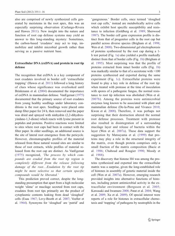
‘gangrenous.’ Border cells, once termed ‘sloughedroot cap cells,’ instead are metabolically active cellswhich exhibit host specific susceptibility and resis-tance to infection (Goldberg et al. 1989; Sherwood1987). The border cell gene expression profile is dis-tinct from that of progenitor cells in the root cap butparallel across diverse species (Brigham et al. 1998;Wen et al. 2008). Two-dimensional gel electrophoresisof proteins synthesized by the root cap during a 1-h test period (Fig. 1a) also yielded a profile markedlydistinct from that of border cells (Fig. 1b) (Brigham etal. 1995). Most surprising was that the profile ofproteins extracted from intact border cells (Fig. 1b)was markedly similar to that of a secretome with >100proteins synthesized and exported during the sameexperiment (Fig. 1c). Extracellular proteins werefound to play a key role in defense of the root tip:when treated with protease at the time of inoculationwith spores of a pathogenic fungus, the normal resis-tance to root tip infection was abolished (Wen et al.2007b). Among the proteins were antimicrobialenzymes long known to be associated with plant andmammalian defense (De-la-Pena and Vivanco 2010;Kwon et al. 2008). Therefore, it was perhaps notsurprising that their destruction altered the normalroot defense processes. Treatment with proteasealso resulted in disintegration of a surroundingmucilage layer and release of bacteria within thelayer (Wen et al. 2007a). These data support thesuggestion by Matsuyama et al. (1999) that pro-teins may play a role in the structural integrity ofthe matrix, even though protein comprises only asmall fraction of the matrix composition (Bacic etal. 1986; Chaboud and Rougier 1990; Moody etal. 1988).
The discovery that histone H4 was among the pro-teins synthesized and exported into the extracellularmatrix was a surprise, given the long-established roleof histones in assembly of genetic material inside thecell (Wen et al. 2007a). However, emerging researchprovided insights into alternative functions of histo-nes, including potent antimicrobial activity in the ex-tracellular environment (Bergsson et al. 2005;Kawasaki and Iwamuro 2008; Patat et al. 2004; Wanget al. 2009; Xu et al. 2009). Of special interest werereports of a role for histones in extracellular chemo-taxis and ‘trapping’ of pathogens by neutrophils in the
Plant Soil (2012) 355:1–16 3
mammalian immune response, because a very similarprocess occurs in border cells in response to plantpathogens (Gochnauer et al. 1990; Goldberg et al.1989; Gunawardena et al. 2005; Hawes and Pueppke1987; Hawes et al. 1988; Zhu et al. 1997). ‘Neutrophilextracellular traps’ (NETs) were first described byZychlinsky and coworkers (Brinkmann et al. 2004),and now have been implicated in defense againstdiverse pathogens and other aspects of immuneresponses in mammals (Abdallah et al. 2012; Amulicand Hayes 2011; Brinkmann and Zychlinsky 2007;Harding and Kubes 2012; Medina 2009; Mitroulis etal. 2011; Park et al. 2012; Urban et al. 2006; Wardiniet al. 2010; Wen et al. 2012; Yost et al. 2009; Young etal. 2011). As with the border cell slime layer (Fig. 2),NET formation can occur rapidly in response to spe-cific signals, in the absence of cell death (Pilszik et al.2010). Experiments therefore were carried out to de-termine (1) whether the presence of extracellular his-tone surrounding border cells, like neutrophils, isassociated with exDNA; and if so, (2) to determine ifenzymatic degradation of border cell exDNA, likeNET exDNA, interferes with resistance to infection(Gunawardena and Hawes 2002). The results of theseexperiments revealed that, like the plant proteinsexported from the root cap and border cells (Brighamet al. 1995), plant DNA is synthesized and exportedinto the root cap extracellular matrix during a 1-hperiod when no cell death occurs (Wen et al. 2009).
Initial sequence analysis revealed that the exDNAstructure is related to nuclear DNA, but is enrichedin repetitive sequences. When this exDNA was de-graded by addition of DNase I concomitant with theinoculation by a pathogenic fungus, the frequency ofroot cap infection increased from a mild local necro-sis in <5 % of inoculated roots to 100 % infection,with rotting of each root tip and proliferation offungal hyphae (Wen et al. 2009). As in exDNA-based extracellular trapping in mammals, the roottip resistance to fungal infection is associated withaggregation of the fungus and inhibition of itsgrowth (Gunawardena et al. 2005; Medina 2009).The extracellular trapping phenomenon is host-microbe specific, with no aggregation or growthinhibition of nonpathogenic fungi (Gunawardenaand Hawes 2002; Jaroszuk-Scisel et al. 2009).
Host specific chemotaxis and extracellular trappingof pathogens by border cells was described previously,but was presumed to involve aspects of pathogenesis,not defense (Goldberg et al. 1989; Hawes andPueppke 1987; Hawes and Smith 1989). Agrobacte-rium tumefaciens chemotaxis toward border cells of ahost species was measured using swarm agar assays(Fig. 2a) (Hawes et al. 1988) or direct microscopicobservation (Fig. 2b,c). Within hours, strings andstrands of immobilized bacteria develop (Fig. 2b).Bacteria adhere to the surface in a layer that is imper-vious to removal by washing in water (Fig. 2c).
Root cap Border cells Supernatant
a b c
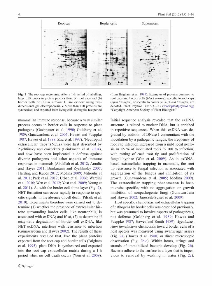
Fig. 1 The root cap secretome. After a 1-h period of labelling,large differences in protein profiles from (a) root caps and (b)border cells of Pisum sativum L. are evident using two-dimensional gel electrophoresis. c More than 100 proteins aresynthesized and exported from living cells during the test period
(from Brigham et al. 1995). Examples of proteins common toroot caps and border cells (black arrows), specific to root caps(open triangles), or specific to border cells (closed triangles) aredenoted. Plant Physiol 143:773–783 (www.plantphysiol.org)“Copyright American Society of Plant Biologists”
4 Plant Soil (2012) 355:1–16
Adding the plant pathogen to border cells of a nonhostspecies triggers no chemotaxis or attachment withinthe surrounding mucilage (Fig. 2d). The human path-ogen E. coli added to border cells (Fig. 2e, f) was notassociated with chemotaxis, attachment, or productionof a mucilage layer in either plant species. Minimalgrowth can be measured in remaining unattached bac-teria or in bacteria growing on mucilage as a solecarbon source, but whether the trapped pathogenicbacteria are viable is unclear (Knee et al. 2001; Zhuet al. 1997). Similar patterns of specificity were
reported in association between maize border cellsand bacterial species including Rhizobium, E. coli,Pseudomonas, Bacillus, Streptomyces and Cytophaga(Gochnauer et al. 1990). It will be of interest to exam-ine the role of exDNA in this phenomenon, and toexplore the possibility that clusters and strings ofviable but not culturable (VBNC) colonies found inthe rhizosphere might be related to exDNA basedtrapping (Gamalero et al. 2004).
The high molecular weight polysaccharide-basedmucilage exported from root caps has been studied
a b
c d
e f
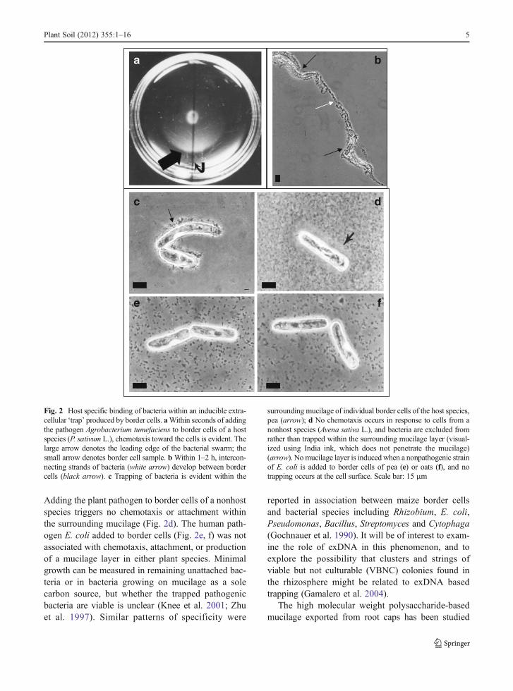
Fig. 2 Host specific binding of bacteria within an inducible extra-cellular ‘trap’ produced by border cells. aWithin seconds of addingthe pathogen Agrobacterium tumefaciens to border cells of a hostspecies (P. sativum L.), chemotaxis toward the cells is evident. Thelarge arrow denotes the leading edge of the bacterial swarm; thesmall arrow denotes border cell sample. b Within 1–2 h, intercon-necting strands of bacteria (white arrow) develop between bordercells (black arrow). c Trapping of bacteria is evident within the
surrounding mucilage of individual border cells of the host species,pea (arrow); d No chemotaxis occurs in response to cells from anonhost species (Avena sativa L.), and bacteria are excluded fromrather than trapped within the surrounding mucilage layer (visual-ized using India ink, which does not penetrate the mucilage)(arrow). Nomucilage layer is induced when a nonpathogenic strainof E. coli is added to border cells of pea (e) or oats (f), and notrapping occurs at the cell surface. Scale bar: 15 μm
Plant Soil (2012) 355:1–16 5
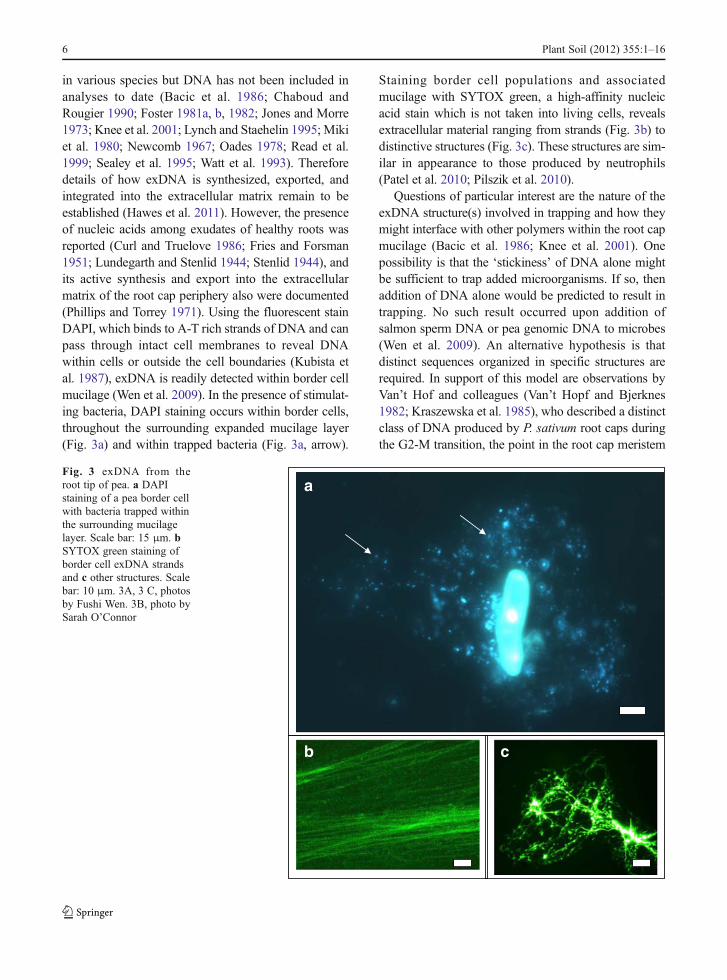
in various species but DNA has not been included inanalyses to date (Bacic et al. 1986; Chaboud andRougier 1990; Foster 1981a, b, 1982; Jones and Morre1973; Knee et al. 2001; Lynch and Staehelin 1995; Mikiet al. 1980; Newcomb 1967; Oades 1978; Read et al.1999; Sealey et al. 1995; Watt et al. 1993). Thereforedetails of how exDNA is synthesized, exported, andintegrated into the extracellular matrix remain to beestablished (Hawes et al. 2011). However, the presenceof nucleic acids among exudates of healthy roots wasreported (Curl and Truelove 1986; Fries and Forsman1951; Lundegarth and Stenlid 1944; Stenlid 1944), andits active synthesis and export into the extracellularmatrix of the root cap periphery also were documented(Phillips and Torrey 1971). Using the fluorescent stainDAPI, which binds to A-T rich strands of DNA and canpass through intact cell membranes to reveal DNAwithin cells or outside the cell boundaries (Kubista etal. 1987), exDNA is readily detected within border cellmucilage (Wen et al. 2009). In the presence of stimulat-ing bacteria, DAPI staining occurs within border cells,throughout the surrounding expanded mucilage layer(Fig. 3a) and within trapped bacteria (Fig. 3a, arrow).
Staining border cell populations and associatedmucilage with SYTOX green, a high-affinity nucleicacid stain which is not taken into living cells, revealsextracellular material ranging from strands (Fig. 3b) todistinctive structures (Fig. 3c). These structures are sim-ilar in appearance to those produced by neutrophils(Patel et al. 2010; Pilszik et al. 2010).
Questions of particular interest are the nature of theexDNA structure(s) involved in trapping and how theymight interface with other polymers within the root capmucilage (Bacic et al. 1986; Knee et al. 2001). Onepossibility is that the ‘stickiness’ of DNA alone mightbe sufficient to trap added microorganisms. If so, thenaddition of DNA alone would be predicted to result intrapping. No such result occurred upon addition ofsalmon sperm DNA or pea genomic DNA to microbes(Wen et al. 2009). An alternative hypothesis is thatdistinct sequences organized in specific structures arerequired. In support of this model are observations byVan’t Hof and colleagues (Van’t Hopf and Bjerknes1982; Kraszewska et al. 1985), who described a distinctclass of DNA produced by P. sativum root caps duringthe G2-M transition, the point in the root cap meristem
a
b c
Fig. 3 exDNA from theroot tip of pea. a DAPIstaining of a pea border cellwith bacteria trapped withinthe surrounding mucilagelayer. Scale bar: 15 μm. bSYTOX green staining ofborder cell exDNA strandsand c other structures. Scalebar: 10 μm. 3A, 3 C, photosby Fushi Wen. 3B, photo bySarah O’Connor
6 Plant Soil (2012) 355:1–16
cell cycle when border cell separation occurs (Brighamet al. 1998). Like root cap exDNA (Wen et al. 2009), this‘extrachromosomal DNA’ is related to nuclear DNA butis distinguishable based on the prevalence of repetitivesequences (Kraszewska et al. 1985). The programmeddelivery of characteristic exDNA patterns as an integralcomponent of the matrix could provide a tool to exam-ine underlying patterns of rhizosphere carbon depositionand microbial colonization and allow progress to-ward exploiting the system for crop improvement.Factors known to influence border cell delivery aresummarized below.
Factors controlling delivery of exDNA-based trapsfrom root caps
Border cell populations
The presence of DNA from plants and other organismsin the soil is well established (Izano et al. 2008;Vlassov et al. 2007; Whitchurch et al. 2002). Plant
exDNA has been presumed to be derived by leakagefrom dead cells (Levy-Booth et al. 2007). The discov-ery that secretion of exDNA from root caps instead is acomponent of a complex, inducible, and carbon-expensive defense mechanism may be useful in track-ing as well as modelling rhizosphere community struc-ture. The programmed separation of cells from the rootcap was long presumed to be a product of continuouscell cycle activity within the root cap meristem inparallel with such activity in the apical meristem(e.g. Clowes 1971; Whipps and Lynch 1983). If cor-rect, then a continuous detection of exudates at the tipwould be a predicted result. Direct observations ofrhizosphere structure even under controlled conditionsdo not support this paradigm (Iijima et al. 2003). Theviability and number of border cells that a root cap canrelease daily are conserved within families and canrange from 0 to 10,000 cells a day (Hawes et al.2003; Hawes and Pueppke 1986). For a given root,the process of root cap turnover is not continuous butinstead is induced or repressed in a species- andgenotype- specific manner in response to endogenous
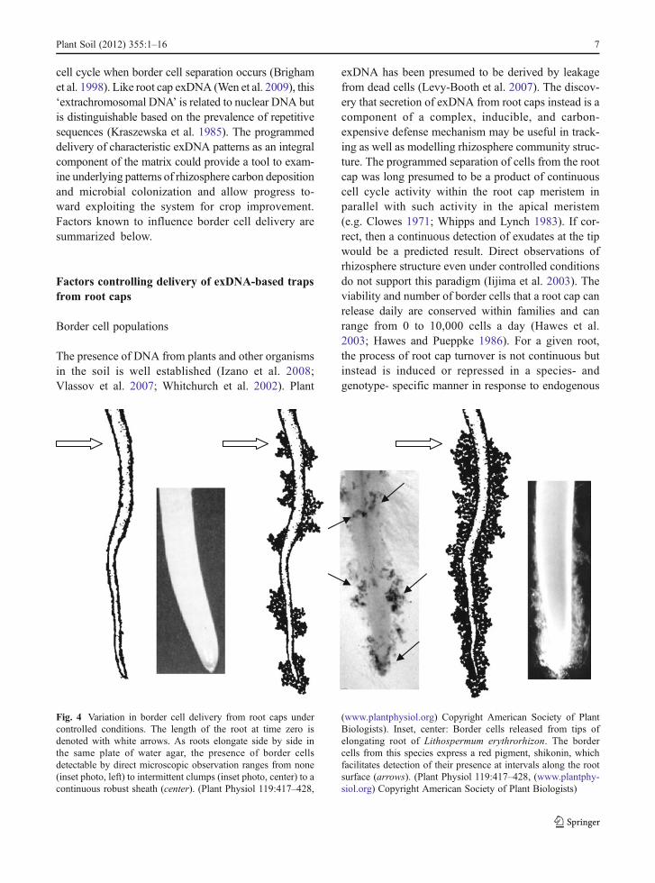
Fig. 4 Variation in border cell delivery from root caps undercontrolled conditions. The length of the root at time zero isdenoted with white arrows. As roots elongate side by side inthe same plate of water agar, the presence of border cellsdetectable by direct microscopic observation ranges from none(inset photo, left) to intermittent clumps (inset photo, center) to acontinuous robust sheath (center). (Plant Physiol 119:417–428,
(www.plantphysiol.org) Copyright American Society of PlantBiologists). Inset, center: Border cells released from tips ofelongating root of Lithospermum erythrorhizon. The bordercells from this species express a red pigment, shikonin, whichfacilitates detection of their presence at intervals along the rootsurface (arrows). (Plant Physiol 119:417–428, (www.plantphy-siol.org) Copyright American Society of Plant Biologists)
Plant Soil (2012) 355:1–16 7
and environmental signals (Brigham et al. 1998;Ponce et al. 2005). Therefore, when seedlings aregrown under identical conditions side by side in petridishes, the delivery of mucilage and border cells canvary from nothing to intermittent clumps to a contin-uous sheath surrounding the root from base to tip(Fig. 4). The variation is illustrated schematically be-cause even with direct microscopic observation onsterile plates the differences can be difficult to detect(Fig. 4, inset photos). Some species exhibit bordercell specific expression of pigmented metaboliteswhich provide a convenient marker for cell dispersal(Brigham et al. 1999). Thus, Saccharum officinarum,Sorghum vulgare and Lithospermum erythrorhizonhave pink, purple, and red border cells, respectively.This pigmentation facilitates recognition of rhizo-sphere distribution patterns that otherwise wouldbe obscure (Fig. 4, inset center). Variation in rootexudation and rhizosphere colonization has beenproposed to be a major obstacle to agronomic appli-cation of promising discoveries like biological control(Cooper and Rao 2006; Sylvia et al. 1998). Under-standing factors controlling carbon delivery via bordercells may be key to monitoring and controlling rhizo-sphere community structure (Lee and Hirsch 2006;Smucker and Erickson 1987).
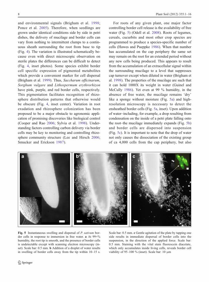
For roots of any given plant, one major factorcontrolling border cell release is the availability of freewater (Fig. 5) (Odell et al. 2008). Roots of legumes,cereals, cucurbits and most other crop species areprogrammed to produce a species-specific number ofcells (Hawes and Pueppke 1986). When that numberhas accumulated on the cap periphery the same setmay remain on the root for an extended period withoutany new cells being produced. This appears to resultfrom the accumulation of an extracellular signal withinthe surrounding mucilage to a level that suppressescap turnover except when diluted in water (Brigham etal. 1998). The properties of the mucilage are such thatit can hold 1000X its weight in water (Guinel andMcCully 1986). Yet even at 99 % humidity, in theabsence of free water, the mucilage remains ‘dry’like a sponge without moisture (Fig. 5a) and high-resolution microscopy is necessary to detect theensheathed border cells (Fig. 5a, inset). Upon additionof water–including, for example, a drop resulting fromcondensation on the inside of a petri plate falling ontothe root–the mucilage immediately expands (Fig. 5b)and border cells are dispersed into suspension(Fig. 5c). It is important to note that the drop of waternot only causes the dissociation of the existing groupof ca 4,000 cells from the cap periphery, but also
a b c
Fig. 5 Instantaneous swelling and dispersal of P. sativum bor-der cells in response to immersion in free water. a At 99+%humidity, the root tip is smooth, and the presence of border cellsis undetectable except with scanning electron microscopy (in-set). Scale bar: 0.5 mm. b Addition of a droplet of water resultsin swelling of border cells away from the tip within 10–15 s.
Scale bar: 0.5 mm. c Gentle agitation of the plate by tapping oneside results in immediate dispersal of border cells into thesuspension, in the direction of the applied force. Scale bar:0.5 mm. Staining with the vital stain fluorescein diacetate,which only accumulates inside living cells, reveals border cellviability of 95–100 % (inset). Scale bar: 10 μm
8 Plant Soil (2012) 355:1–16

triggers renewed cell cycle instantaneously (Brighamet al. 1998). Within 5 min, mitosis increases within theroot cap meristem, and dozens of new cells emergefrom the periphery. Activation of the quiescent centeralso occurs, and cell production proceeds until a newset has accumulated within 24 h (Ponce et al. 2005). Itseems obvious that within the soil environment, wherea continuous film of free water at the root tip would beintermittent for most crops in most conditions, thisfactor alone could account for much of the variabilityin root tip carbon deposition that occurs.
Other factors that can vary the number of bordercells and associated products released into the rhizo-sphere, include soil type, physical abrasion, daylength, root age and growth rate (Iijima et al. 2000,2003; Odell et al. 2008; Somasundaram et al. 2008;Wuyts et al. 2006). Sodium fluoride added to wheatroots can stimulate changes in number of border cellsand in level of protein secretion (Bozhkov et al. 2007).Carbon dioxide, aluminum, boron, and plant patho-gens stimulate changes in border cell production in aplant species- and genotype-specific manner withdistinct responses at different developmental stages(Cannesan et al. 2011; Chen et al. 2008; Liu et al.2007; Miyasaka and Hawes 2001; Pan et al. 2004;Tamas et al. 2005; Zhao et al. 2000; Zhu et al. 2003).For example, increased carbon dioxide inhibits bordercell production in P. sativum during germination, butresults in increased cell production in seedlings (Fig. 6).Border cell production in Medicago sativa seedlings, incontrast, is impervious to similar changes in carbondioxide (Zhao et al. 2000). Continuous culture of rootsin high concentrations of certain sugars and secondary
metabolites results in marked increases in mucilageproduction by maize roots (Jones and Morre 1973;Knudson 1917). Transient exposure of roots to metabo-lites including rhamnose, caffeine, and flavonoids forseveral minutes, a condition more likely to occur undernatural conditions, can specifically induce or repressborder cell production without affecting rate of rootgrowth (Curlango-Rivera et al. 2010). Altered expres-sion of genes controlling cell cycle or cell wall solubi-lization at the cap periphery results in altered border cellproduction, and transient changes in their expressiondue to diverse environmental signals could influencethe process as well (Wen et al. 1999; Woo et al. 2004).A new study reporting an ‘extraordinary sheath’ ofmaterial triggered on roots of Acacia magnum grownin hydroponic culture, highlights the importance of un-derstanding factors controlling this avenue of carbondeposition and their impact on rhizosphere structure(Endo et al. 2011).
Single cells
Morphology of border cell detachment from the capperiphery can range from a population of single cellsin suspension to finger-like strands of cells to an entireroot cap (Endo et al. 2011; Hamamoto et al. 2006;Vicre et al. 2005; Wen et al. 2008). The significance ofthese variations with respect to exDNA-based trappingis unknown, but the variation in amount and compo-sition of carbon-based material can be substantial evenon a single-cell basis. For many years, border cellswere called ‘sloughed root cap cells’ to reflect thepresumption that delivery of the cell populations must
Fig. 6 Effects of carbon dioxide on border cell separation fromroot tips (arrows) of P. sativum seedlings. a Cells from a singleroot tip observed with a dissecting microscope after 3 days in(A) ambient (0.03 % CO2 vs 21 % O2); or (b) 6 % CO2 vs 15 %
O2). Increased O2 alone had no effect on cell production. FromPlant Physiology 122:181–188, used with permission. (www.plantphysiol.org) Copyright American Society of PlantBiologists
Plant Soil (2012) 355:1–16 9
reflect a process of falling away from the root as aconsequence of cell death (e.g. Uren 2001). This no-tion prevailed, despite repeated documentation that thecells from most species are metabolically active asthey detach from the root cap and can survive forextended periods in liquid culture (Caporali 1983;Gautheret 1933; Hawes and Wheeler 1982; Stubbs etal. 2004). Knudson (1919) reported that border cellsreleased from Zea mays or P. sativum grown in hydro-ponic culture, with or without glucose, remained100 % viable for more than one month. Even moresurprising was the observation that the cells exportenzymes and other proteins (Rogers et al. 1942) andcan remain metabolically active after detachment intothe soil environment (Vermeer and McCully 1982).Continued secretion of mucilage from border cellscan occur for days after detachment from rootsgrown in soil (Hawes and Brigham 1992; Hawes etal. 1998). Like white blood cell ‘granules’, bordercells contain abundant storage particles which mayprovide energy for survival and response to signalsin the extracellular environment (Feldman 1985;Newcomb 1967).
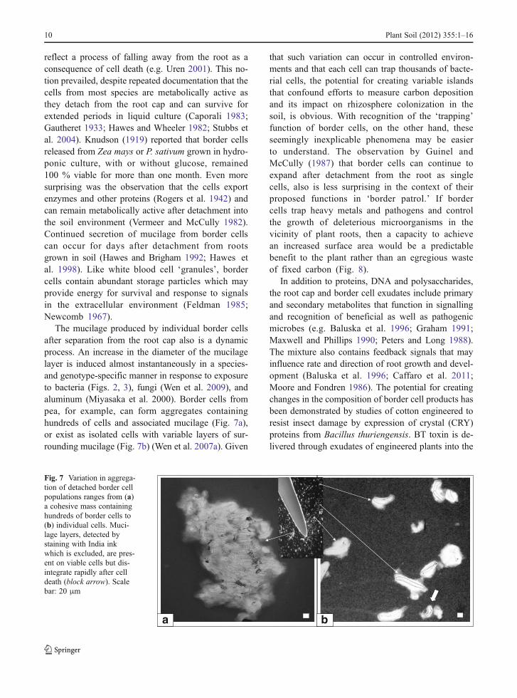
The mucilage produced by individual border cellsafter separation from the root cap also is a dynamicprocess. An increase in the diameter of the mucilagelayer is induced almost instantaneously in a species-and genotype-specific manner in response to exposureto bacteria (Figs. 2, 3), fungi (Wen et al. 2009), andaluminum (Miyasaka et al. 2000). Border cells frompea, for example, can form aggregates containinghundreds of cells and associated mucilage (Fig. 7a),or exist as isolated cells with variable layers of sur-rounding mucilage (Fig. 7b) (Wen et al. 2007a). Given
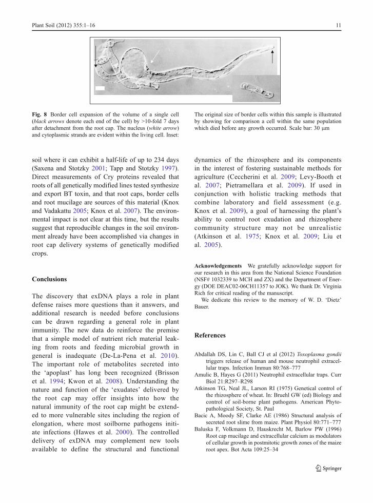
that such variation can occur in controlled environ-ments and that each cell can trap thousands of bacte-rial cells, the potential for creating variable islandsthat confound efforts to measure carbon depositionand its impact on rhizosphere colonization in thesoil, is obvious. With recognition of the ‘trapping’function of border cells, on the other hand, theseseemingly inexplicable phenomena may be easierto understand. The observation by Guinel andMcCully (1987) that border cells can continue toexpand after detachment from the root as singlecells, also is less surprising in the context of theirproposed functions in ‘border patrol.’ If bordercells trap heavy metals and pathogens and controlthe growth of deleterious microorganisms in thevicinity of plant roots, then a capacity to achievean increased surface area would be a predictablebenefit to the plant rather than an egregious wasteof fixed carbon (Fig. 8).
In addition to proteins, DNA and polysaccharides,the root cap and border cell exudates include primaryand secondary metabolites that function in signallingand recognition of beneficial as well as pathogenicmicrobes (e.g. Baluska et al. 1996; Graham 1991;Maxwell and Phillips 1990; Peters and Long 1988).The mixture also contains feedback signals that mayinfluence rate and direction of root growth and devel-opment (Baluska et al. 1996; Caffaro et al. 2011;Moore and Fondren 1986). The potential for creatingchanges in the composition of border cell products hasbeen demonstrated by studies of cotton engineered toresist insect damage by expression of crystal (CRY)proteins from Bacillus thuriengensis. BT toxin is de-livered through exudates of engineered plants into the
a b
Fig. 7 Variation in aggrega-tion of detached border cellpopulations ranges from (a)a cohesive mass containinghundreds of border cells to(b) individual cells. Muci-lage layers, detected bystaining with India inkwhich is excluded, are pres-ent on viable cells but dis-integrate rapidly after celldeath (block arrow). Scalebar: 20 μm
10 Plant Soil (2012) 355:1–16
soil where it can exhibit a half-life of up to 234 days(Saxena and Stotzky 2001; Tapp and Stotzky 1997).Direct measurements of Cry proteins revealed thatroots of all genetically modified lines tested synthesizeand export BT toxin, and that root caps, border cellsand root mucilage are sources of this material (Knoxand Vadakattu 2005; Knox et al. 2007). The environ-mental impact is not clear at this time, but the resultssuggest that reproducible changes in the soil environ-ment already have been accomplished via changes inroot cap delivery systems of genetically modifiedcrops.
Conclusions
The discovery that exDNA plays a role in plantdefense raises more questions than it answers, andadditional research is needed before conclusionscan be drawn regarding a general role in plantimmunity. The new data do reinforce the premisethat a simple model of nutrient rich material leak-ing from roots and feeding microbial growth ingeneral is inadequate (De-La-Pena et al. 2010).The important role of metabolites secreted intothe ‘apoplast’ has long been recognized (Brissonet al. 1994; Kwon et al. 2008). Understanding thenature and function of the ‘exudates’ delivered bythe root cap may offer insights into how thenatural immunity of the root cap might be extend-ed to more vulnerable sites including the region ofelongation, where most soilborne pathogens initi-ate infections (Hawes et al. 2000). The controlleddelivery of exDNA may complement new toolsavailable to define the structural and functional
dynamics of the rhizosphere and its componentsin the interest of fostering sustainable methods foragriculture (Ceccherini et al. 2009; Levy-Booth etal. 2007; Pietramellara et al. 2009). If used inconjunction with holistic tracking methods thatcombine laboratory and field assessment (e.g.Knox et al. 2009), a goal of harnessing the plant’sability to control root exudation and rhizospherecommunity structure may not be unrealistic(Atkinson et al. 1975; Knox et al. 2009; Liu etal. 2005).
Acknowledgements We gratefully acknowledge support forour research in this area from the National Science Foundation(NSF# 1032339 to MCH and ZX) and the Department of Ener-gy (DOE DEAC02-06CH11357 to JOK). We thank Dr. VirginiaRich for critical reading of the manuscript.
We dedicate this review to the memory of W. D. ‘Dietz’Bauer.
References
Abdallah DS, Lin C, Ball CJ et al (2012) Toxoplasma gondiitriggers release of human and mouse neutrophil extracel-lular traps. Infection Immun 80:768–777
Amulic B, Hayes G (2011) Neutrophil extracellular traps. CurrBiol 21:R297–R298
Atkinson TG, Neal JL, Larson RI (1975) Genetical control ofthe rhizosphere of wheat. In: Bruehl GW (ed) Biology andcontrol of soil-borne plant pathogens. American Phyto-pathological Society, St. Paul
Bacic A, Moody SF, Clarke AE (1986) Structural analysis ofsecreted root slime from maize. Plant Physiol 80:771–777
Baluska F, Volkmann D, Hauskrecht M, Barlow PW (1996)Root cap mucilage and extracellular calcium as modulatorsof cellular growth in postmitotic growth zones of the maizeroot apex. Bot Acta 109:25–34
Fig. 8 Border cell expansion of the volume of a single cell(black arrows denote each end of the cell) by >10-fold 7 daysafter detachment from the root cap. The nucleus (white arrow)and cytoplasmic strands are evident within the living cell. Inset:
The original size of border cells within this sample is illustratedby showing for comparison a cell within the same populationwhich died before any growth occurred. Scale bar: 30 μm
Plant Soil (2012) 355:1–16 11
Bednarek P, Kwon C, Schulze-Lefert P (2010) Not a peripheralissue: secretion in plant-microbe interactions. Curr OpinPlant Biol 13:378–385
Bergsson G, Agerberth B, Jornvall H, Gudmundsson GH (2005)Isolation and identification of antimicrobial componentsfrom the epidermal mucus of Atlantic cod (Gadusmorhua). FEBS J 272:4960–4969
Bowen GD, Rovira AD (1976) Microbial colonization of plantroots. Ann Rev Phytopathol 114:121–144
Bozhkov AI, Kuznetsova YA, Menzyanova NG (2007) Interre-lationship between the growth rate of wheat roots, theirexcretory activity and the number of border cells. Russian JPlant Physiol 54:97–103
Brady NC, Weil RR (2010) Elements of the nature and proper-ties of soils. Prentice Hall
Brigham LA, Woo HH, Nicoll SM, Hawes MC (1995) Differ-ential expression of proteins and messenger-RNAs fromborder cells and root tips of pea. Plant Physiol 109:457–463
Brigham LA, Woo HH, Wen F, Hawes MC (1998) Meristem-specific suppression of mitosis and a global switch in geneexpression in the root cap of pea by endogenous signals.Plant Physiol 118:1223–1231
Brigham LA, Michaels PJ, Flores HE (1999) Cell-specific pro-duction and antimicrobial activity of naphthoquinones inroots of Lithospermum erythrorhizon. Plant Physiol119:417–428
Brinkmann V, Zychlinsky A (2007) Beneficial suicide: whyneutrophils die to make NETs. Nat Rev Microbiol 5:577–582
Brinkmann V, Brichard U, Goosmann C et al (2004) Neutrophilextracellular traps kill bacteria. Science 303:1532–1535
Brisson RF, Tenhaken R, Lamb C (1994) Function of oxidativecross linking of cell wall structural proteins in plant diseaseresistance. Plant Cell 6:1703–1712
Broeckling CD, Broz AK, Bergelson J, Manter DK, Vivanco J(2008) Root exudates regulate soil fungal community com-position and diversity. Appl Environ Microbiol 74:738–744
Bruehl GW (1987) Soilborne plant pathogens. Macmillan Pub-lishing Company, NY, USA
Caffaro MM, Vivanco JM, Gutierrez BFH et al (2011) Theeffect of root exudates on root architecture in Arabidopsisthaliana. Plant Growth Regul 64:241–249
Cannesan MA, Gangneux C, Lanoue A et al (2011) Associationbetween border cell responses and localized root infectionby pathogenic Aphanomyces euteiches. Ann Bot 108:459–469
Caporali L (1983) Cytological study of cultured cells frommaize root cap. Plant Sci Lett 31:231–236
Ceccherini MT, Ascher J, Agnelli A et al (2009) Experimentaldiscrimination and molecular characterization of the extra-cellular soil DNA fraction. Antonie Van Leeuwenhoek96:653–657
Chaboud A, Rougier M (1990) Comparison of maize root muci-lages isolated from root exudates and root surface extractsby complementary cytological and biochemical investiga-tions. Protoplasma 156:163–173
Chen W, Liu P, Xu G et al (2008) Effects of aluminum on thebiological characteristics of cowpea root border cells. ActaPhysiol Plant 30:303–308
Clowes FAL (1971) The proportion of cells that divide in rootmeristems of Zea mays L. Ann Bot 35:249–261
Compant S, Duffy B, Nowak J et al (2005) Use of plant growthpromoting bacteria for biocontrol of plant diseases: princi-ples, mechanisms of action and future prospects. ApplEnviron Microbiol 71:4951–4959
Cooper JE, Rao JR eds (2006) Molecular approaches to soil,rhizosphere and plant microorganism analysis. CABI,Oxfordshire, UK, Cambridge MA, USA
Curl EA, Truelove B (1986) The rhizosphere. Advanced seriesin agricultural sciences, vol. 15, Springer-Verlag, Berlin-Heidelberg-New York-Tokyo
Curlango-Rivera G, Hawes MC (2011) Root tips movingthrough soil: an intrinsic vulnerability. Plant Signal Behav-ior 6:1–2
Curlango-Rivera G, Duclos DV, Ebolo JJ, Hawes MC (2010)Transient exposure of root tips to primary and secondarymetabolites: impact on root growth and production ofborder cells. Plant Soil 332:267–275
Darrah PR, Roose T (2001) Modeling the rhizosphere. In:Pinton R, Varanini Z, Nannipieri P (eds) The rhizosphere:biochemistry and organic substances at the soil-plant inter-face. Marcel Dekker, Inc., New York, pp 327–372
De-la-Pena C, Vivanco JM (2010) Root-microbe interactions:the importance of protein secretion. Curr Proteonomics7:265–274
De-la-Pena C, Badri DV, Lei Z et al (2010) Root secretion ofdefense-related proteins is development-dependent andcorrelated with flowering time. J Biol Chem 285:30654–30666
Dennis PG, Miller AJ, Hirsch PR (2010) Are root exudates moreimportant than other sources of rhizodeposits in structuringrhizosphere bacterial communities? FEMS Microbiol Ecol72:313–327
Donato JJ, Moe LA, Converse BJ et al (2010) Metagenomicanalysis of apple orchard soil reveals antibiotic resistancegenes encoding predicted bifunctional proteins. Appl En-viron Microbiol 76:4396–4401
Endo I, Tange T, Osawa H (2011) A cell-type-specific defect inborder cell formation in the Acacia mangium root capdeveloping an extraordinary sheath of sloughed-off cells.Ann Bot 108:279–290
Esau K (1967) Plant anatomy. Wiley, New YorkFeldman LJ (1985) Root gravitropism. Physiol Plant 65:341–344Foster RC (1981a) Polysacharrides in soil fabrics. Science
214:665–667Foster RC (1981b) The ultrastructure and histochemistry of the
rhizosphere. New Phytol 89:263–273Foster RC (1982) The fine structure of epidermal cell mucilages
of roots. New Phytol 91:727–740Foster RC, Rovira AD, Cock TW (1983) Ultrastructure of the
root-soil interface. American Phytopathological Society,St. Paul
Fries N, Forsman B (1951) Quantitative determination of certainnucleic acid derivatives in pea root exudate. Physiol Plant4:410–420
Gamalero E, Lingua G, Capri FG et al (2004) Colonizationpattern of primary tomato roots by Pseudomonas fluores-cens A6RI characterized by dilution plating, flow cytom-etry, fluorescence, confocal and scanning electronmicroscopy. FEMS Microbiol Ecol 48:79–87
12 Plant Soil (2012) 355:1–16

Gamalero E, Lingua G, Tombolini R et al (2005) Colonizationof tomato root seedling by Pseudomonas fluorescens92rkG5: spatio-temporal dynamics, localization, organiza-tion, viability and culturability. Microbial Ecol 50:289–297
Gautheret MR (1933) Cultures of cells isolated from the rootcap. CR Acad Sci 186:638–640
Gilbert GS, Clayton MK, Handelsman J, Parke JL (1996) Use ofcluster and discriminant analysis to compare rhizospherebacterial populations following biological perturbation.Microbial Ecol 32:123–147
Gochnauer MB, Sealey LJ, McCully ME (1990) Do detachedroot cap cells influence bacteria associated with maizeroots? Plant Cell Environ 13:793–801
Goldberg NP, Hawes MC, Stanghellini ME (1989) Specific attrac-tion to and infection of cotton root cap cells by zoospores ofPythium dissotocum. Can J Bot 67:1760–1767
Graham TC (1991) Flavonoid and isoflavonoid distribution indeveloping soybean seedling tissues and in seed and rootexudates. Plant Physiol 95:594–603
Griffin GJ, Hale MG, Shay FJ (1975) Nature and quantity ofsloughed organic matter produced by roots of axenic pea-nut plants. Soil Biol Biochem 8:29–32
Guinel FC, McCully ME (1986) Some water-related physicalproperties of maize root cap mucilage. Plant Cell Environ9:657–666
Guinel FC, McCully ME (1987) The cells shed by the root capof Zea: their origin and some structural and physiologicalproperties. Plant Cell Environ 10:565–578
Gunawardena U, Hawes MC (2002) Tissue specific localizationof root infection by fungal pathogens: role of root bordercells. Mol Plant Microbe Int 15:1128–1136
Gunawardena U, Rodriguez M, Straney D et al (2005) Tissuespecific localization of root infection by Nectria haemato-cocca: mechanisms and consequences. Plant Physiol137:1363–1374
Hamamoto L, Hawes MC, Rost TL (2006) The production andrelease of living root cap border cells is a function of rootapical meristem type in dicotyledonous angiosperm plants.Ann Bot 97:917–923
Handelsman J (2004) Metagenomics: application of genomics touncultured microorganisms. Microbiol Mol Biol Rev68:669–685
Handelsman J, Stabb EV (1996) Biocontrol of soilborne plantpathogens. Plant Cell 8:1855–1869
Harding M, Kubes P (2012) Innate immunity in the vasculature:interactions with pathogenic bacteria. Curr Opin Microbiol15:85–91
Hawes MC, Wheeler H (1982) Factors affecting victorin-induced cell death: temperature and plasmolysis. PhysiolPlant Pathol 20:137–144
Hawes MC, Pueppke SG (1986) Sloughed peripheral root capcells: yield from different species and callus formationfrom single cells. Am J Bot 73:1466–1473
Hawes MC, Pueppke SG (1987) Correlation between bind-ing of Agrobacterium tumefaciens by root cap cellsand susceptibility of plants to crown gall. Plant CellRep 6:287–290
Hawes MC, Smith LY (1989) Requirement for chemotaxisin pathogenicity of Agrobacterium tumefaciens onroots of soil-grown pea plants. J Bacteriol 171:5668–5671
Hawes MC, Brigham LA (1992) Impact of root border cells onmicrobial populations in the rhizosphere. Adv Plant Pathol8:119–148
Hawes MC, Smith LY, Howarth AJ (1988) Agrobacteriumtumefaciens mutants deficient in chemotaxis to root exu-dates. Mol Plant Microbe Int 1:182–186
Hawes MC, Brigham LA, Wen F, Woo HH, Zhu Y (1998)Function of root border cells in plant health: pioneers inthe rhizosphere. Ann Rev Phytopathol 36:311–327
Hawes MC, Gunawardena U, Miyasaka S, Zhao X (2000) Therole of root border cells in plant defense. Trends Plant Sci5:128–133
Hawes MC, Bengough G, Cassab G, Ponce G (2003) Root capsand rhizosphere. J Plant Growth Regul 21:352–367
Hawes MC, Curlango-Rivera G, Wen F et al (2011) Extracellu-lar DNA: the tip of root defenses. Plant Sci 180:741–745
Hinsinger P (2001) Bioavailability of soil inorganic P in therhizosphere as affected by root-induced chemical changes:a review. Plant Soil 237:173–195
Hinsinger P, Brauman A, Devau N et al (2011) Acquisition ofphosphorus and other poorly mobile nutrients by roots.Where do plant nutrition models fail? Plant Soil 348:29–61
Hirsch AM (2004) Plant-microbe symbioses: a continuum fromcommensalism to parasitism. Symbiosis 37:345–363
Humphris SN, Bengough AG, Griffiths BS et al (2005) Rootcap influences root colonisation by Pseudomonas fluores-cens SBW25 on maize. FEMS Microbiol Ecol 54:123–130
Iijima M, Griffiths B, Bengough AG (2000) Sloughing of capcells and carbon exudation from maize seedling roots incompacted sand. New Phytol 145:477–482
Iijima M, Sako Y, Rao TP (2003) A new approach for thequantification of root cap mucilage exudation in the soil.Plant Soil 255:399–407
Izano EA, Amarante MA, Kher WB, Kaplan JB (2008) Differen-tial roles of poly-N-acetylglucosamine surface polysaccha-ride and extracellular DNA in Staphylococcus aureus and S.epidermidis biofilms. Appl Environ Microbiol 74:470–476
Jaroszuk-Scisel J, Kurek E, Rodzik B, Winiarczyk K (2009)Interactions between rye (Secale cereale) root border cells(RBCs) and pathogenic and nonpathogenic rhizospherestrains of Fusarium culmorum. Mycol Res 113:1053–1061
Jones DD, Morre DJ (1973) Golgi apparatus mediated polysac-charide secretion by outer root cap cells of Zea mays. III.Control by exogenous sugars. Physiol Plant 29:68–75
Jones DL, Nguyen C, Finlay RD (2009) Carbon flow in therhizosphere: carbon trading at the soil-root interface. PlantSoil 321:5–33
Kawasaki H, Iwamuro S (2008) Potential roles of histones inhost defense as antimicrobial agents. Infect Disord DrugTargets 8:195–205
Knee EM, Gong FC, Gao MS et al (2001) Root mucilage frompea and its utilization by rhizosphere bacteria as a solecarbon source. Mol Plant Microbe Int 14:775–784
Knox OGG, Vadakattu GVSR (2005) Evaluation of border cellnumber and cry protein expression from root tips of Gos-sypium hirsutum. In: Cote JC, Otvos IS, Schwartz JL (eds)Pacific rim conference on the biotechnology of Bacillusthuringiensis and its environmental impact
Knox OGG, Gupta VVSR, Nehl DB, Stiller WN (2007) Con-stitutive expression of cry proteins in roots and border cellsof transgenic cotton. Euphytica 154:83–90
Plant Soil (2012) 355:1–16 13
Knox OGG, Gupta VVSR, Lardner R (2009) Cotton cultivarselection impacts on microbial diversity and function.Aspects Appl Biol 98:1–8
Knudson L (1917) The toxicity of galactose and mannose forgreen plants and the antagonistic action of other sugarstoward these. Am J Bot 4:430–437
Knudson L (1919) Viability of detached root cap cells. Am J Bot6:309–310
Kraszewska EK, Bjerknes CA, Lamm SS, Van’t Hopf J (1985)Extrachromosomal DNA of pea-root (Pisum sativum) hasrepeated sequences and ribosomal genes. Plant Mol Biol5:353–361
Kubista M, Akerman B, Norden B (1987) Characterization ofinteraction between DNA and 4,6-diamidino- 2- phenylin-dole by optical spectroscopy. Biochemistry 26:4545–4553
Kuzyakov YV (2001) Tracer studies of carbon translocation byplants from the atmosphere into the soil. Eurasian Soil Sci34:28–42
KwonC, Bednarek P, Schulze-Lefert P (2008) Secretory pathwaysin plant immune responses. Plant Physiol 147:1575–1583
Lagopodi AL, Ram AFJ, Lamers GEM et al (2002) Novelaspects of tomato root colonization and infection by Fusa-rium oxysporum f. sp radicis-lycopersici revealed by con-focal laser scanning microscopic analysis using the greenfluorescent protein as a marker. Mol Plant Microbe Int15:172–179
Lee A, Hirsch AM (2006) Signals and responses: choreograph-ing the complex interaction between legumes and alpha-and beta-rhizobia. Plant Signal Behavior 1:161–168
Levy-Booth DJ, Campbell RG, Gulden RH et al (2007) Cyclingof extracellular DNA in the soil environment. Soil BiolBiochem 39:2977–2991
Liu B, Zeng Q, Yan F et al (2005) Effects of transgenic plants onsoil microorganisms. Plant Soil 271:1–13
Liu J, Yu M, Wang W, Feng Y (2007) Influence of boron andaluminum on production and viability of root border cellsof pea. Adv Plant Animal Boron Nutrition, pp 67–74
Loh J, Pierson EA, Pierson LS III, Stacy G, Chatterjee A (2002)Quorum sensing in plant-associated bacteria. Curr OpinPlant Biol 5:1–5
Lundegarth H, Stenlid G (1944) On the exudation of nucleotidesand flavonone from living roots. Arkiv f Bot 31A:10
Luster J, Gottlein A, Nowack B, Sarret G (2009) Sampling,defining, characterizing and modeling the rhizosphere—the soil science tool box. Plant Soil 321:457–482
Lynch JM, Whipps JM (1990) Substrate flow in the rhizosphere.Plant Soil 129:1–10
Lynch MA, Staehelin LA (1995) Immunocytochemical locali-zation of cell wall polysaccharides in the root tip of Avenasativa. Protoplasma 188:115–127
Marschner P, Crowley D, Rengel Z (2011) Rhizosphere inter-actions between microorganisms and plants govern ironphosphorus acquisition along the root axis—model andresearch methods. Soil Biol Biochem 43:883–894
Matsuyama T, Satoh H, Yamada Y, Hashimoto T (1999) Amaize glycine-rich protein is synthesized in the lateral rootcap and accumulates in the mucilage. Plant Physiol120:665–674
Maxwell CA, Phillips DA (1990) Concurrent synthesis andrelease of nod gene inducing flavonoids from alfalfa roots.Plant Physiol 93:1552–1558
McDougall BM, Rovira AD (1970) Sites of exudation of C-14-labelled compounds from wheat roots. New Phytol 69:999
Medina E (2009) Neutrophil extracellular traps: a strategic tacticto defeat pathogens with potential consequenes for thehost. J Innate Immun 1:176–179
Miki NK, Clarke KJ, McCully ME (1980) A histological andhistochemical comparison of the mucilages on the root tipsof several grasses. Can J Bot 58:2581–2593
Mitroulis I, Kambas K, Chrysanthopoulou A et al (2011) Neu-trophil extracellular trap formation is associated with IL-1beta and autophagy-related signaling in gout. PLoS One6:e29318
Miyasaka S, Hawes MC (2001) Possible role of root border cellsin detection and avoidance of aluminum toxicity. PlantPhysiol 125:1978–1987
Moody SF, Clarke AE, Bacic A (1988) Structural analysis ofsecreted slime from wheat and cowpea roots. Phytochem-ical 27:2857–2861
Moore R, Fondren WM (1986) The possible involvement ofroot-cap mucilage in gravitropism and calcium movementacross root tips of Allium cepa L. Ann Bot 58:381–387
Morris CE, Monier J-M (2003) The ecological significance ofbiofilm formation by plant-associated bacteria. Ann RevPhytopathol 41:429–453
Newcomb EH (1967) Fine structure of protein-storing plastidsin bean root tips. J Cell Biol 33:143–163
Oades JM (1978) Mucilage at the root surface. J Soil Sci 29:1–16
Odell RE, Dumlao MR, Samar D, Silk WK (2008) Stage-dependent border cell and carbon flow from roots to rhi-zosphere. Am J Bot 95:441–446
Pan J, Ye D, Wang L et al (2004) Root border cell developmentis a temperature-insensitive and Al-sensitive process inbarley. Plant Cell Physiol 45:751–760
Park SJ, Pai KS, Kim JH, Shin JI (2012) ANCA-associated glo-merulonephritis in a patient with infections endocarditis: therole of neutrophil extracellular traps? Revue Med Int 33:57
Patat SA, Carnegie RB, Kingsbury C et al (2004) Antimicrobialactivity of histones from hemocytes of the pacific whiteshrimp. Eur J Biochem 271:4825–4833
Patel S, Kumar S, Jyoti A et al (2010) Nitric oxide donorsrelease extracellular traps from human neutrophils by aug-menting free radical generation. Nitric Oxide 22:226–234
Peters NK, Long SR (1988) Alfalfa root exudates and com-pounds which promote or inhibit induction of Rhizobiummeliloti nodulation genes. Plant Physiol 88:396–400
Phillips HL, Torrey JG (1971) Deoxyribonucleic acid synthesisin root cap cells of cultured roots of Convolvulus. PlantPhysiol 48:213–218
Pierson LS III, Pierson EA (2007) Roles of diffusible signals incommunication among plant-associated bacteria. Phytopa-thology 97:227–232
Pietramellara G, Ascher J, Borgogni F et al (2009) ExtracellularDNA in soil and sediment: fate and ecological relevance.Biol Fertil Soils 45:219–235
Pilszik FH, Salina D, Poon KK et al (2010) A novel mechanism ofrapid nuclear neutrophil extracellular trap formation in re-sponse to Staphylococcus aureus. J Immunol 185:7413–7425
Pinton R, Varanini Z, Nanipieri P, eds (2007) The rhizosphere:biochemistry and organic substances at the soil-plant inter-face. Marcel Dekker, Inc. New York, Basel
14 Plant Soil (2012) 355:1–16
Ponce G, Barlow PW, Feldman LJ, Cassab GI (2005) Auxin andethylene interactions control mitotic activity of the quies-cent centre, root cap size, and pattern of cap cell differen-tiation in maize. Plant Cell Environ 28:719–732
Read DB, Gregory PJ, Bell AE (1999) Physiological propertiesof axenic maize root mucilage. Plant Soil 211:87091
Rogers HT, Pearson RW, Pierre WH (1942) The source andphosphatase activity of exoenzyme systems of corn andtomato roots. Soil Sci 54:353–365
Rovira AD (1969) Plant root exudates. Bot Rev 35:35–57Rovira AD (1991) Rhizosphere research: 85 years of progress
and frustration. In: Keister DL, Cregan PB (eds) The rhi-zosphere and plant growth. Kluwer, Dordrecht, pp 3–13
Saxena D, Stotzky G (2001) Bacillus thuringiensis (Bt) toxinreleased from root exudates and biomass of Bt corn has noapparent effect on earthworms, nematodes, protozoa, bac-teria, and fungi in soil. Soil Biol Biochem 33:1225–1230
Schroth MN, Snyder WC (1961) Efect of host exudates onchlamydospore germination of the bean root rot fungusFusarium solani f sp phaseoli in soil. Phytopathology52:279–285
Sealey LJ, McCully ME, Canny MJ (1995) The expansion ofmaize root cap mucilage during hydration. I. Kinetics.Physiol Plant 93:38–46
Sherwood RT (1987) Papilla formation in corn root cap cellsand leaves inoculated with Colletotrichum graminicola.Phytopathology 77:930–934
Smucker AJM, Erickson AE (1987) Anaerobic stimulation ofroot exudates and disease of peas. Plant Soil 99:423–433
Somasundaram S, Fukuzono S, Iijima M (2008) Dynamics of rootborder cells in rhizosphere soil of Zea mays L.: crushed cellsduring root penetration, survival in soil, and long term soilcompaction effect. Plant Prod Sci 11:440–446
Stenlid G (1944) Physicochemical properties of the surface ofgrowing plant cells. Nature 153:618–619
Stubbs VEC, Standing D, Knox OGG et al (2004) Root bordercells take up and release glucose-C. Ann Bot 93:221–224
Sylvia DM, Fuhrmann JJ, Hartel PG, Zuberer DA (1998) Principlesand applications of soil microbiology. Prentice Hall, USA
Tamas L, Budikova S, Huttova J et al (2005) Aluminum-induced cell death of barley root border cells is correlatedwith peroxidase and oxalate oxidase-mediated hydrogenperoxide production. Plant Cell Rep 24:189–194
Tapp H, Stotzky G (1997) Monitoring the fate of insecticidaltoxins from Bacillus thuringiensis in soil with flow cytom-etry. Can J Microbiol 43:1074–1078
Urban CF, Lourido S, Zychlinsky A (2006) How do microbesevade neutrophil killing? Cell Microbiol 8:1687–1696
Uren NC (2001) Types, amounts, and possible functions ofcompounds released into the rhizosphere by soil-grownplants. In: Pinton R, Varanini P, Nannipieri P (eds) Therhizosphere: biochemistry and organic substances at thesoil-plant interface. Marcel Dekker, Inc., New York, Basel.pp 19–40
VanEgeraat AWSM (1975) Exudation of ninhydrin-positivecompounds by pea seedling roots: a study of the sites ofexudation and of the composition of the exudate. Plant Soil42:37–47
Van’t Hopf J, Bjerknes CA (1982) Cells of pea (Pisum sativum)that differentiate from G2 phase have extrachromosomalDNA. Mol Cell Biol 2:339–345
Vermeer J, McCully ME (1982) The rhizosphere in Zea: newinsight into its structure and development. Planta 156:45–61
Vicre M, Santaella C, Blanchet S, Gateau A, Driouich A (2005)Root border like cells of Arabidopsis. Microscopical char-acterization and role in the interaction with rhizobacteria.Plant Physiol 138:998–1008
Vlassov VV, Laktionov PP, Rykova EY (2007) Extracellularnucleic acids. Bioessays 29:654–667
Voeller BR, Ledbetter MC, Porter KR (1964) The plant cell:aspects of its form and function. In: Bracket J, Minsky EE(eds) The cell, vol 6. Academic, London, pp 245–312
Wang Y, Li M, Stadler S, Correll S et al (2009) Histone hyper-citrullination mediates chromatin decondensation and neu-trophil extracellular trap formation. J Cell Biol 184:205–213
Wardini AB, Guimaraes-Costa AB, Nascimento MT et al (2010)Characterization of neutrophil extracellular traps in catsnaturally infected with feline leukemia virus. J Gen Virol91:259–264
Watt M, McCully ME, Jeffree CE (1993) Plant and bacterialmucilages of the maize rhizosphere: comparison of theirsoil binding properties and histochemistry in a model sys-tem. Plant Soil 151:151–165
Watt M, Silk WK, Passioura J (2006) Rates of root and organ-ism growth, soil conditions and temporal and spatial de-velopment of the rhizosphere. Ann Bot 97:839–855
Weller DM (1988) Biological control of soilborne plant patho-gens in the rhizosphere with bacteria. Annu Rev Phytopa-thol 26:379–407
Wen F, Zhu Y, Brigham LA, Hawes MC (1999) Expression of aninducible pectinmethylesterase gene is required for bordercell separation from roots of pea. Plant Cell 11:1129–1140
Wen F, Curlango-Rivera G, Hawes MC (2007a) Proteins amongthe polysaccharides: a new perspective on root cap slime.Plant Signal Behavior 2:1–3
Wen F, VanEtten HD, Tsaprailis G, Hawes MC (2007b) Extra-cellular proteins in pea root tip and border cell exudates.Plant Physiol 143:773–783
Wen F, Woo HH, Pierson EA et al (2008) Synchronous elicita-tion of development in root caps induces transient geneexpression changes common to legume and gymnospermspecies. Plant Mol Biol Rep 27:58–68
Wen F, White GJ, VanEtten HD et al (2009) Extracellular DNAis required for root tip resistance to fungal infection. PlantPhysiol 151:820–829
Wen F, Shen A, Choi A, Shi J (2012) A xenograft pancreaticcancer mouse model to study the function of extracellularDNA in metastasis. Proceedings, American Association ofCancer Research
Whipps J, Lynch JM (1983) Substrate flow and utilization in therhizosphere of cereals. New Phytol 95:605–623
Whitchurch CB, Tolker-Nielsen T, Ragas PC, Mattick JS (2002)Extracellular DNA required for bacterial biofilm formation.Science 295:1487
Woo HH, Hirsch AM, Hawes MC (2004) Altered susceptibilityto infection by Sinorhizobium meliloti and Nectria haema-tococca in alfalfa roots with altered cell cycle. Plant CellRep 12:967–973
Wood RKS (1967) Physiological plant pathology. Blackwell,Oxford
Plant Soil (2012) 355:1–16 15
Wuyts N, Maung ZTZ, Swennen R, De Waele D (2006) Bananarhizodeposition: characterization of root border cell produc-tion and effects on chemotaxis and motility of the parasiticnematode Radopholus similis. Plant Soil 283:217–228
Xu J, Zhang X, Pelayo R, Monestier M et al (2009) Extracellu-lar histones are major mediators of death in sepsis. NatMed 15:1318–1321
Yost CC, Cody MJ, Harris ES et al (2009) Impaired NETformation: a novel innate immune deficiency of humanneonates. Blood 113:6419–6427
Young RL, Malcolm KC, Kret JE, Caceres SM et al (2011)Neutrophil extracellular trap (NET)-mediated killing ofPseudomonas aeruginosa: evidence of acquired resistancewithin the cystic fibrosis airway, independent of CFTR.PLoS One 6:e23637
Zentmyer G (1963) Biological control of Phytophthora root rotof avocado with alfalfa meal. Phytopathology 53:1383
Zhao X, Misaghi IJ, Hawes MC (2000) Stimulation of bordercell production in response to increased carbon dioxidelevels. Plant Physiol 122:181–188
Zhu M, Ahn S, Matsumoto H (2003) Inhibition of growth anddevelopment of root border cells in wheat by Al. PhysiolPlant 117:359–367
Zhu Y, Pierson LS, Hawes MC (1997) Induction of microbialgenes for pathogenesis and symbiosis by chemicals fromroot border cells. Plant Physiol 115:1691–1698
Zobel RW, Wright SF (2005) Roots and soil management:interactions between roots and the soil. American Societyof Agronomy, Inc., Crop Science Society of America, Inc.,Soil Science Society of America, Inc. Madison WI, USA
16 Plant Soil (2012) 355:1–16