Roles of active site tryptophans in substrate binding and catalysis by -1,3 galactosyltransferase
28
1 24 June 2004 Roles of active site tryptophans in substrate binding and catalysis by a-1,3 galactosyltransferase Yingnan Zhang 1,4 , Ashlesha Deshpande 2,5 , Zhihong Xie 1 , Ramanathan Natesh 2,6 , K. Ravi Acharya 2 and Keith Brew 1,3 1 Department of Biomedical Sciences, Florida Atlantic University, Boca Raton, Florida 33341, USA 2 Department of Biology and Biochemistry, University of Bath, Claverton Down, Bath BA2 7AY, UK 3 To whom correspondence should be addressed; e-mail: [email protected] 4 Present address: Department of Protein Engineering, Genentech, Inc., 1 DNA way, South San Francisco, CA 94080 5 Present address: CDFD, Hyderabad 500 076, India 6 Present address: ICGEB, Aruna Asaf Ali Marg, New Delhi 110 067, India Running title- Tryptophans and substrate binding in a3GT The abbreviations used are: a3GT, b-galactosyl a-1,3 galactosyltransferase; GT, glycosyltransferase; r.m.s.d, root mean square deviation; ITC, isothermal titration calorimetry; LacNAc, N-acetyl lactosamine; Lac, lactose; Gal, galactose; Glc, glucose. Key words: glycosyltransferase/ mutation/ crystal structure/ substrate binding/ catalysis Glycobiology © Oxford University Press 2004; all rights reserved. Glycobiology Advance Access published June 30, 2004 by guest on June 27, 2014 http://glycob.oxfordjournals.org/ Downloaded from
Transcript of Roles of active site tryptophans in substrate binding and catalysis by -1,3 galactosyltransferase
1
24 June 2004
Roles of active site tryptophans in substrate binding and catalysis
by a-1,3 galactosyltransferase
Yingnan Zhang1,4, Ashlesha Deshpande2,5, Zhihong Xie1, Ramanathan Natesh2,6,
K. Ravi Acharya2 and Keith Brew1,3
1Department of Biomedical Sciences, Florida Atlantic University, Boca Raton, Florida 33341, USA
2Department of Biology and Biochemistry, University of Bath, Claverton Down, Bath BA2 7AY, UK
3To whom correspondence should be addressed; e-mail: [email protected]
4Present address: Department of Protein Engineering, Genentech, Inc., 1 DNA way, South San
Francisco, CA 94080
5Present address: CDFD, Hyderabad 500 076, India
6Present address: ICGEB, Aruna Asaf Ali Marg, New Delhi 110 067, India
Running title- Tryptophans and substrate binding in a3GT
The abbreviations used are: a3GT, b-galactosyl a-1,3 galactosyltransferase; GT, glycosyltransferase;
r.m.s.d, root mean square deviation; ITC, isothermal titration calorimetry; LacNAc, N-acetyl lactosamine;
Lac, lactose; Gal, galactose; Glc, glucose.
Key words: glycosyltransferase/ mutation/ crystal structure/ substrate binding/ catalysis
Glycobiology © Oxford University Press 2004; all rights reserved.
Glycobiology Advance Access published June 30, 2004 by guest on June 27, 2014
http://glycob.oxfordjournals.org/D
ownloaded from
2
Aromatic amino acids are frequent components of the carbohydrate binding sites of lectins
and enzymes. Previous structural studies have shown that in a-1,3 galactosyltransferase, the
binding site for disaccharide acceptor substrates is encircled by four tryptophans, residues 249,
250, 314 and 356. To investigate their roles in enzyme specificity and catalysis, we expressed and
characterized variants of the catalytic domain of a-1,3 galactosyltransferase with substitutions for
each tryptophan. Substitution of glycine for tryptophan249, whose indole ring interacts with the
non-polar B face of glucose or GlcNAc, greatly increases the Km for the acceptor substrate. In
contrast, the substitution of tyrosine for tryptophan314, which interacts with the b-galactosyl
moiety of the acceptor and UDP-galactose, decreases kcat for the galactosyltransferase reaction but
does not affect the low UDP-galactose hydrolase activity. Thus, this highly conserved residue
stabilizes the transition state for the galactose transfer to disaccharide but not to water. High
resolution crystallographic structures of the Trp249Gly mutant and the Trp314Tyr mutant indicate
that the mutations do not affect the overall structure of the enzyme or its interactions with ligands.
Substitutions for tryptophan250 have only small effects on catalytic activity, but mutation of
tryptophan356 to threonine reduces catalytic activity for both transferase and hydrolase activities
and reduces affinity for the acceptor substrate. This residue is adjacent to the flexible C-terminus
that becomes ordered on binding UDP, to assemble the acceptor binding site and influence
catalysis. The results highlight the diverse roles of these tryptophans in enzyme action and the
importance of kcat changes in modulating glycosyltransferase specificity.
by guest on June 27, 2014http://glycob.oxfordjournals.org/
Dow
nloaded from
3
Introduction
Major contributions to the recognition of specific carbohydrates by lectins and glycosidases are
made by interactions of the protein with hydroxyl groups of monosaccharides, particularly through H-
bonds with the side chains of polar amino acids and coordination bonds with protein-bound Ca2+ ions
(Sharon and Lis, 2001; Weiss and Drickamer, 1996). Aromatic amino acids are also found in a large
proportion of carbohydrate binding sites where they make hydrophobic interactions with the non-polar
faces of monosaccharide pyranose rings; the side chains of tryptophan and tyrosine also H-bond with
monosaccharide hydroxyl groups. The importance of aromatic side chains in the specificity of lectins
and glycosidases has been previously discussed (Muraki, 2002). Glycosyltransferases, enzymes that
catalyze the transfer of a monosaccharide from an activated derivative into a defined linkage with a
specific acceptor, determine the structures of glycan components of glycoconjugates produced by cells.
The structural basis of their substrate specificities is therefore of fundamental significance in
glycobiology, yet few structures have been determined for eukaryotic glycosyltransferases and there is
limited information available regarding the basis of their substrate specificity. Although it is likely that
the interactions between glycosyltransferases and oligosaccharides will be similar to those in
glycosidases and lectins, the multisubstrate character of glycosyltransfer reactions suggest that the
determinants of substrate specificity in these enzymes are likely to be more complex.
UDP-galactose b galactosyl a1-3-galactosyltransferase (a3GT1, EC 2.4.1.151) is a retaining GT
that catalyzes the synthesis of a1,3-galactosyl b-OR structures in glycoconjugates (Van den Eijnden et
al., 1983) and is a component of trans-Golgi membranes. An inactive form of this enzyme is produced in
humans and their closest relatives among the primates (Galili et al., 1988; Galili and Swansen, 1991). The
absence of active a3GT allows the production of natural antibodies (1-3% of circulating IgG) to the
product of its action, the a-Gal epitope, which provide defences against pathogens (Avili et al., 1989) and
viruses arising from mammalian hosts that have active a3GT (Takeuchi et al., 1996). a3GT is a member
by guest on June 27, 2014http://glycob.oxfordjournals.org/
Dow
nloaded from

4
of a family of homologous retaining galactosyl- and N-acetylgalactosaminyl-transferases that form a-1,3
linkages to b-galactosyl and b-N-acetylgalactosaminyl-residues in glycoconjugates but differ in
specificity: the histo-blood group A and B glycosyltransferases (Yamamato et al., 1990), Forssman
glycolipid (Gb5) synthase (Haslam and Baenziger, 1996) and isogloboside 3 (iGb3) synthase (Keusch et
al., 2000). These enzymes, like most eukaryotic glycosyltransferases, are all type II membrane proteins
with short cytosolic N-terminal domains, a membrane spanning region, a stem and C-terminal catalytic
domains (Paulson and Colley, 1989). Structures have been determined for recombinant truncated catalytic
domains of bovine a3GT (Gastinel et al., 2001; Boix et al., 2001, 2002) and human histo-blood group A
and B glycosyltransferases (Patenaude et al., 2002).
Two distinct structural states have been described for a3GT, a lower resolution tetragonal form
(Form I), in which the C-terminus of the enzyme is disordered (Gastinel et al., 2001) and a higher
resolution monoclinic form (Form II) which has a distinct and highly ordered conformation for the C-
terminal region (Boix et al., 2001). The structured C-terminus was found, by mutagenesis, to be important
for the catalytic activity of the enzyme (Boix et al., 2001). We have reported Form II structures for a3GT
in complexes with Mn2+ and UDP at 1.53 Å resolution (Boix et al., 2001), Mn2+/UDP-galactose (UDP-
Gal), Mn2+/UDP-glucose (UDP-Glc) and with both Mn2+/UDP and the acceptor substrates lactose and N-
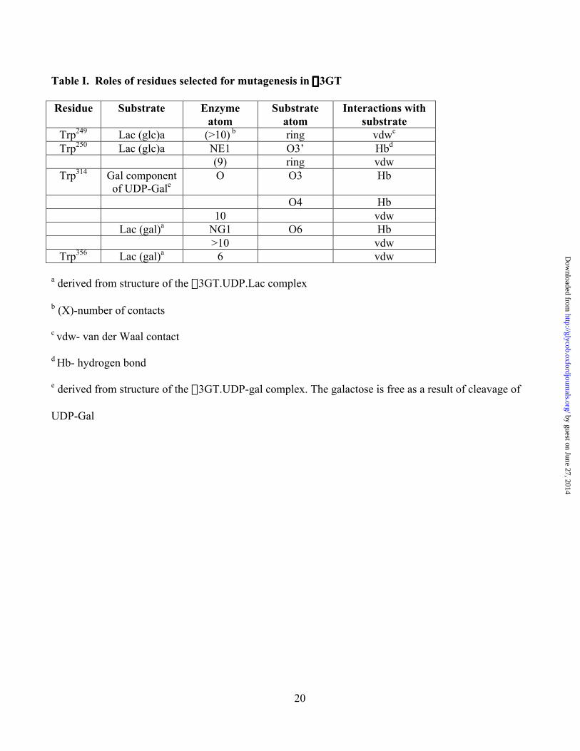
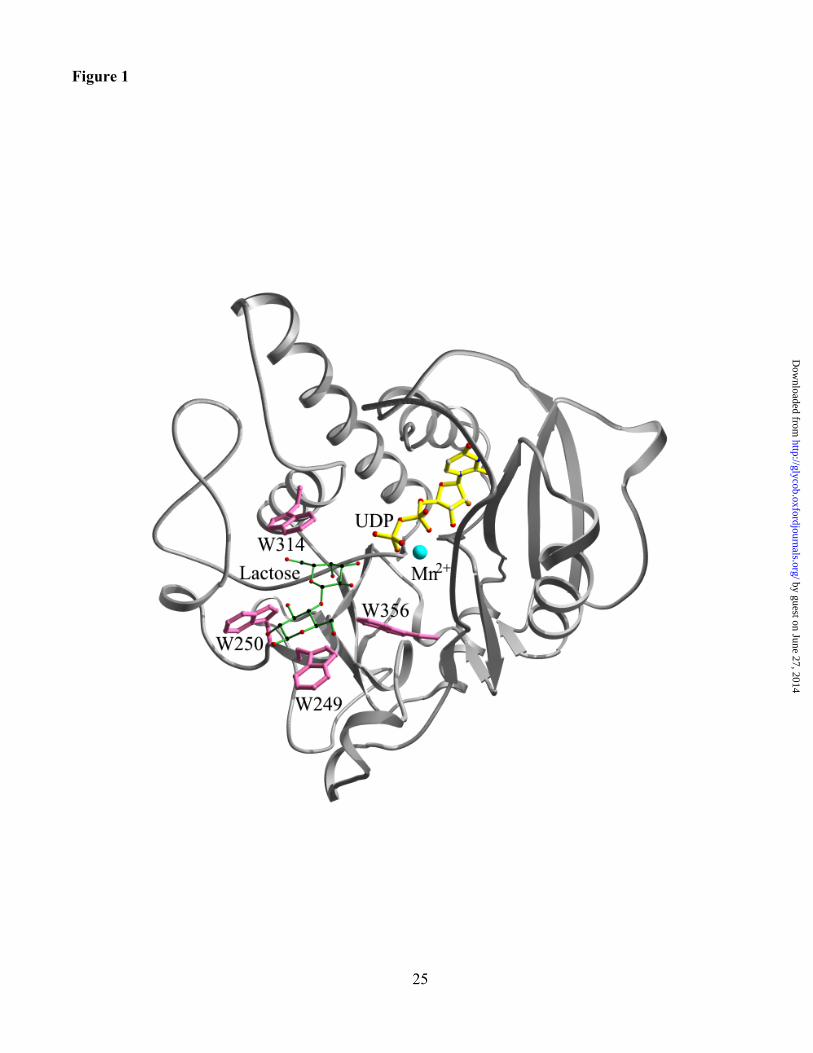
acetyllactosamine (Boix et al., 2002). The structures of these complexes identify four tryptophans,
residues 249, 250, 314 and 356 in the active site of a3GT that interact with acceptor substrates (see Table
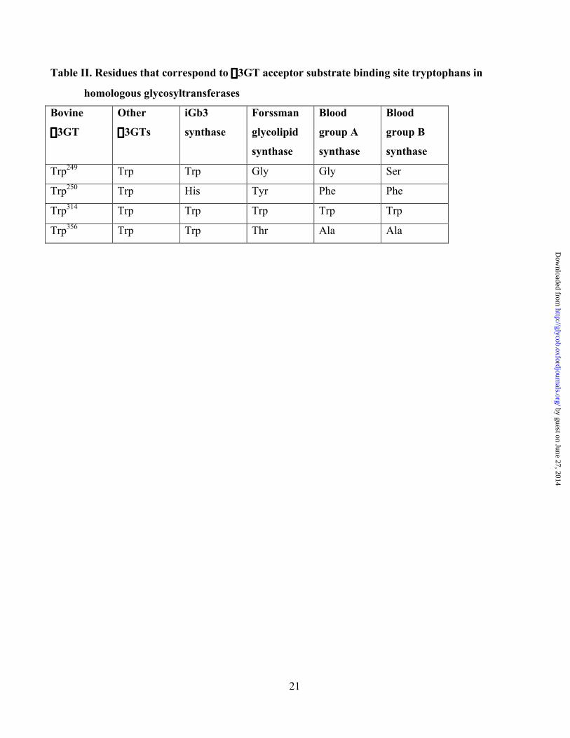
I and Figure 1). Molecular modelling studies by Heissigerova et al (2003) suggest that these specific
tryptophans and the corresponding residues in other members of the a3GT family may be important in
acceptor substrate selectivity.
To investigate the roles of the four tryptophans of a3GT in substrate specificity and catalysis, we
have introduced homology-based (Table II) and structurally conservative substitutions. Such mutations
are expected to have minor effects on overall enzyme structure and to perturb, but not eliminate, catalytic
by guest on June 27, 2014http://glycob.oxfordjournals.org/
Dow
nloaded from
5
activity. The quantitative effects of the mutations on individual kinetic parameters for two catalytic
activities of a3GT, galactosyltransferase and UDP-Gal hydrolase activity, were then determined to
evaluate the role of each residue in enzyme function (Zhang et al., 2001). Two mutants containing
substitutions for Trp249 (Gly) and Trp314 (Tyr), residues that make stacking interactions with the glucose
and galactose components of the lactose acceptor, respectively (Boix et al., 2002), have major but distinct
effects on the catalytic properties. Crystallographic structural analyses show that neither mutation
significantly changes the overall structure of the enzyme or its active site. Nevertheless, the Trp249Gly
substitution greatly reduces the affinity of the enzyme for lactose and the Trp314Tyr mutation selectively
perturbs kcat for galactosyl transfer to lactose without perturbing the rate of galactose transfer to water.
Mutations of Trp250 have relatively subtle effects on activity while the Trp356Thr mutation lowers the
catalytic activity for both reactions and weakens lactose binding. These different effects appear reflect the
proximity of the residues to the enzyme catalytic center and their roles in localized conformational
changes during catalysis.
Results
Design and Production of a3GT Trp mutants
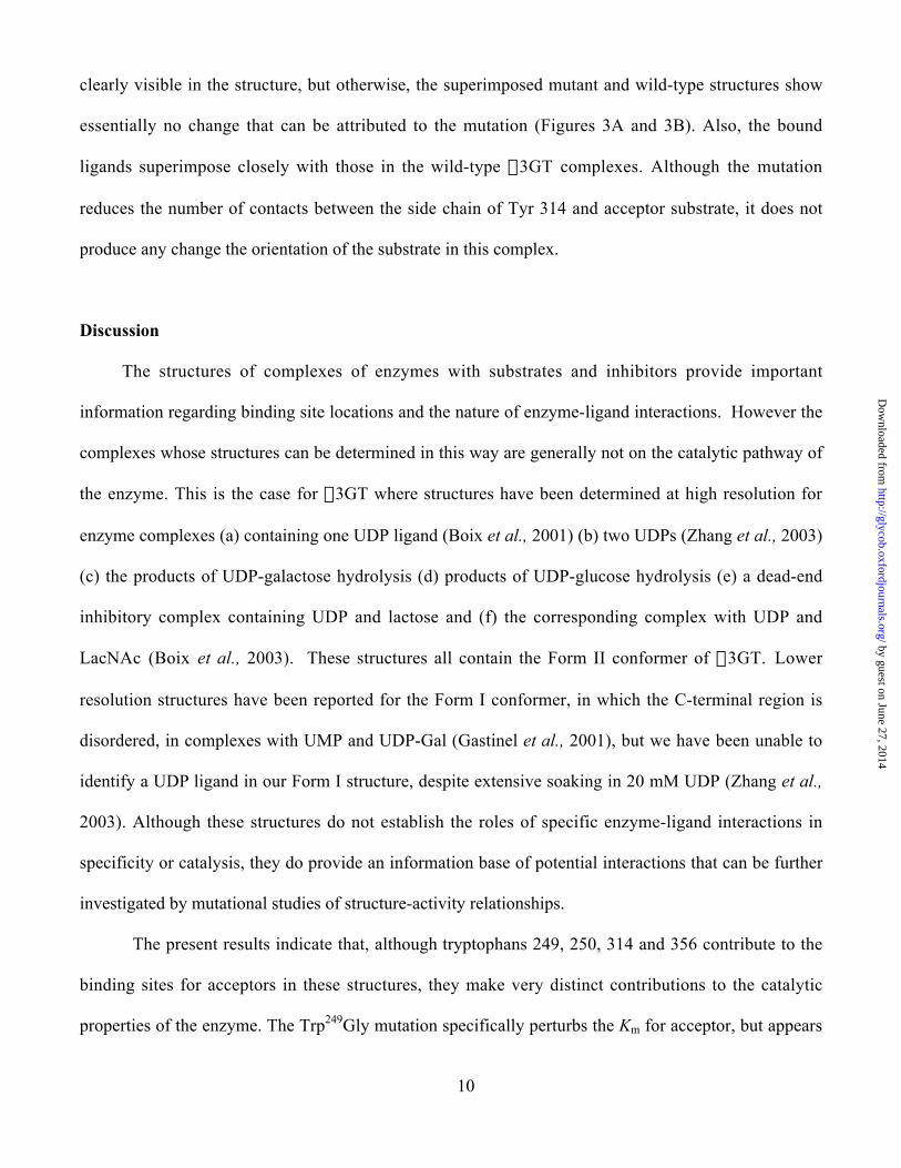
The side chains of tryptophans 249, 250, 314 and 356 surround the binding site for lactose and LacNAc
in their complexes with a3GT, UDP and Mn2+ (Figure 1). The indole ring of Trp249 is nearly parallel
with the reducing monosaccharide (Glc or GlcNAc) of the acceptor substrates and makes non-polar van
der Waals interactions with the hydrophobic B face of the pyranose ring (Table I). However, in the
absence of acceptor substrate, the side chain adopts two alternative conformations (Boix et al., 2002).
As indicated in Table II, tryptophan is conserved at this site in iGb3 synthase but not in Forssman
glycolipid synthase or in Histo-blood group A and B synthases (Yamamato et al., 1990; Haslam and
Baenziger, 1996; Keusch et al., 2000). The Gly mutation introduces the amino acid present at the
by guest on June 27, 2014http://glycob.oxfordjournals.org/
Dow
nloaded from
6
corresponding site in the histo blood group A transferase that catalyzes GalNAc transfer to a similar
acceptor in which fucose is a-linked to the 2-OH of galactose.
The side chain of Trp250 is located near the glucose or GlcNAc component of acceptor substrates
in their complexes with enzyme (Boix et al., 2002). However, unlike Trp249, the indole ring is
approximately perpendicular to the pyranose ring, lying at the edge of the entrance of lactose binding
groove. Specific interactions of Trp250 with acceptor substrates are an H-bond between the indole ring N
and the 3’-OH of both lactose and LacNAc and packing interactions of the indole ring with the 2’-
acetamido group of LacNAc. There is no corresponding interaction with lactose but the tryptophan
makes about 7 additional non-polar contacts with both acceptors. Mutants were expressed with
conservative substitutions of Tyr and Phe, corresponding to residues present in Forssman glycolipid
synthase and the blood group enzymes (Table II).
Trp314 is conserved in all homologues of a3GT. In a3GT, the side chain has more than 10
hydrophobic interactions with the b-galactosyl moiety of the acceptor and the N of the indole ring is
within H-bonding distance of O6 of the glucose moiety of lactose. Also, in the enzyme complexes with
UDP-Gal and UDP-Glc (in which the substrate is cleaved) it has 10 and 8 hydrophobic contacts,
respectively, with the monosaccharide ring. In the complex with UDP-Gal, the carbonyl OH H-bonds
with the 2- and 3-OH groups of the galactose (Table I, Boix et al., 2002).
Trp356 is part of the acceptor binding site making 6 to 7 nonpolar van der Waals contacts with
acceptor substrates. It is adjacent to the C-terminal section of the polypeptide chain that undergoes a
rearrangement between the Form I and Form II structures, that is linked with UDP binding (Boix et al.,
2001). This residue was mutated to Thr, the amino acid present at the corresponding site in the histo
blood group A and B transferases (Table II).
All of the a3GT mutants were expressed in soluble active form in good yield and showed similar
solubility to the wild-type enzyme.
by guest on June 27, 2014http://glycob.oxfordjournals.org/
Dow
nloaded from

7
Effects of Mutations on Activity
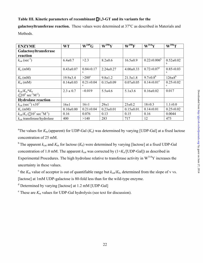
(a) Trp249 to Gly
Acceptor substrate (lactose) binding is so strongly perturbed by this mutation that the relationship
between velocity and lactose concentration up to 100 mM at 1 mM UDP-Gal is linear. The limited
solubility of lactose in water precludes the use of higher lactose concentrations in assays. Although kcat
and Kb with lactose as substrate, cannot be determined for this mutant, the value of kcat/Kb (apparent),
determined from the slope of v vs [lactose] plot is 80-fold lower than that of the wild-type enzyme
(Table III). The lack of curvature indicates that the Km for lactose must exceed 200 mM. It was also not
possible to achieve saturation with higher-affinity acceptor substrates including LacNAc and p-NO2-
phenyl-LacNAc because of their limited solubility in water. The apparent Km for UDP-Gal, determined
at a fixed non-saturating concentration of lactose (25 mM), is similar to the wild type enzyme (Table III)
while the apparent kcat determined at 25mM lactose is 0.26 sec-1, a value that is much lower than the true
kcat because of the low acceptor concentration relative to the Km. The kcat and Km for UDP-Gal hydrolysis
are essentially unchanged (Table III) and it is possible that kcat for the transferase reaction is also
unchanged, in which case the Km for lactose would be about 1.5 M. Isothermal titration calorimetry
studies (see Boix et al., 2002) indicate that the free energy and enthalpy of binding of UDP and UDP-
Gal to the enzyme in the presence of Mn2+ were not significantly changed by the mutation. However,
unlike wild-type a3GT, the binding of lactose in the presence of UDP and Mn2+ to the Trp249Gly mutant
was not detected, suggesting that the negative enthalpy of binding and/or affinity of lactose for this
inhibitory enzyme complex are reduced (data not shown).
(b) Trp250 to Tyr or Phe
The activity levels of the two mutants under standard assay conditions are similar to those of the wild-
type protein. A detailed analysis indicates that, for galactose transfer to lactose, kcat is slightly higher
than in the wild-type for both mutants while Ka, the Km for UDP-Gal is increased 6-10 fold. Both
by guest on June 27, 2014http://glycob.oxfordjournals.org/
Dow
nloaded from
8
mutants also have slightly increased Km and kcat values for UDP-Gal hydrolysis (Table III). In the
ordered sequential mechanism for a3GT:
†
Ek2
k1 A[ ]
´EAk4
k3 B[ ]
´ EAB ´ EPQk6 [P ]
k5
´EQk8 [Q ]
k7
´E
where A is the donor substrate, UDP-Gal, B is the acceptor substrate, and P and Q are expected to be the
trisaccharide product and UDP, respectively.
Ka=k5k7/{k1(k5+k7)} and kcat=k5k7/(k5+k7) so that kcat/Ka = k1, the on-rate for UDP-Gal binding (Purich
and Allison, 2000).
Both of these mutations therefore increase the overall rate of product-release steps and lower the
on-rate of UDP-Gal binding 3-4 fold. This interpretation is consistent with the slightly increased kcat for
UDP-Gal hydrolysis and may reflect a local structural change resulting from the mutation.
The Km of a3GT for LacNAc is about 12-fold lower than the Km for lactose, which could reflect
contributions to the free energy of binding arising from interactions of the LacNAc 2-acetamido group
with Trp250. The substitution of Tyr or Phe, residues with smaller side chains than Trp, may perturb
these interactions and affect the binding of LacNAc but not lactose. To test this hypothesis, apparent Km
and kcat were determined with LacNAc for the wild–type enzyme and the two mutants (at 1mM UDP-
Gal). The values of these parameters were: wild type: Km, 2.67± 0.27 mM, kcat, 4.59±0.18 sec-1;
Trp250Phe: Km, 1.85±0.25 mM, kcat, 5.97±0.26 sec-1; Trp250Tyr: Km, 2.76±0.24 mM; kcat, 6.19±0.22 sec-1.
The apparent kinetic parameters for the wild-type enzyme are similar to true values determined in
experiments in which the concentrations of both UDP-Gal and LacNAc were varied: Km: 1.6±0.14 mM;
kcat, 5.6±0.2 sec-1. These results indicate that the substitutions for Trp250 do not selectively disfavor the
binding of LacNAc relative to lactose.
(c) Trp314 to Tyr
Galactosyltransferase activity is greatly reduced in the Trp314Tyr mutant but UDP-Gal hydrolase activity
is essentially unchanged relative to the wild-type enzyme (Table III). As discussed previously for a
by guest on June 27, 2014http://glycob.oxfordjournals.org/
Dow
nloaded from
9
different mutant of a3GT (Zhang et al., 2003), the high proportion of hydrolase activity makes it
difficult to determine kinetic parameters with high precision using the radiochemical assay and we
determined apparent kinetic parameters for the transferase reaction. These indicate that the main effect
of the mutation is to reduce the kcat for the galactosyltransferase reaction 30-fold but the kinetic
parameters for UDP-Gal hydrolase activity are insignificantly affected. These effects are closely similar
to those in the previously described Gln247Glu mutant (Zhang et al., 2003).
(d) Trp356 to Thr
This mutant has a reduced level of overall catalytic activity. The loss in hydrolase activity reflects a 15-
fold reduction in kcat but, for the galactosyltransferase reaction, a reduction in kcat and an increase in the
Km for lactose (determined at 1 mM UDP-Gal) contribute to a greater reduction in activity. There is
some uncertainty in the precise values of the parameters for the transferase reaction because the highest
substrate concentration in the assay (100 mM) is less than the calculated value of Kb (126 mM) but it is
clear from the results that kcat is reduced 10-15-fold for both reactions and acceptor substrate affinity is
about 5-fold lower than for the wild-type enzyme (Table III).
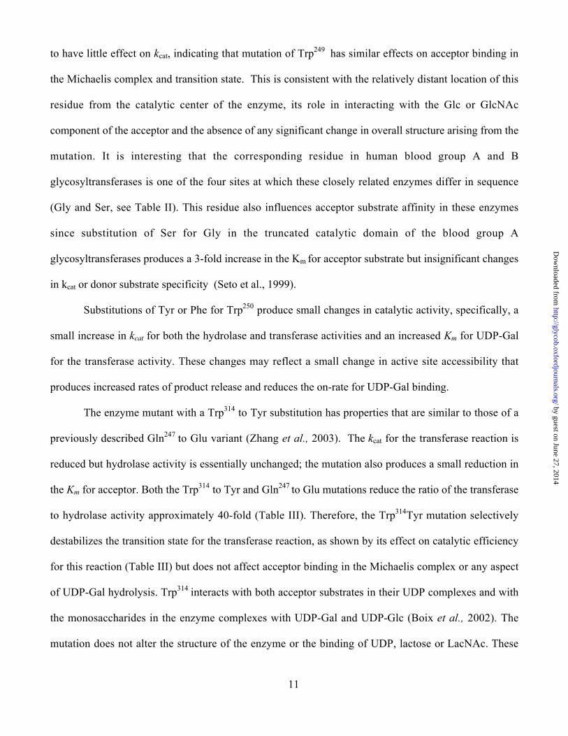
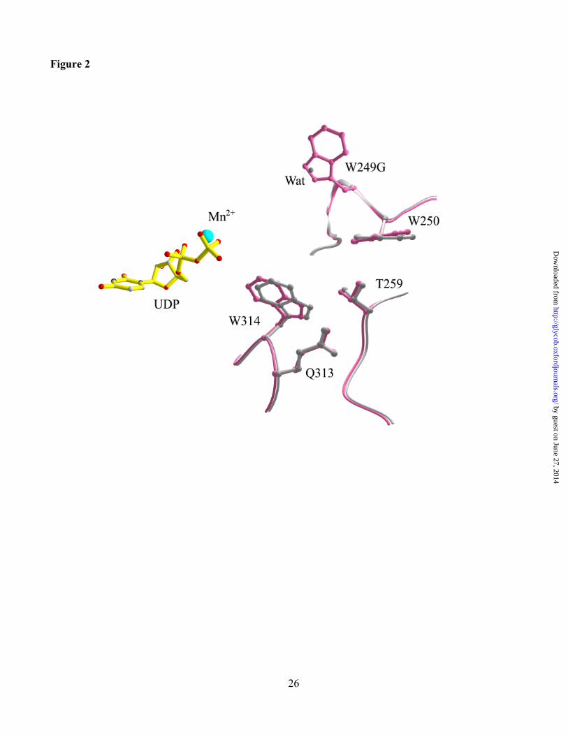
Structures of the Trp249Gly and Trp314Tyr mutants
The Form II structure of the complex of Trp249Gly with UDP and Mn2+ was determined at 2.07Å
resolution (Table IV). The mutation has no significant effect on the overall structure of the enzyme and
the active site. The root mean square deviation from the structure of the wild-type enzyme is 0.11 Å, and
there are only minor structural changes in the vicinity of the mutation that are shown by superimposing
the structures of the mutant and wild-type enzymes (Figure 2). Two specific structural changes can be
directly attributed to the mutation; firstly the loss of the Trp side chain disrupts an interaction between
Trp249 and Asp340 and, secondly, a new structured water molecule is present in the mutant enzyme
located in the space produced by removal of the Trp side chain (Figure 2).
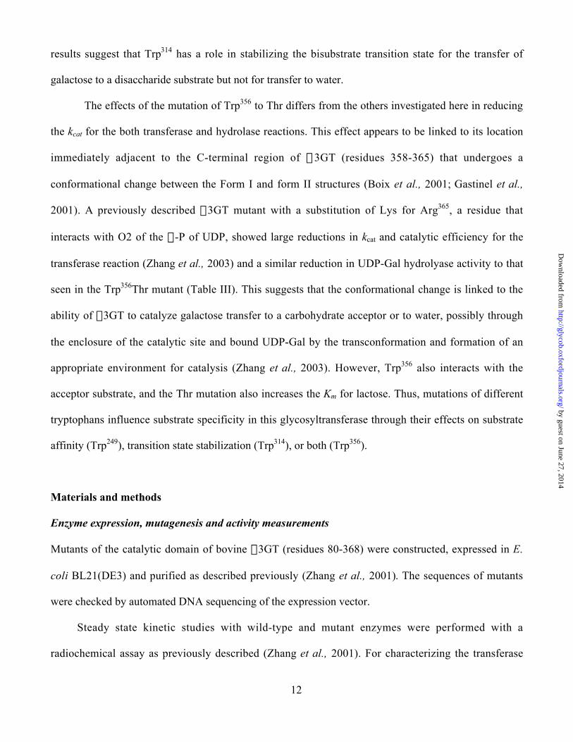
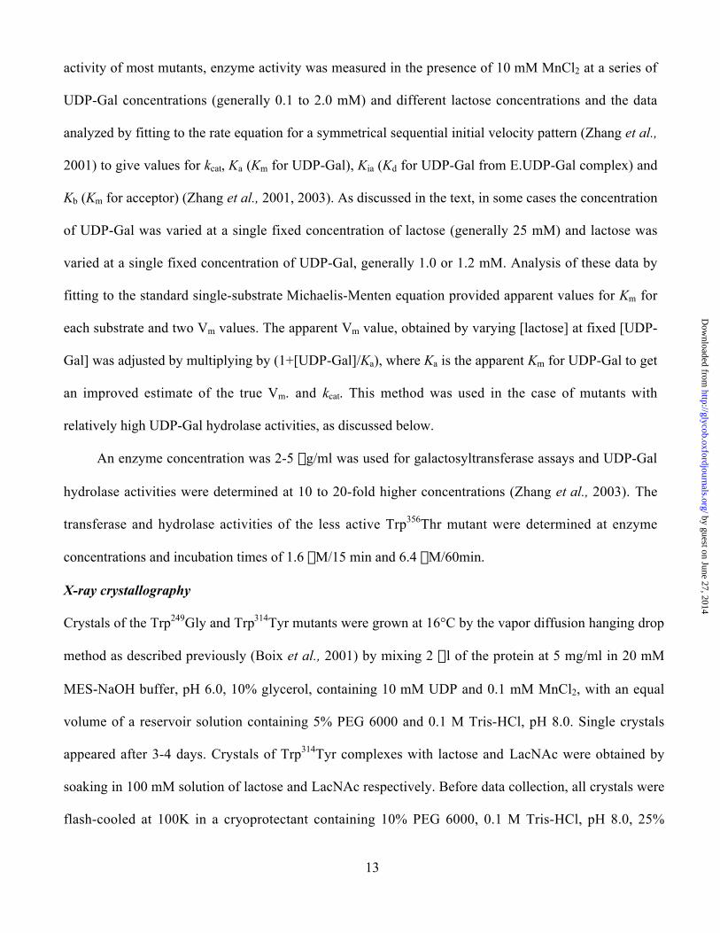
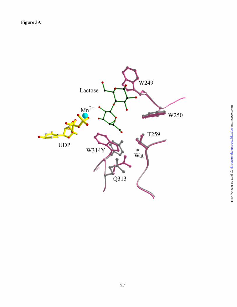
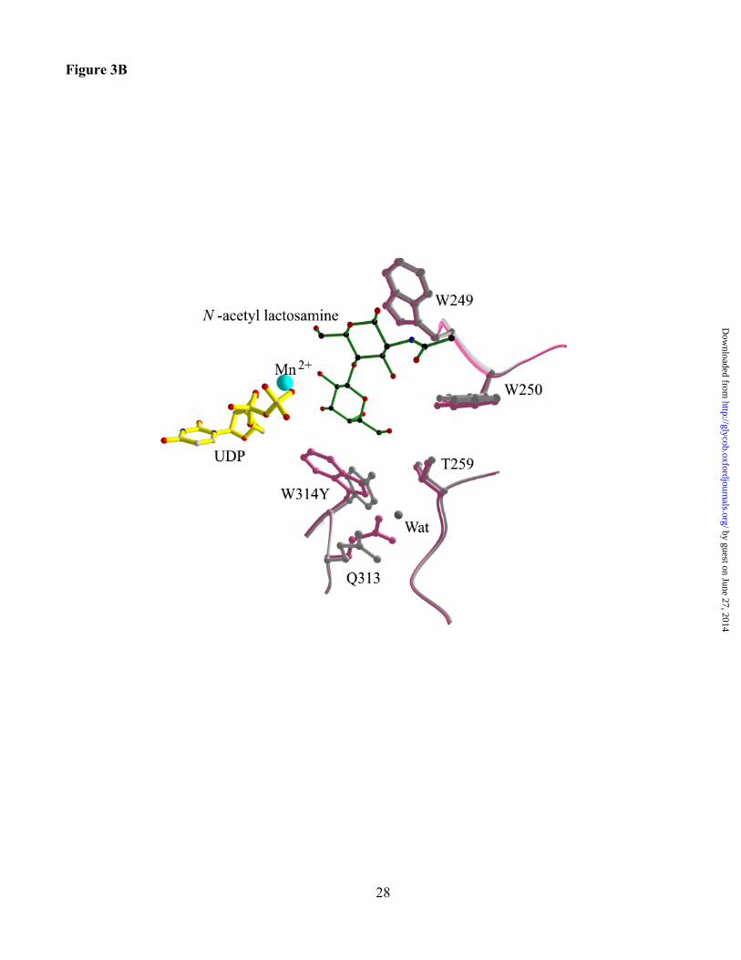
Structures were determined for the Trp314Tyr mutant in complexes with UDP and lactose and
UDP and LacNAc at 1.7 and 2.1 Å, respectively (Table IV). The change in the amino acid side chain is
by guest on June 27, 2014http://glycob.oxfordjournals.org/
Dow
nloaded from
10
clearly visible in the structure, but otherwise, the superimposed mutant and wild-type structures show
essentially no change that can be attributed to the mutation (Figures 3A and 3B). Also, the bound
ligands superimpose closely with those in the wild-type a3GT complexes. Although the mutation
reduces the number of contacts between the side chain of Tyr 314 and acceptor substrate, it does not
produce any change the orientation of the substrate in this complex.
Discussion
The structures of complexes of enzymes with substrates and inhibitors provide important
information regarding binding site locations and the nature of enzyme-ligand interactions. However the
complexes whose structures can be determined in this way are generally not on the catalytic pathway of
the enzyme. This is the case for a3GT where structures have been determined at high resolution for
enzyme complexes (a) containing one UDP ligand (Boix et al., 2001) (b) two UDPs (Zhang et al., 2003)
(c) the products of UDP-galactose hydrolysis (d) products of UDP-glucose hydrolysis (e) a dead-end
inhibitory complex containing UDP and lactose and (f) the corresponding complex with UDP and
LacNAc (Boix et al., 2003). These structures all contain the Form II conformer of a3GT. Lower
resolution structures have been reported for the Form I conformer, in which the C-terminal region is
disordered, in complexes with UMP and UDP-Gal (Gastinel et al., 2001), but we have been unable to
identify a UDP ligand in our Form I structure, despite extensive soaking in 20 mM UDP (Zhang et al.,
2003). Although these structures do not establish the roles of specific enzyme-ligand interactions in
specificity or catalysis, they do provide an information base of potential interactions that can be further
investigated by mutational studies of structure-activity relationships.
The present results indicate that, although tryptophans 249, 250, 314 and 356 contribute to the
binding sites for acceptors in these structures, they make very distinct contributions to the catalytic
properties of the enzyme. The Trp249Gly mutation specifically perturbs the Km for acceptor, but appears
by guest on June 27, 2014http://glycob.oxfordjournals.org/
Dow
nloaded from
11
to have little effect on kcat, indicating that mutation of Trp249 has similar effects on acceptor binding in
the Michaelis complex and transition state. This is consistent with the relatively distant location of this
residue from the catalytic center of the enzyme, its role in interacting with the Glc or GlcNAc
component of the acceptor and the absence of any significant change in overall structure arising from the
mutation. It is interesting that the corresponding residue in human blood group A and B
glycosyltransferases is one of the four sites at which these closely related enzymes differ in sequence
(Gly and Ser, see Table II). This residue also influences acceptor substrate affinity in these enzymes
since substitution of Ser for Gly in the truncated catalytic domain of the blood group A
glycosyltransferases produces a 3-fold increase in the Km for acceptor substrate but insignificant changes
in kcat or donor substrate specificity (Seto et al., 1999).
Substitutions of Tyr or Phe for Trp250 produce small changes in catalytic activity, specifically, a
small increase in kcat for both the hydrolase and transferase activities and an increased Km for UDP-Gal
for the transferase activity. These changes may reflect a small change in active site accessibility that
produces increased rates of product release and reduces the on-rate for UDP-Gal binding.
The enzyme mutant with a Trp314 to Tyr substitution has properties that are similar to those of a
previously described Gln247 to Glu variant (Zhang et al., 2003). The kcat for the transferase reaction is
reduced but hydrolase activity is essentially unchanged; the mutation also produces a small reduction in
the Km for acceptor. Both the Trp314 to Tyr and Gln247 to Glu mutations reduce the ratio of the transferase
to hydrolase activity approximately 40-fold (Table III). Therefore, the Trp314Tyr mutation selectively
destabilizes the transition state for the transferase reaction, as shown by its effect on catalytic efficiency
for this reaction (Table III) but does not affect acceptor binding in the Michaelis complex or any aspect
of UDP-Gal hydrolysis. Trp314 interacts with both acceptor substrates in their UDP complexes and with
the monosaccharides in the enzyme complexes with UDP-Gal and UDP-Glc (Boix et al., 2002). The
mutation does not alter the structure of the enzyme or the binding of UDP, lactose or LacNAc. These
by guest on June 27, 2014http://glycob.oxfordjournals.org/
Dow
nloaded from
12
results suggest that Trp314 has a role in stabilizing the bisubstrate transition state for the transfer of
galactose to a disaccharide substrate but not for transfer to water.
The effects of the mutation of Trp356 to Thr differs from the others investigated here in reducing
the kcat for the both transferase and hydrolase reactions. This effect appears to be linked to its location
immediately adjacent to the C-terminal region of a3GT (residues 358-365) that undergoes a
conformational change between the Form I and form II structures (Boix et al., 2001; Gastinel et al.,
2001). A previously described a3GT mutant with a substitution of Lys for Arg365, a residue that
interacts with O2 of the a-P of UDP, showed large reductions in kcat and catalytic efficiency for the
transferase reaction (Zhang et al., 2003) and a similar reduction in UDP-Gal hydrolyase activity to that
seen in the Trp356Thr mutant (Table III). This suggests that the conformational change is linked to the
ability of a3GT to catalyze galactose transfer to a carbohydrate acceptor or to water, possibly through
the enclosure of the catalytic site and bound UDP-Gal by the transconformation and formation of an
appropriate environment for catalysis (Zhang et al., 2003). However, Trp356 also interacts with the
acceptor substrate, and the Thr mutation also increases the Km for lactose. Thus, mutations of different
tryptophans influence substrate specificity in this glycosyltransferase through their effects on substrate
affinity (Trp249), transition state stabilization (Trp314), or both (Trp356).
Materials and methods
Enzyme expression, mutagenesis and activity measurements
Mutants of the catalytic domain of bovine a3GT (residues 80-368) were constructed, expressed in E.
coli BL21(DE3) and purified as described previously (Zhang et al., 2001). The sequences of mutants
were checked by automated DNA sequencing of the expression vector.
Steady state kinetic studies with wild-type and mutant enzymes were performed with a
radiochemical assay as previously described (Zhang et al., 2001). For characterizing the transferase
by guest on June 27, 2014http://glycob.oxfordjournals.org/
Dow
nloaded from
13
activity of most mutants, enzyme activity was measured in the presence of 10 mM MnCl2 at a series of
UDP-Gal concentrations (generally 0.1 to 2.0 mM) and different lactose concentrations and the data
analyzed by fitting to the rate equation for a symmetrical sequential initial velocity pattern (Zhang et al.,
2001) to give values for kcat, Ka (Km for UDP-Gal), Kia (Kd for UDP-Gal from E.UDP-Gal complex) and
Kb (Km for acceptor) (Zhang et al., 2001, 2003). As discussed in the text, in some cases the concentration
of UDP-Gal was varied at a single fixed concentration of lactose (generally 25 mM) and lactose was
varied at a single fixed concentration of UDP-Gal, generally 1.0 or 1.2 mM. Analysis of these data by
fitting to the standard single-substrate Michaelis-Menten equation provided apparent values for Km for
each substrate and two Vm values. The apparent Vm value, obtained by varying [lactose] at fixed [UDP-
Gal] was adjusted by multiplying by (1+[UDP-Gal]/Ka), where Ka is the apparent Km for UDP-Gal to get
an improved estimate of the true Vm. and kcat. This method was used in the case of mutants with
relatively high UDP-Gal hydrolase activities, as discussed below.
An enzyme concentration was 2-5 mg/ml was used for galactosyltransferase assays and UDP-Gal
hydrolase activities were determined at 10 to 20-fold higher concentrations (Zhang et al., 2003). The
transferase and hydrolase activities of the less active Trp356Thr mutant were determined at enzyme
concentrations and incubation times of 1.6 mM/15 min and 6.4 mM/60min.
X-ray crystallography
Crystals of the Trp249Gly and Trp314Tyr mutants were grown at 16°C by the vapor diffusion hanging drop
method as described previously (Boix et al., 2001) by mixing 2 ml of the protein at 5 mg/ml in 20 mM
MES-NaOH buffer, pH 6.0, 10% glycerol, containing 10 mM UDP and 0.1 mM MnCl2, with an equal
volume of a reservoir solution containing 5% PEG 6000 and 0.1 M Tris-HCl, pH 8.0. Single crystals
appeared after 3-4 days. Crystals of Trp314Tyr complexes with lactose and LacNAc were obtained by
soaking in 100 mM solution of lactose and LacNAc respectively. Before data collection, all crystals were
flash-cooled at 100K in a cryoprotectant containing 10% PEG 6000, 0.1 M Tris-HCl, pH 8.0, 25%
by guest on June 27, 2014http://glycob.oxfordjournals.org/
Dow
nloaded from
14
glycerol and appropriate ligands. High resolution data sets (space group P21 with 2 molecules in the
asymmetric unit) were collected at the Synchrotron Radiation Source, Daresbury, UK (stations PX9.6 and
PX14.1 using a CCD-ADSC detector system). Raw data images were indexed and scaled using the
DENZO and SCALEPACK modules of the HKL suite (Otwinowski and Minor, 1997), see Table IV.
The cell dimensions for all the data sets were isomorphous with the previously reported Form II
a3GT•UDP structure (Boix et al., 2001), PDB entry 1K4V (Table IV). Therefore these coordinates were
used as a starting model. Crystallographic refinement was performed using the CNS program package
(Brünger et al., 1998). Multiple rounds of bulk solvent correction, energy minimization, individual
isotropic B-factor refinement, simulated annealing and model building using the ‘O’ program (Jones et
al., 1991) were carried until the Rfree value was optimized. Appropriate ligands (UDP, lactose, LacNAc
and Mn2+ ion) were incorporated into each model after careful observation of the respective electron
density map. Water molecules were gradually included into the model at positions corresponding to peaks
in the |Fo|-|Fc| electron density map with heights greater than 3s and at H-bond distance from appropriate
atoms. The final refinement statistics are given in Table IV.
For the Trp314Tyr- lactose complex structure at 1.7 Å resolution, further refinement was carried
out using SHELX-97 (Sheldrick and Schneider, 1997). Initial conjugate gradient least squares
refinement in SHELXL was carried out restraining all the 1,2- and 1,3- distances with Engh and Huber
(Engh and Huber, 1991) restraints. The atomic displacement parameters were kept isotropic and clear
alternate conformations for some of the side chains were modeled. The multiple conformation site
occupancy factors were refined constraining their sum to unity. Occupancy of atoms in molecules was
constrained as 1.0 or 0.5 depending on their peak heights in the electron density maps. Geometric
restrints were set to default values as specified in the program documentation. Final analysis of the
Ramachandran (j-y) plot showed that all residues lie in the allowed regions for all three structures. The
final refinement statistics are included in Table IV. . The atomic coordinates for a3GT Trp249Gly
by guest on June 27, 2014http://glycob.oxfordjournals.org/
Dow
nloaded from
15
mutant, Trp314Tyr mutant-lactose complex and Trp314Tyr mutant-N-acetyl-lactosamine complex (codes
1VZT, 1VZU and 1VZX respectively) have been deposited in the RCSB Protein Data Bank. All figures
were generated using MOLSCRIPT (Kraulis, 1991).
Acknowledgments
This work was supported by Grant GM58773 to K.B. from NIH (U.S.A.) and Wellcome Trust (U.K.)
Project Grant 059603 to K.R.A. We thank the staff at the synchrotron radiation source, Daresbury, UK
for help during X-ray data collection and Shalini Iyer for preparing the figures.
by guest on June 27, 2014http://glycob.oxfordjournals.org/
Dow
nloaded from
16
References
Avila J. L, Rojas M and Galili U. (1989) Immunogenic Gal a 1-3Gal carbohydrate epitopes are present
on pathogenic American Trypanosoma and Leishmania. J. Immunol. 142, 2828-2834.
Boix, E., Swaminathan, G. J., Zhang, Y., Natesh, R., Brew, K. and Acharya, K.R. (2001) Structure of
UDP complex of UDP-galactose: b-galactoside-a-1,3-galactosyltransferase at 1.53Å resolution
reveals a conformational change in the catalytically important C-terminus. J. Biol. Chem. 276,
48608-48614.
Boix, E., Zhang, Y., Swaminathan, G.J., Brew, K., and Acharya, K.R. (2002) Structural basis of ordered
binding of donor and acceptor substrates to the retaining glycosyltransferase: a-1,3
galactosyltransferase. J. Biol. Chem. 277, 28310-28318.
Brünger, A. T., Adams, P. D., Clore, G. M., DeLano, W. L., Gros, P., Grosse-Kunstleve, R. W., Jiang, J.
S., Kuszewski, J., Nilges, M., Pannu, N. S., Read, R. J., Rice, L. M., Simonson, T. and Warren, G.
L. (1998) Crystallography & NMR system: A new software suite for macromolecular structure
determination. Acta Crystallogr. D54, 905-921.
Engh, R. A. and Huber, R. (1991). Accurate bond and angle parameters for x-ray protein structure
refinement. Acta Crystallogr. A47, 392-400.
Galili U., Shohet, S.B., Kobrin, E., Stults, C.L., and Macher, B.A. (1988) Man, apes and old world
monkeys differ from other animals in the expression of a-galactosyl epitopes on nucleated cells. J.
Biol. Chem. 263, 17755-17762.
Galili U., and Swanson K. (1991) Gene sequences suggest inactivation of a-1,3-galactosyltransferase in
catarrhines after the divergence of apes from monkeys. Proc. Natl. Acad. Sci. USA, 88, 7401-
7404.
by guest on June 27, 2014http://glycob.oxfordjournals.org/
Dow
nloaded from

17
Gastinel, L.N., Bignon, C., Misra, A.K., Hindsgaul, O., Shaper, J.H., and Joziasse, D.H. (2001) Bovine
a1,3-galactosyltransferase catalytic domain structure and its relationship with ABO histo-blood
group and glycosphingolipid glycosyltransferases. EMBO J. 20, 638-649.
Haslam, D.B., and Baenziger, J.U. (1996) Expression cloning of Forssman glycolipid synthetase: a
novel member of the histo-blood group ABO family. Proc. Natl. Acad. Sci. USA 93, 10697-10702.
Heissigerová, H., Breton, C., Moravcová, and Imberty, A. (2003) Molecular modeling of
glycosyltransferases involved in the biosynthesis of blood group A, blood group B, Forssman, and
igb3 antigens and their interaction with substrates. Glycobiology 13, 377-386.
Jones, T. A., Zou, J. Y., Cowan, S. W. and Kjeldgaard M. (1991) Improved methods for building protein
models in electron density maps and the location of errors in these models. Acta Crystallogr. A47,
110-119.
Keusch,,J..J.., Manzella,S.M., Nyame,K.A., Cummings,R.D. and Baenziger,,J.U. (2000) Expression
cloning of a new member of the ABO blood group glycosyltransferases, iGb3 synthase, that
directs the synthesis of isoglobo-glycosphingolipids. J. Biol. Chem. 275, 25308-25314.
Kraulis, P.J. (1991) MOLSCRIPT: a program to produce both detailed and schematic plots of protein
structures. J. Appl. Crystallogr. 24, 946-950.
Muraki, M. (2002) The importance of CH/pi interactions to the function of carbohydrate binding
proteins. Protein Pept. Lett. 9, 195-209.
Otwinowski, Z., and Minor, W. (1997) Processing of X-ray diffraction data collected in oscillation
mode. Methods Enzymol. 276, 307-326.
Patenaude, S.I., Seto, N.O., Borisova, S.N., Szpacenko, A., Marcus, S.L., Palcic, M.M., and Evans, S.V.
(2002) The structural basis for specificity in human ABO(H) blood group biosynthesis. Nature
Struct. Biol. 9, 685-690.
by guest on June 27, 2014http://glycob.oxfordjournals.org/
Dow
nloaded from
18
Paulson, J.C., and Colley, K.J. (1989) Glycosyltransferases. Structure, localization, and control of cell
type-specific glycosylation. J. Biol. Chem. 264, 17615-17618.
Purich, D.L., and Allison, R.D. (2000) In “Handbook of Biochemical Kinetics” pp 524-525 Academic
Press, San Diego, CA.
Seto, N.O.L., Compston, C.A., Evans, S.V., Bundle, D.R., Narang, S.A., and Palcic, M.M. (1999) Donor
substrate specificity of recombinant human blood group A, B and hybrid A/B glycosyltransferases
expressed in Escherichia coli. Eur J. Biochem. 259, 770-775.
Sharon, N., and Lis, H. (2001) The structural basis for carbohydrate recognition by lectins. Adv. Exp.
Med. Biol. 491, 1-16.
Sheldrick, G. M. and Schneider, T. R. (1997) SHELXL: High resolution refinement. Methods Enzymol.
277, 319-343.
Takeuchi, Y., Porter, C.D., Strahan, K.M., Preece, A.F., Gustafsson, K., Cosset, F.L., Weiss, R.A., and
Collins, M.K. (1996) Sensitization of cells and retroviruses to human serum by (a 1-3)
galactosyltransferase. Nature 379, 85-88.
van den Eijnden, D.H., Blanken, W.M., Winterwerp, H., and Schiphorst, W.E.C.M. (1983) Identification
and characterization of an UDP-gal: N-acetyllactosaminide a-1,3-D-galactosyltransferase in calf
thymus. Eur J. Biochem. 134, 523-530.
Weiss, W.I., and Drickamer, K. (1996) Structural basis of lectin-carbohydrate recognition. Annu. Rev.
Biochem. 65, 441-473.
Yamamato, F-I., Clausen, H., White, T., Marken, J., and Hakamori, S-I (1990) Molecular genetic basis
of the histo-blood group ABO system. Nature 345, 229-233.
Zhang, Y., Wang, P.G., and Brew, K. (2001) Specificity and mechanism of metal ion activation in UDP-
galactose b-galactoside a-1,3-galactosyltransferase. J. Biol. Chem. 276, 11567-11574.
by guest on June 27, 2014http://glycob.oxfordjournals.org/
Dow
nloaded from
19
Zhang, Y., Swaminanthan, G.J., Deshpande, A., Natesh, R., Boix, E., Xie, Z., Acharya, K.R., and Brew,
K. (2003) Roles of individual enzyme-substrate interactions by a-1,3 galactosyltransferase in
catalysis and specificity. Biochem. 42, 13512-13521.
by guest on June 27, 2014http://glycob.oxfordjournals.org/
Dow
nloaded from
20
Table I. Roles of residues selected for mutagenesis in a3GT
Residue Substrate Enzymeatom
Substrateatom
Interactions withsubstrate
Trp249 Lac (glc)a (>10) b ring vdwc
Trp250 Lac (glc)a NE1 O3’ Hbd
(9) ring vdwTrp314 Gal component
of UDP-GaleO O3 Hb
O4 Hb10 vdw
Lac (gal)a NG1 O6 Hb>10 vdw
Trp356 Lac (gal)a 6 vdw
a derived from structure of the a3GT.UDP.Lac complex
b (X)-number of contacts
c vdw- van der Waal contact
d Hb- hydrogen bond
e derived from structure of the a3GT.UDP-gal complex. The galactose is free as a result of cleavage of
UDP-Gal by guest on June 27, 2014http://glycob.oxfordjournals.org/
Dow
nloaded from
21
Table II. Residues that correspond to a3GT acceptor substrate binding site tryptophans in
homologous glycosyltransferases
Bovine
a3GT
Other
a3GTs
iGb3
synthase
Forssman
glycolipid
synthase
Blood
group A
synthase
Blood
group B
synthase
Trp249 Trp Trp Gly Gly Ser
Trp250 Trp His Tyr Phe Phe
Trp314 Trp Trp Trp Trp Trp
Trp356 Trp Trp Thr Ala Ala
by guest on June 27, 2014http://glycob.oxfordjournals.org/
Dow
nloaded from
22
Table III. Kinetic parameters of recombinant a1,3-GT and its variants for the
galactosyltransferase reaction. These values were determined at 37°C as described in Materials and
Methods.
ENZYME WT W249G W250Y W250F W314Y W356TGalactosyltransferasereactionkcat (sec-1) 6.4±0.7 >2.3 8.2±0.6 16.5±0.9 0.22±0.006b 0.52±0.02
b
Ka (mM) 0.43±0.07 0.84±0.17a
2.24±0.27 4.00±0.33 0.72±0.07a 0.85±0.03a
Kb (mM) 19.9±3.4 >200c 9.8±1.2 21.5±1.8 9.7±0.8b 126±8b
Kia (mM) 0.14±0.03 0.21±0.04e
0.15±0.09 0.07±0.05 0.14±0.01e 0.25±0.02e
kcat/Kia*Kb
(¥106 sec-1M-2)2.3 ± 0.7 ~0.019 5.5±4.6 5.1±3.6 0.16±0.02 0.017
Hydrolase reactionkcat (sec-1) x103 16±1 16±1 29±1 23±0.2 18±0.3 1.1±0.0Ka (mM) 0.10±0.00 0.21±0.04 0.23±0.01 0.15±0.01. 0.14±0.01 0.25±0.02kcat/Ka (¥103 sec-1M-1) 0.16 0.076 0.13 0.15 0.16 0.0044kcat transferase/hydrolase 400 >140 283 717 12 473
aThe values for Km (apparent) for UDP-Gal (Ka) was determined by varying [UDP-Gal] at a fixed lactose
concentration of 25 mM.b The apparent kcat and Km for lactose (Kb) were determined by varying [lactose] at a fixed UDP-Gal
concentration of 1.0 mM. The apparent kcat was corrected by (1+Ka/[UDP-Gal]) as described in
Experimental Procedures. The high hydrolase relative to transferase activity in W314Y increases the
uncertainty in these values.c the Km value of acceptor is out of quantifiable range but kcat/Kb, determined from the slope of v vs.
[lactose] at 1mM UDP-galactose is 80-fold less than for the wild-type enzyme.d Determined by varying [lactose] at 1.2 mM [UDP-Gal]e These are Km values for UDP-Gal hydrolysis (see text for discussion).
by guest on June 27, 2014http://glycob.oxfordjournals.org/
Dow
nloaded from
23
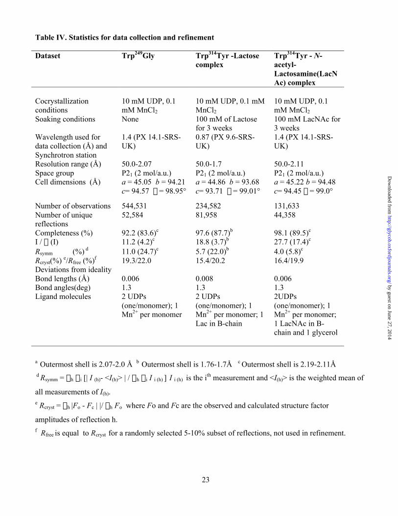
Table IV. Statistics for data collection and refinement
Dataset Trp249Gly Trp314Tyr -Lactosecomplex
Trp314Tyr - N-acetyl-Lactosamine(LacNAc) complex
Cocrystallizationconditions
10 mM UDP, 0.1mM MnCl2
10 mM UDP, 0.1 mMMnCl2
10 mM UDP, 0.1mM MnCl2
Soaking conditions None 100 mM of Lactosefor 3 weeks
100 mM LacNAc for3 weeks
Wavelength used fordata collection (Å) andSynchrotron station
1.4 (PX 14.1-SRS-UK)
0.87 (PX 9.6-SRS-UK)
1.4 (PX 14.1-SRS-UK)
Resolution range (Å) 50.0-2.07 50.0-1.7 50.0-2.11Space group P21 (2 mol/a.u.) P21 (2 mol/a.u.) P21 (2 mol/a.u.)Cell dimensions (Å) a = 45.05 b = 94.21
c= 94.57 b = 98.95°a = 44.86 b = 93.68c= 93.71 b = 99.01°
a = 45.22 b = 94.48c= 94.45 b = 99.0°
Number of observations 544,531 234,582 131,633Number of uniquereflections
52,584 81,958 44,358
Completeness (%) 92.2 (83.6)c 97.6 (87.7)b 98.1 (89.5)c
I / s (I) 11.2 (4.2)c 18.8 (3.7)b 27.7 (17.4)c
Rsymm (%) d 11.0 (24.7)c 5.7 (22.0)b 4.0 (5.8)c
Rcryst(%) e/Rfree (%)f 19.3/22.0 15.4/20.2 16.4/19.9Deviations from idealityBond lengths (Å) 0.006 0.008 0.006Bond angles(deg)Ligand molecules
1.32 UDPs(one/monomer); 1Mn2+ per monomer
1.32 UDPs(one/monomer); 1Mn2+ per monomer; 1Lac in B-chain
1.32UDPs(one/monomer); 1Mn2+ per monomer;1 LacNAc in B-chain and 1 glycerol
a Outermost shell is 2.07-2.0 Å b Outermost shell is 1.76-1.7Å c Outermost shell is 2.19-2.11Å d Rsymm = Sh Si [| I (h)- <I(h)> | / Sh Si I i (h) ] I i (h) is the ith measurement and <I(h)> is the weighted mean of
all measurements of I(h).e Rcryst = Sh |Fo - Fc | |/ Sh Fo where Fo and Fc are the observed and calculated structure factor
amplitudes of reflection h.f Rfree is equal to Rcryst for a randomly selected 5-10% subset of reflections, not used in refinement.
by guest on June 27, 2014http://glycob.oxfordjournals.org/
Dow
nloaded from

24
Figure Legends
Figure 1. Location of tryptophans 249, 250, 314 and 356 in the acceptor substrate binding site of
the a3GT-UDP-lactose complex. The Ca backbone of the enzyme is shown in grey, tryptophan side
chains are pink, the UDP is yellow, the Mn2+ cofactor in blue and the lactose is green with C atoms
colored black and O and N atoms in red.
Figure 2. Structure of the active site of the UDP complex of the Trp249Gly mutant of a3GT
superimposed with the wild-type enzyme. The mutant enzyme is grey and the wild-type enzyme, pink.
The UDP is yellow and the Mn2+ cofactor, blue. The location of a water molecule that replaces the
tryptophan side chain is displayed as a grey ball.
Figure 3. Structures of the active site of the Trp314Tyr mutant of a3GT (pink) superimposed with
the wild-type enzyme (white) with UDP (yellow), Mn2+ (blue) and (A) lactose, (B) LacNAc. The
mutant enzyme structure is shown in grey, wild-type in pink. Acceptor substrates are green with black C
atoms and red O and N atoms.
by guest on June 27, 2014http://glycob.oxfordjournals.org/
Dow
nloaded from
25
Figure 1
by guest on June 27, 2014http://glycob.oxfordjournals.org/
Dow
nloaded from
26
Figure 2
by guest on June 27, 2014http://glycob.oxfordjournals.org/
Dow
nloaded from
27
Figure 3A
by guest on June 27, 2014http://glycob.oxfordjournals.org/
Dow
nloaded from
28
Figure 3B
by guest on June 27, 2014http://glycob.oxfordjournals.org/
Dow
nloaded from