Introducci´n o Introducci´n.......... o Simulaci´n en la Industria. o Definici´n
Upload
independentCategory
view
1download
0
REPRODUCTIONRESEARCH
Role of the N, N 0-dimethylbiguanide metformin in thetreatment of female prepuberal BALB/c micehyperandrogenized with dehydroepiandrosterone
Valeria Sander, Carolina Griselda Luchetti, Marıa Emilia Solano, Evelin Elia,Guillermo Di Girolamo1, Claudio Gonzalez1 and Alicia Beatriz Motta
Centro de Estudios Farmacologicos y Botanicos (CEFYBO), Consejo Nacional de Investigaciones Cientıficas yTecnologicas (CONICET), Buenos Aires, Argentina and 1Facultad de Medicina, Departamento de Farmacologıa,Buenos Aires, Argentina
Correspondence should be addressed to A B Motta, Laboratorio de Fisiopatologıa Ovarica, Centro de Estudios Farmacologicos y
Botanicos (CEFYBO), UBA-CONICET. Paraguay 2155, (1121) Buenos Aires, Argentina; Email: [email protected]
Abstract
The present study investigated the role of the N, N{0}-dimethylbiguanide metformin (50 mg/100 g body weight in 0.05 ml
water, given orally with a canulla) in the prevention of endocrine and immune disorders provoked by the hyperandrogeniza-
tion with dehydroepiandrosterone (DHEA) in prepuberal BALB/c mice. The treatment with DHEA (6 mg/100 g body weight in
0.1 ml oil) for 20 consecutive days, recreates a mouse model that resembles some aspects of the human polycystic ovary
syndrome (PCOS). The treatment with DHEA did not modify either body mass index (BMI) or blood glucose levels, but did
increase fasting insulin levels when compared with controls. Markers of ovarian function – serum estradiol (E), progesterone
(P) and ovarian prostaglandin E (PGE) – were evaluated. The treatment with DHEA increased serum E and P levels while ovar-
ian PGE diminished. When metformin was administered together with DHEA, serum insulin, E and P levels, and ovarian PGE
values did not differ when compared with controls. Using flow cytometry assays we found that the treatment with DHEA
diminished the percentage of the CD4 1 T lymphocyte population and increased the percentage of the CD8 1 T lymphocyte
population from both ovarian tissue and retroperitoneal lymph nodes. However, when metformin was administered together
with DHEA, the percentages of CD4 1 and CD8 1 T lymphocyte populations from both ovarian tissue and retroperitoneal
lymph nodes were similar to those observed in controls. Finally, when DHEA was administered alone it increased the serum
tumor necrosis factor-alpha (TNF-a) levels when compared with controls; however, when metformin was administered
together with DHEA, serum TNF-a levels were similar to controls. These results indicate that metformin is able, directly or
indirectly, to avoid the endocrine and immune alterations produced when mice are hyperandrogenized with DHEA.
Reproduction (2006) 131 591–602
Introduction
Polycystic ovary syndrome (PCOS) – which is character-
ized by hyperandrogenemia, hirsutism, oligo- or amenor-
rhea and anovulation – is one of the most common
endocrinological diseases encountered in premenopausal
women (Franks 1995, Asuncion et al. 2000). It has been
reported that hyperinsulinemia, frequently associated with
PCOS, increases both the risk of cardiovascular diseases
and the development of diabetes mellitus (Abbott et al.
2002). Although during the last decade several clues have
emerged from human and animal studies, little is known
about the etiology and pathophysiology of PCOS. The bat-
tery of animal models used for the study of polycystic
ovaries has allowed researchers to investigate different
aspects of the pathology (Billiar et al. 1985, Szkiewicz &Uilenbroek 1998, Weil et al. 1999, West et al. 2001,Abbott et al. 2002). After it was found that dehydroepian-drosterone (DHEA) levels were increased in women withPCOS (Malesh & Greenblatt 1962), Roy et al. (1962) pro-duced an animal model using DHEA for induction ofPCOS. Subsequent studies established that the DHEA–PCOS murine model exhibits some of the salient featuresof human PCOS, such as hyperandrogenism, abnormalmaturation of ovarian follicules and anovulation (Lee et al.1991, 1998, Anderson et al. 1992, Henmi et al. 2001).These findings, together with the fact that increasing evi-dence indicates that DHEA has, in addition, potent immu-noregulatory functions (Meikle et al. 1992, Okabe et al.1995, Hernandez Pardo et al. 1998, Zhang et al. 1999) led
q 2006 Society for Reproduction and Fertility DOI: 10.1530/rep.1.00941
ISSN 1470–1626 (paper) 1741–7899 (online) Online version via www.reproduction-online.org
us to use the DHEA–mice model to study some aspectsrelated to the endocrine and immune responses involved.
In a previous study, we have demonstrated that treat-ment of prepuberal BALB/c mice with DHEA alters ovar-ian morphology, induces ovarian cysts, increases serum17 beta estradiol (E) and progesterone (P) levels, and mod-ifies ovarian prostaglandin E (PGE) concentration. There-fore, the treatment with DHEA led to a selective ovarianCD4 þ and CD8 þ T lymphocyte expression character-ized by both a diminution of the percentage of CD4 þ
lymphocytes and an increase in the percentage ofCD8 þ T lymphocytes (Luchetti et al. 2004). In view ofour previous studies and those reported by Lu et al.(2002), who demonstrated that production of cytokines byB cells is controlled by an enriched CD8 þ T lymphocytepopulation and steroid hormones, experiments weredesigned to evaluate whether DHEA was able to lead to aselective T lymphocyte population that could, in turn,regulate cytokine production. Considering the fact thattumor necrosis factor-alpha (TNF-a) regulates androgenproduction by theca cells (Sayin et al. 2003) and that ithas been reported to be associated with PCOS (Desh-pande et al. 2000, Araya et al. 2002, Korhonen et al.2002, Peral et al. 2002), the aim of this study was to inves-tigate the concentration of serum TNF-a after treatmentwith DHEA.
Multiple concomitant therapies have been applied inPCOS to address the variety of symptoms and to achievebetter results. Recent studies have investigated the role ofa family of insulin-sensitizing agents: the biguanides.Thus, the use of metformin (N, N{0}-dimethylbiguanide) isbecoming increasingly accepted and widespread. How-ever, this drug is being clinically used without a completeunderstanding of the mechanisms involved. Metforminhas been shown to be useful in the reduction of insulinresistance by restoring insulin sensitivity (Fedorcsak et al.2003, Harbone et al. 2003, Lord et al. 2003). In addition,it has been demonstrated that metformin is able, eitherdirectly or indirectly, to regulate ovarian steroidogenesis(La Marca et al. 2002, Mansfield et al. 2003).
Controversial results have been reported with regards tometformin and its relationship with the immune system. Inpatients with type-2 diabetes, the biguanide enhances thetyrosine kinase activity of the insulin receptor by modulat-ing the plasma cell differentiation antigen (PC-1) (Stefano-vic et al. 1999). Nevertheless, Ruat et al. (2003) havefailed to demonstrate any relationship between metforminand proliferation assays of T cells of lymph nodes.
The aim of the present work was to assess the efficacyof metformin on some immune and endocrine parametersaltered after hyperandrogenization with DHEA. We there-fore evaluated body mass index (BMI), the glucose:insulinratio (in order to determine the homeostasis model assess-ment (HOMA) index), and serum E and P levels (as indi-cators of ovarian function). We also quantified ovarianPGE production, because PGE is not only involved ininflammatory processes and displays immunomodulatory
properties, but is also altered in PCOS (Navarra et al.1996, Wojtowicz-Praga 1997). With regards to theimmune system we also designed experiments to investi-gate the role of metformin in modulating both the CD4 þ
and CD8 þ T cell population of ovarian tissue and sec-ondary immune tissues (axillar and retroperitoneal lymphnodes). In addition, the production of the pro-inflamma-tory cytokine TNF-a was evaluated.
Materials and Methods
Animals and experimental protocol
The hyperandrogenized environment of PCOS was repro-duced in mice by injection of DHEA (Luchetti et al.2004). Briefly, female prepuberal (25 days old) mice of theBALB/c strain were injected daily with DHEA (6 mg/100 gbody weight, dissolved in 0.10 ml sesame oil) for 20 con-secutive days (DHEA group). The animals of the DHEA þ
metformin group were injected with DHEA and givenmetformin orally (50 mg/100 g body weight in 0.05 mlwater, given orally with a canulla) for 20 days. The doseof metformin administered was equivalent to that used inthe treatment of women with PCOS. The controls con-sisted of three groups: (a) animals injected with oil(0.1 ml) and given water orally (0.05 ml) for 20 consecu-tive days (control vehicle); (b) the metformin-alone groupwhich consisted of mice treated orally with 50 mg metfor-min/kg body weight in 0.05 ml water for 20 days; (c) theuntreated group formed by animals that did not receivetreatment. Mice were housed under controlled tempera-ture (22 8C) and illumination (14 h light:10 h darkness;lights on at 0500 h) and were allowed free access to Pur-ina rat chow and water. All procedures involving animalswere conducted in accordance with the Animal Care andUse Committee of Consejo Nacional de InvestigacionesCientıficas y Tecnicas (CONICET) 1996.
Throughout the whole treatment, the animals wereweighed daily and, in addition, the stage of the sexualcycle was determined. Vaginal smears were taken dailyup to the day of autopsy. As previously found (Luchettiet al. 2004), mice treated with DHEA remained in con-stant estrus. In contrast, mice administered with DHEAtogether with metformin showed irregular sexual cycles.None of the animals in the experimental groups displayeda complete sexual cycle at the time of killing. These find-ings led us to use, for the control and metformin-alonegroups, only those animals that at the time of killing werein the estrus stage of the sexual cycle. In other words, inorder to compare the different groups, we have treatedsufficient animals from groups other than the DHEA-alonegroup to ensure that there were ten animals per group atestrus stage on the day of killing.
After 20 days of treatment, ten mice (45 days old) pergroup were anesthetized with ether and killed by decapi-tation. Blood was collected and fasting glucose levelswere immediately determined. Then, serum was isolated
592 V Sander and others
Reproduction (2006) 131 591–602 www.reproduction-online.org

and stored at 270 8C until assayed for E, P, insulin andTNF-a. Twenty freshly dissected ovaries per group weredivided as follows: ten ovaries of each group were pre-pared to evaluate CD4 þ and CD8 þ T lymphocyteexpression by flow cytometry assay; ten ovarian tissueswere immediately frozen at 270 8C until PGE radio-immunoassay. Lymphoid tissues (axillar and retroperito-neal nodes) from the ten animals of each group wereimmediately collected and processed to determine theCD4 þ and CD8 þ T cell expression by flow cytometryassay. All experiments were repeated three times.
Assays for glucose and insulin quantification
Fasting blood glucose was determined by using the Haemo-glukotest (Roche) test strips for visual determination in therange of 20–800 mg/100 ml (1–44 mmol/l). The testprinciple uses the glucose-oxidase/peroxidase reaction.Results are expressed in millimoles of glucose per liter.
Serum insulin levels were assayed by the Coat-A-Countinsulin method (Diagnostic Products Corporation, LA,USA) following the manufacturer’s instructions. Briefly, thedetermination consists of a solid-phase 125I radioimmuno-assay designed for the quantitative measurement of insulinin serum. The antibody is immobilized to the wall of apolypropylene tube and simply decanting the supernatantsuffices to terminate the competition and to isolate theantibody- bound fraction of the radiolabeled insulin. Thesamples were counted in a gamma counter. Analyticalsensitivity was 1.2mIU insulin/serum ml.
E and P determination
Serum E levels were evaluated as described previously(Luchetti et al. 2004). Briefly, the blood was allowed toclot and the serum removed and frozen until E and P con-centrations were determined by radioimmunoassay. Anti-sera were provided by Dr G D Niswender (Colorado StateUniversity, Fort Collins, CO, USA). The E antiserumshowed low cross-reactivity: ,1% for P and testosterone,,5% for estriol and ,10% for estrone. The P antiserumwas highly specific and showed low cross-reactivity:,2% for 20-a-dihydro- progesterone and deoxycorticos-terone, and 1% for other steroids normally present inserum. Results are expressed in nanograms per serummilliliter.
Prostaglandin radioimmunoassay
The measurement of PGE was carried out in the incu-bation media of ovarian tissues as previously reported(Luchetti et al. 2004). Briefly, the tissue (each ovary) wasweighed and incubated in Krebs–Ringer–bicarbonate(KRB) with glucose (11.0 mmol/l) as external substrate(pH 7.0) for 1 h in a Dubnoff metabolic shaker under anatmosphere of 5% CO2 in 95% O2 at 37 8C. At the endof the incubation period, the tissue was removed and thesolution acidified to pH 3.0 with 1 M HCl and extracted
for prostaglandin determination three times with 1 volumeof ethyl acetate. Pooled ethyl acetate extracts were driedunder an atmosphere of N2 and stored at 220 8C untilprostaglandin radioimmunoassay was perfomed. PGE wasquantified using a rabbit antiserum from Sigma. Sensitivitywas 10 pg/tube and cross-reactivity was 100% with PGEand ,0.1% with other prostaglandins. Results areexpressed in picograms PGE per milligram of protein.Ovarian protein content was determined using the Brad-ford method (1976).
Flow cytometry
To carry out the flow cytometry assays, ovarian tissue andlymph nodes (axillar and retroperitoneal) must be dis-persed (Luchetti et al. 2004). Briefly, tissues were enzy-matically dissociated in culture medium (medium 199,25 mM Hepes, 26 mM NaHCO3 and 50 UI/ml penicillin)with collagenase (trypsin-free, 740 IU/100 mg tissue) andDNAse (14 IU/100 mg tissue). After 90 min, cells werewashed twice with culture medium, twice with Dul-becco—phosphate-buffered saline free of Ca2þ and Mg2þ
(PBS) and twice with culture medium containing EDTA(1 mM). To remove blood cells, suspensions were appliedto Ficoll-hystopaque gradient 1.077 (Sigma), centrifugedat 400 g for 45 min and washed with PBS/ 0.1% BSA.Cells were counted in a hemocytometer (viability was.80% as assessed by the trypan blue exclusion method)and then processed for direct immunofluorescence. Thus,100ml of each cellular suspension, at a concentration of106 cells/ml, were incubated for 30 min at 4 8C with: (a)30ml phycoerythrin (PE) Rat IgG2a K Isotype Control plus30ml fluorescein isothiocyanate (FITC) Rat IgG2a K Iso-type Control (eBioscience, San Diego, CA, USA) corre-sponding to the isotype control sample; or (b) 4ml PE anti-mouse CD4 plus 4ml FITC anti-mouse CD8 (eBioscience,USA), corresponding to control, DHEA and DHEA þ
metformin assay according to the cellular suspension.Antibodies were used at saturating concentrations, asestablished after titration by flow cytometry. Samples werethen washed with PBS and PBS-EDTA, fixed with 4% par-aformaldehyde and stored at 4 8C in darkness until theanalysis was performed within 6 days of labeling. Fluor-escence analysis was evaluated with FACScan andWinmdi 2.8 software (Scripps Research Institute). Bothovarian and lymph suspensions were analyzed usingdifferent physical characteristics (i.e. size and complexity),using both forward (FSC: cell size) and side scatter (SSC:cell complexity) parameters. Thus, the settings correlatedto cellular size and granularity of mouse T lymphocytes.The resulting gate and quadrant were maintained through-out the analysis. Flow cytometric determination wasperformed using standard fluorescence 1 (FL1; FITCanti-mouse CD8 þ T lymphocyte) and fluorescence 2(FL2; PE-anti-mouse CD4 þ T lymphocyte). The analysiswas based on quantification of 50 000 cells for each assayfor ovarian tissue and 10 000 cells for lymph nodes.
Metformin as treatment of hyperandrogenized mice 593
www.reproduction-online.org Reproduction (2006) 131 591–602

Serum TNF-a determination
A TNF-a enzyme immunometric assay (EIA) kit (AssayDesign’s mouse, MI, USA) was employed following themanufacturer’s instructions to quantify serum TNF-a fromsamples of the three experimental groups. Results areexpressed in picograms per serum milliliter.
Statistical analysis
Statistical analyses were carried out using the Instat pro-gram (GraphPAD software, San Diego, CA, USA). ANOVAwas performed using Newman–Keuls test to compare allpairs of columns and P , 0.05 was considered significant.All results are presented as the means^S.E.M.
Results
Effect of metformin on BMI, homeostasis modelassessment, fasting glucose and insulin levels
These experiments were performed to determine whetherthe treatment with metformin was able to modify someparameters of the homeostasis and to study the role, ifany, of metformin when administered together withDHEA. The weight of the animals was not modified byany of the treatments (Table 1), and thus the BMI (definedas weight (kg)/height (m2)) was similar in all the groupsanalyzed (Table 1). Glucose levels did not vary in any ofthe groups studied (Fig. 1A), while fasting insulin levelsincreased with DHEA treatment when compared withcontrols (untreated, control vehicle and metformin-alonegroups). HOMA index (defined as: HOMA ¼ insulin(mUI/ml) £ glucose (mmol/l):BMI (kg/m2)) increased afterthe treatment with DHEA when compared with controls.When metformin was administered together with DHEA,the effects of DHEA on both insulin levels (Fig. 1B) andthe HOMA index were attenuated (Fig. 1c).
Effect of metformin on ovarian function: serum E andP levels and ovarian PGE production
To evaluate both ovarian function after treatment withDHEA and the possible role of metformin as a treatmentfor the ovarian anomalies produced by DHEA, wedesigned experiments to quantify serum E and P levels,and ovarian PGE production. Treatment with DHEA for 20consecutive days increased both serum E and P levels(Fig. 2A and B) when compared with controls (untreated,control vehicle and metformin alone). When metformin
was administered together with DHEA, serum E and Plevels showed similar patterns to those of control values(Fig. 2A; DHEA þ metformin).
On the other hand, the production of PGE by ovarian tis-sue was diminished after treatment with DHEA (Fig. 2C),but did not significantly differ from the control values whenmetformin was administered together with DHEA (Fig. 2C).
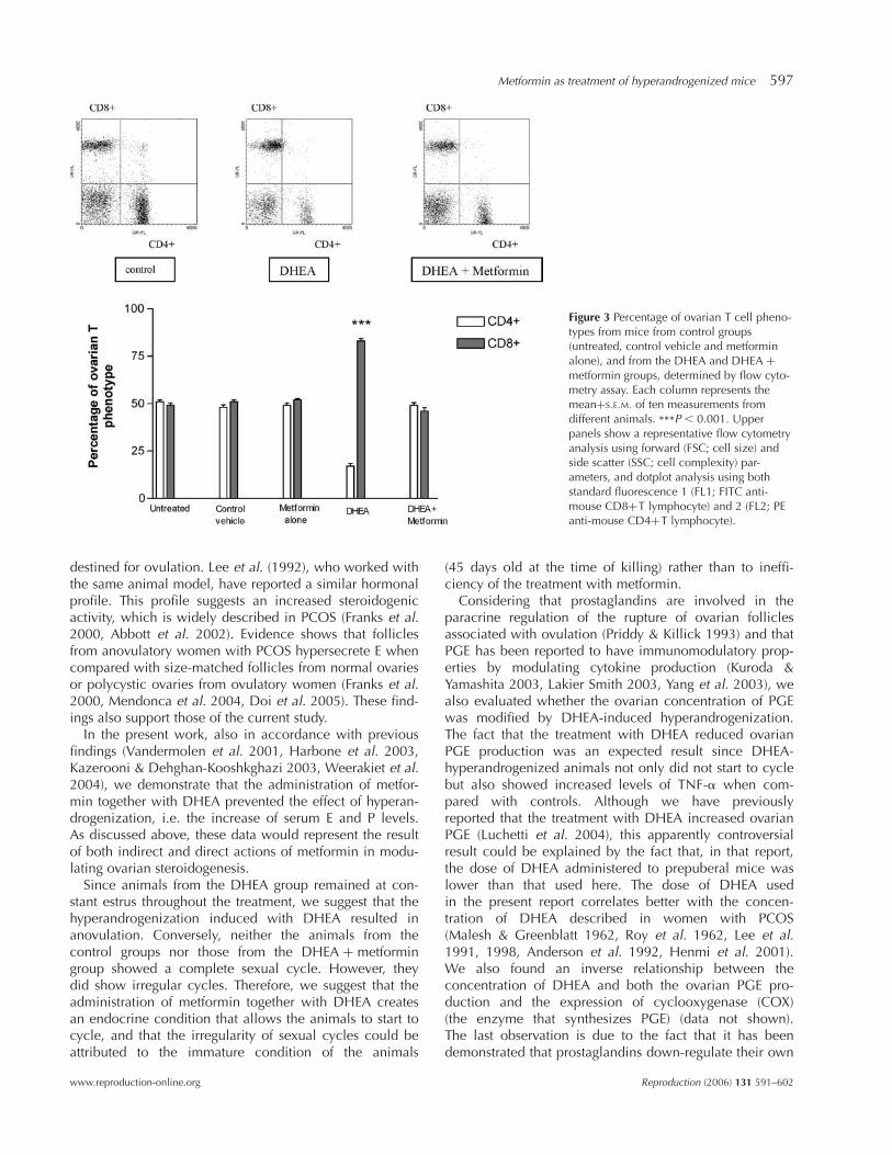
Role of metformin in ovarian CD4 1 and CD8 1T lymphocyte expression
Flow cytometry analysis was employed to determine boththe effect of DHEA treatment on the expression of ovarianCD4 þ (or helpers) and CD8 þ (or cytotoxic/suppressors)T lymphocytes and the role of metformin when adminis-tered together with DHEA. For the 50 000 cells analyzed,we found that the control groups showed equivalent per-centages of ovarian CD4 þ and CD8 þ T lymphocytes(Fig. 3). However, treatment with DHEA diminished thepercentage of CD4 þ T cells (17 ^ 5%) and increased thepercentage of CD8 þ T cells (83 ^ 4%) when comparedwith controls (Fig. 3). In contrast, when metformin wasadministered together with DHEA, we observed the samepercentages of CD4 þ and CD8 þ T cells as in thecontrols (Fig. 3).
Role of metformin in T lymphocyte expression inlymph nodes
In order to establish whether the effects of DHEA treat-ment and the administration of metformin together withDHEA also involved secondary lymphoid tissues, the per-centages of CD4 þ and CD8 þ T cells were quantified inboth axillar and retroperitoneal lymph nodes. Figure 4Aillustrates the flow cytometry analysis of axillar nodes. Itcan be seen that all the control groups yielded equivalentpercentages of CD4 þ and CD8 þ T lymphocytes, andthat neither the treatment with DHEA nor treatment withDHEA and metformin together were able to modify thepercentages of CD4 þ and CD8 þ T lymphocytes fromaxillar nodes.
The flow cytometry analysis of retroperitoneal lymphnodes (Fig. 4B) showed equivalent percentages of CD4 þ
and CD8 þ T lymphocytes in all the control groups. How-ever, treatment with DHEA diminished the percentage ofCD4 þ T cells, and increased the CD8 þ T cells from ret-roperitoneal lymph nodes (Fig. 4B; CD4 þ , 24 ^ 6%;CD 8 þ , 64 ^ 4%). In contrast, when metformin wasadministered together with DHEA, the percentages of
Table 1 Effect of DHEA on BMI.
Untreated Control vehicle Metformin-alone DHEA DHEA 1 metformin
Age (days) 45 45 45 45 45Weight (g) 14.0 ^ 1.2 15.0 ^ 3.0 14.0 ^ 2.5 14.8 ^ 1.8 15.2 ^ 2.4BMI (kg/m2) 5.6 ^ 0.2 5.7 ^ 0.5 5.7 ^ 0.4 5.4 ^ 0.6 5.3 ^ 0.8
594 V Sander and others
Reproduction (2006) 131 591–602 www.reproduction-online.org

CD4 þ and CD8 þ T cells from retroperitoneal lymphnodes were similar to those of controls (Fig. 4B).
Role of metformin on serum TNF-a concentration
As shown in Fig. 5, mice injected with DHEA for 20 daysshowed increased serum TNF-a concentration when com-pared with controls, while those injected with metformintogether with DHEA showed a concentration similar tothat of controls.
Discussion
In this work, we studied some aspects related to both theendocrine and the immune responses in a polycystic ovar-ian condition induced by hyperandrogenization withDHEA in BALB/c mice. The aim of the present study wasalso to investigate the ability of metformin to modulatethese aspects.
The mechanism by which administration of DHEAbrings about the cascade of hormonal events thatproduces ovarian failure remains unknown. However, it is
known that the experimental model used in this workreproduces the main aspects of human PCOS (Lee et al.1991, 1998, Anderson et al. 1992, Henmi et al. 2001).Our data show that the hyperandrogenic environmentrecreated by the treatment with DHEA did not modify theweight of the animals or, consequently, the BMI. Wecould then infer that neither hyperlipidemia nor obesity,which can sometimes be associated with PCOS (Frankset al. 1997, Abbott et al. 2002), were induced in this ani-mal model.
Both hyperinsulinemia and hyperandrogenism play apathogenic role in PCOS since they contribute to anovula-tion (Shoupe et al. 1983, Dunaif et al. 1989, Franks et al.2000), impair folliculogenesis and affect follicular devel-opment (Dunaif et al. 1989, Gougeon 1996, Musso et al.2005). For these reasons, insulin-sensitizing drugs such asmetformin can improve the rate of spontaneous ovulation(De Leo et al. 1999, Glueck et al. 1999, Vandermolenet al. 2001). Although we did not directly assess insulinsensitivity, we measured surrogate markers of insulin sen-sitivity such as fasting serum insulin, fasting blood glucoseand the HOMA index (which reflects the glucose–insulin
Figure 1 Role of DHEA in (A) fasting glucose levels,(B) fasting insulin levels and (C) HOMA index andeffect of metformin treatment. P , 0.001, b vs a;P , 0.05, c vs a. Each group represents themean^S.E.M. of ten different animals.
0.0
2.5
5.0
7.5 Untreated
Control vehicle
Metformin alone
DHEA
DHEA+Metformin
Fast
ing
gluc
ose
leve
ls(m
mol
/l)
0
100
200UntreatedControl vehicleMetformin aloneDHEADHEA+Metformin
Fast
ing
insu
lin le
vels
(uIU
/ml s
erum
)
a a a
b
c
0
5
10
15Untreated
Control vehicle
Metformin alone
DHEA
DHEA+Metformin
Hom
eost
asis
Mod
elA
sses
smen
t (H
OM
A)
a aa
b
c
A
B
C
Metformin as treatment of hyperandrogenized mice 595
www.reproduction-online.org Reproduction (2006) 131 591–602

relationship). Our results showed that treatment withDHEA increased the serum insulin levels without affectingfasting glucose, while treatment with metformin andDHEA together led to a HOMA index similar to that ofcontrols. Although metformin is usually used for the treat-ment of chronic obese, insulin-resistant type-2 diabetic,and PCOS patients (Fedorcsak et al. 2003, Harbone et al.2003, Lord et al. 2003), the role of this drug during con-ditions of normal glucose concentration remainsunknown. Our findings are in agreement with previousstudies that have demonstrated that metformin increasesperipheral insulin sensitivity in non-diabetic women withPCOS (Diamanti-Kandarakis et al. 1998, Moghetti et al.2000, Vandermolen et al. 2001).
Since metformin modulates insulin concentration and,in turn, insulin controls ovarian steroidogenesis, it can besaid that metformin acts indirectly on the steroidogenicactivity of theca and granulosa cells (La Marca et al.2002). However, recently, Mansfield et al. (2003) have
demonstrated that metformin exerts a direct effect on cul-tured ovarian cells. Therefore, we could speculate thatmetformin would regulate ovarian steroidogenesis both bymodulating insulin levels and by acting directly on ovar-ian cells. These two pathways would contribute to ensurethe accuracy of ovarian function. However, we are design-ing further experiments to clarify both this point and themolecular mechanisms involved in the action ofmetformin.
The data presented here show that mice from the DHEAgroup exhibited increased levels of both serum E and Pand were in constant estrus. In view of the fact that onlythose follicles that show significant amounts of aromatiz-able androgens and low production of E are classified asselectable follicles (i.e. appropriate to ovulate) (Gougeon1996), we can assume that in addition to the hyperandro-genized environment created by the daily injection ofDHEA, the enhanced concentration of serum E wouldresult in unfavorable conditions for producing follicles
0
10
20
30
40
*** Untreated
Control vehicle
Metformin alone
DHEA
DHEA+Metformin
Est
radi
ol(n
g/m
l ser
um)
0
1
2
3*** Untreated
Control vehicle
Metformin alone
DHEA
DHEA+Metformin
Pro
gest
eron
e(n
g/m
l ser
um)
0
1000
2000
***
Untreated
Control vehicle
Metformin alone
DHEA
DHEA+Metformin
Pro
stag
land
in E
(pg/
mg
prot
ein)
A
B
C
Figure 2 (A) Serum 17 beta estradiol levels from micefrom control groups, and the DHEA and DHEA þ
metformin groups. (B) Serum progesterone levelsfrom mice from control groups, and the DHEA andDHEA þ metformin groups. (C) Ovarian PGE concen-tration from mice from control groups, and theDHEA and DHEA þ metformin groups. Each columnrepresents the mean^S.E.M. of ten measurementsfrom different animals. ***P , 0.001.
596 V Sander and others
Reproduction (2006) 131 591–602 www.reproduction-online.org
destined for ovulation. Lee et al. (1992), who worked withthe same animal model, have reported a similar hormonalprofile. This profile suggests an increased steroidogenicactivity, which is widely described in PCOS (Franks et al.2000, Abbott et al. 2002). Evidence shows that folliclesfrom anovulatory women with PCOS hypersecrete E whencompared with size-matched follicles from normal ovariesor polycystic ovaries from ovulatory women (Franks et al.2000, Mendonca et al. 2004, Doi et al. 2005). These find-ings also support those of the current study.
In the present work, also in accordance with previousfindings (Vandermolen et al. 2001, Harbone et al. 2003,Kazerooni & Dehghan-Kooshkghazi 2003, Weerakiet et al.2004), we demonstrate that the administration of metfor-min together with DHEA prevented the effect of hyperan-drogenization, i.e. the increase of serum E and P levels.As discussed above, these data would represent the resultof both indirect and direct actions of metformin in modu-lating ovarian steroidogenesis.
Since animals from the DHEA group remained at con-stant estrus throughout the treatment, we suggest that thehyperandrogenization induced with DHEA resulted inanovulation. Conversely, neither the animals from thecontrol groups nor those from the DHEA þ metformingroup showed a complete sexual cycle. However, theydid show irregular cycles. Therefore, we suggest that theadministration of metformin together with DHEA createsan endocrine condition that allows the animals to start tocycle, and that the irregularity of sexual cycles could beattributed to the immature condition of the animals
(45 days old at the time of killing) rather than to ineffi-ciency of the treatment with metformin.
Considering that prostaglandins are involved in theparacrine regulation of the rupture of ovarian folliclesassociated with ovulation (Priddy & Killick 1993) and thatPGE has been reported to have immunomodulatory prop-erties by modulating cytokine production (Kuroda &Yamashita 2003, Lakier Smith 2003, Yang et al. 2003), wealso evaluated whether the ovarian concentration of PGEwas modified by DHEA-induced hyperandrogenization.The fact that the treatment with DHEA reduced ovarianPGE production was an expected result since DHEA-hyperandrogenized animals not only did not start to cyclebut also showed increased levels of TNF-a when com-pared with controls. Although we have previouslyreported that the treatment with DHEA increased ovarianPGE (Luchetti et al. 2004), this apparently controversialresult could be explained by the fact that, in that report,the dose of DHEA administered to prepuberal mice waslower than that used here. The dose of DHEA usedin the present report correlates better with the concen-tration of DHEA described in women with PCOS(Malesh & Greenblatt 1962, Roy et al. 1962, Lee et al.1991, 1998, Anderson et al. 1992, Henmi et al. 2001).We also found an inverse relationship between theconcentration of DHEA and both the ovarian PGE pro-duction and the expression of cyclooxygenase (COX)(the enzyme that synthesizes PGE) (data not shown).The last observation is due to the fact that it has beendemonstrated that prostaglandins down-regulate their own
Figure 3 Percentage of ovarian T cell pheno-types from mice from control groups(untreated, control vehicle and metforminalone), and from the DHEA and DHEA þ
metformin groups, determined by flow cyto-metry assay. Each column represents themeanþS.E.M. of ten measurements fromdifferent animals. ***P , 0.001. Upperpanels show a representative flow cytometryanalysis using forward (FSC; cell size) andside scatter (SSC; cell complexity) par-ameters, and dotplot analysis using bothstandard fluorescence 1 (FL1; FITC anti-mouse CD8þT lymphocyte) and 2 (FL2; PEanti-mouse CD4þT lymphocyte).
Metformin as treatment of hyperandrogenized mice 597
www.reproduction-online.org Reproduction (2006) 131 591–602

synthesis (Ivanov & Romanovsky 2004). We thereforeassume that the higher dose of DHEA injected in the pre-sent report led to an accumulation of ovarian PGE whichin turn could inhibit the expression of COX and conse-quently the production of ovarian PGE, as observed here.
On the other hand, we found that animals from theDHEA þ metformin group showed a pattern of ovarianPGE synthesis similar to that of the control groups.
Although the action of this biguanide on improving lipidmetabolism has been widely reported (Caballero 2004,Dominguez & Sowers 2005, Rautio et al. 2005), our datarepresent the first evidence that metformin modulates theproduction of ovarian prostaglandins.
TNF has been found to be increased in patients withPCOS (Sayin et al. 2003). In addition, a mutation of theTNF receptor has been associated with hyperandrogenism
Figure 4 Percentage of (A) T cell phenotypesfrom axillar lymph nodes and (B) T cell phe-notypes from retroperitoneal lymph nodesdetermined by flow cytometry assay. Eachcolumn represents the mean^S.E.M. of tenmeasurements from different animals.
***P , 0.001. Upper panels show a repre-sentative flow cytometry analysis usingforward (FSC; cell size) and side scatter(SSC; cell complexity) parameters, and dot-plot analysis using both standard fluor-escence 1 (FL1; FITC anti-mouse CD8þTlymphocyte) and 2 (FL2; PE anti-mouseCD4þT lymphocyte).
598 V Sander and others
Reproduction (2006) 131 591–602 www.reproduction-online.org

(Peral et al. 2002). In agreement with these findings, wefound that animals treated with DHEA had higher serumTNF-a levels when compared with controls, a fact thatcorrelates both with increased levels of PGE and withanovulatory cycles. It has been reported that TNF-a modu-lates steroidogenesis of both granulosa and theca-intersti-tial cells by a mechanism independent of those inducedby insulin and insulin-like growth factor-I (IGF-I)(Spaczynski et al. 1999). For this reason we could inferthat the increase of serum TNF-a observed in animalsfrom the DHEA group would be an additional mechanismto those involved in hyperinsulinemia, which impair ovar-ian steroidogenesis. In the present work, we demonstratedthat the administration of metformin together with DHEAled to serum TNF-a levels similar to control values. In theliterature, conflicting results have been reported withregards to the relationship between metformin and theregulation of TNF-a (Cacicedo et al. 2004, Bruun et al.2005, Di Gregorio et al. 2005, Kiortsis et al. 2005).
Finally, the data presented here show that DHEA-induced hyperandrogenization increased the percentageof the CD8 þ T population and diminished the percentageof CD4 þ T lymphocytes when compared with controls.Although the receptor for DHEA has not been identifiedyet, a specific DHEA binding activity has been detected inT cells (Meikle et al. 1992, Okabe et al. 1995) and for thisreason we hypothesized that endocrine disturbancescould be directly related to T lymphocyte differentiation.It has been documented, for example, that expression ofthe most mature thymocytes is regulated by P and E levels,and is also related to the high propensity of autoimmunediseases in females (Leposavic et al. 2001, Obradovicet al. 2001). Moreover, Yan et al. (2000) have reportedthat both autoimmune premature ovarian failure (POF)and insulin-dependent diabetes mellitus (IDDM) patientspresent increased numbers of CD8 þ T cells. In addition,it has been demonstrated that production of cytokines byB cells is controlled by an enriched CD8 þ T population(Lu et al. 2002).
Data presented here show that metformin treatmentresulted in similar percentages of CD4 þ and CD8 þ T
lymphocytes to those seen in the controls. As metforminmodulates P and E levels and since, in turn, P and Econtrol T lymphocyte differentiation (Yan et al. 2000,Leposavic et al. 2001, Obradovic et al. 2001, Lu et al.2002), we can suggest that metformin would actindirectly in modulating the percentages of ovarianCD4 þ and CD8 þ T lymphocytes. However, other path-ways (such as regulation of reactive oxygen species andthe induction of AMP-activated protein kinase (AMPK) ofT lymphocytes) described in other tissues (Bonnefont-Rousselot et al. 2003, Cacicedo et al. 2004, Huypenset al. 2005, McCarthy 2005) must not be disregarded andare currently being studied at our laboratory. Both thefact that the T cell population from axillar nodes was notaffected during DHEA- induced hyperandrogenizationand that the T cell population from retroperitoneal lymphnodes was affected – and also the fact that hyperandro-genization induced a similar T cell population whensamples from retroperitoneal lymph tissue were com-pared with those obtained from ovarian tissue – lead usto suggest that the local inflammatory status would becontributing to a selective differentiation of T cells.According to this hypothesis, lymph nodes and sex ster-oids are related to different systems, thus suggesting acoordinated organ-specific and steroid hormone relation-ship (Chantakru et al. 2003). Moreover, the E receptorexpressed by follicular dendritic cells in lymph nodes hasrecently been proposed as a novel pathological marker(Sapino et al. 2003) and the deficiency of E caused byovariectomy or menopause is involved in the T lympho-cyte status (Safadi et al. 2000). In summary, the presentstudy describes the role of metformin in the regulation ofsome aspects of the intricate network that relates theendocrine and the immune pathway in a hyperandrogen-ized environment. As the development of immune cellsurface markers is beginning to be used in diagnosisprior to the development of complete ovarian failure, webelieve that understanding the role of the immune pro-cesses involved in PCOS could also be important in themanipulation of this pathology.
0
10
20
30Untreated
Control vehicle
Metformin alone
DHEA
DHEA+Metformin
TN
F a
lpha
(pg/
ml s
erum
)
***
Figure 5 Serum TNF from mice from control groups (untreated, control vehicle and metformin alone), the DHEA group and the DHEA þ
metformin group. Each column represents the meanþS.E.M. of ten measurements from different animals. ***P , 0.001.
Metformin as treatment of hyperandrogenized mice 599
www.reproduction-online.org Reproduction (2006) 131 591–602
Funding
These studies were supported by PIP CONICET (reference6051) and Agencia Nacional de Promocion Cientıfica yTecnologica (PICT) (reference 05-10901).
References
Abbott DH, Dumesic DA & Franks S 2002 Developmental origin ofpolycystic ovary syndrome – a hypothesis. Journal of Endocrin-ology 174 1–5.
Anderson E, Lee MT & Lee G 1992 Cystogenesis of the ovarian antralfollicle of the rat: ultrastructural changes and hormonal profile fol-lowing the administration of dehydroepiandrosterone. AnatomicalRecord 234 359–382.
Araya AV, Aguirre A, Romero C, Miranda C, Molina MC & Ferreira A2002 Evaluation of tumor necrosis factor alpha production in exvivo short term culture whole blood from women with polycysticovary syndrome. European Cytokine Network 13 419–424.
Asuncion M, Calvo RM, San Millan JL, Sancho J, Avila S & Escobar-Morreale HF 2000 A prospective study of the prevalence of thepolycystic ovary syndrome in unselected Caucasian women fromSpain. Journal of Clinical Endocrinology and Metabolism 852434–2438.
Billiar RB, Richardson D, Anderson E, Mahajan D & Little B 1985The effect of chronic and acyclic elevation of circulating androste-nedione or estrone concentrations on ovarian function in the rhe-sus monkey. Endocrinology 116 2209–2220.
Bonnefont-Rousselot D, Raji B, Walrand M, Gardes-Albert M, JoreD, Legrand A, Peynet J & Vasson MP 2003 An intracellular modu-lation of free radical production could contribute to the beneficialeffects of metformin towards oxidative stress. Metabolism 52586–589.
Bradford M 1976 A rapid and sensitive method for the quantificationof microgram quantities of protein utilizing the principle of pro-tein-dye binding. Analytical Biochemistry 72 248–254.
Bruun JM, Lihn AS, Pedersen SB & Richelsen B 2005 Monocyte che-moattractant protein-1 release is higher in visceral than subcu-taneous human adipose tissue (AT): implication of macrophagesresident in the AT. Journal of Clinical Endocrinology and Metab-olism 90 2282–2289.
Caballero AE 2004 Endothelial dysfunction, inflammation, and insu-lin resistance: a focus on subjects at risk for type 2 diabetes. Cur-rent Diabetic Report 4 237–246.
Cacicedo JM, Yagihashi N, Keaney JF Jr, Ruderman NB & Ido Y 2004AMPK inhibits fatty acid-induced increases in NF-kappaB transac-tivation in cultured human umbilical vein endothelial cells. Bio-chemical and Biophysical Research Communications 261204–1209.
Chantakru S, Wang WC, Van den Heuvel M, Bashar S, Simpson A,Chen Q, Croy BA & Evans SS 2003 Coordinate regulation of lym-phocyte-endothelial interactions by pregnancy-associated hor-mones. Journal of Immunology 171 4011–4019.
De Leo V, La Marca A, Ditto A, Morgante G & Cianci A 1999 Effectsof metformin on gonadotropin-induced ovulation in women withpolycystic ovary syndrome. Fertility and Sterility 72 282–285.
Deshpande RR, Chang MY, Chapman JC & Michael SD 2000 Altera-tion of cytokine production in follicular cystic ovaries induced inmice by neonatal estradiol injection. American Journal of Repro-duction and Immunology 44 80–88.
Diamanti-Kandarakis E, Kouli C, Tsianateli T & Bergiele A 1998Therapeutic effects of metformin on insulin resistance and hyper-androgenism in polycystic ovary syndrome. European Journal ofEndocrinology 138 269–274.
Di Gregorio GB, Yao-Borengasser A, Rasouli N, Yarma V, Lu T, MilesLM, Ranganathan G, Peterson CA, McGehee RE & Kern PA 2005
Expression of CD68 and macrophage chemoattractant protein-1genes in human adipose and muscle tissues: association with cyto-kine expression, insulin resistance, and reduction by pioglitazone.Diabetes 54 2305–2313.
Doi SA, Al-Zaid M, Towers PA, Scott CJ & Al-Shoumer KA 2005 Irre-gular cycles and steroid hormones in polycystic ovary syndrome.Human Reproduction 20 2402–2408.
Dominguez LJ & Sowers JR 2005 Metabolic syndrome therapy: pre-vention of vascular injury by antidiabetic agents. Current Hyper-tension Reports 7 110–116.
Dunaif A, Segal KR, Futterweit W & Dobriansky A 1989 Profoundperipheral insulin resistance, independent of obesity in polycysticovary syndrome. Diabetes 38 1165–1174.
Fedorcsak P, Dale PO, Storeng R, Abyholm T & Tanbo T 2003 Theeffect of metformin on ovarian stimulation and in vitro fertilizationin insulin-resistant women with polycystic ovary syndrome: anopen-label randomized cross-over trial. Gynecological Endocrin-ology 17 207–214.
Franks S 1995 Polycystic ovary syndrome. New England Journal ofMedicine 333 853–861.
Franks S, Gharani N, Waterworth D, Batty S, White D, Williamson R& McCarthy M 1997 The genetic basis of polycystic ovary syn-drome. Human Reproduction 12 2641–2648.
Franks S, Mason H & Willis D 2000 Follicular dynamics in the poly-cystic ovary syndrome. Molecular Cellular Endocrinology 2549–52.
Glueck CJ, Wang P, Fontaine R, Tracy T & Sieve-Smith L 1999 Met-formin-induced resumption of normal menses in 39 of 43 (91%)previously amenorrheic women with the polycystic ovary syn-drome. Metabolism 48 511–519.
Gougeon A 1996 Regulation of ovarian follicular development inprimates: facts and hypotheses. Endocrine Reviews 17 121–155.
Harbone L, Fleming R, Lyall H, Sattar N & Norman J 2003 Metfor-min or antiandrogen in the treatment of hirsutism in polycysticovary syndrome. Journal of Clinical Endocrinology and Metab-olism 88 4116–4123.
Henmi H, Endo T, Nagasawa K, Hayashi T, Chida M, Akutagawa N,Iwasaki M, Kitajima Y, Kiya T, Nishikawa A, Manase K & Kudo R2001 Lysyl oxidase and MMP-2 expression in dehydroepian-drosterone-induced polycystic ovary in rats. Biology of Reproduc-tion 64 157–162.
Hernandez Pardo R, Streber MDL, Orozco H, Arriaga K, Pavon L,Al-Nakhli SA & Rook GA 1998 The effects of androstenedione anddehydroepiandrosterone on the course and cytokine profile oftuberculosis in BALB/c mice. Immunology 95 234–241.
Huypens P, Quartier E, Pipeleers D & Van de Casteele M 2005 Met-formin reduces adiponectin protein expression and release in 3T3-L1 adipocytes involving activation of AMP activated proteinkinase. European Journal of Pharmacology 518 90–95.
Ivanov AI & Romanovsky AA 2004 Prostaglandin E2 as a mediator offever: synthesis and catabolism. Frontiers in Bioscience 11977–1993.
Kazerooni T & Dehghan-Kooshkghazi M 2003 Effects of metformintherapy on hyperandrogenism in women with polycystic ovariansyndrome. Gynecological Endocrinology 17 51–56.
Kiortsis DN, Filippatos TD & Elisaf MS 2005 The effects of orlistat onmetabolic parameters and other cardiovascular risk factors. Dia-betes and Metabolism 31 15–22.
Korhonen S, Romppanen EL, Hiltunen M, Mannernaa A, PunnonenK, Hippelainen M & Heinoneu D 2002 Lack of associationbetween C-850T polymorphism of the gene encoding tumornecrosis factor-alpha and polycystic ovary syndrome. Gynecologi-cal Endocrinology 16 271–274.
Kuroda E & Yamashita U 2003 Mechanisms of enhanced macro-phage-mediated prostaglandin E2 production and its suppressiverole in Th1 activation in Th2-dominant BALB/c mice. Journal ofImmunology 15 757–764.
600 V Sander and others
Reproduction (2006) 131 591–602 www.reproduction-online.org

Lakier Smith L 2003 Overtraining, excessive exercise, and alteredimmunity: is this a T helper-1 versus T helper-2 lymphocyteresponse? Sports and Medicine 33 347–364.
La Marca A, Morgante G, Palumbo M, Cianci A, Petraglia F & De Leo V2002 Insulin-lowering treatment reduces aromatase activity inresponse to follicle-stimulating hormone in women with polycysticovary syndrome. Fertility and Sterility 78 1234–1239.
Lee GY, Croop JM & Anderson E 1998 Multidrug resistance geneexpression correlates with progesterone production in dehy-droepiandrosterone-induced polycystic equine chorionic gonado-tropin-stimulated ovaries of prepuberal rats. Biology ofReproduction 58 330–337.
Lee MT, Anderson E & Lee GY 1991 Changes in ovarian morphologyand serum hormones in the rat after treatment with dehydroepian-drosterone. Anatomical Record 231 185–192.
Leposavic G, Obradovic S, Kosec D, Pejcic-Karapetrovic B & Vidic-Dankovic B 2001 In vivo modulation of the distribution ofthymocyte subsets by female sex steroid hormones. InternationalImmunopharmacology 1 1–12.
Lord JM, Flight IH & Norman RJ 2003 Insulin-sensitising drugs (met-formin, troglitazone, rosiglitazone, pioglitazone, D-chiro-inositol)for polycystic ovary syndrome. Cochrane Database SystemReviews 3 CD0033053.
Lu FX, Abel K, Ma Z, Rourke T, Lu D, Torten J, McChesney M &Miller CJ 2002 The strength of B cell immunity in female rhesusmacaques is controlled by CD8 þ T cells under the influence ofovarian steroid hormones. Clinical Experimental Immunology 12810–20.
Luchetti CG, Solano ME, Sander V, Barreiro-Arcos ML, Gonzalez C,Di Girolamo G, Chiocchio S, Cremaschi G & Motta AB 2004Effects of dehydroepiandrosterone on ovarian cystogenesisand immune function. Journal of Reproductive Immunology64 59–74.
McCarthy MF 2005 AMPK activation as a strategy for reversing theendothelial lipotoxicity underlying the increased vascular risk as-sociated with insulin resistance syndrome. Medical Hypotheses 641211–1215.
Malesh VB & Greenblatt RB 1962 Isolation of dehydroepiandroster-one and 17a- hydroxy- pregnenolone from polycystic ovaries ofthe Stein–Leventhal syndrome. Journal of Clinical Endocrinologyand Metabolism 22 441–448.
Mansfield R, Galea R, Brincat M, Hole D & Mason H 2003 Metfor-min has direct effects on human ovarian steroidogenesis. Fertilityand Sterility 79 956–962.
Meikle AW, Dorchuck RW, Araneo BA, Stringham JD, Evans TG,Spruance SL & Daynes RA 1992 The presence of dehydroepian-drosterone-specific receptor binding complex in murine T cells.Journal of Steroid Biochemisry and Molecular Biology 42293–304.
Mendonca HC, Montenegro RM, Foss MC, Silva de Sa MF & FerrianiRA 2004 Positive correlation of serum leptin with estradiol levelsin patients with polycystic ovary syndrome. Brazilian Journal ofMedical and Biological Research 37 729–736.
Moghetti P, Castello R, Negri C, Tosi F, Perrone F, Caputo M,Zanolin E & Muggeo M 2000 Metformin effects on clinical fea-tures, endocrine and metabolic profiles, and insulin sensitivityin polycystic ovary syndrome: a randomized, double-blind, pla-cebo-controlled 6 month trial, followed by open, long-termclinical evaluation. Journal of Clinical Endocrinology andMetabolism 85 139–146.
Musso C, Shawker T, Cochran E, Javor ED, Young J & Gorden P2005 Clinical evidence that hyperinsulinaemia independent ofgonadotropins stimulates ovarian growth. Clinical Endocrinology(Oxford) 63 73–78.
Navarra P, Andreani CL, Lazzarin N, Pierro E, Mirtella A, Lanzone A &Mancuso S 1996 Increased production and release of prostaglandinE2 by human granulosa cells from polycystic ovaries. Prostaglandins52 187–197.
Obradovic S, Vidic-Dankovic B, Pejcic-Karapetrovic B, Kosec D &Leposavic G 2001 In vivo modulation of the splenocyte yield andcomposition by female sex steroid hormones. Pharmazie 56235–238.
Okabe T, Haji M, Takayanagi R, Adachi M, Imasaki K, Kurimoto F,Watanabe T & Nawata H 1995 Up-regulation of high-affinitydehydroepiandrosterone binding activity by dehydroepiandroster-one in activated human T lymphocytes. Journal of Clinical Endo-crinology and Metabolism 80 2993–2996.
Peral B, San Millan JL, Castello R, Morghetti P & Escobar-MorrealeHF 2002 Comment: the methionine 196 arginine polymorphism inexon 6 of the TNF receptor 2 gene (TNFRSFIB) is associated withthe polycystic ovary syndrome and hyperandrogenism. Journal ofClinical Endocrinology and Metabolism 87 3977–3983.
Priddy AR & Killick SR 1993 Eicosanoids and ovulation. Prostaglan-dins Leukotrienes and Essential Fatty Acids 49 827–1993.
Rautio K, Tapanainen JS, Ruokonen A & Mouri-Papunen LC 2005Effects of metformin and ethinyl estradiol-cyproterone acetate onlipid levels in obese and non-obese women with polycystic ovarysyndrome. European Journal of Endocrinology 152 269–275.
Roy S, Mahesh VB & Greenblatt RB 1962 Effect of dehydroepiandroster-one and d4-androstenedione on the reproductive organs of femalerats: production of cystic changes in the ovary. Nature 196 2–6.
Ruat C, Faure L, Choquet-Kastylevsky G, Ravel G & Descotes J 2003Tritiated thymidine incorporation does not enhance sensitivity ofthe politeal lymph node assay. Toxicology 188 29–33.
Safadi FF, Dissanayake IR, Goodman GG, Jago RA, Baker AE,Bowman AR, Sass DA, Popoff SN & Epstein S 2000 Influence ofestrogen deficiency and replacement on T-cell populations in ratlymphoid tissues and organs. Endocrine 12 81–88.
Sapino A, Cassoni P, Ferrero E, Bongiovanni M, Righi L, Fortunati N,Crafa P, Chiarle R & Bussolati G 2003 Estrogen receptor alpha is anovel marker expressed by follicular dendritic cells in lymphnodes and tumor-associated lymphoid infiltrates. American Journalof Pathology 163 1313–1320.
Sayin NC, Gucer F, Balkanli-Kaplan P, Yuce MA, Ciftci S, Kucuk M &Yardim T 2003 Elevated serum TNF-alpha levels in normal-weightwomen with polycystic ovaries syndrome. Journal of ReproductiveMedicine 48 165–170.
Shoupe D, Kumar DD & Lobo RA 1983 Insulin resistance in poly-cystic ovary syndrome. American Journal of Obstetrics and Gyne-cology 147 588–592.
Spaczynski RZ, Arici A & Duleba AJ 1999 Tumor necrosis factor-alpha stimulates proliferation of rat ovarian theca-interstitial cells.Biology of Reproduction 61 993–998.
Stefanovic V, Antic S, Mitic-Zlatkovic M & Vlahovic P 1999 Reversalof increased lymphocyte PC-1 activity in patients with type 2 dia-betes treated with metformin. Diabetes and Metabolism ResearchReviews 15 400–404.
Szkiewicz D & Uilenbroek JT 1998 Polycystic ovary syndrome –searching for an animal model. Journal of Medicine 29 259–275.
Vandermolen DT, Ratts VS, Evans WS, Stovall DW, Kauma SW &Nestler JE 2001 Metformin increases the ovulation rate and preg-nancy rate from clomiphene citrate in patients with polycysticovary syndrome who are resistant to clomiphene citrate alone.Fertility and Sterility 75 310–315.
Weerakiet S, Srisombut C, Rojanasakul A, Panburana P, ThakkinstianA & Herabutya Y 2004 Prevalence of gestational diabetes mellitusand pregnancy outcomes in Asian women with polycystic ovarysyndrome. Gynecological Endocrinology 19 134–140.
Weil S, Vendola K, Zhou J & Bondy CA 1999 Androgen and follicle-stimulating hormone interactions in primate ovarian follicle devel-opment. Journal of Clinical Endocrinology and Metabolism 842951–2956.
West C, Foster DL, Evans NP, Robinson J & Padmanabhan V 2001Intra-follicular activin availability is altered in prenatally-androgen-ized lambs. Molecular and Cellular Endocrinology 185 51–59.
Metformin as treatment of hyperandrogenized mice 601
www.reproduction-online.org Reproduction (2006) 131 591–602

Wojtowicz-Praga S 1997 Reversal of tumor-induced immunosuppres-sion: a new approach to cancer therapy. Journal of Immunotherapy20 165–177.
Yan G, Schoenfeld D, Penney C, Hurxthal K, Taylor AE & FaustmanD 2000 Identification of premature ovarian failure patients withunderlying autoimmunity. Journal of Women’s Health and GenderBased Medicine 9 275–287.
Yang L, Yamagata N, Yadav R, Brandon S, Courtney RL, Morrow JD,Shyr Y, Boothby M, Joyce S, Carbone DP & Breyer RM 2003 Can-cer-associated immunodeficiency and dendritic cell abnormalitiesmediated by the prostaglandin EP2 receptor. Journal of ClinicalInvestigation 111 727–735.
Zhang Z, Araghi-Niknam M, Liang B, Inserra P, Ardestani SK, Jiang S,Chow S & Watson RR 1999 Prevention of immune dysfunction andvitamin E loss by dehydroepiandrosterone and melatonin supplemen-tation during murine retrovirus infection. Immunology 96 291–297.
Received 28 August 2005First decision 20 October 2005Revised manuscript received 31 October 2005Accepted 15 November 2005
602 V Sander and others
Reproduction (2006) 131 591–602 www.reproduction-online.org
Copyright © 2022 FDOKUMEN