Role of Sodium Nitroprusside in the Improvement of Rat Liver Preservation in University of Wisconsin...
8
Role of Sodium Nitroprusside in the Improvement of Rat Liver Preservation in University of Wisconsin Solution: A Study in the Isolated Perfused Liver Model Joaquı ´n V. Rodriguez,* ,1 Edgardo E. Guibert,² Alejandra Quintana,‡ Angel Scandizzi,* and Luciana Almada* *Farmacologia, Departamento de Ciencias Fisiolo ´gicas; ²Biologı´a Molecular; Departamento de Ciencias Biolo ´gicas; and ‡Morfologı´a, Departamento de Ciencias Biolo ´gicas, Facultad de Ciencias Bioquı ´micas y Farmace ´uticas, Universidad Nacional de Rosario, Suipacha 531 (2000) Rosario, Argentina Submitted for publication April 26, 1999 Background. Long-term liver preservation is needed to transform liver transplantation from an emergency operation into an elective procedure and, therefore, to improve the results of liver transplantation. Aims. We have studied the possibility of extending the period of cold ischemia of the rat liver, maintain- ing good hemodynamics and functional conditions, by adding the NO donor sodium nitroprusside (NPNa) to the preservation solution. Materials and Methods. Rat livers were preserved for 24, 48, and 72 h in University of Wisconsin solution (UW) at 4°C (groups I, II, and III) or UW to which 500 mM NPNa was added (groups IV, V, and VI). Following the preservation time, liver viability was assessed us- ing the isolated perfused liver model. Lactate dehy- drogenase (LDH) and K 1 release, bile flow, and portal resistance were evaluated in each group and com- pared with those of liver controls (group VII) excised and perfused without preservation. Results. Some deleterious effects can be seen during cold storage conditions as assessed by an increment in intrahepatic resistance and a diminution in the capac- ity of the organ to produce bile. On histological obser- vation, we see vacuolated hepatocytes and free endo- thelial cells (detached) in the sinusoidal lumen. Addition of 500 mM NPNa to UW significantly moder- ates these injuries, with an improvement in intrahe- patic circulation (less intrahepatic resistance), an in- crement in bile production, and better histological appearance of the organ. We were also able to deter- mine the capacity of the UW 1 NPNa to produce NO. Conclusion. We assume that the beneficial vascular effects of NPNa are mediated by NO produc- tion. © 1999 Academic Press Key Words: sodium nitroprusside; cold preservation; University of Wisconsin solution; cold ischemia; iso- lated perfused liver. INTRODUCTION Hypothermic preservation of organs in University of Wisconsin solution (UW) is a widespread and well- accepted method of maintaining organs for transplan- tation [1]. UW solution is currently considered the most effective solution for cold liver preservation. How- ever, liver viability can be reliably preserved for only 8 –12 h. Some authors suggest that cold ischemia du- ration longer than 12 h is a significant risk factor for graft failure [2]. The liver is known to be highly sus- ceptible to ischemic injury; this is the most common cause of graft dysfunction after surgery [3]. Other stud- ies have indicated that damage to the hepatic micro- vasculature, particularly endothelial cells, may be the primary injury after long-term hypothermic preserva- tion. The microvascular damage leads to disturbances in perfusion and increased vascular resistance, imped- ing oxygen transport and delivery of nutrients from blood to hepatocytes. This phenomenon always pre- cedes hepatocyte injury, suggesting that abnormalities in microcirculation could play the primary role in the pathogenesis of graft nonfunction. In this way, protec- tion of ischemically altered organs might be achieved following potentially useful measures such as improv- ing the recovery of hepatic energy metabolism [4] and adding to the preservation solution calcium antagonist [5] or vasoactive agents such as vasodilators [6] or substances such as g-hydroxybutyrate [7]. 1 To whom correspondence should be addressed. Fax: 0054-341- 4804601. E-mail: [email protected]. Journal of Surgical Research 87, 201–208 (1999) Article ID jsre.1999.5750, available online at http://www.idealibrary.com on 201 0022-4804/99 $30.00 Copyright © 1999 by Academic Press All rights of reproduction in any form reserved.
-
Upload
independent -
Category
Documents
-
view
0 -
download
0
Transcript of Role of Sodium Nitroprusside in the Improvement of Rat Liver Preservation in University of Wisconsin...

toi
tiat
f(mtidrpa
ciivtAapcam
4
JA
Role of Sodium Nitroprusside in the Improvement of Rat LiverPreservation in University of Wisconsin Solution:
A Study in the Isolated Perfused Liver Model
Joaquın V. Rodriguez,*,1 Edgardo E. Guibert,† Alejandra Quintana,‡ Angel Scandizzi,* and Luciana Almada*
*Farmacologia, Departamento de Ciencias Fisiologicas; †Biologıa Molecular; Departamento de Ciencias Biologicas;and ‡Morfologıa, Departamento de Ciencias Biologicas, Facultad de Ciencias Bioquımicas y Farmaceuticas,
ournal of Surgical Research 87, 201–208 (1999)rticle ID jsre.1999.5750, available online at http://www.idealibrary.com on
Universidad Nacional de Rosario, Suipacha 531 (2000) Rosario, Argentina
icat
et
Ul
Watme8rgccivptiibciptf
Submitted for publ
Background. Long-term liver preservation is neededo transform liver transplantation from an emergencyperation into an elective procedure and, therefore, tomprove the results of liver transplantation.
Aims. We have studied the possibility of extendinghe period of cold ischemia of the rat liver, maintain-ng good hemodynamics and functional conditions, bydding the NO donor sodium nitroprusside (NPNa) tohe preservation solution.Materials and Methods. Rat livers were preserved
or 24, 48, and 72 h in University of Wisconsin solutionUW) at 4°C (groups I, II, and III) or UW to which 500M NPNa was added (groups IV, V, and VI). Following
he preservation time, liver viability was assessed us-ng the isolated perfused liver model. Lactate dehy-rogenase (LDH) and K1 release, bile flow, and portalesistance were evaluated in each group and com-ared with those of liver controls (group VII) excisednd perfused without preservation.Results. Some deleterious effects can be seen during
old storage conditions as assessed by an increment inntrahepatic resistance and a diminution in the capac-ty of the organ to produce bile. On histological obser-ation, we see vacuolated hepatocytes and free endo-helial cells (detached) in the sinusoidal lumen.ddition of 500 mM NPNa to UW significantly moder-tes these injuries, with an improvement in intrahe-atic circulation (less intrahepatic resistance), an in-rement in bile production, and better histologicalppearance of the organ. We were also able to deter-ine the capacity of the UW 1 NPNa to produce NO.Conclusion. We assume that the beneficial vascular
ia[s
1 To whom correspondence should be addressed. Fax: 0054-341-804601. E-mail: [email protected].
201
ion April 26, 1999
ffects of NPNa are mediated by NO produc-ion. © 1999 Academic Press
Key Words: sodium nitroprusside; cold preservation;niversity of Wisconsin solution; cold ischemia; iso-
ated perfused liver.
INTRODUCTION
Hypothermic preservation of organs in University ofisconsin solution (UW) is a widespread and well-
ccepted method of maintaining organs for transplan-ation [1]. UW solution is currently considered theost effective solution for cold liver preservation. How-
ver, liver viability can be reliably preserved for only–12 h. Some authors suggest that cold ischemia du-ation longer than 12 h is a significant risk factor forraft failure [2]. The liver is known to be highly sus-eptible to ischemic injury; this is the most commonause of graft dysfunction after surgery [3]. Other stud-es have indicated that damage to the hepatic micro-asculature, particularly endothelial cells, may be therimary injury after long-term hypothermic preserva-ion. The microvascular damage leads to disturbancesn perfusion and increased vascular resistance, imped-ng oxygen transport and delivery of nutrients fromlood to hepatocytes. This phenomenon always pre-edes hepatocyte injury, suggesting that abnormalitiesn microcirculation could play the primary role in theathogenesis of graft nonfunction. In this way, protec-ion of ischemically altered organs might be achievedollowing potentially useful measures such as improv-
ng the recovery of hepatic energy metabolism [4] anddding to the preservation solution calcium antagonist5] or vasoactive agents such as vasodilators [6] orubstances such as g-hydroxybutyrate [7].0022-4804/99 $30.00Copyright © 1999 by Academic Press
All rights of reproduction in any form reserved.

dicmvstomrnshpr
tatNpa
tusis
1r(wwmavIab
tKsmT(1
oabdwlbm
(vtahtpdwt
spcprwa(pa(caaoeooaTrtdzt0la3ro
t
2 H:
Nitric oxide (NO), identified as an endothelium-erived relaxing factor, has been reported to be a phys-ologic regulator of microcirculation as well as of vas-ular permeability. NO released by endothelial cellsaintains vascular homeostatic properties by relaxing
ascular smooth muscle, inhibiting neutrophil adhe-ion and platelet aggregation, and maintaining endo-helial barrier properties [8–10]. It has been pointedut that if endothelial cell damage represents the pri-ary event of injury during cold preservation, it is
easonable to think that its critical role in the mainte-ance of vascular integrity will be impaired as a con-equence of cold ischemia. Moreover, other authorsave reported that endogenous NO levels were de-ressed after ischemia or reperfusion or both [11] in aat cardiac transplantation model.
Sodium nitroprusside (NPNa) is a potent vasodilatorhat releases NO by a nonenzymatic process: it acts as
direct vasodilator independent of endothelial func-ion [12, 13]. We hypothesized that the vasodilatorPNa given at the onset of cold preservation mightrotect the liver from preservation–reperfusion dam-ge.In summary, the aim of this study was to investigate
he potential protective effect of NPNa on rat liversndergoing 72 h of hypothermic preservation in UWolution. For assessment of reperfusion, we used thesolated perfused liver; this model mimics the majorteps of the transplantation procedure.
MATERIALS AND METHODS
Solutions. The composition of UW in this study was as follows:00 mM lactobionic acid, 25 mM K2HPO4, 5 mM MgSO4, 30 mMaffinose, 5 mM adenosine, 1 mM allopurinol, 3 mM glutathioneGSH), 0.25 mg/ml streptomycin, and 10 IU/ml penicillin G. GSHas added to UW before use, UW was brought to pH 7.40 at 25°Cith 5 M KOH. The final Na1 and K1 concentrations were 30 and 125M, respectively. The solution was bubbled with 100% N2 for 15 min
t 4°C before use [14]. Sodium nitroprusside was dissolved in a smallolume of distilled water and added to UW immediately before use.n the UW used in this study, hydroxyethyl starch, dexamethasone,nd insulin were omitted with respect to the commercial solution [1],ecause their roles are still controversial [15].Composition of the perfusate Krebs–Henseleit–BSA. The syn-
hetic medium consisted of 2% bovine serum albumin (BSA) inrebs–Henseleit bicarbonate buffer (KH) with the following compo-
ition: NaCl 118 mM, KCl 4.8 mM, NaHCO3 25 mM, KH2PO4 1.2M, MgSO4 1.2 mM CaCl2 1.5 mM, heparin 2 IU/ml, glucose 5 mM.he final pH of the KH solution after equilibration with carbogen
O2:CO2, 95:5%) was 7.40. The perfusate was filtered through a.5-mm glass-fiber filter before use.Hepatectomy and cold storage. The experiments were performed
n male Wistar rats weighing 250–350 g. The animals had freeccess to standard rat chow and tap water, and were not fastedefore surgery. The experiments described in this report were con-
02 JOURNAL OF SURGICAL RESEARC
ucted according to the animal care and use committee. The ratsere anesthetized with sodium thiopental (50 mg/kg, ip) and their
ivers were prepared according to the standard technique [16]. Inrief, the bile duct was cannulated with a PE-50 catheter (Intra-edic USA), and 0.2 ml of saline containing 500 IU of heparin
tipi
Abbot, Argentina) was injected into the femoral vein. The portalein was cannulated with a large catheter (14 gauge, 2-mm i.d.) andhe hepatic artery was ligated. The liver was perfused with oxygen-ted Krebs–Henseleit buffer (37°C) at 15 ml/min. Then, the supra-epatic inferior vena cava (IVC) was cannulated with polyethyleneubing (3-mm i.d.) and the liver was removed without stopping theerfusion. In the control group the perfusion was performed imme-iately, whereas in the experimental groups the livers were flushedith 30 ml of cold UW for approximately 2 min and then transferred
o a vessel containing the preservation solution maintained at 4°C.Perfusion system. The livers were perfused using a recirculating
ystem modified from Alexander et al. [17] and shown in Fig. 1. Theerfusion system consisted of an oxygenator and a liver perfusionircuit connected in parallel to a common reservoir. A Masterflexump equipped with two heads pumped the perfusate from the maineservoir into two independent circuits. In one circuit, the perfusateas pumped first through a thermostated, water-jacketed glass coilnd a plastic device with a nylon filter, then to a calibrated flowmeterCole–Parmer G-32001-32) and a bubble trap, and finally to theortal cannula. In the other circuit, the perfusate was recirculatednd oxygenated while passing through oxygen-permeable tubingsilicone tubing, 0.078-in. i.d.; Catalog No. T5715-9, Baxter Health-are Corp., USA) inside an appropriate glass container with 95% O2
nd 5% CO2, at a constant pressure of 90 mm Hg. Air bubbles werevoided by connecting a disposable nylon filter inline between thexygenator and the inflow. A plastic device inserted between the heatxchanger and the flowmeter was used to ”smoothe out“ the pulsatileutput of the peristaltic pump [18]. The isolated liver was mountedn a glass container, continuously perfused through the portal vein,nd the effluent escaped through the IVC cannula to the reservoir.he perfusion pressure was measured, continuously monitored, andecorded with a strain gauge pressure transducer (P23ID) attachedo a side-arm placed just proximal to the perfusion cannula. A hy-rostatic manometer was used to calibrate the transducer, with theero placed at the level of the liver. To support bile flow, sodiumaurocholate in KH–BSA was infused into the reservoir at the rate of.3 mmol min21 with a syringe pump. The temperature close to theiver was measured using a thermocouple probe connected to anssistant infrared lamp device designed to maintain the liver at7°C. The recirculating perfusate volume was 180 ml. The pH in theeservoir was monitored and maintained at 7.40 6 0.50 with 1 M HClr 8.4% NaHCO3.
FIG. 1. Schematic diagram of the isolated liver perfusion sys-em.
VOL. 87, NO. 2, DECEMBER 1999
Experimental groups. Seven groups of livers were compared inhis study: (I) 24 h preservation in UW (n 5 4), (II) 48 h preservationn UW (n 5 4), (III) 72 h preservation in UW (n 5 3), (IV) 24 hreservation in UW 1 500 mM NPNa (n 5 4), (V) 48 h preservationn UW 1 500 mM NPNa (n 5 4), (VI) 72 h preservation in UW 1 500

mb
ppimrmg0csatammedsti
(
I
R
w6i
wmz
towgelmg0
dgcpsaagcb
1To
pvscttswda
b1aawlcio
tebirbf
wUop
P
gatobrvgdp
ece
US
M NPNa (n 5 3), (VII) controls, hepatectomy immediately followedy perfusion (n 5 5).Perfusion of the isolated liver for assessment of liver function after
reservation. Once the liver had been installed in the glass support,erfusion with KH commenced immediately, first in a nonrecirculat-ng system, increasing the flow rate between 5 and 25 ml/min in 10
in; then the perfusate was changed to KH–BSA and switched to aecirculating system. The flow rate was increased to 40 ml/min in 30in; this time was sufficient to stabilize the preparation. In all
roups, the livers were then perfused for 60 min at 40 ml/min (3.29 6.39 ml min21 g liver21 n 5 27), the portal pressure was recordedontinuously, and intrahepatic resistance was calculated. The intrin-ic resistance of the system was measured (0.004 mm Hg ml21 z min)nd subtracted from the values. Bile was collected in preweighedubes every 15 min and bile flow was estimated gravimetricallyssuming the bile density to be equal to water and expressed as mlin21 g liver21. Perfusate samples were taken from the reservoir foreasurement of lactate dehydrogenase (LDH) and K1 release. At the
nd of the perfusion, the livers were collected, weighed, and imme-iately fixed in a 4% formaldehyde buffered with phosphate-bufferedaline (PBS) and processed for paraffin embedding. Sections 7 mmhick were cut and stained with hematoxylin–eosin, where histolog-cal examination was performed.
Calculations. Intrahepatic resistance after 60 min of perfusionIR) was calculated as
R ~mm Hg ml21 z min z g liver!
5 portal pressure (mm Hg) 4 portal flow (ml min21 g liver21).
elease of LDH by the liver during perfusion was calculated as
LDH ~mIU min21 g liver21! 5 @~C60 2 C0!V# 4 ~LW 3 60 min!,
here C 60 is the LDH concentration in the perfusion medium after0 min of reperfusion and C 0 at the end of the stabilization period, Vs the perfusate volume (150 ml), and LW is the liver weight.
Potassium released to effluent perfusate was calculated as
@K1#effluent 4 @K1]perfusate,
here [K1]effluent is the K1 concentration of effluent perfusate after 60in and [K1]perfusate is the K1 concentration at the end of the stabili-
ation period.Effect of NPNa on biochemical parameters during liver preserva-
ion. To determine whether the vascular effect of NPNa was thenly mechanism involved in the improvement of liver preservation,e studied the evolution of biochemical parameters as glycogen,lucose, and water content of livers during hypothermic storage. Twoxperimental groups of livers were preserved up to 72 h at 4°C: (i)ivers preserved in UW, n 5 4, and (ii) livers preserved in UW 1 500M NPNa, n 5 4. The hepatic concentrations of glycogen andlucose and tissue water content were measured in biopsies taken at, 24, 48, and 72 h.Influence of NPNa concentration in UW solution on NO production
uring cold preservation. To investigate the role of NPNa in theeneration of NO during cold preservation, we analyzed the timeourse accumulation of NO2
2 in UW solution. This approach is sup-orted by the observation of Ignarro et al. [19] that the principalpontaneous oxidation product of NO in aqueous solution in the
2
RODRIGUEZ ET AL.: SODIUM NITROPR
bsence of biological contaminating substances is NO2 . Thus, theccumulation of NO2
2 in the medium can be used to assess NOeneration in situ from NPNa during hypothermic storage. The timeourse of NO2
2 production was determined in freshly prepared UWubbled with 100% N2. The following solutions were assayed: UW 1
Um(a
00 mM NPNa, UW 1 250 mM NPNa, and UW 1 500 mM NPNa.hese solutions were incubated at 4°C, up to 72 h. The concentrationf NO2
2 was measured at fixed times.Analytical methods. LDH concentration was measured in 1-ml
erfusate samples by following the rate at which NADH was con-erted to NAD1 at 340 nm in the presence of pyruvate [20]. Potas-ium concentration was measured by flame photometry. Liver gly-ogen was calculated from the amount of glucose released byreatment of homogenized tissue with amyloglucosidase followinghe determination of free glucose content [21]. Water content of liveramples was determined gravimetrically as the difference betweenet and dry weight of liver specimens. Tissue samples were oven-ried (48 h, 120°C), performed in duplicate to produce a mean valuend given as percentage wet weight.NO2
2 was assayed spectrophotometrically by using a procedureased on the Griess reaction as follows: 200 ml of UW was mixed with.3 ml of water and then 0.5 ml of 0.33% sulfanilic acid in 21% aceticcid and 0.5 ml of 0.05% a-naphthylamine in 21% acetic acid weredded. After 25 min at room temperature, the absorbance at 490 nmas measured. The concentrations of NO2
2 in the media were calcu-ated after comparison of absorbance values with those of a standardurve constructed with different concentrations of NaNO2. All chem-cal compounds were from Sigma Chemical Company; NPNa wasbtained from Merck.Statistical analysis. Results are presented as means 6 SD and
he number of preparations analyzed was 3 or more, as indicated inach figure. Statistical analysis of the differences between values ofile flow was evaluated by Mann–Whitney U test. Statistical signif-cance for changes in intrahepatic resistance and liver viability pa-ameters was assessed using distribution-free multiple comparisonsased on Kruskal–Wallis rank sums (approximation offered by Dunnor unequal sample sizes) [22].
RESULTS
In previous studies done to choose the dose of NPNa,e worked with 100, 250, and 500 mM NPNa added toW. We chose 500 mM because with this dose we
btained the best improvement in liver function afterreservation.
reservation
Table 1 shows the evolution in time of water, glyco-en, and glucose content of the livers preserved in UWnd UW 1 NPNa. Tissue water content was assessedo detect any changes in tissue water as a direct resultf the treatment. The percentage of water content inoth groups decreased after 24 h preservation andemained unchanged thereafter. Thus all metabolitealues are subsequently reported in terms of milli-rams per gram of tissue dry weight. There were noifferences between both groups of livers during thereservation period.Glycogen content of livers was significantly influ-
nced by cold storage and, as expected, markedly de-reased after 24 h preservation in both groups. How-ver, no significant differences were found between the
203SIDE AND RAT LIVER PRESERVATION
W and UW 1 NPNa groups. After 72 h of hypother-ic storage the 51 6 9% (UW group) and 47 6 13%
UW 1 NPNa) of the glycogen stores are used. Table 1lso summarizes the rise in liver glucose content dur-
id
L
rite00nmwgog
B
tIbVg
V
ps
ntr
1nts
rt
6
U
U
i
2 H:
ng the preservation period; there were statistically noifferences between the groups during cold storage.
iver Hemodynamics and Function after ColdPreservation
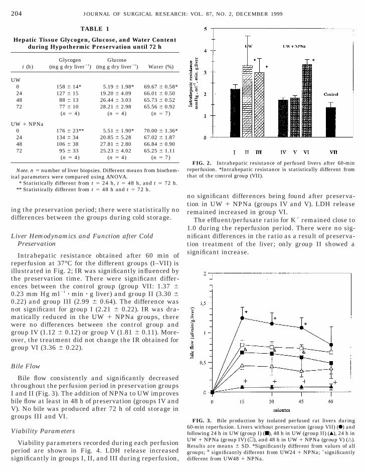
Intrahepatic resistance obtained after 60 min ofeperfusion at 37°C for the different groups (I–VII) isllustrated in Fig. 2; IR was significantly influenced byhe preservation time. There were significant differ-nces between the control group (group VII: 1.37 6.23 mm Hg ml21 z min z g liver) and group II (3.30 6.22) and group III (2.99 6 0.64). The difference wasot significant for group I (2.21 6 0.22). IR was dra-atically reduced in the UW 1 NPNa groups, thereere no differences between the control group androup IV (1.12 6 0.12) or group V (1.81 6 0.11). More-ver, the treatment did not change the IR obtained forroup VI (3.36 6 0.22).
ile Flow
Bile flow consistently and significantly decreasedhroughout the perfusion period in preservation groupsand II (Fig. 3). The addition of NPNa to UW improvesile flow at least in 48 h of preservation (groups IV and). No bile was produced after 72 h of cold storage inroups III and VI.
iability Parameters
TABLE 1
Hepatic Tissue Glycogen, Glucose, and Water Contentduring Hypothermic Preservation until 72 h
t (h)Glycogen
(mg g dry liver21)Glucose
(mg g dry liver21) Water (%)
W0 158 6 14* 5.19 6 1.98* 69.67 6 0.58*24 127 6 15 19.20 6 4.09 66.01 6 0.5048 88 6 13 26.44 6 3.03 65.73 6 0.5272 77 6 10 28.21 6 2.98 65.56 6 0.92
(n 5 4) (n 5 4) (n 5 7)
W 1 NPNa0 176 6 23** 5.51 6 1.90* 70.00 6 1.36*24 134 6 34 20.85 6 5.28 67.02 6 1.8748 106 6 38 27.81 6 2.80 66.84 6 0.9072 95 6 33 25.23 6 4.02 65.25 6 1.11
(n 5 4) (n 5 4) (n 5 7)
Note. n 5 number of liver biopsies. Different means from biochem-cal parameters were compared using ANOVA.
* Statistically different from t 5 24 h, t 5 48 h, and t 5 72 h.** Statistically different from t 5 48 h and t 5 72 h.
04 JOURNAL OF SURGICAL RESEARC
Viability parameters recorded during each perfusioneriod are shown in Fig. 4. LDH release increasedignificantly in groups I, II, and III during reperfusion,
fURgd
o significant differences being found after preserva-ion in UW 1 NPNa (groups IV and V). LDH releaseemained increased in group VI.The effluent/perfusate ratio for K1 remained close to
.0 during the reperfusion period. There were no sig-ificant differences in the ratio as a result of preserva-ion treatment of the liver; only group II showed aignificant increase.
FIG. 2. Intrahepatic resistance of perfused livers after 60-mineperfusion. *Intrahepatic resistance is statistically different fromhat of the control group (VII).
FIG. 3. Bile production by isolated perfused rat livers during0-min reperfusion. Livers without preservation (group VII) (F) and
VOL. 87, NO. 2, DECEMBER 1999
ollowing 24 h in UW (group I) (■), 48 h in UW (group II) (Œ), 24 h inW 1 NPNa (group IV) (h), and 48 h in UW 1 NPNa (group V) (‚).esults are means 6 SD. *Significantly different from values of allroups; & significantly different from UW24 1 NPNa; 1significantlyifferent from UW48 1 NPNa.

H
Uaaross
NvltsrTw
N
at
ooruppaa
ehpasnoflrswpAshamwicdcejvnfitlc4aastpgip
v
pp[
US
istological Assessment
The histological study of livers preserved for 48 h inW solution and reperfused showed hepatocytesround the central veins and between the centrilobularnd portal areas with cytoplasmic vacuoles, while pe-iportal areas remained normal (Fig. 5). Blebs are seenn the hepatocyte cell membrane. Most endothelialwollen cells or at least some of them can be seen in theinusoidal lumen; lobule architecture is conserved.The 48-h-preserved livers in UW containing 500 mMPNa after reperfusion, showed hepatocytes with no
acuolation within the entire parenchyma. In someivers vacuoles appeared but only in a few areas aroundhe central vein. Livers perfused after 72 h of coldtorage in UW with or without NPNa showed dis-upted hepatocyte cords and little cell to cell contact.he general architecture of the hepatic parenchymaas lost (picture not shown).
O Production in UW during Cold Storage
FIG. 4. Liver viability parameters during isolated rat liver ex-eriments in the seven groups studied: (A) LDH release after 60-minerfusion and (B) potassium release expressed as [K1]effluent 4K1]perfusate. *Significantly different from control group.
RODRIGUEZ ET AL.: SODIUM NITROPR
The time course of NO22 concentration in UW stored
t 4°C is shown in Fig. 6. We assayed three concentra-ions of NPNa: 100, 250, and 500 mM. A reaction time
((am
f 4 h was more than sufficient to allow the appearancef NO2
2 in the UW 1 500 mM NPNa. In all cases, NPNaeleased small amounts of NO, which was chemicallynstable and was oxidized to NO2
2 [19]. The slow rateroduction of NO2
2 obtained under our conditions isrobably due to: (a) low temperature of incubation 4°C,nd (b) low concentration of O2 in UW (see Materialnd Methods).
DISCUSSION
The present study shows that the vasodilator NPNaxerts a protective effect on liver function after 48 h ofypothermic preservation. Cold storage of the liver waserformed in UW at 4°C and hepatic hemodynamicsnd function were studied immediately after reperfu-ion in the isolated perfusion organ system. This tech-ique was applied to control and preserved livers, withr without NPNa addition. We have chosen constant-ow perfusion because, with this system, intrahepaticesistance changes are evaluated by measuring pres-ure changes induced by the treatment. In this way,ith this model it is possible to appreciate how coldreservation affects liver hemodynamics and function.s soon as the liver is removed and harvested in coldtorage several changes start to occur. After 24 h ofypothermic preservation, it is possible to appreciaten increment on intrahepatic resistance (60%) and di-inished bile flow (40%) (see Figs. 2 and 3). However,hen storage was prolonged to 48 and 72 h, significant
mpairment of liver function occurred. IR was signifi-antly increased, and bile flow was significantly re-uced at 48 h and was maximally reduced after 72 h ofold storage. LDH release increased in relation to pres-rvation time. Histological examination showed in-ured cells around the central vein, with cytoplasmicacuoles and detached endothelial cells inside the si-usoidal lumen as was described [23]. An interestingnding of this investigation is that 500 mM NPNa ishe concentration that achieves a real improvement iniver hemodynamics after cold storage: IR was signifi-antly reduced and LDH release was low, and after8 h bile production appeared to improve with theddition of NPNa. Finally, as indicated by histologicalnalysis, the liver parenchyma appeared well pre-erved with no vacuolate cells and conserved architec-ure. Because there were no changes in biochemicalarameters during cold storage (assessed by glucose,lycogen, and water content) we may conclude that themprovement in liver function is due to vascular effectsroduced by NPNa.The cold ischemia of the liver produces intrahepatic
asoconstriction due to (1) the low temperature used
205SIDE AND RAT LIVER PRESERVATION
4°C) and (2) the high potassium concentration of UW125 mM), and this vasoconstrictive effect may persistfter reperfusion. In preliminary experiments, the ad-inistration of NPNa during reperfusion of 48-h-cold-

tE53
FIG. 5. Histological features of 48-h-preserved grafts after 60 min of reperfusion. (A) Grafts preserved 48 h in UW. Hepatocytes aroundhe hepatic vein and between the centrilobular and portal areas show numerous cytoplasmic vacuoles (arrows) and blebs (not shown).ndothelial cells are swollen and some of them can be seen in the sinusoidal lumen (arrowheads). (B) Grafts preserved 48 h in UW containing00 mM NPNa. Hepatocytes appear intact and rounded within the entire parenchyma. Vacuoles are not observed. Original magnification,303. H, hepatic vein; P, portal area.

sncd
coadtmltkamsast
lrni
tts
accoItpmbicsflsmt
PSGMac
umpNm
US
tored livers was not successful in improving hemody-amics (data not shown). Therefore, other mechanismsould account for the intrahepatic vasoconstriction pro-uced during cold ischemia of the liver.Stanby et al. [24] showed that endothelin-1 (ET-1)
ould be released by the hepatic vasculature duringrgan harvest and cold preservation. These studieslso demonstrated that ET-1 appears to be releaseduring the ischemic period itself and accumulates inhe vascular spaces of the liver. Furthermore, Podka-eni et al. [25] showed that ET-1 and interleukin-1
eakage occurs during cold preservation and is propor-ional to the storage time. In addition, ET-1 is alsonown to have localized hemodynamic effects, and itsccumulation during cold storage may alter the hepaticicrocirculation, causing disturbances in tissue perfu-
ion, and could enhance reperfusion damage. Zhang etl. [26] provided evidence that endothelins act as apecific sinusoidal constrictor in intact rat liver, andhis effect can be inhibited by the NO donor NPNa.
Other authors [27] using the isolated perfused rativer, have shown that both ET-1 and ET-3 increaseesistance to flow through the portal vascular bed oformal livers and that the vasoconstrictor action of ET
s effectively reversed by NPNa.Our data suggest that the improvement in liver func-
FIG. 6. Time course of NO22 concentration in UW stored at 4°C
p to 72 h. Open bars: UW 1 100 mM NPNa; shaded bars: UW 1 250M NPNa; black bars: 500 mM NPNa. Aliquots (200 ml) of thereservation solution were removed at fixed times and assayed forO2
2 (see Materials and Methods for details). Values representeans 6 SD of duplicate determinations from three experiments.
RODRIGUEZ ET AL.: SODIUM NITROPR
ion seen experimentally using NPNa during preserva-ion may be effected by overcoming localized vasocon-trictor effects such as would be produced by ET-1.On the other hand, we have demonstrated that the
1
ddition of 500 mM NPNa to UW produces a slow butonstant release of NO assessed by the increase in NO2
2
oncentration. This might be the mechanism related tor involved with the improvement of hepatic function.rrespective of these results there remain some ques-ions without clear answers, e.g., Why was there norotection at 72 h when NO2
2 concentration reached aaximum. We speculate that there is a compromise
etween the rate of NO production and the capacity ofnhibition of ET during cold storage. One should beautious in extrapolating these results to the clinicalituation, because reperfusion in our study was per-ormed with bloodless solution and a possible impact ofeukocyte–endothelium interaction on intrahepatic re-istance was not evaluated. This liver preservationethod should now be further investigated using a
ransplantation model in animals.
ACKNOWLEDGMENTS
This study was supported in part by National Grant CONICET,ICT 0482, P.BID 802/OC-AR, and a Grant from the Argentineociety of Gastroenterology (SAGE). We gratefully acknowledge Lic.raciela Furno’s invaluable help (Department of Statistics andathematics) for providing much needed consultation in statistical
nalysis. Special thanks are due Lic. Alejandra Martinez for techni-al assistance with histological studies.
REFERENCES
1. Belzer, F. O., and Southard, J. H. Principles of solid organpreservation by cold storage. Transplantation 45: 673, 1988.
2. Adam, R., Bismuth, H., and Diamond, T. Effect of extended coldischemia with UW solution on graft function after liver trans-plantation. Lancet 340: 1373, 1992.
3. Ploeg, R. I., D’Alessandro, A. M., Knichte, S. J., Stegall, M. D.,Pirsch, J. D., Hoffman, R. M., and Sasaki, T. Risk factors forprimary dysfunction after liver transplantation: A multivariateanalysis. Transplantation 55: 807, 1993.
4. Churchill, T. A., Green, C. J., and Fuller, B. J. Protective prop-erties of amino acids in liver preservation: Effects of glycine anda combination of amino acids on anaerobic metabolism andenergetics. J. Hepatol. 23: 720, 1995.
5. Jacobsson, J., Sundberg, R., Valdivia, L. A., and Starzl, T. E.Liver preservation with Lidoflazine and the University of Wis-consin solution: A dose-finding study. Transplantation 56: 472,1993.
6. Chazouilleres, O., Ballet, F., Chretien, Y., Marteau, P., Rey, C.,Maillard, D., and Poupon, R. Protective effects of vasodilatorson liver function after long hypothermic preservation: A studyin the isolated perfused rat liver. Hepatology 9: 824, 1989.
7. Sherman, I. A., Saibil, F. G., and Janossy, T. I.g-Hydroxyubutyrate mediated protection of liver function afterlong-term hypothermic storage. Transplantation 57: 8, 1994.
8. Kubes, P., and Granger, D. N. Nitric oxide modulates microvas-cular permeability. Am. J. Physiol. 262: H611, 1992.
9. Kubes, P., Suzuki, M., and Granger, D. N. Nitric oxide: Anendogenous modulator of leukocyte adhesion. Proc. Natl. Acad.
207SIDE AND RAT LIVER PRESERVATION
Sci. USA 88: 323, 1991.0. Moncada, S., Palmer, R. M. J., and Higgs, E. A. Nitric oxide:
Physiology, pathophysiology, and pharmacology. Pharmacol.Rev. 43: 109, 1991.

1
1
1
1
1
1
1
1
1
2
2
2
2
2
2
2
2 H:
1. Pinsky, D. J., Oz, M. C., and Koga, S. Cardiac preservation isenhanced in a heterotopic rat transplant model by supplement-ing the nitric oxide pathway. J. Clin. Invest. 93: 2291, 1994.
2. Feelisch, M., and Noack, E. Correlation between nitric oxideformation during degeneration of organic nitrate and activationof guanylate cyclase. Eur. J. Pharmacol. 139: 19, 1987.
3. Fullerton, D. V., Mitchell, M. V., and McIntyre, R. C. Coldischemia and reperfusion each produce pulmonary vasomotordysfunction in the transplanted lung. J. Thorac. Cardiovasc.Surg. 106: 1213, 1993.
4. Mamprin, M. E., Rodriguez, J. V., and Guibert, E. E. Theimportance of pH in resuspension media on viability of hepato-cytes preserved in University of Wisconsin solution. Cell Trans-plant. 4: 269, 1995.
5. Chazouillieres, O., Calmus, Y., Vaubordolle, M., and Ballet, F.Preservation-induced liver injury: Clinical aspects, mechanismand therapeutic approaches. J. Hepatol. 18: 123, 1993.
6. Miller, L. L. Technique of isolated rat liver perfusion. In Iso-lated Liver Perfusion and Its Applications. I. Bartosek, A. Gua-tain, and L. L. Miller, Eds.), Raven Press, New York, 1973. Pp.11–52.
7. Alexander, B., Aslarn, M., and Benjamin, I. S. Hepatic functionduring prolonged isolated rat liver perfusion using a new min-iaturized perfusion circuit. J. Pharmacol. Toxicol. Methods 34:203, 1995.
8. Grossman, H. J., and Bathal, P. S. Application of the isolatedperfused rat liver preparation to pharmacological studies of the
08 JOURNAL OF SURGICAL RESEARC
intrahepatic portal vascular bed. Methods Find. Exp. Clin.Pharmacol. 6: 33, 1984.
9. Ignarro, L. J., Fukuto, J. M., Griscavage, J. M., Rogus, N. E.,and Byrns, R. E. Oxidation of nitric oxide in aqueous solution to
2
nitrite but not nitrate: Comparison with enzymatically formednitric oxide from L-arginine. Proc. Natl. Acad. Sci. USA 90:8103, 1993.
0. Bergmeyer, H. U., and Bernt, E. Lactic dehydrogenase UVassay with pyruvate and NADH. In H. U. Bergmeyer (Ed.),Methods of Enzymatic Analysis, Vol. 2 San Diego: AcademicPress, 1974. Pp. 574–579.
1. Carr, R. S., and Neff, J. M. Quantitative semi-automated enzy-matic assay for tissue glycogen. Comp. Biochem. Physiol. 77:447, 1984.
2. Hollander, M., and Wolfe, D. Nonparametric Statistical Meth-ods. New York: Wiley, 1972. P. 124.
3. Caldwell-Kenkel, J. C., Currin, R. T., Tanaka, Y., Thurman,R. G., and LeMasters, J. J. Reperfusion injury to endothelialcells following cold schemic storage of rats livers. Hepatology10: 292, 1989.
4. Stanby, G., Fuller, B., Jeremy, J., Cheetam, K., and Rolleres, K.Endothelin release: A facet of reperfusion injury in clinical livertransplantation. Transplantation 56: 239, 1993.
5. Podkameni, D., Van Ness, K., Schwartz, M., Boros, P., andMiller, C. Release of endothelin-1 and IL-1 from cold stored ratlivers is proportional to preservation time. Hepatology 20(Pt. 2):192A, 1994.
6. Zhang, J. H., Pegoli, W., and Clemens, M. G. Endothelin-1induces direct constriction of hepatic sinusoids. Am. J. Physiol.266: G624, 1994.
VOL. 87, NO. 2, DECEMBER 1999
7. Elliot, A. J., Vo, L. T., Grossman, H. J., Grossman, V. L., andBathal, P. S. Endothelin-induced vasoconstriction in isolatedperfused preparations from normal and cirrhotic rats. Hepatol-ogy 20: 100A, 1994.