Quality of life among patients undergoing transcatheter aortic valve implantation
Upload
independentCategory
view
5download
0
RNA-based gene therapy for HIV with lentiviral vector-modifiedCD34(+) cells in patients undergoing transplantation for AIDS-related lymphoma
David L. DiGiusto1, Amrita Krishnan1, Lijing Li1, Haitang Li3, Shirley Li2, Anitha Rao1, ShuMi5, Priscilla Yam2, Sherri Stinson6, Michael Kalos7, Joseph Alvarnas1, Simon F. Lacey5,Jiing-Kuan Yee2, Mingjie Li9, Larry Couture2,4, David Hsu4, Stephen J. Forman1, John J.Rossi3, and John A. Zaia2
1 Department of Hematology/Hematopoietic Cell Transplantation, Duarte, CA2 Department of Virology, Duarte, CA3 Department of Molecular Biology, Duarte, CA4 Center for Applied Technology Development, Duarte, CA5 Clinical Immunobiology Correlative Studies, Duarte, CA6 GCRC, City of Hope, Duarte, CA7 Department of Pathology and Laboratory Medicine, University of Pennsylvania School ofMedicine9 Dept. of Neurology, Washington University School of Medicine, St Louis, MO
AbstractAIDS patients undergoing autologous transplantation for lymphoma were treated with gene-modified peripheral blood derived (CD34+) hematopoietic progenitor cells (HPC) expressing 3RNA-based anti-HIV moieties (Tat/Rev shRNA, TAR decoy and CCR5 ribozyme). In vitroanalysis of gene-modified HPC showed no differences in the hematopoietic potential comparedwith non-transduced cells. In vitro estimates of gene marking were as high as 22% but declined to~1% over 4 weeks of culture. Ethical study design required that patients were transplanted withboth gene modified and unmanipulated hematopoietic progenitor cell apheresis products (HPC-A).All 4 infused patients engrafted (ANC>500) by day 11 post-infusion and showed no unexpectedinfusion related toxicities. Persistent vector marking in multiple cell lineages has been observed atlow levels for up to 24 months as has expression of siRNA and ribozyme. This is the firstdemonstration of siRNA expression in human blood cells following transplantation of autologousgene-modified CD34+ HPC. These results support the development of an RNA-based cell therapyplatform for HIV.
Author Contributions: DD and AK contributed equally to this study. DD developed and supervised cell manufacturing operations andin vitro correlative studies, interpreted data and wrote the manuscript. AK was responsible for trial design, patient recruiting,enrollment and treatment on trial and wrote the manuscript. LL, AR and PY performed manufacturing of cell products and performedin vitro correlative assays. SL performed sample processing and distribution. HL performed RNA analysis on in vitro and in vivosamples. SS performed manufacturing operations. JKY conceived of and developed vector production system, LC and DHmanufactured lentiviral vectors, JJR conceived and designed the triple gene vector, supervised the development of the siRNA andCCR5 ribozyme assays from clinical samples and wrote the manuscript. ML constructed the clinical vector and assisted in optimizingvector production strategies. MK supervised the development and qualification of the in vivo gene marking assays and product releasetesting, SM performed the in vivo marking assays, SFL supervised in vivo gene marking assays, SJF conceptualized and designedclinical studies. JAZ coordinated the clinical, laboratory, and regulatory processes, interpreted data and wrote the manuscript.
NIH Public AccessAuthor ManuscriptSci Transl Med. Author manuscript; available in PMC 2011 July 6.
Published in final edited form as:Sci Transl Med. 2010 June 16; 2(36): 36ra43. doi:10.1126/scitranslmed.3000931.
NIH
-PA Author Manuscript
NIH
-PA Author Manuscript
NIH
-PA Author Manuscript
Summary—Stem cell gene therapy for HIV results in sustained RNA expression in the blood ofpatients for up to 2 years following transplant.
IntroductionHighly active antiretroviral therapy (HAART) has dramatically improved survival ofpatients with HIV infection, but it is likely never to be curative(1). Although complianceproblems has been improved with new multiple-drug formulations, virus replicationcontinues, and the risk of development of antiviral resistance remains a concern.Importantly, medications represent up to 84% of AIDS-related healthcare costs(2), thus,development of genetic therapies could decrease the need for continuous medication and itsattendant cost. An approach which would allow a single genetic manipulation to delay orprevent the progression of HIV infection to AIDS remains an important goal.
Since it was first proposed that gene transfer might “immunize” against intracellularinfection(3), there has been significant investigation into the use of genetic medicine to treatHIV. Given the difficulties, risks and failures associated with human gene therapy, it hasremained unclear how a single gene manipulation could have a lasting, significant impact inthis disease setting. Recently however, the transplantation of allogeneic hematopoietic stemcells (HSC) with a HIV-resistant genotype based on a naturally occuring 32-bp deletion inthe chemokine receptor 5 gene (Δ32CCR5), used in conjunction with myeloablative therapyfor leukemia, resulted in apparent elimination of HIV in the recipient(4). This genetic “test-of-concept” supports the idea that replacing a susceptible immune system with a geneticallymodified, virus-resistant one would likely result in reduced viral load and perhaps preventprogression to AIDS. However, the frequency and logistics of finding matched allogeneichomozygous Δ32CCR5 donors precludes widespread use of this strategy. Alternatively, ifex-vivo genetic modification of autologous HSC rendered their progeny HIV resistant,transplantation of these cells could potentially be therapeutic.
Multiple anti-viral strategies have been proposed for AIDS gene therapy including bothprotein(5–11) and RNA-based approaches(12–16). Several approaches have been tested inwhich autologous T-cells or CD34+ cells were transduced with a retroviral vector constructencoding either a surface fusion peptide(17), a mutant Rev molecule(18, 19), or a ribozymetargeting vpr/tat(20) or tat/rev(21) and then infused into HIV individuals. All were shown tobe safe and well tolerated, but, resulted in very low level of long-term genetic marking ofperipheral blood cells and variable to no detectable expression of the therapeutic transcriptin the peripheral blood.
Based on our experience with HSC transplantation of AIDS lymphoma patients(22), thispopulation appears to offer a unique opportunity to evaluate RNA-based anti-HIV strategiesin an ethically acceptable clinical setting including marrow ablation. Long-term expressionof the RNA transgenes will be necessary for success of this procedure, and thus, geneintegration will be a requirement in the progeny cells if the therapeutic effect is to besustained. In this regard, lentiviral vectors have been promoted as an ideal gene-deliverysystem since they have been reported to integrate into non-dividing cells and do notpreferentially locate near gene promoters(23–25). We describe here a clinical trial in whichautologous hematopoietic progenitor cells (HPCs) are programmed with an expressed shorthairpin RNA (shRNA) in combination with two novel forms of HIV-specific RNA-basedinhibitors (a nucleolar localizing RNA decoy and hammerhead ribozyme) (26). We show forthe first time long-term expression of an ectopically expressed siRNA and ribozyme in themultiple peripheral blood cells lineages of patients transplanted with gene modifiedprogenitor cells
DiGiusto et al. Page 2
Sci Transl Med. Author manuscript; available in PMC 2011 July 6.
NIH
-PA Author Manuscript
NIH
-PA Author Manuscript
NIH
-PA Author Manuscript
ResultsParticipants Enrolled
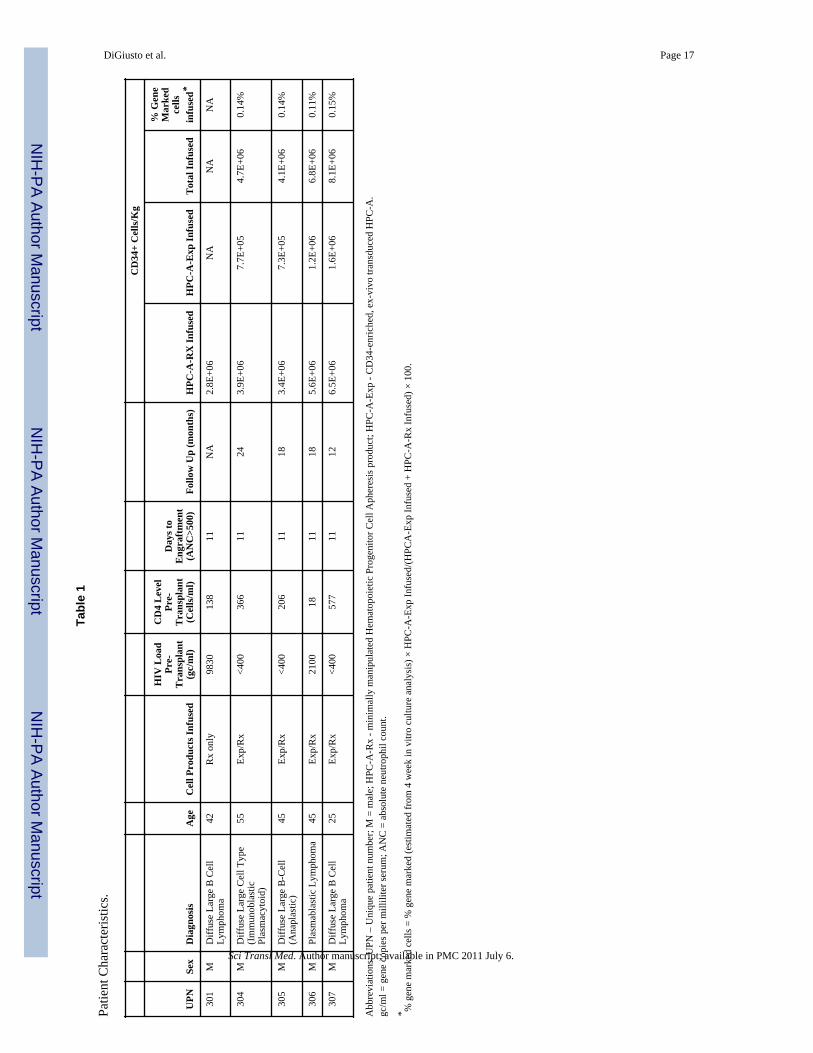
Seven patients were consented to the study and treated according to the schema shown inFigure 1. All patients had non-Hodgkin lymphoma (NHL), two had relapsed disease and twowere induction failures, two in first partial remission and one in high risk first remission.Secondary exclusion criteria required that patients could mobilize sufficient HPCs forcollection by apheresis (HPC-A) of two cell products of at least 2.5 × 10⁶ CD34+ cells/kgeach; one a therapeutic product that was otherwise unmanipulated (HPC-A-Rx) and theother an experimental research product for genetic transduction (HPC-A-Exp). Two patientswere removed from the study prior to HPC collection due to either progressive NHL(UPN0302) or to inadequate HPC-A collections (UPN0303). The remaining five patientsmobilized an adequate number of cells for the investigational portion of the trial (Table 1).One of these (UPN 301), however, received only HPC-A-Rx because the HPC-A-Exp failedto pass the viability release test. The 4 patients that underwent transplantation with bothtransduced and untransduced cell products were a median age of 42.5 years (range 25–55),had CD4 counts at study entry ranging from 18–577 cell/μl, and a viral load ranging frombelow the limit of detection − 25,000 copies/ml. Following transplantation, HIV RNA inplasma and CD4 counts were followed at monthly intervals (Table S1). All patients weremaintained on HAART and HIV loads were undetectable (<50 gc/ml) except for patientUPN0306 who had a viral load of 270 copies/ml at the time of transplant (Day 0) and patientUPN0304 who temporarily discontinued HAART therapy between 12 and 15 months posttransplant resulting in a transient viral load of 890 copies/ml at 15 months after which heresumed prior anti-retroviral dosing.
CD34 cell selection and lentiviral transductionCD34-selection resulted in products highly enriched (Avg. = 121-fold, range 63–205) forCD34+ cell frequency (N=5). Starting with an average of 312 × 106 CD34+ cells (range122–618 ×106), 151 × 106 CD34+ cells (range 47–317 × 106) were recovered (48% averageyield) (Figure S1). CD34-selected cells were cryopreserved in the vapor phase of liquidnitrogen until 3 days prior to infusion at which time they were thawed and transduced.
In vitro analysis of Transduced HPC-A ProductsIn vitro analysis was conducted to determine the effects of transduction on hematopoieticpotential and extent of gene marking of cells. In methyl cellulose culture, the frequency ofcolony forming units for transduced and non-transduced control cells, as a measure oftoxicity of the transduction process, showed no significant differences in either thefrequency or types of colonies formed (Figure S2). Cells were also placed in liquid culturewith a cytokine cocktail designed to promote myelo/erythropoiesis or on a murine stromalcell line previously demonstrated to support the growth of B-lymphoid cells(26, 27).Samples were taken at weekly intervals for phenotypic analysis of myeloid, erythroid, B-lymphocytes and CD34+ stem/progenitor cells. No differences in the kinetics or magnitudeof lineage development were observed between the transduced and non-transduced cellcultures (Figure S3). Taken together, these results support the overall safety of the celltransduction process with respect to in vitro hematopoietic potential.
Cells from these cultures were also analyzed weekly for the presence of the rHIV7-shITAR-CCR5RZ transgene. Quantitative PCR analysis was performed using primers specific for theWPRE-marker of the transgene and for apolipoprotein B (ApoB), a single copyhousekeeping gene to normalize for cell number. By calculating the ratio of copies ofWPRE:ApoB, we observed gene marking levels as high as 23% (Avg. = 18% range 5–23%,N=5) after one week of culture (Figure 2a). However, the percentage of gene-marked cells
DiGiusto et al. Page 3
Sci Transl Med. Author manuscript; available in PMC 2011 July 6.
NIH
-PA Author Manuscript
NIH
-PA Author Manuscript
NIH
-PA Author Manuscript
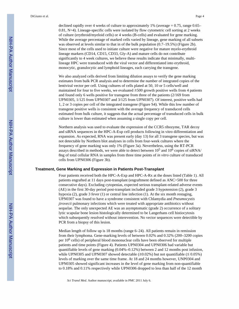
declined rapidly over 4 weeks of culture to approximately 1% (average = 0.75, range 0.65–0.81, N=4). Lineage-specific cells were isolated by flow cytometric cell sorting at 2 weeksof culture (erythroid/myeloid cells) or 4 weeks (B-cells) and evaluated for gene marking.While the average percentage of marked cells varied by lineage, gene marking of all subsetswas observed at levels similar to that in of the bulk population (0.7–19.5%) (Figure 2b).Since most of the cells used to initiate culture were negative for mature myelo-erythroidlineage markers (CD14, CD15, CD33, Gly-A) and mature cells do not contributesignificantly to 4 week cultures, we believe these results indicate that minimally, multi-lineage HPC were transduced with the viral vector and differentiated into erythroid,monocytic, granulocytic and lymphoid lineages, each carrying the transgene.
We also analyzed cells derived from limiting dilution assays to verify the gene markingestimates from bulk PCR analysis and to determine the number of integrated copies of thelentiviral vector per cell. Using cultures of cells plated at 50, 10 or 5 cells/well andmaintained for four to five weeks, we evaluated ≥500 growth positive wells from 4 patientsand found only 6 wells positive for transgene from three of the patients (2/169 fromUPN0305, 1/125 from UPN0307 and 3/125 from UPN0307). Of interest, positive wells had1, 2 or 3 copies per cell of the integrated transgene (Figure S4). While this low number oftransgene positive wells is consistent with the average frequency of transduced cellsestimated from bulk culture, it suggests that the actual percentage of transduced cells in bulkculture is lower than estimated when assuming a single copy per cell.
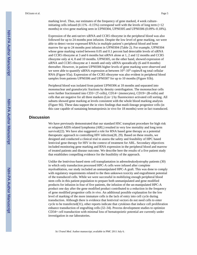
Northern analysis was used to evaluate the expression of the CCR5 ribozyme, TAR decoyand siRNA sequences in the HPC-A-Exp cell products following in vitro differentiation andexpansion. As expected, RNA was present early (day 13) for all 3 transgene species, but wasnot detectable by Northern blot analyses in cells from four-week cultures where thefrequency of gene marking was only 1% (Figure 3a). Nevertheless, using the RT-PCRassays described in methods, we were able to detect between 105 and 106 copies of siRNA/8ng of total cellular RNA in samples from three time points of in vitro culture of transducedcells from UPN0306 (Figure 3b).
Treatment, Gene Marking and Expression in Patients Post-TransplantFour patients received both the HPC-A-Exp and HPC-A-Rx at the doses listed (Table 1). Allpatients engrafted at 11 days post-transplant (engraftment defined as ANC>500 for threeconsecutive days). Excluding cytopenias, expected serious transplant-related adverse events(AE) in the first 30-day period post-transplant included grade 3 hypotension (2), grade 3hypoxia (2), grade 3 fever (1) or central line infection (1). At the six month restaging,UPN0307 was found to have a syndrome consistent with Chlamydia and Pneumocystisjirovecii pulmonary infections which were treated with appropriate antibiotics withoutsequelae. The only unexpected AE was an asymptomatic (grade 2) occurrence of a solitarylytic scapular bone lesion histologically determined to be Langerhans cell histiocytosiswhich subsequently resolved without interevention. No vector sequences were detectible byPCR from a biopsy of this lesion.
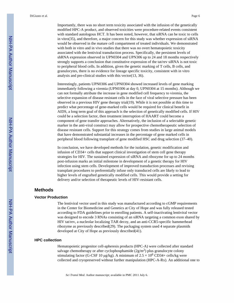
Median length of follow up is 18 months (range 6–24). All patients remain in remissionfrom their lymphoma. Gene-marking levels of between 0.02% and 0.32% (200–3200 copiesper 106 cells) of peripheral blood mononuclear cells have been observed for multiplepatients and time points (Figure 4). Patients UPN0304 and UPN0306 had variable butquantifiable levels of gene marking (0.04%–0.12%) between 2 and 12 months post infusion,while UPN0305 and UPN0307 showed detectable (≥0.02%) but not quantifiable (≤ 0.05%)levels of marking over the same time frame. At 18 and 24 months however, UNP0304 andUPN0305 showed significant increases in the level of gene marking from non-quantifiableto 0.18% and 0.11% respectively while UPN0306 dropped to less than half of the 12 month
DiGiusto et al. Page 4
Sci Transl Med. Author manuscript; available in PMC 2011 July 6.
NIH
-PA Author Manuscript
NIH
-PA Author Manuscript
NIH
-PA Author Manuscript
marking level. Thus, our estimates of the frequency of gene marked, 4 week culture-initiating cells infused (0.11% –0.15%) correspond well with the levels of long term (>12months) in vivo gene marking seen in UPN0304, UPN0305 and UPN0306 (0.09%–0.18%).
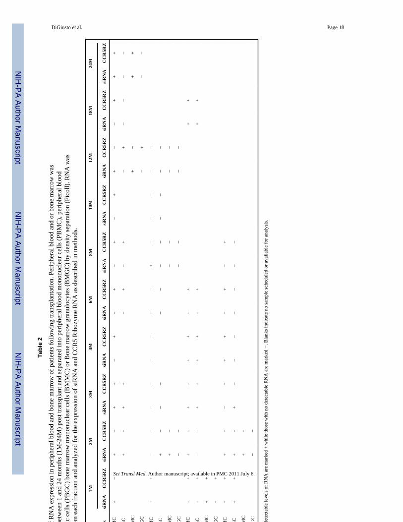
Expression of the anti-tat/rev siRNA and CCR5 ribozyme in the peripheral blood was alsofollowed for up to 24 months post infusion. Despite the low level of gene marking, we wereable to detect vector expressed RNAs in multiple patient’s peripheral blood and bonemarrow for up to 24 months post infusion in UPN0304 (Table 2). For example, UPN0304whose gene marking varied between 0.05 and 0.1 percent had detectable levels of siRNAand CCR5 ribozyme at 3 and 6 months but siRNA alone at 1, 2 and 12 months and CCR5ribozyme only at 4, 8 and 10 months. UPN0305, on the other hand, showed expression ofsiRNA and CCR5 ribozyme at 1 month and only siRNA sporadically (6 and 8 months)thereafter. However, in patient UPN0306 higher levels of gene marking were observed andwe were able to quantify siRNA expression at between 103–105 copies/8 ng total cellularRNA (Figure S5a). Expression of the CCR5 ribozyme was also evident in peripheral bloodsamples from patients UPN0306 and UPN0307 for up to 10 months (Figure S5b).
Peripheral blood was isolated from patient UPN0306 at 18 months and separated intomononuclear and granulocytic fractions by density centrifugation. The mononuclear cellswere further fractionated into CD3+ (T-cells), CD14+ (monocytes), CD19+ (B-cells) andcells that are negative for all three markers (Lin−) by fluorescence activated cell sorting. Allsubsets showed gene marking at levels consistent with the whole blood marking analysis(Figure S6). These data support the in vitro findings that mutli-lineage progenitor cells (inthis case capable of sustaining hematopoiesis in vivo for 18 months) were in fact transduced.
DiscussionWe have previously demonstrated that our standard HSC transplant procedure for high riskor relapsed AIDS related lymphoma (ARL) resulted in very low mortality and long termsurvival(22). We have also suggested a role for RNA based gene therapy as a potentialtherapeutic approach to controlling HIV infection(28, 29). Based on these results, wedesigned and conducted a clinical trial to assess the safety and feasibility of HPC basedlentiviral gene therapy for HIV in the context of treatment for ARL. Secondary objectivesincluded monitoring gene marking and RNA expression in the peripheral blood and marrowof treated patients and disease outcome. We describe here the results of a five patient studythat establishes compelling evidence for the feasibility of the approach.
Unlike the lentivirus-based stem cell transplantation in adrenoleukodystrophy patients (30)in which only transduction processed HPC-A cells were infused after completemyeloablation, our study included an unmanipulated HPC-A graft. This was done to complywith regulatory requirements related to the then unknown toxicity and engraftment potentialof the transduced cells. While we were successful in mobilizing enough peripheral bloodstem cells in this patient population to prepare both unmanipulated and gene modifiedproducts for infusion in four of five patients, the infusion of the un-manipulated HPC-Aproduct one day after the gene modified product contributed to a reduction in the frequencyof gene modified progenitor cells in vivo. An additional possible explanation for the lowlevel of marking of the more immature cells is the lack of entry into cell cycle duringtransduction. Although there is evidence that lentiviral vectors do not need cells to entercycle to be transfected(31), other reports indicate that cytokines that induce cell proliferationenhance transduction of engrafting cells (32–34). Process development studies to optimizeCD34+ cell transduction with minimal loss of hematopoietic potential are currently underinvestigation in our laboratories.
DiGiusto et al. Page 5
Sci Transl Med. Author manuscript; available in PMC 2011 July 6.
NIH
-PA Author Manuscript
NIH
-PA Author Manuscript
NIH
-PA Author Manuscript
Importantly, there was no short term toxicity associated with the infusion of the geneticallymodified HPC-A product, and observed toxicities were procedure-related events consistentwith standard autologous HCT. It has been noted, however, that siRNA can be toxic to cellsin vitro(35), and therefore, a major concern for this study was whether expression of siRNAwould be observed in the mature cell compartment of treated individuals. We demonstratedwith both in vitro and in vivo studies that there was no overt hematopoietic toxicityassociated with the lentiviral transduction process. Specifically, the persistent levels ofshRNA expression observed in UPN0304 and UPN306 up to 24 and 18 months respectivelystrongly supports a conclusion that constitutive expression of the tat/rev siRNA is not toxicto peripheral blood cells. In addition, given the genetic marking of T cells, B cells, andgranulocytes, there is no evidence for lineage specific toxicity, consistent with in vitroanalysis and pre-clinical studies with this vector(13, 36).
Interestingly, patients UPN0306 and UPN0304 showed increased levels of gene markingimmediately following a viremia (UPN0306 at day 0, UPN0304 at 15 months). Although wecan not formally attribute the increase in gene modified cell frequency to viremia, theselective expansion of disease resistant cells in the face of viral selective pressure has beenobserved in a previous HIV gene therapy trial(19). While it is not possible at this time topredict what percentage of gene-marked cells would be required for clinical benefit inAIDS, a long term goal of this approach is the selection of genetically modified cells. If HIVcould be a selection factor, then treatment interruption of HAART could become acomponent of gene transfer approaches. Alternatively, the inclusion of a selectable geneticmarker in the anti-viral construct may allow for prospective chemotherapeutic selection ofdisease resistant cells. Support for this strategy comes from studies in large animal modelsthat have demonstrated substantial increases in the percentage of gene marked cells inperipheral blood following transplant of gene modified HSC and drug selection (37–40).
In conclusion, we have developed methods for the isolation, genetic modification andinfusion of CD34+ cells that support clinical investigation of stem cell gene therapystrategies for HIV. The sustained expression of siRNA and ribozyme for up to 24 monthspost-infusion marks an initial milestone in development of a genetic therapy for HIVinfection using stem cells. Development of improved transduction processes and revisingtransplant procedures to preferentially infuse only transduced cells are likely to lead tohigher levels of engrafted genetically modified cells. This would provide a setting fordelivery and/or selection of therapeutic levels of HIV-resistant cells.
MethodsVector Production
The lentiviral vector used in this study was manufactured according to cGMP requirementsin the Center for Biomedicine and Genetics at City of Hope and was fully released testedaccording to FDA guidelines prior to enrolling patients. A self-inactivating lentiviral vectorwas designed to encode 3 RNAs consisting of an siRNA targeting a common exon shared byHIV tat/rev, a nucleolar localizing TAR decoy, and an anti-CCR5-specific hammerheadribozyme as previously described(29). The packaging system used 4 separate plasmidsdeveloped at City of Hope as previously described(41).
HPC collectionHematopoietic progenitor cell-apheresis products (HPC-A) were collected after standardsalvage chemotherapy or after cyclophosphamide (2g/m2) plus granulocyte colonystimulating factor (G-CSF 10 μg/kg). A minimum of 2.5 × 10⁶ CD34+ cells/kg werecollected and cryopreserved without further manipulation (HPC-A-Rx). An additional one to
DiGiusto et al. Page 6
Sci Transl Med. Author manuscript; available in PMC 2011 July 6.
NIH
-PA Author Manuscript
NIH
-PA Author Manuscript
NIH
-PA Author Manuscript
two apheresis collections were performed to collect cells for the genetic modification (HPC-A-Exp). CD34+ cells were enriched from the HPC-A-Exp collection over a CliniMACS™
device according to manufacturer’s instructions and cryopreserved using a control ratefreezer. CD34+ cells were thawed on day −2 before HCT and pre-stimulated for 16–20 hrsin X-vivo15 medium (Lonza, Walkersville, MD) containing 2mM L-Glutamine, 100ng/mLof Stem Cell Factor (SCF), 100ng/mL of Flt-3 ligand (Flt-3L), and 10ng/mL ofThrombopoietin (TPO) (CellGenix, Antioch, IL) at density of 2×10⁶ cells/ml. Pre-stimulatedcells were transduced with lentiviral vector (rHIV7-shI-TAR-CCR5RZ) at an MOI of 5 for16–24 hours in 75 cm2 tissue culture flasks flasks coated with 25μg/cm2 of fibronectin(Retronectin™, Takara Bio Inc. Shiga, Japan). A sample of CD34+ cells was incubated asdescribed but in the absence of lentiviral vector to serve as a transduction control.Transduced cells were pooled, washed with X-vivo15 medium and resuspended in finalformulation buffer (CliniMACs PBS/EDTA buffer with 0.5% HSA) at density of 1×10⁶
cells/mL for infusion as described below. A sample was taken from each product and testedfor total cell count, viability, sterility and endotoxin.
Transplant eligibility criteriaPatients with HIV and Non Hodgkins Lymphoma (NHL) in first CR with high or highintermediate IPI scores were eligible for transplant(42). All patients had to be oncombination antiretroviral therapy (ART) that did not include zidovudine, maintain an HIVviral load < 50,000 gc/ml, and be willing to suspend antiretroviral therapy during the periodof HPC mobilization and collection. The City of Hope’s Institutional Review Board andInstitutional Biosafety Committee approved the protocol, and informed written consent wasobtained from each patient in the presence of a patient advocate.
Transplantation ProcedureApproximately one week after completion of aphereses to collect HPC, patients wereadmitted to the transplant unit and received chemotherapy exactly as previouslydescribed(22). On the day of HCT (day 0), the transduced product (HPC-A-Exp) underwentrelease testing for endotoxin, sterility and viability and was then infused. On day +1, theunmanipulated product (HPC-A-Rx) was thawed at the bedside and infused as perguidelines approved by the Foundation for the Accreditation of Cellular Therapy (FACT).Safety was assessed for immediate effects of the treatment using the hematologic CommonTerminology Criteria for Adverse Events (CTCAE) version 3.0 grading system at 24 and 48hours and 14 days after infusion and at all subsequent visits from 1–18 months.
In Vitro Cell CultureMethylcellulose Cultures of CD34+ cells. Samples from each patient were assayed forhematopoietic potential using standard methylcellulose-based colony forming unit assaysand 4 week liquid and stromal cell cultures. The murine stromal cell line used in thesestudies to support the growth of B-lymphoid lineages from patient CD34+ cells has beenpreviously described(27). Limiting dilution assay. Limiting dilution analysis (LDA) of cellsfrom each patient was used to isolate the progeny of primitive, clonogenic progenitor cellsfor gene marking and copy number analysis.
Cell Phenotype and AnalysisAliquots of cells from bulk liquid and stromal culture were taken weekly for phenotypic andqPCR analysis. Antibodies to lineage specific cell surface antigens were used to followhematopoiesis during culture and isolate subpopulations by fluorescent activated cellsorting. Phenotypic data was collected on an FC500 flow cytometer (Beckman Coulter,Fullerton, CA) and analyzed with FCS express V3 software (De Novo Software, Ontario,
DiGiusto et al. Page 7
Sci Transl Med. Author manuscript; available in PMC 2011 July 6.
NIH
-PA Author Manuscript
NIH
-PA Author Manuscript
NIH
-PA Author Manuscript
Canada). Samples labeled as described were also sorted based on surface marker expressionto >98% purity using a MoFlo cell sorter (Beckman Coulter Corporation, Brea, California)for subsequent analysis of DNA marking and gene expression.
DNA Analysis of in vitro-derived cellsSamples from in vitro culture were analyzed weekly for the presence of integrated viralvector. Quantitative PCR (qPCR) was performed to detect the number of copies ofintegrated vector per cell. In each case (except for limiting dilution analysis) genomic DNAfrom approximately 20,000 cells was used in each reaction. For colonies derived from LDA,DNA from the entire colony was used. The number of copies of WPRE detected wasnormalized to cell number using qPCR for a housekeeping gene (apolipoprotein B). Resultsare reported as average percent gene marked cells or WPRE copies/100 cells. Results fromqPCR of individual colonies from LDA is reported as absolute copy number per cell.
RNA AnalysisRNA analysis was performed on gene modified products from in vitro culture and peripheralblood and bone marrow cells from patients at specific time points following transductionand infusion. Northern analysis was used to detect expression of all three RNA moietiesfrom in vitro cultures but was not sensitive enough to detect RNA in patient peripheral bloodsamples. More sensitive detection of expression of the CCR5RZ was accomplished by areal-time RT-PCR while expression of the tat/rev coding siRNA was analyzed by RT–PCRusing the TaqMan® MicroRNA Reverse Transcription kit. RT PCR products were analyzedby gel electrophoresis in a 1% agarose gel, blotting onto nitrocellulose and hybridizing withradio-labeled sequence specific probes.
Measurement of in vivo gene markingThe presence of shI-TAR-CCR5RZ-marked cells in peripheral blood and was also assessedby qPCR analysis of WPRE sequences using a fixed amount (50 ng) of genomic DNA fromperipheral blood. The average % WPRE+ DNA in samples was determined using a standardcurve generated from DNA isolated from a clone with a single copy integration of theWPRE-containing lentivirus and used in a standard curve to titrate DNA isolated frompatient specimens. A parallel set of qPCR amplifications was performed on all test andreference samples using primers specific for the p21 promoter as an internal control.
Supplementary MaterialRefer to Web version on PubMed Central for supplementary material.
AcknowledgmentsWe thanks Nancy Gonzalez and Amira Ahmed (Div. of Hematology) for Quality Systems Support, Lan-Feng Cao(CPDM Laboratory) for QC release testing, Guadalupe Duarte and Michelle Wardlow (Div. of Hematology) forpatient care. Cecilia Arbayo for assistance in patient consenting. Suenell Broyer and Rodica Stan (Center forApplied Technology Development) for assistance with the IND application and the other regulatory applicationsand Chy-Anh Tran and Lucy Brown for assistance with cell sorting. The authors are grateful to Alexandra Levinefor her advice and encouragement and to the staff at Benitec Ltd, especially Ken Reed and Sue MacLeman, forcontinuing support in the preclinical and clinical phases of this project.
Funding: The authors acknowledge support from NIH grants AI42552 and HL07470 (JJR), NIH P50 CA107399(Lymphoma SPORE), P30 CA33572-26 (CCSG) (SJF), NIH AI61839 (JAZ), NCRR S10RR025083-01 (DD), andGCRC grant M01 RR00043 (JAZ). A grant from Benitec Ltd supported vector manufacture, process development,and a portion of the clinical trial.
DiGiusto et al. Page 8
Sci Transl Med. Author manuscript; available in PMC 2011 July 6.
NIH
-PA Author Manuscript
NIH
-PA Author Manuscript
NIH
-PA Author Manuscript
References1. Richman DD, Margolis DM, Delaney M, Greene WC, Hazuda D, Pomerantz RJ. The challenge of
finding a cure for HIV infection. Science New York, NY. 2009; 323:1304–1307.2. Chen RY, Accortt NA, Westfall AO, Mugavero MJ, Raper JL, Cloud GA, Stone BK, Carter J, Call
S, Pisu M, Allison J, Saag MS. Distribution of health care expenditures for HIV-infected patients.Clin Infect Dis. 2006; 42:1003–1010. [PubMed: 16511767]
3. Baltimore D. Gene therapy. Intracellular immunization. Nature. 1988; 335:395–396. [PubMed:3166513]
4. Hutter G, Nowak D, Mossner M, Ganepola S, Mussig A, Allers K, Schneider T, Hofmann J,Kucherer C, Blau O, Blau IW, Hofmann WK, Thiel E. Long-term control of HIV by CCR5Delta32/Delta32 stem-cell transplantation. N Engl J Med. 2009; 360:692–698. [PubMed: 19213682]
5. Bevec D, Dobrovnik M, Hauber J, Bohnlein E. Inhibition of human immunodeficiency virus type 1replication in human T cells by retroviral-mediated gene transfer of a dominant-negative Revtransactivator. Proc Natl Acad Sci U S A. 1992; 89:9870–9874. [PubMed: 1409715]
6. Bonyhadi ML, Moss K, Voytovich A, Auten J, Kalfoglou C, Plavec I, Forestell S, Su L, Bohnlein E,Kaneshima H. RevM10-expressing T cells derived in vivo from transduced human hematopoieticstem-progenitor cells inhibit human immunodeficiency virus replication. Journal of virology. 1997;71:4707–4716. [PubMed: 9151864]
7. Hammer D, Wild J, Ludwig C, Asbach B, Notka F, Wagner R. Fusion of Epstein-Barr virus nuclearantigen-1-derived glycine-alanine repeat to trans-dominant HIV-1 Gag increases inhibitoryactivities and survival of transduced cells in vivo. Human gene therapy. 2008; 19:622–634.[PubMed: 18533892]
8. Perez EE, Riley JL, Carroll RG, von Laer D, June CH. Suppression of HIV-1 infection in primaryCD4 T cells transduced with a self-inactivating lentiviral vector encoding a membrane expressedgp41- derived fusion inhibitor. Clin Immunol. 2005; 115:26–32. [PubMed: 15870017]
9. Plavec I, Voytovich A, Moss K, Webster D, Hanley MB, Escaich S, Ho KE, Bohnlein E, DiGiustoDL. Sustained retroviral gene marking and expression in lymphoid and myeloid cells derived fromtransduced hematopoietic progenitor cells. Gene therapy. 1996; 3:717–724. [PubMed: 8854097]
10. Vallanti G, Lupo R, Federico M, Mavilio F, Bovolenta C. T Lymphocytes transduced with alentiviral vector expressing F12-Vif are protected from HIV-1 infection in an APOBEC3G-independent manner. Mol Ther. 2005; 12:697–706. [PubMed: 16039909]
11. van Griensven J, Zhan X, Van Maele B, Pluymers W, Michiels M, De Clercq E, Cherepanov P,Debyser Z. Expression of HIV-1 integrase in CEM cells inhibits HIV-1 replication. J Gene Med.2004; 6:268–277. [PubMed: 15026988]
12. Aagaard LA, Zhang J, von Eije KJ, Li H, Saetrom P, Amarzguioui M, Rossi JJ. Engineering andoptimization of the miR-106b cluster for ectopic expression of multiplexed anti-HIV RNAs. Genetherapy. 2008; 15:1536–1549. [PubMed: 18800151]
13. Anderson J, Li MJ, Palmer B, Remling L, Li S, Yam P, Yee JK, Rossi J, Zaia J, Akkina R. Safetyand Efficacy of a Lentiviral Vector Containing Three Anti-HIV Genes-CCR5 Ribozyme, Tat-revsiRNA, and TAR Decoy-in SCID-hu Mouse-Derived T Cells. Mol Ther. 2007; 15:1182–1188.[PubMed: 17406343]
14. Asparuhova MB, Barde I, Trono D, Schranz K, Schumperli D. Development and characterizationof a triple combination gene therapy vector inhibiting HIV-1 multiplication. J Gene Med. 2008;10:1059–1070. [PubMed: 18642399]
15. Rossi JJ, June CH, Kohn DB. Genetic therapies against HIV. Nat Biotechnol. 2007; 25:1444–1454.[PubMed: 18066041]
16. Yamamoto T, Miyoshi H, Yamamoto N, Yamamoto N, Inoue J, Tsunetsugu-Yokota Y. Lentivirusvectors expressing short hairpin RNAs against the U3-overlapping region of HIV nef inhibit HIVreplication and infectivity in primary macrophages. Blood. 2006; 108:3305–3312. [PubMed:16857988]
17. van Lunzen J, Glaunsinger T, Stahmer I, von Baehr V, Baum C, Schilz A, Kuehlcke K, NaundorfS, Martinius H, Hermann F, Giroglou T, Newrzela S, Muller I, Brauer F, Brandenburg G,Alexandrov A, von Laer D. Transfer of autologous gene-modified T cells in HIV-infected patients
DiGiusto et al. Page 9
Sci Transl Med. Author manuscript; available in PMC 2011 July 6.
NIH
-PA Author Manuscript
NIH
-PA Author Manuscript
NIH
-PA Author Manuscript
with advanced immunodeficiency and drug-resistant virus. Mol Ther. 2007; 15:1024–1033.[PubMed: 17356541]
18. Kang EM, de Witte M, Malech H, Morgan RA, Phang S, Carter C, Leitman SF, Childs R, BarrettAJ, Little R, Tisdale JF. Nonmyeloablative conditioning followed by transplantation of geneticallymodified HLA-matched peripheral blood progenitor cells for hematologic malignancies in patientswith acquired immunodeficiency syndrome. Blood. 2002; 99:698–701. [PubMed: 11781257]
19. Podsakoff GM, Engel BC, Carbonaro DA, Choi C, Smogorzewska EM, Bauer G, Selander D, CsikS, Wilson K, Betts MR, Koup RA, Nabel GJ, Bishop K, King S, Schmidt M, von Kalle C, ChurchJA, Kohn DB. Selective survival of peripheral blood lymphocytes in children with HIV-1following delivery of an anti-HIV gene to bone marrow CD34(+) cells. Mol Ther. 2005; 12:77–86.[PubMed: 15963923]
20. Amado RG, Mitsuyasu RT, Rosenblatt JD, Ngok FK, Bakker A, Cole S, Chorn N, Lin LS, BristolG, Boyd MP, MacPherson JL, Fanning GC, Todd AV, Ely JA, Zack JA, Symonds GP. Antihumanimmunodeficiency virus hematopoietic progenitor cell-delivered ribozyme in a phase I study:myeloid and lymphoid reconstitution in human immunodeficiency virus type-1-infected patients.Human gene therapy. 2004; 15:251–262. [PubMed: 15018734]
21. Mitsuyasu RT, Merigan TC, Carr A, Zack JA, Winters MA, Workman C, Bloch M, Lalezari J,Becker S, Thornton L, Akil B, Khanlou H, Finlayson R, McFarlane R, Smith DE, Garsia R, Ma D,Law M, Murray JM, von Kalle C, Ely JA, Patino SM, Knop AE, Wong P, Todd AV, Haughton M,Fuery C, Macpherson JL, Symonds GP, Evans LA, Pond SM, Cooper DA. Phase 2 gene therapytrial of an anti-HIV ribozyme in autologous CD34+ cells. Nat Med. 2009; 15:285–292. [PubMed:19219022]
22. Krishnan A, Molina A, Zaia J, Smith D, Vasquez D, Kogut N, Falk PM, Rosenthal J, Alvarnas J,Forman SJ. Durable remissions with autologous stem cell transplantation for high-risk HIV-associated lymphomas. Blood. 2005; 105:874–878. [PubMed: 15388574]
23. Ciuffi A, Mitchell RS, Hoffmann C, Leipzig J, Shinn P, Ecker JR, Bushman FD. Integration siteselection by HIV-based vectors in dividing and growth-arrested IMR-90 lung fibroblasts. MolTher. 2006; 13:366–373. [PubMed: 16325473]
24. Connolly JB. Lentiviruses in gene therapy clinical research. Gene therapy. 2002; 9:1730–1734.[PubMed: 12457288]
25. Wang GP, Levine BL, Binder GK, Berry CC, Malani N, McGarrity G, Tebas P, June CH,Bushman FD. Analysis of lentiviral vector integration in HIV+ study subjects receivingautologous infusions of gene modified CD4+ T cells. Mol Ther. 2009; 17:844–850. [PubMed:19259065]
26. Szilvassy SJ, Weller KP, Lin W, Sharma AK, Ho AS, Tsukamoto A, Hoffman R, Leiby KR,Gearing DP. Leukemia inhibitory factor upregulates cytokine expression by a murine stromal cellline enabling the maintenance of highly enriched competitive repopulating stem cells. Blood.1996; 87:4618–4628. [PubMed: 8639830]
27. DiGiusto D, Chen S, Combs J, Webb S, Namikawa R, Tsukamoto A, Chen BP, Galy AH. Humanfetal bone marrow early progenitors for T, B, and myeloid cells are found exclusively in thepopulation expressing high levels of CD34. Blood. 1994; 84:421–432. [PubMed: 7517715]
28. Li MJ, Bauer G, Michienzi A, Yee JK, Lee NS, Kim J, Li S, Castanotto D, Zaia J, Rossi JJ.Inhibition of HIV-1 infection by lentiviral vectors expressing Pol III-promoted anti-HIV RNAs.Mol Ther. 2003; 8:196–206. [PubMed: 12907142]
29. Li MJ, Kim J, Li S, Zaia J, Yee JK, Anderson J, Akkina R, Rossi JJ. Long-term inhibition ofHIV-1 infection in primary hematopoietic cells by lentiviral vector delivery of a triple combinationof anti-HIV shRNA, anti-CCR5 ribozyme, and a nucleolar-localizing TAR decoy. Mol Ther. 2005;12:900–909. [PubMed: 16115802]
30. Cartier N, Hacein-Bey-Abina S, Bartholomae CC, Veres G, Schmidt M, Kutschera I, Vidaud M,Abel U, Dal-Cortivo L, Caccavelli L, Mahlaoui N, Kiermer V, Mittelstaedt D, Bellesme C, LahlouN, Lefrere F, Blanche S, Audit M, Payen E, Leboulch P, l’Homme B, Bougneres P, Von Kalle C,Fischer A, Cavazzana-Calvo M, Aubourg P. Hematopoietic stem cell gene therapy with a lentiviralvector in X-linked adrenoleukodystrophy. Science (New York, NY. 2009; 326:818–823.
DiGiusto et al. Page 10
Sci Transl Med. Author manuscript; available in PMC 2011 July 6.
NIH
-PA Author Manuscript
NIH
-PA Author Manuscript
NIH
-PA Author Manuscript
31. Naldini L, Blomer U, Gallay P, Ory D, Mulligan R, Gage FH, Verma IM, Trono D. In vivo genedelivery and stable transduction of nondividing cells by a lentiviral vector. Science (New York,NY. 1996; 272:263–267.
32. Sutton RE, Reitsma MJ, Uchida N, Brown PO. Transduction of human progenitor hematopoieticstem cells by human immunodeficiency virus type 1-based vectors is cell cycle dependent. Journalof virology. 1999; 73:3649–3660. [PubMed: 10196257]
33. Verhoeyen E, Negre D, Cosset FL. Production of lentiviruses displaying ‘‘early-acting’’ cytokinesfor selective gene transfer into hematopoietic stem cells. Methods in molecular biology (Clifton,NJ. 2008; 434:99–112.
34. Zielske SP, Gerson SL. Cytokines, including stem cell factor alone, enhance lentiviral transductionin nondividing human LTCIC and NOD/SCID repopulating cells. Mol Ther. 2003; 7:325–333.[PubMed: 12668128]
35. An DS, Qin FX, Auyeung VC, Mao SH, Kung SK, Baltimore D, Chen IS. Optimization andfunctional effects of stable short hairpin RNA expression in primary human lymphocytes vialentiviral vectors. Mol Ther. 2006; 14:494–504. [PubMed: 16844419]
36. Banerjea A, Li MJ, Bauer G, Remling L, Lee NS, Rossi J, Akkina R. Inhibition of HIV-1 bylentiviral vector-transduced siRNAs in T lymphocytes differentiated in SCID-hu mice and CD34+progenitor cell-derived macrophages. Mol Ther. 2003; 8:62–71. [PubMed: 12842429]
37. Neff T, Beard BC, Kiem HP. Survival of the fittest: in vivo selection and stem cell gene therapy.Blood. 2006; 107:1751–1760. [PubMed: 16269617]
38. Persons DA, Allay JA, Bonifacino A, Lu T, Agricola B, Metzger ME, Donahue RE, Dunbar CE,Sorrentino BP. Transient in vivo selection of transduced peripheral blood cells using antifolatedrug selection in rhesus macaques that received transplants with hematopoietic stem cellsexpressing dihydrofolate reductase vectors. Blood. 2004; 103:796–803. [PubMed: 12920024]
39. Hanazono Y, Nagashima T, Takatoku M, Shibata H, Ageyama N, Asano T, Ueda Y, Dunbar CE,Kume A, Terao K, Hasegawa M, Ozawa K. In vivo selective expansion of gene-modifiedhematopoietic cells in a nonhuman primate model. Gene therapy. 2002; 9:1055–1064. [PubMed:12140733]
40. Maier P, Spier I, Laufs S, Veldwijk MR, Fruehauf S, Wenz F, Zeller WJ. Chemoprotection ofhuman hematopoietic stem cells by simultaneous lentiviral overexpression of multidrug resistance1 and O(6)-methylguanine- DNA methyltransferase(P140K). Gene therapy. 17:389–399.[PubMed: 19865182]
41. Yam PY, Li S, Wu J, Hu J, Zaia JA, Yee JK. Design of HIV vectors for efficient gene delivery intohuman hematopoietic cells. Mol Ther. 2002; 5:479–484. [PubMed: 11945076]
42. A predictive model for aggressive non-Hodgkin’s lymphoma. The International Non-Hodgkin’sLymphoma Prognostic Factors Project. N Engl J Med. 1993; 329:987–994. [PubMed: 8141877]
DiGiusto et al. Page 11
Sci Transl Med. Author manuscript; available in PMC 2011 July 6.
NIH
-PA Author Manuscript
NIH
-PA Author Manuscript
NIH
-PA Author Manuscript
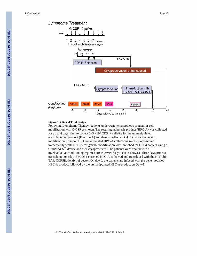
Figure 1. Clinical Trial DesignFollowing Lymphoma Therapy, patients underwent hematopoietic progenitor cellmobilization with G-CSF as shown. The resulting apheresis product (HPC-A) was collectedfor up to 4 days; first to collect 2–5 ×10⁵ CD34+ cells/kg for the unmanipulatedtransplantation product (Fraction A) and then to collect CD34+ cells for the geneticmodification (Fraction B). Unmanipulated HPC-A collections were cryopreservedimmediately while HPC-A for genetic modification were enriched for CD34 content using aCliniMACS™ device and then cryopreserved. The patients were treated with amyeloablative conditioning regimen (BCNU/VP16/Cytoxan as shown). Three days prior totransplantation (day -3) CD34-enriched HPC-A is thawed and transduced with the HIV-shI-TAR-CCR5Rz lentiviral vector. On day 0, the patients are infused with the gene modifiedHPC-A product followed by the unmanipulated HPC-A product on Day+1.
DiGiusto et al. Page 12
Sci Transl Med. Author manuscript; available in PMC 2011 July 6.
NIH
-PA Author Manuscript
NIH
-PA Author Manuscript
NIH
-PA Author Manuscript
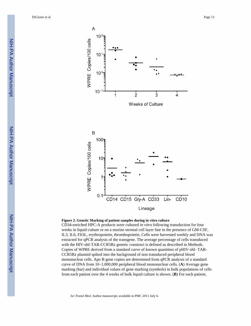
Figure 2. Genetic Marking of patient samples during in vitro cultureCD34-enriched HPC-A products were cultured in vitro following transduction for fourweeks in liquid culture or on a murine stromal cell layer line in the presence of GM-CSF,IL3, IL6, Flt3L, erythropoietin, thrombopoietin. Cells were harvested weekly and DNA wasextracted for qPCR analysis of the transgene. The average percentage of cells transducedwith the HIV-shI-TAR-CCR5Rz genetic construct is defined as described in Methods.Copies of WPRE derived from a standard curve of known quantities of pHIV-shI- TAR-CCR5Rz plasmid spiked into the background of non transduced peripheral bloodmononuclear cells. Apo B gene copies are determined from qPCR analysis of a standardcurve of DNA from 10–1,000,000 peripheral blood mononuclear cells. (A) Average genemarking (bar) and individual values of gene marking (symbols) in bulk populations of cellsfrom each patient over the 4 weeks of bulk liquid culture is shown. (B) For each patient,
DiGiusto et al. Page 13
Sci Transl Med. Author manuscript; available in PMC 2011 July 6.
NIH
-PA Author Manuscript
NIH
-PA Author Manuscript
NIH
-PA Author Manuscript
cells from in vitro culture were also sorted at 2 weeks (CD14, CD15, CD33, Gly-A andLin-) or 4 weeks (CD10) for lineage specific marking analysis as described above (b).
DiGiusto et al. Page 14
Sci Transl Med. Author manuscript; available in PMC 2011 July 6.
NIH
-PA Author Manuscript
NIH
-PA Author Manuscript
NIH
-PA Author Manuscript
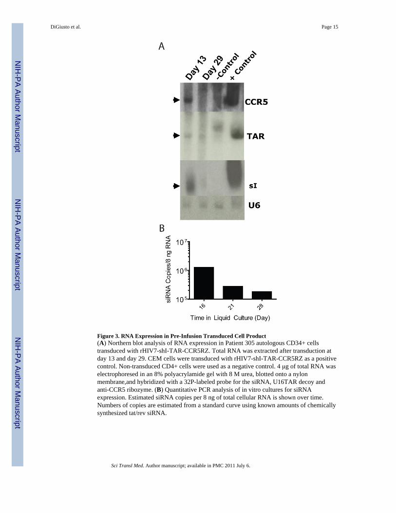
Figure 3. RNA Expression in Pre-Infusion Transduced Cell Product(A) Northern blot analysis of RNA expression in Patient 305 autologous CD34+ cellstransduced with rHIV7-shI-TAR-CCR5RZ. Total RNA was extracted after transduction atday 13 and day 29. CEM cells were transduced with rHIV7-shI-TAR-CCR5RZ as a positivecontrol. Non-transduced CD4+ cells were used as a negative control. 4 μg of total RNA waselectrophoresed in an 8% polyacrylamide gel with 8 M urea, blotted onto a nylonmembrane,and hybridized with a 32P-labeled probe for the siRNA, U16TAR decoy andanti-CCR5 ribozyme. (B) Quantitative PCR analysis of in vitro cultures for siRNAexpression. Estimated siRNA copies per 8 ng of total cellular RNA is shown over time.Numbers of copies are estimated from a standard curve using known amounts of chemicallysynthesized tat/rev siRNA.
DiGiusto et al. Page 15
Sci Transl Med. Author manuscript; available in PMC 2011 July 6.
NIH
-PA Author Manuscript
NIH
-PA Author Manuscript
NIH
-PA Author Manuscript
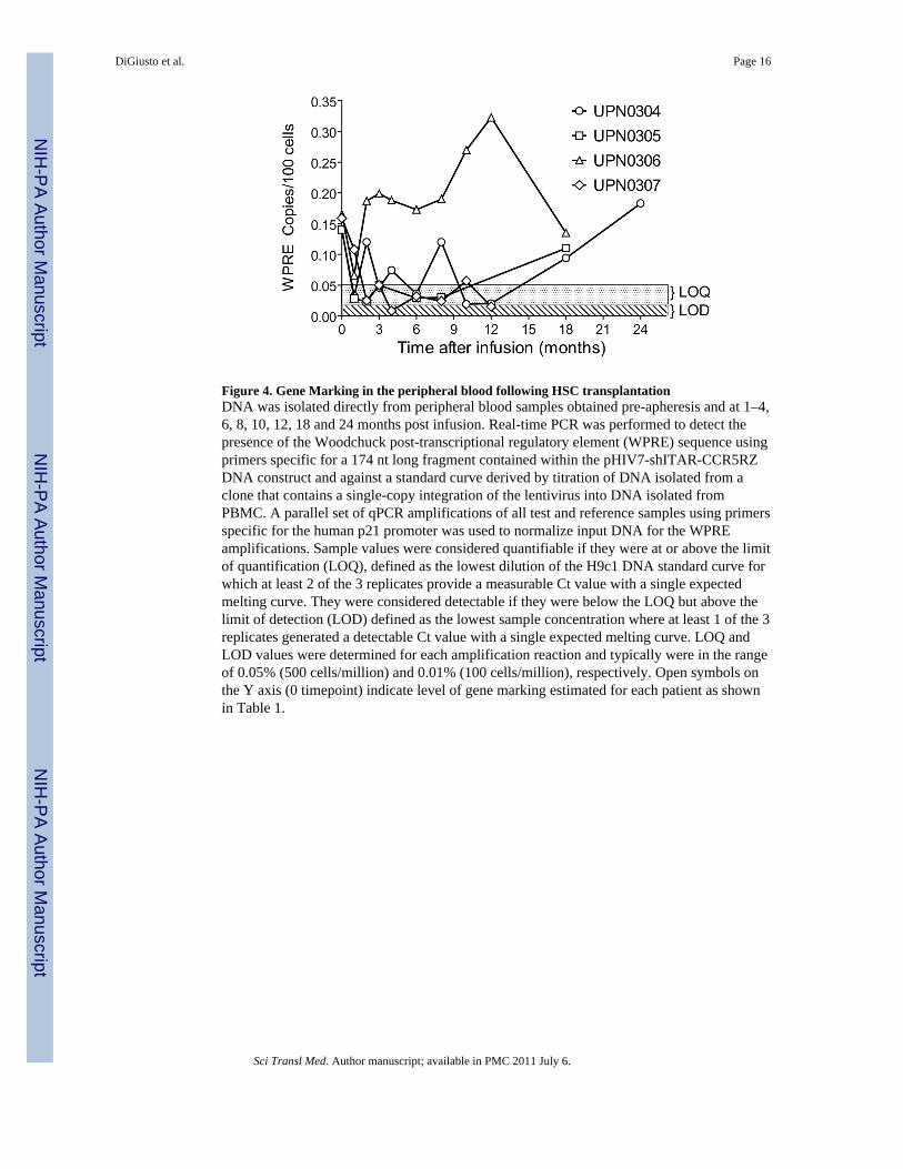
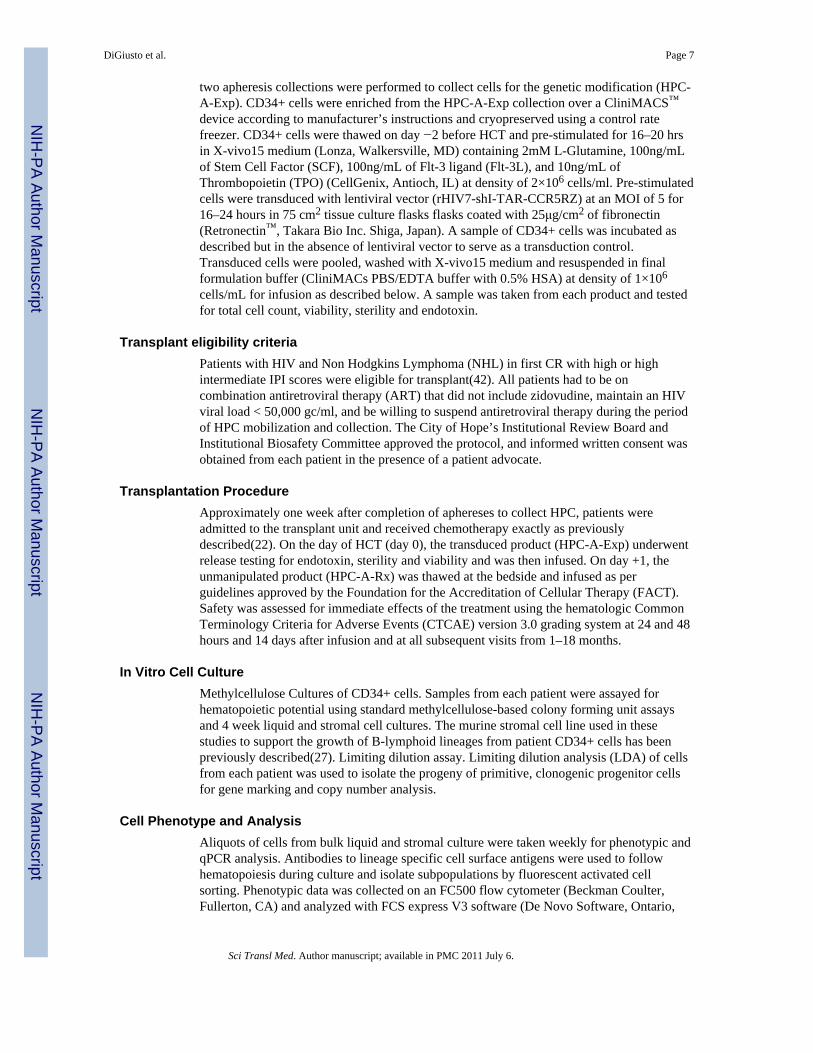
Figure 4. Gene Marking in the peripheral blood following HSC transplantationDNA was isolated directly from peripheral blood samples obtained pre-apheresis and at 1–4,6, 8, 10, 12, 18 and 24 months post infusion. Real-time PCR was performed to detect thepresence of the Woodchuck post-transcriptional regulatory element (WPRE) sequence usingprimers specific for a 174 nt long fragment contained within the pHIV7-shITAR-CCR5RZDNA construct and against a standard curve derived by titration of DNA isolated from aclone that contains a single-copy integration of the lentivirus into DNA isolated fromPBMC. A parallel set of qPCR amplifications of all test and reference samples using primersspecific for the human p21 promoter was used to normalize input DNA for the WPREamplifications. Sample values were considered quantifiable if they were at or above the limitof quantification (LOQ), defined as the lowest dilution of the H9c1 DNA standard curve forwhich at least 2 of the 3 replicates provide a measurable Ct value with a single expectedmelting curve. They were considered detectable if they were below the LOQ but above thelimit of detection (LOD) defined as the lowest sample concentration where at least 1 of the 3replicates generated a detectable Ct value with a single expected melting curve. LOQ andLOD values were determined for each amplification reaction and typically were in the rangeof 0.05% (500 cells/million) and 0.01% (100 cells/million), respectively. Open symbols onthe Y axis (0 timepoint) indicate level of gene marking estimated for each patient as shownin Table 1.
DiGiusto et al. Page 16
Sci Transl Med. Author manuscript; available in PMC 2011 July 6.
NIH
-PA Author Manuscript
NIH
-PA Author Manuscript
NIH
-PA Author Manuscript
NIH
-PA Author Manuscript
NIH
-PA Author Manuscript
NIH
-PA Author Manuscript
DiGiusto et al. Page 17
Tabl
e 1
Patie
nt C
hara
cter
istic
s.
CD
34+
Cel
ls/K
g
UPN
Sex
Dia
gnos
isA
geC
ell P
rodu
cts I
nfus
ed
HIV
Loa
dPr
e-T
rans
plan
t(g
c/m
l)
CD
4 L
evel
Pre-
Tra
nspl
ant
(Cel
ls/m
l)
Day
s to
Eng
raftm
ent
(AN
C>5
00)
Follo
w U
p (m
onth
s)H
PC-A
-RX
Infu
sed
HPC
-A-E
xp In
fuse
dT
otal
Infu
sed
% G
ene
Mar
ked
cells
infu
sed*
301
MD
iffus
e La
rge
B C
ell
Lym
phom
a42
Rx
only
9830
138
11N
A2.
8E+0
6N
AN
AN
A
304
MD
iffus
e La
rge
Cel
l Typ
e(I
mm
unob
last
icPl
asm
acyt
oid)
55Ex
p/R
x<4
0036
611
243.
9E+0
67.
7E+0
54.
7E+0
60.
14%
305
MD
iffus
e La
rge
B-C
ell
(Ana
plas
tic)
45Ex
p/R
x<4
0020
611
183.
4E+0
67.
3E+0
54.
1E+0
60.
14%
306
MPl
asm
abla
stic
Lym
phom
a45
Exp/
Rx
2100
1811
185.
6E+0
61.
2E+0
66.
8E+0
60.
11%
307
MD
iffus
e La
rge
B C
ell
Lym
phom
a25
Exp/
Rx
<400
577
1112
6.5E
+06
1.6E
+06
8.1E
+06
0.15
%
Abb
revi
atio
ns: U
PN –
Uni
que
patie
nt n
umbe
r; M
= m
ale;
HPC
-A-R
x - m
inim
ally
man
ipul
ated
Hem
atop
oiet
ic P
roge
nito
r Cel
l Aph
eres
is p
rodu
ct; H
PC-A
-Exp
- C
D34
-enr
iche
d, e
x-vi
vo tr
ansd
uced
HPC
-A.
gc/m
l = g
ene
copi
es p
er m
illili
ter s
erum
; AN
C =
abs
olut
e ne
utro
phil
coun
t.
* % g
ene
mar
ked
cells
= %
gen
e m
arke
d (e
stim
ated
from
4 w
eek
in v
itro
cultu
re a
naly
sis)
× H
PC-A
-Exp
Infu
sed/
(HPC
A-E
xp In
fuse
d +
HPC
-A-R
x In
fuse
d) ×
100
.
Sci Transl Med. Author manuscript; available in PMC 2011 July 6.
NIH
-PA Author Manuscript
NIH
-PA Author Manuscript
NIH
-PA Author Manuscript
DiGiusto et al. Page 18
Tabl
e 2
Ana
lysi
s of R
NA
exp
ress
ion
in p
erip
hera
l blo
od a
nd b
one
mar
row
of p
atie
nts f
ollo
win
g tra
nspl
anta
tion.
Per
iphe
ral b
lood
and
or b
one
mar
row
was
harv
este
d be
twee
n 1
and
24 m
onth
s (1M
-24M
) pos
t tra
nspl
ant a
nd se
para
ted
into
per
iphe
ral b
lood
mon
onuc
lear
cel
ls (P
BM
C),
perip
hera
l blo
odgr
anul
ocyt
ic c
ells
(PB
GC
) bon
e m
arro
w m
onon
ucle
ar c
ells
(BM
MC
) or B
one
mar
row
gra
nulo
cyte
s (B
MG
C) b
y de
nsity
sepa
ratio
n (F
icol
l). R
NA
was
isol
ated
from
eac
h fr
actio
n an
d an
alyz
ed fo
r the
exp
ress
ion
of si
RN
A a
nd C
CR
5 R
iboz
yme
RN
A a
s des
crib
ed in
met
hods
.
1M2M
3M4M
6M8M
10M
12M
18M
24M
UPN
Cel
lssi
RN
AC
CR
5RZ
siR
NA
CC
R5R
Zsi
RN
AC
CR
5RZ
siR
NA
CC
R5R
Zsi
RN
AC
CR
5RZ
siR
NA
CC
R5R
Zsi
RN
AC
CR
5RZ
siR
NA
CC
R5R
Zsi
RN
AC
CR
5RZ
siR
NA
CC
R5R
Z
304
PBM
C+
−+
−+
+−
++
+−
+−
++
−−
++
+
PBG
C+
++
++
+−
+−
+−
−−
−
BM
MC
+−
++
BM
GC
−+
−−
305
PBM
C+
+−
−−
−−
−+
−+
−−
−−
−
PBG
C+
−−
−−
−−
−−
−−
−
BM
MC
+−
−−
−−
BM
GC
−−
−−
−−
306
PBM
C+
++
++
++
++
++
+
PBG
C+
+−
−+
++
++
++
+
BM
MC
++
BM
GC
++
307
PBM
C+
++
+−
++
++
+−
+
PBG
C+
++
++
−−
−−
−−
−
BM
MC
++
BM
GC
−−
Sam
ples
with
det
ecta
ble
leve
ls o
f RN
A a
re m
arke
d +
whi
le th
ose
with
no
dete
ctab
le R
NA
are
mar
ked −
. Bla
nks i
ndic
ate
no sa
mpl
e sc
hedu
led
or a
vaila
ble
for a
naly
sis.
Sci Transl Med. Author manuscript; available in PMC 2011 July 6.
Copyright © 2022 FDOKUMEN