Rhubarb (Rhei Rhizoma) has been used as an important ...
7
3132 Vol. 35 (1987) Chem. Pharm. Bull. 35( 8 )3132-3138(1987). New Laxative Constituents of Rhubarb. Isolation and Characterization of Rheinosides A, B, C and D1) TAKASHI YAMAGISHI,a MAKOTO NISHIZAWA ,a,2) MITSUHIKO IKURA,b KUNIO HIKICHI,b GEN-ICHIRO NONAKA,c and ITSUO NISHIOKA*, c Hokkaido Institute of Public Health,a N-19, W-12, Kita-ku, Sapporo 060 , Japan, High-Resolution NMR Laboratory, Department of Chemistry, Facultyof Sciences, Hokkaido University,b N-10, W-8,Kita-ku, 060 Sapporo, Japan, and Faculty of Pharmaceutical Sciences, Kyushu University,' Maidashi, Higashi-ku,Fukuoka 812,Japan (Received January 29, 1987) Four new laxative constituents, rheinosides A, B, C and D, were isolated from commercial rhubarb (Rhei Rhizoma). On the basis of high-resolution nuclear magnetic resonance spectral data and chemical correlation with rhein, the structures were characterized as stereoisomers of 8-O-ƒÀ-D- glucosy1-10-hydroxy-10-C-ƒÀ-n-glucosyl rhein-9-anthrone (rheinosides A and B), and 8-O-ƒÀ-D- glucosy1-10-C-ƒÀ-n-glucosyl rhein-9-anthrone (rheinosides C and D). Keywords rhubarb; Rhei Rhizoma; Polygonaceae; rheinoside; C-glucosyl anthrone; rhein anthrone; laxative activity Rhubarb (Rhei Rhizoma) has been used as an important traditional crude drug in China and Japan, and as a laxative medicine in European countries. Many investigators have studied the constituents of rhubarb, and it has been shown to contain anthraquinones,3) dianthrones (sennosides),4) naphthalenes,5) stilbenes,6, 7) phenylbutanones,7 -9) chromones,10) etc. Recently, we have reported the isolation and characterization of highly water-soluble polyphenolic compounds (rhatannin)8) which decreased urea nitrogen concentration in rat serum, and other tannin-related compounds.8,9,11,12) Chemical and pharmacological studies of rhubarb have revealed that sennosides A-F are active principles.4) Our systematic investigation on the water-soluble fraction of rhubarb, however, has resulted in the isolation of four new laxative C-glucosides. In this paper, we report the characterization and laxative activity of these new principles. A commercial rhubarb (Gaoh, produced in Sichuan province in China) obtained in Osaka market was extracted with 50% aqueous acetone, and the extract was partitioned between ethyl acetate and water. The water-soluble portion containing a mixture of Fig. 1. High-Performance Liquid Chromato- gram of Rheinosides Conditions: column, Nucleosil 5C18 (4 i.d. •~ 250 mm); solvent, . CH3CN : H2O: HCOOH = 12 : 88 : 1; flow rate, 1.2 ml/min; detection, 350 nm. 1, rheinoside A; 2, rheinoside B; 3, rheinoside C; 4, rheinoside D.
-
Upload
khangminh22 -
Category
Documents
-
view
0 -
download
0
Transcript of Rhubarb (Rhei Rhizoma) has been used as an important ...

3132 Vol. 35 (1987)
Chem. Pharm. Bull. 35( 8 )3132-3138(1987).
New Laxative Constituents of Rhubarb. Isolation and
Characterization of Rheinosides A, B, C and D1)
TAKASHI YAMAGISHI,a MAKOTO NISHIZAWA,a,2) MITSUHIKO IKURA,b KUNIO HIKICHI,b GEN-ICHIRO NONAKA,c
and ITSUO NISHIOKA*, c
Hokkaido Institute of Public Health,a N-19, W-12, Kita-ku, Sapporo 060, Japan, High-Resolution NMR Laboratory, Department of Chemistry, Faculty of Sciences,
Hokkaido University,b N-10, W-8, Kita-ku, 060 Sapporo, Japan, and Faculty of Pharmaceutical Sciences, Kyushu University,'
Maidashi, Higashi-ku, Fukuoka 812, Japan
(Received January 29, 1987)
Four new laxative constituents, rheinosides A, B, C and D, were isolated from commercial
rhubarb (Rhei Rhizoma). On the basis of high-resolution nuclear magnetic resonance spectral data
and chemical correlation with rhein, the structures were characterized as stereoisomers of 8-O-ƒÀ-D-
glucosy1-10-hydroxy-10-C-ƒÀ-n-glucosyl rhein-9-anthrone (rheinosides A and B), and 8-O-ƒÀ-D-
glucosy1-10-C-ƒÀ-n-glucosyl rhein-9-anthrone (rheinosides C and D).
Keywords rhubarb; Rhei Rhizoma; Polygonaceae; rheinoside; C-glucosyl anthrone; rhein
anthrone; laxative activity
Rhubarb (Rhei Rhizoma) has been used as an important traditional crude drug in China and Japan, and as a laxative medicine in European countries. Many investigators have studied the constituents of rhubarb, and it has been shown to contain anthraquinones,3) dianthrones (sennosides),4) naphthalenes,5) stilbenes,6, 7) phenylbutanones,7 -9) chromones,10) etc. Recently, we have reported the isolation and characterization of highly water-soluble
polyphenolic compounds (rhatannin)8) which decreased urea nitrogen concentration in rat serum, and other tannin-related compounds.8,9,11,12) Chemical and pharmacological studies of rhubarb have revealed that sennosides A-F are active principles.4) Our systematic investigation on the water-soluble fraction of rhubarb, however, has resulted in the isolation of four new laxative C-glucosides. In this paper, we report the characterization and laxative activity of these new principles.
A commercial rhubarb (Gaoh, produced in Sichuan province in China) obtained in Osaka market was extracted with 50% aqueous acetone, and the extract was partitioned between ethyl acetate and water. The water-soluble portion containing a mixture of
Fig. 1. High-Performance Liquid Chromato-
gram of Rheinosides
Conditions: column, Nucleosil 5C18 (4 i.d. •~ 250
mm); solvent, . CH3CN : H2O: HCOOH = 12 : 88 : 1;
flow rate, 1.2 ml/min; detection, 350 nm.
1, rheinoside A; 2, rheinoside B; 3, rheinoside C;
4, rheinoside D.
No. 8 3133
polyphenolics, sennosides and C-glucosides, was applied to a Sephadex LH-20 column. The
column was eluted with water, and then 50% aqueous acetone. The polyphenolic compounds
were efficiently adsorbed on the Sephadex column, and a mixture of C-glycosides was obtained
from the water eluate. High-performance liquid chromatographic (HPLC) analysis of the
mixture of C-glycosides showed four major peaks in the chromatogram (Fig. 1). These C-
glycosides, named rheinosides A (1), B (2), C (3) and D (4), were isolated by repeated
preparative liquid chromatography on a reversed-phase column using two solvent systems
(acetonitrile-water-acetic acid and methanol-water-acetic acid), as pale yellow amorphous
powders.
All of these compounds showed blue colorations with the FeCl3 reagent and were soluble
in dilute aqueous NaOH, giving yellow solutions which showed intense yellow fluorescence
under ultraviolet (UV) light (360 nm). The infrared (IR) spectra of these compounds showed
absorptions at 1640 cm (ketone) and 1700 cm-1 (carboxylic acid), and the UV spectra
showed absorption maxima around 268, 295 and 330 nm which closely resemble those of
barbaloin (aloe-emodin anthrone 10- C-ƒÀ-D-glucoside).13)
The field desorption mass spectra (FD-MS) of 1 and 2 showed the same (M + Na) ion
peak at m/z 633, and the elemental analyses established the molecular formulae to be
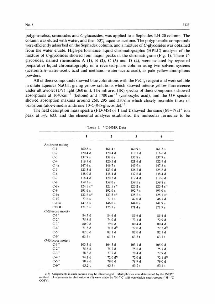
TABLE I. 13C-NMR Data
a, b) Assignments in each column may be interchanged. Multiplicities were determined by the INEPT method. Assignments in rheinoside A (1) were made by 1H-13C shift correlation spectroscopy (1 H-13C COSY).

3134 Vol. 35 (1987)
C27H30016. The carbon-13 nuclear magnetic resonance (13C-NMR) spectra of 1 and 2 showed
the presence of two aromatic rings, a carbonyl carbon, a carboxyl carbon, and a quaternary
carbon bearing an oxygen function, together with two sugar moieties (see Table I). The proton
nuclear magnetic resonance (1H-NMR) spectra showed the presence of 14 protons assignable
to sugar moieties and 5 aromatic protons.
On hydrolysis with 0.5 N H2SO4, 1 afforded a sugar and a pale yellow compound (la), mp
230 •Ž, [a]25D; + 34 •‹ (c = 0.50, methanol). El-MS m I z: 448 (M+). The sugar was identified as D-
glucose by HPLC on Nucleosil 5NH2 and from its optical rotation. On the other hand,
oxidative degradation of 1 with 2 N H2 SO4 in the presence of FeC13 afforded rhein, D-glucose
and D-arabinose. Hay and Haynes13) have already reported that treatment of barbaloin with
FeCl3 in 50% H2SO4 gave aloe-emodin and D-arabinose, and that D-arabinose was artificially
formed from the C-glucosyl moiety by the retro-Prince reaction.14) Our results, combined with
these findings, suggested that 1 was a compound analogous to barbaloin, having a rhein
anthrone skeleton with both O- and C-glucosyl moieties.
All signals in the 1H-NMR spectrum of 1 were assigned by measurement of two-
dimensional shift correlation (COSY, Fig. 2) and two-dimensional J-resolved (2D-J) spectra.
The anomeric protons of O-glucosyl and C-glucosyl moieties were observed at (55.19 (f=
7.9 Hz) and 6 3.34 (J = 9.8 Hz), respectively, and there was no signal due to the C-10 proton. In
the 13C-NMR spectrum of 1, a quaternary carbon bearing a hydroxy group was observed at
6 77.6 which was assigned to the C-10 carbon of the rhein anthrone moiety. These
(A)
(B)
( C )
(D)
(E)
(F)
Fig. 2. Two-Dimensional Shift Correlation (COSY) Spectrum of Rheinoside A (Sugar Proton Region)
1, C1"-H; 2, C6-H; 3, C6"-H'; 4, C2"-H; 5, C5"-H; 6, C4"-H; 7, C3"-H; 8, C6,-H; 9, C6,-H'; 10, C1'-H; 11, C3'-H; 12, C5'-H; 13, C4'-H; 14, C3'-H.
Fig. 3. Nuclear Overhauser Effect (NOE) Dif-ferential Spectra of la and 2
(A) NOE differential spectrum of la irradiated at C5-H (7.35 ppm).
(B) NOE differential spectrum of la irradiated at C4-H (7.75 ppm).
(C) Normal spectrum of la. (D) NOE differential spectrum of 2 irradiated at
C4-H (7.82 ppm). (E) NOE differential spectrum of 2 irradiated at
C5-H (7.65 ppm). (F) Normal spectrum of 2.
No. 8 3135
(B)
(A)
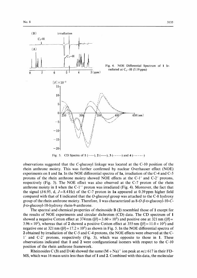
observations suggested that the C-glucosyl linkage was located at the C-10 position of the rhein anthrone moiety. This was further confirmed by nuclear Overhauser effect (NOE) experiments on 1 and la. In the NOE differential spectra of la, irradiation of the C-4 and C-5
protons of the rhein anthrone moiety showed NOE effects at the C-1' and C-2' protons, respectively (Fig. 3). The NOE effect was also observed at the C-7 proton of the rhein anthrone moiety in 1 when the C-1" proton was irradiated (Fig. 4). Moreover, the fact that the signal (6 6.95, d, J= 8.4 Hz) of the C-7 proton in la appeared at 0.39 ppm higher field compared with that of 1 indicated that the O-glucosyl group was attached to the C-8 hydroxy
group of the rhein anthrone moiety. Therefore, 1 was characterized as 8-O-fl-D-glucosy1-10-C-fi-D-glucosy1-10-hyd roxy rhein-9-anthrone.
The spectral and chemical properties of rheinoside B (2) resembled those of 1 except for the results of NOE experiments and circular dichroism (CD) data. The CD spectrum of 1 showed a negative Cotton effect at 374 nm ([0] = 3.60 x 10³) and positive one at 321 nm ([01= 3.96 x 10³), whereas that of 2 showed a positive Cotton effect at 355 nm ([01= 11.0 x 10³) and negative one at 321 nm ([0] = 17.2 x 10³) as shown in Fig. 5. In the NOE differential spectra of 2 obtained by irradiation of the C-5 and C-4 protons, the NOE effects were observed at the C-1' and C-2' protons, respectively (Fig. 3), which was opposite to those in 1. These observations indicated that 1 and 2 were configurational isomers with respect to the C-10
position of the rhein anthrone framework. Rheinosides C (3) and D (4) showed the same (M +Na)+ ion peak at m I z 617 in their FD-
MS, which was 16 mass units less than that of 1 and 2. Combined with this data, the molecular
Fig. 4. NOE Differential Spectrum of 1 Ir-radiated at C1" -H (5.19 ppm)
Fig. 5. CD Spectra of 1 (-), 2 (------), 3 ( - - )) and 4 (------)

3136 Vol. 35 (1987)
formulae of 3 and 4 were determined to be C27H30015 by elemental analyses. The 1H-NMR
spectra of 3 and 4 resembled those of 1 and 2 except for the presence of doublets at (5 4.24 (J=
1.8 Hz for 3) and 6 4.12 (J=0.5 Hz for 4). The 13C-NMR spectra of 3 and 4 also resembled
those of 1 and 2, although methine carbon signals were observed at 6 47.0 (3) and 6 46.7 (4)
instead of the quaternary carbons (6 77.6 for 1 and 6 77.7 for 2). These results implied the
absence of the hydroxy group at C-10 of the rhein anthrone framework.
On oxidative degradation in the presence of FeC13, 3 gave rhein, D-glucose and D-
arabinose. In the NOE differential spectra of 3, irradiation of the C-1" proton showed an
NOE effect at the C-7 proton of rhein anthrone moiety. Irradiation of the C-1' proton showed
NOE effects at the C-4 and C-10 protons, and irradiation of the C-2' proton showed NOE
effects at the C-5 and C-10 protons. These results indicated that the 0- and C-glucosyl
moieties were located at the C-8 and C-10 positions of rhein anthrone, respectively.
The NOE experiment on 4 also showed the presence of the O-glucosyl moiety at the C-8
position. In the NOE differential spectra of 4, irradiation of C-1' proton showed NOE effects
at the C-5 and C-10 protons, and irradiation of the C-2' proton showed NOE effects at the C-
4 and C-10 protons. In the CD spectra, 3 and 4 showed almost opposite Cotton effects around
365 and 320 nm (Fig. 5), indicating that 3 and 4 differ only in the configuration at the C-10
position. Therefore, 3 and 4 were characterized as a pair of stereoisomers at the C-10 position
of 8-O-ƒÀ-D-glucosy1-10-C-ƒÀ-D-glucosyl rhein-9-anthrone.
The laxative effect of rheinoside A was tested in Wistar rats, and ED50 was determined to
be 15.6 mg/kg. In some commercial rhubarbs, the total rheinosides content exceeded 10 mg/g,
being comparable to that of sennosides. Therefore, rheinosides may also play an important
role in the laxative activity of rhubarb. Elucidation of the absolute structures of the
rheinosides is in progress.
Experimental
Melting points were determined on a Yanagimoto micro-melting point apparatus (model MS-S3) and are
uncorrected. UV and IR spectra were obtained with Hitachi model 200-10 and model 260-10 spectrometers,
respectively. El and FD-MS were measured with a Shimadzu 2000-DF spectrometer. H- and 13C-NMR spectra were
taken with JEOL GX-500 and FX-200 spectrometers using sodium 3-trimethylsilylpropane sulfonic acid (DSS) as an
internal standard, and chemical shifts are given in ƒÂ (ppm). Optical rotations were determined with a JASCO DIP-4
digital polarimeter and CD spectra with a JASCO CD J-500 spectropolarimeter in methanol solution (1.0 mg/20 ml).
HPLC was performed on a Hitachi model 655 liquid chromatograph equipped with a Nucleosil 5C,8 (Macherey-
Nagel) column (4 mm i.d. x 250 mm) and a variable-wavelength spectrophotometric detector operated at 280 and
350 nm. The mobile phase was prepared by mixing 1 ml of formic acid with 100 ml of water containing acetonitrile
(10-20%). Column chromatographies were carried out on Sephadex LH-20 (20-100 p, Pharmacia Fine Chemical
Co., Ltd.) with a water-acetone system, and Fuji gel ODS-Q3 (30-50 p, Fuji gel Hanbai Co., Ltd.) with a solvent
system similar to that employed for HPLC.
Isolation of Rheinosides The powdered Rhei Rhizoma (710 g, Gaoh [雅黄] produced in Sichuan province of
Chart 1

No. 8 3137
China) was extracted twice with 2 1 each of 50% aqueous acetone at room temperature. The extracts were
concentrated under reduced pressure below 40 °C, and then extracted with 1 1 of ethyl acetate. The aqueous layer was
concentrated to 500 ml. Acidification with 10 ml of acetic acid afforded dark brown precipitates, which were removed
by decantation after standing at room temperature for 12 h. The supernatant was, after concentration, passed
through a column of Sephadex LH-20 (24 i.d. x 200 mm) with water and then 50% aqueous acetone. The water eluate
was repeatedly chromatographed on Sephadex LH-20 with the same solvent system, yielding a fraction (38.4 g)
containing a mixture of rheinosides, which was treated with methanol. The methanol-soluble portion was dissolved in
water and applied to a column of ODS-Q3 with a solvent system of 5-15% acetonitrile in water containing acetic
acid (10 m1/1), to give rheinosides A (14.18 g), B (3.64 g), C (2.64 g) and D (4.56 g).
Rheinoside A (1) A pale yellow amorphous powder, [a]g - 53 •‹ (c =0.5, Me0H), IR v °Icm-1: 3600-3100
(OH), 1700 (COOH), 1640 (C =0). UV AMeOHmax nm (a): 267 (6300), 296 (8800), 346 (7000). FD-MS m/z: 633
(M +Na)+ . CD (c =0.005, Me0H) [0125 (nm): - 3.6 x 10³ (374), 4.0 x 10³ (321), 9.6 x 10³ (292). 1H-NMR (D20) 5:
7.88 (s, C4-H), 7.75 (dd, J=7.2, 7.6, C6-H), 7.67 (d, J=7.6, C5-H), 7.38 (s, C2-H), 7.34 (d, J= 7.3, C7-H), 5.19 (d, J=
7.9, C,-H), 3.98 (d, J= 12.5, C6-H), 3.82 (dd, J=5.5, 12.5, C6-H'), 3.79 (dd, J=7.9, 9.1, C2-H), 3.71 (m, C5-H),
3.65 (dd, J=8.9, 9.1, C3-H), 3.60 (dd, J=8.9, 9.1, C4-H), 3.60 (brd, J= 12.2, C6,-H), 3.43 (dd, J=5.5, 12.2, C6,-H'),
3.34 (d, J=9.8, C1,-H), 3.33 (t, J=9.2, C3,-H), 3.03 (m, C5,-H), 2.95 (t, J=9.2, C4,-H), 2.94 (dd, J=9.2, 9.8, C2,-H).
Anal. Calcd for C27H30016.2H20: C, 50.15; H, 4.99. Found: C, 50.19; H, 4.96.
Hydrolysis of Rheinoside A A solution of rheinoside A (300 mg) in 5 ml of 0.5 N H2SO4 was refluxed for 2 h.
After cooling, the reaction mixture was extracted with 50 ml each of ethyl acetate three times. The combined ethyl
acetate layers were washed with water, dried over Na2SO4, and then concentrated to dryness. The product was
purified on a column of ODS-Q2 using acetonitrile (15-20%)-water-acetic acid (1 %) as an eluent to yield the C-
glucoside la (114 mg, 52%). After neutralization with Ba(OH)2, the aqueous layer was passed through a Sep-pak
ODS (Waters Associates Co., Ltd.). D-Glucose ([01; + 33.5 °, c =0.8 water) was separated from the water eluate by
preparative HPLC on a Nucleosil 10 NH2 column.
la: Pale yellow crystals, mp 230 °C, [ce + 34 ° (c =0.5, Me0H). El-MS m/z: 448 (M +). 1H-NMR (D20) 6 : 7.75
(d, J=1.2, C4-H), 7.63 (dd, J=8.1, 8.4, C6-H), 7.39 (d, J=1.2, C2-H), 7.35 (d, J=8.1, C5-H), 6.95 (d, J=8.4, C7-H),
3.66 (dd, J=1.8, 12.1, C6,-H), 3.46 (dd, J=6.2, 12.1, C6.-H'), 3.52 (d, J=9.5, C1,-H), 3.22 (dd, J=8.8, 9.2, C3,-H),
3.08 (m, C5,-H), 2.88 (dd, J=8.8, 9.5, C4,-H), 2.63 (dd, J=9.2, 9.5, C2,-H). 13C-NMR (D20) 5: 194.7 (C9, C =0),
171.1 (COOH), 163.2 (s), 162.3 (s), 148.2 (s), 147.1 (s), 139.5 (d), 138.8 (s), 121.2 (d, 2C), 120.6 (s), 120.6 (d), 120.1 (d),
118.2 (s), 86.1 (d), 82.1 (d), 80.0 (d), 77.0 (s), 73.7 (d), 71.9 (s), 63.8 (t). Anal. Calcd for C21H20011 1.51120: C, 53.05;
H, 4.88. Found: C, 52.84; H, 4.58.
Oxidative Degradation of Rheinoside A A solution of 10 mg of rheinoside A and 50 mg of FeCl3 in 5 ml of 2 N
H2SO4 was refluxed for 30 min. After cooling, the reaction mixture was diluted with water (50 ml) and extracted with
ethyl acetate (30 ml x 3). The combined ethyl acetate layers were dried over Na2SO4 and concentrated. HPLC showed
a peak identical with that of rhein. The aqueous layer was neutralized with Ba(OH)2 and passed through a Sep-pak
ODS. The fraction eluted with water was analyzed by HPLC on a column of Nucleosil 10NH2 (mobile phase:
CH3CN : H20 = 70 : 30), and showed peaks corresponding to glucose and arabinose.
Rheinoside B (2) A pale yellow amorphous powder, [a]l; - 43 ° (c =0.5, Me0H). IR v°1 (cm-1): 3600-
3100 (OH), 1700 (C =0), 1640 (COOH). UV A.,=11 nm (a): 269 (6200), 298 (9100), 345 (6600). FD-MS m/z: 633
(M + Na)+ . CD (c=0.005, Me0H) [O]25 (nm): +11.0 x 10³ (355), -17.2 x 10³ (321), - 5.6 x 10³ (275). 1H-NMR
(D20) 5: 7.82 (d, J=1.2, C4-H), 7.71 (dd, J=7.6, 8.2, C6-H), 7.65 (d, J=7.6, C5-H), 7.42 (d, J=1.2, C2-H), 7.37 (d,
J=8.2, C7-H), 5.20 (d, J=7.6, C,,,-H), 3.96 (dd, J= 1.8, 12.5, C6-H), 3.80 (dd, J= 5.5, 12.5, C6-H'), 3.74 (dd, J=
7.6, 9.2, C2,,-H), 3.68 (dd, J=9.2, 9.5, C3,,-H), 3.66 (ddd, J=1.8, 5.5, 9.5, C5,,-H), 3.58 (t, J=9.2, C4,,-H), 3.56 (brd,
J = 12.2, C6, -H), 3.44 (dd, J =5.5,12.2, C6,-H'), 3.38(d, J n 9.8, C1,-H), 3.34 (t, J = 9.2, C3, -H), 3.03 (ddd, J = 1.9, 7.9,
9.8, C5,-H), 2.93 (t, J =9 .5 , C4,-H), 2.89.(dd, J =9 .2, 9.8, C2, -H). Anal. Calcd for C27H30016•EH20: C, 51.59; H, 5.27.
Found: C, 51.27; H, 5.13.
Rheinoside C (3) A pale yellow amorphous powder, [a]g - 40 ° (c = 0.5, Me0H). IR vNujolmax cm-1:3600-3100
(OH), 1700 (C=O), 1640 (COOH). UV (s): nm (s): 268 (7500), 292 (10500), 335 (7500). FD-MS m/z: 617
(M +Na)+ . CD (c = 0.005, Me0H) [O]2' (nm): - 5.6 x 10³ (378), + 6.3 x 10³ (325), -14.0 x10³ (298). 1H-NMR (D20)
6 : 7.60 (dd, J= 7.6, 8.2, C6-H), 7.24 (d, J=8.2, C5-H), 7.23 (d, J= 1.5, C4-H), 7.08 (d, J= 7.6, C7-H), 7.03 (d, J=1.5,
C2-H), 5.21 (d, J=7.9, C,-H), 4.24 (d, J=1.8, C10-H), 3.99 (dd, J=2.2, 12.5, C6-H), 3.833 (dd, J= 5.5, 12.5, C6.-
H'), 3.827 (d, J= 7.9, 9.5, C2,,-H), 3.731 (m, C5..-H), 3.727 (t, J-9.5, C4,,-H), 3.63 (t, J=9.5, C3.,-H), 3.49 (dd, J=1.8,
12.2, C6,-H), 3.38 (dd, J= 5.2, 12.2, C6,-H'), 3.31 (t, J=8.9, C3,-H), 3.26 (dd, J=1.8, 9.8, C1,-H), 2.88-2.98 (m, C2.-
H, C4.-H, C5,-H). Anal. Calcd for C27H30015.2H20: C, 51.43; H, 5.44. Found: C, 51.71; H, 5.31.
Oxidative Degradation of Rheinoside C A solution of 10 mg of rheinoside C and 50 mg of FeC13 in 5 ml of 2 N
H2SO4 was refluxed for 30 min, and treated as mentioned above. The HPLC analyses showed peaks corresponding to
rhein, glucose and arabinose.
Rheinoside D (4) A pale yellow amorphous powder, [oc]V - 56 ° (c = 0.5, Me0H). IR vNujolmax cm- I-: 3600-3100
(OH), 1700 (C =0), 1640 (COOH). UV ,IMeOHmax nm (s): 270 (7100), 295 (9800), 327 (7600). FD-MS ml z: 617
(M +Na)+. CD (c=0.005, MeCoH) [0125 (nm): +9.6 x 10³ (361), -19.4 x 10³ (319), -4.5 x 10³ (275). 'H-NM.R
3138 Vol. 35 (1987)
(D20) ƒÂ: 7.65 (t, J=8.5, C6-H), 7.34 (d, J=8.5, C5-H), 7.25 (d, J=1.6, C4-H), 7.17 (d, J=1.6, C2-H), 7.16 (d, J=8.5,
C7-H), 5.09 (d, J=7.6,C1"-H 4.12 (d, J=0.5, C10-H), 4.02 (brd, J=12.0, C6"-H), 3.88 (dd, J=5.2, 12.0, C6"-H'),
3.73 (dd, J=7.6, 8.9, C2"-H), 3.61-3.68 (m, C3"-H, C4"-H, C5"-H), 3.46 (brd, J=12.2, C6"-H), 3.32 (brd, J=9.5,
C1'-H), 3.31 (dd, J=5.5, 12.2, C6'-H'), 2.85-2.93 (m, C3'-H, C4'-H, C5'-H), 2.81 (t, J=9.5, C2'-H). Anal. Calcd for
C27H30015•E2.5H20: C, 50.70; H, 5.52. Found: C, 50.70; H, 5.23.
Bioassey Laxative activity of rheinoside A was determined according to Ishii et al.15) Doses of 21.0, 14.2, 10.2
and 7.1 mg/kg of rheinoside A were administered to groups of five Wistar rats with an average body weight of 120 g,
and the activities were measured after 20 h. The ED50 value was calculated by the Litchfield-Wilcoxon method.
References and Notes
1) This paper forms Part XI of "Studies on Rhubarb (Rhei Rhizoma)." Part X: Y. Kashiwada, G. Nonaka, and I. Nishioka, Chem. Pharm. Bull., 34, 4083 (1986).
2) Present address: Faculty of Pharmaceutical Sciences, Hokkaido University, N-12, W-6, Kitaku, Sapporo 060, Japan.
3) H. Okabe, K. Matsuo, and I. Nishioka, Chem. Pharm. Bull., 21, 1254 (1973); L. Holzschuh, B. Kopp, and W. Kubelka, Planta Med., 46, 159 (1982); L. HOrhammer, H. Wagner, and E. Muller, Justus Liebigs Ann. Chem., 98, 2859 (1965); L. HOrhammer, L. Farkas, H. Wagner, and E. Midler, ibid., 97, 1662 (1964); M. Uchibayashi and T. Matsuoka, Chem. Pharm. Bull., 9, 234 (1961); H. Wagner and L. HOrhammer, Z. Natiirforschg, 18b, 89
(1963); K. Tsukida and M. Yoneshige, Yakugaku Zasshi, 74, 379 (1954). 4) M. Miyamoto, S. Imai, M. Shinohara, S. Fujioka, M. Goto, T. Matsuoka, and H. Fujimura, Yakugaku Zasshi,
87, 1040 (1967); H. Oshio, S. Imai, S. Fujioka, T. Sugawara, M. Miyamoto, and M. Tsukui, Chem. Pharm. Bull., 22, 823 (1974).
5) M. Tsuboi, M. Minami, G. Nonaka, and I. Nishioka, Chem. Pharm. Bull., 25, 2708 (1977). 6) G. Nonaka, M. Minami, and I. Nishioka, Chem. Pharm. Bull., 25, 2300 (1977); A. Yagi, Y. Koizumi, and I.
Nishioka, Shoyakugaku Zasshi, 25, 52 (1971); J. Banks and W. Cameron, Aust. J. Chem., 24, 2427 (1971). 7) T. Murakami and K. Tanaka, Yakugaku Zasshi, 93, 733 (1973). 8) G. Nonaka, I. Nishioka, T. Nagasawa, and H. Oura, Chem. Pharm. Bull., 29, 2862 (1981). 9) G. Nonaka and I. Nishioka, Chem. Pharm. Bull., 31, 1652 (1983).
10) Y. Kashiwada, G. Nonaka, and I. Nishioka, Abstracts of Papers, 103rd Annual Meeting of the Pharmaceutical Society of Japan, Tokyo, April 1983, p. 219.
11) Y. Kashiwada, G. Nonaka, and I. Nishioka, Chem. Pharm. Bull., 34, 4083 (1986). 12) V. H. Friedrich and J. Hohle, Planta Med., 4, 363 (1966); G. Nonaka, E. Ezaki, K. Hayashi, and I. Nishioka,
Phytochemistry, 22, 1695 (1983); W. Mayer, G. Schultz, S. Werde, and G. Schilling, Justus Liebigs Ann. Chem., 1975, 946.
13) J. E. Hay and L. J. Haynes, J. Chem. Soc. (C), 1956, 3141. 14) K. Hata, K. Baba, and M. Kozawa, Chem. Pharm. Bull., 26, 3792 (1978). 15) Y. Ishii, H. Tanizawa, C. Ikemoto, and Y. Takio, Yakugaku Zasshi, 101, 254 (1981).