Micelles and nanoparticles for ultrasonic drug and gene delivery
Upload
khangminh22Category
view
2download
0
antibiotics
Article
Rhamnolipids Nano-Micelles as a Potential Hand Sanitizer
Marwa Reda Bakkar 1 , Ahmed Hassan Ibrahim Faraag 1,2 , Elham R. S. Soliman 3 , Manar S. Fouda 4,Amir Mahfouz Mokhtar Sarguos 5, Gary R. McLean 6,7 , Ali M. S. Hebishy 8, Gehad E. Elkhouly 9,10,Nermeen R. Raya 9,10 and Yasmin Abo-zeid 9,10,*
�����������������
Citation: Bakkar, M.R.; Faraag, A.H.I.;
Soliman, E.R.S.; Fouda, M.S.;
Sarguos, A.M.M.; McLean, G.R.;
Hebishy, A.M.S.; Elkhouly, G.E.;
Raya, N.R.; Abo-zeid, Y.
Rhamnolipids Nano-Micelles as a
Potential Hand Sanitizer. Antibiotics
2021, 10, 751. https://doi.org/
10.3390/antibiotics10070751
Academic Editors: Alan J. Hibbitts
and Sofia A. Papadimitriou
Received: 3 February 2021
Accepted: 24 May 2021
Published: 22 June 2021
Publisher’s Note: MDPI stays neutral
with regard to jurisdictional claims in
published maps and institutional affil-
iations.
Copyright: © 2021 by the authors.
Licensee MDPI, Basel, Switzerland.
This article is an open access article
distributed under the terms and
conditions of the Creative Commons
Attribution (CC BY) license (https://
creativecommons.org/licenses/by/
4.0/).
1 Botany and Microbiology Department, Faculty of Science, Helwan University, Ain Helwan, Cairo 11795, Egypt;[email protected] (M.R.B.); [email protected] (A.H.I.F.)
2 Bioinformatics Center, Faculty of Science, Helwan University, Ain Helwan, Cairo 11795, Egypt3 Cytogenetics and Molecular Genetics Unit, Botany and Microbiology Department, Faculty of Science,
Helwan University, Ain Helwan, Cairo 11795, Egypt; [email protected] Biochemistry and Chemistry Department, Faculty of Science, Helwan University, Ain Helwan, Cairo 11795, Egypt;
[email protected] Biotechnology Department, Faculty of Science, Helwan University, Ain Helwan, Cairo 11795, Egypt;
[email protected] Cellular and Molecular Immunology Research Centre, London Metropolitan University, 166-220 Holloway Road,
London N7 8DB, UK; [email protected] National Heart and Lung Institute, Imperial College London, Norfolk Place, London W2 1PG, UK8 Chemistry Department, Faculty of Science, Helwan University, Cairo 11795, Egypt;
[email protected] Department of Pharmaceutics, Faculty of Pharmacy, Helwan University, Cairo 11795, Egypt;
[email protected] (G.E.E.); [email protected] (N.R.R.)10 Helwan Nanotechnology Center, Helwan University, Helwan, Cairo 11795, Egypt* Correspondence: [email protected]; Tel.: +20-10-928-927-46
Abstract: COVID-19 is a pandemic disease caused by the SARS-CoV-2, which continues to causeglobal health and economic problems since emerging in China in late 2019. Until now, there are nostandard antiviral treatments. Thus, several strategies were adopted to minimize virus transmission,such as social distancing, face covering protection and hand hygiene. Rhamnolipids are glycolipidsproduced formally by Pseudomonas aeruginosa and as biosurfactants, they were shown to have broadantimicrobial activity. In this study, we investigated the antimicrobial activity of rhamnolipids againstselected multidrug resistant bacteria and SARS-CoV-2. Rhamnolipids were produced by growingPseudomonas aeruginosa strain LeS3 in a new medium formulated from chicken carcass soup. Theisolated rhamnolipids were characterized for their molecular composition, formulated into nano-micelles, and the antibacterial activity of the nano-micelles was demonstrated in vitro against bothGram-negative and Gram-positive drug resistant bacteria. In silico studies docking rhamnolipids tostructural and non-structural proteins of SARS-CoV-2 was also performed. We demonstrated theefficient and specific interaction of rhamnolipids with the active sites of these proteins. Additionally,the computational studies suggested that rhamnolipids have membrane permeability activity. Thus,the obtained results indicate that SARS-CoV-2 could be another target of rhamnolipids and couldfind utility in the fight against COVID-19, a future perspective to be considered.
Keywords: COVID-19; SARS-CoV-2; rhamnolipids; nano-micelles; Pseudomonas aeruginosa; antibacterialagent; antiviral agent; docking studies
1. Introduction
COVID-19 (coronavirus disease-2019), a pandemic infection caused by the newlyemerged coronavirus, SARS-CoV-2, reported in Wuhan, China [1] and has spread globallywith 159,845,155 confirmed cases and 3,321,212 deaths across 192 countries by 12 May 2021(according to the COVID-19 Dashboard by the Center for Systems Science and Engineering
Antibiotics 2021, 10, 751. https://doi.org/10.3390/antibiotics10070751 https://www.mdpi.com/journal/antibiotics

Antibiotics 2021, 10, 751 2 of 21
(CSSE) at Johns Hopkins University). This disease is an acute respiratory disorder, char-acterized by pneumonia, dry cough, fever and body pain with a high rate of mortality,particularly in older people (>80 years) or those with underlying health conditions [2].Due to the long incubation period of the virus in humans of up to 14 days and airbornetransmission via aerosols, the main transmission source of virus is through human tohuman interactions [3]. Moreover, SARS-CoV-2 is also estimated to remain infectious onsolid surfaces for up to 9 days [4], and this has been postulated to facilitate spread ofinfection by self-inoculation of mucous membranes of the nose, eyes or mouth, similarly toother human coronaviruses [5,6].
Due to a lack of standard treatments and identification of mutated variants of SARS-CoV-2 in several countries that might render the developed vaccines ineffective in thefuture, the WHO has advised a healthy lifestyle and the adoption of proper social distancingmeasures for infection prevention and control. To help in containing COVID-19 infection,WHO recommends hand washing or sanitation frequently with soap or ≥60–80% alcoholichand sanitizer, respectively. Alcohol-based hand sanitizers were promoted due to theirbroad-spectrum antimicrobial activity, their availability and good safety profiles [7].
However, under the current pandemic conditions and the possibility of misuse bypeople in the community, alcohol-based sanitation was reported to raise several hazards tohumans and the environment [8]. For example, dermal contact of ethanol was reported tocause irritation and allergic conditions of skin and eyes with prolonged exposure resultingin dryness or cracking of the skin with peeling redness or itching [7]. Such frequent use ofsanitizer, especially during the current pandemic, has been reported to be responsible forskin damage, which reduce its ability to work as a barrier against other harmful pathogens,thus increasing the possibility of further infections by microorganisms [9]. Overuse ofalcohol-based hand sanitizers in some cases was also reported to increase the risk of viraloutbreaks [9,10], such as, increased risk of norovirus [11].
Furthermore, alcohol-based hand sanitizer use can result in antimicrobial resistancewhere microorganisms mutate as a defense mechanism to the repeated exposure to geno-toxic chemicals, leading to the development of resistant strains and increased burdenon already struggling healthcare professionals [9,12]. For example, Enterococcus faeciumwas reported to be 10 times more resistant to alcohol-based hand rubs than older iso-lates after repeated exposure to alcohol-based sanitizer [13]. It has been also reportedthat Escherichia coli and Pseudomonas aeruginosa were 48% and 64% resistant, respectively,against all available sanitizers on the market [14].
Therefore, there is a high demand to find novel alternative approaches for handsanitation with minimal adverse effects. Biosurfactants are microbially produced surfaceactive compounds [15], characterized by their biodegradability [16], low toxicity [17],low skin irritation potential [18], and have antimicrobial activity against a variety ofpathogens [19,20].
Rhamnolipids (Rha(s)) (Figure 1) are one such biosurfactant reported to have an-timicrobial activity with no cytotoxic effect when applied onto rabbit skin [21]. They areeconomically produced by Pseudomonas aeruginosa, a bacterial source with potential toscale-up their production [22].
Rha(s) were previously reported to be effective against multidrug resistant Grampositive and Gram-negative bacteria such as Escherichia coli, Micrococcus luteus, Alcaligenesfaecalis, Serratia marcescens, Mycobacterium phlei and Staphylococcus epidermidis, methicillin-resistant Staphylococcus aureus (MRSA) [23], Staphylococcus aureus and Enterococcus faecalisI27 [24,25]. The antiviral activity of Rha(s) was also reported against herps simplex viruses,HSV1 and HSV2 resulting in virus inactivation in-vitro [26]. The structure of herpesviruses consists of a large double-stranded DNA genome encased within an icosahedralprotein capsid wrapped in a lipid bilayer envelope, which also contains embedded viralglycoproteins. It was argued that the antiviral activity of Rha(s) against HSV1 and HSV2was due to: (1) their ability as a biosurfactant to interact with the lipid membranes of these

Antibiotics 2021, 10, 751 3 of 21
viruses and (2) the beta hydroxyalkanoic acids found in the structure of Rha(s) inducingchanges to the lipid envelop viral glycoproteins [26].
Figure 1. Chemical structure of (A) rhamnolipids R1 (mono-rhamnolipids; Rha(s)1) and (B) rhamnolipids R2 (di-rhamnolipids; Rha(s)2). Chemical structures were retrieved from PubChem; https://pubchem.ncbi.nlm.nih.gov (accessedon 16 June 2021).
SARS-CoV-2 is also an enveloped virus containing a positive-sense RNA genome [27,28].The lipid bilayer contains viral spike glycoprotein, whose receptor binding domain (S1-RBD) allows attachment of virus to the host cell receptors, angiotensin converting enzyme-2(ACE2), essential for virus entry into cells and thus infectivity [29]. Based on the structuresimilarity with herpes simplex viruses, we assume that Rha(s) can interact with the SARS-CoV-2 lipid envelop and spike glycoproteins rendering them inactive. Based on thesefindings, we hypothesize that Rha(s) could be a good alternative for alcohol-based handsanitizers and find utility in the COVID-19 pandemic.
In the current work, Rha(s) were produced by Pseudomonas aeruginosa using a newproduction medium to increase the yield of Rha(s). Rha(s) nano-micelles were then pre-pared and tested. To ascertain the broad antimicrobial activity of Rha(s) nano-micelles,the antibacterial activity of Rha(s) nano-micelles was first investigated against selecteddrug resistant bacterial strains: Staphylococcus aureus, Streptococcus pneumoniae, SalmonellaMontevideo and Salmonella Typhimurium. Subsequently, a docking study was also per-formed to investigate the potential antiviral activity of Rha(s) against SARS-CoV-2. Thedocking study was performed to understand the possible interactions of Rha(s) with theS1- RBD and the lipid envelop of SARS-CoV-2. Moreover, the interaction of Rha(s) with theactive sites of different enzymes involved in SARS-CoV-2 replication; EndoRNAse, helicase,RNA-dependent RNA polymerase and main protease was also assessed in the dockingstudy. Collectively, these studies aim to determine the possibility of Rha(s) application as atreatment for viral infections. However, their antiviral activity in vitro and safety profilefor treatment still requires addressing.
2. Results2.1. Isolation and Identification of Bacterial Strain
Initially our bacterial strain LeS3 was isolated and suspected as Pseudomonas aeruginosadue to its green fluorescence colonies on agar plates. The PCR amplification products of 16SrRNA encoding gene showed a fragment length of 1500 bp (see Figure S1 in SupplementaryMaterials). The PCR product sequence was aligned to the bacterial (taxid:2) nucleotide

Antibiotics 2021, 10, 751 4 of 21
collection of the NCBI database using the (megablast) algorithm. The alignment showed a99% query coverage with 97% identity to Pseudomonas aeruginosa. The 16S rRNA encodinggene sequence of Pseudomonas aeruginosa strain LeS3 was deposited to NCBI gene bankwith accession number MN960161 (https://www.ncbi.nlm.nih.gov/nuccore/MN960161,accessed on 16 June 2021).
2.2. Production and Characterization of Rha(s)
Pseudomonas aeruginosa strain LeS3 previously identified in Section 2.1 was cultivatedto produce Rha(s) using two different culturing media, namely chicken carcass soup (CCS)and glycerol supplemented nutrient broth (GSNB) in a trial to optimize the yield of Rha(s).The yield of Rha(s) produced in CCS was 5 times (0.5g L−1) more than that recovered byGSNB (0.1 g L−1). Therefore, all the following experiments and characterization tests wereperformed on Rha(s) produced using CCS media.
2.2.1. Characterization of Rha(s)
Rha(s) mixture produced from Pseudomonas aeruginosa strain LeS3 grown in newdesigned culture medium (CCS) for 72 h was analyzed using the ESI-MS spectrometercoupled to UPLC (LC/ESI-MS). To confirm the structural composition, mass spectra wereacquired in both positive and negative ion modes. Analysis of the obtained mass spectrawas performed by calculating the elemental composition depending on acquired knowl-edge concerning mass spectrometry of the related compounds and comparing the obtaineddata with those available in the literature [30–33]. Our data (Table 1) revealed that mostspectra peaks were found in the positive pseudo-molecular ion. All Rha(s) congeners ap-peared in the positive spectra accomplished with adduct ions; [M+H]+, [M+K]+, [M+Na]+
and [M-H+Na2]+, especially [M+Na]+. Only 3 Rha(s) congeners were detected in bothpositive and negative ion modes. This matched with what was reported previously byPantazaki and colleagues [33] who attributed these results to the presence of formic acid,methanol and water in the sample.
Table 1. Congeners composition of rhamnolipids mixture produced by P. aeruginosa strain LeS3 as analyzed by LC/ESI-MSat both positive and negative modes.
Rha(s) Congenersm/z
Mol f Mol wt [M-H]− [M+H]+ [M+Na]+ [M+K]+ [M-H+Na2]+ % AbundanceMono-Rhamnolipid (Rha(s)1) Congeners
R-C8 C14H26O7 306 351 12.8R-C8:1 C14H24O7 304 327 25.2R-C8:2 C14H22O7 302 325 29.4R-C9:1 C15H26O7 318 341 0.3R-C10 C16H30O7 334 357 379 13.6
R-C10:2 C16H26O7 330 353 0.12R-C12 C18H34O7 362 385 0.24
R-C12:2 C18H30O7 358 359 381 2.28R-C13 C19H36O7 376 421 0.56
R-C13:2 C19H32O7 372 395 0.8R-C14 C20H38O7 390 413 0.06R-C15 C21H40O7 404 443 0.24
R-C8 -C12, R-C9-C11,R-C10-C10, R-C12 -C8, R-C11-C9
C26H48O9 504 503 527 543 6.8
R-C8-C14, R-C9-C13, R-C10-C12,R-C11-C11
C28H52O9 532 531 577 0.52
R-C8-C14:1, R-C9-C13:1,R-C10-C12:1, R-C11-C11:1,R-C8:1-C14, R-C9:1-C13,
R-C10:1-C12, R-C11:1-C11
C28H50O9 530 553 0.6
R-C11-C16, R-C12-C15,R-C13-C14
C33H62O9 602 603 641 0.52
R-C14-C16:2, R-C15-C15:2,R-C14:2-C16, R-C15:2-C15
C36H64O9 640 641 0.8

Antibiotics 2021, 10, 751 5 of 21
Table 1. Cont.
Rha(s) Congenersm/z
Mol f Mol wt [M-H]− [M+H]+ [M+Na]+ [M+K]+ [M-H+Na2]+ % AbundanceDi-rhamnolipid (Rha(s)2) congeners
R-R-C12:1 C24H42O11 506 551 0.22R-R-C16:1 C28H50O11 562 601 0.62
R-R-C8-C10:2, R-R-C9-C9:2,R-R-C8:2-C10, R-R-C9:2-C9
C30H50O13 618 641 657 1.42
R-R-C8 -C12, R-R-C9 -C11,R-R-C10 -C10, R-R-C12-C8,
R-R-C11 -C9,C32H58O13 650 649 673 1.6
R-R-C16-C16:2, R-R-C16:2-C16 C44H78O13 815 816 0.03
Mol F, Molecular formulaMol wt, Molecular weight
R, Rhamnose
Rha(s) mixture was found to contain the four major groups of rhamnolipids; mono-rhamnolipid-mono-lipidic, mono-rhamnolipid-di-lipidic (Rha(s)1), di-rhamnolipid-mono-lipidic and di-rhamnolipid-di-lipidic (Rha(s)2). The lipid chains varied between saturated andunsaturated fatty acids. Out of 22 Rha(s) congeners detected by LC/ESI-MS, 17 congenersbelonged to rhamnolipids with one rhamnose moiety (Rha(s)1), and 5 congeners belongedto rhamnolipids with two rhamnose moieties (Rha(s)2). The congeners ration of Rha(s)1 toRha(s)2 was; 94.84%: 3.89%, therefore, Rha(s)1 congeners are the dominating form.
In our study, we obtained a mixture of Rha(s) congeners with a molecular weightranging from 302 to 815. Among the congeners, Rha(s)1 with the polyunsaturated β-hydroxy-fatty acid chains (R-C8:2) was found to predominate with a relative abundanceof 29.4% and m/z 325 [M+Na]+ followed by, R-C8:1, R-C10 and R-C8, with a relativeabundance of 25.2%, 13.6% and 12.8%, respectively. The predominant Rha(s)2 congenerwas detected at m/z 673 [M+Na]+ and at m/z 649 [M-H]− with a relative abundance of1.6%. Interestingly, 11 Rha(s) congeners with mono and di unsaturated fatty acid chainswere identified in both Rha(s)1 (R-C8:1, R-C8:2, R,C9:1, R-C10:2, R-C12:2, R-C13:2, R-C8-C14:1,R-C9-C13:1, R-C10-C12:1, R-C11-C11:1, R-C14-C16:2 and R-C15-C15:2) and Rha(s)2 (R-R-C12:1,R-R-C16:1, R-R-C8-C10:2, R-R-C9-C9:2 and R-R-C16-C16:2).
2.3. Preparation and Characterization of Rhamnolipids Nano-Micelles
Rha(s) nano-micelles were prepared in phosphate buffered saline, pH 7.4 (PBS, pH 7.4)using a probe sonicator. The particle size and zeta potential were recorded by Malvernzeta-sizer instrument and presented in Table 2 as average diameter (D, nm) ± SD andaverage Zeta potential (mv) ± SD, respectively. As revealed from Table 2, the size ofnano-micelles ranged from 164 ± 1 to 274 ± 50 nm, and all samples had a zeta potentialvalue ≥ −50 mv. The polydispersity index (PDI) value indicated a monodisperse sample(PDI ≤ 0.3) except for nano-micelles prepared at 1 mg mL−1 (PDI > 0.3).
Table 2. Characterization of Rha(s) nano-micelles prepared at different concentrations; 1, 5 and10 mg mL−1 in PBS buffer.
Concentrations of Rhamnolipids(mg mL−1)
Particle Size(D nm ± SD)
Polydispersity Index(PDI)
Zeta Potential(mv ± SD)
1 274 ± 50 0.55 −50.4 ± 1.75 164 ± 1 0.30 −62.07 ± 3.810 169 ± 10 0.27 −66.77 ± 2.62
Transmission electron microscopy (TEM) image of Rha(s) nano-micelles (5 mg mL−1)is presented in Figure 2 and shows the particle size ranged from 85.7 to 143 nm. This wasreduced slightly more than that recorded by the Malvern zeta-sizer instrument. The TEMimage also shows spherical nano-micelles with no sign of aggregation.

Antibiotics 2021, 10, 751 6 of 21
Figure 2. TEM image of rhamnolipids nano-micelles prepared at a concentration of 5 mg mL−1.
2.4. Antibacterial Activity of Rhamnolipids Nano-Micelles
The antibacterial activity of Rha(s) nano-micelles against selected human drug resis-tant bacterial pathogens was performed by agar well diffusion method and results obtainedare presented in Table 3. Rha(s) nano-micelles were active against both Gram-positiveand Gram-negative bacteria, however, they showed a higher antibacterial activity againstGram-positive bacteria as revealed by minimum inhibitory concentration (MIC) for Gram-positive strains (Streptococcus pneumoniae and Staphylococcus aureus) that was significantly(p < 0.05) lower than MIC for Gram-negative strains (Salmonella Montevideo and SalmonellaTyphimurium). The antibacterial activity of Rha(s) nano-micelles was concentration andsize dependent. An increase of nano-micelles concentration was associated with an im-provement of the antibacterial activity against both Gram-positive and Gram-negativebacteria as demonstrated by the significant increase (p < 0.05) of zone of inhibition diameterin each bacterial strain with increasing concentration (Table 3). Nano-micelles prepared at5 and 10 mg mL−1 had a smaller size, 164 and 169 nm, respectively than those prepared at1 mg mL−1 (274 nm) and showed a significantly (p < 0.01) higher antibacterial activity, asrevealed by zone of inhibition diameter in Table 3.

Antibiotics 2021, 10, 751 7 of 21
Table 3. The antimicrobial activity of rhamnolipids nano-micelles was presented as average ± standarddeviation. Results are average of two independent experiments with three replicates in each.
Bacterial Strain
Concentration of Rha(s) (mg mL−1)
MIC1 5 10
Corresponding Zone of Inhibition (mm) ± SDGram-positive bacteria
Streptococcus pneumoniae 0.031 9.6 ± 1.2 16.5 ± 1 23 ± 2
Staphylococcus aureus 0.031 17.8 ± 0.76 25 ± 1 30 ± 1.5Gram-negative bacteria
Salmonella Montevideo >0.5 7.1 ± 1 15 ± 1 21 ± 1.5
Salmonella Typhimurium >0.5 6.8 ± 0.76 12.1 ± 1.2 18.1 ± 1.7
2.5. Docking Studies2.5.1. Molecular Docking
Molecular docking studies were performed to identify and understand the interactionand binding affinity of Rha(s)1 and 2 with trimeric SARS-CoV-2 spike glycoproteins (S1-N-terminal domain (NTD) and S2 part) and the active site of different enzymes that areessential for virus replication; EndoRNAse, helicase and RNA-dependent RNA polymeraseand protease. These studies were designed to address the potential application of Rha(s)for controlling and treating COVID-19 infection.
The outcome of the docking studies of Rha(s)1 and 2 with SARS-CoV-2 spike glycoproteins(S1-N-terminal domain (NTD) and S2 part) are presented in Figure 3 and Table 4. The bindingfree energy of Rha(s)1 (−45 kcal/mol) was slightly lower than Rha(s)2 (−44.6 kcal/mol)indicating the higher stability of the Rha(s)1-spike glycoproteins complex (Table 4). Thus, spikeglycoproteins favored the interaction with Rha(s)1 over Rha(s)2. The interaction of Rha(s)1with spike glycoproteins involved the formation of three hydrogen bonds with Gln 52 (chain C)and Thr 739 (chain A) and having a total intermolecular energy of −14.7 kcal/mol (Table 4). Incontrast, the Rha(s)2 interaction involved the formation of just one hydrogen bond with Gly757 (chain A) with a total intermolecular energy of −11.8 kcal/mol (Table 4).
Figure 3. In silico docking study revealing the interactions between (A) Rha(s)1 and (B) Rha(s)2with spike glycoproteins (S1-N-terminal domain (NTD) and S2 part) of SARS-CoV-2. PDB accessionnumber for spike glycoproteins is 7CWU.

Antibiotics 2021, 10, 751 8 of 21
Table 4. The docking interaction parameters of both Rha(s)1 and Rha(s)2 with spike glycoproteins of SARS-CoV-2 andenzymes involved in viral replication: EndoRNAse, helicase, RNA-dependent RNA polymerase and protease.
Ligands Binding Free Energy(kcal/mol)
Total IntermolecularEnergy (kcal/mol) Interacting Amino Acids Hydrogen Bonds
Spike glycoproteinsRha(s)1 −45 14.7 Gln 52 and Thr 739 3H bonds
Rha(s)2 −44.6 11.8 Gly 757 1H bondsEndoRNAse
Rha(s)1 −61 20.5 Glu 41, Glu 44 and Glu 266 5H bonds
Rha(s)2 −53.9 11.5 Asp, Glu44, and Lys 46 4H bondsHelicase
Rha(s)1 −66.4 14.2 Gln 537, Glu 375, and Lys 288 3H bonds
Rha(s)2 −35.5 5.7 Asp542, Glu 540, and Lys 508 4H bondsRNA-dependent RNA polymerase
Rha(s)1 −62.1 17.3 Arg 555, Arg 624, Asp 618, Thr 556 and Lys 621 7H bonds
Rha(s)2 −55.8 13.7 Arg 555, Arg 624, Asp 618, Thr 556, Arg 553, and Lys 621 6H bondsProtease
Rha(s)1 −77 22.1 Glu 288, Glu 290, Leu 282 and Lys 5 7H bonds
Rha(s)2 −61.1 12.1 Glu 288, Glu 290, Gly 283 and Lys 5 6H bonds
The docking studies of the Rha(s)1 and 2 interactions with the active sites of EndoRNAse,helicase and RNA-dependent RNA polymerase and protease are presented in Table 4 andFigures 4–7. The binding free energy recorded with Rha(s)1 was lower than Rha(s)2 forEndoRNAse, helicase, RNA-dependent RNA polymerase and protease enzymes (Table 4).These values were −61, −66.4, −62.1 and −77 kcal/mol, respectively for Rha(s)1 versus −54,−36, −14 and −12 kcal/mol for Rha(s)2. Thus, the Rha(s)1 had a stronger interaction withthe active sites of each of these enzymes than Rha(s)2.
Figure 4. In silico docking study revealing interactions between (A) Rha(s)1 and (B) Rha(s)2 with theactive sites of SARS-CoV-2 EndoRNAse. PDB accession number for EndoRNAse is 6X1B.

Antibiotics 2021, 10, 751 9 of 21
Figure 5. In silico docking study revealing interactions between (A) Rha(s)1 and (B) Rha(s)2 with theactive sites of SARS-CoV-2 Helicase. PDB accession number for helicase is 5RL6.
Figure 6. In silico docking study revealing interactions between (A) Rha(s)1 and (B) Rha(s)2 withthe active sites of SARS-CoV-2 RNA-dependent RNA polymerase. PDB accession number for RNA-dependent RNA polymerase is 7CYQ.
Antibiotics 2021, 10, 751 10 of 21
Figure 7. In silico docking study revealing interactions between (A) Rha(s)1 and (B) Rha(s)2 with theactive sites of SARS-CoV2 main protease. PDB accession number for main protease is 6Y2G.
The interactions of Rha(s)1 and 2 with EndoRNAse involved the formation of five andfour hydrogen bonds, respectively. For Rha(s)1, the hydrogen bonds formed with Glu 41chain (B), Glu 44 chain (B), Glu 41 chain (F) and Glu 266 chain (D) (Table 4 and Figure 4A),whereas for Rha(s)2, the hydrogen bonds formed with Asp 91 chain (B) and Glu44 chain(F) (Table 4 and Figure 4B). The total intermolecular free energy recorded for Rha(s)1 and2 interactions with EndoRNAse was 21 and 11.5 kcal/mol, respectively (Table 4).
The interactions of Rha(s)1 and 2 with helicase involved the formation of three andfour hydrogen bonds, respectively. For Rha(s)1, the hydrogen bonds formed with Gln 537,Glu 375, Lys 288 and Ser 536 (Table 4 and Figure 5A), whereas for Rha(s)2, the hydrogenbonds formed with Ala 509, Glu 540, and Lys 508 and Tyr 541 (Table 5 and Figure 5B). Thetotal intermolecular free energy recorded for Rha(s)1 and 2 interactions with helicase was14 and 5.7 kcal/mol, respectively (Table 4).
Table 5. Computational membrane permeability of rhamnolipids (Rha(s)).
LigandMembrane Permeability Prediction
1 Membrane *dGInsert
2 MembraneHDLD
3 Membrane GB4 MembraneState Penalty
5 Log PermRRCK (cm/s)
MembraneEnergy
Rha(s)1 9.909 5.516 −3.033 9.909 −5.854 13.416
Rha(s)2 6.004 1.610 −6.789 6.004 −5.466 −1.146
* Partition energy “dG” Insert prediction; 1 Membrane dG Insert: the total free energy penalty for the ligand to change state and enter themembrane. This is the net of the energy of Membrane HDLD and Membrane State Penalty; 2 Membrane HDLD: the free energy penalty forthe neutral form of the ligand in its conformation inside the membrane to enter the membrane (i.e., move from the high dielectric region tothe low dielectric region, hence HDLD). 3 Membrane GB: an implicit membrane generalized born theory model closely reproduces thePoisson–Boltzmann (PB) electrostatic solvation energy profile across the membrane. 4 Membrane State Penalty: a tautomerization penaltyis derived from possible tautomer states and their estimated relative populations. These two processes are combined as a state penalty, ∆Gstate, that represents the free energy cost for the permeant to adopt a particular neutral, tautomeric form for membrane permeation. 5 LogPerm RRCK: logarithm of the RRCK permeability in cm/s. This property is optimized to reproduce RRCK permeability assay results, withfitted energy.
The interactions of Rha(s)1 and 2 with RNA-dependent RNA polymerase and proteaseinvolved the formation of seven and six hydrogen bonds, respectively. In the case of RNA-dependent RNA polymerase, Rha(s)1 and 2 formed hydrogen bonds with Arg 555, Arg 624,Asp 618, Thr 556 and Lys 621 and additionally with Arg 553 for Rha(s)2 (Table 4 and
Antibiotics 2021, 10, 751 11 of 21
Figure 6A,B). For proteases, Rha(s)1 and 2 formed hydrogen bonds with Glu 288, Glu290 and Lys 5 and additionally with Leu 282 and Gly 283 for Rha(s)1 and 2, respectively(Table 4 and Figure 7A,B).
These data imply the possibility of application of Rha(s) for treatment of COVID-19infections and warrant investigations of Rha(s) on SARS-CoV-2 protein activity, a futureperspective to be considered.
2.5.2. Computational Membrane Permeability and Mode of Action of Rhamnolipids
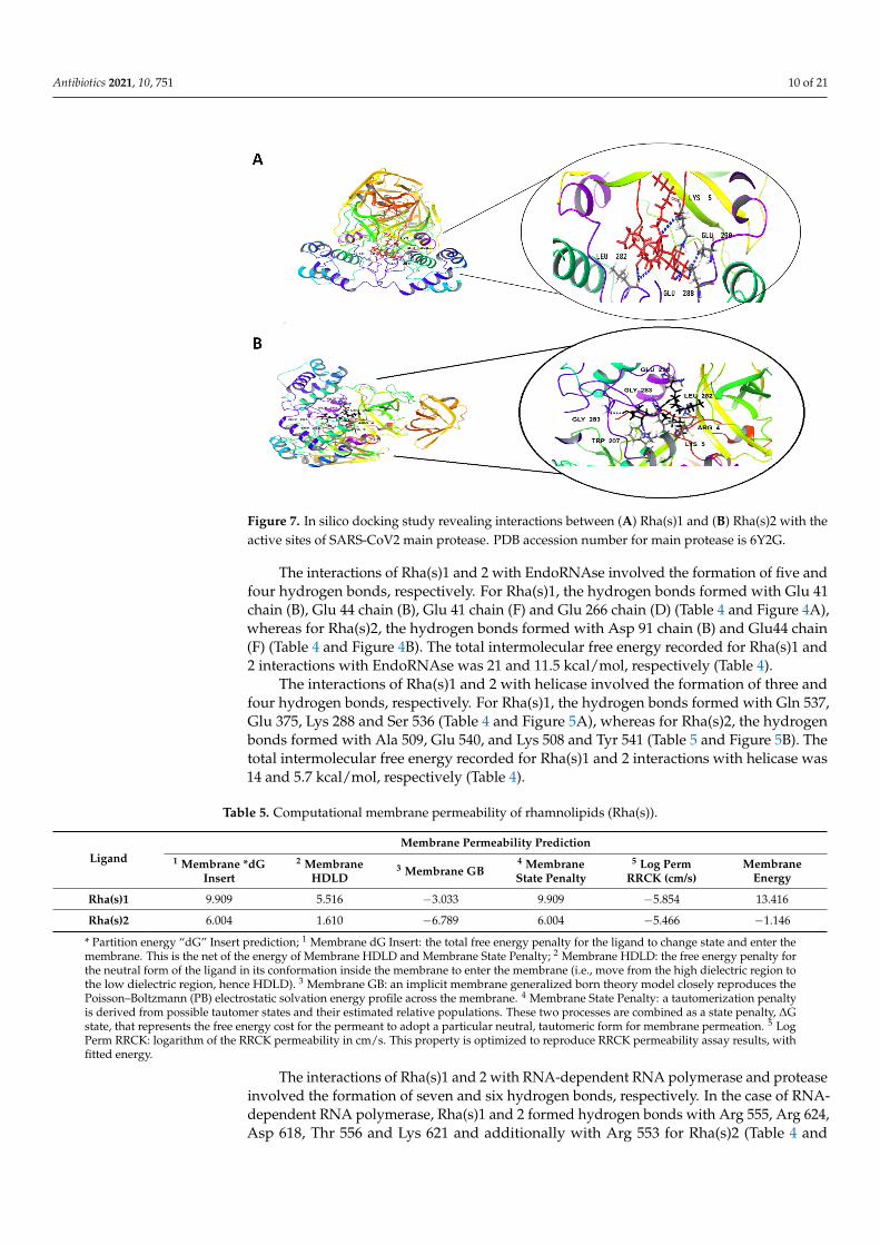
The effect of Rha(s)1 and 2 on the membrane permeability and the resultant harmfuleffect on the lipid envelop of SARS-CoV-2 was also investigated with the data presentedin Table 5. Membrane dG Insert value of Rha(s)1 and 2 were 9.909 and 6.004, respectively,suggesting their high permeability across the viral lipid envelope (Table 5). The calculatedmembrane permeability values of Rha(s)1 and 2 “Log Perm RRCK” were −5.854 and−5.466 cm/s, respectively (Table 5) [34]. Log Perm RRCK of Rha(s)2 was a less negativevalue, which is indicative of its higher permeability compared to Rha(s)1. The resultantharmful effect of Rha(s)1 and 2 on the lipid bilayer of the virus envelope and spike gly-coproteins integrity is schematically presented in Figure 8. A complete disruption of thevirus envelope was expected to occur after exposure to Rha(s).
Figure 8. Sketch presenting the damage effect caused by rhamnolipids on the lipid bilayer of virus envelope and spike glycoproteins.
3. Discussion
COVID-19, a pandemic infectious disease, has caused numerous health and economicproblems around the globe. Around 159 million people have been infected, and the numberof deaths had exceeded 3 million across 192 countries. The high rate of viral spread isdue to airborne transmission, the long asymptomatic period of the virus and its ability toremain infectious on the contaminated solid surfaces. Although, several new vaccines arecurrently being distributed globally, the protective effect of vaccine application globally isnot yet fully clear despite its promising efficacy in clinical trials and the associated positiveoutcomes on disease severity. Moreover, mutated strains of SARS-CoV-2 have appeared innumerous countries, some of which can partially escape vaccine-induced immunity andwill inevitably require development of updated vaccines in the future. Thus, there is still ahigh demand to identify a virus-specific treatment for COVID-19. Such antiviral therapies

Antibiotics 2021, 10, 751 12 of 21
would be a promising strategy for controlling the virus spread within hospitals and amongpeople in communities.
Proper hand hygiene was previously reported to decrease the incidence of infectionin hospitals among healthcare providers. Alcohol-based sanitizers are commonly useddue to their high efficacy and safety profiles, however, their misuse during the pandemicis reported to be associated with several adverse effects [7,9] as previously discussed.Therefore, finding alternative strategies for hand hygiene with less hazardous side effectsare highly recommended.
The antimicrobial activity of Rha(s) was previously reported [18,26,35] and their safetyprofiles have been established after its topical and ocular applications [18,21]. Rha(s) aremembers of the glycolipid biosurfactant and they were initially found to be produced bythe bacterium Pseudomonas aeruginosa [36]. They remain the best characterized and mostfrequently applied micro-organism for Rha(s) production [37,38]. Rha(s) are produced as amixture of various ligands with highly similar structures, functions and properties [39].As we show in Figure 1, they possess an amphiphilic property due to the compositionincluding a hydrophilic head with one or two rhamnose sugar residues and the lipophiliclipid tail comprising one or two fatty acid residues [40,41].
In our study, a bacterial strain with a faint green fluorescent pigment, strain LeS3,was isolated from lettuce leaves. Based on rRNA sequencing data and alignment to theNCBI database, the isolate was identified as Pseudomonas aeruginosa. The yield of producedRha(s) and ratios of its congeners (Rha(s)1 to Rha(s)2) depend on several factors, suchas the carbon source and the fermentation conditions [38,42]. For economic purposes,there is a continuous search for finding cost effective and renewable substrates to growPseudomonas aeruginosa [43,44]. Poultry carcass yields are typically about 70–75% of the livebird weight and is one of the waste products resulting from the poultry industry [45]. Car-casses contain a high content of organic matter [46] and this renders them good substratesfor use in the production of important products instead of their disposal by conventionalmethods. In our study, Rha(s) were produced by growing Pseudomonas aeruginosa strainLeS3 in a new medium formulated from chicken carcass soup (CCS) in a trial designed tooptimize the yield and the ratio of Rha(s)1 to 2. To the best of our knowledge, this is thefirst study using broth media wholly formulated from CCS for this purpose.
Our results indicated that Rha(s) yield was increased by 5 times with CCS mediumcompared to the commonly used medium, (GSNB), which may be due to the presenceof growth factors supporting the growth of the bacterium and subsequent production ofRha(s). We are assuming that the presence of some amino acids, vitamins and differentminerals might be responsible for such increment and this was matched with what wasreported previously when some byproducts such as molasses and whey were used for theproduction of Rha(s) using Pseudomonas aeruginosa [43,47]. However further studies arecurrently being investigated to figure out the role of different factors using such mediumin increasing Rha(s) yield.
The presence of a hydrophobic carbon source (chicken fats) favored the dominance ofRha(s)1 over Rha(s)2 as revealed by ESI-MS analysis shown in Section 2.2.1. Our resultsmatch those previously reported [42,48,49] where Rha(s)1 were the dominant compoundsproduced when the bacteria were grown in a medium supplemented with hydropho-bic carbon sources, whereas using hydrophilic carbon sources favored the dominanceof Rha(s)2.
In our study, R-C8:2 with the polyunsaturated β-hydroxy-fatty acid chains was thepredominant mono-rhamnolipid congener identified and this has been identified previ-ously [50]. In contrast to that reported by Nitschke and his colleagues [51], where Rha(s)1(R-C10C10) was the main component of Rha(s) produced when a hydrophobic carbonsource was used for growing Pseudomonas strains, while hydrophilic carbon sources leadto predominance of the Rha(s)2 such as (R-R-C10C10).Therefore, the new cost effectivemedium (CCS) used in our study was enriched with a hydrophobic carbon source and
Antibiotics 2021, 10, 751 13 of 21
favored the production of Rha(s)1. As was previously presented, Rha(s)1 showed a betterantiviral activity than Rha(s)2 against SARS-CoV-2.
Nanotechnology application is a growing field where it is interested in productionof fibers [52] and particles in nanometer scale to improve the therapeutic activity, reduceside effects of medicines and could be also used for diagnostic purposes [53,54]. Currentlynanotechnology is applied to combat the current COVID-19 pandemic [55–57]. Nanopar-ticles (NPs) have been applied previously for the treatment of several viral infectionswith promising results [27,54,58–60] and they were also demonstrated a good antibacterialactivity against multidrug resistant bacteria [61–63]. Therefore, the previously isolatedand characterized Rha(s) mixture was used to produce Rha(s) nano-micelles containingdifferent concentrations of rhamnolipids (1, 5 and 10 mg mL−1). The formed nano-micellesranged in size from 164 to 274 nm as measured by Malvern Zeta sizer. TEM images ob-tained for samples prepared at 5 mg mL−1 showed spherical nano-micelles with no signsof aggregation but with a reduced size than that recorded by Malvern zeta sizer. Thisdifference most likely reflects variations of size discrimination by these techniques. Allsamples produced were stable as revealed from Zeta potential values (≥−50). Importantly,samples were monodispersed with a lower tendency to aggregate as indicated by PDIvalue (PDI ≤ 0.3). However, those samples prepared at 1 mg mL−1 showed greater aggre-gation properties. This is consistent with other studies reporting that PDI < 0.3 [64–66] isindicative of good homogeneity and to be suitable for drug delivery applications. Whereasanother study revealed that nano-micelles of PDI > 0.3 is indicative of a highly polydispersesample [67].
The concentration and size dependent antibacterial activity of Rha(s) nano-micellesagainst selected Gram-negative and Gram-positive drug resistant bacterial strains wasdemonstrated by the agar well diffusion method. By increasing the concentration anddecreasing the size of nano-micelles, an increase of antibacterial activity against bothGram-positive and Gram-negative bacteria was noted with MIC values of 0.031 mg mL−1
and >0.5 mg mL−1 for Gram-positive and Gram-negative tested strains, respectively. Theimprovement of antibacterial activity by decreasing the size of nano-micelles might beattributed to the larger effective surface area offered by smaller nano-micelles for interactionwith the bacteria.
The antibacterial activity of Rha(s) could be due to their solubilizing effect on thephospholipid bilayer of the bacteria, thereby increasing the permeability and flow out ofmetabolites. Such a change in phospholipid bilayer structure and function was previouslyreported to affect protein conformation, transport and energy generation, ultimately leadingto bacterial cell death [49].
The antimicrobial activity of Rha(s) is affected by the Rha(s) 1 to Rha(s) 2 ratio [23]. Ourresults revealed a higher antibacterial activity against Gram-positive bacteria that contrastswith what was reported by Das and his colleagues [68] who found that changing theproportion of Rha(s)1 to Rha(s)2 is accompanied with a change of the antimicrobial activityof Rha(s) mixture where an increase of mono to di species, the Rha(s) mixture becomesmore effective against Gram-negative bacterial strains. It was concluded that an increase ofemulsification index could enhance the penetration of Rha(s) across the cell wall of Gram-negative bacteria, resulting in death. Our data supports what was commonly reported inthe literature where Rha(s) showed greater antibacterial activity against Gram-positive overGram-negative bacteria [23,69–71]. The reduced effect on Gram negative bacteria could beattributed to the presence of the lipopolysaccharide outer membrane that confers protectionto the cell. Similarly, de Freitas Ferreira and colleagues [70] reported a predominance ofRha(s)1 to Rha(s)2 by a ratio of 2:1, also revealed a resistance of Gram-negative strains. Thisis contrary to the Rha(s) mixture produced by P. aeruginosa 47T2 with the predominance ofRha(s)2 homologs and was reported to inhibit the growth of Escherichia coli and SalmonellaTyphimurium with MIC values of 64 µg mL−1 and 128 µg mL−1 respectively [18]. Therefore,the differences observed in the sensitivity of Gram-negative strains to Rha(s) might bedependent not only on the composition of biosurfactant and its purity but also on the

Antibiotics 2021, 10, 751 14 of 21
bacterial adaptation ability [72]. The nutritional and environmental conditions might alsohave additional influence on the antimicrobial activity as previously reported [70].
A molecular docking study was also performed to investigate the potential antiviralactivity of Rha(s) against SARS-CoV-2. Compounds that interact with the SARS-CoV-2 spikeglycoproteins (S1-N-terminal domain (NTD) and S2 part) are hypothesized to interferewith virus attachment to host entry receptors (ACE-2) with a consequent loss of viralinfectivity. As revealed from our docking studies, Rha(s)1 and 2 interact efficiently withthe SARS-CoV-2 spike glycoproteins with Rha(s)1 forming the most stable complex asindicated by its lower free energy. These interactions are expected to result in irreversiblechanges to the virus spike proteins structure and inhibition of viral infectivity. Our data alsoshowed the ability of Rha(s) as a biosurfactant to interact with the lipid membranes (lipidenvelope) of SARS-CoV-2, which is expected to be associated with disruption of membranepermeability similar to that previously observed for HSV1 and HSV2 [73–76]. Takentogether, Rha(s) could therefore be a promising agent to control the spread of COVID-19infection and warrants further in vitro study of viral infection. Ultimately, Rha(s) couldbe used by patients with COVID-19 and health care providers as a hand sanitizer in thecurrent pandemic.
Further docking studies were performed to understand the interaction of Rha(s)1 and 2with the active sites of enzymes involved in virus replication. These included four keyviral proteins including EndoRNAse, helicase, RNA-dependent RNA polymerase and themain protease. Our results showed that the free energy from interaction of Rha(s)1 withthese enzymes was lower than that obtained with Rha(s)2. These Rha(s) interactions wereexpected to cause detrimental conformational changes at the active binding sites of theseenzymes rendering them inactive in virus replication. Therefore, Rha(s) nano-micellesmight be recommended for treatment of COVID-19 infection, however, their safety profilemust be addressed and further in vitro studies confirmed.
Although the docking studies revealed the interactions between Rha(s) as a singlemolecule with SARS-CoV-2 proteins, these interactions could however also be appliedto Rha(s) nano-micelles. The proposed interactions of surfactant as a single molecule orin the form of nano-micelles has been previously reported [77,78] and is presented herein Figure 9. The interactions of surfactant either side of the critical micelle concentration(CMC) with SARS-CoV-2 were previously reported [77]. Below CMC, it was reported [79]that the phospholipid in the bilayer and the surfactant monomers interact via hydrophobicinteractions between the lipid tails and the surfactant tails. Molecules of the added surfac-tants are also inserted into the bilayer, competing with the phospholipids, thus disturbingthe orderly arranged structure of the membrane. When the surfactant concentration ap-proaches the CMC, the lipid–surfactant mixed bilayers become saturated and no longeraccommodate additional surfactants. This induces solubilization of the phospholipidsvia phase transformation of the mixed bilayer into mixed (lipid–surfactant) micelles [80].Above CMC, when the surfactant-to-lipid concentration ratio increases, micellization iscompleted, i.e., the lipid bilayer is completely solubilized by the surfactants and only themicellar aggregates remain in the solution [81]. Thus, the complete solubilization of theprotective lipid bilayer leads to the potential disintegration of the virus into fragments,neutralizing infectivity. Alternatively, micelles are able to completely entrap the viralparticle internally via hydrophobic–hydrophobic interactions [77]. Based on these finding,Rha(s) nano-micelles show a high potential for use against SARS-CoV-2 as a hand sanitizerto combat the current pandemic infections.
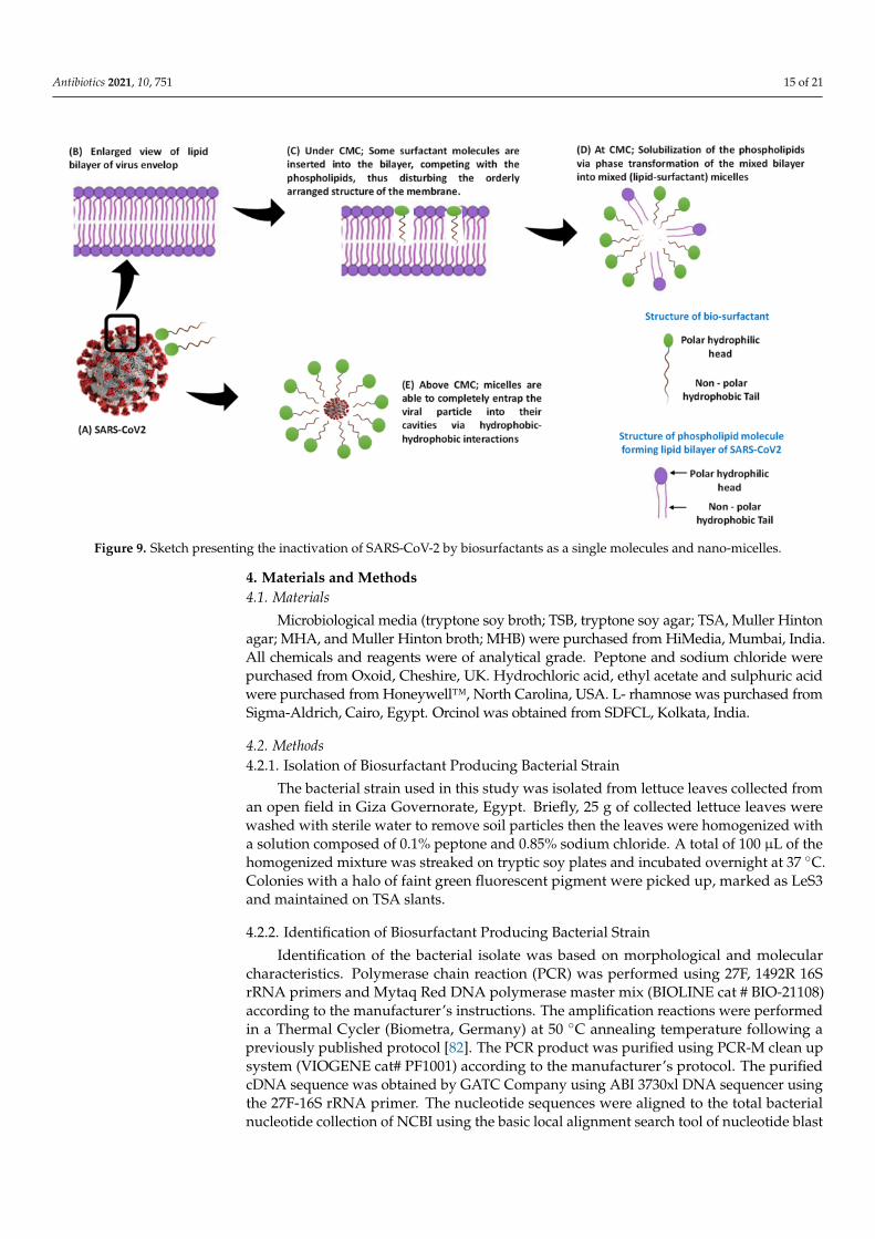
Antibiotics 2021, 10, 751 15 of 21
Figure 9. Sketch presenting the inactivation of SARS-CoV-2 by biosurfactants as a single molecules and nano-micelles.
4. Materials and Methods4.1. Materials
Microbiological media (tryptone soy broth; TSB, tryptone soy agar; TSA, Muller Hintonagar; MHA, and Muller Hinton broth; MHB) were purchased from HiMedia, Mumbai, India.All chemicals and reagents were of analytical grade. Peptone and sodium chloride werepurchased from Oxoid, Cheshire, UK. Hydrochloric acid, ethyl acetate and sulphuric acidwere purchased from Honeywell™, North Carolina, USA. L- rhamnose was purchased fromSigma-Aldrich, Cairo, Egypt. Orcinol was obtained from SDFCL, Kolkata, India.
4.2. Methods4.2.1. Isolation of Biosurfactant Producing Bacterial Strain
The bacterial strain used in this study was isolated from lettuce leaves collected froman open field in Giza Governorate, Egypt. Briefly, 25 g of collected lettuce leaves werewashed with sterile water to remove soil particles then the leaves were homogenized witha solution composed of 0.1% peptone and 0.85% sodium chloride. A total of 100 µL of thehomogenized mixture was streaked on tryptic soy plates and incubated overnight at 37 ◦C.Colonies with a halo of faint green fluorescent pigment were picked up, marked as LeS3and maintained on TSA slants.
4.2.2. Identification of Biosurfactant Producing Bacterial Strain
Identification of the bacterial isolate was based on morphological and molecularcharacteristics. Polymerase chain reaction (PCR) was performed using 27F, 1492R 16SrRNA primers and Mytaq Red DNA polymerase master mix (BIOLINE cat # BIO-21108)according to the manufacturer’s instructions. The amplification reactions were performedin a Thermal Cycler (Biometra, Germany) at 50 ◦C annealing temperature following apreviously published protocol [82]. The PCR product was purified using PCR-M clean upsystem (VIOGENE cat# PF1001) according to the manufacturer’s protocol. The purifiedcDNA sequence was obtained by GATC Company using ABI 3730xl DNA sequencer usingthe 27F-16S rRNA primer. The nucleotide sequences were aligned to the total bacterialnucleotide collection of NCBI using the basic local alignment search tool of nucleotide blast

Antibiotics 2021, 10, 751 16 of 21
(https://blast.ncbi.nlm.nih.gov/Blast.cgi), accessed date 18 January 2020 and released date23 January 2020).
4.2.3. Production and Characterization of Rha(s)Production of Rha(s)
Rha(s) production was carried out following a literature protocol using shake flasktechnique and separated from the production medium by acid precipitation followedby organic solvent extraction [83]. New production medium formulated from chickencarcass soup (CCS) containing 5% chicken fat, 0.5% NaCl was compared with glycerolsupplemented nutrient broth (GSNB) (nutrient broth medium containing 10 g L−1 glycerol)to optimize the yield of Rha(s). Briefly, Pseudomonas aeruginosa strain LeS3 was grown inTSB to obtain OD600 of 0.8, which corresponds to a density of 8 log cfu mL-1 A 250 mLErlenmeyer flask containing 100 mL of either sterilized GSNB or CCS was then inoculatedwith 1% of prepared bacterial culture. Inoculated flasks were incubated in the orbitalshaker (Vision Scientific Co., Ltd. Korea. VS-8480SR) at 30 ◦C and 150 rpm for 5 days.
Following the incubation period, bacterial cells were removed from the culture brothby centrifugation at 10,000 rpm at 5 ◦C for 10 min (Sigma, Germany, 3-16PK) to obtaincell free supernatant (CFS). The CFS was acidified to pH 2.0 using 1N HCl and storedovernight at 5 ◦C. Rha(s) were then extracted using an equal volume of ethyl acetate. Theyellow/brown viscous paste was then stored at 4 ◦C until further characterization.
Quantification of Rha(s) was carried out using the orcinol assay [83], 333 µL of CFSwas extracted twice with ethyl acetate and evaporated followed by the addition of 0.5 mLdistilled water. The assay mixture consisted of freshly prepared reagent containing 0.19%orcinol in 53% sulphuric acid that was added to the extracted biosurfactant in the ratio of9:1. This mixture was heated in a water bath at 80 ◦C for 30 min and allowed to cool to roomtemperature before measuring its optical density at 421 nm. The Rha(s) concentrations werecalculated from a standard curve prepared with L-rhamnose and expressed as rhamnoseequivalents (RE) by multiplying rhamnose values by a coefficient of 3.4 obtained from thecorrelation of pure Rha(s)/rhamnose [84].
Characterization of Rha(s)
ESI-MS Analysis of Rha(s) MixtureA mixture of Rha(s) (crude extract) was prepared at a concentration of 100 µg mL−1
for ESI-MS analysis. Chromatographic separation was performed on an Acquity UPLCsystem (BEH C18 Column, 130 Å, 1.7 µm, 2.1 mm × 50 mm) with gradients elution ofMeOH/H2O at a flow rate of 0.2 mL min−1. The column temperature was maintained at25 ◦C and the injection volume was 10 µL. The UPLC is coupled to an online PDA and MSdetector. The ESI-MS analysis in both positive and negative ion mode was carried out on aXEVO TQD triple quadruple instrument (Waters Corporation, Milford, MA 01757, USA,mass spectrometer).
Data ProcessingThe peaks and spectra were processed using the Maslynx 4.1 software and tentatively
identified by comparing its retention time (Rt) and mass spectrum with reported data.
4.2.4. Preparation and Characterization of Rha(s) Nano-Micelles
Rha(s) nano-micelles were prepared using different concentrations (1, 5 and 10 mg mL−1)of Rha(s) crude extract that were dispersed in PBS, pH7.4 using a probe sonicator for 3 min.Particle size and zeta potential were determined using a Malvern Zeta-sizer Nano ZS(Malvern Instruments Ltd., Malvern, UK) at 25 ± 0.1 ◦C.
Rha(s) nano-micelles were imaged by TEM (H-700, Hitachi Ltd., Japan), at an ac-celerated voltage of 80 kv using the negative staining method. The solution of Rha(s)nano-micelles was diluted (1:50) with distilled water then a drop of the diluted solutionwas spread on a mesh copper grid coated with carbon film and was kept for 5 min to dry.

Antibiotics 2021, 10, 751 17 of 21
Then after, a drop of phospho-tungstic acid (2% w/w) was added on the grid for 50 s, beforeexcess liquid was removed using filter paper.
4.2.5. The Antibacterial Activity of Rha(s)
The antibacterial activity of produced Rha(s) was evaluated against selected multi drugresistant strains, Streptococcus pneumoniae, Staphylococcus aureus, Salmonella Montevideoand Salmonella Typhimurium using agar well diffusion method according to a publishedprotocol [85]. Briefly, Mueller–Hinton agar plate surface was spread with a volume offreshly prepared microbial inoculum containing 5 log cfu mL−1. Then, a hole with adiameter equivalent to 7 mm was punched under aseptic conditions using a sterile bluetip base. Then, the formed wells were filled in with 100 µL of the previously preparedRha(s) nano-micelles colloidal solutions. Following inoculation, plates were incubated at37 ◦C for 24 h and examined for bacterial growth and clear zone formation. Minimuminhibitory concentration (MIC) of Rha(s) against specific bacterial strains was determinedusing a microdilution assay [86]. Briefly, two-fold serial dilutions of Rha(s) (ranging from 1to 0.0195 mg mL−1) in MHB were prepared and added to 96 well plates. Wells were theninoculated with 10 µL of 5 log cfu mL−1 (final inoculum) of bacteria. Inoculated plates werethen incubated for 18–24 h at 37 ◦C. The MIC value was defined as the lowest concentrationthat inhibited visible growth.
4.2.6. Docking Study
For the COVID-19 in silico study, the chemical and 3D structure of Rha(s)1 and Rha(s) 2presented in Figure 1 were obtained from the PubChem database (https://pubchem.ncbi.nlm.nih.gov, accessed on 16 June 2021). The Protein Data Bank (PDB) database(https://www.rcsb.org/, accessed on 16 June 2021) was used to obtain the complete3D structure of SARS-CoV-2 EndoRNAse (PDB accession number: 6X1B), helicase (PDBaccession number: 5RL6), RNA-dependent RNA polymerase (PDB accession number:7CYQ), spike glycoproteins (PDB accession number: 7CWU) and protease enzyme (PDBaccession number: 6Y2G)
In Silico Molecular Modelling
The interaction of rhamnolipids, Rha(s)1 and 2 with SARS-CoV-2 spike glycoproteinsand enzymes involved in viral replication: EndoRNAse, helicase, RNA-dependent RNApolymerase and protease were studied using in silico docking. The ligands binding studieswere carried out using Maestro 11.9 software. Energy minimization of ligand was optimizedand ligand preparation was performed using LigPrep 2.4 software. Docking of ligands wasachieved using Schrodinger Maestro 11.9 software and Glide’s Extra Precision (XP) [87].The size of grid box for each protein was set to 20 Å by default.
4.2.7. Statistical Analysis
Statistical analysis was done by Minitab version 17 at a confidence level of 95% usinga two-way ANOVA.
5. Conclusions
Rha(s) isolated from a novel strain of Pseudomonas aeruginosa strain LeS3 were shownto form a variety of typical structures with the congener R-C8:2 containing the polyunsatu-rated β-hydroxy-fatty acid chains predominating. Rha(s) nano-micelles prepared from aRha(s) mixture had a size ranged from 164 ± 1 to 274 ± 50 nm and by increasing Rha(s)concentration ≥5 mg mL−1, a monodisperse sample was obtained as revealed by a PDIvalue that was ≤0.3. Rha(s) nano-micelles showed antibacterial activity against both Gram-positive (Streptococcus pneumoniae and Staphylococcus aureus) and Gram-negative (SalmonellaMontevideo and Salmonella Typhimurium) human bacterial pathogens. The antibacterialactivity was recorded to be size and concentration dependent with preferred antibacterialactivity against Gram-positive bacteria where MIC (0.031 mg mL−1) with Gram-positive

Antibiotics 2021, 10, 751 18 of 21
bacteria was significantly (p < 0.05) lower than its value (>0.5 mg mL−1) for Gram-negativebacteria. The molecular docking studies showed interactions with SARS-CoV-2 structuraland non-structural proteins. Computational studies also demonstrated membrane per-meability activities suggesting that Rha(s) nano-micelles have multiple mechanisms ofaction and could be applied as antiviral agents in addition to their antibacterial function.Therefore, Rha(s) nano-micelles could be recommended to replace alcohol-based handsanitizer in general communities and health care settings under the current pandemicinfection of COVID-19. Furthermore, Rha(s) nano-micelles might be also recommended to beinvestigated in the treatment of COVID-19. However, further studies should be performedto address their antiviral effects and safety profiles. These additional studies are currentlyunderway to establish its antiviral activity and development as interventions for COVID-19.
Supplementary Materials: The following are available online at https://www.mdpi.com/article/10.3390/antibiotics10070751/s1, Figure S1: PCR amplification products of 16S-rRNA encoding genes ofPseudomonas aeruginosa strain LeS3 showed a fragment length of 1500 bp. (1 kb+) refers to DNA ladder.
Author Contributions: Conceptualization, M.R.B., A.H.I.F. and Y.A.-z.; Data curation, M.R.B.,A.H.I.F., E.R.S.S., M.S.F., A.M.M.S., A.M.S.H., G.E.E., N.R.R. and Y.A.-z.; Formal analysis, M.R.B.,A.H.I.F., A.M.S.H. and Y.A.-z.; Investigation, M.R.B., A.H.I.F., E.R.S.S., M.S.F., A.M.M.S., G.R.M.,A.M.S.H., G.E.E., N.R.R. and Y.A.-z.; Methodology; M.R.B., A.H.I.F., E.R.S.S., M.S.F., A.M.M.S.,A.M.S.H., G.E.E., N.R.R. and Y.A.-z.; Visualization, M.R.B., A.H.I.F., G.R.M. and Y.A.-z.; Writing—original draft, M.R.B., A.H.I.F., G.R.M. and Y.A.-z.; project administration; Y.A.-z.; funding acquisition;Y.A.-z. All authors have read and agreed to the published version of the manuscript.
Funding: This research was funded by the Science, Technology and Innovation Funding Authority(STIFA), Emergency Targeted Program COVID-19 Emergency call 2020, grant number 43766 andHelwan University.
Data Availability Statement: All authors are happy to share all data (including supplementary data)presented in this manuscript to the public repository.
Conflicts of Interest: The authors declare no conflict of interest.
References1. Huang, C.; Wang, Y.; Li, X.; Ren, L.; Zhao, J.; Hu, Y.; Zhang, L.; Fan, G.; Xu, J.; Gu, X.; et al. Clinical features of patients infected
with 2019 novel coronavirus in Wuhan, China. Lancet 2020, 395, 497–506. [CrossRef]2. Lai, C.C.; Shih, T.P.; Ko, W.C.; Tang, H.J.; Hsueh, P.R. Severe acute respiratory syndrome coronavirus 2 (SARS-CoV-2) and
coronavirus disease-2019 (COVID-19): The epidemic and the challenges. Int. J. Antimicrob. Agents 2020, 55, 105924. [CrossRef][PubMed]
3. Kratzel, A.; Todt, D.; V’kovski, P.; Steiner, S.; Gultom, M.; Thao, T.T.N.; Ebert, N.; Holwerda, M.; Steinmann, J.; Niemeyer, D.; et al.Inactivation of Severe Acute Respiratory Syndrome Coronavirus 2 by WHO-Recommended Hand Rub Formulations and Alcohols.Emerg. Infect. Dis. 2020, 26, 1592–1595. [CrossRef] [PubMed]
4. Kampf, G.; Todt, D.; Pfaender, S.; Steinmann, E. Persistence of coronaviruses on inanimate surfaces and their inactivation withbiocidal agents. J. Hosp. Infect. 2020, 104, 246–251. [CrossRef]
5. Otter, J.A.; Donskey, C.; Yezli, S.; Douthwaite, S.; Goldenberg, S.D.; Weber, D.J. Transmission of SARS and MERS coronavirusesand influenza virus in healthcare settings: The possible role of dry surface contamination. J. Hosp. Infect. 2016, 92, 235–250.[CrossRef] [PubMed]
6. Dowell, S.F.; Simmerman, J.M.; Erdman, D.D.; Wu, J.-S.J.; Chaovavanich, A.; Javadi, M.; Yang, J.-Y.; Anderson, L.J.; Tong, S.; Ho, M.S.Severe Acute Respiratory Syndrome Coronavirus on Hospital Surfaces. Clin. Infect. Dis. 2004, 39, 652–657. [CrossRef]
7. Emami, A.; Javanmardi, F.; Keshavarzi, A.N.P. Hidden threat lurking behind the alcohol sanitizers in CoVID-19 outbreak.Dermatol. Ther. 2020, 33. [CrossRef]
8. Slaughter, R.J.; Mason, R.W.; Beasley, D.M.G.; Vale, J.A.; Schep, L.J. Isopropanol poisoning. Clin. Toxicol. 2014, 52, 470–478.[CrossRef]
9. Mahmood, A.; Eqan, M.; Pervez, S.; Ahmed, H.; Bari, A. COVID-19 and frequent use of hand sanitizers; human health andenvironmental hazards by exposure pathways. Sci. Total Environ. 2020, 742, 1–7. [CrossRef]
10. Vogel, L. Hand sanitizers may increase norovirus risk. CMAJ 2011, 183, 799–800. [CrossRef]11. Blaney, D.D.; Daly, E.R.; Kirkland, K.B.; Tongren, J.E.; Kelso, P.T.; Talbot, E.A. Use of alcohol-based hand sanitizers as a risk factor
for norovirus outbreaks in long-term care facilities in northern New England: December 2006 to March 2007. Am. J. Infect. Control2011, 39, 296–301. [CrossRef] [PubMed]

Antibiotics 2021, 10, 751 19 of 21
12. Jiang, L.; Shen, C.; Long, X.; Zhang, G.; Meng, Q. Rhamnolipids elicit the same cytotoxic sensitivity between cancer cell andnormal cell by reducing surface tension of culture medium. Appl. Microbiol. Biotechnol. 2014. [CrossRef] [PubMed]
13. Pidot, S.J.; Gao, W.; Buultjens, A.H.; Monk, I.R.; Guerillot, R.; Carter, G.P.; Lee, J.Y.H.; Lam, M.M.C.; Grayson, M.L.; Ballard, S.A.; et al.Increasing tolerance of hospital Enterococcus faecium to handwash alcohols. Sci. Transl. Med. 2018, 10. [CrossRef]
14. Hayat, A.; Munnawar, F. Antibacterial Effectiveness of Commercially Available Hand Sanitizers. Int. J. Biol. Biotech. 2016, 13,427–431.
15. Rosenberg, E.; DeLong, E.F.; Thompson, F.; Lory, S.; Stackebrandt, E. The prokaryotes: Applied bacteriology and biotechnology.Prokaryotes Appl. Bacteriol. Biotechnol. 2013, 9783642313, 1–393. [CrossRef]
16. Kumar, R.; Das, A.J. Rhamnolipid Biosurfactant: Recent Trends in Production and Application; Springer: Singapore, 2018; ISBN9789811312892.
17. Johann, S.; Seiler, T.B.; Tiso, T.; Bluhm, K.; Blank, L.M.; Hollert, H. Mechanism-specific and whole-organism ecotoxicity ofmono-rhamnolipids. Sci. Total Environ. 2016. [CrossRef] [PubMed]
18. Haba, E.; Pinazo, A.; Jauregui, O.; Espuny, M.J.; Infante, M.R.; Manresa, A. Physicochemical characterization and antimicrobialproperties of rhamnolipids produced by Pseudomonas aeruginosa 47T2 NCBIM 40044. Biotechnol. Bioeng. 2003. [CrossRef]
19. Banat, I.M.; Makkar, R.S.; Cameotra, S.S. Potential commercial applications of microbial surfactants. Appl. Microbiol. Biotechnol.2000, 53, 495–508. [CrossRef]
20. Marchant, R.; Banat, I.M. Biosurfactants: A sustainable replacement for chemical surfactants? Biotechnol. Lett. 2012, 34, 1597–1605.[CrossRef]
21. Bharali, P.; Das, S.; Ray, A.; Pradeep Singh, S.; Bora, U.; Kumar Konwar, B.; Singh, C.B.; Sahoo, D. Biocompatibility natural effectof rhamnolipids in bioremediation process on different biological systems at the site of contamination. Bioremediat. J. 2018, 22,91–102. [CrossRef]
22. Müller, M.M.; Hörmann, B.; Syldatk, C.; Hausmann, R. Pseudomonas aeruginosa PAO1 as a model for rhamnolipid production inbioreactor systems. Appl. Microbiol. Biotechnol. 2010. [CrossRef]
23. Lotfabad, T.B.; Shahcheraghi, F.; Shooraj, F. Assessment of antibacterial capability of rhamnolipids produced by two indigenousPseudomonas aeruginosa strains. Jundishapur J. Microbiol. 2013, 6, 29–35. [CrossRef]
24. Carrazco-Palafox, J.; Rivera-Chavira, B.E.; Adame-Gallegos, J.R.; Rodríguez-Valdez, L.M.; Orrantia-Borunda, E.; Nevárez-Moorillón, G.V. Rhamnolipids from Pseudomonas aeruginosa Rn19a Modifies the Biofilm Formation over a Borosilicate Surfaceby Clinical Isolates. Coatings 2021, 11, 136. [CrossRef]
25. Sana, S.; Datta, S.; Biswas, D.; Sengupta, D. Assessment of synergistic antibacterial activity of combined biosurfactants revealedby bacterial cell envelop damage. Biochim. Biophys. Acta Biomembr. 2018, 1860, 579–585. [CrossRef]
26. Remichkova, M.; Galabova, D.; Roeva, I.; Karpenko, E.; Shulga, A.; Galabov, A.S. Anti-herpesvirus activities of Pseudomonas sp.S-17 rhamnolipid and its complex with alginate. Zeitschrift Fur Naturforsch. Sect. C J. Biosci. 2008, 63, 75–81. [CrossRef]
27. Abo-Zeid, Y.; Ismail, N.S.; McLean, G.R.; Hamdy, N.M. A Molecular Docking Study Repurposes FDA Approved Iron OxideNanoparticles to Treat and Control COVID-19 Infection. Eur. J. Pharm. Sci. 2020, 153, 105465. [CrossRef] [PubMed]
28. Andersen, K.G.; Rambaut, A.; Lipkin, W.I.; Holmes, E.C.; Garry, R.F. The proximal origin of SARS-CoV-2. Nat. Med. 2020, 26,450–452. [CrossRef]
29. Choudhary, S.; Malik, Y.S.; Tomar, S. Identification of SARS-CoV-2 Cell Entry Inhibitors by Drug Repurposing Using in silicoStructure-Based Virtual Screening Approach. Front. Immunol. 2020. [CrossRef] [PubMed]
30. Déziel, E.; Lépine, F.; Dennie, D.; Boismenu, D.; Mamer, O.A.; Villemur, R. Liquid chromatography/mass spectrometry analysisof mixtures of rhamnolipids produced by Pseudomonas aeruginosa strain 57RP grown on mannitol or naphthalene. Biochim.Biophys. Acta Mol. Cell Biol. Lipids 1999, 1440, 244–252. [CrossRef]
31. Zhao, F.; Shi, R.; Ma, F.; Han, S.; Zhang, Y. Oxygen effects on rhamnolipids production by Pseudomonas aeruginosa. Microb. Cell Fact.2018, 17, 1–11. [CrossRef]
32. Zhao, F.; Jiang, H.; Sun, H.; Liu, C.; Han, S.; Zhang, Y. Production of rhamnolipids with different proportions of mono-rhamnolipids using crude glycerol and a comparison of their application potential for oil recovery from oily sludge. RSC Adv.2019, 9, 2885–2891. [CrossRef]
33. Pantazaki, A.A.; Papaneophytou, C.P.; Lambropoulou, D.A. Simultaneous polyhydroxyalkanoates and rhamnolipids productionby thermus thermophilus HB8. AMB Express 2011, 1, 1–13. [CrossRef]
34. Leung, S.S.F.; Sindhikara, D.; Jacobson, M.P. Simple Predictive Models of Passive Membrane Permeability Incorporating Size-Dependent Membrane-Water Partition. J. Chem. Inf. Model. 2016. [CrossRef] [PubMed]
35. Ndlovu, T.; Rautenbach, M.; Vosloo, J.A.; Khan, S.; Khan, W. Characterisation and antimicrobial activity of biosurfactant extractsproduced by Bacillus amyloliquefaciens and Pseudomonas aeruginosa isolated from a wastewater treatment plant. AMB Express2017, 7. [CrossRef] [PubMed]
36. Jarvis, F.G.; Johnson, M.J. A Glyco-lipide Produced by Pseudomonas Aeruginosa. J. Am. Chem. Soc. 1949. [CrossRef]37. Tiso, T.; Thies, S.; Müller, M.; Tsvetanova, L.; Carraresi, L.; Bröring, S.; Jaeger, K. Rhamnolipids: Production, Performance, and
Application. In Consequences of Microbial Interactions with Hydrocarbons, Oils, and Lipids: Production of Fuels and Chemicals; Lee, S.Y., Ed.;Springer: Cham, Switzerland, 2017; ISBN 9783319504360.
38. Chong, H.; Li, Q. Microbial production of rhamnolipids: Opportunities, challenges and strategies. Microb. Cell Fact. 2017, 16,1–12. [CrossRef]

Antibiotics 2021, 10, 751 20 of 21
39. Abdel-Mawgoud, A.M.; Lépine, F.; Déziel, E. Rhamnolipids: Diversity of structures, microbial origins and roles. Appl. Microbiol. Biotechnol.2010, 86, 1323–1336. [CrossRef]
40. Nie, M.; Yin, X.; Ren, C.; Wang, Y.; Xu, F.; Shen, Q. Novel rhamnolipid biosurfactants produced by a polycyclic aromatichydrocarbon-degrading bacterium Pseudomonas aeruginosa strain NY3. Biotechnol. Adv. 2010. [CrossRef] [PubMed]
41. Kaskatepe, B.; Yildiz, S. Rhamnolipid biosurfactants produced by pseudomonas Species. Brazilian Arch. Biol. Technol. 2016.[CrossRef]
42. Arutchelvi, J.; Doble, M. Characterization of glycolipid biosurfactant from Pseudomonas aeruginosa CPCL isolated frompetroleum-contaminated soil. Lett. Appl. Microbiol. 2010, 51, 75–82. [CrossRef]
43. Tan, Y.N.; Li, Q. Microbial production of rhamnolipids using sugars as carbon sources. Microb. Cell Fact. 2018, 17, 1–13. [CrossRef][PubMed]
44. Banat, I.M.; Satpute, S.K.; Cameotra, S.S.; Patil, R.; Nyayanit, N.V. Cost effective technologies and renewable substrates forbiosurfactants’ production. Front. Microbiol. 2014, 5, 1–18. [CrossRef] [PubMed]
45. Ozdemir, S.; Yetilmezsoy, K. A mini literature review on sustainable management of poultry abattoir wastes. J. Mater. CyclesWaste Manag. 2020, 22, 11–21. [CrossRef]
46. IA, B. Traditional Methods of Carcass Disposal: A Review. J. Dairy Vet. Anim. Res. 2017, 5, 21–27. [CrossRef]47. Makkar, R.S.; Cameotra, S.S.; Banat, I.M. Advances in utilization of renewable substrates for biosurfactant production. AMB Express
2011, 1, 1–19. [CrossRef]48. Sabturani, N.; Latif, J.; Radiman, S.; Hamzah, A. Analisis spektroskopik ramnolipid yang dihasilkan oleh P. aeruginosa UKMP14T.
Malays. J. Anal. Sci. 2016, 20, 31–43. [CrossRef]49. Rikalovic, M.G.; Gojgic-Cvijovic, G.; Vrvic, M.M.; Karadžic, I. Production and characterization of rhamnolipids from Pseudomonas
aeruginosa san-ai. J. Serbian Chem. Soc. 2012, 77, 27–42. [CrossRef]50. Abalos, A.; Pinazo, A.; Infante, M.R.; Casals, M.; García, F.; Manresa, A. Physicochemical and antimicrobial properties of new
rhamnolipids produced by Pseudomonas aeruginosa AT10 from soybean oil refinery wastes. Langmuir 2001. [CrossRef]51. Nitschke, M.; Paulista, U.E.; Nitschke, M.; Costa, S.G.V.A.O.; Haddad, R.; Gonc, L.A.G.; Eberlin, M.N.; Contiero, J. Oil Wastes as
Unconventional Substrates for Rhamnolipid Biosurfactant Production by Pseudomonas aeruginosa LBI Oil Wastes as Unconven-tional Substrates for Rhamnolipid Biosurfactant Production by Pseudomonas aeruginosa LBI. Biotechnol. Prog. 2005, 3, 1562–1566.[CrossRef] [PubMed]
52. Burgess, K.; Li, H.; Abo-Zeid, Y.; Fatimah; Williams, G.R. The effect of molecular properties on active ingredient release fromelectrospun eudragit fibers. Pharmaceutics 2018, 10, 103. [CrossRef] [PubMed]
53. Wang, Y.; Zhang, Z.; Abo-zeid, Y.; Bear, J.C.; Davies, G.; Lei, X.; Williams, G.R. SiO2-coated layered gadolinium hydroxides forsimultaneous drug delivery and magnetic resonance imaging. J. Solid State Chem. 2020. [CrossRef]
54. Abo-zeid, Y.; Garnett, M.C. Polymer nanoparticle as a delivery system for ribavirin: Do nanoparticle avoid uptake by Red BloodCells? J. Drug Deliv. Sci. Technol. 2020, 56, 101552. [CrossRef]
55. Chintagunta, A.D.; Krishna, M.S.; Nalluru, S.; Sampath, S.K. Nanotechnology: An emerging approach to combat COVID-19.Emergent Mater. 2021. [CrossRef]
56. Rangayasami, A.; Kannan, K.; Murugesan, S.; Radhika, D.; Sadasivuni, K.K.; Reddy, K.R.; Raghu, A.V. Influence of nanotechnologyto combat against COVID-19 for global health emergency: A review. Sens. Int. 2021, 2, 100079. [CrossRef]
57. Campos, E.V.R.; Pereira, A.E.S.; De Oliveira, J.L.; Carvalho, L.B.; Guilger-Casagrande, M.; De Lima, R.; Fraceto, L.F. How cannanotechnology help to combat COVID-19? Opportunities and urgent need. J. Nanobiotechnol. 2020, 18, 1–23. [CrossRef]
58. Abo-zeid, Y.; Urbanowicz, R.A.; Thomsonb, B.J.; William, L.; Irvingb, A.W.T.; Garnett, M.C. Enhanced nanoparticle uptake intovirus infected cells: Could nanoparticles be useful in antiviral therapy? Int. J. Pharm. 2018, 547, 572–581. [CrossRef] [PubMed]
59. Abo-zeid, Y.; Williams, G.R.; Touabi, L.; Mclean, G.R. An investigation of rhinovirus infection on cellular uptake of poly(glycerol-adipate) nanoparticles. Int. J. Pharm. 2020, 119826. [CrossRef] [PubMed]
60. Abo-zeid, Y.; Mantovani, G.; Irving, W.L.; Garnett, M.C. Synthesis of nucleoside-boronic esters hydrophobic pro-drugs: A possibleroute to improve hydrophilic nucleoside drug loading into polymer nanoparticles. J. Drug Deliv. Sci. Technol. 2018, 46, 354–364.[CrossRef]
61. AlMatar, M.; Makky, E.A.; Var, I.; Koksal, F. The Role of Nanoparticles in the Inhibition of Multidrug-resistant Bacteria andBiofilms. Curr. Drug Deliv. 2017, 15. [CrossRef] [PubMed]
62. Baptista, P.V.; McCusker, M.P.; Carvalho, A.; Ferreira, D.A.; Mohan, N.M.; Martins, M.; Fernandes, A.R. Nano-strategies to fightmultidrug resistant bacteria-”A Battle of the Titans”. Front. Microbiol. 2018, 9, 1–26. [CrossRef] [PubMed]
63. Ssekatawa, K.; Byarugaba, D.K.; Kato, C.D.; Ejobi, F.; Tweyongyere, R.; Lubwama, M.; Kirabira, J.B.; Wampande, E.M. Nan-otechnological solutions for controlling transmission and emergence of antimicrobial-resistant bacteria, future prospects, andchallenges: A systematic review. J. Nanoparticle Res. 2020, 22. [CrossRef]
64. Pepic, I.; Lovric, J.; Hafner, A.; Filipovic-Grcic, J. Powder form and stability of Pluronic mixed micelle dispersions for drugdelivery applications. Drug Dev. Ind. Pharm. 2014, 40, 944–951. [CrossRef]
65. Danaei, M.; Dehghankhold, M.; Ataei, S.; Hasanzadeh Davarani, F.; Javanmard, R.; Dokhani, A.; Khorasani, S.; Mozafari, M.R.Impact of particle size and polydispersity index on the clinical applications of lipidic nanocarrier systems. Pharmaceutics 2018, 10, 57.[CrossRef]
Antibiotics 2021, 10, 751 21 of 21
66. Putri, D.C.A.; Dwiastuti, R.; Marchaban, M.; Nugroho, A.K. Optimasi Suhu Pencampuran Dan Durasi Sonikasi Dalam PembuatanLiposom. J. Pharm. Sci. Community 2017, 14, 79–85. [CrossRef]
67. Piazzini, V.; D’Ambrosio, M.; Luceri, C.; Cinci, L.; Landucci, E.; Bilia, A.R.; Bergonzi, M.C. Formulation of nanomicelles toimprove the solubility and the oral absorption of silymarin. Molecules 2019, 24, 1688. [CrossRef]
68. Das, P.; Yang, X.P.; Ma, L.Z. Analysis of biosurfactants from industrially viable Pseudomonas strain isolated from crude oilsuggests how rhamnolipids congeners affect emulsification property and antimicrobial activity. Front. Microbiol. 2014, 5, 1–8.[CrossRef] [PubMed]
69. Bharali, P.; Saikia, J.P.; Ray, A.; Konwar, B.K. Rhamnolipid (RL) from Pseudomonas aeruginosa OBP1: A novel chemotaxis andantibacterial agent. Colloids Surfaces B Biointerfaces 2013, 103, 502–509. [CrossRef] [PubMed]
70. de Freitas Ferreira, J.; Vieira, E.A.; Nitschke, M. The antibacterial activity of rhamnolipid biosurfactant is pH dependent. Food Res. Int.2019, 116, 737–744. [CrossRef]
71. Díaz De Rienzo, M.A.; Kamalanathan, I.D.; Martin, P.J. Comparative study of the production of rhamnolipid biosurfactants by B.thailandensis E264 and P. aeruginosa ATCC 9027 using foam fractionation. Process Biochem. 2016, 51, 820–827. [CrossRef]
72. Otzen, D.E. Biosurfactants and surfactants interacting with membranes and proteins: Same but different? Biochim. Biophys. Acta Biomembr.2017, 1859, 639–649. [CrossRef]
73. Kaczorek, E.; Chrzanowski, Ł.; Pijanowska, A.; Olszanowski, A. Yeast and bacteria cell hydrophobicity and hydrocarbonbiodegradation in the presence of natural surfactants: Rhamnolipides and saponins. Bioresour. Technol. 2008. [CrossRef] [PubMed]
74. Zeng, G.M.; Shi, J.G.; Yuan, X.Z.; Liu, J.; Zhang, Z.B.; Huang, G.H.; Li, J.B.; Xi, B.D.; Liu, H.L. Effects of Tween 80 and rhamnolipidon the extracellular enzymes of Penicillium simplicissimum isolated from compost. Enzyme Microb. Technol. 2006. [CrossRef]
75. Zeng, G.; Liu, Z.; Zhong, H.; Li, J.; Yuan, X.; Fu, H.; Ding, Y.; Wang, J.; Zhou, M. Effect of monorhamnolipid on the degradation ofn-hexadecane by Candida tropicalis and the association with cell surface properties. Appl. Microbiol. Biotechnol. 2011. [CrossRef][PubMed]
76. Shao, B.; Liu, Z.; Zhong, H.; Zeng, G.; Liu, G.; Yu, M.; Liu, Y.; Yang, X.; Li, Z.; Fang, Z.; et al. Effects of rhamnolipids onmicroorganism characteristics and applications in composting: A review. Microbiol. Res. 2017, 200, 33–44. [CrossRef]
77. Chaudhary, N.K.; Chaudhary, N.; Dahal, M.; Guragain, B.; Rai, S.; Chaudhary, R.; Sachin, K.M.; Lamichhane-Khadka, R.;Bhattarai, A. Fighting the SARS CoV-2 (COVID-19) Pandemic with Soap. Preprints 2020, 060, 1–19.
78. Smith, M.L.; Gandolfi, S.; Coshall, P.M.; Rahman, P.K.S.M. Biosurfactants: A Covid-19 Perspective. Front. Microbiol. 2020, 11, 1–8.[CrossRef] [PubMed]
79. Roth, Y.; Opatowski, E.; Lichtenberg, D.; Kozlov, M.M. Phase behavior of dilute aqueous solutions of lipid-surfactant mixtures:Effects of finite size of micelles. Langmuir 2000, 16, 2052–2061. [CrossRef]
80. Kragh-hansen, U.; Maire, M.; Møller, J.V. The mechanism of detergent solubilization of liposomes and protein-containingmembranes. Biophys. J. 1998, 75, 1–15. [CrossRef]
81. Koynova, R.; Tenchov, B. Interactions of surfactants and fatty acids with lipids. Curr. Opin. Colloid Interface Sci. 2001, 6, 277–286.[CrossRef]
82. Fredriksson, N.J.; Hermansson, M.; Wilén, B.-M. The Choice of PCR Primers Has Great Impact on Assessments of BacterialCommunity Diversity and Dynamics in a Wastewater Treatment Plant. PLoS ONE 2013, 8, e76431. [CrossRef]
83. Rahman, P.K.S.M.; Pasirayi, G.; Auger, V.; Ali, Z. Production of rhamnolipid biosurfactants by Pseudomonas aeruginosa DS10-129in a microfluidic bioreactor. Biotechnol. Appl. Biochem. 2010, 55, 45–52. [CrossRef] [PubMed]
84. Benincasa, M.; Abalos, A.; Oliveira, I.; Manresa, A. Chemical structure, surface properties and biological activities of thebiosurfactant produced by Pseudomonas aeruginosa LBI from soapstock. Antonie Leeuwenhoek Int. J. Gen. Mol. Microbiol. 2004, 85,1–8. [CrossRef] [PubMed]
85. Balouiri, M.; Sadiki, M.; Ibnsouda, S.K. Methods for in vitro evaluating antimicrobial activity: A review. J. Pharm. Anal. 2016, 6,71–79. [CrossRef] [PubMed]
86. CLSI Clinical and Laboratory Standards Institute. Performance Standards for Antimicrobial Susceptibility Testing Supplement M100S;CLSI: Wayne, PA, USA, 2016; ISBN 1-56238-923-8.
87. Friesner, R.A.; Murphy, R.B.; Repasky, M.P.; Frye, L.L.; Greenwood, J.R.; Halgren, T.A.; Sanschagrin, P.C.; Mainz, D.T. Extraprecision glide: Docking and scoring incorporating a model of hydrophobic enclosure for protein-ligand complexes. J. Med. Chem.2006, 49, 6177–6196. [CrossRef] [PubMed]
Copyright © 2022 FDOKUMEN