Revision of the genus Rutacarus (Acari, Parasitengona, Hydrachnidia) with the description of a new...
7
Accepted by O. Seeman: 6 Dec. 2007; published: ?? Month 2007 1 ZOOTAXA ISSN 1175-5326 (print edition) ISSN 1175-5334 (online edition) Copyright © 2008 · Magnolia Press Zootaxa 0000: 0–0000 (2008) www.mapress.com/ zootaxa/ Revision of the genus Rutacarus (Acari, Parasitengona, Hydrachnidia) with the description of a new species from Central America ANTONIO G. VALDECASAS Museo Nacional Ciencias Naturales c/José Gutiérrez Abascal, 2 28006-Madrid. Spain. E-mail: [email protected] Abstract A review of the morphology and distribution of the genus Rutacarus Lundblad, 1937 is provided with the description of a new species, R. annae n. sp. from Coiba, an island off the Pacific coast of Panama. A key to all described species, five already known, one new, is included. Key words: Hydrachnidia, Rutacarus, Anisitsiellidae, interstitial water mites Introduction There are five species of Rutacarus Lundblad, 1937 (Hydrachnidia, Anisitsiellidae) described from South America and Australia. The first species, R. pyriformis Lundblad, 1937 was found in a creek in the state of Santa Catarina, in southern Brazil. Several decades later, David R. Cook (1980) found R. ferradasae in three localities in northern Argentina, at latitude similar to the previous species. The habitat for this species has been described as ‘bottom deposits’, a term that may designate an interstitial environment. The geographical range of the genus was widened with the finding of R. angelieri Orghidan and Gruia, 1983 in a creek in north- ern Venezuela, in the ‘hyporhéique’ environment, a denomination for interstitial areas close to the stream bot- tom. Lastly, two additional species have been described from Australia, R. sasonus Cook, 1986 and R. stygius Harvey, 1990 from Tasmania, Victoria, New South Wales and Queensland. Both come from river habitats, with R. sasonus from a Karaman-Chappuis sample. Herewith we describe the species R. annae n. sp. from a small stream on the island of Coiba (Panama), provide a key for all species and review the external morphol- ogy of the genus. Material and methods Water mites were sampled by the Karaman-Chappuis method. The morphological description follows the ter- minology of Cook (1974). Despite repeated efforts to standardize terminology and homology of glandularia and sclerites of water mites (Lundblad 1927; Wiles 1997; Smith et al. 2001; Davids et al. 2007), there is no general agreement among specialists. Hence, I employ Cook’s unspecific designation. The holotype and the paratype are deposited in the Hydrachnidia collection of the Museo Nacional de Ciencias Naturales, Madrid. In the description of the new species, measurements are given in μm, first for the holotype followed by the paratype in brackets. The key has been produced with the help of the software DELTA (available at: (http:// delta-intkey.com/). Coiba Island is the largest of all the Central America Pacific islands. The climate is tropi- cal moist monsoon with mean temperature 25.9º, 3500 mm per year precipitation and a mean relative humid-
Transcript of Revision of the genus Rutacarus (Acari, Parasitengona, Hydrachnidia) with the description of a new...

Accepted by O. Seeman: 6 Dec. 2007; published: ?? Month 2007 1
ZOOTAXAISSN 1175-5326 (print edition)
ISSN 1175-5334 (online edition)Copyright © 2008 · Magnolia Press
Zootaxa 0000: 0–0000 (2008) www.mapress.com/zootaxa/
Revision of the genus Rutacarus (Acari, Parasitengona, Hydrachnidia) with the description of a new species from Central America
ANTONIO G. VALDECASASMuseo Nacional Ciencias Naturales c/José Gutiérrez Abascal, 2 28006-Madrid. Spain. E-mail: [email protected]
Abstract
A review of the morphology and distribution of the genus Rutacarus Lundblad, 1937 is provided with the description ofa new species, R. annae n. sp. from Coiba, an island off the Pacific coast of Panama. A key to all described species, fivealready known, one new, is included.
Key words: Hydrachnidia, Rutacarus, Anisitsiellidae, interstitial water mites
Introduction
There are five species of Rutacarus Lundblad, 1937 (Hydrachnidia, Anisitsiellidae) described from SouthAmerica and Australia. The first species, R. pyriformis Lundblad, 1937 was found in a creek in the state ofSanta Catarina, in southern Brazil. Several decades later, David R. Cook (1980) found R. ferradasae in threelocalities in northern Argentina, at latitude similar to the previous species. The habitat for this species hasbeen described as ‘bottom deposits’, a term that may designate an interstitial environment. The geographicalrange of the genus was widened with the finding of R. angelieri Orghidan and Gruia, 1983 in a creek in north-ern Venezuela, in the ‘hyporhéique’ environment, a denomination for interstitial areas close to the stream bot-tom. Lastly, two additional species have been described from Australia, R. sasonus Cook, 1986 and R. stygiusHarvey, 1990 from Tasmania, Victoria, New South Wales and Queensland. Both come from river habitats,with R. sasonus from a Karaman-Chappuis sample. Herewith we describe the species R. annae n. sp. from asmall stream on the island of Coiba (Panama), provide a key for all species and review the external morphol-ogy of the genus.
Material and methods
Water mites were sampled by the Karaman-Chappuis method. The morphological description follows the ter-minology of Cook (1974). Despite repeated efforts to standardize terminology and homology of glandulariaand sclerites of water mites (Lundblad 1927; Wiles 1997; Smith et al. 2001; Davids et al. 2007), there is nogeneral agreement among specialists. Hence, I employ Cook’s unspecific designation. The holotype and theparatype are deposited in the Hydrachnidia collection of the Museo Nacional de Ciencias Naturales, Madrid.In the description of the new species, measurements are given in µm, first for the holotype followed by theparatype in brackets. The key has been produced with the help of the software DELTA (available at: (http://delta-intkey.com/). Coiba Island is the largest of all the Central America Pacific islands. The climate is tropi-cal moist monsoon with mean temperature 25.9º, 3500 mm per year precipitation and a mean relative humid-

VALDECASAS2 · Zootaxa 0000 © 2008 Magnolia Press
ity above 80%. Declared National Park in 1991, its vegetation and freshwater courses are in an almost pristinecondition. For a detailed description of Coiba Island biology, see Castroviejo (1997).
General morphology of the genus
Rutacarus has complete dorsal and ventral shields. The basic shape varies from nearly circular to elongated-oval: female length/width ratio in R. stygius 1.2, in R. angelieri 1.3, in R. annae n. sp. 1.3–1.4, in R. pyriformisand R. sasonus 1.6, and in R. ferradasae 1.5–1.6.
Dorsal shield. Distribution of glandularia on dorsal shield and dorsal furrow is variable (Figs. 1–3, 7).Besides the postocularia, the dorsal shield may bear three or four pairs of glandularia although, as mentionedby Cook (1980), they are not necessarily homologous among the species: The anterior glandularia near thepostocularia in R. pyriformis does not seem to be homologous with the anterior glandularia of the other spe-cies. The most posterior glandularia is incorporated into the dorsal shield and gives it a rounded shape in R.sasonus, stygus, pyriformis and ferradasae, or it is located in the dorsal furrow and gives it an indented shapein R. angelieri and annae. The dorsal shield has ridges, only in the anterior half (R. pyriformis and R. ange-lieri ) or in both halves (R. sasonus, R. ferradasae and R. annae n. sp.). R. stygius seems to be lacking ridges,however this is not clear. In the same publication in which R. stygius was described (Harvey, 1990), the dorsalshield of R. sasonus was redrawn without ridges. They are present, however, in the original description givenby Cook (1986). Harvey didn´t draw the ridges ‘to allow the identification of the glandularia’ (Harvey 1990)but ‘the ridges [in R. sasonus] are definitely present, as illustrated by Dave Cook’ (Harvey in litteris). Theglandularia on the dorsal shield are accompanied by long setae in R. ferradasae, stygius, angelieri and annaen. sp. and shorter setae in R. pyriformis. No information is available on the width of the dorsal furrow as thereare no published measurements and drawings always show dorsal and ventral shields split.
Ventral shield. Anterior end of first and second coxae projects beyond the ventral shield in all the speciesexcept in R. angelieri. The median margin of fourth coxae reaches the posterior end of the genital field in R.pyriformis, ferradasae, angelieri, and annae n. sp. and about half the length of the genital field in R. sasonusand stygius. Epimeroglandularia 2 (sensu Cook 1974) close to the posterior margin of fourth coxae in R. pyri-formis, sasonus, angelieri and stygius and at a certain distance in R. ferradasae and annae n. sp. Well-devel-oped ridges on each side extend anteriorly from region of insertion of fourth leg in R. ferradasae, angelieri,sasonus and annae n. sp. In the description of R. pyriformis and stygius no ridges are described, however thischaracter should be checked in these two species. Harvey (1990), when drawing R. sasonus, does not includethis ridge, but it is present in Cook’s (1986) original description.
Legs. With simple claws in the three anterior pairs of legs. The fourth legs carry long setae in the last seg-ment in R. ferradasae, angelieri, sasonus and annae n. sp. No setae are described at the IV-leg-6 in R. pyrifor-mis and stygius.
Palps. Only R. pyriformis has two setae on ventral side of P-II, the other species have one seta. P-IV bearsa ventral projection in R. ferradasae, sasonus, stygius and annae n. sp.; this projection is absent in R. pyrifor-mis and angelieri. In R. pyriformis P-IV is widened anteriorly and P-V has divergent claw-like setae (Fig. 4).The other species show a typical anisitsiellid palp.
Remarks. Rutacarus shows a typical neotropical-australian distribution and the scarcity of records couldbe explained by its interstitial habitat, which has been poorly sampled. There seem to be several evolutionarylines within the genus, but with four species being described only from the female and with one or two speci-mens, it is premature to speculate on that subject. Cook (1980) established the subgenus Eorutacarus for thespecies with a typical “anisitsiellid” palp ‘, a single ventral seta on P-II and eyes incorporated into the ventralshield. The main characteristics of the nominal subgenus are two setae on P-II, an anteriorly widened P-IVand P-V with divergent claw-like setae. Orghidan and Gruia (1983) described the subgenus Neorutacarus forspecies with a chitinized strip connecting the dorsal furrow glandularia. They assigned R. angelieri to

Zootaxa 0000 © 2008 Magnolia Press · 3REVISION OF THE GENUS RUTACARUS
FIGURES 1–3. Dorsal shields from Rutacarus pyriformis, R. ferradasae and R. angelieri (from Lundblad, 1941; Cook,1980 and Orghidan and Gruia, 1983).FIGURE 4. Palp R. pryriformis (from Lundblad, 1941).FIGURES 5–6. Ventral shields from R. angelieri and R. sasonus (from Orghidan and Gruia, 1983 and Cook, 1986).

VALDECASAS4 · Zootaxa 0000 © 2008 Magnolia Press
FIGURES 7–11. Rutacarus annae n. sp. (holotype). 7, dorsal Shield; 8, ventral shield; 9, palp; 10, last segment of fourthleg; 11, chelicera.
Neorutacarus, but this species has been transferred to Eorutacarus by Panesar (2004). This adscription issomewhat arbitrary. Neorutacarus could be defined as: ‘coxae not projecting beyond the ventral shield andposterior glandularia of the dorsal furrow connected with a chinitized strip’. However, until more material of
Zootaxa 0000 © 2008 Magnolia Press · 5REVISION OF THE GENUS RUTACARUS
the genus is found we follow Panesar (2004), and the species are assigned to two subgenera: the nominal sub-genera and Eorutacarus Cook, 1980.
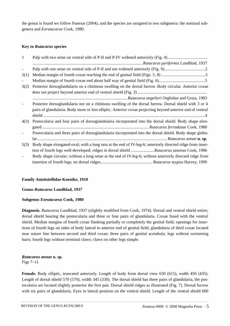
Key to Rutacarus species
1 Palp with two setae on ventral side of P-II and P-IV widened anteriorly (Fig. 4)............................................................................................................................................ Rutacarus pyriformis Lundblad, 1937
- Palp with one setae on ventral side of P-II and not widened anteriorly (Fig. 9)......................................22(1) Median margin of fourth coxae reaching the end of genital field (Figs. 5, 8) .........................................3- Median margin of fourth coxae end about half way of genital field (Fig. 6)...........................................53(2) Posterior dorsoglandularia on a chitinous swelling on the dorsal furrow. Body circular. Anterior coxae
does not project beyond anterior end of ventral shield (Fig. 3) ..........................................................................................................................................................Rutacarus angelieri Orghidan and Gruia, 1983
- Posterior dorsoglandularia not on a chitinous swelling of the dorsal furrow. Dorsal shield with 3 or 4pairs of glandularia. Body more or less elliptic. Anterior coxae projecting beyond anterior end of ventralshield ........................................................................................................................................................4
4(3) Postocularia and four pairs of dorsoglandularia incorporated into the dorsal shield. Body shape elon-gated.................................................................................................... Rutacarus ferradasae Cook, 1980
- Postocularia and three pairs of dorsoglandularia incorporated into the dorsal shield. Body shape globu-lar.......................................................................................................................... Rutacarus annae n. sp.
5(3) Body shape elongated-oval; with a long seta at the end of IV-leg-6; anteriorly directed ridge from inser-tion of fourth legs well-developed; ridges in dorsal shield......................Rutacarus sasonus Cook, 1986
- Body shape circular; without a long setae at the end of IV-leg-6; without anteriorly directed ridge frominsertion of fourth legs; no dorsal ridges................................................ Rutacarus stygius Harvey, 1990
Family Anisitsiellidae Koenike, 1910
Genus Rutacarus Lundblad, 1937
Subgenus Eorutacarus Cook, 1980
Diagnosis. Rutacarus Lundblad, 1937 (slightly modified from Cook, 1974). Dorsal and ventral shield entire;dorsal shield bearing the postocularia and three or four pairs of glandularia. Coxae fused with the ventralshield. Median margins of fourth coxae flanking partially or completely the genital field; openings for inser-tions of fourth legs on sides of body lateral to anterior end of genital field; glandularia of third coxae locatednear suture line between second and third coxae; three pairs of genital acetabula; legs without swimminghairs; fourth legs without terminal claws; claws on other legs simple.
Rutacarus annae n. sp.Figs 7–11
Female. Body elliptic, truncated anteriorly. Length of body from dorsal view 630 (615), width 450 (435).Length of dorsal shield 570 (570), width 345 (330). The dorsal shield has three pairs of glandularia, the pos-tocularia are located slightly posterior the first pair. Dorsal shield ridges as illustrated (Fig. 7). Dorsal furrowwith six pairs of glandularia. Eyes in lateral position on the ventral shield. Length of the ventral shield 600

VALDECASAS6 · Zootaxa 0000 © 2008 Magnolia Press
(610), width 440 (435). First and second coxae projecting beyond the anterior end of the ventral shield (Fig.8). One glandularia on a small chitinous islet between the second and third coxae , a second glandularia islocated nearby on the third coxae. The genital area has three pairs of acetabula covered by genital flaps.Length of the genital field (outline of the genital flaps) 135, width 80. One female had six eggs and the otherfive. Palps with a single ventral setae on P-II and four or five setae on P-IV; the longest is on a small tubercle.Dorsal length of palp segments: P-I 19; P-II 80; P-III 42; P-IV 98; P-V 22. Length of capitulum 115. Lengthof chelicera 205 (Fig. 11). Length of chelicera claw 60. Length of subterminal seta at IV-leg-6 50 (Fig. 10). I-to III-leg with simple claws. Length of the last segments of legs: I-Leg-4 110; I-Leg-5 90; I-Leg-6 90; II-Leg-4 80; II-Leg-5 80; II-Leg-6 75; III-Leg-4 105; III-Leg-5 100; III-Leg-6 90; IV-Leg-4 100; IV-Leg-5 120; IV-Leg-6 90.
Type material. Holotype: female, 8-viii-1994, Rio Escondido, immediately above waterfall. CoibaIsland, Panama. Dissected and slide mounted in glycerin jelly.Paratype: female same locality and date as holotype. Undissected in Koenike´s fluid.At this location the Rio Escondido is a stony stream, at 1.800 m distance from the sea; 3.6 m wide and 30 cmdeep; 25ºC water temperature, pH 7.9 and 131 µS/cm conductivity.
Etymology. The species is dedicated to my colleague Dr. Ana I. Camacho for her tireless encouragementand continuous help.
Habitat. Interstitial habitat. Discussion. Rutacarus annae n. sp. is close to R. angelieri and R. ferradasae. It is distinguished from R.
angelieri by the lack of a chitinized strip connecting the posterior glandularia of the dorsal shield and thecoxae projecting beyond the ventral shield. R. ferradasae is differentiated from R. annae n. sp. by its moreelongated body shape and having four glandularia besides the postocularia on the dorsal shield. Additionally,the sizes mentioned by Cook (1980) for the chelicera and the capitulum of the female of R. sasonus must be amisprint, or they will be another difference between R. annae n. sp. from R. sasonus not presented in the key.
Acknowledgement
Santiago Castroviejo made possible the sampling trips to Coiba (Panama). Ana I. Camacho has helped inmany ways. Mark S. Harvey, T. Goldschmit and an anonymous referee made very useful suggestions. SarahYoung corrected the manuscript. Work done under grant CGL2005-02217
References
Castroviejo, S. (1997) Flora y Fauna del Parque Nacional de Coiba (Panamá). AECI, Madrid, 534 pp.Cook, D.R. (1974) Water mite genera and subgenera. Memoirs of the American Entomological Institute 21, Ann Arbor,
Michigan, 860 pp.Cook, D.R. (1980) Studies on Neotropical water mites. Memoirs of the American Entomological Institute 31, Ann
Arbor, Michigan, 645 pp.Cook, D.R. (1986) Water mites from Australia. Memoirs of the American Entomological Institute 40, Ann Arbor, Mich-
igan, 568 pp.Davis, C., Di Sabatino, A., Gerecke, R., Gledhill, T., Smit, H. & van der Hammen, H. (2007) Acari: Hydrachnidia I. In:
Gerecke, R. (Ed.) Chelicerata: Aranae, Acari I. Süsswasserfauna von Mitteleuropa 7/2-1. Elsevier Spektrum Aka-demischer Verlag, Munich, 241–376.
Harvey, M.S. (1990) A review of the water mite family Anisitsiellidae in Australia (Acarina). Invertebrate Taxonomy, 3,629–646.
Lundblad, O (1927) Die Hydracarinen Schwedens. I. Beitrag zur Systematik, Embryologie, Ökologie und Verbreitungs-geschichte der schwedischen Arten. Zoologica Bidragen, 11, 181–540.
Lundblad, O. (1937) Fünfte Mitteilung über neue Wassermilben aus Santa Catharina in Südbrasilien. Zoologischer

Zootaxa 0000 © 2008 Magnolia Press · 7REVISION OF THE GENUS RUTACARUS
Anzeiger, 120, 11–12.Lundblad, O. (1941) Die Hydracarinenfauna Südbrasiliens und Paraguays. Erster Teil. Svensk Vetenskapsakad Handling
Stockholm (s. 3), 19, 1–183.Orghidan, T. & Gruia, M. (1983) Diagnose de cinq especes nouvelles d'hydracariens de Venezuela. Travaux de l'Institut
de Speologie "Emile Racovitza", 22, 103–105.Panesar, A. (2004) Evolution in water mites (Hydrachnellae, Actinedida, Acari). A revision of the Anisitsiellidae. Bon-
ner Zoologische Monographien, 52, 1–144.Smith, I. M., Cook, D. R. & Smith, B.P. (2001) Water Mites (Hydrachnida) and other arachnids. In: Thorp, J.H. & Cov-
ich, A.P. (Ed.) Ecology and Classification of North American Freshwater Invertebrates. Academic Press, SanDiego, 551–659.
Wiles, P.R. (1997) The homology of glands and glandularia in the water mites (Acari: Hydrachnidia). Journal of NaturalHistory, 31, 1237– 1251.