Revision of the Centipede Genus Hemiscolopendra ... - Brill
34
Revision of the Centipede Genus Hemiscolopendra Kraepelin, 1903: Description of H. marginata (Say, 1821) and possible misidentifications as Scolopendra spp.; proposal of Akymnopellis, n. gen., and redescriptions of its South American components (Scolopendromorpha: Scolopendridae: Scolopendrinae) Rowland M. Shelley Research Lab., North Carolina State Museum of Natural Sciences, MSC #1626, Raleigh, NC 27699-1626 USA. E-mail: [email protected] Abstract Hemiscolopendra Kraepelin, 1903, is a monotypic genus. Its sole species, H. marginata (Say, 1821), possibly misidentified in older publications as Scolopendra morsitans L., 1758, and S. pachygnatha Pocock, 1905, occurs from the southern Atlantic Coast of the United States to west Texas, in the east/west dimension, and, north/south, from southern Ohio-Illinois to Chiapas, Mexico. Individuals throughout this area vary through the same limits, proving that they are conspecific. Akymnopellis, n. gen., is erected to accommo- date the South American species previously assigned to Hemiscolopendra – chilensis (Gervais, 1847), laevi- gata (Porat, 1876), and platei (Attems, 1903). Two new synonymies are proposed – Otostigma michaelseni Attems, 1903, under A. laevigata, and Hemiscolopendra perdita Chamberlin, 1955, under A. platei. Key words Hemiscolopendra, H. marginata, Akymnopellis, North America, South America, United States Introduction A major biogeographic incongruity in the chilopod subfamily Scolopendrinae (Scolo- pendromorpha: Scolopendridae) is the allopatric occurrence of Hemiscolopendra Kraepelin, 1903, in North and South America. Not only has the genus never been © Koninklijke Brill NV and Pensoft, 2008 DOI: 10.1163/187525408X395931 International Journal of Myriapodology 2 (2008) 171-204 Sofia–Moscow Downloaded from Brill.com01/08/2022 03:35:38PM via free access
-
Upload
khangminh22 -
Category
Documents
-
view
1 -
download
0
Transcript of Revision of the Centipede Genus Hemiscolopendra ... - Brill
Revision of Hemiscolopendra 171
Revision of the Centipede Genus Hemiscolopendra Kraepelin, 1903:
Description of H. marginata (Say, 1821) and possible misidentifi cations as Scolopendra spp.;
proposal of Akymnopellis, n. gen., and redescriptions of its South American components
(Scolopendromorpha: Scolopendridae: Scolopendrinae)
Rowland M. Shelley
Research Lab., North Carolina State Museum of Natural Sciences, MSC #1626, Raleigh, NC 27699-1626 USA. E-mail: [email protected]
AbstractHemiscolopendra Kraepelin, 1903, is a monotypic genus. Its sole species, H. marginata (Say, 1821), possibly misidentifi ed in older publications as Scolopendra morsitans L., 1758, and S. pachygnatha Pocock, 1905, occurs from the southern Atlantic Coast of the United States to west Texas, in the east/west dimension, and, north/south, from southern Ohio-Illinois to Chiapas, Mexico. Individuals throughout this area vary through the same limits, proving that they are conspecifi c. Akymnopellis, n. gen., is erected to accommo-date the South American species previously assigned to Hemiscolopendra – chilensis (Gervais, 1847), laevi-gata (Porat, 1876), and platei (Attems, 1903). Two new synonymies are proposed – Otostigma michaelseni Attems, 1903, under A. laevigata, and Hemiscolopendra perdita Chamberlin, 1955, under A. platei.
Key wordsHemiscolopendra, H. marginata, Akymnopellis, North America, South America, United States
Introduction
A major biogeographic incongruity in the chilopod subfamily Scolopendrinae (Scolo-pendromorpha: Scolopendridae) is the allopatric occurrence of Hemiscolopendra Kraepelin, 1903, in North and South America. Not only has the genus never been
© Koninklijke Brill NV and Pensoft, 2008 DOI: 10.1163/187525408X395931
International Journal of Myriapodology 2 (2008) 171-204 Sofi a–Moscow
Downloaded from Brill.com01/08/2022 03:35:38PMvia free access

R.M. Shelley / International Journal of Myriapodology 2 (2008) 171-204172
critically assessed but neither has H. marginata (Say, 1821), the lone North American component; prior authors, myself included, have unquestioningly assumed conspeci-fi city of the forms such that the absence of the distoventral spur on the 1st tarsi of leg pairs 1-20, which is present in Scolopendra L., 1758, alone diagnoses the species. Hoff man & Shelley (1996), for example, stated that in the event of partitioning, Hemi-scolopendra would denote “a monotypic Nearctic genus,” an assumption that warrants corroboration. Herein I address this query by examining forms throughout the ranges on both continents; those in North America are indeed conspecifi c, but those in South America are not even congeneric. I therefore propose Akymnopellis, n. gen., for the three South American species and restrict Hemiscolopendra to H. marginata.
In the United States (US), Hemiscolopendra and H. marginata extend from the south-ern Atlantic Coast to west Texas (Fig. 1); occurrence in Mexico, discounted by Shelley (2002), is confi rmed, but only three localities are known, from Tamaulipas, Guerrero, and Chiapas, the last two in the Neotropics (Figs 1, 2). Th e taxa therefore span the Tropic of Cancer and the border between two biogeographic regions. Akymnopellis, however, is known only from South America and traverses both the Equator and the Tropic of Cap-ricorn; based on present knowledge, two species are restricted to Chile and Argentina.
Figure 1. Distributions of Hemiscolopendra and H. marginata in the US and northern Mexico. A smooth curve is drawn around northern range extremes in the US and selected peripheral records are shown. Th e circle denotes the locality of Scolopendra morsitans coerulescens Cragin in Barbour Co., Kansas.
1
Downloaded from Brill.com01/08/2022 03:35:38PMvia free access

Revision of Hemiscolopendra 173
I present full descriptive accounts of all taxa including the fi rst for H. marginata; anatomical terminology follows the recommendations of Lewis et al. (2005). Not all specimens were useable for all characters examined; some were either missing an antenna or one or both antennae were aborted, as evidenced by fewer than the 17 articles typical for the order and, usually, by a black irregular tip where the append-age had broken. Some specimens were missing one or both ultimate legs, while one caudal leg of still others had a markedly higher number and irregularly arranged prefemoral spines, indicative of regeneration. Th e variation sections unavoidably in-corporate several stadia; I eliminated obvious early instars, but no quick and precise method exists to distinguish adults from subadults in epimorphic chilopods. Conse-quently, one either measures and examines every individual that appears to represent an advanced, “near adult” stadium or omits variation altogether. As no species herein has received a comprehensive account and this study seeks to provide such, I opted to assess variation in 21 external anatomical features even though most samples prob-ably contain mixed stadia.
Repository acronyms of preserved material are AMNH, American Museum of Natural History, New York, New York, USA; ANSP, Academy of Natural Sciences, Philadelphia, Pennsylvania, USA; BMNH, Th e Natural History Museum, London, UK; BYU, Monte L. Bean Life Science Museum, Brigham Young University, Provo, Utah, USA; DMNH, Danish Museum of Natural History, Copenhagen, Denmark; FMNH, Field Museum of Natural History, Chicago, Illinois, USA; FSCA, Florida State Collection of Arthropods, Gainesville, Florida, USA; MCZ, Museum of Com-parative Zoology, Harvard University, Cambridge, Massachusetts, USA; MHNG, Muséum d’Histoire Naturelle, Genève, Switzerland; MNHP, Muséum National
Figure 2. Known occurrences of Hemiscolopendra and H. marginata in Mexico.
2
Downloaded from Brill.com01/08/2022 03:35:38PMvia free access

R.M. Shelley / International Journal of Myriapodology 2 (2008) 171-204174
d’Histoire Naturelle, Paris, France; NCSM, North Carolina State Museum of Natural Sciences, Raleigh, USA; NMNH, National Museum of Natural History, Smithsonian Institution, Washington, DC, USA; NHMV, Naturhistorisches Museum, Vienna, Austria; SMNH, Swedish Museum of Natural History, Stockholm, Sweden; UCO, Biology Department, University of Central Oklahoma, Edmond, Oklahoma, USA; VMNH, Virginia Museum of Natural History, Martinsville, USA; and ZMUH, Zoo-logisches Museum, University of Hamburg, Germany. Missing data in the new lo-cality records was not provided on vial labels, and the number of specimens in the sample is provided after the institutional acronym.
Taxonomy
Order Scolopendromorpha Pocock, 1895Family Scolopendridae Newport, 1844
Subfamily Scolopendrinae Newport, 1844
Genus Hemiscolopendra Kraepelin, 1903
Hemiscolopendra Kraepelin, 1903: 212-214; Verhoeff 1907: 261; Chamberlin 1911: 477, 1955: 43; Crabill 1960: 10; Kevan 1983: 2944; Shelley 2002: 40-41; Jeekel 2005: 54.
Cormocephalus (Hemiscolopendra): Attems 1930 (in part): 110-111.
Type species. Scolopendra punctiventris Newport, 1844, by subsequent designation of Attems (1930).
Diagnosis. Moderate-size Scolopendrinae (maximum adult length ca. 55 mm) characterized by absenses of spurs on 1st tarsi of ambulatory appendages and of setae excepting antennae; latter with transition from sparsely to densely hirsute articles varying from antennomeres 6-9. Cephalic plate overlapping anterior margin of T1; latter with prominent, procurved anterior transverse suture, with or without variably complete or incomplete paramedian sutures. T2-T20 with complete paramedian su-tures; T21 with complete or variably incomplete middorsal suture. Podomeres of ultimate legs generally elongate; spines on ventral surfaces of prefemora arranged generally in three variably sublinear rows, two lateral and one medial to midlines; prefemoral processes relatively short and bulbous, with two apical/subapical spines, more distal curved caudad.
Species. One. Distribution. Th e southcentral and southeastern US through southern Mexico
(Figs 1, 2). Remarks. My analysis of variation confi rms that forms throughout the range are
conspecifi c and that Hemiscolopendra is monotypic. Individuals from Mexico conform to the pattern of variation exhibited by US specimens.
Downloaded from Brill.com01/08/2022 03:35:38PMvia free access

Revision of Hemiscolopendra 175
Hemiscolopendra marginata (Say, 1821)Figs 3-58
Scolopendra marginata Say, 1821: 110; Bollman 1893: 147; Shelley 2006: 7.Scolopendra punctiventris Newport, 1844: 100, 1845: 386-387; Gervais 1847: 277; Pocock
1895: 17, pl. II, fi gs. 6, 6a-c; Shelley 2006: 10.Scolopendra parva Wood, 1861: 10.Scolopendra inaequidens (not Gervais, 1847): Wood 1862: 24-25, 1865: 162.Scolopendra woodi Meinert, 1886: 198-199; McNeill 1887: 326, 1888: 17; Bollman 1888a:
348, 1888b: 6, 1888c: 341, 1888d: 408, 1893: 174; Kenyon 1893: 16; Brölemann 1896: 49-50; Williams & Hefner 1928: 137; Brimley 1938: 501; Wray 1950: 155, 1967: 155.
Hemiscolopendra punctiventris: Kraepelin 1903: 217, fi g. 145; Chamberlin 1918a: 375, 1918b: 24, 1920: 43, 1944: 182-183, 1955: 43; Shelley 1978: 221, 1987: 501-503, fi gs 1, 11; Summers 1979: 696; Summers et al. 1980: 245, 1981: 59; Kevan 1983: 2944; Hoff man 1994: 33, 1995: 24-25; Bücherl 1974: 110, fi g. 24e.
Cormocephalus (Hemiscolopendra) punctiventris: Attems 1930: 111-112, fi gs 134-135; Crabill 1950: 201, 1955a: 259, 1955b: 39.
Scolopendra viridis (not Say, 1821): Brimley 1938: 501; Wray 1950: 155, 1967: 155.Scolopendra morsitans (not Linnaeus, 1758): Brimley 1938: 501; Wray 1950: 155, 1967: 155.Hemiscolopendra marginata: Hoff man & Shelley 1996: 37-40, fi gs 1-3; Shelley 2002: 41-46,
fi g. 65.
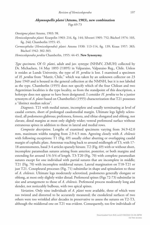
Figures 3-8. Hemiscolopendra marginata, external features of an individual from Dare Co., NC. 3, ce-phalic plate and tergites 1-5, dorsal view. 4, coxosternum and forcipules, ventral view. 5, right coxopleural process, ventral view. 6. left prefemoral process, submedial view. 7, ventral spinulation of right ultimate prefemur. 8, dorsal spinulation of the same.
3
4
5
6
7 8
Downloaded from Brill.com01/08/2022 03:35:38PMvia free access

R.M. Shelley / International Journal of Myriapodology 2 (2008) 171-204176
Type specimens. Of S. marginata, neotype and paraneotype (VMNH) collected by R.L. Hoff man, 21 February 1994, at Picolata, St. Johns County (Co.), Florida, USA. Of S. punctiventris, holotype (BMNH) collected by E. Doubleday on an unknown date prior to 1844 at an unspecifi ed site in Florida. As noted by Hoff man & Shelley (1996), Dou-bleday collected substantial natural history material from “St. John’s Bluff ,” Florida, that he sent to the BMNH. Shelley (2002) reported that “St. John’s Bluff ” was an early settlement on the St. John’s River about 8 km (5 mi) W Mayport, Duval Co., and is now encompassed by Fort Caroline National Memorial, part of Timucuan Ecological and Historic Preserve; the site is approximately 22.4 km (14 mi) NE of downtown Jacksonville. Of S. parva, two syntypes (ANSP) collected by J. LeConte on an un-known date prior to 1861 at an unknown locality in Georgia. Of S. woodi, holotype (MCZ) taken by an unknown collector on an unknown date prior to 1886 on Hilton Head Island, Beaufort Co., South Carolina.
Diagnosis. With the characters of the genus.Composite description. Lengths of examined specimens varying from 27.8-54.8
mm; maximum widths ranging from 2.7-7.8 mm. Cephalic plate (Fig. 3): slightly wider than long, slightly prolonged and rounded between antennal bases, indented slightly in midline and usually continuing into short suture extending caudad to varying levels but no farther than midlength of ocelli or ca. 1/4 of length of plate; sides sublinear, curving only at anterior and posterior corners, caudal margin straight, overlapping entire anterior margin of T1 occasionally as far as anterior transverse suture; surface lightly punctate but generally glabrous. Antennae: reaching back to around T3; dorsal surfaces of basal 6-9 (usually 7) antennomeres sparsely hirsute, more distal articles with dense, fi ne pubescences on all surfaces. Coxosternum (Fig. 4): with moderately long, narrowly segregated tooth plates (Figs 4, 22-29) demarcated by subangular sulci, ex-tending anteriad for roughly 2/3 the lengths of trochanteroprefemoral processes, usu-ally with 4 strong, apical teeth, three medial teeth coalesced basally, two medialmost more so and often fused for most of lengths, lateral teeth (one per side) well separated; surface glabrous and lightly punctate, with only short sulcus arising between tooth plates and extending caudad for ca. 1/8-1/4 of coxosternal length. Forcipules (Fig. 4): approximately 1/3 as wide basally as coxosternum; trochanteroprefemoral process (Figs 4, 17-21) strong, lateral surface concave, medial margin narrow and ridge-like, usu-ally lightly but variably scalloped; tarsungula overlapping apically in closed position. Terga (Figs 3, 9-16): smooth and glabrous, with at most only a few randomly scat-tered, short, fi ne setae. T1 with strong, procurved, anterior transverse suture, with or without complete or variably incomplete paramedian sutures between latter and caudal margin, sometimes with network of fi ne meshed sulci. T2-T20 with complete paramedian sutures. T21 with complete or variably incomplete middorsal suture, cau-dal margins variably extended and rounded in midlines, caudolateral corners slightly prolonged. Lateral margination present on tergites 17-21 only, usually incomplete ex-cept for ultimate tergite, often faint and diffi cult to perceive. Sterna: generally smooth and glabrous, relatively long and wide, slightly narrower caudad. S1 short, anterior margin gently convex; complete paramedian sutures present on S2-S20; S21 (Hoff man
Downloaded from Brill.com01/08/2022 03:35:38PMvia free access

Revision of Hemiscolopendra 177
& Shelley 1996: 40, fi g. 3) without sutures, narrowing caudad, apical margin sublin-ear or gently rounded. Coxopleural processes (Figs 5, 30-33): elongated and narrow, with variable numbers of short, darkly pigmented spines in varying positions, usually distad, on all surfaces. Leg pairs 1-20 (Hoff man & Shelley 1996: 40, fi g. 2; Shelley 2002: 42, fi g. 65): glabrous and with 6 podomeres, becoming progressively longer and stronger caudad, without spines or adornments except for pair of accessory spurs on ventral surfaces at bases of claws, latter sharply acuminate. Ultimate legs (Hoff man & Shelley 1996: 40, fi g. 3): long, slender, and moderately sclerotized, entirely glabrous, more heavily sclerotized and moderately larger than preceding appendages, prefemora, femora, and tibiae expanded only at distal extremities, often indistinct and not notice-able. Prefemora (Figs 7-8, 34-58; Hoff man & Shelley 1996: 40, fi g. 3) with variable numbers of short, darkly pigmented spines in varying positions on ventral, medial, and dorsomedial surfaces, ventral surfaces with spines generally arranged in three irregu-larly sublinear rows, two lateral and one medial to midlines, rows angling or curving proximad basally toward small midline swellings, lobes, or elevations, latter often with 1-2 or so minute spines; prefemoral processes (Figs 6-8, 34-58) short, bulbous, and narrowly rounded apically, with 2 apical or 1 apical and 1 subapical spines, more distal one curving strongly caudad (Figs 6, 8, 50-58). Femora (Hoff man & Shelley 1996: 40, fi g. 3) shorter than prefemora, without spines; remaining podomeres subparallel-sided and without spines, becoming progressively shorter caudad; claws acuminate, usually without (rarely with) accessory spines.
Variation. Re-examining all or even most of the thousands of preserved specimens of H. marginata was prohibitive, so I checked 83 individuals from 71 sites throughout the range, including those from Chiapas and at least one from every US state except Indiana. For states in which the species is widespread, I examined samples from the peripheries and centers. Features that are constant throughout the distribution include the positions of the antennae, the sizes and general shapes of the cephalic plates, sizes of the forcipules, absences of sutures and sulci on S1 and S21, presences of paramedian sutures on T2-T20 and S2-S20, presences of two accessory spines at the bases of the terminal claws on leg pairs 1-20, and absences of other spines on these appendages.
Body dimensions. Seventy-nine individuals were in suffi ciently good condition to be straightened for length and width measurements. Data are summarized below.
Lengths WidthsRange: 27.8-54.8 mm Range: 2.7-7.8 mmMean: 40.5 mm Mean: 5.3 mmAverage: 41.0 mm Average: 5.1 mmNumber of individuals: 79 Number of individuals: 79
Antennal features: In many individuals with aborted antennae, the break was distal to the pilosity transition point, which was thus evident and could be recorded. On all antennae, the dorsal surfaces of the basal four articles were sparsely hirsute; the pilosity then gradually increased for a few articles before a sudden increase to dense pilosity
Downloaded from Brill.com01/08/2022 03:35:38PMvia free access

R.M. Shelley / International Journal of Myriapodology 2 (2008) 171-204178
that continued to the ultimate antennomere. Th e “transition point” is the position of this dramatic increase in density, and it varies, even between the left and right antennae on many individuals. While the transition point was at antennomere borders in 109 antennae (70.8 % of the total), it was on article surfaces in 45 antennae (29.2 % of the total); in these cases, I estimated the distances as 1/4, 1/2, or 3/4 of the total an-tennomere length. Th e number of sparsely hirsute articles may correlate with age and stadium number, but this can only be determined from live individuals that are reared in captivity and observed after each molt.
Transition PointRange: 6-9 Mean: 7.5Average: 7.22Number of antennae: 1546 articles - 19 antennae (12.3%)6 ½ articles - 9 antennae (5.8%)6 ¾ articles - 5 antennae (3.2%)7 articles - 57 antennae (37.0%)7 ¼ articles - 3 antennae (1.9%)7 ½ articles - 15 antennae (9.7%)7 ¾ articles - 7 antennae (4.5%)8 articles - 29 antennae (18.8%)8 ¼ articles - 1 antennae (0.6%)8 ½ articles - 4 antennae (2.6%)8 ¾ articles - 1 antennae (0.6%)9 articles - 4 antennae (2.6%)
Coxosternal teeth. Th e most common arrangement is shown in Fig. 4, where three black, subequal, medial teeth are coalesced basally, with a fourth tooth at the lateral margin being separated by a narrow gap. Figures 22-29 show eight variants, one of which (Fig. 29) has fi ve teeth with the medial four coalesced.
Figures 9-16. Representative sutural variation on T1 of H. marginata, specimen localities as follows: 9, Miami-Dade Co., Florida. 10, Mitchell Co., Georgia. 11. Pitt Co., NC. 12, Hyde Co., NC. 13. Dare Co., NC. 14, Stone Co., Mississippi. 15, Pike Co., Arkansas. 16, Presidio Co., Texas.
9
13
10
14
11
15
12
16
Downloaded from Brill.com01/08/2022 03:35:38PMvia free access

Revision of Hemiscolopendra 179
Trochanteroprefemoral process. Th e lateral margin of this structure is concave, and the black, medial margin varies from curvilinear (Fig. 20) to lightly scalloped (Figs. 4, 17, 19) to more strongly scalloped and somewhat lobate (Figs 18, 21). Th e most com-mon condition is the lightly scalloped confi guration in fi gures 4 and 17, in which there are two slight swellings at 1/3 and 2/3 length.
Sutural variation. Th e cephalic plate exhibits a short, faint middorsal suture that arises from the anterior margin at the depression between the antennae and extends caudad to around midlength of the ocelli, or ca. 1/4 of the length of the plate. Like-wise, there is a short, faint, midventral suture on the coxosternum that arises from the anterior margin between the tooth plates and extends for about 1/4 of the coxosternal length. Th e most complex variation occurs on T1 (Figs 3, 9-16). Caudal to the anterior transverse suture, there may be no sutures, essentially complete paramedians on either or both sides of the midline, and many stages in between; some individuals even have an essentially complete suture on one side and a variably incomplete one on the other. Partial, incomplete sutures vary from short ones arising from the anterior transverse suture and/or the caudal margin and extending for around 1/8, 1/4, or 1/3 of the length of the tergite; these are somewhat curved in some individuals, while in others, they arise from both ends, run beside each other, and do not actually connect. Many individuals exhibit short, angular sulci anterior to the anterior transverse suture, and
Figures 17-21. Representative trochanteroprefemoral variation in H. marginata, specimen localities as follows: 17, Dare Co., NC. 18, Lee Co., Alabama. 19, Durham Co., NC. 20, Wake Co., NC. 21, Gwin-nett Co., Georgia. Figures 22-29. Representative prosternal tooth variation in H. marginata, specimen localities as follows: 22, Miami-Dade Co., Florida. 23, Barnwell Co., South Carolina. 24, Hancock Co., Georgia. 25, Pitt Co., NC. 26, Cumberland Co., NC. 27, Hempstead Co., Arkansas. 28, Coryell Co., Texas. 29, Johnson Co., Texas.
17 18 19 20 21
2223
2627 28 29
24 25
Downloaded from Brill.com01/08/2022 03:35:38PMvia free access

R.M. Shelley / International Journal of Myriapodology 2 (2008) 171-204180
some have short, irregular, horizontal sulci near the latter and the caudal tergal margin. One aberrant individual from Real Co., Texas, lacked the anterior transverse suture, but the other two in the same sample possessed it.
Lateral margination. Th is feature is restricted to T17-T21. Th ere is substantial vari-ation, and the 33 individuals tabulated below demonstrate nine variants. Margins are usually complete and strongly demarcated on T21 only, whereas they are usually vari-ably distinct and incomplete on others. In two of the six individuals in the T18 (faint)-T21 category, margination was evident only on the caudal 1/4 of T18; margination in two specimens in the T19(faint)-T21 category was evident only on the caudal 1/2 and 1/4 of T19. Th is trait may correlate with age and stadium number, but this cannot be determined from dead, preserved individuals.
Margination condition Number of individuals (% of total)T17 (faint)-T21 1 (3.0%)T18-T21 4 (12.1%)T18 (faint)-T21 6 (18.2%)T18-T21 (faint on 18 and 19) 1 (3.0%)T19-T21 8 (24.2%)T19 (faint)-T21 4 (12.1%)T20-T21 3 (9.1%)T20 (faint)-T21 3 (9.1%)T21 only 3 (9.1%)
Coxopleura (Figs 5, 30-33). Some coxopleura possess a small spine along the cau-dal margin at or near the dorsal extremity of the pore fi eld and lateral to the coxopleural process; it can be on either or both sides or absent altogether (see following table). Individuals in the same sample, collected at the same time and place, often demon-strated diff erent conditions. In a sample from Pike Co., Arkansas, one individual had no basal spines, and the other had a spine on both sides. One of the four individuals from Chiapas had no basal spines, another had spines on both sides, and two had a spine on one side only.
Figures 30-33. Representative spinulation variation in the coxopleural process of H. marginata, specimen localities as follows: 30-31, Miami-Dade Co., Florida. 32, Barnwell Co., South Carolina. 33, Cleveland Co., Arkansas.
30 31 32 33
Downloaded from Brill.com01/08/2022 03:35:38PMvia free access

Revision of Hemiscolopendra 181
Condition Number of individuals (n=81)No basal coxopleural spines 38 (46.9% of total)Basal spine on one side only 26 (32.1%)Basal spine on both sides 17 (21.0%)
Th e processes themselves are moderately sclerotized and subsimilar in length and shape with little variation in the numbers and positions of the spines. In general, they are not heavily spinulate, the most common spine arrangement (Fig. 31) being one apical, one subapical on the dorsal side, one subapical ventral, and one at ca. ¾ length on the lateral or ventrolateral surfaces. Th e dorsoapical and lateral/ventrolateral spines may be absent (Figs 32, 33); there may be an additional spine around 1/2-2/3 length on the ventral surface (Figs 30, 33); and rarely there is an additional spine on the medial/ventromedial surfaces. Twenty-nine of the 81 individuals (35.8% of the total) exhibited the same pattern on both the left and right processes.
Ultimate legs. Th e ultimate legs are moderately long and glabrous with oblong or elongate podomeres that are slightly but variably wider distad. Spines occur exclusively on the prefemora and prefemoral processes, and only on the ventral, ventromedial, me-dial, and dorsomedial surfaces of the former. Th e ventral surfaces exhibit low, rounded swellings or elevations basally in the midlines that may carry a couple of small, lightly pigmented spines (Figs 34-49) and are typically masked in situ by the overhanging coxopleural processes. Th e swelling marks the proximal end of a shallow depression that is bounded by diverging or subparallel ridge-like elevations that extend distad
Figures 34-49. Representative spinulation variation on the ventral surfaces of the ultimate prefemora in H. marginata; specimen localities as follows: 34, Sevier Co., Tennessee. 35, Miami-Dade Co., Florida. 36, Monroe Co., Florida. 37, Barnwell Co., SC. 38, Hancock Co., Georgia. 39, Pitt Co., NC. 40, Dare Co., NC. 41, Duval Co., Florida. 42-43, Cleveland Co., Arkansas. 44, Columbia Co., Arkansas. 45, Johnson Co., Arkansas. 46, Pike Co., Arkansas. 47, Coryell Co., Texas. 48-49, Real Co., Texas.
34
42 43 44 45 46 47 48 49
35 36 37 38 39 40 41
Downloaded from Brill.com01/08/2022 03:35:38PMvia free access
R.M. Shelley / International Journal of Myriapodology 2 (2008) 171-204182
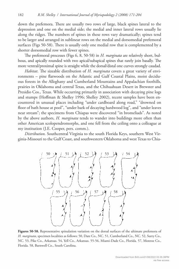
down the prefemora. Th ere are usually two rows of large, black spines lateral to the depression and one on the medial side; the medial and inner lateral rows usually lie along the ridges. Th e numbers of spines in these rows vary dramatically; spines tend to be larger and arranged in sublinear rows on the medial and dorsomedial prefemoral surfaces (Figs 50-58). Th ere is usually only one medial row that is complemented by a shorter dorsomedial row with fewer spines.
Th e prefemoral processes (Figs 6, 8, 50-58) in H. marginata are relatively short, bul-bous, and apically rounded with two apical/subapical spines that rarely join basally. Th e more ventral/proximal spine is straight while the dorsal/distal one curves strongly caudad.
Habitat. Th e sizeable distribution of H. marginata covers a great variety of envi-ronments – pine fl atwoods on the Atlantic and Gulf Coastal Plains, moist decidu-ous forests in the Alleghany and Cumberland Mountains and Appalachian foothills, prairies in Oklahoma and central Texas, and the Chihuahuan Desert in Brewster and Presidio Cos., Texas. While occurring primarily in association with decaying pine logs and stumps (Hoff man & Shelley 1996; Shelley 2002), recent samples have been en-countered in unusual places including “under cardboard along road,” “drowned on fl oor of bath house at pool”, “under bark of decaying hardwood log”, and “under leaves near stream”; the specimens from Chiapas were discovered “in bromeliads”. As noted by the above authors, H. marginata tends to wander into buildings more often than other American scolopendromorphs, and one fell from the ceiling onto a colleague at my institution (J.E. Cooper, pers. comm.).
Distribution. Southcentral Virginia to the south Florida Keys, southern West Vir-ginia-Missouri to the Gulf Coast, and southwestern Oklahoma and west Texas to Chia-
Figures 50-58. Representative spinulation variation on the dorsal surfaces of the ultimate prefemora of H. marginata, specimen localities as follows: 50, Dare Co., NC. 51, Cumberland Co., NC. 52, Surry Co., NC. 53, Pike Co., Arkansas. 54, Yell Co., Arkansas. 55-56, Miami-Dade Co., Florida. 57, Monroe Co., Florida. 58, Barnwell Co., South Carolina.
50
55 56 57 58
51 52 53 54
Downloaded from Brill.com01/08/2022 03:35:38PMvia free access

Revision of Hemiscolopendra 183
pas (Figs 1, 2). In fi gure 1 a smooth curve is drawn through northern range extremities to depict the approximate boundary, which loops around the southern Appalachian Mountains in western North Carolina (NC), northern Georgia, and eastern Tennes-see. H. marginata is rarely encountered in these mountains, and then almost always on the peripheries, suggesting dispersal from the adjoining lowlands; however, one record from Mitchell Co., NC, lies well off the boundary shown in fi gure 1. Th e centipede occurs throughout peninsular Florida and even on Key Vaca south of the mainland. Th ere is only one Missouri record, from Ranken, St. Louis Co., that I (Shelley 2002: 45, fi g. 71) did not incorporate into the contiguous distribution; I do so now because it seems authentic and lies at roughly the same latitude as Bloomington, Monroe Co., Indiana, where H. marginata has been taken more than once. Only one Oklahoma record was previously available, from Latimer Co. in the east, but the species has re-cently been encountered in Oklahoma and Comanche Cos., extending the distribution substantially westward in this state.
Th e distribution in Texas is substantially modifi ed from 2002. I misidentifi ed samples from Loving and Lynn Cos., in west Texas, which are Arthrorhabdus pygmaeus (Pocock, 1895) instead, so the boundary actually lies substantially to the southeast; however, the Presidio Co. record is indeed H. marginata, so the range does extend westward into the Chihuahuan Desert. Th e southernmost east Texas locality is in Brooks Co., only around 75 mi (120 km) north of the Rio Grande and 315 mi (504 km) north of Tampico, Tamaulipas. Th e distribution between these locales is likely subcontinuous, so unlike Shelley (2002: 45, fi g. 71) I do not show a southern boundary in this area although the centipede has not been taken in the well-collected Rio Grande Valley. Samples docu-menting the two Mexican localities reported by Pocock (1895), Tampico and Omilteme, Guerrero (a settlement 10 mi (16 km) west-southwest of Chilpancingo (Selander & Vaurie 1962)), exist in the BMNH and are indeed H. marginata. Th ey are some 335 mi (536 km) apart, but there is no reason to believe they are mislabeled, and the Chiapas specimens lend them credence; consequently, I show these sites in fi gure 2 and omit a southern boundary for both the genus and species. Occurrence in Chiapas suggests that H. marginata inhabits southwestern Guatemala, only around 120 mi (192 km) away. Furthermore, its presence in Presidio and Brewster Cos., Texas, less than 10 mi (16 km) from the Rio Grande, suggests occurrence in neighboring Chihuahua and Coahuila. More material is needed to accurately hypothesize the distribution in Mexico, but the present samples suggest widespread occurrence through the heart of the country.
In summary, H. marginata occupies an area in the southern Nearctic and northern Neotropical biogeographic regions that spans the Tropic of Cancer and extends some 1,645 mi (2,632 km) east/west and 1,585 mi (2,536 km) north/south. In the US, the distribution covers parts of 11 physiographic provinces and 18 states including all or essentially all of South Carolina, Florida, Alabama, Mississippi, Kentucky, Arkansas, and Louisiana. It also spans such major rivers as the Roanoke, Neuse, Cape Fear, Pee Dee, Santee-Cooper, Savannah, Altamaha, St. Johns, Apalachicola, Alabama, Tennes-see, Cumberland, Ohio, Wabash, Mississippi, Arkansas, Red, Canadian, Trinity, Colo-rado (of Texas), Pecos, and Rio Grande.
Downloaded from Brill.com01/08/2022 03:35:38PMvia free access

R.M. Shelley / International Journal of Myriapodology 2 (2008) 171-204184
Published records. As cited by Shelley (2002: 42-43).New Records: USA: ARKANSAS: Cleveland, Columbia, Hempstead, Johnson, Little
River, Nevada, Pike, Polk, and Yell Cos. (NCSM).FLORIDA: Duval Co., Jacksonville, Lone Star Rd. nr Ft. Caroline National Me-
morial, 13 April 2002, R.M. Shelley (NCSM-1, VMNH-1). Topotypes of Scolopen-dra punctiventris.
GEORGIA: Hancock Co., 10.6 mi (17 km) S Sparta, 20 April 1997, R.M. Shelley (NCSM-1).
KENTUCKY: Laurel Co., Levi Jackson St. Pk., 9 June 2001, R.M. Shelley (FMNH-1).
LOUISIANA: Beauregard Par., De Ridder, 1944, E.L. Bell (AMNH-2).MISSISSIPPI: Harrison Co., Biloxi, 14 June 1889, Kroyer (DMNH-1). Lafayette
Co., Oxford, 14 May 1989, P.K. Lago (VMNH-1). Stone Co., Wiggins, Flint Cr. Water Park, 9 April 1999, R.M. Shelley (NCSM-2).
NORTH CAROLINA: Dare Co., Roanoke I., Manteo, 28 March 1997, A.E. & I. Bogan (NCSM-1) and 3 mi (4.8 km) WNW Manteo, Ft. Raleigh National Historic Site, 29 April 1997, J.C. Beane, L.H. Terry (NCSM-1); Nags Head Woods Ecological Preserve, 29 April 2001, J.C. Beane (NCSM-1); and 5.25 mi (8.4 km) SSE East Lake, 14 April 2006, J.C. Beane, L.T. Pusser (NCSM-1). Hyde Co., Ocracoke I., Ocracoke Village, 29 December 2000, J.C. Beane, J.T. Finnegan (NCSM-1); and 0.6 mi (1.0 km) ESE Ocracoke Village, 30 December 2006, J.C. Beane, J.T. Finnegan (NCSM-1). Pitt Co., Greenville, Green Mill Run Greenway, 10 October 2004, P. Marek (NCSM-1).
OKLAHOMA: Comanche Co., Ft. Sill, inside Natural Resources Bldg., 24 April 2003, J.O. Schmidt (VMNH-1). Oklahoma Co., Edmond, 28 April 1965, Dyer (UCO-1).
SOUTH CAROLINA: Barnwell Co., 2.75 mi (4.4 km) NW Snelling, Craig Pond Heritage Preserve, 27 June 1998, J.C. Beane (NCSM-2). Dillon Co., 1 mi (1.6 km) NW Floydale, 9 May 1994, R.G. Arndt (NCSM-2).
TENNESSEE: Sequatchie Co., 6 mi (9.6 km) E Dunlap, R.E. Crabill (NMNH-1).
TEXAS: Cass, Cherokee, Coryell, Dallas, Grayson, Harrison, Houston, Johnson, Mont-gomery, Morris, Newton, Nacogdoches, Polk, Real, Taylor, and Tom Green Cos. (FMNH, FSCA, NCSM, NHMV, VMNH, sight records by the author).
VIRGINIA: Charles City, Cumberland, Franklin, Henry, Isle of Wight, James City, Pittsylvania, Prince Edward, and Surry Cos., and Virginia Beach City (BYU, VMNH).
WEST VIRGINIA: Fayette Co., Ansted, September-October 2001, M. Lubert (NCSM-1).
MEXICO: CHIAPAS: 9.2-12.5 mi (14.7-20.0 km) N Ocozocuautla, 3,200’, 3-4 March 1966, G.E. Ball, D.R. Whitehead (NMNH, NCSM-4). Southernmost record of the genus and species.
Remarks. During this study, a historically important sample of H. marginata was discovered at the MNHP that consists of a number of individuals and was collected by E. Simon on an unknown date at an unknown locality in Louisiana.
Downloaded from Brill.com01/08/2022 03:35:38PMvia free access

Revision of Hemiscolopendra 185
Akymnopellis, new genus
Hemiscolopendra (not Kraepelin): Chamberlin 1914: 188, 1955: 43; Bücherl 1974: 103.Cormocephalus (Hemiscolopendra) (not Kraepelin): Bücherl 1939: 252, 1942: 301; Coscarón
1955: 372; Kraus 1957: 383.
Type species. Scolopendra chilensis Gervais, 1847.Diagnosis. Moderate-size Scolopendrinae (maximum adult length ca. 85 mm)
characterized by absences of spurs on 1st tarsi of ambulatory appendages, of anterior transverse suture on T1, and of setae excepting antennae on all components and ul-timate legs of A. laevigata. Transition from sparsely to densely hirsute antennomeres varying from articles 3-6. Cephalic plate either abutting, overlapping, or overlapped by T1; latter devoid of sutures. T2-T3 with variably incomplete (occasionally com-plete) paramedian sutures, T4-T20 with complete paramedian sutures; T21 with or without incomplete middorsal suture. Podomeres of ultimate legs varying from elon-gate to strongly clavate; spines on ventral surfaces of prefemora arranged generally in four variably sublinear rows, two lateral and two medial to midlines; prefemoral processes varying from short and nubbin-like to elongated, with or without two straight apical spines.
Species. Th ree.Distribution (Fig. 59). Literature records document occurrence throughout most
of South America, from near the Caribbean coast in Venezuela to the southern 1/3 of Argentina; most records, however, are from central Chile to southern Uruguay. All three species occur in Chile and northern Argentina, but only A. laevigata is known from Uruguay, Brazil, Peru, Colombia, Venezuela, and French Guiana. No records are available from Surinam, Guyana, Ecuador, Bolivia, and Paraguay.
Etymology. Th e generic name, feminine, derives from the Greek word “akymon” and the Latin word “pellis”, and translates as “barren skin”. It refers to the nearly featureless terga and leg pairs 1-20. Th e cephalic plates and coxosterna have at most only short, faint anterior sutures; T1 is glabrous and plain; lateral margination is con-fi ned to the caudalmost tergites; and the legs are devoid of spines and hairs except for standard accessory spurs.
Remarks. According to Gervais (1847), the color of the type of S. chilensis was (translated) “greenish brown”, while that of S. pallida was “pale tawny”. All the speci-mens that I examined had been preserved for at least 40 years, and 20 were collected in the mid to late 19th century. Consequently, their bodies were faded and discolored; virtually all traces of living color had disappeared. However, many individuals col-lected in the past 60 years still had a vague overall olive hue, and a few still had light spots of greenish speckling on the cephalic plates near the ocelli. Shades of greenish-olive therefore seem to be the general colors in life.
Downloaded from Brill.com01/08/2022 03:35:38PMvia free access

R.M. Shelley / International Journal of Myriapodology 2 (2008) 171-204186
Akymnopellis chilensis (Gervais, 1847), new combinationFigs 60-66
Scolopendra chilensis Gervais, 1847: 285; Meinert 1886: 199-200; Shelley 2006: 12.Scolopendra pallida Gervais, 1847: 285-286; Kohlrausch 1881: 123-124; Shelley 2006: 12.Scolopendra longipleura Silvestri, 1895: 2, 1897: 2.Hemiscolopendra chilensis: Kraepelin 1903: 214-215, fi g. 141; Silvestri 1905: 751; Bücherl
1974: 103, fi gs. 14a 2, 24a; Chamberlin 1955: 45.Cormocephalus (Hemiscolopendra) chilensis: Attems 1930: 112, fi g. 136; Bücherl 1942: 51-52.
Type specimens. Of S. chilensis, holotype (MNHP) collected by M. C. Gay on an un-known date prior to 1847 at an unknown locality in Chile. Of S. pallida, holotype
Figure 59. Distribution of Akymnopellis. Stars, A. chilensis; dots, A. laevigata; triangles, A. platei.
59
Downloaded from Brill.com01/08/2022 03:35:38PMvia free access
Revision of Hemiscolopendra 187
(MNHP) collected by “Pissis” on an unknown date in 1840 at an unknown location in Chile. Of S. longipleura, paratype (ZMUH) taken by an unknown collector on April 10 of an unknown year (label appears to state “03”, or 1903, which is impossible) at San Miguel de Tucumán, Tucumán Prov., Argentina. Th e label also states “Typus for H. longipleura Silvestry, Mus. Turin”, suggesting that the holotype may reside in this Italian city.
Diagnosis. T21 without middorsal, longitudinal suture; cephalic plate either entire-ly abutting T1 or overlapping corners or entire anterior margin of latter; ultimate legs glabrous, prefemora, femora, and tibia elongated and oblong, at most expanded only at distal extremity, ventral prefemoral surfaces with spines generally in 2 lateral and 2 medial longitudinal rows connected basally by 4-5 additional, smaller spines arranged in an arc, without additional extraneous spines.
Composite description. Lengths of examined specimens varying from 37.2-84.9 mm; maximum widths ranging from 2.7-7.9 mm. Cephalic plate (Fig. 60): slightly longer than wide, slightly prolonged and rounded between antennal bases, indent-ed slightly in midline and usually continuing into short suture extending caudad to around midlength of ocelli or ca. 1/4 of length of plate, sides sublinear, curving only at anterior and posterior corners, caudal margin straight, either overlapping or abutting entire anterior margin of T1 or overlapping anteriolateral corners of T1 and abutting rest of terga; surface lightly punctate but generally glabrous. Antennae: reaching back for varying distances depending on number of antennomeres and degree of article compression, usually to T3; dorsal surfaces of basal 3 ½-6 articles glabrous to sparsely hirsute, more distal articles with dense, fi ne pubescence on all surfaces. Coxosternum (Fig. 62): with moderately long, narrowly segregated tooth plates demarcated by sub-angular sulci, extending anteriad for roughly half the lengths of trochanteroprefemoral processes and usually with 4 strong, apical teeth, three medials coalesced basally, lateral teeth well separated; surface glabrous and lightly punctate, with only short sulcus aris-ing between tooth plates and extending caudad for ca. 1/8-1/4 of coxosternal length. Forcipules (Fig. 62): approximately 1/3 as wide basally as coxosternum; trochantero-prefemoral processes strong, lateral surfaces concave, medial margins angling anteriad to distolateral corners, with one broad, blunt subterminal tooth apiece coalesced ba-sally to shorter, indistinct tooth; tarsungula overlapping in closed position. Terga (Figs 60-61): smooth and glabrous, with at most only a few randomly scattered short, fi ne setae. T2-T3 with or without variably incomplete to complete paramedian sutures, T2 occasionally without sutures. Lateral margination present on caudal tergites, varying from T17-T21 to T21 alone, often faint and/or incomplete on anteriormost tergite. T21 otherwise without sutures, caudal margin variably extended and rounded in mid-line, caudolateral corners slightly prolonged. Sterna: generally smooth and glabrous, relatively long and wide, slightly narrower caudad. S1 short, anterior margin gently convex; S21 (Fig. 64) without sutures, narrowing caudad, apical margin sublinear or gently rounded. Coxopleural processes (Fig. 64): elongated and narrow, with variable numbers of short, darkly pigmented spines in varying positions on all surfaces. Leg pairs 1-20 (Fig. 63): glabrous and with 6 podomeres, becoming progressively longer
Downloaded from Brill.com01/08/2022 03:35:38PMvia free access

R.M. Shelley / International Journal of Myriapodology 2 (2008) 171-204188
and stronger caudad, without spines or adornments except for pairs of accessory spurs on ventral surfaces at bases of claws, latter sharply acuminate. Ultimate legs: long, slender, and lightly sclerotized, glabrous, not signifi cantly larger than preceding ap-pendages; prefemora, femora, and tibiae elongated and oblong, at most expanded only at distal extremities. Prefemora (Figs. 65-66) with variable numbers of short, darkly pigmented spines in varying positions on ventral, medial, and dorsomedial surfaces, ventral surfaces with spines generally arranged in 4 irregularly sublinear rows, 2 medial
Figures 60-66. Akymnopellis chilensis, specimen from Valparaiso, Valparaiso Reg., Chile. 60, cephalic plate and tergites 1-6, dorsal view. 61, tergites 19-21, dorsal view. 62, coxosternum and forcipules, ventral view. 63, midbody leg, caudal view. 64, coxopleura and sternites 20-21, ventral view. 65, ventral spinulation of left ultimate prefemur and femus. 66, dorsal spinulation of the same.
61
64
6360
62
65
66
Downloaded from Brill.com01/08/2022 03:35:38PMvia free access

Revision of Hemiscolopendra 189
and 2 lateral to midlines, rows angling or curving proximad basally toward variably-sized midline swellings, lobes, or elevations, these usually with 2-4 or so minute apical spines, additional smaller spines arranged in an arc; prefemoral processes variable but usually short, apically rounded, and somewhat bulbous, usually with 2 apical spines and none on other surfaces. Femora shorter than prefemora, without spines; remain-ing podomeres parallel-sided and without spines, becoming progressively shorter dis-tad; claws acuminate, with or without accessory spurs.
Variation. Th e same 21 characters were assessed for variation as in H. marginata; over 40 specimens of A. chilensis were available.
Body dimensions. Th irty-six individuals were in suffi ciently good condition to be straightened for length and width measurements. Th e others could not be measured because they had been crammed into vials that were so small that the bodies became entangled and fi xed into twisted, distorted positions after decades, or over a century, in preservative. Th ese centipedes could not be straightened for accurate length mea-surements with vernier calipers, and I did not measure widths on these individuals to eliminate a potentially inaccurate fi gure for this dimension as well.
Lengths WidthsRange: 37.2-84.9 mm Range: 2.7-7.9 mmMean: 53.4 mm Mean: 5.3 mmAverage: 55.1 mm Average: 5.1 mmNumber of individuals: 36 Number of individuals: 36
Antennal features: In addition to shortened and obviously broken or aborted anten-nae, 11 possessed more than 17 articles; 7 had 18 antennomeres, and 4 had 19. On all antennae, the dorsal surfaces of the basal two articles were essentially glabrous, then pilos-ity gradually increased for a few articles before the “transition point”, which was at anten-nomere borders in 38 individuals (67.9% of the total) and article surfaces in 18 (32.1%).
Transition PointRange: 3 ½ -6 Mean: 4 ¾ (4.75)Average: 4.93Number of antennae: 56 3 ½ articles - 1 antenna (1.8%)4 articles - 1 antenna (1.8%)4 ½ articles - 7 antennae (12.5%)4 ¾ articles - 2 antennae (3.6%)5 articles - 28 antennae (50.5%)5 ¼ articles - 3 antennae (5.4%)5 ½ articles - 5 antennae (8.9%)6 articles - 9 antennae (16.1%)
Downloaded from Brill.com01/08/2022 03:35:38PMvia free access

R.M. Shelley / International Journal of Myriapodology 2 (2008) 171-204190
Sutural variation. Th e cephalic plate may exhibit a short, faint middorsal suture that arises from the anterior margin at the depression between the antennae. It extends cau-dad for varying distances but usually barely below the level of the antennae; the longest run to around midlength of the ocelli, or ca. 1/4 of the length of the plate. Likewise, there may or may not be a short, faint, midventral suture on the coxosternum that arises from the anterior margin between the tooth plates; its length also varies with the longest extending to the level of the basal forcipular article or ca. 1/4 of the coxosternal length.
Th e most complex variation involves the paramedian sutures on T2 and T3. Th e con-dition on the former varies from no sutures to essentially complete ones on either or both sides; some individuals have an essentially complete suture on one side and a variably incomplete suture on the other. Partial, incomplete sutures vary from short ones on either or both sides, arising from either the anterior or both margins and extending for around 1/8, 1/4, or 1/3 of the length of the tergite. Similarly on T3, variably incomplete sutures may arise on either or both sides of the midline from either the anterior or posterior or both margins and extend for 1/8, 1/4, or 1/3 of the length of the sclerite. T3 diff ers, how-ever, in that there is always at least a short, partial suture that may be complete or faint and subcomplete. I also examined individuals with partial sutures arising from the ante-rior margin and extending caudad for ca. 3/4 of the tergal length; in others, partial sutures arose from the posterior margin and extended anteriad for ca. 2/3 of the tergal length.
Lateral margination. In A. chilensis, this feature is restricted to T17-T21, but even with this limitation there is substantial variation. In some individuals, lateral margins are clearly and strongly demarcated on some tergites while being faint and indistinct in others. Th e number of permutations is greater if one considers that margination on the anteriormost tergite can be partial as well as complete and arise from either the anterior or caudal borders. In two of the six individuals in the T18(faint)-T21 category, mar-gination was evident only on the caudal 1/4 of T18; margination in two specimens in the T19(faint)-T21 category was evident only on the caudal 1/2 and 1/4 of T19.
Margination condition Individuals demonstrating (% of total)T17 (faint)-T21 1 (3.0%)T18-T21 4 (12.1%)T18 (faint)-T21 6 (18.2%)T18-T21 (faint on 18 and 19) 1 (3.0%)T19-T21 8 (24.2%)T19 (faint)-T21 4 (12.1%)T20-T21 3 (9.1%)T20 (faint)-T21 3 (9.1%)T21 only 3 (9.1%)
Coxopleura. Most of the coxopleura possess a lateral spine along the caudal mar-gins at or near the dorsal extremities of the pore fi elds; some also have a somewhat smaller spine between the former and the coxopleural process, either halfway between them or beside the base of the process. Th e processes themselves are moderately scler-
Downloaded from Brill.com01/08/2022 03:35:38PMvia free access

Revision of Hemiscolopendra 191
otized (in contrast to the heavily sclerotized ones in A. laevigata) and subsimilar in length and shape; however, there is substantial variation in the number and positions of the spines. While they are drawn similarly in fi gure 64, I did not examine even one in-dividual in which the spinulations were the same on both the right and left processes. I also could not discern a pattern in spine positions, although there are fewer basally and the number increases distad. Th ere are 1-4 apical spines with most individuals having 3-4; varying numbers of spines also encircle the structure at varying positions on the ventral, ventrolateral, lateral, dorsolateral, dorsal, dorsomedial, medial, and ventrome-dial surfaces. Overall, the coxopleural processes possess 6-9 spines, with the medial and ventro/dorsomedial surfaces being least adorned.
Ultimate legs. Except for a few that obviously have regenerated, these appendages are long, slender, and glabrous with elongated or oblong podomeres that are at most only slightly or imperceptibly wider distad. Excepting one leg with a retrorse spine ven-trally on the femur (Fig. 65), spines occur exclusively on the prefemora and prefemoral processes, and only on the ventral, ventromedial, medial, and dorsomedial surfaces of the former. As in H. marginata, the ventral surfaces exhibit low, rounded swellings or elevations basally in the midlines that usually carry 2-3 small, lightly pigmented spines; they mark the proximal end of a shallow, midline depression that becomes wider distad. Th e elevation gives rise to diverging, ridge-like medial and lateral elevations that extend distad and vaguely connect to two lateral and two medial rows of large, black spines via 2-4 or so small, lightly pigmented ones on each side. Th e inner medial and lateral rows run along the elevations bordering the depression; the “connecting spines” may be absent, in which case the rows are detached from the basal elevation, and the outer rows sometimes are more properly ventrolaterad and ventromediad than ventrad. Th e number of spines in these rows varies from 2-7 but most number 4-5; they also tend to become larger, sharper, and farther apart distad. Additionally, the rows may be linear, angling, or curving proximad toward the basal elevation, and one or more spines may be displaced laterad or mediad. A prefemur may exhibit any number of spines within any row and any permutation of four linear or curvilinear rows to ones with variably displaced spines. In extreme cases, the spines are so displaced as to appear randomly positioned.
Likewise, the spines on the medial prefemoral surface are arranged in sublinear rows, but here there are fewer spines (2-5) that tend to be larger. Th ere is usually only one medial row that is complemented by a shorter dorsomedial row with even fewer spines and occasionally by an additional spine or two a bit more dorsad.
Th e prefemoral processes vary from barely detectable lobes to low, rounded nub-bins to distinct, short projections that in turn vary from bulbous to oblong and narrow. Most processes have two apical spines, but a few have one and three; occasionally there is a subapical or subbasal spine on the dorsal surface.
Distribution. From the Pacifi c Coast of central Chile to northcentral Argentina (Fig. 59). Published records. ARGENTINA: Eastern Argentina (Attems 1930; Bücherl 1942).
Córdoba Prov., Córdoba (Kohlrausch 1881; Meinert 1886; Bücherl 1974), Deán Funes and vicinity (Chamberlin 1955; Bücherl 1974). Tucumán Prov., San Miguel de Tucu-mán (Silvestri 1895). Unknown Prov., Estancia San Felipe (Silvestri 1897).
Downloaded from Brill.com01/08/2022 03:35:38PMvia free access

R.M. Shelley / International Journal of Myriapodology 2 (2008) 171-204192
CHILE: Chile in general (Attems 1930; Bücherl 1942). Atacamba Reg., Copiapó (Meinert 1886; Bücherl 1942, 1974). Valparaiso, Aconcagua, and Copiapó Regs., Tal-cahuano, Villa Rica, and Juncal. Coquimbo Reg., Coquimbo Peninsula SW of Guay-acán; 18-31.3 mi (28.8-50 km), E San Carlos; Crest of the Sierra Nahuelbuta (Cham-berlin 1955). Concepción, Valparaiso, Aconcagua, Coquimbo, Guayacán (Meinert 1886; Bücherl 1942, 1974).
Material examined. ARGENTINA: Córdoba Prov., 5 mi (8 km) N Deán Funes, 8 February 1951, E.S. Ross, A.E. Michaelbacher (ESR, AEM) (NMNH-1). Tucu-mán Prov., 1967, W.L. Brown (NMNH-1); San Miguel de Tucumán, 10 April 1903 (ZMUH-1); and San Xavier, 11 February 1951, ESR, AEM (NMNH-1).
CHILE: Bío-Bío Reg., Concepción, 10 November 1964, T. Cephalovic (NM-NH-1); and 11-31.3 mi (18-50 km) E San Carlos, 24-26 December 1950, ESR, AEM (NMNH-5). De la Araucanía Reg., 12.5 mi (20 km) E Temuco, 8 January 1951, ESR, AEM (NMNH-many); Temuco, 5-7 March 1945, E.A. Chapin (NMNH-2); and W of Angol, crest of Sierra Nahuelbuto, ESR, AEM (NMNH-2). Region unknown, “Cor-dillera de Chillen,” 5-10 December 1892, ?H. Brölemann (MNHP-4).
Akymnopellis laevigata (Porat, 1876), new combinationFigs 67-68
Cormocephalus laevigatus Porat, 1876: 17-18.Scolopendra cormocephalina Kohlrausch, 1878: 26, 1879: page unknown, 1881: 123.Scolopendra longispina Meinert, 1886: 199.Otostigma michaelseni Attems, 1903: 97-98. New Synonymy.Hemiscolopendra laevigata: Kraepelin 1903: 215, fi g. 142; Chamberlin 1914: 188; Bücherl
1974: 103, fi g. 24b.Hemiscolopendra michaelseni: Kraepelin 1903: 215-216, fi g. 143; Bücherl 1974: 103, fi g. 24c.Cormocephalus (Hemiscolopendra) michaelseni: Attems 1930: 112-113, fi g. 137; Bücherl 1942: 302.Cormocephalus (Hemiscolopendra) laevigatus: Attems 1930: 113, fi g. 138; Bücherl 1939: 252,
1942: 302.
Type specimens. Of C. laevigatus, juv. holotype (SMNH) collected by Kinberg, 13 Janu-ary 1852, at Montevideo, Montevideo Dept., Uruguay. Of S. cormocephalina, holotype (ZMUH) taken by unknown collector on an unknown date in Montevideo. Of S. longispina, 2 adult and 1 juv. syntypes (MCZ) collected by T.G. Carey on an un-known date in Maldonado, Maldonado Dept., Uruguay, incorrectly recorded as Brazil by Meinert (1886). Of O. michaelseni, syntypes collected by Dr. Michaelsen in May 1893 in Valparaiso, Valparaiso Reg., Chile (Attems 1903). I was unable to borrow these specimens (there were at least two), which are not at the ZMUH and are presumably at the NHMV if they exist, and this synonymy is based on the original and subsequent written accounts. Attems (1903) mentioned that the caudal legs are rather broad with short articles (“Analbeine recht dick mit kurzen Gliedern,” which is consistent with observed variation in A. laevigata), and Kraepelin (1903) concluded that michaelseni
Downloaded from Brill.com01/08/2022 03:35:38PMvia free access

Revision of Hemiscolopendra 193
is possibly only a variety of laevigata (“Die Art der vorigen sehr nahe und ist vielleicht nur Varietät derselben.”).
Diagnosis. T21 with medial suture, incomplete and usually terminating at level of caudal corners, short of extended caudal margin. Ultimate legs strongly sclerotized; prefemora, femora, and tibia glabrous and distinctly clavate; femora, tibiae, and 1st and 2nd tarsi lightly hirsute; ventral prefemoral surface usually with a single distomedial spine in addition to those in lateral and medial rows.
Composite description. Lengths of examined specimens varying from 19.4-65.2 mm, maximum widths ranging from 2.0-5.8 mm. Agreeing closely with A. chilensis with following exceptions: Cephalic plate with sides angling slightly caudomediad, either entirely abutting T1 or overlapping corners or entire anterior margin of lat-ter. Dorsal surfaces of basal 4-6 (usually 5) antennomeres glabrous to sparsely hirsute. Tooth plates with 3+1 base pattern as in A. chilensis, but two medial teeth occasionally completely fused forming 2+1+1 pattern; all three medial teeth often coalesced to form long, molar-like structure. Lateral margination present on T17-T21 or T20-T21. T21 with distinct, incomplete middorsal suture (Fig. 67). Coxopleural processes (Fig. 68) subsimilar in shape and spinulations to those of A. chilensis but more heavily sclero-tized. Ultimate legs stout, heavily sclerotized; prefemora-1st tarsi variably enlarged and swollen distad, strongly and distinctly clavate, 1st tarsi clavate but not appreciably en-larged or swollen; tibiae and 1st and 2nd tarsi lightly hirsute. Ventral rows of prefemoral spines (Figs 67-68) irregular with more spines more often displaced than in A. chilensis, also with an additional distal spine detached from those in rows; dorsal spines as in A. chilensis, but with or without an additional basomedial spine. Prefemoral process long and spiniform, narrowly rounded apically with two apical spines.
Variation. I examined around 30 specimens of A. laevigata and found variation like that described for A. chilensis. Readers are referred to its account for more details.
Lengths WidthsRange: 33.6-65.2 mm Range: 2.8-5.8 mmMean: 49.4 mm Mean: 4.3 mmAverage: 47.4 mm Average: 4.4 mmNumber of individuals: 26 Number of individuals: 26
Transition PointRange: 4-6Mean: 5Average: 4.9Number of antennae: 564 articles - 5 antennae (8.9%)4+ articles - 1 antenna (1.8%)4 ½ articles - 1 antenna (1.8%)5 articles - 48 antennae (85.7%)6 articles - 1 antenna (1.8%)
Downloaded from Brill.com01/08/2022 03:35:38PMvia free access

R.M. Shelley / International Journal of Myriapodology 2 (2008) 171-204194
Antennal features: Twelve antennae of A. laevigata possessed more than 17 articles; 3 had 18, 8 had 19, and 1 had 20. Th e transition point was on an antennomere surface in only two appendages, at midlength on the 5th segment in one and just distal to the articulation (indicated as 4+) in the other.
Sutural variation. As in A. chilensis, the cephalic plate may or may not exhibit a short, faint middorsal suture arising from the anterior margin, and the coxosternum may or may not have a similar midventral suture arising between the tooth plates. However, paramedian sutural variation on T2 and T3 is more complex; I found 16 conditions on T2, ranging from absent to complete, and 18 on T3, ranging from short sutures arising from the anterior margin and extending for around 1/4 of the tergal length to complete ones. No individual lacked sutures entirely on T3, and two exhibited complete paramedians from T2-T20 as opposed to T4-T20. On T2, partial paramedians, varying from 1/8-3/4 of the tergal length, can arise from either the an-terior, posterior, or both margins, and in some individuals, those on the left and right sides diff er in lengths; two individuals had complete sutures on one side and short, partial ones on the other. Similarly on T3, partial sutures varying from 1/8-2/3 of the length can arise from either the anterior or both margins, but no individual had them from the posterior margin alone. Two individuals exhibited complete sutures on one side and partial or incomplete ones on the other, and two others had complete ones on both sides that were faint and barely detectable at midlengths.
T21 exhibits an incomplete middorsal suture that extends from the anterior mar-gin to the level of the caudolateral corners, terminating short of the caudal margin on the medial prolongation. Th is suture is faint on some individuals.
Coxosternal teeth. Th e three medial teeth are coalesced basally and can be distinct, as in A. chilensis, but a greater degree of coalescence is also possible including fusion of the two middle or all three teeth into a broad, “molar-like” structure with a fl attened surface. Th e last condition was observed in juveniles and 41.1% of the total individuals (table below). In three individuals with the 3+1 formula, the two middle teeth were fused to a greater degree such that only the tips were separate. One additional specimen exhibited both conditions, “broad+1” on the left tooth place and 3+1 on the right.
Tooth condition Individuals demonstrating (% of total)3+1 on both sides 16 ½ (58.9%), 3 with 2 middlemost almost completely fusedBroad + 1 11 ½ (41.1%)
Lateral margination. Eleven conditions were observed in A. laevigata; no individual
was margined only on T21.
Margination Condition Individuals demonstrating (% of total)T17 (ant. ½)-T21 2 (7.1%)T17 (ant. 2/3)-T21 1 (3.6%)T17 (ant. ¾)-T21 1 (3.6%)T17-T21 4 (14.3%)
Downloaded from Brill.com01/08/2022 03:35:38PMvia free access

Revision of Hemiscolopendra 195
Margination Condition Individuals demonstrating (% of total)T18-T21 3 (10.7%)T18 (faint)-T21 4 (14.3%)T19 (ant. ½)-T21 2 (7.1%)T19-T21 6 (21.4%)T19 (faint)-T21 1 (3.6%)T19 (faint, middle only)-T21 1 (3.6%)T20-21 3 (10.7%)
Coxopleura. All but two possessed a lateral marginal spine at or near the dorsal extremity of the pore fi eld; a few also had a smaller spine between the latter and the coxopleural process, usually near its base. Th e processes are heavily sclerotized and essentially equivalent to those of A. chilensis in both length and shape; again, there is substantial variation in the number and the positions of the spines, and I did not fi nd any specimens with the same arrangements on both the right and left processes. Again, no pattern is evident in the spine positions; there are fewer basally and a progressively increasing number distad. Th e processes possessed 5-10 total spines, and there are 2-4 (usually 3) apical spines.
Ultimate legs. In most individuals, the prefemora-1st tarsi of A. laevigata are dis-tinctly clavate, and they seem to be larger and more clavate to the east in Uruguay as opposed to Chile. Th e clavate nature culminates in the massive appendages in the holotype of S. longispina, which is only a variant of the basic condition and does not warrant taxonomic recognition. In all individuals, one or more podomeres are hirsute with moderately long, parallel-sided setae; the pilosity varies in density though always being light with well separated setae. Th e number of hirsute podomeres varies from the femur- 2nd tarsus to just the latter; the prefemur is always glabrous.
As in A. chilensis, ventral prefemoral spines, arranged in four rows with two on each side of the midline, curve or angle proximad toward a basal elevation possessing small to minute spines; the inner rows run along elevations bordering the central depression. Th e spines seem larger than those in A. chilensis, and the rows seem more irregular with more displaced spines. Most individuals also possess an additional distomedial spine, which is so prevalent as to be diagnostic for A. laevigata. Th e medial and dorsal spines are as in A. chilensis, but there may or may not be an additional basomedial spine; one individual also possessed a single, proximomedial spine on the dorsal surface of the left femur without a counterpart on the right. Prefemoral processes are uniformly long, spiniform, and heavily sclerotized with 2 apical spines.
Distribution. As characterized for the genus; A. laevigata occupies the entire generic range.Published records: ARGENTINA: Buenos Aires (Bücherl 1942, 1974). Las Plumas;
Jujuy Prov., Humahuaca, 5 mi (8 km) N Humahuacha; Cerro San Xavier, Tucumán Prov., Tucumán (Chamberlin 1955; Bücherl 1942, 1974).
BRAZIL: Pará Est., Belém; São Paulo Est. (Chamberlin 1914; Bücherl 1942).
Downloaded from Brill.com01/08/2022 03:35:38PMvia free access

R.M. Shelley / International Journal of Myriapodology 2 (2008) 171-204196
CHILE: Valparaiso, Viña del Mar, Quilpué, Coquimbo, Coquimbo Peninsula, Salto, Los Vilos and vicinity, Zapallar, Guardia Vieja, Aconcagua (Bücherl 1942, 1974; Chamberlin 1955). Santiago (Bücherl 1974).
COLOMBIA (=New Granada): Colombia in general (Bücherl 1942, 1974).FRENCH GUIANA: French Guiana in general (Bücherl 1942). Cayenne
(Bücherl 1974).PERU: Arequipa Reg., S of. Mollendo (Kraus 1957; Bücherl 1974). URUGUAY: Montevideo (Kohlrausch 1878, 1881; Bücherl 1942, 1974). Maldo-
nado (Meinert, 1886; Bücherl 1974).Material examined. ARGENTINA: Buenos Aires Prov., Bragado (MHNG-5); and
Buenos Aires, Duhamel (MNHP-1) and May 1947, Galathea (DMNH-1). Santa Fe Prov., Arequito, 8 April 1949, L. Zolessi (NMNH-3). Unknown Prov., Patagonia, ?1898, M. de la Vaulx (MNHP-1).
CHILE: Atacama Reg., Copiapo, 23 August 1940, P.A. Berry (NMNH-1). Co-quimbo Reg., Coquimbo, ?1896, H. Gaudichaud (MNHP-4); and 22 mi (35.2 km) N Los Vilos, 13 December 1950, ESR, AEM (NMNH-1). Valparaiso Reg., Cerros de Valparaiso, 1902, ?H. Brölemann (MNHP-1); Valparaiso, 14 May 1893, Michaelsen (ZMUH-1) and September 1894, C. Porter-Satante (MNHP-4); and Viña del Mar, 14 May 1893, Michaelsen (NHMV-3).
COLOMBIA: Cauca Dept., Popayán, 1876, A. David (MNHP-2).FRENCH GUIANA: Cayenne, 1880, Lacordaire (MNHP-2).URUGUAY: Canelones Dept., Canelones, June 1916, F. Felippone (NMNH-1). La-
valleja Dept., 6 August 1967, C. Rodriguez (NMNH-1); Pan de Azucar, 17 October 1948, L. Covelo (NMNH-1); and Minas, 30 August 1949, L. Zolessi (NMNH-1). Maldonado Dept., Sierra de las Animas, 16 July 1950, L. Zolessi (NMNH-1) and 30 October 1967, P.R. San Martin (NMNH-9); and Maldonado, T.G. Carey (MCZ-3). Montevideo Dept., Montevideo (ZMUH-1), 4 May 1891 (MNHP-3), and 4 July 1947 (DMNH-1).
VENEZUELA: Carabobo Est., Valencia, 26 June 1966, L. Zolessi (NMNH-5).Remarks. Th e caudal legs are less enlarged and swollen on the juv. holotype of C. lae-
vigatus and the juv. syntype of S. longispina, and the prefemoral processes are shorter. After 155 years in preservative, the former specimen is extremely fragile and in bad condition with basically just broken exoskeleton remaining today; the few remaining legs and ex-oskeletal shards fl oat in the vial. Th e prefemur and femur of one caudal leg remain attached to the exoskeletal remains of the caudal segments, and I saw a faint middorsal suture on T21. I judge the specimen to be a juv. both by the less clavate condition of the remains of that caudal leg and by the size reported by Porat (1876), length 30 mm, width 2.5 mm. While more substantial than the holotype of C. laevigatus, that of S. cormocephalina also is in poor condition with most legs missing; I placed the caudal legs in a microvial.
Otostigma michaelseni Attems, 1903, is considered a junior synonym of A. laevigata. Th e ZMUH possesses fi ve syntypes of Cormocephalus michaelseni Kraepelin, 1908, that were collected by possibly a diff erent Michaelsen in Collie, Western Australia, Austral-ia, in June 1908. Th ey, however, represent a valid species of Cormocephalus Newport, 1844 and not this South American scolopendrid (Koch 1983).
Downloaded from Brill.com01/08/2022 03:35:38PMvia free access
Revision of Hemiscolopendra 197
Akymnopellis platei (Attems, 1903), new combinationFigs 69-73
Otostigma platei Attems, 1903: 98. Hemiscolopendra platei: Kraepelin 1903: 216, fi g. 144; Silvestri 1905: 752; Bücherl 1974: 103,
fi g. 24d; Chamberlin 1955: 45.Cormocephalus (Hemiscolopendra) platei: Attems 1930: 113-114, fi g. 139; Kraus 1957: 383;
Bücherl 1942: 302-303.Hemiscolopendra perdita Chamberlin, 1955: 44-45. New Synonymy.
Type specimens. Of O. platei, adult and juv. syntype (NHMV, ZMUH) collected by Dr. Michaelsen, 14 May 1893 (?1895) in Valparaiso, Valparaiso Reg., Chile. Unless it resides at Lunds University, the type of H. perdita is lost. I examined a specimen of H. perdita from “Mattit, Chile,” which was taken by an unknown collector on 23 June 1949 and is housed in the general collection at the NMNH, but it is not labeled as the type. Chamberlin (1955) does not specify which of the four Chilean and two Argentinian localities is the type locality, so from the standpoint of this description, a holotype does not appear to have been designated. I consider H. perdita to be a junior synonym of A. platei based on Chamberlin’s (1955) characterization that T21 possesses a “distinct median sulcus”.
Diagnosis. T21 with medial suture, incomplete and usually terminating at level of caudal corners, short of prolonged caudomedial margin. Ultimate legs lightly sclero-tized, all podomeres glabrous; prefemora, femora, and tibiae elongated and oblong, not clavate, distal margins at most only slightly wider; ventral prefemoral surface without extraneous spines in addition to those in lateral and medial rows.
Composite description. Lengths of examined specimens varying from 34.9-42.0 mm, maximum widths ranging from 2.9-4.5 mm. Agreeing closely with A. chilensis with following exceptions: T1 (Fig. 69) usually either abutting or overlapping caudal margin of cephalic plate. Antennae reaching back to around midlength of T3, with 17-18 antennomeres, basal 3-4 articles sparsely hirsute. T2 (Fig. 69) with or without short, incomplete paramedian sutures arising from anterior, posterior, or both margins and extending for around 1/4-3/4 of length. T3-T20 (Fig. 70) with complete paramedian sutures except for one individual with partial sutures that are incomplete in middle; T21 (Fig. 70) with incomplete middorsal suture. Lateral margination on T18-T21 or just T21. Coxopleural processes (Fig. 71) subsimilar in shape and spinulation to those of A. chilensis. Ultimate legs moderately sclerotized, podomeres generally elongate or oblong, at most only slightly wider distad. Prefemoral spines (Figs 72-73) subsimilar in size and arrangement to those of A. chilensis. Prefemoral process moderately long and slender, not noticeably bulbous, with two apical spines.
Variation. Only nine individuals of A. platei were available, three of which were too twisted and distorted to be accurately measured; the exoskeletal surfaces of two others were too wrinkled after decades in preservative to assess the sutures on T2-T3, although the middorsal one on T21 was evident. Consequently, too few individuals of
Downloaded from Brill.com01/08/2022 03:35:38PMvia free access
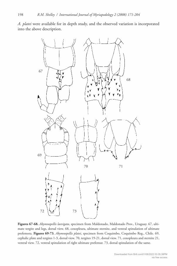
R.M. Shelley / International Journal of Myriapodology 2 (2008) 171-204198
A. platei were available for in depth study, and the observed variation is incorporated into the above description.
Figures 67-68. Akymnopellis laevigata, specimen from Maldonado, Maldonado Prov., Uruguay. 67, ulti-mate tergite and legs, dorsal view. 68, coxopleura, ultimate sternite, and ventral spinulation of ultimate prefemora. Figures 69-73, Akymnopellis platei, specimen from Coquimbo, Coquimbo Reg., Chile. 69, cephalic plate and tergites 1-3, dorsal view. 70, tergites 19-21, dorsal view. 71, coxopleura and sternite 21, ventral view. 72, ventral spinulation of right ultimate prefemur. 73, dorsal spinulation of the same.
67
68
69
70
72 73
71
Downloaded from Brill.com01/08/2022 03:35:38PMvia free access

Revision of Hemiscolopendra 199
Distribution. Central Chile and northern Argentina (Fig. 59).Published records. ARGENTINA: Jujuy Prov., 5 mi (8 km) N Humahuaca; Tucumán
Prov., Tucumán, Cerro San Xavier (Chamberlin 1955). CHILE: Aconcagua Reg., Guardia Vieja and Zapallar; Coquimbo Reg., Coquimbo,
Fray Jorge Rancho; and 22 mi (35.2 km) N Los Vilos, N of Coquimbo; Valparaiso Reg., Valparaiso, Viña del Mar, and Quilqué (Chamberlin 1955).
Material examined. CHILE: Aconcagua Reg., Zapallar, 27 November 1950, ESR, AEM (NMNH-3). Atacama Reg., Copiapo, 23 August 1940, P. A. Berry (NMNH-1). Coquimbo Reg., Ovalle, 11 December 1950, ESR, AEM (NMNH-1); and 35.2 km (22 mi) N Los Vilos, 13 December 1950, ESR, AEM (NMNH). Valparaiso Reg., Valparai-so, 14 May 1893, Michaelsen (ZMUH-juv.); Cerros de Valparaiso, September 1894, C. Porter-Satante (MHNP-1); and Viña del Mar, Michaelsen (NHMV). Unknown Reg., Mattit, 23 June 1949 (NMNH-1).
Reports of Scolopendra morsitans L., 1758, and S. pachygnatha Pocock, 1895, in the US; possible misidentifi cations of H. marginata
Shelley et al. (2005) reported the introduction of Scolopendra morsitans L., 1758, into Duval Co., Florida, the fi rst authentic record from the North American continent (voucher specimen in FSCA). However, a few early works, lacking extant vouchers, reported S. morsitans from sites in the continental US that are dubious and probably represent misidentifi cations of native species; most antedate widespread human intro-ductions and are especially doubtful. Wood (1862) suspected that “S. morsitans is not an inhabitant of the United States” and correctly characterized the species as having “8-9 spines in three rows” on the ventral surfaces of the ultimate prefemora. Acknowl-edging Wood’s misgivings, Underwood (1887) nevertheless reported S. morsitans from Florida and the “southern states”. Earlier, Cragin (1885) described S. morsitans coerules-cens from Barber Co., Kansas, stating that its color “is a uniform light blue, or greenish blue”; Bollman (1893) placed this name in synonymy under S. morsitans, which was subsequently reiterated by Gunthorp (1913, 1921). Chamberlin (1911) and Cham-berlin & Mulaik (1940) reported S. morsitans from Big Spring, Howard Co., Texas, based on a young specimen whose identity they questioned. As H. marginata possesses three rows of ventral spines on the ultimate prefemora (Figs 34-40, 43-49), I suspect that some reports of S. morsitans represent misidentifi cations of H. marginata and that the authors missed the absence of pretarsal spines on leg pairs 1-20. Th is is especially possible for Underwood’s and Chamberlin & Mulaik’s records, which are within the range of H. marginata. Th e record of S. morsitans coerulescens (Fig. 1, open circle) lies outside the postulated range but not unreasonably so, and the possibility of its being H. marginata is supported by the “blue or greenish blue” color, which is consistent with that of this species (Hoff man & Shelley 1996).
Another erroneous scolopendrid record is that of S. pachygnatha Pocock, 1895, from Archbold Biological Station near Lake Placid, Polk Co., Florida (Chamberlin
Downloaded from Brill.com01/08/2022 03:35:38PMvia free access

R.M. Shelley / International Journal of Myriapodology 2 (2008) 171-204200
1951); otherwise, it is known only from the type locality – Mezquital del Oro, Zaca-tecas, Mexico (Shelley 2006). Arthropods have been heavily sampled at Archbold, and the only scolopendrids known are H. marginata and S. viridis Say, 1821. Th e record of S. pachygnatha could be a misidentifi cation of either species, but the latter seems more likely since Pocock (1895) stated in the original description that the tarsi of all legs of S. pachygnatha are spurred.
Acknowledgements
I am deeply grateful to the following curators and collection managers for access to and loans of typical and non-typical material from the collections under their supervi-sions: J. Weintraub (ANSP), syntypes of S. parva; J. Beccaloni (BMNH), holotype of S. punctiventris; L. Leibensperger (MCZ), syntypes of S. longispina; H. Dastych (ZMUH), holotype of S. cormocephalina, syntypes of Otostigmus platei and Cormoceph-alus michaelseni, and a paratype of S. longipleura; J.-J. Geoff roy (MNHP), types of S. chilensis and S. pallida; V. Stagl (NHMV), syntypes of O. platei; T. Krönestedt (SMNH), holotype of C. laevigatus; and R.L. Hoff man (VMNH), neotype and paraneotype of S. marginata. Th e following curators and collection managers provided access to or loaned non-typical material from the indicated repositories: L. Prendini (AMNH), R.W. Baumann (BYU), H. Enghoff (DMNH), P. Sierwald (FMNH), G.B. Edwards (FSCA), P. Schwendinger (MHNG), and J. Coddington (NMNH). R.L. Hoff man, J.G.E. Lewis, and H. Enghoff provided benefi cial reviews.
References
Attems, C. (1903) Beiträge zur Myriopodenkunde. – Zoologische Jahrbücher, Abteilung für Systematik 18(1): 63-154.
Attems, C. (1930) Myriapoda 2. Scolopendromorpha. – Das Tierreich 54: 1-308.Bollman, C.H. (1888a) Notes upon some myriapods belonging to the U. S. National Museum.
– Proceedings of the United States National Museum 11: 343-350.Bollman, C.H. (1888b) A preliminary list of the Myriapoda of Arkansas, with descriptions of
new species. – Entomologica Americana 4: 1-8.Bollman, C.H. (1888c) Notes on a collection of Myriapods from Mossy Creek, Tenn., with
a description of a new species. – Proceedings of the United States National Museum 11: 339-342.
Bollman, C.H. (1888d) Catalogue of the myriapods of Indiana. – Proceedings of the United States National Museum 11: 403-410.
Bollman, C.H. (1893) Th e Myriapods of North America. – Bulletin of the United States Na-tional Museum 46: 1-210.
Brimley, C.S. (1938) Insects of North Carolina. – North Carolina Department of Agriculture, Division of Entomology, Raleigh, 560 pp.
Downloaded from Brill.com01/08/2022 03:35:38PMvia free access

Revision of Hemiscolopendra 201
Brölemann, H.W. (1896) Liste de Myriapodes des États-Unis, et principalement de la Caroline du Nord, faisant partie des collections de M. Eugène Simon. – Annales de la Société Ento-mologique de France 65: 43-70.
Bücherl, W. (1939) Os quilopodos do Brasil. – Memorias do Instituto Butantan 13: 43-362.Bücherl, W. (1942) Catálogo dos quilópodos da Zona Neotrópica. – Memorias do Instituto
Butantan 15: 251-372.Bücherl, W. (1974) Die Scolopendromorpha der Neotropischen Region. – Symposia of the
Zoological Society of London 32: 99-133.Chamberlin, R.V. (1911) Th e Chilopoda of California II. – Pomona College Journal of Ento-
mology and Zoology 3: 470-479.Chamberlin, R.V. (1914) Th e Stanford expedition to Brazil, 1911, John C. Branner, Director.
Th e Chilopoda of Brazil. – Bulletin of the Museum of Comparative Zoology at Harvard College 58(3): 151-221.
Chamberlin, R.V. (1918a) Myriapods from Okefenokee Swamp, Georgia, and from Natchito-ches Parish, Louisiana. – Annals of the Entomological Society of America 11: 369-380.
Chamberlin, R.V. (1918b) Myriapods from Nashville, Tennessee. – Psyche 25: 23-30.Chamberlin, R.V. (1920) A new diplopod from Texas and a new chilopod from Alaska. – Pro-
ceedings of the Biological Society of Washington 33: 41-44.Chamberlin, R.V. (1944) Chilopods in the collections of Field Museum of Natural History. –
Zoological Series Field Museum of Natural History 28(4): 173-216.Chamberlin, R.V. (1951) Records of American millipeds and centipedes collected by Dr. D.
Elden Beck in 1950. – Great Basin Naturalist 11(1-2): 27-35.Chamberlin, R.V. (1955) Th e Chilopoda of the Lunds University and California Academy of
Science Expeditions. – Lunds Universitets Årsskrift N. F. Avd. 2 51(5): 1-61.Chamberlin, R.V. & Mulaik, S. (1940) On a collection of centipeds from Texas, New Mexico,
and Arizona (Chilopoda). – Entomological News 51: 107-110, 125-128, 156-158.Coscarón, S. (1955) Los quilópodos escolopendromorfos del Museo de La Plata. – Revista del
Museo de la Universidad de La Plata (Nueva Serie) 6, Sección Zoologia: 359-418.Crabill, R.E., Jr. (1950) On a collection of centipedes from western South Carolina. – Ento-
mological News 60: 10.Crabill, R.E., Jr. (1955a) A checklist of the Chilopoda known to occur in Kentucky. – Ento-
mological News 66: 257-261.Crabill, R.E., Jr. (1955b) A preliminary report on the Chilopoda of Missouri. – Entomological
News 66: 36-41.Crabill, R.E., Jr. (1960) A new American genus of cryptopid centipede, with an annotated
key to the scolopendromorph genera from America north of Mexico. – Proceedings of the United States National Museum 111: 1-15.
Cragin, F.W. (1885) First contribution to a knowledge of the Myriapoda of Kansas. – Bulletin of the Washburn Laboratory of Natural History 4: 143-145.
Gervais, P. (1847) Myriapodes. – In: Walckenaer & Gervais (eds.), Histoire naturelle des In-sectes. Aptères 4: 1-333, 577-595.
Gunthorp, H. (1913) Annotated list of the Diplopoda and Chilopoda, with a key to the Myr-iapoda of Kansas. – Th e Kansas University Science Bulletin 7(6): 161-182.
Downloaded from Brill.com01/08/2022 03:35:38PMvia free access

R.M. Shelley / International Journal of Myriapodology 2 (2008) 171-204202
Gunthorp, H. (1921) Cragin’s collection of Kansas Myriapoda. – Canadian Entomologist 53: 87-91.
Hoff man, R.L. (1994) Th e occurrence of Hemiscolopendra punctiventris (Newport), an ecophil-ous centiped, in Virginia (Chilopoda: Scolopendromorpha). – Banisteria 3: 33-34.
Hoff man, R.L. (1995) Th e centipeds (Chilopoda) of Virginia: a fi rst list. – Banisteria 5: 20-32.Hoff man, R.L. & Shelley, R.M. (1996) Th e identity of Scolopendra marginata Say (Chilopoda:
Scolopendromorpha: Scolopendridae). – Myriapodologica 4: 35-42.Jeekel, C.A.W. (2005) Nomenclator generum et familiarum Chilopodorum: A list of the genus
and family-group names in the Class CHILOPODA from the 10th edition of Linnaeus, 1758, to the end of 1957. – Myriapod Memoranda Supplement I: 1-130.
Kenyon, F.C. (1893) A preliminary list of the Myriapoda of Nebraska, with descriptions of new species. – Publications of the Nebraska Academy of Science 3: 14-18.
Kevan, D.K.McE. (1983) A preliminary survey of known and potentially Canadian and Alaskan centipedes (Chilopoda). – Canadian Journal of Zoology 61: 2938-2955.
Koch, L.E. (1983) Revision of the Australian centipedes of the genus Cormocephalus Newport (Chilopoda: Scolopendridae: Scolopendrinae). – Australian Journal of Zoology 31: 799-833.
Kohlrausch, E. (1878) Beiträge zur Kenntniss der Scolopendriden. – Inaugural-Dissertation zur Erlangung der Doctorwürde bei Hochlöblicher Philosophischen Facultät zu Marburg eingereicht von Ernst Kohlrausch aus Hannover. Marburg, 1878, 27 pp.
Kohlrausch, E. (1879) Beiträge zur Kenntniss der Scolopendriden. – Journal Museum Godef-froy 14: 51-74.
Kohlrausch, E. (1881) Gattungen und Arten der Scolopendriden. – Archiv für Naturgeschichte 47: 50-132.
Kraepelin, K. (1903). Revision der Scolopendriden. – Mitteilungen aus dem Naturhistorischen Museum in Hamburg 20: 1-276.
Kraus, O. (1957) Myriapoden aus Peru, VI: Chilopoden. – Senckenbergiana Biologia 38(5/6): 359-404.
Lewis, J.G.E., Edgecombe, G.D. & Shelley, R.M. (2005) A proposed standardised terminology for the external taxonomic characters of the Scolopendromorpha (Chilopoda). – Frag-menta Faunistica 48(1): 1-8.
McNeill, J. (1887) List of the myriapods found in Escambia County, Florida, with descriptions of six new species. – Proceedings of the United States National Museum 10: 323-327.
McNeill, J. (1888) A list, with brief descriptions of all the species, including one new to sci-ence, of Myriapoda of Franklin Co., Ind. – Bulletin of the Brookville Society of Natural History 3: 1-20.
Meinert, F. (1886) Myriapoda Musei Cantabrigensis, Mass. Part I. Chilopoda. – Proceedings of the American Philosophical Society 23(122): 161-232.
Newport, G. (1844) A list of the Myriapoda, Order Chilopoda, contained in the cabinets of the British Museum, with synoptic descriptions of forty-seven new species. – Annals and Magazine of Natural History (series 1) 8: 94-101.
Newport, G. (1845) Monograph of the Class Myriapoda, Order Chilopoda; with observations on the general arrangement of the Articulata, Part 2. – Transactions of the Linnean Society of London (1844) 19: 349-439.
Downloaded from Brill.com01/08/2022 03:35:38PMvia free access

Revision of Hemiscolopendra 203
Pocock, R.I. (1895-1910) Chilopoda and Diplopoda. – Biologia Centrali-Americana, 217 pp. [Fascicle treating North American Scolopendromorpha distributed in December 1895].
Porat, C.O. von (1876) Om Några exotiska myriopoder. – Bihang till Kongliga Svenska Ve-tenskaps Akademiens Handlingar 4(7): 1-48.
Say, T. (1821) Descriptions of the Myriapodae of the United States. – Journal of the Academy of Natural Sciences at Philadelphia 2: 102-114.
Selander, R.B. & Vaurie, P. (1962) A gazetteer to accompany the “Insecta” volumes of the “Bio-logia Centrali-Americana”. – American Museum Novitates Number 2099: 1-70.
Shelley, R.M. (1978) Class Chilopoda. p. 221. – In: Zingmark, R.G. (ed.), Annotated Check-list of the Biota of the Coastal Zone of South Carolina. University of South Carolina Press, Columbia, 364 pp.
Shelley, R.M. (1987) Th e scolopendromorph centipedes of North Carolina, with a taxonomic assessment of Scolopocryptops gracilis peregrinator (Crabill) (Chilopoda: Scolopendro-morpha). – Th e Florida Entomologist 70: 498-512.
Shelley, R.M. (2002) A synopsis of the North American centipedes of the order Scolopendro-morpha (Chilopoda). – Virginia Museum of Natural History Memoir 5: 1-108.
Shelley, R.M. (2006) A chronological catalog of the New World species of Scolopendra L., 1758 (Chilopoda: Scolopendromorpha: Scolopendridae). – Zootaxa 1253: 1-50.
Shelley, R.M., Edwards, G.B. & Chagas, A. (2005) Introduction of the centipede Scolopendra morsitans L., 1758, into northeastern Florida, the fi rst authentic North American record, and a review of its global occurrences (Scolopendromorpha: Scolopendridae: Scolopendri-nae). – Entomological News 116(1): 39-58.
Silvestri, F. (1895) Viaggio del dottor Alfredo Borelli nella Republica Argentina e nel Paraguay XIV. Chilopodi e Diplopodi. – Bollettino dei Musei di Zoologia ed Anatomia comparata della R. Università di Torino 10(203): 1-12.
Silvestri, F. (1897) Viaggio del Dott. Alfredo Borelli nel Chaco Boliviano e nella Repubblica Argentina. IV. Chilopodi e Diplopodi. – Bollettino dei Musei di Zoologia ed Anatomia comparata della R. Università di Torino 12(283): 1-11.
Silvestri, F. (1905) Fauna Chilensis. Myriapoda. – Zoologische Jahrbücher 6(3): 715-772.Summers, G. (1979) An illustrated key to the chilopods of the north-central region of the
United States. – Journal of the Kansas Entomological Society 52: 690-700.Summers, G., Beatty, J.A. & Magnuson, N. (1980) A checklist of Illinois centipedes (Chilo-
poda). – Great Lakes Entomologist 13: 241-257.Summers, G., Beatty, J.A. & Magnuson, N. (1981) A checklist of Illinois centipedes (Chilo-
poda): Supplement. – Great Lakes Entomologist 14: 59-62.Underwood, L.M. (1887) Th e Scolopendridae of the United States. – Entomologica Americana
3(4): 61-65.Verhoeff , K.W. (1907 [1902-1925]) Chilopoda, pp. 1-725. – In: Bronn, H.G., Klassen und
Ordnungen des Tier-Reichs, wissenschaftlich dargestellt in Wort und Bild 5(2): 63-101.Williams, S.R. & Hefner, R.A. (1928) Th e millipedes and centipedes of Ohio. – Bulletin of the
Ohio Biological Survey 18, 4(3)[Ohio State University Bulletin 33(7)]: 93-146.Wood, H.C. (1861) Descriptions of new species of Scolopendra in the collection of the Acad-
emy. – Proceedings of the Academy of Natural Sciences, Philadelphia 13: 10-15.
Downloaded from Brill.com01/08/2022 03:35:38PMvia free access

R.M. Shelley / International Journal of Myriapodology 2 (2008) 171-204204
Wood, H.C. (1862) On the Chilopoda of North America, with a catalogue of all the specimens in the collection of the Smithsonian Institution. – Journal of the Academy of Natural Sci-ences, Philadelphia (ser. 2) 5: 2-52.
Wood, H.C. (1865) Th e Myriapoda of North America. – Transactions of the American Philo-sophical Society 13: 137-248.
Wray, D.L. (1950) Insects of North Carolina, Second Supplement. – North Carolina Depart-ment of Agriculture, Division of Entomology, Raleigh, 181 pp.
Wray, D.L. (1967) Insects of North Carolina, Th ird Supplement. – North Carolina Depart-ment of Agriculture, Division of Entomology, Raleigh, 181 pp.
Downloaded from Brill.com01/08/2022 03:35:38PMvia free access