Reviewer #1 Reviewer #2 - Biogeosciences
27
Response to reviewers Reviewer #1 Reviewer [R]: This is a very exciting research. The paper is very well-written and interesting and should be published with very minor revisions. It brings a new perspective on cable bacteria and nanocables over geologic time scales. Well done. [R] 1 – Note that the concept of biogeobattery was proved through geophysical measuremens much before the Nielsen and Risgaard-Petersen (2012) by Naudet, V., A. Revil, J.-Y. Bottero, and P. Bégassat, Relationship between self-potential (SP) signals and redox conditions in contaminated groundwater, Geophys. Res. Lett., 30(21), 2091, doi: 10.1029/2003GL018096, 2003; Naudet, V., A. Revil, E. Rizzo, J.-Y. Bottero, and P. Bégassat, Groundwater redox conditions and conductivity in a contaminant plume from geoelectrical investigations, Hydrology and Earth System Sciences (HESS), 8(1), 8-22, 2004; Naudet, V. and A. Revil, A sandbox experiment of the relationship between redox and self-potential and its application to the interpretation of self-potential data over contaminant plumes, Geophysical Research Letters, 32, L11405, doi: 10.1029/2005GL022735, 2005; Linde, N., and A. Revil, Inverting self-potential [..] , the occurrence of self-potential associated with the biogeobattery concept was proposed before the cited papers in the papers mentioned above. Authors [A]: Ad. 1 – Of course, all of these paper listed in this point and referred to the biogeobattery idea before Nielsen & Risgaard-Petersen (2012) should be cited and we added them to the text and references. [R]: 2 – The introduction implies that the results from Nielsen and Risgaard-Petersen (2012) are related to microcable (or nanowires). This is not the case: it has been shown that they are related to cable bacteria, see details in Risgaard-Petersen, N., L. R. Damgaard, A. Revil, and L. P. Nielsen, Mapping electron sources and sinks in a marine biogeobattery, Journal of Geophysical Research Biogeosciences, 119(8), 1475–1486, doi:10.1002/ 2014JG002673, 2014. [A]: Ad. 2 – Risgaard-Petersen et al. (2014) showed that the so-called cable bacteria of the family Desulfobulbaceae form a biogeobattery in marine sediments. Nevertheless, we did not mention this paper as well as the paper by Nielsen and Risgaard-Petersen (2012) in the introduction. Instead, we mentioned nanowires and biofilm in the context of biogeobaterries in page 17712 (lines 20–21), however, this is consistent with the biogeobattery mechanism described as a “network of microbial nanowires” by Nielsen and Risgaard-Petersen (2015; wrongly cited as 2014 in our manuscript, see page 17721, lines 30–32) where authors followed the paper by Revil et al. (2010). [R]: 3 – Figure 3 is great, but does not show the half-redox reactions. Note it is also possible that these bacteria are using pyrite to exchange electrons and support indeed the idea of a collaboration between these bacteria and the mineralogy. This idea was actually proposed in Naudet et al. (2003, 2004) much before the references you cite. [A]: Ad. 3 – Thank you for this suggestion – we included half-reactions in figure 3 with appropriate references and also added the suggested references in text (chapter 6). [R]: 4 – Conclusion 1: about the electrical circuit, I think a better reference is Risgaard-Petersen, N., L. R. Damgaard, A. Revil, and L. P. Nielsen, Mapping electron sources and sinks in a marine biogeobattery, Journal of Geophysical Research Biogeosciences, 119(8), 1475–1486, doi:10.1002/ 2014JG002673, 2014. [A]: Ad. 4 – Right – it is changed. [R]: 5 – Lovely or Lovley, please double check. [A]: Ad. 5 – Lovley – we corrected this mistake. Reviewer #2 Kedzierski and coworkers present here a very original interpretation of the trace fossil Trichichnus. From scanning electron microscope data including 3-D microCT data they propose that these structures are formed from Thioploca sheets, which upon on a later stage of development are
-
Upload
khangminh22 -
Category
Documents
-
view
0 -
download
0
Transcript of Reviewer #1 Reviewer #2 - Biogeosciences

Response to reviewers
Reviewer #1
Reviewer [R]: This is a very exciting research. The paper is very well-written and interesting and should be published with very minor revisions. It brings a new perspective on cable bacteria and nanocables over geologic time scales. Well done.
[R] 1 – Note that the concept of biogeobattery was proved through geophysical measuremens much before the Nielsen and Risgaard-Petersen (2012) by Naudet, V., A. Revil, J.-Y. Bottero, and P. Bégassat, Relationship between self-potential (SP) signals and redox conditions in contaminated groundwater, Geophys. Res. Lett., 30(21), 2091, doi: 10.1029/2003GL018096, 2003; Naudet, V., A.Revil, E. Rizzo, J.-Y. Bottero, and P. Bégassat, Groundwater redox conditions and conductivity in a contaminant plume from geoelectrical investigations, Hydrology and Earth System Sciences (HESS), 8(1), 8-22, 2004; Naudet, V. and A. Revil, A sandbox experiment of the relationship between redox and self-potential and its application to the interpretation of self-potential data over contaminant plumes, Geophysical Research Letters, 32, L11405, doi: 10.1029/2005GL022735, 2005; Linde, N., and A. Revil, Inverting self-potential [..] , the occurrence of self-potential associated with the biogeobattery concept was proposed before the cited papers in the papers mentioned above.Authors [A]: Ad. 1 – Of course, all of these paper listed in this point and referred to the biogeobattery idea before Nielsen & Risgaard-Petersen (2012) should be cited and we added them to the text and references.[R]: 2 – The introduction implies that the results from Nielsen and Risgaard-Petersen (2012) are related to microcable (or nanowires). This is not the case: it has been shown that they are related to cable bacteria, see details in Risgaard-Petersen, N., L. R. Damgaard, A. Revil, and L. P. Nielsen, Mapping electron sources and sinks in a marine biogeobattery, Journal of Geophysical Research Biogeosciences, 119(8), 1475–1486, doi:10.1002/ 2014JG002673, 2014.[A]: Ad. 2 – Risgaard-Petersen et al. (2014) showed that the so-called cable bacteria of the family Desulfobulbaceae form a biogeobattery in marine sediments. Nevertheless, we did not mention this paper as well as the paper by Nielsen and Risgaard-Petersen (2012) in the introduction. Instead, we mentioned nanowires and biofilm in the context of biogeobaterries in page 17712 (lines 20–21), however, this is consistent with the biogeobattery mechanism described as a “network of microbial nanowires” by Nielsen and Risgaard-Petersen (2015; wrongly cited as 2014 in our manuscript, see page 17721, lines 30–32) where authors followed the paper by Revil et al. (2010).[R]: 3 – Figure 3 is great, but does not show the half-redox reactions. Note it is also possible that these bacteria are using pyrite to exchange electrons and support indeed the idea of a collaboration between these bacteria and the mineralogy. This idea was actually proposed in Naudet et al. (2003, 2004) much before the references you cite.[A]: Ad. 3 – Thank you for this suggestion – we included half-reactions in figure 3 with appropriate references and also added the suggested references in text (chapter 6).[R]: 4 – Conclusion 1: about the electrical circuit, I think a better reference is Risgaard-Petersen, N., L. R. Damgaard, A. Revil, and L. P. Nielsen, Mapping electron sources and sinks in a marine biogeobattery, Journal of Geophysical Research Biogeosciences, 119(8), 1475–1486, doi:10.1002/ 2014JG002673, 2014.[A]: Ad. 4 – Right – it is changed.[R]: 5 – Lovely or Lovley, please double check.[A]: Ad. 5 – Lovley – we corrected this mistake.
Reviewer #2Kedzierski and coworkers present here a very original interpretation of the trace fossil Trichichnus. From scanning electron microscope data including 3-D microCT data they propose that these structures are formed from Thioploca sheets, which upon on a later stage of development are

colonized by bacteria. These bacteria may eventually attach to framboids formed in the sheets and through nanowire – mineral interaction they may form a conductive network, similar to that proposed in the biogeobattery model. The whole idea, however is in my view is only, loosely founded in observations of structures, that might or might not be interpreted as remains of Thioploca filaments and indications of framboids that might or might not have been colonized by nanowire forming bacteria. I would prefer to see more hard that data that necessitate the author’s interpretation and exclude other possibilities. While the interpretation of the Trichichnus fossil as remains of Thioploca sheets might be a convenient alternative to the classical interpretation (that is a deep-tier burrow produced by unknown invertebrates), justified from observations of filamentous structures of a size that is comparable to Thioploca filaments , the hypothesis proposing the functionof the structure as electric wires is not supported by the data. Though nothing in the data set contradicts the idea, my point is that idea is not needed to explain the Trichichnus fossil scientifically. Further, as there to my knowledge are no data demonstrating that biogeobatteries do form in empty Thioploca sheets (I’m not excluding that this might occur), the “Trichichnus-biogeobattery” hypothesis is not needed to understand better phenomena observed at present in nature. In other words: the hypothesis is superfluous and according to the principle of Ockham’s razor it should therefore not be included in a scientific theory. I therefore recommend that the authors reconsider the presentation of their data: A) Focus on the thioploca interpretation and include eventually here eventually the work of Schulz et al., 2000: (Schulz, H. N., B. Strotmann, V. A. Gallardo, and B. B. Jorgensen (2000), Population study of the filamentous sulfur bacteria Thioploca spp. off the Bay of Concepcion, Chile, Mar. Ecol.Prog. Ser., 200, 117-126) as documentation for presence of iron sulfide encrusted filaments. When discussing the role of microorganisms in element turn over make sure that correct terms are used. Desulfittobacterium frappier is not related to Thioploca as indicated in the text. (p 17715 l. 20). Is this species at all present in Thioploca mats? – If not it, it is irrelevant in the context. B) Tone down the Trichichnus-biogeobattery idea. It might be a (somewhat wild) perspective that can be expressed in a few lines in the end of the manuscript, but without substantial evidence it cannot be the main message of a scientific paper,
Authors We are grateful to Referee #2 for remarks which stimulated us to clarify some of our ideas. Referee #2 claimed that the trace fossil Trichichnus “[...] might or might not be interpreted as remains of Thioploca filaments and indications of framboids that might or might not have been colonized by nanowire forming bacteria.”, and continues [...] the hypothesis proposing the function of the structure as electric wires is not supported by the data. Though nothing in the data set contradicts the idea, my point is that idea is not needed to explain the Trichichnus fossil scientifically.” In our paper there are two important points, partly time-related, listed and discussed below. Indeed, we do not need to explain Trichichnus as an electric wire. We consider this trace fossil as a remnant of the Thioploca-like bacterial mat. Such starting point entails further considerations that brought usto the (bio)geobattery idea. 1) Thioploca-like bacteria as the possible Trichichnus trace makerNone of the known organisms can produce Trichichnus-like structure visible in our CT scanner images, except for large sulphur bacteria constructing an extensive and dense mat with vertical filaments reaching the Trichichnus dimensions. Therefore, we narrowed our interpretation to Thioploca-like bacteria. At the present stage, the interpretation of Thioploca-like bacteria as the Trichichnus trace maker is based only on a rough comparison of 3D pictures. The similarity is striking and in our opinion this is the best explanation of origin of this trace fossil. As referee #2 says “[..] the interpretation of the Trichichnus fossil as remains of Thioploca sheets might be a convenient alternative to the classical interpretation [..]”.
2) transformation of Thioploca-like sheaths into Trichichnus, including their bacterial consortia and iron sulfide infilling, which could serve as „a bioelectric cable”.
Trichichnus is a unique structure within which several electrical processes can not only be met but also extended in time: a/ strictly biogenic; b/ combining bacteria and iron sulfides; and c/ related to iron sulfides as conductors. The filamentous forms of the family Beggiatoacea (comprising Thioploca spp.) were just pointed out as the main direct competitors to cable bacteria (see Nielsen and Risgaard-Petersen, 2015) In addition, Thioploca's sheaths are inhabited by consortia of different bacteria (Kojima et al., 2006; Teske et al., 2009) or even protists (Buck et al., 2013). Positive hybridization with probe CoSRB385 points out that these consortia are related to members of deltaproteobacterial family Desulfobactereaceae, in competitive hybridization with almost identical (single mismatch) probe InSRB385 designed for the families Desulfobulbaceae and Desulfovibrionaceae (Teske et al., 2009). Close relationship between Thioploca and iron sulfides is observed already at the early stages of bacterial mat formation. Studies of Thioploca spp. from the Bay of Conception showed that filaments of the short-cell morphotype disappear from sheaths during autumn/winter time and these empty sheaths are stained with iron sulfide (Schulz et al. 2000;we add this paper to references). A complex electroactive biofilm, interconnecting bacterial cells and iron sulfide surfaces (see refs. in the manuscript), can form the long-distance wires reaching further than it has been documented, so far. Pyrite (iron disulfide, forming via magnetic greigite Fe3S4 stage) framboids, which typically infill Trichichnus, can extent electron transfer into later stages of diagenesis (a model described in the last chapter of the manuscript), when the metal semiconductor (pyrite wire in this case) crosses over the redox boundary, and fulfills the geobattery idea. Sulfur in iron sulfide framboids results from bacterial (typically Desulfovibrio desulfuricans) sulfate reduction and is sometimes modified by bacterial sulfur oxidation.Referee #2 expects us to tone down the Trichichnus-biogeobattery idea because we do not have a substantial evidence. However, the idea is always a good starting point. There are not many or, bet-ter to say, there are practically no examples of palaeogeobatteries. In our paper, we present a poten-tial structure from the geological past where several bioprocesses related to electron transfer in sedi-ment could proceed. Number of evidence for such processes, both in the field and laboratory invest-igations, has been growing significantly for last few years. Development of new technologies, e.g. multipurpose electrodes which combine reactive measurements with electrical geophysical meas-urements (Zhang et al., 2010) opens new frontiers in monitoring microbial processes in sediments. We believe that our idea enhances special attention to Thioploca's endobionts in respect of their electron exchange along the cell-to-mineral wires. The fossilized filamentous sulphur oxidizing bac-teria operating at the oxic-anoxic interface are reported even from the Palaeoproterozic (e.g., Hiatt et al., 2015). However, we should be aware that discovery of fossilized “wires” that operated in sed-iment alone can be difficult. Since a “cable bacteria” trigger a rapid oxygenation of the sediment, they also allow bioturbating macrofauna to colonize the sediment in the next step and to destroy them mechanically (e.g., Malkin et al., 2014). It seems that Trichichnus is the only record of bio-electrical processes in the fossil state due to deep-tier occurrence of the Thioploca sheaths, far be-low the range of majority bioturbating organisms, i.e., at the depth of 20–30 cm. The sentence (p. 17715, l. 19-20) “Regarding Thioploca sulphur bacteria it is interesting to note thatbacterial species Desulfitobacterium frappier is capable of both reducing [...]” is rephrased into “Interestingly, some Firmicutes bacteria species Desulfitobacterium frappieri is capable of both reducing [...]”. The Firmuctes bacteria species D. frappieri has not been recognized yet as thioploca's endobiont which mostly belongs to δ-proteobacteria. However, both of these groups represent microbes contributing to iron redox cycling.
References:Hiatt, E.E., Pufahl, P.K. & Edwards, C.T., 2015. Sedimentary phosphate and associated fossil bacteria in a Paleoproterozoic tidal flat in the 1.85 Ga Michigamme Formation Michigan, USA. Sedimentary Geology, 319: 24–39.Malkin, S., Rao, A.M.F., Seitaj, D., Vasquez-Cardenas, D., Zetsche, E.-M., Hidalgo-Martinez, S., Boschker, H.T.S. & Meysman, F.J.R., 2014. Natural occurrence of microbial sulphur oxidation by

long-range electron transport in the seafloor. The ISME Journal, 8: 1843–1854.Nielsen, L. & Risgaard-Petersen, N., 2015. Rethinking sediment biogeochemistry after the discovery of electric currents. Annual Review of Marine Sciences, 7: 21.1–21.18.Schulz, H.N., Strotmann B., Gallardo, V.A. & Jorgensen, B.B., 2000. Population study of the filamentous sulfur bacteria Thioploca spp. off the Bay of Concepcion, Chile. Marine Ecology Progress Series, 200: 117–126. Zhang, C., Ntarlagiannis, D., Slater, L., Doherty, R. 2010. Monitoring microbial sulfate reduction inporous media using multi-purpose electrodes. Journal of Geophysical Research: Biogeosciences, 115: G00G09, doi:10.1029/2009JG001157

Fossilized bioelectric wire – the trace fossil Trichichnus
M. Kędzierski1, A. Uchman1, Z. Sawlowicz1 & A. Briguglio2,3
1Institute of Geological Sciences, Jagiellonian University, Oleandry 2a, 30-063 Kraków,
Poland, [email protected] für Paläontologie, Universität Wien, Geozentrum, Althanstrasse 14, 1090 Vienna,
Austria 3Faculty of Science, Department of Petroleum Geoscience, Universiti Brunei Darussalam,
Jalan Tungku Link, Gadong BE1410, Brunei Darussalam
Abstract
The trace fossil Trichichnus is proposed as an indicator of fossil bioelectric bacterial activity
at the interface oxic—anoxic zone of marine sediments. This fulfils the idea that such
processes, commonly found in the modern realm, should be also present in the geological
past. Trichichnus is an exceptional trace fossil due to its very thin diameter (mostly less than 1
mm) and common pyritic filling. It is ubiquitous in some fine-grained sediments, where it has
been interpreted as a burrow formed deeper than any other trace fossils, below the redox
boundary. Trichichnus formerly referred to as deeply burrowed invertebrates, has been found
as remnant of a fossilized intrasediment bacterial mat that is pyritized. As visualized in 3-D by
means of X-ray computed microtomography scanner, Trichichnus forms dense filamentous
fabric, which reflects that produced by modern large, mat-forming, sulphide-oxidizing
bacteria, belonging mostly to Thioploca-related taxa, which are able to house a complex
bacterial consortium. Several stages of Trichichnus formation, including filamentous,
bacterial mat and its pyritization, are proposed to explain an electron exchange between oxic
and suboxic/anoxic layers in the sediment. Therefore, Trichichnus can be considered a
fossilized “electric wire”.
1 Introduction
Bioelectric bacterial processes are omnipresent phenomena in the recent oxic–anoxic
transition zones of marine sediments (see Nielsen and Risgaard-Petersen, 20154 for review).
It is intriguing that they have not so far been recognized from the geological past as it was
suggested by Risgaard-Petersen et al. (2012). One reason could be that effects of such
1
5
10
15
20
25

bacterial processes, leading to oxygenation of the anoxic sediments on the sea floor, are
eventually destroyed by subsequent bioturbation (Malkin et al., 2014). We propose here that
the usually pyritized marine trace fossil Trichichnus Frey, 1970 can be interpreted as a fossil
record of a complex bioelectrical microoperations-cable resulting from bacterial activities in
the oxygen depleted part of marine sediments. Trichichnus is a branched or unbranched,
straight to winding, hair-like, cylindrical structure, mostly 0.1–0.7 mm in diameter, commonly
pyritized, oriented at various angles (mostly vertical) with respect to the bedding. The
common pyritization is a particular feature of Trichichnus, which is generally absent in other
trace fossils. Trichichnus is reported from both shallow- and deep-sea, mostly fine-grained
sediments (Frey, 1970; Wetzel, 1983). It ranges from the Cambrian (Stachacz, 2012) to the
Holocene (Wetzel, 1983). There are distinguished Trichichnus appendicus displaying thin
appendages; and T. linearis or T. simplex probably differ only in the presence of a lining,
which likely is diagenetic in origin (see Uchman, 1999, for review). Trichichnus is common in
sediments in which pore waters were poorly oxygenated (McBride and Picard, 1991;
Uchman, 1995) and is considered to be one of the first trace fossils recording colonization of
the sea floor after improvement of oxygenation, penetrating sediments below the redox
boundary, and having a connection to oxygenated waters on the sea floor (Uchman, 1995). It
belongs to the ethological category chemichnia distinguished for trace fossils produced by
organisms feeding by means of chemosymbiosis (Uchman, 1999).
Trichichnus was previously interpreted as a deep-tier burrow produced by unknown
opportunistic invertebrates in poorly oxygenated sediments (McBride and Picard, 1991;
Uchman, 1995). It has also been compared to an open burrow in modern deep-sea sediments
more than 2 m long and no more than 0.5 mm thick (Thomson and Wilson, 1980; Weaver and
Schultheiss, 1983). The sipunculid worm Golfingia has been considered as a possible modern
example of tracemakers of very narrow and long tubes similar to Trichichnus (Romero-
Wetzel, 1987). However, 3-D microCT scanner images of Trichichnus presented in this paper
reflect fabric produced by modern, large, sulphur bacteria. Therefore, a new interpretation of
Trichichnus is possible by its comparison to modern sulphur bacteria such as Thioploca spp.
and their post-mortem history. Moreover, we propose a biogeochemical model of Trichichnus
assisted electron transfer between different redox zones of the sediment.
2 Material and methods summary
2
5
10
15
20
25
30

The morphology and chemistry of Trichichnus from several samples, represented by Albian
turbiditic marly mudstones (Silesian Nappe, Polish Carpathians); Valanginian–Hauterivian
pelagic marlstones (Kościeliska Marl Formation, the Tatra Mts.) and Eocene silty shales
(Magura Nappe, Polish Carpathians), were studied using a scanning electron microscope with
field emission (FE-SEM, Hitachi S4700), equipped with EDS (Noran Vantage) which allows
to determine of chemical compounds of the examined objects. The studies on morphology and
infilling of Trichichnus were also supported using the polarising light microscope Nikon
Eclipse Pol E-600. All of the above were carried out at the Institute of Geological Sciences of
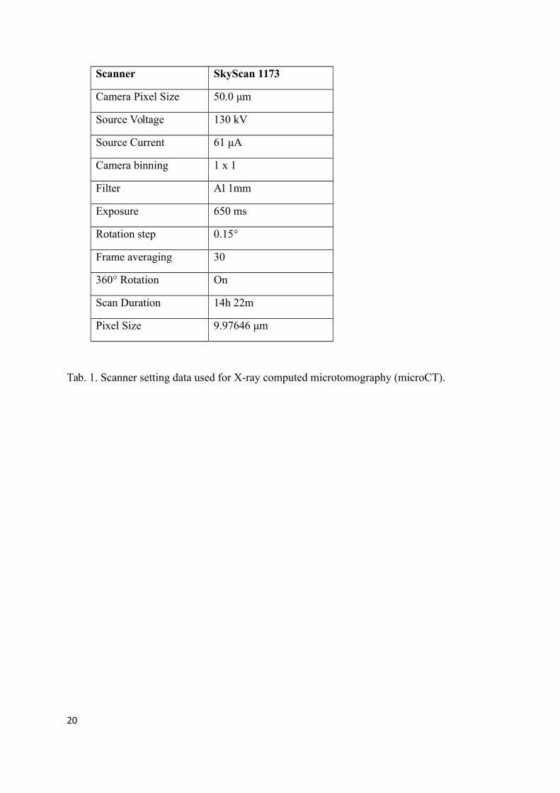
the Jagiellonian University in Kraków. Moreover, the X-ray computed microtomography
(microCT) scanner (SkyScan 1173) available at the Institute of Palaeontology, University of
Vienna, was used to carry out the main research on the spatial organization of the Trichichnus
(Tab. 1).
All samples of trace fossil Trichichnus presented in the paper are housed at the Institute of
Geological Sciences, Jagiellonian University, Kraków, Poland, collection of Alfred Uchman
and Zbigniew Sawlłowicz. No permits were required for the described study, which complied
with all relevant regulations.
3 Results
With the naked eye or under light microscope Trichichnus is easily distinguished in all rocks
samples mainly by its different colour (Fig. 1a–d). Macroscopically Trichichnus is similar in
all samples, and shows a light halo around the trace, except in burrows without iron mineral
infillings (Fig. 1c, and d). Neither the length nor width of the trace depends on the type of the
host rock. The boundary between the trace fossil and the rock is usually sharp (Fig. 1e).
The SEM studies revealed that majority of the studied Trichichnus is filled with framboids
and framboidal aggregates (Fig. 1e, and f). Framboidal forms are composed of iron sulphides
(most probably pyrite), Fe oxy/hydroxides or a mixture of both. The microcrystals
composingbuilding framboids are generally well-developed and their sizes are usually about
1–2 µm, generally dependent on the size of the framboid. Most of the microcrystals in
framboids are closely packed. SEM-EDS study shows that the colourful halo around traces
does not reveal any chemical differences from the composition of the surrounding rock. It is
likely that the amount of iron minerals in the halo is below the detection limit of the EDS
3
5
10
15
20
25
30

method. Apart from the iron minerals, subordinate amounts of several other minerals,
generally corresponding to the host rock, such as calcite, quartz and gypsum, are observed.
MicroCT scans allowed three dimensional visualization of several Trichichnus embedded in a
dark mudstone from the Oligocene Menilite Formation in the Western Carpathians, Poland.
The density difference between the sediment matrix and the pyritic trace fossil fillings
allowed extremely accurate three dimensional visualization (Fig. 2). The results show a very
dense fabric composed of Trichichnus fillings of variable size and orientation, which are only
partly visible on a sample surface. Since the resolution of the 3-D model here presented is
9.97 μm, all the structures larger than 10 μm are visible and renderable. The distribution of
diameters of the traces displays two major peaks: 600 μm for the thickest traces and 90 μm for
the very thin ones (Fig. 2a; Ssupplementary movie file).
The Trichichnus morphology seen in 3-D microCT scanner image (Fig. 2a—c) is similar to
structures produced by filamentous bacteria (e.g., Pfeffer et al., 2012) or to large, mat-
forming, filamentous sulphur bacteria, e.g., Thioploca spp. (Fossing et al., 1995 – Ffig. 3aA;
Schulz et al., 1996 – Ffig. 8; Jørgensen and Gallardo, 1999 – Ffig. 5) (see also Heutell et al.,
1996). The microCT scanner images also reveal different spatial organizations, density,
diameters and shapes of the Trichichnus. These correspond to different parts of vertical
system of the Thioploca mat in sediment. Also the density of Trichichnus (Fig. 2a), which
constitutes 3.1% of the whole scanned sediment volume, is comparable to the Thioploca mat
in their upper shallow subsurface part. Other 3-D scanner images (Fig. 2b, and c) show
lowered density of Trichichnus, comparable to middle and bottom parts of the Thioploca
filamentous mat spatial system.
4 Discussion
Three-dimensional reconstructions of Thioploca spp. (Fossing et al., 1995; Schulz et al., 1996;
Jørgensen and Gallardo, 1999) resemble Trichichnus in the 3-D scanner images. Moreover,
the common pyritization of Trichichnus ismay be related to sulphate-reducing bacteria, like δ-
proteobacteria Desulfovibrio sp., which can co-occur with sulphur-oxidizing bacteria, such as
Beggiatoa or Thioploca (e.g., Jiang et al., 2012). the life-mode of various sulphur bacteria,
which specialized in sulphide oxidation using electron transportation and nitrate stored in cell
vacuoles as a terminal electron acceptor (Jørgensen and Gallardo, 1999; Jørgensen and
4
5
10
15
20
25
30

Nelson, 2004). Therefore, we consider Thioploca-like, mat-forming, large, sulphur bacteria
and related filamentous sulphide-oxidizing bacteria (see Teske et al., 1995) supposedly
sulphide-oxidizing bacteria as a tracemaker of Trichichnus tunnels and their spatial
organization. The Thioploca-like mat, usually hosting other bacteria or small protists (e.g.,
Buck et al., 2014), combined with the post-mortem history of sulphur bacterial mat systems,
are referred to the idea of bioelectrochemical systems (BESs), including the microbial fuel
cells (MFCs) and biogeobatteries (e.g., Naudet et al., 2003, 2004; Naudet and Revil, 2005;
Linde and Revil, 2007; Ntarlagiannis et al., 2007; Logan, 2009; Teske et al., 2009; Nielsen
and Girguis, 2010; Revil et al., 2010; Borole et al., 2011; Hubbard et al., 2011; Risgaard-
Petersen et al., 2014; Nielsen and Risgaard-Petersen, 20154 for review).
In biogeobattery systems, bacteria are interconnected cells to cells or cells to mineral surfaces
via electrically conductive appendages – bacterial pili (nanowires) – making a complex
electroactive network (biofilm) (Ntarlagiannis et al., 2007; Revil et al., 2010; Borole et al.,
2011; Reguera et al., 2005; Gorby et al., 2006; El-Naggar et al., 2010; Nielsen et al., 2010;
Risgaard-Petersen et al., 2014; Nielsen and Risgaard-Petersen, 2015). The electron transport
in BESs may be realized in various ways, including transfer through long bacterial nanowires
or along biofilm matrix (Loveley, 2008). Its magnitude and duration mostly depend on
atmospheric oxygen (electron acceptor) availability that generates electrical self-potentials in
the sediments between oxidized and anoxic zones (see Ntarlagiannis, 2007; Risgaard-Petersen
et al., 2014). Studies reporting extracellular bioelectric current concerned dissimilatory metal-
reducing bacteria, such as Geobacter sulfphurreducens, Shewanella oneidensis MR-1 and the
thermophilic, fermentative bacterium Pelotomaculum thermopropionicum or the oxygenic
phototrophic cyanobacterium Synechocystis (Borole et al., 2011). Natural conductive minerals
(e.g., magnetite, greigite – an intermediate stage for pyrite formation) can facilitate electron
transfer between different species of microbes. Presumably microbes should preferentially use
mineral particles, discharging electrons to and accepting them from mineral surfaces, in
particular for long-distance electron transfer (Kato et al., 2012). The interaction between
minerals and bacteria can be quite complex as Kato et al. (2013) showed that e.g., G.
sulfurreducens constructed two distinct types of extracellular electron transfer (EET) paths, in
the presence and absence of iron-oxide minerals. It is worth noting that large scale sulphide
ores, where pyrite is commonly a main component, have beenare regarded as geobatteries for
many years and used by geophysicists for an their exploration. In natural electrochemical
5
5
10
15
20
25
30

processes ore serves as a conductor to transfer the electrons from anoxic to oxic environments
(Sato and Mooney, 1960; Naudet et al., 2003). Newer reports show that coupling of
geochemical reactions between shallower and deeper layers of the sediment can be also
realized by vertical centimetre-long bacterial filaments of multicellular bacteria of the family
Desulfobulbaceae, the so-called cable bacteria (Pfeffer et al., 2012; Risgaard-Petersen et al.,
2014; Schauer et al., 2014). Nevertheless, only the giant sulphur bacteria, so-called a “'macro-
bacteria”' are able to produce mat spatial system in the scale of Trichichnus. Macro-bacteria of
Beggiatoacea family are considered to be the most direct competitors to cable bacteria
representing Desulfobulbaceae family at the interface of oxic–anoxic zone of marine
sediments (see Nielsen and Risgaard-Petersen, 20154). We propose Thioploca spp., one of the
genus of Beggiatoacea family (see Salman et al., 2011), as a tracemaker of Trichichnus.
Large communities of the sulphur bacteria Thioploca spp. produce the filaments consisting of
thousands of cells that can penetrate 5–15 cm down into the sediment. These filaments
(trichomes), bundled in common slime sheaths, make a dense bacterial mat, where the density
decreases with depth leaving single sheaths in the deepest part of the spatial system of the
mat. The Thioploca sheaths, rarely branched, extend in different directions, mostly vertically.
One sheath can accommodate up to 100 filaments of 15–40 µm in diameter each, giving about
4 mm for the maximum diameter of the sheath ( Fossing et al., 1995; Schulz et al., 1996).
Thioploca trichomes gliding within sheaths can migrate through several redox horizons,
coupling the reduction of nitrate in the overlying waters and the oxidation of dissolved
sulphide in the sulphate reduction zone ( Fossing et al., 1995; Heutell et al., 1996). Therefore,
the sheaths are used as communication tubes and enable the sulphur bacteria to switch
vertically between nitrate and sulphide (electron acceptor and donor, respectively). This
sulphide oxidization mechanism leads to accumulation of elemental sulphur globules in the
bacterial cells. Moreover, Thioploca trichomes may leave their sheaths and move into other
sheaths, thus, one sheath can be occupied by different species (Jørgensen and Gallardo, 1999).
Empty sheaths were found even at >20 cm depth into the sediment. In contrast, the sheaths
were not found shallower than a depth of a few centimetres in the sediments. Therefore, the
surface and topmost part of the Thioploca mat system is formed by single bacterial filaments
only (Schulz et al., 1996). However, brackish/freshwater T. ingrica may not form any mats at
the sediment surface, showing biomasses peak 4–7 cm below the sediment surface (Høgslund
et al., 2010).
6
5
10
15
20
25
30

The Thioploca sheaths – either filled with trichomes or abandoned – may be closely
associated with other filamentous sulphate-reducing bacteria like Desulfonema sp. (Jørgensen
and Gallardo, 1999; Teske et al., 2009). Empty sheaths were found even at >20 cm depth into
the sediment. In contrast, sheaths were not found shallowly than a depth of a few centimetres
in the sediments. Therefore, the surface and topmost part of the Thioploca mat system is
formed by single bacterial filaments only (Schulz et al., 1996). Thioploca spp. are known
from both marine and lake environments. Nevertheless, their mass occurrences are related to
high organic production and oxygen depletion, e.g., the Chile and Peru offshore upwelling
system (Jørgensen and Gallardo, 1999).
Pyritized organic remains are common in the sedimentary record and various mechanisms of
fossil pyritization have been described (e.g., Canfield and Raiswell, 1991; Briggs et al., 1996).
Apart from the similarity between the Trichichnus fabric and spatial organization of the
Thioploca mat system, the common pyritization of Trichichnus also indicates its close relation
to the sulphate-reducingur bacteria. Schulz et al. (2000) observed that empty sheaths of
Thioploca from the Bay of Conception during autumn/winter time were stained with iron
sulphides. As the most abundant metal sulphide in nature, pyrite FeS2 has a major influence
on the biogeochemical cycles of sulphur and iron, and through its oxidation also of oxygen.
There have been several important reviews on sedimentary pyrite formation [36, 37, 38 and
refs. therein](e.g., Berner, 1970; Schoonen, 2004; Rickard and Luther, 2007, and references
therein), and specifically on iron sulphide framboids (e.g., Love and Amstutz, 1966; Raiswell,
1982; Wilkin and Barnes, 1997; Sawłowicz, 2000; Ohfuji and Rickard, 2005, and referencess.
therein). It is important to stress that at low temperature pyrite growth is usually preceded by
the formation of unstable iron monosulphides, such as mackinawite and greigite. The latter is
the ferrimagnetic inverse thiospinel of iron. Pyrite, like other sulphide minerals, is a
semiconductor determined to be “semi-metallic” but its conductivity in different admixtures
varies widely between 0.02 and 562 (Ωcm)-1 (Rimstidt and Vaughan, 2003, and referencess.
therein).
Pyrite has a great potential in both interaction with microorganisms and electron transfer as its
range of morphological, chemical and electric characteristics is quite large. Both iron and
sulphur, necessary for pyrite formation, are pivotal for microbes. For instance, more than 50%
of reduced sulphate in sediments inhabited by Thioploca off the Chilean coast is accumulated
in the pyrite pool (Ferdelman et al., 1997). Iron is an important carrier of electrons in
7
5
10
15
20
25
30
microbial ecosystems (Wielenga et al., 2001) and bacteria play important role in different
processes of iron oxidation and reduction (Weber et al., 2006). This is interesting to note that
oInterestingly, some Firmicutes bacteria Obacterial species Desulfitobacterium frappieri is
capable of both reducing Fe3+ with H2 as an electron donor and oxidizing Fe2+ with nitrate as
an electron acceptor (Shelobolina, 2003). In sedimentary environments the major source of
sulphur incorporated into iron sulphides is H2S or HS- resulting from bacterial sulphate
reduction. Sulphate-reducing bacteria (SRB) use sulphate as the terminal electron acceptor of
their electron transport chain (e.g., Widdel and Hansen, 1992). SRB are usually regarded as
strictly anaerobic, but recent investigations showed that SRB are both abundant and active in
the oxic zones of mats (see Baumgartner et al., 2006 for review). Iron monosulphides and
pyrite, including framboids, are quite common in some microbial mats (e.g., Popa et al., 2004;
Huerta-Diaz et al., 2012) but their formation is complicated and difficult to study.
Nevertheless, sulphur content and sulphur speciation may not correlate to microbial metabolic
processes (Engel et al., 2007).
Oxidation of iron sulphides is one of the most common processes in marine sediments. For
example, Passier et al. (1999) reported that in the Mediterranean sapropels the percentage of
initially formed iron sulphides that were re-oxidized varied from 34 to 80%. Oxidation of iron
sulphide framboids to iron (hydro)oxides is often observed in fossils (e.g., Luther et al., 1982;
Sawłowicz, 2000). There is a significant difference between oxidation of pyrite and its
precursors. For example, FeS, but not pyrite, can be oxidized microbially with NO3- as an
electron acceptor (Schippers and Jørgensen, 2002).
6 Conclusions and model of Trichichnus
Trichichnus is a good example of microbiological and mineralogical collaboration,
propounded earlier by Naudet et al. (2003, 2004). Its role in electron transfer is still enigmatic,
but several stages can be proposed (Fig. 3). It should also be noted that the relatively large
length of Trichichnus and its continuation through different redox zones may result in its
vertical internal zonation. As suggested earlier, Trichichnus may reflects former slime sheaths,
where large sulphur bacterial trichomes, like Thioploca community, move up and down.
There is growing evidence (e.g., Teske et al., 2009; Prokopenko et al., 2013; Buck et al., 2014
and referencess. therein), that Thioploca sheaths house a complex bacterial consortium. In our
8
5
10
15
20
25
30

opinion it is a matter of time before additional microbes will be found associated with
Thioploca or their post-mortem sheaths.
(1) The earliest stage of the Trichichnus formation is related to a sulphuric microbial mat in
the dysoxic zone, connecting the anoxic-sulphidic substrate. Long, filamentous bacteria
structured like electric cables facilitate electron transport over centimetre distances in marine
sediment (Pfeffer et al., 2012; Risgaard-Petersen et al., 2014). The filamentous multicellular,
aerobic bacteria of the family Desulfobulbaceae (cable bacteria), living together with
Thioploca in the sheath, conduct electrons through internal insulated wires from cells in the
sulphide oxidation zone to cells in the oxygen reduction zone Pfeffer et al. (2012). For
Thioploca itself such behaviour has not been proved yet (see Nielsen and Risgaard-Petersen,
20154 for review). The electric circuit is balanced by charge transport by ions in the
surrounding environment (Risgaard-Petersen et al., 20142; Nielsen and Risgaard-Petersen,
2015).
(2) The next stage is related to the iron sulphidization process. Microorganisms can influence
both the precipitation of sulphides and their morphology. Bacterial components, e.g., cell
walls, facilitate mineralization by sequestration of metallic ions from solution and provide
local sites for nucleation and growth (Beveridge and Fyfe, 1985). Iron sulphides (framboids)
can form within a matrix of bacteria and biopolymers both during the life of bacterial
consortium (see MacLean et al., 2008) and after their death. It should be noted that the
process of sheath infilling varies within the system. For example, larger sheaths may be
abandoned by trichomes of Thioploca faster, compared to smaller ones (Schulz et al., 1996).
The biofilm in a proto-Trichichnus capsule seems to be an ideal place for the formation of
iron sulphides. Formation of pyrite, probably formerly ferromagnetic greigite, depends on
availability of soluble Fe(II) from a surrounding sediment and sulphides (H2S or
polysulphides). Crystals in framboids are generally closely packed, and framboids themselves
often too, but when it is not the case, the bacterial pili are believed to connect dispersed
conductive minerals (Revil et al., 2010). Cell-to-mineral wires support the thesis that the
metallic conductor-like pyrite occurring in sediment might be responsible for the electron
flow (see also Nielsen et al., 2010). Growth of a biofilm – developed in the mucus of the large
sulphur bacteria sheath – on pyrite can enhance conductivity supporting the biogeobattery
idea. The oldest microbial communities colonising sedimentary pyrite grains were found in a
~3.4 billion-year-old sandstone from Australia (Wacey et al., 2011).
9
5
10
15
20
25
30

(3) The process of electron transfer can continue also after deathtermination of microbial
consortia. Decomposition of organic matter inside the channel creates the local reducing
microenvironment, which promotes pyrite formation (e.g., Berner, 1980; Raiswell, 1982),
even when the surrounding sediment is not fully anoxic. It is tempting to propose that perhaps
some iron sulphide forms resulted from a replacement of sulphur globules (stored intra- and
extracellularly by chemolithoautotrophic bacteria, e.g., Thioploca or by purple and green
sulphur bacteria, Oschmann, 2000), either inside sulphur bacteria cells or after their lysing.
Pyrite might forms the sulphur globules stored intra- and extracellularly by
chemolithoautotrophic bacteria (e.g., Thioploca) or by purple and green sulphur bacteria
(Oschmann, 2000). Spheroidal crystalline aggregates representing early pyrite and
subsequently replaced by iron hydroxides were found in Silurian cyanobacterial filaments
(Tomescu et al., 2006). Formation of pyrite framboids via replacement of sulphur grains was
also shown experimentally by Křibek (1975).
A halo observed around Trichichnus (Fig. 1c and, d) results from oxidation of pyrite or former
iron monosulphidexide infilling the trace fossil. The oxidation stage can take place both
during the earliest and later stages of diagenesis (the latter is out of the scope of this paper).
Re-oxidation processes of pyrite framboids are common in tropical upwelling area, caused by
bioturbation and possible contributions from sulphide-oxidizing bacteria (Diaz et al., 2012
and referencess. therein). It would additionally expand, but also complicate, the process of
electron transfer in sediments.
Summarizing, we believe that Trichichnus is a good example of fossilized bacterial
bioelectrical wirea potential ancient place where severeal bioprocesses related to electron
transfer in sediment could proceed. A number of evidences for such processes, both in the
field and laboratory investigations, has been growing significantly for last few years.
Development of new technologies, e.g., multipurpose electrodes which combine reactive
measurements with electrical geophysical measurements (Zhang et al., 2010), opens new
frontiers in monitoring microbial processes in sediments. We believe that our idea enhances
special attention to Thioploca endobionts in respect of their electron exchange along the cell-
to-mineral wires. SuchOur interpretation greatlyshows improves our knowledge about how
this phenomenon hascould have been widespread in sedimentary environments and through
the geological time.
10
5
10
15
20
25
30

Supplementary information Video captured from an X-ray CT microscanner provides
dimensional visualization of the dense Trichichnus fabric comparable with the upper part of
the bacterial filamentous Thioploca-like mat system.
Acknowledgments We are grateful to Sally Sutton (Colorado State University) for her
comments and suggestions which improved the manuscript. We are indebted to the Editor
Jack Middelburg and two anonymous reviewers for their constructive comments and
suggestions, which helped us to improve the manuscript. The use of the MicroCT has been
possible due to the project P 23459-B17 granted by the Austrian Science Fund. The work was
supported by the Jagiellonian University in Kraków – DS funds project No. K/ZDS/004873
Author Contributions MK conceived the study and contributed to the bacterial part of paper.
AU delivered the samples of Trichichnus and contributed to the trace fossil part of paper. ZS
contributed to the geochemical part of paper. AB delivered the X-ray computed
microtomography scanner data. MK, AU and ZS contributed to the model of Trichichnus.
11
5
10

References
Baumgartner, L. K., Reid, R. P., Dupraz, C., Decho, A. W., Buckley, D. H., Spear, J. R.,
Przekop, K. M. and Visscher, P. T.: Sulphate reducing bacteria in microbial mats: cChanging
paradigms, new discoveries, Sediment. Geol., 185, 131–145, 2006.
Berner, R. A.: Sedimentary pyrite formation, Am. J. Sci., 268, 1–23, 1970.
Berner, R. A.: Early dDiagenesis: aA tTheoretical aApproach, in: Princeton Series in
Geochemistry, Princeton University Press, Princeton, p. 256 pp45., 1980.
Berner, R. A.: Sedimentary pyrite formation, Am. J. Sci., 268, 1–23, 1970.
Beveridge, T. J. and Fyfe, W. S.: Metal fixation by bacterial cell walls, Can. J. Earth Sci., 22,
1893–1898, 1985.
Borole, A. P., Reguera. G., Ringeisen, B., Wang, Z. W., Feng, Y. and Kim, B. H.: Electroactive
biofilms: Current status and future research needs, Energ. Environ. Sci., 4, 4813–4834, 2011.
Briggs, D. E. G., Raiswell, R., Bottrell, S. H., Hatfield, D. and Bartels, C.: Controls on the
pyritization of exceptionally preserved fossils: an analysis of the Lower Devonian Hunsrück
Slate of Germany, Am. J. Sci., 296, 633–663, 1996.
Buck, K. R., Barry, J. P. and Hallam, S. J.: Thioploca spp. sheaths as niches for bacterial and
protistan assemblages, Mar. Ecol., 35, 395–400, doi:10.1111/maec.12076, 2014.
Canfield, D. E. and Raiswell, R.: Taphonomy: Releasing the Data Locked in the fossil record,
in: Topics Geobiology, 9, 337–387Pyrite formation and fossil preservation, in: Taphonomy:
Releasing the Data Locked in the Fossil Record, edited by: Allison, P. A. and Briggs, D. E. G.,
Topics in Geobiology, 9, Plenum, New York, 411–453, 1991.
Diaz, R., Moreira, M., Mendoza, U., Machado, W., Böttcher, M. E., Santos, H., Belém, A.,
Capilla, R., Escher, P. and Albuquerque, A. L.: Early diagenesis of sulfur in a tropical
upwelling system, Cabo Frio, southeastern Brazil, Geology, 40, 879–882, 2012.
El-Naggar, M. Y., Wanger, G., Leung, K. M., Yuzvinsky, T. D., Southam, G., Yang, J., Lau, W.
M., Nealson, K. H. and Gorby, Y. A.: Electrical transport along bacterial nanowires from
Shewanella oneidensis MR-1, P. Natl. Acad. Sci. USA, 107, 18127-18131, 2010.
12
5
10
15
20
25

Engel, A. S., Lichtenberg, H., Prange, A. and Hormes, J.: Speciation of sulfur from
filamentous microbial mats from sulfidic cave springs using X ray absorption near edge ‐ ‐
spectroscopy, FEMS Microbiol. Lett., 269, 54–62, 2007.
Ferdelman, T. G., Lee, C., Pantoja, S., Harder, J., Bebout, B. M. and Fossing, H.: Sulfate
reduction and methanogenesis in a Thioploca-dominated sediment off the coast of Chile,
Geochim. Cosmoch. Ac., 61, 3065–3079, 1997.
Fossing, H., Gallardo, V.A., Jørgensen, B.B., Huttel, M., Nielsen, L.P., Schulz, H., Canfield,
D.E., Forster, S., Glud, R.N., Gundersen, J.K., Kuver, J., Ramsing, N.B., Teske, A.,
Thamdrup, B. and Ulloa, O.: Concentration and transport of nitrate by the mat-forming
sulphur bacterium Thioploca, Nature, 374, 713–715, 1995.
Frey, R. W.: Trace fossils of Fort Hays Limestone Member of Niobrara Chalk (Upper
Cretaceous), West-Central Kansas, The University of Kansas, Paleontological Contributions,
53, 1–41, 1970.
Gorby, Y. A., Yanina, S., McLean, J. S., Rosso, K. M., Moyles, D., Dohnalkova, A.,
Beveridge, T. J., Chang, I. S., Kim, B. H., Kim, K. S., Culley, D. E, Reed, S. B., Romine, M.
F., Saffarini, D. A., Hill, E. A., Shi, L., Elias, D. A., Kennedy, D. W., Pinchuk, G., Watanabe,
K., Ishii, S., Logan, B., Nealson, K. H. and Fredrickson, J. K.: Electrically conductive
bacterial nanowires produced by Shewanella oneidensis strain MR-1 and other
microorganisms, P. Natl. Acad. Sci. USA, 103, 11358–11363, 2006.
Heutell, M., Forster, S., Klöser, S. and Fossing, H.: Vertical migration in the sediment-
dwelling sulphur bacteria Thioploca spp. in overcoming diffusion limitations, Appl. Environ.
Microb., 62, 1863–1872, 1996.
Høgslund, S., Nielsen, J. L. and Nielsen, L. P.: Distribution, ecology and molecular
identification of Thioploca from Danish brackish water sediments, FEMS Microbiol. Ecol.,
73, 110–120, 2010.
Hubbard, C. G., West, L. J., Morris, K., Kulessa, B., Brookshaw, D., Lloyd, J. R. and Shaw,
S.: In search of experimental evidence for the biogeobattery, J. Geophys. Res., 116, G04018,
doi:10.1029/2011JG001713, 2011.
Huerta-Diaz, M. A., Delgadillo-Hinojosa, F., Siqueiros-Valencia, A., Valdivieso-Ojeda, J.,
Reimer, J. J. and Segovia-Zavala, J. A.: Millimeter-scale resolution of trace metal
13
5
10
15
20
25
30
distributions in microbial mats from a hypersaline environment in Baja California, Mexico,
Geobiology, 10, 531–47, 2012.
Jiang, L., Cai, C. F., Zhang, Y. D., Mao, S. Y., Sun, Y. G. Li, K. K., Xiang, L., Zhang, C. M.:
Lipids of sulfate-reducing bacteria and sulfur-oxididzing bacteria found in the Dongsheng
uranium deposit, Chinese Sci. Bull., 57, 1311–1319, 2012.
Jørgensen, B. B. and Gallardo, V. A.: Thioploca spp.: filamentous sulphur bacteria with nitrate
vacuoles, FEMS Microbiol. Ecol., 28, 301–313, 1999.
Jørgensen, B. B. and Nelson, D. C.: Sulphur bBiogeochemistry – pPast and pPresent, in:
Geol. S. Am. S., 379, 63–81, 2004.
Kato, S., Hashimoto, K. and Watanabe, K.: Iron-oxide minerals affect extracellular electron-
transfer paths of Geobacter spp., Microbes Environment., 28, 141–148, 2013.
Kato, S., Hashimoto, K. and Watanabe, K.: Microbial interspecies electron transfer via
electric currents through conductive minerals, P. Natl. Acad. Sci. USA, 109, 10042–10046,
2012.
Křibek, B.: The origin of framboidal pyrite as a surface effect of sulphur grains, Miner.
Deposita, 10, 389–396, 1975.
Linde, N. and Revil, A.: Inverting self-potential data for redox potentials of contaminant
plumes, Geophys. Res. Lett., 34, L14302, doi:10.1029/2007GL030084, 2007.
Logan, B. E.: Exoelectrogenic bacteria that power microbial fuel cells, Nat. Rev. Microbiol.,
7, 375–381, 2009.
Love, L. G. and Amstutz, G. C.: Review of microscopic pyrite from the Devonian
Chattanooga shale and Rammelsberg Banderz, Fortschr. Mineral., 43, 273–309, 1966.
Loveley, D. R.: The microbe electric: conversion of organic matter to electricity, Curr. Opin.
Biotech., 19, 1–8, 2008.
Luther III, G. W. III, Giblin, A., Howarth, R. W., Ryans, R. A.: Pyrite and oxidized iron
mineral phases formed from pyrite oxidation in salt marsh and estuarine sediments, Geochim.
Cosmochim. Ac., 46, 2665–2669, 1982.
14
5
10
15
20
25

MacLean, L. C. W., Tyliszczak, T., Gilbert, P. U. P. A., Zhou, D., Pray, T. J., Onstott, T. C. and
Southam, G.: A high-resolution chemical and structural study of framboidal pyrite formed
within a low-temperature bacterial biofilm, Geobiology, 6, 471–480, 2008.
Malkin, S. Y., Rao, A. M. F., Seitaj, D., Vasquez-Cardenas, D., Zetsche, E.-M., Hidalgo-
Martinez, S., Boschker, H. T. S. and Meysman, F. J. R.: Natural occurrence of microbial
sulphur oxidation by long-range electron transport in the seafloor, The ISME J., 1 – 12, 2014.
McBride, E. F. and Picard, M. D.: Facies implications of Trichichnus and Chondrites in
turbidites and hemipelagites, Marnoso-arenacea Formation (Miocene), Northern Apennines,
Italy, Palaios, 6, 281–290, 1991.
Naudet, V. and Revil, A.: A sandbox experiment to investigate bacteria-mediated redox
processes on self-potential signals, Geophys. Res. Lett., 32, L11405, doi:
10.1029/2005GL022735, 2005.
Naudet, V., Revil, A., and Bottero, J. -Y.: Relationship between self-potential (SP) signals and
redox conditions in contaminated groundwater, Geophys. Res. Lett., 30, 2091,
doi:10.1029/2003GL018096, 2003.
Naudet, V., Revil, A., Rizzo, E., Bottero, J. -Y. and Bégassat, P.: Groundwater redox
conditions and conductivity in a contaminant plume from geoelectrical investigations, Hydrol.
Earth Syst. Sc., 8, 8–22, 2004.
Nielsen, L. P. and Risgaard-Petersen, N.: Rethinking sediment biogeochemistry after the
discovery of electric currents, Annu. Rev. Mar. Sci., 7, doi:10.1146/annurev-marine-010814-
015708, 21.1–21.18, 2015.
Nielsen, M. E. and Girguis, P. R.: Evidence for hydrothermal vents as “Biogeobatteries”,
American Geophysical Union, Fall Meeting 2010, Abstract #NS33A-02, 2010.
Nielsen, L. P., Risgaard-Petersen, N., Fossing, H., Christensen, P. B. and Sayama, M.: Electric
currents couple spatially separated biogeochemical processes in marine sediment, Nature,
463, 1071–1074, 2010.
Nielsen, L. P. and Risgaard-Petersen, N.: Rethinking Sediment Biogeochemistry After the
Discovery of Electric Currents, Annu. Rev. Mar. Sci., 7, doi:10.1146/annurev-marine-010814-
015708, 21.1–21.18, 2014.
15
5
10
15
20
25
Nielsen, M. E. and Girguis, P. R.: Evidence for hydrothermal vents as “Biogeobatteries”,
American Geophysical Union, Fall Meeting 2010, Abstract #NS33A-02, 2010.
Ntarlagiannis, D., Atekwana, E. A., Hill, E. A. and Gorby, Y.: Microbial nanowires: Is the
subsurface “hardwired”?, Geophys. Res. Lett., 34, L17305, doi:10.1029/2007GL030426,
2007
Ohfuji, H. and Rickard, D. T.: Experimental syntheses of framboids — a review, Earth-Sci.
Rev., 71, 147–170, 2005.
Oschmann, W.: Microbial sediments, edited by: Riding, R., Awramik, S. M., Springer-Verlag,
Heidelberg, 137–148Microbes and black shales, in: Microbial Ssediments, edited by: Riding,
R., and Awramik, S. M., Springer Verlag, Heidelberg, 137–148, 2000.
Passier, H. F., Middelburg, J. J., De Lange, G. J. and Noettcher, M. E.: Modes of sapropel
formation in the eastern Mediterranean: some constraints based on pyrite properties, Mar.
Geol., 153, 199–219, 1999.
Pfeffer, C., Larsen, S., Song, J., Dong, M., Besenbacher, F., Meyer, R. L., Kjeldsen, K. U.,
Schreiber, L., Gorby, Y. A., El-Naggar, M. Y., Leung, K. M., Schramm, A., Risgaard-Petersen,
N. and Nielsen, P.: Filamentous bacteria transport electrons over centimetre distances, Nature,
491, 218–221, 2012.
Popa, R., Kinkle, B. K. and Badescu, A.: Pyrite framboids as biomarkers for iron-sulfur
systems, Geomicrobiol. J., 21, 193–206, 2004.
Prokopenko, M. G., Hirst, M. B., De Brabandere, L., Lawrence, D. J., Berelson, W. L.,
Granger, J., Chang, B. X., Dawson, S., Crane, III E. J., Chong, L., Thamdrup, B., Townsend-
Small and A., Sigman, D. L.: Nitrogen losses in anoxic marine sediments driven by
Thioploca–anammox bacterial consortia, Nature, 500, 194–198, 2013.
Raiswell, R.: Pyrite texture, isotopic composition, and the availability of iron, Am. J. Sci.,
282, 1244–1263, 1982.
Reguera, G., McCarthy, K. D., Mehta. T., Nicoll, J. S., Tuominen, M. T. and Loveley, D. R.:
Extracellular electron transfer via microbial nanowires, Nature, 435, 1098–1101, 2005.
16
5
10
15
20
25

Revil, C. A., Mendonça, E. A., Atekwana, B., Kulessa, S. S., Hubbard, K. J. and Bohlen, K.
J.: Understanding biogeobatteries: wWhere geophysics meets microbiology, J. Geophys. Res.-
Biogeo. (2005–2012), 115, G00G02, doi:10.1029/2009JG001065, 2010.
Rickard, D. and Luther III, G. W. III: Chemistry of iron sulphides, Chem. Rev., 107, 514–562,
2007.
Rimstidt, J. D. and Vaughan, D. J.: Pyrite oxidation: a state-of-the-art assessment of the
reaction mechanism, Geochim. Cosmoch. Ac., 67, 873–880, 2003.
Risgaard-Petersen, N., Revil, A., Meister, P. and Nielsen, L. P.: Sulphur, iron-, and calcium
cycling associated with natural electric currents running through marine sediment, Geochim.
Cosmochim. Ac., 92, 1–13, 2012.
Risgaard-Petersen, N., Damgaard, L. R., Revil, A. and Nielsen, L. P.: Mapping electron
sources and sinks in a marine biogeobattery, J. Geophys. Res.-Biogeo., 119, 1475–1486,
doiDOI: 10.1002/2014JG002673, 1475–1486, 2014.
Risgaard-Petersen, N., Revil, A., Meister, P. and Nielsen, L. P.: Sulphur, iron-, and calcium
cycling associated with natural electric currents running through marine sediment, Geochim.
Cosmochim. Ac., 92, 1–13, 2012.
Romero-Wetzel, M. B.: Sipunculans as inhabitants of very deep, narrow burrows in deep-sea
sediments, Mar. Biol., 96, 87–91, 1987.
Salman, V., Amann, R., Girnth, A.-C., Polerecky, L., Bailey, J. V., Høgslund, S., Jessen, G.,
Pantoja, S. and Schulz-Vogt, H. N.: A single-cell sequencing approach to the classification of
large, vacuolated sulfur bacteria, Syst. Appl. Microbiol., 34, 243–259,
doi:10.1016/j.syapm.2011.02.001, 243 – 259, 2011.
Sato, M. and Mooney, H. M.: The electromechanical mechanism of sulphide self-potentials,
Geophysics, 25, 226–249, 1960.
Sawłowicz, Z.: Framboids: from their origin to application, Prace Mineralogiczne, Polish
Academy of Science, Div. Kraków, PAN, 88, 1–80, 2000.
Schauer, R., Risgaard-Petersen, N., Kjeldsen, K. U., Tataru Bjerg, J. J., Jørgensen, B. B.,
Schramm, A. and Nielsen, L. P.: Succession of cable bacteria and electric currents in marine
sediment, The ISME J., 8, 1314–1322, doi:10.1038/ismej.2013.239, 2014.
17
5
10
15
20
25

Schippers, A. and Jørgensen, B. B.: Biogeochemistry of pyrite and iron sulfide oxidation in
marine sediments, Geochim. Cosmochim. Ac., 66, 85–92, 2002.
Schoonen, M. A. A.: Mechanisms of sedimentary pyrite formation, Geol. S. Am. S., 379, 117–
134, 2004.
Schulz, H. N., Jørgensen, B. B., Fossing, H. A. and Ramsing, N. B.: Community structure of
filamentous, sheath-building sulphur bacteria, Thioploca spp., off the coast of Chile, Appl.
Environ. Microb., 62, 1855–1862, 1996.
Schulz, H. N., Strotmann B., Gallardo, V. A. and Jorgensen, B. B.: Population study of the
filamentous sulfur bacteria Thioploca spp. off the Bay of Concepcion, Chile, Marine Ecol.
Prog. Ser., 200, 117–126, 2000.
Shelobolina, E. S., Gaw Van Praagh, C. and Lovley, D.R.: Use of ferric and ferrous iron
containing minerals for respiration by Desulfitobacterium frappieri, Geomicrobiol. J., 20,
143–156, 2003.
Stachacz, M.: Ichnology of Czarna Shale Formation (Cambrian, Holy Cross Mountain,
Poland), Ann. Soc. Geol. Pol., 82, 105–120, 2012.
Teske, A. and Nelson, D. C.: The genera Beggiatoa and Thioploca, Prokaryotes, 6, 784–810,
2006.
Teske, A., Jørgensen, B. B. and Gallardo, V. A.: Filamentous bacteria inhabiting the sheaths of
marine Thioploca spp. on the Chilean continental shelf, FEMS Microbiol. Ecol., 68, 164–172,
2009.
Teske, A., Ramsing, N. B., Küver, J., Fossing, H.: Phylogeny of Thioploca and related
filamentous sulfide-oxidizing bacteria, Syst. Appl. Microbiol. 18, 517–526, 1995.
Thomson, J. and Wilson, T. R. S.: Burrow-like structures at depth in a Cape Basin red clay
core, Deep-Sea Res., 27A, 197–202, 1980.
Tomescu, A. M. F., Rothwell, G. W. and Honegger, R.: Cyanobacterial macrophytes in an
Early Silurian (Llandovery) continental biota: Passage Creek, lower Massanutten Sandstone,
Virginia, USA, Lethaia, 39, 329–338, 2006.
18
5
10
15
20
25

Uchman, A.: Taxonomy and palaeoecology of flysch trace fossils: the Marnoso-arenacea
Formation and associated facies (Miocene, Northern Apennines, Italy), Beringeria,15, 3–115,
1995.
Uchman, A.: Ichnology of the Rhenodanubian Flysch (Lower Cretaceous-Eocene) in Austria
and Germany, Beringeria, 25, 65–171, 1999.
Uchman, A.: Taxonomy and palaeoecology of flysch trace fossils: the Marnoso-arenacea
Formation and associated facies (Miocene, Northern Apennines, Italy), Beringeria,15, 3–115,
1995.
Wacey, D., Saunders, M., Brasier, M. D. and Kilburn, M. R.: Earliest microbially mediated
pyrite oxidation in ~3.4 billion-year-old sediments, Earth Planet. Sc. Lett., 301, 393–402,
2011.
Weaver, P. P. E. and Schultheiss, P. J.: Vertical open burrows in deep-sea sediments 2 m in
length, Nature, 301, 329–331, 1983.
Weber, K. A., Achenbach, L. A. and Coates, J. D.: Microorganisms pumping iron: Anaerobic
microbial iron oxidation and reduction, Nat. Rev. Microbiol., 4, 752–764, 2006.
Wetzel, A.: Biogenic structures in modern slope to deep-sea sediments in the Sulu Sea Basin
(Philippines), Palaeogeogr. Palaeocl., 42, 285–304, 1983.
Widdel, F. and Hansen, T. A.: The pProkaryotes, in: Handbook on the Biology of Bacteria:
Ecophysiology, Isolation, Identification, Applications, vol. 1, edited by: Balows, A., Truper,
H. G., Dworkin, M., Harder, W. and Schleifer, K. H., New York, Springer1, 582–624, 1992.
Wielinga, B., Mizuba, M. M., Hansel, C. M. and Fendorf, S.: Iron promoted reduction of
chromate by dissimilatory iron-reducing bacteria, Environ. Sci. Technol.. 35. 522–527, 2001.
Wilkin, R. T. and Barnes, H. L.: Formation processes of framboidal pyrite, Geochim.
Cosmoch. Ac., 61, 323–339, 1997.
Zhang, C., Ntarlagiannis, D., Slater, L., and Doherty, R.: Monitoring microbial sulfate
reduction in porous media using multi-purpose electrodes. Jour. Geophys. Res.,
Biogeosciences, 115, G00G09, doi:10.1029/2009JG001157, 2010.
19
5
10
15
20
25
Scanner SkyScan 1173
Camera Pixel Size 50.0 μm
Source Voltage 130 kV
Source Current 61 μA
Camera binning 1 x 1
Filter Al 1mm
Exposure 650 ms
Rotation step 0.15°
Frame averaging 30
360° Rotation On
Scan Duration 14h 22m
Pixel Size 9.97646 μm
Tab. 1. Scanner setting data used for X-ray computed microtomography (microCT).
20

Fig. 1. Macro- and microimages of Trichichnus in the rock fractures and slabs (a–c -
Albian turbidtic marly mudstones, Kozy, Polish Carpathians; d – Valanginian–Hauterivian
pelagic marlstones. Dolina Kościeliska valley, Tatra Mountains, Poland, e, f – Eocene silty
shales, Zbludza, Polish Carpathians). a, b, subhorizontal network of Trichichnus. c, d, halo
(diffusion zone of Fe (oxy)hydroxides) around horizontal (c) and vertical (d) Trichichnus. e,
infilling of a fragment of Trichichnus with abundant pyrite framboids and Fe (oxy)hydroxides
(SEM BSE). f, magnification of image e. Specimen numbers: a – INGUJ147P70, b –
INGUJ147P71, c – INGUJ147P72, d – INGUJ155P20, e, f – INGUJ144P159.
21
5

Fig. 2. X-ray computed microtomography scanner images of different parts of
Trichichnus spatial complex, Oligocene, Skole Nappe Polish Flysch Carpathians. a, dense
Trichichnus fabric, similar to the upper part of the Thioploca mat system. b, Trichichnus
fabric comparable with the middle part of the Thioploca mat system. c, Trichichnus fabric
depicting the lower part of the Thioploca mat system.
22
5

Fig. 3. Model of the origin of Trichichnus. The ecology of Thioploca mat system and their
sulphide oxidation chemistry (left). The Thioploca sheaths system below the diffusive
boundary layer forms a construction which may be inhabited by small bacteria making the
conductive nanowire-pyrite framboids biofilm. This triggers the electric self-potential
between the sulphidic zone and mixed layer, thus, the electron flow (right). The different part
of the Trichichnus compared with the different part of the former Thioploca mat system are
shown for the comparison (right). Thioploca spp. nitrogen, carbon and sulphur metabolism
reactions taken from Teske and Nelson (2006); half-reactions on Trichichnus adapted from
Nielsen and Risgaard-Petersen, 2015.
23
5
10