Retinoid and lipid patterns in the blubber of common dolphins (Delphinus delphis): implications for...
10
Comparative Biochemistry and Physiology Part B 137 (2004) 391–400 1096-4959/04/$ - see front matter 2004 Elsevier Inc. All rights reserved. doi:10.1016/j.cbpc.2004.01.001 Retinoid and lipid patterns in the blubber of common dolphins (Delphinus delphis): implications for monitoring vitamin A status Victoria Tornero *, Asuncion Borrell , Jaume Forcada , Eva Pubill , Alex Aguilar a, a b a a ´ Department of Animal Biology (Vertebrates), Faculty of Biology, University of Barcelona, Diagonal 645, 08071 Barcelona, Spain a Biological Sciences Division, NERC, British Antarctic Survey, High Cross, Madingley Road, Cambridge CB3 0ET, UK b Received 31 July 2003; received in revised form 22 December 2003; accepted 6 January 2004 Abstract We determined retinoid concentrations in various body positions of the blubber of 25 common dolphins (Delphinus delphis) to study topographical variation in concentrations. Specimens were obtained from incidental catches and were apparently healthy. We found concentrations to be high and therefore conclude that blubber represents a significant contribution to total retinoid body load. Consequently, blubber is proposed as a tissue of choice for monitoring retinoid status in this species. Anterior-ventral blubber had the highest vitamin A concentration and posterior-dorsal the lowest. Therefore, when assessing retinoid status, topographical variation should be taken into account to ensure consistent sampling. This pattern appeared to be explained by a parallel variation in lipid content. Thus, the dynamics and body distribution of retinoids appear to be basically governed by the lipophilicity of the molecules. The highest lipid richness found in the anterior-ventral region might indicate that this region is comparatively more important for insulation and lipid storage than the dorsal posterior region. Retinoid levels did not appear to vary according to sex, but they did vary with lipid content. This should be taken into account when designing sampling protocols; for monitoring purposes, biopsies from healthy, free-ranging individuals should be preferred to samples from stranded animals. 2004 Elsevier Inc. All rights reserved. Keywords: Retinoids; Lipid content; Common dolphin; Marine mammals; Blubber; Lipophilicity; Biomarker; North-western Spain 1. Introduction Retinoids, a group of compounds collectively known as vitamin A, are fat-soluble molecules that play a key role in mammal physiology, particularly in vision, reproduction, growth, immune function and cellular differentiation (Blomhoff, 1994). Reti- noids are not produced endogenously and can thus only be acquired through food intake (Blomhoff et al., 1991). Liver is usually the main body site for retinoid storage, but extrahepatic tissues, main- *Corresponding author. Tel.: q34-934021453; fax: q34- 934034426. E-mail address: [email protected] (V. Tornero). ly kidney, adipose tissue, lung or testis also play a significant role in the storage and mobilization of these compounds (Borrell et al., 2002). Biological traits such as age, sex, diet, incidence of disease, occurrence of lactation and lipid composition affect retinoid levels of tissues in varying degrees (Borrell et al., 2002). However, in many species, plasma retinoid levels are homeostatically regulat- ed, ensuring that retinoids are constantly available to vitamin A-dependent cells (Blomhoff et al., 1991). Exposure to organochlorine pollutants, particu- larly PCBs, dioxins and DDTs, is known to signif- icantly affect vitamin A dynamics (Borrell et al.,
Transcript of Retinoid and lipid patterns in the blubber of common dolphins (Delphinus delphis): implications for...

Comparative Biochemistry and Physiology Part B 137(2004) 391–400
1096-4959/04/$ - see front matter� 2004 Elsevier Inc. All rights reserved.doi:10.1016/j.cbpc.2004.01.001
Retinoid and lipid patterns in the blubber of common dolphins(Delphinus delphis): implications for monitoring vitamin A status
Victoria Tornero *, Asuncion Borrell , Jaume Forcada , Eva Pubill , Alex Aguilara, a b a a´
Department of Animal Biology (Vertebrates), Faculty of Biology, University of Barcelona, Diagonal 645, 08071 Barcelona, Spaina
Biological Sciences Division, NERC, British Antarctic Survey, High Cross, Madingley Road, Cambridge CB3 0ET, UKb
Received 31 July 2003; received in revised form 22 December 2003; accepted 6 January 2004
Abstract
We determined retinoid concentrations in various body positions of the blubber of 25 common dolphins(Delphinusdelphis) to study topographical variation in concentrations. Specimens were obtained from incidental catches and wereapparently healthy. We found concentrations to be high and therefore conclude that blubber represents a significantcontribution to total retinoid body load. Consequently, blubber is proposed as a tissue of choice for monitoring retinoidstatus in this species. Anterior-ventral blubber had the highest vitamin A concentration and posterior-dorsal the lowest.Therefore, when assessing retinoid status, topographical variation should be taken into account to ensure consistentsampling. This pattern appeared to be explained by a parallel variation in lipid content. Thus, the dynamics and bodydistribution of retinoids appear to be basically governed by the lipophilicity of the molecules. The highest lipid richnessfound in the anterior-ventral region might indicate that this region is comparatively more important for insulation andlipid storage than the dorsal posterior region. Retinoid levels did not appear to vary according to sex, but they did varywith lipid content. This should be taken into account when designing sampling protocols; for monitoring purposes,biopsies from healthy, free-ranging individuals should be preferred to samples from stranded animals.� 2004 Elsevier Inc. All rights reserved.
Keywords: Retinoids; Lipid content; Common dolphin; Marine mammals; Blubber; Lipophilicity; Biomarker; North-western Spain
1. Introduction
Retinoids, a group of compounds collectivelyknown as vitamin A, are fat-soluble molecules thatplay a key role in mammal physiology, particularlyin vision, reproduction, growth, immune functionand cellular differentiation(Blomhoff, 1994). Reti-noids are not produced endogenously and can thusonly be acquired through food intake(Blomhoffet al., 1991). Liver is usually the main body sitefor retinoid storage, but extrahepatic tissues, main-
*Corresponding author. Tel.:q34-934021453; fax:q34-934034426.
E-mail address: [email protected](V. Tornero).
ly kidney, adipose tissue, lung or testis also play asignificant role in the storage and mobilization ofthese compounds(Borrell et al., 2002). Biologicaltraits such as age, sex, diet, incidence of disease,occurrence of lactation and lipid compositionaffect retinoid levels of tissues in varying degrees(Borrell et al., 2002). However, in many species,plasma retinoid levels are homeostatically regulat-ed, ensuring that retinoids are constantly availableto vitamin A-dependent cells(Blomhoff et al.,1991).
Exposure to organochlorine pollutants, particu-larly PCBs, dioxins and DDTs, is known to signif-icantly affect vitamin A dynamics(Borrell et al.,

392 V. Tornero et al. / Comparative Biochemistry and Physiology Part B 137 (2004) 391–400
2002). In several land mammals such as rats,minks and otters, exposure to these contaminantshas been shown to lead to depletion of retinoidreserves(Brunstrom et al., 1991; Hakansson et al.,¨ ˚1992; Zile, 1992; Chu et al. 1995, 1998; Murk etal., 1998; Kakela et al., 1999, 2002, 2003; Kelley¨ ¨et al., 2000; Rolland, 2000; Simpson et al., 2000).Few studies have been carried out on this regardin marine mammals, but disruption of plasmaretinoids in harbour seals(Phoca vitulina),Northern elephant seals(Mirounga angustirostris),grey seals(Halichoerus grypus) and the polar bear(Ursus maritimus) has been demonstrated in bothcaptive and wild individuals(Brouwer et al., 1989;De Swart et al., 1994; Jenssen et al., 1995; Beck-men et al., 1997; Simms et al., 2000; Skaare etal., 2001). The disruption of retinoids may induceseveral physiological dysfunctions, such as repro-ductive impairment, embryonic mortality, growthretardation and decreased resistance to infections(Thompson, 1976; Peakall, 1992). Disruption ofretinoids appears to occur even at moderate levelsof organochlorine exposure(Hakansson et al.,˚1992; Jenssen et al., 1995) and the original con-centrations are restored when pollutants disappearor significantly decrease(Brouwer et al., 1989).Therefore, retinoids have been proposed as sensi-tive biomarkers of environmental exposure toorganochlorine pollutants(Simms and Ross, 2000;Borrell et al., 2002). Since plasma retinoid con-centrations are, in general, tightly controlled byhomeostasis, changes in retinoid concentration indepot tissues(e.g. fat and liver) may be expectedto better reflect overall retinoid status than blood,making them optimal targets for environmentalmonitoring studies. However, baseline research inthis regard in marine mammals is restricted to thework of Nyman et al.(2003), who studied retinoidconcentration in the liver of ringed seals(Phocahispida) and grey seals, and found a significantdecrease with increasing organochlorineconcentrations.
Retinoid status in mammals is commonlyassessed through liver concentrations(Schweigertand Buchholz, 1995; Kakela et al., 2002; Higashi¨ ¨and Senoo, 2003). However, in cetaceans, liver isin most cases inappropriate for monitoring; accessto the organ is not possible in free-ranging indi-viduals and the tissue decomposes rapidly post-mortem, thus rendering dead animals foundstranded of limited use. Blubber, or hypodermicfatty layer, has been proposed as an alternative
(Schweigert et al., 1987, 2002; Kakela et al., 1997;¨ ¨Borrell et al., 1999; Mos and Ross, 2002). Blubberrepresents a considerable proportion of the totalbody mass, usually 20–40%(Bryden, 1972), andgiven its high lipid content, 35–90%(Lockyer etal., 1985; Aguilar and Borrell, 1990; Lockyer,1991, 1993, 1995), and the lipophilic nature ofretinoids, it has been found to be a significant sitefor retinoid storage(Schweigert et al., 1987).Moreover, hypodermic fat can easily be sampledfrom both free-ranging and captured individualsusing biopsy techniques(Aguilar and Borrell,1994).
However, the use of blubber for monitoringretinoid status may be hampered by the heteroge-neous structure of the tissue. Blubber is structuredin layers, of which the biochemical compositionvaries according to body location(Aguilar andBorrell, 1990, 1994; Lockyer, 1995; Olsen andGrahl-Nielsen, 2003). Since lipophilic compoundsdistribute in tissues following molecular affinities,this heterogeneity may potentially affect patternsof retinoid deposition within the fatty tissues.Blubber is a massive compartment that practicallyconstitutes the whole body cover of cetaceans;therefore, an understanding of distribution patternsis critical to identifying appropriate sampling loca-tions to be used for monitoring.
Information on retinoid concentrations in thehypodermic fat of cetaceans is extremely limited.Mori and Saiki(1950) reported retinoid values inthe blubber of sperm whales(Physeter macroce-phalus), Iida et al.(1998) in that of minke whales(Balaenoptera acutorostrata), and Borrell et al.(1999) in that of harbour porpoises(Phocoenaphocoena). However, there are no studies on thedistribution of retinoids within the tissue. In thepresent paper, we examine retinoid concentrationsin different body locations of the blubber ofcommon dolphins(Delphinus delphis) and inves-tigate the effect of sex, body size and bodycondition on these concentrations.
2. Materials and methods
2.1. Sample collection
We examined and sampled 25 fresh carcasses ofcommon dolphins(15 males and 10 females)incidentally caught by fishing vessels in North-western Spanish waters in 2001 and 2002. Necrop-sies were carried out onboard the boats less than
393V. Tornero et al. / Comparative Biochemistry and Physiology Part B 137 (2004) 391–400
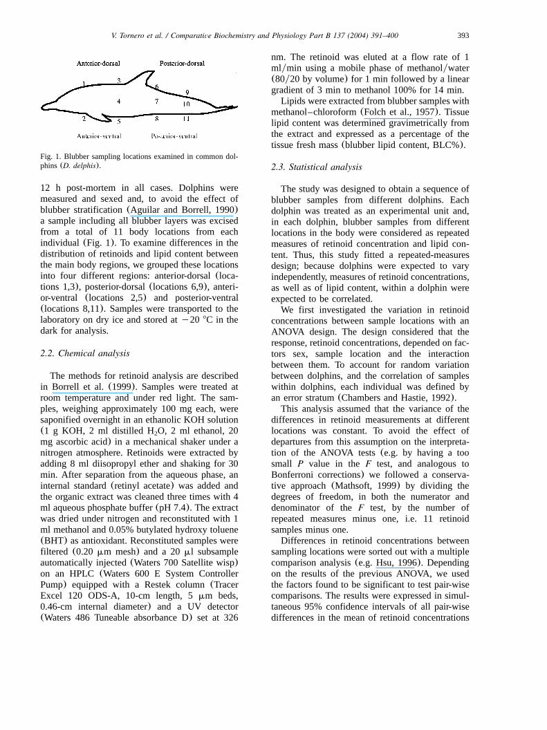
Fig. 1. Blubber sampling locations examined in common dol-phins(D. delphis).
12 h post-mortem in all cases. Dolphins weremeasured and sexed and, to avoid the effect ofblubber stratification(Aguilar and Borrell, 1990)a sample including all blubber layers was excisedfrom a total of 11 body locations from eachindividual (Fig. 1). To examine differences in thedistribution of retinoids and lipid content betweenthe main body regions, we grouped these locationsinto four different regions: anterior-dorsal(loca-tions 1,3), posterior-dorsal(locations 6,9), anteri-or-ventral (locations 2,5) and posterior-ventral(locations 8,11). Samples were transported to thelaboratory on dry ice and stored aty20 8C in thedark for analysis.
2.2. Chemical analysis
The methods for retinoid analysis are describedin Borrell et al. (1999). Samples were treated atroom temperature and under red light. The sam-ples, weighing approximately 100 mg each, weresaponified overnight in an ethanolic KOH solution(1 g KOH, 2 ml distilled H O, 2 ml ethanol, 202
mg ascorbic acid) in a mechanical shaker under anitrogen atmosphere. Retinoids were extracted byadding 8 ml diisopropyl ether and shaking for 30min. After separation from the aqueous phase, aninternal standard(retinyl acetate) was added andthe organic extract was cleaned three times with 4ml aqueous phosphate buffer(pH 7.4). The extractwas dried under nitrogen and reconstituted with 1ml methanol and 0.05% butylated hydroxy toluene(BHT) as antioxidant. Reconstituted samples werefiltered (0.20 mm mesh) and a 20ml subsampleautomatically injected(Waters 700 Satellite wisp)on an HPLC (Waters 600 E System ControllerPump) equipped with a Restek column(TracerExcel 120 ODS-A, 10-cm length, 5mm beds,0.46-cm internal diameter) and a UV detector(Waters 486 Tuneable absorbance D) set at 326
nm. The retinoid was eluted at a flow rate of 1mlymin using a mobile phase of methanolywater(80y20 by volume) for 1 min followed by a lineargradient of 3 min to methanol 100% for 14 min.
Lipids were extracted from blubber samples withmethanol–chloroform(Folch et al., 1957). Tissuelipid content was determined gravimetrically fromthe extract and expressed as a percentage of thetissue fresh mass(blubber lipid content, BLC%).
2.3. Statistical analysis
The study was designed to obtain a sequence ofblubber samples from different dolphins. Eachdolphin was treated as an experimental unit and,in each dolphin, blubber samples from differentlocations in the body were considered as repeatedmeasures of retinoid concentration and lipid con-tent. Thus, this study fitted a repeated-measuresdesign; because dolphins were expected to varyindependently, measures of retinoid concentrations,as well as of lipid content, within a dolphin wereexpected to be correlated.
We first investigated the variation in retinoidconcentrations between sample locations with anANOVA design. The design considered that theresponse, retinoid concentrations, depended on fac-tors sex, sample location and the interactionbetween them. To account for random variationbetween dolphins, and the correlation of sampleswithin dolphins, each individual was defined byan error stratum(Chambers and Hastie, 1992).
This analysis assumed that the variance of thedifferences in retinoid measurements at differentlocations was constant. To avoid the effect ofdepartures from this assumption on the interpreta-tion of the ANOVA tests(e.g. by having a toosmall P value in the F test, and analogous toBonferroni corrections) we followed a conserva-tive approach(Mathsoft, 1999) by dividing thedegrees of freedom, in both the numerator anddenominator of theF test, by the number ofrepeated measures minus one, i.e. 11 retinoidsamples minus one.
Differences in retinoid concentrations betweensampling locations were sorted out with a multiplecomparison analysis(e.g. Hsu, 1996). Dependingon the results of the previous ANOVA, we usedthe factors found to be significant to test pair-wisecomparisons. The results were expressed in simul-taneous 95% confidence intervals of all pair-wisedifferences in the mean of retinoid concentrations

394 V. Tornero et al. / Comparative Biochemistry and Physiology Part B 137 (2004) 391–400
Table 1Date of capture, sex, length, retinoid concentrations(mean"S.D.) and blubber lipid content(BLC) (mean"S.D.) of common dolphins(D. delphis)
Dolphin Date Sex Length(cm) Retinoids(mg g )y1 BLC (%)
1 28-03-2001 M 187 75.16"32.72 60.58"8.572 11-07-2001 M 202 67.40"28.72 55.79"8.243 11-07-2001 M 206 56.01"23.25 58.02"7.574 18-07-2001 F 189 47.59"14.96 56.26"8.895 19-07-2001 F 179 33.48"6.59 59.50"8.776 17-07-2001 F 197 59.91"13.97 62.36"7.157 23-07-2001 M 204 53.56"18.64 52.85"6.788 23-07-2001 M 175 29.04"8.82 64.12"6.339 23-07-2001 M 204 49.95"23.88 59.63"10.30
10 23-07-2001 M 208 64.20"17.61 53.25"8.2711 23-07-2001 M 205 45.23"13.91 58.60"4.3412 23-07-2001 M 200 48.50"13.30 63.07"9.6813 23-07-2001 M 190 58.97"17.82 49.84"10.6114 04-07-2002 F 176 50.50"14.71 67.00"5.4115 18-07-2002 F 206 80.31"24.79 63.42"4.6416 18-07-2002 F 187 45.98"16.34 67.55"7.5317 18-07-2002 F 180 53.55"6.47 68.32"5.9623 25-07-2002 F 200 29.65"10.62 55.38"5.9818 31-01-2000 M 176 44.47"10.55 67.47"4.2120 31-07-2002 M 207 44.27"24.09 65.29"7.2921 31-07-2002 M 183 30.86"9.48 68.94"5.4722 31-07-2002 M 182 52.98"18.14 58.64"11.7724 31-07-2002 M 201 70.81"27.60 67.60"5.3719 22-08-2002 F 194 104.20"28.81 62.25"6.3025 10-09-2002 F 198 77.12"27.56 62.53"6.06
Note: M, male; F, female; S.D., standard deviation.
Table 2Retinoid concentrations(mean"S.D.) and blubber lipid con-tent, BLC(mean"S.D.), in the blubber locations examined incommon dolphins(D. delphis)
Blubber location n Retinoids(mg g )y1 BLC (%)
1 25 50.01"22.73 60.74"9.832 25 57.02"22.16 66.23"6.033 25 46.88"16.75 62.81"6.724 25 67.90"25.47 62.75"9.355 25 81.51"33.66 65.18"6.926 25 40.85"12.68 58.33"9.587 25 50.14"17.91 60.59"8.568 25 66.59"26.74 61.45"9.189 25 47.63"17.03 59.51"7.81
10 25 49.27"28.97 57.81"10.6011 25 45.31"20.59 57.01"8.65
Note: S.D., standard deviation.
based on the different sampling locations. Thesame repeated-measures and multiple comparisonanalysis were used to assess differences in lipidcontent between sampling locations.
Due to the lipophilic character of vitamin A, thelipid content of a sample could be a confoundingfactor in the understanding of retinoid distribution
in the dolphin blubber. To investigate the possibleassociation of highest retinoid concentrations tosamples of highest lipid content, we compareddifferences in retinoid concentration among sam-ples with differences in lipid content among sam-ples with another ANOVA design with repeatedmeasures. The response was retinoid concentrationand depended on lipid content and the interactionof sample location and lipids, with lipid contentsnested on sample, and additional effects were sexand body size. To account for random variation,each individual was defined by an error stratum.
Differences in retinoid levels and lipid contentbetween the main body blubber regions weredetermined using a repeated-measure design anal-ysis of variance(ANOVA) and multiple compari-sons, as used for the 11 sampling locationsexplained above.
3. Results
Table 1 details the biological characteristics ofeach dolphin sampled. The length of the individ-uals ranged from 175 to 208 cm, so they compose

395V. Tornero et al. / Comparative Biochemistry and Physiology Part B 137 (2004) 391–400
Fig. 2. Box-plots of blubber retinoid concentration(mg g )y1
in different sampling locations by sexes in common dolphins(D. delphis).
Table 3Significant differences of the means of retinoid concentrationsand of the lipid content between sampling locations in commondolphins(D. delphis), as determined by the multicomparisonanalysis
Sampling 1 2 3 4 5 6 7 8 9 10 11location
1 R2 R L L3 R4 R R5 R R R R RL6 R78 R9
1011
R: significant differences on retinoid concentrations atP-0.05.L: significant differences on lipid content atP-0.05.
Fig. 3. Box-plots of blubber lipid content(%) in different sam-pling locations by sexes in common dolphins(D. delphis).
a reasonably homogenous group. Mean retinoidvalues varied greatly among dolphins, rangingfrom 29 to 104mg g . Table 2 summarizes they1
concentrations of retinoids in the different blubberlocations; mean concentrations ranged from 41 to81 mg g . Retinoid concentration presented ay1
very high variation among locations both withinand between individuals. This variation maskedthe potential differences between sexes(Fig. 2)and the ANOVA indicated that the effect of sexon retinoid concentrations was not significanteither between individuals or as an interaction withsample location. Therefore, in subsequent analysismales and females were computed together. Incontrast, sample location was found to be a highlysignificant source of variation within-individuals(conservativeF-test: P-0.001). Table 3 showsthe results of the multicomparison analysis detail-ing levels of significance between body locations.
Mean blubber lipid content(BLC) in individualsranged from 50 to 69%(Table 1); mean BLC ineach body location ranged from 35 to 83%(Table2). An ANOVA with percent lipid content asresponse, sex and sample location as factors, andan error stratum associated to individual dolphin,indicated no significant variation in lipid contentby sex. However, sampling location was a signif-icant source of within-individual variation(con-servative F-test: Ps0.048). Similarly to therelation between retinoid concentration and samplelocation, individual variability in lipid content washigh enough to mask potential differences between
sexes(Fig. 3). The result of the multiple compar-ison analyses of BLC is shown in Table 3. Sincethe lipid content was highly variable betweenindividuals, most of the differences between loca-tions were not significant.
The concentration of retinoids was associatedwith the lipid content of the blubber samples. Therelation was not very linear, as indicated by asimple linear model of retinoid on lipid content(F-statistic: 3.352; r s0.012; Ps0.0682) and2
again, this was mostly due to high individualvariation in both parameters. The results of theANOVA indicated that between dolphins, neithersex, body size or lipid contents were significant

396 V. Tornero et al. / Comparative Biochemistry and Physiology Part B 137 (2004) 391–400
Table 4Significant differences of the means of retinoid concentrations and of the lipid content between body regions in common dolphins(D.delphis), as determined by the multicomparison analysis
Body region Anterior-dorsal Posterior-dorsal Anterior-ventral Posterior-ventral
Anterior-dorsal RPosterior-dorsal RLAnterior-ventral LPosterior-ventral
R: significant differences on retinoid concentrations atP-0.05.L: significant differences on lipid content atP-0.05.
sources of variability. However, within dolphins,retinoid concentration was positively related tolipid contents(conservativeF-test:Ps0.012), andthe interaction of sample location and lipid con-tents was highly significant(conservativeF-test:Ps0.001). Within dolphins, neither sex nor bodysize explained significant variation in retinoidconcentration.
Body region was a significant source of varia-tion of retinoid concentration within-individuals(conservativeF-test:Ps0.013), as well as of lipidcontent(conservativeF-test:Ps0.046) (Table 4).We found the highest concentrations of both reti-noids and lipid content in the anterior-ventralregion and the lowest in the dorsal-posteriorregion.
4. Discussion
Retinoids are lipophilic and thus concentrate inlipid-rich tissues. Since blubber is the largest fatcompartment in marine mammals, in pinnipeds ithas been proposed as the main body depot forretinoids(Schweigert et al., 1987, 2002). In ceta-ceans, information available is comparativelymuch more limited(Borrell et al., 2002). Wefound retinoids to be present in the blubber ofcommon dolphins at concentrations averaging52.76 mg g (females) and 57.93 mg gy1 y1
(males). These values are comparable to thosereported for harbour porpoises(range: 30–80mgg ; Borrell et al., 1999), grey seals(range: 20–y1
74 mg g ; Schweigert et al., 1987, 2002;y1
Schweigert and Buchholz, 1995; Nyman et al.,2003), slightly higher than those found in ringedseals(range: 21–32mg g ; Kakela et al., 1997;y1 ¨ ¨Nyman et al., 2003), and some terrestrial mam-mals, such as canines(range: 15–30mg g ;y1
Schweigert and Buchholz, 1995), but much higherthan those found in the blubber of harp seals,Phoca groenlandica (3.6 mg g ; Rodahl andy1
Davies, 1949) and in the visceral fat of humans(1.5 mg g ; Raica et al., 1972).y1
Taking into account the large proportion thatblubber represents of the body of cetaceans andthe high blubber retinoid concentrations found inthe common dolphins here investigated, as well inthat of harbour porpoises found in a previous study(Borrell et al., 1999), it is reasonable to concludethat blubber also represents a significant contribu-tion to total retinoids in small odontocetes. There-fore, blubber is proposed as tissue of choice formonitoring retinoid status in these animals. How-ever, the large mass of the blubber compartmentin marine mammals, as well as its distributionover practically the whole body surface, makes itnecessary to define precise sampling protocols toavoid inconsistencies. Understanding within-blub-ber topographical variation in retinoid concentra-tions is relevant in this context.
Marine mammal blubber is far from being ahomogeneous, single-function tissue. It is com-monly accepted that it plays four main roles: tosupply insulation to the body core, to serve asenergy store, to contribute to streamline the body,and to regulate buoyancy(Ryg et al., 1988; Iver-son, 2002). However, the information available ontopographical variation in histological structure,composition, and physicochemical properties ofblubber indicate that the importance of each func-tion varies among body regions. In balaenopteridcetaceans the anterior ventral blubber forms theventral feeding grooves, which are semi-elastic topermit distension of the mouth and the throatduring feeding. As a consequence, the blubberfrom this region is composed of abundant struc-tural collagen and its lipid content in fin whales(Balaenoptera physalus), sei whales(Balaenop-tera borealis) and minke whales(B. acutorostrata)is much lower than that of the dorsal posteriorregion (Pedersen, 1950; Watanabe and Suzuki,1950a; Ackman et al., 1975; Lockyer et al., 1984,

397V. Tornero et al. / Comparative Biochemistry and Physiology Part B 137 (2004) 391–400
1985; Kvadsheim et al., 1996). This, together withthe fact that the dorsal posterior muscle is alsoricher in lipids than the ventral(Næss et al., 1998),indicates that this latter region is the major bodysite for energy storage(Lockyer et al., 1984).Although in sperm whales(P. macrocephalus) theanterior ventral region has no specialized feedingfunctions, the dorsal posterior and lateral regionsof the body also display the highest lipid contents(Watanabe and Suzuki, 1950b; Lockyer, 1991). Ithas been suggested that since the blubber layer isalso thicker in the latter area in balaenopteridwhales and sperm whales, lipids accrue in thisregion to shape the body and possibly to regulatebuoyancy(Lockyer et al., 1984; Iverson, 2002).
Information on small cetaceans is comparativelymuch more limited. In pilot whales(Globicephalamelas), topographical variation in lipid content isnon-existent or at least masked by seasonal varia-tion (Lockyer, 1993). In harbour porpoises thepattern of variation of lipid content has been foundto be either non-existent(Tilbury et al., 1997) orto be the opposite of that found in large cetaceans;this is lowest in the blubber of the dorsal posteriorregion and highest in the anterior ventral one(Calambokidis, 1986; Ishaq et al., 2000). It hasbeen suggested that in harbour porpoises the tissueis divided into two functional components, oneanterior and one posterior, with the line of divisionoccurring at the level of the anus; thus, both partswould play insulatory and buoyancy activities, butthe posterior section would not be significant forenergy storage and would mainly serve as a struc-tural element to streamline the body(Koopman,1998). In the present study, we found that concen-tration of lipids progressively decreased from headto tail and from belly to back, confirming thepattern observed in harbour porpoises. This sug-gests that in common dolphins the anterior-ventralregion is also comparatively more important forinsulation and lipid storage than the dorsal poste-rior region, perhaps to provide thermal protectionto the viscera.
Retinoids followed an almost identical pattern,which is not surprising given their lipophilicity.Thus, it appears that after the incorporation ofretinoids into the organism via food, the dynamicsand body distribution of these compounds aregreatly controlled by the physicochemical proper-ties of their molecule. As a consequence, not onlyretinoids accumulate in lipid-rich tissues, but alsotheir concentration within a given tissue is propor-
tional to the lipid content of the different compo-nents of this tissue. While retinoid concentrationin blubber can be considered a reliable indicatorof vitamin A status in common dolphins, it shouldbe stressed that sampling location must be consid-ered as a significant source of variability and takeninto account to design sampling protocols. Thus,consistency in sampling body location is criticalto ensure comparability of results.
In all large cetacean species, and independentlyof body location, blubber shows a stratified struc-ture that also reflects heterogeneous function ofthe various layers(Ackman et al., 1975; Lockyeret al., 1985; Aguilar and Borrell, 1990; Lockyer,1991). Since blubber thickness in these animalscan be up to 3–50 cm depending on the species(Iverson, 2002), added difficulties are posed torepresentative sampling as the suitable tissue massfor analysis is only a few grams. However, insmall cetaceans, such as common dolphins orharbour porpoises, this effect, although existing islimited (Koopman et al., 1996) and can be readilyovercome by collecting and analysing chunks ofblubber containing all blubber strata. In this way,all layers are pooled into a single analytical sampleproviding a composite average of the whole blub-ber thickness, as it has been done in the presentstudy.
No significant differences in blubber retinoidconcentrations were found between males andfemales. This is in agreement with the only com-parable study so far undertaken in cetaceans(Bor-rell et al., 1999). However, sex-related differencesin blubber vitamin A concentrations have beenreported for adult grey seals(Schweigert et al.,1987) and harbour seals(Nyman et al., 2003).The reasons for such interspecific differences insex-related patterns are unclear, but may be relatedto taxonomic, dietary or life-cycle dissimilaritiesbetween species.
The positive relationship found in this studybetween retinoid concentration and lipid contentapparently differs from results of previous studiesby Rodahl and Davies(1949) on harp and hoodedseals(Cystophora cristata), Borrell et al. (1999)on harbour porpoises, and Mos and Ross(2002)on harbour seal pups. Fat is mobilized by seasonalvariation in food intake or the increased energyexpenditure caused by migration, lactation, diseaseor other factors. This mobilization may also induceretinoid mobilization from blubber. The fate ofthese retinoids is unclear. They may either be

398 V. Tornero et al. / Comparative Biochemistry and Physiology Part B 137 (2004) 391–400
redistributed or excreted, but significant alterationin blubber concentrations is to be expected. Thiseffect should be taken into account when designingsampling protocols. Moreover, it points out thatstranded cetaceans, often found in poor nutritivecondition, are likely to present altered retinoidconditions and should therefore not be used tomonitor retinoid status of populations. Blubberbiopsies can be obtained from healthy, free-rangingindividuals using non-destructive methods and arealready a standard technique used in ecotoxicol-ogical surveys of cetaceans(Aguilar and Borrell,1994). The biopsy includes all blubber layers andthus avoids potential bias due to stratification ofretinoids, lipid content or pollutants, thus consti-tuting a more reliable alternative.
Acknowledgments
Fieldwork was possible thanks to the assistanceof collaborating fishermen and biologists, in par-ticular Marıa del Mar Fernandez Contreras, Nuria´ ´Garcıa Caro, Griselda Julian Juvina and Joan´ ´ ˜ ´Gonzalvo Villegas. Samples were supplied by theBMA Environmental Tissue Bank created with thesupport of the Pew Fellows Program in MarineConservation and Earthtrust. While doing thiswork, V. Tornero was supported by an F.P.I. fellow-ship from the Generalitat of Catalonia, and A.Borrell by a contract under the ‘Ramon y Cajal’´Program. Field and laboratory work was fundedby the Comision Interministerial de Ciencia y´Tecnologıa-CICYT(project AMB 99-0640), Fun-´dacio pel Desenvolupament Sostenible(FDS), and´the General Directorate for Nature Conservationof Spain’s Ministry of the Environment.
References
Ackman, R.G., Hingley, J.H., Eaton, C.A., Sipos, J.C., Mitch-ell, E.D., 1975. Blubber fat deposition in mysticeti whales.Can. J. Zool. 53, 1332–1339.
Aguilar, A., Borrell, A., 1990. Patterns of lipid content andstratification in the blubber of fin whales(Balaenopteraphysalus). J. Mammal. 71, 544–554.
Aguilar, A., Borrell, A., 1994. Assessment of organochlorinepollutants in cetaceans by means of skin and hypodermicbiopsies. In: Fossi, M.C., Leoncio, C.(Eds.), Non-destruc-tive Biomarkers in Vertebrates. Lewis Publishers, BocaRaton, FL, pp. 245–267.
Beckmen, K.B., Lowenstine, L.J., Newman, J., Hill, J., Hanni,K., Gerber, J., 1997. Clinical and pathological characterisa-tion of northern elephant seal skin disease. J. Wildl. Dis.33, 438–449.
Blomhoff, R., 1994. Overview of vitamin A metabolism andfunction. In: Blomhoff, R.(Ed.), Vitamin A in Health andDisease. Marcel Dekker, New York, pp. 1–35.
Blomhoff, R., Green, M.H., Green, J.B., Berg, T., Norum,K.R., 1991. Vitamin A metabolism: new perspectives onabsorption, transport, and storage. Physiol. Rev. 71,951–990.
Borrell, A., Cantos, G., Aguilar, A., Lockyer, C., Brouwer, A.,Heide-Jorgensen, M.P., et al., 1999. Patterns of variabilityof retinol levels in a harbour porpoise population from anunpolluted environment. Mar. Ecol. Prog. Ser. 185, 85–92.
Borrell, A., Tornero, V., Aguilar, A., 2002. Retinoids in marinemammals and their use as biomarkers of organochlorinecompounds. J. Cetacean Res. Manage. 4, 203–211.
Brouwer, A., Reijnders, P.J.H., Koeman, J.H., 1989. Polychlor-inated biphenyl(PCB)-contaminated fish induces vitaminA and thyroid hormone deficiency in the common seal(Phoca vitulina). Aquat. Toxicol. 15, 99–106.
Brunstrom, B., Hakansson, H., Lundberg, K., 1991. Effects of¨ ˚a technical PCB preparation and fractions thereof on ethox-yresorufinO-deethylase activity, vitamin A levels and thy-mic development in the mink(Mustela vison). Pharmacol.Toxicol. 69, 421–426.
Bryden, M.M., 1972. Growth and development of marinemammals. In: Harrison, R.J.(Ed.), Functional Anatomy ofMarine Mammals, Vol. 1. Academic Press, London, pp.1–81.
Calambokidis, J. 1986. Chlorinated hydrocarbons in harbourporpoise from Washington, Oregon, and California: Regionaldifferences in pollutant ratios. National Marine FisheriesService, Southwest Fisheries Center Administrative ReportLJ-86-35C.
Chambers, J.M., Hastie, T.J., 1992. Statistical Models. Wads-worth and Brooks Cole Advanced Books and Software.Pacific Grove, CA.
Chu, I., Villeneuve, D.C., Yagminas, A., Lecavalier, P., Hak-˚ansson, H., Ahlborg, U.G., et al., 1995. Toxicity of PCB 77(3,39,4,49-tetrachlorobiphenyl) and PCB 118(2,39,4,49,5-pentachlorobiphenyl) in the rat following subchronic dietaryexposure. Fund. Appl. Toxicol. 26, 282–292.
Chu, I., Poon, R., Yagminas, A., Lecavalier, P., Hakansson,˚H., Valli, V.E., et al., 1998. Subchronic toxicity of PCB 105(2,3,39,4,49-pentachlorobiphenyl) in rats. J. Appl. Toxicol.18, 285–292.
De Swart, R.L., Ross, P.S., Vedder, L.J., Timmerman, H.H.,Heisterkamp, S., Van Loveren, H., et al., 1994. Impairmentof immune function in harbour seals(Phoca vitulina)feeding on fish from polluted waters. Ambio 23, 155–159.
Folch, J., Lees, M., Stanley, G.H.S., 1957. A simple methodfor the isolation and purification of total lipids from animaltissues. J. Biol. Chem. 226, 497–509.
Hakansson, H., Manzoor, E., Ahlborg, U.G., 1992. Effects of˚technical PCB preparation and fractions thereof on VitaminA levels in the mink (Mustela vison). Ambio 21 (8),588–590.
Higashi, N., Senoo, H., 2003. Distribution of vitamin A –storing lipid droplets in hepatic stellate cells in liver lobules– A comparative study. Anat. Rec. 271A, 240–248.
Hsu, J.C., 1996. Multiple Comparisons: Theory and Methods.Chapman and HallyCRC, London.
Iida, H., Murata, Y., Matsumoto, G., Toda, S., Yamashita, Y.,Yokoyama, M., 1998. Chemical composition of the edible

399V. Tornero et al. / Comparative Biochemistry and Physiology Part B 137 (2004) 391–400
parts of minke whaleBalaenoptera acutorostrata. Bull. Nat.Res. Inst. Fish. Sci. 11, 27–36.
Ishaq, R., Karlson, K., Naf, C., 2000. Tissue distribution of¨polychlorinated naphthalenes(PCNs) and non-ortho chlori-nated byphenyls(non-ortho CBs) in harbour porpoises(Phocoena phocoena) from Swedish waters. Chemosphere41, 1913–1925.
Iverson, S.J., 2002. Blubber. In: Perrin, W.F., Wiirsig, B.,Thewissen, J.G.M.(Eds.), Encyclopedia of Marine Mam-mals. Academic Press, San Diego, CA, pp. 107–112.
Jenssen, B.M., Skaare, J.U., Woldstad, S., Nastad, A.T., Hau-gen, O., Kloven, B., et al., 1995. Biomarkers in blood toassess effects of polychlorinated biphenyls in free-livinggrey seal pups. In: Blix, A.S., Walloe, L., Ulltang, O.(Eds.),Whales, Seals, Fish and Man. Elsevier Science, Amsterdam,pp. 607–615.
Kakela, R., Hyvarinen, H., Kakela, A., 1997. Vitamins A-1¨ ¨ ¨ ¨ ¨(retinol), A-2, (3,4 didehydroretinol) and E(alpha-tocoph-erol) in the liver and blubber of lacustrine and marine ringedseals(Phoca hispida sp.). Comp. Biochem. Physiol. B 116,27–33.
Kakela, R., Kakela, A., Hyvarinen, H., Asikainen, J., Dahl,¨ ¨ ¨ ¨ ¨S.K., 1999. Vitamins A , A , and E in minks exposed to1 2
polychlorinated biphenyls(Aroclor 1242 and copper, via�
diet based on freshwater or marine fish. Environ. Toxicol.Chem. 18, 2595–2599.
Kakela, A., Kakela, R., Hyvarinen, H., Asikainen, J., 2002.¨ ¨ ¨ ¨ ¨Vitamins A and A in hepatic tissue and subcellular1 2
fractions in mink feeding on fish-based diets and exposedto Aroclor 1242. Environ. Toxicol. Chem. 21, 397–403.
Kakela, A., Kakela, R., Hyvarinen, H., Nieminen, P., 2003.¨ ¨ ¨ ¨ ¨Effects of Aroclor 1242 and different fish-based diets onvitamins A (retinol) and A (3,4 didehydroretinol), and1 2
their fatty acyl esters in mink plasma. Environ. Res. 91,104–112.
Kelley, S., Nilsson, C.B., Green, M.H., Green, J.B., Hakansson,˚H., 2000. Mobilisation of vitamin A stores in rats afteradministration of 2,3,7,8-tetrachlorodibenzo-p-dioxin: akinetic analysis. Toxicol. Sci. 55, 478–484.
Koopman, H.N., 1998. Topographical distribution of the blub-ber of harbour porpoises(Phocoena phocoena). J. Mammal.79, 260–270.
Koopman, H.N., Iverson, S.J., Gaskin, D.E., 1996. Stratifica-tion and age-related differences in blubber fatty acids of themale harbour porpoise(Phocoena phocoena). J. Comp.Physiol. B165, 628–639.
Kvadsheim, P.H., Folkow, L.P., Blix, A.S., 1996. Thermalconductivity of minke whale blubber. J. Therm. Biol. 21,123–128.
Lockyer, C.H., 1991. Body composition of the sperm whale,Physeter catodon, with special reference to the possiblefunctions of fat depots. Rit Fiskideildar 12, 1–24.
Lockyer, C.H., 1993. Seasonal changes in body fat conditionof northeast Atlantic pilot whales, and their biologicalsignificance. Rep. Int. Whal. Commn.(Spec. Issue) 14,324–350.
Lockyer, C.H., 1995. Aspects of the morphology, body fatcondition and biology of the harbour porpoise,Phocoenaphocoena, in British waters. Rep. Int. Whal. Commn.(Spec.Issue) 16, 199–209.
Lockyer, C.H., Mc Connell, L.C., Waters, T.D., 1984. Thebiochemical composition of fin whale blubber. Can. J. Zool.62, 2553–2562.
Lockyer, C.H., Mc Connell, L.C., Waters, T.D., 1985. Bodycondition in terms of anatomical and biochemical assess-ment of body fat in North Atlantic fin and sei whales. Can.J. Zool. 63, 2328–2338.
Mathsoft 1999. S-PLUS 2000 Guide to Statistics, Vol. 1. DataAnalysis Products Division, Mathsoft, Seattle, Washington,585.
Mori, T., Saiki, M., 1950. Properties of fats and oils containedin various parts of a sperm whale body. Sci. Rep. WhalesRes. Inst. 3, 79–84.
Mos, L., Ross, P., 2002. Vitamin A physiology in the precociusharbour seal(Phoca vitulina): a tissue-based biomarkerapproach. Can. J. Zool. 80, 1511–1519.
Murk, A.J., Leonards, P.E.G., Van Hattum, B., Luit, R.,Van der Weiden, M.E.J., Smit, M., 1998. Application ofbiomarkers for exposure and effect of polyhalogenatedaromatic hydrocarbons in naturally exposed European otters(Lutra lutra). Environ. Toxicol. Pharmacol. 6, 91–102.
Næss, A., Haug, T., Nilssen, E.M., 1998. Seasonal variationin body condition and muscular lipid content in NortheastAtlantic minke whaleBalaenoptera acutorostrata. Sarsia83, 211–218.
Nyman, M., Bergknut, M., Fant, M.L., Raunio, H., Jestoi, M.,Bengs, C., et al., 2003. Contaminant exposure and effectsin Baltic ringed seals as assessed by biomarkers. Mar.Environ. Res. 55, 73–99.
Olsen, E., Grahl-Nielsen, O., 2003. Blubber fatty acids ofminke whales: stratification, population identification andrelation to diet. Mar. Biol. Berlin 142, 13–24.
Peakall, D., 1992. Animal Biomarkers as Pollution Indicators.Ecotoxicological Series 1. Chapman and Hall, London.
Pedersen, T., 1950. Studies in whale oils. On the content ofsaturated fatty acids in whale oils. Hvalradets Skrifter 34,1–54.
Raica, N., Scott, J., Lowry, L., Sauberlich, H.E., 1972. VitaminA concentration in human tissues collected from five areasin the United States. Am. J. Clin. Nutr. 25, 291–296.
Rodahl, K., Davies, A.W., 1949. Vitamin A in seals. Biochem-istry 45, 408–412.
Rolland, R.M., 2000. A review of chemically-induced altera-tions in thyroid and vitamin A status from field studies ofwildlife and fish. J. Wildl. Dis. 36, 615–635.
Ryg, M., Smith, T.G.,�ritsland, N.A., 1988. Thermal signif-icance of the topographical distribution of blubber in ringedseals(Phoca hispida). Can. J. Fish Aquat. Sci. 45, 985–992.
Schweigert, F.J., Buchholz, Y., 1995. Vitamin A metabolismin carnivores with special reference to fur bearing animals.Scientifur 19, 305–307.
Schweigert, F.J., Stobo, W.T., Zucker, H., 1987. Vitamin Astatus in the grey seal(Halichoerus grypus) on Sable Island.Int. J. Vitam. Nutr. Res. 57, 239–245.
Schweigert, F.J., Luppertz, M., Stobo, W.T., 2002. Fasting andlactation effect fat-soluble vitamin A and E levels in bloodand their distribution in tissue of grey seals(Halichoerusgrypus). Comp. Biochem. Physiol. A 131, 901–908.
Simms, W., Ross, S., 2000. Vitamin A physiology and itsapplication as a biomarker of contaminant-related toxicity
400 V. Tornero et al. / Comparative Biochemistry and Physiology Part B 137 (2004) 391–400
in marine mammals: a review. Toxicol. Ind. Health 16,291–302.
Simms, W., Jeffries, S.J., Ikonomou, M.G., Ross, P.S., 2000.Contaminant-related disruption of vitamin A dynamics infree-ranging harbor seal(Phoca vitulina) pups from BritishColumbia, Canada and Washington State, USA. Environ.Toxicol. Chem. 19, 2844–2849.
Simpson, V.R., Bain, M.S., Brown, R., Brown, B.F., Lacey,R.F., 2000. A long-term study of vitamin A and polychlor-inated hydrocarbon levels in otters(Lutra lutra) in SouthWest England. Environ. Poll. 110, 267–275.
Skaare, J.U., Bernhoft, A., Wiig, O., Norum, K.R., Haug, E.,Eide, D.M., et al., 2001. Relationships between plasmalevels of organochlorines, retinol and thyroid hormonesfrom polar bears(Ursus maritimus) at Svalbard. J. Toxicol.Environ. Health A62, 227–241.
Thompson, J.N., 1976. Fat-soluble vitamins. Comp. Anim.Nutr. 1, 99–135.
Tilbury, K.L., Stein, J.E., Meador, J.P., Krone, C.A., Chan,S.L., 1997. Chemical contaminants in harbour porpoise(Phocoena phocoena) from the North Atlantic coast: tissueconcentrations and intra- and inter-organ distribution. Che-mosphere 34, 2159–2181.
Watanabe, H., Suzuki, K., 1950a. Chemical composition ofvarious parts of whale. Jpn. Soc. Sci. Fish. 15, 741–743.
Watanabe, H., Suzuki, K., 1950b. Chemical composition ofvarious parts of sperm whale. Jpn. Soc. Sci. Fish. 15,735–740.
Zile, M.H., 1992. Vitamin A homeostasis endangered byenvironmental pollutants. Proc. Soc. Exp. Biol. Med. 201,141–153.