Transcriptional study of hyperoxaluria and calcium oxalate ...
Upload
independentCategory
view
2download
0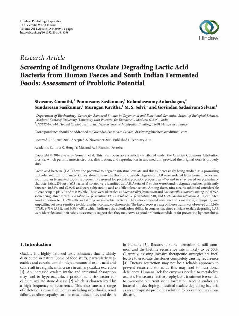
Research ArticleScreening of Indigenous Oxalate Degrading Lactic AcidBacteria from Human Faeces and South Indian FermentedFoods: Assessment of Probiotic Potential
Sivasamy Gomathi,1 Ponnusamy Sasikumar,1 Kolandaswamy Anbazhagan,2
Sundaresan Sasikumar,1 Murugan Kavitha,1 M. S. Selvi,1 and Govindan Sadasivam Selvam1
1 Department of Biochemistry, Centre for Advanced Studies in Organismal and Functional Genomics, School of Biological Sciences,Madurai Kamaraj University (University with Potential for Excellence), Madurai 625 021, India
2 INSERM-U844, Hopital St. Eloi, Institut des Neuroscience de Montpellier Building, 34091 Montpellier, France
Correspondence should be addressed to Govindan Sadasivam Selvam; [email protected]
Received 30 August 2013; Accepted 27 November 2013; Published 11 February 2014
Academic Editors: K. Hong, Y. Mu, and A. J. Piantino Ferreira
Copyright © 2014 Sivasamy Gomathi et al. This is an open access article distributed under the Creative Commons AttributionLicense, which permits unrestricted use, distribution, and reproduction in any medium, provided the original work is properlycited.
Lactic acid bacteria (LAB) have the potential to degrade intestinal oxalate and this is increasingly being studied as a promisingprobiotic solution to manage kidney stone disease. In this study, oxalate degrading LAB were isolated from human faeces andsouth Indian fermented foods, subsequently assessed for potential probiotic property in vitro and in vivo. Based on preliminarycharacteristics, 251 out of 673 bacterial isolateswere identified as LAB.A total of 17 strainswere found to degrade oxalate significantlybetween 40.38% and 62.90% and were subjected to acid and bile tolerance test. Among them, nine strains exhibited considerabletolerance up to pH3.0 and at 0.3%bile.Thesewere identified asLactobacillus fermentum andLactobacillus salivariususing 16S rDNAsequencing.Three strains, Lactobacillus fermentum TY5, Lactobacillus fermentumAB1, and Lactobacillus salivariusAB11, exhibitedgood adhesion to HT-29 cells and strong antimicrobial activity. They also conferred resistance to kanamycin, rifampicin, andampicillin, but were sensitive to chloramphenicol and erythromycin.The faecal recovery rate of these strains was observed as 15.16%(TY5), 6.71% (AB1), and 9.3% (AB11) which indicates the colonization ability. In conclusion, three efficient oxalate degrading LABwere identified and their safety assessments suggest that they may serve as good probiotic candidates for preventing hyperoxaluria.
1. Introduction
Oxalate is a highly oxidized toxic substance that is widelydistributed in nature. Some of food stuffs, particularly veg-etables and cereals, contain high amounts of oxalic acid andcan result in a significant increase in urinary oxalate excretion[1]. An increased oxalate intake and intestinal absorptionmay lead to hyperoxaluria, a predominant risk factor forcalcium oxalate stone disease [2] which is characterized bya high frequency of recurrence. This also causes a rangeof deleterious clinical outcomes including urolithiasis, renalfailure, cardiomyopathy, cardiac misconductance, and death
in humans [3]. Recurrent stone formation is still com-mon and the lifetime recurrence rate is likely to be 50%.Currently, existing invasive therapeutic strategies are inef-fective to eradicate the stones completely causing recurrence[4]. Dietary restriction may not be a reliable approach toprevent recurrent stones as this may lead to nutritionaldeficiency. Humans lack the enzymes needed to metabolizeoxalate.Hence, an effective prophylactic treatment is essentialto overcome recurrent stone formation. Recent studies arefocused on developing intestinal oxalate degrading bacteriaas an appropriate probiotics solution to prevent kidney stonedisease.
Hindawi Publishing Corporatione Scientific World JournalVolume 2014, Article ID 648059, 11 pageshttp://dx.doi.org/10.1155/2014/648059

2 The Scientific World Journal
Probiotics are being abundantly used as preventive thera-peutic agent for several diseases [5]. Probiotics are defined aslive microorganisms which, when administered in adequateamounts, confer a health benefit on the host [6]. It canbe implicated in stabilizing gut microbiota and enhance-ment of immune response and act as competitor againstenteric pathogens [7]. Several studies on probiotic bac-terial treatments have demonstrated promising results inameliorating diseases including inflammatory bowel disease,irritable bowel syndrome, pouchitis, and acute infantile orantibiotic-associated diarrhea [8]. Numerous studies havedocumented that gut microbes maintain the oxalate home-ostasis via utilizing the intestinal oxalate, while reducing theurinary oxalate excretion [9, 10]. Oxalobacter formigenes (O.formigenes) is an oxalate degrading bacterium, which usesintestinal oxalate as a sole source of carbon in order toregulate the oxalate homeostasis. However, its probiotic usehas been limited due to fastidious nutrient requirements, lesscolonization ability, and specialized oxalotrophic nature. Lac-tic acid bacteria (LAB) are vital residents of human intestinalecosystem andhave been extensively used as probiotics owingto their health promoting benefits to the host [7]. Studies haveconfirmed the correlation between oral administration ofLactobacillus or Bifidobacterium species and their importantrole in luminal oxalate reduction, which decreased the risk ofurinary oxalate excretion in humans and animals [2, 11–13].Turroni et al. [14] reported a range of oxalate degrading lac-tobacilli from pharmaceutical and dairy products and foundsignificant oxalate degradation in Lactobacillus acidophilusand Lactobacillus gasseri. However, the number of identifiedoxalate degrading bacterial species is limited and there is noreport regarding the ability of oxalate degrading LAB fromhuman gut microbiota. Alternatively, the use of recombinantLAB expressing heterogeneous oxalate degrading gene asa probiotic tool to control enteric hyperoxaluria was alsosuggested [15–17]. The present study is aimed to screen anefficient oxalate degrading LAB fromhuman faeces and southIndian fermented foods and to evaluate the safety assess-ment of potential probiotic characteristics both in vitro andin vivo.
2. Materials and Methods
2.1. Sampling and Isolation of LAB. Human faecal sampleswere collected from thirty healthy individuals (mean age of23–40) who had not taken antibiotics and probiotics at least
for the past three months. Samples were collected in sterilecontainer, kept in ice box, transported to laboratory withinone hour, and processed immediately. South Indian tradi-tional fermented foods used in this study were homemadepreparations. Fresh curd, fermented appam batter (preparedby grinding the presoaked parboiled rice and dehulled blackgram and allowed for natural fermentation for 12–24 h), andfermented wheat kali (paste prepared by slowly adding thewheat flour into boiling water and stirred continuously untilcorrect consistency. This was made as balls, soaked in water,and allowed to ferment naturally) were used to isolate oxalatedegrading LAB.
To isolate LAB, one gram of each faecal and fermentedfood sample was added separately to 9mL of 1% peptonewater and homogenized. Tenfold dilution was prepared withpeptone water and appropriate dilutions of each samplewere spread on de Man Rogosa Sharpe (MRS) (Himedia,Mumbai, India) agar plate and incubated at 37∘C for 72 h.From each sample, 15–30 colonies were randomly selectedand purified by streaking withMRS agar plates. Pure cultureswere preliminarily characterized based on gram staining,catalase reaction, and clear zone formation in 0.5% of CaCO
3
plate, glucose fermentation, and arginine hydrolysis [18].Tentatively identified LAB isolates were stored at −80∘C inMRS broth with 20% glycerol.
2.2. Determination of Oxalate Degrading Ability. The pre-sumptive LAB was screened for oxalate utilization usingagar well-diffusion method in calcium oxalate plate and wasprepared as described by Allison et al. and Campieri et al. [9,11]. Wells of 6mm diameter were prepared in calcium oxalateplate and each well was inoculated with 0.1mL of overnightculture and incubated at 37∘C for 12 h. The oxalate utilizingbacteria can form clear zone around the well due to oxalatedecomposition by the isolates. Zone diameter was measuredand the isolates displaying 10mm of zone were subjectedto quantitative determination of oxalate degradation. Toexamine their ability to degrade soluble oxalate, the isolateswere cultured in 5mL of MRS broth supplemented with10mM of potassium oxalate (KOX) for 5 days and MRSbroth without bacterial inoculum was used as control. Priorto oxalate determination, the control as well as bacterialculture was processed usingmethod of Federici et al. [19].Theoxalate concentration from the supernatant was determinedas described by Hodgkinson and Williams [20]. Consider
(%)Oxalate degradation =(Oxalate concentration in KOX control −Oxalate concentration in supernatant)
(Oxalate concentration in KOX control)× 100.
(1)
2.3. Acid and Bile Tolerance Test. The acid tolerance of LABisolates was evaluated in simulated gastric juices and bilesalt tolerance of LAB isolates was determined at 0.3% bileconcentration using the modified method of Wang et al.[21]. Cell suspensions containing ∼1 × 10⁷–109 cells wereinoculated into MRS broth supplemented with or without
0.3% (w/v) of bile. After 12 h incubation, 0.1mL of eachculture was serially diluted with 1% of PBS and spread onMRS agar plate.The plates were incubated aerobically at 37∘Cfor 72 h. The viable cells were counted using plate countmethod and expressed as mean log CFU/mL. The survival(%) of the bacteria was calculated as follows:

The Scientific World Journal 3
% Survival = (log number of viable cells survived (CFU/mL)
log number of initial viable cells inoculated (CFU/mL)) × 100. (2)
2.4. DNA Extraction, PCR Amplification, and Sequencing of16S rDNA. The bacterial genomic DNA was extracted asdescribed elsewhere [22]. All fine chemicals and primerswereprocured from Sigma Aldrich (USA). PCR amplification of16S rDNA was carried out by using primers Lab-0677 (CACCGCTACACATGGAG) andBact17 (AGAGTTTGATCATG-GCTCAG) which produce 700 bp amplicon [23]. PCR reac-tion was performed in GeneAmp PCR system 2700 (AppliedBiosystem, USA). The cyclic program consisted of an initialdenaturation at 94∘C for 5min; 35 cycles of 94∘C for 40 s, 56∘Cfor 45 s, and 72∘C for 1min were followed by a final extensionperiod at 72∘C for 7min. The amplified PCR products werepurified using Gene Elute PCR Cleanup Kit (Sigma Aldrich,USA) and sequenced commercially (Eurofins GenomicsIndia Pvt., Ltd., India). The 16S rDNA sequences were com-pared with the sequence in GenBank Public database usingBLAST software (http://blast.ncbi.nlm.nih.gov/Blast.cgi).
2.5. Antimicrobial Activity Assay. The antimicrobial activitywas determined as described by Wang et al. (2010) againstEscherichia coli ATCC 25922 (E. coli), Staphylococcus aureusATCC 6538 (S. aureus), Pseudomonas aeruginosa ATCC27853 (P. aeruginosa), Bacillus cereus NCIM 245 (B. cereus),Bacillus subtilisATCC 6633 (B. subtilis), and Salmonella typhi25 (S. typhi) which are the indicator strains.
2.6. Antibiotic Susceptibility Test. Antibiotic susceptibilitytest was carried out for nine isolates as described by Bauer etal. [24]. The concentrations of antibiotics used per disc were10 𝜇g of ampicillin, 30 𝜇g of tetracycline, 30 𝜇g of kanamycin,15 𝜇g of erythromycin, 30 𝜇g of chloramphenicol, and 5 𝜇gof rifampicin. The zone of inhibition was measured and theresults were expressed as susceptible (S), intermediate (I), andresistance (R) as per NCCLS standards [25].
2.7. In Vitro Adherence Assay. Adherence capacity of iso-lates was evaluated using HT-29 monolayer cells describedby Verdenelli et al. [26] with some modifications. Briefly,cells were routinely grown in minimal essential medium(MEM) (Himedia, India) containing 2mM L-glutamine,1mM sodium pyruvate, 1% nonessential amino acid, 1.5 g/Lsodium bicarbonate, 10% fetal bovine serum, 50 U/mLpenicillin, and 0.05mg/mL streptomycin. To investigate theadhesion ability of isolates, HT-29 cells were seeded at 1.5 ×10⁵ cells per well in 24-well tissue culture plate and incubatedat 37∘C with 5% carbon dioxide for 24 h incubation followedby washing three times with phosphate buffered saline (PBS).Each bacterial culture was diluted up to 108 cells/mL byMEMmedium and inoculated into HT-29 monolayer cells. After2 h of incubation, the monolayer was washed three timeswith 1mL of PBS to remove nonadhered cells and lysedby the addition of 0.25mL of 0.1% (v/v) Triton-X100 in
PBS for 10min at 37∘C. The lysate was plated on MRS agarafter a series of dilutions and incubated for 24 h to 48 h forbacterial enumeration. Adherence percentage was calculatedby comparing the adhered cells to the total cells of bacterialsuspension.
2.8. Growth Kinetic Analysis of Oxalate Degrading Bacteria.Kinetic analysis of growth and oxalate degradation of isolateswas evaluated. The oxalate degrading strains were inoculatedin MRS broth supplemented with 10mM potassium oxalateand noninoculated broth was used as control. The growthwas monitored by reading absorbance at 600 nm at 24 htime interval. The absorbance at A600 nm versus time curvewas plotted to reveal the growth kinetics. Similarly, theoxalate degrading ability was also determined for every 24 h.Quantifying oxalate in the growth medium was determinedas previously described [20].
2.9. Intestinal Colonization Ability of Oxalate Degrading LABIsolates in Rat Model. Based on oxalate degrading efficiencyand adherence ability, three representative strains Lactobacil-lus fermentum TY5 (L. fermentum TY5), Lactobacillus sali-variusAB11 (L. salivariusAB11), and Lactobacillus fermentumAB1 (L. fermentum AB1) were selected. The survivability andcolonization ability of oxalate degrading LAB were assessedusing a rifampicin-resistant spontaneous variant (RifR) [27].The RifR strains were tested for growth properties and oxalatedegrading abilities compared to the original strains. Theselected RifR strains were used to colonize the rat intestineby oral administration. Sixteen male albino Wistar rats withan average body weight of 150 g–180 g were used in the study.Food andwaterwere provided ad libitum for the study period.The animal protocol was approved by the Internal Researchand Review Board, Ethical Clearance, Biosafety and AnimalWelfare Committee of Madurai Kamaraj University. Ratsrandomly assigned into Group 1 were given 1mL of saline byesophageal gavage (control). Group 2 received 108 cells/day ofL. fermentumTY5.Group 3 received 108 cells/day ofL. salivar-iusAB11. Group 4 received 108 cells/day of L. fermentumAB1.The above strains were administrated for one week, whichwas followed by a washout period of one week. The faecalpellets were collected prior to probiotic administration (day0) and on days 3, 7 and 14.
The impact of probiotics on rat gut microbiota wasevaluated using culture-dependent analysis in faecal samples.Rat faecal sample was processed in the same way as humanfaecal sample and suitable dilutions were plated in duplicateon selective plates as described by Bernbom et al. [28].Reinforced clostridial agar (Himedia, India) was used toselectively grow members of clostridia family; MacConkeyagar number 3 (Himedia, India) was used for total coliforms;

4 The Scientific World Journal
Lactobacillus was cultured in MRS agar. Nutrient agar wasused for total facultative aerobes (Himedia, India). Thiogly-colate agar was used for total anaerobes (Himedia, India);finally MRS containing rifampicin 0.1mg/mL was used toselect RifR probiotics. The viable cells were counted andexpressed as log CFU/g of faeces.
2.10. Statistical Analysis. The results were expressed as mean± standard deviation (SD). Student’s paired t-test was per-formed to comparemean oxalate degradation and to comparethe tolerance (acid and bile) test, the viable counts were trans-formed to log
10values before statistical analysis. Student’s
unpaired t-test was used in a case of animalmodel to comparelactobacilli fed group versus control. A significant differencewas accepted at 𝑃 < 0.05. Statistical analysis was performedusing XLSTAT (2013.4.04).
3. Results
3.1. Identification of Oxalate Degrading LAB. Screening ofoxalate degrading bacteria from human faecal samplesdemonstrated the presence of significant population ofoxalate degrading LAB in the human intestine. Among 30individuals, 22 possessed LAB with oxalate degrading abil-ity while eight individuals contained Lactobacillus withoutoxalate degrading property. Totally, 673 strains were isolatedfrom human faeces and fermented foods (appam batter,wheat kali, and curd), among which 251 strains were LABbased on preliminary identification. All strains were Grampositive and catalase negative and able to form zone in0.5% CaCO
3and to be positive for glucose fermentation.
Ammonia production was observed in 93 strains. The pre-sumptive LAB was examined for oxalate utilization usingcalcium oxalate plate. A total of 92 oxalate degrading strainswere detected. Among them thirty-seven colonies showedclear zone above 10mm, and they were further subjected toevaluation of oxalate degrading efficiency. Table 1 shows theoxalate degrading ability of selected isolates.The isolates wereable to grow and degrade oxalate in the presence of 10mMpotassium oxalate, but the degree of oxalate degradationwas varied. Significant oxalate degradation was observed inseventeen strains, out of which ten strains utilized morethan 50% of oxalate. In particular, the isolates AB11 andTY12 appeared to be active and showed the highest oxalateutilization.
3.2. Acid and Bile Tolerance. In order to choose potent LABcandidates for the use as probiotics, strains showing highdegree of oxalate degradation were selected for probioticassessment. All the tested isolates were viable at pH 3.0 for 3 hindicating that almost all strains were acid tolerant (Table 2).However, only eight strains exhibited significant survivalrate (P < 0.05). The maximal survival rate was observed inisolates AM3 and ER5 with 89.5% and 94%, respectively. Fourstrains showed reduced viable cell count and the remainingstrains showed the least survivability. When exposed to pH2, the survival rate was strongly reduced in AM15, AM20,MSS39, andW21 and no viability was observed in TY14, PR3,
Table 1: Determination of oxalate degrading ability of LAB isolates.
Source IsolatesOxalate
concentration insupernatant (mM)
Oxalatedegradation (%)
Humanfaeces
TY2 5.4 ± 0.72 48.07TY5 4.7 ± 1.17a 54.8TY12 4.33 ± 0.8a 58.3TY14 5.5 ± 1.18a 47AM2 5.83 ± 0.57 43.94AM3 4.5 ± 1.05a 56.7AM12 6.4 ± 1.23 38.46AM15 5.66 ± 0.59a 46.15AM19 5.4 ± 1.2 48AM20 4.5 ± 0.96a 56.73AM48 7.03 ± 1.02 32.35PR3 5.5 ± 0.96a 47.11PR14 6.2 ± 0.7a 40.38PR16 6.3 ± 1.77 39.42PR36 6.67 ± 0.65 35.86PR45 6.16 ± 0.77a 40.77PR56 5.9 ± 0.75 43.27PR63 7.21 ± 1.19 30.67TH14 6.6 ± 1.19 36.54ER1 6.3 ± 0.6 39.42ER3 7.3 ± 1.4 29.76ER5 5.83 ± 0.6a 43.94ER48 8.3 ± 0.75 20.19MM3 8.5 ± 0.83 18.2MM8 6.3 ± 0.5 39.68MM38 6.23 ± 1.07 40MM39 4.7 ± 0.66a 54.8MM40 6.7 ± 1.25 35.57MM42 6.9 ± 0.60 33.65MSS10 4.93 ± 0.57a 52.8AB11 3.9 ± 0.4a 62.49
South Indianfermentedfood
C2 5.53 ± 0.61a 46.82C14s 6.83 ± 0.45 34.32AB1 4.6 ± 0.5a 55.76AB8 4.22 ± 0.66a 59.42AB10 6.64 ± 0.54 36.15W21 6.08 ± 0.3
a 41.5KOX
Control 10.4 ± 1.00
Values are expressed as mean ± SD from three trials. aSignificant difference(𝑃 < 0.05) in oxalate degradation compared with KOX control usingStudent’s paired t-test.
PR45, and C2 strains, which suggested that these isolateswere highly sensitive to highly acidic pH. The toleranceto bile at 0.3% showed survivability of oxalate degradingLAB (Table 3). Among the tested strains, six strains showed

The Scientific World Journal 5
Table 2: Survivability of LAB isolates at acidic condition.
Isolates Initial mean count1 pH 2 Survival rate (%) pH 3 Survival rate (%)TY5 7.25 ± 0.27 4.63 ± 0.15a 63.8 5.74 ± 0.36a 79.17TY12 6.67 ± 0.85 4.59 ± 0.84a 68.89 5.43 ± 0.88a 81.48TY14 6.5 ± 0 — — 3.96 ± 0.28 66AM3 7.29 ± 0.20 4.21 ± 0.97 57.75 6.52 ± 0.1 89.5AM15 8.41 ± 0.77 3.35 ± 0.07 39.8 4.99 ± 0.76 59.3AM20 7.27 ± 0.05 3.76 ± 0.25 37.96 5.4 ± 0.5 74.27PR3 6.41 ± 0.05 — — 3.66 ± 0.31 57.09PR14 6.51 ± 0.15 3.41 ± 0.07 52.38 4.46 ± 0.45 68.5PR45 6.81 ± 0.06 — — 4 ± 0.12a 54ER5 6.8 ± 0.19 4.06 ± 0.36 67.7 6.37 ± 0.25a 94MM39 7.62 ± 1.07 2.12 ± 0.1 34.2 4.83 ± 0.04 63.38MSS10 7.22 ± 0.07 3.44 ± 0.08 60.3 5.88 ± 0.14 81.44AB11 7.24 ± 0.09 6.02 ± 0.16a 67.44 6.73 ± 0.11a 88.7C2 7.57 ± 0.45 — — 3.06 ± 0.33 40.42AB1 8.65 ± 0.07 6.71 ± 0.17 61.5 6.88 ± 0.18a 79.53AB8 8.35 ± 0.26 7.12 ± 0.07 72.72 7.05 ± 0.09a 84.43W21 7.88 ± 0.36 2.64 ± 0.26 33.50 4.71 ± 0.28a 43.111Viable counts were transformed to log CFU/mL and expressed as mean ± SD in three experiments. aSignificant difference (𝑃 < 0.05) at log10 measurementsafter 3 h incubation compared to the initial count. (—) No growth.
Table 3: Survivability of LAB isolates at 0.3% bile.
Isolates Initial mean count1 0.3% bile24 h Survival rate (%)
TY5 8.8 ± 0.77 7.02 ± 0.04 79.77TY12 8.58 ± 0.57 7.33 ± 0.76a 85.4TY14 6.42 ± 0.74 3.57 ± 0.18 55.6AM3 7.44 ± 0.62 5.5 ± 0.28 73.92AM15 6.69 ± 0.46 4.23 ± 0.82 63.2AM20 8.64 ± 0.45 6 ± 0.14 71PR3 8.93 ± 0.26 5.46 ± 0.81 61PR14 6.58 ± 0.79 5.64 ± 0.07 85.7PR45 8.44 ± 0.14 5.45 ± 0.60 64.6ER5 7.95 ± 0.09 6.11 ± 0.03a 76.8MM39 7.78 ± 0.2 7.18 ± 0.2a 92.28MSS10 8.43 ± 0.65 6.79 ± 0.48a 74.13AB11 7.8 ± 0.30 6.6 ± 0.31a 84.61C2 8.11 ± 0.04 3.93 ± 0.80 48.45AB1 8.7 ± 0.2 6.86 ± 0.17a 80.3AB8 7.52 ± 0.09 5.15 ± 0.02 68.48W21 6.83 ± 0.08 3.54 ± 0.56 51.831Viable counts were transformed to log CFU/mL and expressed as mean± SD from three experiments. aSignificant difference (𝑃 < 0.05) atlog10 measurements after 12 h incubation at 0.3% compared to the initialcount.
significant survival rate (TY12, AM3, ER5, MM39, AB11, andAB1) while other strains were sensitive to bile. Based on acidand bile tolerance survival rate, nine strains (TY5, TY12,AM3, ER5, MM39, MSS10, AM3, AB11, AB1, and AB8) were
selected for further 16S rDNA identification and probioticevaluation.
3.3. Identification of Isolates Using 16S rDNA Sequencing.The 16S rDNA sequence of isolates was subjected to BLASTprogram for analyzing sequence similarity at NCBI database.Based on 16S rDNA sequencing, the isolates were found tobelong to five L. fermentum, twoW. confusa, one L. salivarius,and oneW. cibaria species (Table 4).
3.4. Antimicrobial Activity. Nine isolates selected on the basisof their acid and bile tolerance were subjected to furtherprobiotic evaluation. Table 5 shows antagonistic activity ofLAB isolates against six pathogens with variable inhibitoryeffect. The selected strains exhibited antagonistic activity,but varied in their inhibitory effect against each indicatorbacterium and inhibited both Gram-positive and Gram-negative pathogens. P. aeruginosa ATCC 27853 was stronglyinhibited by TY5, TY12, and AM 3, while other strainsshowed moderate inhibition. Most of the isolates showedmoderate inhibition fairly against B. subtilisATCC 6633. Fivestrains exhibited strong inhibition against B. cereus NCIM2458. AM3 and AB11 showed inhibition against S. aureusATCC6538. S. typhi 25wasmore resistant tomost lactobacilliisolates, except TY5 andMSS10 that showed weak inhibition.Faecal isolates showed better inhibition towards pathogensthan food product isolates.
3.5. Antibiotic Test. The antibiotic susceptibility of LABisolates is showed in Table 6. All strains were resistant tokanamycin and rifampicin (except AB8) and susceptible tochloramphenicol (except ER5). Seven strains were resistant

6 The Scientific World Journal
Table 4: Identification of oxalate degrading LAB strains by 16S rDNA sequences.
Isolates GenBank accession number The most matched organisms Max identity (%)TY5 KF588358 L. fermentumM059 99TY12 KF588359 L. fermentum F-6 99AM3 KF588357 L. fermentum F-6 100ER5 KF588363 W. confusa K1-lb5 97MSS10 KF588361 W. cibaria 4213 95MM39 KF588362 W. confusa FS066 99AB11 KF588360 L. salivarius LB-33 99AB8 KF588355 L. fermentum F-6, 6.1 99AB1 KF588356 L. fermentum IFO 3956 99
Table 5: Antimicrobial activity of LAB isolates.
Isolates P. aeruginosa B. subtilis S. aureus E. coli B. cereus S. typhi 25TY5 +++ ++ ++ + ++ ±
TY12 +++ ++ ++ +++ +++ −
AM3 +++ ++ +++ +++ − −
ER5 ++ ++ ++ ± ++ −
MM39 ++ + ++ + ++ −
MSS10 ++ ++ + − +++ ±
AB11 ++ ++ +++ ++ +++ −
AB1 + + + − ++AB8 + + − ++ ++ −
− No inhibition, ± 0–4mm, + 4–8mm, ++ 8–12mm, +++ ≥ 20mm.
Table 6: Antibiotic susceptibility test of LAB isolates.
Isolates Ampicillin Chloramphenicol Erythromycin Tetracycline Rifampicin KanamycinTY5 R S S S R RTY12 I S S I R RAM3 R S I I R RER5 R I R R R RMM39 R S I R R RMSS10 R S S S R RAB11 R S S I R RAB1 S S S R R RAB8 R S S I S RR: resistant; S: susceptible; I: intermediate.
to ampicillin and one strain was intermediate whereas twostrains were sensitive. Most of the isolates were susceptibleto erythromycin, two strains were intermediate, and twowere resistant. Tetracycline resistance was observed in threeisolates, four were intermediate, and two were susceptible.
3.6. Adhesive Property of Isolates. The result of adherenceability of isolates is shown in Figure 1. The adherence abilityof strains to HT-29 cells noticeably differed from eachtested strain. L. fermentum TY5, a faecal isolate, was themost adhesive strain with 12.9% of bacteria bound to theHT-29 cells. Surprisingly, fermented food product isolateof L. fermentum AB1 adhered with 10.8% to HT-29 cells.
Other strains L. salivarius AB11, L. fermentum AM3, andL. fermentum AB8 exhibited moderate adhesion with 8.6%,7.7%, and 6.9%, respectively.
3.7. Kinetic Analysis of Oxalate Degrading Ability Correlatedwith Glucose Consumption. To determine the impact ofoxalate on growth, the growth kinetics and oxalate degradingability were analyzed at 24 h time interval for 5 days. Theoxalate degrading profile demonstrated that the majorityof oxalate was degraded during 24 h, while gradual oxalatedegradation rate was observed up to 72 h and it was sus-tained until 120 h (Figure 2). The degrading efficiency variedbetween isolates, but the pattern of the oxalate degradation

The Scientific World Journal 7
0
2
4
6
8
10
12
14
TY5
TY12
ER5
MM
39
MSS
10
AM
3
AB1
1
AB1
AB8
Adhe
sion
abili
ty (%
)
Figure 1: Adhesion ability of LAB isolates to HT-29 cells. The resultwas represented as mean ± SD of duplicates. The error bar indicatesstandard deviation.
0
0.2
0.4
0.6
0.8
1
1.2
1.4
1.6
1.8
0
2
4
6
8
10
12
0 24 48 72 96 120
Oxa
late c
once
ntra
tion
(mM
)
Time (h)
TY5AB11AB1
TY5AB11AB1
O.D
at600
nm
Figure 2: Kinetic analysis of oxalate degradation and growth curveof isolates in the presence of 10mM KOX. Oxalate concentrationpresent in the supernatant (straight line) was mostly consumedduring 24 h growth (dotted line) corresponding to active growthphase of isolates.
was the same in all the isolates.The oxalate degrading activitywas also examined in the absence of glucose with 10mMKOX. In this condition, the isolates were unable to grow andutilize oxalate, implying the need for glucose to utilize oxalate.
3.8. Analysis of Intestinal Colonization Ability of LAB Isolatesin Rat. The intestinal colonizing ability and faecalmicrobiotaafter oral administration of LAB isolates were analyzed inrat model by conventional plate count method on selectiveagar. Figure 3 shows the viable counts of microbial changesof faecal microbiota. None of the groups showed significantchanges in total anaerobic and total aerobic counts. All groupsshowed significant increase in Lactobacillus species up to 7
days but it was reduced on washout period. L. fermentumTY5 and L. salivarius AB11 fed group showed significantreduction of coliforms and also showed considerable survivalup to 14 days. The survivability of RifR oxalate degradingLAB was tested on 0.1mg/mL Rif plate. Before probioticsadministration, faecal sample was tested for presence of RifRcolonies from lactobacilli fed group and control. None of theRif plates showed RifR colonies, which confirmed the absenceof RifR colonies. The RifR colonies were observed in faecesfrom lactobacilli fed rats on day 3 of administration whichshows the gastric transit ability of consumed lactobacilli.On day 7, the faecal recovery rate was observed as 15.16%,9.3%, and 6.71% in L. fermentum TY5, L. salivarius AB11,and L. fermentum AB1, respectively, and this was reduced onwashout period (Figure 4).This represents that the consumedlactobacilli have the gastrointestinal transit and intestinalcolonizing ability.
4. Discussion
In the present study, human faecal samples and traditionalsouth Indian fermented food products were chosen in orderto identify efficient oxalate degrading bacteria which couldbe used as probiotic supplements for their application inhyperoxaluria prevention.
Human intestinal isolates showed high degree of vari-ability in oxalate degradation. Turroni et al. [14] found thatLactobacillus acidophilus and Lactobacillus gasseri showedsignificant oxalate degradation in 5mM oxalate whereasother strains showed less oxalate consumption especially,Lactobacillus salivariuswhich showed 20% oxalate degradingability. In this study, the maximum oxalate degradation wasdetected in isolates of L. salivarius AB11 (62.59%) and L.fermentum TY12 (58.3%) and five strains of L. fermentumsp. differed in their oxalate degrading ability. These resultsshowed that oxalate degradation was both species and strainspecific. Murphy et al. [27] also reported that oxalate uti-lization among probiotics in vitro was interspecies depen-dent. Among the identified oxalate degrading LAB strains,Weissella confusa and Weissella cibaria have the ability todegrade oxalate between 40 and 50%. Members of the genusWeissella have been isolated from various sources includinghuman faeces, Korean kimchi, fresh vegetables, sugar cane,carrot juice, raw milk and sewage, milking machine slime,soil, fermented sausages, and meat products [29].
Seventeen efficient oxalate degrading strains were eval-uated for acid and bile tolerance activity. Among the testedisolates, eight strains exhibit significant tolerance at pH 3after 3 h incubation but the viability was reduced at lowacidic condition. Previous studies have reported that mostof the Lactobacillus sp. exhibited good survival at pH 3,with lower viability when exposed to pH 2 [21]. Resistanceamong Weissella sp. varied and the survivability rate waslow at pH 2 [30]. Their findings support our result on acidtolerance of faecal and fermented food product LAB isolates.Several studies have demonstrated that the L. fermentumstrains could survive at pH 2 and the outstanding survivalrate was observed as around 96% at pH 3 [31, 32]. Similarly,

8 The Scientific World Journal
0
2
4
6
8
10
12
0 3 7 14 0 3 7 14 0 3 7 14 0 3 7 14
Days Days Days Days
Total anaerobesColiforms
Lactobacilli
TY5 fed rats
Total aerobes
Log
CFU
/g o
f fae
ces
Group 1: controlAB11 fed rats AB1 fed rats
∗
∗
∗
∗
∗
∗
∗ ∗
Group 2: L. fermentum Group 3: L. salivarius Group 4: L. fermentum
Figure 3: Analysis of faecal microbiota composition in rats before and after oral administration of selected Lactobacillus. Each ratadministered ∼108 cells/day for one week. Viable counts of total anaerobes, coliforms, lactobacilli, and total aerobes expressed in terms oflog CFU/g of faeces on days 0, 3, 7, and 14. Each value was represented as mean ± SD of log CFU/g wet faeces (𝑛 = four rats per group).∗Significant difference in viable counts.
0
1
2
3
4
5
6
7
8
9
3 7 14
Days
Log
cell
num
ber C
FU/g
of f
aece
s
L. fermentum TY5L. salivarius AB11L. fermentum AB1
Figure 4: Recovery of RifR strains from rat faecal samples on days3, 7, and 14. The data is represented as mean value of log CFU/gfaeces. RifR strains were recovered on days 3 and 7 and decreasedon washout period.
in this study, L. fermentum AM3 strains showed 89.5% ofsurvival rate in agreement with previous reports. Since LABneed to attain resistance to physiological bile concentration(0.3–0.5%) in the gastrointestinal tract, LAB isolates wereevaluated for bile acid resistance. Five strains exhibitedsignificant tolerance against 0.3% bile; L. salivarius AB11 andW. cibaria MM39 showed the highest bile tolerance activitysimilar to the findings observed in previous reports [30, 33].
Nine strains were selected on the basis of acid and biletolerance for further study on antibacterial activity, one of
the criteria in selection of probiotics, which is essential toprotect host gastrointestinal tract from invading pathogens.This can be achieved by possible colonization of probioticsagainst pathogens [34]. Almost all the strains exhibited antag-onistic activity against both Gram-positive and -negativepathogens. L. fermentum TY12 and AM3 and L. salivariusAB11 exhibited strong antagonistic activity against pathogenswhile other strains showed weak inhibition. L. fermentumTY12 and AM3 strains showed strong inhibition whereasL. fermentum AB1 and AB8 strains showed weak inhibitiontowards indicator strains this indicates the production ofdifferent antimicrobial substance by the selected isolates.The antagonistic activity of different lactobacilli strains var-ied towards pathogens due to strain specific nature [35]and differences in production of inhibitory compounds bylactobacilli spp. such as lactic acid, H
2O2, and bacteriocin
[36].Safety assessment of probiotics including antibiotic resis-
tance is an essential task in the selection of probiotics.Lactobacillus possessing any transferable resistance plasmidis believed to be a risk for human and animal use [37]. Intrin-sic antibiotic resistant probiotics possibly will promote thepatients with disturbed microbiota due to the administrationof other antibiotics [38]. All nine strains were kanamycinand rifampicin resistant and susceptible to chloramphenicolthat is in accordance with previous results [21]. Danielsenand Wind [39] found that transferable resistance gene suchas chloramphenicol, erythromycin, and tetracycline maybe present among LAB. However, three strains showedtetracycline resistance which is similar to previous report[21]. Several studies have proven that potential probioticswith atypical tetracycline resistance were conquered by plas-mid curing which could have the susceptible phenotype[40]. In this study, most of the isolates were sensitive to

The Scientific World Journal 9
chloramphenicol and erythromycin which revealed that thepossibility of gene transfer to pathogen is less.
Adhesion and colonisation of probiotic bacteria in thegastrointestinal tract of the host are believed to be one ofthe essential features required for the delivery of their healthbenefits [41]. The adhesion capacity of strains was variedfrom 0.75 to 2.9% which showed strain specificity. This isconsistent with earlier reports which suggested that adhesionto Caco-2 cells was found to be a discriminative parameter,showing marked variation among the strains independentof the species [42]. Among the tested strains, L. fermentumTY5 was the most adhesive strain which showed 12.9%adhesion to HT-29 cells similar to previous studies reportedbyVerdenelli et al. [26]. Fermented food strains also exhibitedgood adhesion to HT-29 while other isolates from faecesshowed weak adhesion. This result is contrary to Wang etal. [43] who reported that adhesive Lactobacillus strains havehost- residential characteristics.Wang et al. [21] also reportedthat fermented food isolate C06 exhibited strong adhesionthan faecal isolate.
Lactobacilli play a pivotal role in maintaining the gutmicrobial balance and provide a barrier effect via the specificcompetitive action to pathogen for colonizing the intesti-nal mucosa [44]. The probiotic administration to healthyorganism should not alter the naturally existing microbialbalance in intestine. Otherwise, the subjects with imbalancedmicrobiota show adverse effect which was observed in case ofantibiotic-associated colitis, infectious disease, and chronicpancreatitis [45, 46]. In this study, intestinal colonizationability and faecal microbiota changes were evaluated in vivousing rat model. Lactobacillus count was increased whilecoliform count was reduced significantly in two groups ofrats. The microbial changes of rat intestine are similar toprevious reports by Wang et al. [21] who reported significantincrease in Lactobacillus and decrease in faecal coliform.Yang et al. [47] also observed the reduced faecal coliformcounts due to appropriate beneficial role of Lactobacillus andBifidobacterium proliferation and the inhibited invasion ofpathogens in rat gut. The RifR colonies that were presentin rat faeces after probiotic administration revealed thatthe strains were viable in gastric juice of stomach and guttransit. The good recovery rate in faeces suggested goodcolonization ability of strains. The recovery of RifR colonieswas in accordance with previous report by Oozeer et al. [48]that suggested thatmultiplication ofLactobacillus in the colonleads to high faecal survival rate.
5. Conclusion
The present study showed oxalate degrading ability andprobiotic assessment of human faeces and fermented foodisolates. Three strains of LAB, L. fermentum AB1, L. fermen-tum TY5, and L. salivarius AB11, have efficient oxalate degra-dation togetherwith good adherence toHT-29 cells, toleranceto acid and bile, strong inhibition against pathogens, andabsence of transferable antibiotic resistance, which indicatesthat these strains show satisfactory properties for probioticapplications. Additionally, these strains survived well during
gastrointestinal transit and reduced the coliform counts infaeces with good faecal recovery rate in rat model suggestingthat these interesting probiotic properties make them aspotentially good probiotic candidates which could be utilizedfor prophylaxis of calcium oxalate stone disease.
Conflict of Interests
The authors have no conflict of interests to declare.
Acknowledgments
This work was financially supported by the University GrantCommission (UGC), New Delhi, India. The authors alsothank the central instrumentation facility at SBS, MKU,through CEGS, NRCBS, DST-FIST, DBT-IPLS, and DST-PURSE programme.
References
[1] R. Siener, D. Ebert, C. Nicolay, and A. Hesse, “Dietary riskfactors for hyperoxaluria in calciumoxalate stone formers,”Kid-ney International, vol. 63, no. 3, pp. 1037–1043, 2003.
[2] J. Okombo and M. Liebman, “Probiotic-induced reduction ofgastrointestinal oxalate absorption in healthy subjects,” Urolog-ical Research, vol. 38, no. 3, pp. 169–178, 2010.
[3] M. Hatch and R. W. Freel, “Alterations in intestinal transport ofoxalate in disease states,” Scanning Microscopy, vol. 9, no. 4, pp.1121–1126, 1995.
[4] L. Borghi, A. Nouvenne, and T. Meschi, “Probiotics and dietarymanipulations in calcium oxalate nephrolithiasis: two sides ofthe same coin,” Kidney International, vol. 78, no. 11, pp. 1063–1065, 2010.
[5] H. S. Gill and F. Guarner, “Probiotics and human health: a clin-ical perspective,” Postgraduate Medical Journal, vol. 80, no. 947,pp. 516–526, 2004.
[6] “Evaluation of health and nutritional properties of probioticsin food including powder milk with live lactic acid bacteria,”Report of a Joint FAO/WHO Expert Consultation, FAO/WHO,Cordoba, Spain, 2001.
[7] R. X. Gu, Z. Q. Yang, Z. H. Li, S. L. Chen, and Z. L. Luo, “Probi-otic properties of lactic acid bacteria isolated from stool sam-ples of longevous people in regions of Hotan, Xinjiang andBama, Guangxi, China,” Anaerobe, vol. 14, no. 6, pp. 313–317,2008.
[8] A. Borthakur, R. K. Gill, S. Tyagi et al., “The probiotic Lac-tobacillus acidophilus stimulates chloride/hydroxyl exchangeactivity in human intestinal epithelial cells,” Journal of Nutrition,vol. 138, no. 7, pp. 1355–1359, 2008.
[9] M. J. Allison, K. A. Dawson, W. R. Mayberry, and J. G. Foss,“Oxalobacter formigenes gen. nov., sp. nov.: oxalate-degradinganaerobes that inhabit the gastrointestinal tract,”Archives ofMi-crobiology, vol. 141, no. 1, pp. 1–7, 1985.
[10] H. Ito, T. Kotake, and M. Masai, “In vitro degradation of oxalicacid by human feces,” International Journal of Urology, vol. 3, no.3, pp. 207–211, 1996.
[11] C. Campieri, M. Campieri, V. Bertuzzi et al., “Reduction ofoxaluria after an oral course of lactic acid bacteria at highconcentration,” Kidney International, vol. 60, no. 3, pp. 1097–1105, 2001.

10 The Scientific World Journal
[12] J. C. Lieske, D. S. Goldfarb, C. de Simone, and C. Regnier, “Useof a probioitic to decrease enteric hyperoxaluria,” Kidney Inter-national, vol. 68, no. 3, pp. 1244–1249, 2005.
[13] C. Kwak, B. C. Jeong, J. H. Ku et al., “Prevention of nephrolithi-asis by Lactobacillus in stone-forming rats: a preliminary study,”Urological Research, vol. 34, no. 4, pp. 265–270, 2006.
[14] S. Turroni, B. Vitali, C. Bendazzoli et al., “Oxalate consumptionby lactobacilli: evaluation of oxalyl-CoA decarboxylase andformyl-CoA transferase activity in Lactobacillus acidophilus,”Journal of Applied Microbiology, vol. 103, no. 5, pp. 1600–1609,2007.
[15] A. Kolandaswamy, L. George, and S. Sadasivam, “Heterologousexpression of oxalate decarboxylase in Lactobacillus plantarumNC8,” Current Microbiology, vol. 58, no. 2, pp. 117–121, 2009.
[16] K. Anbazhagan, S. Ponnusamy, S. Gomathi, P. H. Priya, andG. S. Selvam, “In vitro degradation of oxalate by recombinantLactobacillus plantarum expressing heterologous oxalate decar-boxylase,” Journal of Applied Microbiology, vol. 115, no. 3, pp.880–887, 2013.
[17] P. Sasikumar, S. Gomathi, K. Anbazhagan, and G. S. Selvam,“Secretion of biologically active heterologous oxalate decar-boxylase (OxdC) in Lactobacillus plantarum WCFS1 usinghomologous signal peptides,” BioMed Research International,vol. 2013, Article ID 280432, 9 pages, 2013.
[18] N. Thapa, J. Pal, and J. P. Tamang, “Phenotypic identificationand technological properties of lactic acid bacteria isolated fromtraditionally processed fish products of the Eastern Himalayas,”International Journal of Food Microbiology, vol. 107, no. 1, pp.33–38, 2006.
[19] F. Federici, B. Vitali, R. Gotti et al., “Characterization and het-erologous expression of the oxalyl coenzyme a decarboxylasegene from Bifidobacterium lactis,” Applied and EnvironmentalMicrobiology, vol. 70, no. 9, pp. 5066–5073, 2004.
[20] A. Hodgkinson and A. Williams, “An improved colorimetricprocedure for urine oxalate,” Clinica Chimica Acta, vol. 36, no.1, pp. 127–132, 1972.
[21] C. Y. Wang, P. R. Lin, C. C. Ng, and Y. T. Shyu, “Probioticproperties of Lactobacillus strains isolated from the feces ofbreast-fed infants and Taiwanese pickled cabbage,” Anaerobe,vol. 16, no. 6, pp. 578–585, 2010.
[22] F. M. Ausubel, R. Brent, R. E. Kingston, and D. D. Moore, ShortProtocols in Molecular Biology, John Wiley & Sons, New York,NY, USA, 1999.
[23] H. G. H. J. Heilig, E. G. Zoetendal, E. E. Vaughan, P. Marteau,A. D. L. Akkermans, andW. M. De Vos, “Molecular diversity ofLactobacillus spp. and other lactic acid bacteria in the humanintestine as determined by specific amplification of 16S ribos-omal DNA,” Applied and Environmental Microbiology, vol. 68,no. 1, pp. 114–123, 2002.
[24] A.W. Bauer,W.M. Kirby, J. C. T. Sherris, andM. Turck, “Antibi-otic susceptibility testing by a standardized single disk meth-od,” The American Journal of Clinical Pathology, vol. 45, no. 4,pp. 493–496, 1966.
[25] National Committee for Clinical Laboratory Standards, “Meth-ods for antimicrobial susceptibility testing of anaerobic bacte-ria, 4th ed. Approved standard M11-A4,” National Committeefor Clinical Laboratory Standards, Wayne, Pa, USA, vol. 17, no.22, 1997.
[26] M. C. Verdenelli, F. Ghelfi, S. Silvi, C. Orpianesi, C. Cecchini,and A. Cresci, “Probiotic properties of Lactobacillus rhamnosusand Lactobacillus paracasei isolated from human faeces,” Euro-pean Journal of Nutrition, vol. 48, no. 6, pp. 355–363, 2009.
[27] C. Murphy, S. Murphy, F. O’Brien et al., “Metabolic activity ofprobiotics—oxalate degradation,” Veterinary Microbiology, vol.136, no. 1, pp. 100–107, 2009.
[28] N. Bernbom,B.Nørrung, P. Saadbye, L.Mølbak, F. K.Vogensen,and T. R. Licht, “Comparison of methods and animal modelscommonly used for investigation of fecal microbiota: effects oftime, host and gender,” Journal of Microbiological Methods, vol.66, no. 1, pp. 87–95, 2006.
[29] J. Y. Liu, A. H. Li, C. Ji, and W. M. Yang, “First descriptionof a novel Weissella species as an opportunistic pathogen forrainbow trout Oncorhynchus mykiss (Walbaum) in China,”Veterinary Microbiology, vol. 136, no. 3-4, pp. 314–320, 2009.
[30] K. W. Lee, J. Y. Park, H. R. Jeong, H. J. Heo, N. S. Han, and J.H. Kim, “Probiotic properties ofWeissella strains isolated fromhuman faeces,” Anaerobe, vol. 18, no. 1, pp. 96–102, 2012.
[31] W. H. Lin, B. Yu, S. H. Jang, and H. Y. Tsen, “Different probioticproperties for Lactobacillus fermentum strains isolated fromswine and poultry,” Anaerobe, vol. 13, no. 3-4, pp. 107–113, 2007.
[32] E. Likotrafiti, K. M. Tuohy, G. R. Gibson, and R. A. Rastall,“Development of antimicrobial synbiotics using potentially-probiotic faecal isolates of Lactobacillus fermentum and Bifi-dobacterium longum,” Anaerobe, vol. 20, pp. 5–13, 2013.
[33] E. Kirtzalidou, P. Pramateftaki, M. Kotsou, and A. Kyriacou,“Screening for lactobacilliwith probiotic properties in the infantgut microbiota,” Anaerobe, vol. 17, no. 6, pp. 440–443, 2011.
[34] G. Reid, A. W. Bruce, J. A. McGroarty, K. J. Cheng, andJ. W. Costerton, “Is there a role for lactobacilli in preventionof urogenital and intestinal infections?” Clinical MicrobiologyReviews, vol. 3, no. 4, pp. 335–344, 1990.
[35] S. B. Gaudana, A. S. Dhanani, and T. Bagchi, “Probiotic attrib-utes of Lactobacillus strains isolated from food and of humanorigin,”TheBritish Journal of Nutrition, vol. 103, no. 11, pp. 1620–1628, 2010.
[36] K. Kucerova, J. Chumchalova, K. Mıkova, S. Cupakova, R.Karpıskova, and L. Ho, “Screening of lactic acid bacteria forantimicrobial properties from mayonnaise-based products andraw materials,” European Food Research and Technology, vol.226, no. 1-2, pp. 265–272, 2007.
[37] L.Morelli andA. V.Wright, “Probiotic bacteria and transferableantibiotic resistance-safety aspect,” in Demonstration of theNutritional Functionality of Probiotic Foods News Letter, vol. 2,pp. 9–14, 1997.
[38] S. Salminen, A. von Wright, L. Morelli et al., “Demonstrationof safety of probiotics—a review,” International Journal of FoodMicrobiology, vol. 44, no. 1-2, pp. 93–106, 1998.
[39] M. Danielsen and A. Wind, “Susceptibility of Lactobacillus spp.to antimicrobial agents,” International Journal of Food Microbi-ology, vol. 82, no. 1, pp. 1–11, 2003.
[40] G. Huys, K. D’Haene, and J. Swings, “Genetic basis of tetra-cycline and minocycline resistance in potentially probioticLactobacillus plantarum strain CCUG 43738,” AntimicrobialAgents and Chemotherapy, vol. 50, no. 4, pp. 1550–1551, 2006.
[41] M. Bernet-Camard,V. Lievin,D. Brassart, J. Neeser, A. L. Servin,and S. Hudault, “The human Lactobacillus acidophilus strainLA1 secretes a nonbacteriocin antibacterial subtance(s) activein vitro and in vivo,” Applied and Environmental Microbiology,vol. 63, no. 7, pp. 2747–2753, 1997.
[42] C. N. Jacobsen, V. R. Nielsen, A. E. Hayford et al., “Screeningof probiotic activities of forty-seven strains of Lactobacillus spp.by in vitro techniques and evaluation of the colonization abilityof five selected strains in humans,” Applied and EnvironmentalMicrobiology, vol. 65, no. 11, pp. 4949–4956, 1999.

The Scientific World Journal 11
[43] B. Wang, H. Wei, J. Yuan et al., “Identification of a surfaceprotein from Lactobacillus reuteri JCM1081 that adheres toporcine gastric mucin and human enterocyte-like HT-29 cells,”Current Microbiology, vol. 57, no. 1, pp. 33–38, 2008.
[44] A. C. Ouwehand, P. V. Kirjavainen, C. Shortt, and S. Salminen,“Probiotics: mechanisms and established effects,” InternationalDairy Journal, vol. 9, no. 1, pp. 43–52, 1999.
[45] E. B.Minelli, A. Benini,M.Marzotto et al., “Assessment of novelprobiotic Lactobacillus casei strains for the production of func-tional dairy foods,” International Dairy Journal, vol. 14, no. 8, pp.723–736, 2004.
[46] G. Zoppi, M. Cinquetti, A. Benini, E. Bonamini, and E. B. Min-elli, “Modulation of the intestinal ecosystem by probiotics andlactulose in children during treatment with ceftriaxone,” Cur-rent Therapeutic Research—Clinical and Experimental, vol. 62,no. 5, pp. 418–435, 2001.
[47] S. C. Yang, J. Y. Chen, H. F. Shang, T. Y. Cheng, S. C. Tsou,and J. R. Chen, “Effect of synbiotics on intestinal microflora anddigestive enzyme activities in rats,” World Journal of Gastroen-terology, vol. 11, no. 47, pp. 7413–7417, 2005.
[48] R. Oozeer, A. Leplingard, D. D. G. Mater et al., “Survival of Lac-tobacillus casei in the human digestive tract after consumptionof fermented milk,” Applied and Environmental Microbiology,vol. 72, no. 8, pp. 5615–5617, 2006.

Submit your manuscripts athttp://www.hindawi.com
Hindawi Publishing Corporationhttp://www.hindawi.com Volume 2014
Anatomy Research International
PeptidesInternational Journal of
Hindawi Publishing Corporationhttp://www.hindawi.com Volume 2014
Hindawi Publishing Corporation http://www.hindawi.com
International Journal of
Volume 2014
Zoology
Hindawi Publishing Corporationhttp://www.hindawi.com Volume 2014
Molecular Biology International
GenomicsInternational Journal of
Hindawi Publishing Corporationhttp://www.hindawi.com Volume 2014
The Scientific World JournalHindawi Publishing Corporation http://www.hindawi.com Volume 2014
Hindawi Publishing Corporationhttp://www.hindawi.com Volume 2014
BioinformaticsAdvances in
Marine BiologyJournal of
Hindawi Publishing Corporationhttp://www.hindawi.com Volume 2014
Hindawi Publishing Corporationhttp://www.hindawi.com Volume 2014
Signal TransductionJournal of
Hindawi Publishing Corporationhttp://www.hindawi.com Volume 2014
BioMed Research International
Evolutionary BiologyInternational Journal of
Hindawi Publishing Corporationhttp://www.hindawi.com Volume 2014
Hindawi Publishing Corporationhttp://www.hindawi.com Volume 2014
Biochemistry Research International
ArchaeaHindawi Publishing Corporationhttp://www.hindawi.com Volume 2014
Hindawi Publishing Corporationhttp://www.hindawi.com Volume 2014
Genetics Research International
Hindawi Publishing Corporationhttp://www.hindawi.com Volume 2014
Advances in
Virolog y
Hindawi Publishing Corporationhttp://www.hindawi.com
Nucleic AcidsJournal of
Volume 2014
Stem CellsInternational
Hindawi Publishing Corporationhttp://www.hindawi.com Volume 2014
Hindawi Publishing Corporationhttp://www.hindawi.com Volume 2014
Enzyme Research
Hindawi Publishing Corporationhttp://www.hindawi.com Volume 2014
International Journal of
Microbiology
Copyright © 2022 FDOKUMEN