Renal-related perinephric fluid collections: MRI findings
6
Renal-related perinephric fluid collections: MRI findings N. Cem Balci a, T , Elif Akun b , Mehmet Erturk c , Sezer Saglam d , Nagihan Inan a , Yesim Balci e a Department of Radiology, SLU, St. Louis, MO 63110, USA b Department of Internal Medicine, Kadir Has University, Istanbul 36600, Turkey c Department of Radiology, Sisli Etfal Hospital, Istanbul 64460, Turkey d Department of Medical Oncology, Istanbul Medical Faculty, Istanbul 29980, Turkey e Department of Anesthesiology, Istinye Hospital, Istanbul 36460, Turkey Received 7 October 2004; accepted 11 April 2005 Abstract We retrospectively reviewed MR studies on 10 patients with renal-related perinephric fluid collections who underwent MRI in three institutions between January 2001 and August 2004. All patients underwent MRI of the abdomen and T1-weighted, T2-weighted and serial contrast-enhanced images, including delayed-phase contrast-enhanced images 10 –12 min after contrast injection, were obtained. Perinephric fluid collections in 5 patients revealed MRI findings of simple fluid content (i.e., hypointense on T1-weighted images and hyperintense on T2-weighted images). In another 5 patients, a complex perinephric fluid content (i.e., mixed hyper/hypointense on T1-weighted images and mixed hypo/hyperintense on T2-weighted images compatible with blood breakdown products and pus) was observed. In 5 patients, contrast extravasation on late-phase images that was compatible with urine leak was demonstrated. Our results suggest that MRI may determine the content of perinephric fluid collections on noncontrast T1-weighted and T2-weighted images and that contrast extravasation on late-phase images is associated with urine extravasation from renal collecting systems. D 2005 Elsevier Inc. All rights reserved. Keywords: Kidney; Hemorrhage; Urinoma; Urine extravasation 1. Introduction Retroperitoneal fluid collections in the perinephric spaces are not uncommon. They may contain pus, urine, blood and lymph or transudate fluid resulting from kidney-related pathologies or from retroperitoneal structures [1]. Fluid collections that have communication with the renal paren- chyma and collecting system need to be depicted immedi- ately because of the insidious accumulation of renal-related fluid [1–3]. CT has been the method of choice for the assessment of retroperitoneal fluid collections, with a sensitivity of 100% and specificity of 77% [3,4]. However, MRI findings of retroperitoneal fluid collections have also been reported [3]. MRI can evaluate the nature of fluid collections in the abdomen. Differentiation between transu- date, abscess and hemorrhage is possible with MRI [3]. It is sensitive for the specific determination of the age of a hemorrhage [5–8]. Nevertheless, MRI findings of renal- related retroperitoneal fluid collections have not been investigated in larger series. The aim of this study is to describe the spectrum of MRI findings of renal-related perinephric fluid collections. 2. Materials and methods Over a period of 3 years, we observed 10 patients (6 males and 4 females; age range, 6–78 years) with retroperitoneal fluid collection in the perinephric spaces on MRI. All these patients were referred to MRI after an initial abdominal ultrasound examination that detected perinephric fluid collection. MRI was performed to further investigate the cause of the perirenal fluid collections: 2 patients were under follow-up of known hydronephrosis, 2 had a recent history of blunt abdominal trauma and 4 underwent MRI to rule out spontaneous fornix and/or ureter rupture. One patient underwent fine-needle biopsy of a renal cystic mass. Another patient was undergoing an anticoagulation therapy and had decreased INR levels. All patients underwent surgical intervention to clean up the perinephric fluid. 0730-725X/$ – see front matter D 2005 Elsevier Inc. All rights reserved. doi:10.1016/j.mri.2005.04.003 T Corresponding author. E-mail address: [email protected] (N.C. Balci). Magnetic Resonance Imaging 23 (2005) 679 – 684
-
Upload
istanbulbilim -
Category
Documents
-
view
0 -
download
0
Transcript of Renal-related perinephric fluid collections: MRI findings

Magnetic Resonance Im
Renal-related perinephric fluid collections: MRI findings
N. Cem Balcia,T, Elif Akunb, Mehmet Erturkc, Sezer Saglamd, Nagihan Inana, Yesim Balcie
aDepartment of Radiology, SLU, St. Louis, MO 63110, USAbDepartment of Internal Medicine, Kadir Has University, Istanbul 36600, Turkey
cDepartment of Radiology, Sisli Etfal Hospital, Istanbul 64460, TurkeydDepartment of Medical Oncology, Istanbul Medical Faculty, Istanbul 29980, Turkey
eDepartment of Anesthesiology, Istinye Hospital, Istanbul 36460, Turkey
Received 7 October 2004; accepted 11 April 2005
Abstract
We retrospectively reviewed MR studies on 10 patients with renal-related perinephric fluid collections who underwent MRI in three
institutions between January 2001 and August 2004. All patients underwent MRI of the abdomen and T1-weighted, T2-weighted and serial
contrast-enhanced images, including delayed-phase contrast-enhanced images 10–12 min after contrast injection, were obtained. Perinephric
fluid collections in 5 patients revealed MRI findings of simple fluid content (i.e., hypointense on T1-weighted images and hyperintense on
T2-weighted images). In another 5 patients, a complex perinephric fluid content (i.e., mixed hyper/hypointense on T1-weighted images and
mixed hypo/hyperintense on T2-weighted images compatible with blood breakdown products and pus) was observed. In 5 patients, contrast
extravasation on late-phase images that was compatible with urine leak was demonstrated. Our results suggest that MRI may determine the
content of perinephric fluid collections on noncontrast T1-weighted and T2-weighted images and that contrast extravasation on late-phase
images is associated with urine extravasation from renal collecting systems.
D 2005 Elsevier Inc. All rights reserved.
Keywords: Kidney; Hemorrhage; Urinoma; Urine extravasation
1. Introduction
Retroperitoneal fluid collections in the perinephric spaces
are not uncommon. They may contain pus, urine, blood and
lymph or transudate fluid resulting from kidney-related
pathologies or from retroperitoneal structures [1]. Fluid
collections that have communication with the renal paren-
chyma and collecting system need to be depicted immedi-
ately because of the insidious accumulation of renal-related
fluid [1–3]. CT has been the method of choice for the
assessment of retroperitoneal fluid collections, with a
sensitivity of 100% and specificity of 77% [3,4]. However,
MRI findings of retroperitoneal fluid collections have also
been reported [3]. MRI can evaluate the nature of fluid
collections in the abdomen. Differentiation between transu-
date, abscess and hemorrhage is possible with MRI [3]. It is
sensitive for the specific determination of the age of a
hemorrhage [5–8]. Nevertheless, MRI findings of renal-
0730-725X/$ – see front matter D 2005 Elsevier Inc. All rights reserved.
doi:10.1016/j.mri.2005.04.003
T Corresponding author.
E-mail address: [email protected] (N.C. Balci).
related retroperitoneal fluid collections have not been
investigated in larger series. The aim of this study is to
describe the spectrum of MRI findings of renal-related
perinephric fluid collections.
2. Materials and methods
Over a period of 3 years, we observed 10 patients
(6 males and 4 females; age range, 6–78 years) with
retroperitoneal fluid collection in the perinephric spaces on
MRI. All these patients were referred to MRI after an initial
abdominal ultrasound examination that detected perinephric
fluid collection. MRI was performed to further investigate
the cause of the perirenal fluid collections: 2 patients were
under follow-up of known hydronephrosis, 2 had a recent
history of blunt abdominal trauma and 4 underwent MRI to
rule out spontaneous fornix and/or ureter rupture. One
patient underwent fine-needle biopsy of a renal cystic mass.
Another patient was undergoing an anticoagulation therapy
and had decreased INR levels. All patients underwent
surgical intervention to clean up the perinephric fluid.
aging 23 (2005) 679–684

N.C. Balci et al. / Magnetic Resonance Imaging 23 (2005) 679–684680
All MR studies were retrospectively reviewed by two
investigators (N.C.B. and M.E.). The location, nature and
source of the fluid and its association with renal pathologies
were assessed in each patient. The signal intensities of the
fluid collections were compared with the psoas muscle and
considered hypointense if the signal was lower and
hyperintense if the signal was higher than the signal of the
psoas muscle.
All patients underwent MR examination on a 1.5-T
scanner in three institutions [Sonata (n=5) and Vision
(n=1), Siemens Medical Systems, Erlangen, Germany;
Signa Horizon (n=1), GE Medical Systems, Milwaukee,
WI, USA; Intera (n=3), Philips Medical Systems, Best, the
Netherlands]. Axial T1-weighted breath-hold spoiled gradi-
ent echo (SGE) images (TR/TE/FA, 150–180/4.2/80 in
phase and 150–180/2.3/80 out of phase) were obtained from
all patients; axial T1-weighted breath-hold SGE images with
spectral fat suppression (TR/TE/FA, 150–180/4.2/80) were
obtained from 7. T2-weighted images included T2-
weighted half-Fourier single-shot fast spin echo images
obtained with and without fat suppression in the axial plane
(TR/TE, l/90) and without fat suppression in the coronal
plane (n=6), respiratory-gated T2-weighted fast spin echo
Fig. 1. MRI of a 32-year-old patient who presented with retroperitoneal fibrosi
balanced fast field echo image (TR/TE/FA, 4.8/2.1/70) showing hyperintense perin
single-shot fast spin echo images showing dilated ureter (white straight arrow, B) a
perinephric fluid collection is also visible (black straight arrows, B). (C) Late co
retroperitoneal contrast extravasation (arrow).
images (TR/TE/ETL, 4800/90/8) with fat suppression in the
axial plane (n=1) and T2-weighted balanced fast field echo
(TR/TE/FA, 4.8/2.1/70) images in both axial and coronal
planes (n=8). Gadolinium chelate (Magnevist, Schering,
Berlin, Germany) was administered at a dose of 0.1 mmol/kg
as a 5-s hand-injected bolus followed by a rapid flush of
10 ml of normal saline. In-phase SGE (TR/TE/FA, 150–180/
4.2/80) images were acquired immediately after contrast
administration (arterial phase) and at 45 and 90 s (interme-
diate phase) and at 7–10 min (delayed phase) after
completion of the normal saline flush. Late contrast images
were obtained with the use of SGE sequence (TR/TE/FA,
150–180/4.2/80) with (n=7) and without (n=3) fat suppres-
sion in coronal and axial planes in all patients.
3. Results
The perinephric fluid collections were confined in the
perirenal space in three patients. In seven patients, perineph-
ric fluid collections were located in more than one compart-
ment: perirenal and anterior pararenal spaces (n=4); perirenal
and posterior pararenal spaces (n=2); and all of the perirenal
anterior and posterior pararenal spaces (n=1).
s with complicated hydronephrosis on follow-up. (A) Axial T2-weighted
ephric fluid collection (arrows). (B and C) Coronal T2-weighted half-Fourier
nd the causing retroperitoneal fibrosis (curved arrow, B). The retroperitoneal
ntrast-enhanced T1-weighted SGE image (TR/TE/FA, 150/4.2/80) showing
N.C. Balci et al. / Magnetic Resonance Imaging 23 (2005) 679–684 681
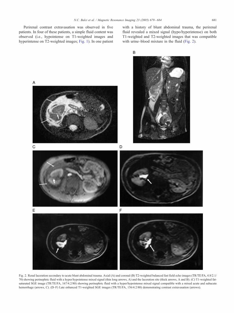
Perirenal contrast extravasation was observed in five
patients. In four of these patients, a simple fluid content was
observed (i.e., hypointense on T1-weighted images and
hyperintense on T2-weighted images; Fig. 1). In one patient
Fig. 2. Renal laceration secondary to acute blunt abdominal trauma. Axial (A) and c
70) showing perinephric fluid with a hyper/hypointense mixed signal (thin long arr
saturated SGE image (TR/TE/FA, 167/4.2/80) showing perinephric fluid with a hy
hemorrhage (arrows, C). (D–F) Late enhanced T1-weighted SGE images (TR/TE/
with a history of blunt abdominal trauma, the perirenal
fluid revealed a mixed signal (hypo/hyperintense) on both
T1-weighted and T2-weighted images that was compatible
with urine–blood mixture in the fluid (Fig. 2).
oronal (B) T2-weighted balanced fast field echo images (TR/TE/FA, 4.8/2.1/
ows, A) and the laceration site (thick arrows, A and B). (C) T1-weighted fat-
per/hypointense mixed signal compatible with a mixed acute and subacute
FA, 156/4.2/80) demonstrating contrast extravasation (arrows).
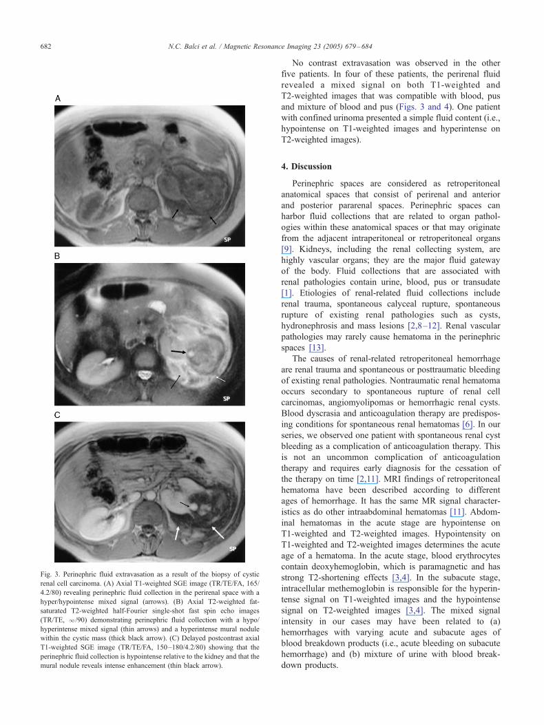
Fig. 3. Perinephric fluid extravasation as a result of the biopsy of cystic
renal cell carcinoma. (A) Axial T1-weighted SGE image (TR/TE/FA, 165/
4.2/80) revealing perinephric fluid collection in the perirenal space with a
hyper/hypointense mixed signal (arrows). (B) Axial T2-weighted fat-
saturated T2-weighted half-Fourier single-shot fast spin echo images
(TR/TE, l/90) demonstrating perinephric fluid collection with a hypo/
hyperintense mixed signal (thin arrows) and a hyperintense mural nodule
within the cystic mass (thick black arrow). (C) Delayed postcontrast axial
T1-weighted SGE image (TR/TE/FA, 150–180/4.2/80) showing that the
perinephric fluid collection is hypointense relative to the kidney and that the
mural nodule reveals intense enhancement (thin black arrow).
N.C. Balci et al. / Magnetic Resonance Imaging 23 (2005) 679–684682
No contrast extravasation was observed in the other
five patients. In four of these patients, the perirenal fluid
revealed a mixed signal on both T1-weighted and
T2-weighted images that was compatible with blood, pus
and mixture of blood and pus (Figs. 3 and 4). One patient
with confined urinoma presented a simple fluid content (i.e.,
hypointense on T1-weighted images and hyperintense on
T2-weighted images).
4. Discussion
Perinephric spaces are considered as retroperitoneal
anatomical spaces that consist of perirenal and anterior
and posterior pararenal spaces. Perinephric spaces can
harbor fluid collections that are related to organ pathol-
ogies within these anatomical spaces or that may originate
from the adjacent intraperitoneal or retroperitoneal organs
[9]. Kidneys, including the renal collecting system, are
highly vascular organs; they are the major fluid gateway
of the body. Fluid collections that are associated with
renal pathologies contain urine, blood, pus or transudate
[1]. Etiologies of renal-related fluid collections include
renal trauma, spontaneous calyceal rupture, spontaneous
rupture of existing renal pathologies such as cysts,
hydronephrosis and mass lesions [2,8–12]. Renal vascular
pathologies may rarely cause hematoma in the perinephric
spaces [13].
The causes of renal-related retroperitoneal hemorrhage
are renal trauma and spontaneous or posttraumatic bleeding
of existing renal pathologies. Nontraumatic renal hematoma
occurs secondary to spontaneous rupture of renal cell
carcinomas, angiomyolipomas or hemorrhagic renal cysts.
Blood dyscrasia and anticoagulation therapy are predispos-
ing conditions for spontaneous renal hematomas [6]. In our
series, we observed one patient with spontaneous renal cyst
bleeding as a complication of anticoagulation therapy. This
is not an uncommon complication of anticoagulation
therapy and requires early diagnosis for the cessation of
the therapy on time [2,11]. MRI findings of retroperitoneal
hematoma have been described according to different
ages of hemorrhage. It has the same MR signal character-
istics as do other intraabdominal hematomas [11]. Abdom-
inal hematomas in the acute stage are hypointense on
T1-weighted and T2-weighted images. Hypointensity on
T1-weighted and T2-weighted images determines the acute
age of a hematoma. In the acute stage, blood erythrocytes
contain deoxyhemoglobin, which is paramagnetic and has
strong T2-shortening effects [3,4]. In the subacute stage,
intracellular methemoglobin is responsible for the hyperin-
tense signal on T1-weighted images and the hypointense
signal on T2-weighted images [3,4]. The mixed signal
intensity in our cases may have been related to (a)
hemorrhages with varying acute and subacute ages of
blood breakdown products (i.e., acute bleeding on subacute
hemorrhage) and (b) mixture of urine with blood break-
down products.
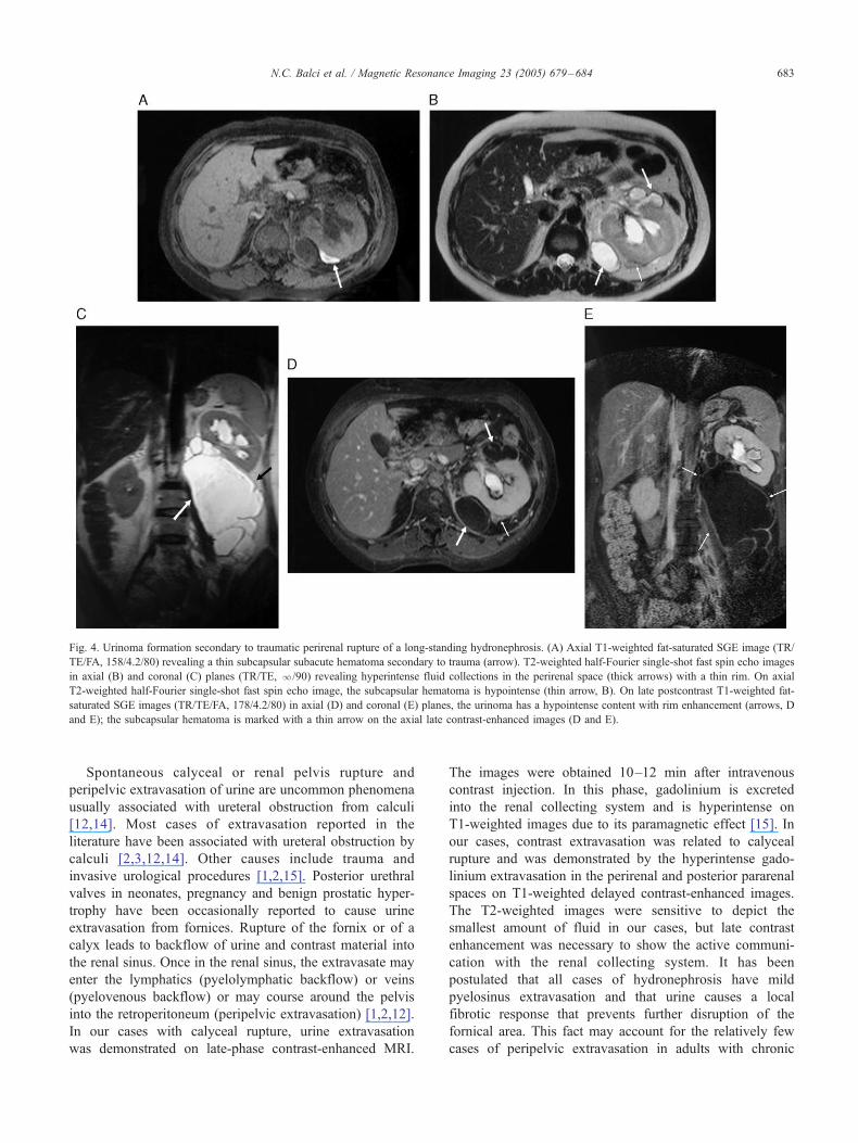
Fig. 4. Urinoma formation secondary to traumatic perirenal rupture of a long-standing hydronephrosis. (A) Axial T1-weighted fat-saturated SGE image (TR/
TE/FA, 158/4.2/80) revealing a thin subcapsular subacute hematoma secondary to trauma (arrow). T2-weighted half-Fourier single-shot fast spin echo images
in axial (B) and coronal (C) planes (TR/TE, l/90) revealing hyperintense fluid collections in the perirenal space (thick arrows) with a thin rim. On axial
T2-weighted half-Fourier single-shot fast spin echo image, the subcapsular hematoma is hypointense (thin arrow, B). On late postcontrast T1-weighted fat-
saturated SGE images (TR/TE/FA, 178/4.2/80) in axial (D) and coronal (E) planes, the urinoma has a hypointense content with rim enhancement (arrows, D
and E); the subcapsular hematoma is marked with a thin arrow on the axial late contrast-enhanced images (D and E).
N.C. Balci et al. / Magnetic Resonance Imaging 23 (2005) 679–684 683
Spontaneous calyceal or renal pelvis rupture and
peripelvic extravasation of urine are uncommon phenomena
usually associated with ureteral obstruction from calculi
[12,14]. Most cases of extravasation reported in the
literature have been associated with ureteral obstruction by
calculi [2,3,12,14]. Other causes include trauma and
invasive urological procedures [1,2,15]. Posterior urethral
valves in neonates, pregnancy and benign prostatic hyper-
trophy have been occasionally reported to cause urine
extravasation from fornices. Rupture of the fornix or of a
calyx leads to backflow of urine and contrast material into
the renal sinus. Once in the renal sinus, the extravasate may
enter the lymphatics (pyelolymphatic backflow) or veins
(pyelovenous backflow) or may course around the pelvis
into the retroperitoneum (peripelvic extravasation) [1,2,12].
In our cases with calyceal rupture, urine extravasation
was demonstrated on late-phase contrast-enhanced MRI.
The images were obtained 10–12 min after intravenous
contrast injection. In this phase, gadolinium is excreted
into the renal collecting system and is hyperintense on
T1-weighted images due to its paramagnetic effect [15]. In
our cases, contrast extravasation was related to calyceal
rupture and was demonstrated by the hyperintense gado-
linium extravasation in the perirenal and posterior pararenal
spaces on T1-weighted delayed contrast-enhanced images.
The T2-weighted images were sensitive to depict the
smallest amount of fluid in our cases, but late contrast
enhancement was necessary to show the active communi-
cation with the renal collecting system. It has been
postulated that all cases of hydronephrosis have mild
pyelosinus extravasation and that urine causes a local
fibrotic response that prevents further disruption of the
fornical area. This fact may account for the relatively few
cases of peripelvic extravasation in adults with chronic

N.C. Balci et al. / Magnetic Resonance Imaging 23 (2005) 679–684684
urinary tract obstruction [2,5,14,16]. This may explain the
confined urinary extravasation in one of our cases with
chronic hydronephrosis resulting in urinoma formation. The
absence of active communication of the urinoma was
demonstrated with the lack of contrast in the fluid. MRI
findings of urinoma have been described in a case report
previously. Urinomas may contain transudate compatible
with urine or may be infected. Extravasated urine incites a
low-grade inflammatory reaction followed by an avascular
deposition of collagen and fibrous tissue accounting for the
urinoma formation [17,18].
In conclusion, we described the spectrum of MRI
findings of renal-related perinephric fluid collections.
Perinephric fluid collections of pus, hemorrhage, urine
and cystic fluid revealed unique imaging characteristics
on noncontrast T1-weighted and T2-weighted images. The
renal relation of the fluid collections was demonstrated
in cases of active communication on delayed contrast-
enhanced images with the contrast extravasation.
References
[1] Haddad MC, Hawary MM, Khoury NJ, Faysal SA-F, Ammouri NF,
Al-Kutoubi OA. Radiology of perinephric fluid collections. Clin
Radiol 2002;57:339–46.
[2] Albi G, Del Campo D, Tagarro D. Wqnderlich syndrome: causes,
diagnosis and radiological management. Clin Radiol 2002;57:840–5.
[3] Negus S, Sidhu S. MRI of retroperitoneal collections: a comparison
with CT. Br J Radiol 2000;73:907–12.
[4] Korobkin M, Silverman PM, Quint LE, Francis IR. CT of the
extraperitoneal space: normal anatomy and fluid collections. AJR Am
J Roentgenol 1992;159:933–42.
[5] Unger CE, Glazer HS, Lee JKT, Ling D. MRI of extracranial
hematomas: preliminary observations. AJR Am J Roentgenol 1986;
146:403–7.
[6] Chang S-Y, Ma C-P, Lee S-K. Spontaneous retroperitoneal hemor-
rhage from kidney causes. Eur Urol 1988;15:281–4.
[7] Balci NC, Semelka RC, Noone TC, Ascher SM. Acute and subacute
liver-related hemorrhage: MRI findings. Magn Reson Imaging
1999;17:207–11.
[8] Rubin JI, Gomori JM, Grossman RI, Geffer WB, Kressel HY. High-
field MR imaging of extracranial hematomas. AJR Am J Roentgenol
1987;148:813–7.
[9] Lim JH, Kim B, Auh YH. Anatomical communications of the
perirenal space. Br J Radiol 1998;71:450–6.
[10] Marcos HB, Noone TC, Semelka RC. MRI evaluation of acute renal
trauma. J Magn Reson Imaging 1998;8:989–90.
[11] Balci NC, Sirvanci M, Tufek I, Onat L, Duran C. Spontaneous
retroperitoneal hemorrhage secondary to subcapsular renal hematoma:
MRI findings. Magn Reson Imaging 2001;19:1145–8.
[12] Akpinar H, Kural AR, Tufek I, Obek C, Demirkesen O, Solok V, et al.
Spontaneous ureteral rupture: is immediate surgical intervention
always necessary? Presentation of four cases and review of the
literature. J Endourol 2002;16:179–83.
[13] Manabe Y, Yoshioka K, Yanada J. Spontaneous rupture of a dissection
of the left ovarian artery. J Med Invest 2002;49:182–5.
[14] Genes DM, Vachon L. Ultrasound finding of peripelvic urine
extravasation in ureteropelvic junction obstruction. Pediatr Radiol
1989;20:122–3.
[15] Nolte-Ernsting CC, Staatz G, Tacke J, Gunther RW. MR urography
today. Abdom Imaging 2003;28:191–209.
[16] Ulreich S, Lund DA, Jacobson JJ. Spontaneous rupture of a calyceal
diverticulum during urography. AJR Am J Roentgenol 1978;131:
337–8.
[17] Titton RL, Gervais DA, Hahn PF, Harisinghani MG, Arellano RS,
Mueller PR. Urine leaks and urinomas: diagnosis and imaging-guided
intervention. Radiographics 2003;23:1133–47.
[18] Hutcheson JC, Canning DA, Hubbard AM, Johnson MP, Carr MC.
Magnetic resonance imaging of fetal urinoma. Urology 2002;60:697.