Release of Si from silicon, a ferrosilicon (FeSi) alloy and a synthetic silicate mineral in...
12
Release of Si from Silicon, a Ferrosilicon (FeSi) Alloy and a Synthetic Silicate Mineral in Simulated Biological Media Gunilla Herting*, Tao Jiang, Carin Sjo ¨ stedt, Inger Odnevall Wallinder KTH Royal Institute of Technology, Division of Surface and Corrosion Science, School of Chemical Science and Engineering, Stockholm, Sweden Abstract Unique quantitative bioaccessibility data has been generated, and the influence of surface/material and test media characteristics on the elemental release process were assessed for silicon containing materials in specific synthetic body fluids at certain time periods at a fixed loading. The metal release test protocol, elaborated by the KTH team, has previously been used for classification, ranking, and screening of different alloys and metals. Time resolved elemental release of Si, Fe and Al from particles, sized less than 50 mm, of two grades of metallurgical silicon (high purity silicon, SiHG, low purity silicon, SiLG), an alloy (ferrosilicon, FeSi) and a mineral (aluminium silicate, AlSi) has been investigated in synthetic body fluids of varying pH, composition and complexation capacity, simple models of for example dermal contact and digestion scenarios. Individual methods for analysis of released Si (as silicic acid, Si(OH) 4 ) in synthetic body fluids using GF-AAS were developed for each fluid including optimisation of solution pH and graphite furnace parameters. The release of Si from the two metallurgical silicon grades was strongly dependent on both pH and media composition with the highest release in pH neutral media. No similar effect was observed for the FeSi alloy or the aluminium silicate mineral. Surface adsorption of phosphate and lactic acid were believed to hinder the release of Si whereas the presence of citric acid enhanced the release as a result of surface complexation. An increased presence of Al and Fe in the material (low purity metalloid, alloy or mineral) resulted in a reduced release of Si in pH neutral media. The release of Si was enhanced for all materials with Al at their outermost surface in acetic media. Citation: Herting G, Jiang T, Sjo ¨ stedt C, Odnevall Wallinder I (2014) Release of Si from Silicon, a Ferrosilicon (FeSi) Alloy and a Synthetic Silicate Mineral in Simulated Biological Media. PLoS ONE 9(9): e107668. doi:10.1371/journal.pone.0107668 Editor: Christophe Egles, Universite ´ de Technologie de Compie `gne, France Received April 24, 2014; Accepted August 21, 2014; Published September 16, 2014 Copyright: ß 2014 Herting et al. This is an open-access article distributed under the terms of the Creative Commons Attribution License, which permits unrestricted use, distribution, and reproduction in any medium, provided the original author and source are credited. Data Availability: The authors confirm that all data underlying the findings are fully available without restriction. All relevant data are within the paper. Funding: The research reported in this article was funded by the silicon and ferrosilicon consortium. The funders had no role in study design, data collection and analysis, decision to publish, or preparation of the manuscript. Competing Interests: The authors have declared that no competing interests exist. * Email: [email protected] Introduction Silicon is the second most abundant element found in the Earths crust and is commonly found as silica (SiO 2 ) or as silicates, the latter being the most abundant mineral group [1,2]. The silicon surface is, unless modified, covered with a few nm thick layer of amorphous silica [3,4]. Such an amorphous layer causes an otherwise crystalline material to behave similarly to amorphous silica in terms of dissolution [5]. As a metalloid, silicon possess desirable properties applicable for semiconductors and solar cells, but it is also widely used in glasses, ceramics and refractory materials and as an alloying element for many steel grades and aluminium alloys. This may cause human exposures to a wide variety of silicon containing powder particles at e.g. occupational settings. A vast number of investigations on the stability of engineered silica particles in humans and their dissolution properties have been performed during the past 60 years [4]. Airborne particles are associated with adverse effects on human health [4] and several studies using both in-vitro and in- vivo approaches have been performed to evaluate how humans may be affected by for example silica and asbestos [6–11]. These studies have shown silica dissolution to depend on pH [11–13], particle size [14–16], presence of cations and salts [17–26] and presence of complexing agents [27–30]. However, only few studies have addressed silica dissolution in synthetic biological solutions [4,11]. The dissolution process of silica in water is depolymerisation through hydrolysis, where the hydroxyl ion, OH 2 , acts as a catalyst and temporarily changes the coordination number of silicon atoms on the surface resulting in weaker oxygen bonds to the underlying bulk silicon [4]. In alkaline solutions the initiation of the process is governed by adsorption of the hydroxyl ion followed by release of Si(OH) 5 2 into solution [4]. Below pH 11 silicon is quickly hydrolysed to Si(OH) 4 and OH 2 , and the hydroxyl ion is free to repeat the process. This process is depressed at acidic conditions. Some hydroxyl ions are also involved in the formation of Si(OH) 5 2 . Above pH 11 is Si(OH) 4 converted to Si(OH) 5 2 resulting in a non-saturated solution and a continued dissolution of silica [4]. Aluminium has been shown to reduce the solubility of silica at alkaline conditions through different mechanisms. At a weakly alkaline pH, Al in solution forms negatively charged aluminosil- icate sites on the silicon surface that repel OH 2 interactions, thereby decreasing the dissolution rate of Si [19]. Even small PLOS ONE | www.plosone.org 1 September 2014 | Volume 9 | Issue 9 | e107668
Transcript of Release of Si from silicon, a ferrosilicon (FeSi) alloy and a synthetic silicate mineral in...
Release of Si from Silicon, a Ferrosilicon (FeSi) Alloy anda Synthetic Silicate Mineral in Simulated BiologicalMediaGunilla Herting*, Tao Jiang, Carin Sjostedt, Inger Odnevall Wallinder
KTH Royal Institute of Technology, Division of Surface and Corrosion Science, School of Chemical Science and Engineering, Stockholm, Sweden
Abstract
Unique quantitative bioaccessibility data has been generated, and the influence of surface/material and test mediacharacteristics on the elemental release process were assessed for silicon containing materials in specific synthetic bodyfluids at certain time periods at a fixed loading. The metal release test protocol, elaborated by the KTH team, has previouslybeen used for classification, ranking, and screening of different alloys and metals. Time resolved elemental release of Si, Feand Al from particles, sized less than 50 mm, of two grades of metallurgical silicon (high purity silicon, SiHG, low puritysilicon, SiLG), an alloy (ferrosilicon, FeSi) and a mineral (aluminium silicate, AlSi) has been investigated in synthetic bodyfluids of varying pH, composition and complexation capacity, simple models of for example dermal contact and digestionscenarios. Individual methods for analysis of released Si (as silicic acid, Si(OH)4) in synthetic body fluids using GF-AAS weredeveloped for each fluid including optimisation of solution pH and graphite furnace parameters. The release of Si from thetwo metallurgical silicon grades was strongly dependent on both pH and media composition with the highest release in pHneutral media. No similar effect was observed for the FeSi alloy or the aluminium silicate mineral. Surface adsorption ofphosphate and lactic acid were believed to hinder the release of Si whereas the presence of citric acid enhanced the releaseas a result of surface complexation. An increased presence of Al and Fe in the material (low purity metalloid, alloy ormineral) resulted in a reduced release of Si in pH neutral media. The release of Si was enhanced for all materials with Al attheir outermost surface in acetic media.
Citation: Herting G, Jiang T, Sjostedt C, Odnevall Wallinder I (2014) Release of Si from Silicon, a Ferrosilicon (FeSi) Alloy and a Synthetic Silicate Mineral inSimulated Biological Media. PLoS ONE 9(9): e107668. doi:10.1371/journal.pone.0107668
Editor: Christophe Egles, Universite de Technologie de Compiegne, France
Received April 24, 2014; Accepted August 21, 2014; Published September 16, 2014
Copyright: � 2014 Herting et al. This is an open-access article distributed under the terms of the Creative Commons Attribution License, which permitsunrestricted use, distribution, and reproduction in any medium, provided the original author and source are credited.
Data Availability: The authors confirm that all data underlying the findings are fully available without restriction. All relevant data are within the paper.
Funding: The research reported in this article was funded by the silicon and ferrosilicon consortium. The funders had no role in study design, data collection andanalysis, decision to publish, or preparation of the manuscript.
Competing Interests: The authors have declared that no competing interests exist.
* Email: [email protected]
Introduction
Silicon is the second most abundant element found in the Earths
crust and is commonly found as silica (SiO2) or as silicates, the
latter being the most abundant mineral group [1,2].
The silicon surface is, unless modified, covered with a few nm
thick layer of amorphous silica [3,4]. Such an amorphous layer
causes an otherwise crystalline material to behave similarly to
amorphous silica in terms of dissolution [5]. As a metalloid, silicon
possess desirable properties applicable for semiconductors and
solar cells, but it is also widely used in glasses, ceramics and
refractory materials and as an alloying element for many steel
grades and aluminium alloys. This may cause human exposures to
a wide variety of silicon containing powder particles at e.g.occupational settings. A vast number of investigations on the
stability of engineered silica particles in humans and their
dissolution properties have been performed during the past 60
years [4]. Airborne particles are associated with adverse effects on
human health [4] and several studies using both in-vitro and in-
vivo approaches have been performed to evaluate how humans
may be affected by for example silica and asbestos [6–11]. These
studies have shown silica dissolution to depend on pH [11–13],
particle size [14–16], presence of cations and salts [17–26] and
presence of complexing agents [27–30]. However, only few studies
have addressed silica dissolution in synthetic biological solutions
[4,11].
The dissolution process of silica in water is depolymerisation
through hydrolysis, where the hydroxyl ion, OH2, acts as a
catalyst and temporarily changes the coordination number of
silicon atoms on the surface resulting in weaker oxygen bonds to
the underlying bulk silicon [4]. In alkaline solutions the initiation
of the process is governed by adsorption of the hydroxyl ion
followed by release of Si(OH)52 into solution [4]. Below pH 11
silicon is quickly hydrolysed to Si(OH)4 and OH2, and the
hydroxyl ion is free to repeat the process. This process is depressed
at acidic conditions. Some hydroxyl ions are also involved in the
formation of Si(OH)52. Above pH 11 is Si(OH)4 converted to
Si(OH)52 resulting in a non-saturated solution and a continued
dissolution of silica [4].
Aluminium has been shown to reduce the solubility of silica at
alkaline conditions through different mechanisms. At a weakly
alkaline pH, Al in solution forms negatively charged aluminosil-
icate sites on the silicon surface that repel OH2 interactions,
thereby decreasing the dissolution rate of Si [19]. Even small
PLOS ONE | www.plosone.org 1 September 2014 | Volume 9 | Issue 9 | e107668
amounts of Al, 40–100 mg/L, present in solution have proved to
significantly reduce the solubility of Si [4]. Hydrolysed aluminium
has at acidic conditions been shown to weaken the Si-O bonds in
silica gel, thus increasing the dissolution of silicon [20].
The mechanism of Fe in combination with silica has not been as
thoroughly investigated as for Al, but has shown a similar, albeit
weaker, effect on the dissolution rate [4]. Other cations such as
Mg2+, Ca2+, Ba2+, Na+, K+ and Li+ also have a strong effect on the
dissolution of silica particles, however addition of these ions to pH
neutral solutions increase dissolution of quartz [23,24,31,32].
Other compounds that commonly are in contact with silica are
organic acids, for example citric acid [27–30]. Citric acid enhances
both dissolution of silicon and aluminium in the entire pH range
although its effect decreases with decreasing pH [28].
A test protocol for evaluation of metal release from metals and
alloys in synthetic body fluids, elaborated by the KTH team,
previously used for classification, ranking and screening of
different alloys and metals [33–35] was utilised to obtain data
relevant for risk assessment within the scope of REACH. The aims
of this investigation were to fill knowledge gaps related to the
release of silicon and other elements from particles of silicon
metalloids of different purity, a ferrosilicon alloy and an
aluminium silicate mineral in different synthetic biological fluids,
and to investigate whether the release behaviour could be related
to the bulk and/or surface composition of the materials. The
synthetic fluids are simple models for different human exposure
routes of particles of relevance for inhalation, dermal contact and
ingestion [36–39].
Materials and Methods
MaterialsFour different silicon containing materials, two grades of
metallurgical silicon (SiHG, SiLG), one ferrosilicon alloy (FeSi)
and one aluminium silicate mineral, (Mullite, AlSi) were investi-
gated, table 1. As three of the test items (SiHG, SiLG and FeSi)
are commercially relevant in different solid forms and particle
sizes, they were pre-treated (crushed, sieved, re-crushed), still
maintaining their general material characteristics, to generate
particles sized less than 50 mm of relevance to human inhalation,
dermal contact and digestion. Crushing was accomplished using a
Retsch Jaw crusher with crushing jaws made of manganese steel
and wearing plates of stainless steel. Generated items less than
5 mm in size were separated from larger items and consecutively
sieved until the desired size, smaller than 50 mm, was obtained.
The entire procedure was performed at the Norwegian University
of Science and Technology. The aluminium silicate, AlSi, powder
was purchased from Sigma Aldrich.
Particle size distribution measurements were performed in
phosphate buffered saline (PBS) using a Malvern Mastersizer 2000
laser diffraction equipment and refractive indexes for Si (3.5) and
water (1.33). The specific surface area (m2/g) was determined by
BET analysis using a Micromeritics Gemini V surface area
analyser performed at Kanthal AB, Sweden. The procedure is
described in detail elsewhere [40]. The specific surface area (BET)
and particle size distribution of the four materials investigated in
this study are given in table 2. Since SiHG, SiLG and FeSi were
pre-treated in the same way, they revealed similar size distribu-
tions in PBS with a median particle size (d0.5) of 29.563.6 mm,
table 2. The similarity in particle size distribution was also
Table 1. Nominal bulk composition of the investigated silicon-containing materials.
Materials Abbreviation Si [wt%] Fe [wt%] Al [wt%] Other elements
High purity silicon SiHG 99.1 0.4 0.1 Ca, Mn, Ti
Low purity silicon SiLG 98.6 0.5 0.3 Ca, Mn, Ti
Ferrosilicon alloy FeSi 75 24 0.7 Ca, C
Aluminium silicate (Mullite) AlSi 13.2 - 38 O
doi:10.1371/journal.pone.0107668.t001
Table 2. Specific surface area, BET, (m2/g) of the different silicon-containing test items with corresponding median particlediameters (d0.5) and the 10% (d0.1) and 90% (d0.9) size distribution cut-off points as a percentage of volume (mass), determinedusing laser diffraction.
Test item d0.1 [mm] d0.5 [mm] d0.9 [mm] BET- surface area [m2/g]
SiHG 6.7 29.4 70.9 0.96
SiLG 5.8 26.0 67.8 0.97
FeSi 8.3 33.1 75.6 0.52
AlSi 16.0 30.2 53.1 0.19
doi:10.1371/journal.pone.0107668.t002
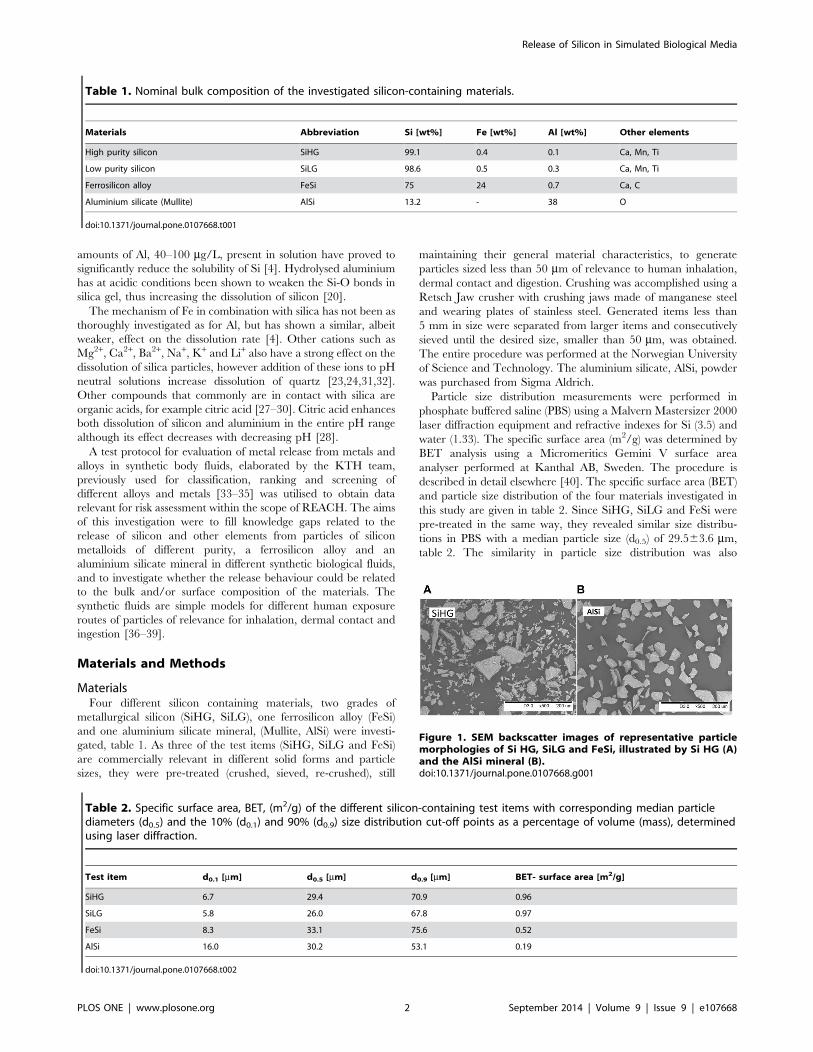
Figure 1. SEM backscatter images of representative particlemorphologies of Si HG, SiLG and FeSi, illustrated by Si HG (A)and the AlSi mineral (B).doi:10.1371/journal.pone.0107668.g001
Release of Silicon in Simulated Biological Media
PLOS ONE | www.plosone.org 2 September 2014 | Volume 9 | Issue 9 | e107668
reflected in the specific surface areas for SiHG and SiLG (0.96 and
0.97 m2/g, respectively), whereas FeSi revealed a slightly smaller
specific surface area (0.52 m2/g).
Particle shape and morphology were investigated by means of
scanning electron microscopy (SEM), with a Hitachi TM100
instrument. All images were obtained in back-scattered electron
mode with an acceleration voltage of 15 keV.
The commercially relevant materials (Si HG, Si LG, FeSi)
displayed a similar visual appearance, illustrated by SiHG, Fig. 1,
(A). Sharp edged particles of varying size ranging from larger (50–
100 mm) to smaller sizes (,10 mm) were evident. The smaller sized
particles were predominantly present at the surface of the larger
sized particles. The AlSi mineral, Fig. 1 (B), revealed the most
uniform size distribution with only few particles smaller than
10 mm.
X-ray photoelectron spectroscopy (XPS) was employed to
evaluate the chemical composition of the outermost surface layer
(5–10 nm) using an UltraDLD spectrometer from Kratos Analyt-
ical, Manchester, UK, with a monochromatic Al-Ka x-ray source
(10 mA, 15 kV). Wide spectra and detailed high resolution spectra
of Si 2p, Al 2p, Fe 2p, O 1s and C 1s were run. All binding
energies were calibrated by assigning the carbon-hydrocarbon
peak (C–H, or C–C) to 285 eV. All peak areas were determined by
assigning a linear base line.
Elemental releaseTo avoid any risk of contamination all experiments were
performed using acid-cleaned lab equipment, 10% HNO3 for at
least 24 h, followed by rinsing four times with ultra-pure water
(MilliQ, 18.2 MVcm) and drying in ambient laboratory air. As a
precaution against contamination of Si no glassware was used in
the experimental work. Release studies were carried out using
triplicate samples of each material exposed for 2, 4, 8, 24 and
168 h. In addition, one blank reference sample for each material
and time period containing only the test solution was incubated
together with the triplicates. Four different synthetic body fluids
were investigated, phosphate buffered saline (PBS), artificial sweat
(ASW), artificial lysosomal fluid (ALF) and artificial gastric fluid
(GST). Their chemical compositions and pH are presented in
table 3.
560.5 mg of each material was weighed in a TPX Nalge jar
using a Mettler AT20 balance with readability of 2 mg. 50 mL of
the test solution was then added to the TPX Nalge jar containing
the powder sample before being incubated in a Stuart platform-
rocker incubator, regulated at 3760.5uC. The solutions were
gently shaken (bi-linearly) at an intensity of 25 cycles per minute.
After the test periods, the samples were allowed to reach
ambient room temperature before the final pH of the test solution
was measured. The test media was then separated from the
powder particles by centrifugation at 3000 rpm for 10 min
resulting in a visually clear supernatant with remaining particles
in the bottom of the centrifuging tube. The supernatant was
carefully poured into 25 mL HDPE bottles for storage before
analysis. An efficient removal of all particles from the supernatant
was confirmed by dynamic light scattering (Malvern Zetasizer
nano ZS instrument).
Graphite furnace analysis GF-AAS (Perkin–Elmer AAnalyst 800
instrument) was conducted to determine total released concentra-
tions of Al, Si and Fe, without considering their chemical
speciation. Therefore the released elements are denoted only as
Al, Si and Fe. All parameters used for analysis are presented in
table 4.
The instrument was calibrated prior to each set of samples to be
analysed and the calibration curves were repeatedly verified
during analysis by running quality control samples of known
concentration every 6 to 8 samples. Mean released concentrations
were based on five (Si) or three (Al and Fe) replicate readings of
each sample. The limits of detection for total Al, Si and Fe in the
various test media are presented in table 5.
The resulting supernatant was poured into 25 mL HDPE
bottles. Samples destined for Al and Fe analysis were acidified (pH
,2) with 65% supra pure HNO3. In contrast to most elements,
Table 3. Chemical composition (g/L) and pH of the different synthetic biological fluids [56–58].
Chemicals PBS ASW ALF GST
MgCl2 0.050
NaCl 8.77 5 3.21
Na2HPO4 1.28 0.071
Na2SO4 0.039
CaCl2N2H2O 0.128
C6H5Na3O7N2H2O (sodium citrate) 0.077
NaOH 6.00
Citric acid 20.8
Glycine 0.059
C4H4O6Na2N2H2O (Na2TartrateN2H2O) 0.090
C3H5NaO3 (NaLactate) 0.085
C3H5O3Na (NaPyruvate) 0.086
KH2PO4 1.36
Urea 1
Lactic acid 940 mL
HCl 10
pH 7.2 4.5 1.7
doi:10.1371/journal.pone.0107668.t003
Release of Silicon in Simulated Biological Media
PLOS ONE | www.plosone.org 3 September 2014 | Volume 9 | Issue 9 | e107668
Table
4.AASan
alytical
param
eters
foreachelement.
Pre-treatm
ent
Furn
ace
pro
gram
Wavelength
Injectionvolume
Matrix
modifier
Calibrationstandard
s
Element
T[uC]
Ramp
time[s]
Hold
time[s]
[nm]
[mL]
[mg/L]
SipHad
justmentto
pH8–9
110
130
251.6
15
Mg(NO3) 2Ca(NO3) 2
0,600,1000
pHad
justmentto
pH8–9
130
20
35
251.6
15
Mg(NO3) 2Ca(NO3) 2
0,600,1000
pHad
justmentto
pH8–9
1200
10
20
251.6
15
Mg(NO3) 2Ca(NO3) 2
0,600,1000
pHad
justmentto
pH8–9
2350
05
251.6
15
Mg(NO3) 2Ca(NO3) 2
0,600,1000
pHad
justmentto
pH8–9
2600
15
251.6
15
Mg(NO3) 2Ca(NO3) 2
0,600,1000
FepHad
justmentto
pH,2
110
135
248.3
20
Mg(NO3) 2
0,50,100,300
pHad
justmentto
pH,2
130
20
30
248.3
20
Mg(NO3) 2
0,50,100,300
pHad
justmentto
pH,2
1400
10
20
248.3
20
Mg(NO3) 2
0,50,100,300
pHad
justmentto
pH,2
2100
05
248.3
20
Mg(NO3) 2
0,50,100,300
pHad
justmentto
pH,2
2550
13
248.3
20
Mg(NO3) 2
0,50,100,300
Al
pHad
justmentto
pH,2
110
10
40
309.3
15
Mg(NO3) 2Pd(NO3) 2
0,30,60,100
pHad
justmentto
pH,2
130
15
40
309.3
15
Mg(NO3) 2Pd(NO3) 2
0,30,60,100
pHad
justmentto
pH,2
1200
10
20
309.3
15
Mg(NO3) 2Pd(NO3) 2
0,30,60,100
pHad
justmentto
pH,2
2400
05
309.3
15
Mg(NO3) 2Pd(NO3) 2
0,30,60,100
pHad
justmentto
pH,2
2450
13
309.3
15
Mg(NO3) 2Pd(NO3) 2
0,30,60,100
doi:10.1371/journal.pone.0107668.t004
Release of Silicon in Simulated Biological Media
PLOS ONE | www.plosone.org 4 September 2014 | Volume 9 | Issue 9 | e107668
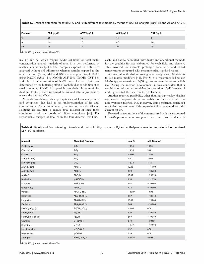
like Fe and Al, which require acidic solutions for total metal
concentration analysis, analysis of total Si is best performed at
alkaline conditions (pH 8–8.5). Samples exposed in PBS were
analysed without pH adjustment whereas samples exposed to the
other test fluid (ASW, ALF and GST) were adjusted to pH 8–8.5
using NaOH (ASW- 1% NaOH; ALF-25% NaOH; GST 8%
NaOH). The concentration of NaOH used for each fluid was
determined by the buffering effect of each fluid as as addition of as
small amounts of NaOH as possible was desirable to minimise
dilution effects. pH was measured before and after adjustment to
ensure the desired effect.
At acidic conditions, silica precipitates and form compounds
and complexes that lead to an underestimation of its total
concentration. As a consequence, neutral or weakly alkaline
solutions are essential to analyse total released Si since these
conditions break the bonds of silicon complexes [41]. For
reproducible analysis of total Si in the four different test fluids,
each fluid had to be treated individually and operational methods
for the graphite furnace elaborated for each fluid and element.
This involved for example prolonged time steps and raised
temperatures compared with recommended standard values.
A universal method of improving metal analysis with GF-AAS is
to use matrix modifiers [42]. For Si it is recommended to use
Mg(NO3)2, or sometimes Ca(NO3)2, to improve the reproducibil-
ity. During the method development it was concluded that a
combination of the two modifiers in a solution of pH between 8
and 9 generated the best results, c.f. Table 5.
Another reported possibility, other than having weakly alkaline
conditions to improve the reproducibility of the Si analysis is to
add hydrogen fluoride, HF. However, tests performed concluded
negligible improvement of the reproducibility compared with the
current set-up.
Released concentrations of silicon measured with the elaborated
GF-AAS protocol were compared determined with inductively
Table 5. Limits of detection for total Si, Al and Fe in different test media by means of AAS-GF analysis [mg/L] (Si and Al) and AAS-F.
Element PBS [mg/L] ASW [mg/L] ALF [mg/L] GST [mg/L]
Si 45 10 15 5
Al 1.0 1.0 0.5 2.0
Fe 12 15 20 12
doi:10.1371/journal.pone.0107668.t005
Table 6. Si-, Al-, and Fe-containing minerals and their solubility constants (Ks) and enthalpies of reaction as included in the VisualMINTEQ database.
Mineral Chemical formula Log Ks DHr [kJ/mol]
Chalcedony SiO2 23.55 19.70
Cristobalite SiO2 23.35 20.01
Quartz SiO2 24.00 22.36
SiO2 (am, gel) SiO2 22.71 14.00
SiO2 (am, ppt) SiO2 22.74 15.15
Al(OH)3 (am) Al(OH)3 10.80 2111.00
Al(OH)3 (Soil) Al(OH)3 8.29 2105.00
Al2O3(s) Al2O3(s) 19.65 2258.59
Boehmite c-AlO(OH) 8.58 2117.70
Diaspore a-AlO(OH) 6.87 2103.05
Gibbsite (C) Al(OH)3 7.74 2105.00
Variscite AlPO4?2 H2O 222.07 29.40
Halloysite Al2Si2O5(OH)4 9.57 2181.43
Imogolite Al2SiO3(OH)4 13.00 2193.60
Kaolinite Al2Si2O5(OH)4 7.44 2148.00
Fe(OH)2.7Cl0.3 (s) Fe(OH)2.7Cl0.3 23.04 0.00
Ferrihydrite Fe(OH)3 3.20 2100.40
Ferrihydrite (aged) Fe(OH)3 2.69 2100.40
Goethite a-FeO(OH) 0.49 260.58
Hematite a-Fe2O3 21.42 2128.99
Lepidocrocite c-FeO(OH) 1.37 0.00
Maghemite c-Fe2O3 6.39 0.00
Strengite FePO4?2 H2O 226.40 29.36
doi:10.1371/journal.pone.0107668.t006
Release of Silicon in Simulated Biological Media
PLOS ONE | www.plosone.org 5 September 2014 | Volume 9 | Issue 9 | e107668
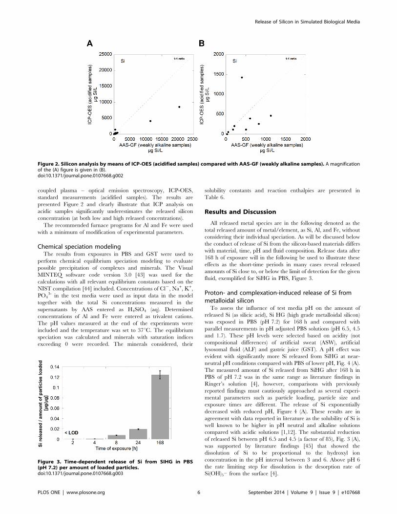
coupled plasma – optical emission spectroscopy, ICP-OES,
standard measurements (acidified samples). The results are
presented Figure 2 and clearly illustrate that ICP analysis on
acidic samples significantly underestimates the released silicon
concentration (at both low and high released concentrations).
The recommended furnace programs for Al and Fe were used
with a minimum of modification of experimental parameters.
Chemical speciation modelingThe results from exposures in PBS and GST were used to
perform chemical equilibrium speciation modeling to evaluate
possible precipitation of complexes and minerals. The Visual
MINTEQ software code version 3.0 [43] was used for the
calculations with all relevant equilibrium constants based on the
NIST compilation [44] included. Concentrations of Cl2, Na+, K+,
PO43- in the test media were used as input data in the model
together with the total Si concentrations measured in the
supernatants by AAS entered as H4SiO4 (aq). Determined
concentrations of Al and Fe were entered as trivalent cations.
The pH values measured at the end of the experiments were
included and the temperature was set to 37uC. The equilibrium
speciation was calculated and minerals with saturation indices
exceeding 0 were recorded. The minerals considered, their
solubility constants and reaction enthalpies are presented in
Table 6.
Results and Discussion
All released metal species are in the following denoted as the
total released amount of metal/element, as Si, Al, and Fe, without
considering their individual speciation. As will be discussed below
the conduct of release of Si from the silicon-based materials differs
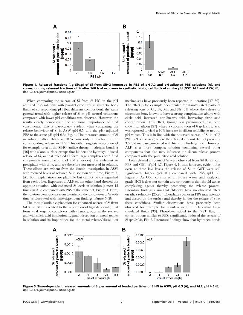
with material, time, pH and fluid composition. Release data after
168 h of exposure will in the following be used to illustrate these
effects as the short-time periods in many cases reveal released
amounts of Si close to, or below the limit of detection for the given
fluid, exemplified for SiHG in PBS, Figure 3.
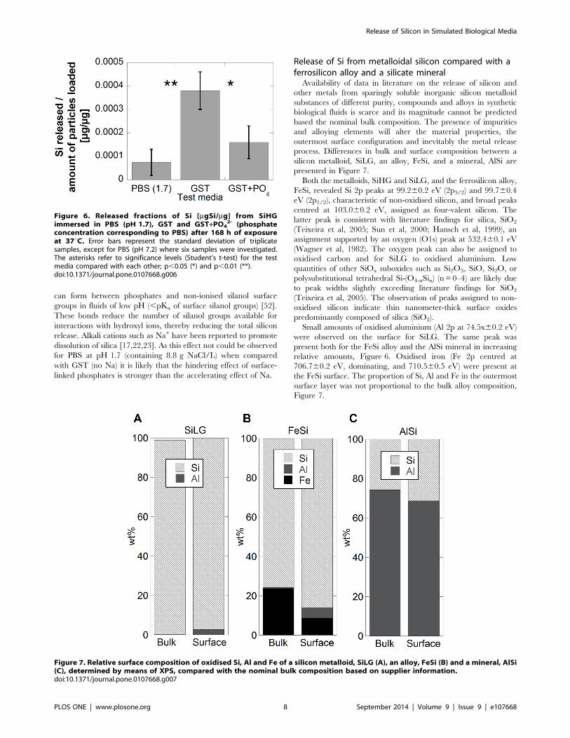
Proton- and complexation-induced release of Si frommetalloidal siliconTo assess the influence of test media pH on the amount of
released Si (as silicic acid), Si HG (high grade metalloidal silicon)
was exposed in PBS (pH 7.2) for 168 h and compared with
parallel measurements in pH adjusted PBS solutions (pH 6.5, 4.5
and 1.7). These pH levels were selected based on acidity (not
compositional differences) of artificial sweat (ASW), artificial
lysosomal fluid (ALF) and gastric juice (GST). A pH effect was
evident with significantly more Si released from SiHG at near-
neutral pH conditions compared with PBS of lower pH, Fig. 4 (A).
The measured amount of Si released from SiHG after 168 h in
PBS of pH 7.2 was in the same range as literature findings in
Ringer’s solution [4], however, comparisons with previously
reported findings must cautiously approached as several experi-
mental parameters such as particle loading, particle size and
exposure times are different. The release of Si exponentially
decreased with reduced pH, Figure 4 (A). These results are in
agreement with data reported in literature as the solubility of Si is
well known to be higher in pH neutral and alkaline solutions
compared with acidic solutions [1,12]. The substantial reduction
of released Si between pH 6.5 and 4.5 (a factor of 85), Fig. 3 (A),
was supported by literature findings [45] that showed the
dissolution of Si to be proportional to the hydroxyl ion
concentration in the pH interval between 3 and 6. Above pH 6
the rate limiting step for dissolution is the desorption rate of
Si(OH)52 from the surface [4].
Figure 2. Silicon analysis by means of ICP-OES (acidified samples) compared with AAS-GF (weakly alkaline samples). A magnificationof the (A) figure is given in (B).doi:10.1371/journal.pone.0107668.g002
Figure 3. Time-dependent release of Si from SIHG in PBS(pH 7.2) per amount of loaded particles.doi:10.1371/journal.pone.0107668.g003
Release of Silicon in Simulated Biological Media
PLOS ONE | www.plosone.org 6 September 2014 | Volume 9 | Issue 9 | e107668
When comparing the release of Si from Si HG in the pH
adjusted PBS solutions with parallel exposures in synthetic body
fluids of corresponding pH (but different composition), the same
general trend with higher release of Si at pH neutral conditions
compared with lower pH conditions was observed. However, the
results clearly demonstrate the additional importance of fluid
constituents. This is particularly evident when comparing the
release behaviour of Si in ASW (pH 6.5) and the pH- adjusted
PBS to the same pH (pH 6.5), Fig. 4. The measured amount of Si
in solution after 168 h in ASW was only a fraction of the
corresponding release in PBS. This either suggests adsorption of
for example urea at the SiHG surface through hydrogen bonding
[46] with silanol surface groups that hinders the hydroxyl-induced
release of Si, or that released Si form large complexes with fluid
components (urea, lactic acid and chlorides) that sediment or
precipitate with time, and are therefore not measured in solution.
These effects are evident from the kinetic investigation in ASW
with reduced levels of released Si in solution with time, Figure 5,
(A). Both explanations are plausible but cannot be distinguished
from each other. Exposures in ALF on the other hand showed the
opposite situation, with enhanced Si levels in solution (almost 15
times) in ALF compared with PBS of the same pH, Figure 4. Here,
the solution components seem to enhance the dissolution of Si with
time as illustrated with time-dependent findings, Figure 5 (B).
The most plausible explanation for enhanced release of Si from
SiHG in ALF is related to the adsorption of ligands (citrate) that
form weak organic complexes with silanol groups at the surface
and with silicic acid in solution. Ligand-adsorption on metal oxides
in solution and its importance for the metal release/dissolution
mechanisms have previously been reported in literature [47–50].
The effect is for example documented for stainless steel particles
releasing ions of Cr, Fe, Mn and Ni [51] where the release of
chromium ions, known to have a strong complexation ability with
citric acid, increased non-linearly with increasing citric acid
concentration. This effect, though less pronounced, has been
shown for silicon [27] where a concentration of 6 g/L citric acid
was reported to yield a 10% increase in silicon solubility at neutral
pH values. This is in line with the observed release of Si in ALF
(20.8 g/L citric acid) where the released amount did not present a
3.5 fold increase compared with literature findings [27]. However,
ALF is a more complex solution containing several other
components that also may influence the silicon release process
compared with the pure citric acid solution.
Low released amounts of Si were observed from SiHG in both
PBS and GST of pH 1.7, Figure 4. It was, however, evident that
even at these low levels the release of Si in GST were still
significantly higher (p,0.01) compared with PBS (pH 1.7),
Figure 6. As GST consists of ultra-pure water and analytical
grade HCl it does not contain any components that should act as
complexing agents thereby promoting the release process.
Literature findings claim that chlorides have no observed effect
on silica solubility [25,26]. Phosphate species in PBS may interact
and adsorb on the surface and thereby hinder the release of Si at
these conditions. Similar observations have previously been
observed for example for stainless steel in pH-neutral lung-
simulated fluids [33]. Phosphate added to the GST fluid in
concentrations similar to PBS, significantly reduced the release of
Si (p,0.05), Fig. 6. Literature findings show that hydrogen bonds
Figure 4. Released fractions [mg Si/mg] of Si from SiHG immersed in PBS of pH 7.2 and pH-adjusted PBS solutions (A), andcorresponding released fractions of Si after 168 h of exposure in synthetic biological fluids of similar pH (GST, ALF and ASW) (B).doi:10.1371/journal.pone.0107668.g004
Figure 5. Time-dependent released amounts of Si per amount of loaded particles of SiHG in ASW, pH 6.5 (A), and ALF, pH 4.5 (B).doi:10.1371/journal.pone.0107668.g005
Release of Silicon in Simulated Biological Media
PLOS ONE | www.plosone.org 7 September 2014 | Volume 9 | Issue 9 | e107668
can form between phosphates and non-ionised silanol surface
groups in fluids of low pH (,pKa of surface silanol groups) [52].
These bonds reduce the number of silanol groups available for
interactions with hydroxyl ions, thereby reducing the total silicon
release. Alkali cations such as Na+ have been reported to promote
dissolution of silica [17,22,23]. As this effect not could be observed
for PBS at pH 1.7 (containing 8.8 g NaCl/L) when compared
with GST (no Na) it is likely that the hindering effect of surface-
linked phosphates is stronger than the accelerating effect of Na.
Release of Si from metalloidal silicon compared with aferrosilicon alloy and a silicate mineralAvailability of data in literature on the release of silicon and
other metals from sparingly soluble inorganic silicon metalloid
substances of different purity, compounds and alloys in synthetic
biological fluids is scarce and its magnitude cannot be predicted
based the nominal bulk composition. The presence of impurities
and alloying elements will alter the material properties, the
outermost surface configuration and inevitably the metal release
process. Differences in bulk and surface composition between a
silicon metalloid, SiLG, an alloy, FeSi, and a mineral, AlSi are
presented in Figure 7.
Both the metalloids, SiHG and SiLG, and the ferrosilicon alloy,
FeSi, revealed Si 2p peaks at 99.260.2 eV (2p3/2) and 99.760.4
eV (2p1/2), characteristic of non-oxidised silicon, and broad peaks
centred at 103.060.2 eV, assigned as four-valent silicon. The
latter peak is consistent with literature findings for silica, SiO2
(Teixeira et al, 2005; Sun et al, 2000; Hansch et al, 1999), an
assignment supported by an oxygen (O1s) peak at 532.460.1 eV
(Wagner et al, 1982). The oxygen peak can also be assigned to
oxidised carbon and for SiLG to oxidised aluminium. Low
quantities of other SiOx suboxides such as Si2O3, SiO, Si2O, or
polysubstitutional tetrahedral Si-(O4-nSin) (n = 0–4) are likely due
to peak widths slightly exceeding literature findings for SiO2
(Teixeira et al, 2005). The observation of peaks assigned to non-
oxidised silicon indicate thin nanometer-thick surface oxides
predominantly composed of silica (SiO2).
Small amounts of oxidised aluminium (Al 2p at 74.5x60.2 eV)
were observed on the surface for SiLG. The same peak was
present both for the FeSi alloy and the AlSi mineral in increasing
relative amounts, Figure 6. Oxidised iron (Fe 2p centred at
706.760.2 eV, dominating, and 710.560.5 eV) were present at
the FeSi surface. The proportion of Si, Al and Fe in the outermost
surface layer was not proportional to the bulk alloy composition,
Figure 7.
Figure 6. Released fractions of Si [mgSi/mg] from SiHGimmersed in PBS (pH 1.7), GST and GST+PO4
2- (phosphateconcentration corresponding to PBS) after 168 h of exposureat 37uC. Error bars represent the standard deviation of triplicatesamples, except for PBS (pH 7.2) where six samples were investigated.The asterisks refer to significance levels (Student’s t-test) for the testmedia compared with each other; p,0.05 (*) and p,0.01 (**).doi:10.1371/journal.pone.0107668.g006
Figure 7. Relative surface composition of oxidised Si, Al and Fe of a silicon metalloid, SiLG (A), an alloy, FeSi (B) and a mineral, AlSi(C), determined by means of XPS, compared with the nominal bulk composition based on supplier information.doi:10.1371/journal.pone.0107668.g007
Release of Silicon in Simulated Biological Media
PLOS ONE | www.plosone.org 8 September 2014 | Volume 9 | Issue 9 | e107668
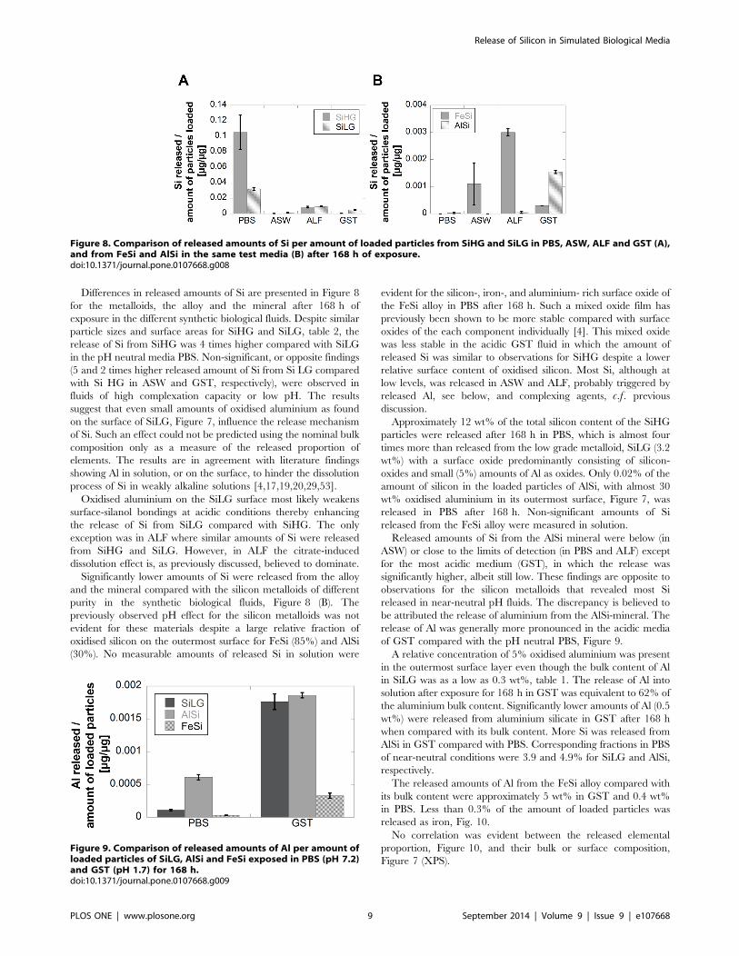
Differences in released amounts of Si are presented in Figure 8
for the metalloids, the alloy and the mineral after 168 h of
exposure in the different synthetic biological fluids. Despite similar
particle sizes and surface areas for SiHG and SiLG, table 2, the
release of Si from SiHG was 4 times higher compared with SiLG
in the pH neutral media PBS. Non-significant, or opposite findings
(5 and 2 times higher released amount of Si from Si LG compared
with Si HG in ASW and GST, respectively), were observed in
fluids of high complexation capacity or low pH. The results
suggest that even small amounts of oxidised aluminium as found
on the surface of SiLG, Figure 7, influence the release mechanism
of Si. Such an effect could not be predicted using the nominal bulk
composition only as a measure of the released proportion of
elements. The results are in agreement with literature findings
showing Al in solution, or on the surface, to hinder the dissolution
process of Si in weakly alkaline solutions [4,17,19,20,29,53].
Oxidised aluminium on the SiLG surface most likely weakens
surface-silanol bondings at acidic conditions thereby enhancing
the release of Si from SiLG compared with SiHG. The only
exception was in ALF where similar amounts of Si were released
from SiHG and SiLG. However, in ALF the citrate-induced
dissolution effect is, as previously discussed, believed to dominate.
Significantly lower amounts of Si were released from the alloy
and the mineral compared with the silicon metalloids of different
purity in the synthetic biological fluids, Figure 8 (B). The
previously observed pH effect for the silicon metalloids was not
evident for these materials despite a large relative fraction of
oxidised silicon on the outermost surface for FeSi (85%) and AlSi
(30%). No measurable amounts of released Si in solution were
evident for the silicon-, iron-, and aluminium- rich surface oxide of
the FeSi alloy in PBS after 168 h. Such a mixed oxide film has
previously been shown to be more stable compared with surface
oxides of the each component individually [4]. This mixed oxide
was less stable in the acidic GST fluid in which the amount of
released Si was similar to observations for SiHG despite a lower
relative surface content of oxidised silicon. Most Si, although at
low levels, was released in ASW and ALF, probably triggered by
released Al, see below, and complexing agents, c.f. previous
discussion.
Approximately 12 wt% of the total silicon content of the SiHG
particles were released after 168 h in PBS, which is almost four
times more than released from the low grade metalloid, SiLG (3.2
wt%) with a surface oxide predominantly consisting of silicon-
oxides and small (5%) amounts of Al as oxides. Only 0.02% of the
amount of silicon in the loaded particles of AlSi, with almost 30
wt% oxidised aluminium in its outermost surface, Figure 7, was
released in PBS after 168 h. Non-significant amounts of Si
released from the FeSi alloy were measured in solution.
Released amounts of Si from the AlSi mineral were below (in
ASW) or close to the limits of detection (in PBS and ALF) except
for the most acidic medium (GST), in which the release was
significantly higher, albeit still low. These findings are opposite to
observations for the silicon metalloids that revealed most Si
released in near-neutral pH fluids. The discrepancy is believed to
be attributed the release of aluminium from the AlSi-mineral. The
release of Al was generally more pronounced in the acidic media
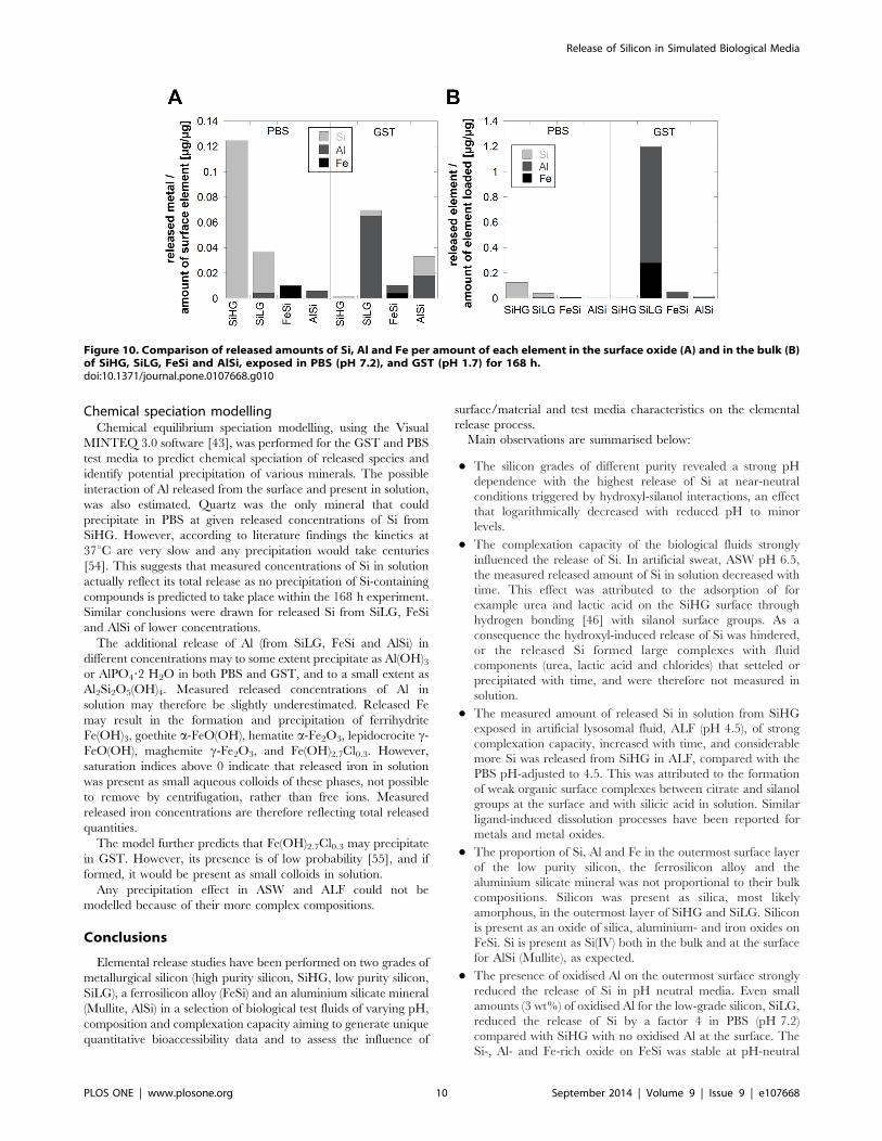
of GST compared with the pH neutral PBS, Figure 9.
A relative concentration of 5% oxidised aluminium was present
in the outermost surface layer even though the bulk content of Al
in SiLG was as a low as 0.3 wt%, table 1. The release of Al into
solution after exposure for 168 h in GST was equivalent to 62% of
the aluminium bulk content. Significantly lower amounts of Al (0.5
wt%) were released from aluminium silicate in GST after 168 h
when compared with its bulk content. More Si was released from
AlSi in GST compared with PBS. Corresponding fractions in PBS
of near-neutral conditions were 3.9 and 4.9% for SiLG and AlSi,
respectively.
The released amounts of Al from the FeSi alloy compared with
its bulk content were approximately 5 wt% in GST and 0.4 wt%
in PBS. Less than 0.3% of the amount of loaded particles was
released as iron, Fig. 10.
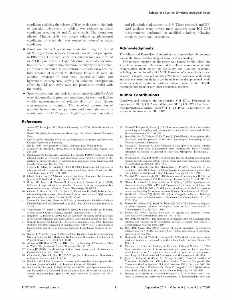
No correlation was evident between the released elemental
proportion, Figure 10, and their bulk or surface composition,
Figure 7 (XPS).
Figure 8. Comparison of released amounts of Si per amount of loaded particles from SiHG and SiLG in PBS, ASW, ALF and GST (A),and from FeSi and AlSi in the same test media (B) after 168 h of exposure.doi:10.1371/journal.pone.0107668.g008
Figure 9. Comparison of released amounts of Al per amount ofloaded particles of SiLG, AlSi and FeSi exposed in PBS (pH 7.2)and GST (pH 1.7) for 168 h.doi:10.1371/journal.pone.0107668.g009
Release of Silicon in Simulated Biological Media
PLOS ONE | www.plosone.org 9 September 2014 | Volume 9 | Issue 9 | e107668
Chemical speciation modellingChemical equilibrium speciation modelling, using the Visual
MINTEQ 3.0 software [43], was performed for the GST and PBS
test media to predict chemical speciation of released species and
identify potential precipitation of various minerals. The possible
interaction of Al released from the surface and present in solution,
was also estimated. Quartz was the only mineral that could
precipitate in PBS at given released concentrations of Si from
SiHG. However, according to literature findings the kinetics at
37uC are very slow and any precipitation would take centuries
[54]. This suggests that measured concentrations of Si in solution
actually reflect its total release as no precipitation of Si-containing
compounds is predicted to take place within the 168 h experiment.
Similar conclusions were drawn for released Si from SiLG, FeSi
and AlSi of lower concentrations.
The additional release of Al (from SiLG, FeSi and AlSi) in
different concentrations may to some extent precipitate as Al(OH)3or AlPO4?2 H2O in both PBS and GST, and to a small extent as
Al2Si2O5(OH)4. Measured released concentrations of Al in
solution may therefore be slightly underestimated. Released Fe
may result in the formation and precipitation of ferrihydrite
Fe(OH)3, goethite a-FeO(OH), hematite a-Fe2O3, lepidocrocite c-FeO(OH), maghemite c-Fe2O3, and Fe(OH)2.7Cl0.3. However,
saturation indices above 0 indicate that released iron in solution
was present as small aqueous colloids of these phases, not possible
to remove by centrifugation, rather than free ions. Measured
released iron concentrations are therefore reflecting total released
quantities.
The model further predicts that Fe(OH)2.7Cl0.3 may precipitate
in GST. However, its presence is of low probability [55], and if
formed, it would be present as small colloids in solution.
Any precipitation effect in ASW and ALF could not be
modelled because of their more complex compositions.
Conclusions
Elemental release studies have been performed on two grades of
metallurgical silicon (high purity silicon, SiHG, low purity silicon,
SiLG), a ferrosilicon alloy (FeSi) and an aluminium silicate mineral
(Mullite, AlSi) in a selection of biological test fluids of varying pH,
composition and complexation capacity aiming to generate unique
quantitative bioaccessibility data and to assess the influence of
surface/material and test media characteristics on the elemental
release process.
Main observations are summarised below:
N The silicon grades of different purity revealed a strong pH
dependence with the highest release of Si at near-neutral
conditions triggered by hydroxyl-silanol interactions, an effect
that logarithmically decreased with reduced pH to minor
levels.
N The complexation capacity of the biological fluids strongly
influenced the release of Si. In artificial sweat, ASW pH 6.5,
the measured released amount of Si in solution decreased with
time. This effect was attributed to the adsorption of for
example urea and lactic acid on the SiHG surface through
hydrogen bonding [46] with silanol surface groups. As a
consequence the hydroxyl-induced release of Si was hindered,
or the released Si formed large complexes with fluid
components (urea, lactic acid and chlorides) that setteled or
precipitated with time, and were therefore not measured in
solution.
N The measured amount of released Si in solution from SiHG
exposed in artificial lysosomal fluid, ALF (pH 4.5), of strong
complexation capacity, increased with time, and considerable
more Si was released from SiHG in ALF, compared with the
PBS pH-adjusted to 4.5. This was attributed to the formation
of weak organic surface complexes between citrate and silanol
groups at the surface and with silicic acid in solution. Similar
ligand-induced dissolution processes have been reported for
metals and metal oxides.
N The proportion of Si, Al and Fe in the outermost surface layer
of the low purity silicon, the ferrosilicon alloy and the
aluminium silicate mineral was not proportional to their bulk
compositions. Silicon was present as silica, most likely
amorphous, in the outermost layer of SiHG and SiLG. Silicon
is present as an oxide of silica, aluminium- and iron oxides on
FeSi. Si is present as Si(IV) both in the bulk and at the surface
for AlSi (Mullite), as expected.
N The presence of oxidised Al on the outermost surface strongly
reduced the release of Si in pH neutral media. Even small
amounts (3 wt%) of oxidised Al for the low-grade silicon, SiLG,
reduced the release of Si by a factor 4 in PBS (pH 7.2)
compared with SiHG with no oxidised Al at the surface. The
Si-, Al- and Fe-rich oxide on FeSi was stable at pH-neutral
Figure 10. Comparison of released amounts of Si, Al and Fe per amount of each element in the surface oxide (A) and in the bulk (B)of SiHG, SiLG, FeSi and AlSi, exposed in PBS (pH 7.2), and GST (pH 1.7) for 168 h.doi:10.1371/journal.pone.0107668.g010
Release of Silicon in Simulated Biological Media
PLOS ONE | www.plosone.org 10 September 2014 | Volume 9 | Issue 9 | e107668
conditions reducing the release of Si to levels close to the limit
of detection. However, its stability was reduced at acidic
conditions releasing Fe and Al as a result. The aluminium
silicate, Mullite, AlSi was poorly soluble at pH-neutral
conditions, an effect that was somewhat reduced at acidic
conditions.
N Based on chemical speciation modelling using the Visual
MINTEQ software released Si in solution did not precipitate
in PBS or GST, whereas some precipitation may occur for Al
as Al(OH)3 or AlPO4?2 H2O. Measured released concentra-
tions of Al in solution may therefore be slightly underestimat-
ed, whereas measured Si concentrations in solution reflect the
total amount of released Si. Released Fe and Al were, in
addition, predicted to form small colloids of oxides and
hydroxides consequently staying in solution. Precipitation
effects in ALF and ASW were not possible to predict with
the model.
N Specific operational methods for silicon analysis with GF-AAS
were elaborated and protocols established for each test fluid to
enable measurements of reliable data on total silicon
concentrations in solution. This involved optimisation of
graphite furnace step temperatures and times, the use of a
combination of Ca(NO3)2 and Mg(NO3)2 as matrix modifiers
and pH solution adjustment to 8–9. These protocols and GF-
AAS analyses were proven more accurate than ICP-OES
measurements performed on acidified solutions following
standard operational procedures.
Acknowledgments
The Silicon and Ferrosilicon Consortium are acknowledged for commis-
sioning the bioaccessibility study of silicon and silicon alloys.
The research reported in this article was funded by the silicon and
ferrosilicon consortium. The silicon and ferrosilicon consortium retain data
compensation rights under all regulatory and statutory programs,
including, but not limited to REACH. Possession of a copy of this article,
in whole or in part, does not constitute ‘‘legitimate possession’’ of the study
and does not create any rights to use the study or the data presented herein
for any chemical registration (such as, but not limited to the REACH
registration program) or any other commercial purpose.
Author Contributions
Conceived and designed the experiments: GH IOW. Performed the
experiments: GH TJ CS. Analyzed the data: GH TJ CS IOW. Contributed
reagents/materials/analysis tools: GH TJ CS IOW. Contributed to the
writing of the manuscript: GH IOW.
References
1. Atkins PW, Beran JA (1992) General Chemistry. New York: Scientific AmericanBooks.
2. Nesse WD (2009) Introduction to Mineralogy. New York: Oxford University
Press.
3. Kisa M (2007) Oxidation of Silicon and Germanium by Atomic and Molecular
Oxygen: University of Pittsburg.
4. Iler R (1979) The Chemistry of Silica. Hoboken: John Wiley & Sons.
5. Dempster PB, Ritchie PD (1952) Surface of Finely-Ground Silica. Nature 169:
538–539.
6. Pandurangi RS, Seehra MS, Razzaboni BL, Bolsaitis P (1990) Surface and bulk
infrared modes of crystalline and amorphous silica particles: a study of therelation of surface structure to cytotoxicity of respirable silica. Environmental
Health Perspectives 86: 327–336.
7. Fornero E, Belluso E, Capella S, Bellis D (2009) Environmental exposure to
asbestos and other inorganic fibres using animal lung model. Science of The
Total Environment 407: 1010–1018.
8. Choi I, Smith RW (1972) Kinetic study of dissolution of asbestos fibers in water.Journal of Colloid and Interface Science 40: 253–262.
9. Brown SC, Kamal M, Nasreen N, Baumuratov A, Sharma P, et al. (2007)Influence of shape, adhesion and simulated lung mechanics on amorphous silica
nanoparticle toxicity. Advanced Powder Technology 18: 69–79.
10. Clouter A, Brown D, Hohr D, Borm P, Donaldson K (2001) Inflammatory
effects of respirable quartz collected in workplaces versus standard DQ12 quartz:particle surface correlates. Toxicol Sci 63: 90–98.
11. Larson RR, Story SG, Hegmann KT (2010) Assessing the Solubility of SiliconDioxide Particles Using Simulated Lung Fluid. The Open Toxicology Journal 4:
51–55.
12. Vogelsberger W, Seidel A, Rudakoff G (1992) Solubility of silica gel in water.
Journal of the Chemical Society, Faraday Transactions 88: 473–476.
13. Bergstrom L, Bostedt E (1990) Surface chemistry of silicon nitride powders:
Electrokinetic behaviour and ESCA studies. Colloids and Surfaces 49: 183–197.
14. Borm P, Klaessig FC, Landry TD, Moudgil B, Pauluhn J, et al. (2006) Researchstrategies for safety evaluation of nanomaterials, Part V: Role of dissolution in
biological fate and effects of nanoscale particles. Toxicological Sciences 90: 23–
32.
15. Roelofs F, Vogelsberger W (2004) Dissolution Kinetics of Synthetic AmorphousSilica in Biological-Like Media and Its Theoretical Description. The Journal of
Physical Chemistry B 108: 11308–11316.
16. Alexander GB, Heston WM, Iler RK (1954) The Solubility of Amorphous Silica
in Water. The Journal of Physical Chemistry 58: 453–455.
17. Lewin JC (1961) The dissolution of silica from diatom walls. Geochimica et
Cosmochimica Acta 21: 182–198.
18. Okamoto G, Okura T, Goto K (1957) Properties of silica in water. Geochimica
et Cosmochimica Acta 12: 123–132.
19. Iler RK (1973) Effect of adsorbed alumina on the solubility of amorphous silicain water. Journal of Colloid and Interface Science 43: 399–408.
20. Bouallou M, Vielvoye L, El Shafei GMS, Stone WEE (1998) Dissolution of Silicaand Formation of a Dispersed Phase Induced at Low pH by the Association of
Soluble Aluminum Ionic Species with Solid Silica Gel. Langmuir 14: 1072–
1080.
21. Gerard F, Francois M, Ranger J (2002) Processes controlling silica concentrationin leaching and capillary soil solutions of an acidic brown forest soil (Rhone,
France). Geoderma 107: 197–226.
22. Dove PM, Han N, Wallace AF, De Yoreo JJ (2008) Kinetics of amorphous silicadissolution and the paradox of the silica polymorphs. Proceedings of the
National Academy of Sciences.
23. Tanaka M, Takahashi K (2003) Changes of silica species in sodium chloridesolution by fast atom bombardment mass spectrometry: Silicate complex
substituted by sodium ion. Journal of Trace and Microprobe Techniques 21:561–576.
24. Icenhower JP, Dove PM (2000) The dissolution kinetics of amorphous silica into
sodium chloride solutions: effects of temperature and ionic strength. Geochimicaet Cosmochimica Acta 64: 4193–4203.
25. Pak TM, Hauzenberger CA, Baumgartner LP (2003) Solubility of the
assemblage albite+K-feldspar+andalusite+quartz in supercritical aqueous chlo-ride solutions at 650uC and 2 kbar. Chemical Geology 200: 377–393.
26. Marshall WL, Warakomski JM (1980) Amorphous silica solubilities–II. Effect of
aqueous salt solutions at 25uC. Geochimica et Cosmochimica Acta 44: 915–924.
27. Ohman L-O, Nordin A, Iran Fattahpour S, Sjoberg S (1991) Equilibrium and
Structural Studies of Silicon(IV) and Aluminium(III) in Aqueous Solution. 28.
Formation of Soluble Silicic Acid–Ligand Complexes as Studied by Potentio-metric and Solubility Measurements. Acta Chemica Scandinavica 45: 335–341.
28. Welch SA, Ullman WJ (1993) The effect of organic acids on plagioclase
dissolution rates and stoichiometry. Geochimica et Cosmochimica Acta 57:2725–2736.
29. Bennett PC, Melcer ME, Siegel DI, Hassett JP (1988) The dissolution of quartz
in dilute aqueous solutions of organic acids at 25uC. Geochimica etCosmochimica Acta 52: 1521–1530.
30. Bennett PC (1991) Quartz dissolution in organic-rich aqueous systems.Geochimica et Cosmochimica Acta 55: 1781–1797.
31. Dove PM, Nix CJ (1997) The influence of the alkaline earth cations, magnesium,
calcium, and barium on the dissolution kinetics of quartz. Geochimica etCosmochimica Acta 61: 3329–3340.
32. Dove PM, Crerar DA (1990) Kinetics of quartz dissolution in electrolyte
solutions using a hydrothermal mixed flow reactor. Geochimica et Cosmochi-mica Acta 54: 955–969.
33. Herting G, Odnevall Wallinder I, Leygraf C (2007) Metal release from various
grades of stainless steel exposed to synthetic body fluids. Corrosion Science 49:103–111.
34. Midander K, Frutos Ad, Hedberg Y, Darrie G, Odnevall Wallinder I (2010)
Bioaccessibility studies of ferro-chromium alloy particles for a simulatedinhalation scenario: A comparative study with the pure metals and stainless
steel. Integrated Environmental Assessment and Management 6: 441–455.
35. Jiang T, Odnevall Wallinder I, Herting G (2012) Chemical Stability ofChromium Carbide and Chromium Nitride Powders Compared with
Chromium Metal in Synthetic Biological Solutions. ISRN Corrosion 2012: 10.
36. Midander K, Pan J, Wallinder IO, Heim K, Leygraf C (2007) Nickel releasefrom nickel particles in artificial sweat. Contact Dermatitis 56: 325–330.
37. Hedberg Y, Midander K, Odnevall Wallinder I (2010) Particles, sweat, and
tears: A comparative study on bioaccessibility of ferrochromium alloy and
Release of Silicon in Simulated Biological Media
PLOS ONE | www.plosone.org 11 September 2014 | Volume 9 | Issue 9 | e107668
stainless steel particles, the pure metals and their metal oxides, in simulated skin
and eye contact. Integrated Environmental Assessment and Management 6:456–468.
38. Stopford W, Turner J, Cappellini D, Brock T (2003) Bioaccessibility testing of
cobalt compounds. Journal of Environmental Monitoring 5: 675–680.39. de Meringo A, Morscheidt C, Thelohan S, Tiesler H (1994) in vitro assessment
of biodurability: acellular systems. Environ Health Perspect 102.40. Midander K, Cronholm P, Karlsson HL, Elihn K, Moller L, et al. (2009) Surface
Characteristics, Copper Release, and Toxicity of Nano- and Micrometer-Sized
Copper and Copper(II) Oxide Particles: A Cross-Disciplinary Study. Small 5:389–399.
41. PerkinElmerPrecisely (2003) WinLab 32 for AA 6.2.0.0079.42. Butcher DJ, Sneddon J (1998) A Practical Guide to Graphite Furnace Atomic
Absorption Spectroscopy; Winefordner JD, editor. New York: John Wiley &Sons, Inc.
43. Gustafsson JP (2013) Visual MINTEQ 3.0.
44. Smith RM, Martell AE, Standards NIo, Technology (1998) NIST criticallyselected stability constants of metal complexes database: National Institute of
Standards & Technology.45. Baumann H (1955) Solubility of silica in water. Beitr Silikose-Forsch: 45–71.
46. Ledoux RL, White JL (1966) Infrared studies of hydrogen bonding interaction
between kaolinite surfaces and intercalated potassium acetate, hydrazine,formamide, and urea. Journal of Colloid and Interface Science 21: 127–152.
47. Zhang Y, Kallay N, Matijevic E (1985) Interaction of metal hydrous oxides withchelating agents. 7. Hematite-oxalic acid and -citric acid systems. Langmuir 1:
201–206.48. Amirbahman A, Sigg L, von GU (1997) Reductive dissolution of Fe(III)
(hydr)oxides by cysteine: kinetics and mechanism. J Colloid Interface Sci 194:
194–206.
49. Dos Santos Afonso M, Morando PJ, Blesa MA, Banwart S, Stumm W (1990)
The reductive dissolution of iron oxides by ascorbate: the role of carboxylate
anions in accelerating reductive dissolution. J Colloid Interface Sci 138: 74–82.
50. Carbonaro RF, Gray BN, Whitehead CF, Stone AT (2008) Carboxylate-
containing chelating agent interactions with amorphous chromium hydroxide:
Adsorption and dissolution. Geochimica et Cosmochimica Acta 72: 3241–3257.
51. Hedberg Y, Hedberg J, Liu Y, Wallinder I (2011) Complexation- and ligand-
induced metal release from 316L particles: importance of particle size and
crystallographic structure. BioMetals 24: 1099–1114.
52. Prazeres DMF (2011) Plasmid Biopharmaceuticals: Basics, Applications, and
Manufacturing. Hoboken: John Wiley & Sons, Inc.
53. Denny JJ, Robson WD, Irwin DA (1939) The Prevention of Silicosis by Metallic
Aluminum II. The Canadian Medical Association Journal 40: 213–228.
54. Rimstidt JD, Barnes HL (1980) The kinetics of silica-water reactions.
Geochimica et Cosmochimica Acta 44: 1683–1699.
55. Schwertmann U, Cornell RM, Rochelle M (2000) Iron Oxides in the
Laboratory: Precipitation and Characterization. Weinheim: Wiley-VCH Verlag
GmbH.
56. Norlin A, Pan J, Leygraf C (2002) Investigation of interfacial capacitance of Pt,
Ti and TiN coated electrodes by electrochemical impedance spectroscopy.
Biomolecular Engineering 19: 67–71.
57. EN1811 (1998) Reference test method for release of nickel from products
intended to come into direct and prolonged contact with the skin. European
Standard, EN1811: European Committee for Standardization.
58. de Meringo A, Morscheidt C, Thelohan S, Tiesler H (1994) In vitro assessment
of biodurability: acellular systems. Environ Health Perspect 102 47–53.
Release of Silicon in Simulated Biological Media
PLOS ONE | www.plosone.org 12 September 2014 | Volume 9 | Issue 9 | e107668