relazioni convegno - Società Italiana di Patologia Aviare
88
1 RELAZIONI CONVEGNO 1 H. D. Chapman Department of Poultry Science, University of Arkansas, Fayetteville, AR, 72701, USA CONTROLLING COCCIDIOSIS IN BROILERS: EXPERIENCE FROM THE USA. 2 M. Tamba Istituto Zooprofilattico Sperimentale della lombardia e dell’Emili Romagna, Sez. di Bologna COCCIDIOSI DEL POLLO – LA SITUAZIONE ITALIANA 3 G. Tosi Istituto Zooprofilattico Sperimentale della lombardia e dell’Emili Romagna, Sez. di Forlì HISTOMONIASI DEL TACCHINO : ESPERIENZE NAZIONALI 4 F. M. Tomley & M. W. Shirley Institute for Animal Health, Compton, Newbury, Berkshire, UK, RG20 7NN COCCIDIOSIS VACCINES: PAST, PRESENT AND FUTURE 5 G. Cringoli Facoltà di Medicina Veterinaria, Università “Federico II” di Napoli LA DIAGNOSTICA COPROLOGICA IN PARASSITOLOGIA: QUALE NOVITA’? 6 G. Pampiglione Università di Bari GLI ECTOPARASSITI DEL COMPARTO AVICOLO ELENCO LAVORI 1 ANTIMICROBIAL SUSCEPTIBILITIY OF SALMONELLA SPP. STRAINS ISOLATED FROM LAYER HENS IN CAMPANIA REGION FROM 2000 TO 2003 Ludovico Dipineto, Claudia Scarpetta, Mariarosaria Calabria, Mariangela Sensale, Antonio Baiano, Lucia Francesca Menna, Alessandro Fioretti 2 PREVALENCE OF CAMPYLOBACTER JEJUNI IN POULTRY BREEDER FLOCKS Lucia Francesca Menna, Gianluca Matteoli, Marzia Fontanella, Alessandra Cuomo, Antonio De Paola, Tiziana Pepe, Isolina Di Marco , Ludovico Dipineto 3 SANCASSANIA BERLESEI (MICHAEL, 1903): AN OPPORTUNISTIC MITE INFESTING LITTERS IN POULTRY FARMS CAUSING DERMATITIS IN HUMANS AND ANIMALS. Mario Principato, Federica Lisi, Iolanda Moretta, Nada Samra, Francesco Puccetti 4 THE ALTERATIONS OF PLUMAGE OF PARASITIC ORIGIN Mario Principato, Federica Lisi, Iolanda Moretta, Nada Samra, Francesco Puccetti 5 SEROLOGICAL EVIDENCES SHOWING THE INVOLVEMENT OF FREE- LIVING PHEASANTS IN THE INFLUENZA ECOLOGY (NORTHERN ITALY, 1995-2002) Maria Alessandra De Marco, Laura Campitelli, Mauro Delogu, Elisabetta Raffini, Emanuela Foni, Livia di Trani, Michele Scaffidi, Isabella Donatelli 6 FIELD TRIALS WITH THE USE OF A LIVE ATTENUATED TEMPERATURE- SENSITIVE VACCINE FOR THE CONTROL OF MYCOPLASMA GALLISEPTICUM INFECTION IN MEAT-TYPE TURKEYS Enrico Alessandri, Paola Massi, Francesca Paganelli, Francesco Prandini, Mario Saita
-
Upload
khangminh22 -
Category
Documents
-
view
7 -
download
0
Transcript of relazioni convegno - Società Italiana di Patologia Aviare

1
RELAZIONI CONVEGNO
1 H. D. Chapman Department of Poultry Science, University of Arkansas, Fayetteville, AR, 72701, USA CONTROLLING COCCIDIOSIS IN BROILERS: EXPERIENCE FROM THE USA.
2 M. Tamba Istituto Zooprofilattico Sperimentale della lombardia e dell’Emili Romagna, Sez. di Bologna COCCIDIOSI DEL POLLO – LA SITUAZIONE ITALIANA
3 G. Tosi Istituto Zooprofilattico Sperimentale della lombardia e dell’Emili Romagna, Sez. di Forlì HISTOMONIASI DEL TACCHINO : ESPERIENZE NAZIONALI
4 F. M. Tomley & M. W. Shirley Institute for Animal Health, Compton, Newbury, Berkshire, UK, RG20 7NN COCCIDIOSIS VACCINES: PAST, PRESENT AND FUTURE
5 G. Cringoli Facoltà di Medicina Veterinaria, Università “Federico II” di Napoli LA DIAGNOSTICA COPROLOGICA IN PARASSITOLOGIA: QUALE NOVITA’?
6 G. Pampiglione Università di Bari GLI ECTOPARASSITI DEL COMPARTO AVICOLO
ELENCO LAVORI
1 ANTIMICROBIAL SUSCEPTIBILITIY OF SALMONELLA SPP. STRAINS ISOLATED FROM LAYER HENS IN CAMPANIA REGION FROM 2000 TO 2003
Ludovico Dipineto, Claudia Scarpetta, Mariarosaria Calabria, Mariangela Sensale, Antonio Baiano, Lucia Francesca Menna, Alessandro Fioretti
2 PREVALENCE OF CAMPYLOBACTER JEJUNI IN POULTRY BREEDER FLOCKS
Lucia Francesca Menna, Gianluca Matteoli, Marzia Fontanella, Alessandra Cuomo, Antonio De Paola, Tiziana Pepe, Isolina Di Marco, Ludovico Dipineto
3 SANCASSANIA BERLESEI (MICHAEL, 1903): AN OPPORTUNISTIC MITE INFESTING LITTERS IN POULTRY FARMS CAUSING DERMATITIS IN HUMANS AND ANIMALS.
Mario Principato, Federica Lisi, Iolanda Moretta, Nada Samra, Francesco Puccetti
4 THE ALTERATIONS OF PLUMAGE OF PARASITIC ORIGIN Mario Principato, Federica Lisi, Iolanda Moretta, Nada Samra, Francesco Puccetti
5 SEROLOGICAL EVIDENCES SHOWING THE INVOLVEMENT OF FREE-LIVING PHEASANTS IN THE INFLUENZA ECOLOGY (NORTHERN ITALY, 1995-2002)
Maria Alessandra De Marco, Laura Campitelli, Mauro Delogu, Elisabetta Raffini, Emanuela Foni, Livia di Trani, Michele Scaffidi, Isabella Donatelli
6 FIELD TRIALS WITH THE USE OF A LIVE ATTENUATED TEMPERATURE-SENSITIVE VACCINE FOR THE CONTROL OF MYCOPLASMA GALLISEPTICUM INFECTION IN MEAT-TYPE TURKEYS
Enrico Alessandri, Paola Massi, Francesca Paganelli, Francesco Prandini, Mario Saita
2
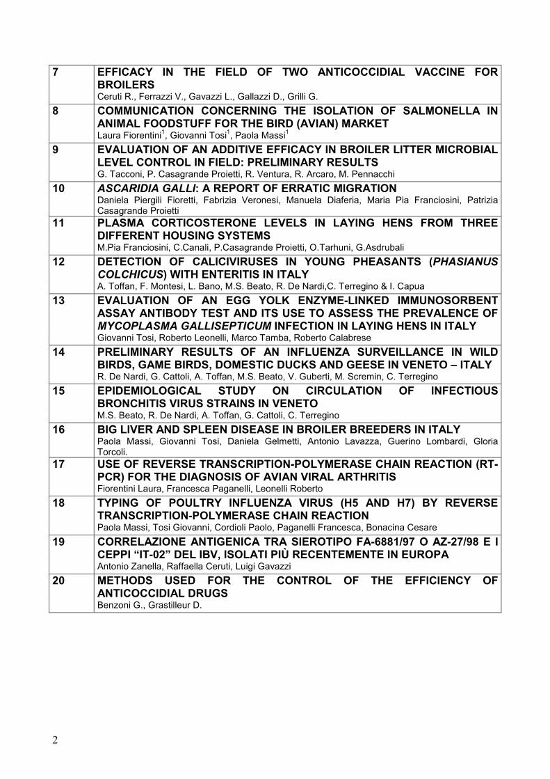
7 EFFICACY IN THE FIELD OF TWO ANTICOCCIDIAL VACCINE FOR BROILERS
Ceruti R., Ferrazzi V., Gavazzi L., Gallazzi D., Grilli G.
8 COMMUNICATION CONCERNING THE ISOLATION OF SALMONELLA IN ANIMAL FOODSTUFF FOR THE BIRD (AVIAN) MARKET
Laura Fiorentini1, Giovanni Tosi1, Paola Massi1
9 EVALUATION OF AN ADDITIVE EFFICACY IN BROILER LITTER MICROBIAL LEVEL CONTROL IN FIELD: PRELIMINARY RESULTS
G. Tacconi, P. Casagrande Proietti, R. Ventura, R. Arcaro, M. Pennacchi
10 ASCARIDIA GALLI: A REPORT OF ERRATIC MIGRATION Daniela Piergili Fioretti, Fabrizia Veronesi, Manuela Diaferia, Maria Pia Franciosini, Patrizia
Casagrande Proietti
11 PLASMA CORTICOSTERONE LEVELS IN LAYING HENS FROM THREE DIFFERENT HOUSING SYSTEMS
M.Pia Franciosini, C.Canali, P.Casagrande Proietti, O.Tarhuni, G.Asdrubali
12 DETECTION OF CALICIVIRUSES IN YOUNG PHEASANTS (PHASIANUS COLCHICUS) WITH ENTERITIS IN ITALY
A. Toffan, F. Montesi, L. Bano, M.S. Beato, R. De Nardi,C. Terregino & I. Capua
13 EVALUATION OF AN EGG YOLK ENZYME-LINKED IMMUNOSORBENT ASSAY ANTIBODY TEST AND ITS USE TO ASSESS THE PREVALENCE OF MYCOPLASMA GALLISEPTICUM INFECTION IN LAYING HENS IN ITALY
Giovanni Tosi, Roberto Leonelli, Marco Tamba, Roberto Calabrese
14 PRELIMINARY RESULTS OF AN INFLUENZA SURVEILLANCE IN WILD BIRDS, GAME BIRDS, DOMESTIC DUCKS AND GEESE IN VENETO – ITALY
R. De Nardi, G. Cattoli, A. Toffan, M.S. Beato, V. Guberti, M. Scremin, C. Terregino
15 EPIDEMIOLOGICAL STUDY ON CIRCULATION OF INFECTIOUS BRONCHITIS VIRUS STRAINS IN VENETO
M.S. Beato, R. De Nardi, A. Toffan, G. Cattoli, C. Terregino
16 BIG LIVER AND SPLEEN DISEASE IN BROILER BREEDERS IN ITALY Paola Massi, Giovanni Tosi, Daniela Gelmetti, Antonio Lavazza, Guerino Lombardi, Gloria
Torcoli.
17 USE OF REVERSE TRANSCRIPTION-POLYMERASE CHAIN REACTION (RT-PCR) FOR THE DIAGNOSIS OF AVIAN VIRAL ARTHRITIS
Fiorentini Laura, Francesca Paganelli, Leonelli Roberto
18 TYPING OF POULTRY INFLUENZA VIRUS (H5 AND H7) BY REVERSE TRANSCRIPTION-POLYMERASE CHAIN REACTION
Paola Massi, Tosi Giovanni, Cordioli Paolo, Paganelli Francesca, Bonacina Cesare
19 CORRELAZIONE ANTIGENICA TRA SIEROTIPO FA-6881/97 O AZ-27/98 E I CEPPI “IT-02” DEL IBV, ISOLATI PIÙ RECENTEMENTE IN EUROPA
Antonio Zanella, Raffaella Ceruti, Luigi Gavazzi
20 METHODS USED FOR THE CONTROL OF THE EFFICIENCY OF ANTICOCCIDIAL DRUGS
Benzoni G., Grastilleur D.

3
RELAZIONI STATO SANITARIO
INFLUENZA AVIARIA IN ITALIA 1997-2004: EVOLUZIONE DELLA SITUAZIONE EPIDEMIOLOGICA E STRATEGIE DI CONTROLLO
C. Terregino, M. Dalla Pozza L. Bonfanti, S. Marangon & I. Capua
PRESENTAZIONE CASI CLINICI
1 CASI DI ROTTURA EPATICA IN GIOVANI FARAONE
Rampin T., Manarolla G., Recordati C., Longoni C., Sironi G., Sartorelli P., Spagnolo V.
2 AVVERSA REAZIONE VACCINALE IN TACCHINI DA CARNE SOTTOPOSTI A PROFILASSI ANTINFLUENZALE
Alessandri E., Saita M., Rampin T. ,Manarolla G.
3 IL FENOMENO DELLA RESISTENZA AGLI INSETTICIDI NELLE POPOLAZIONI DI MOSCHE CON PARTICOLARE RIFERIMENTO ALL’USO DEI PRODOTTI LARVICIDI IN AVICOLTURA
Pampiglione G , E. Rossi, E. Gnassi, P. Tampieri, P. Massi
4 MICOBATTERIOSI IN CARDELLINI MUTATI Manarolla G., Ferrazzi V., Gallazzi D.
5 CONTROLLO DI MYCOPLASMA GALLISEPTICUM IN UN GRUPPO DI ALLEVAMENTI DI TACCHINI DA CARNE ATTRAVERSO L’APPLICAZIONE DI UN VACCINO INATTIVATO
Alessandri E., Saita M., Acco P.
6 ESOFAGITE ULCERATIVA DA STREPTOCARA INCOGNITA IN ANATRE MUTE (CAIRINA MOSCHATA DOMESTICUS): PRIMA SEGNALAZIONE IN ITALIA
L. Bano , A. Natale , M. Vascellari , D. Comin , F. Agnoletti , F. Mutinelli
7 ARCHIVIO BIBLIOGRAFICO DI ECTOPARASSITI G. Pampiglione, P. Massi
8 IDENTIFICAZIONE E TIPIZZAZIONE DEL PNEUMOVIRUS AVIARE TRAMITE TECNICHE MOLECOLARI Francesca Paganelli, Laura Fiorentini, Giovanni Tosi
9 EMANGIOMI EPATICI IN FARAONE Bolognesi P.G., Catelli E., Cecchnato M., De Matteo P., Frasnelli M., Raffini E., Marzadori F.,
Thiene G.
4
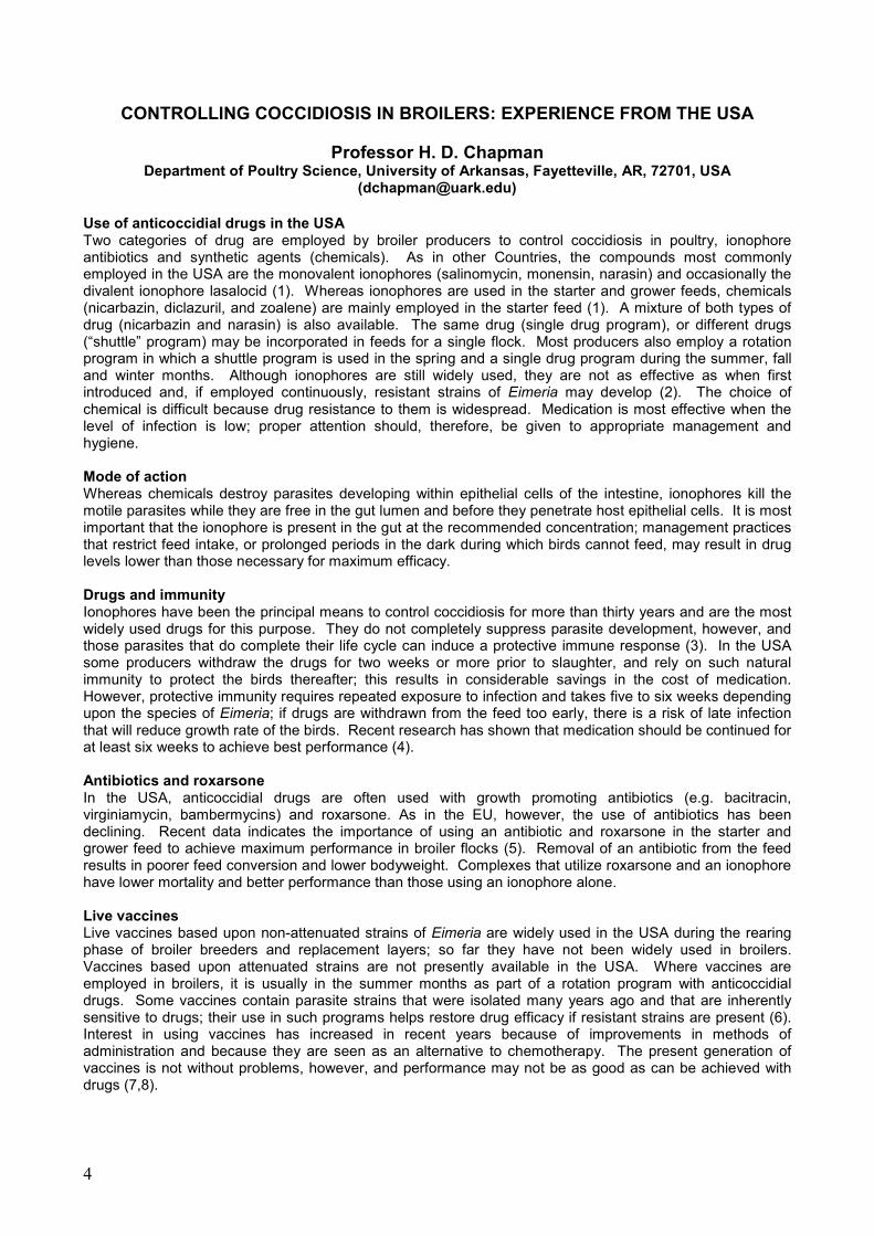
CONTROLLING COCCIDIOSIS IN BROILERS: EXPERIENCE FROM THE USA
Professor H. D. Chapman Department of Poultry Science, University of Arkansas, Fayetteville, AR, 72701, USA
([email protected]) Use of anticoccidial drugs in the USA Two categories of drug are employed by broiler producers to control coccidiosis in poultry, ionophore antibiotics and synthetic agents (chemicals). As in other Countries, the compounds most commonly employed in the USA are the monovalent ionophores (salinomycin, monensin, narasin) and occasionally the divalent ionophore lasalocid (1). Whereas ionophores are used in the starter and grower feeds, chemicals (nicarbazin, diclazuril, and zoalene) are mainly employed in the starter feed (1). A mixture of both types of drug (nicarbazin and narasin) is also available. The same drug (single drug program), or different drugs (“shuttle” program) may be incorporated in feeds for a single flock. Most producers also employ a rotation program in which a shuttle program is used in the spring and a single drug program during the summer, fall and winter months. Although ionophores are still widely used, they are not as effective as when first introduced and, if employed continuously, resistant strains of Eimeria may develop (2). The choice of chemical is difficult because drug resistance to them is widespread. Medication is most effective when the level of infection is low; proper attention should, therefore, be given to appropriate management and hygiene. Mode of action Whereas chemicals destroy parasites developing within epithelial cells of the intestine, ionophores kill the motile parasites while they are free in the gut lumen and before they penetrate host epithelial cells. It is most important that the ionophore is present in the gut at the recommended concentration; management practices that restrict feed intake, or prolonged periods in the dark during which birds cannot feed, may result in drug levels lower than those necessary for maximum efficacy. Drugs and immunity Ionophores have been the principal means to control coccidiosis for more than thirty years and are the most widely used drugs for this purpose. They do not completely suppress parasite development, however, and those parasites that do complete their life cycle can induce a protective immune response (3). In the USA some producers withdraw the drugs for two weeks or more prior to slaughter, and rely on such natural immunity to protect the birds thereafter; this results in considerable savings in the cost of medication. However, protective immunity requires repeated exposure to infection and takes five to six weeks depending upon the species of Eimeria; if drugs are withdrawn from the feed too early, there is a risk of late infection that will reduce growth rate of the birds. Recent research has shown that medication should be continued for at least six weeks to achieve best performance (4). Antibiotics and roxarsone In the USA, anticoccidial drugs are often used with growth promoting antibiotics (e.g. bacitracin, virginiamycin, bambermycins) and roxarsone. As in the EU, however, the use of antibiotics has been declining. Recent data indicates the importance of using an antibiotic and roxarsone in the starter and grower feed to achieve maximum performance in broiler flocks (5). Removal of an antibiotic from the feed results in poorer feed conversion and lower bodyweight. Complexes that utilize roxarsone and an ionophore have lower mortality and better performance than those using an ionophore alone. Live vaccines Live vaccines based upon non-attenuated strains of Eimeria are widely used in the USA during the rearing phase of broiler breeders and replacement layers; so far they have not been widely used in broilers. Vaccines based upon attenuated strains are not presently available in the USA. Where vaccines are employed in broilers, it is usually in the summer months as part of a rotation program with anticoccidial drugs. Some vaccines contain parasite strains that were isolated many years ago and that are inherently sensitive to drugs; their use in such programs helps restore drug efficacy if resistant strains are present (6). Interest in using vaccines has increased in recent years because of improvements in methods of administration and because they are seen as an alternative to chemotherapy. The present generation of vaccines is not without problems, however, and performance may not be as good as can be achieved with drugs (7,8).
5
References 1. Chapman, H. D. (2001). Use of anticoccidial drugs in broiler chickens in the USA: analysis for the years
1995-1999. Poultry Sci. 80: 572-580. 2. Chapman, H. D. (1997). Biochemical, genetic, and applied aspects of drug resistance in Eimeria
parasites of the fowl. Avian Pathol. 26: 221-244. 3. Chapman, H. D. (1999). Anticoccidial drugs and their effects upon the development of immunity to
Eimeria infections in poultry. Avian Pathol. 28: 521-535. 4. Chapman, H. D., Matsler, P. & LaVorgna, M.W. (2004). The effects of salinomycin and roxarsone upon the
performance of broilers when included in the feed for four, five, or six weeks and infected with Eimeria species during the starter or grower phase of production. Poultry Sci. 83:761-764.
5. Chapman, H. D. & Johnson, Z.B. (2002). Use of antibiotics and roxarsone in broiler chickens
in the USA: analysis for the years 1995-2000. Poultry Sci. 81: 356-364. 6. Chapman, H. D. (1994). Sensitivity of field isolates of Eimeria to monensin following the use of a
coccidiosis vaccine in broiler chickens. Poultry Sci. 73: 476-478. 7. Chapman, H. D., Cherry, T.E., Danforth, H. D., Richards, G., Shirley, M.W., & Williams, R.B. (2002).
Sustainable coccidiosis control in poultry production: the role of live vaccines. International Journal for Parasitology 32: 617-629.
8. Chapman, H. D. (2000). Practical use of vaccines for the control of coccidiosis in the chicken. W’lds Poultry Sci. J. 56: 7-20.

6
COCCIDIOSIS VACCINES: PAST, PRESENT AND FUTURE.
Fiona M Tomley and Martin W. Shirley Institute for Animal Health, Compton, Newbury, Berkshire, UK, RG20 7NN.
([email protected]) Introduction Coccidiosis, an intestinal disease of intensively reared livestock, is caused by parasites of the genus Eimeria. Control of coccidiosis in poultry is absolutely essential because rearing large numbers of birds in contact with their faeces in moist, warm conditions favours the transmission, replication and rapid build-up of parasites in the litter. Without adequate control, outbreaks of severe coccidiosis are inevitable and devastating. Economically, the most important Eimeria species are the seven that infect chickens: Eimeria acervulina, Eimeria brunetti, Eimeria maxima, Eimeria mitis, Eimeria praecox, Eimeria necatrix and Eimeria tenella. Three species, E. acervulina, E. maxima and E. tenella are most frequently diagnosed in coccidiosis of intensively reared poultry so their control is usually top priority, especially in broilers. For the past ~60 years coccidiosis has been controlled, for the most part, by in-feed prophylactic medication with a range of chemical and antibiotic (ionophorous) anti-coccidial drugs. Live vaccines, based on the established principle that chickens infected with small numbers of Eimeria parasites quickly develop protective immune responses against subsequent challenge with the same species of Eimeria, were introduced in the 1950’s. However, vaccination remained a very minor method of control until the late 1980’s when safer, live-attenuated vaccines were introduced and rapidly taken up throughout Europe and other parts of the world by the egg-laying sector of the industry. Coccidiosis control in the intensive broiler sector remained almost entirely dependent on drugs, due in part to the relatively high cost and limited availability of vaccines and in part to the cautiousness of producers in switching intensive operations over to non-drug based coccidiosis control. However, over the past 15 years anti-coccidial drugs have faced enormous pressures, especially within Europe where legislations on drug re-registration and on the administration of in-feed substances to poultry have removed several drugs from use. In addition, the inexorable rise of parasite drug resistance has rendered several compounds useless and compromised the efficiency of even the most potent ionophores. With no new drugs on the horizon and the withdrawal of many large pharmaceutical companies from anti-coccidial research and development, the role of vaccines has recently increased dramatically and a number of new vaccine products are coming to the marketplace, many of which are designed specifically for broilers. In the beginning – live virulent vaccines The development of live coccidiosis vaccines was based on the understanding that to protect against disease there is no need to totally prevent infection, but only to limit the numbers of parasites to which naïve chicks are exposed. Vaccines based on the administration of small numbers of sporulated oocysts of fully virulent parasites were developed ~50 years ago and products such as Coccivax, introduced in 1952 (Schering-Plough Animal Health) and Immucox, introduced in 1984 (Vetech Laboratories, Canada) are still widely available in formulations designed for both layers and broilers. All of these contain drug sensitive parasites. Crucial to the success of these vaccines is effective, uniform delivery, because uneven uptake of vaccine within a flock leads to outbreaks of disease when vaccinal oocysts are replicated and subsequently ingested by birds that were not immunised in the initial vaccination. Without careful administration, virulent vaccines can cause disease and therapy with anticoccidial drugs may be required for a period after vaccination. For this reason, despite their relatively low cost, live virulent vaccines were not widely taken up by the intensive poultry industry for many years. However, by the late 1980’s when problems with drug resistance were becoming rife, interest in vaccination as a viable alternative to anti-coccidial drugs was renewed. This was partly fuelled by the introduction of the safer, live-attenuated vaccines but also helped by the development of new delivery methods that achieved better vaccine uptake across flocks, thus making virulent vaccines a lower risk for causing disease. The second generation – live attenuated vaccines The risk of disease associated with live vaccination can be eliminated by the use of attenuated, rather than fully virulent, organisms. The first live-attenated vaccine, Paracox 8 (Schering Plough Animal Health), which contains attenuated parasites of all seven avian Eimeria species was introduced in 1989 and was rapidly adopted by the egg-laying and broiler-breeder sectors within Europe. Livacox Q (Biopharm), which contains four species, was introduced in 1992 and has also been very successful in many countries throughout the world, although it is not registered for use in Europe. More recently different formulations of these two products, containing fewer species of parasites, have been developed for use in the intensive broiler market and although figures of their usage are not generally available, they appear to be making a positive impact.

7
The success of these first two live-attenuated vaccines has stimulated several small companies to develop and manufacture similar vaccines, most of which are aimed at geographically defined local markets. Examples include Eimeriavax (Australia BioProperties Ltd, Australia) and Gel-Cox (Inmuner Laboratories, Argentina). Most live-attenuated vaccines contain populations of Eimeria that were obtained by repeated passage through birds with selection for the first oocysts to emerge during infection (precocious parasites), a phenomenon first described by Tom Jeffers in 1975. As rounds of selection proceed, the pre-patent time is reduced and the parasites that evolve have shorter endogenous life-cycles than their wild-type parent, usually lacking one or two of the late asexual stages. This drastically reduces the numbers of parasites produced during infection, which in turn causes a marked attenuation of virulence without any significant loss of immunogenicity. To develop precocious parasites as useful vaccines, a balance must be achieved between the degree of attenuation and the reproductive potential of the parasite. It is crucial that the attenuation phenotype is genetically stabilised, usually by propagating the precursor of the vaccine seed stocks from a single sporocyst or oocyst that has the desired precocious phenotype. The approach has been very successful and precocious lines are the major source of laboratory-attenuated organisms incorporated into live-attenuated coccidiosis vaccines, although Livacox vaccines include an attenuated egg-adapted line of E. tenella. Like the virulent vaccines Coccivac and Immucox, the attenuated vaccines Paracox and Livacox are composed of drug sensitive parasites. This offers some advantages since the introduction of drug sensitive vaccines into poultry houses has been shown to restore sensitivity to drug treatment in subsequent non-vaccinated grow-outs, thus allowing the possibility for combination of drug and vaccine programmes. The mechanism by which restoration of drug sensitivity is achieved has not been defined but could be due to simple competition between wild-type and vaccinal parasites or may be indicative of genetic recombination between populations, resulting in the substitution of mutated drug resistance genes with drug sensitive alleles. Whilst attenuated parasites can be selected by passage in the laboratory, naturally occurring strains display a range of virulence and some less virulent strains may be included within vaccines. A recently registered vaccine, Nobilis®COX ATM (Intervet) contains drug-resistant, naturally attenuated field strains and the vaccine is formulated to allow concurrent use of ionophores to achieve control of Clostridium perfringens, a gram+ve anaerobe that can cause necrotic enteritis.
Formulation and delivery of live vaccines The composition of individual live vaccines varies, with products intended for broilers generally containing fewer species of Eimeria than those used in layers or breeders that live for much longer and so may need protection against more species. All vaccines contain E. acervulina, E. maxima and E. tenella as it is essential to protect all birds against these three species. Some vaccines for layers contain all seven species of Eimeria but others contain the most important three species plus some other problematic species, such as E. mitis or E. necatrix. For broilers, some vaccines contain only the three main species whereas others incorporate a fourth species, usually E. mitis. Formulations take into account factors such as the cost of parasite production (especially for the more expensive live-attenuated vaccines) and the epidemiology of coccidiosis within the target country or husbandry system. For example, antigenic diversity within E. maxima is well documented throughout the world and many live vaccines now incorporate two strains of this species in order to guarantee full protection against field challenge. There are many different delivery systems available for live coccidiosis vaccines, whether virulent or attenuated. Early recommendations were for vaccinal oocysts to be administered within the poultry houses either by spraying onto the food or suspended in the drinking water. More recently, and especially for broilers, there has been a major shift towards vaccinating birds at one day of age within the hatchery and alongside vaccination for other common infections such as Marek’s disease, Infectious Bronchitis and Gumboro.. This not only ensures that birds develop immunity to coccidiosis during the first week of life, but also allows for high throughput spray delivery systems that directly apply vaccine onto chicks from where it is rapidly ingested by preening or pecking. When carried out by trained workers, this type of application has a high level of reliability and efficacy. Most recently a new product, Inovocox (Embrex, USA), is going through registration, which is a live vaccine consisting of E. acervulina, E. maxima and E. tenella that is administered by injection into the amniotic cavity of embryonated eggs at day 18 of development using the Inovoject technology developed by Embrex. This approach ensures 100% uptake of the vaccine and early results of trials indicate that good immunity is induced in the hatchlings using in ovo vaccination and moreover that the vaccination is compatible with co-administration of other vaccines such as Gumboro.
8
A novel approach – maternal immunisation to protect the offspring against disease Another very recent commercial development has been the introduction of the first killed vaccine for coccidiosis, CoxAbic (Abic, Israel). This vaccine consists of a preparation of crude parasite antigens that are extracted from chickens previously infected with E. maxima. The main components of the antigen preparation are derived from the macrogametes and when the antigen is inoculated into laying hens it induces a powerful antibody response to several parasite proteins. The antibodies produced by the hen are transferred into eggs, via the yolk, during lay and provided that the antibody titre remains high it is claimed that offspring chicks are passively protected against challenge infection in the field. Interestingly with this approach, which is dependent in the first instance on the passively received antibodies rather than on acquired protection, there appears to be some cross-protection between species such that the chicks are protected not only against exposure to E. maxima but also against the other avian species. This vaccine is only now beginning to be used in the field, so it will be interesting to see how it fares over the next few years. Prospects for recombinant vaccines Only small numbers of live parasites need to be given to chickens to induce effective protective responses suggesting that Eimeria parasites express antigens that are highly immunoprotective. However, not only is it very difficult to pinpoint the antigens that are responsible for inducing protective immunity, it is also clear that the optimal methods for delivering protective antigens so that they stimulate appropriate, and protective immune responses remain to be determined. There are currently a number of very powerful approaches being brought to bear on the parasite that should help to unravel some of these difficulties. These include the derivation of the complete genome sequence of E. tenella and the identification and characterisation of all the parasite’s genes; the mapping of proteins to immunologically important targets such as the parasite surface and the secretory organelles; the development of transfection techniques that will allow direct genetic manipulation of parasites including the expression of antigens from several parasites within a single species and finally the use of classical parasite genetics to map regions of the genome that are linked to loci encoding important immunoprotective antigens. So, although recombinant vaccines are unlikely to be just around the corner, the longer term prospects of developing such products remain good. Summary and perspective It is clear that vaccination now plays an important role in the control of coccidiosis. Debates on the relative merits of vaccines and/or drugs will no doubt continue especially considering the perceived need within Europe for ‘greener’ chickens and the need also to attend to practical matters such as how to deal with the gram +ve anaerobes that are kept under control by the ionophorous antibiotics but left untouched by the anti-coccidial vaccines. The ease with which live vaccines, both virulent and attenuated, can be developed suggests that this market is likely to continue to expand, especially with the growth of smaller companies that are producing products on a small, local scale. However, even with much expansion it seems unlikely that sufficient stocks of high quality vaccines could be produced to supply the total potential market. For the longer term, a clear objective will be to develop simpler vaccines that require fewer or no chickens for their manufacture and to achieve such a goal it is clear that there needs to be continued investment into the underpinning research that is likely to lead to the development of such products for the future.
9
HISTOMONIASI DEL TACCHINO: ESPERIENZE NAZIONALI
Dr. Giovanni Tosi Sezione Diagnostica di Forlì
Istituto Zooprofilattico Sperimentale della Lombardia e dell’Emilia Romagna
ASPETTI EZIOLOGICI La histomoniasi è causata da un protozoo, Histomonas meleagridis, appartenente alla classe Sarcomastigophora. Ciò significa che esso possiede sia caratteristiche ameboidi (Sarcodina) che flagellate (Mastigophora). In effetto, nel cieco dell’ospite si muove grazie ad un flagello che perde quando migra negli altri tessuti, assumendo movimenti ameboidi. Histomonas meleagridis colpisce numerose specie aviari. Tra le più sensibili si segnalano, oltre al tacchino, il pollo, la faraona, la pernice e il pavone. Nel tacchino, forme enteriche possono essere causate anche da altri protozoi flagellati quali Hexamita meleagridis, Cochlosoma spp. e Tetratrichomonas gallinarum. CICLO DEL PARASSITA E PATOGENESI Il ciclo vitale di H.meleagridis inizia quando l’ospite definitivo ingerisce uova di Heterakis gallinarum contenenti la larva in fase L2 a sua volta parassitata dal protozoo. Nei ciechi H.meleagridis viene liberato e, nella sua forma flagellare, colonizza i tessuti dell’ospite nutrendosi, per fagocitosi, di batteri presenti nell’intestino. Esiste una correlazione negativa tra la vitalità delle uova di Heterakis gallinarum e la virulenza di H.meleagridis. Ciò potrebbe spiegare, almeno in parte, la comparsa di gravi forme di histomoniasi in assenza di apparenti infestazioni del nematode. In alcuni casi il protozoo attraversa la parete intestinale. I fattori che condizionano questa fase dell’infezione sono poco chiari. Tra quelli dimostrati o ipotizzati molti sono legati all’ospite: variazioni della flora batterica intestinale, coccidiosi cecale, specie, linea genetica, età. Tuttavia sono state evidenziate differenze di virulenza tra ceppi diversi di H.meleagridis. La presenza di numerosi fattori condizionanti porta a pensare che la prevalenza sub-clinica del protozoo sia sottostimata. Nel momento in cui H.meleagridis attraversa la parete intestinale perde il flagello e, attraverso il circolo portale, giunge al fegato. In questa fase il protozoo si nutre, grazie al rilascio di enzimi istolitici, di tessuti dell’ospite causandogli lesioni gravi e spesso fatali. Il ciclo si completa nel momento in cui Heterakis gallinarum ingerisce, nell’intestino dell’ospite, il protozoo. H.meleagridis attraversa la parete intestinale del nematode migrando nell’apparato riproduttore. Di conseguenza H.gallinarum eliminerà uova contenenti il protozoo. E’ da segnalare che H.gallinarum può parassitare anche fagiano, faraona e pollo e che alcune specie appartenenti al genere Ascaridia (come ad esempio Ascaridia dissimilis) possono essere parassitate dal protozoo (Norton et al., 1999). Alcuni vermi terricoli (come ad esempio Lumbricus terrestris) possono comportarsi da ospite paratenico. Essi non solo fungono da vettore di H.meleagridis, ma svolgono una funzione collettrice. Nel verme terricolo infatti le uova di Heterakis gallinarum schiudono e le larve in fase L2 (contenenti il protozoo) si accumulano nei suoi tessuti. E’ possibile anche una trasmissione diretta di H.meleagridis (Hu et al., 2003). Data la scarsa resistenza del protozoo nell’ambiente esterno, questa via di trasmissione può contribuire solo alla diffusione dell’infezione una volta che essa è stata introdotta dall’ospite intermedio. Inoltre, a causa della sensibilità del protozoo agli ambienti acidi (come ad esempio il contenuto gastrico), la trasmissione diretta non si verifica per via orale, ma per via cloacale. SITUAZIONE EPIDEMIOLOGICA La histomoniasi è considerata una patologia riemergente per due motivi:
A) la diffusione di sistemi di allevamento free-range in cui aumentano le possibilità di contatto con l’ospite intermedio.
B) La messa al bando, nell’Unione Europea, di farmaci efficaci per il controllo della malattia quali il dimetridazolo (nel maggio 2002) e il nifursol (a partire dal 31 marzo 2003).
A partire dalla messa al bando del nifursol, in Francia (in particolare nella Loira e nei dipartimenti del sud-ovest) la prevalenza della malattia ha raggiunto indici del 10% (nella produzione del tacchino “label”) e dell’1-8% (in funzione della zona e della stagione) negli allevamenti convenzionali. In Francia vengono segnalati casi anche in allevamenti da riproduzione. Un incremento della prevalenza, sia pure lieve, è stato osservato in Germania. Negli altri paesi dell’UE (compresa l’Italia) la situazione appare fino ad oggi sotto controllo. Nel nostro paese i casi di malattia conclamata diagnosticati negli allevamenti intensivi si possono definire sporadici. Nel corso del 2004 sono stati segnalati complessivamente 5 casi. Pur trattandosi di un numero esiguo i focolai osservati sono stati caratterizzati da pesanti indici di mortalità che, in un caso, hanno raggiunto il 35%. E’ inoltre da segnalare l’incremento dell’incidenza di un’altra forma enterica sostenuta da protozoi flagellati: la tricomoniasi, osservata in tacchino, faraona, pernice e fagiano.
10
STRUMENTI DIAGNOSTICI In condizioni di campo la diagnosi di histomoniasi è agevolata dall’osservazione delle caratteristiche lesioni macroscopiche a carico del fegato e dei ciechi. Nella gallina ovaiola tuttavia vengono descritti casi di malattia caratterizzati da lesioni poco visibili a occhio nudo e localizzate al fegato. La diagnosi di laboratorio si basa sull’impiego dei seguenti sistemi:
1) osservazione diretta del parassita nel contenuto intestinale di soggetti appena soppressi. 2) Esame isto-patologico. 3) Isolamento e coltivazione del parassita. Presso il nostro laboratorio i risultati migliori sono stati
ottenuti impiegando un terreno di coltura descritto da Dwier (Dwier, 1970). 4) Tecniche di biologia molecolare quali la Polymerase Chain Reaction (PCR) (Hafez et al., 2004).
SISTEMI DI CONTROLLO E PROSPETTIVE FUTURE
- Controllo della coccidiosi: la presenza di coccidi, anche a livelli sub-clinici, favorisce e aggrava la malattia (McDougald et al., 2001).
- Controllo dell’infestazione da nematodi intestinali: la lotta nei confronti dell’ospite intermedio è essenziale e deve riguardare sia Heterakis gallinarum che i parassiti appartenenti al genere Ascaridia.
- Biosicurezza: applicazione del vuoto sanitario tra un ciclo produttivo e l’altro, controllo dei volatili selvatici (possibili escretori di uova di H.gallinarum contenenti il protozoo), controllo di mosche e coleotteri (vettori passivi del parassita), trattamento di lettiere e terreno (per gli allevamenti all’aperto) con calce o sale per eliminare i vermi terricoli.
- Controllo della flora batterica intestinale: la virulenza di H.meleagridis è correlata al livello di batteri anaerobi (in particolare di Clostridium perfringens) nei ciechi (Springer et al., 1970).
- Implementazione dei sistemi diagnostici: l’isolamento e la coltivazione del protozoo può servire allo studio dell’efficacia in vitro di nuove molecole per il controllo della malattia. Grazie alla loro sensibilità e specificità le metodiche di biologia molecolare (quali la PCR) possono essere impiegate nel monitoraggio e nella diagnosi precoce dell’infezione (Van Beek, 2003).
- Controllo farmacologico: numerosi oli essenziali si configurano quali possibili candidati per il controllo della histomoniasi e di altre forme protozoarie flagellate. Per alcuni di essi è documentata l’efficacia in vitro. Negli USA vengono impiegati prodotti arsenicati (rotarsene e nitarsone) e la clortetraciclina (di quest’ultima è stata dimostrata l’efficacia solo nel controllo di Hexamita meleagridis) (De Gussem et al., 2004).
BIBLIOGRAFIA 1. De Gussem K., 2004. The control of Histomonas and other flagellates in turkeys in the USA.
Proceedings of the 27th Technical Turkey Conference pag.29. 2. Dwyer D.M., 1970. An improved method for cultivating Histomonas meleagridis. Journal of Parasitology
56:191-192. 3. Hafez, H.M., Luschow D., McDougald L., 2004. Investigation on the sensitivity and specificity of PCR for
detection of Histomonas meleagridis. Proceedings of 53th Western Poultry Disease Conference, Sacramento, USA pag.71.
4. Hu J., McDougald L., 2003. Direct lateral trasmission of Histomonas meleagridis in turkeys. Avian Diseases 47:489-492.
5. McDougald L., Hu J., 2001. Blackhead disease aggravated in broiler chickens by concurrent infection with cecal coccidiosis (Eimeria tenella). Avian Diseases 45:307-312.
6. Norton R.A., Clark F.D., Beasley J.N., 1999. An outbreak of histomoniasis in turkey infected with a modearate level of Ascaridia dissimilis but no Heterakis gallinarum. Avian Disease 43:342-348.
7. Springer W.T., Johnson J., Reid W.M., 1970. Histomonas meleagridis and several bacteria as agents of infectious enterohepatitis in gnotobiotic turkey. Experimental Parasitology 19:91-101.
8. Van Beek P., 2003. Histomoniasis in turkey flocks: clinical observations and some future strategies in prevention, early diagnosis and treatment. Proceedings of turkey production: balance act between consumer protection, animal welfare and economic aspects, Berlin, Ger, pag.29.
11
GLI ECTOPARASSITI DEL COMPARTO AVICOLO
G. Pampiglione*, P. Massi** * Università degli studi di Bari, Facoltà di Medicina Veterinaria, Dipartimento di Sanità e Benessere Animale. ** Istituto Zooprofilattico Sperimentale della Lombardia e dell’Emilia Romagna Sezione di
Forlì (FC)
Gli ectoparassiti del comparto avicolo sono per definizione i parassiti della superficie esterna dell’ospite (cute, piume), organismi infestanti che possono creare con la loro presenza un danno economico o un disagio sia per l’uomo che per gli avicoli in produzione. Ad essi vengono aggiunti per comodità pratica alcune specie di roditori (topi e ratti). Sommariamente possiamo suddividerli in 3 categorie. INSETTI che comprendono Coleotteri e Ditteri, ma anche Mallofagi, Sifonatteri (Afanitteri), Emitteri , ARACNIDI (Acari e Zecche) e RODITORI. Per ogni specie interessata è fondamentale conoscerne la biologia, i metodi di controllo disponibili sul territorio , i pericoli che l’uso improprio degli insetticidi può creare all’ambiente (aria, suolo, acqua; uomo, animali, prodotti derivati) e naturalmente i loro costi/benefici. In considerazione di quanto detto si suggerisce: ai Medici Veterinari,
- data loro sua posizione frequente negli allevamenti, Essi sono in una posizione favorevole per poter stimolare sia il mondo della ricerca che quello dell’industria farmaceutica al fine di sviluppare vere e proprie esperienze/prove di campo.
- E’ opportuno, o meglio necessario, che essi si aggiornino periodicamente sulla materia in esame. Agli allevatori,
- Le categorie degli allevatori dovrebbero richiedere un coinvolgimento diretto negli aggiornamenti sia tecnici che teorici dalle industrie farmaceutiche nel settore della disinfestazione. Una politica di appoggio tecnico-scientifica e costante nel tempo deve sostituire la politica delle vendite di prodotti che spesso è l’unica presente sul territorio.
- Le associazioni degli allevatori dovrebbero avere un loro referente entomologico per tutelare i propri interessi in materia di disinfestazioni, salute degli applicatori di insetticidi, benessere animale. Inoltre tale figura avrebbe l’interfaccia giusta (starebbe cioè nella giusta situazione) tra il mondo della ricerca scientifica e quello industriale.
- Dall’esperienza positiva di certe aziende bisognerebbe trarne modelli guida per il miglioramento di altre aziende.
Al settore della ricerca,
1. Svolgere ricerche sugli ectoparassiti, sugli insetti molesti (mosche) e sui roditori presenti sul territorio.
2. Svolgere ricerche sulle specie di nemici naturali degli artropodi infestanti (parassitoidi e predatori) presenti e sulla compatibilità dei nemici naturali ai principi chimici impiegati e studio del loro inserimento in programmi di lotta integrata.
3. Ricerche sul ruolo eventuale di vettori di malattie dei diversi insetti presenti nella pollina. 4. Prove di efficacia degli insetticidi/larvicidi utilizzati sul territorio. 5. Indagini sullo stato della resistenza agli insetticidi. 6. Ricerca in collaborazione con l’industria farmaceutica di nuovi principi attivi per il controllo degli
acari quali adulticidi e/o ovicidi. Con particolare attenzione alla persistenza ambientale del principio attivo in relazione alla compatibilità con gli animali e dei loro prodotti derivati (uova/carne).
7. Sviluppo di un opuscolo informativo, curato scientificamente e regolarmente aggiornato, per allevatori e veterinari relativo ai programmi di controllo integrato contro gli artropodi molesti in avicoltura con riferimento ai pericoli ed ai disagi derivanti da un uso improprio degli insetticidi/larvicidi.
8. Svolgere aggiornamenti tecnici per i veterinari della regione a scadenza annuale.
12
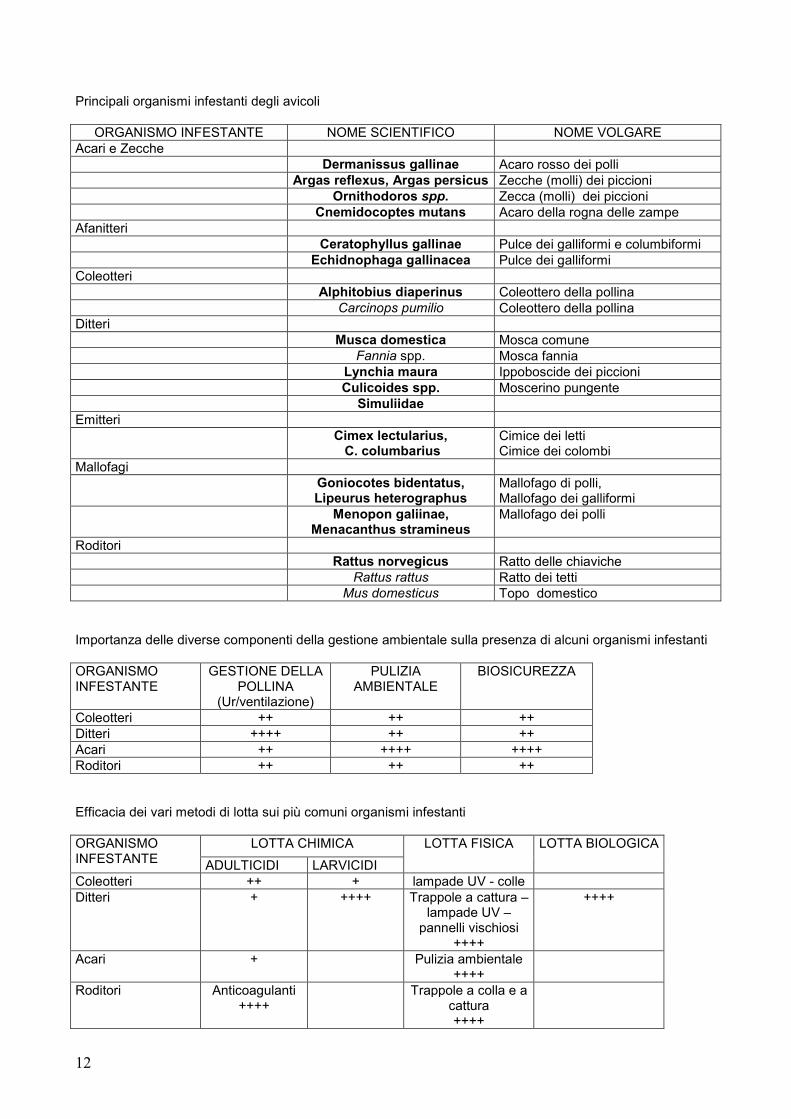
Principali organismi infestanti degli avicoli
ORGANISMO INFESTANTE NOME SCIENTIFICO NOME VOLGARE Acari e Zecche
Dermanissus gallinae Acaro rosso dei polli Argas reflexus, Argas persicus Zecche (molli) dei piccioni Ornithodoros spp. Zecca (molli) dei piccioni Cnemidocoptes mutans Acaro della rogna delle zampe
Afanitteri Ceratophyllus gallinae Pulce dei galliformi e columbiformi Echidnophaga gallinacea Pulce dei galliformi Coleotteri
Alphitobius diaperinus Coleottero della pollina Carcinops pumilio Coleottero della pollina
Ditteri Musca domestica Mosca comune Fannia spp. Mosca fannia
Lynchia maura Ippoboscide dei piccioni Culicoides spp. Moscerino pungente Simuliidae Emitteri
Cimex lectularius, C. columbarius
Cimice dei letti Cimice dei colombi
Mallofagi Goniocotes bidentatus,
Lipeurus heterographus Mallofago di polli, Mallofago dei galliformi
Menopon galiinae, Menacanthus stramineus
Mallofago dei polli
Roditori Rattus norvegicus Ratto delle chiaviche Rattus rattus Ratto dei tetti Mus domesticus Topo domestico
Importanza delle diverse componenti della gestione ambientale sulla presenza di alcuni organismi infestanti ORGANISMO INFESTANTE
GESTIONE DELLA POLLINA
(Ur/ventilazione)
PULIZIA AMBIENTALE
BIOSICUREZZA
Coleotteri ++ ++ ++ Ditteri ++++ ++ ++ Acari ++ ++++ ++++ Roditori ++ ++ ++ Efficacia dei vari metodi di lotta sui più comuni organismi infestanti ORGANISMO INFESTANTE
LOTTA CHIMICA LOTTA FISICA LOTTA BIOLOGICA
ADULTICIDI LARVICIDI Coleotteri ++ + lampade UV - colle Ditteri + ++++ Trappole a cattura –
lampade UV – pannelli vischiosi
++++
++++
Acari + Pulizia ambientale ++++
Roditori Anticoagulanti ++++
Trappole a colla e a cattura ++++

13
Raccolta ed identificazione di ectoparassiti L’identificazione tassonomica degli ectoparassiti in avicoltura è il primo punto per poter capire come intervenire nel loro controllo. E’ per questo che gli autori sono a disposizione sia dei veterinari che degli allevatori per l’identificazione degli esemplari di ectoparassiti da essi eventualmente inviati. BIBLIOGRAFIA Per eventuali approfondimenti contattare: [email protected]
14
COMUNICAZIONI
1) ANTIMICROBIAL SUSCEPTIBILITIY OF SALMONELLA SPP. STRAINS ISOLATED
FROM LAYER HENS IN CAMPANIA REGION FROM 2000 TO 2003
Ludovico Dipineto, Claudia Scarpetta, Mariarosaria Calabria, Mariangela Sensale, Antonio Baiano, Lucia Francesca Menna, Alessandro Fioretti
Dipartimento di Patologia e Sanità Animale, Università di Napoli Federico II, Italy
Corresponding author: Prof. Alessandro Fioretti, Dipartimento di Patologia e Sanità Animale. Facoltà di Medicina Veterinaria, Università di Napoli Federico II. Via Delpino, 1, 80137 Napoli (NA), Italy - Tel. +39 081451802 - Fax: +39 0815091993 - Email: [email protected] Abstract The aim of this study was to determine the antimicrobial resistance in 60 Salmonella strains (S.enteritidis, S.thyphimurium, S.gallinarum) isolated from layer hens in Campania region from 2000 to 2003. S.gallinarum showed resistance against ciprofloxacin and enrofloxacin, in contrast, S. enteritidis and S. typhimurium were fully susceptibile. In all of isolates high levels of resistance were observed for neomycin, gentamycin and oxytetracycline. Also, one significant observation was that all of the isolates showed full susceptibility to Sulphamethoxazole/Trimethoprime. These results suggest importance to restrict the use of antibiotics in layers hens flocks in order to reduce the selection and spread of multiresistant strains Key words: Antimicrobial resistance, Salmonella strains Sensibilità agli Antibiotici di Salmonella spp. Isolati da Galline Ovaiole in Campania nel Triennio 2000/2003 Riassunto Scopo del presente lavoro è stato quello di testare la sensibilità antibiotica di 60 ceppi di Salmonella (S.enteritidis, S.typhimurium, S.gallinarum ) isolati da galline ovaiole nel periodo compreso tra il 2000 e il 2003. S.gallinarum mostrava resistenza nei confronti di ciprofloxacina ed enrofloxacina (rispettivamente 15% e 23%), al contrario di S.enteritidis e S.typhimurium che manifestavano una completa sensibilità. Tutti i sierotipi valutati presentavano alte percentuali di resistenza nei confronti di neomicina, gentamicina e ossitetraciclina. Nei confronti dei sulfamidici i ceppi testati presentavano resistenza nulla. Alla luce di quanto esposto si consiglia un uso più moderato e mirato degli antibiotici negli allevamenti in modo da ridurre la selezione e diffusione di ceppi multiresistenti. Parole chiave: resistenza antimicrobica, Salmonelle Introduction Antimicrobial resistance is the capacity of bacteria to survive exposure to a defined concentration of an antimicrobial substance. It is the natural response of bacterium to defend itself against the effects of an antibiotic. The development of antimicrobial resistance is an ecological phenomenon. Any antibiotic use, whether in humans, animals or plants/environment may lead to resistance (OIE, 2003). The extensive use of antibiotics, not only in human and veterinary medicine, but also in livestock production for disease prevention or as growth-promoting feed additives, has led to a serious increase in, and spread of, multiple antibiotic-resistant bacteria (Cruchaga et al., 2001). All this caused considerable problems to approach prophylactics and therapeutics plans versus various bacterial pathologies. Generally, the increased application of antimicrobials in veterinary and human medicine has been implicated as a contributing factor in the emergence of antimicrobial-resistant pathogens and the evolution of multiple drug-resistant strains. Salmonellosis has a particular role in avian medicine whether host-specific serotypes (S.gallinarum, S.pullorum) or non-host specific serotypes (S.enteritidis, S.typhimurium) implicated in foodborne zoonoses. Within the routine control programmes carried out in the poultry farms organized from the avian pathology section of the Dipartimento di Patologia e Sanità Animale; the Salmonella strains isolated showed an increase of antibiotic resistance pattern. The aim of this study was to determine the antimicrobial resistance in Salmonella strains (S.enteritidis, S.thyphimurium, S.gallinarum) isolated from layer hens in Campania region from 2000 to 2003.

15
Materials and methods Sample collection From Januray 2000 to November 2003 a total of 60 Salmonella strains were isolated from layer hens flocks, respectively belonged to S.gallinarum, S.enteritidis, S.typhimurium. The strains collected were 20 for each serotype. Isolation and identification procedure The Salmonella isolation procedures were carried out following the WHO standard methods (WHO, 1994). All the strains were serotyped at National Reference Centre for Salmonella (Istituto Zooprofilattico Sperimentale delle Venezie, Padova - Italy). Antimicrobial susceptibility tests Antimicrobial susceptibility profiles of the isolates were determined by the disk diffusion method according to the NCCLS guidelines (National Committee for Clinical Laboratory Standards, 1997). The antimicrobial agents (Oxoid) tested and corresponding concentration were as follows: Ciprofloxacin 5µg (C), Enrofloxacin 5µg (E), Flumequine 30µg (F), Nalidixic acid 30µg (NA), Apramycin 15µg (AP), Amoxicillin 10µg (A), Neomycin 30µg (N), Gentamicin 10µg (G), Oxytetracycline 30µg (O), Sulphamethoxazole/Trimethoprime 25µg (S/T). The diameters of the inhibition zone for the interpretation of resistance and susceptibility were those recommended by the NCCLS (National Commitee for Clinical Laboratory, 2002). Results were scored as susceptible, moderately susceptible or resistant according to NCCLS criteria (2002). The Escherichia coli ATCC 25 922 was used as reference strain. Results As seen from Table 1, resistance of S. gallinarum was significantly higher than other two serotypes examined. In particular, S. gallinarum showed resistance against two fluoroquinolone (ciprofloxacin and enrofloxacin, respectively 15% and 23%), in contrast, S. enteritidis and S. typhimurium were fully susceptibile. In all of isolates high levels of resistance were observed for neomycin, gentamycin and oxytetracycline. It was also found that S. enteritidis and S. gallinarum were resistant to apramycin (33,3% and 38,5% respectively) and S. gallinarum was resistant to amoxicillin too (23,1%). In conclusion, one significant observation was that all of isolates showed full susceptibility to Sulphamethoxazole/Trimethoprime. Table 1. Antimicrobial resistance of 60 Salmonella spp. isolates.
Antimicrobial resistance (%) Serotype n° C E NA AP A N G F S/T O
S. enteritidis 20 0 0 6 33,3 0 55,5 38,8 5,5 0 33,3 S. typhimurium 20 0 0 13 0 0 12,5 25 12,5 0 50 S. gallinarum 20 15 23 15 38,5 23,1 53,8 55,3 23,1 0 46,1
Discussion An increase in the incidence of antibiotic resistance in Salmonella isolated from humans and animals related to exhaustive application of antibiotics in both groups has been documented worldwide (Chruchaga et al., 2001). Recently, Lee et al. (Lee et al., 2003) reported, in an antimicrobial susceptibility test against 258 isolates of S.gallinarum, a reduced susceptibility to ampicillin (13.0%), gentamicin (43.4%), kanamycin (69.6%), enrofloxacin (6.5%), ciprofloxacin (10.9%), norfloxacin (52.5%) and ofloxacin (82. 6%). A study on antimicrobial-resistant Salmonella enterica serovars isolated from chickens in Spain showed high percentage of resistance to chloramphenicol (44.6%), ampicillin (34.8%) and tetracycline (33.9%) (Hernandez et al., 2002). Jones et al. (2002) reported S.typhimurium strains resistant to ampicillin, sulphonamides, streptomycin, chloramphenicol and tetracyclines as well as S.typhimurium isolated from poultry resistant to nalidixic acid (Jones et al., 2002). Fluoroquinolones resistance was rarely found among Salmonella species until Heisig reported S.typhimurium serovar Copenhagen from cattle was highly resistant to ciprofloxacin (Heisig, 1993). Conclusions An important finding is the antimicrobial resistance observed in S.gallinarum against fluoroquinolones (ciprofloxacin and enrofloxacin). High percentage of resistance observed in S.typhimurium and S.enteritidis for neomycin, gentamycin and oxytetracycline demonstrate improper use of these antibiotics in the control of avian salmonellosis, particularly in metaphylactic sense. These results confirm importance to restrict the use of antibiotics in layers hens flocks in order to reduce the selection and spread of multiresistant strains and underlines the need for integrated surveillance systems of antibiotic resistance that consider isolates not only from human disease but also from the animal reservoirs and the food vehicles.

16
Acknowledgements We would like to thank Dr. Antonia Ricci, National Reference Centre for Salmonella - Istituto Zooprofilattico Sperimentale delle Venezie, Padova, Italia, for the serotyping of our isolates and Mrs. Fortuna Pisa for her collaboration. References 1. Cruchaga, S., Echeita, A., Aladuena, A., Garcia-Pena, J., Frias, N., Usera, M. A., 2001. Antimicrobial
resistance in salmonellae from humans, food and animals in Spain in 1998. J. Antimicrob. Chemother. 47(3):315-21.
2. Heisig, P., 1993. High-level fluoroquinolone resistance in a Salmonella typhimurium isolate due to alterations in both gyrA and gyrB genes. J. Antimicrob. Chemother. 32(3):367-77.
3. Hernandez, T., Rodriguez-Alvarez, C., Arevalo, M. P., Torres, A., Sierra, A., Arias, A., 2002. Antimicrobial-resistant Salmonella enterica serovars isolated from chickens in Spain. J. Chemother. 14(4):346-50.
4. Jones, Y. E., Chappell, S., McLaren, I. M., Davies, R. H., Wray, C., 2002. Antimicrobial resistance in Salmonella isolated from animals and their environment in England and Wales from 1988 to 1999. Vet. Rec. 25;150(21):649-54.
5. Lee, Y. J., Kim, K. S., Kwon, Y. K., Tak, R. B., 2003. Biochemical characteristics and antimicrobials susceptibility of Salmonella gallinarum isolated in Korea. J. Vet. Sci. 4(2):161-6.
6. NCCLS (National Commitee for Clinical Laboratory Standards), 2002. Performance standards for antimicrobial disk and dilution susceptibility tests for bacteria isolated from animals, second edition: approved standard M31-A2. Wayne, PA, USA.
7. OIE (Office International des Épizooties), 2003. OIE International Standards on Antimicrobial Resistance. Paris, France.
8. WHO, 1994. Guidelines on detection and monitoring of Salmonella infected poultry flocks with particolar reference to Salmonella enteritidis. In: Wray, C., Davies, R. H., (eds.), WHO, Veterinary Public Health Unit.
2) PREVALENCE OF CAMPYLOBACTER JEJUNI IN POULTRY BREEDER FLOCKS
Lucia Francesca Menna1, Gianluca Matteoli1, Marzia Fontanella1, Alessandra Cuomo1, Antonio De Paola2, Tiziana Pepe3, Isolina Di Marco3, Ludovico Dipineto1
1 Dipartimento di Patologia e Sanità Animale, Università di Napoli Federico II, Italy,
2 Arena Holding Spa, Loc. Monteverde, Bojano (CB), Italy,
3 Dipartimento di Scienze Zootecniche ed Ispezione degli Alimenti, Università di Napoli Federico II, Italy
Corresponding author: Prof. Lucia Francesca Menna, Dipartimento di Patologia e Sanità Animale. Facoltà di Medicina Veterinaria, Università di Napoli Federico II. Via Delpino, 1, 80137 Napoli (NA), Italy - Tel. +39 081451802 - Fax: +39 0815091993 - Email: [email protected]
Abstract The aim of this work is to present the preliminary results of a study about breeders carrying out in order to demonstrate the supposed correlation between Campylobacter isolated from breeders and the ones isolated from broilers, supporting the theory of the vertical transmission of the germ. It was examined three different breeder flocks of Bojano in Molise region. A total of 360 cloacal swabs and 80 enviromental swabs was collected. Of the 3 flocks studied, 6,9% tested were positive for Campylobacter spp. The most-prevalent isolated species is C. jejuni (8,2%). Only 3 of the 360 cloacal swabs samples examined were associated with C. coli. The environmental swabs resulted negative. This results confirms again that poultry is a reservoir of this germ. Indagine sulla Prevalenza di Campylobacter jejuni in Gruppi di Riproduttori Avicoli Riassunto Il presente lavoro si propone di illustrare i risultati preliminari, sui riproduttori, di uno studio più ampio volto a stabilire, mediante studi di biologia molecolare, una eventuale correlazione tra i Campylobacter isolati dai riproduttori e quelli isolati dai broiler in modo da definire l’eventuale trasmissione verticale del germe. Sono stati esaminati 3 gruppi di riproduttori ubicati a Bajano in Molise. Dei tre gruppi valutati, il 6,9% risultava positivo a Campylobacter spp. C. jejuni era la principale specie isolata (8,2%). Solo 3 dei 360 tamponi cloacali esaminati era associata a C. coli. I tamponi ambientali risultavano negativi. Tali risultati confermano ancora una volta il ruolo del pollame come reservoir di questo microrganismo.
17
Introduction Campylobacter jejuni is the leading cause of bacterial foodborne illnesses in human medicine (Newell and Fearnley, 2003). The vast majority of human campylobacteriosis cases primarily result from consumption of undercooked poultry or other foods cross-contaminated with raw poultry meat during food preparation. However, other risk factors besides poultry such as contact with house pets, or consumption of raw milk, untreated water, and undercooked beef or pork have also been linked to human infections (Corry and Atabay, 2001). As poultry is considered a major reservoir for human campylobacteriosis, reduction or elimination of poultry contamination with C. jejuni would greatly reduce the risk of Campylobacter for public health. Although numerous farm-based studies have been conducted in the past decades, the sources of flock infection, modes of transmission, and the host and environmental factors affecting the spread of Campylobacter on poultry farms are still poorly understood (Sahin et al. 2002). Potential sources of flock infection include used litter, untreated drinking water, other farm animals, domestic pets, wildlife species, house flies, insects, farm equipment and workers, and transport vehicles (Newell and Fearnley, 2003). However, none of these suspected sources has been conclusively identified as the formal source of infection for broilers farms. Despite these observations, vertical transmission of C. jejuni is still questionable because live Campylobacter have not detected in the eggs of commercial breeders, young hatchlings or hatcheries under natural conditions. Therefore the exact role of vertical transmission in introducing Campylobacter to broiler flocks remains unclear (Sahin et al., 2003). An estimated 2.1-2.5 million cases of human campylobacteriosis, characterized by watery and/or bloody diarrhoea, occur annually in the United States, exceeding the cases of salmonellosis (Friedman et al. 2000). The reported incidence of Campylobacter infection in Europe is estimated to be 1000-2300 cases per 100.000 (Padungton and Kaneene, 2003). The aim of this work is to present the preliminary results of a study about breeders carrying out in order to demonstrate the supposed correlation between Campylobacter isolated from breeders and the ones isolated from broilers, supporting the theory of the vertical transmission of the germ. Materials and Methods This study was conducted during the period October 2003/July 2004 in the Arena breeders-farm of Bojano in Molise region. Samples collection It was examined three different breeder flocks respectively named A, B and C. Each flock was visited five times. The first visit occurred during cleaning and disinfection procedures before placing the chicks. The second visit took place at one day of age, the third visit at 4 weeks of age, the fourth at 20 weeks of age and the last visit took place at 30 weeks. During every visit 30 cloacal swabs samples and 10 environmental samples (wall, water, litter, feed) was collected. Isolation and identification procedure The samples were added to Campylobacter Selective Enrichment broth (Oxoid) and incubated at 42° .C for 24 h under microaerophilic conditions. Then, each sample was streaked onto Campylobacter blood free selective agar base - Modified CCDA Preston (Oxoid) plates. Plates were incubated at 42° .C under microaerophilic conditions for 48 h. Therefore, the isolates was streaked onto blood-agar plates and incubated at 42°C for 24 h. Isolates were identified using a commercial identification method (API Campy, bioMérieux). Multiplex PCR A multiplex PCR assay was carried out to all isolates in accordance with the Cloak and Fratamico procedure (Cloak and Fratamico, 2002). The primers employed in this assay are shown in table 1. Table 1. PCR primers for C.jejuni and C.coli employed in the multiplex PCR Species targeted Product size (bp) Primer name (target gene) Sequence (5’ – 3’) C. coli/C. jejuni 400 cadF2B (cadF)
cadR1B TTGAAGGTAATTTAGATATG CTAATACCTAAAGTTGAAAC
C. coli 894 COL 1 (ceuE) COL 2
ATGAAAAAATATTTAGTTTTTGCA ATTTTATTATTTGTAGCAGCG
C. jejuni 160 C-1 (?) C-2
CAAATAAAGTTAGAGGTAGAATGT GGATAAGCACTAGCTAGCTGAT
Results and Discussion Of the 3 flocks studied, 6,9% tested were positive for Campylobacter spp. (table 2). The most-prevalent isolated species is C. jejuni (8,2%). Only 3 of the 360 cloacal swabs samples examined were associated with C. coli. The environmental swabs resulted negative.

18
Table 2. Percentage of positivity from cloacal swabs
Number of Cloacal swabs
30 30 30 30
Age of breeder flocks 1 day of age 4 weeks of age 20 weeks of ages 30 weeks of age
Flock A 0% 0% 50% 40% Flock B 0% 0% 3,3% 3,3% Flock C 0% 0% 10% 3,3% The results of this study show a low prevalence of Campylobacter in the breeder flocks examined. This result confirms again that poultry is a reservoir of this germ. Conclusions The literature suggests that standard biosecurity procedures are inadequate for the maintenance of flock negativity (Newell and Fearnley, 2003). This is a consequence of high exposure, low dose, and rapid bird-to-bird transmission rates. Nevertheless, stringent biosecurity may either delay positivity or reduce the number of flocks that become positive. However, it is generally considered that adeguate biosecurity procedures are difficult to substain in the farm environment (Pattison, 2001). For example, routine procedures such as the effective use of hygiene barriers, hand washing, and boot disinfection may be readily undertaken under normal conditions, but during emergencies, such as fan failure, such procedures may be ignored. Well-designed and well-located farms, the development of appropriate standard operating procedures to minimize risk factors, staff education, and incentives to maintain biosecurity at the highest level would all contribute to the reduction of flock positivity. Acknowledgements We would like to thank Mrs. Fortuna Pisa for her technique collaboration. References 1. Cloak, O. M., Fratamico, P. M., 2002. A multiplex polymerase chain reaction for the differentiation of
Campylobacter jejuni and Campylobacter coli from a swine processing facility and characterization of isolates by pulsed-field gel electrophoresis and antibiotic resistance profiles. J. Food. Prot. .65(2):266-73.
2. Corry, J. E., Atabay, H. I., 2001. Poultry as a source of Campylobacter and related organisms. J. Appl. Microbiol. 90:96-114.
3. Friedman, C. R., Neimann, J., Wegener, H. C., Tauxe, R. V., 2000. Epidemiology of C. jejuni infections in the United States and other industrialized nations. In: Nachamkin, I. and Blaser, M. J. (eds.) Campylobacter, 2nd edn., ASM Press., Washington, DC, pp. 121-138.
4. Newell, D. G., Fearnley, C., 2003. Sources of Campylobacter Colonization in Broiler Chickens. Appl. Environ. Microbiol. 69(8):4343-4351.
5. Padungton, P., Kaneene, J. B., 2003. Campylobacter spp in human, chickens, pigs and their antimicrobial resistance. J. Vet. Med. Sci. 65(2):161-170.
6. Sahin, O., Morishita, T. Y., Zhang, Q., 2002. Campylobacter colonization in poultry: sources of infection and modes of transmission. Anim. Health. Res. Rev. 3(2):95-105.
7. Sahin, O., Kobalka, P., Zhang, Q., 2003. Detection and survival of Campylobacter in chicken eggs. J. Appl. Microbiol. 95(5):1070-1079.
8. Pattison, M., 2001. Practical intervention strategies for campylobacter. Symp. Ser. Soc. Appl. Microbiol. 30:121-125.
19
3) SANCASSANIA BERLESEI (MICHAEL, 1903): AN OPPORTUNISTIC MITE INFESTING LITTERS IN POULTRY FARMS CAUSING DERMATITIS IN HUMANS AND
ANIMALS
Mario Principato, Federica Lisi, Iolanda Moretta, Nada Samra, Francesco Puccetti Dipartimento di Scienze Biopatologiche Veterinarie, Sezione di Parassitologia. Università di Perugia, Italy
Corresponding author: Prof. Mario Principato, Dipartimento di Scienze Biopatologiche Veterinarie, Sezione di Parassitologia, Via S. Costanzo, 4 – 06100 Perugia, Italy. Tel +39 0755857741 – Fax: +39 0755857743 – E-mail: [email protected] ABSTRACT Reported herein are some cases of human dermatitis caused by S. berlesei, a mite coming from seriously infested poultry farms. It appears unable to determine traumatic lesions on human skin, but it causes itch and inflammation also at the level of mucosas. Besides this mite can be found accidentally also on reared fowls’wounds by peak. Key words: mite (Sancassania berlesei), dermatitis, itch, exam of home dusts. SANCASSANIA BERLESEI (MICHAEL, 1903): UN ACARO OPPORTUNISTA CONTAMINANTE LA LETTIERA DI ALLEVAMENTI AVICOLI, CAUSA DI DERMATITI NELL’UOMO E NEGLI ANIMALI. RIASSUNTO Vengono presentati alcuni episodi di dermatite umana provocati da Sancassania berlesei, un acaro proveniente da allevamenti avicoli fortemente infestati. Questa specie non sembra in grado di determinare lesioni traumatiche sulla cute dell’uomo, ma, piuttosto, prurito e infiammazione, soprattutto a livello delle mucose dei genitali. Questo acaro si rinviene, occasionalmente, anche nelle ferite da beccata di volatili in allevamento. Parole chiave: acaro (Sancassania berlesei), dermatite, prurito, esame delle polveri ambientali Introduction Sancassania berlesei, better known as Caloglyphus berlesei, is an environmental mite of zootechnic interest, for it develops both in dried feedstuff and in litters of big industrial poultry farms. The species was described morphologically in 1903 by Michael and Sancassania genus in 1916 by Oudemans. The interest for this mite in veterinary medicine was reported for the first time in Italy by Principato et al. (1987). Besides recording the presence of that mite both in dried feedstuff and inside the farms, they described some lesions on fowls from which S. berlesei was isolated. The hosts, where the mite was observed, were Gallus gallus, Numida meleagris, Phasianus calchicus e Turdus merula. Crusty lesions were variously scattered on the hosts’ skin, above all in periocular areas, around the beak, but the mites were observed also on the feathers. The inflammation caused itch and in some samples, particularly infested, the symptom of diarrhea was reported as well. Afterwards Principato et al. (1991) supplied with a S.E.M. description of adults and hypopial deutonymphs (Figure 1) of this species and in 1992 they recorded its presence in some umbrian intensive chicken farms. Nowadays, S. berlesei seems to be widespread and it is recorded everywhere, sometimes even in dwellings. Reported herein are some cases of human and animal dermatitis referred to the presence of S. berlesei. Material and methods The cases of dermatitis herein described were recorded through our exams of environmental dusts carried out from 2000 to 2004 in dwellings, whose owners had frequent contact with conserved farinaceous food and feedstuff and besides that took care of rearing fowls, such as chickens, turkeys, pigeons and geese. A number of 76 exams of dust samples was examined, coming from those dwellings where symptoms of dermatitis and itch of indefinite cause were reported. A number of 27 were recorded in spring-summer and 49 in autumn-winter. The diagnosis was made by examining both home dusts and the dust removed from the poultry houses. The exam was effected by flotation with a saturated solution of NaCl after filtering and precipitation in absolute ethyl alcohol. Results and discussion

20
The direct exam of home dusts of owners of fowls revealed the presence of S. berlesei in 68 % of dermatites observed in spring-summer (Figure 2) and 14 % of the cases reported in autumn-winter (Figure 3). In all the cases in which S. berlesei was isolated in the houses, a great spread of that mite was recorded also in fowl runs with an average of mites of about n.
4000/g of dust in spring-summer and about n.1000/g of dust in autumn-winter. The number of hypopial deutonymphs increased about at half the cycle of rearing of animals. The dermatites observed were in most cases itchy, though not continuously. The mites were frequently isolated in clothes and in underwear and itch occurred more frequently on patients’genitals, inguinal area, arms and head. In the most serious cases it was spread also on their trunk and neck. The dermatites observed appeared always as folliculitis complicated by scratching. Mites could be isolated from some pets’ skin. On dogs the inflammation of their skin of abdomen and of the internal part of tighs resulted accompanied by a strong stimulus to scratch themselves. On Passeriformes and on Psittaciformes the tendency of animals to pluck their feathers was evident. In chickens S. berlesei was present mainly inside the wounds caused by peaks, in their feathers and periocular and cloacal areas. In general in spring-summer a higher percentage of cases of dermatitis by S. berlesei was reported (72%) in comparison with the autumn-winter period (28%) (Figure 3).
Conclusions Although the cases of human dermatitis caused by S. berlesei from birds are very few, it is a fact that this mites can determine itch and allergy in humans and animals. The presence of the arthropod also in underwear, with the high frequency of itch in the genital and perigenital areas appears to be interesting. S. berlesei is a mite that colonizes , though accidentally, animals’ wounds and mucosas and its presence also in women’s mucosa of vulva and vagina cannot be excluded. In this case its sanitary interest is to be correlated to the presence of bacteria of which the mite is certainly a reservoir. The possibility for this mite to trasform itself in hypopes makes it possible the infestation from poultry houses to human dwellings with problems of allergy that can arise even some years after the contagion. Since S. berlesei is a mite present in a lot of poultry farms and it is easily adaptable to any fowl run, it is necessary to contain its number through targeted treatments of litters, in order to avoid the chance of human contagion.
Figure 5. Frequency of cases of human dermatitis caused by S.
berlesei in spring-summer and in Autumn-Winter in people dealing with poultry houses
Autumn-winter 28%
Spring-summer 72%
Figure 2. Frequency of cases of human dermatitis
caused by environmental mites in spring-summer in
people dealing with poultry houses
D. farinae 11%
D. pteronyssinus
0%
Tydeus sp. 7%
L.destructor 7%
G. domesticus 0%S. berlesei 68%
A. siro 7%
Figure 3. Frequency of human dermatitis caused by
environmental mites in autumn-winterin people dealing
with poultry houses
L.destructor 42%
G. domesticus 8%
S. berlesei 14%Tydeus sp. 2%
A. siro 12%
D. farinae 8%
D. pteronyssinus
14%
21
A useful note for a differential diagnosis is that, contrarily to the dermatitis caused by Glycyphagus domesticus, the one caused by S. berlesei appears mainly in springtime and in summer without strophuloid lesions. References 1. Principato M., Coletti M., Tacconi G., 1987. Studio sull’acarofauna dei volatili. Il ruolo degli acari negli
stati patologici aspecifici. Isolamento di nuove specie patogene. Summa, 4: 229-237. 2. Principato M., Grossi M., Polidori G.A., 1991. Osservazioni al M.E.S. sulle forme invasive di Caloglyphus
berlesei (Acarina: Acaridae). Atti del XVIII Congresso di Microscopia Elettronica, 99-100. 3. Principato M., Rossodivita M.E., Grossi M., Polidori G.A., 1991. Le ninfe ipopiali di Caloglyphus berlesei
(Michael) (Acarina: Acaridae): osservazioni al M.E.S. Atti del XVIII Congresso di Microscopia Elettronica, 95-96.
4. Principato M., Galli R., Sannipoli C.G.T., 1991. Observations on the presence of Caloglyphus berlesei (Astigmata: Acaridae) in the litter of some industrial poultry farms in Umbria. Zootecnica International, 2: 40-45.
4) THE ALTERATIONS OF PLUMAGE OF PARASITIC ORIGIN
Mario Principato, Federica Lisi, Iolanda Moretta, Nada Samra, Francesco Puccetti Dipartimento di Scienze Biopatologiche Veterinarie, Sezione di Parassitologia. Università di Perugia, Italy
Corresponding author: Prof. Mario Principato, Dipartimento di Scienze Biopatologiche Veterinarie, Sezione di Parassitologia, Via S. Costanzo, 4 – 06100 Perugia, Italy. Tel +39 0755857741 – Fax: +39 0755857743 – E-mail: [email protected]
ABSTRACT Described herein are the main lesions to the plumage caused by insects and mites, both on the vane or the calamus of feathers. Practical data are given, aimed to make a correct differential diagnosis. Key words: insects, mites, plumage, calamus, vane, lesions. ALTERAZIONI DEL PIUMAGGIO DI ORIGINE PARASSITARIA RIASSUNTO Vengono descritte le principali lesioni al piumaggio prodotte da insetti ed acari, sia sulla parte vessillare delle penne, che sul calamo, fornendo elementi concreti per una corretta diagnosi differenziale. Parole chiave: insetti, acari, piumaggio, calamo, vessillo, lesioni. Introduction Arhropods can interact with fowls damaging their plumage, breaking, perforating and also causing its loss. Some attack preferably the calamus, some others the vane of the feathers. The lesions reported are almost always well distinguishable to the naked eye or by the aid of a stereomicroscope, but it is not always easy to find out the arthropod that causes them. To this aim, the main kinds of lesions of feathers occurred to our observation during the past ten years were selected in order to make it easy to effect a differential diagnosis and quickly to reveal the agent causing the pathology. Material and methods A number of 520 fowls was examined belonging to the families Struthionidae, Turnicidae, Phasianidae, Anatidae, Psittacidae, Columbidae; Passeriformes of the families Cinclidae, Troglodytidae, Sturnidae, Estrildidae, Fringillidae, Corvidae, Ploceidae, Turdidae, Alaudidae, Hirundinidae, Motacillidae, Sylviidae and Paridae. The macroscopic exam of plumage was carried out on them, by a stereomicroscope and at the same time the isolation of all the arthropods present was effected by using micro-needles and thin-pointed pincers. The feathers damaged and the arthropods isolated were kept in 80% alcohol, whereas some samples were clarified in warm lactic acid and mounted on slide in Berlese’s solution to be identified. To circumscribe the field of our reseach, in this study some mites causing mange and other causing indirectly the loss of feathers without lesions observable macroscopically were excluded. Results and discussion The arthropods identified as agents causing evident lesions of plumage belonged to two classes: Insecta and Acarina. In the former, two orders were recorded of particular interest for the plumage: Mallophaga and Coleoptera; in the latter, the order Actinedida (=Prostigmata) and Acaridida (=Astigmata).
22
The insects are a cause mainly of macroscopic lesions on the vane of feathers which can weaken and break, whereas mites cause less evident lesions both to the feather barb and to the calamus, determining in some cases its loss. Among insects, the first to be isolated are Mallophaga belonging to the suborders Amblycera and Ischnocera. All of them have a strong masticatory mouth apparatus, but unable to cut the rachis of bigger feathers, such as the flight feathers (remiges - primaries, secondaries and tertials) or the tail feathers (rectrices). On the contrary they can cut the dorsal and pectoral small feathers and the feathers of sides under the wings. Their action on the barb is linear and very precise. The feather appears cut horizontally and each barb is cut singularly. When Mallophaga are very young, they cut the barbules and make small holes close to each other, without cutting the barbs completely. Other insects, instead, less known under this aspect than Mallophaga, are the Coleoptera of family Dermestidae, Dermestes genus. Those insects are able to attack the alive animals’ plumage, if the feathers are dirty of faeces or of feedstuff. This occurs frequently, for instance, in intensive pheasant farms, where the animals are often kept to sleep on the ground, even near carcasses of new-born poulets or close to broken eggs. Those materials attract Coleoptera adults and larvae of Dermestes genus, that, by their masticatory mouth, stronger and bigger than Mallophaga’s one, can break sleeping animals’ feather barbs, by clenching them in groups. This determines a typical bend in the place of the cut which is the differential element in comparison with lesions caused by Mallophaga. Besides the Dermestidae can break the rachis of smaller feathers and inlay the one of the bigger feathers. Typical lesion caused by Dermestidae beetles is the one at the level of pheasants’tail, which, under the action of Dermestidae at the end breaks with a consequent depreciation of the birds. Among the mites, a distinction is to be made between those localizing in the feather vane and those localizing at the level of calamus. Among the formers are Astigmata mites, belonging to the suborder Psoroptidia with many families, such as Pterolichidae, Analgidae, Proctophyllodidae, Dermoglyphidae and others. Many species of those families live on barbs, near the rachis in the internal part of feathers, often at the level of primaries (the localization varies depending on the species). All these mites cannot cut the barbs, but can shift or cut off the barbules, producing small holes hardly visible if the feather is held up against the light. The holes are very small and scattered and in this they are different from those caused by young Mallophaga that, generally,producing holes on the same line.The second group of mites we observed localizes,instead, at the level of feather calamus. They belong either to Actinedida, prostigmates of the suborder Eleutherengona, family Syringophilidae, either to the above mentioned order of Astigmata Psoroptidia, family Knemidokoptidae. Mites of family Syringophilidae localize in the internal part of calamus of feathers, whereas Mesoknemidokoptes laevis, a species of family Knemidokoptidae, mins externally the calamus by producing some cortical tunnels. Both of them, but mainly the mites of family Syringophilidae, cause the inflammation of feather papilla and its consequent fall. There can be itch and the animal can tear it by itself. Finally, we observed another group of mites localizing at the basis of feathers and among the barbs. They are Prostigmata mites belonging to the suborder Eleutherengona, of the family Cheyletidae. Eggs of this mite are kept inside silky cobwebs spun from substance secreted by female mites. Feathers appear sticky and barbs are attached like a web. Mites belonging to the suborder Eleutherengona, family Harpyrhynchidae, lay their eggs on the barbs at the basis of feathers, above all the periocular ones and around the auditory meatus in Passeriformes. In this case the feather has a typical look and white-orange colour. Conclusions The main lesions by arthropods to feathers are certainly those caused by Mallophaga, whereas those caused by typical mites of the feathers (Astigmata mites of the suborder Psoroptidia) are less evident. In Passeriformes lesions to feathers by Harpyrhynchus and Syringophilus are frequently recorded (Principato et al., 1992,1995). Lesions caused by Ornithochyletia and Mesoknemidokoptes result to be more rare (Principato et al., 1987,1995). On the contrary it is frequent to observe the attack of plumage by Dermestidae, but it is caused by the presence of organic rests and poor hygiene of the farms (Théodoridèd, 1949). A differential diagnosis may be easy if one takes into account not only the morphology of lesions, but also their place and the host’s species. A treatment with parasiticides is not always successful, as it is in the case of Syringophilosis, and anyhow a possible treatment must be necessarily carried out considering the role of the environment as well (for instance: Dermestidae or contagion with feathers fallen on the ground) in the upset of the pathology. References 1. Principato M., Coletti M., Tacconi G., 1987. Studio sull’acarofauna dei volatili. Il ruolo degli acari negli
stati patologici aspecifici. Isolamento di nuove specie patogene. Summa, 4: 229-237. 2. Principato M., Grelloni V., Coletti M., Tacconi G., 1992. Arpirinchiasi dei volatili: osservazioni su un raro
caso di acariasi cistica in Coccothraustes coccothraustes (Passeriformes: Fringillidae). Zootecnica International, 29-33
23
3. Principato M., Franciosini M.P., Del Rossi E., .1995. Rogna deplumante della tortora da Mesoknemidokoptes laevis Railliet, 1885 (Acari: Knemidokoptidae): un acaro nuovo per l’ItaliaZootecnica International, 123-126.
4. Principato M., Tacconi G., Liberti L., 1995. Siringofilosi dei volatili: un’acariosi poco conosciuta. Zootecnica International, 127-130
5. Théodoridèd J. 1949. Les coléoptères nuisibles aux animaux domestiques. Ann. De Parasitologie, 24: 116-123.
5) SEROLOGICAL EVIDENCES SHOWING THE INVOLVEMENT OF FREE-LIVING PHEASANTS IN THE INFLUENZA ECOLOGY (NORTHERN ITALY, 1995-2002)
Maria Alessandra De Marco1, Laura Campitelli2, Mauro Delogu3, Elisabetta Raffini4,
Emanuela Foni5, Livia di Trani6, Michele Scaffidi3, Isabella Donatelli2
1 Istituto Nazionale per la Fauna Selvatica “A. Ghigi”, Ozzano Emilia, Italy
2 Dipartimento di Malattie Infettive, Parassitarie e Immunomediate, Istituto Superiore di Sanità, Roma, Italy
3 Dipartimento di Sanità Pubblica Veterinaria e Patologia Animale, Università di Bologna, Italy
4 Istituto Zooprofilattico Sperimentale della Lombardia e dell' Emilia Romagna, Lugo (RA), Italy
5 Istituto Zooprofilattico Sperimentale della Lombardia e dell' Emilia Romagna, Parma, Italy
6 Dipartimento di Sanità Animale e Alimentare, Istituto Superiore di Sanità, Roma, Italy
Corresponding author: Dr. Maria Alessandra De Marco. Istituto Nazionale per la Fauna Selvatica “A. Ghigi”. Via Ca’ Fornacetta 9, 40064 Ozzano dell’Emilia (BO), Italy – Tel. +39 051 6512244 – Fax: +39 051 796628 – Email: [email protected]
ABSTRACT From 1995 to 2002, 219 sera were collected in Northern Italy from wild pheasants. A serological survey for avian influenza viruses (AIVs) was carried out by ELISA test in order to to detect type A influenza antibodies. The overall seroprevalence was 12.3%, with yearly values ranging from 0% to 42.5%. No antibodies against either H5 or H7 AIV subtypes were found by hemagglutination-inhibition test. Data from 16 recaptured birds, among 113 animals banded for individual identification, showed seroconversions in 2 pheasants. Our results indicate AIV circulation in free-living pheasants; the involvement of this land-based bird species in influenza ecology is discussed. KEY WORDS: Pheasant, Avian influenza, Influenza ecology, Serological survey INTRODUCTION Land-based birds belonging to the Galliformes Order include species, such as turkey, chicken, and quail, that are highly susceptible to avian influenza viruses (AIVs) primarily harboured in wild aquatic birds (Webster et al., 1992). In addition to heavy economic losses due to influenza epidemics in poultry, important public health implications could arise from AIV circulation in land-based birds, recently indicated as a potential source of pandemic strains (Perez et al., 2003). AIV infections have been described in Italy in reared pheasants (Phasianus colchicus) both as limited outbreaks (Rinaldi et al., 1967) or associated to severe poultry epidemics (Capua et al., 2003). Although sporadic isolations of AIVs have been reported in free-living pheasants (Romváry et al., 1976), the epidemiological role played by wild populations of this Galliformes species is not well understood, to date. Aims of this serological survey, carried out on wild pheasants trapped in northern Italy, were: i) to establish the occurrence of type A influenza infection; ii) possibly, to detect the circulation of H5 and H7 AIV subtypes. MATERIALS AND METHODS Sampling - Free-living pheasants were monitored on an estate (about 35 hectares) located in a protected lowland area (Bologna province, Emilia Romagna region). The number of birds occupying the study area ranged from 150 to 40 in autumn and spring, respectively. From 1995 to 2002 a total of 196 pheasants were trapped; 113 of them were banded for individual identification then released into the wild. Overall, 219 sera were collected and examined (Table 1) including 23 recapture samples (Table 2). Bird sex and age were recorded whenever possible. Serological test - Sera were assayed for antibodies against influenza A virus nucleoprotein using an ELISA test (NP-ELISA) performed as described (De Marco et al., 2003b). Available NP-ELISA positive sera were assayed as described (De Marco et al., 2004) by hemagglutination-inhibition (HI) test, in order to detect antibodies against 5 different Italian strains belonging to both H5 and H7 subtypes of AIVs (Table 1).

24
Recapture data (Table 2) were analysed in order to evidence a significant increase in antibody titres (De Marco et al., 2003b). Statistical analysis – Chi-square test was performed in order to test non-random associations between the overall seroprevalences and: i) pheasant age; ii) pheasant sex. The significance level was set at a P<0.05. RESULTS AND DISCUSSION As shown in Table 1, the overall NP-ELISA antibody frequency to avian influenza viruses (AIVs), including data from 23 recaptures, was 12.3% (27/219). Pheasants seropositive for influenza A viruses were found in 4 of the 5 sampling periods, and the prevalence of sera found positive ranged from 0% (in 1995) to 42.5% (in 2001). No sex-related differences were found whereas the age-related NP-ELISA seroprevalences resulted significantly higher in the juvenile birds, compared to the adult ones, thus suggesting a higher susceptibility of juvenile pheasants that congregate in post-breeding periods to AIV transmission. No H5 and H7 positive sera were found by the HI assay (Table 1). Among 16 birds captured more than once, seroconversion for type A influenza viruses (Table 2, data in bold type) was observed in pheasants n. 3 and n. 5, indicating that AIVs circulated in the study area during the winter 2000. CONCLUSIONS Our findings indicate the occurrence of avian influenza virus (AIV) infection in free-living pheasants. In general wild birds are potentially exposed to AIVs perpetuated by natural reservoirs, in particular the pheasants examined in the present study drank from small ponds located in the study area and occasionally used by migrating ducks. Faecal contamination of waters could represent an ecological interface between primary hosts of AIVs and other susceptible bird species (Webster et al., 1992). The high population density characterising the study area could facilitate the virus circulation; indeed the data we observed are in contrast with seronegative results obtained from pheasants living in protected sites characterised by a lower population density (De Marco et al., 2003a). Fortunately, our results indicate that neither H5 nor H7 subtypes of AIV, recently involved in Italian poultry epidemics (Capua et al, 2003; Campitelli et al., 2004), circulated within the examined bird population. Further surveillance studies will enable us to acquire information to better understand the dynamics of influenza infection in pheasants, a land-based bird species potentially implicated in the interspecies transmission of AIVs harboured in natural reservoirs (Wood et al., 1985; Perez et al., 2003). ACKNOWLEDGEMENTS Thanks are due to: Dr. Fausto Marzadori, and Mr. Paolo Tozzoli (IZS della Lombardia e dell’Emilia-Romagna, Lugo); the “Provincia di Bologna”, Mr. Gino Rossi and other game management personnel; Mr. Valter Trocchi (Istituto Nazionale per la Fauna Selvatica).
REFERENCES 1. Campitelli, L., Mogavero, E., De Marco, M. A., Delogu, M., Puzelli, S., Frezza, F., Facchini, M.,
Chiapponi,C.,Foni, E., Cordioli, P., Webby, R., Barigazzi, G., Webster, R. G., Donatelli, I. 2004. Interspecies transmission of an H7N3 influenza virus from wild birds to intensively reared domestic poultry in Italy. Virology 323: 24-36.
2. Capua, I., Marangon, S., Dalla Pozza, M., Terregino, C., Cattoli, G., 2003. Avian Influenza in Italy 1997-2001. Avian Dis. 47:839-843.
3. De Marco, M. A., Campitelli, L., Foni, E., Raffini, E., Barigazzi, G., Delogu, M., Guberti, V., Di Trani, L., Tollis, M., Donatelli, I. 2004. Influenza surveillance in birds in Italian wetlands (1992-1998): is there a host restricted circulation of influenza viruses in sympatric ducks and coots? Vet. Microbiol. 98:197-208.
4. De Marco, M. A., Foni, E., Campitelli, L., Raffini, E., Delogu, M., Donatelli, I., 2003a. Long-term monitoring for avian influenza viruses in wild bird species in Italy. Vet. Res. Commun. 27 (Suppl. 1):107-114.
5. De Marco, M. A., Foni, E., Campitelli, L., Raffini, E., Di Trani, L., Delogu, M., Guberti, V., Barigazzi, G., Donatelli, I. 2003b. Circulation of influenza viruses in wild waterfowl wintering in Italy during the 1993-1999 period: evidence of virus shedding and seroconversion in wild ducks. Avian Dis. 47:861-866.
6. Perez, D. R., Webby, R. J., Hoffmann, E., Webster, R. G., 2003. Land-based birds as potential disseminators of avian/mammalian reassortant influenza A viruses. Avian Dis. 47:1114-1117.
7. Rinaldi, A., Nardelli, L., Pereira, H. G., Mandelli, G. C., Gandolfi, R., Cervio, G., 1967. Focolaio di Influenza A nel Fagiano (Phasianus colchicus L.). Atti S.I.S.Vet. XXI:867-872.
8. Romváry, J., Mészáros, J., Tanyi, J., Rósza, J., Fábián, L., 1976. Influenza infectedness of captured and shot wild birds on north-eastern and south-eastern parts of Hungary. Acta Vet. Acad. Sci. H. 26(3):363-368.
9. Webster, R. G., Bean, W. J., Gorman, O. T., Chambers T. M., Kawaoka, Y., 1992. Evolution and ecology of Influenza A viruses. Microbiol. Rev. 56(1):152-179.

25
10. Wood, J. M., Webster, R. G., Nettles, V. F., 1985. Host range of A/chicken/Pennsylvania/83 (H5N2) influenza virus. Avian Dis. 29(1): 198-207.
Table 1 – Serological results for antibodies against avian influenza viruses in 196 free-living pheasants
trapped in a protected area of the Emilia Romagna region (Northern Italy). Seroprevalences were calculated on overall sera sampled, including 23 sera obtained from 16 recaptured bird.
Sampling period
NP-ELISA prevalence %
(positive/tested sera)
HI antibody frequencies (positive/tested sera) calculated on NP-ELISA positive sera against the following AIV subtypes
(*):
H5N2 H5N3 H5N9 H7N1 H7N3 Mar. 1995 0 (0/34) n.d. n.d. n.d. n.d. n.d. Jan. 1999/Feb. 1999 3.8 (2/53) 0 (0/2) 0 (0/2) 0 (0/2) 0 (0/2) 0 (0/2) Feb. 2000/Mar. 2000 5.6 (3/54) 0 (0/2) 0 (0/2) 0 (0/2) 0 (0/2) 0 (0/2) Jan. 2001/Mar. 2001 42.5 (17/40) 0 (0/15) 0 (0/16) 0 (0/16) 0 (0/15) 0 (0/16) Jan. 2002 13.2 (5/38) 0 (0/4) 0 (0/5) 0 (0/5) 0 (0/4) 0 (0/5) Total 1995/2002 12.3 (27/219) 0 (0/23) 0 (0/25) 0 (0/25) 0 (0/23) 0 (0/25) (*) Low-pathogenic AIV strains used: A/mallard/Italy/80/93(H5N2); A/mallard/Italy/208/00(H5N3); A/chicken/Italy/9097/97(H5N9); A/turkey/Italy/6423-1/99(H7N1); A/mallard/Italy/33/01(H7N3); n.d. not done.
Table 2 – Serological data for antibodies against influenza A viruses obtained from 16 free-living pheasants
trapped between 1995 and 2002 in the study area, and recaptured once or twice. Bird
No.
Bird
age^
1st capture time
(NP-ELISA result)
1st recapture time
(NP-ELISA result)
2nd recapture time
(NP-ELISA result)
1 Ad 14/03/95 (−) 02/02/00 (−) n.d.
2 Ad 03/02/99 (−) 22/02/00 (−) 02/03/00 (−)
3 Ad 04/02/99 (−) 22/02/00 (−) 29/02/00 (+)^^
4 Ad 09/02/00 (−) 18/02/00 (−) 24/02/00 (−)
5 Ad 09/02/00 (−) 14/02/00 (−) 29/02/00 (+)^^
6 Juv 11/02/00 (−) 15/02/00 (−) n.d.
7 Un 11/02/00 (−) 22/02/00 (−) 01/03/00
8 Ad 11/02/00 (−) 28/01/02 (−) n.d.
9 Ad 22/02/00 (−) 24/02/00 (−) 29/02/00 (−)
10 Ad 23/02/00 (−) 29/02/00 (−) n.d.
11 Ad 23/02/00 (−) 08/02/01 (−) n.d.
12 Juv 23/02/00 (−) 01/03/00 (−) n.d.
13 Juv 24/02/00 (−) 29/01/02 (−) n.d.
14 Ad 29/02/00 (−) 06/02/01 (−) 28/01/02 (−)
15 Ad 13/02/01 (−) 27/02/01 (−) n.d.
16 Un 01/03/01 (+)^^ 06/03/01 (+)^^ n.d.
Ad adult; Juv juvenile; Un unknown; (+) positive; (−) negative; n.d. not done; ^ determined during the 1st capture; ^^ positive antibody titre = 8.

26
6) FIELD TRIALS WITH THE USE OF A LIVE ATTENUATED TEMPERATURE-SENSITIVE VACCINE FOR THE CONTROL OF MYCOPLASMA GALLISEPTICUM
INFECTION IN MEAT-TYPE TURKEYS
Enrico Alessandri1, Paola Massi2, Francesca Paganelli2, Francesco Prandini3, Mario Saita4
1Gruppo PAI, Malo (Vicenza), Italy.
2Istituto Zooprofilattico Sperimentale della Lombardia e dell’Emilia
Romagna, Sezione diagnostica di Forlì, Italy 3Merial Italia S.p.A., Assago (MI), Italy.
4Dipartimento di
Patologia Animale, Igiene e Sanità Pubblica Veterinaria, Sezione di Anatomia Patologica Veterinaria e Patologia Aviare, Università degli Studi di Milano, Italy
Corresponding author: Dr. Francesco Prandini. MERIAL Italia S.p.A. Milanofiori Strada 6 Palazzo E/5, 20090 Assago (MI), Italy – Tel. +39 02 577661 – Fax: +39 02 57766307 – Email: [email protected]
ABSTRACT Mycoplasma gallisepticum (MG) continues to be an important pathogen of poultry, causing significant production losses in many parts of the world. Eradication is the preferred method of control but it could result impractical after the organism has been introduced in an area with high density of poultry farms. TS-11®, a temperature-sensitive live attenuated MG vaccine is currently utilized in several countries for the control of MG infections in commercial layers and broiler breeders. In the present field trial, conducted in an industrial meat-turkey farm, (belonging to an integrated company), previously affected by severe MG infections, the ability of TS-11® in effectively colonizing the upper respiratory tract in a turkey flock was evaluated (“TS-11®” flock). A second flock grown in an adjacent pen of the same farm was vaccinated with an inactivated MG vaccine (“Inactivated” flock). Polymerase Chain Reaction (PCR) and Random Amplified Polymorphic DNA (RAPD) analysis were applied for the detection and differentiation of TS-11® from other MG strains possibly present in the same flocks, such as the field strains and the 6/85 live vaccine strain currently utilized in commercial layers in Italy. PCR-RAPD results achieved in the “TS-11®“ flock were compared with those of a flock of turkey grown in the same farm but vaccinated with an inactivated MG vaccine. Encouraging results were achieved by means of PCR-RAPD detection of TS-11® from all of the samples up to eight weeks post vaccination, whereas it was never detected in the “Inactivated” flock. Moreover, the field strain was never detected in the “TS-11®“ flock but in the “Inactivated” one it was detected either 5 and 8 weeks after the vaccination. The aggregate production data of the two flocks resulted significantly improved when compared to the performance of the previous flocks grown in the same farm and similar to the production standard of the integrated company. Key words: Mycoplasma gallisepticum, Temperature-sensitive live MG vaccine, Random Amplified Polymorphic DNA, Meat turkeys Prove di campo nell’impiego di un vaccino vivo attenuato termo-sensibile per il controllo dell’ infezione da Mycoplasma gallisepticum in tacchini da ca RIASSUNTO Mycoplasma gallisepticum (MG) continua ad essere un importante agente patogeno del pollame, dove provoca gravi perdite produttive in diverse aree del mondo. L’eradicazione rappresenta il miglior metodo per il controllo di MG, tuttavia può risultare difficilmente praticabile una volta che l’infezione è stata introdotta in un’area caratterizzata da elevata concentrazione di allevamenti avicoli. TS-11®, un vaccino MG vivo attenuato termo-sensibile, viene attualmente impiegato in diversi Paesi del mondo per il controllo delle infezioni da MG nelle galline ovaiole commerciali e nei riproduttori pesanti. Nella prova di campo, condotta in un allevamento commerciale di tacchini da carne (appartenente ad un gruppo avicolo integrato), colpito nei cicli produttivi precedenti da gravi infezioni MG, è stata valutata la capacità di TS-11® di colonizzare efficacemente le vie respiratorie superiori (gruppo “TS-11®“). Un secondo gruppo allevato in un capannone adiacente nella stessa azienda è stato vaccinato con un vaccino inattivato (gruppo “Inattivato”). Le tecniche PCR e RAPD sono state applicate per l’individuazione e la differenziazione di TS-11® da altri ceppi di MG eventualmente presenti negli stessi gruppi, quali i ceppi di campo ed il vaccino vivo 6/85 attualmente utilizzato nelle ovaiole commerciali in Italia. I risultati dei test PCR-RAPD ottenuti nel gruppo “TS-11®“ sono stati comparati con quelli del gruppo “Inattivato”, allevato in un capannone adiacente, nella stesso allevamento. Sono stati così ottenuti risultati incoraggianti, infatti TS-11® è stato individuato sistematicamente su tutti i campioni prelevati tra le 2 e le 8 settimane dopo la vaccinazione mentre non è mai stato identificato nei tamponi prelevati dal gruppo “Inattivato”. Inoltre, il ceppo di campo non è mai stato evidenziato nel gruppo “TS-11®“ mentre nel gruppo “Inattivato” i tamponi tracheali sono risultati positivi rispettivamente a 5 e 8 settimane post-vaccinazione. I dati produttivi complessivi sono risultati significativamente migliori rispetto alle performance dei cicli precedenti allevati nella stessa azienda ed in linea con lo standard produttivo del gruppo avicolo integrato.
27
Parole chiave: Mycoplasma gallisepticum, Vaccino vivo termo-sensibile, Random Amplified Polymorphic DNA (RAPD), Tacchini da carne Introduction Mycoplasma gallisepticum (MG) plays an important role as pathogen of poultry, causing significant economic losses, particularly in the areas where higher is the concentration of industrial farms. In meat-type turkeys infected with MG, severe respiratory syndromes occur with losses due to increased mortality rate and condemnation of carcasses, poor performance and increased medication costs. Strategies to minimize if not eliminate the impact of MG infections can include eradication programmes, specific antimicrobial treatments and immunization with live or inactivated vaccines. TS-11® is a temperature-sensitive live attenuated MG vaccine, currently utilized in Italy and many other Countries for the control of MG infections in commercial layers. The vaccination of boiler chicken breeders prevented the infection by field MG in their tracheas and infraorbital sinuses and in the vitelline membrane of their embryos (Barbour et al., 2000). In spite of its use on a large scale in commercial layers in the United States, it was never isolated from field turkeys (Kleven et al., 2000). In a vaccination trial conducted by S.H.Kleven, (2000), administration of TS-11® to meat turkeys resulted safe and there was some protection detected against lesions in the upper respiratory tract, thus indicating a potential use in certain conditions. During the last years, in some densely poultry populated areas of Northern Italy, an increasing number of severe MG outbreaks, occurred consecutively in the same industrial turkey farms; either individual and mass antimicrobial treatments were not able to control the clinical disease. The promiscuity of the staff and the equipment utilized in the farms of the same area was probably critical in perpetuating the infection, in spite of an all in-all out policy applied in every single farm. The main purpose of the present trial, conducted in one of the farms, (belonging to an integrated company), affected by MG infections in the previous cycles, was to examine the ability of TS-11® in colonizing in the mucosa of the upper respiratory tract of turkey and to evaluate the persistency and its possible effect in inducing protection against field MG strains. PCR and RAPD analysis (Paganelli et al., 2003) applied on tracheal swabs drawn at regular intervals from turkey flocks, enabled the detection and the genetic differentiation of TS-11® from the field strains. Materials and methods MG vaccines TS-11® (Merial Italia S.p.A.), a frozen temperature-sensitive live attenuated MG vaccine (Whithear et al., 1990), was stored at –20°C then thawed in a water bath at 30 to 35°C just prior to the administration; each bird of the first flock (“TS-11®“) received one dose, containing at least 107.4 Colour-Changing Units (CCU) in 50 microlitres, applied by ocular instillation. One dose (0.5 ml) of a commercial inactivated oil emulsified MG vaccine was administered subcutaneously to the second flock (“Inactivated”) in the same farm. MG serological tests Rapid plate agglutination test (RPAT) for MG antibodies was carried out with a commercial antigen. A commercial Enzyme-Linked Immuno-Sorbent Assay (ELISA) test kit was used to detect MG antibodies. DNA extraction and MG identification The Polymerase Chain Reaction (PCR) protocol was adapted from the procedure described by Lauermann et al. (2003) and performed on DNA after extraction from tracheal swabs (Paganelli et al., 2003). Each sample (100-2000 ng/µl) was treated with PCR reagents included in Accuprime TaqPCRx DNA Polymerase (Invitrogen®,USA). We used primers “forward” MG-F+ and primers “reverse” MG-R (table 1). DNA amplification is performed in a thermocycler (TpersonalTM, Biometra, Germany). The optimized PCR program was as follows: 5 minutes at 94°C (hot start), 35 cycles with 3 steps: 94°C for 30 sec. (denaturation), 55°C for 30 sec. (annealing), 72°C for 1 minute (extension); finally 1 minute at 72°C for final extension. PCR products were separated using electrophoresis on a 1,7% agarose gel containing ethidium bromide for 30 minutes at 80V. MG amplicons were visualized with an ultraviolet transilluminator along with control samples (positive and negative) and PCR marker GeneRuler 100bp DNA ladder (Fermentans, Lithuania). When MG positive, amplified product presented a 185bp fragment (Paganelli et al., 2002). RAPD analysis A further PCR on extracted DNA was performed using a commercial kit (Ready-to go RAPD Analysis – Pharmacia Biotech, Piscataway, USA) containing a set of primers. Primer 6 was used for the first reaction. A combination of primers 3 and 4 was used for the second reaction (Table 1) (Paganelli et al., 2003). RAPD was performed in a thermocycler (TpersonalTM, Biometra, Germany). RAPD program was as follows: 5 minutes at 95°C (hot start), 45 cycles with 3 steps: 95°C for 1 minute, 36°C for 1 minute, 72°C for 2 minutes; finally 1 cycle at 4°C. Each reaction included the vaccine strains TS-11 and 6-85.

28
RAPD products were separated using electrophoresis on a 2% agarose gel containing ethidium bromide for 30 minutes at 80V. MG amplicons (185 bp) were visualized with an ultraviolet transilluminator, comparing patterns each other, along with marker GeneRuler 100bp DNA ladder (Fermentans, Lithuania) (Paganelli et al., 2003). Table 1. Sequence of RAPD primers
MG-F 5’ GAGCTAATCTGTAAAGTTGGTC 3’
MG-R 5’ GCTTCCTTGCGGTTAGCAAC 3’
RAPD 3 5’-d [GTAGACCCGT]- 3’
RAPD 4 5’-d [AAGAGCCCGT]- 3’
RAPD 6 5’-d [CCCGTCAGCA]- 3’
Field trial design 8500 day-old turkey toms were divided in two flocks of the same size and housed in adjacent pens in the same farm (belonging to an integrated company), previously undertaken to cleaning, disinfection and downtime. Before vaccination 15 swabs were randomly taken from trachea and choanal cleft respectively and 20 blood samples were randomly drawn from the turkeys of each flock to exclude any MG positivity. At three weeks of age “TS-11®“ flock was eye-drop vaccinated while “Inactivated” flock was vaccinated subcutaneously. At the age of 5, 8, 11 and 15 weeks respectively, from each of the two flocks were randomly drawn 15 blood samples and 15 tracheal swabs; from every set of 15 swabs 3 pools were obtained and tested by PCR-RAPD. Due to practical reasons, the production parameters of the two flocks could not be registered separately but the aggregate data were compared with the correspondent data of the previous production cycle. Results and discussion Clinical monitoring detected no reaction after TS-11® vaccination and no considerable treatments were needed during the whole production cycle. The results of PCR and RAPD monitoring on pooled tracheal swabs are summarized in Table 2. Table 3 shows the serological results. In Table 4 the aggregate performance of the two flocks are compared to the results of the previous cycle and to the production standard of the Integrated Company Table 2. Results of PCR and RAPD tests applied on pools of tracheal swabs drawn at different ages from “TS-11®“ and “Inactivated” flocks Age 3 weeks 5 weeks 8 weeks 11 weeks 15 weeks Test PCR RAPD PCR RAPD PCR RAPD PCR RAPD PCR RAPD
TS-11® Neg. - Positive
3/3 TS-11®
Positive 3/3
TS-11® Positivo
3/3 TS-11® Neg. -
Inactivated Neg. - Neg. - Positive
1/3 ‘Field’ MG
Positive 2/3
‘Field’ MG
Neg. -
Table 3. Percentage of turkeys sero-positive to MG at different ages in “TS-11®“ and “Inactivated” flocks Age 3 weeks 5 weeks 8 weeks 11 weeks 15 weeks Test RPAT ELISA RPAT ELISA RPAT ELISA RPAT ELISA RPAT ELISA TS-11® 0 - 0 0 0 0 100 60 100 0 Inactivated 0 - 0 0 0 80 27 87 100 53 Table 4. Performance compared the previous cycle and to the Company production standard
Parameters Age days
Mortality %
Avg. Body weight Kg
Feed Conversion F.C.
Medication costs Euro/kg
Field trial 137 8.82 18.7 2.57 0.017 Previous cycle 134 13.88 16.0 2.62 0.076 Company standard 135 9.50 18.5 2.56 0.025 PCR and RAPD analysis enabled the identification of TS-11® in the trachea of turkeys up to 8 weeks after the vaccination. The live vaccine strain was detected only in the TS-11® flock whereas the field infection was demonstrated only in the “Inactivated” flock. Serological results showed a late response in the TS-11® flock, more evident with the RPA Test; however, a possible effect of the field infection could not be ruled out even if not detected by PCR-RAPD techniques. To the knowledge of the authors, data on serological response of
29
turkeys vaccinated with TS-11® are not reported in the relevant literature, yet in a trial conducted in an isolation room, no serological response was detected (unpublished data). In any case the protection against MG seems non correlated with serological titres. Production results of the two flocks, although were not registered separately, showed a significant improvement of all the parameters when compared to those of the previous cycle grown in the same farm and were similar to the production standard of the integrated company. Conclusions The data of PCR and RAPD analysis, even if achieved only by a field trial, enable to suppose the ability of infection of TS-11® in the upper respiratory tract of turkey and the possibility of a protection against the field infection. It is fair to consider that the traditional microbiological techniques make it possible to isolate MG strains with the capability of replication whereas the PCR test can only detect the bacterial genome; however, the recovery of TS-11® repeated during a long time interval would give evidence of its ability in the tracheal colonization and persistence. Good production performance in spite of the field infection in the farm, support the hypothesis of the protective effect of this live Mg vaccine. Further trials are needed to confirm the results achieved, before considering the possibility of utilization of TS-11® on a large scale as an additional tool for the control of MG infections in turkeys. Acknowledgments The authors would like to thank the technical staff of the Istituto Zooprofilattico Sperimentale della Lombardia e dell’Emilia Sezione Diagnostica di Forlì, for the valuable support. REFERENCES 1. Barbour, E. K., Hamadeh, S. K. Eid, A., 2000. Infection and immunity in broiler chicken breeders
vaccinated with a temperature-sensitive mutant of Mycoplasma gallisepticum and impact on performance of offspring. Poultry Science 79:1730-1735.
2. Kleven, S. H., 2000. Mycoplasma update. The Poultry Informed Professional 42:1-4. 3. Kleven, S. H., Garcia, M. L., Tongrui, Beiting, V., Hall B., 2000. Centralized laboratory effort dedicated to
the archiving and characterization (fingerprinting) of pathogenic species of avian Mycoplasma. Final Report to U.S. Poultry and Egg Association.
4. Lauerman L. H., Hoer F. J., Sharpton A. R., Shan S. M., Saten V. L., 1993. Development and application of a polymerase chain reaction assay for Mycoplasma synoviae. Avian Diseases 37:829-834.
5. Paganelli F., Fiorentini L., Leonelli R., 2003. Utilizzo della random amplified polymorphic DNA (RAPD) per differenziare ceppi di mycoplasma gallisepticum vaccinali da ceppi di campo. Large Animals Review, anno 9, numero 6, bimestrale 2003.
6. Paganelli F., Massi P., Tosi G., 2002. Applicazione del metodo polymerase chain reaction alla diagnosi di Mycoplasma gallisepticum e di Mycoplasma synoviae. Large Animals Review, anno 8, numero 6, bimestrale 2002.
7. Whithear, K. G., Soeripto, Harrigan, K. E., Ghiocas E., 1990 .Immunogenicity of a temperature-sensitive mutant Mycoplasma gallisepticum vaccine. Aust. Vet. J., 67:168-174.
7) EFFICACY IN THE FIELD OF TWO ANTICOCCIDIAL VACCINES FOR BROILERS
Ceruti R.1, Ferrazzi V.2, Gavazzi L.1, Gallazzi D.2, Grilli G.2
1Gruppo Amadori, Cesena, Italy
2 Dipartimento di Patologia Animale, Igiene and Sanità Pubblica Veterinaria, Sez. di Anatomia
Patologica and Patologia Aviare, Università degli Studi, Via Celoria 10, 20133 Milan, Italy
Corresponding author: Guido Grilli, Dipartimento di Patologia Animale, Igiene and Sanità Pubblica Veterinaria, Sez. di Anatomia Patologica and Patologia Aviare, Università degli Studi, Via Celoria 10, 20133 Milan, Italy, E-mail: [email protected] Summary We compared two attenuated anticoccidial vaccines, administered to broilers by spray into the incubator (88,000 males and 210,100 females). Vaccine A container five species of Eimeria and vaccine B three. Zootechnical performance was similar in the two groups, with mean lesion scores no higher than 1; vaccine A caused only duodenal lesions. while vaccine B also caused typhlitis. Maximum oocyst count was 23,000/g feces at age 28 days with vaccine A and 38,000 at 21 days with B. Broilers vaccinated with B had more frequent enteric symptoms, and C. perfringens. Key words: broilers, coccidiosis, vaccine.
30
Riassunto Sono stati confrontati due vaccini anticoccidici attenuati in broilers (88.000 maschi e 101.000 femmine) somministrati per spray in incubatoio. Il vaccino A conteneva 5 specie di Eimeria e il B 3. Le performance zootecniche sono state simili nei due gruppi, lo score delle lesioni mediamente non superava il valore 1, anche se A dava lesioni solo duodenali, mentre con B era presente anche tiflite. L’emissione massima di oocisti era di 23.000 per grammo di feci (OPG) a 28 gg di vita in A e 38.000 OPG a 21 gg per B. Nei broilers vaccinati con B erano più frequenti sintomi enterici con isolamento di C. perfringens, che ha richiesto terapia specifica per arginare la mortalità. Parole chiave: pollo da carne, coccidiosi, vaccinazione. Introduction Attenuated anticoccidial vaccines containing various species of Eimeria pathogenic for poultry are the only alternative to anticoccidial drugs (Chapman et al., 2002). Live attenuated vaccines have been under investigation for several years now (Shirley, 1989). Most of these consist of a stabilized suspension of sporulated oocysts of Eimeria species from chickens, selected from precocious lines (shorter cycle, reduced reproductive potential, maintenance of the immunogenic capacity). Another method of attenuation is to produce lines of coccidia, especially Eimeria tenella, by passages in embryonated hen’s eggs (Shirley and Bedrník, 1997). The Eimeria lines in commercial vaccines, even though attenuated, can cause some lesions to the intestinal mucosa (Williams and Andrews, 2001) and, in less than optimal breeding conditions, these can interact with other intestinal pathogens, such as Clostridium perfringens, facilitating the onset of pathologies such as necrotic enteritis (NE) (Waldenstedt et al., 1999). This study compared the efficacy of two anticoccidial vaccines in eight commercial broiler farms. Materials and Methods Vaccines: We used two commercial vaccines, indicated here as A and B. Vaccine A contained five species of a precocious line of Eimeria (E. acervulina, E. maxima (two lines), E. mitis and E. tenella). Vaccine B container two precocious lines, E. acervulina and E. maxima, and one egg-adapted line, E. tenella. Both were administered using a spray machine (Breuil model), following the manufacturer’s instructions, directly into the incubator where all the chicks for controlled breeding were hatched. Breeding establishments: Eight commercial chicken farms (numbered 1-8) were checked; four only bred males, and four only females, for a total of 298,100 broilers (hybrid ROSS 508). Vaccine A was given to the chicks in farms nos.1 (26,000 males), 2 (24,000 males), 3 (75,600 females), and 4 (46,000 females). Vaccine B was given to the chicks in farms nos. 5 (9,000 males), 6 (29,000 males), 7 (37,500 females) and 8 (51,000 females). Chicks were housed between 31/05/04 and 10/06/04; the breeding density (male:10; female: 16 chicks/sq.m) and feed were comparable in all eight farms. Laboratory tests: From each shed we took 50 individual samples of feces, and counted the oocysts according to McMaster’s method on days 7, 14, 21, 28 and 35 for males and females, and on days 42 and 49 as well for males. At the ages of 21, 28, 35 days for females and 21, 28, 35, 42, 49 days for males, we sampled ten chicks per farm to check for coccidial lesions, using the method of Johnson and Reid (1970). From six chicks for each age group we also removed samples of the intestine (duodenum, jejunum/ileus and cecum), which were fixed in 10% iso-osmotic formalin and examined histologically, according to routine methods. Semiquantitative bacteriological counts were also done on the same animals, as recommended by Elanco Animal Health (2002). Results and Discussion In the farms where chicks had been given vaccine A we found no clinical symptoms of intestinal pathology, and bedding remained in good condition. Table 1 shows the scores for lesions. Chicks vaccinated with A had few lesions due to E. acervulina and E. maxima and only very few to E. tenella, in accordance with the report by Williams and Andrews (2001). Semiquantitative examination of the intestinal flora showed initial mild abnormality with a prevalence of Gram-negative microorganisms up to day 28; from then onwards until slaughter there was an increase in C. perfringens, but no gross or microscopic lesions due NE. There are, however, reports of interactions between field coccidia or attenuated anticoccidial vaccines and NE (Williams et al., 2003). Output of oocysts was near-nil in the first two weeks but peaked at 28 days, as already reported by Williams and Gobbi (2002), declining steeply thereafter (Figure 1). In three of the breeding farms (nos. 6, 7 and 8) the broilers vaccinated with B had intestinal symptoms (catarrhal enteritis), with a rise in mortality, between the third and fifth weeks. The symptoms stopped only after repeated antibiotic treatments. Bacteriological tests in these B-vaccinated chicks showed a rise in C. perfringens already from the third week, when oocyst output was maximum – much higher than in the groups given vaccine A (Figure 1). The lesion score for vaccine B was not very different from A, except for the constant finding of lesions due to E. tenella, unlike in the report by Rois et al. (2002) in similar settings.
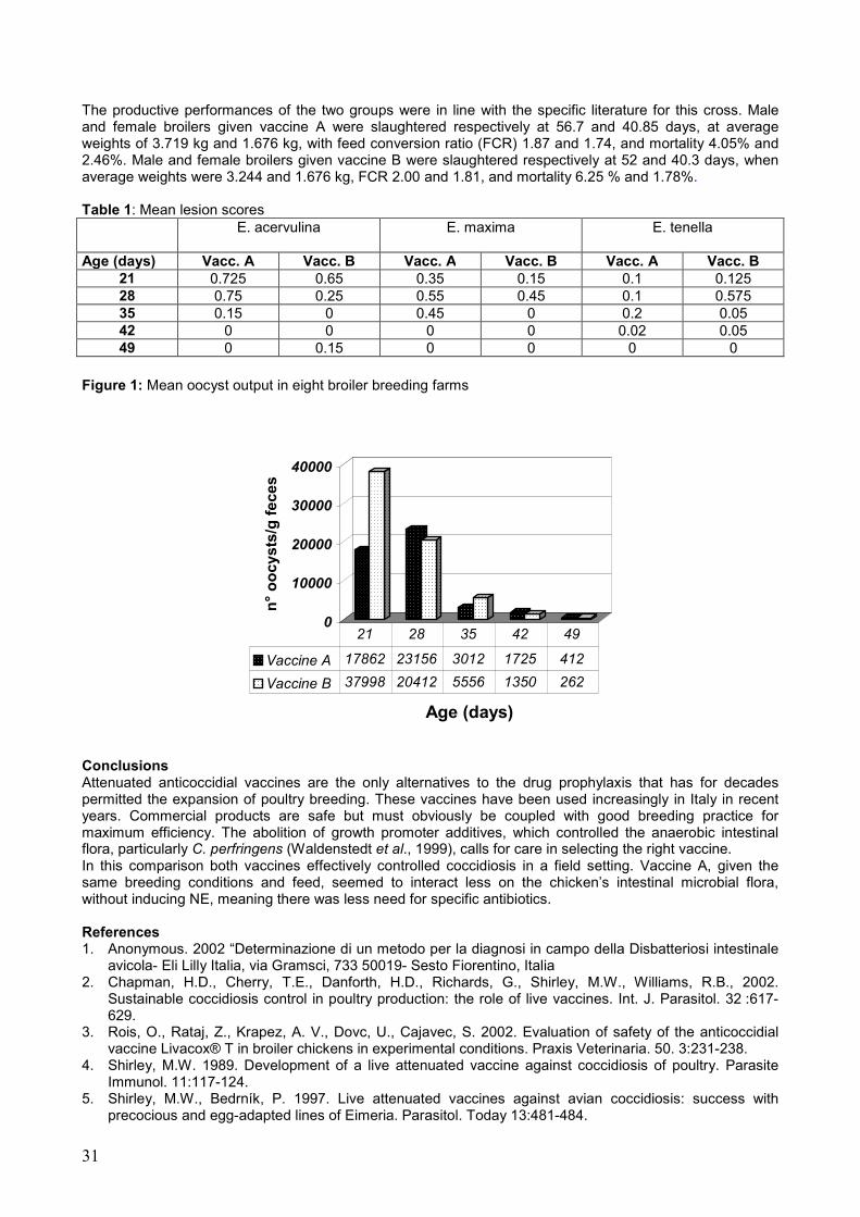
31
The productive performances of the two groups were in line with the specific literature for this cross. Male and female broilers given vaccine A were slaughtered respectively at 56.7 and 40.85 days, at average weights of 3.719 kg and 1.676 kg, with feed conversion ratio (FCR) 1.87 and 1.74, and mortality 4.05% and 2.46%. Male and female broilers given vaccine B were slaughtered respectively at 52 and 40.3 days, when average weights were 3.244 and 1.676 kg, FCR 2.00 and 1.81, and mortality 6.25 % and 1.78%.
Table 1: Mean lesion scores E. acervulina E. maxima E. tenella
Age (days) Vacc. A Vacc. B Vacc. A Vacc. B Vacc. A Vacc. B
21 0.725 0.65 0.35 0.15 0.1 0.125 28 0.75 0.25 0.55 0.45 0.1 0.575 35 0.15 0 0.45 0 0.2 0.05 42 0 0 0 0 0.02 0.05 49 0 0.15 0 0 0 0
Figure 1: Mean oocyst output in eight broiler breeding farms
0
10000
20000
30000
40000
n°
oo
cyst
s/g
fec
es
Age (days)
Vaccine A 17862 23156 3012 1725 412
Vaccine B 37998 20412 5556 1350 262
21 28 35 42 49
Conclusions Attenuated anticoccidial vaccines are the only alternatives to the drug prophylaxis that has for decades permitted the expansion of poultry breeding. These vaccines have been used increasingly in Italy in recent years. Commercial products are safe but must obviously be coupled with good breeding practice for maximum efficiency. The abolition of growth promoter additives, which controlled the anaerobic intestinal flora, particularly C. perfringens (Waldenstedt et al., 1999), calls for care in selecting the right vaccine. In this comparison both vaccines effectively controlled coccidiosis in a field setting. Vaccine A, given the same breeding conditions and feed, seemed to interact less on the chicken’s intestinal microbial flora, without inducing NE, meaning there was less need for specific antibiotics. References 1. Anonymous. 2002 “Determinazione di un metodo per la diagnosi in campo della Disbatteriosi intestinale
avicola- Eli Lilly Italia, via Gramsci, 733 50019- Sesto Fiorentino, Italia 2. Chapman, H.D., Cherry, T.E., Danforth, H.D., Richards, G., Shirley, M.W., Williams, R.B., 2002.
Sustainable coccidiosis control in poultry production: the role of live vaccines. Int. J. Parasitol. 32 :617-629.
3. Rois, O., Rataj, Z., Krapez, A. V., Dovc, U., Cajavec, S. 2002. Evaluation of safety of the anticoccidial vaccine Livacox® T in broiler chickens in experimental conditions. Praxis Veterinaria. 50. 3:231-238.
4. Shirley, M.W. 1989. Development of a live attenuated vaccine against coccidiosis of poultry. Parasite Immunol. 11:117-124.
5. Shirley, M.W., Bedrník, P. 1997. Live attenuated vaccines against avian coccidiosis: success with precocious and egg-adapted lines of Eimeria. Parasitol. Today 13:481-484.
32
6. Waldenstedt, L., Lunden, A., Elwinger K., Thebo, P., Uggla, A. 1999. Comparison between a live, attenuated anticoccidial vaccine and an anticoccidial ionophore, on performance of broilers raised with or without a growth promoter, in an initially Eimeria-free environment. Acta Vet. Scand. 40:11-21.
7. Williams, R.B., Andrews, S. J. 2001. The origins and biological significance of the coccidial lesions that occur in chickens vaccinated with a live attenuated anticoccidial vaccine. Avian Path. 30:215-220.
8. Williams, R.B., Gobbi, L. 2002. Comparison of an attenuated anticoccidial vaccine and an anticoccidial drug programme in commercial broiler chickens in Italy. Avian Path. 31:253-265.
9. Williams, R.B., Marshall, R.N., La Ragione, R.M:. Catchpole, J. 2003. A new method for the experimental production of necrotic enteritis and its use for studies on the relationships between necrotic enteritis. coccidiosis and anticoccidial vaccination of chickens. Parasitol. Res. 90:19-26
8) ISOLATION OF SALMONELLA IN ANIMAL FOODSTUFF FOR POULTRY
Laura Fiorentini1, Giovanni Tosi1, Paola Massi1
1Istituto Zooprofilattico Sperimentale della Lombardia e dell’Emilia Romagna
Corresponding author: Dr. Laura Fiorentini. Istituto Zooprofilattico Sperimentale della Lombardia e dell’Emilia Romagna, Sezione Diagnostica di Forlì. Via Marchini 1, 47100 Forlì, Italy- Tel. & Fax +39 0543 721533 Email: [email protected]
Abstract Here we have reported data referring to the isolation of Salmonella spp. in foodstuffs and non-medicated composite feed destined for poultry. We found that the foodstuffs were frequently contaminated compared to the composite non-medicated feed. The most common species found was S. seftemberg that belongsto E group that was isolated from Soya protein flour and wheat germ. (We are presently putting a monitoring plan into force that deals with the qualitative study of salmonella in animal feed. It should be noted however that quicker and quantitative techniques should also be activated in the future. Key words: Salmonella spp., raw materials, non-medicated composite feed ISOLAMENTO DI SALMONELLA SPP. NEGLI ALIMENTI ZOOTECNICI Riassunto Questa comunicazione riporta alcuni dati relativi agli isolamenti di Salmonella spp. da materie prime e mangimi composti non medicati destinati al settore avicolo. Le materie prime sono risultate maggiormente contaminate dei mangimi composti non medicati con maggior prevalenza della specie seftenberg appartenente al gruppo E, isolata da farina di soja proteica e da crusca. Attualmente viene attuato un piano monitoraggio che prevede la ricerca qualitativa della Salmonella negli alimenti destinati all’alimentazione zootecnica, anche se è auspicabile che in futuro saranno previste tecniche di tipo quantitativo e più tempestive. Introduction There is presently a monitoring programme in force throughout the European Union that has been designed to evaluate the contamination levels of Salmonella spp. in raw materials used to produce vegetarian animal feed used either as they are or in composite food stuffs for poultry, pigs, cattle, birds, goats, fish and rabbits. It is clear that even vegetable based feed can contain pathogenic agents for animals and man that therefore create food and public health problems. At the moment there are no Community laws referring to zoonotic agents in animal feed specifically from vegetarian based origins. It is for this reason that this monitoring programme could be useful to integrate into the hygiene guidelines and quality control programs of production, transport, storing and handling of zoo-technical food products, that will enter into force on 1st of January 2006. Therefore, the microbiological controls have recently been intensified in order to measure the frequency and level of Salmonella spp contamination in vegetarian based animal feed. From 1st January 2003 to 10th July 2004, the IZSLER microbiology laboratories at Forlì analysed 304 samples; 125 of which were foodstuffs and 179 complete non-medicated animal feed all of which were destined for poultry. These were all samples supplied directly from the producers and by the breeders according to strict internal quality control. Materials and Methods. The feed samples were analysed according to a five consecutive step microbiological programme:

33
• Pre-enrichment of the non-selective liquid medium (tamponed peptonated water) inoculated with the examined sample and then once it was homogenised andincubated at 37°C for 24 hours.
• Enrichment in selective liquid medium (selenite cystine mixture and Rappaport Vassiliadis mixture) incubated at 37°C and 42°C respectively for 24 hours.
• Petri dishes prepared with selective and differentiated solid medium Hektoen enteric Agar were inoculated and then incubated at 37°C for 24 hours.
• Suspect colonies were transplanted to Kligler Iron Agar and incubated at 37°C for 24 hours. • Biochemical and serological identification of the transplanted colonies (Zavanella M., 2001; Division
of Microbiology. Center for Food safety and applied nutrition, 1984; Norme ISO 6579, 1993). Results The detailed results are shown in the tables 1, 2 and 3. About 10% (28/304) of the samples were positive for Salmonella spp. with a higher frequency of S. seftemberg group E species, isolated from the protein Soya flour and wheat germ. Of the 28 strains isolated, 4 were not typed. Worth mentioning was the isolation of Salmonella typhimurium from protein Soya flour and of Salmonella enteritidis from complete non-medicated feed. Both of these strains are considered pathogenic species, as is Salmonella kedougou, which apart from being particularly rare was also responsible for an episode of food poisoning in 1994 in the Marche region in patients who had eaten cooked pork (Staffolani M et al., 2003). The foodstuffs were more frequently contaminated than the non-medicated composite feed. Discussion Salmonella is more frequently present in foodstuffs and Soya can be considered a good substrate. Composite feed are less frequently contaminated probably due to the production process, the use of additives and their lower humidity. We should not forget that animals become infected with salmonella and possibly develop salmonellosis also from contaminated feed, that justify a a monitoring programme. However presently this monitoring program is only qualitative even given the ever greater need to find also the quantitative data as well as prevent salmonella and any other zoonotic agents as early as possible in the food chain. References 1. Division of Microbiology. Center for Food safety and applied nutrition. 1984. U.S.F.D.A. Bacteriological
analitical. 6th Edition, Association of Official Analytical Chemists, Arlington 2. Norme ISO 6579. 1993. Directives generales concernant les methods de recherchè des Salmonella,
AFNOR. 3. Staffolani M., Fisichella S. 2003. Focolaio epidemico umano di Salmonella kedougou nella Regione
Marche. V Congresso nazionale SIDiLV. Pisa 4. Zavanella M. 2001. Tipizzazione delle Salmonelle. Ed. Fondazione Iniziative Zooprofilattiche e
Zootecniche. Brescia Table 1: Raw materials N. samples raw materials
108 protein Soya flour 7 Mais 5 Crusca 2 FARINA DI GRANO TENERO 1 FARINA D’OSTRICHE 1 FARINA DI PESCE 1 FARINA DI GIRASOLE
125 Total raw materials

34
Table 2: Salmonella in raw materials raw materials (28 strains Salmonella/125 samples)
raw materials Group Salmonella (species) SOJA E SENFTENBERG SOJA C ARDWICK/RISSEN SOJA C ARDWICK/RISSEN SOJA E SENFTENBERG SOJA E SENFTENBERG SOJA C ARDWICK/RISSEN SOJA E SENFTENBERG SOJA E GRUPPO E (non tipizzata) SOJA E GRUPPO E (non tipizzata)
CRUSCA E SENFTENBERG / IDIKAN SOJA C/ not categorised KEDOUGOU/MBANDAKA SOJA C/E S. MONTEVIDEO/RISSEN SOJA E MUENSTER SOJA E HAVANA SOJA E ENTERICA Subsp. ENTERICA SOJA B TYPHIMURIUM SOJA E MUENSTER SOJA E SENFTENBERG SOJA not categorised DERBY SOJA C MONTEVIDEO SOJA E ENTERICA Subsp. ENTERICA SOJA not categorised MBANDAKA
Table.3: Salmonella in non-medicated composite feed
non-medicated composite feed 4positive /179 samples
group Salmonella (species) C KENTUCKY
not categorised BRAENDERUP D ENTERITIDIS
not categorised ENTERICA Subsp. ENTERICA
9) EVALUATION OF AN ADDITIVE EFFICACY IN BROILER LITTER MICROBIAL LEVEL CONTROL IN FIELD: PRELIMINARY RESULTS
G. Tacconi1, P. Casagrande Proietti1, R. Ventura2, R. Arcaro2, M. Pennacchi2
1Dipartimento di Scienze Biopatologiche Veterinarie, Università di Perugia, Italy
2Laurea in SPA
Corresponding author: Prof. Giuseppina Tacconi. Dipartimento di Scienze Biopatologiche Veterinarie, Sezione di Patologia e Igiene Veterinaria, Università degli Studi di Perugia - Facoltà di Medicina Veterinaria, Via S. Costanzo,4 - 06126, Perugia, Italy. Tel: +39755857736. Fax: +39755857738 - E-mail: [email protected] ABSTRACT The present study was conducted to evaluate in field the efficacy of an additive (SOP® C Poultry), as an agent for the control of micro-organisms in broiler litter. The Total aerobic Microbial Count, Staphylococcus spp., Coliforms, and Salmonella spp. in broiler litter samples of both the Houses, 2 and 3, were determined, and also at the end of each cycle the mortality rate was recorded. The results showed significant differences of all the microbial counts between treated litter samples and the control. Significant resulted also the difference in mortality rate recorded between H2 and H3. Key words: litter additive, environment, broiler, TMC, Coliforms, Stapylococcus spp., Salmonella spp., mortality.
35
Prova di efficacia di un additivo di nuova concezione nel controllo di alcune componenti microbiche della lettiera di polli da carne. Primi risultati. RIASSUNTO Prove di campo sono state effettuate per valutare l’efficacia di un prodotto igienizzante di nuova concezione (SOP® C Poultry), nel controllo di alcune componenti microbiche della lettiera di polli da carne. Sono stati considerati, come parametri la Carica Microbica Totale aerobia (CMT), Staphilococcus spp., Salmonella spp. e Coliformi ed inoltre alla fine di ogni ciclo è stata valutata la percentuale di mortalità. I risultati hanno mostrato una significativa riduzione dei valori medi per ciascuno dei parametri microbiologici valutati nei campioni di lettiera trattata rispetto al controllo; significativa è risultata anche la riduzione della percentuale di mortalità riscontrata nel capannone trattato rispetto al controllo. Parole chiave: prodotto igienizzante, lettiera, ambiente, polli da carne, CMT, Coliformi, Staphyloccus spp., Salmonella spp., mortalità. Introduction The environment in the poultry house is a combination of physical and biological factors which interact as a complex dynamic system of social interactions, husbandry system, light, temperature and the aerial environment (Sainsbury,1992; Kristensen and Wathes, 2000). The high stocking density in the modern poultry house may lead to reduce air quality with high concentrations of aerial pollutants (Curtis and Drummond, 1982; Maghirang et al., 1991; Feddes and Licsko, 1993; Kristensen and Wathes, 2000). Their concentrations in poultry houses approach, and sometimes exceed, recommended occupational limits for humans (Kristensen and Wathes, 2000). Litter is considered one of the major sources of pollutants in poultry houses, then the need to ménage it using additives has been considered since the last past years (Vanchev et al., 1989; Nestof and Petkov, 1994; Ivanov, 2001) but has not yet been resolved conclusively. The present study investigated the use of an additive as an agent for the control of micro-organisms in broiler litter, and also the possible effect on the mortality. Material and methods Planning. This study was carried out from February 2002 to March 2004 in two large commercial broiler houses, House2 and House3 (H2 and H3) of one Umbrian Company farm, in which broilers were reared to 7-8 weeks of age. The buildings were of conventional layout. The litter was cut wheat straw. In H2 and H3 the ventilation system comprised 2 propeller fans of 40,000 m3/hour and 26,000 m3/hour capacity each, mounted on the windward side of the poultry house to increase the velocity of the air as it blows through the building. Treatment. Litter in H2 was treated as follow: 2 g. of additive plus 25 g. of calcium carbonate per m2 were added to the litter the day before the arrival of the chicks, and the treatment repeated every two weeks until the end of each cycle. After the first month the additive dose was reduced to 1 g. per m2. Additive. The field trials were performed with Calcium sulphate (gypsum) and essential oils (lemon grass and lavender) used as carriers. By the SIRIO OPERATING PROCESS® method such components are activated by an energetic modulation process and enriched with oxygen and specific information about litter components. Samples. Litter was sampled one day during the first and the seventh week of each cycle. Composite samples of about 500 g. were obtained from ten different sites within each house and placed immediately into sterile plastic bags, sealed and refrigerated until microbiological evaluation was made. Microbiological analysis. Twenty-five grams of each sample was transferred into a sterile plastic bag and 225 ml of sterile 1% buffered peptone water was added. After treatment with Stomacker Circulator 400 (PBI, Milan) the samples were allowed to sit for 30-45 min at room temperature with frequent shaking. One ml of this samples (1:10 dilution) was diluted serially via 10-fold dilutions (from 10-1 to 10-8).Total aerobic bacteria, Staphylococcus spp. (Staph. spp.), Salmonella spp. and Coliforms in 1 g-1 of litter were determined by plating, in duplicated, 0.1-ml of appropriate dilution on SPGCA (Standard Plate Count Agar), BP (Baird Parker agar) and VRBA (Violet Red Bile Agar). The cultures were incubated at 37°C for 24-48 hr and the number of grown colonies was determined. The Salmonella spp. isolation procedure used in this study included Selenite-Cystine broth and Rappaport-Vassiliadis broth (Oxoid, Milan), as enrichment media and two plating media. The Selenite-Cystine Broth was incubated aerobically at 37° C for 24 hours and the Rappaport-Vassiliadis broth was incubated aerobically at 43° C for 24 hours. The subcultures from the enrichment media were made onto Hectoen enteric agar (Oxoid) and then incubated aerobically at 37° C for 24 hours. The composition of each selective medium is detailed in the OXOID Manual. Mortality rate was recorded at the end of each eight cycles. Statistical Analysis. The mean values of all parameters evaluated were compared by t-test.

36
Results and discussion The results from eight cycles on treatment of litter are summarised in Table 1 and 2. Significant differences between experimental and control samples, with regard both the microbial cell counts (Table 1), and the mortality (Table 2) were observed. Also the bacterial counts of the treated litter were reduced to about 70 % of the control values. Sex strains of Escherichia coli were isolated throughout the sampling period: 3 strains (1 from H2 and 2 from H3) during 2002-2003, and 3 strains (1 from H2 and 2 from H3) in 2004. Standard procedures were used to identify E. coli, which do not differentiate between pathogenic and non pathogenic. The number of E. coli in the litter, like total aerobic bacteria, Staphylococcus spp., and Coliforms resulted in treated litter lower than the control house litter. The treatment of the litter proved to be effective in control of some microbial litter components. Of interest is the reduced mortality rate of broilers because in field conditions the health problems are known to be associated with litter. Although all litter sampled was examined for Salmonella spp. none was found. Several workers suggested that bird health is harmed by chronic exposure to modest burdens, especially in the presence of simultaneous challenge by respiratory pathogens (Anderson et al., 1964; Oyetunde et al., 1978; Carpenter et al., 1986), but the concentration of most pollutants often rises in poultry houses. The rise is consequence of an increased generation rates from the major sources, that is the birds themselves and particularly the litter, which acts as a nutritious reservoir for micro-organisms (Conceição et al., 1989). Our data pointed out the high litter bacteria concentrations, considered common in broiler houses (Ivanov, 2001), and make its control essential for better health and performance of birds. The data of the present study seem to indicate a significant reduction of the bacteria evaluated in the treated litter. Based on the results of these field trials, it was concluded that the additive studied during this investigation has an inhibitory effect on the survivals of micro-organisms in broiler house litter. Table 1. Results of some microbial parameters in litter samples from H2 (treated with SOP C POULTRY) and H3 (control) (mean values from eight cycles are expressed in CFU/g-1).
Parameters TMC TMC Staph. spp. Staph. spp. Coliforms Coliforms
Dilution 10-8 10-8 10-8 10-8 10-6 10-6
Houses treated control treated control treated control
µ 153,69 416,42 31,14 185,48 58,05 328,34 t test 0,0078 0,0021 0,0541
% -63,1 -83,21 -82,32 Table 2. Mortality rate (%) recorded at the end of eight cycles in H2 and H3 (treated with SOP C POULTRY and control). Cycles 1st 2nd 3rd 4th 5th 6th 7th 8th treated 5 4,3 3,1 8,4 3,4 3,1 3,3 3,9 control 9 4,7 4,3 10,8 5,7 3,4 5,1 4,5 P= 0,00106 Acknowledgements The writers wish to express their gratitude to the SOP S.r.l. for furnishing the additive used in this study and for providing financial support for the project. Il lavoro è stato supervisionato dal traduttore di madre lingua della Facoltà di Medicina Veterinaria di Perugia. REFERENCES 1. Anderson, D.P., Beard, C.W. & Hanson, R.P. 1964. The adverse effects of ammonia on chickens
including resistance to infection with Newcastle disease virus. Avian Diseases. 8: 369-379 2. Carpenter, G.A., Smith, W.K., Maclaren, A.P.C. & Spackman, D. 1986. Effects of internal air filtration on
the performance of broilers and the aerial concentrations of dust and bacteria. British Poultry Sci., 27:471-480.
3. Conceição, M. A. P., Johnson, H. E. & Wathes, C. M. 1989. Air hygiene in a pullet house: Spatial homogeneity and aerial pollutants. British Poultry Science. 7: 189-198.
4. Curtis, S. E., & Drummond, J. G. 1982. Air environment and animal performance. In : Handbook of Agricultural Productivity, volume 2, Animal Productivity (Rechcigl, M. Jr, Ed.) CRC Press Inc, Florida, USA.
5. Feddes, J. J. R. & Licsko, Z. J. 1993. Air quality in commercial turkey housing. Canadian Agricultural Engineering. 35: 147-150.
6. Kristensen H. H. & Wathes, C. M. 2000 Ammonia and poultry welfare: a review. World’s Poultry Science Journal. 56: 235-245.

37
7. Ivanov, I. E. 2001. Treatment of boiler bitter with organic acids. Research in Veterinary Science, 70, 169-173.
8. Netsov, N., & Petkov, G. 1944. Zoohygiene, Sofia, 17-19:284-285. 9. Maghirang R. G., Manbeck, H. B., Roush, W. B. & Muir, F. V. 1991. Air contaminant distributions in a
commercial laying house. Transactions of the American society of Agricultural Eingeneering. 34: 2171-2180.
10. Oyetunde, O.O. F., Thomson, R. G. & Carlson, H. C. 1978. Aerosol exposure of ammonia, dust and Escherichia coli in broiler chickens. Canadian Veterinary Journal. 19: 187-193.
11. Sainsbury, D. 1992. Poultry Health and Management- Chickens, Turkeys, Ducks, Geese, Quail, 3rd ed, Blackwell scientific Ltd, Oxford, UK.
12. Vanchev, T., Donchev, R., & Kaitazov, G. 1989. Poultry breeding, Sofia, 147-149.
38
10) ASCARIDIA GALLI: A REPORT OF ERRATIC MIGRATION
1Daniela Piergili Fioretti, 1Fabrizia Veronesi, 1Manuela Diaferia, 2Maria Pia Franciosini 2Patrizia Casagrande Proietti
1Dipartimento di Scienze Biopatologiche Veterinarie, Sezione di Parassitologia.
2Dipartimento di Scienze Biopatologiche Veterinarie, Sezione di Patologia ed Igiene Veterinaria, Università di
Perugia, Italy.
Corresponding author: Prof.ssa Daniela Piergili Fioretti. Dipartimento di Scienze Biopatologiche Veterinarie. Università di Perugia. Via S. Costanzo 4. 06126 Perugia, Italy. Tel. +39 075 5857740- Fax +39 075 5857743- Email: [email protected].
Abstract This paper describes a case of an unusual recovery of adult Ascaridia galli in hen’s egg . Several data are available on this occurrence but it appears to be the first case described in Italy. The worm was identified as an adult female, 6,8 cm in lenght, with three trilobed lips, cervical narrow alae, oesophagus club-shaped without posterior bulb, vulva near the middle of body, with gravid uteri containing a large number of eggs. The presence of Ascaridia galli in hen’s eggs cannot be considered as hazard for public health but may be cause of a potential consumer complaint. Moreover it is a sign of presence of ascaridiosis, parasitosis that still produces economic losses in modern poultry production system. Key words: Ascaridia galli, Erratic migration, Hen’s egg.
ASCARIDIA GALLI: UN CASO DI MIGRAZIONE ERRATICA Riassunto Nel seguente lavoro viene descritto l’ inconsueto ritrovamento nell’albume di un uovo commerciale di gallina di un esemplare adulto di Ascaridia galli. Dalla bibliografia a nostra disposizione, relativa all’argomento, questa risulta essere la prima segnalazione effettuata nel nostro Paese. Il nematode isolato è stato identificato come femmina adulta di Ascaridia galli, di 6,8 cm di lunghezza, morfologicamente caratterizzato da bocca trilabiata, esofago claviforme con assenza di bulbo posteriore, strette ali cefaliche, vulva situata nella parte mediana del corpo. L’esemplare inoltre era gravido con un utero contenente numerose e caratteristiche uova. Il riscontro di Ascaridia galli nelle uova di gallina, pur non rappresentando un rischio per la salute pubblica, è tuttavia fonte di estremo disagio per i consumatori. Recentemente l’affermarsi di sistemi di allevamento alternativi ha di nuovo reso attuale la presenza di questa parassitosi nel settore avicolo. Parole chiave: Ascaridia galli, Migrazione erratica, Uovo di gallina. Introduction Ascaridia galli, the largest and the most common helminth of the small intestine of chickens, is a parasite with a direct life cycle and poultry is infected by ingestion of embryonated eggs containing the second larval stage (L2). The infection is a direct consequence of faecal contamination of environment. The parasite completes its life cycle exclusively in the intestinal tract with a larval migratory phase into the enteric wall. The main pathogenic effect is seen during the prepatent phase, when the larvae are in the mucosa. There are no clinical signs in moderate infection, while the presence of a considerable number of adult worms may cause intestinal occlusion and death, because of the large size of these worms. The adult subjects are often symptomless carriers while the chickens before feathering are particularly susceptible. Usually the larvae are responsible for catarrhal enteritis, but haemorrhagic lesions can be observed in heavy infections. One of the most striking effects of infection is the occasional finding of this parasite in the hen’s egg. Although several observations of this phenomenon have been made in literature (Akinyemi J.O., 1980; Omran L.A., 1982, Manna B., 1992) we considered interesting to describe this case, since, as far as we know, it is the first case reported in Italy . Material and Methods A fresh chicken egg, with a white filiform structure referable to a round worm in the albumen, was submitted to attention to the Veterinary Parasitology Laboratory of Perugia University. The egg, bought by a private consumer, came from an Umbrian commercial farming. The worm was isolated, washed in distilled water, kept in physiological solution for 2 hours at 40° C to help the extension, fixed in 70% alcohol for 24 hours, clarified in lactophenol of Amman for 10 hours, mounted on a microscope slide and observed by light microscopy at 10x- 40x. The morphometric identification of the worm was based on the criteria reported by Yamaguti (1961). The albumen was examined by floatation method using a sugar NaNo3 solution (specific density 1300).
39
Results The worm was 6,8 cm in lenght, with three trilobed lips, cervical narrow alae, oesophagus club-shaped without posterior bulb, vulva near the middle of body, gravid uteri containing a large number of eggs. On the basis of morphometric characteristics the worm was identified as an adult female of Ascaridia galli. The floatation method allowed to evidence the presence in the albumen of typical Ascaridia galli eggs, oval in shape, with smooth shell and size of 75 x 30 µm. Discussion This appears to be the first report of the presence of Ascaridia galli in hen’s egg occurring in Italy . It is possible that this occurrence is quite frequent but the common use of hard-boiled eggs could make unnoticeable the presence of these worms in the boiled egg albumen. A primary localization in the genital apparatus of Ascaridia galli larva L2 with subsequent maturation to aduld worm has to be excluded because the egg production is more quick than the time required for maturity of larva. In addition the adult worm in egg was gravid and Ascaridia galli is not an hermaphrodite nematode. Presumably the worms migrate up the oviduct via the intestinal wall and peritoneal cavity or via the cloaca with subsequent inclusion in the egg. Although the presence of Ascaridia galli in hen’s eggs cannot be considered as hazard for public health, it may be cause of potential consumers complaint. This finding is noteworthy since it indirectly points out the problems related to ascaridiosis, that at present is playing an important role in the alternative rearing systems, such as the organic farming . It is known that the development of alternative housing systems for laying hens in response to public disquiet on the use of battery cages has caused a recrudescence of the parasitic diseases. Moreover the most of the conventional farming could be shortly converted on the floor system on the basis of 74/99 CEE regulation. Recent epidemiological studies carried out on different poultry breeding systems showed a Ascaridia galli prevalence of 63,9% in organic chickens and a prevalence of 41,9% in chickens reared in covered strawyard. The prevalence reported in breeders and in caged hens was respectively 37,5% and 5% (Permin et al., 1999). On the basis of these epidemiological data and of our report further investigations are suggested to obtain more information on the occurrence of Ascaridiosis in Umbria (Central Italy) in relation to diffusion of alternative housing systems in hens. REFERENCES Akinyemi J.O., Ogunji F.O. and Dipeolu O.O., 1980. A case of adult Ascaridia galli in hen’s egg. Int. J. Zoon., 7: 171-172; Manna B., 1992. A report of Ascaridia galli within the albumen of hen’s eggs. Ind. J. An. Heal., 31 (1): 89; Omran Laila A.M., 1982. Ascaridia galli (Shrank, 1788): an erratic parasite in a fowl’s egg albumin. J. Egypt Soc. Parasitol., 12 (1): 167-168. Permin A., Bisgaard M., Frandsen F., Pearman M., Kold J., Nansen P., 1999. Prevalence of gastrointesrinal helminths in different poultry production system. Br. Poult. Sci., 40 (4): 439-443. Yamaguti S., 1961. Systema Helminthum.1st ed. Interscience. Publishers Inc. New York, USA.
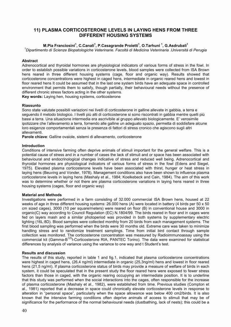
40
11) PLASMA CORTICOSTERONE LEVELS IN LAYING HENS FROM THREE DIFFERENT HOUSING SYSTEMS
M.Pia Franciosini1, C.Canali1, P.Casagrande Proietti1, O.Tarhuni 1, G.Asdrubali1
1Dipartimento di Scienze Biopatologiche Veterinarie. Facoltà di Medicina Veterinaria .Università di Perugia
Abstract Adrenocortical and thyroidal hormones are physiological indicators of various forms of stress in the fowl. In order to establish possible variations in corticosterone levels, blood samples were collected from ISA Brown hens reared in three different housing systems (cage, floor and organic way). Results showed that corticosterone concentrations were highest in caged hens, intermediate in organic reared hens and lowest in floor reared hens It could be assumed that in the last one system birds have an adequate space in controlled environment that permits them to satisfy, though partially, their behavioural needs without the presence of different chronic stress factors acting in the other systems. Key words: Laying hen, housing systems, corticosterone Riassunto Sono state valutate possibili variazioni nei livelli di corticosterone in galline allevate in gabbia, a terra e seguendo il metodo biologico. I livelli più alti di corticosterone si sono riscontrati in gabbia mentre quelli più bassi a terra. Una situazione intermedia era ascrivibile al gruppo allevato biologicamente. E’ verosimile ipotizzare che l’allevamento a terra, fornendo alle galline un adeguato spazio, permetta di soddisfare alcune loro esigenze comportamentali senza la presenza di fattori di stress cronico che agiscono sugli altri allevamenti. Parole chiave: Galline ovaiole, sistemi di allevamento, corticosterone Introduction Conditions of intensive farming often deprive animals of stimuli important for the general welfare. This is a potential cause of stress and in a number of cases the lack of stimuli and or space has been associated with behavioural and endocrinological changes indicative of stress and reduced well being. Adrenocortical and thyroidal hormones are physiological indicators of various forms of stress in the fowl (Edens and Siegel, 1975). Elevated plasma corticosterone levels have been associated with thirst, hunger or heat stress in laying hens (Beuving and Vonder, 1978). Management conditions also have been shown to influence plasma corticosterone levels in laying hens (Mashaly et al., 1984; Koelkebeck and Cain, 1984). The aim of this work was to determine whether or not there are plasma corticosterone variations in laying hens reared in three housing systems (cages, floor and organic way) Material and Methods Investigations were performed in a farm consisting of 32.000 commercial ISA Brown hens, housed at 22 weeks of age in three different housing systems: 26.000 hens (A) were located in battery (4 birds per 50 x 50 cm sized cages), 3000 (10 per square/meters) were reared on floor (B) in conventional house and 3000 in organic(C) way according to Council Regulation (EC) N.1804/99. The birds reared in floor and in cages were fed on layers mash and a similar photoperiod was provided in both systems by supplementary electric lighting (16L:8D). Blood samples were collected monthly from 20 birds from each management systems. The first blood sampling was performed when the birds were 30 months old. Extreme care was taken to minimize handling stress and to randomize treatment samplings. Time from initial bird contact through sample collection was monitored. The corticosterone concentration was measured by Radioimmunoassay using the commercial kit (Gamma-B125I-Corticosterone RIA, PANTEC Torino). The data were examined for statistical differences by analysis of variance using the variance to one way and t Student’s test. Results and discussion The results of this study, reported in table 1 and fig.1, indicated that plasma corticosterone concentrations were highest in caged hens, (26,4 ng/ml) intermediate in organic (25,3ng/ml) hens and lowest in floor reared hens (21,5 ng/ml). If plasma corticosterone status of birds may provide a measure of welfare of birds in each system, it could be speculated that in the present study the floor reared hens were exposed to fewer stress factors than those in caged, with the organic rearing occupying an intermediate position. It is to underline that this study was performed when the social interactions into the cages, often responsible for the increase of plasma corticosterone (Mashaly et al., 1982), were established from time. Previous studies (Compton et al., 1981) reported that a decrease in space could chronically elevate corticosterone levels in response to alteration in “personal space”, particularly when the space allowance was below 400 cm2/birds. It is also known that the intensive farming conditions often deprive animals of access to stimuli that may be of significance for the performance of the normal behavioural needs (dustbathing, lack of nests); this could be a

41
potential cause of a chronic stress. However this conclusion should be considered with caution since the discrepancy of results was observed in different studies. Koelkebeck and Cain (1984) indeed reported the lowest mean plasma corticosterone levels occurred among hens in cage and the highest on litter, indicating that the social relationship within a large group may act as a greater stress factor than conditions found in small groups reared in cage. Data obtained from organic hens (tab.1) showed that mean corticosterone levels were higher then those reported in hens reared on floor, though the organic system should be the best in satisfying the animal welfare. The results related to corticosterone plasma levels in organic hens also showed significant differences among the three blood samplings (fig.2). On April, indeed, we observed an increase of corticosterone, likely explained by the fact that in this month the hens started to come out and many uncontrolled agents could act as chronic stress factors (prey presence, a number of visual and olfactory stimulation). The corticosterone reduction detected in the last sampling (20,97 ng/ml) may be sign of establishing of an increasing “environmental confidence” from the hens. In our study the lowest corticosterone levels were detected in floor reared hens. It seems reasonable to speculate that in this rearing system birds have an adequate space allowance in controlled environment that permits them to satisfy, though partially, their behavioural needs without the presence of various stress factors acting in the other systems. The time of blood sampling collection should not have any influence on the corticosterone concentration since blood was taken within 1 min. in each bird from all systems. Craig and Craig (1985) showed that delays of 2 and 3 min. after catching have little, if any, effect on basal plasma corticosterone concentration. Further investigations are necessary to establish if plasma corticosterone can be an useful measure of long term stress or welfare in hens since the physiological level of the “stress” hormone in the hens is not still well defined and there are difficulties with the interpretation of circulating hormone concentrations due to diurnal patterns and to the sampling procedures. It may be corrected in concluding that other parameters, analogously to other species (Barone et al.), as practical estimates of welfare should be considered in different housing conditions, particularly in organic farming, where the corticosterone response may be dependent on a complex interaction with a number of other environmental variables difficult to identify. Tab.1 Mean plasma corticosterone levels and standard deviation in differently housed hens in relation to the date of sampling.
CAGE (a)
FLOOR (b)
ORGANIC (c)
Sampling date
Mean Standard Deviation
Mean Standard deviation
Mean Standard deviation
Significance
March 04
Corticosterone
Temp. °C
27.25
22-23
Γ5.11 20.04
20
Γ5.63 25.91
20
Γ5.40 a-b pΡ0.01 b-c pΡ0.01
a-c n.s.
April 04 Corticosterone
Temp. °C
25.69
22-23
Γ7.94 22.63
22
Γ5.91 28.91
23
Γ6.90 a-b n.s. b-c pΡ0.01
a-c n.s.
May 04 Corticosterone
Temp. °C
25.71
22-23
Γ5.59 21.73
24
Γ5.11 20.97
24
Γ6.90 a-b pΡ0.05 b-c n.s.
a-c. pΡ0.05
total
26.4
Γ6.3
21.5
Γ5.6
25.3
Γ7.2 a-b pΡ0.01 b-c pΡ0.01
a-c n.s.
References Barone A.S., Canali C., Diverio S., Federici C. and Pelliccia C. 2003. The effects of farming system and slaughtering on rabbit welfare. Proceedings of UFAW Symposium 2-4 April. Edinburgh Beuving G. and Vonder G.M.A. 1978. Effect of stressing factors on corticosterone levels in the plasma of laying hens. Gen.Comp.Endocrinol.35: 153-159 Compton M.M., Van Krey H.P.. Ruszler P.L. and Gwasdauskas F.C. 1981. The effect of claw removal and cage design on the production performance, gonadal steroids and stress response in caged laying hens. Poultry Sci.60:2127-2135
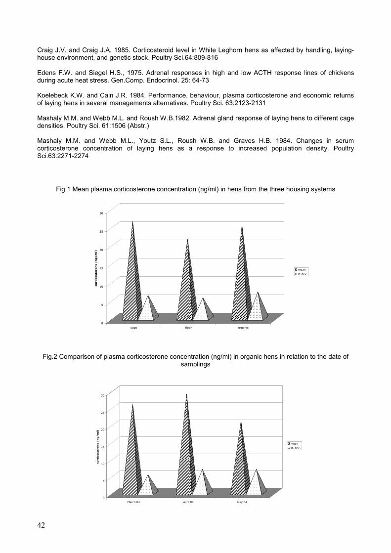
42
Craig J.V. and Craig J.A. 1985. Corticosteroid level in White Leghorn hens as affected by handling, laying-house environment, and genetic stock. Poultry Sci.64:809-816 Edens F.W. and Siegel H.S., 1975. Adrenal responses in high and low ACTH response lines of chickens during acute heat stress. Gen.Comp. Endocrinol. 25: 64-73 Koelebeck K.W. and Cain J.R. 1984. Performance, behaviour, plasma corticosterone and economic returns of laying hens in several managements alternatives. Poultry Sci. 63:2123-2131 Mashaly M.M. and Webb M.L. and Roush W.B.1982. Adrenal gland response of laying hens to different cage densities. Poultry Sci. 61:1506 (Abstr.) Mashaly M.M. and Webb M.L., Youtz S.L., Roush W.B. and Graves H.B. 1984. Changes in serum corticosterone concentration of laying hens as a response to increased population density. Poultry Sci.63:2271-2274
Fig.1 Mean plasma corticosterone concentration (ng/ml) in hens from the three housing systems
Fig.2 Comparison of plasma corticosterone concentration (ng/ml) in organic hens in relation to the date of
samplings
0
5
10
15
20
25
30
corti
coste
ron
e (
ng
/m
l)
cage floor organic
mean
st.dev.
0
5
10
15
20
25
30
co
rti
co
ste
ro
ne (
ng
/m
l)
March 04 April 04 May 04
mean
st. dev.
43
12) DETECTION OF CALICIVIRUSES IN YOUNG PHEASANTS (PHASIANUS COLCHICUS) WITH ENTERITIS IN ITALY
A. Toffan1, F. Montesi1, L. Bano2, M.S. Beato3, R. De Nardi3,C. Terregino3 & I. Capua3
1 Electron Microscopy Laboratory, Istituto Zooprofilattico Sperimentale delle Venezie,Italy
2 Laboratory of Treviso, Istituto Zooprofilattico Sperimentale delle Venezie, Italy
3 Virology Department, Istituto Zooprofilattico Sperimentale delle Venezie, Italy
Corresponding author: Dr. Anna Toffan, Istituto Zooprofilattico delle Venezie, Laboratorio di Microscopia Elettronica, Viale dell’Università 10, 35020 Legnaro (PD), Italy -Tel. +39 049 8084389 –Fax +39 8084360 -E-mail: [email protected]
Running title: Calicivirus in pheasants with enteritis
Summary During June 2004 a severe enteritis was reported in a farm of 28 day-old pheasants reared in intensive conditions in North-Eastern Italy. Mortality in the flock had reached 25%. Virological investigations on cell culture of the gut content yielded reoviruses while electron microscopy examination revealed viral particles morphologically related to calicivirus in association with parvovirus-like and rod shaped virus-like particles. This paper is the first report of calicivirus associated enteritis in pheasants in Italy. Key words: Pheasant, Enteritis, Calicivirus
Infezione da Calicivirus associata ad una sindrome enterica in giovani fagiani (Phasianus colchicus)
Riassunto Il presente lavoro descrive un grave episodio di sindrome enterica osservato in un gruppo di fagiani di allevamento di 28 giorni d’età. Oltre all’isolamento di reovirus dal contenuto intestinale, gli esami di microscopia elettronica hanno permesso di evidenziare la presenza di particelle virali morfologicamente riferibili a calicivirus associate a parvovirus-like e rod shaped virus-like particles. Dalla letteratura consultata questo risulta essere il secondo caso di enterite del fagiano associato a infezione da calicivirus, ed in considerazione della grave sintomatologia osservata sarebbe auspicabile un approfondimento della casistica per chiarire il ruolo di questo patogeno nel determinismo delle enteriti dei volatili in generale e del fagiano in particolare. Parole chiave: Fagiano, Enterite, Calicivirus
Introduction Viral enteritis is commonly reported in young birds, and among these, game birds appear to be highly susceptible particularly if reared in intensive farming conditions. Young pheasants (Phasianus colchicus) suffer this pathologic condition with mortality rates ranging between 3% to 30% (Gough et al. 1985). Most viral enteric problems in pheasants are caused by rotavirus and parvovirus-like, which can also be found in combination. Calicivirus infection is commonly reported in enteric diseases in mammals (human, canine, bovine, porcine) (Murphy et al. 1999), but are less frequently observed in birds (Gough et al., 1992). The present paper reports of the results of laboratory investigations in a flock of pheasants with acute enteritis, in which the examination of the gut content by electron microscopy resulted in the detection of viruses which are unusually found in this species Materials and methods In June 2004, 28 day-old dead pheasant chicks which had experienced severe enteritis were submitted for laboratory investigations. The birds originated from an industrial pheasant farm consisting of 70,000 birds located in northern Italy. The clinical condition was characterized by depression, dehydration, severe enteritis and increased mortality rate. Between day 18 to day 22 of age the overall mortality rate reached 25% and then decreased to 3% per day. The birds had been been vaccinated for Newcastle disease and Marble Spleen disease and treated with ampicillin two weeks prior to the onset of the clinical condition. Birds were necropsied and bacteriological (aerobic and anaerobic) and parassitological investigations were performed by routine methods. Intestinal contents were processed for attempted virus isolation in chicken embryo liver (CEL) cultures as described (Gough et al. 1988) and processed by negative contrast electron microscopy. Due to the gross findings, the liver was tested for micotoxin detection (aflatoxin B1,B2, G1,G2, 17-epossi-tricotecen and ocratoxin A) with a commercial ELISA kit and fixed in buffered formalin 20% for histological examination.
44
Results On post-mortem examination cloacal pasting, haemorrhagic catharral enteritis , undigested food in the lumen, swelling of intestinal loops and thinning of intestinal wall were observed. The liver was enlarged and congested. The bone marrow also exhibited congestion. Blood appeared watery and the kidneys and gizzard exhibited haemorrhages. Bacteriological and parassitological examinations did not yield any pathogens, except for rare coccidial oocistis which were found in two birds. Ocratoxin A was detected in an irrelevant amount (4 ppb) in the liver. Histological examination showed diffuse haemorrhages with necrosis and necrobiosis of hepatocytes, lymphocytic infiltration of the hepatic cords and intranuclear inclusion bodies in the hepatocytes. All these features are considered to be characteristic of viral infection. Virus isolation attempts yielded reoviruses on first passage in CEL cultures. Electron microscopy examination revealed three different particles. The most plentiful ones, measuring 30-35 nm, showed a typical cup-shaped depression of capsomeres arranged in icosahedral symmetry typical of Caliciviruses (Wyeth et al. 1981) (Figure 1). The second smaller particles (18-20 nm of diameter) were morphologically related to Parvoviruses. In addition, elongated, striated particles measuring 50-60 nm in length by 18-20 nm in diameter (rod shaped virus-like particles) of unknown significance, but often reported in association with other intestinal viruses (Lavazza et al. 1990) were also detected. Discussion The present report confirms other findings (Gough, 1992, Gelmetti et al. 1996) in which multiple viral infections are associated with enteritis in game birds. Reoviruses have been associated with enteritis in pheasants (Reynolds et al. 1986, Gough et al. 1985) as have parvoviruses (Gelmetti et al. 1996). Although it appears extremely difficult to ascertain the role of each virus in the development of the clinical condition, the presence of a great amount of Calicivirus particles in the gut suggest that these viruses could be responsible for at least part of the intestinal lesions and related enteritis observed. Calicivirus infection of pheasants appears to be a rare finding, however, due o the severity of the clinical condition further studies should be carried out in order to establish the role of this virus in the development of enteritis in pheasants. References GELMETTI, D., FABBI, M., SIRONI, G., GRILLI, G., LAVAZZA, A., 1996 Identification of parvovirus-like particles associated with three outbreaks of mortalità in young pheasants (Phasianus colchicus) J Vet Diagn Invest 8:108-112 GOUGH, R.E., WOOD, G.W., COLLINS, M.S., SPACKMAN, D., KEMP, J.,GIBSON, L.A.C. 1985 Rotavirus infection in pheasant poults. Vet Rec 116:295 GOUGH, R.E., ALEXANDER, D.J., COLLINS, M.S., LISTER, S.A., COX, W.J., 1988 Routine virus isolation or detection in the diagnosis of disease in birds. Avian Path 17:893-907 GOUGH, R.E., DRURY, S.E.D., BYGRAVE, A.C., MECHIE, S.C., 1992 Detection of calicivirus from pheasants with enteritis. Vet Rec 131:13 209-291 LAVAZZA, A., PASCUCCI, S., GELMETTI, D.,1990 Rod shaped virus-like particles in intestinal contents of three avian species . The Vet Rec. 9:581 MURPHY F.A., GIBBS E .P.J., HORZINEK M.C., STUDDERT M.J., 1999 Caliciviridae. In Vet Virol, 3th ed. Academic Press, San Diego, USA REYNOLDS, D.L., THEIL, K.W., SAIF, Y.M., 1986 Demonstration of Rotavirus and Rotavirus-like virus in the intestinal contents of diarrheic pheasant chicks.Avian Dis 31:376-379 WYETH P.J., CHETTLE N.J., LABRAM J., 1981 Avian calicivirus. Vet Rec 21:109 477

45
13) EVALUATION OF AN EGG YOLK ENZYME-LINKED IMMUNOSORBENT ASSAY ANTIBODY TEST AND ITS USE TO ASSESS THE PREVALENCE OF MYCOPLASMA
GALLISEPTICUM INFECTION IN LAYING HENS IN ITALY
Giovanni Tosi1, Roberto Leonelli1, Marco Tamba1, Roberto Calabrese1 1 Istituto Zooprofilattico Sperimentale della Lombardia e dell’Emilia Romagna
Corresponding author: Giovanni Tosi. Istituto Zooprofilattico Sperimentale della Lombardia e dell’Emilia Romagna- Sezione Diagnostica di Forlì. Via Marchini 1, 47100 Forlì. Tel.+39 0543 721533 – Fax+39 0543 721533 – Email: [email protected] ABSTRACT The prevalence of Mycoplasma gallisepticum infection in commercial layers was established by the presence of antibodies in eggs. Saline-extracted yolks were used with a commercial enzyme-linked immunosorbent assay kit. For the prevalence study, yolks from 30 eggs were obtained from each of 66 flocks coming from 36 layer farms. The prevalence of egg antibodies to Mycoplasma gallisepticum was 33.3% in single-age farms and 77.8% in multi-age farms. In 27 flocks, antibody titers were compared with results obtained from blood samples taken in the same flock and in the same period and analyzed with the same kit. This study has confirmed that egg yolk enzyme-linked immunosorbent assay antibody test is a suitable and practical approach for assessing the flock prevalence of Mycoplasma gallisepticum infection in layer hens. Key Words: Mycoplasma gallisepticum, commercial layer farms, enzyme-linked immunosorbent assay antibody test, egg yolk.
APPLICAZIONE DI UN TEST IMMUNOENZIMATICO PER LA DETERMINAZIONE DEGLI ANTICORPI NEL TUORLO E SUO IMPIEGO NELLA VALUTAZIONE DELLA PREVALENZA DELL’INFEZIONE DA Mycoplasma gallisepticum NEGLI ALLEVAMENTI DI GALLINE OVAIOLE DA CONSUMO IN ITALIA
RIASSUNTO La prevalenza dell’infezione da Mycoplasma gallisepticum in allevamenti di galline ovaiole da consumo è stata determinata attraverso la valutazione del titolo anticorpale nel tuorlo (dopo estrazione in soluzione salina) mediante un test immunoenzimatico commerciale. Per lo studio di prevalenza sono stati esaminati campioni di 30 uova provenienti ciascuno da 66 gruppi di galline ovaiole appartenenti a 36 aziende. In 27 gruppi i titoli anticorpali rilevati nel tuorlo sono stati confrontati con quelli riscontrati in analoghe campionature di sangue. La prevalenza di anticorpi nel tuorlo nei confronti di Mycoplasma gallisepticum è stata del 33.3% in allevamenti costituiti da un’unica unità produttiva e del 77.8% in allevamenti multi-età. Lo studio ha inoltre confermato che la determinazione del titolo anticorpale nel tuorlo nei confronti di Mycoplasma gallisepticum fornisce risultati sovrapponibili a quelli ottenuti dalla medesima ricerca eseguita in campioni di sangue. Rispetto a quest’ultima, inoltre, fornisce alcuni vantaggi di ordine pratico. Parole chiave: Mycoplasma gallisepticum, galline ovaiole da consumo, test immunoenzimatico, tuorlo d’uovo. INTRODUCTION Mycoplasma gallisepticum (MG) is an important pathogen of poultry worldwide. MG infection is considered an important problem in broilers, breeders and commercial layers. Economic losses in the poultry industry caused by this infection are significant. In breeders and layers the disease causes a 10% to 20% decrease in egg production (nearly fewer 16 eggs per hen) and a 5% to 10% increase in embryo mortality. In addition MG cause respiratory disease commonly complicated by other agents such as Escherichia coli. The mortality can be low in uncomplicated disease but may reach 15-20% in complicated outbreaks. Few data are available about the prevalence of MG infection in commercial layers in Italy. In routine work detection of MG infection may be achieved by detecting antibodies against MG in the host organism. For this purpose various tests are used including serum plate agglutination (SPA), haemagglutination-inhibition (HI) and enzyme-linked immunosorbent assay (ELISA). Several ELISA kits are available. This test is more specific than SPA and more sensitive than HI. In addition, ELISA test is used for testing of yolk samples. The aims of this study were the following: 1) to evaluate a commercial ELISA kit when used to measure MG antibodies in eggs. 2) To study the prevalence of MG infection in layer farms located in different parts of Italy. MATERIAL AND METHODS ELISA test: the ProFLOK MG ELISA kit (Synbiotics corporation) was used in this study according to the manufacturer’s instructions. The absorbance was read at 405 nm on a Biorad 550 microplate reader (BIORAD). In order to be valid the mean negative control absorbance should be below 0.200 and the

46
corrected positive control value range should be between 0.250 and 0.900. The serum/positive ratio (SP) was calculated as: (mean of test sample – mean of negative control) / (mean of positive control – mean of negative control). An MG titer was calculated by the following suggested equation: Log10 titer= (1.464 X
Log10 SP) + 3.197. Yolk samples were pre-diluted (1:10) using phospate buffer solution (PBS). According to
the manufacturer’s instructions the SP and the ELISA titer was interpreted using the following value ranges: SP less than 0.200 (titer range 0)= negative; SP 0.200 to 0.599 (titer range 149 to 743)= probable or not conclusively; SP greater or equal to 0.600 (titer 744 or greater)= positive. A flock was considered positive when antibody titer was greater than 744 in more than 10% of the tested samples. In multi-age farms, a farm was considered positive when one or more production unit (flock) resulted positive. Prevalence study: 30 eggs/flock were collected. In some flocks blood samples were also obtained. For every flock, anamnestic data including age, production type (cage or floor farms), location of the farm, number of animal housed and clinical signs were registered. Statistical analysis: the geometric mean titers and the standard deviations for every sample of yolk and blood were calculated. Comparison between serum and yolk titers were made by a t-test. RESULTS AND DISCUSSION Prevalence study: during the period November 2003 – January 2004, 36 commercial layer farms located in 7 different Italian regions were monitored (table 1). Overall, eggs from 66 flocks were tested. In 27 flocks blood samples were also examinated. Approximately 2,200,000 of housed layers were involved in this study. 33 of 66 flocks tested resulted positive. The correlation between MG antibody status of the flock and the observation of clinical signs related to MG infection (respiratory signs and/or drop in egg production and/or decreased egg shell quality) is summarized in table 2. 14 of 18 multi-age farms resulted positive for MG antibodies giving a prevalence of 77.8%. In single-age farms the prevalence was 33.3%. Table 1: geographical distribution of tested farms. REGION SINGLE-AGE FARMS MULTI-AGE FARMS TOTAL Emilia Romagna 5 11 16 Veneto 3 5 8 Lombardia 5 1 6 Marche 3 0 3 Toscana 0 1 1 Piemonte 1 0 1 Campania 1 0 1 Table 2: correlation between MG antibody status and clinical signs. MG antibody status / clinical signs Number of Flocks MG+ / clinical signs + 14 MG+ / clinical signs - 19 MG- / clinical signs + 8 MG- / clinical signs - 25 TOTAL 66 Comparison between yolk and serum titers: comparison between blood and yolk titers were made in 27 flocks. Descriptive statistics are reported in table 3. For all tested flocks no significant differences in level of antibodies were detected between antibody titers in layer serum and these obtained in egg yolk (t=0.947; d.f.=1013; p=0.344) (Figure 1). Table 3: descriptive statistics. Statistical data Egg yolk Blood Number of Tested Samples 810 205 Geometric Mean 156.8 137.3 Arithmetic Mean 664.1 734.7 Median 132 115 Standard Deviation 1310.9 1613.9 Min. 1 1 Max. 7762 9333
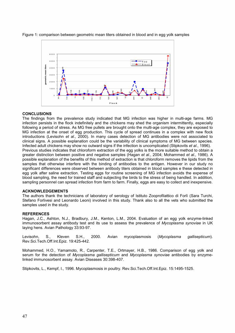
47
Figure 1: comparison between geometric mean titers obtained in blood and in egg yolk samples
CONCLUSIONS The findings from the prevalence study indicated that MG infection was higher in multi-age farms. MG infection persists in the flock indefinitely and the chickens may shed the organism intermittently, especially following a period of stress. As MG free pullets are brought onto the multi-age complex, they are exposed to MG infection at the onset of egg production. This cycle of spread continues in a complex with new flock introductions (Levisohn et al., 2000). In many cases detection of MG antibodies were not associated to clinical signs. A possible explanation could be the variability of clinical symptoms of MG between species. Infected adult chickens may show no outward signs if the infection is uncomplicated (Stipkovits et al., 1996). Previous studies indicates that chloroform extraction of the egg yolks is the more suitable method to obtain a greater distinction between positive and negative samples (Hagan et al., 2004; Mohammed et al., 1986). A possible explanation of the benefits of this method of extraction is that chloroform removes the lipids from the samples that otherwise interfere with the binding of antibodies to the antigen. However in our study no significant differences were observed between antibody titers obtained in blood samples e these detected in egg yolk after saline extraction. Testing eggs for routine screening of MG infection avoids the expense of blood sampling, the need for trained staff and subjecting the birds to the stress of being handled. In addition, sampling personnel can spread infection from farm to farm. Finally, eggs are easy to collect and inexpensive. ACKNOWLEDGEMENTS The authors thank the technicians of laboratory of serology of Istituto Zooprofilattico di Forlì (Sara Turchi, Stefano Forlivesi and Leonardo Leoni) involved in this study. Thank also to all the vets who submitted the samples used in the study. REFERENCES Hagan, J.C., Ashton, N.J., Bradbury, J.M., Kenton, L.M., 2004. Evaluation of an egg yolk enzyme-linked immunosorbent assay antibody test and its use to assess the prevalence of Mycoplasma synoviae in UK laying hens. Avian Pathology 33:93-97. Levisohn, S., Kleven S.H., 2000. Avian mycoplasmosis (Mycoplasma gallisepticum). Rev.Sci.Tech.Off.Int.Epiz. 19:425-442. Mohammed, H.O., Yamamoto, R., Carpenter, T.E., Ortmayer, H.B., 1986. Comparison of egg yolk and serum for the detection of Mycoplasma gallisepticum and Mycoplasma synoviae antibodies by enzyme-linked immunosorbent assay. Avian Diseases 30:398-407. Stipkovits, L., Kempf, I., 1996. Mycoplasmosis in poultry. Rev.Sci.Tech.Off.Int.Epiz. 15:1495-1525.
28/B26
/C
26/A
24/B
23
19/D
19/B
6/B
18
11/B
15/A
15/C
17/B
0
1 0 0 0
2 0 0 0
3 0 0 0
4 0 0 0
5 0 0 0
6 0 0 0
6/A
11/A 13
15/B
17/A
-1
17/C
19/A
19/C 22
24/A 25
26/B
28/A 30
F l o c k
ge
om
etr
ic m
ea
n t
ite
rs
E g g
B lo o d
48
14) PRELIMINARY RESULTS OF AN INFLUENZA SURVEILLANCE IN WILD BIRDS, GAME BIRDS, DOMESTIC DUCKS AND GEESE IN VENETO – ITALY
R. De Nardi 1, G. Cattoli 1, A. Toffan 1, M.S. Beato 1, V. Guberti 2, M. Scremin 2 & C., Terregino 1
1OIE and National Reference Laboratoy for Newcastle Disease and Avian Influenza, Istituto Zooprofilattico
Sperimentale delle Venezie, Italy 2Istituto Nazionale per la Fauna Selvatica “A. Ghigi”, Bologna, Italy
Corresponding author: Dr. Calogero Terregino, Istituto Zooprofilattico delle Venezie, Virology Department, Viale dell’Università 10, 35020 Legnaro (PD), Italy -Tel. +39 049 8084389 –Fax +39 8084360 -E-mail: [email protected]
Running title: Influenza survellance in wild birds in Veneto
Summary Following the avian influenza (AI) epidemics which occurred in Italy between 1997 and 2003, a surveillance program, funded by the Italian Ministry of Health was implemented. Among the tasks of this surveillance program was an investigation on wild and domestic birds to asses circulation of avian influenza viruses in their natural reservoirs. In this study we collected samples from migratory wild birds (Anseriformes and Charadriiformes), from national and imported game fowl, and from 7 backyard farms of geese and ducks. Cloacal swabs were screened by means of RRT-PCR and/or directly processed for attempted virus isolation in embryonated fowl’s SPF eggs and blood samples were tested for presence of antibodies against avian influenza viruses. Avian influenza viruses were only obtained from migratory waterfowl belonging to the family Anseriformes, and not from domestic waterfowl or game birds. This study confirms that the risk of introduction of novel influenza viruses in densely populated areas of poultry farms in Veneto is linked to migratory wild birds and in particular to birds belonging to the family Anseriformes. Key words: avian influenza, wild bird, game birds, ducks, geese INDAGINE SUL RISCHIO DI INTRODUZIONE NEL POLLAME DOMESTICO DI VIRUS DELL’INFLUENZA
AVIARIA DA POSSIBILI RESEVOIR IN VENETO Riassunto In questi ultimi anni l’allevamento avicolo veneto è stato gravemente colpito da alcune ondate epidemiche di influenza aviaria (AI) responsabili di ingenti danni economici diretti ed indiretti al patrimonio avicolo nazionale. Lo scopo della presente indagine è stato quello di verificare la presenza, nei principali serbatoi naturali, di influenzavirus A. La ricerca si è svolta mediante l’analisi di campioni prelevati da uccelli acquatici selvatici (anatidi e limicoli), da selvaggina di allevamento di provenienza nazionale ed estera e da allevamenti rurali di oche e anatre. I risultati indicano che nel nord-est dell’Italia, come nel resto del mondo, gli anatidi selvatici migratori rivestano un ruolo estremamente importante nell’epidemiologia dell’AI. Nessun virus influenzale è stato isolato dai limicoli, ulteriori indagini potranno chiarire in futuro il reale ruolo di questi uccelli in Italia. Tutti gli allevamenti di selvaggina e di anatidi monitorati sono risultati negativi. Parole chiave: Influenza aviaria, uccelli selvatici, selvaggina, anatre, oche Introduction In previous years Italy was affected by several epidemic waves of Avian Influenza (AI) caused by type A Influenza viruses, thus determining heavy economic losses for the poultry industry and devastating consequences for the social community (Marangon, 2003). The epidemics occurred primarily in the densely populated poultry areas (DPPA) of Veneto and Lombardia regions. Waterfowl, particularly Anseriformes (Alexander, 2000) and Charadriiformes (Stallknecht, 1988) are believed to be natural hosts and reservoirs of Influenza A viruses. During the recent H5N1 outbreaks in Eastern Asia the presence of H5N1 viruses in dead migratory birds suggests that wild bird populations may be involved in spreading Highly Pathogenic Avian Influenza (HPAI). In fact the timing and distribution of H5N1 infection in poultry in China in 2001 coincides with the general period of winter bird migration from northern to southern part of the country (Guan, 2004). In Europe, Anseriformes coming from the North-east use wetlands located in Veneto as a wintering site. Charadriiformes originating from the same areas, but with different habitats, use Italian wetlands as foddering sites before they leave for Africa. Thus, several species congregate in Italian wetlands in autumn, and this represents an ideal ecological situation for the perpetuation of avian influenza viruses. On the contrary, in winter there are fewer species but the numbers of birds present in the wetlands are more significant than those which can be found in the previous season. In addition, the majority of birds which are present in the wetlands are juveniles, born during the previous spring and theoretically
49
highly susceptible to Avian Influenza virus (AIV). Aim of the present study was to evaluate the circulation of AIV in wild birds, game birds, domestic ducks and geese in Veneto region. Materials and methods During 2003, 742 cloacal swabs and 309 serum samples were collected. Of these, 560 cloacal swabs were collected from 9 different species of migratory wild Anseriformes and from one species of Charadriiformes, (Caladris alpina) present in wetlands area of Veneto region. The remaining swabs were collected from 12 game farms rearing birds of national and imported origin and from 7 backyard farms rearing geese and ducks. Serum samples were tested by means of the haemagglutination inhibition test (HI) and by agar gel immunodiffusion test (AGID) as described in European Union Directive 92/40/CEE (CEC, 1992). The species and samples tested are illustrated in Table 1. Cloacal swabs from migratory birds were screened by means of a real-time RT-PCR (RRT-PCR) (Cattoli, 2004), and the positive samples were subsequently processed for virus isolation. Cloacal swabs, diluted in phosphate-buffered saline with antibiotics, were inoculated into the allantoic cavity of 9-day-old embryonating specific pathogen free (SPF) eggs for attempted virus isolation according to EU Directive 92/40. Haemagglutinating isolates were identified by means of the haemagglutination-inhibition test, and if shown to be influenza isolates, were fully characterised by means of the neuraminidase inhibition (NI) test. Results All serum samples resulted negative for antibodies to avian influenza. 25 out 560 cloacal swabs (9 mallards, 1 teal, 4 pintails, 8 wigeons, and 3 shovellers) tested were positive for type A avian influenza in RRT-PCR. Following virus isolation attempts, two isolates were obtained, an H1N1 subtype, was obtained from a male mallard and an H10N4 subtype was obtained from a female pintail. All cloacal swabs collected from game birds and rural geese and ducks were negative for AIV (Table 1). Discussion The results of the present investigation support previous studies which indicate that migratory waterfowl represent a source of infection for domestic birds. In an investigation carried out between 1992 to 1998 in which a total of 802 cloacal swabs were collected from migratory and resident waterfowl on the west coast of Italy, 22 isolates were obtained (18 H1N1, 1 H3N8, 1 H5N2, and 2 H10N8). In USA birds belonging to the order Charadriiformes are considered one of main reservoirs of AIVs (Stallknecht, 1988). As part of the investigation we collected 82 cloacal swabs from dunlins (Caladris alpina) in order to obtain information on the role of Charadriiformes as reservoirs of AI viruses in Italy, since this information is currently lacking. No isolates were obtained from the samples collected. The discrepancy between the number of samples which resulted positive at the RRT-PCR and the virus isolation attempts, probably lies in the greater sensitivity of the molecular-based test (Cattoli et al., 2004, Foni et al., 2002) compared to virus isolation. In conclusion, the results of this study emphasise the continuous need to monitor wild bird populations in order to gain more knowledge on the ecology of AI infections. References Alexander, D.J., 2000 A review of avian influenza in different bird species. Vet Microbiol, 74:3-13. Cattoli, G., Drago, A., Maniero, S., Toffan, A., Bertoli, E., Fassina, S., Terregino, C., Robbi, C., Vicenzoni, G., Capua, I., 2004 Comparison of three rapid detection systems for type A influenza virus on tracheal swabs of experimentally and naturally infected birds. Avian Path. In press. CEC (1992) Council Directive 92/40/EEC of 19 May 1992 introducing Community measures for the control of avian influenza. Official Journal of the European Commission, L167, 1-15. De Marco, M.A., Campitelli, L., Foni, E., Raffini, E., Barigazzi, G., Delogu, M., Guberti, V., Di Trani, L., Tollis, M., Donatelli, I., 2004 Influenza surveillance in birds in Italian westlands (1992-1998): is there a host restricted circulation of influenza viruses in sympatric ducks and coots?. Vet Microbiol, 98: 197-208. Foni, E., Chiapponi, C., Lori, D., De Marco, M.A., Delogu, M., Raffini, E., Massi, P., 2002 Applicazione di RT-PCR e metodiche tradizionali nella ricerca di virus influenzale di tipo A in corso di infezione sperimentale in anatre. Large Anim Rev, anno 8, n°. 6: 99-100. Guan, K.S., Wang, J., Smith, G.J.D., Xu, K.M., Duan, L. et al., 2004 Genesis of a highly pathogenic and potentially pandemic H5N1 influenza virus in eastern Asia. Nature, 430: 209-213.

50
Marangon, S., Bortolotti, L., Capua, I., Bettio, M., Dalla Pozza, M., 2003 Low-pathogenicity avian influenza (LPAI) in Italy (2000-01): epidemiology and control. Avian Dis 47:1006-1009. Stallknecht, D.E., Shane, M., 1988 Host range of avian influenza virus in free-living birds. Vet Resh Commun, 12: 125-141. Tab.1: Results of serological and virological tests.
RESULTS OF LABORATORY TESTS
SPECIES N. SAMPLES COLLECTED
CLOACAL SWABS SERA
RRT-PCR (N. positve/N.
samples tested)
VIRUS ISOLATION
(N positive/ N. samples tested)
SEROLOGICAL TESTS (HI, AGID) (N positive/ N. samples
tested) GAME FARMS PHEASANT (Phasianus colchicus) 60 219 ND 0/60 0/219
PARTRIDGE (Perdix perdix) 10 45 ND 0/10 0/45
ROCK PARTRIDGE (Alectoris greca) 0 5 ND ND 0/5
DUCKS AND GEESE FARMS
DOMESTIC DUCK (A. platyrhynchos var. domestica)
62 0 ND 0/62 ND
MUSCHOVY DUCK Cairina moscata) 35 25 ND 0/35 0/25
MALLARD (Anas platyrhynchos) 5 5 ND 0/5 0/5
GOOSE (Anser anser var. domestica) 10 10 ND 0/10 0/10
WILD BIRDS MALLARD (Anas platyrhynchos) 206 0 9/206 1/9 ND
TEAL (Anas crecca) 27 0 1/27 0/27 ND
PINTAIL (Anas acuta) 115 0 4/115 1/115 ND
WIGEON (Anas Penelope) 92 0 8/92 0/92 ND
COOT (Fulica atra) 2 0 0/2 0/2 ND
GADWALL (Anas strepera) 6 0 0/6 0/6 ND
SHOVELER (Anas clipeata) 23 0 3/23 0/23 ND
POCHARD (Aythya ferina) 5 0 0/5 0/5 ND
TUFTED DUCK (Aythya fuligula) 2 0 0/2 0/2 ND
DUNLIN (Calidris alpina) 82 0 0/82 ND ND
ND=not done
51
15) EPIDEMIOLOGICAL STUDY ON CIRCULATION OF INFECTIOUS BRONCHITIS VIRUS STRAINS IN VENETO
M.S. Beato1, R. De Nardi1, A. Toffan1, G. Cattoli1 & C. Terregino1
1Istituto Zooprofilattico Sperimentale delle Venezie
OIE and National Refrence Laboratory for Newcastle Disease and Avian Influenza
Corresponding author: Dr.Terregino, Istituto Zooprofilattico delle Venezie, Virology Department, Viale dell’Università 10, 35020 Legnaro (PD), Italy -Tel. +39 049 8084389 –Fax +39 8084360 -E-mail: [email protected]
Running title: Infectious bronchitis virus circulation in Veneto
ABSTRACT Infectious Bronchitis (IB) is still a major health problem in the poultry industry, as it is endemic in probably all countries which raise chickens. Previous investigations have shown that several IB variants are present in the Italian poultry industry. In order to establish which serotypes are circulating in the Veneto and Lombardia regions an investigation was carried out during 2004 in broiler and egg-layer farms. A total of 101 samples were collected from 29 farms with 16 IBV isolations. Of these, seven isolates were identified as being of the IT-02 serotype and six as 793-B on the basis of nucleotide sequencing. Key words: Infectious bronchitis, broilers, layers
INDAGINE EPIDEMIOLOGICA SULLA CIRCOLAZIONE DEI VIRUS DELLA BRONCHITE INFETTIVA AVIARIA IN VENETO
RIASSUNTO La Bronchite Infettiva (IB) rappresenta ancora oggi uno dei principali problemi sanitari per il pollame allevato intensivamente. Allo scopo di approfondire le conoscenze sulla sua diffusione e sui sierotipi circolanti, nel 2004 si è dato inizio ad un’indagine epidemiologia nel Triveneto ed in alcune province della Lombardia. Dal gennaio 2004 sono stati monitorati 29 allevamenti di broiler ed ovaiole, per un totale di 101 campioni esaminati. 16 allevamenti sono risultati positivi per IBV. I risultati dell’indagine hanno mostrato una diffusione ubiquitaria dei sierotipi 793-B e IT-02. Parole chiave: Bronchite infettiva, broilers, ovaiole
Introduction Infectious Bronchitis (IB) is still a major health problem in the poultry industry, as it is endemic in probably all countries which raise chickens. IB is a severe systemic disease resulting in a variable morbility depending on the virulence and tissue tropism of the strain. The clinical disease, is generally characterized by respiratory signs and nephrosis in broilers and drop in egg production in egg layers and broiler breeders. IB is caused by avian Coronavirus, an enveloped single stranded, positive-sense RNA with a large spike glicoprotein (S) that is responsible of the host antibody response. IBV may be classified in serotypes that can be characterized by serological and molecular biology techniques. Previous investigations have shown that several IB variants are present in the Italian poultry industry. Among these, some have been isolated primarily in Italy, such as variant 624-I (Capua et al., 1999), while others originate from other European countries such as 793-B and B-1648. Between 2000 and 2003 no surveys on IB were performed in the densely populated poultry areas of North-eastern Italy, although clinical signs indicative of this infection were observed. In order to establish which serotypes are circulating in the Veneto and Lombardia regions an investigation was carried out during 2004 in broiler and egg-layer farms in collaboration with field veterinarians. Materials and methods The study has had as target the broiler and egg layer farms in selected areas of Veneto and Lombardia. In order to improve isolation rates, SPF (Specific Pathogen Free) chickens were introduced in the farms for a period of 7-10 days as sentinels. Tissue samples, collected from sentinels (trachea, lung, kidney and cecal tonsils) were submitted for virological investigations. The tissue homogenates were inoculated into the allantoic cavity of 9-to-11-day-old embryonating SPF eggs (Gelb et al, 1998). Following a minimum of two and a maximum of four blind passages, the allantoic fluid was harvested and examined by negative contrast electron microscopy for the presence of coronavirus particles (Hayat, 1985). In order to characterise the isolates, viral RNA was extracted from the infectious allantoic fluid. The isolates were analysed in RT-PCR, with specific primers for the S1-gene (Adzhar et al. 1996), in order to generate a complementary DNA (cDNA). This cDNA, was sequenced (Keeler et al., 1998), and isolates were typed on the basis of the sequence. A total of 101 samples were collected from 29 farms.

52
Results Sixteen IB isolates were isolated from 29 farms. Of these, seven isolates were identified as being of the IT-02 serotype and six as 793-B on the basis of nucleotide sequencing (Table 1). None of the farms were affected by severe clinical signs, in the farms in which 793-B was isolated, respiratory and nephrosis forms prevailed. Respiratory and enteric forms were observed with IT-02 infection. Discussion The results presented above indicate that regardless of the geographical origin of the isolate strains, 793-B and IT-02 are present in Veneto and Lombardia regions. The findings of the present study indicate that strain 793-B is still present in Italy (Catelli & Lavazza, 2000, Tosi, 2001). Although evidence of the presence of this virus has been collected throughout the years, it is not possible to establish how many isolates are pathogenic field strains and how many are vaccine strains. In fact vaccination against this variant has been practised extensively, and currently there is no tool available to differentiate field from vaccine strains. What appears of great interest is the significant number of isolations of strain IT-02. This strain appears to be widespread in broiler farms. Since at present there is no information on the pathogenicity and antigenicity of this novel variant, challenge experiments and cross protection studies should be performed in order to evaluate on one hand the potential damage which this variant can cause to the poultry industry, and on the other the possibility of using licensed products to protect birds against the clinical condition caused by this variant. Table 1: Province of isolation, strains isolated, lesions and clinical signs observed in the field.
Province Type of farm and age of
birds
Molecular characterization of
isolates
Lesions and clinical signs
Verona Broiler - 36 days IBV serotype 793-B∗ Light respiratory form
Rovigo Broiler - 36 days IBV serotype IT-02 Not reported
Cremona Broiler - 45 days IBV serotype IT-02 Not reported
Mantova Broiler - 60 days IBV - serotype 793-B Respiratory form
Verona Broiler - 53 days IBV Not reported
Padova Broiler - 60 days IBV Respiratory form
Pordenone Layers - 20 weeks IBV Respiratory form, depigmentation of the eggshell
Brescia Broiler - 42 days IBV serotype 793-B Respiratory form, kidney lesions, growth problem, mortality
Verona Broiler - 35 days IBV serotype IT-02 Kidney lesions, enteric form, mortality
Verona Broiler - 45 days IBV serotype IT-02 Enteric form
Cremona Broiler - 53 days IBV serotype IT-02 Respiratory and enteric forms
Padova Broiler - 50 days IBV serotype 793-B Light respiratory form
Padova Broiler - 43 days IBV serotype IT-02 Light respiratory form
Vicenza Broiler - 51 days IBV serotype IT-02 Light respiratory form
Brescia Broiler - 45 days IBV serotype 793B* Not reported
Mantova Broiler - 50 days IBV serotype 793-B∗ Light respiratory form
∗ Farms where a live attenuated vaccine serotype 793/B was used References Adzhar, A., Shaw, K., Britton, P., Cavanagh, D., 1996. Universal oligonucleotides for detection of infectious bronchitis virus by the polymerase chain reaction Avian Path 25:817-836. Capua, I., Minta, Z., Karpinska, E., Mawditt, K., Britton, P., Cavanagh, D., Cough, R. E. 1999. Co-circulation of four serotypes of infectious bronchitis virus (793-B, 624-I, B1648 and Massachustts). Avian path 28:587-592.
53
Catelli, E., Lavazza, A., 2000. Relazione sullo stato sanitario dell’allevamento avicolo nel 2000. La selezione Veterinaria 11:963-970. Gelb, J., JacKwood, M.W., 1998. Infectious Bronchitis. In: Isolation and identification of avian pathogens, (Fourth ed.), The American Association of Avian Pathologists, pp 169-174. Gelb, J., Rosenberg, J.K. Jr., Fries, P.A., Cloud, S.S., Odor, E.M., Dohms, J.E. and Jaeger, J.S., 1989. Protection afforded infectious bronchitis virus-vaccinated sentinel chickens raised in a commercial environment. Avian Dis 33:764-769. Hayat, A.M., 1986. Basic techniques for transmission electron microscopy. Academic Press, Inc. Keeler, C.L., Reed, K.L., Nix, W.A., Gelb, J., 1998. Serotype identification of avian infectious bronchitis virus (IBV) by RT-PCR of the peplomer (S-1) gene. Avian Dis 42(2):275-84. Tosi, G., 2001. Relazione sullo stato sanitario dell’allevamento avicolo nel 2001. Large Anim Rev 6:37-40.
16) BIG LIVER AND SPLEEN DISEASE IN BROILER BREEDERS IN ITALY
Paola Massi1, Giovanni Tosi1, Daniela Gelmetti1, Antonio Lavazza1, Guerino Lombardi1, Gloria
Torcoli1. Istituto Zooprofilattico Sperimentale della Lombardia e dell’Emilia Romagna
Corresponding author: Dr. Paola Massi. Istituto Zooprofilattico Sperimentale della Lombardia e dell’Emilia Romagna, Sezione Diagnostica di Forlì. Via Marchini 1, 47100 Forlì, Italy – Tel.+39 0543 721533 – Fax +39 0543 721533 – Email: [email protected]
ABSTRACT For the first time in Italy, we have reported two outbreaks resembling big liver and spleen disease in broiler breeder flocks. The combination of clinical signs and pathological findings and the laboratory investigation results appear to be very similar to previously recorded diseases in other countries. KEY WORDS: Big liver and spleen disease, broiler breeders, calicivirus-like virus
“BIG LIVER AND SPLEEN DISEASE” IN RIPRODUTTORI PESANTI IN ITALIA
RIASSUNTO Per la prima volta in Italia vengono descritti due casi riferibili alla sindrome denominata “big liver and spleen disease” in gruppi di polli riproduttori pesanti. I segni clinici e le lesioni anatomo-patologiche osservate nonché i risultati delle indagini di laboratorio condotte sui gruppi colpiti appaiono sovrapponibili a quelle già descritte in passato in altri paesi. INTRODUCTION Big liver and spleen disease (BLS) is an infectious, transmissible disease of uncertain, but probably viral, etiology. It is characterized by decreased egg production, increased mortality and enlargement of the liver and spleen of mature chickens, especially broiler breeders and, less commonly, egg layers. First recognized in Australia in 1980, BLS is considered to be the most economically significant disease of broiler breeders in that country. Beside Australia, serological evidence indicates the BLS agent have also infected flocks in the United Kingdom and the United States. Moreover infection with the BLS agent has been associated with a condition known as “primary feather and drop syndrome” in the United States (Barnes, 1997). Until now there are no reports of this syndrome in Italy. This paper gives a report on the clinical, pathological and laboratory findings noted in two broiler breeder flocks affected with a disease very similar to BLS. MATERIAL AND METHODS Carcasses and blood samples from two broiler breeder farms were submitted for diagnostic examination and laboratory investigation at the Istituto Zooprofilattico Sperimentale of Forlì. Anamnestic data were collected, postmortem examinations were carried out on the carcasses and appropriate samples were taken for the following laboratory investigation: Bacteriology: samples of liver, spleen and ovary were inoculated on blood agar and Hektoen Enteric Agar and incubated aerobically at 37°C.
54
Virology: samples of liver, spleen, intestine, ovary, trachea, lung and brain were inoculated on Chicken Embryo Liver Cell Cultures (CEL) and Specific Pathogen Free (SPF) embryonated chicken eggs A Polymerase Chain Reaction (PCR) for group 2 avian adenovirus was also performed. Serology: the following serological tests were performed on blood samples: 1) agar-gel diffusion precipitin (AGP) for group 1 avian adenovirus. 2) Enzyme-linked immunosorbent assay (ELISA) for group 2 avian adenovirus (Synbiotics). 3) ELISA test for Human Epatitis E Virus (Nuclear Laser Medicine). Electron Microscopy: samples of liver and spleen were frozen and thawed twice, the supernatant was harvested and centrifuged at 4,000 g for 20 min. and at 9300 g for 10 min. for clarification. The second supernatant (85 µl) was then ultracentrifuged in Airfuge Beckman for 15 min. at 21 psi (82000 g). Grids were then stained using 2% sodium phosphotungstate (NaPt), pH 6.8, for 1.5 min., and observed with a TEM Philips CM10. Histology: representative samples of spleen, liver, heart and kidney were fixed in 10% buffered formalin, dehydrated in ethanol, cleared in xylene and embedded in paraffin, according to standard techniques. Thin sections (4m) were cut and stained with haematoxylin-eosin. RESULTS AND DISCUSSION During January 2004, 37 week-old Ross 508 broiler breeders from a farm of 17000 animals housed in three flocks experienced a slightly increased mortality (+0.2%) in two flocks and a 2% drop in egg production for three weeks. A large number of chickens had a drop of feathers (resembling premature moulting) that persisted for several weeks after recovery from the disease. In the same period a similar syndrome was observed in another farm of 44 week-old Ross 508 broiler breeders of 24000 animals. At necropsy, all the dead birds were in good condition. The most frequently found lesion was an enlarged spleen. The liver was also enlarged and friable. Other lesions included: cyanosis of the head, hydropericardium, pulmonary congestion and oedema, enteritis, ovarian regression and congestion, swollen kidneys. Microscopically the following lesions were recognized: congestion and oedema of the liver associated with multiple foci of necrosis and an increase of perivascular lymphoid tissue; widespread necrosis of lymphoid tissue and proliferation of macrophages in the spleen; adipositas cordi in the heart.;congestion and interstitial infiltration of lymphoid tissue in the kidneys. Bacteriologically a strain of Escherichia coli was isolated from the liver. Virological examinations (i.e. embryo egg inoculation, and PCR for group 2 adenovirus) were negative. A cytopathic effect (characterized by the formation of syncytia) was observed after inoculation of the CEL with a liver and spleen homogenate . By negative staining Electron Microscopy few viral isolated particles morphologically resembling calicivirus were observed in the liver and spleen. Serological tests demonstrated reacting antibodies with group 1 and group 2 avian adenovirus. Furthermore, 3 of 15 blood samples collected from the first observed outbreak were positive with ELISA test for human hepatitis E virus antibodies. CONCLUSIONS Clinical signs, macroscopic and microscopic lesions, age and production type of the affected flocks were sufficient for a presumptive diagnosis of BLS. Other diseases that produce splenomegaly were excluded. Seroconversion toward group 2 avian adenovirus wasn’t associated to other features of the avian adenovirus group 2 splenomegaly chicken disease (AAS) . Escherichia coli strain recovered from affected birds is a consequence of BLS, but it is not involved in causing the disease (Barnes, 1997). To confirm the diagnosis of BLS, the electron microscopy observation, , of few isolatd calicivirus-like particles in the liver and spleen, was very interesting as well as the histological lesions and the detection of antibodies reacting with human hepatitis E virus. Recently, in the United States, a new virus, designated avian hepatitis E virus (avian HEV) was identified and characterized from chickens with hepatitis-splenomegaly syndrome (HS), a disease very similar to BLS described in Australia (Haqshenas et al, 2001). So far, it is likely that BLS in Australia and the HS syndrome in North America are caused by variant strains of the same virus (Haqshenas et al, 2002). Avian HEV is genetically related to but distinct from human and swine HEV. The antigenic cross reactivity among avian and human HEVs was confirmed by ELISA (Huang et al., 2002). HEVs (including human, swine and avian strains) were formerly classified as a member of the family Caliciviridae. But the lack of common features between HEVs and caliciviruses has led to the recent removal of HEV from this family (Berke et al., 2000). In conclusion field and laboratory data reported in this work lead us to confirm the diagnosis of BLS in the broiler breeder flocks observed. An implementation of other diagnostic procedures, such as PCR, in association with more specific serological tests, are needed to further confirm the presence of this syndrome in Italy. REFERENCES Barnes H.J., 1997. Big liver and spleen disease. In : B.W.Calnek (ed.) Diseases of Poultry. 10th editino Iowa State University Press, Ames, Iowa.
55
Berke T., Matson D.O., 2000. Reclassification of the Caliciviridae into distinct genera and exclusion of hepatitis E virus from the family on the basis of comparative phylogenetic analysis. Archives of virology, 145:1421-1436
Haqshenas G., Shivaprasad H.L., Woolcock P.R., Read D.H., Meng X.J., 2001. Genetic identification and characterization of a novel virus related to human hepatitis-splenomegaly syndrome in the United States. Journal of General Virology. 82:2449-2462
Haqshenas G., Huang F.F., Fenaux M., Guenette D.K., Pierson F.W., Larsen C.T., Shivaprasad H.L., Toth T.E., Meng X.J., 2002. The putative capsid protein of the newly identified avian hepatitis E virus shares antigenic epitopes with that of swine and human hepatitis E viruses and chicken big liver and spleen disease virus. Journal of General Virology. 83:2201-2209
Huang F.F., Haqshenas G., Shivaprasad H.L., Guenette D.K., Woolcock P.R., Larsen C.T., Pierson F.W., Elvinger F., Toth T.E., Meng X.J., 2002. Heterogeneity and Seroprevalence of a Newly Identified Avian Hepatitis E Virus from Chickens in the United States. Journal of Clinical Microbiology.vol 40, No.11: 4197-4202 17) USE OF REVERSE TRANSCRIPTION-POLYMERASE CHAIN REACTION (RT-PCR)
FOR THE DIAGNOSIS OFAVIAN VIRAL ARTHRITIS
Fiorentini Laura1, Francesca Paganelli, Leonelli Roberto1. 1Istituto Zooprofilattico Sperimentale della Lombardia e dell’Emilia Romagna.
Corresponding author: Dr. Laura Fiorentini. Istituto Zooprofilattico Sperimentale della Lombardia e dell’Emilia Romagna, Sezione di Forlì, via Marchini 1, 47100 Forlì, Italy. Tel. +39 0543 721533 Fax +39 0543 721533. E-mail: [email protected] Summary Viral arthritis or infective tenosynovitis is a contagious disease of chickens and more rarely, turkeys. It causes articular symptoms and lesions of the synovia tendon structure. There are various diagnostic tests used today including RT-PCR (reverse transcription-polymerase chain reaction) which can be used both as support diagnosis, as a confirmation of infected cell cultures as well as directly in the homogenised materials obtained from pathological tissue taken during necropsy Key words: avian reovirus, RT-PCR, viral arthritis tenosynovitis.
Applicazione della reverse transcriptase-polymerase chain reaction (RT-PCR) nella diagnosi dell’artrite-tenosinovite virale
Riassunto L’artrite virale o tenosinovite infettiva è una malattia contagiosa del pollo e, più raramente, del tacchino. Si manifesta con sintomi articolari e lesioni a carico delle strutture sinoviali tendinee. Diversi sono gli strumenti diagnostici attualmente in uso, tra questi, la tecnica di RT-PCR (reverse transcriptase-polymerase chain reaction), applicabile sia come diagnosi di supporto e conferma alle colture cellulari infettate, sia direttamente all’omogenato ottenuto da tessuti patologici prelevati in sede autoptica. Parole chiave: reovirus aviare, RT-PCR, artrite-tenosinovite virale Introduction Avian reovirus is associated in broilers to a series of pathologies including contagious arthritis tenosynovitis and malabsorption syndrome. Viral arthritis mainly affects heavy meat producing breeds from an age of between one and three months. The disease is transmitted by both vertical and horizontal pathaways. Contaminated faeces, food and drink are the most important contagious sources. Morbidity varies according to age and mortality is usually low except when there are bacterial complications. The symptoms occur in the chicks at three to four weeks of age and include mono- and bilateral lameness. In breeding birds there is delayed growth and lameness, sometimes with complete immobility of the tibiotarsic articulation sometimes leading to the breaking of the gastrocnemium tendons with subsequent haemorrhage of the peri-articular tissue. This causes economic loss, which results not only from mortality but also from the reduced performances and the inability to consume birds with obvious articular lesions when slaughtered. Direct (bio-security) and indirect (vaccination of the breeders) preventive techniques have partially contained the spread of this disease even if this is not complete. It is ever more necessary therefore, to have an early diagnostic technique that can allow us to organise intervention plans adapted to this pathology. Thus, PCR that can be applied directly to pathological samples has been considered particularly useful.
56
Material and methods Six clinical cases were taken which had an anatomo-pathological profile which could be attributed to avian viral arthritis. These were broilers of between three and five weeks of life, with articular symptoms including lameness and ankylosis. From an anatomo-pathological point of view there was mainly bilateral arthritis and tenosynovitis, shortening of the tibia, flattening and lateral slipping of the gastrocnemium tendons as well as peritendonal and intra-articular yellow serous exudates The samples for the virological exam (cell cultures and RT-PCR) were prepared starting with the tendons and exudates. At the same time other samples of the same materials were taken for the microbiology laboratory and the PCR for the Mycoplasma synoviae as differential diagnoses. Viral Isolation The initial sample was homogenised with antibiotated PBS, centrifuged at 3000 x g and filtered at 450nm. The primary liver cultures of chicken SPF (specific pathogen free)(Spafas, USA) embryos (13 days) were used for the avian reovirus (ARV) () research. The primary liver cultures were then infected once they had fully confluenced They were observed using an optical microscope once every 24 hours to check for any possible syncytium cytopathic effects, which are typical of ARV. An electronic microscope test was used as confirmation of the virus (Lavazza et al. 1990). ARV identification by RT-PCR The extraction and purification of viral RNA was carried out both directly on the pathological material taken from the autoptical site and on the cell suspension usingTrizol-Reagent (Invitrogen, USA): this is a monobasic phenol and isothiocyanate gaunidinium solution used according to the modified technique described by Chomezynski and Sacchi (1987). Once we had obtained our RNA sample we proceeded to the retro-transcription using the ProstarTM Kit (Stratagene, USA). The product of the retro-transcription was then used directly in the PCR with the addition of the relevant reagent and the tampon (Accuprime TaqPCRx DNA Polymerase) (Invitrogen, USA). The MK87 (5’ GGT GCG ACT GCT GTA TTT GGT AAC 3’) forward primer and the reverse primer MK88 (5’ AAT GGA ACG ATA GCG TGT GGG 3’) acted as the trigger for the amplification reaction. These primers were selected on the basis of published data on the S1 gene of the avian reovirus (Zhixun et al. 1997) , that is different from the mammal reovirus (Shapouri et al, 1995)., and they produce a fragment of 532 bp DNA contained in the S1 gene sequence. The reaction occurred according to the following amplification profile: 94°C for 5 minutes, (hot start) followed by 35 cycles of three steps: 94°C for one minute (denaturation), 55°C for one minute (annealing), 72°C for one minute (extension), at the end of cycles there was an additional 10 minutes at 72°C for any possible extensions. The amplified result then underwent an electrophoresis course in agarose gel at 1% with ethidium bromide and was visualised using a UV trans-illuminator. Results and Discussion Four of the six samples analysed from different clinical cases were positive on the cell cultures and confirmed by electronic microscopy. Their positivity was further confirmed by the RT-PCR technique, both directly on the pathological samples after approximately 48 hours and on the cell suspension after 15 days. Conclusions These first results show the validity of RT-PCR in identifying avian reovirus even from pathological material taken from the necroscopic site. Moreover, this method represents a rapid and sensitive alternative to traditional culture methods which require specialised media and reagents and are time-consuming. Results of PCR test can be obtained in two days, as opposed to the usual one to three weeks for isolation and identification of reovirus. References
Chomezyski P., Sacchi N. 1987. Single-step method of RNA isolation by acid guanidinium hiocyanate-phenol-chloroform exstraction. Analitic Biochemistry 162:156-159.
Lavazza A., Pascucci S., Gelmetti D. 1990. Rod-shaped virus-like particles in intestinal contents of three avian species.Vet. Rec.126:581.
Shapouri M.R., Kane M., Letarte M., Bergeron J., Arella M., Silim A. 1995. Clonino, sequencing and exspression of the S1 gene of avian reovirus. J. Gen. Virol. 76 : 1515-1520.
Zhixun Xie, Amin A. Fadl, Theodore Girshick, Mazhar I. Khan. 1997. Amplification of avian reovirus RNA using the reverse transcriptase-polymerase chain reaction. Avian Diseases 41:654-660.
57
18) TYPING OF POULTRY INFLUENZA VIRUS (H5 AND H7) BY REVERSE TRANSCRIPTION-POLYMERASE CHAIN REACTION
Paola Massi1, Tosi Giovanni1, Cordioli Paolo1, Paganelli Francesca1, Bonacina Cesare1
1Istituto Zooprofilattico Sperimentale della Lombardia e dell’Emilia Romagna.
Corresponding author: Dr. Paola Massi. Istituto Zooprofilattico Sperimentale della Lombardia e dell’Emilia Romagna, Sezione di Forlì, via Marchini 1, 47100 Forlì, Italy. Tel. +39 0543 721533 Fax +39 0543 721533. E-mail: [email protected] ABSTRACT The ability of the influenza Orthomixovirus to undergo to continually antigenically changes that can affect its pathogenicity and its diffusion, explains the growing seriousness of this disease and the recent epizoozies in various parts of the world. There have been 15 HA and 9 NA type A sub-types of the influenza virus identified all of which are present in birds. Until now the very virulent avian influenza viruses identified were all included to the H5 and H7 sub-types. We here show that is possible to identify the H5 and H7 sub-types with reverse transcription-polymerase chain reaction (RT-PCR) by using a set of specific primers for each HA sub-type. The RT-PCR is a quick and sensitive method of identifying the HA sub-types of the influenza virus directly from homogenised organs. Key words: avian influenza virus, hemagglutinine, H5, H7, reverse transcription-polymerase chain reaction (RT-PCR)
TIPIZZAZIONE DEI VIRUS INFLUENZALI AVIARI (H5 E H7) TRAMITE REVERSE TRANSCRIPTION-POLYMERASE CHAIN REACTION
RIASSUNTO La capacità dell’Orthomixovirus dell’influenza di andare incontro a continue variazioni antigeniche, determinanti la patogenicità e la diffusibilità, spiega l’accresciuta importanza di questa malattia e le recenti epizoozie segnalate in varie parti del mondo. Nel virus influenzale di tipo A sono stati identificati 15 sottotipi di HA e 9 NA, tutti presenti negli uccelli. Finora i virus influenzali aviari che hanno presentato caratteri d’elevata virulenza sono tutti appartenenti ai sottotipi H5 e H7. E’ possibile identificare i due sottotipi H5 e H7 tramite una reverse transcription-polymerase chain reaction (RT-PCR) utilizzando un set di primers specifici per ogni sottotipo HA. La RT-PCR si presenta come un rapido e sensibile strumento per l’identificazione dei sottotipi HA del virus influenzale direttamente da omogenato d’organi. Parole chiave: virus influenzale aviare, emoagglutinina, H5, H7, reverse transcription-polymerase chain reaction (RT-PCR) INTRODUCTION Influenza was first described by Hippocrates in 412 B.C., and the miniscule virus has continually mutated throughout the centuries, spreading the disease right up to the latest H5N1 epidemics which hit Asia in 2003 and 2004. The influenza virus is part of the Orthomyxoviridae family (genus) and is divided into three genus: A, B and C. The A type viruses have essentially avian origins and only occasionally do they jump the species barrier to affect other animals or man. Recently, knowledge of the virus, its pathogenesis and prevention, have greatly increased but unfortunately this has not allowed us to avoid great (economic) losses due to recurrent epizoozies from highly virulent strains in different parts of the world. The virus contains 8 separate segments of RNA, which codify genes of at least 10 different proteins. This unusual genetic structure explains why re-arrangement of the segments occurs so frequently. Exchange of genetic segments can easily occur which can cause up to 256 different descendents. The A type influenza strains are characterised by the structural changes of 2 glycoproteines; hemagglutinine (HA) and neuroaminidase (NA) which project from the surface of the viral particles. Sub-type sub-division is based on a combination of the 15 HA and 9 NA which have so far been identified, all of which are present in birds (Alexander, 1993; Easterdat et. Al., 1997). Epidemics occur when one or the other of these proteins undergoes a mutation. The unforeseeable nature of the influenza virus comes from their ability to change the surface HA and NA proteins, thus escaping the immune system’s vigilance. If we consider the epidemiological polyedric aspects, the speed of the influenza diagnosis and therefore quick confirmation of the infection becomes particularly important. For these reasons, we can say that reverse transcription-polymerase chain reaction (RT-PCR) has given particularly encouraging results.
58
To date, all highly pathogenic stains that have been isolate are A influenza viruses of the H5 and H7 sub-types. The molecular base for distinguishing the antigenic differences of the HA sub-type are based on the amino-acid sequence differences between 20% and 74% and so this percentage is reflected in the nucleotide sequence of the HA sub-type (Ming-Shiuh et al.; 2001) These characteristics are fundamental for the typing of HA sub-types by RT-PCR as the PCR is determined by the difference in the nucleotide sequence. We have here evaluated a quick identification procedure to identity H5 and H7 subtypes of the influenza virus using RT-PCR. MATERIAL AND METHODS Samples For the study we used organs (tracheal exudates, lungs, cecal tonsils) in that previously the following avian influenza virus strains were isolated and identified: H7N1/99 chicken and H7N3/02 turkey. Moreover, starting from allantoic fluid, the following strains were also used: H5N2/73 England (turkey), H6N2/90 (turkey) and H9N2/86 (chicken). Identification of the Type A influenza virus with RT-PCR The viral RNA was extracted using the Rneasy Minikit (Qiagen, Valencia, CA, USA) protocol. Once we had the RNA sample we proceeded to the retro-transcription using ProstarTM Kit (Stratagene, USA). The retro-transcription product was then used directly in the PCR with the addition of a reagent and the tampon which are part of the Accuprime TaqPCRx DNA Polymerase (Invitrogen USA). The research of the viral genome also includes the use of M52C and M253R primers (Fouchier et al. 2000), which amplifies a region coding for the matrix protein of the type A influenza virus. Typing H5 and H7 with RT-PCR The cDNA obtained by retro-transcription was used for a further PCR according to above protocol. To differentiate the H5 and H7 subtypes, sets of specific primers for each sub-type in respect to their sequence and characteristics were used as triggers (Ming-Shiuh et al.; 2001) (tab. 1). The reactions occurred in the following amplification profile: 95°C for 3 minutes, (hot start) followed by 35 cycles of three steps: 95°C for 30 seconds (denaturation), 55°C for 40 seconds (annealing), 72°C for 40 seconds (extension), at the end of cycle there was an additional 10 minutes at 72°C for any possible extensions. The amplified product then underwent an electrophoresis course in agarose gel at 1% with ethidium bromide and was visualised using a UV trans-illuminator. Table 1. Primer used for the HA-sub-typing of avian influenza viruses by RT-PCR
PRIMER PRIMER SEQUENCES PCR product (bp) H5-155F 5’ ACACATGCYCARGACATACT 3’
545 H5-699R 5’ CTYTGRTTYAGTGTTGATGT 3’ H7-12F 5’ GGGATACAAAATGAAYACTC 3’
634 H7-645R 5’ CCA TABARYYTRGTCTGYTC 3’
RESULTS Results about avian influenza virus (AIV) identification and subtyping are shown in table 2 Table 2. Results of identification and subtyping.
Sample
AIV strain Type Subtype
Allantoic fluid H5N2/73 Turkey England A H5 (545bp) Allantoic fluid H9N2/86 Chicken Italy A // Allantoic fluid H6N2/90 Turkey Italy A //
Organs H7N1/99 Chicken Italy A H7 (634bp) Organs H7N3/02 Turkey Italy A H7 (634bp)
DISCUSSION AND CONCLUSIONS Given the variable nature of the influenza virus, it is almost certain that it can escape immune surveillance, thus making it difficult to keep this disease under control. The particular characteristics of AIV make quick identification and characterisation of the antigenic variants indispensable to be able to intervene as quickly as possible in the more convenient way. Furthermore, early identification if the new A type influenza virus subtypes, which could be possible causes of pandemics, must be one of the main objectives to insure that vaccines include the immunological characteristics of the virus prevalently in circulation.
59
RT-PCR response perfectly to the requirements of identifying the H5 and H7 subtypes both for its precision and its speed (48 hours). This technique could be a valid test in support of classical diagnosis at the same time being an efficient control technique of poultry breeding. It has been shown to be a sensitive and specific method both using homogenised organs and liquid allantoid. REFERENCES Alexander D.J. 1993. Orthomyxovirus Infection In J.B. McFerran and M.S. McNulty (eds) Virus Infections of birds Elsevier Science Publishers B.V. pp. 287-316. Easterday B.C., Hinshaw V.S., Halvorson D.A. 1997. Influenza. In: B.W. Calnek, H.J. Barnes, C.W. Beard, L.R. McDougald, L.M. Saif (eds) Diseases of Poultry 10th ed. Iowa State University Press, Ames, Iowa pp. 583-605. Fouchier Ron A.M., Bestebroer S.H., Van de Kemp L., Rimmelzwaan G.F., Osterhaus A.D. 2000. Detection of Influenza A viruses from different species by PCR amplification of conserved sequences in the matrix gene. Journal of Clinical Microbiological pp. 4096-4101. Ming-Shiuh Lee, Poa-Chun Chang, Jui-Hung Shien, Ming-Chu Cheng, Happy K. Shieh. 2001. Identification and subtyping of avian influenza viruses by reverse transcriptio-PCR. Journal of Virological Methods 97:13-22.
19) CORRELAZIONE ANTIGENICA TRA SIEROTIPO FA-6881/97 O AZ-27/98 E I CEPPI “IT-02” DEL IBV, ISOLATI PIÙ RECENTEMENTE IN EUROPA
Antonio Zanella1, Raffaella Ceruti2 e Luigi Gavazzi2.
1 Già Docente della Sez. di Microbiologia e Immunologia del Dip. di Patologia Animale, Igiene e Sanità
Pubblica Veterinaria, Università di Milano, via Celoria 10, Milano Italy
2Gruppo Amadori – Cesena
Introduzione A partire dal 1966 sono stati riportati numerosi isolamenti di nuovi sierotipi o varianti del IBV in Italia (1-9). Particolare attenzione è stata rivolta in passato al ceppo AZ-23/74, per la durata della sua prevalenza, circa vent’anni; successivamente il ceppo è apparentemente scomparso(7). A partire dal 1997 ed in aree diverse del Paese, sono stati isolati due ceppi immunologicamente simili tra di loro, Fa-6881/97 ed AZ-27/98, da casi di grave nefrite in broiler o da sindrome respiratoria in pollastre.In seguito, nel giro di pochi anni, altri (oltre venti) ceppi di IBV strettamente simili dal punto di vista antigene ai precedenti, sono stati isolati in aree diverse del Paese da polli con manifestazioni respiratorie, enteriche e/o renali. Pertanto, tali ceppi , antigenicamente diversi dai sierotipi più noti, presentatisi sia in Europa che negli altri continenti, sono stati considerati come un nuovo sierotipo o variante, assai diffuso e prevalente in Italia (9). Più recentemente, molti (oltre 100) ceppi di IBV, considerati simili al ceppo Fo-4682/99 e denominati anche “It-02”, isolati in molti paesi dell’Europa occidentale sono stati riferiti durante l’International Symposium of Avian Coronavirus a Giessen, Germania. Scopo di questa preliminare indagine è stato quello di rilevare eventuali correlazioni antigeniche tra gli isolati “It-02” ed il sierotipo rappresentato dai ceppi Fa-6881/97 o AZ-27/98. Materiali e metodi Substrato. Uova embrionate SPF di 10 giorni. Ceppi IBV. Ceppi Fa-6881/97, AZ-27/98 e 793/B della nostra collezione, al loro 12° passaggio e It-02 (o, meglio, FO-4682/99) gentilmente fornitoci dall’ IZSLER, sez. Forlì al 5° passaggio; quest’ultimo è stato ripassato fino al 10° passaggio in uovo embrionato. Antisieri specifici IBV. Antisieri Fa-6881/97, AZ-27/98 e 4/91 (793/B), ottenuti come previamente riportato (10) e antisiero It-02, gentilmente fornitoci da IZS delle Venezie. Virus-Neutralizzazione (VN) test. I test VN sono stati eseguiti secondo il metodo-alfa, virus variante-siero costante (diluizione log10 di virus e 1:5 di siero), metodo questo risultato più sensibile del metodo-beta (8).

60
Le miscele virus + sieri specifici, così come virus + siero negativo sono state tenute a contatto per 1 ora a 25°C e quindi inoculate, alla dose di 0.1ml in gruppi di 5 uova per diluizione, via cavità allantoidea. Speratura uova ogni giorno ed esame per mortalità e lesioni specifiche entro 8 giorni. Calcolo indici di neutralizzazione (NI) secondo il metodo di Reed e Muench. Risultati e considerazioni I risultati vengono dettagliatamene riportati in tabella 1. Questi risultati preliminari permettono di considerare i ceppi FA-6881/97 ed AZ-27/98 come i primi isolamenti di un nuovo sierotipo di IBV in Europa. Ad esso apparterrebbero, o almeno sarebbero molto simili antigenicamente, anche i cosiddetti ceppi “It-02”; di questi ultimi il primo isolamento risalirebbe al 1999, come ceppo FO-4682/99 (Massi et al., comunicazione personale). L’isolamento di molti ceppi “It-02”, ristretto almeno per ora all’Europa occidentale, è stato di recente e ampliamente riportato all’International Symposium of Avian Corona- and Pneumovirus Infections, tenuto nel magnifico castello di Rauishholzhausen, Giessen, Germania, 20-23 giugno 2004 (in corso di stampa). Ulteriori e più profonde indagini sono in corso presso l’IZSLER di Brescia e l’IZS delle Venezie, come la caratterizzazione molecolare e l’analisi filogenetica, con sequenziazione di parte del genoma, la subunità ipervariabile S1, di numerosi ceppi europei di IBV, inclusi i vari ceppi It-02, nonché i ceppi FA 6881/97 e AZ 27/98. Al test RT-PCR, riportato in un precedente lavoro, il ceppo Fa-6881/97 sarebbe risultato correlato al ceppo 793/B, contrariamente ai risultati sierologici di SN crociata. Il ceppo AZ 27/98, nonostante la sua stretta affinità antigenica con FA-6881/98, non sarebbe invece risultato correlato né al 793/B né ad altri più comuni sierotipi del passato (10). Anche se con il test RT-PCR e la sequenziazione, differenze possono essere state rilevate nella sequenza nucleotidica e conseguentemente in quella aminoacidica, ciò non esclude che, almeno parte degli isolati It-02 in questione, possano appartenere allo stesso sierotipo o variante Fa-6881/97 o AZ 27/98 e, conseguentemente, indurre una reazione immunologia crociata, sia in vitro che in vivo. Concludendo pertanto, per stabilire la differenza tra un ceppo e l’altro non ci si può basare, a nostro avviso, solo sul genotipo, ma anche e forse di più, sulle caratteristiche antigeniche o immunogene, che dal punto di vista pratico (immunità) sono le più importanti. Il genotipo potrà invece avere un maggiore significato o importanza, nello studio epidemiologico dell’infezione. Ricerche sull’attenuazione e sua stabilizzazione del ceppo AZ 27/98, nonché la sua efficacia protettiva in vivo sono in corso con risultati apparentemente positivi. Tabella 1: Test di virus-neutralizatione crociata tra i ceppi correlati Fa-6881/97 e AZ-27/98 ed i ceppi 793 B e “It-02” del IBV.
VIRUS
ANTISIERO
Fa-6881/97 AZ-27/98 It-02 793 B or 4/91
Fa-6881/97 7,2 6,2 5,2 < 2,0
AZ-27/98 6,2 6,0 5,3 < 2,0
793 B or 4/91 2,7 2,2 n.d.* 5,2
It–02 or Fo 4682/99
5,0 5,0 6,0 < 2,0
Parte di questi risultati provengono da un lavoro pubblicato da Zanella A. et al., Avian disease, 2003. *: n.d.= non eseguito. Bibliografia Capua I., Gough R.E., Mancini M., Casaccia C., Weiss C. (1994). A. “novel”infectious bronchitis strain infecting broiler chickens in Italy. J Vet. Med B 41:83-89. Pascucci S., Cordioli P., Giovanetti L. (19..). Characterization of a variant strain of infectious bronchitis virus. La Clinica Veterinaria, 109: 55-58.

61
Rinaldi A., Crespi A., Cervio G., & Mandelli G. (1966). Isolamento di un ceppo nefropatogeno del virus della bronchite infettiva del pollo. Selezione Veterinaria, 7: 284-287. Zanella A., (1977). Bronchite infettiva nei polli da carne con particolare riferimento alle forme di nefrite-nefrosi. La Clinica Veterinaria, 100: 407-415 Zanella A., Guallini L., Morini M.T. (1967). Caratteristiche dei ceppi nefropatogeni del virus della bronchite infettiva aviare. Atti SisVet., 21: 895-899. Zanella A., Marchi R., Mellano D., Ponti V. (1988). Avian infectious bronchitis: nephropathogenic and respiratory virus isolates and their spreading in Italy. Proc. Int. Symp. Infectious Bronchitis. Giessen Germany, June 23-26, 1988, pp. 245-255 Zanella A., Martino P.A. (1998). Avian infectious bronchitis in Italy: persistence of nephropathogenic strains related to serotype AZ-23/74. Proc. Int. Symp. Infectious Bronchitis and Pneumovirus Infections in Poultry Giessen Germany, June 15-18, 1998, pp. 189-197. Zanella A., Coaro R., Lavazza A., Moreno Martin A. (2000). Avian Infectious Bronchitis: isolation of the new variant of virus and evaluation of the test of virus-neutralization. La Selezione Veterinaria , 2000: 1358-1365. Zanella A., Coaro R., Fabris G., Marchi R., Lavazza A. (2000). Avian Infectious Bronchitis virus: isolation of an apparently new variant in Italy. Veterinary Record, 146: 191-193 Zanella A., Lavazza A., Marchi R., Moreno Martin A., Paganelli F. (2003). Avian Infectious Bronchitis: characterization of new isolates in Italy, Avian Diseases, 47: 180-185.
20) METHODS USED FOR THE CONTROL OF THE EFFICIENCY OF ANTICOCCIDIAL DRUGS
BENZONI G., GRASTILLEUR D.
EVIALIS INTERNATIONAL Corresponding author : Didier Grastilleur e-mail : [email protected]
ABSTRACT Studies about periodic control of the efficiency of the anticoccidial products against coccidiosis, efficiency of new products.
INTRODUCTION Coccidiosis is an old and recurrent problem for industrial poultry productions. Prevention is based on classical hygienic methods ( litter quality, disinfection of the floor j) and chemical prevention by the use of authorised anticoccidial drugs. The use of vaccines has recently developed but, though efficient, is still expensive and limited to gallus. Chemical prevention is still the more commonly used one despite of its disadvantages ( possibility of cross contamination, withdrawing period j). The number of possible drugs is more and more limited, even very limited for some productions and users have to develop methods in order to preserve the efficiency of the products ( shuttle programs, quick change over ). They also have to control the efficiency of the field programs by using methods such as oocysts count in the faeces at critical periods, control of the lesions, control of bodyweight j Additionally, screenings can also be done at the experimental station to control the efficiency of the products against coccidies from the field or from lab collections. This method can also be used to screen new vegetal products recently appeared on the market. R et D department of EVIALIS group main missions are : to keep “up to date” the technical resources of the group, to find and develop new methods for its own researches, to find and develop new solutions and new products for animal productions. Its facilities for poultry researches are : • Four houses that can be used for the different productions ( broilers, turkeys, ducks j). • A house with 96 roosters for digestibility studies.
• A house with individual cages for digestibility studies and screenings for broilers.
62
• A house equipped with collective cages to study pathology in broilers, turkeys and ducks. At the beginning, it was created to study coccidiosis.
• Two houses for field trials and a network for field observations. It can also use the facilities of other French research centres.
MATERIALS AND METHODS
1. Protocol (broilers)– developed by EVIALIS twenty years ago. • Materials :
• Broilers : 360 chickens as hatched, one day old, allocated in 36 cages of 10 birds. Birds are individually identified.
• House : its is equipped with 36 cages ( 1.42X0.46 m) with a grid floor. Temperatures and light program are the following ones.
Days Temperature in °C Length of the night
1 34 0 10 25 2 h 15 22 2 h 30 21 22 3 h 24 21 3 h 30 28 21 4 h
• Diets: the same feed is used for the various diets. Anticoccidial products are mixed to this feed to
realise the different diets. Conformity of the feed and of the dosage of the different anticoccidial products are controlled.
• Coccidies : E. acervulina, maxima, tenella oocysts come from the INRA station at Nouzilly. • Methods
• Allocation of the birds : the birds are fed with the same feed until they are seven days old. Then, they are individually weighted and allocated to cages by a randomisation procedure that approximately equalise initial mean weights.
• Inoculation : one control is not inoculated. Birds of the other groups are orally inoculated at 15 days with 150000 oocysts ofe E. acervulina, 10000 oocysts of E. maxima and 10000 oocysts of E. tenella. Its a mild inoculation which is adapted to the kind of products we are going to test. We have 12 groups : non treated non inoculated, non treated inoculated and then treated and inoculated; one of the group is treated with 100 ppm of monensin as a reference. Each treatment is repeated thrice.
• Diets : birds are fed ad libitum with the same untreated feed up to 7 days and then with the experimental diet.
• Birds are individually weighted at 7, 15, 21 and 28 days. Feed consumption is measured for each cage at the same ages. Weight gains and feed conversion rates are calculated.
• Mortality et morbidity : la mortality is registered every day and morbidity is evaluated when the birds are weighted.
• Lesion index : it is measured following the method of REID et JOHNSON on 3 birds per cage 6 days after inoculation in order to control the effectiveness of the contamination.
• Oocystal count : at 21, 22, 23, et 28 days faeces are collected under the cages, kept in a solution of potassium bichromate and send to a vet lab in order to be counted.
The same method is used for turkeys with adaptation for the day when the birds are inoculated and, of course, the inoculum. 2. Protocol used for field trials ( two houses) ( turkeys).
Its a very simple design which compares the results of two pens. Its is adapted to confirm the results obtained at the lab or to make pre trials.
• Materials : two houses of 1200 square meters each with the same equipment and located on the same farm. Comparison: two different diets.
• Methods : • Control of performances : a sample of birds is weighted every week. Weights of the birds and feed
consumption at the end of the rearing period. Yield and % of condemnation at the slaughter house are also registered.
• Mortality and morbidity. • Quality of the litter and consistency of the faeces.
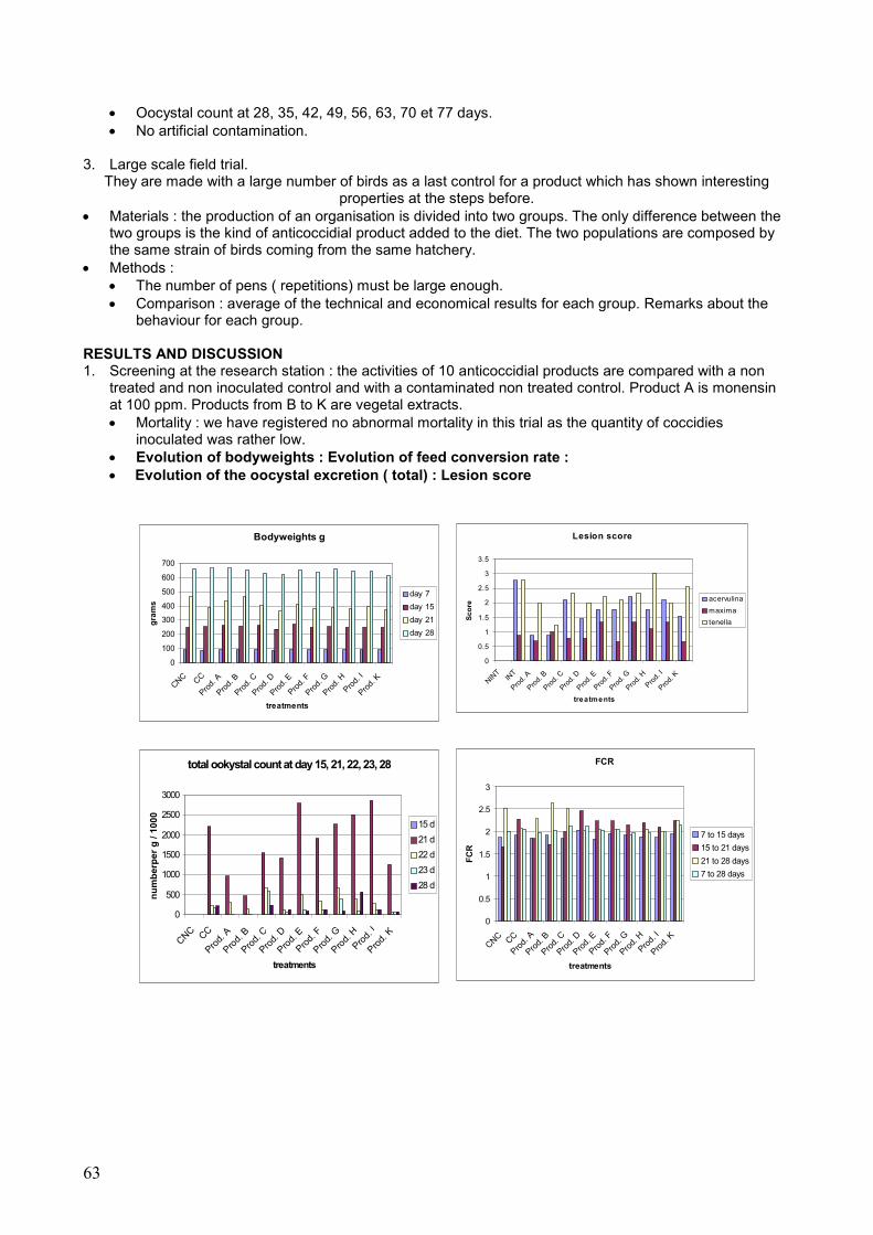
63
• Oocystal count at 28, 35, 42, 49, 56, 63, 70 et 77 days. • No artificial contamination.
3. Large scale field trial.
They are made with a large number of birds as a last control for a product which has shown interesting properties at the steps before.
• Materials : the production of an organisation is divided into two groups. The only difference between the two groups is the kind of anticoccidial product added to the diet. The two populations are composed by the same strain of birds coming from the same hatchery.
• Methods : • The number of pens ( repetitions) must be large enough. • Comparison : average of the technical and economical results for each group. Remarks about the
behaviour for each group. RESULTS AND DISCUSSION 1. Screening at the research station : the activities of 10 anticoccidial products are compared with a non
treated and non inoculated control and with a contaminated non treated control. Product A is monensin at 100 ppm. Products from B to K are vegetal extracts. • Mortality : we have registered no abnormal mortality in this trial as the quantity of coccidies
inoculated was rather low. • Evolution of bodyweights : Evolution of feed conversion rate : • Evolution of the oocystal excretion ( total) : Lesion score
total ookystal count at day 15, 21, 22, 23, 28
0
500
1000
1500
2000
2500
3000
CNC CC
Prod.
A
Prod.
B
Prod.
C
Prod.
D
Prod.
E
Prod.
F
Prod.
G
Prod.
H
Prod.
I
Prod.
K
treatments
nu
mb
erp
er
g /
10
00
15 d
21 d
22 d
23 d
28 d
FCR
0
0.5
1
1.5
2
2.5
3
CNC CC
Prod.
A
Prod.
B
Prod.
C
Prod.
D
Prod.
E
Prod.
F
Prod.
G
Prod.
H
Prod.
I
Prod.
K
treatments
FC
R
7 to 15 days
15 to 21 days
21 to 28 days
7 to 28 days
Bodyweights g
0
100
200
300
400
500
600
700
CNC CC
Prod.
A
Prod.
B
Prod.
C
Prod.
D
Prod.
E
Prod.
F
Prod.
G
Prod.
H
Prod.
I
Prod.
K
treatments
gra
ms
day 7
day 15
day 21
day 28
Lesion score
0
0.5
1
1.5
2
2.5
3
3.5
NINT
INT
Prod.
A
Prod.
B
Prod.
C
Prod.
D
Prod.
E
Prod.
F
Prod.
G
Prod.
H
Prod.
I
Prod.
K
treatments
Sc
ore
acervulina
maxima
tenella

64
2. Field trial : the production is “poulet label” and the region is centre of France. The strain of birds used in this trial is T 457 N . The rearing conditions are the classical ones in France for free range label. • Two programs are compared :
Control Experimental 0 to 28 days Robénidine Robénidine 28 to 73 days Monensin Product B to be tested
• Technical results :
Program Numbers Ages in d Weights in kg FCR Mortality % Control 103500 83.11 2.210 3.19 3.75
Exp 319100 83.09 2.154 3.17 2.82 CONCLUSION Conclusion : the first trial shows that the method used here is well adapted to discriminate various products that can exhibit anticoccidial side effects. Product B has a strong anticoccidial effect even compared with monensin, taking into account that the artificial contamination was not very important and that there is less risk of recontamination as the birds are reared in cages. The field trial confirms the possibility of using product B as an alternative to traditional anticoccidial product. REFERENCES
P.YVORE, 1976; “ revue sur la prévention des coccidioses en aviculture”- Avian Pathology, 5:237-252. P.YVORE et al, 1980 ; « method of evaluating the efficiency of anticoccidial drugs in floor-pen trials with multiple in-feed infection versus « seeding » model » - Ann. Rech. Vet. 11 (1), 99-108. N.HAMET et al. 1982 ; « enquête épidémiologique sur la coccidiose du poulet de chair » - RAA n°260. URAEA KAWAZOE, 1991 ; « sensitivity of field isolates of E. acervulina to salinomycin, maduramicin and a mixure of clopidol and methy benzoquate in chicken », Avian Pathology, 20, 439-446. S.MUIRHEAD, 1995 ; « research examines approaches for keeping coccidia in check », FEEDSTUFFS, November 6, 1995, 12 and 44. I.CREVIEU – GABRIEL, M.NACIRI, 2001, « effet de l’alimentation sur les coccidioses du poulet », INRA Prod. Anim. , 14 ( 4) , 231-246 M. FRANCESCH and J.BRAFAU, 2004, « nutritional factors affecting excreta/litter moisture and quality », W.P.S Journal, vol. 60, March 2004.
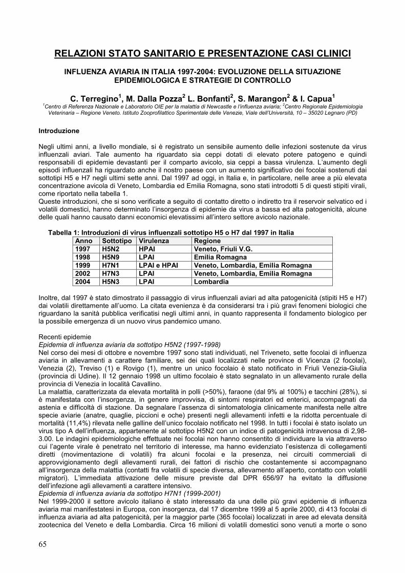
65
RELAZIONI STATO SANITARIO E PRESENTAZIONE CASI CLINICI
INFLUENZA AVIARIA IN ITALIA 1997-2004: EVOLUZIONE DELLA SITUAZIONE EPIDEMIOLOGICA E STRATEGIE DI CONTROLLO
C. Terregino1, M. Dalla Pozza2 L. Bonfanti2, S. Marangon2 & I. Capua1
1Centro di Referenza Nazionale e Laboratorio OIE per la malattia di Newcastle e l’influenza aviaria;
2Centro Regionale Epidemiologia
Veterinaria – Regione Veneto. Istituto Zooprofilattico Sperimentale delle Venezie, Viale dell’Università, 10 – 35020 Legnaro (PD)
Introduzione Negli ultimi anni, a livello mondiale, si è registrato un sensibile aumento delle infezioni sostenute da virus influenzali aviari. Tale aumento ha riguardato sia ceppi dotati di elevato potere patogeno e quindi responsabili di epidemie devastanti per il comparto avicolo, sia ceppi a bassa virulenza. L’aumento degli episodi influenzali ha riguardato anche il nostro paese con un aumento significativo dei focolai sostenuti dai sottotipi H5 e H7 negli ultimi sette anni. Dal 1997 ad oggi, in Italia e, in particolare, nelle aree a più elevata concentrazione avicola di Veneto, Lombardia ed Emilia Romagna, sono stati introdotti 5 di questi stipiti virali, come riportato nella tabella 1. Queste introduzioni, che si sono verificate a seguito di contatto diretto o indiretto tra il reservoir selvatico ed i volatili domestici, hanno determinato l’insorgenza di epidemie da virus a bassa ed alta patogenicità, alcune delle quali hanno causato danni economici elevatissimi all’intero settore avicolo nazionale.
Tabella 1: Introduzioni di virus influenzali sottotipo H5 o H7 dal 1997 in Italia Anno Sottotipo Virulenza Regione 1997 H5N2 HPAI Veneto, Friuli V.G. 1998 H5N9 LPAI Emilia Romagna 1999 H7N1 LPAI e HPAI Veneto, Lombardia, Emilia Romagna 2002 H7N3 LPAI Veneto, Lombardia, Emilia Romagna 2004 H5N3 LPAI Lombardia
Inoltre, dal 1997 è stato dimostrato il passaggio di virus influenzali aviari ad alta patogenicità (stipiti H5 e H7) dai volatili direttamente all’uomo. La citata evenienza è da considerarsi tra i più gravi fenomeni biologici che riguardano la sanità pubblica verificatisi negli ultimi anni, in quanto rappresenta il fondamento biologico per la possibile emergenza di un nuovo virus pandemico umano. Recenti epidemie Epidemia di influenza aviaria da sottotipo H5N2 (1997-1998) Nel corso dei mesi di ottobre e novembre 1997 sono stati individuati, nel Triveneto, sette focolai di influenza aviaria in allevamenti a carattere familiare, sei dei quali localizzati nelle province di Vicenza (2 focolai), Venezia (2), Treviso (1) e Rovigo (1), mentre un unico focolaio è stato notificato in Friuli Venezia-Giulia (provincia di Udine). Il 12 gennaio 1998 un ultimo focolaio è stato segnalato in un allevamento rurale della provincia di Venezia in località Cavallino. La malattia, caratterizzata da elevata mortalità in polli (>50%), faraone (dal 9% al 100%) e tacchini (28%), si è manifestata con l’insorgenza, in genere improvvisa, di sintomi respiratori ed enterici, accompagnati da astenia e difficoltà di stazione. Da segnalare l’assenza di sintomatologia clinicamente manifesta nelle altre specie aviarie (anatre, quaglie, piccioni e oche) presenti negli allevamenti infetti e la ridotta percentuale di mortalità (11,4%) rilevata nelle galline dell’unico focolaio notificato nel 1998. In tutti i focolai è stato isolato un virus tipo A dell’influenza, appartenente al sottotipo H5N2 con un indice di patogenicità intravenosa di 2,98-3.00. Le indagini epidemiologiche effettuate nei focolai non hanno consentito di individuare la via attraverso cui l’agente virale è penetrato nel territorio di interesse, ma hanno evidenziato l’esistenza di collegamenti diretti (movimentazione di volatili) fra alcuni focolai e la presenza, nei circuiti commerciali di approvvigionamento degli allevamenti rurali, dei fattori di rischio che costantemente si accompagnano all’insorgenza della malattia (contatti fra volatili di specie diversa, allevamento all’aperto, contatto con volatili migratori). L’immediata attivazione delle misure previste dal DPR 656/97 ha evitato la diffusione dell’infezione agli allevamenti a carattere intensivo. Epidemia di influenza aviaria da sottotipo H7N1 (1999-2001) Nel 1999-2000 il settore avicolo italiano è stato interessato da una delle più gravi epidemie di influenza aviaria mai manifestatesi in Europa, con insorgenza, dal 17 dicembre 1999 al 5 aprile 2000, di 413 focolai di influenza aviaria ad alta patogenicità, per la maggior parte (365 focolai) localizzati in aree ad elevata densità zootecnica del Veneto e della Lombardia. Circa 16 milioni di volatili domestici sono venuti a morte o sono

66
stati abbattuti e distrutti al fine di eradicare l’infezione, con un danno economico diretto (indennizzi e spese per l’estinzione dei focolai) pari a oltre 110 milioni di euro. A tali costi devono essere aggiunte le rilevanti perdite economiche di tipo indiretto, pari a circa 400 milioni di euro, correlate alla limitazione o al blocco, per diversi mesi, della normale attività produttiva e commerciale dell’industria avicola. E’indispensabile sottolineare che tale epidemia era stata generata dalla circolazione, per circa un anno, di un virus influenzale a bassa virulenza (LPAI) appartenente al sottotipo H7N1, che è successivamente mutato dando origine ad uno stipite ad alta patogenicità (HPAI). Nel mese di agosto 2000, il virus LPAI, sottotipo H7N1, è ricomparso e si è diffuso negli allevamenti avicoli del Veneto con insorgenza, da agosto 2000 a marzo 2001, di 78 focolai di malattia (di cui il 94% in allevamenti di tacchini da carne). L’ultimo focolaio da virus LPAI, sottotipo H7N1, è stato estinto il 6 marzo 2001 e, in totale, circa 2 milioni di volatili sono stati abbattuti o avviati alla macellazione controllata, con un danno economico diretto pari a più di 10 milioni di euro. Epidemia da virus LPAI (H7N3) in Lombardia e Veneto (2002-2003) Nel luglio 2002 un nuovo stipite virale, sottotipo H7N3, è stato individuato in allevamenti di tacchini della Lombardia. Nonostante la pronta adozione di opportune misure di eradicazione, l’infezione si è diffusa rapidamente alle aree ad elevata densità di allevamenti avicoli, principalmente della Lombardia e del Veneto. Le caratteristiche dello stipite in causa hanno consentito di stabilire la recente introduzione del virus influenzale dall’ospite selvatico. In totale, in Italia sono stati individuati 388 focolai di malattia da virus LPAI sottotipo H7N3. Tutti gli animali presenti negli allevamenti infetti sono stati sottoposti ad abbattimento e distruzione o avviati alla macellazione controllata, con un danno economico diretto (indennizzi e spese per l’estinzione dei focolai) stimato a circa 40 milioni di euro. L’ultimo focolaio da virus LPAI del sottotipo H7N3 è stato notificato il 30 settembre 2003.
Situazione attuale Attualmente l’Italia è indenne da influenza aviaria. I piani di monitoraggio in atto hanno però evidenziato con frequenza preoccupante la circolazione di virus influenzali di sottotipi diversi in molte aree del nord Italia, indice di un rischio continuo di introduzione di nuovi stipiti virali nella popolazione di volatili domestici delle aree a maggior densità avicola con potenziale ricomparsa di nuovi virus HPAI. Vengono di seguito schematicamente riportate i reperti di positività virologica e sierologia rispettivamente in volatili domestici e selvatici, rilevati dal 2003 ad oggi. Isolamenti virali in volatili domestici
• virus H1N1 isolato da tamponi cloacali di oche in provincia di Ravenna nel mese di dicembre 2003; • virus H3N8 isolato da tamponi cloacali di anatre in provincia di Ravenna nel mese di dicembre 2003; • virus H1N2 e H9N8 isolati da pool di feci di anatre e oche in provincia di Mantova nel mese di
gennaio 2004; • virus H1N1 isolato da pool di feci di anatre e oche in provincia di Mantova nel mese di febbraio
2004; • virus H5N3 LPAI isolato da pool di feci di anatre in provincia di Brescia nel mese di febbraio 2004; • virus H1N1 isolato da tamponi cloacali di anatre in provincia di Ravenna nel mese di maggio 2004. • virus H4N6 isolato da tamponi cloacali di anatre in provincia di Ravenna nel mese di agosto 2004.
Positività sierologiche in allevamenti di volatili domestici
• Positività sierologiche per il sottotipo H2N2 (titoli in HI: 1:32 - 1:1024) in tacchini da carne (4.000 maschi) della provincia di Cremona. Il 23 ottobre 2003 è stato notificato il focolaio di malattia da virus LPAI sottotipo H2N2, gli animali sono stati abbattuti il 27 ottobre 2003.
• Positività sierologiche per H9 (12 dubbi:1:4 -1:8; 25 positivi: 1:16 – 1:128) in anatre rurali nella provincia di Vercelli riscontrate a maggio 2004 da IZS TO e confermate dal CRN dell’IZS di PD. Cross- positività con antigeni H6N2 e H2N2. Non è stato possibile evidenziare virus in attiva circolazione in azienda (30 tamponi cloacali negativi in PCR e isolamento in uova).
• Positività sierologiche per H9 (7 dubbi:1:4 -1:8; 2 positivi: 1:16 e 1:128) da anatre rurali in provincia di Pavia, riscontrate a giugno 2004 da IZS BS e confermate dal CRN dell’IZS di PD. Dallo stesso allevamento era stato isolato nel 2002 un AIV H9N2. E’stata osservata una cross- positività in HI con antigene H5N9. Non è stato possibile evidenziare virus in attiva circolazione in azienda (tamponi cloacali negativi in isolamento in uova).
Virus isolati da uccelli selvatici
• Virus H10N4 isolato il 12.03.04 da tamponi cloacali di codone (Anas acuta) prelevati in Valle Dragojesolo-Dogà nel mese di dicembre 2003
• Virus H1N1 isolato il 30.03.2004 da tamponi cloacali di germano (Anas platyrhynchos) prelevati in Valle Dragojesolo (VE) nel mese di novembre 2003
Virus isolati da uccelli esotici d’importazione
67
• Virus H3N8 isolati da feci di passeriformi e pappagalli in quarantena provenienti da Argentina, Slovacchia, Cina, Malesia e Pakistan presso un rivenditore nella provincia di Forlì-Cesena nei mesi di dicembre 2002, marzo aprile e maggio 2003 da IZS di Forlì e tipizzati dal CRN di Padova.
Interessante notare come la maggior parte degli isolati provenga da allevamenti di tipo rurale in cui sono presenti anatidi, a riconferma del fatto che tali allevamenti, per le loro caratteristiche di semi-intensività sono maggiormente soggetti a nuove introduzioni virali e quindi importanti da monitorare nell’ambito delle attività di sorveglianza epidemiologica mirate all’individuazione precoce di nuove introduzioni virali dalle popolazioni selvatiche. Strategie di controllo Sulla base delle esperienze passate ci si è resi conto che la prevenzione è l’unica arma in grado di evitare epidemie devastanti come quelle degli anni passati. La prevenzione è stata basata in passato sull’applicazione di misure di biosicurezza e sulle attività di monitoraggio. Dall’inverno 2004, l’Italia attiverà, per la prima volta nel mondo, una campagna di vaccinazione profilattica. Infatti l’Italia si è resa promotrice presso la Commissione Europea di una strategia integrata, basata anche sulla vaccinazione delle popolazioni a rischio con vaccino bivalente H5/H7. Tale campagna di vaccinazione, basata sul sistema “DIVA” dovrebbe generare una popolazione maggiormente resistente alle infezioni primarie, e di conseguenza porre una barriera all’endemizzazione delle infezioni sostenute da questi sottotipi. Biosicurezza In questi ultimi anni una serie di decreti regionali ha regolamentato la gestione degli allevamenti avicoli in modo da limitare al massimo la possibile entrata e diffusione da un allevamento all’altro dei virus influenzali. La possibilità di mettere in atto nel modo più efficace le più elementari norme di biosicurezza ha avuto come punto di partenza la ristrutturazione dei locali di allevamento (capannoni). I requisiti strutturali minimi imposti sono stati:
− pavimento in cemento o in materiale lavabile per facilitare le operazioni di pulizia e disinfezione − pareti e soffitti pulibili; − attrezzature facilmente pulibili e disinfettabili; − efficaci reti antipassero su tutte le aperture. − barriere all’ingresso idonee ad evitare l’ingresso non controllato di automezzi (cancelli o sbarre mobili); − piazzole di carico e scarico dei materiali d’uso e degli animali dotate di un solido fondo ben
mantenuto, lavabili e disinfettabili e di dimensioni minime pari all’apertura del capannone; − caricamento del mangime dall’esterno della recinzione per i nuovi fabbricati destinati all’allevamento
dei riproduttori dovrà − una superficie larga un metro lungo tutta la lunghezza esterna del capannone da mantenere
costantemente pulita; − aree di stoccaggio dei materiali d’uso (lettiere vergini, mezzi meccanici ecc.) protette; − una zona filtro dotata di spogliatoio, lavandini e detergenti all’entrata di ogni azienda. Dovranno essere
previsti una dotazione di calzature e tute specifiche e cartelli di divieto di accesso agli estranei; − le attrezzature d’allevamento e di carico (muletti, pale, nastri e macchine di carico etc.) si considerano
di norma dotazione di ogni singolo allevamento o, se utilizzate da più aziende, devono essere sottoposte ad accurato lavaggio e disinfezione ad ogni ingresso ed uscita;
− deve esserci assenza di qualsiasi materiale nelle zone attigue ai capannoni; − deve essere previsto uno spazio per il deposito temporaneo dei rifiuti.
I decreti emanati per la prevenzione dell’introduzione dei virus influenzale regolamentano inoltre la conduzione dell’allevamento, il trasporto delle uova da cova e da consumo, il carico degli animali destinati alla macellazione, la disinfezione degli automezzi adibiti al trasporto dei volatili, l’accasamento, le operazioni di pulizia e disinfezione dei capannoni e la gestione degli animali morti e delle lettiere. Monitoraggio L’infezione provocata da virus influenzale a bassa patogenicità (LPAI) non sempre è associata all’insorgenza di quadri clinici caratteristici e può passare inosservata in alcune specie lasciando, come unica traccia del suo passaggio, la sieropositività. In ogni caso, in alcune specie, gruppi sieropositivi possono costituire dei serbatoi di infezione in grado di garantire la persistenza del virus, con possibile insorgenza di nuovi eventi epidemici. Alla necessità di individuare precocemente gruppi sieropositivi si aggiunge l’esigenza di effettuare controlli virologici dai volatili serbatoi naturali dell’infezione (anatidi). I piani di monitoraggio messi in atto in questi ultimi anni, durante e dopo le recenti epidemie, hanno dimostrato come si possa efficacemente e tempestivamente individuare la presenza di virus potenzialmente pericolosi. Ciò è testimoniato dall’elevato numero di isolamenti di virus influenzali aviari negli ultimi due anni soprattutto in allevamenti di tipo rurale da sempre poco controllati. Solo attraverso una precoce individuazione dei focolai primari e la rapida messa in opera delle misure di contenimento anche per i virus LPAI appartenenti ai sottotipi H5 e H7, è possibile auspicare che l’infezione non si diffonda a circuiti nei quali risulta difficilmente gestibile in tempi rapidi.

68
L’attuale normativa europea, che prevede l’adozione di misure obbligatorie di eradicazione esclusivamente nei confronti degli stipiti altamente virulenti, è in fase di completa revisione con lo scopo principale di fornire basi legislative univoche a supporto dell’eradicazione di tutti gli stipiti H5 e H7 a prescindere dalla virulenza. In realtà, nonostante la nuova normativa non sia stata ancora definita la dimostrata evoluzione dei virus LPAI, sottotipi H5 e H7, in virus HPAI ha indotto tutti i paesi a zootecnia industriale ad imporre misure di controllo drastiche anche nei confronti di stipiti a bassa virulenza. Negli USA, nel 2002, sono stati abbattuti 5 milioni di volatili infetti da un virus LPAI H7N2, con un danno economico diretto pari a più di 140 milioni di dollari. Da sottolineare che tale stipite sta circolando nei mercati di volatili vivi al dettaglio (live bird markets -LBM) da circa 10 anni. I piani di monitoraggio tuttora in corso hanno interessato prevalentemente le regioni maggiormente coinvolte nelle passate epidemie e prevedono periodici controlli sierologici e/o virologici (secondo metodiche e criteri interpretativi riportati al capitolo 4, allegato III del DPR 656/96) durante il ciclo di allevamento in allevamenti vaccinati e non, prima del carico e al macello. Accanto a questi piani regionali sono stati portati avanti piani estesi su tutto il territorio nazionale. Il piano di monitoraggio nazionale 2004, da ultimare entro il 31/12/04, prevede il controllo in tutto il territorio nazionale, in ottemperanza a quanto disposto dalla decisione 2004/111/CE relativa all’effettuazione di indagini sull’influenza aviaria nel pollame e nei volatili selvatici negli stati membri. In ciascuna regione, sulla base del numero di allevamenti presenti sul territorio regionale, sono stati scelti per ciascuna categoria produttiva un numero di allevamenti statisticamente significativo da controllare. Oltre al controllo delle popolazioni avicole domestiche nel piano è previsto un monitoraggio in popolazioni di volatili selvatici in alcune aree umide del territorio nazionale. In Italia le specie ritenute maggiormente recettive sono rappresentate dagli anatidi e dai limicoli. I primi raggiungono le nostre latitudini dalle aree centro-nord ed est europee e svernano nelle principali zone umide. I secondi, pur provenienti dalle medesime aree geografiche, ma da diversi habitat, tendono ad utilizzare le aree italiane principalmente come aree di foraggiamento prima del grande salto sul Mediterraneo che li vedrà raggiungere i quartieri di svernamento in Africa. Si assiste quindi, durante un breve periodo autunnale, ad una coabitazione di numerose specie, la cui distribuzione spaziale risulta estremamente aggregata in funzione delle residue zone umide presenti nel nostro paese alcune delle quali vicine ad aree ad alta densità di allevamenti avicoli. Il resto dell'inverno è, invece, caratterizzato dalla presenza di poche specie, ma estremamente numerose. Per entrambi i gruppi la maggioranza degli animali presenti o transitanti è costituita da giovani nati nella primavera precedente e quindi, almeno dal punto di vista teorico, più recettivi ai virus influenzali. La caratterizzazione degli ultimi isolati che ha identificato il serbatoio selvatico come origine dei virus responsabili delle più recenti epidemie ed il continuo isolamento di virus influenzali da uccelli acquatici nell’ambito di piani di monitoraggio regionale, ha confermato come sia importante continuare a monitorare per quanto possibile lo stato sanitario di queste popolazioni. Vaccinazione Per controllare la diffusione dei virus a bassa patogenicità i Servizi Veterinari hanno attivato adeguate misure restrittive e di monitoraggio, integrandole con l’implementazione di un piano di vaccinazione nelle aree a più elevata densità avicola. L’impiego della vaccinazione e la contemporanea applicazione di un intenso programma di sorveglianza hanno consentito, sia nel 2001 sia nel 2003, di eradicare rispettivamente le infezioni da H7N1 e H7N3. Durante l’epidemia del 1999-2000 al fine di controllare la diffusione del contagio è stato predisposto ed attivato un programma di vaccinazione, approvato dalla Commissione con Decisione 2000/721/CE, basato sull’impiego di un vaccino inattivato di sottotipo eterologo (H7N3), nell’area della provincia di Verona a maggior rischio di insorgenza della malattia. Come è noto lo scopo della vaccinazione è quello di aumentare la resistenza degli animali all’infezione e contemporaneamente ridurre l’eliminazione del virus negli animali infetti in modo da avere una progressiva riduzione della contaminazione ambientale e del rischio di diffusione del virus. La rigorosa adozione delle misure di biosicurezza negli allevamenti, la messa in atto di efficaci norme di restrizione della movimentazione di animali vivi, la rapida identificazione e l’eliminazione attraverso lo stamping out o la macellazione controllata dei volatili degli allevamenti infetti, unita all’impiego sistematico della vaccinazione e al monitoraggio continuo della situazione epidemiologica, hanno portato all’eradicazione della malattia. Tale strategia è stata usata anche per combattere l’epidemia da H7N3 che ha colpito il Veneto e la Lombardia a partire dal secondo semestre del 2002. Con l’ausilio della vaccinazione e della strategia territoriale anche questa infezione è stata eradicata. Attualmente, in alcune province di Veneto e Lombardia, è ancora in atto la parte finale del programma di vaccinazione confronti del sottotipo H7N3 approvato con Decisione 2002/975/CE. E’evidente che una volta eradicato lo stipite H7N3, il rischio di introduzione di un virus H5 o H7 risulta essere il medesimo, e pertanto la prosecuzione della campagna di vaccinazione solo nei confronti del sottotipo H7 risulta essere insufficiente a proteggere il patrimonio avicolo italiano da nuove introduzioni potenzialmente pericolose Il persistente rischio di introduzione di nuovi stipiti virali, unito alle considerazioni sopraesposte,
69
suggeriscono l’implementazione di un programma vaccinale pilota basato sull’impiego di un vaccino bivalente H5 e H7 nelle aree a maggiore concentrazione di allevamenti avicoli. Tale programma è stato recentemente approvato il 14 luglio 2004 a Brexelles dal Comitato Permanente della Catena Alimentare e Sanità Animale con decisione “documento SANCO” 1024/2004-Rev.1. Tale programma, di durata limitata (12 mesi), si basa sull’impiego, nelle specie a più elevato rischio di infezione, di due soli interventi vaccinali, in grado di indurre un’immunità di base che dovrebbe essere sufficiente ad aumentare significativamente la resistenza degli animali all’infezione da ceppi H5 ed H7. Inoltre, consentirebbe, in caso di emergenza, l’applicazione di un unico intervento di richiamo per aumentare rapidamente l’immunità della popolazione a rischio. Essendo la trasmissione dei virus di nuova introduzione inizialmente poco efficiente, dovuto allo scarso livello di adattamento all’ospite domestico, la vaccinazione profilattica dovrebbe essere in grado di prevenire l’instaurarsi di infezioni produttive. Questa strategia può garantire il rapido controllo di eventuali ceppi virali di nuova introduzione in quanto i piani di monitoraggio che verranno adottati nella zona di vaccinazione permetteranno la precoce individuazione di ogni nuovo caso di infezione. Vi è un valore aggiunto da non sottovalutare a questa strategia. La circolazione del virus H5N1 in Asia ha determinato preoccupazione nei consumatori di carne avicola, che si è ripercossa sul mercato con un calo dei consumi. Fra le domande che più volte venivano poste agli esperti ed alle associazioni di categoria vi era quella sullo stato sanitario del pollame nazionale rispetto a questa infezione. Sarà possibile, qualora la crisi asiatica dovesse ricomparire nei mezzi di informazione, rassicurare i consumatori rispetto allo stato di immunizzazione dei nostri volatili nei confronti di questa virosi. Conclusioni L’Italia, è un paese a rischio per le infezioni influenzali aviarie, per questo motivo si è resa promotrice di una serie di iniziative a livello nazionale e comunitario. Fra queste, la revisione della Direttiva comunitaria sull’Influenza Aviaria e l’istituzione di programmi di vaccinazione specifici che permettano l’esportazione dei prodotti avicoli in ambito comunitario. E’ comunque attraverso la collaborazione fra i produttori ed i responsabili sanitari aziendali con le istituzioni e gli enti pubblici che si occupano dei diversi aspetti di questa virosi, che si possono affrontare e risolvere i problemi sanitari generati da queste infezioni. E’ indispensabile, affinché si possa operare a livello internazionale sorretti dalla credibilità scientifica necessaria per introdurre innovazioni, che la collaborazione fra le entità coinvolte prosegua e si rafforzi nel tempo.
INDAGINE EPIDEMIOLOGICA SULLA CIRCOLAZIONE DEI VIRUS DELLA
BRONCHITE INFETTIVA PRESSO l’IZS DELLE VENEZIE
L’indagine ha avuto inizio nel gennaio 2004 condotta dal Laboratorio di Virologia dell’Istituto Zooprofilattico delle Venezie, in collaborazione con i veterinari del settore avicolo. L’indagine ha avuto come target gli allevamenti di broiler e ovaiole nel territorio del Triveneto e alcune province della Lombardia. Il Laboratorio di Virologia ha fornito alle aziende che hanno aderito al progetto, polli SPF come animali sentinella da introdurre negli allevamenti. Dal gennaio 2004 sono stati monitorati 30 allevamenti per un totale di 101 campioni esaminati. Su 30 allevamenti analizzati sono stati isolati 13 virus della bronchite infettiva esclusivamente da allevamenti di broiler. Di questi 7 sono stati identificati come la variante IT-02 (nella province di Verona, Padova, Vicenza, Rovigo e Cremona), e 6 come 793-B (nelle province di Padova, Verona, Brescia e Mantova). Clinicamente non sono state quasi mai segnalate forme morbose particolarmente gravi, negli allevamenti in cui è stata isolata la variante 793/B sono state diagnosticate prevalentemente forme respiratorie talvolta complicate da nefriti. Una sintomatologia prevalentemente respiratoria ed enterica è stata invece segnalata nelle infezioni da IT-02. Si segnala inoltre un grave episodio di bronchite da virus nefropatogeno in allevamenti di fagiani in provincia di Pordenone. La mortalità in questo caso ha superato il 20% a causa di gravi lesioni renali e gotta viscerale riscontrate soprattutto in fagianotti di 10-30 giorni di età. Il virus isolato è stato identificato come ceppo nefropatogeno IB 1648.
70
CASI CLINICI
1) CASI DI ROTTURA EPATICA IN GIOVANI FARAONE
Rampin T.1, Manarolla G.
1, Recordati C.
1, Longoni C.
3,
Sironi G.1, Sartorelli P.
2, Spagnolo V.
2
1Dipartimento di Patologia Animale, Igiene e Sanità Pubblica Veterinaria;
2Sezione di Anatomia Patologica
Veterinaria e Patologia Aviare; 2Sezione di Patologia Generale Università degli Studi di Milano;
3Veterinario
dipendente di azienda privata. Correspondence: Tiziana Rampin - Dipartimento di Patologia Animale, Igiene e Sanità Pubblica Veterinaria, Sezione di Anatomia Patologica Veterinaria e Patologia Aviare, Università degli Studi di Milano – Via Celoria 10 – 20133 Milano. Email [email protected]
Parole chiave: faraona, emorragia epatica, rottura epatica
Liver rupture in young guinea fowls– Case report
Summary: Several guinea fowl flocks showed increasing mortality due to liver rupture from about 3 weeks of age. Grossly, liver was severely enlarged and characterised by hematomas and subcapsular haemorrhages or capsule rupture. Histological examination of liver samples from dead and sacrificed birds revealed mild to extremely severe circulatory and hepatocellular alterations in absence of amyloidosis or steatosis. Liver damage was also suggested by some altered biochemical parameters in sera from affected flocks. The very high growth rate of this guinea fowl breed can play a role in the pathogenesis of this lesion. Key words: guinea fowl, liver haemorrhage, liver rupture
Introduzione Fenomeni emorragici nel fegato che conducono ad ematomi nel parenchima o in sede sotto capsulare fino alla rottura epatica sono descritti negli uccelli di allevamento in un numero limitato di forme morbose. Tipica di galline in deposizione è la cosiddetta “Fatty liver haemorrhagic syndrome” (FLHS) in associazione a steatosi epatica (1), mentre composti tossici della farina di semi di colza causano la “Hepatic haemorrage” o “Liver haemorrhagic syndrome” (LHS) nelle galline (2) e la “Massive liver haemorrage” in broiler tra le 2 e le 4 settimane (3). Inoltre rottura del fegato si può riscontrare in presenza di amiloidosi epatica, che per esempio abbiamo avuto occasione di riscontrare in pollastre sottoposte a ripetute vaccinazioni con vaccini spenti antibatterici (4) o che si manifesta in gruppi colpiti da Sinovite Infettiva da Mycoplasma synoviae. Riteniamo perciò interessante descrivere casi di rottura epatica (RE) diagnosticati recentemente in faraone di poche settimane di vita, che costituiscono un fenomeno completamente nuovo per questa specie e di cui mancano segnalazioni bibliografiche. Materiali e metodi Animali: i 3 gruppi di faraone in cui veniva segnalata la comparsa di RE appartenevano tutti a una linea genetica caratterizzata da maggior rapidità di crescita rispetto alle linee tradizionali. I criteri di allevamento e di alimentazione erano comunque uguali nelle partite di entrambe le linee genetiche. Dalle faraone dei gruppi con problemi di RE e da faraone di genetica tradizionale della stessa età sono stati prelevati a più riprese campioni di fegato e di altri visceri per indagini istologiche ed istochimiche, nonché il sangue per esami ematochimici. L’esame istologico veniva condotto su sezioni di 4 micron di spessore ottenute dai campioni dopo fissazione in formalina tamponata al 10% e inclusione in paraffina usando la colorazione con Ematossilina-Eosina (EE) e le colorazioni di James (5) per le fibre reticolari e Rosso Congo per l’amiloide. Cinque faraone di circa 4 settimane di un gruppo che manifestava casi di RE e cinque coetanee di un gruppo di genetica tradizionale come controllo sono stati sottoposti a prelievo ematico per ottenere il siero destinato alle indagini biochimiche. Sono stati determinati i seguenti parametri: glucosio, proteine totali, creatinina, urea, acido urico, colesterolo, fosfatasi alcalina (ALP), alanina-aminotransferasi (GPT), gamma-glutamil-transferasi (GGT) e creatin chinasi (CK) mediante l’uso di un analizzatore automatico (Hospitex diagnostics, Firenze) e i reattivi forniti dalla stessa ditta. È inoltre stata eseguita l’elettroforesi delle proteine sieriche, per la determinazione delle diverse frazioni proteiche. Descrizione dei casi osservati In generale, nei gruppi di faraone che manifestano RE la mortalità compare intorno ai venti giorni circa, quando il peso medio dei soggetti già supera il peso medio dei soggetti tradizionali. Inoltre mancando il
71
riscontro di animali sintomatici, sofferenti o con problemi di crescita, è verosimile ritenere che la RE e l’emorragia mortale che ne consegue siano un evento subitaneo. In un gruppo di 35.000 faraone, tra i primi osservati, la mortalità giornaliera è passata da 130 a 600 capi tra i 20 e i 30 giorni di vita ed era dovuta praticamente nella totalità dei casi a RE. A questo punto il gruppo è stato tenuto digiuno per 24 ore e poi alimentato con un mangime addizionato a crusca per diminuirne l’energia di circa il 10%. In questo modo la mortalità si è abbassata a circa cento capi al giorno e il ciclo si è chiuso con una mortalità totale del 24,2%. Risultati Nelle faraone morte dei gruppi colpiti da RE il fegato si presenta notevolmente aumentato di volume e di colore bruno pallido, i fenomeni emorragici possono interessare uno o entrambi i lobi sotto forma di numerosi ematomi parenchimali o di un unico ematoma sottocapsulare che ricopre in larga parte un intero lobo epatico. Frequentemente la lesione progredisce fino a rottura della glissoniana e si rinvengono uno o più coaguli addossati al fegato o sparsi in sede cavitaria. Talora il miocardio mostra un colore rosa pallido. Nessuna alterazione macroscopica di rilievo si rinveniva invece nei 5 soggetti provenienti da un gruppo colpito e sacrificati dopo prelievo ematico. Le alterazioni epatiche rilevate nelle sezioni istologiche dei soggetti morti con RE sono sostanzialmente di duplice natura: circolatoria e regressivo-necrotica epatocitaria, entrambe con aspetti di diversa gravità. I disturbi di circolo interessano soprattutto i sinusoidi, che in qualche caso si presentano dilatati ma che più frequentemente si distanziano dalle filiere epatiche con ampliamento dello spazio di Disse. A questo livello è talvolta evidente la presenza di materiale debolmente eosinofilo amorfo o granulare, di probabile origine plasmatica e Rosso Congo negativo. Anche in sede sottocapsulare possono essere presenti alterazioni riferibili ad edema. Tali fenomeni riconducibili ad alterata permeabilità della parete vasale possono giungere a vistose ectasie dello spazio sinusoidale con scollamento delle corde epatocitarie, stasi marcata o emorragie in senso proprio con scomparsa delle cellule endoteliali. Le alterazioni di circolo più vistose sono rappresentate da ematomi in sede parenchimale nei quali si alternano ora sangue di recente coagulazione ora fibrina in via di organizzazione, delimitata da tessuto connettivo fibroso più o meno denso. Attorno alla lesione può comparire pigmento emosiderinico sparso in modica quantità. Gli epatociti generalmente mostrano diffusi segni di sofferenza con degenerazione torbido-vacuolare, ma in alcune sezioni sono evidenti necrosi zonali, con aspetti di cariolisi o apoptosi, o necrosi epatocellulare individuale. In una sola sezione si è osservato un quadro di necrosi coagulativa zonale confluente con fagocitosi cellulare ad opera delle cellule di Kupfer. Accanto agli aspetti regressivi, tra gli epatociti si nota qualche tentativo rigenerativo con mitosi. Frequentemente accompagnano queste lesioni aspetti di iperplasia dei dotti biliari e talora lieve fibrosi delle aree portali. La colorazione di James per le fibre di reticolina evidenzia differenze e irregolarità nell’aspetto e nella distribuzione dello stroma reticolare che accompagna i sinusoidi dei fegati di faraone del gruppo con RE rispetto a coetanee di altro gruppo e differente genetica e a fegati di pollo. Di 6 soggetti con RE è stato esaminato anche il miocardio che è costantemente interessato da edema e degenerazione torbida delle fibre. In un caso si è rilevata una estesa area cicatriziale a carico della parete del ventricolo sinistro. Anche in uno dei soggetti del gruppo con RE che abbiamo abbattuto dopo prelievo ematico, pur in assenza di fenomeni emorragici macroscopici, si sono riscontrate lesioni epatiche caratterizzate da ampie aree di necrosi periportale e modica dilatazione degli spazi di Disse. Inoltre anche la trama di fibre di reticolina mostra delle aree di irregolarità Nelle 5 faraone del gruppo non interessato da RE non si sono riscontrate alterazioni di sorta a carico del fegato. Per quanto riguarda le indagini biochimiche sui sieri, si evidenzia un’ampia variabilità individuale, in assenza di marcate differenze tra i due gruppi. Le principali alterazioni si sono riscontrate nel siero di un unico soggetto del gruppo con RE, in cui sono stati riscontrati valori nettamente superiori di acido urico e GGT, mentre le prealbumine sono decisamente più basse. Discussione Prima di addentrarci in ipotesi esplicative di questi casi di RE, vorremmo ricordare che supplementi vitaminici, di macro- e microelementi non hanno effetto sulla mortalità, escludendo cosi forme carenziali, paragonabili all’epatosi dietetica del suino, forma gravissima di epatodistrofia rapidamente mortale come la RE (6). Effetti positivi si hanno solo intervenendo sull’alimentazione, infatti la riduzione dell’energia abbassa sensibilmente, anche se non sopprime, la mortalità. Altra considerazione preliminare è che in questi casi la rottura non è dovuta ad amiloidosi né a steatosi epatica come nella FLHS. Anche gli effetti tossici della farina di semi di colza possono essere esclusi, in quanto questa non compare tra i componenti del mangime. Però anche in questo caso sono presenti
72
alterazioni della trama di fibre reticolari come nella FLHS e nella LHS, dove questo fenomeno è stato evidenziato come l’evento necessario per l’insorgenza di fenomeni emorragici (7 e 8). La reticololisi e le alterazioni epatocitarie che potrebbero essere due facce dello stesso processo patogenetico oppure essere tra loro in rapporto di causa-effetto. Il danno epatico risulterebbe anche già presente in uno dei soggetti vivi sulla base dei dati biochimici riscontrati nei pur pochi sieri testati, e cioè dell’aumento della GGT e della diminuzione delle prealbumine, che anche nelle specie aviari sono rappresentate dalla transtiretina, proteina sintetizzata nel fegato. Anche istologicamente in una sezione di fegato di questi soggetti si evidenziano sia alterazioni degli epatociti sia disgregazioni della trama di fibre reticoliniche. Valutando gli effetti positivi del cambio di alimentazione, riteniamo probabile che alla base della RE ci sia un problema metabolico che interessa queste faraone verosimilmente in rapporto alla loro rapida crescita. Rimane da chiarire come poi si giunga alla reticololisi a livello di sinusoidi epatici e alla sofferenza dell’epatocita. Sulla base dei dati qui riportati non ci sentiamo di formulare ipotesi sulle lesioni cardiache presenti in associazione alla RE in quanto riguardano un numero molto limitato osservazioni né ci sentiamo di escludere lesioni ad altri organi che non sono stati campionati nei soggetti da noi esaminati e che eventualmente potrebbero meglio definire il quadro della patologia. Bibliografia 1. Crespo R., Shivaprasad H.L. (2003) Developmental, metabolic and other non infectious disorders in
“Diseases of poultry” eds Saif Y.M. et al., XI edition, Iowa State Press, Ames, IA, pag 1082-1084. 2. 3. Leeson S., Diaz G.J., Summers J.D. (1995) Hepatic haemorrhage (cap. 4) in “Poultry Metabolic
Disorders and Mycotoxins”. University Books, Guelph, Ontario, Canada. pag. 51-54. 4. 5. Gough A.W., Weber L.J. (1978) Massive Liver Hemorrage in Ontario broiler chickens. Avian dis. 22, 205-
210. 6. 7. Rampin T., Sironi G., Gallazzi D. (1989) Amyloidoseepisoden bei Junghennen nach wiederholte
Anwendung von antibakteriellen Olemulsionvakzinen. Deutsche Tierarztliche Wochenschrift. 96: 4, 168-172.
8. James K.R. A simple silver method for the demonstration of reticulin fibres. J. med. Lab. Tech. 24, 49-
51.
9. Kelly W.R. (1993) The liver and biliary system. in “Pathology of domestic animals” eds Jubb K.V.F., Kennedy P.C., Palmer N., IV edition, Academic Press, San Diego, Ca, vol. 2°, pag. 344.
10. Hall S.A. (1974) A hepatosis of laying fowls characterized by a apparent lysis of reticulin and massive liver haemorrhage. Vet. Rec., 94, 42-44.
11. Walzem R.L., Simon C. Morishita T., Lowenstine L., Hansen R.J. (1993) Fatty liver hemorrhagic syndrome in hens overfed a purified diet. Selected enzyme activities and liver histology in relation to liver hemorrhage and reproductive performance. Poultry Science, 72, 1479-1491.
73
2) AVVERSA REAZIONE VACCINALE IN TACCHINI DA CARNE SOTTOPOSTI A PROFILASSI ANTINFLUENZALE.
Alessandri E.1, Saita M.2, Rampin T. 2, Manarolla G. 2
1DVM, libero professionista
2Dipartimento di Patologia Animale, Igiene e Sanità Pubblica Veterinaria,
Sezione di Anatomia Patologica Veterinaria e Patologia Aviare, Università degli Studi di Milano
Correspondence: Mario Saita - Dipartimento di Patologia Animale, Igiene e Sanità Pubblica Veterinaria, Sezione di Anatomia Patologica e Patologia Aviare, Università degli Studi di Milano – Via Celoria 10 – 20133 Milano. E-mail [email protected]
Parole chiave: tacchino, influenza aviaria, vaccinazione
Summary: Two cases of adverse immunological response are reported. Affected flocks had the same origin and were submitted to the 2nd vaccination against avian influenza H7 N1 at 42 days of age. In both cases, 36h post-vaccination a mortality of 1% occurred and animals sampled underwent necropsy. Macroscopic and histological findings were aspecific.
Key words: turkey, avian influenza, vaccination
Introduzione In seguito all’epidemia influenzale che ha interessato gli allevamenti avicoli del Nord Italia negli ultimi anni, sono state approntate delle misure di profilassi vaccinale relativamente all’allevamento del tacchino e della gallina ovaiola commerciale nelle province ad alta densità avicola. In base al Decreto Dirigenziale della Regione Veneto n° 465 del 10/12/2002 tale categoria di allevamenti deve essere sottoposta a profilassi vaccinale contro l’influenza aviaria, sottotipo H7 N1, secondo uno schema vaccinale che prevede al momento della stesura del presente documento 3 interventi vaccinali sia per i maschi che per le femmine. . I ripetuti interventi vaccinali sono necessari affinché si sviluppi un efficace livello anticorpale (Karunakaran et al., 1987, Stone 1987). Per i tacchini maschi tali interventi sono generalmente programmati a ca. 15, 40, 80 giorni di vita. L’intervento consiste nella somministrazione parenterale individuale di un vaccino inattivato (0,5ml/capo). Materiali e metodi In 2 allevamenti di tacchini maschi da carne, di linea Big 6, situati in provincia di Verona. si è verificata una anomala mortalità post-vaccinale. Per quanto riguarda gli allevamenti in oggetto, entrambi dispongono di classici capannoni a pianta rettangolare di ca. 1000 m2 ciascuno. I capannoni dispongono di finestratura laterale e ventilazione naturale coadiuvata da ventilatori da 1KW (ca. 1 elemento ogni 1200 soggetti). I tacchini sono allevati secondo un classico sistema alimentare con 5 differenti formule alimentari; acqua e cibo ad libitum. L’allevamento A ha una consistenza di 14640 capi e l’allevamento B di 7040 capi. La vaccinazione antinfluenzale è stata effettuata utilizzando un vaccino inattivato, in emulsione oleosa, sottotipo H7 N1, inoculato con siringhe tipo “Socorex” nel sottocute del 3° medio del collo in posizione dorsale. Il primo intervento è stato effettuato regolarmente e senza alcun inconveniente a ca. 15 gg. di età in entrambi gli allevamenti in associazione con un vaccino vivo contro la Rinotracheite Infettiva del tacchino. Il 2° intervento vaccinale è stato effettuato a 42 gg. di età sia nell’allevamento A che nell’allevamento B e, a distanza di 36h da tale procedura, in entrambi gli allevamenti si è assistito al verificarsi di una anomala mortalità pari a 150 soggetti. in A (1,02%) e a 80 soggetti in B (1,13%). Inoltre, alcuni tacchini (0,2% - 0,3%) in entrambi i gruppi presentavano: depressione del sensorio, anoressia, atassia, acinesia. Successivamente a tale evento sono stati conferiti alla Sez. di Anatomia Patologica Veterinaria e Patologia Aviare del Dip. di Patologia Animale, Igiene e Sanità Pubblica Veterinaria della Facoltà di Medicina Veterinaria di Milano 15 soggetti deceduti e 3 soggetti vivi dall’allevamento B e 5 soggetti vivi dall’allevamento A per accertamenti diagnostici. I tacchini vivi presentavano la sintomatologia sopra riportata. Tali animali sono stati sottoposti ad indagine necroscopica e sono stati prelevati alcuni campioni per successivi esami istologici. Risultati Il referto autoptico ha evidenziato, per tutti gli animali, un buono stato di nutrizione, ipoplasia splenica e timica (salvo qualche reazione a livello dei lobi distali timici) e gastrite ulcerosa (probabilmente riferibile allo stato anoressico e al conseguente prolungato digiuno dei sogg. colpiti).
74
Dal punto di vista istologico, il sottocute del collo (zona di inoculazione del vaccino) presentava iperemia ed estesi infiltrati monocito-macrofagici caratterizzati da macrofagi con ampi vacuoli citoplasmatici (adiuvante vaccinale). Il timo mostrava reperti di ipoplasia dei lobuli timici da lieve a marcata nelle diverse sezioni esaminate; la milza aveva una marcata deplezione linfocitaria. Il fegato presentava una modica iperplasia duttale e reperti di moderata degenerazione torbido-vacuolare epatocitaria. Infine i reni mostravano una proliferazione delle cellule del mesangio glomerulare e reperti di necrosi tubulare compatibile con uno stato di disidratazione generalizzato. Conclusioni La mortalità in questione non appare correlabile con un’eventuale errata manualità vaccinale. Né con alcuna patologia specifica dell’età dei soggetti vaccinati: entrambi i gruppi apparivano sani all’esame obiettivo generale pre-vaccinale. Inoltre, non sembra evidente alcuna connessione tra gli allevamenti A e B se non la concomitanza dell’intervento vaccinale con lo stesso lotto di vaccino. A fronte di tali generici quadri necroscopici e istologici, il dato di mortalità piuttosto alto risulta comunque degno di considerazione, anche in virtù del fatto che tali segnalazioni, sebbene sporadiche, si verificano con aumentata frequenza in soggetti maschi durante il periodo estivo e in concomitanza con l’inoculazione nel sottocute cervicale. Se per il pollo esiste una discreta bibliografia riguardante tali problemi (Droul 1989, Droul et al. 1990), scarse sono invece le segnalazioni di avversa reazione vaccinale nel tacchino (Lovell 2003), forse anche per la difficile riproduzione in condizioni di laboratorio. Questi fenomeni sembra siano principalmente dovuti alla componente oleosa del vaccino piuttosto che al contenuto antigenico e la separazione degli interventi vaccinali di almeno 7 settimane pare diminuisca l’incidenza di tali eventi; diversamente, l’utilizzo di vaccini polivalenti ne aumenta la frequenza. D’altro canto, però, l’allestimento dei vaccini inattivati per il tacchino si avvale dell’utilizzo di uova S.P.F. di pollo e non sarebbe da escludere una reazione immunitaria da parte del tacchino nei confronti di proteine eterologhe contenute nel vaccino, In entrambi i casi sarebbe comunque auspicabile indagare più approfonditamente la probabile natura anafilattica di queste abnormi reazioni vaccinali. Bibliografia: 1. Droul R., Problems associated with injection of oil adjuvant killed vaccines. 38th Western Poultry Disease
Conference, 1989, p. 119 2. Droul R, Bickford A.A., Charlton B.R., Kuney D.R., Investigation of problems associated with intramuscular
breast injection of oil-adjuvanted killed vaccines in chickens, Avian Dis. 1990 Apr-Jun;34(2):473-8. 3. Karunakaran D. Newman J. A. Halvorson D. A. Abraham A. Evaluation of inactivated influenza vaccines in
market turkeys, Avian Diseases. 1987. 31: 3, 498-503. 4. Lovell E.J., Oil Emulsion Vaccines, osservazioni personali, 2003. 5. Stone H.D., Efficacy of avian influenza oil-emulsion vaccines in chickens of various ages.Avian Dis. 1987
Jul-Sep;31(3):483-90.
75
3) IL FENOMENO DELLA RESISTENZA AGLI INSETTICIDI NELLE POPOLAZIONI DI MOSCHE CON PARTICOLARE RIFERIMENTO ALL’USO DEI PRODOTTI LARVICIDI
IN AVICOLTURA
Pampiglione G.1, Rossi E.2, Gnassi E.3, Tampieri P.3, Massi P.4 1Università degli studi di Bari, Facoltà di Medicina Veterinaria, Dipartimento di Sanità e Benessere Animale.
2Università degli Studi di Pisa, Facoltà di Scienze Agrarie, Dipartimento di Coltivazioni e Difesa delle Specie
Legnose. 3Università degli Studi di Bologna, Facoltà di Medicina Veterinaria, Dipartimento di Sanità Pubblica
e Benessere Animale. 4 Istituto Zooprofilattico Sperimentale Sezione di Forlì (FC)
PREMESSA
In questi ultimi anni il problema delle mosche, nei comprensori con una concentrazione elevata di allevamenti zootecnici, ha suscitato notevoli problemi a vari livelli. Infatti, le conseguenze legate alla presenza di questi Ditteri hanno riflessi non soltanto per i produttori (fastidio degli animali con conseguente minore produttività, possibile diffusione di agenti patogeni), ma spesso coinvolgono anche cittadini abitanti nei dintorni di insediamenti zootecnici, che lamentano invasioni da parte delle mosche. La corretta gestione del problema delle mosche in contesti zootecnici passa attraverso il concetto di lotta integrata. Concetto che Italia non è ancora ben compreso. Il presente fascicolo nasce da una collaborazione tra più persone ed enti con lo scopo comune di trasmettere delle informazioni chiare e semplici su basi scientifiche. E’ rivolto agli allevatori avicoli nonché a tutti coloro che sono coinvolti nel controllo delle mosche. Tabella 1 – Definizione di resistenza di una popolazione di parassiti ad una sostanza biocida Resistenza = La resistenza è il fenomeno che segna un cambiamento genetico in risposta alla selezione con sostanze che inizialmente tossiche possono diventare inefficaci successivamentein campo. Resistenza crociata = si verifica quando in uno stesso individuo un meccanismo conferisce resistenza a più principi attivi. Resistenza multipla: si ha quando in uno stesso individuo coesistono più meccanismi di resistenza a diversi principi attivi.
MECCANISMI CON CUI LA RESISTENZA AGLI INSETTICIDI SI AFFERMA E FATTORI CHE NE POSSONO INFLUENZARE L’INSORGENZA
Tabella 2 – I meccanismi coinvolti nella resistenza agli insetticidi (da Georghiou 1972, modificata).
TIPO MECCANISMI COINVOLTI Biochimico - capacità di detossificare il composto insetticida
Fisiologico - alterazione del “bersaglio” dell’insetticida - diminuzione della penetrazione cuticolare
Comportamentale - irritabilità - repellenza
- stimolo-indipendente
Tabella 3 – Fattori che influenzano la resistenza agli insetticidi (da Georghiou e Taylor, 1976 modificato).
Genetici - frequenza/numero/dominanza degli alleli della resistenza (R ) - penetranza/espressività/interazione degli alleli R - selezione precedente con altri insetticidi
Biologici
- numero di generazioni - fecondità/fertilità - mono-poligamia; partenogenesi - isolamento / mobilità / migrazioni - sopravvivenza fortuita / rifugi
Operazionali
- tipo di insetticida - possibili relazioni con i prodotti usati in precedenza - persistenza, formulazione - numero di trattamenti per stagione - soglia economica - stadio del ciclo vitale selezionato

76
- modalità di applicazione
LIVELLO DI RESISTENZA IN CAMPO Per i molteplici fattori che determinano il fenomeno della resistenza è possibile solo delineare delle utili considerazioni di carattere generale puntualizzando che solo l’indagine di laboratorio può determinare lo stato effettivo della resistenza in una determinata popolazione. A titolo puramente empirico può essere comunque utile pensare di suddividere la resistenza in tre livelli: basso, moderato ed alto (tab. IV). Il primo livello può diminuire l’efficacia dei trattamenti residuali e di quelli aerosol ma la percezione sul campo risulta essere minima. Il livello intermedio di resistenza si manifesta in maniera evidente in quelle situazioni di forte infestazione di mosche. E’ comunque possibile notare un’efficacia iniziale degli insetticidi, soprattutto all’aumentare delle concentrazioni del formulato, seguita da un calo nella loro azione insetticida. Al livello massimo di resistenza si ha un’inefficacia totale e palese dei trattamenti insetticidi o larvicidi.
Tabella 4 - Livelli di resistenza osservabili in campo. LIVELLO DI
RESISTENZA PERCEZIONE SUL
CAMPO I Minima
II Visibile solo in situazioni
di forte infestazione III Palese
STATO DELLA RESISTENZA IN EUROPA NELLE PRINCIPALI CLASSI DI INSETTICIDI E DI LARVICIDI
UTILIZZATI NELLA LOTTA ALLE MOSCHE Le indicazioni sotto riportate sono state raccolte soprattutto dalla relazione del Dott. J. Jespersen del laboratorio degli organismi infestanti del Ministero dell’ Alimentazione Danese (Danish Pest Infestation Laboratory) durante il convegno sul tema del controllo dell’infestazione muscidica tenutosi presso la Fiera Avicola di Forlì il 30 aprile 2004 organizzata dallA LOCALE AUSL /Veterinaria. Il Dott. J. Jespersen è considerato uno dei massimi referenti a livello internazionale su questo tema.
Tabella 5 - INSETTICIDI ADULTICIDI
Classe insetticidi Tipo di principio attivo
(p.a.) Livello di
resistenza (R) Nota tecnica
Fosforganici (OP)
Dimetoato I – III (raro) La resistenza a questo p.a. comporta fenomeni di resistenza crociata con i carbammati
Azametifos I – III Resistenza di tipo comportamentale con sviluppo di repellenza al p.a.
Carbammati (C)
Propoxur / bendiocarb
I – III
Methomyl I – III (molto
raro) P.a. impiegato nei piani di controllo della resistenza
Piretroidi (P)
Non residuali (piretro naturale, biosmetrina, ecc.)
II-III Frequenti nebulizzazioni causano l’insorgenza del livello III
Residuali (cipermetrina, deltametrina tetrametrina, , permetrina ecc..)
I – III
In Danimarca ed in Svezia i piretroidi residuali non sono autorizzati all’impiego perché favoriscono la resistenza
INSETTICIDI LARVICIDI Per semplicità verranno indicati tutti i larvicidi come ormoni regolatori della crescita o IGR (Insect Growth Regulators) facendo riferimento al solo principio attivo. E’ comunque importante sapere che diflubenzuron, ciromazina, triflumuron sono inibitori della sintesi della chitina. Mentre il piriproxifen è considerato ormone giovanile.
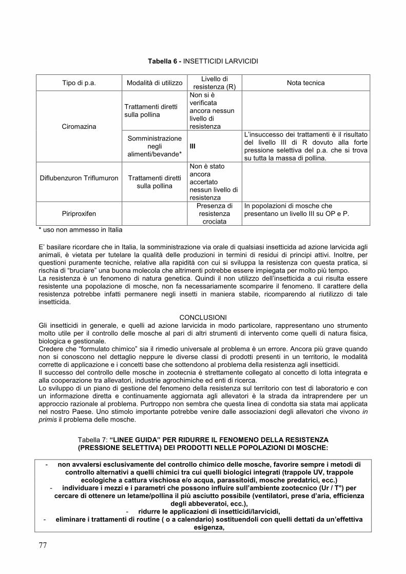
77
Tabella 6 - INSETTICIDI LARVICIDI
Tipo di p.a. Modalità di utilizzo Livello di
resistenza (R) Nota tecnica
Ciromazina
Trattamenti diretti sulla pollina
Non si è verificata ancora nessun livello di resistenza
Somministrazione negli
alimenti/bevande* III
L’insuccesso dei trattamenti è il risultato del livello III di R dovuto alla forte pressione selettiva del p.a. che si trova su tutta la massa di pollina.
Diflubenzuron Triflumuron
Trattamenti diretti sulla pollina
Non è stato ancora accertato nessun livello di resistenza
Piriproxifen Presenza di resistenza crociata
In popolazioni di mosche che presentano un livello III su OP e P.
* uso non ammesso in Italia E’ basilare ricordare che in Italia, la somministrazione via orale di qualsiasi insetticida ad azione larvicida agli animali, è vietata per tutelare la qualità delle produzioni in termini di residui di principi attivi. Inoltre, per questioni puramente tecniche, relative alla rapidità con cui si sviluppa la resistenza con questa pratica, si rischia di “bruciare” una buona molecola che altrimenti potrebbe essere impiegata per molto più tempo. La resistenza è un fenomeno di natura genetica. Quindi il non utilizzo dell’insetticida a cui risulta essere resistente una popolazione di mosche, non fa necessariamente scomparire il fenomeno. Il carattere della resistenza potrebbe infatti permanere negli insetti in maniera stabile, ricomparendo al riutilizzo di tale insetticida.
CONCLUSIONI Gli insetticidi in generale, e quelli ad azione larvicida in modo particolare, rappresentano uno strumento molto utile per il controllo delle mosche al pari di altri strumenti di intervento come quelli di natura fisica, biologica e gestionale. Credere che “formulato chimico” sia il rimedio universale al problema è un errore. Ancora più grave quando non si conoscono nel dettaglio neppure le diverse classi di prodotti presenti in un territorio, le modalità corrette di applicazione e i concetti base che sottendono al problema della resistenza agli insetticidi. Il successo del controllo delle mosche in zootecnia è strettamente collegato al concetto di lotta integrata e alla cooperazione tra allevatori, industrie agrochimiche ed enti di ricerca. Lo sviluppo di un piano di gestione del fenomeno della resistenza sul territorio con test di laboratorio e con un informazione diretta e continuamente aggiornata agli allevatori è la strada da intraprendere per un approccio razionale al problema. Purtroppo non sembra che questa linea di condotta sia stata mai applicata nel nostro Paese. Uno stimolo importante potrebbe venire dalle associazioni degli allevatori che vivono in primis il problema delle mosche.
Tabella 7: “LINEE GUIDA” PER RIDURRE IL FENOMENO DELLA RESISTENZA (PRESSIONE SELETTIVA) DEI PRODOTTI NELLE POPOLAZIONI DI MOSCHE:
- non avvalersi esclusivamente del controllo chimico delle mosche, favorire sempre i metodi di
controllo alternativi a quelli chimici tra cui quelli biologici integrati (trappole UV, trappole ecologiche a cattura vischiosa e/o acqua, parassitoidi, mosche predatrici, ecc.)
- individuare i mezzi e i parametri che possono influire sull’ambiente zootecnico (Ur / T°) per cercare di ottenere un letame/pollina il più asciutto possibile (ventilatori, prese d’aria, efficienza
degli abbeveratoi, ecc.), - ridurre le applicazioni di insetticidi/larvicidi,
- eliminare i trattamenti di routine ( o a calendario) sostituendoli con quelli dettati da un’effettiva esigenza,

78
- rispettare sempre i dosaggi delle etichette, - applicare gli insetticidi nei tempi e nelle modalità corrette,
- limitare l’estensione dei trattamenti con applicazioni mirate, - è sconsigliato l’impiego di insetticidi (OP, P) residuali.
- non nebulizzare l’ambiente con piretroidi residuali. - eseguire trattamenti a spot (lasciare sempre delle zone non trattate), - non somministrare gli IGR negli alimenti solidi o liquidi degli animali,
- non sottoporre le larve e gli adulti all’azione degli stessi principi attivi, - colpire gli insetti nei loro stadi più vulnerabili (larve giovani piuttosto che larve mature)
- non confidare sull’uso esclusivo di un solo principio attivo, - è sconsigliato la miscelazione di insetticidi diversi per aumentare la “carica abbattente” del
prodotto, - l’uso di formulati granulari contribuisce a ritardare la comparsa della resistenza,
- alternanza dei prodotti attraverso una conoscenza precisa di ciò che si impiega cercando di valutare il livello di resistenza eventualmente presente in campo (basso, medio, alto),
- iniziare il controllo integrato delle mosche dall’inizio dell’impianto dell’allevamento e non solamente quando la situazione è ormai esasperata,
- se non si è sicuri di come impiegare un formulato o delineare una strategia di lotta integrata: chiedere l’assistenza di una specialista.
BIBLIOGRAFIA – disponibile presso [email protected]
(il lavoro completo verrà pubblicato sulla rivista di avicoltura, Ed agricole – BO)
4) MICOBATTERIOSI IN CARDELLINI MUTATI
Manarolla G., Ferrazzi V., Gallazzi D. Dipartimento di Patologia Animale, Igiene e Sanità Pubblica Veterinaria, Sezione di Anatomia Patologica
Veterinaria e Patologia Aviare, Università degli Studi di Milano Correspondence: Giovanni Manarolla - Dipartimento di Patologia Animale, Igiene e Sanità Pubblica Veterinaria, Sezione di Anatomia Patologica Veterinaria e Patologia Aviare, Università degli Studi di Milano – Via Celoria 10 – 20133 Milano. Email [email protected]
Parole chiave: passeriformi, micobatteri aviari, allevamento
Mycobacteriosis in colour mutated European Goldfinches – Case report Summary: Some cases of avian mycobacteriosis in a European Goldfinch aviary are described. A non specific wasting syndrome associated with recurrent diarrhoea, dull plumage and sporadic mortality has been affecting these goldfinches for 6-7 months. Grossly dead birds revealed severe emaciation, a diffuse intestine thickening, hepatomegaly and focal lung consolidation. Histological examination of these organs revealed a granulomatous inflammation characterised by abundant epithelioid cells whose cytoplasm contained a large number of acid fast bacilli demonstrated by Ziehl-Neelsen (ZN) stain. ZN stain on faecal smears from every single cage revealed some other infected birds in the avian flock. Excessive housing density, lack of hygiene, continued inbreeding for colour mutations, reluctance of the aviculturists to euthanitized infected birds are discussed to be obstacles to controlling mycobacterioses in companion birds aviaries. Key words: passerines, avian mycobacteria, aviary Introduzione Numerose specie aviarie allevate a scopo amatoriale sono state più volte descritte come sensibili alle infezioni da micobatteri (1,2,3). In anni recenti, le migliorate tecniche diagnostiche hanno permesso di individuare diverse specie, sottospecie e sierovarianti. di micobatteri. L’identificazione è stata ottenuta mediante convenzionali test biochimici e tramite la sequenziazione genomica. Accanto al Mycobacterium avium sono così stati individuati altri batteri alcool-acido resistenti, con predominanza, negli uccelli di

79
allevamento amatoriale, di M. genavense, seguito da M. avium-intracellulare complex, M. fortuitum, M. tuberculosis, M. gordonae e M. nonchromogenicum (4). La principale via di infezione negli uccelli è rappresentata dall’intestino. L’iniziale colonizzazione intestinale è seguita da una batteriemia subclinica che consente ai micobatteri di raggiungere il fegato per via portale e secondariamente i polmoni e poche altre sedi (milza, midollo osseo, sierose). Alla forma classica con tubercoli in diversi organi si associa nei volatili la forma paratubercolare con tipiche lesioni intestinali nodulari o diffuse ed infine si può riscontrare una forma non tubercolare difficile da riconoscere in sede autoptica (2). Istologicamente negli organi colpiti sono presenti cellule macrofagiche disposte in aggregati di dimensione alquanto variabile a cui talvolta possono accompagnarsi infiltrati eterofilici e linfoplasmacellulari (4). Tali macrofagi sono caratterizzati da un ampio citoplasma anfofilo repleto di batteri alcool-acido restenti ben evidenti con colorazione di Ziehl-Neelsen (ZN). Nei passeriformi, che raramente sviluppano la forma tubercolare classica, esistono diverse sensibilità all’infezione; il cardinalino del Venezuela (Carduelis cucullata), ad esempio, risulta particolarmente sensibile (2,3). In tutte queste piccole specie, a cui appartiene anche il cardellino (Carduelis carduelis), il decorso clinico è spesso atipico e la sintomatologia può non essere evidente fino ad uno stadio avanzato della malattia. Si tratta comunque di sintomi aspecifici quali dimagramento, diarrea, epatomegalia fino alI’exitus. Diversi trattamenti sono stati proposti per le infezioni da micobatteri negli uccelli. Queste cure non sono però raccomandabili per la totale inefficacia mostrata nei volatili dai farmaci antitubercolari adoperati in medicina umana nei confronti di questi microrganismi. Di tali farmaci mancano inoltre dati relativi alla farmacocinetica nelle singole specie aviarie. E’ da sottolineare che la prevalente localizzazione intestinale di M. avium e microrganismi correlati si traduce in una continua e massiccia contaminazione ambientale: ciò rappresenta un potenziale pericolo anche per l’uomo per il quale pure non sono noti appropriati metodi terapeutici (2). Materiali e metodi Sono recentemente capitati alla nostra osservazione alcuni cardellini oggetto di selezione pluriennale perché portatori di geni cromogeni particolari (isabellismo, albinismo etc.) che da tempo presentavano mortalità sporadica nel gruppo. L’allevamento, precedentemente dedito alla moltiplicazione del cardinalino del Venezuela, constava di 21 coppie di cardellini e 4 di canarini conviventi a scopo di balia. I volatili erano alloggiati a coppie in volierette disposte su 3 piani di volume pari a circa un terzo di metro cubo ciascuna, poste in un unico locale, che ne risultava stipato. I cardellini erano di provenienza eterogenea, anche estera, e spesso acquistati presso allevatori da anni dediti al lavoro di selezione specifica delle mutazioni della livrea già ricordate. Da aprile a luglio di quest’anno sono stati esaminati 5 soggetti morti ed uno vivo. Quest’ultimo soggetto, di particolare pregio e dunque mantenuto in osservazione sotto terapia sintomatica, si presentava sottopeso e poco reattivo, con piumaggio arruffato ed opaco e fegato ben visibile oltre il limite sternale. Dopo 2 settimane di progressivo peggioramento, l’animale veniva soppresso e sottoposto a necroscopia. Dopo l’esame autoptico, dai soggetti deceduti sono stati allestiti preparati istologici di vari organi seguendo le tecniche di routine. Inoltre, considerati lo scadente stato di nutrizione e la diarrea che caratterizzava altri soggetti in allevamento, si è proceduto alla raccolta di campioni fecali da ogni singola voliera per controllo batterioscopico e parassitologico. Strisci di materiale fecale e altre sezioni istologiche venivano colorate con metodica ZN per evidenziare batteri alcool-acido resistenti. Risultati L’esame anatomopatologico dei soggetti deceduti rivelava in tutti uno scadente stato di nutrizione. In tre soggetti era presente proventricolite con sfiancamento della parete gastrica in presenza di numerosi megabatteri (Macrorhabdos ornithogaster) nel raschiato della mucosa. Tre cardellini su cinque presentavano epatomegalia ed in due soggetti era rilevabile marcato ispessimento della parete intestinale, in particolare dell’ansa duodenale e del primo tratto del digiuno. In questi due animali erano inoltre evidenti focolai di epatizzazione polmonare localizzati prevalentemente alle parti dorsali dei polmoni. Un solo reperto di Isospora spp. è stato trovato a carico dell’intestino del primo soggetto deceduto. Dall’esame istologico si confermavano quadri di proventricolite cronica da megabatteri presenti in grandi fasci sulla superficie e tra le pliche della mucosa gastrica. L’ispessimento diffuso della parete intestinale osservato in 3 soggetti a livello macroscopico corrispondeva istologicamente ad estesa infiltrazione macrofagica della lamina propria dei villi, conseguente compressione e atrofia delle cripte ed in taluni casi interessamento della sierosa. Negli stessi soggetti tale reperto, seppur di minore gravità, era presente anche nelle sezioni di fegato e di polmone. In un soggetto risultava inoltre coinvolto il pericardio ed in un altro l’ovaio. La colorazione ZN su tali sezioni consentiva la visualizzazione di numerosissimi batteri alcool-acido resistenti stipati nell’abbondante citoplasma dei macrofagi. La stessa colorazione effettuata sugli strisci di feci evidenziava 4 campioni positivi: 2 con elevato numero di batteri alcool-acido resistenti 2 con numero assai ridotto.
80
Inoltre in 2 dei 25 strisci esaminati venivano individuati megabatteri. Discussione Le micobatteriosi degli uccelli di affezione sono un reperto costante della pratica clinica ed anatomopatologica, ancorchè la prevalenza dell’infezione non sia molto elevata, attestandosi in genere intorno al 3-6% delle cause di malattia e morte (1,4). Lo studio della tubercolosi degli uccelli da gabbia si presenta tuttavia interessante per diverse ragioni. Innanzi tutto per approfondire la conoscenza dei micobatteri effettivamente responsabili della malattia, visto l’ampio numero di specie, sottospecie e varianti segnalate a distanza di pochissimi anni grazie al fondamentale ausilio delle metodiche diagnostiche molecolari. Anche attraverso queste ultime si conta di meglio definire le caratteristiche epidemiologiche e patogenetiche dei vari micobatteri aviari nei confronti di altre specie d’allevamento o degli esseri umani che per varie condizioni si trovano ad essere immunodepressi. E’ necessario inoltre evidenziare come l’andamento di questa infezione sfugga ad indagini sistematiche a causa soprattutto dell’intenso commercio di cui sono oggetto i volatili d’affezione. Tale commercio facilita la diffusione dei micobatteri tra diversi allevamenti. A ciò si aggiunge la riluttanza degli allevatori a disfarsi degli animali ammalati ed a risanare l’ambiente di allevamento. L’obbligatorietà di tali misure è infatti prescritta solamente in caso di tubercolosi negli psitaccidi (art. 104 DPR 82/1954 n. 320) e non trova applicazione nelle altre numerose specie ornitiche allevate amatorialmente. E’ del resto comprensibile, come nel caso da noi descritto, il rammarico derivato all’allevatore dalla mancanza di terapie risolutive per il trattamento dei soggetti colpiti spesso frutto di anni ed anni di selezione. D’altro canto la tipologia stessa di questi allevamenti incrementa il rischio di trasmissione diretta della tubercolosi (e di altre infezioni) tra i vari soggetti. Questi sono di norma mantenuti in numero elevatissimo all’interno di piccoli locali mal areati che solo sporadicamente vengono vuotati per un’accurata pulizia e disinfezione. Anche la diffusa pratica di rimescolare le coppie a fini selettivi facilita il passaggio diretto dei micobatteri tra i volatili conviventi. La situazione da noi descritta può essere considerata emblematica a questo proposito: 4 dei 6 soggetti ammalati erano stati acquistati lo scorso autunno presso un allevamento belga. Pur non potendone escludere a priori la persistenza nel locale, precedentemente adibito all’allevamento del cardinalino del Venezuela, è lecito supporre che l’infezione in atto sia derivata proprio da questi 4 cardellini e si sia poi trasmessa, nel giro di 6-7 mesi, ad altri volatili di questo allevamento. Da tale analisi parrebbe che l’incubazione della tubercolosi aviaria nei passeriformi possa ridursi a pochi mesi. Sono stati individuati solo 4 campioni fecali positivi per alcool-acido resistenti, ma, nota la scarsa sensibilità del metodo (5), è ipotizzabile un’infezione ben più diffusa nell’allevamento. Degno di nota pare anche il reperto di megabatteriosi in alcuni cardellini esaminati. Questa infezione, descritta primariamente nel canarino (Serinus canarius), ha un andamento solitamente cronico negli adulti, nei quali induce una cachessia progressiva simile alle micobatteriosi complicandone dunque una precoce diagnosi. E’ per questo sconsigliato la convivenza tra le due specie di volatili presente invece, come precedentemente ricordato, nell’allevamento in parola. Allo stesso modo è da evitare il sovraffollamento degli aviari, situazione frequente nell’allevamento amatoriale degli uccelli d’affezione, che risultano molto spesso oggetto di vero e proprio collezionismo. Ulteriori ricerche sono in corso per definire la specie del micobatterio causa di questo episodio ed i cui risultati saranno successivamente comunicati al fine di arricchire gli scarsi dati italiani sulle micobatteriosi dei volatili ornamentali. Bibliografia 1. Forster F., Gerlach H., Kosters J., (1988) Mycobacteria in parrots and parakeets (Psittaciformes).
Deutsche Tierarztliche Wochenschrift. 95: 8, 338-342 2. Gerlach H. L.D. (1994) Bacteria in “Avian Medicine: Principles and application” eds. Ritchie B.W. et al.
Wingers Publishing Inc., Lake Worth, FL, pag. 971-975 3. Schmidt R.E., Reavill D.R., Phalen D.N., (2003) Gastrointestinal System and Pancreas in “Pathology of
pet and aviary birds”, Iowa State Press, Ames, IA, pag. 56
4. Hoop R.K., Bottger E.C., Pfyffer,G.E. (1996) Etiological agents of mycobacterioses in pet birds between 1986 and 1995. Journal of Clinical Microbiology. 34: 4, 991-992
5. Tell L.A., Foley, J., Needham M.L, Walker, R.L. (2003) Diagnosis of avian mycobacteriosis: comparison
of culture, acid-fast stains, and polymerase chain reaction for the identification of Mycobacterium avium in experimentally inoculated Japanese quail (Coturnix coturnix japonica). Avian Diseases. 47: 2, 444-452

81
5) CONTROLLO DI MYCOPLASMA GALLISEPTICUM IN UN GRUPPO DI ALLEVAMENTI DI TACCHINI DA CARNE ATTRAVERSO L’APPLICAZIONE DI UN
VACCINO INATTIVATO
Alessandri E.1, Saita M.2, Acco P.1 1PAI srl;
2 Dipartimento di Patologia Animale, Igiene e Sanità Pubblica Veterinaria, Sezione di Anatomia
Patologica Veterinaria e Patologia Aviare, Università degli Studi di Milano
Correspondence: Mario Saita - Dipartimento di Patologia Animale, Igiene e Sanità Pubblica Veterinaria, Sezione di Anatomia Patologica e Patologia Aviare, Università degli Studi di Milano – Via Celoria 10 – 20133 Milano. Email [email protected]
Parole chiave: tacchino, Mycoplasma gallisepticum, vaccinazione
Summary: Eight flocks of turkeys positive to Mycoplasma gallisepticum were submitted to an intense vaccination program, using an inactived vaccine and others biosecurity measures. Animals were monitored through serology and PCR testing. After a four month period all flocks tested were negative to Mycoplasma gallisepticum. Key words: turkey, Mycoplasma gallisepticum, vaccination Introduzione Le infezioni sostenute da Mycoplasma gallisepticum nell’allevamento del tacchino da carne costituiscono un importante problema sia dal punto di vista sanitario che economico in quanto sono causa dell’aumento degli scarti alla macellazione, dell’aumento dell’indice di conversione e dei costi terapeutici. La sintomatologia è caratterizzata essenzialmente da rantoli respiratori, tosse, sinusite e aerosacculite (Ley e Yoder, 1997), quest’ultima in genere complicata dalla presenza di altri patogeni. L’insorgenza della malattia è lenta e presenta un decorso prolungato. Tutti questi aspetti rendono questa patologia una delle più dispendiose che l’industria avicola si trovi ad affrontare (Carpenter et al. 1981). La profilassi vaccinale può costituire un valido strumento di contrasto soprattutto se applicata in zone dove Mycoplasma gallisepticum si presenta periodicamente e con caratteristiche endemiche. Materiali e metodi Otto allevamenti di tacchini misti (maschi e femmine) da carne, di linea Big 6, situati in Friuli Venezia Giulia erano persistentemente positivi, sia alla siero agglutinazione rapida (SAR), sia all’ELISA, , per Mycoplasma gallisepticum. Tali allevamenti, situati in una zona geograficamente circoscritta, erano caratterizzati dalla promiscuità del personale e delle attrezzature utilizzati per le pratiche veterinarie e per il carico degli animali. Avvalendosi dell’utilizzo di un vaccino contenente Mycoplasma gallisepticum,, inattivato con formalina ed emulsionato in adiuvante oleoso, sono stati programmati due interventi vaccinali, sia nei maschi che nelle femmine, a 4 e a 10 settimane di età. Successivamente al termine del primo ciclo produttivo, il numero degli interventi vaccinali è stato modificato in 2 per i maschi (macellati in genere a 19-20 settimane) e 1 per le femmine (macellate solitamente a 13-14 settimane) Oltre a ciò sono state attuate alcune misure di biosicurezza volte a prevenire la trasmissione dell’infezione e l’insorgenza della stessa in concomitanza con altri patogeni. Questi provvedimenti sono principalmente: l’allungamento del vuoto sanitario tra un ciclo e il successivo da 3 a 4 settimane, la sincronizzazione degli accasamenti e delle macellazioni tra gli allevamenti in oggetto, la disinfezione periodica delle attrezzature e la vaccinazione a 1 gg. di età contro la Rinotracheite Infettiva del tacchino. Gli animali sono stati sottoposti ad un prelievo ematico pre-vaccinale e ad uno 3 settimane dopo la vaccinazione. Il siero ottenuto è stato analizzato attraverso la tecnica ELISA e la SAR, previa inattivazione a 54°C per 20 minuti. La lettura dei campioni è stata effettuata in diluizione scalare, partendo da un valore minimo per la positività di 1/8. In ciascuno degli 8 allevamenti è stata inoltre svolta, al termine del ciclo di allevamento, almeno una Polimerase Chain Reaction per la ricerca di Mycoplasma gallisepticum Risultati L’analisi dei sieri post-vaccinali tramite SAR ha evidenziato titoli anticorpali medi di 1/32, con valori assoluti compresi tra 1/8 e 1/64 e sporadici picchi di 1/128. Anche l’ELISA non ha raggiunto valori tali da dimostrare la presenza di un’infezione con ceppi di campo. Tale ipotesi è stata inoltre confermata dalle PCR effettuate che sono risultate negative.
82
Conclusioni I titoli anticorpali ottenuti sono omogenei e contenuti entro valori tali da poter affermare, congiuntamente alle osservazioni di campo e ad i controlli effettuati tramite PCR, che derivino da una copertura vaccinale e non dall’ingresso di un ceppo di campo di Mycoplasma gallisepticum. La sola vaccinazione sembrerebbe non essere sufficiente a garantire da sola l’eliminazione della micoplasmosi (Talkington e Kleven, 1985; Yoder e Hopkins 1985), Sia utilizzo del vaccino inattivato che la concomitante attuazione di misure di biosicurezza hanno però consentito l’eradicazione di Mycoplasma gallisepticum dalla zona in oggetto. Bibliografia: Carpenter TE, Howitt R, McCapes R, Yamamoto R, Riemann HP., Formulating a control program against Mycoplasma meleagridis using economic decision analysis. Avian Dis. 1981 Apr-Jun;25(2):260-71. Jordan F.T., Immunity to mycoplasma infections of the respiratory system in the domestic fowl and turkey. Dev Biol Stand. 1975;28:590-6. Ley D.H., Yoder H.W., in Calnek B.W. Diseases of Poultry, 10th Edition, Mosby-Wolfe, London, 1997, p.194-207. Talkington FD, Kleven SH., Evaluation of protection against colonization of the chicken trachea following administration of Mycoplasma gallisepticum bacterin. Avian Dis. 1985 Oct-Dec;29(4):998-1003 Yoder HW Jr, Hopkins SR., Efficacy of experimental inactivated mycoplasma gallisepticum oil-emulsion bacterin in egg-layer chickens. Avian Dis. 1985 Apr-Jun;29(2):322-34.

83
6) ESOFAGITE ULCERATIVA DA STREPTOCARA INCOGNITA IN ANATRE MUTE (CAIRINA MOSCHATA DOMESTICUS): PRIMA SEGNALAZIONE IN ITALIA
Bano L.1, Natale A.2, Vascellari M.3, Comin D.1, Agnoletti F.1, Mutinelli F.3
1Laboratorio di Treviso;
2Laboratorio di parassitologia ed ecopatologia;
3Laboratorio di istopatologia
Istituto Zooprofilattico Sperimentale delle Venezie Corrispondence: Luca Bano, Istituto Zooprofilattico Sperimentale delle Venezie – Laboratorio di Treviso, Viale Brigato Treviso 13/A – 31100 Treviso, Italia. E-mail [email protected]
Parole chiave: anatra muta (Cairina moschata domesticus), Streptocara incognita, esofagite First report of parasitic esophagitis by Streptocara incognita in Muscovy ducks (Cairina moschata domesticus) in Italy
Summary: An outbreak of streptocariasis in a backyard flock of 62 ducks (Cairina moschata domesticus) located in Treviso, north-eastern Italy, is described. Fifteen animals died in two weeks showing debilitation and emaciation. Two ducks were submitted for post mortem examination and a parasitic ulcerative esophagitis was observed. Parasites were identified as Streptocara incognita. This is the first outbreak of streptocariasis by Streptocara incognita reported from Italy. Key words: muscovy ducks (Cairina moschata domesticus), Streptocara incognita, esophagitis Introduzione La streptocariasi è una grave malattia parassitaria sostenuta da nematodi appartenenti all’ordine Spirurida. Gli adulti di Streptocara sp. vivono infissi nella mucosa dell’esofago, dell’ingluvie, del proventricolo, dello stomaco muscolare e della laringe provocando lesioni spesso mortali; la sintomatologia clinica dipende dalla localizzazione del parassita (10). Delle sette specie patogene (12), le due più spesso segnalate sono Streptocara crassicauda e Streptocara incognita. Il ciclo biologico di S. incognita è meno conosciuto rispetto a quello di S. crassicauda, ma secondo alcuni autori è probabilmente molto simile (13). S. crassicauda è in grado di parassitare uccelli appartenenti a vari ordini (12). Gli animali colpiti più frequentemente sono uccelli acquatici ed in particolar modo gli anatidi tuffatori (3), ma non mancano segnalazioni anche in specie di interesse zootecnico quali il pollo e la faraona (15). La spiccata sensibilità degli anatidi tuffatori alla malattia è legata sia al ciclo biologico del parassita sia alle abitudini alimentari di questi uccelli: il ciclo biologico di S. crassicauda prevede ospiti intermedi ed ospiti paratenici, che sono frequentemente preda di uccelli acquatici. Gli ospiti intermedi sono crostacei anfipodi nel cui emocele le larve si sviluppano, raggiungendo lo stadio infettante in 19-25 giorni, a seconda della temperatura ambientale. Negli ospiti paratenici, pesci di varie specie, le larve di S. crassicauda si incistano in noduli intestinali dove permangono fino all’ingestione da parte di uccelli ittiofagi. Nell’ospite definitivo le larve di S. crassicauda si sviluppano molto velocemente e dopo due mute, nell’arco di 9-10 giorni, cominciano a deporre uova (2). Mentre S. crassicauda è considerata cosmopolita, la presenza di S. incognita è stata segnalata soltanto in Canada, USA, ex-Yugoslavia (7, 12) e Regno Unito (9). Materiali e metodi Due femmine adulte di anatra muta (Cairina moschata domesticus) appartenenti ad un gruppo di 62 soggetti (21% maschi, 79% femmine), sono state conferite al Laboratorio di Treviso dell’IZS delle Venezie per essere sottoposte ad esame autoptico. Gli animali erano allevati per autoconsumo in condizioni di semilibertà, all’interno di un appezzamento attraversato da un corso d’acqua frequentato anche da anatidi selvatici (Anas platyrhynchos). Al momento del conferimento il proprietario riferiva la morte di altri 15 soggetti (24%) nell’arco di 2-3 settimane, tutti di sesso femminile, con sintomatologia caratterizzata da emaciazione, prostrazione, anoressia, riluttanza al movimento, polidipsia, dispnea. Gli animali venivano alimentati con spezzato di mais associato a mangime complementare e non erano mai stati sottoposti ad alcun trattamento immunoprofilattico o farmacologico. Dopo l’esame autoptico, tutti i parassiti raccolti sono stati misurati, fissati in alcool, chiarificati in lattofenolo e osservati microscopicamente (40X – 400X). L’identificazione di specie è stata effettuata sulla base delle chiavi suggerite da Mc Donald (13). Dalle lesioni di entrambi i soggetti sono stati prelevati campioni per l’esame istologico. Il materiale è stato fissato in formalina tamponata al 10% e processato routinariamente. Dai tessuti inclusi in paraffina sono state allestite sezioni dello spessore di 4 µm, successivamente colorate con ematossilina ed eosina.

84
Risultati All’esame necroscopico, in entrambi i soggetti si sono osservate ectasie esofagee associate ad esofagite pseudomembranosa. Non erano evidenti lesioni a carico degli altri organi. L’osservazione allo stereomicroscopio delle pseudomembrane metteva in luce la presenza di nematodi di aspetto lattiginoso frammisti a materiale necrotico. All’esame istologico la mucosa esofagea appariva fortemente iperemica, con ulcerazioni multifocali coperte da materiale necrotico. Numerosi parassiti erano presenti nel lume esofageo ed infissi nella mucosa, circondati da materiale necrotico e da infiltrato cellulare costituito da eterofili e mononucleati. Per l’identificazione parassitologica sono stati raccolti 512 nematodi presenti nell’esofago di entrambi i soggetti, tutti identificati come Streptocara incognita (tab.1), di cui 405 (79,1%) femmine e 107 (20,9%) maschi (sex ratio: 3,79). I maschi misuravano in media 7 mm in lunghezza e 140 µm in spessore; le femmine erano lunghe in media 9 mm, con uno spessore di 190 µm. I dettagli identificativi (13) per entrambi i sessi erano rappresentati dalla presenza di un collaretto dentato situato immediatamente al di sotto delle labbra, e dalla presenza di una papilla cervicale divisa da una profonda incisione, localizzata aboralmente al vestibolo ad una distanza pari alla metà della lunghezza vestibolare. Nel maschio sono caratteristici il numero delle papille postanali e la lunghezza degli spicoli. Discussione Questo lavoro rappresenta la prima segnalazione di streptocariasi da Streptocara incognita in Italia. Nell’episodio descritto è stata identificata esclusivamente la specie Streptocara incognita, confermando il suo potere patogeno. In Europa la presenza di S. incognita era stata precedentemente descritta solo nell’ex-Yugoslavia e nel Regno Unito (7, 9). L’aspetto più eclatante osservato in questo episodio è la gravità delle lesioni esofagee. Nell’ambito della diagnosi differenziale clinica ed anatomopatologica vanno considerate altre parassitosi sostenute da Capillaria contorta, Epomidiostomum uncinatum, Amidostomum acutum, Tetrameres sp. (8), malattie virali (enterite virale dell’anatra) (4), esofagiti batteriche e micotiche, e causticazioni da agenti chimici. Contrariamente a quanto riportato in bibliografia (3; 11; 8; 10; 14; 6), nell’episodio descritto va sottolineata la mancanza di lesioni parassitarie in organi diversi dall’esofago. La somministrazione di tetramisolo (polvere al 10%) in acqua di bevanda (5 mg/litro) per tre giorni, ripetuta ogni 40 giorni, ha determinato la scomparsa di mortalità a distanza di tre mesi dall’ultimo decesso. Oltre al levamisolo (17), anche il mebendazolo viene indicato come antielmintico efficace in episodi di streptocariasi (5). La streptocariasi è difficilmente osservabile in allevamenti da reddito in quanto il ciclo biologico del parassita è strettamente legato alla presenza dell’ospite intermedio o paratenico e quindi alla presenza di specchi d’acqua. La promiscuità tra specie selvatiche e allevate, osservata anche in questo caso, rappresenta uno dei principali fattori di rischio.
Tabella 1. Classificazione tassonomica di Streptocara incognita (1; 13).
Classe Nematoda Sottoclasse Secernentea Ordine Spirurida Sottordine Spirurina Superfamiglia Acquarioidea Sottofamiglia Seuratiinae Genere Streptocara
Specie Streptocara incognita
Bibliografia. Anderson R. C., Chabaud A. G., Willmott S. 1975. CIH keys to the Nematode Parasites of Vertebrates. N. 3, Key to genera of the order Spirurida. CAB, 1975. Anderson R.C. The Superfamily Acuarioidea. 2000. In: Nematodes Parasites of Vertebrates: their development and transmission, 2nd Edition. CABI Publishing. pp. 465. Bakke, T. A. and V. Barus. 1976. Studies of the Helminth Fauna of Norway XXXIX: Nematodes from Hydrobates pelagicus L. and Rissa tridactyla L. (Aves) in Norway. Norw. J. Zool. 24: 185-189. Calnek B. W. 1997. Enterite virale dell’anatra (Peste dell’anatra). In: patologia aviare, X ed. Piccin, Padova. pp. 751-760. Dalton P.J. Streptocara infestation of ducks. 1980. Vet. Rec. Oct. 18 ; 107 (16) :384.
85
Fox J.G., Snider S.B., Schmidt G.D., Campbell L.H. 1974. Infection with the nematode Streptocara incognita in the Chilean flamingo. J Wildl Dis. Jan; 10(1): 66-9. Karlovic, M., S. Richter and Z. Aleraj. 1959. Streptocarosis in the Japanese goose (Sygnopsis sygnoides L.). Vet. Arkiv. 30:7-12. Kinsella, J. M., and Forrester D. J. 1972. Helminths of the Florida Duck, Anas platyrhyncos fulvigula. Proc. Helminthol. Soc. Wash. 39: 173-176. Lancaster M.B. 1973. The occurrence of Streptocara sp. in ducks in Britain. Vet Rec. Mar 10; 92(10):261-2. Mason, R. W. 1988. Laryngeal streptocariasis causing death from asphyxiation in ducks. Australian Veterinary Journal 65: 335-336. 1988. Mauritz, C. S. and L. Stackhouse. Parasitic ulcerative ventriculitis in mallards (Anas platyrhynchos). J. of Wildlife Diseases 23: 680-682.1987. Mc Donald, M. E. Catalogue of helminths of waterfowl (Anatidae). Bureau of Sport Fisheries, Special Scientific Report - Wildlife No. 126, 1969. Mc Donald M. Key to nematodes reported in waterfowl. Department of the Interior, Bureau of sport Fisheries and Wildlife. Resource publication n. 122, pp. 1-44, 1974. McLaughlin J. D., McGurk B. P. 1987. An analysis of gizzard worm infections in fall migrant ducks at Delta, Manitoba, Canada. Can. J. Zool. 65: 1470-1477. Neveu-Lemaire, M. Helmintologie specialé. 1936. In: Traitè d’helminthologie medicale et veterinaire, ed. Vigot Frères, Paris. pp. 1260-1261. Sterner M. C., Stackhouse L. 1987. Parasitic ulcerative ventriculitis in mallards (Anas platyrhynchos). J. Wildl Dis. Oct; 23 (4): 680-2. Urquhart G. M., J. Armour, J. L. Duncan, A. M. Dunn & F. W. Jennings. 1987. Veterinary parasitology, Longman Scientific & Technical, Harlow, Essex, England.
7) ARCHIVIO BIBLIOGRAFICO DI ECTOPARASSITI
Pampiglione G. 1, Massi P. 2 1Università degli studi di Bari, Facoltà di Medicina Veterinaria,Dipartimento di Sanità e Benessere Animale.
Italy; 2Istituto Zooprofilattico Sperimentale Sezione di Forlì (FC) Italy
L’IZS di Forlì ha creato un archivio bibliografico sugli ectoparassiti del comparto avicolo. Si tratta di una raccolta aggiornata e continuativa di numerosi lavori scientifici stranieri ( Poultry Science, Journal of Economic Entomology, Medical Veterinary Etomology, Pesticide Biochemistry and Physiology , Pesticides in the environment, Pesticide Science), contributi italiani (Parassitologia, Disinfestazione ed igiene ambientale, Avicoltura), tesi di laurea italiane e libri sull’argomento del tema. Sono incluse anche le note tecniche delle industrie produttrici di biocidi. I temi raccolti sono: biologia degli ectoparassiti, sistemi di lotta integrata (IPM), lotta con sistemi fisici, lotta biologica, gestione della pollina, la resistenza delle mosche sugli insetticidi/larvicidi, i larvicidi IGRs, impatto ambientale, ecc. Lo scopo è quello di stimolare e sviluppare le competenze tecniche della componente veterinaria del settore che attualmente risultano essere insufficienti e troppo teoriche. Questa iniziativa offrirà raccomandazioni e linee guida per la lotta integrata agli ectoparassiti e sembra essere la prima in Italia.
86
8) IDENTIFICAZIONE E TIPIZZAZIONE DEL PNEUMOVIRUS AVIARE TRAMITE TECNICHE MOLECOLARI
Francesca Paganelli1, Laura Fiorentini1, Giovanni Tosi1
1Istituto Zooprofilattico Sperimentale della Lombardia e dell’Emilia Romagna
Corresponding author: Dr. Francesca Paganelli. Istituto Zooprofilattico Sperimentale della Lombardia e dell’Emilia Romagna, Sezione Diagnostica di Forlì. Via Marchini 1, 47100 Forlì, Italy- Tel. & Fax +39 0543 721533 Email: [email protected] Riassunto Il Pneumovirus aviare (APV) è l’agente eziologico di due importanti malattie quali la Rinotracheite Infettiva del tacchino (TRT) e la Malattia della testa gonfia (“Swollen Head Syndrome”, SHS). Diversi sono gli strumenti diagnostici attualmente disponibili e tra questi, l’applicazione di una RT-PCR per l’identificazione del virus, seguita da una nested-PCR per la tipizzazione del sottotipo. Parole chiave: Pneumovirus aviare, RT-PCR, nested-PCR. Introduzione La Rinotracheite infettiva del tacchino (TRT) e la Swollen head syndrome (SHS) sono malattie contagiose, rispettivamente del tacchino e del pollo, con carattere prevalentemente respiratorio, frequentemente soggette a complicazioni batteriche secondarie, causate da un virus del genere Pneumovirus (APV) (Jane et al., 2002). Nel tacchino la malattia è caratterizzata da starnuti, rantoli, scolo nasale, congiuntivite e lacrimazione, sinusite ed edema sottomandibolare, grave depressione. Negli adulti si osserva calo della deposizione fino al 70%, respiro a becco aperto, a volte malattia inapparente. Morbilità bassa così come la mortalità salvo complicazioni batteriche spesso presenti. (Pascucci, 1996) In base al sequenzamento del gene G ed alle caratteristiche sierologiche sono stati distinti 4 sottotipi di APV (A, B, C, D). I sottotipi A e B, diffusi in Europa, differiscono sulla base della sequenza nucleotidica del gene G. Il sottotipo C, segnalato negli Stati Uniti d’America, presenta invece differenze genomiche e sierologiche più evidenti, come anche il sottotipo D (Juhasz et al.,1994) . La diagnosi si basa sull’osservazione dei sintomi clinici (non sempre apprezzabili) e delle lesioni anatomopatologiche (non patognomoniche). Risulta quindi di supporto fondamentale la diagnosi di laboratorio che si basa sull’isolamento ed identificazione del virus su anelli tracheali di tacchino (valutazione della ciliostasi quindi identificazione tramite immunofluorescenza, virusneutralizzazione, microscopia elettronica). Il virus può essere adattato a colture primarie di fegato di pollo (FEP), nei quali si osserva effetto citopatico sinciziale. Per le indagini sierologiche viene impiegata di routine un’ ELISA utile sia per monitoraggio che per la diagnosi. In base alle differenze nucleotidiche del gene G è possibile identificare il genoma virale APV con l’utilizzo della RT-PCR ed in seguito con una nested-PCR si riesce a discriminare tra sottotipo A e B. Materiali e metodi Estrazione RNA e RT-PCR La prova in PCR è stata allestita a partire da campioni patologici prelevati in sede autoptica (trachee e tamponi tracheali), di pollo e tacchino. E’ stata inoltre eseguita l’estrazione del genoma virale dai tre vaccini vivi attenuati utilizzati in Italia: Poulvac® TRT (Fort Dodge, Weesp, Holland), Rinovax® (Merial, Milano,Italy), Nobilis®TRT (Intervet, Boxmeer, Holland) a scopo di verifica. Per l’estrazione dell’RNA è stato utilizzato il kit RNeasy® MiniKit (Qiagen, Valencia,CA) seguendo il protocollo allegato. Ottenuto l’RNA si procede alla retrotrascrizione (RT) utilizzando il ProstarTM Kit (Stratagene®, USA), con l’aggiunta del primer “reverse” G6- complementare al trascritto (Cavanagh et al., 1999). Il prodotto di retrotrascrizione viene utilizzato direttamente nel saggio di PCR eseguito secondo il protocollo della Accuprime TaqPCRx DNA Polymerase (Invitrogen®, USA). Fungono da innesco alla reazione i primers G6- e G1+ che producono un frammento di 444bp, identificativo di APV (Cavanagh et al., 1999). La reazione avviene secondo il seguente profilo di amplificazione: 2 minuti a 94°C (hot start) seguito da 30 cicli costituiti da 3 step: 94°C per 1 minuto (denaturazione), 50°C per 1 minuto (annealing), 72°C per 2 minuti (estensione); al termine dei cicli 2 minuti a 72°C per eventuali estensioni. In ogni reazione sono presenti sia il controllo positivo che negativo. Il prodotto amplificato viene sottoposto a corsa ellettroforetica in gel di agarosio al 2% con bromuro di etidio e visualizzazione mediante transilluminatore a raggi UV.

87
Nested-PCR Ottenuta la positività per APV è possibile discriminare il sottotipo A e B in base alle differenze presenti nella sequenza del gene G. L’amplificato di 444 bp viene sottoposto a una nested-PCR utilizzando un primer “reverse” G5- comune per entrambi i sierotipi, e 2 primers “forward”, uno per il sottotipo A (G8 +A) che amplifica un frammento di 268 bp e l’altro primer specifico per il sottotipo B (G9 +B) che produce un cDNA di 316 bp. I prodotti amplificati, dopo corsa elettroforetica in gel di agarosio al 1,7%, sono stati visualizzati mediante colorazione con bromuro di etidio tramite transilluminatore a UV (Cavanagh et al., 1999). Risultati e discussione I tre vaccini analizzati sono risultati rispettivamente: Poulvac® TRT e Nobilis® TRT sottotipo A, Rinovax® sottotipo B. Da settembre 2003 a giugno 2004 sono stati eseguiti 122 ricerche per APV da tacchino e pollo, di cui 5, da tacchino, sono risultate positive per APV. Successivamente si è eseguita una nested-PCR che ha rivelato 3 campioni positivi per il sottotipo B e 2 per il sottotipo A (tab 2). Conclusioni La diagnosi clinica di TRT non è semplice, vista la mancanza di specifiche lesioni patognomoniche. L’isolamento virale è difficoltoso e dispendioso. Sicuramente l’RT-PCR si presenta come la tecnica più rapida e sensibile per l’identificazione del genoma virale. In 48 ore è possibile avere il risultato dell’indagine di laboratorio, quindi si presta bene sia a scopo diagnostico di supporto al quadro anamnestico e anatomopatologico che come monitoraggio. La possibilità di discriminare i due sottototipi A e B rappresenta un ulteriore strumento che può, in taluni casi, consentire di distinguere i ceppi vaccinali da quelli di campo e di fornire informazioni di tipo epidemiologico e filogenetico sul virus. Bibliografia Cavanagh, D., Mawditt K., Britton P., Naylor C.J., 1999. Longitudinal field studies of infectious bronchitis virus and avian pneumovirus in broiler using type-specific polymerase chain reactions. Avian Pathology 28:593-605. Jane, K., Cook A., Cavanagh D., 2002. Detection and differentiation of avian pneumoviruses (metapneumoviruses). Avian Pathology 31:117-132. Juhasz, K., Easton A.J. 1994. Extensive sequence variation in the attachment (G) protein gene of avian pneumovirus: evidence for two distinct subgroups. Journal of General Virology 75:2873-2880. Pascucci S., 1996. Pneumovirosi aviare. Degree Diss. Istituto Zooprofilattico della Lombardia e dell’Emilia Romagna sez. Forlì. Italy Tabella 1. Sequenza dei primers
PRIMER SEQUENZA NUCLEOTIDICA (5’-3’) GENE G6- CTGACAAATTGGTCCTGATT G G1+ GGGACAAGTATCT/CC/AT/GAT G G5- CAAAGAA/GCCAATAAGCCCA G
G8+A CACTCACTGTTAGCGTCATA G G9+B TAGTCCTCAAGCAAGTCCTC G
Tabella 2. Risultati APV SOTTOTIPO
117 negativi /
5 positivi (tacchino)
3 B (soggetti vaccinati con sottotipoA)
2 A(soggetti vaccinati con sottotipoB)

88
9) EMANGIOMI EPATICI IN FARAONE
Bolognesi P.G., Catelli E., Cecchinato M., De Matteo P., Frasnelli M. , Raffini E. , Marzadori F. ,Thiene G.