Relationship of resting hemoglobin concentration to peak oxygen uptake in heart failure patients
22
For Peer Review Relationship of resting haemoglobin concentration to peak oxygen uptake in heart failure patients Journal: American Journal of Hematology Manuscript ID: AJH-10-0051.R1 Wiley - Manuscript type: Research Article Date Submitted by the Author: 23-Feb-2010 Complete List of Authors: Agostoni, Piergiuseppe; Centro Cardiologico Monzino, IRCCS, Dipartimento di Scienze Cardiovascolari, Università di Milano, Milan, Italy Salvioni, Elisabetta; Centro Cardiologico Monzino, IRCCS, Dipartimento di Scienze Cardiovascolari, Università di Milano, Milan, Italy Debenedetti, Chiara; Centro Cardiologico Monzino, IRCCS, Dipartimento di Scienze Cardiovascolari, Università di Milano, Milan, Italy Vignati, Carlo; Centro Cardiologico Monzino, IRCCS, Dipartimento di Scienze Cardiovascolari, Università di Milano, Milan, Italy Cattadori, Gaia; Centro Cardiologico Monzino, IRCCS, Dipartimento di Scienze Cardiovascolari, Università di Milano, Milan, Italy Contini, Mauro; Centro Cardiologico Monzino, IRCCS, Dipartimento di Scienze Cardiovascolari, Università di Milano, Milan, Italy Magrì, Damiano; Centro Cardiologico Monzino, IRCCS, Dipartimento di Scienze Cardiovascolari, Università di Milano, Milan, Italy Palermo, Pietro; Centro Cardiologico Monzino, IRCCS, Dipartimento di Scienze Cardiovascolari, Università di Milano, Milan, Italy Gondoni, Erica; Centro Cardiologico Monzino, IRCCS, Dipartimento di Scienze Cardiovascolari, Università di Milano, Milan, Italy Brusoni, Denise; Centro Cardiologico Monzino, IRCCS, Dipartimento di Scienze Cardiovascolari, Università di Milano, Milan, Italy Fiorentini, Cesare; Centro Cardiologico Monzino, IRCCS, Dipartimento di Scienze Cardiovascolari, Università di Milano, Milan, Italy Apostolo, Anna; Centro Cardiologico Monzino, IRCCS, Dipartimento di Scienze Cardiovascolari, Università di Milano, Milan, Italy Keywords: Anemia, Exercise capacity, Cardiopulmonary exercise test John Wiley & Sons American Journal of Hematology peer-00552314, version 1 - 6 Jan 2011 Author manuscript, published in "American Journal of Hematology (2010)" DOI : 10.1002/ajh.21698
-
Upload
independent -
Category
Documents
-
view
1 -
download
0
Transcript of Relationship of resting hemoglobin concentration to peak oxygen uptake in heart failure patients
For Peer Review
Relationship of resting haemoglobin concentration to peak oxygen uptake in heart failure patients
Journal: American Journal of Hematology
Manuscript ID: AJH-10-0051.R1
Wiley - Manuscript type: Research Article
Date Submitted by the Author:
23-Feb-2010
Complete List of Authors: Agostoni, Piergiuseppe; Centro Cardiologico Monzino, IRCCS, Dipartimento di Scienze Cardiovascolari, Università di Milano, Milan, Italy Salvioni, Elisabetta; Centro Cardiologico Monzino, IRCCS, Dipartimento di Scienze Cardiovascolari, Università di Milano, Milan, Italy Debenedetti, Chiara; Centro Cardiologico Monzino, IRCCS, Dipartimento di Scienze Cardiovascolari, Università di Milano, Milan, Italy Vignati, Carlo; Centro Cardiologico Monzino, IRCCS, Dipartimento di Scienze Cardiovascolari, Università di Milano, Milan, Italy Cattadori, Gaia; Centro Cardiologico Monzino, IRCCS, Dipartimento di Scienze Cardiovascolari, Università di Milano, Milan, Italy Contini, Mauro; Centro Cardiologico Monzino, IRCCS, Dipartimento di Scienze Cardiovascolari, Università di Milano, Milan, Italy Magrì, Damiano; Centro Cardiologico Monzino, IRCCS, Dipartimento di Scienze Cardiovascolari, Università di Milano, Milan, Italy Palermo, Pietro; Centro Cardiologico Monzino, IRCCS, Dipartimento di Scienze Cardiovascolari, Università di Milano, Milan, Italy Gondoni, Erica; Centro Cardiologico Monzino, IRCCS, Dipartimento di Scienze Cardiovascolari, Università di Milano, Milan, Italy Brusoni, Denise; Centro Cardiologico Monzino, IRCCS, Dipartimento di Scienze Cardiovascolari, Università di Milano, Milan, Italy Fiorentini, Cesare; Centro Cardiologico Monzino, IRCCS, Dipartimento di Scienze Cardiovascolari, Università di Milano, Milan, Italy Apostolo, Anna; Centro Cardiologico Monzino, IRCCS, Dipartimento di Scienze Cardiovascolari, Università di Milano, Milan, Italy
Keywords: Anemia, Exercise capacity, Cardiopulmonary exercise test
John Wiley & Sons
American Journal of Hematologype
er-0
0552
314,
ver
sion
1 -
6 Ja
n 20
11Author manuscript, published in "American Journal of Hematology (2010)"
DOI : 10.1002/ajh.21698
For Peer Review
Page 1 of 20
John Wiley & Sons
American Journal of Hematology
123456789101112131415161718192021222324252627282930313233343536373839404142434445464748495051525354555657585960
peer
-005
5231
4, v
ersi
on 1
- 6
Jan
2011
For Peer Review
1
Relationship of resting haemoglobin concentration to peak oxygen
uptake in heart failure patients
Piergiuseppe Agostoni*#, MD, PhD , Elisabetta Salvioni*, PhD, Chiara Debenedetti*, MD, Carlo
Vignati*, MD, Gaia Cattadori*, MD, Mauro Contini*, MD, Damiano Magrì*, MD, PhD, Pietro
Palermo*, MD, Erica Gondoni*, MD, Denise Brusoni*, MD, Cesare Fiorentini*, MD, and Anna
Apostolo*, MD.
Short title: Anaemia and peak O2 in heart failure.
* Centro Cardiologico Monzino, IRCCS, Dipartimento di Scienze Cardiovascolari, Università
di Milano, Milan, Italy, # Division of Critical Care and Respiratory Medicine, Department of
Medicine, University of Washington, Seattle, WA , USA.
None of the authors have any relationship with industry or any conflict of interest.
Key words. Anaemia, exercise capacity, cardiopulmonary exercise test
Corresponding Author:
Piergiuseppe Agostoni, MD, PhD
Centro Cardiologico, IRCCS,
Istituto di Scienze Cardiovascolari, Università di Milano
Via Parea 4, 20138 Milan, Italy.
Phone +390258002299
Fax + 390258002283
E-mail [email protected]
Abstract word count: 187
Text word count: 2233
Tables: 4
Figures: 3
Page 2 of 20
John Wiley & Sons
American Journal of Hematology
123456789101112131415161718192021222324252627282930313233343536373839404142434445464748495051525354555657585960
peer
-005
5231
4, v
ersi
on 1
- 6
Jan
2011
For Peer Review
2
Abstract
Anaemia is frequent in chronic heart failure (HF). In order to calculate what change in peak
oxygen uptake ( O2) should be expected in the event of changes in haemoglobin concentration,
we studied the correlation between peak O2 and haemoglobin concentration in a large HF
population. We carried out retrospective analysis of all cardiopulmonary exercise tests (CPET)
performed in our HF Clinic between June 2001 and March 2009 in HF patients who had a resting
haemoglobin concentration measurement taken within 7 days of the CPET.
We collected 967 CPETs, 704 tests were considered maximal and analysed. We identified 181
patients (26%) as anaemic. Peak O2 was lower (p<0.001) in anaemic patients (971±23 ml/min)
compared to non-anaemic (1243±18 ml/min). The slope of the O2 vs. haemoglobin ratio was
109 ml/min/g/dl at peak exercise. This correlation remained significant also when several
confounding variables were analysed by multivariate analysis. As an average, each g of
haemoglobin accounts, at peak exercise, for 109 ml/min change in O2 which is equivalent to
0.97 ml/min/kg. Therefore, in HF patients anaemia treatment should increase O2 by 109 ml/min
for each g/dl of haemoglobin increase.
Page 3 of 20
John Wiley & Sons
American Journal of Hematology
123456789101112131415161718192021222324252627282930313233343536373839404142434445464748495051525354555657585960
peer
-005
5231
4, v
ersi
on 1
- 6
Jan
2011
For Peer Review
3
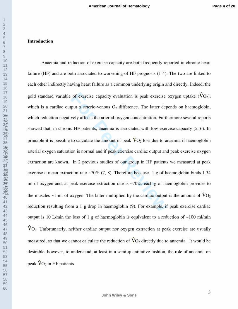
Introduction
Anaemia and reduction of exercise capacity are both frequently reported in chronic heart
failure (HF) and are both associated to worsening of HF prognosis (1-4). The two are linked to
each other indirectly having heart failure as a common underlying origin and directly. Indeed, the
gold standard variable of exercise capacity evaluation is peak exercise oxygen uptake ( O2),
which is a cardiac output x arterio-venous O2 difference. The latter depends on haemoglobin,
which reduction negatively affects the arterial oxygen concentration. Furthermore several reports
showed that, in chronic HF patients, anaemia is associated with low exercise capacity (5, 6). In
principle it is possible to calculate the amount of peak O2 loss due to anaemia if haemoglobin
arterial oxygen saturation is normal and if peak exercise cardiac output and peak exercise oxygen
extraction are known. In 2 previous studies of our group in HF patients we measured at peak
exercise a mean extraction rate ~70% (7, 8). Therefore because 1 g of haemoglobin binds 1.34
ml of oxygen and, at peak exercise extraction rate is ~70%, each g of haemoglobin provides to
the muscles ~1 ml of oxygen. The latter multiplied by the cardiac output is the amount of O2
reduction resulting from a 1 g drop in haemoglobin (9). For example, if peak exercise cardiac
output is 10 L/min the loss of 1 g of haemoglobin is equivalent to a reduction of ~100 ml/min
O2. Unfortunately, neither cardiac output nor oxygen extraction at peak exercise are usually
measured, so that we cannot calculate the reduction of O2 directly due to anaemia. It would be
desirable, however, to understand, at least in a semi-quantitative fashion, the role of anaemia on
peak O2 in HF patients.
Page 4 of 20
John Wiley & Sons
American Journal of Hematology
123456789101112131415161718192021222324252627282930313233343536373839404142434445464748495051525354555657585960
peer
-005
5231
4, v
ersi
on 1
- 6
Jan
2011

For Peer Review
4
The present study was undertaken to study the correlation between peak exercise O2 and
haemoglobin concentration in a large HF population.
Methods
We carried out retrospective analysis of all cardiopulmonary exercise tests (CPET)
performed in our HF Clinic between June 2001 and March 2009 in HF patients in stable clinical
conditions, NYHA Class I to III, with left ventricle ejection fraction ≤ 50% who had performed a
resting haemoglobin concentration measurement in our general laboratory within 7 days of the
exercise test. We excluded subjects with history and/or clinical documentation of pulmonary
embolism or primary valvular heart disease, pericardial disease, severe obstructive lung disease,
primitive pulmonary hypertension or occupational lung disease, asthma, severe renal failure
(serum creatinine >5 mg/dl), significant peripheral vascular disease, advanced atrio-ventricular
block, exercise induced angina and/or relevant ST changes. We also excluded patients whose
exercise was interrupted as the result of a medical decision before maximal effort was reached,
due to severe hypertension or severe arrhythmia.
CPETs were performed on a cycle-ergometer using a progressively increasing work load
generated by a ramp protocol personalized for each patient, with the aim of reaching maximum
exercise in about 10 min. Subjects were asked to pedal at 60 revolutions per minute. The used
load for each individual of the personalized ramp protocol was decided by the physician in charge
according to previous test results, if available, or to exercise performance as reported by patients.
Tests too short (<5 min) or too long (>14 min) were repeated with a more appropriate ramp
protocol (10). Ventilation ( E), O2, and carbon dioxide production ( CO2) were measured
Page 5 of 20
John Wiley & Sons
American Journal of Hematology
123456789101112131415161718192021222324252627282930313233343536373839404142434445464748495051525354555657585960
peer
-005
5231
4, v
ersi
on 1
- 6
Jan
2011

For Peer Review
5
breath by breath (V-max 2900 metabolic cart SensorMedics, Yorba Linda CA for tests performed
between June 2001 and August 2004 and V-max metabolic cart SensorMedics, Yorba Linda CA
for tests performed between September 2004 and March 2009). Twelve-leads ECG and heart rate
were monitored continuously. Anaerobic threshold was measured by the V-slope method and
confirmed by O2 and CO2 ventilatory equivalents and end-tidal O2 and CO2 pressure specific
changes (11). E / CO2 slope was measured from the beginning of loaded pedalling to the end
of the isocapnic buffering period. The latter was identified by an increase of the ventilatory
equivalent for CO2 ( E / CO2) and confirmed by reduction of end-tidal CO2 pressure. The
O2/work ratio was measured throughout the entire exercise.
Echocardiography was performed in our laboratory on each patient within 6 months of the
CPET test. Left ventricular diastolic volume and ejection fraction were measured in the left
lateral decubitus position. Left ventricular volume was derived from the conventional apical 2-
and 4-chamber images and the left ventricle ejection fraction was calculated using biplane
Simpson’s technique (12).
Anaemia was defined by WHO criteria as haemoglobin concentration <12 g/dl in women
and <13 g/dl in men (13). The presence of coronary artery disease associated to HF was assessed
by standard coronary angiography according to Felker et al. (14). We considered only coronary
angiography performed within one year of the CPET. In case of multiple exercise tests we
selected the CPET used in the present analysis in a random manner. The study and the access of
private health information were approved by the local intern review board.
Page 6 of 20
John Wiley & Sons
American Journal of Hematology
123456789101112131415161718192021222324252627282930313233343536373839404142434445464748495051525354555657585960
peer
-005
5231
4, v
ersi
on 1
- 6
Jan
2011

For Peer Review
6
Statistical analysis. Data are reported as mean ± SE. Differences between groups were
analysed by unpaired t-tests with a p < 0.05 considered as statistically significant. Categorical
variables such as NYHA classification, gender differences or treatment were analysed by chi-
square test. Correlation between variables was assessed by linear regression analysis. We
evaluated vs. peak O2 several variables including: age, gender, NYHA class, left ventricle
ejection fraction, E / CO2 slope, peak heart rate and work load. Multivariate analysis was
performed adding in the model all variables which were correlated to peak O2 at monovariate
analysis. A similar analysis was also performed using O2 at the anaerobic threshold as the
dependent variable.
All data were analysed by SPSS 17.0 software collected in an Excel data base.
Results
We collected a total of 967 CPETs of HF patients who matched the clinical study criteria,
who had both a blood test for haemoglobin concentration determination at rest taken at our
central laboratory within one week and an echocardiographic evaluation taken within 6 months.
263 CPETs were discarded because effort was judged submaximal according to peak exercise gas
exchange ratio (RQ < 1.05). Consequently 704 patients were considered in the present report.
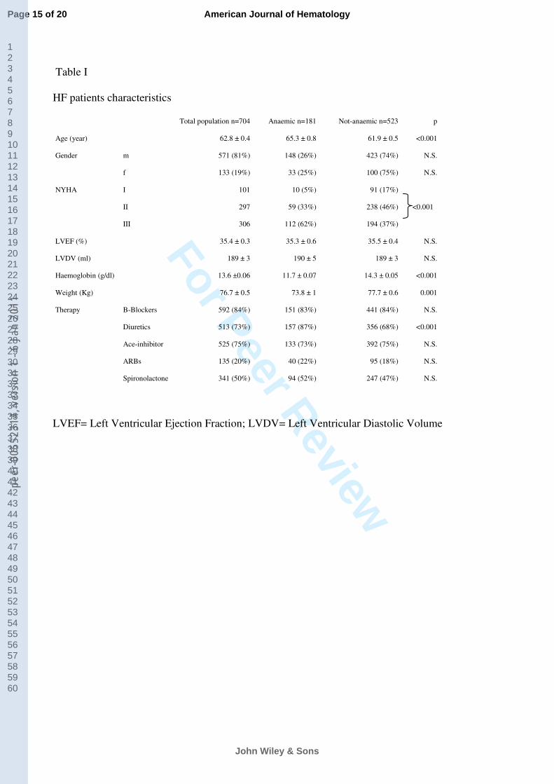
Population characteristics, treatment and heart failure severity are reported in table I. Anaemic
patients were older, had clinically more severe HF and were more frequently treated with
diuretics.
Haemoglobin was 13.7 ± 0.1 g/dl ranging between 7.5 and 17.1 g/dl. 181 patients were
anaemic (26%), representing 33 (25%) of 133 women and 148 (26%) of 571 males. Non-anaemic
Page 7 of 20
John Wiley & Sons
American Journal of Hematology
123456789101112131415161718192021222324252627282930313233343536373839404142434445464748495051525354555657585960
peer
-005
5231
4, v
ersi
on 1
- 6
Jan
2011

For Peer Review
7
patients were 523 (74%), representing 100 (75%) of 133 women and 423 (74%) of 571 males.
Anaerobic threshold was not identified in 12% (n=87) of the 704 patients. CPET results are
reported in table II for the entire study population and for the anaemic and non-anaemic patients,
respectively. Anaemic patients had a lower exercise capacity, with the work load achieved, the
O2 both at peak exercise and at anaerobic threshold being lower. Furthermore, anaemic HF
patients had a greater E / CO2 slope. It should be noted that cardiac function and left
ventricular volume at echocardiography were similar for anaemic and non-anaemic patients (table
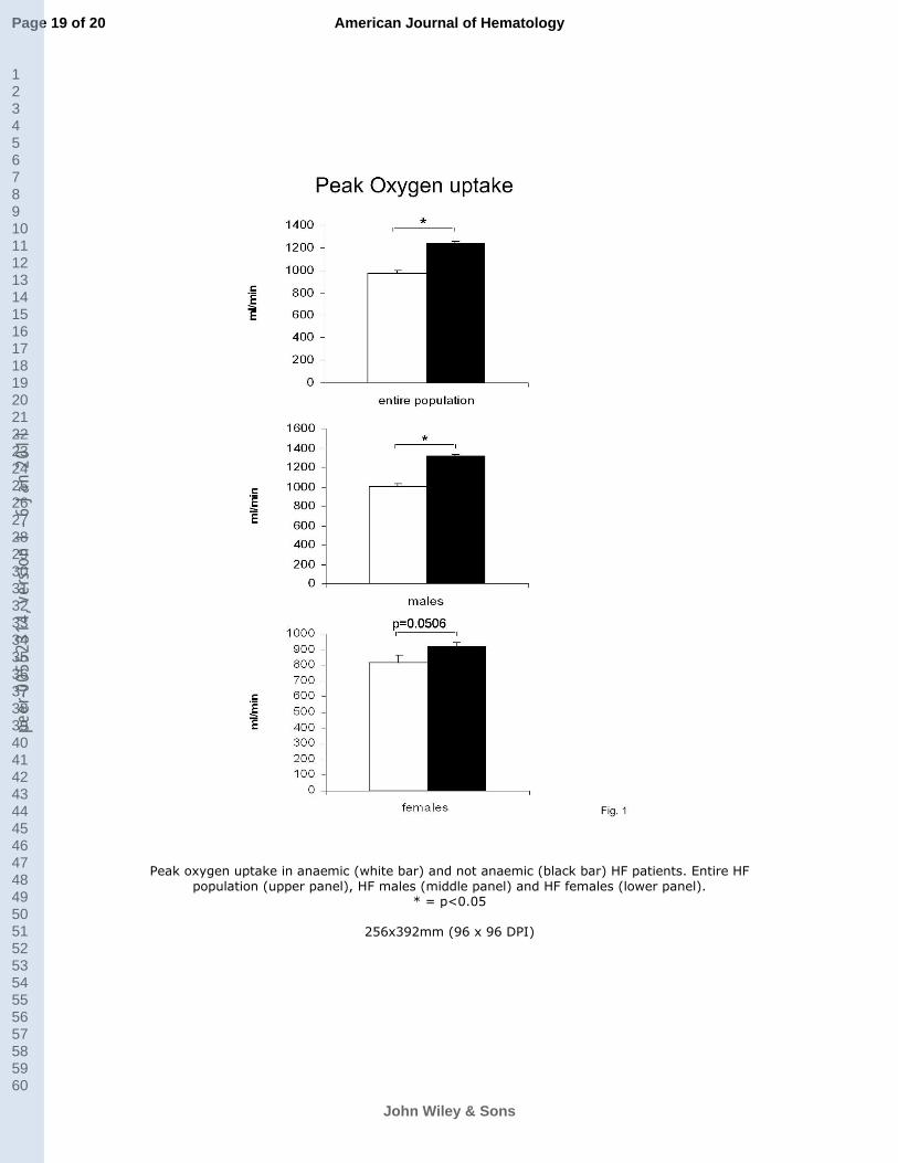
I). Peak O2 was lower in anaemic male patients compared to non-anaemic but not in women
who showed only a trend toward the same difference (p=0.0506) (figure 1).
Coronary angiography was available for 692 of the 704 patients. 332 (47%) patients (30
female and 302 male) were classified as having HF associated to coronary artery disease and 360
(53%) (102 female and 258 male) as not associated to coronary artery disease i.e. with
angiografically normal epicardial coronary arteries. The presence of coronary artery disease did
not significantly influenced the correlation between O2 and haemoglobin both at peak exercise
and at anaerobic threshold (table III). Table IV shows the correlations between several variables
and peak O2. At multivariate analysis only gender (p<0.000), haemoglobin (p<0.01),
E / CO2 slope (p<0.000), the work load achieved (p<0.000) and NYHA class (p<0.000)
remained significantly correlated to peak O2.
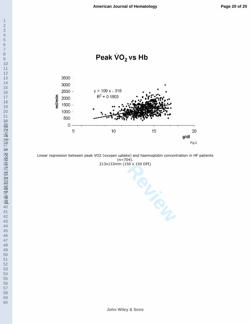
The correlation between peak exercise O2 and haemoglobin at rest is reported in figure
2. Assessing O2 as ml/min/kg the slope of the O2 vs. haemoglobin relationship was 0.97
(ml/kg/min/g/dl). O2 at anaerobic threshold was significantly correlated to the following
Page 8 of 20
John Wiley & Sons
American Journal of Hematology
123456789101112131415161718192021222324252627282930313233343536373839404142434445464748495051525354555657585960
peer
-005
5231
4, v
ersi
on 1
- 6
Jan
2011

For Peer Review
8
variables: age (p<0.000), gender (p<0.000), NYHA class (p<0.000), LVEF (p<0.000),
haemoglobin (p<0.000), E / CO2 slope (p<0.000), peak heart rate (p<0.000) and work load
(p<0.000). At multivariate analysis only age (p<0.05), E / CO2 slope (p<0.000) and work load
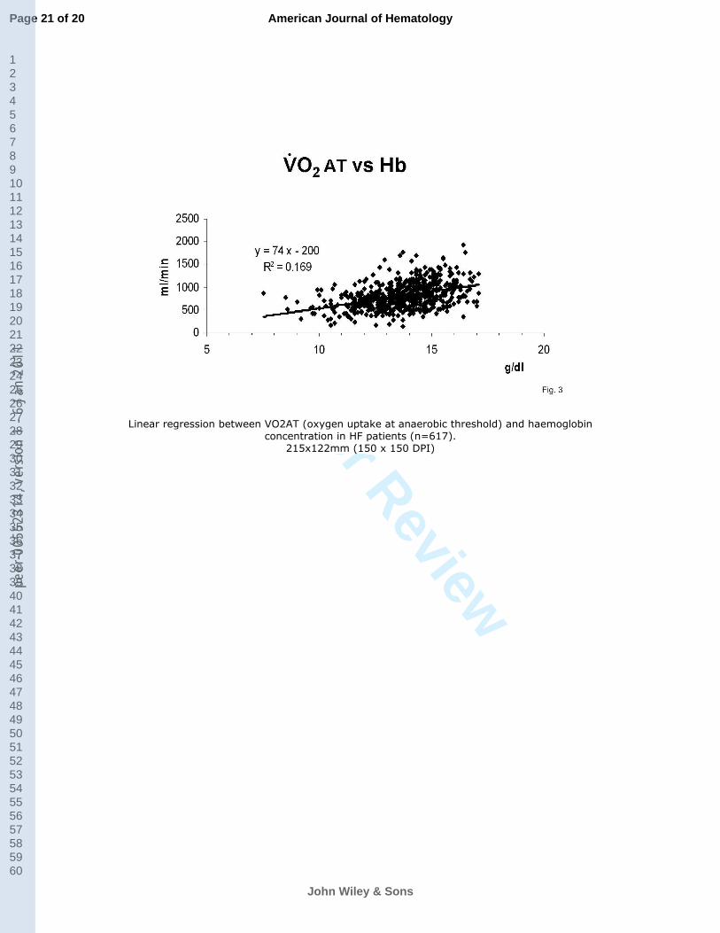
(p<0.000) remained independently correlated to O2. The O2 vs. Hb relationship at the
anaerobic threshold is reported in figure 3.
Discussion
The present study shows, in accordance with several previous reports, that anaemia is
frequent in chronic HF patients and associated with a lower exercise capacity (1-6). Indeed, the
prevalence of anaemia we observed (26%) is in agreement with what has been reported
previously (between 10% and 55%) (4). In HF the presence of anaemia is associated with poor
prognosis, with higher mortality risk both in hospitalised and ambulatory patients, higher
hospitalisation rate and, overall, with a more severe clinical condition for patients (4, 15-17). In
our study, on average, peak O2 and E/ CO2 slope, both of which are independent strong
prognostic indicators of HF (18), were 22% lower and 10% higher, respectively, in HF patients
with anaemia compared to those without anaemia.
It is commonly thought that anaemia should be corrected to improve HF patients’ quality
of life and possibly prognosis, however there is still a lack of a scientific evidence; consequently
anaemia correction is not considered as mandatory in HF treatment guidelines (19, 20). Several
studies have focused on the effect of anaemia correction, usually by iron and erythropoietin or
darbopoietin, on exercise performance, measured either by peak O2, exercise tolerance or by
distance walked at the 6 minutes walking test, in both HF and non-HF patients (21-29). However,
only 4 (21-23) reports all based on small studies, showed the effect of anaemia correction on peak
Page 9 of 20
John Wiley & Sons
American Journal of Hematology
123456789101112131415161718192021222324252627282930313233343536373839404142434445464748495051525354555657585960
peer
-005
5231
4, v
ersi
on 1
- 6
Jan
2011

For Peer Review
9
exercise O2 in HF patients, with discordant findings. Indeed, 3 reports showed that anaemia
treatment increased exercise performance (21, 22, 24) while one did not (23).
Several factors influence exercise performance in HF, including, following the Fick
principle, cardiac output and the arterio-venous oxygen difference. We limited our analysis only
to haemoglobin. To extrapolate the role of haemoglobin we studied the correlation between
resting haemoglobin and O2 at peak and at anaerobic threshold. We observed a correlation
between O2 (ml/min) at peak and haemoglobin with a R2 = 0.18, which means that
haemoglobin accounts for 18 % of the variance in peak O2. This also means that 82% of peak
O2 variance is not related to haemoglobin. The slope of this ratio (figure 2) tells us that, on
average, at peak exercise each g of haemoglobin accounts for a 109 ml/min change in O2 which
is equivalent to 0.97 ml/min/kg.
We analysed the correlation between O2 at peak exercise and resting haemoglobin.
Unfortunately peak exercise haemoglobin data is not available. However, exercise-induced
hemoconcentration, which is well known and is due to both spleen contraction and fluid shift
away from the intravascular space due to an increase in intracellular lactic acid (30, 31), probably
did not significantly influence our findings. Indeed, exercise-induced hemoconcentration is
responsible for approximately 20% of arterio-venous oxygen difference increase in HF subjects,
being on average around 1.0 g/dl in normal subjects and 0.6 g/dl in HF patients (30). Had the
exercise-induced haemoglobin increase been even in patients with different levels of HF severity,
this would have only shifted the O2/Hb ratio upwards, with no effect on the slope of the ratio
and, consequently, with no effect on the predicted O2 change as a result of haemoglobin
Page 10 of 20
John Wiley & Sons
American Journal of Hematology
123456789101112131415161718192021222324252627282930313233343536373839404142434445464748495051525354555657585960
peer
-005
5231
4, v
ersi
on 1
- 6
Jan
2011
For Peer Review
10
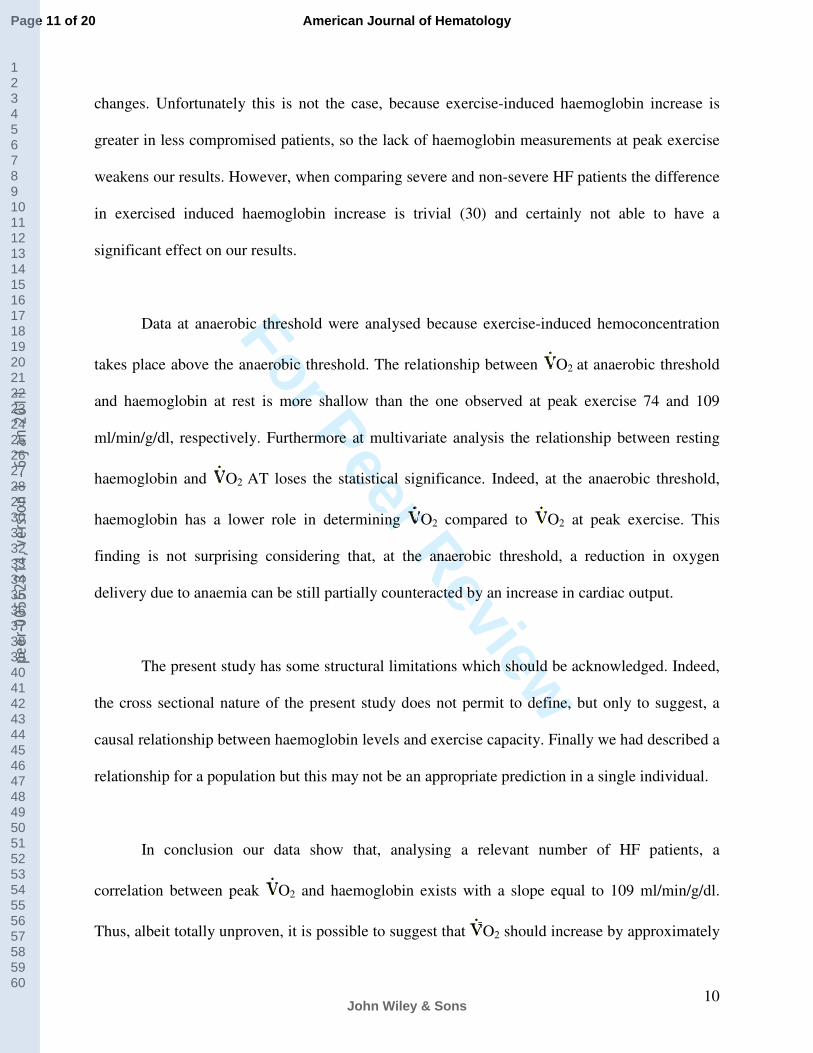
changes. Unfortunately this is not the case, because exercise-induced haemoglobin increase is
greater in less compromised patients, so the lack of haemoglobin measurements at peak exercise
weakens our results. However, when comparing severe and non-severe HF patients the difference
in exercised induced haemoglobin increase is trivial (30) and certainly not able to have a
significant effect on our results.
Data at anaerobic threshold were analysed because exercise-induced hemoconcentration
takes place above the anaerobic threshold. The relationship between O2 at anaerobic threshold
and haemoglobin at rest is more shallow than the one observed at peak exercise 74 and 109
ml/min/g/dl, respectively. Furthermore at multivariate analysis the relationship between resting
haemoglobin and O2 AT loses the statistical significance. Indeed, at the anaerobic threshold,
haemoglobin has a lower role in determining O2 compared to O2 at peak exercise. This
finding is not surprising considering that, at the anaerobic threshold, a reduction in oxygen
delivery due to anaemia can be still partially counteracted by an increase in cardiac output.
The present study has some structural limitations which should be acknowledged. Indeed,
the cross sectional nature of the present study does not permit to define, but only to suggest, a
causal relationship between haemoglobin levels and exercise capacity. Finally we had described a
relationship for a population but this may not be an appropriate prediction in a single individual.
In conclusion our data show that, analysing a relevant number of HF patients, a
correlation between peak O2 and haemoglobin exists with a slope equal to 109 ml/min/g/dl.
Thus, albeit totally unproven, it is possible to suggest that O2 should increase by approximately
Page 11 of 20
John Wiley & Sons
American Journal of Hematology
123456789101112131415161718192021222324252627282930313233343536373839404142434445464748495051525354555657585960
peer
-005
5231
4, v
ersi
on 1
- 6
Jan
2011

For Peer Review
11
109 ml/min for each g/dl of haemoglobin increase. Studies analyzing the acute effects of
haemoglobin changes on exercise performance in anaemic HF patients are needed.
Page 12 of 20
John Wiley & Sons
American Journal of Hematology
123456789101112131415161718192021222324252627282930313233343536373839404142434445464748495051525354555657585960
peer
-005
5231
4, v
ersi
on 1
- 6
Jan
2011

For Peer Review
12
References
1. Anand IS. Anemia and chronic heart failure implications and treatment options. J Am
Coll Cardiol 2008;52:501-511.
2. Felker GM, Adams KF, Jr., Gattis WA, et al. Anemia as a risk factor and therapeutic
target in heart failure. J Am Coll Cardiol 2004;44:959-966.
3. Mitchell JE. Emerging role of anemia in heart failure. Am J Cardiol 2007;99:15D-20D.
4. Tang YD, Katz SD. The prevalence of anemia in chronic heart failure and its impact on
the clinical outcomes. Heart Fail Rev 2008;13:387-392.
5. Kalra PR, Bolger AP, Francis DP, et al. Effect of anemia on exercise tolerance in chronic
heart failure in men. Am J Cardiol 2003;91:888-891.
6. Witte KK, Desilva R, Chattopadhyay S, et al. Are hematinic deficiencies the cause of
anemia in chronic heart failure? Am Heart J 2004;147:924-930.
7. Perego GB, Marenzi GC, Guazzi M, et al. Contribution of PO2, P50, and Hb to changes
in arteriovenous O2 content during exercise in heart failure. J Appl Physiol 1996;80:623-631.
8. Agostoni PG, Wasserman K, Perego GB, et al. Non-invasive measurement of stroke
volume during exercise in heart failure patients. Clin Sci (Lond) 2000;98:545-551.
9. Agostoni P, Cattadori G. Non invasive cardiac output measurement: a new tool in heart
failure. Cardiology (in press) 2009.
10. Agostoni P, Bianchi M, Moraschi A, et al. Work-rate affects cardiopulmonary exercise
test results in heart failure. Eur J Heart Fail 2005;7:498-504.
11. Beaver WL, Wasserman K, Whipp BJ. A new method for detecting anaerobic threshold
by gas exchange. J Appl Physiol 1986;60:2020-2027.
12. Feigenbaum H AW, Ryan T. Hemodynamics. Feigenbaum's Echocardiography: Ed.
Lippincott Williams and Wilkins; 2005. pp 214-246.
13. WHO/UNICEF/UNU. Iron deficency anaemia: assessment, prevention, and control.
Geneva, Switzerland: World Health Organization; 2001.
14. Felker GM, Shaw LK, O'Connor CM. A standardized definition of ischemic
cardiomyopathy for use in clinical research. J Am Coll Cardiol 2002;39:210-218.
15. McMurray JJ. What are the clinical consequences of anemia in patients with chronic heart
failure? J Card Fail 2004;10:S10-12.
16. Ezekowitz JA, McAlister FA, Armstrong PW. Anemia is common in heart failure and is
associated with poor outcomes: insights from a cohort of 12 065 patients with new-onset heart
failure. Circulation 2003;107:223-225.
17. Horwich TB, Fonarow GC, Hamilton MA, et al. Anemia is associated with worse
symptoms, greater impairment in functional capacity and a significant increase in mortality in
patients with advanced heart failure. J Am Coll Cardiol 2002;39:1780-1786.
18. Lang CC, Agostoni P, Mancini DM. Prognostic significance and measurement of
exercise-derived hemodynamic variables in patients with heart failure. J Card Fail 2007;13:672-
679.
19. Dickstein K, Cohen-Solal A, Filippatos G, et al. ESC Guidelines for the diagnosis and
treatment of acute and chronic heart failure 2008: the Task Force for the Diagnosis and Treatment
of Acute and Chronic Heart Failure 2008 of the European Society of Cardiology. Developed in
collaboration with the Heart Failure Association of the ESC (HFA) and endorsed by the
European Society of Intensive Care Medicine (ESICM). Eur Heart J 2008;29:2388-2442.
Page 13 of 20
John Wiley & Sons
American Journal of Hematology
123456789101112131415161718192021222324252627282930313233343536373839404142434445464748495051525354555657585960
peer
-005
5231
4, v
ersi
on 1
- 6
Jan
2011
For Peer Review
13
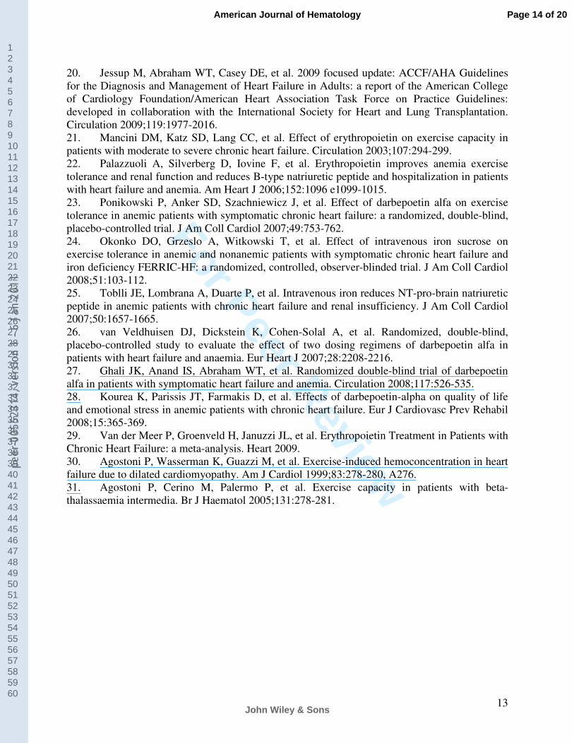
20. Jessup M, Abraham WT, Casey DE, et al. 2009 focused update: ACCF/AHA Guidelines
for the Diagnosis and Management of Heart Failure in Adults: a report of the American College
of Cardiology Foundation/American Heart Association Task Force on Practice Guidelines:
developed in collaboration with the International Society for Heart and Lung Transplantation.
Circulation 2009;119:1977-2016.
21. Mancini DM, Katz SD, Lang CC, et al. Effect of erythropoietin on exercise capacity in
patients with moderate to severe chronic heart failure. Circulation 2003;107:294-299.
22. Palazzuoli A, Silverberg D, Iovine F, et al. Erythropoietin improves anemia exercise
tolerance and renal function and reduces B-type natriuretic peptide and hospitalization in patients
with heart failure and anemia. Am Heart J 2006;152:1096 e1099-1015.
23. Ponikowski P, Anker SD, Szachniewicz J, et al. Effect of darbepoetin alfa on exercise
tolerance in anemic patients with symptomatic chronic heart failure: a randomized, double-blind,
placebo-controlled trial. J Am Coll Cardiol 2007;49:753-762.
24. Okonko DO, Grzeslo A, Witkowski T, et al. Effect of intravenous iron sucrose on
exercise tolerance in anemic and nonanemic patients with symptomatic chronic heart failure and
iron deficiency FERRIC-HF: a randomized, controlled, observer-blinded trial. J Am Coll Cardiol
2008;51:103-112.
25. Toblli JE, Lombrana A, Duarte P, et al. Intravenous iron reduces NT-pro-brain natriuretic
peptide in anemic patients with chronic heart failure and renal insufficiency. J Am Coll Cardiol
2007;50:1657-1665.
26. van Veldhuisen DJ, Dickstein K, Cohen-Solal A, et al. Randomized, double-blind,
placebo-controlled study to evaluate the effect of two dosing regimens of darbepoetin alfa in
patients with heart failure and anaemia. Eur Heart J 2007;28:2208-2216.
27. Ghali JK, Anand IS, Abraham WT, et al. Randomized double-blind trial of darbepoetin
alfa in patients with symptomatic heart failure and anemia. Circulation 2008;117:526-535.
28. Kourea K, Parissis JT, Farmakis D, et al. Effects of darbepoetin-alpha on quality of life
and emotional stress in anemic patients with chronic heart failure. Eur J Cardiovasc Prev Rehabil
2008;15:365-369.
29. Van der Meer P, Groenveld H, Januzzi JL, et al. Erythropoietin Treatment in Patients with
Chronic Heart Failure: a meta-analysis. Heart 2009.
30. Agostoni P, Wasserman K, Guazzi M, et al. Exercise-induced hemoconcentration in heart
failure due to dilated cardiomyopathy. Am J Cardiol 1999;83:278-280, A276.
31. Agostoni P, Cerino M, Palermo P, et al. Exercise capacity in patients with beta-
thalassaemia intermedia. Br J Haematol 2005;131:278-281.
Page 14 of 20
John Wiley & Sons
American Journal of Hematology
123456789101112131415161718192021222324252627282930313233343536373839404142434445464748495051525354555657585960
peer
-005
5231
4, v
ersi
on 1
- 6
Jan
2011
For Peer Review
Table I
HF patients characteristics
Total population n=704 Anaemic n=181 Not-anaemic n=523 p
Age (year) 62.8 ± 0.4 65.3 ± 0.8 61.9 ± 0.5 <0.001
Gender m 571 (81%) 148 (26%) 423 (74%) N.S.
f 133 (19%) 33 (25%) 100 (75%) N.S.
NYHA I 101 10 (5%) 91 (17%)
II 297 59 (33%) 238 (46%)
III 306 112 (62%) 194 (37%)
<0.001
LVEF (%) 35.4 ± 0.3 35.3 ± 0.6 35.5 ± 0.4 N.S.
LVDV (ml) 189 ± 3 190 ± 5 189 ± 3 N.S.
Haemoglobin (g/dl) 13.6 ±0.06 11.7 ± 0.07 14.3 ± 0.05 <0.001
Weight (Kg) 76.7 ± 0.5 73.8 ± 1 77.7 ± 0.6 0.001
Therapy B-Blockers 592 (84%) 151 (83%) 441 (84%) N.S.
Diuretics 513 (73%) 157 (87%) 356 (68%) <0.001
Ace-inhibitor 525 (75%) 133 (73%) 392 (75%) N.S.
ARBs 135 (20%) 40 (22%) 95 (18%) N.S.
Spironolactone 341 (50%) 94 (52%) 247 (47%) N.S.
LVEF= Left Ventricular Ejection Fraction; LVDV= Left Ventricular Diastolic Volume
Page 15 of 20
John Wiley & Sons
American Journal of Hematology
123456789101112131415161718192021222324252627282930313233343536373839404142434445464748495051525354555657585960
peer
-005
5231
4, v
ersi
on 1
- 6
Jan
2011

For Peer Review
Table II
Cardiopulmonary exercise test results
Total population Anaemic Not-anaemic p
Peak O2 (ml/min) 1173± 15 971 ± 23 1243 ± 18 <0.001
Peak O2 (ml/Kg/min) 15.3 ± 0.2 13.2 ± 0.3 16.0 ± 0.2 <0.001
Peak O2 (% of predicted value) 60 ± 01 51 ± 1 63 ± 1 <0.001
E/ CO2 slope 31 ± 0 34 ± 1 31 ± 0 <0.001
O2 /Work (ml/min Watt) 9.6 ± 0.3 9.4 ± 0.4 9.7 ± 0.3 N.S.
O2 AT (ml/min) 807 ± 11 680 ± 18 849 ± 13 <0.001
HR at rest (b/min) 74 ± 1 73 ± 1 74 ± 1 N.S.
HR max (b/min) 120 ± 1 111 ± 2 123 ± 1 <0.001
Delta HR (b) 47 ± 1 38 ± 2 49 ± 1 <0.001
Work load (Watt) 89 ± 1 71 ± 2 95 ± 2 <0.001
Exercise time (min) 8.8 ± 0.1 7.9 ± 0.1 9.1 ± 0.2 <0.001
O2 = oxygen uptake. E / CO2= ventilation/carbon dioxide slope. AT = anaerobic threshold. HR
= heart rate.
Page 16 of 20
John Wiley & Sons
American Journal of Hematology
123456789101112131415161718192021222324252627282930313233343536373839404142434445464748495051525354555657585960
peer
-005
5231
4, v
ersi
on 1
- 6
Jan
2011
For Peer Review
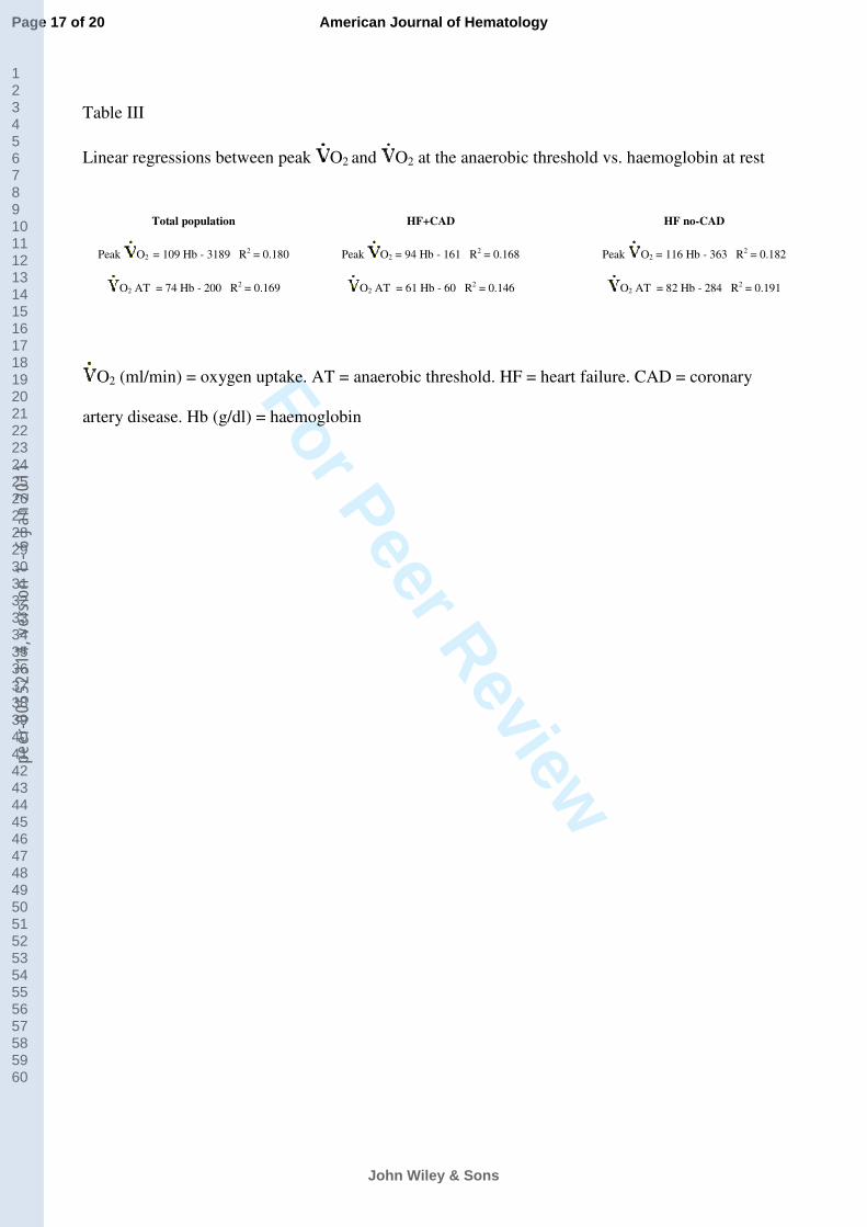
Table III
Linear regressions between peak O2 and O2 at the anaerobic threshold vs. haemoglobin at rest
Total population HF+CAD HF no-CAD
Peak O2 = 109 Hb - 3189 R2 = 0.180 Peak O2 = 94 Hb - 161 R2 = 0.168 Peak O2 = 116 Hb - 363 R2 = 0.182
O2 AT = 74 Hb - 200 R2 = 0.169 O2 AT = 61 Hb - 60 R2 = 0.146 O2 AT = 82 Hb - 284 R2 = 0.191
O2 (ml/min) = oxygen uptake. AT = anaerobic threshold. HF = heart failure. CAD = coronary
artery disease. Hb (g/dl) = haemoglobin
Page 17 of 20
John Wiley & Sons
American Journal of Hematology
123456789101112131415161718192021222324252627282930313233343536373839404142434445464748495051525354555657585960
peer
-005
5231
4, v
ersi
on 1
- 6
Jan
2011

For Peer Review
Table IV
Monovariate correlations between peak O2 and several variables
R2
p
Age 0.197 <0.000
Gender 0.110 <0.000
LVEF(%) 0.08 <0.000
Hb (g/dl) 0.108 <0.000
E/ CO2 slope 0.2 <0.000
HR peak (b/m) 0.192 <0.000
Work load (Watts) 0.872 <0.000
NYHA class 0.589 <0.000
O2 = oxygen uptake. E / CO2= ventilation/carbon dioxide slope. HR = heart rate. Hb =
haemoglobin
Page 18 of 20
John Wiley & Sons
American Journal of Hematology
123456789101112131415161718192021222324252627282930313233343536373839404142434445464748495051525354555657585960
peer
-005
5231
4, v
ersi
on 1
- 6
Jan
2011
For Peer Review
Peak oxygen uptake in anaemic (white bar) and not anaemic (black bar) HF patients. Entire HF
population (upper panel), HF males (middle panel) and HF females (lower panel). * = p<0.05
256x392mm (96 x 96 DPI)
Page 19 of 20
John Wiley & Sons
American Journal of Hematology
123456789101112131415161718192021222324252627282930313233343536373839404142434445464748495051525354555657585960
peer
-005
5231
4, v
ersi
on 1
- 6
Jan
2011
For Peer Review
Linear regression between peak VO2 (oxygen uptake) and haemoglobin concentration in HF patients (n=704).
213x133mm (150 x 150 DPI)
Page 20 of 20
John Wiley & Sons
American Journal of Hematology
123456789101112131415161718192021222324252627282930313233343536373839404142434445464748495051525354555657585960
peer
-005
5231
4, v
ersi
on 1
- 6
Jan
2011
For Peer Review
Linear regression between VO2AT (oxygen uptake at anaerobic threshold) and haemoglobin concentration in HF patients (n=617).
215x122mm (150 x 150 DPI)
Page 21 of 20
John Wiley & Sons
American Journal of Hematology
123456789101112131415161718192021222324252627282930313233343536373839404142434445464748495051525354555657585960
peer
-005
5231
4, v
ersi
on 1
- 6
Jan
2011