Relationship of eye and hand movements during perturbed reaching tasks
44
Iowa State University Digital Repository @ Iowa State University Graduate eses and Dissertations Graduate College 2011 Relationship of eye and hand movements during perturbed reaching tasks Sumit Ranjan Iowa State University Follow this and additional works at: hp://lib.dr.iastate.edu/etd Part of the Kinesiology Commons is esis is brought to you for free and open access by the Graduate College at Digital Repository @ Iowa State University. It has been accepted for inclusion in Graduate eses and Dissertations by an authorized administrator of Digital Repository @ Iowa State University. For more information, please contact [email protected]. Recommended Citation Ranjan, Sumit, "Relationship of eye and hand movements during perturbed reaching tasks" (2011). Graduate eses and Dissertations. Paper 12005.
Transcript of Relationship of eye and hand movements during perturbed reaching tasks

Iowa State UniversityDigital Repository @ Iowa State University
Graduate Theses and Dissertations Graduate College
2011
Relationship of eye and hand movements duringperturbed reaching tasksSumit RanjanIowa State University
Follow this and additional works at: http://lib.dr.iastate.edu/etd
Part of the Kinesiology Commons
This Thesis is brought to you for free and open access by the Graduate College at Digital Repository @ Iowa State University. It has been accepted forinclusion in Graduate Theses and Dissertations by an authorized administrator of Digital Repository @ Iowa State University. For more information,please contact [email protected].
Recommended CitationRanjan, Sumit, "Relationship of eye and hand movements during perturbed reaching tasks" (2011). Graduate Theses and Dissertations.Paper 12005.

Relationship of eye and hand movements during perturbed reaching tasks
by
Sumit Ranjan
A thesis submitted to the graduate faculty
in partial fulfillment of the requirements for the degree of
MASTER OF SCIENCE
Major: Kinesiology (Biological Basis of Physical Activity)
Program of Study Committee: Ann L. Smiley-Oyen, Major Professor
James R. Bloedel Anumantha G. Kanthasamy
Iowa State University
Ames, Iowa
2011
Copyright © Sumit Ranjan, 2011. All rights reserved.

ii
TABLE OF CONTENTS LIST OF FIGURES iii ACKNOWLEDGEMENT iv ABSTRACT v CHAPTER 1. INTRODUCTION 1 CHAPTER 2. REVIEW OF LITERATURE 4 Reach to Grasp Movement 4 Visual Feedback 5 Perturbation Paradigm 7 Eye-Hand Coordination 8 Role of Basal Ganglia in Eye-Hand Coordination 9 Role of Cerebellum in Eye-Hand Coordination 10 Present Study 10 CHAPTER 3. EXPERIMENT 12 Method 12 Subjects 12 Apparatus and Procedures 12 Dependent Variable and Data Analysis 16 Results 18 Aperture at Dowel Touch 20 Aperture Closure Time 21 Post-Perturbation Time 22 Post-Perturbation Distance 23 Summary of Results 24 Discussion 25 CHAPTER 4. CONCLUSION 30 APPENDIX. INFORMED CONSENT DOCUMENT 32 REFERENCES 35
iii
LIST OF FIGURES FIGURE 1. Subject performing reach to grasp movement 13
FIGURE 2. Raw data acquisition record 18
FIGURE 3. Data acquisition diagram explaining different trial types 19
FIGURE 4. Apertures at dowel touch data representing Error 20
FIGURE 5. Aperture closure time data 21
FIGURE 6. Perturbation time data 22
FIGURE 7. Post-perturbation distance data 23

iv
ACKNOWLEDGEMENT
I owe my sincere gratitude to my advisors Dr. Bloedel and Dr. Smiley-Oyen for their
continued support and guidance throughout my thesis.
I owe everlasting gratefulness to my mom and dad, who have always been a source of
inspiration and supported my decisions regarding my career. Their love and support have
helped me to pass many hurdles in my life. Last but not least, I want to thank my beautiful
and lovely wife for her unconditional support.

v
ABSTRACT
It has been suggested that to make a goal-directed reach to grasp movement, precise
visual information of target and hand is important. In spite of various studies relating to
visual feedback in reach to grasp, few researchers have investigated the parameters of this
behavior in relation to saccadic eye movement. Specifically, research in this area lacks
insight about the contribution of visual feedback before and after the occurrence of saccadic
eye movement. The purpose of this study was to investigate the influence of eye movement on
the characteristics of the hand movement during a reach to grasp task. In this experiment, 9
college-age individuals performed the task of reaching and grasping a vertical dowel in two
conditions: (1) with eye movement and (2) without eye movement (by fixating eye on central
dowel). Further, we compared the performance in three sub-conditions: (1) full vision, (2)
vision block before eye movement, and (3) vision block after eye movement. We found that
presence or absence of either eye movement or continuous vision of target and hand does not
modify the accuracy of grasp. Reach duration was shorter with full vision and reach
trajectory was shorter when eye movement was coordinated with hand movement. We
concluded that continuous vision of the hand and target is not necessary for the online
control of these complex movements, but vision is necessary for optimizing the speed of the
movement.

1
CHAPTER 1
INTRODUCTION
Kinematics of hand transport and grasp formation are well coordinated in normal
individuals (Jeannerod, 1984). It has also been established that both the magnitude of
maximum grip aperture during the reach, as well as the temporal characteristics of the reach
are altered when normal vision is not available during the task. However, continuous vision
of an object during reaching is not necessary in healthy subjects for the gradual shaping of
the hand (Santello, 2002). Similarly, Winges, Weber, and Santello (2003) showed that
continuous visual feedback of the hand or the target is not necessary to allow the hand to
gradually conform to target contours.
While, the research in the area of visual feedback in reach to grasp movement has
advanced, few researchers have investigated the parameters of this behavior in relation to
saccadic eye movement. In a pointing task study conducted by Donkelaar and Staub (2000),
it was found that hand movement amplitude was significantly greater when the movements
were made in the absence of eye movement. Knowledge in this area lacks insight about the
contribution of visual feedback before and after the occurrence of saccadic eye movement.
Our present study investigated the influence of eye movement on the characteristics
of the hand movement during a reach to grasp task. In our experiment, 9 college-age
individuals performed the task of reach for and grasping a vertical dowel, the position of
which could shift to left during the movement. The task was performed in two main
conditions: (1) with eye movement and (2) without eye movement (by fixating eye on central
dowel). Further, we compared the performance in three sub-conditions (1) full vision-no

2
vision block, (2) vision block before eye movement, and (3) vision block after eye
movement. The following parameters of the movement were calculated for each trial: (1)
aperture closure time -- time taken from maximum aperture to touch the target; (2) aperture
at dowel touch -- distance between the index finger and thumb at the time of dowel touch; (3)
post-perturbation distance -- distance traveled by wrist IRED from time of perturbation to
dowel touch; (4) post-perturbation time -- time taken from the time of perturbation of target
to target touch.
We found that the error in grasping, which we measured by aperture at dowel touch,
remained unaffected by the presence or absence of vision. In addition, error did not change in
the conditions in which we asked the subjects to fixate their eyes. To examine whether the
maximum grip aperture occurred at a consistent time relative to the grasping of the object,
aperture closure time was measured; aperture closure time was not significantly different
among any of the vision conditions. This implies that aperture closure time is a highly
regulated feature of the movement across different conditions. Additionally, we found that
the time taken to complete the task after perturbation was significantly less in conditions
where vision was available throughout movement when compared to when vision was
blocked or fixated. This implies that restricting the vision of the perturbed target causes
subjects to take extra time to execute the task. We found a significant difference in post-
perturbation distance, when we blocked vision during the task, whether vision was or was not
fixated. This implies that the loss of vision at an intermediate point of the task results in the
hand taking a longer trajectory if eye movement is not associated with hand movement.

3
It is concluded that the availability of precise visual information about the object and
hand throughout the movement does not modify the accuracy of grasp, but it can impact
reach duration. Our study also indicated that a coordinated eye movement accompanying the
hand movement does not contribute to the production of a more accurate grasp, but it can
play a role in generating a shorter reach trajectory when visual information is limited.
This project was started with the intention of studying people with Parkinson’s
disease (PD). Our lab has been active in research related to PD and reach to grasp. Our hope
is to implement the second phase of these experiments on people with PD and if possible in a
cerebellar disease patient group. This will help us to understand the neural underpinning of
this elegant behavior and the role of saccadic eye movement.

4
CHAPTER 2
REVIEW OF LITERATURE
Reach to Grasp Movement
In everyday life we spend much time reaching for and manipulating objects. The
reach to grasp is a fundamental hand movement, and components of this movement are
coordinated well in neurologically healthy people (Jeannerod, 1984). This elegant behavior
has two components – reach and grasp. Reaching is mostly affected by the proximal joints of
the arm and deals with spatial relationships of the objects and the body (Jeannerod, 1984). By
contrast, grasping is a distal behavior involving finger movements that adjust based on the
intrinsic qualities of objects (Jeannerod, 1984).
Ample evidence is available to support the premise that reaching and grasping are
functionally interrelated. The kinematics of hand transport and grasp formation are well
coordinated in normal individuals and indicate that maximum grip aperture is precisely timed
with respect to hand transport (Jeannerod, 1984). Hand pre-shaping takes place as early as in
the first 30% of the movement duration during the reach to grasp movement (Schettino,
Adamovich, & Poizner, 2003). This indicates that these two components work in a parallel
fashion and are temporally related.
There are various studies supporting the view that these two components maintain a
spatial relationship that is remarkably invariant. Wang and Stelmach (1998) suggest that
reach and grasp components are governed by independent neuromotor synergies that are
coordinated spatially by a higher-level synergy. Recently, distance from maximum aperture
to target (aperture closure distance) has been used to support the spatial coordination between

5
the different components of reach to grasp movement (Rand, Smiley-Oyen, Shimansky,
Bloedel, & Stelmach, 2006). Aperture closure distance is defined as the distance of the hand
location from the target at which grip aperture begins to close. Aperture closure distance was
stable under a variety of conditions such as differences in reaching distance and target size
(Wang and Stelmach, 2001). Rand et al. (2006) found that aperture closure distance is stable
even when the target location was shifted instantaneously during the reach movement. In this
study, subjects reached and grasped the vertical dowel whose location was fixed during the
control condition. In the perturbation conditions, the target location was shifted
instantaneously during the reaching movement to left or right. Aperture closure distance was
similar for both the control and perturbation conditions in healthy subjects. In contrast, time
of aperture closure varied considerably across the trials. Stability of aperture closure distance
implies that reach and grasp are coordinated in the spatial domain.
The fundamental aspect of this visuo-motor behavior is the generation and control of
goal-directed arm movements during the action of reaching and grasping. It must include
mechanisms for computing the distance and direction of the target point in space. Visual
feedback is an integral component of reach to grasp movement.
Visual Feedback
When making a goal directed hand movement towards a target, precise information
about this target has to be obtained. Target information can be multimodal, including visual,
auditory, and somatosensory sources of information.
In order to reach and grasp an object, one must not only transform the spatial location
of the object into an appropriate pattern of shoulder and elbow rotations in order to transport

6
the arm to the object, but also transform the visual size, shape, and orientation of the object
into appropriate patterns of finger and wrist movement. Vision of the object as well as the
moving hand is important in executing reach to grasp movement. Describing the mechanism
by which visual and arm proprioceptive signals are integrated for accurate reaching is a key
issue for understanding visuo-motor coordination.
Hand posture is modulated as a function of object contours in a continuous fashion
during the reach. Santello and Soechting (1998) found that specification of kinematic
variables occurs in a time dependent and gradual fashion during the reaching movement.
These results lead to a pertinent question: to what extent is continuous vision of the hand and
object necessary to have a well coordinated movement? There is evidence available that
supports fast processing of visual information in the context of reaching the targets. Zelaznik,
Hawkins, and Kisselburgh (1983) found that the presence of visual feedback during
movements had a positive effect on accuracy compared with that observed in the absence of
visual feedback for movements with durations as short as 190 ms. Further they showed that
there was no effect on accuracy for very short durations of approximately 70 ms.
Researchers have found contrasting results regarding the role of vision in the well
coordinated grasp component of this movement. Schettino, Adamovich, and Poizner (2000)
reported that two components of hand pre-shaping, finger abduction/adduction and
flexion/extension are affected to different extents by reducing the vision of the object and/or
the hand. In another study, however, it was indicated that continuous vision of an object
during reaching was not necessary in healthy subjects for the gradual shaping of the hand
(Santello, 2002). Similarly, Winges et al. (2003) showed that continuous visual feedback of

7
the hand or the target is not necessary to allow the hand to gradually conform to target
contours. In this study, subjects were asked to reach and grasp different shaped objects with
vision blocked at different times after reach using liquid crystal spectacles. Reach duration
increased when vision was occluded early. However, blocking vision at different times did
not have an effect on the covariant pattern of joint rotations of the fingers, indicating that
gradual formation of hand posture occurs in a similar fashion regardless of the presence or
absence of vision.
These previous studies indicate that in the absence of vision, the time taken to
complete the different components of reach to grasp is prolonged but the coordination
between the fingers remains the same. In addition to the studies related to coordination
between the finger movements, further studies are needed to understand the effect of
restricted vision on the relationship of reach to grasp components. Few studies have looked
on the effect of changing location during an ongoing movement, a technique for examining
the online control of movement.
Perturbation Paradigm
Since humans can make precise aiming movements under rapidly changing
conditions, a mechanism dynamically integrating all types of information is necessary to
initiate and guide ongoing goal-directed movements. Perturbation of target location during an
ongoing hand movement can be used to examine the patients’ online control.
In a typical perturbation paradigm, a few interspersed unexpected perturbed trials are
mixed with the non-perturbed trials. This requires subjects to unexpectedly shift the strategy
to successfully complete the action. Most perturbation experiments have introduced the

8
perturbation during the movement. This requires modification of the reach trajectory and
reorganization of grasp. The common finding in most studies using this approach is that the
response to a perturbation generated by changing the location of a target during an ongoing
movement requires additional processing time. People with Parkinson’s disease (PD) showed
a definable transition period between closing one and opening another motor pattern in
contrast to smooth change shown by neurologically healthy controls (Castiello & Bennett,
1994) and it can be expected that people with PD would show dysfunction during transition
phase from the original to final motor output in a perturbation paradigm.
Hand-Eye Coordination
In most human activity in daily life including reach to grasp, the eye moves in
coordination with the hand to facilitate a better movement. Eye-hand coordination is central
to many human activities. Normal eye-hand coordination involves the synergistic function of
several sensorimotor systems including the visual system. The main purpose of eye-hand
coordination is the use of vision to guide movements of the hand. In eye-hand coordination,
following the appearance of a peripheral target, the saccadic eye movement is typically
initiated first and hand movement second (Biguer, Prablanc, & Jeannerod, 1984). Because of
the ballistic nature of saccades, the eye fixates on the peripheral target well before the hand
movement is completed (Carnahan & Marteniuk, 1991). As a result, the retinal and extra-
retinal information derived from the saccade is thought to contribute to the accurate guidance
of the hand to the target, helping in executing many human movements. Tracking
performance is much better if eye and hand follow the same spatial trajectory, but even better
if the eye leads the hand by about 75 to 100 ms (Miall & Reckess, 2001). Miall and Reckess

9
suggested that information from the ocular system feeds into the manual control system to
assist tracking.
Role of Basal Ganglia in Eye-Hand Coordination
Many brain areas contribute in organization and execution of reach and grasp
movement. Basal ganglia preferentially contribute to movements to remembered targets. The
basal ganglio-thalamo-cortical system appears to be preferentially involved in movements
requiring internal cues such as those required to direct the eye and hand to a remembered
target location (van Donkelaar et al., 1999). In an absence of visual feedback, when a subject
has to use internal representation, the basal ganglia are the major contributor to the
processing and execution of the motor program. The people who historically have been used
to investigate and characterize the dysfunction of basal ganglia are those with PD.
Parkinson’s disease is a progressive neurodegenerative disorder associated with a loss
of dopaminergic nigrostriatal neurons in basal ganglia and is recognized as one of the most
common neurological disorders, affecting approximately 1% of individuals older than 60
years-of-age. PD results in various types of motor impairments including bradykinesia,
tremor, and rigidity. Various studies have found that people with PD show problems in
sequencing and timing of complex movements, and in the execution of internally guided
tasks (Weiss, Stelmach, & Hefter, 1997). It has been reported that PD motor performance is
compromised in the absence of the appropriate use of sensory feedback (Flowers, 1976).
Schettino et al. (2004) found that the distance from the target at which people with PD began
aperture closure was significantly shorter than that of healthy subjects. It was suggested that
those with PD rely on visual feedback to control aperture, so that the patients moved the arm

10
until it reached the spatial area where both hand and target were visible simultaneously,
which resulted in shorter aperture closure distance. This supports that they rely on vision of
hand and target for execution of grasp.
Role of the Cerebellum in Eye-Hand Coordination
It has been suggested that the basal ganglia preferentially contribute to movements
made to remembered targets, whereas the cerebellum preferentially contributes to movement
requiring the integration of visual cues (Miall et al., 2001). Various studies have established
the contribution of the cerebellum in eye-hand coordination. The cerebello-thalamo-cortical
system appears to be preferentially involved in movements utilizing external sensory cues
such as those arising from the appearance of a visualized target (Inoue et al., 1998). There
was impaired temporal prediction and eye-hand coordination in patients with cerebellar
lesions (Sailor, Eggert, & Straube, 2005). Patients with cerebellar damage performed better
in tracking tasks without visual feedback than healthy subjects (Liu, Ingram, Palace, & Miall,
1999). This was a single case study, in which the patient was able to adapt to a gain change
in ‘off-line’ visual feedback of hand position during a pointing task. Patient’s adaptation was
less affected than that of control subjects by trial-to-trial random fluctuations in ‘off-line’
visual feedback. This again supports that cerebellum damage does not affect the performance
based on memory as used in ‘off-line’ condition in the experiment.
Present Study
Although various studies have been done on eye-hand coordination of reach to grasp
movement, few researchers have investigated the parameters of this behavior in relation to
saccadic eye movement. Specifically, research in this area lacks insight about the

11
contribution of visual feedback before and after the occurrence of saccadic eye movement.
Our present study mainly investigates the influence of eye movement on the characteristics
of the hand movement during a reach to grasp task. We compared the execution of reach to
grasp in two main conditions: (1) Non-fixation (NF), in which healthy subjects were allowed
to move the eyes naturally with hand movement, and (2) Fixation (F), where subjects were
asked to fixate their eye on the central target for the time of entire movement. We proposed
that eliminating the eye movement associated with hand movement by requiring the subject
to fixate on one location throughout the task would disrupt the coordination of the reach to
grasp because saccadic eye movement is helpful in executing similar task eg. Pointing (van
Donkelaar, 2000)
Furthermore, we compared three different sub-conditions that differed in the
availability of visual feedback during certain parts of the reach to grasp movement. To
achieve this we compared the subjects’ performance under three conditions: (1) full vision
(no blockage of vision) perturbation to the left (PL), (2) vision blockage before the eye
movement to the target was initiated (VB), and (3) vision blockage after the eye movement to
the target on the left occurred (VB-EOG).
12
CHAPTER 3
EXPERIMENT
Method
Subjects
All subjects (five men, four women) were students from Iowa State University whose
age ranged from 20-35 years. All participants were right-handed and were neurologically
healthy based on self-report. This study was approved by the Institute’s Internal Review
Board, overseeing the use of human subjects in research, ensuring that all studies are
performed in accordance with the 1964 Declaration of Helsinki. All participants were
provided forms that permitted them to provide written informed consent prior to
participation.
Apparatus and Procedures
All participants performed reach-to-grasp movements with their right hand. They
were seated comfortably in front of an apparatus on which three target objects were placed.
The start position was a push button located approximately 30 cm to the right of the subject’s
midline and at the end of an extended armrest. The targets were three dowels, each with a
diameter of 2.5 cm and a height of 10 cm. The targets were placed in a semi-circular fashion
centered with respect to the start position, each 55 cm from the start position. The center
target was in front of the start position, and the other targets were 20 cm to the right or to the
left of the center target. A red LED was attached to the top of each dowel to identify the final
target for grasping. Arm and finger positions during reach-to-grasp movements were
recorded using an Optotrak 3D motion analysis system (Northern Digital). Infrared light

13
emitting diodes (IREDS) were placed on the wrist, tip of the index finger and the tip of the
thumb. In addition, an IRED was placed on each dowel in order to record its position and
movement. Positions of the IREDS were sampled at a rate of 200 HZ. Subjects wore liquid
crystal spectacles (PLATO Translucent Technologies, Toronto, ON, Canada) that occluded
the vision at different time.
EOG
START
BUTTON
IRED 3
IRED 1 IRED 2
TARGET
FIGURE 1. Subject performing reach to grasp movement. Picture is during a trial while subject
is lifting the target towel. Index finger (IRED 1), thumb (IRED 2) and wrist (IRED 3) movements
were recorded using an Optotrak 3D motion analysis system. Vision was blocked using liquid
crystal spectacles (EOG). All subjects start the trial keeping the ulnar side of their right hand on
red button shown in the picture and started movement upon illumination of the red LED of
target dowel.

14
Two bars with twenty-four infrared light beams (Banner Mini-Array system, 19.1 mm
beam spacing) were set 17.5 cm away from the start button, perpendicular to the line between
the start button and the center target. This system detected an interruption of the beam during
the reach, making it possible to identify the time at which the participant’s hand reached this
point during the transport. Interrupting the beam was used to trigger the perturbation. The
perturbation consisted of a shift of the target from center location to either right or left during
the arm movement. Participants were instructed to keep their thumb and index finger
together, place the ulnar side of the hand on the table and rest their index finger and thumb at
the start position before each trial.
Each trial was started when the participant pushed the start button down with the
ulnar edge of the hand with the thumb and index finger closed. After a short random delay
(ranging between 1000 and 1500 ms), a tone signal was delivered. In response to the tone,
participants were asked to be ready for movement. At the beginning of a trial, the LED on
one of the three targets was illuminated. The subjects were instructed to reach for and grasp
the illuminated target as quickly as possible. Subjects were asked to reach for and grasp the
lit dowel with only the index finger and thumb at any location along its vertical extent and
lift it a few centimeters off the table. The LED remained lit until the end of the trial. Before
the perturbation set of trials, the subjects were told that the perturbation would occur
randomly across trials but that they were to continue reaching for and grasping the target as
quickly as possible in every trial.
Each subject performed the task in two experimental conditions: (1) no visual fixation
(NF) condition and (2) visual fixation (F) conditions. In the NF condition, participants were

15
allowed to move their eye during execution of the movement, whereas in the F condition
participants were asked to fixate their vision on the central target throughout the trial. Each
condition had sub-conditions randomly spread, in which subjects either had full vision
throughout the trial or had vision blocked at different times during the trial: (1) left
perturbation (PL) sub-condition, in which target perturbed to left but there was no blockage
of vision; (2) vision blockage at beam (VB) sub-condition: vision was blocked as soon as
subject’s hand broke the beam which was used to initiate the perturbation of target; and (3)
vision block after eye movement (VB-EOG) sub-condition: vision was blocked after subjects
moved their eye to left. In all conditions participants were instructed to continue the reaching
movement and grasp the target as rapidly and accurately as possible. Visual perturbation
conditions were randomized throughout the trials. To record the phasic movement of the
eyeballs, the electro-occulogram (EOG) was used. Responses were recorded by means of
surface electrodes placed appropriately just lateral to the lateral canthus of each eye.
Our main focus was to study the reach to grasp movement to the left target after
perturbation. The left target was chosen because the reaching for grasping motions required
less maneuvering of the wrist compared to that required for grasping central or right target.
As described above, the experiment was conducted in two conditions. We first used
the NF condition, which consisted of 3 sets of 49 trials. In each set, PL, VB, or VB-EOG
trials were randomly mixed with control trials to the central target without vision blockage or
perturbation. This was followed by the F condition, which had two sets of randomly mixed
PL and VB sub-conditions with controls to center. There was no VB-EOG sub-condition in

16
fixation trials, since saccadic eye movements were not allowed. In each set, perturbations
were applied when the participant’s hand broke the light beams described above.
Dependent Variable and Data Analysis
Kinematics of the reaching component and grasp component was analyzed using the
OptoTrak system described above. The reaching component was assessed based on changes
in the position of the wrist IRED, and the grasp component was assessed using the changes in
the position of the index finger and thumb IREDS.
The following parameters of the movement were calculated for each trial: (1) aperture
closure time -- Time taken from maximum aperture to touch the target; (2) aperture at dowel
touch -- distance between the index finger and thumb at the time of dowel touch; (3) post-
perturbation distance -- distance traveled by wrist IRED from time of perturbation to dowel
touch; and (4) post-perturbation time -- time taken from the time of perturbation of target to
target touch. These parameters were selected because our preliminary study on pilot subjects
showed differences in measurements between NF and F conditions as well as between
different sub-conditions.
For statistical analysis, mean values of each dependent variable for each participant
were calculated for five sub-conditions. A 2-way ANOVA with repeated measures was
calculated with conditions F and NF as a between-subject factor and sub-conditions as a
within-subject factor to identify any condition and sub-condition effects, respectively. The
ANOVA results were adjusted with the Huynh-Feldt epsilon (ε) correction. The statistical
significance was defined as p< 0.05. All statistical analyses were performed using Statistical

17
Package for Social Science (SPSS for Windows, version 17.0, SPSS Inc, Chicago, IL)
program.

18
Results
We collected data in two different conditions: F and NF, and three different sub-
conditions: PL, VB, and VB-EOG. Below is the diagram showing raw data acquisition for
one single trial in the PL sub-condition in which target moved to left as the subject crossed
the beam with no vision blockage.
Start of the
movement
Perturbationat beam:
shift of the targetEnd of the movement
Post-perturbation Time(PT)/distance(PD)
Aperture at Dowel
contact (ADD)
Aperture graph
EOG graph
Velocity graph
Aperture closure time (ACT)
Line denoting blockage of vision
FIGURE 2: Raw data acquisition record (in the FPL sub-condition as it appeared on our analysis software).
In the upper graph (A), the orange line depicts the aperture of the grasp (Aperture graph), which is the
distance between thumb and index finger IREDS. The light blue wavy line shows the EOG reflecting eye
movement. The first vertical green line denotes the start of movement. The purple vertical line is the point
at which the subject broke the invisible beam which initiated perturbation of target position and also
initiated vision block in VB and VB-EOG trials. The vertical red line denotes the time of dowel touch, which
marks the end of movement. In the lower graph (B), the green line represents the velocity of the wrist
IRED. The light green line in the bottom of graph B, represent the blockage of vision. When vision is
blocked, this line shows an upward deflection. In this diagram, there is no upward deflection, since there is
no blockage of vision in FPL sub-condition. We measured four dependent variables (All shown above):
Aperture at contact (ADD), Aperture closure time (ACT), Post- perturbation time (PT) and Post-
perturbation distance (PD).
A
B

19
The Figure 3 (below) explains the different timing of vision block in different sub-
conditions.
A
B
C
FIGURE 3: Data Acquisition diagram
explaining different trial types.
A. PL: Trial with perturbation of
target to the left with no vision
block.
B. VB: Trial in which vision was
blocked as the subject crossed
the beam. This triggered the
perturbation of the target to
the left. After few ms delay
the vision was blocked. This
occurred before saccadic eye
movement.
C. VB-EOG: Trial with vision
blocked after eye movement
to the left.
The upward deflection of thin Blue
line in the lower graphs denote
the time at which vision was
blocked. In condition PL (Graph A)-
There is no deflection. In VB
condition (graph B) - The upward
deflection is immediately after the
blue line, which denotes the time
when the subject broke the beam.
In the VB-EOG condition (Graph C)
- the upward deflection is
immediately after eye movement.
Vision block triggered by
eye movement
Vision block triggered
by beam break
No vision block
B
C

20
Aperture at Dowel Touch
This variable measured the error produced by subjects in respective conditions. As
indicated in the results of our pilot study, we considered that a less-than-perfect would have
larger aperture upon target contact. Surprisingly, there were no single comparisons between
sub-conditions across the two main conditions fixations (F) and non-fixations (NF) that were
significantly different. The two-way ANOVA for error between sub-conditions NFVB and
FVB revealed no significant difference, F (1,8) = 2.722, p = 0.138 and same comparison
between NFPL and FPL revealed a trend, but did not show significant difference, (p =
0.077). (see Figure 1A and 1B).
The comparison between the two main sub-conditions in the non-fixation condition,
NFVB and NFEOG, was expected to reveal information about the effect of blocking vision
FIGURE 4. Apertures at dowel touch data representing Error. (A) The vision block at Beam in Non-
fixation (NFVB) and Fixation (FVB) sub-conditions. (B) Perturbation to left in Non-fixation (NFPL)
and Fixation (FPL). Error bars are standard deviation. NS= Not significant
p = 0.138

21
before and after saccadic eye movement. However, there was no significant difference
(F(1,8) = 2.136, p = 0.182).
Aperture Closure Time
To examine the effect of saccadic eye movement and visual feedback on the time
taken to execute this component of the reach and grasp, we measured aperture closure time
There were no significant differences between the time taken by subjects to reach the target
after they have achieved the maximum grip aperture. When comparing fixation and non-
fixation conditions when vision was not blocked with target perturbation to left, there were
no significant changes, F(1,8)= 0.425, p = 0.533 (see Figure 5A). Other comparisons also
failed to reveal significant differences. It is important to note that that in NF conditions,
comparison between VB and EOG trials gave results close to significance, F(1,8)= 4.901. p=
0.058 (see Figure 5B).
FIGURE 5. Aperture closure time data. (A) The vision block at beam in non-fixation (NFVB) and
fixation (FVB) sub-conditions. (B) Vision block at beam (NFVB) and after saccadic eye movement
(NFEOG) in non-fixation trial. Later comparison almost yielded in a significant difference. Error
bars are standard deviation.
p = 0.533
NS
p = 0.058
NS

22
Post-perturbation Time
Most of the statistically significant findings relate to comparisons of this variable.
First of all when compared in the NF condition, there was a significant difference between
sub-condition VB and PL (F (1, 8) = 8.283, p= 0.021) (see Figure 6A). The comparison
between same two sub-conditions during fixation condition was also significant, F(1,8)=
13.838, P= 0.006 (see Figure 6B). This indicates that there is significant change in the time
taken by subjects after perturbation to target touch if there vision is blocked as compared to
when vision was not blocked, but this change is not related to the presence or absence of
saccadic eye movement. For the same variable, the difference between VB and EOG in non-
fixation conditions was close to significance (F (1,8)= 4.824, p= 0.059).
FIGURE 6. Perturbation Time data. (A) In non-fixation condition, comparison between vision
block at beam (NFVB) and perturbation to left (NFPL) sub-conditions. (B) In the fixation
condition, comparison between vision block at beam (FVB) and perturbation to left (FPL) sub-
conditions. . Error bars are standard deviations.
P = 0.021
S
P = 0.006
S

23
Post-perturbation Distance
This variable measures the distance completed by the wrist electrode from the time of
the perturbation to target touch. This was measured in a 3 dimensional coordinate system.
The only significant change in this variable was between F and NF sub-condition with vision
blocked at beam (VB), F (1, 8) = 5.791, p= 0.043 (see Figure 4A). Again near significance
was noted when VB and PL sub-conditions were compared in fixation (F) condition, F (1, 8)
= 4.534, p= 0.066 (see Figure 7B)
p = 0.043
S
p = 0.066
NS
FIGURE 7. Post-perturbation Distance data. (A) Significant difference between visual block at
beam in Non-fixation (NFVB) and Fixation (FVB) sub-conditions. (B) Near significance between
perturbation to left (FPL) and vision block at beam (FVB) in fixation condition. Error bars are
standard deviation.
24
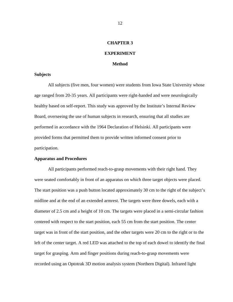
Summary of Results
Our first notable finding was that the error in grasping, which we measured by
aperture at dowel touch, remained unaffected by presence or absence of visual feedback (see
Figure 1A). In addition, error did not change in the conditions where we asked the subjects to
fixate their eyes. Also, aperture closure time was not significantly different in conditions
where vision of perturbed target was or was not present throughout.
Significant findings were found in post-perturbation Time. We found that post-
perturbation Time was significantly different in NFVB and NFPL sub-conditions (see Figure
3A). This comparison was also significant in the visual fixation condition (see Figure 3B).
We also found a significant difference in post-perturbation distance, when comparison was
made between the sub-conditions, visual fixation (FVB) and non-fixation (NFVB) (see
Figure 4A).

25
Discussion
The purpose of this study was to investigate how the parameters of reach to grasp
changed in the absence of visual feedback. A secondary purpose was to investigate the
influence of eye movement on hand movement in performing this task.
We introduced aperture at dowel as a measure of error. We considered that a larger
aperture at dowel touch indicated a less-than-perfect grasp. Our first finding was that the
error in grasping, which we measured by aperture at dowel, remained unaffected by the
presence or absence of visual feedback (see Figure 1A). In addition, error did not change in
the conditions in which we asked the subjects to fixate their eyes. We designed the condition
in which subjects were asked not to move their eyes to investigate the influence of eye
movement on hand movement in reach to grasp. We could not find any significant
differences in error between eye-fixation (F) and non- fixation conditions (NF) (see Figure
1B).
The results from the present study agree with findings from the studies conducted by
Santello et al. (2002) and by Wing et al. (2003). In the study conducted by Santello et al.,
subjects were asked to reach and grasp different shaped objects in the full vision condition
and in the condition in which the visual image of objects was given only for approximately 2
sec. In full vision condition, subjects reached and grasped the objects. In contrast, in the
limited vision condition, subjects were asked to conform to a remembered virtual image. As
stated above, there were no significant differences in the discriminate analysis of hand
postures between vision and impaired-vision conditions. It should be noted that in our

26
experiment, subjects were asked to grasp real objects as compared to virtual objects in the
Santello et al. (2002) study.
In the study by Wing et al. (2003), which was more similar to our study, the vision
was blocked at five different time latencies from the start of the movement. It was found that
different conditions did not have a significant effect on the co-variation pattern of joint
rotations of the hand and wrist. In contrast to Wing et al., we compared the results of
blocking vision before (VB) and after (VE-EOG) eye movement. Also, our experiment
further extended the findings of Wing et al. by comparing the influence of eye movement on
hand movement in the F and NF conditions. Our results suggest that continuous movement of
the eye with hand is not required to make an effective goal-directed grasp.
In a comparison of sub-conditions PL and VB, we found that an error in grasping was
not significantly different whether the task was performed towards a visualized or a
remembered target. Differences between these two sub-conditions were not significant in
both the visual fixation and visual non-fixation conditions. This result further indicates that
eye movement information contributes to the processing of both types of task, visual or
remembered, in a similar manner.
To examine whether the maximum grip aperture occurred at a consistent time relative
to the grasping of the object, aperture closure time was measured as a variable. Rand et al.
(2006) investigated aperture closure time and found that perturbation of target location
during a reaching movement caused variation in aperture closure time. In our study, aperture
closure time was not significantly different between any of the vision conditions. It is
important to note that the difference between NFVB and NFEOG reached near significance

27
(see Figure 2B), but visual fixation did not change aperture closure time significantly (see
Figure 2A). This implies that the blockage of vision under the conditions examined in our
experiment does not influence the timing of grasp closure. The initiation of aperture closure
at a consistent time interval before target contact across different visual sub-conditions
implies that aperture closure time is a highly regulated feature of the movement across
different conditions. More generally, the difference between the results of our study and the
study by Rand et al. (2006) suggests that subjects adopt a movement strategy for performing
the terminal grasp with consistent temporal features in a paradigm in which vision is blocked
in random trials.
We measured the post-perturbation time and distance as a measure of total reach time
and distance. We used perturbation onset as the initial point for measuring these parameters
for two reasons. First, the use of perturbation onset minimized any effects created by
differences in the subjects’ rates of initiating the movement from the start position, and
second, we found definite changes in these parameters in our pilot study.
Our statistically significant findings were related to measurements of post-
perturbation time. We found that the time taken to complete the task after perturbation is
significantly different in NFVB and NFPL sub-conditions (see figure 3A). This implies that
restricting the vision of the perturbed target causes subjects to take extra time to execute the
task. This comparison was also significant in the visual fixation condition (see Figure 3B). It
is important to note that post-perturbation time was not significantly different between the
two sub-conditions F and NF. This implies that eye movement does not influence the hand
movement to the extent that subjects require more time to finish same task if the eyes are not

28
allowed to move with hand. This result of our study is similar to the implications from other
studies (Schettino et al., 2003; Wing et al., 2003) that also indicated that the lack of visual
feedback makes the reach time longer but essentially does not change the grasp component of
the movement. In contrast, in Wing et al. (2003) the increase in duration of reach when visual
feedback was blocked during the initial part of movement was related to an increase in the
duration of closing grip aperture. However, in our study ACT remained significantly
unchanged across different visual sub-conditions.
We investigated post-perturbation distance as a measure of the distance taken by the
wrist in executing the task. We found a significant difference in this measurement when we
blocked vision at the beam in visual fixation (FVB) and non-fixation (NFVB) sub-conditions
(see Figure 4A). The difference was not significant when post-perturbation distance was
compared for the sub-condition FPL and NFPL in which vision was not restricted (see Figure
4B). This implies that the loss of vision at an intermediate point of the task results in the hand
taking a longer trajectory than is the case when vision is not interrupted. Similarly, in the
pointing task study done by van Donkelaar et al. (2000), it was found that the subjects’ hand
movement amplitude was significantly greater when the movements were made in the
absence of eye movement. This implies that there is some similarity in the strategies used to
execute a reach to grasp and a pointing task even though the terminal phases of these
movements are different.
Our study clearly establishes that the accuracy of grasp is not affected by whether or
not the hand and target are visualized throughout the reach in healthy subjects, which is
consistent with most of the previous observations. It has also been established that eye

29
movement does not influence the accuracy of reach to grasp, but if eye movement is not
synchronized with hand movement, the trajectory of the hand is longer. It is also very clear
that the time taken to execute the reach to grasp is significantly affected by the presence or
absence of vision, and the occurrence of the eye movement has no effect on the temporal
relation of reach to grasp.
Limitations of the study warrant discussion. First, our study had small sample size
which included only nine subjects. Second, our study assessed only a few variables, and we
used grip aperture at dowel for measuring error. Results may have been different if we had
investigated other variables for error like number of acceleration peaks near target touch. Our
pilot study indicated that in the trials when vision was blocked, subjects sometimes contacted
the dowel more than once before they finally made a stable grasp. Consequently, the number
of dowel contacts before it was actually grasped and lifted could have been used to quantify
movement error. It is possible that this type of measurement may have been useful in
showing statistically different levels of task performance between conditions.

30
CHAPTER 4
CONCLUSION
Very early work by Jeannerod (1984) indicated that components of the reach to grasp
movement are coordinated well in healthy people. Subsequent research has established that
reach and grasp components are guided by different properties of the target. Reach is
dependent on the location of the target, and grasp is dependent on the inherent qualities of
target such as shape and size (Santello & Soechting, 1998). Since then several studies have
established that when we make a goal directed reach to grasp movement, precise visual
information of target and hand is important. The role of visual feedback during reach to grasp
movements has been examined extensively. Both the magnitude of grip as well as the
temporal characteristics of the reach component is altered when normal vision is not
available during the movements.
Our current study found that the availability of precise visual information about the
object and hand throughout the movement does not modify the accuracy of grasp, but it can
impact reach duration. Our study also indicated that a coordinated eye movement
accompanying the hand movement does not contribute to the production of a more accurate
grasp, but it can play a role in generating a shorter reach trajectory when visual information
is limited. It is possible that other measures of accuracy may be affected by decreased visual
feedback and that visual feedback may be required to perform accurate reach to grasp
movements to objects with more complex shapes.
We can infer from our results that once a subject has the coordinates of the final
target, the brain generates a motor program in the form of different covariant finger, wrist,

31
and arm movements. Continuous vision of the hand and target is not necessary for the online
control of these complex movements, but, as indicated above, vision is necessary for
optimizing the speed of the movement.
We originally designed this study to compare the differences in the performance of
this task between healthy subjects and people with Parkinson’s disease or cerebellar
pathology. It has been established that basal ganglia preferentially contribute to movements
made to remembered targets, whereas the cerebellum preferentially contributes to
movements requiring the integration of visual cues (van Donkelaar et al., 2000). It has been
shown that in Parkinson’s disease, the lack of visual feedback decreases the accuracy of the
grasp as they rely more on sensory input to guide and correct movements (Flowers et al.,
1976). It would be interesting to find out how the lack of eye movement affects the
parameters of reach to grasp in people with PD. Although studies have indicated that these
patients coordinate the reach and grasp in the spatial domain (Albert et al., 2000; Wang &
Stelmach, 2001), the consequences of eliminating eye movement by requiring visual fixation
throughout the task has not been investigated. The effects of blocking vision in this type of
perturbation paradigm also have not been investigated. We intend to investigate these
questions in patients with the Parkinson’s disease at a later date.

32
APPENDIX
INFORMED CONSENT DOCUMENT
Performance of Reach to Grasp Movements by Neurologically Healthy Subjects, Patients with Cerebellar Disorders, and Patients with Parkinsonism
Department of Health and Human Performance: Motor Control and Learning Lab
Iowa State University for Science and Technology
Invitation to Participate You have been identified as a possible candidate for participation in a research study called
“Performance of Reach to Grasp Movements by Neurologically Healthy Subjects, Patients with Cerebellar Disorders, and Patients with Parkinsonism.” Since this research study is experimental and the anticipated results have not been proven, you need to know enough about the risks and benefits to decide if you want to participate. This process is called informed consent.
Doctor Bloedel, the Principal Investigator for this research study, Dr. Smiley-Oyen or one of their associates will discuss this study with you in great detail. This “Informed Consent” document explains what will be expected of you and what risks or benefits you may anticipate if you agree to participate. You should read this document very carefully and ask as many questions as you need to fully understand what your involvement in this study means. Please understand that by signing this document you agree to participate in this experimental study. If you agree, your involvement in this study will be for a total of 1 day, the day on which the experiment is performed. Purpose of Study The purpose of this investigation is to compare the ability of neurologically healthy subjects and cerebellar patients to perform a class of movements called reach-to grasp movements. These movements require that you reach out and attempt to grasp an object between your thumb and second finger (index finger).
This study is part of a series of experiments funded by the National Institutes of Health. A total of approximately 80 research subjects will participate. Explanation of Procedures
Before participating in the experiment, it will be necessary to attach devices to your dominant arm (strongest arm or the arm you use the most). Small bulbs that transmit infrared light (IREDS) will be attached with tape to your shoulder, elbow, wrist, the side of your hand, and fingers. This allows a camera system to assess the movements that you make in reaching for the target. Your eye movements may be monitored using small cameras that are attached to a headband during the experiment. You will be seated comfortably in front of a start button and the target you will be required to grasp. The target may be a dowel or some other object such as a cross. You will wait with your hand on the button until you hear a tone. Once you hear the sound, you will need to remove your hand from the button, reach for and grasp the target with your thumb and index finger as accurately and quickly as possible. The same procedure is repeated over several trials. In some trials,

33
the position of the target may be suddenly changed while you are reaching. In other experiments, a tug will be applied to your arm while you are reaching. You are asked to compensate for this disturbance and grasp the target as soon as you can. The experiment will take a maximum of 1.5 hrs, including the discussion of this form. Risks and Discomforts The only discomfort you may experience is the removal of the tape used to secure the small metal objects to the various positions on your hand and arm. This discomfort is quite minimal, since special tape is employed to decrease this source of discomfort. You also may find that the device you wear on your head feels somewhat heavy and cumbersome as the experiment proceeds. As mentioned below, if this discomfort becomes problematic to you in any way, the experiment will be stopped, and you can withdraw from the study. Potential Benefits
You understand that you may not benefit directly by participating in this research study, but that the knowledge gained from this study may benefit others in the future. Alternative Treatments
If you decide not to participate in this research study, this will not affect the medical care to which you are entitled and will not influence your physicians in any way. New Findings
If you decide to participate, the group in charge of this study will keep you informed of any new findings that are discovered during the course of this experiment, particularly those that may affect your continued willingness to participate in this study. Financial Obligations/Compensation You will be paid $35 for your participation in this experiment. You will receive payment upon completion of the testing. Treatment and Compensation in Case of Injury
If you experience an injury or adverse event, please call Doctor Dr. Bloedel immediately at 294-8344, or if more convenient, contact Dr. Smiley-Oyen at 294-8261. Dr. Bloedel or Dr. Smiley-Oyen will help you contact your physician. If the injury or adverse event is due to your participation in this research, the sponsor of the study will not provide compensation for any medical costs you incur. It should be restated that the likelihood of injury in this study is very low. Confidentiality
Every attempt will be made to keep your participation in this research study and your records confidential. However, representatives from the U.S. Department of Health and Human Services (DHHS), from the Food and Drug Administration (FDA), from the study sponsor, and from Iowa State University may inspect your research records to evaluate the results of this study. The results of this research study may also be published in scientific journals and/or may be presented at scientific meetings, but your identity will not be revealed. In all documents the data from your session will be coded using a numeric system that is uncoupled from your name except in the original records of the

34
session. All representation and presentation of these data will include only these numbers, making it impossible for anyone examining our observations to trace them to any specific individual. Upon publication of the findings, the original code will be destroyed. This will most likely occur within the first two years following this session. However, it may be slightly longer. Study Participation/Withdrawal/Dismissal
You understand that your participation in this research study is voluntary and that you are free to withdraw from it at any time. If you decide not to participate, this decision will not be held against you in any way. You understand that your doctor may also stop your participation in this study if it is felt to be in the best interest of your health. This may be done without your consent. Offer to Answer Questions You are encouraged to ask questions at any time during this study. For further information about the study contact Dr. Ann Smiley-Oyen at 294 – 8261. If you have any questions about the rights of research subjects or research-related injury, please contact the Human Subjects Research Office, 2810 Beardshear Hall, (515) 294-4566; [email protected] or the Research Compliance Officer, Office of Research Compliance, 1138 Pearson Hall, (515-294-4215), [email protected] You are voluntarily deciding whether or not to participate in the research study described in this consent form. Your signature below indicates that you have read and understood the information provided, and that you have decided to participate. You will receive a copy of the signed informed consent document. _________________________________________ _____________________ Signature of Subject Date _________________________________________ _____________________ Signature of Legally Authorized Representative Date _________________________________________ ____________________ Signature of Investigator Date _________________________________________ ____________________ Signature of Witness Date _________________________________________ ____________________ Signature of Interpreter (If Applicable) Date

35
REFERENCES
Arbib, M. A. (1981). Perceptual structure and distributed motor control. Motor Control
Handbook of Physiology, 2, 1449-1480.
Agostino, R., Berardeli, A., Formica, A., Accornero, N., & Manfredi, M. (1992). Sequential
arm movements in patients with Parkinson’s disease, Huntington’s disease and
dystonia. Brain, 115, 1481-1495.
Alberts, J. L., Sailing, M., Adler, C. H., & Stelmach, G. E. (2000). Disruption in the reach to
grasp actions of Parkinson’s patients. Experimental Brain Research, 134, 353-362.
Benecke, R., Rothwell, J. C., Dick, J. P. R., Day, B. L., & Marsden, C. D. (1987). Simple
and complex movements off and on treatment in patients with Parkinson’s disease.
Journal of Neurology, Neurosurgery and Psychiatry,50, 296-303.
Benecke, R., Rothwell, J. C., Dick, J. P., Day, B. L., & Marsden, C. D. (1987). Disturbance
of sequential movements in patients with Parkinson's disease. Brain,110, 361-379.
Biguer, B., Prablanc, C., & Jeannerod, M. (1984). The contribution of coordinated eye and
head movement in hand pointing accuracy. Experimental Brain Research, 55,462-
469.
Carnahan, H., & Marteniuk, R. G. (1991). The temporal organization of hand, eye and head
movement during reaching and pointing. Journal of Motor Behavior, 23,109-119.
Castiello, U., & Bennett, K. M. B. (1994). Parkinson’s disease : Reorganisation of the reach
to grasp movement in response to perturbation in distal motor patterning.
Neuropsychologia, 32,1367-1382.

36
Flowers, K. A (1976). Visual Closed loop & Open loop characteristics of voluntary
movements in patients with Parkinsonism & intentional tremor. Brain; 99, 269-310
Flowers, K. (1978). Lack of prediction in the motor behavior of Parkinsonism. Brain, 101,
35-52.
Ghilardi, M. F., Alberoni, M., Rossi, M., & Franceschi, M. (2000). Visual feedback has
differential effects on reaching movements in Parkinson’s and Alzheimer’s disease.
Brain Research, 876, 112-123.
Graybiel, A. M. (1995). Building action repertories: Memory and learning functions of basal
ganglia. Current opinion in the Neurobiology, 5, 733-741.
Inoue, K., Kawashima, R., Satoh, K., Kinomura, S., Goto, R., Koyama, M., Sugiura, M., Ito,
M., & Fukuda, H (1998). PET study of pointing with visual feedback of moving
hands. Journal of Neurophysiology, 79,117-125.
Isenberg, C., & Conrad, B. (1994). Kinematic properties of slow arm movements in
Parkinsons’s disease. Journal of Neurology, 241, 323-330.
Jeannerod, M. (1984). The timing of a natural prehension movement. Journal of Motor
Behavior, 26, 235-254.
Klockgether, T., Borutta, M., Rapp, H., & Dichgans, J. A. (1995). A defect of kinesthesia in
Parkinson’s disease. Movement Disorders, 10, 460-465.
Liu, X., Ingram, H. A., Palace, J. A., & Miall, R. C. (1999). Dissociation of ‘on-line’ and
‘off-line’ visuomotor control of the arm by focal lesions in the cerebellum and
brainstem. Neuroscience Letters, 264, 121-124.

37
Miall, R. C., Reckess, G. Z., Imamizu, H. (2001). The cerebellum coordinates eye and hand
tracking movements. Nature Neuroscience, 4, 638-644.
Rand, M. K., Smiley-Oyen , A. L., Shimansky, Y. P., Bloedel, J. R., & Stelmach, G. E.
(2006). Control of aperture closure during reach to grasp movements in Parkinson’s
disease. Experimental Brain Research, 168, 131-142.
Rand, M. K., & Stelmach, G. E. (2005). Effect of orienting the finger opposition space in the
control of reach to grasp movements. Journal of motor behavior, 37, 65-78.
Sailer, U., Eggert, T., & Straube, A. (2005). Impaired temporal prediction and eye-hand
coordination in patients with cerebellar lesions. Behavioral Brain Research, 160, 72-
87.
Santello, M., & Soechting, J. F. (1998). Gradual molding of the hand to object contours.
Journal of Neurophysiology, 79,1307-1320.
Santello, M. (2002). Kinematic synergies for the control of hand shape. Archives Italiennes
de Biologie, 140, 221-228.
Santello, M., Flanders, M., & Soechting, J. F. (2002). Patterns of hand motion during
grasping and the influence of sensory guidance. The Journal of Neuroscience,
22,1426-1435.
Schettino, L. F., Adamovich, S. V., & Poizner, H. (2000). Effects of object shape and visual
feedback on hand configuration during grasping. Society for Neuroscience Abstract,
26, 179.

38
Schettino, L. F., Adamovich, S. V., Hening, W., Tunik, E., Sage, J., & Poizner, H. (2006).
Hand preshaping in Parkinson’s disease: effects of visual feedback & medication
state. Experimental Brain Research, 168, 186-202.
van Donkelaar, P., & Staub, J. (2000). Eye hand coordination to visual versus remembered
targets. Experimental Brain Research,133, 414-418.
Wang, J., & Stelmach, G. E. (1998). Coordination among the body segments during reach to
grasp action involving trunk. Experimental Brain Research, 12, 346-350.
Wang, J., & Stelmach, G. E. (2001). Spatial and temporal control of trunk-assisted prehensile
actions. Experimental Brain Research, 136, 231-240.
Weiss, P., Stelmach, G. E., & Hefter, H. (1997). Programming of a movement sequence in
Parkinson’s Disease. Brain, 120, 91-102.
Winges, S. A., Weber, D. J., & Santello, M. (2003). The role of vision on hand preshaping
during reach to grasp. Experimental Brain Research, 152, 489-498.
Zelaznik, H. Z., Hawkins, B., & Kisselburgh, L. (1983). Rapid visual feedback processing in
single-aiming movements. Journal of Motor Behavior, 15, 217-236