Regulation of Sufu activity by p66β and Mycbp provides new insight into vertebrate Hedgehog...
18
Regulation of Sufu activity by p66b and Mycbp provides new insight into vertebrate Hedgehog signaling Chuwen Lin, 1 Erica Yao, 1 Kevin Wang, 2 Yoko Nozawa, 1 Hirohito Shimizu, 2 Jeffrey R. Johnson, 3 Jau-Nian Chen, 2 Nevan J. Krogan, 3 and Pao-Tien Chuang 1 1 Cardiovascular Research Institute, University of California at San Francisco, San Francisco, California 94158, USA; 2 Department of Molecular, Cell, and Developmental Biology, University of California at Los Angeles, Los Angeles, California 90095, USA; 3 Department of Cellular and Molecular Pharmacology, University of California at San Francisco, San Francisco, California 94158 Control of Gli function by Suppressor of Fused (Sufu), a major negative regulator, is a key step in mammalian Hedgehog (Hh) signaling, but how this is achieved in the nucleus is unknown. We found that Hh signaling results in reduced Sufu protein levels and Sufu dissociation from Gli proteins in the nucleus, highlighting critical functions of Sufu in the nucleus. Through a proteomic approach, we identified several Sufu-interacting proteins, including p66b (a member of the NuRD [nucleosome remodeling and histone deacetylase] repressor complex) and Mycbp (a Myc-binding protein). p66b negatively and Mycbp positively regulate Hh signaling in cell-based assays and zebrafish. They function downstream from the membrane receptors, Patched and Smoothened, and the primary cilium. Sufu, p66b, Mycbp, and Gli are also detected on the promoters of Hh targets in a dynamic manner. Our results support a new model of Hh signaling in the nucleus. Sufu recruits p66b to block Gli-mediated Hh target gene expression. Meanwhile, Mycbp forms a complex with Gli and Sufu without Hh stimulation but remains inactive. Hh pathway activation leads to dissociation of Sufu/p66b from Gli, enabling Mycbp to promote Gli protein activity and Hh target gene expression. These studies provide novel insight into how Sufu controls Hh signaling in the nucleus. [Keywords: Sufu; p66b; Mycbp; Gli; Hh signaling] Supplemental material is available for this article. Received July 21, 2014; revised version accepted October 16, 2014. Hedgehog (Hh) signaling plays a key role in diverse aspects of embryonic development and postnatal physi- ology (Ingham et al. 2011; Briscoe and Therond 2013; Chen and Jiang 2013; Petrova and Joyner 2014). Pertur- bation of Hh signaling is also associated with various types of human cancers (Scales and de Sauvage 2009; Barakat et al. 2010; Bijlsma and Roelink 2010). Elucidat- ing the molecular mechanisms of Hh signaling is essen- tial to our fundamental understanding of developmental processes and disease mechanisms. Hh signal transduc- tion is initiated through binding of the lipidated ligand to the 12-pass transmembrane protein Patched (Ptch1/Ptc1), a process also modulated by Hh coreceptors (Wang et al. 2007; Farzan et al. 2008; Beachy et al. 2010; Ryan and Chiang 2012; Filmus and Capurro 2014). This relieves Ptch1 repression on the seven-pass transmembrane protein Smoothened (Smo) and enables Smo to transduce the Hh signal (Robbins et al. 2012), albeit the precise biochemical functions of Ptch1 and Smo have not been defined. Recent studies suggest that lipid binding to Smo at multiple sites may modulate Hh pathway activity (Myers et al. 2013; Nachtergaele et al. 2013). Three Gli transcrip- tion factors (Gli1–3) mediate Hh responses downstream from Smo in mammals (Hui and Angers 2011; Aberger and Ruiz i Altaba 2014). Gli protein levels and activities are primarily regulated by Suppressor of Fused (Sufu), although protein kinase A (PKA) (Tuson et al. 2011; Niewiadomski et al. 2014) and the kinesin Kif7 (Cheung et al. 2009; Endoh-Yamagami et al. 2009; Liem et al. 2009) also play a key role in controlling Gli protein function. Gli3 undergoes limited proteolysis to generate a transcrip- tional repressor (GliR) in the absence of Hh signaling to silence Hh target gene expression (Wang et al. 2000a). Hh Ó 2014 Lin et al. This article is distributed exclusively by Cold Spring Harbor Laboratory Press for the first six months after the full-issue publication date (see http://genesdev.cshlp.org/site/misc/terms.xhtml). After six months, it is available under a Creative Commons License (Attribution-NonCommercial 4.0 International), as described at http:// creativecommons.org/licenses/by-nc/4.0/. Corresponding author: [email protected]. Article is online at http://www.genesdev.org/cgi/doi/10.1101/gad.249425.114. GENES & DEVELOPMENT 28:2547–2563 Published by Cold Spring Harbor Laboratory Press; ISSN 0890-9369/14; www.genesdev.org 2547 Cold Spring Harbor Laboratory Press on December 11, 2014 - Published by genesdev.cshlp.org Downloaded from
-
Upload
independent -
Category
Documents
-
view
0 -
download
0
Transcript of Regulation of Sufu activity by p66β and Mycbp provides new insight into vertebrate Hedgehog...

Regulation of Sufu activity by p66band Mycbp provides new insightinto vertebrate Hedgehog signaling
Chuwen Lin,1 Erica Yao,1 Kevin Wang,2 Yoko Nozawa,1 Hirohito Shimizu,2 Jeffrey R. Johnson,3
Jau-Nian Chen,2 Nevan J. Krogan,3 and Pao-Tien Chuang1
1Cardiovascular Research Institute, University of California at San Francisco, San Francisco, California 94158, USA;2Department of Molecular, Cell, and Developmental Biology, University of California at Los Angeles, Los Angeles, California90095, USA; 3Department of Cellular and Molecular Pharmacology, University of California at San Francisco, San Francisco,California 94158
Control of Gli function by Suppressor of Fused (Sufu), a major negative regulator, is a key step in mammalianHedgehog (Hh) signaling, but how this is achieved in the nucleus is unknown. We found that Hh signaling resultsin reduced Sufu protein levels and Sufu dissociation from Gli proteins in the nucleus, highlighting criticalfunctions of Sufu in the nucleus. Through a proteomic approach, we identified several Sufu-interacting proteins,including p66b (a member of the NuRD [nucleosome remodeling and histone deacetylase] repressor complex) andMycbp (a Myc-binding protein). p66b negatively and Mycbp positively regulate Hh signaling in cell-based assaysand zebrafish. They function downstream from the membrane receptors, Patched and Smoothened, and theprimary cilium. Sufu, p66b, Mycbp, and Gli are also detected on the promoters of Hh targets in a dynamic manner.Our results support a new model of Hh signaling in the nucleus. Sufu recruits p66b to block Gli-mediated Hhtarget gene expression. Meanwhile, Mycbp forms a complex with Gli and Sufu without Hh stimulation butremains inactive. Hh pathway activation leads to dissociation of Sufu/p66b from Gli, enabling Mycbp to promoteGli protein activity and Hh target gene expression. These studies provide novel insight into how Sufu controls Hhsignaling in the nucleus.
[Keywords: Sufu; p66b; Mycbp; Gli; Hh signaling]
Supplemental material is available for this article.
Received July 21, 2014; revised version accepted October 16, 2014.
Hedgehog (Hh) signaling plays a key role in diverseaspects of embryonic development and postnatal physi-ology (Ingham et al. 2011; Briscoe and Therond 2013;Chen and Jiang 2013; Petrova and Joyner 2014). Pertur-bation of Hh signaling is also associated with varioustypes of human cancers (Scales and de Sauvage 2009;Barakat et al. 2010; Bijlsma and Roelink 2010). Elucidat-ing the molecular mechanisms of Hh signaling is essen-tial to our fundamental understanding of developmentalprocesses and disease mechanisms. Hh signal transduc-tion is initiated through binding of the lipidated ligand tothe 12-pass transmembrane protein Patched (Ptch1/Ptc1),a process also modulated by Hh coreceptors (Wang et al.2007; Farzan et al. 2008; Beachy et al. 2010; Ryan andChiang 2012; Filmus and Capurro 2014). This relievesPtch1 repression on the seven-pass transmembrane proteinSmoothened (Smo) and enables Smo to transduce the Hhsignal (Robbins et al. 2012), albeit the precise biochemical
functions of Ptch1 and Smo have not been defined.Recent studies suggest that lipid binding to Smo atmultiple sites may modulate Hh pathway activity (Myerset al. 2013; Nachtergaele et al. 2013). Three Gli transcrip-tion factors (Gli1–3) mediate Hh responses downstreamfrom Smo in mammals (Hui and Angers 2011; Abergerand Ruiz i Altaba 2014). Gli protein levels and activitiesare primarily regulated by Suppressor of Fused (Sufu),although protein kinase A (PKA) (Tuson et al. 2011;Niewiadomski et al. 2014) and the kinesin Kif7 (Cheunget al. 2009; Endoh-Yamagami et al. 2009; Liem et al. 2009)also play a key role in controlling Gli protein function.Gli3 undergoes limited proteolysis to generate a transcrip-tional repressor (GliR) in the absence of Hh signaling tosilence Hh target gene expression (Wang et al. 2000a). Hh
� 2014 Lin et al. This article is distributed exclusively by Cold SpringHarbor Laboratory Press for the first six months after the full-issuepublication date (see http://genesdev.cshlp.org/site/misc/terms.xhtml).After six months, it is available under a Creative Commons License(Attribution-NonCommercial 4.0 International), as described at http://creativecommons.org/licenses/by-nc/4.0/.
Corresponding author: [email protected] is online at http://www.genesdev.org/cgi/doi/10.1101/gad.249425.114.
GENES & DEVELOPMENT 28:2547–2563 Published by Cold Spring Harbor Laboratory Press; ISSN 0890-9369/14; www.genesdev.org 2547
Cold Spring Harbor Laboratory Press on December 11, 2014 - Published by genesdev.cshlp.orgDownloaded from

signaling not only inhibits the production of Gli repres-sors but also facilitates the generation of Gli activators(GliA; largely derived from full-length Gli2) to activateHh target genes, which include Ptch1, Gli1, and Hhip.Gli1 does not undergo limited proteolysis and onlyfunctions as a transcriptional activator. Both Gli2 andGli3 can generate a repressor in the absence of Hhsignaling, but Gli2 proteolysis is inefficient (Pan et al.2006), and the Gli2R plays a minor role in vivo (Li et al.2011). Similar to Gli2, full-length Gli3 can function as anactivator, but the contribution of Gli3 activator to Hhpathway activity in vivo is insignificant (Bai et al. 2004).Hh signal transduction in mammals uses the primary
cilium (Eggenschwiler andAnderson 2007;Wong and Reiter2008; Berbari et al. 2009; DeRouen and Oro 2009; Goetz andAnderson 2010; Bay and Caspary 2012; Drummond 2012;Oh and Katsanis 2012; Kim and Dynlacht 2013; Nozawaet al. 2013; Mukhopadhyay and Rohatgi 2014; Nachury2014), an evolutionarily conserved microtubule-basedorganelle analogous to the flagella found in single-celledeukaryotes such as Chlamydomonas reinhardtii (greenalgae). All core components of vertebrate Hh signalinglocalize to the primary cilium in a dynamic manner(Corbit et al. 2005; Haycraft et al. 2005; Rohatgi et al.2007; Chen et al. 2009; Endoh-Yamagami et al. 2009; Kimet al. 2009; Liem et al. 2009). However, correlating ciliarydistribution and the movement of Hh pathway compo-nents with their biochemical functions in Hh signalingremains a daunting endeavor. A thorough characteriza-tion of the dynamic ciliary movement of Hh pathwaycomponents coupled with functional studies is requiredto address this important issue.Sufu is a major negative regulator of mammalian Hh
signaling. Loss of Sufu in mammals leads to global Hhpathway activation and early embryonic lethality (Cooperet al. 2005; Svard et al. 2006). Sufu thus provides a key toolto understand how Hh signaling controls target geneactivity. It is known thatGli proteins execute their functionin the nucleus. Sufu binds Gli proteins (Ding et al. 1999;Kogerman et al. 1999; Pearse et al. 1999; Stone et al. 1999),which display dynamic shuttling between the cytoplasmand nucleus (Kogerman et al. 1999; Kim et al. 2009; Humkeet al. 2010). We expect that an essential aspect of Sufufunction must reside in its control of Gli activity in thenucleus. Surprisingly, our knowledge of Sufu/Gli activity inthe nucleus is very limited. In contrast, Sufu function inthe cytoplasm or on the primary cilium is better studied.Sufu has been shown to sequester Gli proteins in thecytoplasm (Ding et al. 1999; Kogerman et al. 1999; Muroneet al. 2000; Barnfield et al. 2005), control Gli protein levels(Chen et al. 2009; Jia et al. 2009; Wang et al. 2010), andregulate the production of Gli repressors and activators(Humke et al. 2010; Tukachinsky et al. 2010). ElucidatingSufu’s nuclear function would fill a major gap in ourmechanistic understanding of Hh signaling.Canonical Gli-binding sites (GliBSs) have been identi-
fied in many Hh target genes. How various combinationsof Gli activators and repressors control Hh target geneexpression and confer graded Hh responses in the nucleusis a major unresolved issue in Hh signaling (Hui and
Angers 2011; Rabinowitz and Vokes 2012; Falkensteinand Vokes 2014). This task is particularly challengingbecause different tissues use a unique combination of Gliactivator/repressor to produce specific Hh outputs (i.e.,a specific set of Hh targets) necessary for patterning. Thispoint is illustrated by the observation that the Gli2 activa-tor plays a dominant role in neural tube development (Dinget al. 1998; Matise et al. 1998; Bai et al. 2004), while theGli3R is a key determinant of limb patterning (Bowerset al. 2012; Cao et al. 2013), and a different group of Hhtargets is activated accordingly. Moreover, complex in-teractions between various Gli proteins exist in bothneural tube (Liu et al. 2012) and limb patterning (Bowerset al. 2012), and pinpointing the contribution of a givenGli protein is nontrivial.The basic framework of Hh signaling is established
through the identification and characterization of variousHh pathway components, many of which were initiallyidentified by genetic screens in Drosophila. It has alsobecome clear that while the fundamental aspects of Hhsignaling are conserved across species, divergence in Hhpathway design has occurred during evolution (Wilson andChuang 2010). Moreover, mammalian Hh signaling relieson the primary cilium, while most fly cells do not containa primary cilium (Kornberg 2014; Kuzhandaivel et al. 2014).In this case, Hh regulators specific to the mammalian Hhpathway cannot be identified by genetic screens in flies.We envision that proteomic or genomic approaches willoffer a powerful tool to uncover these components in orderto gain new insight into the mechanism and evolution ofHh signaling (Evangelista et al. 2008; Hillman et al. 2011;Jacob et al. 2011).Through a proteomic method, we identified two new
Hh regulators: p66b and Mycbp (Myc-binding protein).Our results show that vertebrate p66b is a negative regu-lator of Hh signaling, while Mycbp is a positive modulatorof Hh signaling. Both are used in conjunction with Sufu tocontrol Hh target gene expression to produce graded Hhresponses. In our model, in the absence of the Hh ligand,Gli (such as Gli2) is bound by Sufu, which recruits p66bto inhibit Gli protein-mediated activation of Hh targets.Hh pathway activation not only abolishes Sufu/p66binhibition on Gli but also enables Mycbp to promote Gli-mediated Hh target gene expression. These advancesrepresent an important step toward our understanding ofhow Sufu controls Gli activity in the nucleus.
Results
Hh signaling reduces Sufu protein levels in the nucleus
To gain insight into Sufu’s function in the nucleus, weinvestigated whether Hh signaling alters Sufu proteinlevels in the nucleus as a possible means to modulate itsactivity. Hh treatment did not lead to obvious changes intotal protein levels of Sufu. Since Sufu is more abundant inthe cytoplasm than in the nucleus, we reasoned that anychanges in nuclear Sufu protein levels in response to Hhsignaling would be obscured by high levels of cytoplasmicSufu. To test this idea, we fractionated cell lysates to
Lin et al.
2548 GENES & DEVELOPMENT
Cold Spring Harbor Laboratory Press on December 11, 2014 - Published by genesdev.cshlp.orgDownloaded from
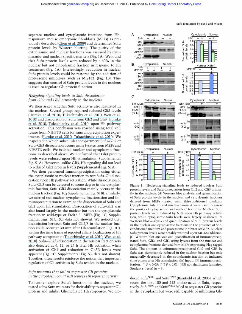
separate nuclear and cytoplasmic fractions from Hh-responsive mouse embryonic fibroblasts (MEFs) as pre-viously described (Chen et al. 2009) and determined Sufuprotein levels by Western blotting. The purity of thecytoplasmic and nuclear fractions was assessed by cyto-plasmic- and nuclear-specific markers (Fig. 1A). We foundthat Sufu protein levels were reduced by ;60% in thenuclear but not cytoplasmic fraction in response to Hhtreatment (Fig. 1A). Interestingly, reduction in nuclearSufu protein levels could be restored by the addition ofproteasome inhibitors (such as MG132) (Fig. 1B). Thissuggests that control of Sufu protein levels in the nucleusis used to regulate Gli protein function.
Hedgehog signaling leads to Sufu dissociationfrom Gli2 and Gli3 primarily in the nucleus
We then asked whether Sufu activity is also regulated inthe nucleus. Several groups reported reduced Gli3 levels(Humke et al. 2010; Tukachinsky et al. 2010; Wen et al.2010) and dissociation of Sufu from Gli2 and Gli3 (Humkeet al. 2010; Tukachinsky et al. 2010) upon Hh pathwayactivation. This conclusion was reached using total celllysate from NIH3T3 cells for immunoprecipitation exper-iments (Humke et al. 2010; Tukachinsky et al. 2010). Weinspected in which subcellular compartment Sufu–Gli2 orSufu–Gli3 dissociation occurs using lysates fromMEFs andNIH3T3 cells. We isolated nuclear and cytoplasmic frac-tions as described above. We confirmed that Gli3 proteinlevels were reduced upon Hh stimulation (SupplementalFig. S1A). However, unlike Gli3, Hh signaling did not leadto reduced Gli2 protein levels (Supplemental Fig. S1A).We then performed immunoprecipitation using either
the cytoplasmic or nuclear fraction to test Sufu–Gli disso-ciation upon Hh pathway activation. While dissociation ofSufu–Gli3 can be detected to some degree in the cytoplas-mic fraction, Sufu–Gli3 dissociation mainly occurs in thenuclear fraction (Fig. 1C; Supplemental Fig. S1B). Similarly,we carried out nuclear–cytoplasmic fractionation and im-munoprecipitation to examine the dissociation of Sufu andGli2 upon Hh stimulation. Dissociation of Sufu–Gli2 wasalso found largely in the nuclear but not the cytoplasmicfraction in wild-type or Ptch1�/� MEFs (Fig. 1C; Supple-mental Figs. S1C, S2; data not shown). We noticed thatdissociation between Sufu and Gli2/3 in the nuclear frac-tion could occur at 30 min after Hh stimulation (Fig. 1C),within the time frame of reported ciliary localization of Hhpathway components (Tukachinsky et al. 2010; Wen et al.2010). Sufu–Gli2/3 dissociation in the nuclear fraction wasalso detected at 6, 12, or 24 h after Hh activation whenactivation of Gli1 and reduction in Gli3R levels wereapparent (Fig. 1C; Supplemental Fig. S3; data not shown).Together, these results reinforce the notion that importantregulation of Gli activities by Sufu resides in the nucleus.
Sufu mutants that fail to sequester Gli proteinsin the cytoplasm could still repress Hh reporter activity
To further explore Sufu’s function in the nucleus, wetested a few Sufumutants for their ability to sequester Gliprotein and inhibit Gli-mediated Hh responses. We pro-
duced SufuN100 and SufuN212 (Barnfield et al. 2005), whichretain the first 100 and 212 amino acids of Sufu, respec-tively. SufuN100 and SufuN212 failed to sequesterGli proteinsin the cytoplasm but were still capable of inhibiting Gli-
Figure 1. Hedgehog signaling leads to reduced nuclear Sufuprotein levels and Sufu dissociation from Gli2 and Gli3 primar-ily in the nucleus. (A) Western blot analysis and quantificationof Sufu protein levels in the nuclear and cytoplasmic fractionsderived from MEFs treated with Shh-conditioned medium.Cytoplasmic tubulin and nuclear lamin A were used to assessthe purity of cytoplasmic and nuclear fractions. Nuclear Sufuprotein levels were reduced by 60% upon Hh pathway activa-tion, while cytoplasmic Sufu levels were largely unaltered. (B)Western blot analysis and quantification of Sufu protein levelsin the nuclear and cytoplasmic fractions in the presence of Shh-conditioned medium and proteasome inhibitor MG132. NuclearSufu protein levels were notably restored upon MG132 addition.(C) Western blot analysis and quantification of immunoprecip-itated Sufu, Gli2, and Gli3 using lysates from the nuclear andcytoplasmic fractions derived from MEFs expressing Flag-taggedSufu. The amount of coimmunoprecipitated Gli2 and Gli3 bySufu was significantly reduced in the nuclear fraction but onlymarginally decreased in the cytoplasmic fraction at indicatedtime points after Hh stimulation. (In) Input; (IP) immunoprecip-itation. (*) P < 0.05; (**) P < 0.01; (NS) not significant (unpairedStudent’s t-test) (n = 3).
Sufu regulation by p66b and Mycbp
GENES & DEVELOPMENT 2549
Cold Spring Harbor Laboratory Press on December 11, 2014 - Published by genesdev.cshlp.orgDownloaded from

mediated Hh transcriptional responses in various celllines, including MEFs, NIH3T3, C2C12, and C3H10T1/2(Supplemental Figs. S4, S5; data not shown). Readouts ofHh activity were determined by quantitative PCR (qPCR)analysis of Hh target gene expression (such as Gli1) ora standard Hh reporter assay (e.g., 8xGliBS-luc, in whicha firefly luciferase [luc] reporter is placed under thecontrol of Hh-responsive element 8xGliBS) (Sasaki et al.1997). This result confirms and extends previous findings(Barnfield et al. 2005) and highlights a critical function ofSufu in controlling Gli activity in the nucleus that isindependent of cytoplasmic sequestering. Taken to-gether, our findings point to an essential role of Sufu inthe nucleus in Hh signaling.
A proteomic approach to identify Sufu-interactingproteins uncovers p66b and Mycbp
In order to further understand themolecular mechanismsby which Sufu regulates Gli protein function, we tooka proteomic approach to identify Sufu-interacting pro-teins other than the three Gli proteins. We generateda stable cell line in MEFs that expresses 3xFlag-Sufu andperformed large-scale immunoprecipitation using anti-Flag antibodies (Fig. 2A). Endogenous Gli2 and Gli3 were
present in the immunoprecipitates, validating our ap-proach (Fig. 2B). Immunoprecipitates were analyzed bymass spectrometry (Jager et al. 2011; Altelaar et al. 2013).We focused on nonstructural proteins with overrepresen-tations of peptides identified through mass spectrometry.They were good candidates for direct interactions withSufu. We also treated MEFs expressing 3xFlag-Sufu withHh-conditioned medium and performed a similar pro-cedure to detect any changes in the levels and composi-tion of proteins that coimmunoprecipitated with Sufu.We selected a number of Sufu-interacting proteins with
overrepresentations of peptides in our proteomic analysisand systematically tested their ability to perturb Hhsignaling through overexpression and knockdown stud-ies. Two of the Sufu-interacting proteins, p66b andMycbp, showed the most striking and robust effects onHh signaling and thus are the primary focus of this study(Fig. 2C). p66b, encoded by Gatad2b (GATA zinc finger
Figure 2. A proteomic approach to identify Sufu-interactingproteins uncovers p66b and Mycbp. (A) Coomassie Blue-stainedgel of control and Sufu immunoprecipitates treated with mock-or Shh-conditioned medium. Distinct bands were detected andwere candidates for new Sufu-interacting proteins. Numbers atthe right indicate locations of protein size standards. Large-scaleimmunoprecipitation (IP) and mass spectrometry were per-formed to identify new Sufu-interacting proteins and Sufuphosphorylation sites. Mass spectrometric analysis was per-formed directly on immunoprecipitates or specific bands cutout from SDS-PAGE gels. Immunoprecipitation and mass spec-trometric analysis were repeated multiple times to eliminatenonspecific Sufu-binding proteins. (B) Western blot analysis ofSufu immunoprecipitates probed with anti-Sufu, anti-Gli2, andGli3 antibodies. Endogenous Gli2 and Gli3 were detected inSufu immunoprecipitates (but not in the control), suggestingthat a physiologically relevant protein complex was pulleddown. (C) Schematic diagram summarizing Sufu-interactingproteins and Sufu phosphorylation sites identified through massspectrometry analysis. Phosphorylation on S342 and S346 inSufu is consistent with previous reports (Chen et al. 2011). (D)Western blot analysis of proteins pulled down by Sufu fromlysates expressing Sufu and the indicated proteins, which wereepitope-tagged. Both p66b and Mycbp physically interacted withSufu by coimmunoprecipitation. Fu and Prc1 served as negativecontrols. (E, top panels) Western blot analysis of Sufu immuno-precipitates using lysates derived from MEFs expressing Flag-tagged Sufu. Endogenous p66b and HDAC1 were coimmuno-precipitated. In contrast, HDAC2 and RBBP7/4 could not bedetected in Sufu immunoprecipitates. (Bottom panels) Westernblot analysis of endogenous Sufu immunoprecipitated by ananti-Sufu antibody. p66b was coimmunoprecipitated by Sufu inwild-type MEFs but not in Sufu-deficient MEFs. p66b/Sufuinteraction was not altered by Hh stimulation (SupplementalFig. S6). (F–H) Immunofluorescence studies to assess the sub-cellular distribution of p66b and Mycbp. MEFs were transfectedor transduced with p66b- and Mycbp-expressing constructs.p66b and Mycbp localized to the nucleus (marked by DAPI) ofHh-responsive cells. Cytoplasmic expression of Mycbp was alsodetected. Acetylated (Ac)-tubulin marks the primary cilium.Interestingly, Mycbp immunoreactivity can also be detected atthe base of the cilium (white arrow).
Lin et al.
2550 GENES & DEVELOPMENT
Cold Spring Harbor Laboratory Press on December 11, 2014 - Published by genesdev.cshlp.orgDownloaded from
Jiang Xuan
Typewriter
3592 3730
Jiang Xuan
Typewriter
pPTC2012
Jiang Xuan
Typewriter
pPTC3896 pPTC3895

domain containing 2B), is a component of the nucleosomeremodeling and histone deacetylase (NuRD) complex (Laiand Wade 2011; Allen et al. 2013). The NuRD complexcontains (1) chromodomain helicase DNA-binding pro-tein 3/4 (CHD3/4), (2) histone deacetylase 1/2 (HDAC1/2),(3) methyl cytosine–guanosine (CpG)-binding domain2/3 (MBD2/3), (4) retinoblastoma-binding proteins 7/4(RBBP7/4), (5) metastasis-associated 1/2/3 (MTA1/2/3),and (6) p66a/b. The association between Sufu and p66boffers an opportunity to investigate whether Sufu controlsGli protein activity via the NuRD complex or the Sufu/p66b complex represents a novel nuclear function ofp66b independent of NuRD. Mycbp was originally dis-covered as a Myc-binding protein capable of stimulatingthe transcriptional activity of Myc (Taira et al. 1998), andits role in other cellular processes has not been explored.
Both p66b and Mycbp physically interact with Sufuand are found in the nucleus
To confirm the physical interaction between Sufu andp66b/Mycbp, we expressed epitope-tagged Sufu, p66b, andMycbp in HEK293T cells and performed immunoprecipi-tation using antibodies against the respective epitopes. Weshowed that Sufu immunoprecipitates contained p66b andvice versa (Fig. 2D; data not shown). In addition, antibodiesthat pulled down endogenous Sufu in wild-type MEFs alsocoimmunoprecipitated endogenous p66b (Fig. 2E, bottompanels; Supplemental Fig. S6). These results indicatephysical interactions between Sufu and p66b either di-rectly or indirectly through additional proteins. Interest-ingly, HDAC1 was coimmunoprecipitated with p66band Sufu, but Sufu did not pull down other knowncomponents of the NuRD repressor (Fig. 2E; Supplemen-tal Figs. S7, S8). We also showed that Sufu and Mycbpwere coimmunoprecipitated in cultured cells (Fig. 2D).This result indicates that Sufu interacts with Mycbpdirectly or indirectly.We examined the subcellular distribution of p66b and
found that p66b proteinwas largely confined to the nucleusof Hh-responsive cells (Fig. 2F), consistent with its pur-ported nuclear function. Similarly, Mycbp distributioncould be found in the nucleus as previously documented(Fig. 2G; Furusawa et al. 2002).
Overexpression of p66b impairs Hh responsesin cultured cells, while p66b knockdown resultsin enhanced Hh pathway activation in cell-based assays
As a first step toward understanding how p66b mediatesSufu function in Hh signaling, we overexpressed p66b inHh-responsive cells (such as NIH3T3 and MEFs) andassessed its effect on Hh pathway activity. Readouts ofHh activity were determined by a standard Hh reporterassay or transcript levels of Hh target genes describedabove. Cotransfection of Gli1 or Gli2 together with Hhreporters resulted in Hh reporter activation (Fig. 3A). Wefound that coexpression of p66b with Gli1 or Gli2 in thisassay severely inhibited Gli-mediated Hh reporter acti-vation or Hh target gene expression, an effect comparablewith Sufu-mediated inhibition of Hh pathway activity
(Fig. 3A; Supplemental Fig. S9). Other signaling pathwayssuch as Wnt signaling, assayed by the TOPFlash reporter(Molenaar et al. 1996), were unaffected by p66b over-expression (Fig. 3F), suggesting that p66b does not exertgeneral inhibition of reporter activities. Conversely, shRNA-mediated knockdown (Hannon 2003) of p66b (SupplementalFigs. S10–S12) resulted in enhanced Hh responses. Whilep66b knockdown had a modest effect on Hh reporteractivity in the absence of the Hh ligand, reduction in p66benhanced Hh responses upon Hh stimulation (Fig. 3B;Supplemental Fig. S13). These results are consistent witha negative role of p66b in controlling Hh signaling (Supple-mental Figs. S14, S15).
Overexpression of Mycbp facilitates Hh responses,while knockdown of Mycbp compromises Hhresponses
We took a similar approach to further understand howMycbp controls Hh signaling by overexpressing Mycbp inHh-responsive cells and assessing its effect on Hh path-way activity. Coexpression of Mycbp with Gli1 or Gli2 inthis assay significantly promoted Gli-mediated Hh re-porter activation or Hh target gene expression (Fig. 3C;Supplemental Fig. S9). Again, Wnt responses were un-altered by Mycbp overexpression (Fig. 3F). These findingsare consistent with a positive role of Mycbp in Hhpathway activation. Since p66b andMycbp exert oppositeeffects on Hh responses, we anticipate that p66b andMycbp will antagonize each other in controlling Hhpathway activity. Indeed, when p66b and Mycbp werecotransfected in wild-type cells, Mycbp could partiallyreverse the inhibitory effects of p66b on Gli transcrip-tional activation, likely depending on the Mycbp/p66bratio (Supplemental Figs. S15, S16).We also determined the functional consequence of
loss of Mycbp through shRNA-mediated knockdown(Hannon 2003) in Hh-responsive cells (SupplementalFig. S10). We showed that Hh pathway activationassayed by Hh reporter activity was severely com-promised when Mycbp was knocked down (Fig. 3D).Moreover, Hh target gene expression such as Gli1 wasgreatly reduced by Western blotting (Fig. 3E). This furthersupports a positive role of Mycbp in enhancing Hhpathway activity.
Expression or knockdown of p66b and mycbpin zebrafish perturbs Hh signaling, consistentwith their respective roles as negative and positiveregulators of Hh signaling
To assess the in vivo function of p66b and mycbp, weused the zebrafish system as a readout of their in vivoactivity. We injected p66bmRNA into zebrafish embryosand investigated the phenotypic consequences associatedwith p66b overexpression. We focused on the developingsomites and fin buds, where Hh pathway perturbationleads to well-characterized phenotypes (Lewis et al. 1999;Neumann et al. 1999). We found that p66b overexpres-sion engendered U-shaped somites and reduced fin buds(Fig. 4A,B; Supplemental Fig. S17; data not shown), both
Sufu regulation by p66b and Mycbp
GENES & DEVELOPMENT 2551
Cold Spring Harbor Laboratory Press on December 11, 2014 - Published by genesdev.cshlp.orgDownloaded from

of which are associated with disruption of Hh signaling.Moreover, Hh target gene expression such as ptch1 wasalso reduced (or lost) in fin buds when p66b was overex-pressed (Fig. 4C,D,F). Conversely,morpholino (MO)-mediatedknockdown of p66b enhanced ptch1 expression (Fig. 4C,E,H). These results support our hypothesis that p66brepresses Hh responses.Similarly, mycbp mRNA or MO was injected into
zebrafish embryos, and Hh responses—including pheno-typic outcomes and Hh target gene expression—weredetermined. We showed that overexpression of mycbp inzebrafish embryos resulted in an increase in Hh target
gene expression (such as ptch1) in fin buds (Fig. 4C,E,G;Supplemental Fig. S17). In contrast, ptch1 expression wasreduced in mycbp morphants (Fig. 4C,D,I). This is consis-tent with a positive role of mycbp in Hh signaling. Takentogether, these studies provide in vivo evidence to supportp66b and Mycbp as new regulators of vertebrate Hhsignaling.
p66b and Mycbp function downstreamfrom Ptch1/Smo/cilia to mediate Sufu activity
Having established the effects of p66b and Mycbp on Hhsignaling, we performed similar assays using cell lines de-
Figure 3. p66b negatively regulates Hh responses, while Mycbp positively regulates Hh signaling in cultured cells. (A) Assessment ofp66b activity on Gli-mediated Hh responses using Hh reporter assays in which the 8xGliBS-luc reporter served as the readouts of Hhsignaling. Overexpression of p66b in NIH3T3 cells abolished Gli-induced Hh reporter activity in a manner similar to Sufu. Thissupports a negative role of p66b in controlling Hh signaling. (B) shRNA-mediated knockdown of p66b in MEFs led to increased Hhresponses. In this experimental setting, submaximal Hh stimulation was used. p66b knockdown had no apparent effect on Hhresponses when cells received maximal Hh stimulation (Supplemental Fig. S13). Note that knockdown of p66b in unstimulated cellsonly caused a modest increase in Hh activity. (C) Assessment of Mycbp activity using Hh reporter assays. Overexpression of Mycbp inNIH3T3 cells potentiated Gli-mediated Hh reporter activity, but Mycbp’s positive effects on Hh signaling could be reversed by thepresence of Sufu. (D) shRNA-mediated knockdown of Mycbp in MEFs led to decreased Hh target gene expression and compromised Hhresponses. Knockdowns of Septin 11 and Prmt5 were used as controls, and no apparent effect on Hh responses was observed. (E)Western blot analysis of Gli1 protein levels in MEFs in which Mycbp was knocked down by shRNA. Gli1 was barely induced by Hhstimulation in the absence of Mycbp, consistent with a positive role of Mycbp in Hh responses. (F) p66b and Mycbp did not affect Wntresponses using the TOPFlash reporter when NIH3T3 cells were treated with Wnt3a-conditioned medium. Note that Gli1/2 werecotransfected with p66b or Mycbp to assess the effects of p66b/Mycbp on Hh signaling. This is because expression of either a negativeor positive component in the Hh pathway (for example, Sufu and Smo, respectively) has little effect on Hh signaling in the steady state(shown in C). In contrast, in the perturbed state (e.g., by expressing Gli), expression of Hh components exerts obvious effects on Hhsignaling. Otherwise, expression of p66b and Mycbp, like Sufu and Smo, has no significant effect on Hh signaling in Hh-responsive cellsin the steady state. (*) P < 0.05; (**) P < 0.01; (NS) not significant (unpaired Student’s t-test) (n = 3).
Lin et al.
2552 GENES & DEVELOPMENT
Cold Spring Harbor Laboratory Press on December 11, 2014 - Published by genesdev.cshlp.orgDownloaded from
Jiang Xuan
Typewriter
pPTC1473 pPTC3985
Jiang Xuan
Typewriter
pPTC920
Jiang Xuan
Typewriter
pPTC2012
Jiang Xuan
Typewriter
pPTC3985
Jiang Xuan
Typewriter
pPTC3730
Jiang Xuan
Typewriter
pPTC3730 pPTC3985
Jiang Xuan
Typewriter
All expression of Gli1:pPTC1473 of Gli2: pPTC920 of Sufu: pPTC2012 of p66beta: pPTC3985 of Mycbp: pPTC3730

ficient in various Hh pathway components (Chen et al. 2009)to reveal the relationship between p66b, Mycbp, and otherHh components. We found that p66b failed to inhibit Gli-mediated Hh reporter activity in Sufu-deficientMEFs (Fig. 5;Supplemental Fig. S18), suggesting that p66b activitydepends on Sufu. In contrast, p66b was capable ofinhibiting Gli-mediated Hh reporter activity in Ptch1-or Smo-deficient MEFs (Fig. 5; Supplemental Fig. S18),placing p66b downstream from Ptch1 and Smo in con-trolling Hh signaling. Furthermore, p66b also inhibitedGli-mediated Hh reporter activity in Kif3a-deficientMEFs (Fig. 5; Supplemental Fig. S18) in which primarycilia fail to form and Hh signaling cannot be transduced.This indicates that p66b activity does not depend on theprimary cilium.Mycbp modestly promoted Gli-mediated Hh reporter
activity in Sufu-deficient MEFs (Fig. 5). We speculate thateither Sufu or Gli can recruit Mycbp, and Mycbp remainsinactive in the absence of the Hh ligand; Mycbp activity isrequired only when theHh signal is transduced. Consistentwith this, we found that knockdown of Mycbp in Sufu-deficient cells blunts Hh target gene expression (Supple-mental Fig. S19). In addition, we found that full-length Gliproteins can interact with Mycbp in the absence of Sufu(Supplemental Fig. S20).Mycbp was capable of stimulating Gli-mediated Hh
reporter activity in Ptch1- or Smo-deficient MEFs (Fig. 5),suggesting that Mycbp functions downstream from Ptch1and Smo. In addition,Mycbp also potentiated Gli-mediatedHh reporter activity in Kif3a-deficient MEFs (Fig. 5), in-dicating that Mycbp activity is independent of the primarycilium. Finally, overexpression of p66b andMycbp does notinfluence ciliary localization of Gli2 (data not shown).Together, these findings show that p66b and Mycbpfunction downstream from Ptch1/Smo/cilia and likely ata similar step in the Hh pathway in mediating Sufu’snuclear activity.
Protein complex formation occurs between Sufu, Gli,and p66b/Mycbp
Since Sufu interacts with both p66b and Gli, we surmisethat Sufu, p66b, and Gli may form a multicomponentprotein complex, and Sufu bridges the interactions be-tween p66b and Gli. Consistent with this model, Gli2immunoprecipitates from MEFs expressing 3xFlag-Gli2contained both Sufu and p66b (Fig. 6A; Supplemental Fig.S21), while coimmunoprecipitation of p66b–Gli2 failed tooccur in Sufu�/� MEFs that express 3xFlag-Gli2 (Fig. 6B).Similar interactions between full-length Gli3, Sufu, andp66b were obtained in MEFs expressing 3xFlag-Gli3 (Fig.6A,B; Supplemental Fig. S21). This suggests that Sufu canrecruit p66b to control Gli activity. Using a parallelapproach, we found that Mycbp bound to full-length
Figure 4. Expression of p66b in zebrafish inhibits Hh signalingwhile expression of mycbp enhances Hh signaling. (A) Lateralview of chevron-shaped somites in wild-type zebrafish embryosat 3.5 d post-fertilization. (B) Injection of p66b mRNA resultedin U-shaped somites, consistent with disruption of Hh signaling.(C–E) Whole-mount in situ hybridization to ptch1 in wild-typezebrafish embryos and embryos injected with either p66b/
mycbp mRNA or p66b/mycbp MO. Embryos were collected at36 h post-fertilization (hpf). ptch1 expression in the fin buds wassignificantly reduced in embryos injected with p66b mRNA ormycbp MO. In contrast, injection of mycbp mRNA or p66b MOincreased ptch1 expression. Since ptch1 expression is similar inembryos receiving p66b mRNA/mycbp MO (ptch1 repression)or mycbp mRNA/p66b MO (ptch1 up-regulation), only onerepresentative image of each is shown to indicate changes inptch1 expression. (F–I) Classification of ptch1 expression levelsin fin buds by in situ hybridization in wild-type embryos andembryos injected with p66b/mycbp mRNA or p66b/mycbp MOas indicated. The number (n) of embryos analyzed is specified.Some variation in ptch1 expression levels in the uninjectedembryos was noted. Compared with uninjected controls,a higher percentage of embryos injected with p66b mRNAexhibited weak ptch1 expression (F), while ptch1 expressionwas enhanced in a significant proportion of p66b morphants (G).Overexpression of p66b (n = 107) resulted in, on average, 27.2%fewer ptch1-expressing embryos than uninjected control em-bryos (n = 191). Conversely, there were 35.0% more ptch1-expressing embryos in the p66b MO-injected group (n = 136)compared with the uninjected control group (n = 65). P-value <
0.0001 (two-proportion z-test) for injection with either p66b
mRNA or p66bMO. The trend of ptch1 expression was reversedin embryos injected with mycbp mRNA or mycbp MO. A largefraction of embryos expressing exogenous mycbp mRNAshowed strong ptch1 expression (H), and mycbp knockdownsignificantly reduced ptch1 expression (I). Overexpressingmycbp (n = 208) resulted in 33.9% more embryos expressingptch1 than uninjected control embryos (n = 303). In contrast,knockdown of mycbp (n = 60) led to 76.7% fewer embryosexpressing ptch1 than uninjected control embryos (n = 46). P-value < 0.0001 (two-proportion z-test) for injection with eithermycbpmRNA ormycbpMO. Off-target effects of MO-mediatedknockdown pose a potential concern (Schulte-Merker andStainier 2014), and future investigations using genome-editingtechnology (Harrison et al. 2014), such as CRISPR/Cas orTALENs, will complement these studies.
Sufu regulation by p66b and Mycbp
GENES & DEVELOPMENT 2553
Cold Spring Harbor Laboratory Press on December 11, 2014 - Published by genesdev.cshlp.orgDownloaded from

Gli2 or Gli3 protein in addition to Sufu (Fig. 6C), providingevidence to support the presence of a Sufu/Mycbp/Gliprotein complex. It is interesting to note that no signifi-cant interaction was detected between Gli repressors andp66b or Mycbp (Supplemental Fig. S21).Since p66b protein levels do not appear to alter upon
Hh stimulation (Fig. 2E), we envision two possible sce-narios for p66b action. Hh pathway activation could leadto dissociation of the Sufu/p66b protein complex fromGli, thus terminating the inhibitory effects on Gli pro-teins. Alternatively, Hh signaling may result in disruptionof the Sufu/p66b protein complex. To distinguish betweenthese possibilities, we performed coimmunoprecipitationexperiments using cell lysates from Hh-responsive cellsexpressing p66b, Sufu, and Gli. Cells were also treated withHh-conditioned medium or agonists to activate the Hhpathway. We found that p66b coimmunoprecipitatedwith Sufu without Hh stimulation, and p66b/Sufu in-teraction was not altered by Hh stimulation (Fig. 2E;Supplemental Fig. S6). Moreover, p66b dissociates fromGli2 (but not Sufu) upon Hh pathway activation in coim-munoprecipitation experiments (Supplemental Fig. S22). Inconjunction with our demonstration of Sufu dissociationfrom Gli2/3 in the nucleus upon Hh activation (Fig. 1C;Supplemental Fig. S1), these results suggest that Sufumediates the interaction between p66b and Gli, and theentire Sufu/p66b protein complex is released from Gliwhen the Hh pathway is activated.
Sufu and p66b occupy the promoter of Hh-responsivegenes in a Hh-dependent manner
To further understand how Sufu and p66b control Hhsignaling, we asked whether Sufu and p66b could occupy
the promoter of Hh-responsive genes, many of whichcontain canonical GliBSs. This would provide mechanis-tic insight into how p66b modulates Sufu activity incontrolling Hh signaling.We first assessed the ability of Sufu and Gli proteins to
recognize the GliBS. We used double-stranded oligonu-cleotides that contain a GliBS (denoted OligoGliBS) (Panet al. 2006) for immunoprecipitation. Oligonucleotidesthat contain the mutant GliBS (OligoΔGliBS) were used asa control. We showed that endogenous Sufu, Gli2, Gli3,and Gli3R were immunoprecipitated by OligoGliBS butnot OligoΔGliBS in the absence of Hh ligand stimulation(Fig. 6D). This provides strong evidence to support thenotion that a Sufu–Gli protein complex can interact withthe GliBS. Interestingly, Sufu failed to be immunoprecip-itated by OligoGliBS in Gli2�/�; Gli3�/� MEFs (Fig. 6E;Zeng et al. 2010) in which all three Gli proteins areabsent, since Gli1 expression relies on active Hh signal-ing via Gli2/3 (Bai et al. 2004). This is consistent with theidea that Sufu association with the GliBS depends on Gliproteins. Hh stimulation led to decreased associationbetween Sufu and the GliBS as well as between Gli3Rand the GliBS, while the amount of Gli2 and full-lengthGli3 pulled down by the GliBS was significantly increased(Fig. 6D). This supports the notion of Sufu dissociationfromGli proteins upon Hh pathway activation. Given theinteractions between Sufu, p66b, and Gli, these findingssuggest that Sufu can bridge p66b and Gli interactions onHh-responsive promoters.To further test this idea, we performed chromatin
immunoprecipitation (ChIP) assays (Collas 2011) on Gli1,Gli2, Sufu, and p66b. Epitope (e.g., 3xFlag)-tagged Gli1,Gli2, Sufu, and p66b were expressed in Hh-responsivecells, and ChIP analysis was performed on known Hh
Figure 5. p66b and Mycbp function down-stream from Ptch1, Smo, and the primarycilium to mediate Sufu activity. Placementof p66b and Mycbp in the Hh pathway usingcell-based assays. Expression of p66b failed toaffect Hh responses assessed by the 8xGliBS-
luc reporter assays in Sufu-deficient MEFs.Sufu-deficient MEFs exhibit strong (albeit sub-maximal) Hh pathway activation in the ab-sence of Hh ligands. This is consistent witha model in which the function of p66b de-pends on Sufu. Mycbp modestly promotedGli-mediated Hh reporter activity in Sufu-deficient MEFs. In contrast, p66b inhibited,while Mycbp potentiated, Gli-mediated Hhresponses in Smo, Ptch1, or Kif3a mutantMEF lines in a pattern similar to that ob-served in wild-type MEFs. This suggests that
p66b and Mycbp function downstream from membrane receptors Ptch1 and Smo and independently of the primary cilium. Note thatPtch1-deficient MEFs exhibit strong or even maximal Hh pathway activation in the absence of Hh ligands; Hh reporters can still beactivated in Ptch1-deficient MEFs if Hh pathway components are exogenously overexpressed. By comparison, Smo-deficient or Kif3a-deficient MEFs are defective in Hh signal reception and transduction, and 8xGliBS-luc reporters can be activated only by exogenousnuclear Gli proteins in these cell lines. Gli1/2 were cotransfected with p66b or Mycbp into Smo- and Kif3a-deficient MEFs to assess theeffects of p66b/Mycbp on Hh signaling. Otherwise, expression of p66b and Mycbp has no significant effect on Hh signaling in theuninduced state as discussed in the legend for Figure 3. (*) P < 0.05; (NS) not significant (unpaired Student’s t-test) (n number isindicated). Similar results and conclusions were obtained by assessing the expression of endogenous Hh targets as the readouts of Hhsignaling (Supplemental Fig. S18).
Lin et al.
2554 GENES & DEVELOPMENT
Cold Spring Harbor Laboratory Press on December 11, 2014 - Published by genesdev.cshlp.orgDownloaded from
Jiang Xuan
Typewriter
Vector is rpptc588 (pEGFP-N1) Mycbp is pPTC3730 p66b is pPTC3985, Gli1 is pPTC1473, GLi2 is pPTC920

target genes (e.g., Ptch1, Gli1, and Hhip) (Goodrich et al.1996; Lee et al. 1997; Chuang and McMahon 1999), thepromoter of which contains canonical GliBSs. We showedthat Gli1, Gli2, and Sufu proteins were enriched on thepromoters of Hh-responsive genes (Fig. 6F; SupplementalFig. S23), consistent with results from immunoprecipita-tion studies usingGliBS oligonucleotides. Importantly, p66bwas also enriched on Hh-responsive promoters (Fig. 6F;Supplemental Fig. S23). This was the first demonstrationof the presence of Sufu and p66b on the promoter of Hh-responsive genes. p66b’s chromatin association was abol-ished in Sufu-deficient MEFs (Supplemental Fig. S23),again consistent with the model in which Sufu recruitsp66b to Hh target gene promoters. These results suggestthat Sufu, Gli, and p66b are present at the GliBSs of Hh-responsive genes prior to Hh pathway activation. In thisway, Sufu recruits p66b to inhibit Gli-mediated Hh geneexpression. Thismodel predicts that Hh pathway activation
would relieve the inhibition of Sufu/p66b on Gli proteinson Hh target gene promoters. Indeed, ChIP analysisrevealed diminished association of both Sufu and p66bwith Hh promoters upon Hh stimulation (Fig. 6F; Sup-plemental Fig. S23). These studies yield novel insightsinto the dynamic interactions of Gli, Sufu, and p66b onthe promoter in the process of Hh pathway activation.
Enhanced interactions between Mycbp and Gliduring Hh signal transduction
Our studies show that p66b inhibits Hh responses, whileMycbp promotesHh responses. Since both p66b andMycbpinteract with Sufu, this led to our hypothesis that Mycbpcan potentiate Gli activity once Hh pathway activationremoves Sufu/p66b from Gli proteins. To further test thisidea, we studied the interaction between Mycbp, Sufu, andGli proteins during Hh pathway activation. We found thatMycbp dissociated from Sufu upon Hh treatment, whileMycbp interaction with full-length Gli2 and Gli3 wasenhanced upon Hh pathway activation (Fig. 6C).
Figure 6. Complex formation between Sufu, p66b, Mycbp, andGli proteins and their dynamic interactions on the promoters ofHh-responsive genes. (A) Western blot analysis of immunopre-cipitates using lysates from MEFs expressing Flag-tagged Gli2or Gli3. Endogenous p66b and Sufu were coimmunoprecipi-tated, consistent with the formation of a Sufu/p66b/Gli proteincomplex. (B) Western blot analysis of immunoprecipitates usinglysates from Sufu-deficient MEFs expressing Flag-tagged Gli2 orGli3. Endogenous p66b failed to be coimmunoprecipitated,suggesting that Sufu bridges the interaction between p66b andGli protein. (C) Western blot analysis of immunoprecipitatesusing lysates from MEFs expressing Flag-tagged Mycbp. Endog-enous Sufu, Gli2, and Gli3 were coimmunoprecipitated, imply-ing the production of a Sufu/Mycbp/Gli protein complex.Moreover, Mycbp dissociated from Sufu upon Hh treatment,while Mycbp interaction with full-length Gli2 and Gli3 wasenhanced upon Hh pathway activation. (*) P < 0.05; (**) P < 0.01;(NS) not significant (unpaired Student’s t-test) (n = 3). Mycbp/Gli3 interaction was increased upon Hh stimulation, butvariations in immunoprecipitations affected the calculatedstatistical value. (D) Immunoprecipitation of Gli2, Gli3, andSufu using oligonucleotides that contain a canonical GliBS orcontrol oligonucleotides in which the GliBS is mutated (denotedas ΔGliBS). Sufu, full-length (FL) Gli2, full-length Gli3, andGli3R were immunoprecipitated by a GliBS without Hh stim-ulation but not by the control ΔGliBS. Hh stimulation led to anincreased binding of Gli2 and Gli3 to the GliBS, while associ-ation of Sufu or Gli3R with the GliBS was weakened. (E) Sufufailed to be immunoprecipitated by the GliBS in Gli2�/�; Gli3�/�
MEFs, indicating that Sufu binding to the GliBS is dependent onGli proteins. (F) ChIP analysis of Gli1, Gli2, Sufu, p66b, andMycbp on the promoter of Hh-responsive genes such as Ptch1.Gli1, Gli2, Sufu, and p66b proteins were enriched on the Ptch1promoter by ChIP. Hh treatment led to the reduced presence ofSufu and p66b on the Ptch1 promoter, while binding of Mycbp tothe Ptch1 promoter was enhanced. Interestingly, enhanced bindingof Mycbp to Hh target gene promoters was abolished in Gli2�/�;Gli3�/� MEFs (Supplemental Fig. S24), suggesting that the pro-moter occupancy of Mycbp is dependent on Gli proteins. (*) P <
0.05 (unpaired Student’s t-test) (n = 3).
Sufu regulation by p66b and Mycbp
GENES & DEVELOPMENT 2555
Cold Spring Harbor Laboratory Press on December 11, 2014 - Published by genesdev.cshlp.orgDownloaded from

We also performed ChIP analysis of Mycbp on the pro-moters of Hh target genes. In the presence of Hh signaling,Mycbp was enriched at the Hh promoters (Fig. 6F; Supple-mental Fig. S23), and enhanced Mycbp binding was abol-ished in Gli2�/�; Gli3�/� MEFs (Supplemental Fig. S24).This is in contrast to reduced binding of Sufu or p66b toHh promoters by Hh pathway activation (Fig. 6F; Supple-mental Fig. 23). These findings suggest that Hh signalingleads to dissociation of Sufu/p66b from Gli, allowingMycbp/Gli to activate Hh target gene expression.
Discussion
Our studies on Sufu-interacting proteins p66b and Mycbpoffer novel insight into the regulation of Hh target geneexpression by Sufu and Gli in the nucleus. In particular,p66b and Mycbp modulate the process of how Sufucontrols Gli protein function in the nucleus (Fig. 7). Ourinvestigation thus provides a new framework for under-standing Hh target gene expression and the production ofgraded Hh responses in diverse tissues, a key unresolvedissue in Hh signaling.
The multiple roles of Sufu in controlling Gli proteinfunctions
Extensive studies on Sufu uncovered its role in regulatingGli activities at several subcellular locales and multiplelevels. Sufu can sequester Gli proteins (Ding et al. 1999;Kogerman et al. 1999; Murone et al. 2000; Barnfield et al.2005), regulate Gli protein levels (Chen et al. 2009; Jiaet al. 2009; Wang et al. 2010), promote the production ofGli repressors, and inhibit the generation of Gli activators(Humke et al. 2010; Tukachinsky et al. 2010). Physicalsequestration of Gli proteins by Sufu in the cytoplasm istraditionally viewed as a major mechanism of inhibitingGli activities. Recently, Sufu was shown to control Gliprotein levels through its antagonistic effects on Spop- orNumb-mediated Gli ubiquitination and degradation (Chenet al. 2009;Wang et al. 2010;DiMarcotullio et al. 2011; Linet al. 2014). In this way, Sufu preserves a pool of Gli proteinthat would be available to activate Hh target gene expres-sion once the Hh signal is transduced. Interestingly, Sufu–Gli association gained a new level of complexity in light ofthe proposal in which Sufu inhibits Gli activity on theprimary cilium (Humke et al. 2010; Tukachinsky et al.2010). In this model, Hh signaling leads to Sufu–Glidissociation on the cilium, resulting in Gli activationand the production of a labile form of Gli, which isproposed to be derived from phosphorylated Gli proteins(Humke et al. 2010; Niewiadomski et al. 2014).The relative contributions of various effects of Sufu to
Gli protein activities have not been accurately assessed. Infact, it has not been unambiguously demonstrated whereSufu controls Gli activities. This is largely due to technicaldifficulty in selective inactivation of endogenous Sufu ina particular subcellular compartment and determining itsfunctional consequence. The notion of a labile active formofGli is consistentwith prior work inDrosophila onCi, theGli homolog (Ohlmeyer and Kalderon 1998). However, it
should be noted that the addition of Hh ligands does notlead to reduced Gli2 protein levels (Supplemental Fig. S1;Kim et al. 2009). Identifying Sufu mutants that affectunique aspects of Gli protein activities will provide criticaltools to reveal the contributions of various Sufu/Gli in-teractions to Hh signaling.While Sufu–Gli dissociation can be detected to some
extent in the cytoplasm, the main site of Sufu–Gli disso-ciation seems to reside in the nucleus. The extent ofdissociation between Gli2/3 and Sufu using nuclear frac-tions seems to be comparable with that using whole-celllysates (Humke et al. 2010). We speculate that changes in
Figure 7. A model of how a p66b/Sufu complex inhibits Glitranscriptional activity and a Mycbp/Gli complex enhances Glitranscriptional activity in the nucleus. In the absence of Hhsignaling, Sufu-bound Gli (such as Gli2) is inactive, since Sufurecruits p66b (and other proteins) to block Gli-mediated Hhtarget gene expression. Hh pathway activation leads to dissoci-ation of Sufu/p66b from Gli, releasing Gli inhibition andpriming Gli protein for activating Hh targets. p66b may recruitcomponents of the NuRD repressor complex or other players toinhibit Gli-mediated Hh responses. We propose that Mycbp isrecruited to the Sufu/Gli protein complex in the absence of Hhsignaling but remains inactive. How Mycbp is inactivated is notknown. Upon Hh pathway activation, Sufu/p66b dissociationfrom Gli (likely Gli1/2) enhances Mycbp/Gli interaction andenables Mycbp to promote Gli protein activity and Hh targetgene expression. The molecular mechanism by which Mycbpenhances Hh responses is unclear. Post-translational modificationsof Sufu, Gli, p66b, and Mycbp could influence their interactionsand confer important properties of Hh responses. It is also possiblethat other activators/repressors also control nuclear Gli activityand exhibit functional redundancy with p66b/Mycbp. (R) Repres-sor; (FL) full-length.
Lin et al.
2556 GENES & DEVELOPMENT
Cold Spring Harbor Laboratory Press on December 11, 2014 - Published by genesdev.cshlp.orgDownloaded from

Sufu and Gli distribution upon Hh activation are dynamicand quantitative, not ‘‘all or none.’’ In this model,a significant fraction of Sufu and Gli still associate witheach other in the cytoplasm, perhaps as a reservoir.Consistent with this idea, Sufu and Gli accumulate inthe primary cilium upon Hh stimulation, and, in fact,Sufu/Gli could still be detected in the cilium 24 h afterHh treatment.Perhaps Sufu–Gli dissociation in different subcellular
locales serves to execute Sufu’s multiple functions in Hhsignaling. For instance, Sufu–Gli dissociation in thecytoplasm/cilium may blunt the generation of Gli re-pressors and lead to the production of Gli activators,while nuclear Sufu–Gli dissociation could free Gli pro-teins of their transcriptional repressors.X-ray crystallographic structures of Sufu in a complex
with a short Gli peptide (containing the Sufu-bindingSYGH motif) revealed a clamp-like structure of Sufu thatinteracts with the Gli peptide (Cherry et al. 2013; Zhanget al. 2013). This raised the possibility that both theN-terminal and C-terminal domains of Sufu are requiredfor Gli binding. However, previous studies have reportedphysical interactions between Sufu truncation mutants(e.g., the N-terminal fragment of Sufu) and Gli proteins(Merchant et al. 2004). Moreover, Gli2 (ΔSYGH) can stillbind Sufu (Santos and Reiter 2014), suggesting the pres-ence of multiple Sufu-binding domains in Gli2. It ispossible that Sufu in a multiprotein complex in its nativeenvironment would display complex dynamic behaviors,and multiple domains of Sufu and Gli can interact witheach other independently. In addition, Gli2 (ΔSYGH) isnot sequestered in the cytoplasm and instead is enrichedin the nucleus but does not lead to Hh pathway hyper-activation (Santos and Reiter 2014). This suggests thatSufu can inhibit Gli2 (ΔSYGH) activity in the nucleus,although it does not sequester Gli2 (ΔSYGH) in thecytoplasm. This is consistent with our finding that thesequestering function of Sufu can be separated from itsability to inhibit Gli-dependent transcription as revealed bythe N-terminal fragments of Sufu (SufuN100 and SufuN212).While SufuN100 and SufuN212 can bind Gli proteins, albeitless strongly than Sufu (Supplemental Fig. S5), and arepotent in inhibiting Hh responses in cell-based assays, it ispossible that overexpression augmented their inhibitoryeffects. The function of SufuN100 and SufuN212 would needto be assessed by introducing these mutants in vivothrough gene targeting or genome editing.Whether Sufu plays a key role in controlling Gli protein
functions on Hh-responsive genes in the nucleus has notbeen extensively studied (Cheng and Bishop 2002). In thisregard, our study represents a major step toward un-derstanding Sufu’s nuclear function. While we focusedon p66b and Mycbp in this study, other Sufu-interactingproteins discovered from our proteomic approach couldshed light on Sufu function.
Regulation of Gli activity by Sufu and p66b
We showed that p66b negatively regulates Hh signalingin cell-based assays and zebrafish, and p66b relies on Sufu
to mediate its inhibitory action on Hh signaling. p66b isa member of the NuRD repressor complex (Lai and Wade2011; Allen et al. 2013), suggesting that p66b functions inthe nucleus. This is further supported by the nucleardistribution of p66b, its presence on Hh promoters, andthe independence of p66b activity on Ptch1/Smo and theprimary cilium.When p66b knockdown cells were treated with a high
dose of Hh, induced Hh activity was similar to that inwild-type cells (Supplemental Fig. S13). We reason thatthis was because Sufu dissociated from Gli (this occurslargely in the nucleus in our model) using high doses ofHh ligand. Under this condition, the level of p66b, whosefunction in Hh signaling depends on Sufu, wouldmake nodifference onHh responses. Hence, p66b knockdown exertslittle effect on Gli if Sufu/Gli dissociation has alreadyoccurred. In contrast, when cells are subjected to submax-imal Hh stimulation, Sufu is not expected to completelydissociate from Gli in the nucleus. As expected, increasedHh responses were observed in p66b knockdown cells.We showed that HDAC1 was coimmunoprecipitated
with p66b and Sufu, but Sufu did not pull down otherknown components of the NuRD repressor. It is possiblethat interactions between p66b/Sufu and other NuRDcomponents are weak or transient. However, it is alsopossible that the p66b/Sufu interaction represents a novelfunction of p66b independent of the NuRD complex.Interestingly, HDAC1 also exhibits NuRD-independentfunction (Canettieri et al. 2010). Whether p66b recruitsadditional components or relies on post-translationalmodifications of p66b and Gli proteins to control Hhsignaling is unknown and would rely on future biochem-ical studies.The phenotypes associated with the loss of p66b in
mice are currently unavailable and would require theproduction of mutant mice and phenotypic analysis.Nevertheless, it is interesting to note that knockout miceof p66a die at 9.5 d post-coitus (dpc) (Marino and Nusse2007). The observation that the loss of p66a does not leadto global cell death at the earliest stage of embryonicdevelopment implies that components of the NuRD com-plex could be involved in specific cellular processes orinteract with particular pathways during development. It isalso plausible that functional redundancy exists betweenp66b and other components, and a better mechanisticunderstanding of p66b function is required for properinterpretation of the phenotypes.
Regulation of Gli activity by Sufu and Mycbp
We showed that Mycbp enhances Hh signaling in culturedcells and zebrafish. Mycbp was initially identified as aMyc-interacting protein and is known to enhance the activity ofMyc (Taira et al. 1998).Mycbp displays cell cycle-dependentshuttling between the nucleus and the cytoplasm (Furusawaet al. 2002). In addition, overexpressed Mycbp can bedetected in both the nucleus and cytoplasm. While Mycbpis present on the promoter of Hh-responsive genes, wecannot rule out its potential roles in cytoplasmic Hhsignaling. In this regard, it is interesting to note that Mycbpcould also be detected at the base of the primary cilium
Sufu regulation by p66b and Mycbp
GENES & DEVELOPMENT 2557
Cold Spring Harbor Laboratory Press on December 11, 2014 - Published by genesdev.cshlp.orgDownloaded from

(Fig. 2H). Mycbp has been previously shown to bind tomodulators of PKA and suppress PKA activity (Furusawaet al. 2002). PKA negatively regulates Hh signaling ina cilium-dependent manner (Tuson et al. 2011). WhetherMycbp exerts its activity on Hh signaling at the ciliumbase is unknown, although overexpression of Mycbp failsto perturb ciliary distributions of Gli proteins.Studies in cultured cells and zebrafish support Mycbp’s
role as a novel positive regulator of Hh signaling. Similarly,insight into the in vivo function of Mycbp in mammalianHh signaling would need to await loss-of-function studiesin mice. In this case, conditional inactivation of Mycbpmay be necessary to overcome its early lethality causedby its pleiotropic effects unrelated to Hh signaling. Alterna-tively, double-mutant analysis may be required if func-tional redundancy exists between Mycbp and othercomponents. Finally, mapping the functional domainsof p66b and Mycbp could facilitate the production ofmutants that have selective disruption of Hh signaling.
p66b and Mycbp on the chromatin of Hh target genes
Our ChIP analysis revealed the presence of p66b andMycbp at the promoter region of Hh target genes. Inaddition, p66b interactions with Gli depend on Sufu,while Sufu’s presence on Hh promoters requires Gliproteins. These results support our model in which Sufurecruits p66b to inhibit Gli activity without Hh signaling.We propose that Hh stimulation leads to dissociation ofthe Sufu/p66b complex from Gli proteins and relieves Gliinhibition, since Sufu/p66b dissociates from Gli proteins,while the Sufu/p66b interaction does not seem to beaffected by Hh signaling. Consistent with this model, Hhpathway activation results in a concomitant reduction inoccupancy on Hh-responsive promoters for both Sufu andp66b. In addition, we demonstrated that Sufu proteinlevels in the nucleus decrease upon Hh activation, andthis could contribute to the relief of Gli inhibition by Sufu.Regulation of Sufu activity at multiple levels ensuresa tight control of Gli protein function.Our coimmunoprecipitation studies suggest that Mycbp
interacts with Sufu and Gli in the absence of Hh signaling.This led to our model that Gli (and/or Sufu) recruitsMycbpto Hh targets, but Mycbp remains inactive. When Hhstimulation promotes dissociation of the Sufu/p66b com-plex from Gli proteins, Mycbp’s association with Hh pro-moters is enhanced, which is correlated with increasedbinding between Mycbp and Gli. This would confer theability of Mycbp to stimulate Gli activity. How Mycbp isinactivated without Hh signaling and how Mycbp activityis switched on upon Hh pathway activation remain un-clear.We suspect that this could involve the recruitment ofother proteins or post-translationalmodifications ofMycbpand Gli proteins as a result of Hh signaling. Biochemicalstudies are required to address these critical issues.We did not have the spatial or temporal resolution to
detect real-time changes of p66b and Mycbp on Hhpromoters. Consequently, we have no insight into thedynamic behaviors of p66b and Mycbp on the chromatinduring Hh target gene activation. For instance, while we
show that Sufu/p66b are released from the Hh promotersupon pathway activation, Sufu/p66b may subsequentlybe recruited to Hh promoters during active transcription.This could provide a means to efficiently turn off Hhsignaling. Gaining insight into these mechanistic issueswould require using new technologies to study the dynam-ics of these proteins on Hh promoters during Hh pathwayactivation. It is also important to note that the dynamicinteractions among p66b, Mycbp, and Gli proteins inresponse to Hh signaling follow a quantitative and not anall-or-none change. This may endow the system with theability to produce graded Hh responses.It is possible that Sufu/p66b/Mycbp controls only a sub-
set of Hh targets or that multiple mechanisms are used tocontrol Hh target gene expression. This would suggest thatadditional regulators other than p66b/Mycbp may beneeded to controlHh target gene expression, and functionalredundancy may exist to ensure tight control of Hh targetgene expression. In fact, Sufu has been reported to associatewith the SAP18–mSin3 repressor complex (Cheng andBishop 2002), although its relevance has not been validatedin vitro (Chen et al. 2009) or in vivo.
p66b and Mycbp in Hh signaling in diverse species
Our analysis of Sufu, p66b, andMycbp in the mammaliansystem suggests that these new Sufu regulators functionin vertebrate Hh signaling. Whether they also play a rolein invertebrate Hh signaling is not known. Interestingly,the Drosophila homolog of p66 was identified as a mod-ifier of Wg signaling. Loss of p66 in fly wing and eye discsdoes not have detectable phenotypes, a result attributedto its redundancy with other histone deacetylase com-plexes (Kon et al. 2005). The biochemical function of Sufuappears to be conserved inHh signaling in different species.Sufu sequesters Ci/Gli proteins and controls their proteinlevels and repressor formation, and studies in Drosophilaalso support a role of Su(fu) in suppressing nuclear ac-tivity of Ci independent of its effects on nuclear import ofCi (Wang et al. 2000b). Nevertheless, Sufu could besubjected to distinct modes of regulation in each organ-ism. It is thus possible that p66b and Mycbp representvertebrate-specific regulators of Sufu and reflect pathwaydivergence.
p66b and Mycbp as potential targets of manipulatingHh pathway activity
The identification of p66b and Mycbp as regulators ofSufu and Gli activity in vertebrate Hh signaling alsoprovides new targets of modulating Hh activity. Inparticular, if the molecular interfaces between p66b andMycbp and Sufu/Gli could be identified, this would allowscreening of molecules that can manipulate Hh signalingwithout affecting other processes in which p66b andMycbp may participate. Many disease processes mayresult from unregulated Gli activity that could even beindependent of upstream Hh components. In this regard,newmodulators of Gli protein function, such as p66b andMycbp, offer a unique opportunity to develop new ther-apies for diseases due to aberrant Gli activity.
Lin et al.
2558 GENES & DEVELOPMENT
Cold Spring Harbor Laboratory Press on December 11, 2014 - Published by genesdev.cshlp.orgDownloaded from

Materials and methods
Cell lines and constructs
MEFs stably expressing 3xFlag-Sufu, GFP-SufuN100, GFP-SufuN212,3xFlag-p66b, 3xFlag-Mycbp, 3xFlag-Gli2, and 3x-Flag-Gli3 weregenerated through retroviral transduction as described (Chen et al.2009). Clones with stable expression of Sufu were confirmed byWestern blotting. HEK293T and NIH3T3 cells and transformedMEF lines were maintained in DMEM supplemented with 10%fetal bovine serum (FBS), penicillin/streptomycin, and L-glutamine(Life Technologies).Wild-type, Sufu�/�, Ptch1�/�,Gli2�/�,Gli3�/�,and Kif3a�/� MEFs have been previously described (Chen et al.2009) and were maintained in medium containing 500 mg/mLG418. Gli2�/�; Gli3�/� MEFs (Zeng et al. 2010) were kindlyprovided by Aimin Liu. Kif7�/� MEFs were derived from Kif7-deficient embryos (Cheung et al. 2009) as previously reported(Chen et al. 2009).
Standard molecular biology was used to construct mouse Flag-SufuN100, Flag-SufuN212, Flag-p66b, Myc-p66b, Flag-Mycbp, andMyc-Mycbp. Flag-Sufu, Myc-Sufu, Flag-Gli1, Flag-Gli2, and Flag-Gli3 have been reported (Chen et al. 2009).
Full-length mouse and zebrafish p66b and Mycbp cDNAs wereC-terminally tagged with 3xFlag, 6xMyc, or GFP and cloned intopcDNA3 or pCS2+ for transient overexpression in cultured cells orzebrafish embryos and into pBABE-puro for retroviral overexpres-sion in cultured cells. Mouse and zebrafish p66b and MycbpcDNAs were also C-terminally tagged with GFP and cloned intopCS2+ for transient expression in zebrafish embryos.
Affinity purification and mass spectrometry
Ten 10-cm plates of MEFs stably expressing Sufu-3xFlag werecultured to confluence. Cells were starved in 0.5% FBS/DMEMfor 12 h and then switched to 0.5% FBS/DMEM with mock- orShh-conditioned medium for 24 h. Cells were lysed in 1.5 mL ofcold lysis buffer (50 mM Tris-Cl at pH 7.5, 150 mM NaCl, 1 mMEDTA, 0.5% Nonidet P40 substitute, complete protease inhib-itor [Roche], PhosSTOP [Roche]). Cells were dounced 20 timeson ice and spun at 3400 rpm for 20 min. The supernatant wasincubated with 60 mL of preclearing beads (mouse IgG agarose;Sigma) for 2 h, and the precleared lysate was incubated with30 mL of Flag M2 beads (Sigma) overnight. The beads werewashed four times with lysis buffer containing 0.2% NonidetP40 substitute followed by three times with lysis buffer withoutdetergents. Proteins were eluted with 40 mL of 50 mM Tris-Cl(pH 7.5), 150 mM NaCl, 1 mM EDTA containing 100 mg/mL3xFlag peptide (ELIM), and 0.05% RapiGest (Waters). One micro-liter of the eluate was analyzed byWestern blotting with anti-Flag(1:2000; Sigma), anti-Gli2 (1:500; R&D Systems), and anti-Gli3(R&D, 1:500) antibodies. Tenmicroliters of eluatewas analyzed byCoomassie Blue and 2 mL was analyzed by silver staining.
Mass spectrometry was performed essentially as described(Jager et al. 2011). Briefly, for gel-free mass spectrometry, 10 mL ofthe eluate from immunoprecipitation was reduced, carboxyami-domethylated, and digested with trypsin. For gel-based analysis,30 mL of eluate was separated by 7% SDS-PAGE and stained withCoomassie Blue. The band of interest was cut out. Each gel piecewas diced into small pieces, which were reduced, carboxyamido-methylated, and digested with trypsin. All samples were analyzedon a Thermo Scientific LTQ Orbitrap XL mass spectrometerequipped with a nanoACQUITY UPLC (Waters) chromatographysystem and a nanoelectrospray source. The data-dependent massspectrometer continuously collected a survey scan in the Orbitrapmass analyzer at 40,000 resolutionwith an automatic gain control(AGC) target of 13 106 followed by collision-induced dissociation
(CID) tandem mass spectrometry scans of the 10 most abundantions in the survey scan in the ion trapwith anAGC target of 5000,a signal threshold of 1000, a 2.0-Da isolation width, and a 30-msecactivation time at 35% normalized collision energy. Raw massspectrometric data were converted into peak lists using Bioworks3.3.1 SP1. The spectra were searched using Prospector version 5.3(http://prospector.ucsf.edu) against a mouse-restricted UniProt data-base. ProteinProspector resultswere filtered by applying aminimumprotein score of 22.0, a minimum peptide score of 15.0, a maximumprotein E-value of 0.01, and a maximum peptide E-value of 0.05.
Nuclear–cytoplasmic fractionation
Subcellular fractionation was performed as reported (Chen et al.2009). The purity of the cytoplasmic and nuclear fractions wasassessed by cytoplasmic- and nuclear-specific markers, includinganti-tubulin (1:5000; Sigma) and anti-Lamin A (1:3000; Abcam).The following antibodies were used for Western blotting ofcytoplasmic and nuclear fractions: rabbit anti-Sufu (1:3000;Santa Cruz Biotechnology), goat anti-Gli2 (1:500; R&D Systems),and goat anti-Gli3 (1:500; R&D Systems).
Coimmunoprecipitation
Different combinations of Myc-Mycbp, Myc-p66b, Flag-Sufu, Flag-Gli1, and Flag-Gli2 were transfected into HEK293T cells by Lipo-fectamine 2000 (Life Technologies). Cells were collected at 48 hpost-transfection and lysed in immunoprecipitation buffer (1% Tri-ton X-100, 150 mM NaCl, 50mM Tris-Cl at pH 7.5, 1 mM EDTA,protease inhibitor cocktail [Roche], PhosSTOP [Roche]). Thelysates were cleared by centrifugation at 12,000 rpm for 10 minat 4°C. The supernatant was removed and bound to 20 mL of anti-Flag M2 beads (Sigma) or anti-Myc beads (Santa Cruz Biotech-nology) overnight at 4°C with constant nutation. Beads werewashed three times with immunoprecipitation buffer and elutedwith SDS sample buffer. Immunoprecipitates were analyzed byWestern blotting using rabbit anti-Flag (1:2000; Sigma) and rabbitanti-Myc (1:2000; Sigma).
For immunoprecipitation of endogenous Sufu, Gli2, and Gli3,a 10-cm plate of confluent wild-typeMEFs or NIH3T3 fibroblastswere starved in 0.5% FBS/DMEM for 12 h and then treatedwith Shh-conditioned medium or 100 mM SAG (Sigma) in 0.5%FBS/DMEM for the indicated time. Cells were lysed in immu-noprecipitation buffer. The lysate was precleared by incubatingwith protein A or protein G beads, and primary antibody wassubsequently added and incubated overnight at 4°C. Protein A(for anti-Sufu antibody) or protein G (for anti-Gli antibody) beadswere added to the lysate. After incubation overnight at 4°C, thebeads were washed with immunoprecipitation buffer and elutedwith SDS sample buffer. The antibodies used were rabbit anti-Sufu (1:3000; Santa Cruz Biotechnology), rabbit anti-Gli2 (1:500;R&D Systems), and goat anti-Gli3 (1:500; R&D Systems).
For immunoprecipitation of wild-type MEFs stably expressingFlag-tagged Sufu, Gli1, Gli2, or Gli3 and Sufu�/� MEFs stablyexpressing Flag-tagged Gli2, Gli3, or Mycbp, cells were lysed inimmunoprecipitation buffer. The lysate was centrifuged andthen precleared by incubating with 30 mL of mouse IgG agarosebeads (Sigma) in a 50% slurry for 1 h at 4°C. The lysate wassubsequently incubated with 20 mL of Flag M2 agarose beads(Sigma) in a 50% slurry overnight at 4°C. The beads were washedrigorously in immunoprecipitation buffer and eluted with SDSsample buffer. Immunoprecipitates were analyzed by Westernblotting using the following antibodies: rabbit anti-p66b (1:500;Millipore), rabbit anti-HDAC1 (1:500; Santa Cruz Biotechnology),rabbit anti-HDAC2 (1:1000; Santa Cruz Biotechnology), goatanti-Rbap46/48 (1:500; Santa Cruz Biotechnology), rabbit anti-
Sufu regulation by p66b and Mycbp
GENES & DEVELOPMENT 2559
Cold Spring Harbor Laboratory Press on December 11, 2014 - Published by genesdev.cshlp.orgDownloaded from

MBD2/3 (1:200; Santa Cruz Biotechnology), and goat anti-MTA1(1:500; Santa Cruz Biotechnology).
shRNA-mediated knockdown
shRNAs were designed using pSicOligomaker (Reynolds et al.2004), and oligonucleotides encoding shRNAs were cloned intothe pLentiLox3.7 vector. Lentiviruses were produced as described(Chen et al. 2009). MEFs at 50% confluence were transduced withlentiviruses supplemented with 8 mg/mL polybrene. Upon reach-ing confluence, cells were starved in 0.5% FBS/DMEM for 12 hand switched to 0.5% FBS/DMEM/Shh-conditioned medium foranother 24 h. The following 19-mer sequences were used forshRNA-mediated knockdown: mouse Mycbp (NM_019660), 59-GCGTTCTGGTTGCACATTA-39 and 59-GCAGCTACCTTGATTGTAA-39; mouse p66b (NM_139304), 59-GCAGTGGTGTCAAGGGTTA-39 and 59-GCAGCAGCTTATCAAGCAA-39; mouseSeptin 11 (NM_001009818), 59-GCAACGGAGTCCAGATATA-39;mouse Prmt5 (NM_013768), 59-GGCAGATGGATCTCTTTAA-39;and GFP, 59-GCAGACCATTATCAACAAA-39. shRNA againstGFP was used as a control.
For Mycbp knockdown, both sets of shRNA were used incombination. For p66b knockdown, shRNA directed against the59-GCAGCAGCTTATCAAGCAA-39 target sequences was usedin most of the data presented. An shRNA-resistant p66b cDNA(p66br) was generated by changing the 59-GCAGCAGCTTATCAAGCAA-39 target sequences to 59-ACAACAACTCATTAAGCAA-39 without altering the amino acids encoded.
The following 19-mer sequences were used for shRNA-mediated knockdown of mouse NuRD complex components:p66a (NM_145596), 59-GTACAGTTAGGGACTTTAA-39;HDAC1
(NM_008228), 59-GAAGAGGCCTTCTATACTA-39 and 59-GAACTCTTCTAACTTCAAA-39; HDAC2 (NM_008229), 59-GAATCCGGATGACTCATAA-39; MBD2 (NM_010773), 59-GTAGCACTTACGTGAAATA-39; and Rbbp4 (NM_009030), 59-GAAATTGGCTGCCCTTTGA-39.
Immunofluorescence
Cells were fixed in 4% paraformaldehyde for 15 min at roomtemperature. Standard procedures for immunostaining were fol-lowed. The primary antibodies used were mouse anti-acetylatedtubulin (1:2000; Sigma), rabbit anti-Flag (1:2000; Sigma), andrabbit anti-Myc (1:1000; Sigma). Secondary antibodies and conju-gates used were donkey anti-mouse Alexa Fluor 594 (1:2000; LifeTechnologies), donkey anti-rabbit Alexa Fluor 488 (1:2000; LifeTechnologies), and DAPI (1:10,000; Sigma).
qPCR analysis
RNA was extracted from NIH3T3 cells or MEFs using Trizol(Life Technologies) and reverse-transcribed to the first strandcDNA using Maxima Reverse Transcriptase (Thermo Scientific).Real-time qPCR was carried out in an ABI Prism 7900HTsystemusing FastStart SYBR Green master mix (Roche). Relativegene expression data were analyzed using the 2�DDCT method(Schmittgen and Livak 2008). GAPDH was used for normaliza-tion of gene expression. The primers used were mouse Ptch1
(forward, 59-TGCTGTGCCTGTGGTCATCCTGATT-39; reverse,59-CAGAGCGAGCATAGCCCTGTGGTTC-39) and mouse Gli1(forward, 59-CCCATAGGGTCTCGGGGTCTCAAAC-39; reverse,59-GGAGGACCTGCGGCTGACTGTGTAA-39).
Hh and Wnt reporter assays
For measuring Hh pathway activities, NIH3T3 cells or MEFswere transfected with a 4:5:1 ratio of pcDNA3.1/expression
construct:8xGliBS-luc:pRL-TK using Lipofectamine 2000 (LifeTechnologies). For assessing Wnt pathway activation, NIH3T3cells or MEFs were transfected with a 4:5:1 ratio of pcDNA3.1/Wnt3a expression construct:TOPFlash:pRL-TK. Luciferase as-says were performed as described (Chen et al. 2009).
ChIP
ChIP was performed using the EZ-ChIP kit (Upstate Biotechnol-ogy) according to the manufacturer’s manual. Briefly, cells werecross-linked in 1% formaldehyde, and the DNA was sonicatedinto a range of 100–600 base pairs (bp) in size using a BioruptorSonicator (Diagenode) for five cycles of 30 sec on/30 sec off. Theextracts were precleared in BSA-blocked protein G and incubatedwith antibodies or IgG control overnight. After washes, the DNAwas eluted and reverse-cross-linked overnight at 65°C. The DNAwas purified and analyzed by qPCR. The antibodies used weremouse anti-Flag M2 monoclonal antibody (Sigma) and normalmouse IgG (Santa Cruz Biotechnology). The primers for qPCRwere mouse Ptch1 promoter (forward, 59-TTATAAAGCAGGTCCCCAAC-39; reverse, 59-ACGCATGTTTGCAAAGTAGA-39), mouse Gli1 promoter (forward, 59-AGGAGATGCTCTGACGCCTA-39; reverse, 59-GGACAAAGAGACCTGGGACA-39),mouseHhip (Hip1) promoter (forward, 59-AATTGCTGCAGACCCTAAAT-39; reverse, 59-TAAAAGGGCACACTTGAAAA-39), andmouse b-actin promoter (forward, 59-AGAAGGACTCCTATGTGGGTGA-39; reverse, 59-ACTGACCTGGGTCATCTTTTC-39).
Zebrafish mRNA injection
For mRNA synthesis, plasmids were linearized by NotI, andmRNA was synthesized using the SP6 mMESSAGE mMachinekit according to the manufacturer’s manual (Ambion). Three-hundred picograms of p66b-6xMyc, p66b-GFP, mycbp-6xMyc,and mycbp-GFP mRNA (in pCS2+ vector) was injected into one-cell zebrafish embryos (Langenbacher et al. 2012). Embryos werecollected at 24 h post-fertilization (hpf), 36 hpf, and 3.5 d post-fertilization. To avoid variations in expression levels, in someexperiments, we also selected injected embryos expressingstrong GFP at 8 and 20 hpf for subsequent analysis. Embryoswere fixed at 32 hfp for whole-mount in situ hybridization usingpatched1 (ptch1) riboprobe and standard procedures. ptch1
riboprobe was synthesized using ptch1 cDNA cloned intopCS2+. Mouse and zebrafish p66b and Mycbp yielded similarresults, although zebrafish homologs produced more robustphenotypes. p66b or mycbp tagged with GFP at the 39 endproduced results similar to those of untagged p66b or mycbp.Results in Figure 4 were derived from embryos injected withzebrafish p66b-GFP and mycbp-GFP mRNA.
MO-mediated knockdown in zebrafish
Zebrafish p66b antisense oligos (59-CCTCCTCAGACATCCGCTCCATCCT-39) and mycbp antisense oligos (59-GCGATGGCGCATTACAGA-39) were synthesized by GeneTools, LLC. Onenanogram of p66b MO was injected into one-cell stage zebrafishembryos, which were fixed at 36 hpf for in situ hybridizationanalysis. Similarly, 4 ng of mycbpMOwas injected into one-cellstage embryos harvested at 36 hpf for in situ hybridization.
Acknowledgments
We thank Cecilia Huang for technical assistance, Aimin Liu forcell lines, C.C. Hui for Kif7 mutant mice, the University ofCalifornia at Los Angeles Statistical Consulting Group for adviceon z-tests, and Thermo Scientific for providing instruments for
Lin et al.
2560 GENES & DEVELOPMENT
Cold Spring Harbor Laboratory Press on December 11, 2014 - Published by genesdev.cshlp.orgDownloaded from

mass spectrometry. Some data for this study were acquired at theNikon Imaging Center at the Cardiovascular Research Institute(CVRI). This work was supported by grants from the NationalInstitutes of Health to J.-N.C. (R01 HL081700 and R01HL096980), N.J.K. (P50 GM081879 and R01 GM084279) andP.-T.C. (R01 GM085097).
References
Aberger F, Ruiz i Altaba A. 2014. Context-dependent signalintegration by the GLI code: the oncogenic load, pathways,modifiers and implications for cancer therapy. Semin CellDev Biol 33: 93–104.
Allen HF, Wade PA, Kutateladze TG. 2013. The NuRD archi-tecture. Cell Mol Life Sci 70: 3513–3524.
Altelaar AF, Munoz J, Heck AJ. 2013. Next-generation proteo-mics: towards an integrative view of proteome dynamics.Nat Rev Genet 14: 35–48.
Bai CB, Stephen D, Joyner AL. 2004. All mouse ventral spinalcord patterning by hedgehog is Gli dependent and involvesan activator function of Gli3. Dev Cell 6: 103–115.
Barakat MT, Humke EW, Scott MP. 2010. Learning from Jekyllto control Hyde: Hedgehog signaling in development andcancer. Trends Mol Med 16: 337–348.
Barnfield PC, Zhang X, Thanabalasingham V, Yoshida M, Hui CC.2005. Negative regulation of Gli1 and Gli2 activator functionby Suppressor of fused through multiple mechanisms. Differ-entiation 73: 397–405.
Bay SN, Caspary T. 2012. What are those cilia doing in theneural tube? Cilia 1: 19.
Beachy PA, Hymowitz SG, Lazarus RA, Leahy DJ, Siebold C.2010. Interactions between Hedgehog proteins and theirbinding partners come into view. Genes Dev 24: 2001–2012.
Berbari NF, O’Connor AK, Haycraft CJ, Yoder BK. 2009. Theprimary cilium as a complex signaling center. Current Biol
19: R526–R535.Bijlsma MF, Roelink H. 2010. Non-cell-autonomous signaling by
Shh in tumors: challenges and opportunities for therapeutictargets. Expert Opin Ther Targets 14: 693–702.
Bowers M, Eng L, Lao Z, Turnbull RK, Bao X, Riedel E, Mackem S,Joyner AL. 2012. Limb anterior–posterior polarity integratesactivator and repressor functions of GLI2 as well as GLI3. Dev
Biol 370: 110–124.Briscoe J, Therond PP. 2013. The mechanisms of Hedgehog
signalling and its roles in development and disease. Nat RevMol Cell Biol 14: 416–429.
Canettieri G, Di Marcotullio L, Greco A, Coni S, Antonucci L,Infante P, Pietrosanti L, De Smaele E, Ferretti E, Miele E, et al.2010. Histone deacetylase and Cullin3-REN(KCTD11) ubiqui-tin ligase interplay regulates Hedgehog signalling through Gliacetylation. Nat Cell Biol 12: 132–142.
Cao T, Wang C, Yang M, Wu C, Wang B. 2013. Mouse limbsexpressing only the Gli3 repressor resemble those of Sonichedgehog mutants. Dev Biol 379: 221–228.
Chen Y, Jiang J. 2013. Decoding the phosphorylation code inHedgehog signal transduction. Cell Res 23: 186–200.
Chen MH, Wilson CW, Li YJ, Law KK, Lu CS, Gacayan R,Zhang X, Hui CC, Chuang PT. 2009. Cilium-independentregulation of Gli protein function by Sufu in Hedgehogsignaling is evolutionarily conserved. Genes Dev 23: 1910–1928.
Chen Y, Yue S, Xie L, Pu XH, Jin T, Cheng SY. 2011. Dualphosphorylation of suppressor of fused (Sufu) by PKA andGSK3b regulates its stability and localization in the primarycilium. J Biol Chem 286: 13502–13511.
Cheng SY, Bishop JM. 2002. Suppressor of Fused represses Gli-mediated transcription by recruiting the SAP18-mSin3 co-repressor complex. Proc Natl Acad Sci 99: 5442–5447.
Cherry AL, Finta C, Karlstrom M, Jin Q, Schwend T, Astorga-Wells J, Zubarev RA, Del Campo M, Criswell AR, de SanctisD, et al. 2013. Structural basis of SUFU-GLI interaction inhuman Hedgehog signalling regulation. Acta Crystallogr
D Biol Crystallogr 69: 2563–2579.Cheung HO, Zhang X, Ribeiro A, Mo R, Makino S, Puviindran
V, Law KK, Briscoe J, Hui CC. 2009. The kinesin protein Kif7is a critical regulator of Gli transcription factors in mamma-lian hedgehog signaling. Sci Signal 2: ra29.
Chuang PT, McMahon AP. 1999. Vertebrate Hedgehog signal-ling modulated by induction of a Hedgehog-binding protein.Nature 397: 617–621.
Collas P. 2011. A chromatin immunoprecipitation protocol forsmall cell numbers. Methods Mol Biol 791: 179–193.
Cooper AF, Yu KP, Brueckner M, Brailey LL, Johnson L,McGrath JM, Bale AE. 2005. Cardiac and CNS defects ina mouse with targeted disruption of suppressor of fused.Development 132: 4407–4417.
Corbit KC, Aanstad P, Singla V, Norman AR, Stainier DY, ReiterJF. 2005. Vertebrate Smoothened functions at the primarycilium. Nature 437: 1018–1021.
DeRouen MC, Oro AE. 2009. The primary cilium: a small yetmighty organelle. J Invest Dermatol 129: 264–265.
Di Marcotullio L, Greco A, Mazza D, Canettieri G, Pietrosanti L,Infante P, Coni S, Moretti M, De Smaele E, Ferretti E, et al.2011. Numb activates the E3 ligase Itch to control Gli1 functionthrough a novel degradation signal. Oncogene 30: 65–76.
Ding Q, Motoyama J, Gasca S, Mo R, Sasaki H, Rossant J, HuiCC. 1998. Diminished Sonic hedgehog signaling and lack offloor plate differentiation in Gli2 mutant mice.Development
125: 2533–2543.Ding Q, Fukami S, Meng X, Nishizaki Y, Zhang X, Sasaki H,
Dlugosz A, Nakafuku M, Hui C 1999. Mouse suppressor offused is a negative regulator of sonic hedgehog signaling andalters the subcellular distribution of Gli1. Curr Biol 9: 1119–1122.
Drummond IA. 2012. Cilia functions in development. Curr
Opin Cell Biol 24: 24–30.Eggenschwiler JT, Anderson KV. 2007. Cilia and developmental
signaling. Annu Rev Cell Dev Biol 23: 345–373.Endoh-Yamagami S, Evangelista M, Wilson D, Wen X, Theunissen
JW, Phamluong K, Davis M, Scales SJ, Solloway MJ, de SauvageFJ et al. 2009. The mammalian Cos2 homolog Kif7 plays anessential role in modulating Hh signal transduction duringdevelopment. Curr Biol 19: 1320–1326.
Evangelista M, Lim TY, Lee J, Parker L, Ashique A, Peterson AS,Ye W, Davis DP, de Sauvage FJ. 2008. Kinome siRNA screenidentifies regulators of ciliogenesis and hedgehog signaltransduction. Sci Signal 1: ra7.
Falkenstein KN, Vokes SA. 2014. Transcriptional regulation ofgraded Hedgehog signaling. Semin Cell Dev Biol 33: 73–80.
Farzan SF, Singh S, Schilling NS, Robbins DJ. 2008. Theadventures of sonic hedgehog in development and repair.III. Hedgehog processing and biological activity. Am J PhysiolGastrointest Liver Physiol 294: G844–G849.
Filmus J, Capurro M 2014. The role of glypicans in Hedgehogsignaling. Matrix Biol 35C: 248–252.
Furusawa M, Taira T, Iguchi-Ariga SM, Ariga H. 2002. AMY-1interacts with S-AKAP84 and AKAP95 in the cytoplasm andthe nucleus, respectively, and inhibits cAMP-dependentprotein kinase activity by preventing binding of its catalyticsubunit to A-kinase-anchoring protein (AKAP) complex.J Biol Chem 277: 50885–50892.
Sufu regulation by p66b and Mycbp
GENES & DEVELOPMENT 2561
Cold Spring Harbor Laboratory Press on December 11, 2014 - Published by genesdev.cshlp.orgDownloaded from

Goetz SC, Anderson KV. 2010. The primary cilium: a signallingcentre during vertebrate development. Nat Rev Genet 11:331–344.
Goodrich LV, Johnson RL, Milenkovic L, McMahon JA, ScottMP. 1996. Conservation of the hedgehog/patched signalingpathway from flies to mice: induction of a mouse patchedgene by Hedgehog. Genes Dev 10: 301–312.
Hannon GJ. 2003. RNAi: a guide to gene silencing. Cold SpringHarbor Laboratory Press, Cold Spring Harbor, NY.
Harrison MM, Jenkins BV, O’Connor-Giles KM, Wildonger J. 2014.A CRISPR view of development. Genes Dev 28: 1859–1872.
Haycraft CJ, Banizs B, Aydin-Son Y, Zhang Q, Michaud EJ, YoderBK. 2005. Gli2 and Gli3 localize to cilia and require theintraflagellar transport protein polaris for processing andfunction. PLoS Genet 1: e53.
Hillman RT, Feng BY, Ni J, Woo WM, Milenkovic L, HaydenGephart MG, Teruel MN, Oro AE, Chen JK, Scott MP. 2011.Neuropilins are positive regulators of Hedgehog signal trans-duction. Genes Dev 25: 2333–2346.
Hui CC, Angers S. 2011. Gli proteins in development anddisease. Annu Rev Cell Dev Biol 27: 513–537.
Humke EW, Dorn KV, Milenkovic L, Scott MP, Rohatgi R. 2010.The output of Hedgehog signaling is controlled by thedynamic association between Suppressor of Fused and theGli proteins. Genes Dev 24: 670–682.
Ingham PW, Nakano Y, Seger C. 2011. Mechanisms and func-tions of Hedgehog signalling across the metazoa. Nat Rev
Genet 12: 393–406.Jacob LS, Wu X, Dodge ME, Fan CW, Kulak O, Chen B, Tang W,
Wang B, Amatruda JF, Lum L. 2011. Genome-wide RNAiscreen reveals disease-associated genes that are common toHedgehog and Wnt signaling. Sci Signal 4: ra4.
Jager S, Gulbahce N, Cimermancic P, Kane J, He N, Chou S,D’Orso I, Fernandes J, Jang G, Frankel AD, et al. 2011.Purification and characterization of HIV-human proteincomplexes. Methods 53: 13–19.
Jia J, Kolterud A, Zeng H, Hoover A, Teglund S, Toftgard R, Liu A.2009. Suppressor of Fused inhibits mammalian Hedgehogsignaling in the absence of cilia. Dev Biol 330: 452–460.
Kim S, Dynlacht BD. 2013. Assembling a primary cilium. Curr
Opin Cell Biol 25: 506–511.Kim J, Kato M, Beachy PA. 2009. Gli2 trafficking links Hedgehog-
dependent activation of Smoothened in the primary cilium totranscriptional activation in the nucleus. Proc Natl Acad Sci
106: 21666–21671.Kogerman P, Grimm T, Kogerman L, Krause D, Unden AB,
Sandstedt B, Toftgard R, Zaphiropoulos PG. 1999. Mamma-lian suppressor-of-fused modulates nuclear-cytoplasmicshuttling of Gli-1. Nat Cell Biol 1: 312–319.
Kon C, Cadigan KM, da Silva SL, Nusse R. 2005. Developmentalroles of the Mi-2/NURD-associated protein p66 in Drosoph-
ila. Genetics 169: 2087–2100.Kornberg TB. 2014. The contrasting roles of primary cilia and
cytonemes in Hh signaling. Dev Biol 394: 1–5.Kuzhandaivel A, Schultz SW, Alkhori L, Alenius M. 2014. Cilia-
mediated hedgehog signaling in Drosophila. Cell Reports 7:672–680.
Lai AY, Wade PA. 2011. Cancer biology and NuRD: a multifac-eted chromatin remodelling complex. Nat Rev Cancer 11:588–596.
Langenbacher AD, Huang J, Chen Y, Chen JN. 2012. Sodiumpump activity in the yolk syncytial layer regulates zebrafishheart tube morphogenesis. Dev Biol 362: 263–270.
Lee J, Platt KA, Censullo P, Ruiz i Altaba A. 1997. Gli1 isa target of Sonic hedgehog that induces ventral neural tubedevelopment. Development 124: 2537–2552.
Lewis KE, Currie PD, Roy S, Schauerte H, Haffter P, Ingham PW.1999. Control of muscle cell-type specification in the zebra-fish embryo by Hedgehog signalling. Dev Biol 216: 469–480.
Li J, Wang C, Pan Y, Bai Z, Wang B 2011. Increased proteolyticprocessing of full-length Gli2 transcription factor reduces thehedgehog pathway activity in vivo. Dev Dyn 240: 766–774.
Liem KF Jr, He M, Ocbina PJ, Anderson KV. 2009. Mouse Kif7/Costal2 is a cilia-associated protein that regulates Sonic hedge-hog signaling. Proc Natl Acad Sci 106: 13377–13382.
Lin C, Chen MH, Yao E, Song H, Gacayan R, Hui CC, ChuangPT. 2014. Differential regulation of Gli proteins by Sufu inthe lung affects PDGF signaling and myofibroblast develop-ment. Dev Biol 392: 324–333.
Liu J, Heydeck W, Zeng H, Liu A. 2012. Dual function ofsuppressor of fused in Hh pathway activation and mousespinal cord patterning. Dev Biol 362: 141–153.
Marino S, Nusse R. 2007. Mutants in the mouse NuRD/Mi2component P66a are embryonic lethal. PLoS ONE 2: e519.
Matise MP, Epstein DJ, Park HL, Platt KA, Joyner AL. 1998. Gli2is required for induction of floor plate and adjacent cells, butnot most ventral neurons in the mouse central nervoussystem. Development 125: 2759–2770.
Merchant M, Vajdos FF, Ultsch M, Maun HR, Wendt U, Cannon J,Desmarais W, Lazarus RA, de Vos AM, de Sauvage FJ. 2004.Suppressor of fused regulates Gli activity through a dualbinding mechanism. Mol Cell Biol 24: 8627–8641.
Molenaar M, van de Wetering M, Oosterwegel M, Peterson-Maduro J, Godsave S, Korinek V, Roose J, Destree O, CleversH. 1996. XTcf-3 transcription factor mediates b-catenin-induced axis formation in Xenopus embryos. Cell 86: 391–399.
Mukhopadhyay S, Rohatgi R. 2014. G-protein-coupled recep-tors, Hedgehog signaling and primary cilia. Semin Cell Dev
Biol 33: 63–72.Murone M, Luoh SM, Stone D, Li W, Gurney A, Armanini M,
Grey C, Rosenthal A, de Sauvage FJ. 2000. Gli regulation bythe opposing activities of fused and suppressor of fused. Nat
Cell Biol 2: 310–312.Myers BR, Sever N, Chong YC, Kim J, Belani JD, Rychnovsky S,
Bazan JF, Beachy PA. 2013. Hedgehog pathway modulationby multiple lipid binding sites on the smoothened effector ofsignal response. Dev Cell 26: 346–357.
Nachtergaele S, Whalen DM,Mydock LK, Zhao Z, Malinauskas T,Krishnan K, Ingham PW, Covey DF, Siebold C, Rohatgi R. 2013.Structure and function of the Smoothened extracellular do-main in vertebrate Hedgehog signaling. eLife 2: e01340.
Nachury MV. 2014. How do cilia organize signalling cas-cades? Philos Trans R Soc Lond B Biol Sci doi: 10.1098/rstb.2013.0456.
Neumann CJ, Grandel H, Gaffield W, Schulte-Merker S, Nusslein-Volhard C. 1999. Transient establishment of anteroposteriorpolarity in the zebrafish pectoral fin bud in the absence of sonichedgehog activity. Development 126: 4817–4826.
Niewiadomski P, Kong JH, Ahrends R, Ma Y, Humke EW, Khan S,Teruel MN, Novitch BG, Rohatgi R. 2014. Gli protein activityis controlled by multisite phosphorylation in vertebrateHedgehog signaling. Cell Reports 6: 168–181.
Nozawa YI, Lin C, Chuang PT. 2013. Hedgehog signaling fromthe primary cilium to the nucleus: an emerging picture ofciliary localization, trafficking and transduction. Curr Opin
Genet Dev 23: 429–437.Oh EC, Katsanis N. 2012. Cilia in vertebrate development and
disease. Development 139: 443–448.Ohlmeyer JT, Kalderon D. 1998. Hedgehog stimulates matura-
tion of Cubitus interruptus into a labile transcriptionalactivator. Nature 396: 749–753.
Lin et al.
2562 GENES & DEVELOPMENT
Cold Spring Harbor Laboratory Press on December 11, 2014 - Published by genesdev.cshlp.orgDownloaded from

Pan Y, Bai CB, Joyner AL, Wang B. 2006. Sonic hedgehog signalingregulates Gli2 transcriptional activity by suppressing itsprocessing and degradation. Mol Cell Biol 26: 3365–3377.
Pearse RV 2nd, Collier LS, Scott MP, Tabin CJ. 1999. Vertebratehomologs ofDrosophila suppressor of fused interact with thegli family of transcriptional regulators. Dev Biol 212: 323–336.
Petrova R, Joyner AL. 2014. Roles for Hedgehog signaling inadult organ homeostasis and repair.Development 141: 3445–3457.
Rabinowitz AH, Vokes SA. 2012. Integration of the transcrip-tional networks regulating limb morphogenesis. Dev Biol
368: 165–180.Reynolds A, Leake D, Boese Q, Scaringe S, Marshall WS,
Khvorova A. 2004. Rational siRNA design for RNA interfer-ence. Nature Biotechnol 22: 326–330.
Robbins DJ, Fei DL, Riobo NA. 2012. The Hedgehog signaltransduction network. Sci Signal 5: re6.
Rohatgi R, Milenkovic L, Scott MP. 2007. Patched1 regulateshedgehog signaling at the primary cilium. Science 317: 372–376.
Ryan KE, Chiang C. 2012. Hedgehog secretion and signal trans-duction in vertebrates. J Biol Chem 287: 17905–17913.
Santos N, Reiter JF. 2014. A central region of Gli2 regulates itslocalization to the primary cilium and transcriptional activ-ity. J Cell Sci 127: 1500–1510.
Sasaki H, Hui C, Nakafuku M, Kondoh H. 1997. A binding sitefor Gli proteins is essential for HNF-3b floor plate enhanceractivity in transgenics and can respond to Shh in vitro.Development 124: 1313–1322.
Scales SJ, de Sauvage FJ. 2009. Mechanisms of Hedgehogpathway activation in cancer and implications for therapy.Trends Pharmacol Sci 30: 303–312.
Schmittgen TD, Livak KJ. 2008. Analyzing real-time PCR databy the comparative C(T) method. Nature Protoc 3: 1101–1108.
Schulte-Merker S, Stainier DY. 2014. Out with the old, in withthe new: reassessing morpholino knockdowns in light ofgenome editing technology. Development 141: 3103–3104.
Stone DM, Murone M, Luoh S, Ye W, Armanini MP, Gurney A,Phillips H, Brush J, Goddard A, de Sauvage FJ, et al. 1999.Characterization of the human suppressor of fused, a negativeregulator of the zinc-finger transcription factor Gli. J Cell Sci
112: 4437–4448.Svard J, Heby-Henricson K, Persson-Lek M, Rozell B, Lauth M,
Bergstrom A, Ericson J, Toftgard R, Teglund S. 2006. Geneticelimination of Suppressor of fused reveals an essential re-pressor function in the mammalian Hedgehog signalingpathway. Dev Cell 10: 187–197.
Taira T, Maeda J, Onishi T, Kitaura H, Yoshida S, Kato H, Ikeda M,Tamai K, Iguchi-Ariga SM, Ariga H. 1998. AMY-1, a novelC-MYC binding protein that stimulates transcription activityof C-MYC. Genes Cells 3: 549–565.
Tukachinsky H, Lopez LV, Salic A. 2010. A mechanism forvertebrate Hedgehog signaling: recruitment to cilia and dis-sociation of SuFu–Gli protein complexes. J Cell Biol 191: 415–428.
Tuson M, He M, Anderson KV. 2011. Protein kinase A acts at thebasal body of the primary cilium to prevent Gli2 activationand ventralization of the mouse neural tube. Development
138: 4921–4930.Wang B, Fallon JF, Beachy PA. 2000a. Hedgehog-regulated pro-
cessing of Gli3 produces an anterior/posterior repressor gra-dient in the developing vertebrate limb. Cell 100: 423–434.
Wang G, Amanai K, Wang B, Jiang J. 2000b. Interactions withCostal2 and suppressor of fused regulate nuclear transloca-
tion and activity of Cubitus interruptus. Genes Dev 14:2893–2905.
Wang Y, McMahon AP, Allen BL. 2007. Shifting paradigms inHedgehog signaling. Curr Opin Cell Biol 19: 159–165.
Wang C, Pan Y, Wang B. 2010. Suppressor of fused and Spopregulate the stability, processing and function of Gli2 andGli3 full-length activators but not their repressors. Develop-
ment 137: 2001–2009.Wen X, Lai CK, Evangelista M, Hongo JA, de Sauvage FJ, Scales
SJ. 2010. Kinetics of hedgehog-dependent full-length Gli3accumulation in primary cilia and subsequent degradation.Mol Cell Biol 30: 1910–1922.
Wilson CW, Chuang PT. 2010. Mechanism and evolution ofcytosolic Hedgehog signal transduction. Development 137:2079–2094.
Wong SY, Reiter JF. 2008. The primary cilium at the crossroadsof mammalian hedgehog signaling. Curr Top Dev Biol 85:225–260.
Zeng H, Jia J, Liu A. 2010. Coordinated translocation ofmammalian Gli proteins and suppressor of fused to theprimary cilium. PLoS ONE 5: e15900.
Zhang Y, Fu L, Qi X, Zhang Z, Xia Y, Jia J, Jiang J, Zhao Y, Wu G.2013. Structural insight into the mutual recognition andregulation between Suppressor of Fused and Gli/Ci. NatCommun 4: 2608.
Sufu regulation by p66b and Mycbp
GENES & DEVELOPMENT 2563
Cold Spring Harbor Laboratory Press on December 11, 2014 - Published by genesdev.cshlp.orgDownloaded from

10.1101/gad.249425.114Access the most recent version at doi: 2014 28: 2547-2563 Genes Dev.
Chuwen Lin, Erica Yao, Kevin Wang, et al. into vertebrate Hedgehog signaling
and Mycbp provides new insightβRegulation of Sufu activity by p66
Material
Supplemental
http://genesdev.cshlp.org/content/suppl/2014/11/12/28.22.2547.DC1.html
References
http://genesdev.cshlp.org/content/28/22/2547.full.html#ref-list-1
This article cites 102 articles, 40 of which can be accessed free at:
License
Commons Creative
.http://creativecommons.org/licenses/by-nc/4.0/at Creative Commons License (Attribution-NonCommercial 4.0 International), as described
). After six months, it is available under ahttp://genesdev.cshlp.org/site/misc/terms.xhtmlsix months after the full-issue publication date (see This article is distributed exclusively by Cold Spring Harbor Laboratory Press for the first
ServiceEmail Alerting
click here.right corner of the article orReceive free email alerts when new articles cite this article - sign up in the box at the top
http://genesdev.cshlp.org/subscriptionsgo to: Genes & Development To subscribe to
© 2014 Lin et al.; Published by Cold Spring Harbor Laboratory Press
Cold Spring Harbor Laboratory Press on December 11, 2014 - Published by genesdev.cshlp.orgDownloaded from