Shared Genetic Factors Influence Amygdala Volumes and Risk for Alcoholism.
Regional Prefrontal Cortex Gray Matter Volumes in Youth atFamilial Risk for Schizophrenia from the Harvard Adolescent HighRisk Study
Isabelle M. Rossoa,b, Nikos Makrisa,c,d, Heidi W. Thermenosa,e,f, Steven M. Hodgec, ArielBrowng, David Kennedyc,d, Verne S. Cavinessc,d, Stephen V. Faraoneg,h, Ming T.Tsuanga,i, and Larry J. Seidmana,e,f,gaDepartment of Psychiatry, Harvard Medical School, Boston, MAbNeuroimaging Center, McLean Hospital, Belmont, MAcCenter for Morphometric Analysis, Massachusetts General Hospital (MGH), Charlestown, MAdDepartments of Neurology and Radiology Services, Harvard Medical School, Boston, MAeMassachusetts Mental Health Center Division of Public Psychiatry, Beth Israel Deaconess MedicalCenter, Boston, MAfMartinos Center for Biomedical Imaging (Massachusetts Institute of Technology, Harvard MedicalSchool and MGH), Charlestown, MAgClinical & Research Program in Pediatric Psychopharmacology, MGH, Boston, MAhDepartments of Psychiatry and Neuroscience and Physiology, SUNY Upstate Medical University,Syracuse, NYiDepartment of Psychiatry, University of California San Diego, San Diego, CA
AbstractBackground—Regional prefrontal cortex gray matter reductions have been identified inschizophrenia, likely reflecting a combination of genetic vulnerability and disease effects. Fewmorphometric studies to date have examined regional prefrontal abnormalities in non-psychoticbiological relatives who have not passed through the age range of peak risk for onset of psychosis.
© 2010 Elsevier B.V. All rights reserved.Corresponding Author: Isabelle M. Rosso Ph.D., McLean Hospital, Neuroimaging Center, 115 Mill Street, Belmont, MA, 02478;[email protected]; phone: 617-855-2607; fax: 617-855-2770.Publisher's Disclaimer: This is a PDF file of an unedited manuscript that has been accepted for publication. As a service to our customerswe are providing this early version of the manuscript. The manuscript will undergo copyediting, typesetting, and review of the resultingproof before it is published in its final citable form. Please note that during the production process errors may be discovered which couldaffect the content, and all legal disclaimers that apply to the journal pertain.These findings were presented at the following meeting: International Congress on Schizophrenia Research, San Diego, CA, March30th 2009.Contributors: Drs. Seidman, Faraone and Tsuang designed the study and wrote the protocol. Drs. Thermenos and Brown collected theneuroimaging data and performed the interviews at the imaging session. Mr. Hodge supervised data management and performed somestatistical analyses. Drs. Makris, Caviness, and Kennedy developed the parcellation methods and provided expert neuroanatomicalconsultation. Dr. Rosso performed the cortical parcellations and statistical analyses, and wrote the first draft of the manuscript. Drs. Rossoand Seidman edited drafts of the manuscript. All authors contributed to the manuscript and approved its final version.Conflict of InterestAll authors declare that they have no conflict of interest.
NIH Public AccessAuthor ManuscriptSchizophr Res. Author manuscript; available in PMC 2011 October 1.
Published in final edited form as:Schizophr Res. 2010 October ; 123(1): 15–21. doi:10.1016/j.schres.2010.06.015.
NIH
-PA Author Manuscript
NIH
-PA Author Manuscript
NIH
-PA Author Manuscript
We conducted a region-of-interest morphometric study of prefrontal subregions in adolescent andyoung adult relatives of schizophrenia patients.
Methods—Twenty-seven familial high-risk (FHR) first-degree relatives of schizophrenia patientsand forty-eight control subjects without a family history of psychosis (ages 13–28) underwent high-resolution magnetic resonance imaging at 1.5 Tesla. The prefrontal cortex was parcellated into polar,dorsolateral, ventrolateral, ventromedial and orbital subregions. The Chapman scales measuredsubpsychotic symptoms. General linear models examined associations of prefrontal subregionvolumes with familial risk and subpsychotic symptoms.
Results—FHR subjects had significantly reduced bilateral ventromedial prefrontal and frontal polegray matter volumes compared with controls. Ventromedial volume was significantly negativelycorrelated with magical ideation and anhedonia scores in FHR subjects.
Conclusions—Selective, regional prefrontal gray matter reductions may differentially markgenetic vulnerability and early symptom processes among non-psychotic young adults at familialrisk for schizophrenia.
Keywordsschizophrenia; magnetic resonance imaging; prefrontal cortex; genetic risk; adolescence; youngadulthood
1. IntroductionSince schizophrenia was first posited to be a brain disorder a century ago (Bleuler, 1911;Kraepelin, 1919), a wealth of postmortem and neuroimaging evidence has provided robustconfirmation (Johnstone et al., 1976; Selemon and Goldman-Rakic, 1999; Shenton et al.,2001). Among many brain regions, the prefrontal cortex (PFC) has been implicated by animpressive variety of empirical research, and figures prominently in theoretical accounts ofschizophrenia (Keshavan et al., 1994; Seidman, 1983; Weinberger, 1987). Schizophrenia hasbeen associated with reduced prefrontal cortical thickness (e.g., (Selemon et al., 1995)), andwith reduced gray matter (GM) in lateral, medial, and orbital prefrontal areas (Gur et al.,2000; Kuperberg et al., 2003; Yamada et al., 2007), although the particular subregionsimplicated vary across studies and negative reports exist (Chemerinski et al., 2002).
Genetic predisposition accounts for approximately 80% of liability for schizophrenia, and isthought to alter brain development in ways that affect an individual’s probability of developingpsychosis (Keshavan and Hogarty, 1999; Tsuang, 2001). Thus, one way of parsing theheterogeneous findings on PFC morphometry is to disambiguate aspects of pathology relatedto genetic risk for schizophrenia from those associated with the disease process. Studies ofpatients’ nonpsychotic first-degree relatives can identify brain alterations that mark genetic(familial) loading for schizophrenia.
Anatomical MRI studies of PFC subregions in older adult relatives, who have passed throughthe age of peak risk for schizophrenia (> age 30), have reported deviations in prefrontal GMintegrity, although their regional specificity has varied. One voxel-based morphometry (VBM)investigation found bilateral orbitofrontal cortex (OFC) GM deficits in 36 adult siblings ofschizophrenia patients compared with 37 control subjects (McDonald et al., 2004), while twosimilar sized studies of discordant twin pairs reported a left-sided OFC GM deficit in relationto genetic loading (Hulshoff Pol et al., 2006), or no OFC deficit (Cannon et al., 2002). Thelatter twin study reported liability-associated GM deficits in the frontal pole and dorsolateralPFC (DLPFC). Some subsequent investigations also found that lateral prefrontal GM wasreduced in adult relatives [(McDonald et al., 2006; McIntosh et al., 2006), but see (Borgwardt
Rosso et al. Page 2
Schizophr Res. Author manuscript; available in PMC 2011 October 1.
NIH
-PA Author Manuscript
NIH
-PA Author Manuscript
NIH
-PA Author Manuscript
et al., 2010; McIntosh et al., 2004)], or was inversely correlated with continuous measures ofgenetic liability (Cannon et al., 2002; McIntosh et al., 2006). However, in the largest VBMstudy of adult relatives, unaffected siblings showed a trend for increased lateral PFC GMcompared with controls, along with significantly decreased medial PFC and frontal pole GMdensities (Honea et al., 2008).
The PFC undergoes maturational alterations in gray matter through the third decade of life,and pathological deviations of these processes may occur in association with both inheritedrisk and emerging psychosis (Cannon, 2005). Yet, there are few morphometric studies of PFCGM abnormalities in relatives younger than 30, who have not passed through the age of peakrisk for psychosis onset (Seidman et al., 2006). In the only region-of-interest (ROI) study ofthe PFC in young relatives, Lawrie and collaborators (2001) found no significant difference intotal PFC GM volumes between FHR and control adolescents of the Edinburgh High RiskProject (EHRP); however, within FHR subjects, total PFC volume did correlate with aquantitative estimate of genetic liability. In subsequent VBM studies, medial PFC GM densityemerged as significantly lower in FHR adolescents than controls, and significantly higher inFHR adolescents than first-episode schizophrenia patients (Job et al., 2003; Lawrie et al.,2008). In a different cohort, Diwadkar and colleagues (2006) found reduced regional GMdensities of the ventral- and dorsal- lateral PFC in FHR adolescents compared with controls;moreover, DLPFC GM deficits were more pronounced among FHR adolescents withsubpsychotic symptoms. In combination, these structural MRI studies in young relatives pointto a dual association of medial and DLPFC GM with risk and early disease processes.
We report on an ROI morphometry study of PFC subregions in young relatives of schizophreniapatients. Hand-traced ROI-based morphometry, though labor-intensive and time-consuming,is still considered the gold standard for validating the more exploratory findings of automatedVBM studies (Giuliani et al., 2005; Honea et al., 2005; Kubicki et al., 2002). This cross-sectional study tested the hypothesis that FHR subjects would show regional reductions inventromedial and DLPFC GM volumes compared with controls, and that GM volumes of thesesubregions would be inversely correlated with subpsychotic symptoms in FHR subjects (i.e.,smaller volumes, more symptoms).
2. Methods2.1. Subjects
Subjects were 27 antipsychotic-naïve FHR children and siblings of persons with DSM-IV(APA, 2000) schizophrenia or schizoaffective disorder, depressed type, and 48 children ofhealthy adults with no family history of psychosis, selected to be comparable on age (13–28years) and other demographic variables. They were recruited as part of the Harvard AdolescentGenetic Risk Study, previously described in detail (Glatt et al., 2006; Seidman et al.,2006a,b).
Participants were excluded if they had any lifetime history of psychotic illness, substancedependence, neurological disease, head injury or medical illness with demonstrated cognitivesequelae, sensory impairments, current psychotropic medication use, or a full-scale IQ estimateless than 70. Control subjects had an additional exclusion criterion of any first-degree biologicalrelative with lifetime history of psychotic disorder.
Adult patient probands were drawn from respondents to local newspaper advertisements andannouncements posted at Boston area hospitals. Adult control probands responded to similaradvertisements in the same catchment areas. After probands gave consent, their children andsiblings were contacted to determine eligibility and willingness to participate as study subjects.Human research committees of Massachusetts Mental Health Center, Massachusetts General
Rosso et al. Page 3
Schizophr Res. Author manuscript; available in PMC 2011 October 1.
NIH
-PA Author Manuscript
NIH
-PA Author Manuscript
NIH
-PA Author Manuscript
Hospital, and Harvard University approved the study. Subjects 18 years and older gave writteninformed consent. Subjects younger than 18 gave assent while their legal guardian providedconsent.
2.2. Psychiatric assessmentAdult patient and control probands completed the Diagnostic and Family Interviews forGenetic Studies (Maxwell, 1996; Nurnberger Jr. et al., 1994). Relatives of probands werescreened for psychosis, substance use, and mood disturbance using the Washington UniversityKiddie Schedule for Affective Disorders and Schizophrenia (Geller et al., 1996).
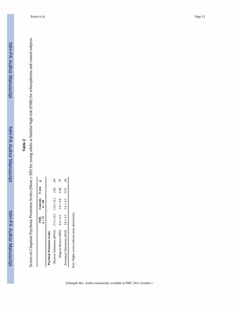
2.3. Subpsychotic symptomsSubjects completed the Chapman psychosis proneness scales. The Revised Physical AnhedoniaScale (RPAS) assesses reduced capacity to experience physical and sensory pleasures (e.g., “Ihave often felt uncomfortable when friends touch me”) (Chapman et al., 1976). The PerceptualAberration Scale (PAS) (Chapman et al., 1978) taps perceptual distortions that don’t reach theseverity of hallucinations (e.g., “Normal colors sometimes seem much too bright to me”). TheMagical Ideation Scale (MIS) (Eckblad and Chapman, 1983) inquires about ideas of referenceand odd beliefs (e.g., “I might cause something to happen just by thinking too much about it”).Subpsychotic symptom data from all study subjects were previously published (Glatt et al.,2006) and those for this sample of subjects with MRI data are presented in Table 2.
2.4. MRI methods2.4.1. Acquisition—Whole brain MR images were collected on a Siemens 1.5 Tesla scannerat the Massachusetts General Hospital (MGH) Martinos Center (Charlestown, Massachusetts).A coronal T2-weighted sequence ruled out clinical neuropathology. Two sagittal 3D MP-RAGE, T1-weighted, non-selective inversion-prepared spoiled gradient echo pulse sequenceswere used for morphometric analyses (TR/TE/T1/flip=2.73s/3.39ms/1.0s/7, bandwidth=190Hz/pixel, sampling matrix=256×192 pixels, FOV=256×256 mm, effective slicethickness=1.33mm on a 170mm slab of 128 partitions). Images were coded for blind imageanalysis and transferred to the MGH Center for Morphometric Analysis (CMA).
2.4.2. Gray and white matter segmentation—Brain images were positionallynormalized to overcome variations in head position by using a standard 3-dimensionalcoordinate system (Filipek et al., 1994). This procedure uses the midpoints of the decussationsof the anterior and posterior commissure lines and the midsagittal plane at the level of theposterior commissure as points of reference for rotation and translation. Images then weresegmented using a semi-automated intensity contour algorithm for external border definitionand signal intensity histogram distributions for delineation of gray-white borders.
2.4.3. Cortical parcellation—The neocortex was divided into 48 parcellation units (PUs)per hemisphere (Caviness et al., 1996; Rademacher et al., 1992). This parcellation systemapproximates architectonic and functional subdivisions, and is based on specific anatomicallandmarks present in all brains. All morphometric measurements were performed blind toidentifying information. The first author (IMR) parcellated 60 of the 75 subjects, afterachieving excellent interrater reliability with a previously trained technician who hadparcellated the other 15 brains (ICCs ≥ .90). Volumes (ml) were calculated by multiplying thearea of each PU by slice thickness, and then summing over all slices containing the PU.
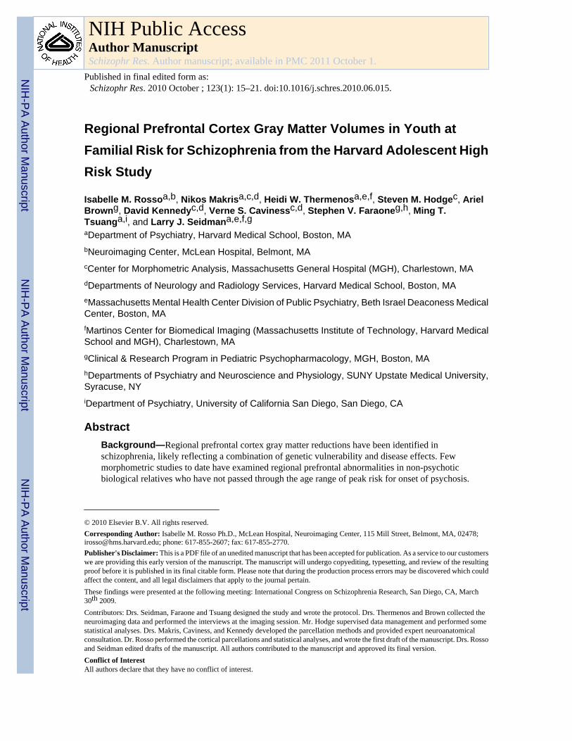
PFC ROIs are shown in Figure 1. The frontal pole (FP) was bordered posteriorly by a coronalplane set at the rostral end of the anterior horizontal ramus of the sylvian fissure (AHRS). FPbordered all other PFC ROIs anteriorly. The dorsolateral PFC (F1+F2) included the superiorand middle frontal gyri, with the central sulcus as its posterior border, the inferior frontal sulcus
Rosso et al. Page 4
Schizophr Res. Author manuscript; available in PMC 2011 October 1.
NIH
-PA Author Manuscript
NIH
-PA Author Manuscript
NIH
-PA Author Manuscript
as its lateral inferior border, and the paracingulate sulcus as its medial inferior border. Theventrolateral PFC (F3t+F3o) had as its superior border the inferior frontal sulcus, the AHRSas its lateral inferior border, and the central sulcus as its posterior border. The ventromedialPFC (FMC) was bordered laterally by the olfactory sulcus, superiorly by the paracingulatesulcus, and medially by the interhemispheric fissure. The orbital PFC (FOC) was borderedposteriorly by the basal forebrain, laterally by the olfactory sulcus, and medially by the AHRS.
2.5. Statistical analysesAll analyses used relative volumes of PFC ROIs (absolute volume/ total cerebral volume *100)to control for scaling effects of brain size. Two sets of hypothesis-driven analyses wereconducted: 1) Repeated measures multivariate analysis of covariance (MANCOVA) examinedgroup differences in regional PFC volumes. Prefrontal volumes were the dependent variablesusing region/ROI (ventromedial, dorsolateral, ventrolateral, orbital, frontal pole) andhemisphere (left, right) as within-subject repeated measures. Familial risk group (FHR,controls) was the independent variable and age was an à priori covariate. Main or interactioneffects of group were followed by least square mean contrasts only if they were statisticallysignificant (p ≤.05) at the multivariate level, and after collapsing across any non-significantwithin-subject dimensions, in order to protect overall Type I error rate. Effect sizes werecomputed using Cohen’s d (Cohen, 1988). Although sex and its interactions were entered inthe initial MANCOVA, they were not included in the final model because their effects wereminimal (p’s >.90). For PFC ROIs found to differ significantly between groups, mixed effectsANCOVAs evaluated the effect of familiality; since these mixed models did not alter anyfindings, their results are not detailed. 2) Pearson’s r examined associations of psychosisproneness with PFC GM within groups, limiting these correlations to ventromedial anddorsolateral subregions as à priori hypotheses. All p-values are two-tailed.
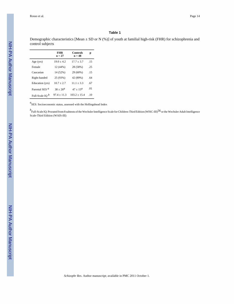
3. ResultsDemographic data are presented in Table 1. Groups were comparable except for a significantlylower parental SES in the FHR group.
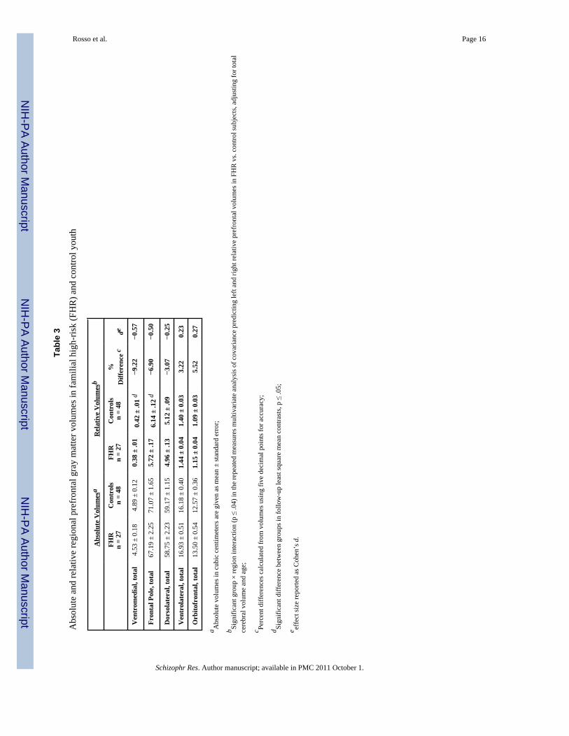
3.1. Repeated measures MANCOVAThe main effect of group was nonsignificant (F= 3.023, df= 1/72, p= .09), while the interactionof group with region was statistically significant (F= 2.62, df= 4/69, p= .04), indicating thatFHR and control subjects differed on some prefrontal regions but not others. The interactionsof group with hemisphere and region-within-hemisphere were not significant (p’s > .20). Agewas a significant covariate (F= 20.40, df= 1/72, p<.001) and interacted significantly with region(F= 5.97, df= 4/69, p≤.001), but not with hemisphere or region-within-hemisphere.
Least square mean contrasts for the significant group × region interaction, adjusting for ageand collapsing across hemispheres, showed the FHR group had significantly smaller GMvolumes than controls for two PFC regions: ventromedial (F= 5.29, df= 1/72, p= .02, d= −0.57)and frontal pole (F= 4.09, df= 1/72, p= .05, d= −0.50) (Table 3).
Follow-up ANCOVAs examined whether significant group differences were affected byentering as covariates two sociodemographic variables that differed significantly (PSES) ormarginally (IQ) between groups. The group difference in ventromedial PFC remainedsignificant (F=5.92, df=1/68, p=.02) when covarying for PSES (F= 0.05, df=1/68, p= .82) andIQ (F= 0.76, df= 1/68, p= .39). Similarly, there remained a significant group difference infrontal pole volume (F= 5.09, df= 1/68, p= .03) after covarying for PSES (F= 0.67, df= 1/68,p= .42) and IQ (F= 1.67, df= 1/68, p= .20).
Rosso et al. Page 5
Schizophr Res. Author manuscript; available in PMC 2011 October 1.
NIH
-PA Author Manuscript
NIH
-PA Author Manuscript
NIH
-PA Author Manuscript

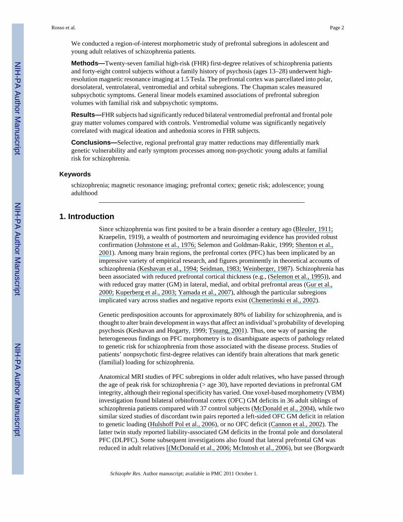
3.2. Correlations of subpsychotic symptomsVentromedial PFC GM volume showed a significant negative correlation with RPAS in FHRsubjects (r= −0.44, p = .03) but not in controls (r= .27, p= .07). Ventromedial PFC GM wasalso significantly negatively associated with MIS scores in FHR subjects (r= −.42, p= .04) butnot in controls (r= .04, p= .77). Ventromedial PFC and PAS scores were non-significantlynegatively correlated in FHR (r= −0.29, p= .15) and control (r= −.04, p= .77) subjects.
DLPFC GM volume was not significantly associated with RPAS (FHR: r= 0.12, p= .53;controls: r= −.01, p= .94), MIS (FHR: r= 0.13, p= .54; controls: r= −.11, p= .44), or PAS (FHR:r= 0.06, p= .78; controls: r= .03, p= .84) scores.
4. DiscussionWe found bilateral reductions of ventromedial and polar PFC GM volumes in adolescent andyoung adult relatives of schizophrenia patients. Neither deficit was explained by differencesin total brain size, SES or IQ. As hypothesized, ventromedial PFC GM was also negativelycorrelated with subpsychotic symptoms in FHR subjects. Contrary to our expectations, DLPFCGM was not related to familial risk for schizophrenia or subpsychotic symptoms.
Our pattern of findings suggests that ventromedial PFC (BA 11,12) GM reductions areassociated with both genetic vulnerability and early disease processes in young relatives ofschizophrenia patients. Compared with controls, FHR subjects had less GM volume in aventromedial PFC area that overlaps with the medial PFC ROI found to be hyperactive in ourfunctional MRI (fMRI) study of a subset of these FHR subjects (Whitfield-Gabrieli et al.,2009), all of whom are included in this report. We therefore have converging anatomical andfunctional imaging evidence in the same sample that medial PFC abnormalities are associatedwith familial risk for schizophrenia in young adulthood. Ventromedial PFC volumes alsocorrelated with self-reported anhedonia and magical ideation, subpsychotic symptoms foundto predict the emergence of full-blown psychosis in certain high-risk samples (Horan et al.,2008; Meehl, 1962). Thus, we postulate that ventromedial PFC GM deficits may partly mediatethe transition to psychosis by becoming more pronounced among FHR adolescents whoconvert. This would dovetail with VBM findings of the EHRP where adolescents at heightenedgenetic risk for schizophrenia had a medial PFC GM density intermediate between that of low-risk adolescents and first-episode schizophrenia patients (Job et al., 2003; Lawrie et al.,2008). In addition, Koutsouleris et al. (2009) found more pronounced ventromedial PFC GMloss in the late versus early stage of the schizophrenia prodrome, suggesting this deficit mayprogress in parallel with emerging disease.
This is the first report of decreased frontal pole (BA 10, 9) GM volume in young biologicalrelatives of schizophrenia patients. Frontal pole deficits have been found in some studies ofolder biological relatives (Cannon et al., 2002; Honea et al., 2008), including a twin studywhere GM declined proportionally with degree of genetic loading for schizophrenia (Cannonet al., 2002). Their presence in both young and older adult biological relatives of patientssuggests the hypothesis that frontal pole GM deficits may be stable markers of genetic risk forschizophrenia.
The ventromedial PFC and frontal pole mediate an array of behaviors that are compromisedin schizophrenia. Both regions have been implicated in socioemotional and self-monitoringfunctions, including mentalizing (i.e., “theory of mind”) and reality monitoring. The frontalpoles are involved in aspects of self/other distinctions, including the ability to distinguishinformation that is perceived in the environment (other-generated) from information that isimagined (self-generated) (Simons et al., 2006). Deficits in these abilities, in turn, may underliethe genesis of psychotic symptoms (Frith, 1992). Medial PFC involvement is the most
Rosso et al. Page 6
Schizophr Res. Author manuscript; available in PMC 2011 October 1.
NIH
-PA Author Manuscript
NIH
-PA Author Manuscript
NIH
-PA Author Manuscript
replicated finding of functional imaging studies of mentalizing (Brunet-Gouet and Decety,2006). Moreover, in the only fMRI study of biological relatives of schizophrenia patientsperforming a mentalizing task, medial PFC activation was positively associated with bothgenetic risk and subpsychotic symptoms (Marjoram et al., 2006).
A lack of significant familial risk group differences in lateral PFC and OFC volumes may beconsistent with research linking anatomical abnormalities in these areas with transition to full-blown psychosis (Smieskova et al., 2010; Wood et al., 2008). In a study of prodromal youth,Pantelis and colleagues (2003) found that subjects who developed psychosis (“converters”)had significantly less baseline right DLPFC GM, and a significant reduction of OFC GM overtime compared with non-converters. In similar longitudinal studies, Borgwardt and colleagues(2007, 2008) found more pronounced reductions of lateral and orbital PFC GM in convertersrelative to non-converters over time, and Sun et al (2009) reported greater contraction of theright DLPFC in association with psychosis onset. Thus, lateral and orbital PFC GM reductionsmay mark transition to psychotic symptoms, more so than genetic predisposition toschizophrenia in youth. Alternatively, these reductions may appear later in the developmentalcourse of the disorder, since the DLPFC completes maturation later than the frontal pole andventromedial PFC (Gogtay et al., 2004). Finally, the absence of significant volume differencesin lateral and orbital PFC does not preclude functional abnormalities. In fact, abnormal DLPFCactivation has been found in two previous fMRI studies of executive functioning in young FHRsubjects (Keshavan et al., 2002), including a subsample from the current study (Seidman et al.,2006b).
As with all studies, this investigation has a number of limitations. Due to the labor- and time-intensive nature of ROI methods, our sample size is limited for the detection of small effects.In addition, the FHR subjects have not passed through the age of risk for onset of psychosis,such that information on clinical outcome is not available. The design of the study alsoprecludes separation of genetic effects from shared environmental effects. Nevertheless, ourfindings encourage further research into PFC GM subregions as markers of risk forschizophrenia, specifically regarding the hypothesis that they may differentially mark inheritedvulnerability and early symptom emergence processes in adolescence and young adulthood.
AcknowledgmentsWe thank the patients with schizophrenia and their family members, control families, and project staff for theirgenerous contributions to the study. Staff included Lisa Gabel, Anthony Giuliano, Stephen Glatt, Jennifer Koch, MarcKorczykowski, Erica Lee, Virna Merino, Elon Mesholam, Raquelle Mesholam-Gately, Caroline Patterson, NicolePeace, William Stone, Rosemary Toomey, and Sharon White.
Role of Funding Source
Funding for this study was provided by the Mental Illness and Neuroscience Discovery (MIND) Institute; NationalAssociation of Research in Schizophrenia and Depression Stone Award (LJS); National Institute of Mental Health(NIMH) R18 MH 43518 and R01 MH 65562 (MTT, LJS), R01 MH 63951, P50 MH80272 & U01 MH81928 (LJS),R25 MH 60485 (to HWT, Training PI: MTT), K01 MH 06987 (IMR); Commonwealth Research Center, MassachusettsDepartment of Mental Health (LJS). This work was also supported in part by The National Center for ResearchResources (P41RR14075). None of these funding sources had any further role in the study design; in the collection,analysis and interpretation of the data; in the writing of the report; or in the decision to submit the paper for publication.
ReferencesAmerican Psychiatric Association (APA). Diagnostic and statistical manual of mental disorders.
Washington DC: Author; 2000.Bleuler, E. Dementia Praecox or the Group of Schizophrenias. Zinkin, TBJ., editor. New York, NY:
International Universities Press; 1911.
Rosso et al. Page 7
Schizophr Res. Author manuscript; available in PMC 2011 October 1.
NIH
-PA Author Manuscript
NIH
-PA Author Manuscript
NIH
-PA Author Manuscript
Borgwardt SJ, McGuire PK, Aston J, Berger G, Dazzan P, Gschwandtner U, Pfluger M, D'Souza M,Radue EW, Riecher-Rossler A. Structural brain abnormalities in individuals with an at-risk mentalstate who later develop psychosis. Br. J. Psychiatry Suppl 2007;51:s69–s75. [PubMed: 18055941]
Borgwardt SJ, McGuire PK, Aston J, Gschwandtner U, Pflüger MO, Stieglitz RD, Radue EW, Riecher-Rössler A. Reductions in frontal, temporal and parietal volume associated with the onset of psychosis.Schizophr. Res 2008;106:108–114. [PubMed: 18789654]
Borgwardt SJ, Picchioni MM, Ettinger U, Toulopoulou T, Murray R, McGuire PK. Regional gray mattervolume in monozygotic twins concordant and discordant for schizophrenia. Biol. Psychiatry2010;67:956–964. [PubMed: 20006324]
Brunet-Gouet E, Decety J. Social brain dysfunctions in schizophrenia: a review of neuroimaging studies.Psychiatry Res 2006;148:75–92. [PubMed: 17088049]
Cannon TD. Clinical and genetic high-risk strategies in understanding vulnerability to psychosis.Schizophr. Res 2005;79:35–44. [PubMed: 16054805]
Cannon TD, Thompson PM, van Erp TG, Toga AW, Poutanen VP, Huttunen M, Lonnqvist J,Standerskjold-Nordenstam CG, Narr KL, Khaledy M, Zoumalan CI, Dail R, Kaprio J. Cortex mappingreveals regionally specific patterns of genetic and disease-specific gray-matter deficits in twinsdiscordant for schizophrenia. Proc. Natl. Acad. Sci. U S A 2002;99:3228–3233. [PubMed: 11867725]
Caviness VS, Kennedy DN, Richelme C, Rademacher J, Filipek PA. The human brain age 7–11 years:a volumetric analysis based on magnetic resonance images. Cereb. Cortex 1996;6:726–736. [PubMed:8921207]
Chapman LJ, Chapman JP, Raulin ML. Scales for physical and social anhedonia. J. Abnorm. Psychol1976;85:374–382. [PubMed: 956504]
Chapman LJ, Chapman JP, Raulin ML. Perceptual aberration in schizophrenia. J. Abnorm. Psychol1978;87:399–407. [PubMed: 681612]
Chemerinski E, Nopoulos PC, Crespo-Facorro B, Andreasen NC, Magnotta V. Morphology of the ventralfrontal cortex in schizophrenia: relationship with social dysfunction. Biol. Psychiatry 2002;52:1–8.[PubMed: 12079724]
Cohen, J. Statistical power analysis for the behavioural sciences. Hillsdale, NJ: Erlbaum Associates;1988.
Diwadkar VA, Montrose DM, Dworakowski D, Sweeney JA, Keshavan MS. Genetically predisposedoffspring with schizotypal features: an ultra high-risk group for schizophrenia? Prog.Neuropsychopharmacol. Biol. Psychiatry 2006;30:230–238. [PubMed: 16318899]
Eckblad M, Chapman LJ. Magical ideation as an indicator of schizotypy. J. Consult. Clin. Psychol1983;51:215–255. [PubMed: 6841765]
Filipek PA, Richelme C, Kennedy DN, Caviness VR. The young adult human brain: an MRI-basedmorphometric study. Cereb. Cortex 1994;4:344–360. [PubMed: 7950308]
Frith, CD. The Cognitive Neuropsychology of Schizophrenia. London: LEA; 1992.Geller, B.; Williams, M.; Zimmerman, B.; Frazier, J. Washington University in St. Louis Kiddie Schedule
for Affective Disorders and Schizophrenia (WASH-U-KSADS). St. Louis, MO: WashingtonUniversity; 1996.
Giuliani NR, Calhoun VD, Pearlson GD, Francis A, Buchanan RW. Voxel-based morphometry versusregion of interest: a comparison of two methods for analyzing gray matter differences inschizophrenia. Schizophr. Res 2005;74:135–147. [PubMed: 15721994]
Glatt SJ, Stone WS, Faraone SV, Seidman LJ, Tsuang MT. Psychopathology, personality traits and socialdevelopment of young first-degree relatives of patients with schizophrenia. Br. J. Psychiatry2006;189:337–345. [PubMed: 17012657]
Gogtay N, Giedd JN, Lusk L, Hayashi KM, Greenstein D, Vaituzis AC, Nugent TF3, Herman DH, ClasenLS, Toga AW, Rapoport JL, Thompson PM. Dynamic mapping of human cortical developmentduring childhood through early adulthood. Proc. Natl. Acad. Sci. U S A 2004;101:8174–8179.[PubMed: 15148381]
Gur RE, Cowell PE, Latshaw A, Turetsky BI, Grossman RI, Arnold SE, Bilker WB, Gur RC. Reduceddorsal and orbital prefrontal gray matter volumes in schizophrenia. Arch. Gen. Psychiatry2000;57:761–768. [PubMed: 10920464]
Rosso et al. Page 8
Schizophr Res. Author manuscript; available in PMC 2011 October 1.
NIH
-PA Author Manuscript
NIH
-PA Author Manuscript
NIH
-PA Author Manuscript
Honea R, Crow TJ, Passingham D, Mackay CE. Regional deficits in brain volume in schizophrenia: ameta-analysis of voxel-based morphometry studies. Am. J. Psychiatry 2005;162:2233–2245.[PubMed: 16330585]
Honea RA, Meyer-Lindenberg A, Hobbs KB, Pezawas L, Mattay VS, Egan MF, Verchinski B,Passingham RE, Weinberger DR, Callicott JH. Is gray matter volume an intermediate phenotype forschizophrenia? A voxel-based morphometry study of patients with schizophrenia and their healthysiblings. Biol. Psychiatry 2008;63:465–474. [PubMed: 17689500]
Horan WP, Reise SP, Subotnik KL, Ventura J, Nuechterlein KH. The validity of Psychosis PronenessScales as vulnerability indicators in recent-onset schizophrenia patients. Schizophr. Res2008;100:224–236. [PubMed: 18221857]
Hulshoff Pol HE, Schnack HG, Mandl RC, Brans RG, van Haren NE, Baare WF, van Oel CJ, CollinsDL, Evans A, Kahn RS. Gray and white matter density changes in monozygotic and same-sexdizygotic twins discordant for schizophrenia using voxel-based morphometry. Neuroimage2006;31:482–488. [PubMed: 16497519]
Job DE, Whalley HC, McConnell S, Glabus M, Johnstone EC, Lawrie SM. Voxel-based morphometryof grey matter densities in subjects at high risk of schizophrenia. Schizophr. Res 2003;64:1–13.[PubMed: 14511796]
Johnstone EC, Crow TJ, Frith CD, Husband J, Kreel L. Cerebral ventricular size and cognitive impairmentin chronic schizophrenia. Lancet 1976;2:924–926. [PubMed: 62160]
Keshavan MS, Anderson S, Pettegrew JW. Is schizophrenia due to excessive synaptic pruning in theprefrontal cortex? The Feinberg hypothesis revisited. J. Psychiatr. Res 1994;28:239–265. [PubMed:7932285]
Keshavan MS, Diwadkar VA, Spencer SM, Harenski KA, Luna B, Sweeney JA. A preliminary functionalmagnetic resonance imaging study in offspring of schizophrenic parents. ProgNeuropsychopharmacol Biol. Psychiatry 2002;26:1143–1149. [PubMed: 12452537]
Keshavan MS, Hogarty GE. Brain maturational processes and delayed onset in schizophrenia. Dev.Psychopathol 1999;11:525–543. [PubMed: 10532623]
Koutsouleris N, Schmitt GJ, Gaser C, Bottlender R, Scheuerecker J, McGuire P, Burgermeister B, BornC, Reiser M, Möller HJ, Meisenzahl EM. Neuroanatomical correlates of different vulnerability statesfor psychosis and their clinical outcomes. Br. J. Psychiatry 2009;195:218–226. [PubMed: 19721111]
Kraepelin, E. Dementia Praecox and Paraphrenia. Barclay, TBR., editor. Huntington, NY: Robert EKrieger Publishing Co; 1919.
Kubicki M, Shenton ME, Salisbury DF, Hirayasu Y, Kasai K, Kikinis R, Jolesz FA, McCarley RW.Voxel-based morphometric analysis of gray matter in first episode schizophrenia. Neuroimage2002;17:1711–1719. [PubMed: 12498745]
Kuperberg GR, Broome MR, McGuire PK, David AS, Eddy M, Ozawa F, Goff D, West WC, WilliamsSC, van der Kouwe AJ, Salat DH, Dale AM, Fischl B. Regionally localized thinning of the cerebralcortex in schizophrenia. Arch. Gen. Psychiatry 2003;60:878–888. [PubMed: 12963669]
Lawrie SM, McIntosh AM, Hall J, Owens DG, Johnstone EC. Brain structure and function changes duringthe development of schizophrenia: the evidence from studies of subjects at increased genetic risk.Schizophr. Bull 2008;34:330–340. [PubMed: 18227083]
Lawrie SM, Whalley HC, Abukmeil SS, Kestelman JN, Donnelly L, Miller P, Best JJ, Owens DG,Johnstone EC. Brain structure, genetic liability, and psychotic symptoms in subjects at high risk ofdeveloping schizophrenia. Biol. Psychiatry 2001;49:811–823. [PubMed: 11343678]
Marjoram D, Job DE, Whalley HC, Gountouna VE, McIntosh AM, Simonotto E, Cunningham-OwensD, Johnstone EC, Lawrie S. A visual joke fMRI investigation into Theory of Mind and enhanced riskof schizophrenia. Neuroimage 2006;31:1850–1858. [PubMed: 16624578]
Maxwell, ME. Family Interview for Genetic Studies: Clinical Neurogenetics Branch, Intramural ResearchProgram. NIMH; 1996.
McDonald C, Bullmore ET, Sham PC, Chitnis X, Wickham H, Bramon E, Murray RM. Association ofgenetic risks for schizophrenia and bipolar disorder with specific and generic brain structuralendophenotypes. Arch. Gen. Psychiatry 2004;61:974–984. [PubMed: 15466670]
Rosso et al. Page 9
Schizophr Res. Author manuscript; available in PMC 2011 October 1.
NIH
-PA Author Manuscript
NIH
-PA Author Manuscript
NIH
-PA Author Manuscript
McDonald C, Marshall N, Sham PC, Bullmore ET, Schulze K, Chapple B, Bramon E, Filbey F, QuraishiS, Walshe M, Murray RM. Regional brain morphometry in patients with schizophrenia or bipolardisorder and their unaffected relatives. Am. J. Psychiatry 2006;163:478–487. [PubMed: 16513870]
McIntosh AM, Job DE, Moorhead TW, Harrison LK, Forrester K, Lawrie SM, Johnstone EC. Voxel-based morphometry of patients with schizophrenia or bipolar disorder and their unaffected relatives.Biol. Psychiatry 2004;56:544–552. [PubMed: 15476683]
McIntosh AM, Job DE, Moorhead WJ, Harrison LK, Whalley HC, Johnstone EC, Lawrie SM. Geneticliability to schizophrenia or bipolar disorder and its relationship to brain structure. Am. J. Med. Genet.B Neuropsychiatr. Genet 2006;141B:76–83. [PubMed: 16342281]
Meehl PE. Schizotaxia, schizotypy, schizophrenia. American Psychologist 1962;17:827–838.Nurnberger JI Jr, Blehar MC, Kaufmann CA, York-Cooler C, Simpson SG, Harkavy-Friedman J, et al.
Diagnostic interview for genetic studies. Rationale, unique features, and training. NIMH geneticsinitiative. Arch. Gen. Psychiatry 1994;51:849–859. [PubMed: 7944874]
Pantelis C, Velakoulis D, McGorry PD, Wood SJ, Suckling J, Phillips LJ, Yung AR, Bullmore ET, BrewerW, Soulsby B, Desmond P, McGuire PK. Neuroanatomical abnormalities before and after onset ofpsychosis: a cross-sectional and longitudinal MRI comparison. Lancet 2003;361:281–288. [PubMed:12559861]
Rademacher J, Galaburda AM, Kennedy DN, Filipek PA, Caviness VS. Human cerebral cortex:Localization, parcellation and morphometry with magnetic resonance imaging. J. Cogn. Neurosci1992;4:352–374.
Seidman LJ. Schizophrenia and brain dysfunction: an integration of recent neurodiagnostic findings.Psychol. Bull 1983;94:195–238. [PubMed: 6356196]
Seidman LJ, Giuliano AJ, Smith CW, Stone WS, Glatt SJ, Meyer E, Faraone SV, Tsuang MT, CornblattB. Neuropsychological functioning in adolescents and young adults at genetic risk for schizophreniaand affective psychoses: results from the Harvard and Hillside Adolescent High Risk Studies.Schizophr. Bull 2006a;32:507–524. [PubMed: 16707777]
Seidman LJ, Thermenos HW, Poldrack RA, Peace NK, Koch JK, Faraone SV, Tsuang MT. Altered brainactivation in dorsolateral prefrontal cortex in adolescents and young adults at genetic risk forschizophrenia: an fMRI study of working memory. Schizophr. Res 2006b;85:58–72. [PubMed:16632333]
Selemon LD, Goldman-Rakic PS. The reduced neuropil hypothesis: a circuit based model ofschizophrenia. Biol. Psychiatry 1999;45:17–25. [PubMed: 9894571]
Selemon LD, Rajkowska G, Goldman-Rakic PS. Abnormally high neuronal density in the schizophreniccortex. A morphometric analysis of prefrontal area 9 and occipital area 17. Arch. Gen. Psychiatry1995;52:805–818. discussion 819-20. [PubMed: 7575100]
Shenton ME, Dickey CC, Frumin M, McCarley RW. A review of MRI findings in schizophrenia.Schizophr. Res 2001;49:1–52. [PubMed: 11343862]
Simons JS, Davis SW, Gilbert SJ, Frith CD, Burgess PW. Discriminating imagined from perceivedinformation engages brain areas implicated in schizophrenia. Neuroimage 2006;32:696–703.[PubMed: 16797186]
Smieskova R, Fusar-Poli P, Allen P, Bendfeldt K, Stieglitz RD, Drewe J, Radue EW, McGuire PK,Riecher-Rössler A, Borgwardt SJ. Neuroimaging predictors of transition to psychosis-A systematicreview and meta-analysis. Neurosci. Biobehav. Rev. 2010 doi:10.1016/j.neubiorev.2010.01.016.
Sun D, Phillips L, Velakoulis D, Yung A, McGorry PD, Wood SJ, van Erp TG, Thompson PM, TogaAW, Cannon TD, Pantelis C. Progressive brain structural changes mapped as psychosis develops in'at risk' individuals. Schizophr. Res 2009;108:85–92. [PubMed: 19138834]
Tsuang MT. Defining alternative phenotypes for genetic studies: what can we learn from studies ofschizophrenia? Am. J. Med. Genet 2001;105:8–10. [PubMed: 11425007]
Weinberger DR. Implications of normal brain development for the pathogenesis of schizophrenia. Arch.Gen. Psychiatry 1987;44:660–669. [PubMed: 3606332]
Whitfield-Gabrieli S, Thermenos HW, Milanovic S, Tsuang MT, Faraone SV, McCarley RW, ShentonME, Green AI, Nieto-Castanon A, LaViolette P, Wojcik J, Gabrieli JD, Seidman LJ. Hyperactivityand hyperconnectivity of the default network in schizophrenia and in first-degree relatives of personswith schizophrenia. Proc. Natl. Acad. Sci. U S A 2009;106:1279–1284. [PubMed: 19164577]
Rosso et al. Page 10
Schizophr Res. Author manuscript; available in PMC 2011 October 1.
NIH
-PA Author Manuscript
NIH
-PA Author Manuscript
NIH
-PA Author Manuscript

Wood SJ, Pantelis C, Velakoulis D, Yücel M, Fornito A, McGorry PD. Progressive changes in thedevelopment toward schizophrenia: studies in subjects at increased symptomatic risk. Schizophr.Bull 2008;34:322–329. [PubMed: 18199631]
Yamada M, Hirao K, Namiki C, Hanakawa T, Fukuyama H, Hayashi T, Murai T. Social cognition andfrontal lobe pathology in schizophrenia: a voxel-based morphometric study. Neuroimage2007;35:292–298. [PubMed: 17240165]
Rosso et al. Page 11
Schizophr Res. Author manuscript; available in PMC 2011 October 1.
NIH
-PA Author Manuscript
NIH
-PA Author Manuscript
NIH
-PA Author Manuscript
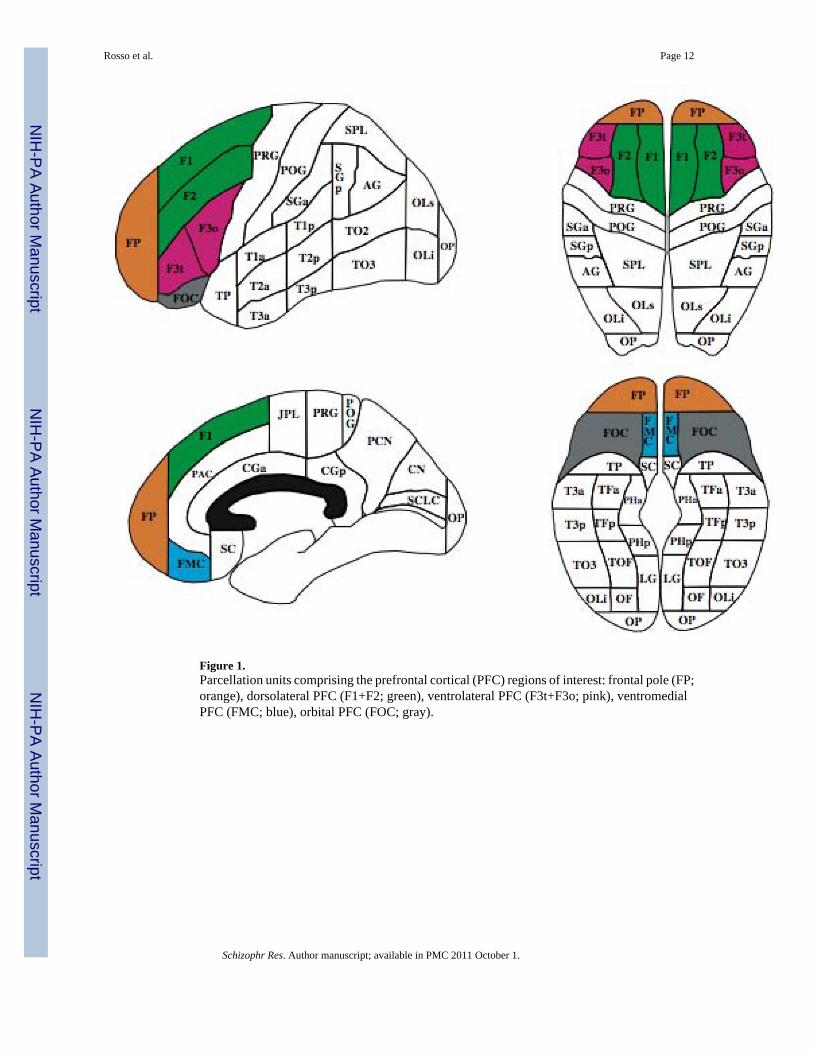
Figure 1.Parcellation units comprising the prefrontal cortical (PFC) regions of interest: frontal pole (FP;orange), dorsolateral PFC (F1+F2; green), ventrolateral PFC (F3t+F3o; pink), ventromedialPFC (FMC; blue), orbital PFC (FOC; gray).
Rosso et al. Page 12
Schizophr Res. Author manuscript; available in PMC 2011 October 1.
NIH
-PA Author Manuscript
NIH
-PA Author Manuscript
NIH
-PA Author Manuscript
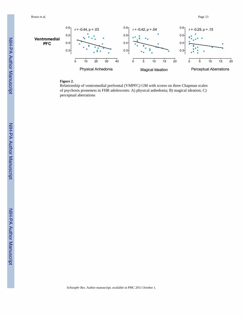
Figure 2.Relationship of ventromedial prefrontal (VMPFC) GM with scores on three Chapman scalesof psychosis proneness in FHR adolescents: A) physical anhedonia; B) magical ideation; C)perceptual aberrations
Rosso et al. Page 13
Schizophr Res. Author manuscript; available in PMC 2011 October 1.
NIH
-PA Author Manuscript
NIH
-PA Author Manuscript
NIH
-PA Author Manuscript
NIH
-PA Author Manuscript
NIH
-PA Author Manuscript
NIH
-PA Author Manuscript
Rosso et al. Page 14
Table 1
Demographic characteristics [Mean ± SD or N (%)] of youth at familial high-risk (FHR) for schizophrenia andcontrol subjects
FHRn = 27
Controlsn = 48
p
Age (yrs) 19.0 ± 4.2 17.7 ± 3.7 .15
Female 12 (44%) 28 (58%) .25
Caucasian 14 (52%) 29 (60%) .15
Right-handed 25 (93%) 42 (89%) .64
Education (yrs) 10.7 ± 2.7 11.1 ± 3.3 .67
Parental SES a 38 ± 26a 47 ± 15a .01
Full-Scale IQ b 97.4 ± 11.3 103.2 ± 15.4 .10
aSES: Socioeconomic status, assessed with the Hollingshead Index
bFull-Scale IQ: Prorated from 8 subtests of the Wechsler Intelligence Scale for Children-Third Edition (WISC-III)56 or the Wechsler Adult Intelligence
Scale-Third Edition (WAIS-III)
Schizophr Res. Author manuscript; available in PMC 2011 October 1.
NIH
-PA Author Manuscript
NIH
-PA Author Manuscript
NIH
-PA Author Manuscript
Rosso et al. Page 15
Tabl
e 2
Scor
es o
n C
hapm
an P
sych
osis
Pro
nene
ss S
cale
s (M
ean
± SD
) for
you
ng a
dults
at f
amili
al h
igh-
risk
(FH
R) f
or sc
hizo
phre
nia
and
cont
rol s
ubje
cts
FHR
n =
27C
ontr
ols
n =
48T
-test
p
Psyc
hosi
s Pro
nene
ss S
cale
s
Phys
ical
Anh
edon
ia (R
PAS)
17.2
± 8
.513
.0 ±
8.1
2.06
.04
Mag
ical
Idea
tion
(MIS
)6.
4 ±
4.5
5.9
± 4.
80.
40.7
0
Perc
eptu
al A
berr
atio
ns (P
AS)
3.8
± 4.
33.
2 ±
4.3
0.55
.58
Not
e: H
ighe
r sco
res i
ndic
ate
mor
e ab
norm
ality
.
Schizophr Res. Author manuscript; available in PMC 2011 October 1.
NIH
-PA Author Manuscript
NIH
-PA Author Manuscript
NIH
-PA Author Manuscript
Rosso et al. Page 16
Tabl
e 3
Abs
olut
e an
d re
lativ
e re
gion
al p
refr
onta
l gra
y m
atte
r vol
umes
in fa
mili
al h
igh-
risk
(FH
R) a
nd c
ontro
l you
th
Abs
olut
e V
olum
esa
Rel
ativ
e V
olum
esb
FHR
n =
27C
ontr
ols
n =
48FH
Rn
= 27
Con
trol
sn
= 48
%D
iffer
ence
cde
Ven
trom
edia
l, to
tal
4.53
± 0
.18
4.89
± 0
.12
0.38
± .0
10.
42 ±
.01
d−9
.22
−0.5
7
Fron
tal P
ole,
tota
l67
.19
± 2.
2571
.07
± 1.
655.
72 ±
.17
6.14
± .1
2 d
−6.9
0−0
.50
Dor
sola
tera
l, to
tal
58.7
5 ±
2.23
59.1
7 ±
1.15
4.96
± .1
35.
12 ±
.09
−3.0
7−0
.25
Ven
trol
ater
al, t
otal
16.9
3 ±
0.51
16.1
8 ±
0.40
1.44
± 0
.04
1.40
± 0
.03
3.22
0.23
Orb
itofr
onta
l, to
tal
13.5
0 ±
0.54
12.5
7 ±
0.36
1.15
± 0
.04
1.09
± 0
.03
5.52
0.27
a Abs
olut
e vo
lum
es in
cub
ic c
entim
eter
s are
giv
en a
s mea
n ±
stan
dard
err
or;
b Sign
ifica
nt g
roup
× re
gion
inte
ract
ion
(p ≤
.04)
in th
e re
peat
ed m
easu
res m
ultiv
aria
te a
naly
sis o
f cov
aria
nce
pred
ictin
g le
ft an
d rig
ht re
lativ
e pr
efro
ntal
vol
umes
in F
HR
vs.
cont
rol s
ubje
cts,
adju
stin
g fo
r tot
alce
rebr
al v
olum
e an
d ag
e;
c Perc
ent d
iffer
ence
s cal
cula
ted
from
vol
umes
usi
ng fi
ve d
ecim
al p
oint
s for
acc
urac
y;
d Sign
ifica
nt d
iffer
ence
bet
wee
n gr
oups
in fo
llow
-up
leas
t squ
are
mea
n co
ntra
sts,
p ≤
.05;
e effe
ct si
ze re
porte
d as
Coh
en’s
d.
Schizophr Res. Author manuscript; available in PMC 2011 October 1.
Copyright © 2022 FDOKUMEN