Refinement of proteins at subatomic resolution with MOPRO
11
electronic reprint Journal of Applied Crystallography ISSN 0021-8898 Refinement of proteins at subatomic resolution with MOPRO Benoit Guillot, Laurence Viry, Regis Guillot, Claude Lecomte and Christian Jelsch Copyright © International Union of Crystallography Author(s) of this paper may load this reprint on their own web site provided that this cover page is retained. Republication of this article or its storage in electronic databases or the like is not permitted without prior permission in writing from the IUCr. J. Appl. Cryst. (2001). 34, 214–223 Benoit Guillot et al. Refinement of proteins
-
Upload
independent -
Category
Documents
-
view
1 -
download
0
Transcript of Refinement of proteins at subatomic resolution with MOPRO

electronic reprint
Journal of
AppliedCrystallography
ISSN 0021-8898
Refinement of proteins at subatomic resolution with MOPRO
Benoit Guillot, Laurence Viry, Regis Guillot, Claude Lecomte and Christian Jelsch
Copyright © International Union of Crystallography
Author(s) of this paper may load this reprint on their own web site provided that this cover page is retained. Republication of this article or itsstorage in electronic databases or the like is not permitted without prior permission in writing from the IUCr.
J. Appl. Cryst. (2001). 34, 214–223 Benoit Guillot et al. � Refinement of proteins

computer programs
214 Benoit Guillot et al. � Refinement of proteins J. Appl. Cryst. (2001). 34, 214±223
Journal of
AppliedCrystallography
ISSN 0021-8898
Received 6 November 2000
Accepted 24 January 2001
# 2001 International Union of Crystallography
Printed in Great Britain ± all rights reserved
Refinement of proteins at subatomic resolution withMOPRO
Benoit Guillot,a Laurence Viry,b Regis Guillot,a Claude Lecomtea and
Christian Jelscha*
aLaboratoire de Cristallographie et ModeÂlisation des MateÂriaux MineÂraux et Biologiques, UMR
CNRS 7036, Faculte des Sciences BP 239, 54506 Vandoeuvre-leÁs-Nancy, France, and bCentre
Charles Hermite, BaÃtiment LORIA, Faculte des Sciences, 54506 Vandoeuvre-leÁs-Nancy, France.
Correspondence e-mail: [email protected]
Crystallography at subatomic resolution permits the observation and measure-
ment of the non-spherical character of the atomic electron density. Charge
density studies are being performed on molecules of increasing size. The
MOPRO least-squares re®nement software has thus been developed, by
extensive modi®cations of the program MOLLY, for protein and supramole-
cular chemistry applications. The computation times are long because of the
large number of re¯ections and the complexity of the multipolar model of the
atomic electron density; the structure factor and derivative calculations have
thus been parallelized. Stereochemical and dynamical restraints as well as the
conjugate gradient algorithm have been implemented. A large number of the
normal matrix off-diagonal terms turn out to be very small and the block
diagonal approximation is thus particularly ef®cient in the case of large
structures at very high resolution.
1. Introduction
The electron cloud around the atoms of a molecule is
deformed as a result of chemical bonding and non-bonding
interactions (notably hydrogen bonds) between the atoms.
Accurate electron density distribution in the crystalline state
can be derived from an ultra-high resolution X-ray diffraction
experiment (Coppens, 1967). Our laboratory has been
involved in charge density studies on molecules of increasing
size: the octapeptide LBZ of helical structure (Jelsch et al.,
1998) and a scorpion toxin (Housset et al., 2000). With the
combined use of synchrotron radiation sources and crystal
cryocooling, the number of protein structures re®ned at a
resolution higher than 1.0 AÊ is increasing continuously.
Recently, the charge density of the protein crambin (46
amino acids) was analysed (Jelsch et al., 2000) at ultra-high
resolution (0.54 AÊ ). The crystal structure was re®ned with a
model for charged non-spherical multipolar atoms in order to
describe the molecular electron density distribution accu-
rately. The initial multipoles and charges were transferred
from our database of average parameters (Pichon-Pesme et al.,
1995) derived from the analysis of several crystals of amino
acids and small peptides. The average electron density para-
meters of the protein main chain were then re®ned against the
crambin diffraction data. The crambin electron density
deformation is illustrated in Fig. 1 (Jelsch et al., 2000). The
analysis of the electron density distribution of human aldose
reductase is also underway. Crystals diffracting to very high
resolution (Lamour et al., 1999) have been grown for this
enzyme of 315 amino acids and diffraction data have been
collected to 0.65 AÊ resolution at the APS synchrotron
(Mitschler et al., 2000).
The least-squares computer program MOPRO was devel-
oped for the charge density analysis of proteins. The software
Figure 1Iso-contour surface of the experimental deformation electron density(level 0.4 e AÊ ÿ3) along the polypeptide chain of crambin. The deforma-tion density represents the difference between the actual electron densityof the molecule and the density calculated for the promolecule, made upof isolated spherical neutral atoms. The density is `static' in that it iscomputed for atoms at rest.
electronic reprint

is derived from the charge density analysis program MOLLY
(Hansen & Coppens, 1978). The atomic electron density is
described in terms of core, valence and multipolar electron
density:
�atom�r� � �core�r� � Pval�3�val��r� �
P
l
�03Rl��0r�P
m
Plmylm�:
�1�
The ®rst two terms on the right describe spherically symmetric
core-plus-valence density and the third term describes the
non-spherical multipolar distribution of the valence electron
density of the atoms. The valence-shell electron populations
Pval account for interatomic charge transfer and the multipole
populations Plm account for non-spherical intra-atomic redis-
tribution of valence electron density. The Rl are Slater-type
radial functions and the ylm� are real spherical harmonic
angular functions. The parameters � and �0 model the
expansion and contraction, respectively, of the spherical and
multipolar parts of the valence electron density.
2. Restraints
In structure re®nement by least squares (LS), one usually
seeks to minimize a residual function E, as a weighted sum
over the re¯ections,
E �PH
WH�jF obsH j ÿ jF calc
H j�2; �2�
and assumes that a structure factor F is a linear function of
each parameter for small changes.
When re®ning the structure of biological macromolecules,
because of the limited resolution and the low variables/
observations ratio, it is necessary to incorporate stereo-
chemical knowledge into the re®nement. Such restrained
reciprocal-space least-squares re®nement (Konnert, 1976;
Konnert & Hendrickson, 1980) is very effective and increases
the robustness of the convergence. The minimized function is
composite, consisting of the crystallographic error term and
other residual components which re¯ect target geometry as
anticipated from small-molecule structures:
E � EX-ray � Erestrain: �3�
2.1. Stereochemistry
The geometric restraints implemented in MOPRO are the
distance, angle and planarity (Table 1). Planarity restraints
were programmed by considering the eigenvalues �i of the 3�3 matrix V (Urzhumtsev, 1991):
Vij �P
atoms
�Xi ÿ hXii��Xj ÿ hXji�; �4�
where X1, X2 and X3 represent respectively the x, y and z
coordinates of the atoms belonging to the plane. This makes it
possible to re®ne the optimum plane orientation at the same
time as the atomic coordinates. The function minimized in
MOPRO is the dimensionless quantity �1�2�3/(�1 + �2 + �3)3
(Table 1) which models planarity more accurately than the
function �1�2�3 proposed by Haneef et al. (1985) and is
simpler to implement than the minimization of the smallest
eigenvalue �min, proposed by Urzhumtsev (1991).
Stereochemical information is generally no longer neces-
sary for the non-hydrogen atoms in small-molecule crystal-
lography. However, for medium-size molecules and
macromolecules at atomic and subatomic resolution, stereo-
chemical information may still be necessary, as some regions
can be disordered. For example, in crambin, as much as 30%
of the protein atoms have multiple conformations (Yamano et
al., 1997; Jelsch et al., 2000). The programming of distance,
angle and planarity restraints was therefore deemed necessary
for the structure and charge density re®nement of crambin.
On the other hand, less electron density is associated with
the hydrogen atoms compared with the heavier atoms as the
hydrogen atoms lack the core electrons. Thus, the hydrogen
atoms may still need to have their positions de®ned by
stereochemical information in small-molecule crystallography.
In crystallographic re®nement software like SHELXL97
(Sheldrick & Schneider, 1997) or CNS (BruÈnger et al., 1998),
the positions of the hydrogen atoms can be constrained
according to standard geometries. Alternatively, this can be
achieved using restraints as in MOPRO. Restraints are a
convenient and smoother way to re®ne properly the positions
of hydrogen atoms in charge density studies of small mole-
cules. Notably, the HÐX distance to the neighbouring atom
can be restrained to standard values derived from neutron
diffraction studies (Allen, 1986).
J. Appl. Cryst. (2001). 34, 214±223 Benoit Guillot et al. � Refinement of proteins 215
computer programs
Table 1List of restraints implemented in MOPRO.
Keyword Description Function minimized
XYZRES Coordinates X, Y, Z (X ÿ Xr)2
DISTAN Distance d between two atoms (d ÿ dr)2
ANGLER Angle � between three atoms (� ÿ �r)2
PLANAR Planarity �1�2�3/(�1 + �2 + �3)3
UIJRES Thermal displacement parameters (Uij ÿ Uijr )
2
RIGIDB Rigid bond (ZU ÿ ZU0)2
URATIO Isotropic thermal displacement parameter riding on bondingatom
(U ÿ Ur)2
ISOTRO Thermal displacement ellipsoid limited anisotropyP
(Ui ÿ Uj)2/hUiii2 = 3[P
i(Uii ÿ hUiii)2 +Pi6�jU
ij2]/hUiii2KAPPAR Expansion coef®cient � and �0 (� ÿ �r)
2
electronic reprint

computer programs
216 Benoit Guillot et al. � Refinement of proteins J. Appl. Cryst. (2001). 34, 214±223
2.2. Thermal motion
The values of the coordinates and thermal displacement
parameters Uij can also be restrained in MOPRO. Such
restraints can be applied if neutron diffraction data are
available for the crystal studied. In the re®nement versus the
X-ray diffraction data, the target values for the coordinates
and thermal displacement parameters can be set to their
values in the neutron structure, the allowed deviation being
set to the neutron experimental error. However, as combined
X-ray and neutron diffraction studies often display a thermal
difference between the two data sets, an additional `thermal
scale factor' may be introduced. Alternatively, in the case of
small compounds, thermal displacement parameters can be
derived from an ab initio calculation of the harmonic force
®eld (Flaig et al., 1998) and could be used as targets in
MOPRO for the Uij restraints. These restraints, which are
more ¯exible than constraints, permit the combination of
information from different sources and are helpful in the
deconvolution of the deformation density and the thermal
motion.
Several restraints on the thermal displacement parameters
have been implemented. Hydrogen atoms are often assigned
an isotropic displacement parameter proportional to the
equivalent B factor of the connecting atom. The multiplier
coef®cient generally used is respectively 1.5 or 1.2 for
hydrogen atoms with or devoid of a rotation degree of
freedom. The isotropic B factor of the hydrogen atoms can be
restrained in MOPRO with a chosen proportionality coef®-
cient. This restraint was applied to the main-chain hydrogen
atoms in the charge density re®nement of the main-chain
polypeptide in crambin (Jelsch et al., 2000). Such information
can also be useful in small-molecule X-ray electron density
studies, as the thermal motion of hydrogen atoms is not well
de®ned.
A restraint limiting the anisotropy of the atomic thermal
motion is also implemented. When the thermal tensorUij of an
atom has three identical eigenvalues, the thermal motion is
isotropic. The anisotropy of theUij tensor can be conveniently
limited with the use of the dimensionless quadratic functionP(Ui ÿ Uj)2/hUii2 describing the global discrepancy between
the three eigenvalues Ui (Table 1). This represents a model
between isotropy and anisotropy and avoids non-positive
de®ned Uij thermal motion tensors. Such isotropy restraints
were applied in the case of crambin (Jelsch et al., 2000) to the
partially occupied water molecules and disordered protein
atoms, for which the electron density map (2Fo ÿ Fc at the 5�level) showed a well de®ned thermal displacement ellipsoid.
Other partially occupied atoms were set to be isotropic.
A rigid-bond restraint, which renders the mean-square
displacement for two covalently bonded atoms similar along
the bond direction, is also programmed. Hirshfeld (1976)
considered that a rigid-bond discrepancy of �ZU < 10ÿ3 AÊ 2
was a good criterion to assess the reliability of the thermal
displacement parameters in a crystal structure. The deviation
from the rigid bond in actual crystal structures devoid of
crystallographic errors and uncertainties can presumably be
expected to be one order of magnitude smaller than this value.
This additional information on the thermal motion can facil-
itate its deconvolution from the deformation density. As
shown by Rosen®eld et al. (1978), the rigid-bond criterion may
be extended to any pair of atoms belonging to a rigid group,
such as aromatic cycles.
2.3. Expansion/contraction of the valence density
The use of external information obtained from accurate
small-molecule crystallographic analyses as restraints in a
macromolecular re®nement can be extended to charge density
parameters. The case of the parameters � and �0 describing theradial expansion/contraction of the valence electron density is
noteworthy, these features often being the most dif®cult to
re®ne in electron density studies, even for small molecules.
Peres et al. (1999) and Volkov et al. (1999, 2000) focus parti-
cularly on the radial expansion �0 of the multipolar deforma-
tion density and suggest that this parameter be constrained.
The expansion/contraction parameters can be restrained in
MOPRO during the multipolar re®nement. For instance,
average values from the database (Pichon-Pesme et al., 1995)
of experimental multipole parameters can be the selected
targets (Table 2). Targets for the � and �0 variables can also bededuced from theoretical calculations (Volkov et al., 1999).
Unlike the constrained re®nement where the value is ®xed,
the re®nement with restraints allows adjustment of the para-
meters with respect to the speci®c chemical environment of
the considered atom. For instance, restraining the � coef®cientof the pyrophosphate atoms was deemed necessary in the
multipolar re®nement of NAD+, the oxidized form of the
nicotinamide adenine dinucleotide molecule (Guillot et al.,
2000), for which charge density analysis is underway. In the
unrestrained re®nement, the electron density maps (Fig. 2a)
show a strong density on the phosphorus nucleus; this is
caused by the � parameter, which re®ned to an unusual value
of 0.917 (3). The charges are unrealistic on the oxygen atoms
(slightly positive) and the phosphorus atom (ÿ1.7 e). The
Table 2Target values for the application of restraints on the expansion/contraction parameters � and �0 of protein main-chain atoms and thephosphate group.
The values are from the updated experimental database of transferable chargedensity parameters (Pichon-Pesme et al., 1995). The root-mean-squaredeviations are shown in parentheses. The values of the � and nl coef®cientsup to the maximal multipolar expansion (dipoles, quadrupoles, . . . ) applied inthe Slater-type radial function are also given. The radial function is of the formRnl
(r) = rnlexp(ÿ�0�r).
Atom type h�i h�0i nl � (bohrÿ1)
C 0.994 (4) 0.93 (2) 2, 2, 3 3.0O 0.975 (6) 0.95 (3) 2, 2, 3 4.5N 0.985 (3) 0.86 (2) 2, 2, 3 3.8C� 0.991 (5) 0.89 (2) 2, 2, 3 3.0C� (Gly) 0.992 (3) 0.94(1) 2, 2, 3 3.0H� 1.16 (2) 1.07 (3) 1 2.26H (N) 1.20 (4) 0.98 (3) 1 2.26P 1.05 (2) 1.04 (3) 6, 6, 6, 6 3.6O (O±P) 0.969 (1) 0.94 (3) 1, 2, 4 4.5
electronic reprint

application of restraints on the radial expansion parameter �leads to a physically meaningful charge density on the NAD+
pyrophosphate (Fig. 2b).
3. Algorithmic optimizations
3.1. Conjugate gradients
In a cycle of least-squares minimization of the function E
[equation (2)], the n parameters vector shift �x to be applied to
the re®ned parameters is obtained by solving a system of n
linear equations of the form
A �x � b; �5�where A is the n2 symmetric positive de®nite matrix of normal
equations and b is a vector of dimension n.
The equation system is solved in MOLLY (Hansen &
Coppens, 1978) by inverting the normal matrix A; this is
computationally prohibitive for large systems with many
variables. This procedure can be replaced by the conjugate
gradient algorithm, which is an iterative procedure, ®rst
described by Hestenes & Stiefel (1952) and Fletcher & Reeves
(1964), that avoids the inversion of the normal matrix and is
less sensitive to matrix singularities.
The solution of the equation system (5) is approximated by
successive displacements along A conjugate directions in the
parameter space until convergence. The convergence is
usually reached in a number of iterations that is much smaller
than the number of variables n. The procedure, as imple-
mented in MOPRO, is described in Appendix A.
3.2. Matrix preconditioning
The convergence of the conjugate gradient algorithm can be
very slow. For example, in the re®nement of aldose reductase
and crambin, the number of necessary iterations can reach
several hundreds (Figs. 3a and 3b). The rate of convergence of
the conjugate gradient algorithm is related to the ratio
between the largest and the smallest eigenvalue, also called
the condition number of the normal matrixA (Tronrud, 1992).
To render the convergence faster and more robust, the
normal matrix A can be preconditioned (Tronrud, 1992) in
order to obtain a condition number closer to unity. Using this
property, it is legitimate to transform the normal matrixA into
Pÿ1A, where the matrix Pÿ1 is a symmetric positive de®nite
matrix called the preconditioner. In the preconditioned case,
equation (5) becomes
Pÿ1A �x � Pÿ1 b: �6�The general preconditioning procedures are detailed in
Appendix B. The preconditioner P used in MOPRO is simply
the diagonal part of the matrix A. The matrix Pÿ1 is then a
rough approximation of the inverse matrix Aÿ1 and the
product Pÿ1A is close to the identity matrix. This is especially
true with high-resolution diffraction data (see the next para-
graph), where the magnitudes of the non-diagonal elements of
the matrix A are often small compared with the diagonal
elements and, a fortiori, when the block-diagonal approx-
imation of the matrix A is used.
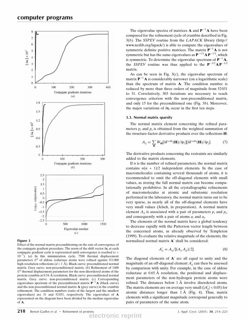
The ef®ciency of the preconditioning is illustrated in Figs.
3(a) and 3(b) for the two protein systems at subatomic reso-
lution. In the re®nement cycle described for aldose reductase,
the convergence requires 370 iterations, while with the
preconditioned matrix only 19 iterations are necessary (Fig.
3a). In both the preconditioned and the non-preconditioned
case, the parameter shifts �xi converge to the value obtained
by matrix inversion.
J. Appl. Cryst. (2001). 34, 214±223 Benoit Guillot et al. � Refinement of proteins 217
computer programs
Figure 2Static deformation electron density map in the ON1±PN±ON2 plane ofthe NAD+ pyrophosphate group: (a) without restraints; (b) with theexpansion/contraction coef®cients � and �0 of the phosphorus andpyrophosphate oxygen atoms restrained to the values extracted from themultipolar-parameters database (Table 2).
electronic reprint
computer programs
218 Benoit Guillot et al. � Refinement of proteins J. Appl. Cryst. (2001). 34, 214±223
The eigenvalue spectra of matrices A and Pÿ1A have been
compared for the re®nement cycle of crambin described in Fig.
3(b). The SSPEV routine from the LAPACK library (http://
www.netlib.org/lapack/) is able to compute the eigenvalues of
symmetric de®nite positive matrices. The matrix Pÿ1A is not
symmetric but has the same eigenvalues as Pÿ1/2APÿ1/2, whichis symmetric. To determine the eigenvalue spectrum of Pÿ1A,the SSPEV routine was thus applied to the Pÿ1/2APÿ1/2
matrix.
As can be seen in Fig. 3(c), the eigenvalue spectrum of
matrix Pÿ1A is considerably narrower (on a logarithmic scale)
than the spectrum of matrix A. The condition number is
reduced by more than three orders of magnitude from 52031
to 31. Correlatively, 303 iterations are necessary to reach
convergence criterion with the non-preconditioned matrix,
and only 15 for the preconditioned one (Fig. 3b). Moreover,
the major variations of �xi occur in the ®rst ten steps.
3.3. Normal matrix sparsity
The normal matrix element concerning the re®ned para-
meters pi and pj is obtained from the weighted summation of
the structure-factor derivative products over the re¯ectionsH:
Aij �P
H
WH�@F calc�H�=@pi��@F calc�H�=@pj�: �7�
The derivative products concerning the restraints are similarly
added to the matrix elements.
If n is the number of re®ned parameters, the normal matrix
contains n(n + 1)/2 independent elements. In the case of
macromolecules containing several thousands of atoms, it is
recommended to omit the off-diagonal elements with small
values, as storing the full normal matrix can become compu-
tationally prohibitive. In all the crystallographic re®nements
of macromolecules at atomic and subatomic resolution
performed in the laboratory, the normal matrix turns out to be
very sparse, as nearly all of the off-diagonal elements have
very small values (Jelsch, in preparation). A normal matrix
element Aij is associated with a pair of parameters pi and pj,
and consequently with a pair of atoms ai and aj.
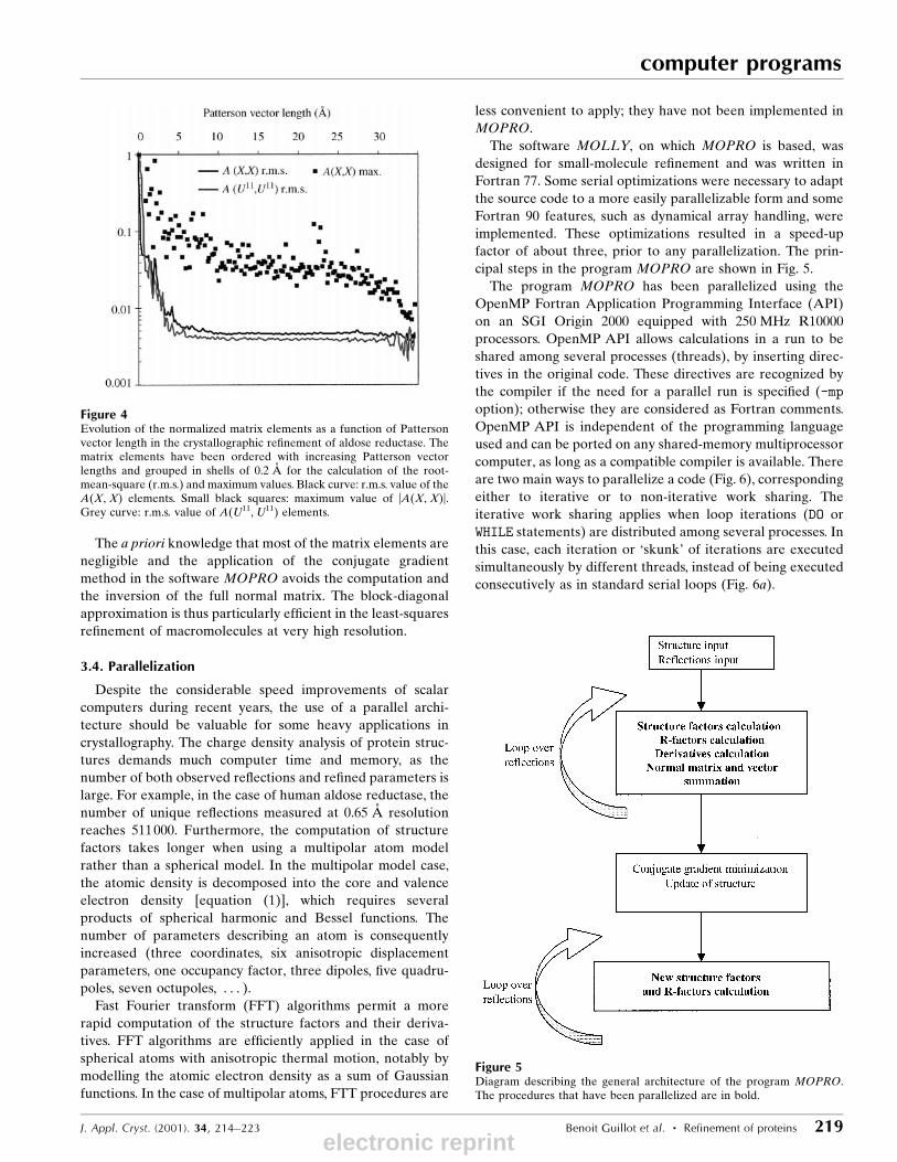
The elements of the normal matrix have a global tendency
to decrease rapidly with the Patterson vector length between
the concerned atoms, as already observed by Templeton
(1999). To evaluate the relative magnitude of the elements, the
normalized normal matrix A0 shall be considered:
A0ij � Aij=��Aii Ajj�=2�: �8�
The diagonal elements of A0 are all equal to unity and the
magnitude of an off-diagonal element A0ij can then be assessed
by comparison with unity. For example, in the case of aldose
reductase at 0.65 AÊ resolution, the positional and displace-
ment parameters of the non-hydrogen protein atoms were
re®ned. The distances below 1 AÊ involve disordered atoms.
The matrix elements are on average very small (|A0ij| < 0.05) for
atomic distances longer than 1 AÊ (Fig. 4). Thus, matrix
elements with a signi®cant magnitude correspond generally to
pairs of parameters of the same atom.
Figure 3Effect of the normal matrix preconditioning on the rate of convergence ofthe conjugate gradient procedure. The norm of the shift vector �xi at eachconjugate gradient cycle is represented until convergence is reached (" <10ÿ7). (a) In this minimization cycle, 7500 thermal displacementparameters Uij of aldose reductase atoms were re®ned against 311000high-resolution re¯ections (d < 1 AÊ ). Black curve: preconditioned normalmatrix. Grey curve: non-preconditioned matrix. (b) Re®nement of 1488Uij thermal displacement parameters for the non-disordered atoms of theprotein crambin at 0.54 AÊ resolution. Black curve: preconditioned normalmatrix. Grey curve: non-preconditioned matrix. (c) Correspondingeigenvalues spectrum of the preconditioned matrix Pÿ1A (black curve)and the non-preconditioned normal matrix A (grey curve) in the crambinre®nement. The condition numbers (ratio of the largest and the smallesteigenvalues) are 31 and 52031, respectively. The eigenvalues of Arepresented on the diagram have been divided by the median eigenvalueof A.
electronic reprint
The a priori knowledge that most of the matrix elements are
negligible and the application of the conjugate gradient
method in the software MOPRO avoids the computation and
the inversion of the full normal matrix. The block-diagonal
approximation is thus particularly ef®cient in the least-squares
re®nement of macromolecules at very high resolution.
3.4. Parallelization
Despite the considerable speed improvements of scalar
computers during recent years, the use of a parallel archi-
tecture should be valuable for some heavy applications in
crystallography. The charge density analysis of protein struc-
tures demands much computer time and memory, as the
number of both observed re¯ections and re®ned parameters is
large. For example, in the case of human aldose reductase, the
number of unique re¯ections measured at 0.65 AÊ resolution
reaches 511000. Furthermore, the computation of structure
factors takes longer when using a multipolar atom model
rather than a spherical model. In the multipolar model case,
the atomic density is decomposed into the core and valence
electron density [equation (1)], which requires several
products of spherical harmonic and Bessel functions. The
number of parameters describing an atom is consequently
increased (three coordinates, six anisotropic displacement
parameters, one occupancy factor, three dipoles, ®ve quadru-
poles, seven octupoles, . . . ).Fast Fourier transform (FFT) algorithms permit a more
rapid computation of the structure factors and their deriva-
tives. FFT algorithms are ef®ciently applied in the case of
spherical atoms with anisotropic thermal motion, notably by
modelling the atomic electron density as a sum of Gaussian
functions. In the case of multipolar atoms, FTT procedures are
less convenient to apply; they have not been implemented in
MOPRO.
The software MOLLY, on which MOPRO is based, was
designed for small-molecule re®nement and was written in
Fortran 77. Some serial optimizations were necessary to adapt
the source code to a more easily parallelizable form and some
Fortran 90 features, such as dynamical array handling, were
implemented. These optimizations resulted in a speed-up
factor of about three, prior to any parallelization. The prin-
cipal steps in the program MOPRO are shown in Fig. 5.
The program MOPRO has been parallelized using the
OpenMP Fortran Application Programming Interface (API)
on an SGI Origin 2000 equipped with 250 MHz R10000
processors. OpenMP API allows calculations in a run to be
shared among several processes (threads), by inserting direc-
tives in the original code. These directives are recognized by
the compiler if the need for a parallel run is speci®ed (-mp
option); otherwise they are considered as Fortran comments.
OpenMP API is independent of the programming language
used and can be ported on any shared-memory multiprocessor
computer, as long as a compatible compiler is available. There
are two main ways to parallelize a code (Fig. 6), corresponding
either to iterative or to non-iterative work sharing. The
iterative work sharing applies when loop iterations (DO or
WHILE statements) are distributed among several processes. In
this case, each iteration or `skunk' of iterations are executed
simultaneously by different threads, instead of being executed
consecutively as in standard serial loops (Fig. 6a).
J. Appl. Cryst. (2001). 34, 214±223 Benoit Guillot et al. � Refinement of proteins 219
computer programs
Figure 4Evolution of the normalized matrix elements as a function of Pattersonvector length in the crystallographic re®nement of aldose reductase. Thematrix elements have been ordered with increasing Patterson vectorlengths and grouped in shells of 0.2 AÊ for the calculation of the root-mean-square (r.m.s.) and maximum values. Black curve: r.m.s. value of theA(X, X) elements. Small black squares: maximum value of |A(X, X)|.Grey curve: r.m.s. value of A(U11, U11) elements.
Figure 5Diagram describing the general architecture of the program MOPRO.The procedures that have been parallelized are in bold.
electronic reprint

computer programs
220 Benoit Guillot et al. � Refinement of proteins J. Appl. Cryst. (2001). 34, 214±223
In the non-iterative work sharing, two or more independent
sections of the code are executed simultaneously by several
threads in the parallel construct (Fig. 6b). In a sequential run,
these sections would have been executed successively.
In both cases, the main process (master thread) of the
sequential part of the program splits into several threads when
a parallel directive is met. At the end of the parallel section or
loop, the program returns to the serial mode. A program with
frequent switches to parallel mode is denoted ®ne-grained, as
opposed to coarse-grained programs. As the threads have to
be synchronized in a parallel calculation and as the system
needs some time to switch to parallel mode, the time saving in
®ne-grained programs may be limited. This is particularly true
when many threads are used. More details about OpenMP are
available at http://www.openmp.org. Optimal parallel calcu-
lations are performed with one thread per processor, as a
number of threads larger than the number of available
processors does not lead to additional gains in speed.
The ef®ciency of the parallelization can be estimated from
the speed-up ratio s(p) = t(p = 1)/t(p), where t(p) is the
execution time for p threads (Fig. 7a). Another criterion is the
ef®ciency e(p) = s(p)/p (Fig. 7b). In the current paper, the
speed-up and ef®ciency are de®ned for the entire optimized
program, i.e. the execution time including both the sequential
and the parallel parts of the code.
In the serial execution of MOPRO, the structure factors,
their derivatives and the normal matrix-element calculations,
and secondly the conjugate gradients procedure, are the most
computer-time-consuming parts of the program, especially for
Figure 6(a) Schematic representation of the iterative work-sharing method. Aloop over N iterations is computed by one thread (top of the ®gure).After parallelization, it is chopped into three loops over N/3 iterationscomputed simultaneously by three threads (bottom). (b) Schematicrepresentation of the non-iterative work-sharing construct. At the top ofthe ®gure, three independent sections of the code are executedconsecutively by one thread. At the bottom, they are executedsimultaneously by three threads in parallel mode.
Figure 7Speed-up (a) and ef®ciency (b) of the parallelization as a function of thenumber of threads during one cycle of coordinates re®nement for aldosereductase at 0.65 AÊ resolution. The structure contains 5916 atoms; 10500variables were re®ned against 470000 observed structure factors.
electronic reprint

large systems with many re®ned parameters. The loop paral-
lelization method (iterative work sharing) was applied to these
parts of the software MOPRO. The computation of the
structure factors, of their derivatives and of the normal matrix
are built inside a loop over all the re¯ections. As each result is
independent from the others, the re¯ections were distributed
among several threads.
The speed-up and ef®ciency obtained by the parallelization
in the case of aldose reductase are shown in Fig. 7. The
re®nement cycle includes the matrix construction from the
structure factors and their derivatives (parallel loop), the
conjugate gradient minimization (serial) and a ®nal structure-
factor calculation (parallel) (Fig. 5). The execution time when
16 processors are used is reduced by a factor of ten to about
2 h. The parallelization thus allows an appreciable time saving
and renders the multipolar re®nement with the software
MOPRO applicable to macromolecules.
4. Electroneutrality constraint
The program MOPRO allows the re®nement of the valence
populations. An electroneutrality constraint on the atomic
charges may be necessary to keep the total number of elec-
trons constant. The electroneutrality constraint method initi-
ally implemented in MOLLY uses some properties of the
variance±covariance matrix (Hamilton, 1964), which is the
inverse of the normal matrix A. In MOPRO, with the use of
the conjugate gradients algorithm, the variance±covariance
matrix is not computed and this method is thus not applicable.
The method described by Raymond (1972) has therefore been
implemented in MOPRO. As the sum of the valence popu-
lations is set constant, the last parameter Pvn can be set to a
linear combination of the n ÿ 1 precedent independent
parameters and the number of variables is decreased by one.
Then, if �Pvi is the shift on the ith re®ned valence population,
the shift to apply to the last parameter is
�Pvn � ÿ P1;nÿ1
�Pvi: �9�
The derivatives of the structure factors F with respect to the n
ÿ 1 linearly independent Pv variables have then to be modi-
®ed accordingly:
@F=@Pvi ! @F=@Pvi ÿ @F=@Pvn: �10�This method leads to the same valence population shifts as
with the Hamilton (1964) method.
5. Estimated standard deviations
The properties of the estimated standard deviations (e.s.d.'s, or
standard uncertainties) of the crambin atomic coordinates
have been analysed at atomic (1 AÊ ) and subatomic (0.54 AÊ )
resolution. The e.s.d.'s discussed here were obtained by
inversion of the (non-full) normal matrix during a coordinates
re®nement cycle. Fig. 8 shows the e.s.d.'s for the X coordinates
as a function of the atomic thermal motion. No stereochemical
restraints were applied in this analysis. When the data were
further truncated at 1.5 AÊ resolution, the matrix turned out to
be singular. This illustrates that geometrical restraints are
necessary information to incorporate at that resolution.
The e.s.d.'s do increase, as expected, with the thermal
motion of the atoms (Stanley, 1965) and there is a twofold
increase of the precision of the coordinates from 1 to 0.54 AÊ
resolution. The e.s.d.'s of the coordinates are also clearly
dependent on the atom type, especially at 1 AÊ resolution (Fig.
8a). The positions of the six sulfur atoms of crambin are
de®ned with the best precision. The higher the scattering
factor of an atom in the considered resolution range, the more
precise are the coordinates. The scattering factor of an atom at
low resolution is proportional to total number of electrons
(core plus valence) of the atom. Therefore, at 1 AÊ resolution,
the carbon, nitrogen and oxygen atoms form three distin-
guished clusters in the B versus e.s.d. graph (Fig. 8a).
J. Appl. Cryst. (2001). 34, 214±223 Benoit Guillot et al. � Refinement of proteins 221
computer programs
Figure 8Estimated standard deviations of the X coordinates as a function of theequivalent isotropic thermal displacement parameter Beq = hBiii. Thecoordinates for the non-disordered parts of the protein crambin werere®ned. (a) All re¯ections to 0.54 AÊ resolution. (b) Truncation at 1 AÊ
resolution. The different atom types can be distinguished (sulfur: squares;oxygen: triangles; nitrogen: circles; carbon: crosses).
electronic reprint

computer programs
222 Benoit Guillot et al. � Refinement of proteins J. Appl. Cryst. (2001). 34, 214±223
On the other hand, at very high resolution (d < 0.9 AÊ ), the
diffraction essentially arises from the core electrons (Jelsch et
al., 1998). Thus, in the upper resolution ranges, the scattering
factors of the carbon, nitrogen and oxygen atoms are basically
similar, as there are two core electrons in all three cases. Thus,
when all re¯ections up to subatomic resolution are used, the
e.s.d.'s are less dissimilar for these three atom types (Fig. 8b).
Moreover, the e.s.d.'s diminish more in relative value for the
atoms with low thermal motion when the resolution is
increased from 1 to 0.54 AÊ . This correlates well with the fact
that the diffraction at subatomic resolution originates essen-
tially from the atoms with low thermal displacement para-
meters (B < 3 AÊ 2).
6. Conclusions
In the past, precise analyses of electronic distribution have
been restricted to small molecules, which generally satisfy the
necessary conditions of subatomic resolution and low thermal
motion. With the availability of third-generation synchrotron
X-ray beamlines, some protein diffraction data can now be
measured at subatomic resolution. The electron density
analysis of such macromolecules has thus become a feasible
challenge. Currently available multipolar re®nement
programs, dedicated to small molecules, would need a prohi-
bitively excessive amount of CPU time if applied to large
structures. Thus, the algorithmic optimizations and the paral-
lelization implemented inMOPRO were necessary in order to
re®ne the charge density of macromolecules, such as aldose
reductase, in a reasonable amount of computer time.
Provided that the protein atoms have standard names, a
transfer program is available to transform a SHELXL struc-
ture ®le into a MOPRO input ®le. The information contained
in the database of multipolar parameters (Pichon-Pesme et al.,
1995), notably the de®nition of the local axis system, is directly
transferred to the MOPRO input ®le containing the coordi-
nates, the thermal displacement and charge density para-
meters. These improvements, in conjunction with the
implementation of restraints necessary in macromolecular
crystallography, make the programMOPRO a reliable tool for
protein charge density analysis.
APPENDIX AThe conjugate gradient algorithm
The starting conditions of the iterative procedure applied in
MOPRO to solve equation (5) are
p0 � r0 � bÿA �x0; �11�
where �x0 is the initial parameters vector shift, usually set to
zero. The following iterative process is repeated to obtain
successive estimations of the parameters shift �xi:
�i � �rTi ri�=�pTi Api�; �12�
�xi�1 � �xi � �i pi; �13�
ri�1 � ri ÿ �iApi; �14�
�i�1 � �rTi�1 ri�1�=�rTi ri�; �15�
pi�1 � ri�1 � �i�1 pi; �16�where pi is the direction of search in the parameter space at
the ith iteration and ri is equal to the residual b ÿ A�xi at theith iteration.
The convergence is assumed to be reached when the resi-
dual norm ri is a signi®cantly small fraction " of the initial
residual norm r0. The tolerance term " is de®ned by the user
and was set to 10ÿ6.
APPENDIX BMatrix preconditioning
To apply the matrix preconditioning, the matrix P can be
decomposed according to the Cholesky factorization:
P � LLT : �17�The system of equations (5) can then be rewritten
Lÿ1ALTÿ1 �LT�x� � Lÿ1 b: �18�The conjugate gradient procedure can be rewritten according
to the previous equation, with A, �x and b replaced by
Lÿ1ALTÿ1, LT�x and Lÿ1b, respectively, but these equations
have the disadvantage that L and Lÿ1 must be computed and
that the solution �x0 must be transformed into the original set
of parameters by the transformation �x = (LT)ÿ1�x0. However,after application of the following variables substitution,
�x0 � LT�x; r0 � Lÿ1 r; p0 � LT p; �19�and with LTÿ1Lÿ1 = Pÿ1, the procedure can be rewritten in a
form in which only Pÿ1 is needed. The initial conditions are
r0 � bÿAx0; p0 � Pÿ1 r0: �20�The preconditioned conjugate gradient iterative procedure is
then unchanged for equations (13) and (14), while the equa-
tions (12), (15) and (16) are modi®ed into
�i � rTi Pÿ1 ri=p
Ti Api; �21�
�i�1 � rTi�1 Pÿ1 ri�1=r
Ti P
ÿ1 ri; �22�
pi�1 � Pÿ1 ri�1 � �i�1 pi: �23�
Professor Niels Hansen is gratefully acknowledged for
helpful discussions. We thank the French Ministry of Research
and Technology for ®nancial support to BG and RG.
References
Allen, F. H. (1986). Acta Cryst. B42, 515±522.BruÈnger, A. T., Adams, P. D., Clore, G. M., DeLano, W. L., Gros, P.,Grosse-Kunstleve, R. W., Jiang, J.-S., Kuszewski, J., Nilges, M.,
electronic reprint

Pannu, N. S., Read, R. J., Rice, L. M., Simonson, T. & Warren, G. L.(1998). Acta Cryst. D54, 905±921.
Coppens, P. (1967). Science, 158, 1577±1579.Flaig, R., Koritsanszky, T., Zobel, D. & Luger, P. (1998). J. Am. Chem.Soc. 120, 2227±2238.
Fletcher, R. & Reeves, C. M. (1964). Comput. J. 7, 149±154.Guillot, B., Jelsch, C. & Lecomte, C. (2000). Acta Cryst. C56, 726±728.
Hamilton, W. C. (1964). Statistics in Physical Science, EstimationHypothesis Testing and Least Squares, pp. 137±139. New York:Ronald Press.
Haneef, I., Moss, D. S., Stanford, M. J. & Borkakoti, N. (1985). ActaCryst. A41, 426±433.
Hansen, N. K. & Coppens, P. (1978). Acta Cryst. A34, 909±921.Hestenes, M. R. & Stiefel, E. (1952) J. Natl Bur. Stand. USA, 49, 409±436.
Hirshfeld, F. L. (1976). Acta Cryst. A32, 239±244.Housset, D., Pichon-Pesme, V., Jelsch, C., Benabicha, F., Maierhofer,A., David, S., Fontecilla-Camps, J. C. & Lecomte, C. (2000). ActaCryst. D56, 151±160.
Jelsch, C., Pichon-Pesme, V. & Lecomte, C. & Aubry, A. (1998). ActaCryst. D54, 1306±1318.
Jelsch, C., Teeter, M. M., Lamzin, V., Pichon-Pesme, V., Blessing, R.H. & Lecomte, C. (2000). Proc. Natl Acad. Sci. (USA), 97, 3171±3176.
Konnert, J. H. (1976). Acta Cryst. A32, 614±617.Konnert, J. H. & Hendrickson, W. A. (1980). Acta Cryst. A36, 344±350.
Lamour, V., Barth, P., Rogniaux, H., Poterszman, A., Howard, E.,Mitschler, A., Vandorsselaer, A., Podjarny, A. & Moras, D. (1999).Acta Cryst. D55, 721±723.
Mitschler, A., Sanishvili, R., Joachimiak, A., Howard, E., Barth, P.,Lamour, V., Guillot, B., Van Zandt, M., Sibley, E., Moras, D. &Podjarny, A. (2000). 19th European Crystallographic Meeting, 25±31 August 2000, Nancy. Poster s7.m0.p1.
Peres, N., Bouhkris, A., Souhassou, M., Gavoille, G. & Lecomte, C.(1999). Acta Cryst. A55, 1038±1048.
Pichon-Pesme, V., Lecomte, C. & Lachekar, H. (1995). J. Phys. Chem.99, 6242±6250.
Raymond, K. N. (1972). Acta Cryst. A28, 163±166.Rosen®eld, R. E. Jr, Trueblood, K. N. & Dunitz, J. D. (1978). ActaCryst. A34, 828±829.
Sheldrick, G. M. & Schneider, T. (1997) SHELXL: High-ResolutionRe®nement, in Methods in Enzymology, Vol. 276, MacromolecularCrystallography, Part B, edited by C. W. Carter Jr & R. M. Sweet.New York: Academic Press.
Stanley, E. (1965). Acta Cryst. 19, 1055±1056.Templeton, D. H. (1999). Acta Cryst. A55, 695±699.Tronrud, D. E. (1992). Acta Cryst. A48, 912±916.Urzhumtsev, A. G. (1991). Acta Cryst. A47, 723±727.Volkov, A., Abramov, Y., Coppens, P. & Gatti, C. (2000). Acta Cryst.A56, 332±339.
Volkov, A., Wu, G. & Coppens, P. (1999). J. Synchrotron Rad. 6, 1007±1015.
Yamano, A., Heo, N. H. & Teeter, M. M. (1997). J. Biol. Chem. 272,9597±9600.
J. Appl. Cryst. (2001). 34, 214±223 Benoit Guillot et al. � Refinement of proteins 223
computer programs
electronic reprint