Reduced transcription of TCOF1 in adult cells of Treacher Collins syndrome patients
7
BioMed Central Page 1 of 7 (page number not for citation purposes) BMC Medical Genetics Open Access Research article Reduced transcription of TCOF1 in adult cells of Treacher Collins syndrome patients Cibele Masotti 1 , Camila C Ornelas 1 , Alessandra Splendore-Gordonos 2 , Ricardo Moura 3 , Têmis M Félix 4 , Nivaldo Alonso 5 , Anamaria A Camargo 3 and Maria Rita Passos-Bueno* 1 Address: 1 Centro de Estudos do Genoma Humano, Instituto de Biociências, Universidade de São Paulo, São Paulo, SP, Brazil, 2 Department of Genetics, Stanford University, California, USA, 3 Ludwig Institute for Cancer Research, São Paulo Branch, Hospital Alemão Oswaldo Cruz, São Paulo, SP, Brazil, 4 Departamento de Genética Médica, Hospital de Clínicas de Porto Alegre, Universidade Federal do Rio Grande do Sul, Porto Alegre, RS, Brazil and 5 Departamento de Cirurgia Plástica, Hospital das Clínicas da Faculdade de Medicina, Universidade de São Paulo, SP, Brazil Email: Cibele Masotti - [email protected]; Camila C Ornelas - [email protected]; Alessandra Splendore- Gordonos - [email protected]; Ricardo Moura - [email protected]; Têmis M Félix - [email protected]; Nivaldo Alonso - [email protected]; Anamaria A Camargo - [email protected]; Maria Rita Passos-Bueno* - [email protected] * Corresponding author Abstract Background: Treacher Collins syndrome (TCS) is an autosomal dominant craniofacial disorder caused by frameshift deletions or duplications in the TCOF1 gene. These mutations cause premature termination codons, which are predicted to lead to mRNA degradation by nonsense mediated mRNA decay (NMD). Haploinsufficiency of the gene product (treacle) during embryonic development is the proposed molecular mechanism underlying TCS. However, it is still unknown if TCOF1 expression levels are decreased in post- embryonic human cells. Methods: We have estimated TCOF1 transcript levels through real time PCR in mRNA obtained from leucocytes and mesenchymal cells of TCS patients (n = 23) and controls (n = 18). Mutational screening and analysis of NMD were performed by direct sequencing of gDNA and cDNA, respectively. Results: All the 23 patients had typical clinical features of the syndrome and pathogenic mutations were detected in 19 of them. We demonstrated that the expression level of TCOF1 is 18-31% lower in patients than in controls (p < 0.05), even if we exclude the patients in whom we did not detect the pathogenic mutation. We also observed that the mutant allele is usually less abundant than the wild type one in mesenchymal cells. Conclusions: This is the first study to report decreased expression levels of TCOF1 in TCS adult human cells, but it is still unknown if this finding is associated to any phenotype in adulthood. In addition, as we demonstrated that alleles harboring the pathogenic mutations have lower expression, we herein corroborate the current hypothesis of NMD of the mutant transcript as the explanation for diminished levels of TCOF1 expression. Further, considering that TCOF1 deficiency in adult cells could be associated to pathologic clinical findings, it will be important to verify if TCS patients have an impairment in adult stem cell properties, as this can reduce the efficiency of plastic surgery results during rehabilitation of these patients. Published: 14 December 2009 BMC Medical Genetics 2009, 10:136 doi:10.1186/1471-2350-10-136 Received: 16 March 2009 Accepted: 14 December 2009 This article is available from: http://www.biomedcentral.com/1471-2350/10/136 © 2009 Masotti et al; licensee BioMed Central Ltd. This is an Open Access article distributed under the terms of the Creative Commons Attribution License (http://creativecommons.org/licenses/by/2.0 ), which permits unrestricted use, distribution, and reproduction in any medium, provided the original work is properly cited.
-
Upload
independent -
Category
Documents
-
view
2 -
download
0
Transcript of Reduced transcription of TCOF1 in adult cells of Treacher Collins syndrome patients
BioMed CentralBMC Medical Genetics
ss
Open AcceResearch articleReduced transcription of TCOF1 in adult cells of Treacher Collins syndrome patientsCibele Masotti1, Camila C Ornelas1, Alessandra Splendore-Gordonos2, Ricardo Moura3, Têmis M Félix4, Nivaldo Alonso5, Anamaria A Camargo3 and Maria Rita Passos-Bueno*1Address: 1Centro de Estudos do Genoma Humano, Instituto de Biociências, Universidade de São Paulo, São Paulo, SP, Brazil, 2Department of Genetics, Stanford University, California, USA, 3Ludwig Institute for Cancer Research, São Paulo Branch, Hospital Alemão Oswaldo Cruz, São Paulo, SP, Brazil, 4Departamento de Genética Médica, Hospital de Clínicas de Porto Alegre, Universidade Federal do Rio Grande do Sul, Porto Alegre, RS, Brazil and 5Departamento de Cirurgia Plástica, Hospital das Clínicas da Faculdade de Medicina, Universidade de São Paulo, SP, Brazil
Email: Cibele Masotti - [email protected]; Camila C Ornelas - [email protected]; Alessandra Splendore-Gordonos - [email protected]; Ricardo Moura - [email protected]; Têmis M Félix - [email protected]; Nivaldo Alonso - [email protected]; Anamaria A Camargo - [email protected]; Maria Rita Passos-Bueno* - [email protected]
* Corresponding author
AbstractBackground: Treacher Collins syndrome (TCS) is an autosomal dominant craniofacial disorder causedby frameshift deletions or duplications in the TCOF1 gene. These mutations cause premature terminationcodons, which are predicted to lead to mRNA degradation by nonsense mediated mRNA decay (NMD).Haploinsufficiency of the gene product (treacle) during embryonic development is the proposed molecularmechanism underlying TCS. However, it is still unknown if TCOF1 expression levels are decreased in post-embryonic human cells.
Methods: We have estimated TCOF1 transcript levels through real time PCR in mRNA obtained fromleucocytes and mesenchymal cells of TCS patients (n = 23) and controls (n = 18). Mutational screening andanalysis of NMD were performed by direct sequencing of gDNA and cDNA, respectively.
Results: All the 23 patients had typical clinical features of the syndrome and pathogenic mutations weredetected in 19 of them. We demonstrated that the expression level of TCOF1 is 18-31% lower in patientsthan in controls (p < 0.05), even if we exclude the patients in whom we did not detect the pathogenicmutation. We also observed that the mutant allele is usually less abundant than the wild type one inmesenchymal cells.
Conclusions: This is the first study to report decreased expression levels of TCOF1 in TCS adult humancells, but it is still unknown if this finding is associated to any phenotype in adulthood. In addition, as wedemonstrated that alleles harboring the pathogenic mutations have lower expression, we hereincorroborate the current hypothesis of NMD of the mutant transcript as the explanation for diminishedlevels of TCOF1 expression. Further, considering that TCOF1 deficiency in adult cells could be associatedto pathologic clinical findings, it will be important to verify if TCS patients have an impairment in adult stemcell properties, as this can reduce the efficiency of plastic surgery results during rehabilitation of thesepatients.
Published: 14 December 2009
BMC Medical Genetics 2009, 10:136 doi:10.1186/1471-2350-10-136
Received: 16 March 2009Accepted: 14 December 2009
This article is available from: http://www.biomedcentral.com/1471-2350/10/136
© 2009 Masotti et al; licensee BioMed Central Ltd. This is an Open Access article distributed under the terms of the Creative Commons Attribution License (http://creativecommons.org/licenses/by/2.0), which permits unrestricted use, distribution, and reproduction in any medium, provided the original work is properly cited.
Page 1 of 7(page number not for citation purposes)
BMC Medical Genetics 2009, 10:136 http://www.biomedcentral.com/1471-2350/10/136
BackgroundTreacher Collins syndrome (TCS; OMIM 154500) is a rareautosomal dominant craniofacial disorder (1:50.000)characterized by bilateral and symmetrical malforma-tions, which frequently includes hypoplasia of the mandi-ble and zygomatic complex, down-slanting palpebralfissures, coloboma of the lower eyelid and absence of eye-lashes medial to this defect, external and middle ear mal-formation, and conductive hearing loss [1]. Thepenetrance is considered to be complete, but there is ahigh inter and intra-familial phenotypic variation, rang-ing from cases with perinatal death due to airway obstruc-tion by severe orofacial malformations to those that arenot clinically diagnosed [2].
In most of the cases the disorder is caused by frameshiftdeletions or duplications of 1-41 bp in TCOF1 codingregion which cause premature termination codons (PTC).Except for a recurring 5 bp deletion in exon 24 that isresponsible for 17% of the cases, mutations are usuallyfamily-specific [3]. TCOF1 gene product is a nucleolarprotein (treacle) involved in rRNA transcription and inpre-rRNA post-transcriptional modification [4]. Trun-cated proteins are not detected in TCS patients' fibroblastsand lymphocytes, suggesting that mRNA bearing PTCs arebeing degraded by nonsense mediated mRNA decay(NMD) [5]; however, NMD has never been demonstratedin human or mouse cells with null mutations in TCOF1.
No genotype-phenotype correlation has been observed inTCS and there is also no evidence of association betweenthe disease severity and parental origin or type of the path-ogenic mutation, male or female sex, sporadic or familialcases [5-9].
TCOF1 is expressed in various adult and embryonic tis-sues and haploinsufficiency of treacle during embryonicdevelopment has been proposed as the molecular mecha-nism underlying TCS [10]. In situ hybridization studies ofthe Tcof1 orthologue demonstrated that there is a peak ofexpression of the gene in E8.5-9.5 mice embryos, espe-cially in the first and second pharyngeal arches [11]. Crit-ical dosage of Tcof1 for appropriate craniofacialdevelopment has been further demonstrated in Tcof1+/-mice [12,13]. Interestingly, a previous study observed thatcellular amount of treacle in fibroblasts and lymphocytesderived from TCS patients was indistinguishable fromthat of control individuals, and the authors suggested thata dosage compensation mechanism could occur in adult-hood to compensate the null allele [5]. However, until thepresent, these findings have not been confirmed.
The relevance of TCOF1 happloinsufficiency during adultlife is still rarely studied: it can predispose to age relatedmacular degeneration [14] and explain the absence oflong-term stable results in mandibular distraction for
facial reconstruction [15-17]. These findings might implythat TCOF1 adult stem cells present a decreased regenera-tive capacity in comparison to wild-type cells. We havetherefore conducted the present study to verify if TCOF1expression is altered in adult TCS cells as well as to iden-tify a source of human tissue suitable for functional stud-ies of treacle in adulthood.
MethodsPatients and controlsWe studied peripheral blood samples from 20 TCSpatients (TCS1 to 20, ranging from 3 to 41 years old)referred to our center for genetic counseling and 12 con-trols (ranging from 20 to 50 years old). We also obtainedtissue samples from TCS16, from three other patients(TCS21 to 23), and from six controls submitted to recon-structive plastic surgery at University of São Paulo MedicalSchool. The study was approved by the ethical committeeof our Institution and informed consent was obtainedfrom both patients and control subjects or from their legaltutors. In total, 23 TCS patients were studied.
Isolation of cells from periosteum of TCS patientsDuring corrective surgery, overlying periosteum from theface of four TCS patients (two males and two females agedfrom 6 to 29 years) was meticulously dissected away fromsurrounding tissues to isolate intact periosteal flaps. Con-trol periosteum was obtained using the same procedurefrom facial region of six subjects (four males and twofemales aged from 11 months to 20 years) with no evi-dence of bone disease during corrective surgery.
Mesenchymal cells were isolated as described by previousreports from our group [18,19]. The periosteal flaps werethoroughly washed with sterile phosphate-buffered saline(PBS) supplemented with 4% antibiotics (100 units/mLpenicillin and 100 mg/mL streptomycin; Invitrogen), anddigested with trypsin solution (TrypLe; Invitrogen) for 1 hat 37°C. Once digested, the tissue was transferred withminimal dissection into 35 mm Petri dishes (Corning,NY) containing Dulbecco's modified Eagle's medium(DMEM; GIBCO) with 10% fetal bovine serum (FBS;GIBCO), 100 units/mL penicillin, and 100 mg/mL strep-tomycin (Invitrogen). After two weeks, cells were washedwith PBS, then dissociated in trypsin solution and seededat 104 cells per 25 cm2 for the first passage. For expansion,cells were cultured in monolayer in growth medium at37°C in a humidified atmosphere of 5% CO2. Themedium was replaced every 3 days. In order to prevent celldifferentiation, cultures were maintained semiconfiuentand subcultured every 4-5 days with daily mediumchanges.
Flow cytometryCells were harvested with TrypLe (Invitrogen), washedwith PBS, and incubated at 4°C for 30 min with the fol-
Page 2 of 7(page number not for citation purposes)
BMC Medical Genetics 2009, 10:136 http://www.biomedcentral.com/1471-2350/10/136
lowing anti-human antibodies: CD29-PE CY5, CD90(Thy-1), CD45-FITC, CD73, CD105, CD117, CD 31-PE(Becton Dickinson) and SH3 (Case Western Reserve Uni-versity). After the wash, unconjugated primary antibodieswere incubated with anti-mouse-PE secondary antibody(Guava Technologies) for additional 15 min at 4°C.Finally, the cell suspension was washed with PBS, and 104
labeled cells were acquired with an EasyCyte flow cytom-eter (Guava Technologies). Control samples were incu-bated with PBS instead of primary antibody, followed byincubation with anti-mouse-PE secondary antibody. Allthe generated plots were analyzed in Guava ExpressPlussoftware (Guava Technologies).
DNA and RNA isolation and cDNA synthesisGenomic DNA from peripheral blood samples wasobtained according to reference [20] and genomic DNAfrom culture cells was extracted using NucleoSpin Tissueextraction kits (Macherey-Nagel). Total RNA was isolatedfrom leucocytes and mesenchymal cells using TRIzol®
(Gibco BRL), treated with DNAse (Promega) and submittedto reverse transcription using Superscript II Reverse Tran-scriptase (Gibco BRL), according to manufacturer's proto-col. Aliquots from DNAse treated RNAs were used foramplification of an intronic region of MLH1 gene as acontrol for DNA contamination (primers sequences onrequest).
Mutation screeningSeven of the 23 patients included in the present reportwere previously studied by our group [3,9,21]. Pathogenicmutations were identified in six of these seven individuals(Table 1). Except for patient TCS17, for whom we did nothave enough DNA, all the other 15 patients were submit-ted to molecular analysis to identify the pathogenic muta-tion. Screening of TCOF1 mutations by direct sequencingof genomic DNA was performed with primers describedelsewhere [9]. Sequencing of the patients' cDNA was per-formed in identical conditions, but using primers thatamplify only the coding region flanking the pathogenicmutation (see Additional file 1).
Real Time PCRReal time quantitative PCR reactions were performed induplicates with a final volume of 20 μl, using 12.5 ng ofcDNA, 1× SYBR Green PCR Master Mix (Applied Biosystems)and 200 or 400 nM of each primer. We used ABI Prism7700 Sequence Detection System (Applied Biosystems) withstandard temperature protocol. Primers were designedwith Primer Express software V.2.0 (Applied Biosystems;primers sequence in Additional file 1) and the amplifica-tion efficiency (E) of each primer was calculated accordingto the equation E = 10(-1/slope). The expression data ofTCOF1 transcripts was determined by relative quantifica-tion in comparison to a pool of RNA from 10 controls.
Four endogenous controls genes (GAPDH, BCRP, HPRT1,and HMBS), were used and their stability was verifiedthrough geNorm VBA applet designed for Microsoft Excel.This tool calculates the most stable reference genes from aset of tested candidate reference genes in a given samplepanel, and calculates the gene expression normalizationfactor (NF) for each target sample based on the geometricmean of a user-defined number of housekeeping genes[22]. We calculated the NF for each sample based on thefour endogenous controls and the expression data was cal-culated according to reference [23].
ResultsMolecular characterization of the TCS patientsPathogenic mutations were identified in 13 of the novel15 screened individuals: five had already been reported aspathogenic [9,21,24] and eight are herein described forthe first time. These new mutations were considered path-ogenic because they are a nonsense mutation(c.1609C>T), disrupt splicing (c.639+1G>A) or alter thereading frame (c.3853delC, c.1298delC,c.218_222insAACC, c.4344dupA, c.431delC). Mutationc.4375_4377delAAG (TCS18) causes an in phase deletionand excludes a Lysine residue from the protein C-terminalnucleolar localization signal [25]. In Table 1, mutationand tissue availability of each patient are depicted.
RT-PCR in leucocytesWe first verified the range of TCOF1 transcript levels inleucocytes from 20 patients (Table 1) and 12 controls byreal-time PCR, using as reference a pool of RNA from 10controls. We observed a wide variation in TCOF1 expres-sion in leucocytes from normal individuals (Figure 1; SD= 1.24). This wide variation of gene expression was alsoobserved among TCS patients (Table 1; SD = 1.46), andwas not correlated to age (p = 0.39; Two-tailed Pearsoncorrelation regression).
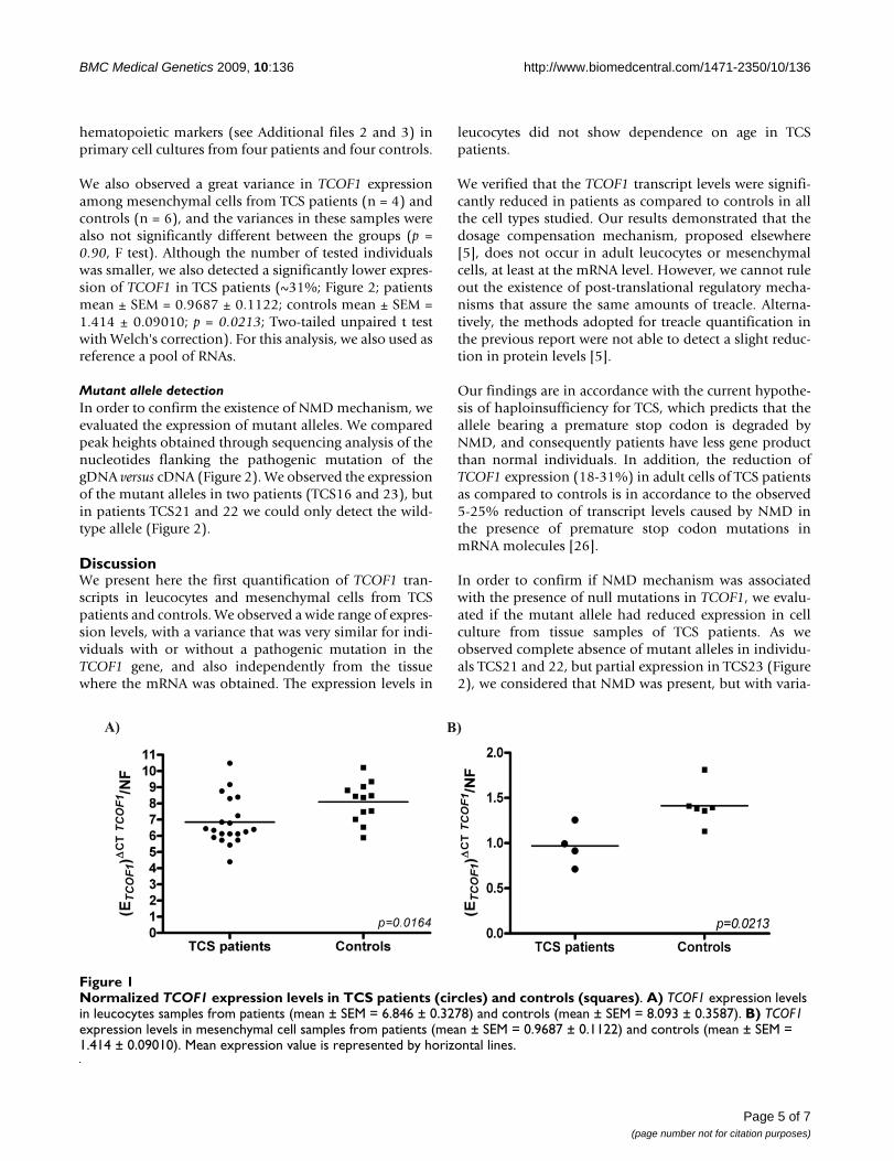
Comparing patients (mean ± SEM = 6.846 ± 0.3278; N =20) and controls (mean ± SEM = 8.093 ± 0.3587; N = 12),we observed that TCOF1 expression levels were signifi-cantly lower in patients than in controls (p = 0.0164; Two-tailed unpaired t test with Welch's correction; Figure 1),and the variances were not significantly different betweenthese two groups (p = 0.5828, F test). TCS patients hadapproximately 18% less TCOF1 transcript levels than nor-mal individuals. Excluding the four patients without anidentified pathogenic mutation, we obtained similarresults (~18.5%, p = 0.0039).
RT-PCR in mesenchymal cellsWe also examined whether TCOF1 expression levels weredecreased in mesenchymal cells. Flow cytometry resultsshowed that most of the cells were positive for mesenchy-mal cell markers (>95%) and negative for endothelial and
Page 3 of 7(page number not for citation purposes)
BMC Medical Genetics 2009, 10:136 http://www.biomedcentral.com/1471-2350/10/136
Page 4 of 7(page number not for citation purposes)
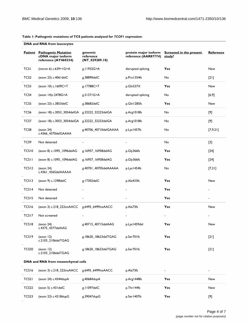
Table 1: Pathogenic mutations of TCS patients analyzed for TCOF1 expression:
DNA and RNA from leucocytes
Patient Pathogenic MutationcDNA major isoformreference (AY460334)
genomicreference (NT_029289.10)
protein major isoformreference (AAR87774)
Screened in the present study?
Reference
TCS1 (intron 6) c.639+1G>A g.11932G>A disrupted splicing Yes New
TCS2 (exon 23) c.4061delC g.38890delC p.Pro1354fs No [21]
TCS3 (exon 10) c.1609C>T g.17788C>T p.Gln537X Yes New
TCS4 (exon 14)c.2478G>A g.21371G>A disrupted splicing No [6,9]
TCS5 (exon 23) c.3853delC g.38682delC p.Gln1285fs Yes New
TCS6 (exon 18) c.3053_3054delGA g.32222_32223delGA p.Arg1018fs No [9]
TCS7 (exon 18) c.3053_3054delGA g.32222_32223delGA p.Arg1018fs No [9]
TCS8 (exon 24) c.4366_4370delGAAAA
g.40706_40710delGAAAA p.Lys1457fs No [7,9,21]
TCS9 Not detected - - No [3]
TCS10 (exon 8) c.1095_1096delAG g.16957_16958delAG p.Gly366fs Yes [24]
TCS11 (exon 8) c.1095_1096delAG g.16957_16958delAG p.Gly366fs Yes [24]
TCS12 (exon 24) c.4361_4365delAAAAA
g.40701_40705delAAAAA p.Lys1454fs No [7,21]
TCS13 (exon 9) c.1298delC g.17302delC p.Ala433fs Yes New
TCS14 Not detected - - Yes -
TCS15 Not detected - - Yes -
TCS16 (exon 3) c.218_222insAACC g.6495_6499insAACC p.Ala73fs Yes New
TCS17 Not screened - - - -
TCS18 (exon 24) c.4375_4377delAAG
g.40715_40715delAAG p.Lys1459del Yes New
TCS19 (exon 12) c.2103_2106delTGAG
g.18620_18623delTGAG p.Ser701fs Yes [21]
TCS20 (exon 12) c.2103_2106delTGAG
g.18620_18623delTGAG p.Ser701fs Yes [21]
DNA and RNA from mesenchymal cells
TCS16 (exon 3) c.218_222insAACC g.6495_6499insAACC p.Ala73fs - -
TCS21 (exon 24) c.4344dupA g.40684dupA p.Arg1448fs Yes New
TCS22 (exon 5) c.431delC g.11097delC p.Thr144fs Yes New
TCS23 (exon 23) c.4218dupG g.39047dupG p.Ser1407fs Yes [9]
BMC Medical Genetics 2009, 10:136 http://www.biomedcentral.com/1471-2350/10/136
hematopoietic markers (see Additional files 2 and 3) inprimary cell cultures from four patients and four controls.
We also observed a great variance in TCOF1 expressionamong mesenchymal cells from TCS patients (n = 4) andcontrols (n = 6), and the variances in these samples werealso not significantly different between the groups (p =0.90, F test). Although the number of tested individualswas smaller, we also detected a significantly lower expres-sion of TCOF1 in TCS patients (~31%; Figure 2; patientsmean ± SEM = 0.9687 ± 0.1122; controls mean ± SEM =1.414 ± 0.09010; p = 0.0213; Two-tailed unpaired t testwith Welch's correction). For this analysis, we also used asreference a pool of RNAs.
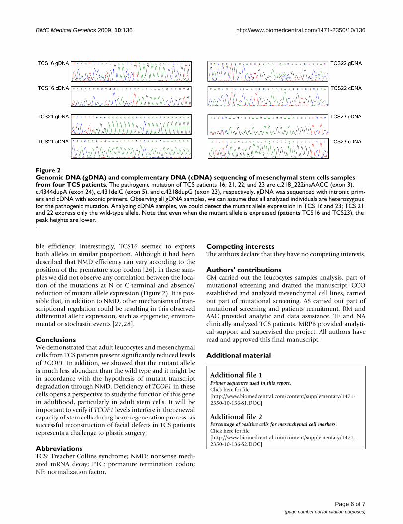
Mutant allele detectionIn order to confirm the existence of NMD mechanism, weevaluated the expression of mutant alleles. We comparedpeak heights obtained through sequencing analysis of thenucleotides flanking the pathogenic mutation of thegDNA versus cDNA (Figure 2). We observed the expressionof the mutant alleles in two patients (TCS16 and 23), butin patients TCS21 and 22 we could only detect the wild-type allele (Figure 2).
DiscussionWe present here the first quantification of TCOF1 tran-scripts in leucocytes and mesenchymal cells from TCSpatients and controls. We observed a wide range of expres-sion levels, with a variance that was very similar for indi-viduals with or without a pathogenic mutation in theTCOF1 gene, and also independently from the tissuewhere the mRNA was obtained. The expression levels in
leucocytes did not show dependence on age in TCSpatients.
We verified that the TCOF1 transcript levels were signifi-cantly reduced in patients as compared to controls in allthe cell types studied. Our results demonstrated that thedosage compensation mechanism, proposed elsewhere[5], does not occur in adult leucocytes or mesenchymalcells, at least at the mRNA level. However, we cannot ruleout the existence of post-translational regulatory mecha-nisms that assure the same amounts of treacle. Alterna-tively, the methods adopted for treacle quantification inthe previous report were not able to detect a slight reduc-tion in protein levels [5].
Our findings are in accordance with the current hypothe-sis of haploinsufficiency for TCS, which predicts that theallele bearing a premature stop codon is degraded byNMD, and consequently patients have less gene productthan normal individuals. In addition, the reduction ofTCOF1 expression (18-31%) in adult cells of TCS patientsas compared to controls is in accordance to the observed5-25% reduction of transcript levels caused by NMD inthe presence of premature stop codon mutations inmRNA molecules [26].
In order to confirm if NMD mechanism was associatedwith the presence of null mutations in TCOF1, we evalu-ated if the mutant allele had reduced expression in cellculture from tissue samples of TCS patients. As weobserved complete absence of mutant alleles in individu-als TCS21 and 22, but partial expression in TCS23 (Figure2), we considered that NMD was present, but with varia-
Normalized TCOF1 expression levels in TCS patients (circles) and controls (squares)Figure 1Normalized TCOF1 expression levels in TCS patients (circles) and controls (squares). A) TCOF1 expression levels in leucocytes samples from patients (mean ± SEM = 6.846 ± 0.3278) and controls (mean ± SEM = 8.093 ± 0.3587). B) TCOF1 expression levels in mesenchymal cell samples from patients (mean ± SEM = 0.9687 ± 0.1122) and controls (mean ± SEM = 1.414 ± 0.09010). Mean expression value is represented by horizontal lines.
Page 5 of 7(page number not for citation purposes)
BMC Medical Genetics 2009, 10:136 http://www.biomedcentral.com/1471-2350/10/136
ble efficiency. Interestingly, TCS16 seemed to expressboth alleles in similar proportion. Although it had beendescribed that NMD efficiency can vary according to theposition of the premature stop codon [26], in these sam-ples we did not observe any correlation between the loca-tion of the mutations at N or C-terminal and absence/reduction of mutant allele expression (Figure 2). It is pos-sible that, in addition to NMD, other mechanisms of tran-scriptional regulation could be resulting in this observeddifferential allelic expression, such as epigenetic, environ-mental or stochastic events [27,28].
ConclusionsWe demonstrated that adult leucocytes and mesenchymalcells from TCS patients present significantly reduced levelsof TCOF1. In addition, we showed that the mutant alleleis much less abundant than the wild type and it might bein accordance with the hypothesis of mutant transcriptdegradation through NMD. Deficiency of TCOF1 in thesecells opens a perspective to study the function of this genein adulthood, particularly in adult stem cells. It will beimportant to verify if TCOF1 levels interfere in the renewalcapacity of stem cells during bone regeneration process, assuccessful reconstruction of facial defects in TCS patientsrepresents a challenge to plastic surgery.
AbbreviationsTCS: Treacher Collins syndrome; NMD: nonsense medi-ated mRNA decay; PTC: premature termination codon;NF: normalization factor.
Competing interestsThe authors declare that they have no competing interests.
Authors' contributionsCM carried out the leucocytes samples analysis, part ofmutational screening and drafted the manuscript. CCOestablished and analyzed mesenchymal cell lines, carriedout part of mutational screening. AS carried out part ofmutational screening and patients recruitment. RM andAAC provided analytic and data assistance. TF and NAclinically analyzed TCS patients. MRPB provided analyti-cal support and supervised the project. All authors haveread and approved this final manuscript.
Additional material
Additional file 1Primer sequences used in this report.Click here for file[http://www.biomedcentral.com/content/supplementary/1471-2350-10-136-S1.DOC]
Additional file 2Percentage of positive cells for mesenchymal cell markers.Click here for file[http://www.biomedcentral.com/content/supplementary/1471-2350-10-136-S2.DOC]
Genomic DNA (gDNA) and complementary DNA (cDNA) sequencing of mesenchymal stem cells samples from four TCS patientsFigure 2Genomic DNA (gDNA) and complementary DNA (cDNA) sequencing of mesenchymal stem cells samples from four TCS patients. The pathogenic mutation of TCS patients 16, 21, 22, and 23 are c.218_222insAACC (exon 3), c.4344dupA (exon 24), c.431delC (exon 5), and c.4218dupG (exon 23), respectively. gDNA was sequenced with intronic prim-ers and cDNA with exonic primers. Observing all gDNA samples, we can assume that all analyzed individuals are heterozygous for the pathogenic mutation. Analyzing cDNA samples, we could detect the mutant allele expression in TCS 16 and 23; TCS 21 and 22 express only the wild-type allele. Note that even when the mutant allele is expressed (patients TCS16 and TCS23), the peak heights are lower.
Page 6 of 7(page number not for citation purposes)
BMC Medical Genetics 2009, 10:136 http://www.biomedcentral.com/1471-2350/10/136
AcknowledgementsWe would like to gratefully acknowledge the patients and their relatives. We also thank Constância Gotto for secretarial assistance, Regina C. Min-groni Netto for some samples, and Diogo Meyer for helpful discussions and for carefully reading this manuscript. This work was supported by grants from Fundação de Amparo à Pesquisa do Estado de São Paulo (FAPESP/CEPID), Coordenação de Aperfeiçoamento de Pessoal de Nível Superior (CAPES), and Conselho Nacional de Pesquisa (CNPq).
References1. Gorlin RJ, Cohen MM, Hennekam RC: Branchial Arch and oral-
Acral Disorders. In Syndromes of the Head and Neck 4th edition.Oxford: Oxford University Press; 2001:790-849.
2. Dixon MJ, Marres HAM, Edwards SJ, Dixon J, Cremers CWRJ:Treacher Collins syndrome: correlation between clinical andgenetic linkage studies. Clin Dysmorphol 1994, 3:96-103.
3. Splendore A, Fanganiello RD, Masotti C, Morganti LSC, Passos-BuenoMR: TCOF1 mutation database: novel mutation in the alter-natively spliced exon 6A and update in mutation nomencla-ture. Hum Mut 2005, 25:429-434.
4. Gonzales B, Henning D, So RB, Dixon J, Dixon MJ, Valdez BC: TheTreacher Collins syndrome (TCOF1) gene product isinvolved in pre-rRNA methylation. Hum Mol Gene 2005,14(14):2035-43.
5. Isaac C, Marsh KL, Panzekas WA, Dixon J, Dixon MJ, Jabs EW, MeierUT: Characterization of the nucleolar gene product, treacle,in Treacher Collins syndrome. Mol Biol Cell 2001, 11:188-192.
6. Gladwin AJ, Dixon J, Loftus SK, Edwards S, Wasmuth JJ, HennekamRC, Dixon MJ: Treacher Collins syndrome may result frominsertions, deletions or splicing mutations, which introducea termination codon into the gene. Hum Mol Gen 1996,5:1533-1538.
7. Edwards SJ, Gladwin AJ, Dixon MJ: The Mutational Spectrum inTreacher Collins Syndrome Reveals a Predominance ofMutations That Create a Premature-Termination Codon.Am J Hum Genet 1997, 60:515-524.
8. Teber OA, Gillessen-Kaesbach G, Fischer S, Böhringer S, Albrecht B,Albert A, Arslan-Kirchner M, Haan E, Hagedorn-Greiwe M, HammansC, Henn W, Hinkel GK, König R, Kunstmann E, Kunze J, NeumannLM, Prott EC, Rauch A, Rott HD, Seidel H, Spranger S, Sprengel M,Zoll B, Lohmann DR, Wieczorek D: Genotyping in 46 patientswith tentative diagnosis of Treacher Collins syndromerevealed unexpected phenotypic variation. Eur J Hum Genet2004, 12(11):879-90.
9. Splendore A, Silva EO, Alonso LG, Richieri-Costa A, Alonso N, RosaA, Carakushanky G, Cavalcanti DP, Brunoni D, Passos-Bueno MR:High mutation detection rate in TCOF1 among TreacherCollins syndrome patients reveals clustering of mutations in16 novel pathogenic changes. Hum Mutat 2000, 16:315-322.
10. The Treacher Collins Collaborative Group: Positional cloning of agene involved in the pathogenesis of Treacher Collins syn-drome. Nat Gen 1996, 12:130-136.
11. Dixon J, Hovanes K, Shiang R, Dixon MJ: Sequence analysis, iden-tification of evolutionary conserved motifs and expressionanalysis of murine Tcof1 provide further evidence for apotential function for the gene and its human homologue,TCOF1. Hum Mol Genet 1997, 6:727-737.
12. Dixon J, Brakebusch C, Fassler R, Dixon M: Increased levels ofapoptosis in the prefusion neural folds underlie the craniofa-cial disorder, Treacher Collins. Hum Mol Gen 2000, 9:1473-1480.
13. Dixon J, Jones NC, Sandell LL, Jayasinghe SM, Crane J, Rey JP, DixonMJ, Trainor PA: Tcof1/Treacle is required for neural crest cellformation and proliferation deficiencies that cause craniofa-cial abnormalities. Proc Natl Acad Sci USA 2006, 103(36):13403-8.
14. Goverdhan SV, Temple IK, Self J, Lotery AJ, Dixon MJ, Evans AR: Macu-lar degeneration associated with a novel Treacher CollinsTCOF1 mutation and evaluation of this mutation in age relatedmacular degeneration. Br J Ophthalmol 2005, 89(8):1063-4.
15. Karp NS, McCarthy JG, Schreiber JS, Sissons HA, Thorne CH: bonelengthening: a serial histological study. Ann Plast Surg 1992,29(1):2-7.
16. Stelnicki EJ, Lin WY, Lee C, Grayson BH, McCarthy JG: Long-termoutcome study of bilateral mandibular distraction: a com-parison of Treacher Collins and Nager syndromes to othertypes of micrognathia. Plast Reconstr Surg 2002, 109(6):1819-25.
17. Gürsoy S, Hukki J, Hurmerinta K: Five year follow-up of mandib-ular distraction osteogenesis on the dentofacial structures ofsyndromic children. Orthod Craniofac Res 2008, 11:57-64.
18. Bueno DF, Kerkis I, Costa AM, Martins MT, Kobayashi GS, ZucconiE, Fanganiello RD, Salles FT, Almeida AB, do Amaral CE, Alonso N,Passos-Bueno MR: New Source of Muscle-Derived Stem Cellswith Potential for Alveolar Bone Reconstruction in Cleft Lipand/or Palate Patients. Tissue Eng Part A 2009, 15(2):427-35.
19. Fanganiello RD, Sertié AL, Reis EM, Yeh E, Oliveira NA, Bueno DF,Kerkis I, Alonso N, Cavalheiro S, Matsushita H, Freitas R, Verjovski-Almeida S, Passos-Bueno MR: Apert p.Ser252Trp mutation inFGFR2 alters osteogenic potential and gene expression ofcranial periosteal cells. Mol Med 2007, 13(7-8):422-42.
20. Miller SA, Dykes DD, Polesky HF: A simple testing out proce-dure for extracting DNA from human nucleated cells. NucleicAcids Res 1998, 16:1215.
21. Splendore A, Jabs EW, Passos-Bueno MR: Screening of TCOF1 inpatients from different populations: confirmation of muta-tional hot spots and identification of a novel missense muta-tion that suggests an important functional domain in theprotein treacle. J Med Genet 2002, 39:493-495.
22. Vandesompele J, De Preter K, Pattyn F, Poppe B, Van Roy N, DePaepe A, Speleman F: Accurate normalization of real-timequantitative RT-PCR data by geometric averaging of multi-ple internal control genes. Genome Biol 2002,3(7):RESEARCH0034.
23. Pfaffl MW: A new mathematical model for quantification inreal-time-RT-PCR. Nucleic Acids Research 2001:2002-7.
24. Dixon J, Ellis I, Bottani A, Temple K, Dixon MJ: Identification ofmutations in TCOF1: use of molecular analysis in the pre- andpostnatal diagnosis of Treacher Collins syndrome. Am J MedGenet A 2004, 127A(3):244-8.
25. Winokur ST, Shiang R: The Treacher Collins syndrome(TCOF1) gene product, treacle, is targeted to the nucleolusby signals in the C-terminus. Hum Mol Genet 1998, 7:1947-1952.
26. Kuzmiak HA, Maquat LE: Applying nonsense-metiated mRNAdacay research to the clinic: progress and challenges.TRENDS Mol Med 2006, 12(7):306-316.
27. Gimelbrant A, Hutchinson JN, Thompson BR, Chess A: Widespreadmonoallelic expression on human autosomes. Science 2007,318(5853):1136-40.
28. Kaern M, Elston TC, Blake WJ, Collins J: Stochasticity in geneexpression: from theories to phenotypes. Nat Rev Genet 2005,6(6):451-64.
Pre-publication historyThe pre-publication history for this paper can be accessedhere:
http://www.biomedcentral.com/1471-2350/10/136/prepub
Additional file 3Flow cytometry analysis of mesenchymal cells from patients TCS16, 21, 22, and 23. Values represent the mean percentage of all assessed cells positively stained by the indicated antigens (CD31, CD73 (SH4), (SH2) CD105) and analyzed by flow cytometry. Graphs show relative number of cells (events) versus fluorescence intensity. Unmarked cells (control) were used as negative controls in both non-conjugated and conjugated antibod-ies. Solid histograms (black) show marker expression; open histograms (grey) show no marker expression. Horizontal lines represent the range of positive cells interval. CD means cluster of differentiation.Click here for file[http://www.biomedcentral.com/content/supplementary/1471-2350-10-136-S3.JPEG]
Page 7 of 7(page number not for citation purposes)
http://www.ncbi.nlm.nih.gov/entrez/query.fcgi?cmd=Retrieve&db=PubMed&dopt=Abstract&list_uids=8055143
http://www.ncbi.nlm.nih.gov/entrez/query.fcgi?cmd=Retrieve&db=PubMed&dopt=Abstract&list_uids=8055143
http://www.ncbi.nlm.nih.gov/entrez/query.fcgi?cmd=Retrieve&db=PubMed&dopt=Abstract&list_uids=8055143
http://www.ncbi.nlm.nih.gov/entrez/query.fcgi?cmd=Retrieve&db=PubMed&dopt=Abstract&list_uids=8894686
http://www.ncbi.nlm.nih.gov/entrez/query.fcgi?cmd=Retrieve&db=PubMed&dopt=Abstract&list_uids=8894686
http://www.ncbi.nlm.nih.gov/entrez/query.fcgi?cmd=Retrieve&db=PubMed&dopt=Abstract&list_uids=8894686
http://www.ncbi.nlm.nih.gov/entrez/query.fcgi?cmd=Retrieve&db=PubMed&dopt=Abstract&list_uids=9042910
http://www.ncbi.nlm.nih.gov/entrez/query.fcgi?cmd=Retrieve&db=PubMed&dopt=Abstract&list_uids=9042910
http://www.ncbi.nlm.nih.gov/entrez/query.fcgi?cmd=Retrieve&db=PubMed&dopt=Abstract&list_uids=9158147
http://www.ncbi.nlm.nih.gov/entrez/query.fcgi?cmd=Retrieve&db=PubMed&dopt=Abstract&list_uids=1497292
http://www.ncbi.nlm.nih.gov/entrez/query.fcgi?cmd=Retrieve&db=PubMed&dopt=Abstract&list_uids=1497292
http://www.ncbi.nlm.nih.gov/entrez/query.fcgi?cmd=Retrieve&db=PubMed&dopt=Abstract&list_uids=9811939