Reduced glycogen availability is associated with increased AMPKα2 activity, nuclear AMPKα2 protein...
12
Reduced glycogen availability is associated with increased AMPK α2 activity, nuclear AMPK α2 protein abundance, and GLUT4 mRNA expression in contracting human skeletal muscle Gregory R. Steinberg, Matthew J. Watt, Sean L. McGee, Stanley Chan, Mark Hargreaves, Mark A. Febbraio, David Stapleton, and Bruce E. Kemp Abstract: Glycogen availability can influence glucose transporter 4 (GLUT4) expression in skeletal muscle through unknown mechanisms. The multisubstrate enzyme AMP-activated protein kinase (AMPK) has also been shown to play an important role in the regulation of GLUT4 expression in skeletal muscle. During contraction, AMPK α2 translocates to the nucleus and the activity of this AMPK isoform is enhanced when skeletal muscle glycogen is low. In this study, we investigated if decreased pre-exercise muscle glycogen levels and increased AMPK α2 activity reduced the associa- tion of AMPK with glycogen and increased AMPK α2 translocation to the nucleus and GLUT4 mRNA expression following exercise. Seven males performed 60 min of exercise at ~70% VO 2 peak on 2 occasions: either with normal (control) or low (LG) carbohydrate pre-exercise muscle glycogen content. Muscle samples were obtained by needle bi- opsy before and after exercise. Low muscle glycogen was associated with elevated AMPK α2 activity and acetyl-CoA carboxylase β phosphorylation, increased translocation of AMPK α2 to the nucleus, and increased GLUT4 mRNA. Transfection of primary human myotubes with a constitutively active AMPK adenovirus also stimulated GLUT4 mRNA, providing direct evidence of a role of AMPK in regulating GLUT4 expression. We suggest that increased activation of AMPK α2 under conditions of low muscle glycogen enhances AMPK α2 nuclear translocation and increases GLUT4 mRNA expression in response to exercise in human skeletal muscle. Key words: exercise, subcellular localization, glycogen binding domain, AMP-activated protein kinase. Résumé : La disponibilité du glycogène peut influencer par des mécanismes non encore connus l’expression de GLUT4 (« glucose transporter 4 ») dans le muscle squelettique. La protéine kinase dépendante de l’AMP (AMPK) jouerait, en présence de plusieurs substrats, un important rôle dans la régulation de l’expression de GLUT4 dans le muscle squelettique. Au cours de la contraction musculaire, l’AMPK α2 passe dans le noyau et l’activité de cette isoforme de l’AMPK est accrue quand le contenu en glycogène est faible. Dans la présente étude, nous vérifions si un faible contenu en glycogène avant l’effort associé à une activité accrue de l’AMPK α2 réduit l’association de l’AMPK au glycogène et augmente la translocation de l’AMPK α2 dans le noyau et l’expression de l’ARNm codant pour GLUT4. Sept hommes participent à 2 moments différents à une séance d’exercice d’une durée de 60 min et à une intensité proche de 70 % du VO 2 de crête : (i) contenu normal de glycogène (control) et (ii) faible contenu de glycogène (LG) avant l’effort. On prélève par biopsie à l’aiguille des échantillons de tissu musculaire avant et après la séance d’exercice. Un faible contenu en glycogène est associé à une augmentation de l’activité de l’AMPK α2 et de la phosphorylation de l’acétyl-CoA carboxylase, à une augmentation de la translocation de l’AMPK α2 dans le noyau et à une augmenta- tion de l’ARNm codant pour GLUT4. La transfection de myotubes primaires humains comportant un adénovirus actif de l’AMPK stimule l’expression de l’ARNm codant pour GLUT4, ce qui constitue une preuve du rôle de l’AMPK Appl. Physiol. Nutr. Metab. 31: 302–312 (2006) doi:10.1139/H06-003 © 2006 NRC Canada 302 Received 6 September 2005. Accepted 5 January 2006. Published on the NRC Research Press Web site at http://apnm.nrc.ca on 9 May 2006. G.R. Steinberg. 1 St. Vincent’s Institute and Department of Medicine, University of Melbourne, 9 Princes St., Fitzroy, Victoria 3065, Australia. M.J. Watt, S. Chan, and M.A. Febbraio. School of Medical Sciences, Royal Melbourne Institute of Technology, Bundoora 3083, Australia. S.L. McGee and M. Hargreaves. School of Exercise and Nutrition Sciences, Deakin University, Burwood 3125, Australia; and Department of Physiology, The University of Melbourne, Parkville, Victoria 3010, Australia. D. Stapleton. St. Vincent’s Institute and Department of Medicine, University of Melbourne, 9 Princes St., Fitzroy, Victoria 3065, Australia; and Bio21 Molecular Science and Biotechnology Institute, The University of Melbourne, Parkville, Victoria 3010, Australia. B.E. Kemp. St. Vincent’s Institute and Department of Medicine, University of Melbourne, 9 Princes St., Fitzroy, Victoria 3065, Australia; and CSIRO Health Sciences and Nutrition, 343 Royal Parade, Parkville, Victoria 3052, Australia. 1 Corresponding author (e-mail: [email protected]).
-
Upload
independent -
Category
Documents
-
view
5 -
download
0
Transcript of Reduced glycogen availability is associated with increased AMPKα2 activity, nuclear AMPKα2 protein...

Reduced glycogen availability is associated withincreased AMPKα2 activity, nuclear AMPKα2protein abundance, and GLUT4 mRNA expressionin contracting human skeletal muscle
Gregory R. Steinberg, Matthew J. Watt, Sean L. McGee, Stanley Chan,Mark Hargreaves, Mark A. Febbraio, David Stapleton, and Bruce E. Kemp
Abstract: Glycogen availability can influence glucose transporter 4 (GLUT4) expression in skeletal muscle throughunknown mechanisms. The multisubstrate enzyme AMP-activated protein kinase (AMPK) has also been shown to playan important role in the regulation of GLUT4 expression in skeletal muscle. During contraction, AMPK α2 translocatesto the nucleus and the activity of this AMPK isoform is enhanced when skeletal muscle glycogen is low. In this study,we investigated if decreased pre-exercise muscle glycogen levels and increased AMPK α2 activity reduced the associa-tion of AMPK with glycogen and increased AMPK α2 translocation to the nucleus and GLUT4 mRNA expressionfollowing exercise. Seven males performed 60 min of exercise at ~70% VO2 peak on 2 occasions: either with normal(control) or low (LG) carbohydrate pre-exercise muscle glycogen content. Muscle samples were obtained by needle bi-opsy before and after exercise. Low muscle glycogen was associated with elevated AMPK α2 activity and acetyl-CoAcarboxylase β phosphorylation, increased translocation of AMPK α2 to the nucleus, and increased GLUT4 mRNA.Transfection of primary human myotubes with a constitutively active AMPK adenovirus also stimulated GLUT4 mRNA,providing direct evidence of a role of AMPK in regulating GLUT4 expression. We suggest that increased activation ofAMPK α2 under conditions of low muscle glycogen enhances AMPK α2 nuclear translocation and increases GLUT4mRNA expression in response to exercise in human skeletal muscle.
Key words: exercise, subcellular localization, glycogen binding domain, AMP-activated protein kinase.
Résumé : La disponibilité du glycogène peut influencer par des mécanismes non encore connus l’expression deGLUT4 (« glucose transporter 4 ») dans le muscle squelettique. La protéine kinase dépendante de l’AMP (AMPK)jouerait, en présence de plusieurs substrats, un important rôle dans la régulation de l’expression de GLUT4 dans lemuscle squelettique. Au cours de la contraction musculaire, l’AMPK α2 passe dans le noyau et l’activité de cetteisoforme de l’AMPK est accrue quand le contenu en glycogène est faible. Dans la présente étude, nous vérifions si unfaible contenu en glycogène avant l’effort associé à une activité accrue de l’AMPK α2 réduit l’association de l’AMPKau glycogène et augmente la translocation de l’AMPK α2 dans le noyau et l’expression de l’ARNm codant pour GLUT4.Sept hommes participent à 2 moments différents à une séance d’exercice d’une durée de 60 min et à une intensitéproche de 70 % du VO2 de crête : (i) contenu normal de glycogène (control) et (ii) faible contenu de glycogène (LG)avant l’effort. On prélève par biopsie à l’aiguille des échantillons de tissu musculaire avant et après la séance d’exercice.Un faible contenu en glycogène est associé à une augmentation de l’activité de l’AMPK α2 et de la phosphorylationde l’acétyl-CoA carboxylase, à une augmentation de la translocation de l’AMPK α2 dans le noyau et à une augmenta-tion de l’ARNm codant pour GLUT4. La transfection de myotubes primaires humains comportant un adénovirus actifde l’AMPK stimule l’expression de l’ARNm codant pour GLUT4, ce qui constitue une preuve du rôle de l’AMPK
Appl. Physiol. Nutr. Metab. 31: 302–312 (2006) doi:10.1139/H06-003 © 2006 NRC Canada
302
Received 6 September 2005. Accepted 5 January 2006. Published on the NRC Research Press Web site at http://apnm.nrc.ca on9 May 2006.
G.R. Steinberg.1 St. Vincent’s Institute and Department of Medicine, University of Melbourne, 9 Princes St., Fitzroy, Victoria3065, Australia.M.J. Watt, S. Chan, and M.A. Febbraio. School of Medical Sciences, Royal Melbourne Institute of Technology, Bundoora 3083,Australia.S.L. McGee and M. Hargreaves. School of Exercise and Nutrition Sciences, Deakin University, Burwood 3125, Australia; andDepartment of Physiology, The University of Melbourne, Parkville, Victoria 3010, Australia.D. Stapleton. St. Vincent’s Institute and Department of Medicine, University of Melbourne, 9 Princes St., Fitzroy, Victoria 3065,Australia; and Bio21 Molecular Science and Biotechnology Institute, The University of Melbourne, Parkville, Victoria 3010,Australia.B.E. Kemp. St. Vincent’s Institute and Department of Medicine, University of Melbourne, 9 Princes St., Fitzroy, Victoria 3065,Australia; and CSIRO Health Sciences and Nutrition, 343 Royal Parade, Parkville, Victoria 3052, Australia.
1Corresponding author (e-mail: [email protected]).

dans la régulation de l’expression de GLUT4. Nous concluons que l’augmentation de l’activation de l’AMPK α2 enprésence d’un faible contenu en glycogène améliore la translocation de l’AMPK α2 et accroît l’expression de l’ARNmcodant pour GLUT4 à la suite d’un exercice musculaire chez l’humain.
Mots clés : exercice physique, localisation infracellulaire, affinité du glycogène, protéine kinase dépendante de l’AMP.
[Traduit par la Rédaction]
Steinberg et al. 312
Introduction
Muscle glycogen availability during contraction can influ-ence the expression of glucose transporter 4 (GLUT4), themajor facilitative glucose transporter in skeletal muscle(Richter et al. 2001). In rodent skeletal muscle, the mainte-nance of low muscle glycogen levels after exercise stimu-lates GLUT4 expression to a greater extent than if muscleglycogen levels are restored after the exercise bout (GarciaRoves et al. 2003). In addition, the overexpression of glyco-gen phosphorylase in cultured human skeletal muscle cells,resulting in increased glycogen turnover, increased GLUT4expression (Baque et al. 1998). The regulation of GLUT4transcription by contraction and glycogen availability is notfully understood, but emerging evidence suggests that AMP-activated protein kinase (AMPK) may play a role in mediat-ing these effects. Low levels of muscle glycogen correlatewith greater activation of the AMPK α2 isoform in contractingskeletal muscle (Wojtaszewski et al. 2003; Derave et al. 2000),whereas activation of AMPK α2 by 5-aminoimidazole-4-carboxamide riboside (AICAR) is suppressed when glycogenlevels are elevated (Aschenbach et al. 2002; Wojtaszewski etal. 2000).
AMPK is a multi-substrate enzyme regulating glucosetransport, glycolysis, lipid metabolism, protein synthesis,and gene transcription in response to many metabolic stresssignals, including exercise (Kemp et al. 2003). Recently,work from our group has shown that an acute bout of exer-cise results in translocation of AMPK α2 to the nucleus inhuman skeletal muscle (McGee et al. 2003), suggesting that,during contraction, a major role of AMPK may be to interactwith nuclear targets and influence gene expression.
Although the classic pathway for AMPK activation in-volves an increase in AMP:ATP, the regulation of AMPK byAMP is complex, involving not only direct covalent activa-tion by LKB1 (Woods et al. 2003; Hawley et al. 2003) tophosphorylate the α subunit at T172, but also allosteric regu-lation through changes in AMP:ATP (Kemp et al. 2003).Moreover, recent studies from our laboratory (Polekhina etal. 2003) and others (Hudson et al. 2003) have identified thepresence of a glycogen-binding domain (GBD) within the β1subunit of AMPK. In this study, we demonstrated that muta-tion of conserved residues within the GBD abolished bind-ing to glycogen in vitro.
We have recently demonstrated that in a low-glycogenstate, AMPK activation is enhanced (Watt et al. 2004). In thepresent study, muscle biopsies from a study by Watt et al.(2004) were used to determine whether enhanced activationof AMPK was associated with a reduced association ofAMPK with glycogen and whether enhanced AMPK activa-tion increased AMPK nuclear translocation and GLUT4mRNA expression. We hypothesized that reduced glycogen
availability would result in reduced binding of AMPK α2 toa glycogen-enriched fraction isolated from skeletal muscle,greater translocation of AMPK α2 to the nucleus, and en-hanced GLUT-4 mRNA expression.
Materials and methods
The subjects’ characteristics, pre-experimental, and exper-imental protocols have previously been described (Watt et al.2004). Seven recreationally active men gave their writteninformed consent to participate in the study, which wasapproved by the RMIT Human Ethics Committee. The sub-jects’ age, mass, and height averaged 24 ± 2 y, 77 ± 3 kg,and 1.81 ± 0.02 m, respectively. Subjects participated in 3–5sessions of aerobic exercise weekly, but none were specifi-cally endurance trained.
Pre-experimental protocolSubjects visited the laboratory and performed an incre-
mental exercise test on a cycle ergometer until they reachedvolitional exhaustion. Peak pulmonary oxygen uptake (VO2 peak)averaged 49.0 ± 3.7 mL·(kg·min)–1. On subsequent visits,subjects were asked to refrain from alcohol, caffeine, andstrenuous exercise for the 24 h before the experimental trial.Glycogen-depleting exercises (see below) were completedon 2 occasions between 4:00 and 5:00 pm the day before theexperimental trial. Subjects then returned to the laboratorythe following morning (8:00 am) to perform the experimen-tal protocol (see below), following the consumption of ahigh- or low-carbohydrate diet that was provided randomly.The high-carbohydrate diet contained 6.2 MJ consisting of79% carbohydrate, 3% fat, and 18% protein (denoted hereinas control); the low-carbohydrate diet contained 6.4 MJ con-sisting of 4% carbohydrate, 59% fat, and 37% protein (de-noted herein as LG). Subjects were instructed to consume allfood before 10:00 pm and were permitted to consume waterthereafter.
The glycogen-depleting exercise involved 2 alternatingperiods of cycling at 70% VO2 peak for 20 min, followed by20 min of intermittent exercise. The intermittent exerciseconsisted of 2 min cycling at 90% VO2 peak, followed by 2min at 50% VO2 peak. Subjects then performed arm-crankingexercise (~50 W) for 20 min and recommenced intermittentcycle exercise until exhaustion. The subjects were then askedto complete further arm-cranking exercise until exhaustion.This regime was adopted to minimize any overnight glyco-gen resynthesis during LG in the lower limbs.
Experimental protocolAfter consuming either the high- or low-carbohydrate
meals, subjects arrived the following morning after an over-night fast. Subjects lay supine while a teflon catheter was
© 2006 NRC Canada
Steinberg et al. 303

inserted into an antecubital vein and 1 leg was prepared forsubsequent needle biopsy by making 2 incisions through theskin and fascia of the vastus lateralis under local anesthesia.A venous blood sample (8 mL) was obtained, and a musclesample was obtained by needle biopsy immediately beforeexercise. The subject subsequently commenced cycling at~70% VO2 peak. Respiratory gases were collected for 4 min at20, 40, and 60 min of exercise and analyzed for VO2 andcarbon dioxide production using an on-line metabolic sys-tem (Medgraphics, St. Paul, Minn.). Venous blood sampleswere also obtained at these times. A second muscle samplewas immediately obtained at 60 min with the subject re-maining on the cycle ergometer. Muscle samples were ob-tained from the contralateral leg in the subsequent trial.
Blood analysisWhole blood was mixed in a sodium–heparin collection
tube and the plasma was obtained after centrifugation at10 000g for 2 min. Plasma samples were frozen at –80 °Cfor later analysis of epinephrine (LDN, Nordhorn, Germany)by radioimmunoassay.
Muscle analysisGlycogen contents and muscle adenine nucleotides were
determined on freeze-dried samples, which were dissectedfree of all visible connective tissue and blood and assayed aswe have described previously (Watt et al. 2002).
AMPK α1 and α2 activities and ACCβ phosphorylationFrozen vastus lateralis muscle samples weighing ~25 mg
were homogenized in 200 µL of ice-cold lysis buffer as de-scribed previously (Chen et al. 2000). Isoform-specificAMPK activity was measured in immunoprecipitates from140 µL of muscle lysates with saturating concentrations ofAMP (0.2 mmol/L) as previously described (Chen et al.1999). The expression and phosphorylation of ACCβ wasmeasured by Western blot of the same muscle lysate used tomeasure AMPK activity as previously described by Chen etal. (1999). ACCβ phosphorylation is a substrate of AMPKand is phosphorylated at Ser221 in human skeletal muscle.The phosphorylation status of ACCβ is considered an in vivoindication of AMPK activity taking into account both theallosteric and covalent regulation of AMPK. ACCβ phos-phorylation was corrected for total ACCβ phosphorylation aspreviously described (Chen et al. 1999).
AMPK glycogen localizationFrozen tissue samples weighing ~50 mg were homoge-
nized into 750 µL glycogen isolation buffer (50 mmol/L Tris(pH 8), 150 mmol/L NaCl, 2 mmol/L EDTA, 2 mmol/LEGTA, and protease inhibitors cocktail (Roche, Basel, Swit-zerland)), spun at 6000g for 10 min, and the resultantsupernatant spun at 50 000g for 30 min. This procedureresulted in tightly packed translucent glycogen pellet under-lying a well-defined looser microsomal pellet that was care-fully removed. The resulting supernatant was called thecytosolic fraction. Protein concentration was determined us-ing the BCA protein assay (Pierce). The glycogen pellet wasresuspended into 250 µL glycogen isolation buffer. Glyco-gen levels were determined by digesting an aliquot of theglycogen pellet with amyloglucosidase at 50 °C for 30 min
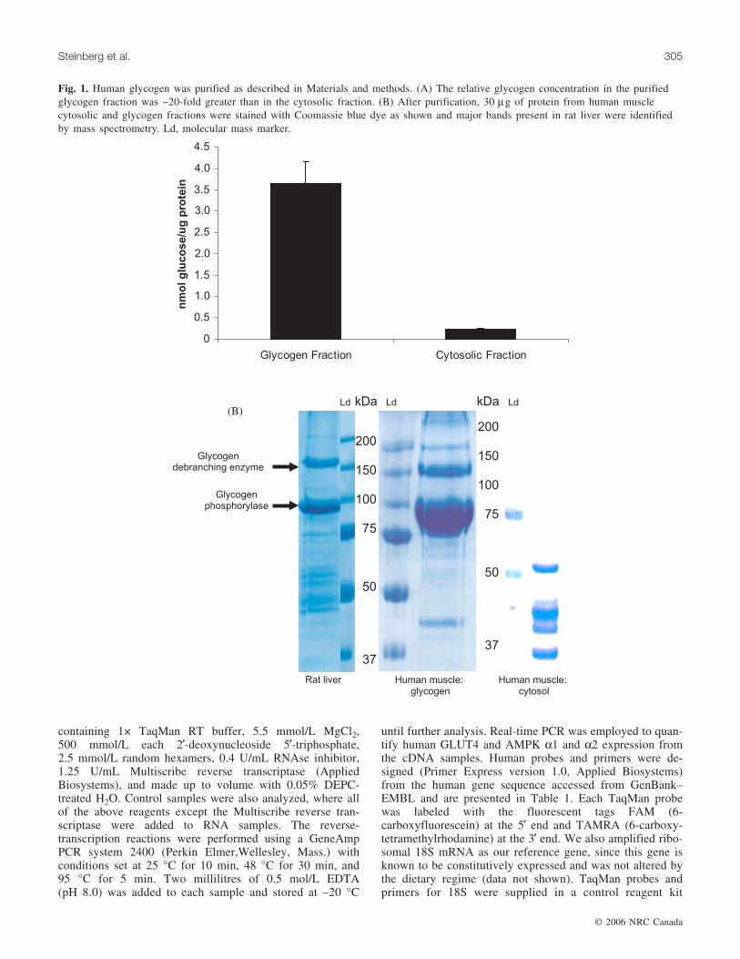
in a buffer containing 0.1 mol/L sodium acetate (pH 6.0).Following centrifugation at 14 000g for 2 min, an aliquotwas removed and glucose levels determined using a 2-enzyme, color-based glucose assay (Sigma Chemicals, St.Louis, Miss.). As demonstrated in Fig. 1A, the relative gly-cogen concentration in the purified glycogen fraction was~20-fold greater then the cytosolic fraction. We then com-pared 30 µg of protein from human muscle cytosolic andglycogen fractions by 12% self-denatured sodiumpolyacrylamide gele electrophoresis (SDS PAGE) stainedwith Coomassie blue dye as shown in Fig. 1B. Furthermore,we identified 2 of the major proteins present in rat liver gly-cogen by mass spectrometry (MS). Briefly, the proteinswere excised and digested with protein sequencing gradetrypsin overnight at 30 °C with the resulting peptides de-salted by ZipTipTM (Millipore, Bedford, Mass.) and identi-fied by tandem MS on a Q(q)TOF-type mass spectrometerAPI QSTAR Pulsar i (Applied Biosytems, Foster City,Calif.). The larger polypeptide was identified as glycogen-debranching enzyme with 4 peptides were identified by tan-dem MS and searching with the Mascot search engine (datanot shown). The smaller polypeptide was positively identi-fied as rat liver glycogen phosphorylase based on the massesof 14 peptides (data not shown). The 2 major polypeptidesshown in the human glycogen fraction have not been posi-tively identified by mass spectrometry, but appear to beglycogen phosphorylase and glycogen-debranching enzymegiven their identical size and relative quantities when com-pared with the rat liver glycogen fraction. These proteins areclearly not visible in the cytosolic fraction shown in Fig. 1B.
After the completion of quality-control experiments, hu-man muscle biopsies were extracted as described above intoglycogen and cytosolic fractions. One hundred microgramsof protein were solubilized into glycogen-isolation buffercontaining 1% NP-40. AMPK α2 antibody (3 µg) was addedto each fraction together with 100 µL of 20% Protein Asepharose and incubated overnight at 4 °C with mixing.Immunoprecipitates were collected, washed twice with gly-cogen isolation buffer containing 1% NP-40, and analyzedby Western blot by probing with an AMPK α2 antibody.Densitometry was used to quantify the amount of AMPK α2immunoreactivity in the glycogen fraction of each sample.
AMPK nuclear localizationNuclear proteins were isolated and immunoblotted with
antibodies for AMPK α1 and α2 as previously described(McGee et al. 2003). To further characterize the nuclear andcytosolic fractions and to ensure purification of the fractions,quality-control experiments were performed using antibodiesagainst DHPR α1 (plasma membrane, t-tubules), SERCA2(sarcoplasmic reticulum (SR)), uncoupling protein-3 (UCP3,mitochondria), and histone 1 (nucleus).
Quantification of GLUT4, and AMPK α1 and α2mRNA using real-time RT-PCR
A portion of muscle (~30 mg) was extracted for totalRNA using a modification of the acid guanidium thiocyanate –phenol chloroform extraction method described previously(Febbraio and Koukoulas 2000). The total RNA was subse-quently quantified 2–3 more times before 1 µg of each totalRNA sample was reverse transcribed in a 10 µL reaction
© 2006 NRC Canada
304 Appl. Physiol. Nutr. Metab. Vol. 31, 2006
containing 1× TaqMan RT buffer, 5.5 mmol/L MgCl2,500 mmol/L each 2′-deoxynucleoside 5′-triphosphate,2.5 mmol/L random hexamers, 0.4 U/mL RNAse inhibitor,1.25 U/mL Multiscribe reverse transcriptase (AppliedBiosystems), and made up to volume with 0.05% DEPC-treated H2O. Control samples were also analyzed, where allof the above reagents except the Multiscribe reverse tran-scriptase were added to RNA samples. The reverse-transcription reactions were performed using a GeneAmpPCR system 2400 (Perkin Elmer,Wellesley, Mass.) withconditions set at 25 °C for 10 min, 48 °C for 30 min, and95 °C for 5 min. Two millilitres of 0.5 mol/L EDTA(pH 8.0) was added to each sample and stored at –20 °C
until further analysis. Real-time PCR was employed to quan-tify human GLUT4 and AMPK α1 and α2 expression fromthe cDNA samples. Human probes and primers were de-signed (Primer Express version 1.0, Applied Biosystems)from the human gene sequence accessed from GenBank–EMBL and are presented in Table 1. Each TaqMan probewas labeled with the fluorescent tags FAM (6-carboxyfluorescein) at the 5′ end and TAMRA (6-carboxy-tetramethylrhodamine) at the 3′ end. We also amplified ribo-somal 18S mRNA as our reference gene, since this gene isknown to be constitutively expressed and was not altered bythe dietary regime (data not shown). TaqMan probes andprimers for 18S were supplied in a control reagent kit
© 2006 NRC Canada
Steinberg et al. 305
Glycogendebranching enzyme
Glycogenphosphorylase
200
150
100
75
50
37
Ld kDa
200
150
100
75
50
37
kDa
Rat liver Human muscle:cytosol
Human muscle:glycogen
(B)
0
0.5
1.0
1.5
2.0
2.5
3.0
3.5
4.0
4.5
Glycogen Fraction Cytosolic Fraction
nm
olg
luco
se/u
gp
rote
in
Ld Ld
Fig. 1. Human glycogen was purified as described in Materials and methods. (A) The relative glycogen concentration in the purifiedglycogen fraction was ~20-fold greater than in the cytosolic fraction. (B) After purification, 30 µg of protein from human musclecytosolic and glycogen fractions were stained with Coomassie blue dye as shown and major bands present in rat liver were identifiedby mass spectrometry. Ld, molecular mass marker.

(Applied Biosystems). We quantified gene expression usinga multiplex comparative critical threshold (CT) method (Bio-Rad i Cycler IQ™, Hercules, Calif.) as previously described(Febbraio and Koukoulas 2000). PCRs were carried out in25 µL reactions of TaqMan universal PCR master mix (1×),50 nmol/L TaqMan 18S probe, 20 nmol/L 18S forwardprimer, 80 nmol/L 18S reverse primer, and probes and prim-ers at specific concentrations ranging from 50 nmol/L to150 nmol/L (probes) and from 50 nmol/L to 900 nmol/L(primers) for each gene of interest. The sequences of the for-ward (F) and reverse (R) primers and probes (P) are listedfrom 5′-3′ and are as follows: AMPKα1 (F) CAGGGACTGCTACTCCACAGAGA, (R) CCTTGAGCCTCAGCATCTGAA, (P) TCAGTTAGCAACTATCGATCTTGCCAAAGGAGT; AMPK α2 (F) CAACTGCAGAGAGCCATTCACTT,(R) GTGAAACTGAAGACAATGTGCTT, (P) CTGGCTCTCTCACTGGCTCTTTGACCG; and GLUT4 (F) CCTGCCAGAAAGAGTCTGAAGC, (R) ATCCTTCAGCTCAGCCAGCA, (P) CAGAAACATCGGCCCAGCCTGTCA. Thespecific concentrations for each gene were optimized in pre-liminary experiments. cDNA and control preparations notcontaining RT were amplified using the following condi-tions: 50 °C for 2 min, 95 °C for 10 min, and 40 cycles of95 °C for 15 s and 60 °C for 1 min.
Cell culture experimentsPrimary skeletal muscle cell culture was established from
muscle biopsies of 5 male subjects (24 ± 2 y, 75 ± 5 kg;VO2 peak = 41 ± 3 mL·(kg·min)–1) as described (Chen et al.2005). Cells were grown to 80% confluence in α-MEMsupplemented with 10% v/v FBS, 0.5% v/v penicillin, and0.5% v/v Fungizone in a 37 °C incubator with 5% CO2before being differentiated for 4 d in α-MEM, 2% horseserum, and 1% each of penicillin, streptomycin, andamphotericin B (PSA). Cells were then infected with a con-stitutively active AMPK adenovirus (100 plaque-formingunits (PFU)/plate) or control vector (AdGo, 100 PFU/plate)as described (Woods et al. 2000). Expression levels of bothcontrol and CA-AMPK infected cells was determined by vi-sual examination of green fluorescence protein under ultravi-olet light. Seventy-two hours post infection ~85% of allmyotubes were infected in both control and CA-AMPK-infected cells (data not shown) and ~90% of myotubes weredifferentiated (data not shown). mRNA was then extractedaccording to manufacturer’s instructions using the RNeasy
mini kit (Qiagen, Doncaster, Australia), reverse transcribedusing the thermoscript RT-PCR system (Invitrogen, Mt.Waverley, Australia), and GLUT4 mRNA was analysed us-ing quantitative real-time PCR as previously described (Chenet al. 2005).
Calculations and statisticsFree ADP and AMP concentrations were calculated as de-
scribed previously (McGee et al. 2003). Statistical analysiswas performed by 2-way analysis of variance with repeatedmeasures (time × trial) and specific differences were locatedusing a Student–Newman–Keuls post hoc test. For cell cul-ture experiments, a t test was performed. Statistical signifi-cance was set at p ≤ 0.05. Data are expressed as the mean ±SEM.
Results
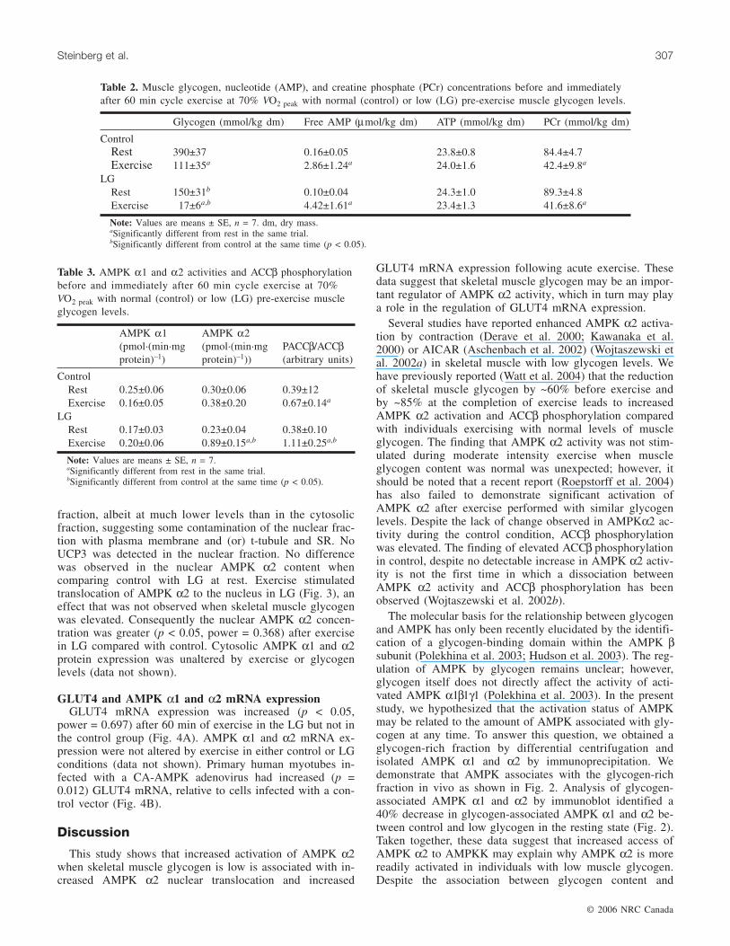
The exercise load (175 ± 18 W), oxygen uptake, and ven-tilation were not different at any point during exercise(Table 1), whereas the respiratory exchange ratio was lowerin the low-glycogen trial. Plasma epinephrine was elevatedwith exercise in the low-glycogen condition relative to thecontrol condition (60 min: 3.95 ± 0.5 nmol/L vs. 2.05 ±0.3 nmol/L). The exercise–diet regimen was successful inproducing normal (control) and low muscle glycogen (LG,p < 0.05) levels at the onset of exercise (Table 2). Muscleglycogen content was higher (p < 0.05) in Control at com-pletion of exercise (Table 2) and glycogen use was greater(p < 0.05). Muscle nucleotide, creatine, and creatine phos-phate concentrations were also unaltered by glycogen status(Table 2). Circulating insulin and glucose values have previ-ously been reported (Watt et al. 2004).
AMPK α2 activity and ACCβ phosphorylationAMPK activities and ACCβ phosphorylation have been re-
ported previously (Watt et al. 2004) and are summarized inTable 3. AMPK α1 activity was unaltered by glycogen statusor exercise. AMPK α2 activity was not different betweencontrol and LG groups at rest, but was greater (+134%, p =0.05) in the LG compared with the control group at the ces-sation of exercise. Exercise increased ACCβ phosphorylationin the LG group (+112%, p = 0.05); however, in the controlgroup, the increase in ACCβ did not achieve statistical sig-nificance.
AMPK association with human skeletal muscleglycogen
As shown in Fig. 2, AMPK α2 associates with glycogenin both control and LG, with less associated in the LGcondition. At rest, glycogen-bound AMPK α1 and α2immunoreactivity decreased by ~40% in the LG conditionwhen compared with the control. However, after exercise,glycogen-bound AMPK α2 immunoreactivity did not changein the LG condition when compared with control (Fig. 2).
AMPK α2 nuclear translocationThe nuclear protein histone 1 was only observed within
the nuclear fraction, suggesting significant purification ofthis fraction from skeletal muscle. However, there was someexpression of DHPR α1 and SERCA2 in the nuclear
© 2006 NRC Canada
306 Appl. Physiol. Nutr. Metab. Vol. 31, 2006
VE (L/min) VO2 (mL(kg·min)–1) RER
ControlEx 20 70.3±3.8 36.1±2.0 0.93±0.01Ex 40 66.4±4.6 34.2±2.7 0.90±0.01Ex 60 62.3±3.5 33.6±2.7 0.88±0.01
LGEx 20 73.2±5.7 39.2±2.2 0.85±0.01a
Ex 40 75.4±7.5 37.3±2.7 0.81±0.02a
Ex 60 73.3±7.3 36.1±2.5 0.81±0.01a
Note: VE, ventilatory exchange, VO2, pulmonary oxygen uptake; RER,respiratory exchange ratio; Ex, exercise. Values are means ± SE; n = 7.
Table 1. VE, VO2, and RER in subjects during cycle exercise at70% VO2 peak with normal (control) or low (LG) pre-exercisemuscle glycogen levels.
fraction, albeit at much lower levels than in the cytosolicfraction, suggesting some contamination of the nuclear frac-tion with plasma membrane and (or) t-tubule and SR. NoUCP3 was detected in the nuclear fraction. No differencewas observed in the nuclear AMPK α2 content whencomparing control with LG at rest. Exercise stimulatedtranslocation of AMPK α2 to the nucleus in LG (Fig. 3), aneffect that was not observed when skeletal muscle glycogenwas elevated. Consequently the nuclear AMPK α2 concen-tration was greater (p < 0.05, power = 0.368) after exercisein LG compared with control. Cytosolic AMPK α1 and α2protein expression was unaltered by exercise or glycogenlevels (data not shown).
GLUT4 and AMPK α1 and α2 mRNA expressionGLUT4 mRNA expression was increased (p < 0.05,
power = 0.697) after 60 min of exercise in the LG but not inthe control group (Fig. 4A). AMPK α1 and α2 mRNA ex-pression were not altered by exercise in either control or LGconditions (data not shown). Primary human myotubes in-fected with a CA-AMPK adenovirus had increased (p =0.012) GLUT4 mRNA, relative to cells infected with a con-trol vector (Fig. 4B).
Discussion
This study shows that increased activation of AMPK α2when skeletal muscle glycogen is low is associated with in-creased AMPK α2 nuclear translocation and increased
GLUT4 mRNA expression following acute exercise. Thesedata suggest that skeletal muscle glycogen may be an impor-tant regulator of AMPK α2 activity, which in turn may playa role in the regulation of GLUT4 mRNA expression.
Several studies have reported enhanced AMPK α2 activa-tion by contraction (Derave et al. 2000; Kawanaka et al.2000) or AICAR (Aschenbach et al. 2002) (Wojtaszewski etal. 2002a) in skeletal muscle with low glycogen levels. Wehave previously reported (Watt et al. 2004) that the reductionof skeletal muscle glycogen by ~60% before exercise andby ~85% at the completion of exercise leads to increasedAMPK α2 activation and ACCβ phosphorylation comparedwith individuals exercising with normal levels of muscleglycogen. The finding that AMPK α2 activity was not stim-ulated during moderate intensity exercise when muscleglycogen content was normal was unexpected; however, itshould be noted that a recent report (Roepstorff et al. 2004)has also failed to demonstrate significant activation ofAMPK α2 after exercise performed with similar glycogenlevels. Despite the lack of change observed in AMPKα2 ac-tivity during the control condition, ACCβ phosphorylationwas elevated. The finding of elevated ACCβ phosphorylationin control, despite no detectable increase in AMPK α2 activ-ity is not the first time in which a dissociation betweenAMPK α2 activity and ACCβ phosphorylation has beenobserved (Wojtaszewski et al. 2002b).
The molecular basis for the relationship between glycogenand AMPK has only been recently elucidated by the identifi-cation of a glycogen-binding domain within the AMPK βsubunit (Polekhina et al. 2003; Hudson et al. 2003). The reg-ulation of AMPK by glycogen remains unclear; however,glycogen itself does not directly affect the activity of acti-vated AMPK α1β1γ1 (Polekhina et al. 2003). In the presentstudy, we hypothesized that the activation status of AMPKmay be related to the amount of AMPK associated with gly-cogen at any time. To answer this question, we obtained aglycogen-rich fraction by differential centrifugation andisolated AMPK α1 and α2 by immunoprecipitation. Wedemonstrate that AMPK associates with the glycogen-richfraction in vivo as shown in Fig. 2. Analysis of glycogen-associated AMPK α1 and α2 by immunoblot identified a40% decrease in glycogen-associated AMPK α1 and α2 be-tween control and low glycogen in the resting state (Fig. 2).Taken together, these data suggest that increased access ofAMPK α2 to AMPKK may explain why AMPK α2 is morereadily activated in individuals with low muscle glycogen.Despite the association between glycogen content and
© 2006 NRC Canada
Steinberg et al. 307
Glycogen (mmol/kg dm) Free AMP (µmol/kg dm) ATP (mmol/kg dm) PCr (mmol/kg dm)
ControlRest 390±37 0.16±0.05 23.8±0.8 84.4±4.7Exercise 111±35a 2.86±1.24a 24.0±1.6 42.4±9.8a
LGRest 150±31b 0.10±0.04 24.3±1.0 89.3±4.8Exercise 17±6a,b 4.42±1.61a 23.4±1.3 41.6±8.6a
Note: Values are means ± SE, n = 7. dm, dry mass.aSignificantly different from rest in the same trial.bSignificantly different from control at the same time (p < 0.05).
Table 2. Muscle glycogen, nucleotide (AMP), and creatine phosphate (PCr) concentrations before and immediatelyafter 60 min cycle exercise at 70% VO2 peak with normal (control) or low (LG) pre-exercise muscle glycogen levels.
AMPK α1(pmol·(min·mgprotein)–1)
AMPK α2(pmol·(min·mgprotein)–1))
PACCβ/ACCβ(arbitrary units)
ControlRest 0.25±0.06 0.30±0.06 0.39±12Exercise 0.16±0.05 0.38±0.20 0.67±0.14a
LGRest 0.17±0.03 0.23±0.04 0.38±0.10Exercise 0.20±0.06 0.89±0.15a,b 1.11±0.25a,b
Note: Values are means ± SE, n = 7.aSignificantly different from rest in the same trial.bSignificantly different from control at the same time (p < 0.05).
Table 3. AMPK α1 and α2 activities and ACCβ phosphorylationbefore and immediately after 60 min cycle exercise at 70%VO2 peak with normal (control) or low (LG) pre-exercise muscleglycogen levels.

AMPK α2 expression and activity, we feel there are somedifficulties in the current experimental design, which limitour interpretation of these results. Although we demonstratea significant enrichment in glycogen content (~25-fold) andan association of this enriched glycogen fraction withAMPK α2 expression, we are unable to distinguish betweennon-specific trapping and (or) colocalization of AMPKwithin the glycogen-enriched fraction and direct binding. Al-though we (Polekhina et al. 2003) and others (Hudson et al.2003) have demonstrated a direct interaction in mammaliancells the observation that AMPK α1 expression was alsoreduced despite no change in activity suggests that non-specific trapping of proteins within the glycogen pellet mayhave occurred. Owing to the limited size of human musclebiopsies, it was not possible to determine if the associationof other proteins with glycogen was also reduced after exer-cise in the LG condition. These preliminary data suggest thatfuture studies in human skeletal muscle in vivo examiningthe relationship between muscle glycogen and AMPK ex-pression activity are warranted.
Despite the possibility of a direct effect of glycogen onAMPK activity, other explanations are plausible. Firstly, al-
though we demonstrated increased AMPK α2 activity afterexercise in LG in the absence of increased intramuscularconcentrations of AMP, there is a possibility of a potentialphysiological effect of non-statistical, but biologically rele-vant, elevations in AMP due to the sensitivity of AMPK tosmall changes in AMP:ATP (Kemp et al. 2003). Secondly,the hormonal responses to altered glycogen levels in re-sponse to exercise could not be controlled in the presentstudy. In rodents, pharmacological concentrations ofisoprotenerol increase AMPK α2 activity, which suggests animportant role of catecholamines in regulating AMPK(Minokoshi et al. 2002). In the present study, epinephrinelevels were elevated during the low glycogen trial comparedwith the control trial (Watt et al. 2004), suggesting a possi-ble stimulatory role for β-adernergic regulation of AMPK.Alternatively increased release of interleukin-6 (IL-6) underconditions of low muscle glycogen (Steensberg et al. 2001)may also be a contributing factor to the enhanced activationof AMPK (Kelly et al. 2004; MacDonald et al. 2003). Futurestudies investigating these possibilities are warranted.
A single exercise bout enhances GLUT4 mRNA expres-sion (Rodnick et al. 1990). Initial studies demonstrating that
© 2006 NRC Canada
308 Appl. Physiol. Nutr. Metab. Vol. 31, 2006
0
20
40
60
80
100
120
Alpha-2 Alpha-1
AM
PK
/pro
tein
(%)
Control
LG
aa
Fig. 2. Representative Western blot of AMPK α2 (above) and densitometry/quantification (below) of AMPK α1 and α2 expressionimmunoprecipitated from cytosolic and glycogen fractions at rest. Values are means ± SE (n = 7). aSignificantly different from control(p < 0.05).

exercise stimulated AMPK activity suggested that AMPKmay be a potential regulator of GLUT4 transcription (Winderand Hardie 1996). In support of this idea are observations inrodent skeletal muscle demonstrating that the pharmacologi-cal activation of AMPK by AICAR increases GLUT4expression to a similar degree as chronic exercise training(Holmes et al. 1999; Winder et al. 2000; Ojuka et al. 2000).Similarly, infection of rodent skeletal muscle cells (Fryer etal. 2002) with a constitutively active AMPK adenovirus alsoincreases GLUT4 expression demonstrating a direct effect ofAMPK on GLUT4 transcription, an effect which we havereplicated in primary human myotubes (Fig. 4B). Taken to-gether these data support the concept that AMPK can influ-ence GLUT4 transcription. It should be noted that thesestudies do not indicate that AMPK is the only regulator ofGLUT4 gene transcription. Indeed, recent data by Holmes etal. (Holmes et al. 2004) elegantly demonstrate in a AMPKdominant negative mouse that AMPK is not obligatory forthe effects of exercise on GLUT4 expression. Furthermore,there was trend for resting GLUT4 mRNA to be higher inthe low glycogen condition, in the absence of any differencein AMPK α2 activity. Other factors, such as the calciumcalmodulin dependent protein kinase (Ojuka et al. 2002)may also contribute to exercise induced GLUT4 biogenesis
and may be capable of compensating for reduced AMPKactivity in the AMPK dominant negative model. The mecha-nisms by which AMPK activation may increase GLUT4 ex-pression are unknown, but an important role of myocyteenhancer factor (MEF) 2A, MEF2D, and peroxisomeproliferator-activated receptor gamma coactivator-1 (PGC-1)has been demonstrated following AMPK activation by eitherexercise (Baar et al. 2002; Pilegaard et al. 2003) or AICARtreatment (Ojuka et al. 2002). In support of this concept, werecently demonstrated translocation of AMPK α2 to the nu-cleus with exercise in skeletal muscle (McGee et al. 2003),and increased phosphorylation of MEF-2 (McGee andHargreaves 2004). In this study, nuclear AMPK α2translocation after exercise occurred in the low glycogentrial only. Furthermore, the enhanced AMPK α2 nucleartranslocation was associated with elevated GLUT4 mRNAfollowing exercise. The mechanism(s) contributing to thetranslocation of AMPK α2 to the nucleus are unknown. Ex-ercise in the Control trial did not significantly increase α2nuclear translocation, consistent with the lack of change inAMPK α2 activity in this group, which was unexpected(Table 3). Future studies are needed to discern whetherdifferent degrees of AMPK activation result in alteredtranslocation of AMPKα2 to the nucleus.
© 2006 NRC Canada
Steinberg et al. 309
C LG C LG
Rest Exercise
0
1
2
3
4
5
6
7
8
9
Rest Exercise
GL
UT
4m
RN
A
(re
lati
ve
toC
ON
0)
Control
LG
b
Fig. 3. Representative Western blot (above) and densitometry/quantification (below) of nuclear localization of AMPK α2 before andimmediately after 60 min cycle exercise at 70% VO2 peak with normal (CON) or low (LG) pre-exercise muscle glycogen content. Valuesare means ± SE (n = 7). bSignificantly different from 0 min of the same trial (p < 0.05).
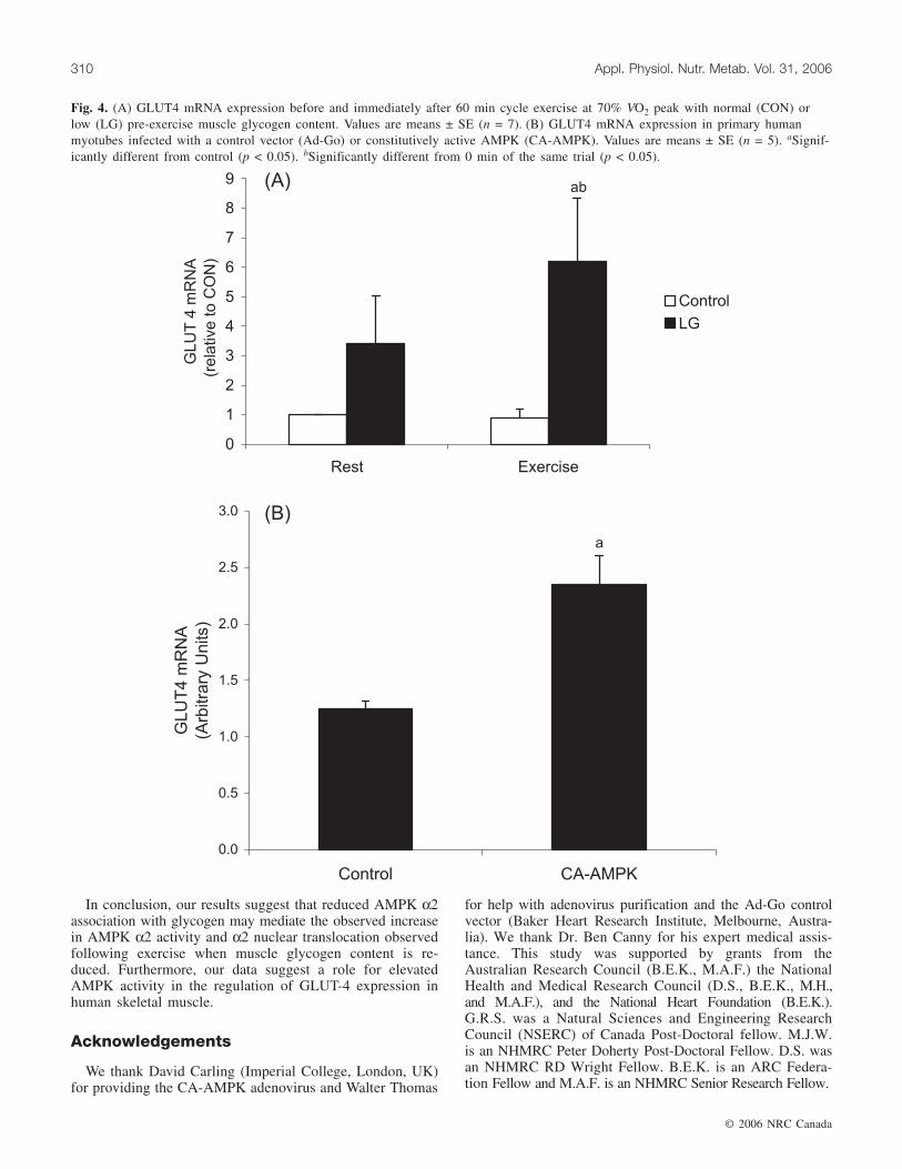
In conclusion, our results suggest that reduced AMPK α2association with glycogen may mediate the observed increasein AMPK α2 activity and α2 nuclear translocation observedfollowing exercise when muscle glycogen content is re-duced. Furthermore, our data suggest a role for elevatedAMPK activity in the regulation of GLUT-4 expression inhuman skeletal muscle.
Acknowledgements
We thank David Carling (Imperial College, London, UK)for providing the CA-AMPK adenovirus and Walter Thomas
for help with adenovirus purification and the Ad-Go controlvector (Baker Heart Research Institute, Melbourne, Austra-lia). We thank Dr. Ben Canny for his expert medical assis-tance. This study was supported by grants from theAustralian Research Council (B.E.K., M.A.F.) the NationalHealth and Medical Research Council (D.S., B.E.K., M.H.,and M.A.F.), and the National Heart Foundation (B.E.K.).G.R.S. was a Natural Sciences and Engineering ResearchCouncil (NSERC) of Canada Post-Doctoral fellow. M.J.W.is an NHMRC Peter Doherty Post-Doctoral Fellow. D.S. wasan NHMRC RD Wright Fellow. B.E.K. is an ARC Federa-tion Fellow and M.A.F. is an NHMRC Senior Research Fellow.
© 2006 NRC Canada
310 Appl. Physiol. Nutr. Metab. Vol. 31, 2006
0
1
2
3
4
5
6
7
8
9
Rest Exercise
GLU
T4
mR
NA
(rela
tive
toC
ON
)
Control
LG
0.0
0.5
1.0
1.5
2.0
2.5
3.0
Control CA-AMPK
GLU
T4
mR
NA
(Arb
itra
ryU
nits)
(A)
(B)
ab
a
Fig. 4. (A) GLUT4 mRNA expression before and immediately after 60 min cycle exercise at 70% VO2 peak with normal (CON) orlow (LG) pre-exercise muscle glycogen content. Values are means ± SE (n = 7). (B) GLUT4 mRNA expression in primary humanmyotubes infected with a control vector (Ad-Go) or constitutively active AMPK (CA-AMPK). Values are means ± SE (n = 5). aSignif-icantly different from control (p < 0.05). bSignificantly different from 0 min of the same trial (p < 0.05).

References
Aschenbach, W.G., Hirshman, M.F., Fujii, N., Sakamoto, K.,Howlett, K.F., and Goodyear, L.J. 2002. Effect of AICAR treat-ment on glycogen metabolism in skeletal muscle. Diabetes, 51:567–573.
Baar, K., Wende, A.R., Jones, T.E., Marison, M., Nolte, L.A.,Chen, M., Kelly, D.P., and Holloszy, J.O. 2002. Adaptations ofskeletal muscle to exercise: rapid increase in the transcriptionalcoactivator PGC-1. FASEB J. 16: 1879–1886.
Baque, S., Montell, E., Camps, M., Guinovart, J., Zorzano, A., andGomez-Foix, A. 1998. Overexpression of glycogen phos-phorylase increases GLUT4 expression and glucose transport incultured skeletal human muscle. Diabetes, 47: 1185–1192.
Chen, Z., Heierhorst, J., Mann, R.J., Mitchelhill, K.I., Michell,B.J., Witters, L.A., et al. 1999. Expression of the AMP-activatedprotein kinase beta1 and beta2 subunits in skeletal muscle.FEBS Lett. 460: 343–348.
Chen, X.-P., McConell, G.K., Michell, B.J., Snow, R.J., Canny,B.J., and Kemp, B.E. 2000. AMPK signaling in contracting hu-man skeletal muscle: acetyl-CoA carboxylase and NO synthasephosphorylation. Am. J. Physiol. 279: E1202–1206.
Chen, M.B., McAinch, A.J., Macaulay, S.L., Castelli, L.A.,O’Brien, P.E., Dixon, J.B., et al. 2005. Impaired activation ofAMP-kinase and fatty acid oxidation by globular adiponectin incultured human skeletal muscle from obese type 2 diabetics. J.Clin. Endocrinol. Metab. 90: 3665–72.
Derave, W., Ai, H., Ihlemann, J., Witters, L., Kristiansen, S., Rich-ter, E., and Ploug, T. 2000. Dissociation of AMP-activated pro-tein kinase activation and glucose transport in contracting slow-twitch muscle. Diabetes, 49: 1281–1287.
Febbraio, M.A., and Koukoulas, I. 2000. HSP72 gene expressionprogressively increases in human skeletal muscle during pro-longed exhaustive exercise. J. Appl. Physiol. 89: 1055–1060.
Fryer, L.G., Foufelle, F., Barnes, K., Baldwin, S.A., Woods, A.,and Carling, D. 2002. Characterization of the role of the AMP-activated protein kinase in the stimulation of glucose transportin skeletal muscle cells. Biochem. J. 363: 167–174.
Garcia Roves, P.M., Han, D.-H., Song, Z., Jones, T.E., Hucker,K.A., and Holloszy, J.O. 2003. Prevention of glycogensupercompensation prolongs the increase in muscle GLUT4 af-ter exercise. Am. J. Physiol. 285: E729–E736.
Hawley, S., Boudeau, J., Reid, J., Mustard, K., Udd, L., Makela,T., Alessi, D., and Hardie, D.G. 2003. Complexes between theLKB1 tumor suppressor, STRADalpha/beta andMO25alpha/beta are upstream kinases in the AMP-activatedprotein kinase cascade. J. Biol. 2: 28.
Holmes, B.F., Kurth-Kraczek, E.J., and Winder, W.W. 1999.Chronic activation of 5′-AMP-activated protein kinase increasesGLUT-4, hexokinase, and glycogen in muscle. J. Appl. Physiol.87: 1990–1995.
Holmes, B.F., Lang, D.B., Birnbaum, M.J., Mu, J., and Dohm,G.L. 2004. AMP Kinase is not required for the GLUT4 responseto exercise and denervation in skeletal muscle. Am. J. Physiol.287: E739–E743.
Hudson, E.R., Pan, D.A., James, J., Lucocq, J.M., Hawley, S.A.,Green, K.A., Baba, O., Terashima, T., and Hardie, D.G. 2003. Anovel domain in AMP-activated protein kinase causes glycogenstorage bodies similar to those seen in hereditary cardiacarrhythmias. Curr. Biol. 13: 861–866.
Kawanaka, K., Nolte, L.A., Han, D.-H., Hansen, P.A., andHolloszy, J.O. 2000. Mechanisms underlying impaired GLUT-4translocation in glycogen-supercompensated muscles of exercis-ing rats. Am. J. Physiol. 279: E1311–E1318.
Kelly, M., Keller, C., Aviluea, P.R., Keller, P., Luo, Z., Ziang, X.,et al. 2004. AMPK activity is diminished in tissues of IL-6knockout mice: the effect of exercise. Biochem. Biophys. Res.Commun. 320: 449–454.
Kemp, B.E., Stapleton, D., Campbell, D.J., Chen, Z.P., Murthy, S.,Walter, M., et al. 2003. AMP-activated protein kinase, supermetabolic regulator. Biochem. Soc. Trans. 31: 162–168.
MacDonald, C., Wojtaszewski, J.F., Pedersen, B.K., Kiens, B., andRichter, E.A. 2003. Interleukin-6 release from human skeletalmuscle during exercise: relation to AMPK activity. J. Appl.Physiol. 95: 2273–2277.
McGee, S.L., and Hargreaves, M. 2004. Exercise and myocyteenhancer factor 2 regulation in human skeletal muscle. Diabetes,53: 1208–1214.
McGee, S.L., Howlett, K.F., Starkie, R.L., Cameron-Smith, D.,Kemp, B.E., and Hargreaves, M. 2003. Exercise increases nu-clear AMPKα2 in human skeletal muscle. Diabetes, 52: 926–928.
Minokoshi, Y., Kim, Y.B., Peroni, O.D., Fryer, L.G., Muller, C.,Carling, D., and Kahn, B.B. 2002. Leptin stimulates fatty-acidoxidation by activating AMP-activated protein kinase. Nature(London), 415: 339–343.
Ojuka, E.O., Nolte, L.A., and Holloszy, J.O. 2000. Increased ex-pression of GLUT-4 and hexokinase in rat epitrochlearis mus-cles exposed to AICAR in vitro. J. Appl. Physiol. 88: 1072–1075.
Ojuka, E.O., Jones, T.E., Nolte, L.A., Chen, M., Wamhoff, B.R.,Sturek, M., and Holloszy, J.O. 2002. Regulation of GLUT4biogenesis in muscle: evidence for involvement of AMPK andCalcium. Am. J. Physiol. 282: E1008–E1013.
Pilegaard, H., Saltin, B., and Neufer, P.D. 2003. Exercise inducestransient transcriptional activation of the PGC-1alpha gene inhuman skeletal muscle. J. Physiol. 546: 851–858.
Polekhina, G., Gupta, A., Michell, B.J., van Denderen, B., Murthy,S., Feil, S.C., et al. 2003. AMPK beta subunit targets metabolicstress sensing to glycogen. Curr. Biol. 13: 867–871.
Richter, E.A., Derave, W., and Wojtaszewski, J.F. 2001. Glucose,exercise and insulin: emerging concepts. J. Physiol. 535: 313–322.
Rodnick, K.J., Holloszy, J.O., Mondon, C.E., and James, D.E.1990. Effects of exercise training on insulin-regulatable glucose-transporter protein levels in rat skeletal muscle. Diabetes, 39:1425–1429.
Roepstorff, C., Vistisen, B., Donsmark, M., Nielsen, J.N., Galbo,H., Green, K.A., et al. 2004. Regulation of hormone sensitivelipase activity and Ser563 and Ser565 phosphorylation in humanskeletal muscle during exercise. J. Physiol. (London), 560: 551–562.
Steensberg, A., Febbraio, M.A., Osada, T., Schjerling, P., van Hall,G., Saltin, B., and Pedersen, B.K. 2001. Interleukin-6 produc-tion in contracting human skeletal muscle is influenced by pre-exercise muscle glycogen content. J. Physiol. 537: 633–639.
Watt, M.J., Heigenhauser, G.J., Dyck, D.J., and Spriet, L.L. 2002.Intramuscular triacylglycerol, glycogen and acetyl group metab-olism during 4 h of moderate exercise in man. J. Physiol. 541:969–978.
Watt, M.J., Steinberg, G.R., Chan, S., Garnham, A., Kemp, B.E.,and Febbraio, M.A. 2004. β-adrenergic stimulation of skeletalmuscle HSL can be overridden by AMPK signaling. FASEB J.18: 1445–1446.
Winder, W.W., and Hardie, D.G. 1996. Inactivation of acetyl-CoAcarboxylase and activation of AMP-activated protein kinase inmuscle during exercise. Am. J. Physiol. 270: E299–E304.
Winder, W.W., Holmes, B.F., Rubink, D.S., Jensen, E.B., Chen,M., and Holloszy, J.O. 2000. Activation of AMP-activated pro-
© 2006 NRC Canada
Steinberg et al. 311

tein kinase increases mitochondrial enzymes in skeletal muscle.J. Appl. Physiol. 88: 2219–2226.
Wojtaszewski, J.F.P., Hansen, B.F., Gade, J., Kiens, B., Markuns,J.F., Goodyear, L.J., and Richter, E.A. 2000. Insulin signalingand insulin sensitivity after exercise in human skeletal muscle.Diabetes, 49: 325–331.
Wojtaszewski, J.F., Jorgensen, S.B., Hellsten, Y., Hardie, D.G., andRichter, E.A. 2002a. Glycogen-dependent effects of 5-aminoimidazole-4-carboxamide (AICA)-riboside on AMP-activated protein kinase and glycogen synthase activities in ratskeletal muscle. Diabetes, 51: 284–292.
Wojtaszewski, J.F., Mourtzakis, M., Hillig, T., Saltin, B., andPilegaard, H. 2002b. Dissociation of AMPK activity and ACCβphosphorylation in human muscle during prolonged exercise.Biochem. Biophys. Res. Commun. 298: 309–316.
Wojtaszewski, J.F., MacDonald, C., Nielsen, J.N., Hellsten, Y.,Hardie, D.G., Kemp, B.E., et al. 2003. Regulation of 5′AMP-activated protein kinase activity and substrate utilization in exer-cising human skeletal muscle. Am. J. Physiol. 284: E813–E822.
Woods, A., Azzout-Marniche, D., Foretz, M., Stein, S.C.,Lemarchand, P., Ferre, P., et al. 2000. Characterization of therole of AMP-activated protein kinase in the regulation of glu-cose-activated gene expression using constitutively active anddominant negative forms of the kinase. Mol. Cell Biol. 20:6704–6711.
Woods, A., Johnstone, S.R., Dickerson, K., Leiper, F.C.,Fryer, L.G., Neumann, D., et al. 2003. LKB1 is the upstreamkinase in the AMP-activated protein kinase cascade. Curr. Biol.13: 2004–2008.
© 2006 NRC Canada
312 Appl. Physiol. Nutr. Metab. Vol. 31, 2006