Recovery of human metapneumovirus from cDNA: optimization of growth in vitro and expression of...
13
Recovery of human metapneumovirus from cDNA: optimization of growth in vitro and expression of additional genes Ste ´phane Biacchesi, Mario H. Skiadopoulos, Kim C. Tran, Brian R. Murphy, Peter L. Collins, and Ursula J. Buchholz * Laboratory of Infectious Diseases, National Institute of Allergy and Infectious Diseases, Bethesda, MD 20892-8007, USA Received 11 November 2003; returned to author for revision 10 December 2003; accepted 22 December 2003 Abstract Human metapneumovirus (HMPV) is a recently recognized causative agent of respiratory tract disease in individuals of all ages and especially young infants. HMPV remains poorly characterized and has been reported to replicate inefficiently in vitro. Complete consensus sequences were recently determined for two isolates representing the two proposed HMPV genetic subgroups (Biacchesi et al., Virology 315 (1) (2003) 1). We have developed a reverse genetic system to produce one of these isolates, CAN97-83, entirely from cDNA. We also recovered a version, rHMPV– GFP, in which the enhanced green fluorescent protein (GFP) was expressed from a transcription cassette inserted as the first gene, leaving the 41-nt leader region and first 16 nt of the N gene undisturbed. The ability to monitor GFP expression in living cells greatly facilitated the initial recovery of this slow-growing virus. In addition, the ability to express a foreign gene from an engineered transcription cassette confirmed the identification of the HMPV transcription signals and identified the F gene-end signal as being highly efficient for transcription termination. The ability to recover virus containing a foreign insert in this position indicated that the viral promoter is contained within the 3V -terminal 57 nt of the genome. Recombinant HMPV replicated in vitro as efficiently as biologically derived HMPV, whereas the kinetics and final yield of rHMPV – GFP were reduced several-fold. Conditions for trypsin treatment were investigated, providing for improved virus yields. Another version of HMPV, rHMPV+G1F23, was recovered that contained a second copy of the G gene and two extra copies of F in promoter-proximal positions in the order G1 – F2 – F3. Thus, this recombinant genome would encode 11 mRNAs rather than eight and would be 17.3 kb long, 30% longer than that of the natural virus. Nonetheless, the rHMPV+G1F23 virus replicated in vitro with an efficiency that was only modestly reduced compared to rHMPV and was essentially the same as rHMPV– GFP. Northern blot analysis showed that the increased number and promoter-proximal location of the added copies of the F and G genes resulted in a more than 6- and 14- fold increase in the expression of F and G mRNA, respectively, and sequence analysis confirmed the intactness of the added genes in recovered virus. Thus, it should be feasible to construct an HMPV vaccine virus containing extra copies of the G and F putative protective antigen genes to increase antigen expression or to provide representation of additional antigenic lineages or subgroups of HMPV. Published by Elsevier Inc. Keywords: Metapneumovirus; Paramyxovirus; Recombinant virus; Respiratory disease; Vaccines; Expression vector Introduction Human metapneumovirus (HMPV) was first recognized in 2001 in the Netherlands from infants and children experiencing acute respiratory tract disease (van den Hoo- gen et al., 2001). HMPV has since been isolated in several continents and is thought to be worldwide in prevalence (Ebihara et al., 2003; Freymouth et al., 2003; Jartti et al., 2002; Maggi et al., 2003; Nissen et al., 2002; Peiris et al., 2003; Peret et al., 2002). HMPV was shown to have a very high seroprevalence in samples taken from children and adults over a 40-year period (Boivin et al., 2002, 2003; Ebihara et al., 2003; Freymouth et al., 2003; Maggi et al., 2003; Peret et al., 2002; van den Hoogen et al., 2001), suggesting that it is a newly recognized rather than newly emerging virus. HMPV resembles human respiratory syn- cytial virus (HRSV) with regard to disease signs and the ability to infect and cause disease in the young infant in particular as well as in individuals of all ages (Boivin et al., 2002; Falsey et al., 2003; Greensill et al., 2003; Maggi et al., 0042-6822/$ - see front matter. Published by Elsevier Inc. doi:10.1016/j.virol.2003.12.020 * Corresponding author. Laboratory of Infectious Diseases, National Institute of Allergy and Infectious Diseases, Building 50, Room 6507, 50 South Drive MSC 8007, Bethesda, MD 20892-8007. Fax: +1-301-496- 8312. E-mail address: [email protected] (U.J. Buchholz). www.elsevier.com/locate/yviro Virology 321 (2004) 247– 259
-
Upload
independent -
Category
Documents
-
view
2 -
download
0
Transcript of Recovery of human metapneumovirus from cDNA: optimization of growth in vitro and expression of...
www.elsevier.com/locate/yviro
Virology 321 (2004) 247–259
Recovery of human metapneumovirus from cDNA: optimization of
growth in vitro and expression of additional genes
Stephane Biacchesi, Mario H. Skiadopoulos, Kim C. Tran, Brian R. Murphy,Peter L. Collins, and Ursula J. Buchholz*
Laboratory of Infectious Diseases, National Institute of Allergy and Infectious Diseases, Bethesda, MD 20892-8007, USA
Received 11 November 2003; returned to author for revision 10 December 2003; accepted 22 December 2003
Abstract
Human metapneumovirus (HMPV) is a recently recognized causative agent of respiratory tract disease in individuals of all ages and
especially young infants. HMPV remains poorly characterized and has been reported to replicate inefficiently in vitro. Complete consensus
sequences were recently determined for two isolates representing the two proposed HMPV genetic subgroups (Biacchesi et al., Virology 315
(1) (2003) 1). We have developed a reverse genetic system to produce one of these isolates, CAN97-83, entirely from cDNA. We also
recovered a version, rHMPV–GFP, in which the enhanced green fluorescent protein (GFP) was expressed from a transcription cassette inserted
as the first gene, leaving the 41-nt leader region and first 16 nt of the N gene undisturbed. The ability to monitor GFP expression in living cells
greatly facilitated the initial recovery of this slow-growing virus. In addition, the ability to express a foreign gene from an engineered
transcription cassette confirmed the identification of the HMPV transcription signals and identified the F gene-end signal as being highly
efficient for transcription termination. The ability to recover virus containing a foreign insert in this position indicated that the viral promoter is
contained within the 3V-terminal 57 nt of the genome. Recombinant HMPV replicated in vitro as efficiently as biologically derived HMPV,
whereas the kinetics and final yield of rHMPV–GFP were reduced several-fold. Conditions for trypsin treatment were investigated, providing
for improved virus yields. Another version of HMPV, rHMPV+G1F23, was recovered that contained a second copy of the G gene and two
extra copies of F in promoter-proximal positions in the order G1–F2–F3. Thus, this recombinant genome would encode 11 mRNAs rather
than eight and would be 17.3 kb long, 30% longer than that of the natural virus. Nonetheless, the rHMPV+G1F23 virus replicated in vitro with
an efficiency that was only modestly reduced compared to rHMPV and was essentially the same as rHMPV–GFP. Northern blot analysis
showed that the increased number and promoter-proximal location of the added copies of the F and G genes resulted in a more than 6- and 14-
fold increase in the expression of F and G mRNA, respectively, and sequence analysis confirmed the intactness of the added genes in recovered
virus. Thus, it should be feasible to construct an HMPV vaccine virus containing extra copies of the G and F putative protective antigen genes
to increase antigen expression or to provide representation of additional antigenic lineages or subgroups of HMPV.
Published by Elsevier Inc.
Keywords: Metapneumovirus; Paramyxovirus; Recombinant virus; Respiratory disease; Vaccines; Expression vector
Introduction
Human metapneumovirus (HMPV) was first recognized
in 2001 in the Netherlands from infants and children
experiencing acute respiratory tract disease (van den Hoo-
gen et al., 2001). HMPV has since been isolated in several
continents and is thought to be worldwide in prevalence
0042-6822/$ - see front matter. Published by Elsevier Inc.
doi:10.1016/j.virol.2003.12.020
* Corresponding author. Laboratory of Infectious Diseases, National
Institute of Allergy and Infectious Diseases, Building 50, Room 6507, 50
South Drive MSC 8007, Bethesda, MD 20892-8007. Fax: +1-301-496-
8312.
E-mail address: [email protected] (U.J. Buchholz).
(Ebihara et al., 2003; Freymouth et al., 2003; Jartti et al.,
2002; Maggi et al., 2003; Nissen et al., 2002; Peiris et al.,
2003; Peret et al., 2002). HMPV was shown to have a very
high seroprevalence in samples taken from children and
adults over a 40-year period (Boivin et al., 2002, 2003;
Ebihara et al., 2003; Freymouth et al., 2003; Maggi et al.,
2003; Peret et al., 2002; van den Hoogen et al., 2001),
suggesting that it is a newly recognized rather than newly
emerging virus. HMPV resembles human respiratory syn-
cytial virus (HRSV) with regard to disease signs and the
ability to infect and cause disease in the young infant in
particular as well as in individuals of all ages (Boivin et al.,
2002; Falsey et al., 2003; Greensill et al., 2003; Maggi et al.,

S. Biacchesi et al. / Virology 321 (2004) 247–259248
2003; Osterhaus and Fouchier, 2003; Peret et al., 2002;
Viazov et al., 2003). Even though the epidemiology of
HMPV and its impact on human health need to be further
studied and defined, it is thought to be an important agent of
pediatric respiratory tract disease for which a vaccine should
be developed. Live attenuated vaccines are being developed
for HRSV and human parainfluenza viruses types 1, 2, and
3, and a live attenuated HMPV vaccine might be appropriate
to include as part of this strategy.
HMPV is an enveloped virus with a genome that is a
single negative strand of RNA of approximately 13 kb
(Biacchesi et al., 2003; van den Hoogen et al., 2001,
2002). It has been classified presumptively, together with
avian metapneumovirus (AMPV), in the Metapneumovirus
genus, Pneumovirus subfamily, Paramyxovirus family of
the Order Mononegavirales. The Pneumovirus subfamily
contains a second genus, Pneumovirus, represented by
HRSV. A nearly complete genome sequence was deter-
mined for the prototype Netherlands 00-1 strain of HMPV
(van den Hoogen et al., 2002), and complete genome
sequences were determined for two Canadian strains,
CAN97-83 and CAN98-75, which represent the two pro-
posed HMPV genetic subgroups (Biacchesi et al., 2003).
These studies confirmed that like AMPV, the 3Vto 5VHMPV
gene order is N-P-M-F-M2-SH-G-L. The mRNA encoded
by the M2 gene contains two overlapping open reading
frames (ORFs) that have the potential to encode separate
proteins, M2-1 and M2-2, as is the case for HRSV (Ber-
mingham and Collins, 1999; Collins et al., 1990, 1996; Jin
et al., 2000). By analogy to AMPV and HRSV, the HMPV
proteins are (listed according to gene order) N, nucleocapsid
RNA binding protein; P, phosphoprotein; M, matrix protein;
F, fusion glycoprotein; M2-1, transcription elongation fac-
tor; M2-2, RNA synthesis regulatory factor; SH, small
hydrophobic surface protein; G, major attachment protein;
and L, major polymerase subunit. To date, none of these
predicted HMPV proteins have been identified or charac-
terized by direct biochemical means, and their functions
remain to be confirmed. HMPV has been described as being
difficult to isolate and propagate, reflecting its trypsin
dependence, slow replication, and limited range of suscep-
tible cell lines (Boivin et al., 2002; van den Hoogen et al.,
2001).
To characterize this newly recognized Pneumovirus, we
have developed a reverse genetic system based on the
CAN97-83 isolate (HMPV83), one of the isolates for which
a complete genome consensus sequence was recently pub-
lished (Biacchesi et al., 2003). The development of this
recovery system was facilitated by the expression of en-
hanced green fluorescent protein (GFP) from an additional
gene inserted into the genome of recombinant HMPV
(rHMPV), by which the recovery of this slowly growing
virus was monitored. We show that rHMPV replicates in cell
culture with an efficiency comparable to that of the biolog-
ically derived virus, that reasonably efficient growth can be
achieved in vitro including in a cell line appropriate for
vaccine manufacture, and that additional genes can be
accommodated by HMPV with only a small effect on in
vitro replication. This provides the basis for developing a
live attenuated, multivalent HMPV vaccine.
Results
Construction of a plasmid encoding the full-length HMPV83
antigenomic RNA
A cDNA clone encoding the complete 13335-nt anti-
genomic RNA of HMPV isolate HMPV83 was constructed
as described in Fig. 1. Three overlapping cloned subge-
nomic fragments were created: fragment 1 contained the
leader and the putative N, P, and M genes, bordered on the
upstream end by a T7 RNA polymerase promoter (T7p) and
on the downstream end by an NheI site that was created by
four nucleotide substitutions in the putative M–F intergenic
region as a marker to distinguish between cDNA-derived
and biologically derived HMPV (Fig. 1). Three non-viral G
residues were added to the 5V end of the antigenome to
enhance promoter efficiency. Fragment 2 included the
putative F, M2, SH, and G genes and was bordered on the
upstream side by the added NheI site and on the downstream
side by a naturally occurring Acc65I site. Fragment 3
consisted of the putative L gene and trailer sequence,
bordered on the upstream side by the Acc65I site and on
the downstream side by part of the hepatitis delta virus
ribozyme sequence ending in an RsrII site that occurs
naturally within the ribozyme. The antigenomic cDNA
was cloned in vector pBSKSII, which supplied the remain-
der of the hepatitis delta virus ribozyme followed by a
terminator for T7 RNA polymerase (T7t) (Durbin et al.,
1997a). The sequence of the complete assembled cDNAwas
confirmed in its entirety, showing that the encoded anti-
genome (13335 nt long, exclusive of non-viral nt) differed
from the consensus sequence of biologically derived
HMPV83 (Biacchesi et al., 2003) only by the four nt
involved in the NheI marker.
Construction of pHMPV–GFP
It was anticipated that the poor growth and relative lack
of empirical knowledge for HMPV would complicate the
recovery and identification of the cDNA-derived virus.
Therefore, the pHMPV antigenomic cDNA was modified
by insertion of an additional gene encoding the enhanced
green fluorescent protein (GFP). This would provide a
means to monitor the recovery of rHMPV in living cells
at all stages of transfection and passage. One prerequisite for
the expression of a foreign ORF inserted into a mononega-
virus genome is that it be flanked by appropriate GS and GE
signals to direct transcription by the viral polymerase.
Putative consensus sequences of the HMPV GS (GGGA-
CAAnTnnnAATG) and GE (AGTTAATTAAAAA) motifs
Fig. 1. Construction of plasmid pHMPV83 expressing the complete antigenomic RNA of HMPV83. Three overlapping cDNA fragments (numbered 1–3)
covering the complete antigenome were generated by RT-PCR, and were assembled in pBSKSII, which contains the hepatitis delta virus ribozyme (y ribo)
(Perrotta and Been, 1991) followed by a terminator for T7 RNA polymerase (T7t). This vector also contained a polylinker with AatII, NheI, and Acc65I sites,
which served to accept the cloned fragments 1, 2, and 3. The complete antigenomic cDNA was designed and confirmed to be identical to the published
consensus sequence of biologically derived HMPV83 (Biacchesi et al., 2003) except for 4 nt substitutions (underlined) that were introduced to create an NheI
marker restriction site in the M-F intergenic region. The leader end of the antigenome cDNAwas flanked by the T7 promoter (T7p), and three additional, non-
viral G residues were added to the leader end to improve the efficiency of the promoter. The complete pHMPV83 plasmid contains 16333 bp.
S. Biacchesi et al. / Virology 321 (2004) 247–259 249
were determined previously from sequence alignments of
noncoding sequences (Biacchesi et al., 2003; van den
Hoogen et al., 2002). The GFP ORF was inserted into the
HMPV genome such that its ATG was replaced by that of
the N gene. This left undisturbed the 41-nt leader region and
first 16 nt of the N gene, including the N GS signal (Fig. 2).
Fig. 2. Insertion of a transcription cassette encoding the green fluorescent protein
Materials and methods, two PCR fragments were constructed: Fragment A contain
nt 12–57 of the HMPV antigenome, including a naturally occurring MluI site, mo
initiate with the ATG start codon of the N gene (underlined) and thus is preceded b
downstream side by an HMPV GE motif that was taken from the F gene and cont
end a partial copy of a GE signal including the PacI site, followed by a 2-nt inter
copy of the N GS signal and the upstream end of the N ORF, ending at a naturall
window of the pHMPV antigenome plasmid. In the sequences shown, translatio
italicized and underlined, and GS and GE motifs are boxed. The sequence num
antigenome. The total length of the added GFP transcription cassette (which begi
before the nucleotide numbered 42) was 746 nt.
The placement of GFP as the first gene should provide for a
high level of transcription. The downstream end of the GFP
ORF was modified to be followed by the GE signal of the
HMPV F gene, and was followed in turn by a 2-nt intergenic
region identical to that found between the N and P genes,
followed by the complete HMPV N gene. As described for
(GFP) into the promoter-proximal position of HMPV. As described in the
ed the GFP ORF (black rectangle) that was flanked on the upstream side by
st of the leader region, and the N GS motif. The GFP ORF was designed to
y the first 57 nt of the HMPVantigenome. The GFP ORF was flanked on its
ains a naturally occurring PacI site. Fragment B contained on the upstream
genic (ig) region that is identical to that of the N-P junction, followed by a
y occurring AvrII site. The two fragments were cloned into the MluI –AvrII
nal initiation and termination codons are underlined, restriction sites are
bering refers to the complete sequence of biologically derived HMPV83
ns in fragment A after the nucleotide numbered 41 and ends in fragment B
S. Biacchesi et al. / Virology 321 (2004) 247–259250
pHMPV, the complete sequences of the antigenomic
HMPV-GFP cDNA and flanking sequences in pHMPV–
GFP also were confirmed by sequence analysis. The length
of the encoded rHMPV–GFP antigenome, exclusive of
non-viral nt, would be 14083 nt, 5.6% larger than the
naturally occurring genome for HMPV83.
Recovery of infectious rHMPV and rHMPV–GFP
The antigenome plasmid pHMPV–GFP was transfected
into BSR T7/5 cells, which stably express the T7 RNA
polymerase, together with support plasmids encoding the N,
P, L, and M2-1 proteins. Two days post-transfection, trypsin
was added, the cells were incubated for 3 days and scraped
into the medium, and the total suspension was passaged
onto LLC-MK2 or Vero cells. When transfected, cells were
examined by fluorescent microscopy on successive days
post-transfection, green cells were visualized by day 2 that
initially consisted of scattered isolated cells and subsequent-
ly formed small foci of two or more cells that later on
exhibited cytopathic effect consistent with HMPV. When
the transfection monolayer was passaged to fresh cells,
single green cells were visualized about 24 h post-infection
and developed over successive days into multicellular foci.
The ability to monitor GFP expression greatly facilitated the
initial recovery, which was then also readily achieved for
rHMPV lacking the GFP marker.
Infectious rHMPV–GFP also was recovered when the
M2-1 support plasmid was omitted from the panel of
support plasmids (not shown). This result alone does not
necessarily indicate that M2-1 is not required for recovery,
because the possibility exists that M2-1 is expressed from
the antigenome plasmid, as was found in the case of RSV
(Collins et al., 1999). Inclusion of pT7-M2-1 in the trans-
fection had a positive effect on recovery, resulting in two- or
Fig. 3. Northern blot analysis of GFP mRNA expressed by the rHMPV–GFP virus
of 3 PFU/cell with HMPV83 (lanes 2 and 6), rHMPV (lanes 3 and 7), or rHMPV–
electrophoresed on 1% agarose-formaldehyde gels, transferred to charged nylon,
specific to the GFP (lanes 1–4) or M (lanes 5–8) gene. The identities and calcu
threefold more initial recovery events per well. No infec-
tious virus was recovered when the N, P, or L support
plasmid was omitted.
The expression of GFP in cells infected with rHMPV–
GFP showed that the virus was cDNA-derived and not a
contaminating biologically derived HMPV. The rHMPVand
rHMPV–GFP viruses were confirmed to be HMPV based
on reactivity in both an immunofluorescence assay and a
viral plaque immunostaining assay with rabbit antiserum
that had been raised against gradient-purified, biologically
derived HMPV (data not shown). For rHMPV–GFP, the
number of foci of GFP-expressing cells counted before
fixation equaled the number of foci that were reactive after
immunostaining, showing that the virus population was
homogeneous and stably expressing GFP after five passages
in cell culture. In addition, RT-PCR performed on virion
RNA (vRNA) following three passages in vitro confirmed
that both the rHMPV and rHMPV–GFP genomes were
efficiently copied and amplified with HMPV-specific pri-
mers, that both contained the NheI restriction marker site
whereas biologically derived HMPV did not, and that the
rHMPV–GFP virus contained the expected GFP insert (not
shown). The expression of the inserted GFP coding se-
quence also was characterized by Northern blot hybridiza-
tion. Cells were infected with biologically derived
HMPV83, rHMPV, rHMPV–GFP, or were mock infected,
and total intracellular RNA was isolated 3 days later and
analyzed by Northern blot hybridization with double-strand-
ed DNA probes specific to the GFP or M genes. As shown
in Fig. 3, the GFP-specific probe hybridized only with RNA
from rHMPV–GFP-infected cells (lane 4), and detected an
abundant RNA band of the appropriate size to be the
predicted 746-nt (exclusive of polyA) GFP mRNA tran-
scribed from the inserted transcription cassette. This showed
that the putative HMPV GS and GE transcription signals
. LLC-MK2 cells were mock-infected (lanes 1 and 5) or infected at an MOI
GFP (lanes 4 and 8). Three days later, total intracellular RNAwas isolated,
and analyzed by hybridization to double-stranded 32P-labeled DNA probe
lated sizes of individual RNA species are indicated.

S. Biacchesi et al. / Virology 321 (2004) 247–259 251
indeed functioned in the context of the foreign ORF to direct
the synthesis of a monocistronic GFP RNA. The GFP probe
also hybridized to several larger RNAs that were of low
abundance and appeared to represent GFP-N, GFP-N-P, and
GFP-N-P-M readthrough mRNAs, as well as to a large, faint
band that was of the appropriate size to contain rHMPV–
GFP genome and antigenome RNA. Analysis with the M-
specific probe identified bands of the appropriate sizes to be
the predicted 853-nt monocistronic M mRNA as well as
bands representing the P-M and N-P-M readthrough
mRNAs. The M probe also detected the genome and
antigenome RNA band for each virus, which in rHMPV–
GFP would be 748 nt larger than HMPV83 or rHMPV due
to the presence of the GFP transcription cassette (Fig. 3,
lanes 3, lanes 6, 7, and 8).
The rHMPV virus was compared to its biologically
derived HMPV83 parent and to rHMPV–GFP with regard
to the efficiency of multicycle replication in vitro in LLC-
MK2 cells. As shown in Fig. 4, rHMPV replicated with an
efficiency that was similar to that of its biologically derived
counterpart. This confirmed that rHMPV was fully compe-
tent for multicycle growth, indicating that the previously
determined HMPV83 consensus sequence is functional and
appears to encode a wild-type virus. HMPV83 and rHMPV
reached peak titers 11 days after infection. In comparison,
the titer of rHMPV–GFP continued to rise throughout the
13 days of the experiment. Compared to the rHMPV peak
titer reached on day 11, the highest rHMPV–GFP titer
obtained on day 13 was 7.5-fold reduced. The final titer
of rHMPV at day 13 of the experiment was threefold higher
Fig. 4. Comparison of the multi-step growth kinetics of biologically derived
HMPV83, rHMPV, and rHMPV–GFP. LLC-MK2 cells were infected at a
multiplicity of infection of 0.01 with HMPV83 (5), rHMPV (.), or
rHMPV–GFP (E). Supernatants (0.5 ml out of a total medium volume of 2
ml per well) were taken on indicated days post-infection and replaced by an
equivalent volume of fresh medium containing 5 Ag/ml of trypsin. The
samples were flash frozen and analyzed later by plaque assay and
immunostaining. Each time point was represented by two wells, and each
virus titration was done in duplicate. Means are shown.
than that of rHMPV–GFP. Thus, the insertion of a foreign
insert can be accommodated by HMPV without a drastic
effect on in vitro replication.
Optimized trypsin regimen to increase rHMPV–GFP
growth
It was previously shown that influenza A virus replica-
tion in Vero cells was impaired by rapid inactivation of
trypsin in the culture fluids, and that the repeated addition
of fresh trypsin to the culture medium restored the multi-
cycle growth pattern and high viral yields (Kaverin and
Webster, 1995). We therefore examined the effect of
various conditions of trypsin treatment on the growth of
rHMPV–GFP. Two different cell lines, LLC-MK2 and
Vero, were infected with 0.01 PFU of rHMPV–GFP per
cell and incubated at 32 jC with the following trypsin
regimen: (i) no trypsin, (ii) 5 Ag/ml of trypsin added on day
0 only, 2.5 Ag/ml added on day 0 and replenished thereafter
at (iii) 24 or (iv) 48 h intervals, or 5 Ag/ml added on day 0
and replenished thereafter at (v) 24 h or (vi) 48 h intervals,
with the last trypsin addition on day 8 (24 h interval series)
or 9 (48 h interval series). Higher levels of trypsin were not
tested because of the destructive effect on the cell mono-
layers. The cells were photographed under fluorescent light
at daily intervals up to day 9 to visualize GFP expression;
some of these data are shown in Fig. 5A. The medium
overlying the cells was completely removed at the time
points indicated in Figs. 5B and C up to day 12, and
analyzed by plaque titration.
In the absence of added trypsin, the expression of GFP
could be visualized by 24 h in a small subset of cells
whose numbers appeared to remain essentially constant
throughout the course of the experiment (Fig. 5A). It is not
known whether the GFP-positive cells observed on, for
example, day 9 were the same ones that were initially
infected on day 0 or whether they represent a low
cycling of cell destruction and reinfection on subsequent
days. Plaque titration of the medium overlay harvested at
24 h intervals showed that the release of virus reached a
maximum of over 103 PFU/ml on day 4 and remained at
that approximate level for the remainder of the experi-
ment (Figs. 5B, C). Thus, apparently, a few cells pro-
duced virus over this time period that was poorly
infectious for multicycle infection in the absence of trypsin
but could be scored by plaque assay performed in the
presence of trypsin. The results were similar for LLC-MK2
versus Vero cells, although the LLC-MK2 cells yielded
somewhat higher titers of virus.
In cells that received a single dose of 5 Ag/ml of trypsin
on day 0, an increase in the number of GFP-expressing cells
was evident with time out to day 12 (day 9 is shown in Fig.
5A). Somewhat surprisingly, the monolayers remained
largely intact throughout the 12-day period of incubation
despite the large number of infected cells. CPE consisting of
scattered enlarged cells with disrupted membrane structures

Fig. 5. Effect of different trypsin regimens on the growth of rHMPV–GFP. LLC-MK2 and Vero cells were infected at a multiplicity of infection of 0.01 with
rHMPV–GFP and various trypsin regimens were tested. (A) The expression of GFP was monitored daily by fluorescent microscopy. Photographs are shown
for 5, 7, and 9 days post-transfection for cultures that received no trypsin, 5 Ag/ml of trypsin added on day 0 only, or 5 Ag/ml of trypsin added on day 0 and
replenished every 24 h. Multi-step growth curves are presented for LLC-MK2 cells (B) and Vero cells (C). Infected cells received no trypsin (n), 5 Ag/ml of
trypsin added on day 0 only (E), 2.5 Ag/ml of trypsin added on day 0 and replenished, on indicated time points, every 24 h (.) or 48 h (�), or 5 Ag/ml of
trypsin added on day 0 and replenished, on indicated time points, every 24 h (x) or 48 h (+) (with the last trypsin addition on day 8 or 9, respectively). The
complete medium overlay was harvested at indicated time points. Each time point was represented by two wells, and each virus titration was done in duplicate.
The detection limit of this assay is 0.4 log10 PFU per ml of sample. Means are shown.
S. Biacchesi et al. / Virology 321 (2004) 247–259252
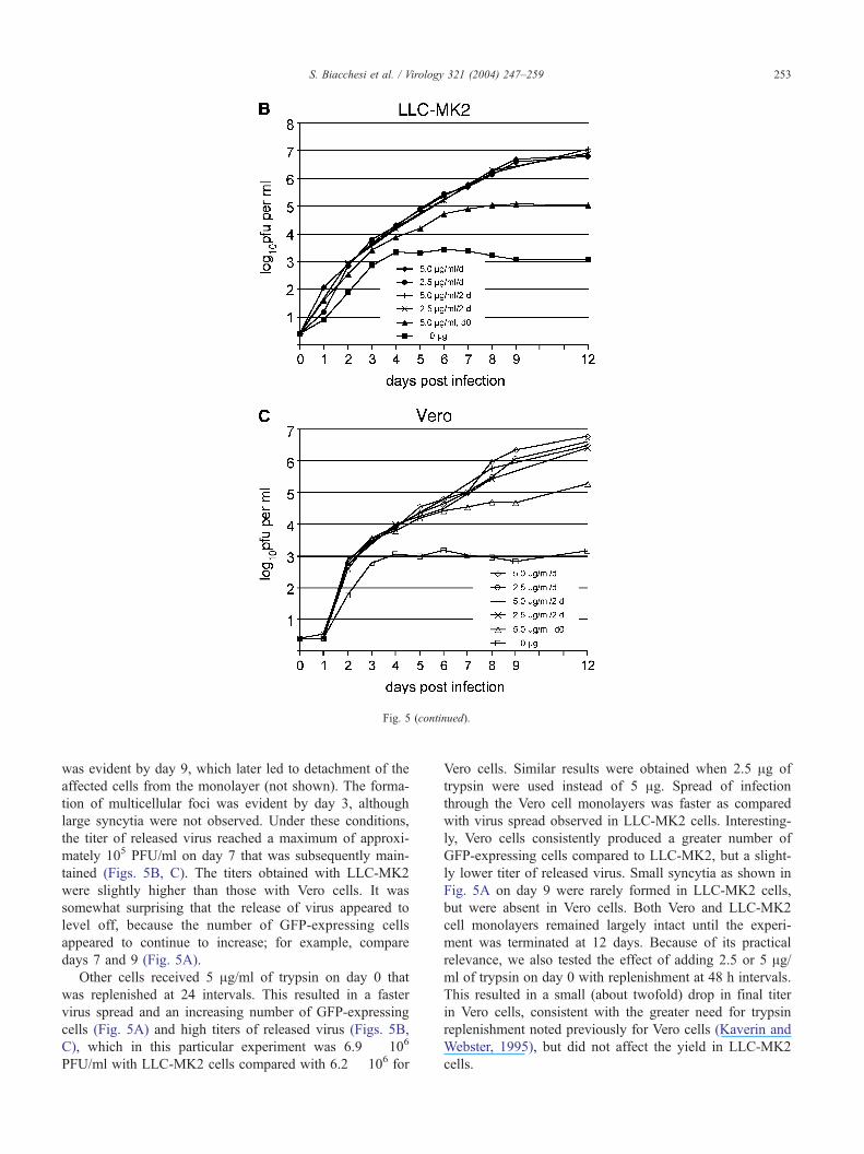
Fig. 5 (continued).
S. Biacchesi et al. / Virology 321 (2004) 247–259 253
was evident by day 9, which later led to detachment of the
affected cells from the monolayer (not shown). The forma-
tion of multicellular foci was evident by day 3, although
large syncytia were not observed. Under these conditions,
the titer of released virus reached a maximum of approxi-
mately 105 PFU/ml on day 7 that was subsequently main-
tained (Figs. 5B, C). The titers obtained with LLC-MK2
were slightly higher than those with Vero cells. It was
somewhat surprising that the release of virus appeared to
level off, because the number of GFP-expressing cells
appeared to continue to increase; for example, compare
days 7 and 9 (Fig. 5A).
Other cells received 5 Ag/ml of trypsin on day 0 that
was replenished at 24 intervals. This resulted in a faster
virus spread and an increasing number of GFP-expressing
cells (Fig. 5A) and high titers of released virus (Figs. 5B,
C), which in this particular experiment was 6.9 � 106
PFU/ml with LLC-MK2 cells compared with 6.2 � 106 for
Vero cells. Similar results were obtained when 2.5 Ag of
trypsin were used instead of 5 Ag. Spread of infection
through the Vero cell monolayers was faster as compared
with virus spread observed in LLC-MK2 cells. Interesting-
ly, Vero cells consistently produced a greater number of
GFP-expressing cells compared to LLC-MK2, but a slight-
ly lower titer of released virus. Small syncytia as shown in
Fig. 5A on day 9 were rarely formed in LLC-MK2 cells,
but were absent in Vero cells. Both Vero and LLC-MK2
cell monolayers remained largely intact until the experi-
ment was terminated at 12 days. Because of its practical
relevance, we also tested the effect of adding 2.5 or 5 Ag/ml of trypsin on day 0 with replenishment at 48 h intervals.
This resulted in a small (about twofold) drop in final titer
in Vero cells, consistent with the greater need for trypsin
replenishment noted previously for Vero cells (Kaverin and
Webster, 1995), but did not affect the yield in LLC-MK2
cells.

rology 321 (2004) 247–259
Insertion of multiple extra genes into rHMPV
The insertion of the GFP transcription cassette into
rHMPV reduced virus growth only slightly, as described
above. It was of interest to investigate the effect of adding
multiple, larger extra genes. We therefore replaced the GFP
cassette of rHMPV–GFP with a transcription cassette con-
taining a copy of the HMPV G ORF and, in addition, added
two copies in tandem of a transcription cassette containing
the F ORF in the order G1-F2-F3 (Fig. 6). This resulted in
the plasmid pHMPV+G1F23, whose encoded antigenome
contains sequences encoding 11 mRNAs rather than the 8 of
the natural genome, and would be 17343 nt long, an increase
of 30% over the natural genome of 13335 nt. This plasmid
was stable in bacteria despite the presence of two identical
copies of the G ORF and three identical copies of the F ORF.
Recombinant virus was readily recovered from transfections
with pHMPV+G1F23, and the virus appeared to replicate
with an efficiency similar to that of rHMPV–GFP.
The presence and intactness of the additional genes in
the genome of recovered rHMPV+G1F23 were investigated
by RT-PCR performed on total RNA from recovered virus
following three passages in Vero cells. RT-PCR was per-
formed with a forward primer hybridizing to the HMPV
leader region and a reverse primer hybridizing to N
sequence, which yielded a single, major band of approxi-
mately 4.1 kb, the appropriate size to contain the tandem
G1F2F3 supernumerary genes (not shown). The sequence
S. Biacchesi et al. / Vi254
Fig. 6. Insertion of three additional transcription cassettes into rHMPV. The G OR
the leader region including a naturally occurringMluI site, and a copy of the G GS
PacI site. The F ORF was modified by PCR to be flanked on the upstream side b
identical to that normally found between the N and P genes, and a copy of the N G
GE signal including a PacI site. One copy of theMluI–PacI G transcription casset
the MluI –PacI window of pHMPV–GFP in the order G-F-F, replacing the GFP t
signals are underlined, restriction sites are italicized and underlined, and GS and GE
of the supernumerary G1F2F3 genes could not be deter-
mined directly by RT-PCR consensus sequencing of viral
RNA because primers specific to G or F would prime on
each of the two copies of G and three copies of F,
precluding analysis of each gene individually. Therefore,
we first amplified the supernumerary genes by RT-PCR in
three separate, overlapping segments. The first RT-PCR
segment used a forward (positive-sense) primer (which also
served as the RT primer) from the leader region and a
reverse primer from the F gene (840 nt downstream of the F
gene start signal): although this latter primer would prime
on each copy of F, priming from the promoter-proximal
copy (F2, numbered as in rHMPV+G1F23) would be the
most efficient in combination with this forward primer, and
yielded a f1.6 kb fragment that contained the leader, the
G1 gene, and about 840 bp of the F2 gene, and was purified
by gel electrophoresis for sequence analysis. The second
segment was amplified by RT-PCR with an RT/forward
primer located about 670 nt downstream of the F gene start,
and a reverse primer located 554 nt downstream of the F
gene start: although the forward primer would prime in all
three copies of F and the reverse primer in all three copies
of F, priming in F2 and F3 would be the only combination
to result in successful amplification of a PCR fragment
(f1.5 kb) that contained the downstream 978 bp part of the
F2 gene, and a 554 bp upstream part of F3, and was
purified by gel electrophoresis for sequence analysis. The
third fragment was generated with an RT/forward primer
F was modified by PCR to be flanked on the upstream side by nt 12–41 of
signal, and on the downstream side by a copy of the F GE signal including a
y a partial GE signal, including a PacI site, an intergenic dinucleotide (GT)
S signal. On the downstream side, the F ORF was flanked by a copy of the F
te and two copies of the PacI –PacI F transcription cassette were cloned into
ranscription cassette. In the sequences shown, translation initiation and stop
signals are boxed. The total length of the additional sequence was 4008 nt.
Fig. 7. Northern blot analysis of mRNAs expressed by rHMPV+G1F23. LLC-MK2 cells were mock-infected (lanes 1, 5, and 9) or infected at an MOI of 3
PFU/cell with HMPV83 (lanes 2, 6, and 10), rHMPV (lanes 3, 7 and 11), or rHMPV+G1F23 (lanes 4, 8, and 12). Three days later, total intracellular RNAwas
isolated, electrophoresed on 1% agarose-formaldehyde gels, transferred to charged nylon, and analyzed by hybridization to double-stranded 32P-labeled DNA
probe specific to the F (lanes 1–4), G (lanes 5–8), or M (lanes 9–12) gene. The identities and calculated sizes of individual RNA species are indicated.
Fig. 8. Comparison of the multi-step growth kinetics of rHMPV, rHMPV–
GFP, and rHMPV+G1F23. LLC-MK2 cells were infected at a multiplicity
of infection of 0.01 PFU/cell with rHMPV (.), rHMPV–GFP (E), or
rHMPV+G1F23 (5). Medium samples were taken on indicated days post-
infection and replaced by an equivalent volume of fresh medium containing
5 Ag/ml of trypsin. The samples were flash frozen and analyzed later by
plaque assay and immunostaining. Each time point was represented by two
wells, and each virus titration was done in duplicate. Means are shown.
S. Biacchesi et al. / Virology 321 (2004) 247–259 255
from the upstream end of F (14 nt downstream the F gene
start signal) and a reverse primer from the upstream end of
N: the forward primer would prime in all three copies of F,
but priming in F3 would be the most efficient and would
yield a f1.75 kb product that contained most of the F3
gene, and 120 bp of the N gene, and was purified by gel
electrophoresis for sequence analysis. This provided an RT-
PCR consensus sequence of the G1F2F3 supernumerary
genes that was free of mutations, and all GS/GE signals and
intergenic sequences were correct, indicating that these
added genes were stably recovered in recombinant virus.
To determine the effect of the gene additions on viral gene
expression, cells were mock-infected or infected with
biologically derived HMPV83, rHMPV, or rHMPV+
G1F23, and total intracellular RNA was isolated 3 days
later and subjected to Northern blot analysis with double-
stranded probes to the F, G, or M genes (Fig. 7). The F
probe detected a major band of the appropriate size to be
the 1644-nt F mRNA, as well as a fainter band of genome
and antigenome, which in rHMPV+G1F23 would be 4008
nt larger compared with HMPV83 or rHMPV due to the
presence of the three added gene copies (Fig. 7, lane 4).
There were only trace amounts of readthrough mRNAs
detected with the F probe. Phosphorimager analysis indi-
cated that the amount of genome and antigenome for
rHPMV+G1F23 compared to with HMPV83 and rHMPV
was 0.9 and 0.5, respectively (corrected for the two extra
copies of the F gene present in rHMPV+G1F23). In
comparison, the relative amount of F mRNA for
rHMPV+G1F23 compared to HMPV83 and rHMPV was
six in each case. Normalized to the respective value of
genome and antigenome, this corresponded to a 6.6- to 12-
fold higher level of F mRNA for rHMPV+G1F23 compared
with HMPV83 and rHMPV.
Northern blot analysis with the G probe detected, for
each of the three viruses, a band of the appropriate size to be
the 711-nt G mRNA as well as bands corresponding to SH-
G and M2-SH-G readthrough mRNAs and a band
corresponding to antigenome and genome RNAs (Fig. 7,
lanes 7, lanes 6, 7, and 8). Phosphorimager analysis indi-
cated that the amount of genome and antigenome for
rHMPV+G1F23 compared with HMPV83 and rHMPV
was 1.0 and 0.5, respectively (corrected for the extra copy

S. Biacchesi et al. / Virology 321 (2004) 247–259256
of the G gene present in rHMPV+G1F23), values very
similar to that observed with the F probe noted above. In
comparison, the relative amount of G mRNA for rHMPV–
G1F23 compared with HMPV83 and rHMPV was 14-fold
and 15-fold, respectively. This would correspond to a 14- to
30-fold higher level of G mRNA for rHMPV+G1F23
compared with HMPV83 and rHMPV.
The rHMPV+G1F23 virus was compared to rHMPV and
rHMPV–GFP with regard to the efficiency of multicycle
replication in vitro. LLC-MK2 cells were inoculated with
0.01 PFU/cell and were incubated and sampled in the same
manner as for the experiment in Fig. 4. As shown in Fig. 8,
rHMPV+G1F23 replicated with an efficiency that was
moderately reduced compared with rHMPV but was slightly
higher than rHMPV–GFP. Thus, although HMPV is a rather
slow-growing virus, it readily accommodates sequence
modifications and additions, which opens possibilities for
its future use as a vector for mucosal immunization.
Discussion
In this study, we describe the recovery of infectious
HMPV from cDNA. HMPV was first recognized in 2001,
and the available reports unanimously indicated that it was
difficult to isolate and grow in cell culture due to its slow
replication, trypsin dependence, small range of susceptible
cell lines, and slow development of virus-induced CPE as a
marker of virus growth. Therefore, we first constructed a
recombinant virus containing an additional transcription
cassette encoding GFP, inserted as first promoter-proximal
gene. The ability to directly visualize virus spread in living
cells compensated technically for the slow development of
CPE, and the rHMPV–GFP virus proved to be very helpful
both during the establishment of the HMPV reverse genetics
system and in optimizing conditions for HMPV replication
in vitro. In the future, rHMPV–GFP will also be useful to
facilitate diagnostic tests, such as neutralization assays,
because this recombinant can be visualized directly. More-
over, rHMPV–GFP will be used to study HMPV tropism
and pathogenesis in animal models.
We subsequently recovered rHMPV that differed from
the biologically derived virus only by the insertion of an
NheI marker site. This virus replicated in vitro as efficiently
as biologically derived HMPV, indicating that it will be
appropriate as a starting point to develop a live attenuated
HMPV vaccine. The recovery of wild-type-like rHMPV
from cloned cDNA provided the first identification of
functional sequences for the HMPV genome and its encoded
macromolecules. Although the recovery of infectious virus
from cloned cDNA has been achieved for several mono-
negaviruses, HMPV has the distinction of being a recently
recognized, poorly characterized agent of human disease,
and the development of functional sequences and a reverse
genetics system is an important step for molecular epide-
miology and vaccine development.
Replenishment of the trypsin in infected cell cultures
yielded virus titers that increased over 10–12 days, with a
50- to 100-fold increased final virus yield as compared
with titers obtained using a single initial trypsin dose.
Trypsin replenishment had a somewhat greater effect on
HMPV replication in Vero cells than in LLC-MK2 cells,
which is consistent with an observed rapid trypsin inac-
tivation in Vero cell culture fluids (Kaverin and Webster,
1995). From our ongoing experience in propagating
HMPV, we have modified this protocol so that 5 Ag/ml
fresh trypsin is added every second or third day without
exchanging the medium. When this regimen is applied
during routine production of HMPV stocks, titers of 1.0–
4.0 � 107 PFU/ml can be routinely achieved in monolayer
cultures, which is about 1000-fold improved compared to
with titers described in literature (van den Hoogen et al.,
2001) and likely could be further improved in optimized
cell fermentators. Importantly, these improved titers can be
achieved in Vero cells, which are an acceptable substrate
for vaccine manufacture. Conditions for efficient replica-
tion are important for virological studies in general, and
are particularly important for vaccine manufacture to be
feasible. These results indicate that it indeed should be
feasible to develop and manufacture a live attenuated
vaccine for HMPV.
The HMPV CPE that is described in the literature occurs
late in infection, on average on day 17 (Boivin et al., 2002)
or between day 10 and 14 (van den Hoogen et al., 2001).
These observations are consistent with our findings, and the
present results indicate that CPE lagged several days behind
infection and GFP expression. This was true even when 5
Ag/ml of trypsin was added daily, indicating that the slow
development of CPE did not reflect a lack of available
trypsin. Although these results were obtained using
rHMPV–GFP, similar results were obtained with infections
involving biologically derived HMPV83 or rHMPV, in
which indirect immunofluorescence assays performed on
replicate HMPV-infected cells fixed at different time points
after infection showed a similar situation in which most of
the cells were infected days before the onset of CPE would
occur (not shown). This showed that the slow onset of CPE
is not due to a reduced replication or restricted spread of
rHMPV–GFP.
Detection of GFP mRNA by HMPV showed that the
putative N GS and F GE signals that were used to construct
the transcription cassette indeed functioned to produce a
monocistronic mRNA. The different Northern blot probes
used in this study revealed substantial differences in the
level of readthrough mRNAs associated with various
HMPV genes and the GFP gene. For example, the M gene
was associated with a high level of readthrough into
downstream genes, whereas the GFP and F genes were
associated with little readthrough. The GFP transcription
cassette contained the F GE signal, suggesting that this
common element is a highly efficient termination signal that
is responsible for the lack of observed readthrough mRNAs.

S. Biacchesi et al. / Virology 321 (2004) 247–259 257
As such, this signal should be optimal for the efficient
expression of added genes.
The ability to recover infectious virus containing an
insert following nt 57 relative to the 3V(leader) end of the
genome indicates that HMPV, like RSV (Fearns et al., 2000;
Fearns et al., 2002), probably has a ‘‘short’’ promoter rather
than the dipartite 96-nt promoter that is characteristic of
Paramyxovirinae and involves a second promoter element
contained within the first gene (Keller et al., 2001; Tapparel
et al., 1998). The multi-step growth of rHMPV–GFP in
vitro resulted in final titers that were about threefold reduced
compared with rHMPV. Because both the rHMPV and
rHMPV–GFP full-length plasmids were sequenced in their
entirety, and differed only in the presence of the GFP gene,
the reduced in vitro growth observed for rHMPV–GFP can
be attributed solely to effects from the additional gene. This
might indicate that the insert effected a modest reduction in
the functioning of a cis-acting element such as the promoter.
Alternatively, it might be an effect of increased genome
length and gene number. The genome of rHMPV–GFP is
748 nt (5.6%) longer compared with rHMPV. In addition to
the difference in genome length, the presence of an addi-
tional gene as first gene in the genome might contribute to
the reduced growth due to attenuation of the transcription of
downstream genes.
To further explore the limits of HMPV as a vector, we
designed a recombinant virus rHMPV+G1F23 that contains
three additional genes in the promoter-proximal position,
namely one additional copy of the HMPV G gene and two
additional copies of the HMPV F gene. The rHMPV+
G1F23 virus has a total genome length that was increased
compared with the wild-type virus by approximately 4 kb
(30%). The production of infectious virus during multi-step
growth in vitro was reduced only modestly compared with
rHMPVand was equivalent to or slightly greater than that of
rHMPV–GFP. This suggested that although insertion of an
additional transcriptional unit into the HMPV genome
resulted in a modest decrease in virus replication, this effect
was not augmented further by a longer insert or a larger
number of transcription cassettes within the range tested.
This is offered with the caveat that virus replication might
have been enhanced by the expression of the additional
copies of G and F, which might have counteracted possible
inhibitory effects due to genome length or gene number.
Regardless of interpretation, the growth phenotype of
rHMPV+G1F23 shows that the HMPV genome is quite
versatile, and that the strategy described here can be used to
design HMPV vaccine candidates containing additional
copies of the putative antigenic determinant genes to
achieve an increased gene dose for higher expression.
Indeed, Northern blot analysis showed that the expression
of F or G mRNA was increased more than 6- or 14-fold,
respectively. The approach of adding glycoprotein genes can
also be used to generate multivalent rHMPV vaccine can-
didates expressing antigenic determinants of HMPVs of
heterologous antigenic lineages or subgroups in addition
to the homologous set of genes present in the recombinant
backbone.
It is generally thought that protection against respiratory
disease caused by members of the Pneumovirinae can be
best induced by live attenuated vaccines, particularly given
the adverse effects of a previously tested formalin-inacti-
vated HRSV vaccine (Collins and Murphy, 2002). This is
where reverse genetic systems have an essential role, as they
allow the rational design of safe attenuated vaccine candi-
dates (Collins and Murphy, 2002). HMPV represents an
important agent of pediatric respiratory tract disease for
which a vaccine should be developed, which will be an
important future application of the HMPV reverse genetic
system.
Materials and methods
Cells and viruses
LLC-MK2 (ATCC CCL 7.1) and Vero (ATCC CCL-81)
cells were maintained in OptiMEM I (Invitrogen GIBCO)
supplemented with 5% fetal bovine serum. BSR T7/5 cells
are baby hamster kidney 21 (BHK-21) cells that constitu-
tively express T7 RNA polymerase (Buchholz et al., 1999).
They were maintained in Glasgow MEM supplemented with
glutamine and amino acids (Invitrogen) and 10% fetal
bovine serum. The Canadian HMPV isolate HMPV83 (Peret
et al., 2002) and the recombinant HMPVs described below
were propagated in Vero, LLC-MK2, or BSR T7/5 cells in
the absence of serum and the presence of 5 Ag/ml of trypsin.
Virus titers were determined by plaque assay on LLC-MK2
cells under methylcellulose overlay containing 5 Ag/ml
trypsin in the absence of serum, and plaques were visualized
6 days later by immunostaining with a 1:1000 dilution of a
polyclonal rabbit serum raised against gradient-purified
HMPV83. Growth curves were done with duplicate wells,
and each aliquot was titrated in duplicate, and means were
calculated.
Viral RNA isolation
Confluent monolayers of LLC-MK2 cells were infected
with HMPV83 and incubated at 32 jC in the presence of 5
Ag/ml trypsin. Ten days post-infection, clarified superna-
tants were harvested, and virion RNA (vRNA) was isolated
using the QIAamp viral RNA kit (Qiagen) according to the
manufacturer’s instructions.
Construction of T7 expression plasmids encoding the
HMPV antigenome and N, P, M2-1, and L support proteins
A complete cDNA copy of the HMPV83 genome was
constructed (Fig. 1) by assembling three overlapping
cDNA clones generated by RT-PCR with specific primers
designed from the published HMPV83 consensus se-

S. Biacchesi et al. / Virology258
quence (Genbank accession number, AY297749) (Biac-
chesi et al., 2003). Primer sequences are available from
the authors upon request. First strand cDNA was generat-
ed from purified virion RNA using Superscript II reverse
transcriptase (Invitrogen). PCR was carried out using
Platinum Pfx DNA polymerase (Invitrogen). The nucleo-
tide sequences of cloned RT-PCR fragments were deter-
mined using an ABI 3100 sequencer with the Big-Dye
terminator ready reaction kit v1.1 (Applied Biosystems).
Between two and six clones of each fragment were
sequenced completely or in part to identify clones that
contained the desired sequence free of error. ORFs encod-
ing the putative nucleoprotein N (1185 nt), phosphopro-
tein P (885 nt), transcription elongation factor M2-1 (564
nt), and RNA polymerase L (6018 nt) were amplified by
RT-PCR from virion RNA using specific primers whose
sequences are available from the authors upon request,
and were cloned into the T7 expression vector pTM1,
wherein translational initiation is mediated by the internal
ribosome entry site of encephalomyocarditis virus.
Construction of pHMPV–GFP
The antigenomic pHMPV83 plasmid was modified by
the promoter-proximal insertion of a transcription cassette
containing the ORF for enhanced GFP (Clonetech, Inc.).
This cassette was assembled from two PCR fragments.
The first fragment (fragment A in Fig. 2) contained, in 5Vto 3V order relative to the antigenome, the naturally
occurring MluI site at nt 12 of the leader region, followed
by the remainder of the 41-nt leader region, followed by
the 16-nt putative N gene-start (GS) sequence (GGGA-
CAAGTGAAAATG, positive sense, HMPV83 nt 42–57,
N ORF initiation codon underlined), followed by the GFP
ORF that was modified to use the ATG of the N GS signal
as its initiation codon, followed by a 14-nt putative gene-
end (GE) sequence derived from the F gene (AGTTAAT-
TAAAAAA), containing a naturally occurring PacI site
(underlined). The second fragment (fragment B in Fig.
2) contained, in 5V to 3V order, a partial GE signal
containing a PacI site, a 2-nt (GT) intergenic region
identical to that of the N–P gene junction, and the N
GS signal and partial N gene up to the naturally occurring
AvrII site at HMPV83 nt 649. Fragments A and B were
digested by MluI/PacI and PacI/AvrII, respectively, and
cloned simultaneously into an intermediate construct con-
taining cDNA fragment 1 of pHMPV83 (Fig. 1) that had
been digested with MluI/AvrII. This modified cDNA
fragment 1 was then substituted into the AatII–NheI
window of the complete pHMPV83 antigenomic cDNA
clone, leading to the final construct pHMPV–GFP. The
length of the encoded rHMPV–GFP antigenome, exclu-
sive of non-viral sequence, would be 14083 nt. The
sequences of the regions that had been subjected to
PCR, namely the leader, GFP, and upstream end of the
N gene, were confirmed by nucleotide sequencing.
Construction of pHMPV+G1F23
cDNAs of the HMPVG or F ORF were amplified by PCR
using primers that placed a G GS signal and an F GE signal at
the beginning and end, respectively, of the G ORF, or an N
GS signal and F GE signal at the beginning and end of the F
ORF. In each case, this was followed by a GT dinucleotide
intergenic region; in addition, the G transcription cassette
was flanked by MluI and PacI sites, and the F cassette by
PacI sites. The MluI–PacI G fragment was used to replace
the MluI–PacI GFP fragment (Fig. 2) from an intermediate
construct containing fragment 1 (comprising the antigenome
from leader to NheI site) of pHMPV–GFP. Next, two copies
of the F fragment were cloned into the PacI site of this
intermediate plasmid, and restriction digestion was used to
identify a clone in which the ORFs of both fragments were in
the correct orientation. This G1F23-bearing intermediate
fragment was combined with pHMPV fragments 2 and 3
(Fig. 1) to yield pHMPV+G1F23. The length of the encoded
rHMPV+G1F23 antigenome, exclusive of non-viral se-
quence, would be 17343 nt. The sequences of the G and F
cassettes were confirmed before assembly by nucleotide
sequencing of intermediate clones each bearing an individual
cassette.
rHMPV recovery
Confluent BSR T7/5 cells in 6-well dishes were trans-
fected with 5 Ag of antigenomic plasmid pHMPV,
pHMPV–GFP, or pHMPV+G1F23, 2 Ag each of pT7-N
and pT7-P, and 1 Ag each of pT7-M2-1 and pT7-L support
plasmids per well. Transfections were done with Lipofect-
amine 2000 (Invitrogen) in OptiMEM without trypsin or
serum and maintained overnight at 32 jC. The Lipofect-
amine transfection medium was removed 1 day later and
replaced with Glasgow MEM without trypsin or serum.
Trypsin was added on day 3 to a final concentration of 5 Ag/ml, and cell-medium mixtures were passaged onto fresh
LLC-MK2 cells on day 6. The expression of GFP was
monitored by fluorescent microscopy.
Northern blot analysis
LLC-MK2 cells were mock-infected or infected with
HMPV83 or the indicated recombinant virus, each at an
input MOI of 3 PFU/cell, incubated for 3 days, and
processed to purify total intracellular RNA using an RNeasy
kit (Qiagen) according to the manufacturer’s instructions.
RNA was electrophoresed in a 1% agarose gel in the
presence of 0.44 M formaldehyde, transferred to charged
nylon membrane (Hybond-N+, Amersham Biosciences),
fixed by UV cross-linking, and analyzed by hybridization
with denatured double-stranded 32P-labeled DNA probes
generated by random priming from GFP, M, G, or F gene
cDNA fragments. Radioactivity was detected by analysis
with a phosphorimager.
321 (2004) 247–259

S. Biacchesi et al. / Virology 321 (2004) 247–259 259
Acknowledgments
We thank Christopher Hanson for sequencing work and
Elaine Lamirande for excellent technical assistance.
References
Bermingham, A., Collins, P.L., 1999. The M2-2 protein of human respira-
tory syncytial virus is a regulatory factor involved in the balance be-
tween RNA replication and transcription. Proc. Natl. Acad. Sci. U.S.A.
96 (20), 11259–11264.
Biacchesi, S., Skiadopoulos, M.H., Boivin, G., Hanson, C.T., Murphy,
B.R., Collins, P.L., Buchholz, U.J., 2003. Genetic diversity between
human metapneumovirus subgroups. Virology 315 (1), 1–9.
Boivin, G., Abed, Y., Pelletier, G., Ruel, L., Moisan, D., Cote, S., Peret,
T.C., Erdman, D.D., Anderson, L.J., 2002. Virological features and clin-
ical manifestations associated with human metapneumovirus: a new
paramyxovirus responsible for acute respiratory-tract infections in all
age groups. J. Infect. Dis. 186 (9), 1330–1334.
Boivin, G., De Serres, G., Cote, S., Gilca, R., Abed, Y., Rochette, L.,
Bergeron, M.G., Dery, P., 2003. Human metapneumovirus infections
in hospitalized children. Emerg. Infect. Dis. 9 (6), 634–640.
Buchholz, U.J., Finke, S., Conzelmann, K.K., 1999. Generation of bovine
respiratory syncytial virus (BRSV) from cDNA: BRSV NS2 is not
essential for virus replication in tissue culture, and the human RSV
leader region acts as a functional BRSV genome promoter. J. Virol.
73 (1), 251–259.
Collins, P.L., Murphy, B.R., 2002. Respiratory syncytial virus: reverse
genetics and vaccine strategies. Virology 296 (2), 204–211.
Collins, P.L., Hill, M.G., Johnson, P.R., 1990. The two open reading frames
of the 22K mRNA of human respiratory syncytial virus: sequence com-
parison of antigenic subgroups A and B and expression in vitro. J. Gen.
Virol. 71 (Pt 12), 3015–3020.
Collins, P.L., Hill, M.G., Cristina, J., Grosfeld, H., 1996. Transcription elon-
gation factor of respiratory syncytial virus, a nonsegmented negative-
strand RNA virus. Proc. Natl. Acad. Sci. U.S.A. 93 (1), 81–85.
Collins, P.L., Camargo, E., Hill, M.G., 1999. Support plasmids and support
proteins required for recovery of recombinant respiratory syncytial vi-
rus. Virology 259 (2), 251–255.
Durbin, A.P., Hall, S.L., Siew, J.W., Whitehead, S.S., Collins, P.L., Mur-
phy, B.R., 1997a. Recovery of infectious human parainfluenza virus
type 3 from cDNA. Virology 235 (2), 323–332.
Ebihara, T., Endo, R., Kikuta, H., Ishiguro, N., Yoshioka, M., Ma, X.,
Kobayashi, K., 2003. Seroprevalence of human metapneumovirus in
Japan. J. Med. Virol. 70 (2), 281–283.
Falsey, A.R., Erdman, D., Anderson, L.J., Walsh, E.E., 2003. Human
metapneumovirus infections in young and elderly adults. J. Infect.
Dis. 187 (5), 785–790.
Fearns, R., Collins, P.L., Peeples, M.E., 2000. Functional analysis of the
genomic and antigenomic promoters of human respiratory syncytial
virus. J. Virol. 74 (13), 6006–6014.
Fearns, R., Peeples, M.E., Collins, P.L., 2002. Mapping the transcription
and replication promoters of respiratory syncytial virus. J. Virol. 76 (4),
1663–1672.
Freymouth, F., Vabret, A., Legrand, L., Eterradossi, N., Lafay-Delaire, F.,
Brouard, J., Guillois, B., 2003. Presence of the new human metapneu-
movirus in French children with bronchiolitis. Pediatr. Infect. Dis. J. 22
(1), 92–94.
Greensill, J., McNamara, P.S., Dove, W., Flanagan, B., Smyth, R.L., Hart,
C.A., 2003. Human metapneumovirus in severe respiratory syncytial
virus bronchiolitis. Emerg. Infect. Dis. 9 (3), 372–375.
Jartti, T., van den Hoogen, B., Garofalo, R.P., Osterhaus, A.D., Ruuskanen,
O., 2002. Metapneumovirus and acute wheezing in children. Lancet 360
(9343), 1393–1394.
Jin, H., Cheng, X., Zhou, H.Z., Li, S., Seddiqui, A., 2000. Respiratory
syncytial virus that lacks open reading frame 2 of the M2 gene (M2-2)
has altered growth characteristics and is attenuated in rodents. J. Virol.
74 (1), 74–82.
Kaverin, N.V., Webster, R.G., 1995. Impairment of multicycle influenza
virus growth in Vero (WHO) cells by loss of trypsin activity. J. Virol. 69
(4), 2700–2703.
Keller, M.A., Murphy, S.K., Parks, G.D., 2001. RNA replication from the
simian virus 5 antigenomic promoter requires three sequence-dependent
elements separated by sequence-independent spacer regions. J. Virol. 75
(8), 3993–3998.
Maggi, F., Pifferi, M., Vatteroni, M., Fornai, C., Tempestini, E., Anzilotti,
S., Lanini, L., Andreoli, E., Ragazzo, V., Pistello, M., Specter, S.,
Bendinelli, M., 2003. Human metapneumovirus associated with respi-
ratory tract infections in a 3-year study of nasal swabs from infants in
Italy. J. Clin. Microbiol. 41 (7), 2987–2991.
Nissen, M.D., Siebert, D.J., Mackay, I.M., Sloots, T.P., Withers, S.J., 2002.
Evidence of human metapneumovirus in Australian children. Med. J.
Aust. 176 (4), 188.
Osterhaus, A., Fouchier, R., 2003. Human metapneumovirus in the com-
munity. Lancet 361 (9361), 890–891.
Peiris, J.S., Tang, W.H., Chan, K.H., Khong, P.L., Guan, Y., Lau, Y.L., Chiu,
S.S., 2003. Children with respiratory disease associated with metapneu-
movirus in Hong Kong. Emerg. Infect. Dis. 9 (6), 628–633.
Peret, T.C., Boivin, G., Li, Y., Couillard, M., Humphrey, C., Osterhaus,
A.D., Erdman, D.D., Anderson, L.J., 2002. Characterization of human
metapneumoviruses isolated from patients in North America. J. Infect.
Dis. 185 (11), 1660–1663.
Perrotta, A.T., Been, M.D., 1991. A pseudoknot-like structure required for
efficient self-cleavage of hepatitis delta virus RNA. Nature 350 (6317),
434–436.
Tapparel, C., Maurice, D., Roux, L., 1998. The activity of Sendai virus
genomic and antigenomic promoters requires a second element past the
leader template regions: a motif (GNNNNN)3 is essential for replica-
tion. J. Virol. 72 (4), 3117–3328.
van den Hoogen, B.G., de Jong, J.C., Groen, J., Kuiken, T., de Groot, R.,
Fouchier, R.A., Osterhaus, A.D., 2001. A newly discovered human
pneumovirus isolated from young children with respiratory tract dis-
ease. Nat. Med. 7 (6), 719–724.
van den Hoogen, B.G., Bestebroer, T.M., Osterhaus, A.D., Fouchier, R.A.,
2002. Analysis of the genomic sequence of a human metapneumovirus.
Virology 295 (1), 119–132.
Viazov, S., Ratjen, F., Scheidhauer, R., Fiedler, M., Roggendorf, M., 2003.
High prevalence of human metapneumovirus infection in young chil-
dren and genetic heterogeneity of the viral isolates. J. Clin. Microbiol.
41 (7), 3043–3045.