Recent Advances in the Biological Activity of s-Triazine Core ...
19
Citation: Maliszewski, D.; Drozdowska, D. Recent Advances in the Biological Activity of s-Triazine Core Compounds. Pharmaceuticals 2022, 15, 221. https://doi.org/ 10.3390/ph15020221 Academic Editor: Pawel Kafarski Received: 5 January 2022 Accepted: 10 February 2022 Published: 12 February 2022 Publisher’s Note: MDPI stays neutral with regard to jurisdictional claims in published maps and institutional affil- iations. Copyright: © 2022 by the authors. Licensee MDPI, Basel, Switzerland. This article is an open access article distributed under the terms and conditions of the Creative Commons Attribution (CC BY) license (https:// creativecommons.org/licenses/by/ 4.0/). pharmaceuticals Review Recent Advances in the Biological Activity of s-Triazine Core Compounds Dawid Maliszewski * and Danuta Drozdowska * Department of Organic Chemistry, Medical University of Bialystok, 15-222 Bialystok, Poland * Correspondence: [email protected] (D.M.); [email protected] (D.D.) Abstract: An effective strategy for successful chemotherapy relies on creating compounds with high selectivity against cancer cells compared to normal cells and relatively low cytotoxicity. One such approach is the discovery of critical points in cancer cells, i.e., where specific enzymes that are potential therapeutic targets are generated. Triazine is a six-membered heterocyclic ring compound with three nitrogen replacing carbon-hydrogen units in the benzene ring structure. The subject of this review is the symmetrical 1,3,5-triazine, known as s-triazine. 1,3,5-triazine is one of the oldest heterocyclic compounds available. Because of its low cost and high availability, it has attracted researcher attention for novel synthesis. s-Triazine has a weak base, it has much weaker resonance energy than benzene, therefore, nucleophilic substitution is preferred to electrophilic substitution. Heterocyclic bearing a symmetrical s-triazine core represents an interesting class of compounds possessing a wide spectrum of biological properties such as anti-cancer, antiviral, fungicidal, insecticidal, bactericidal, herbicidal and antimicrobial, antimalarial agents. They also have applications as dyes, lubricants, and analytical reagents. Hence, the group of 1,3,5-triazine derivatives has developed over the years. Triazine is not only the core amongst them, but is also a factor increasing the kinetic potential of the entire derivatives. Modifying the structure and introducing new substituents makes it possible to obtain compounds with broad inhibitory activity on processes such as proliferation. In some cases, s-triazine derivatives induce cell apoptosis. In this review we will present currently investigated 1,3,5-triazine derivatives with anti-cancer activities, with particular emphasis on their inhibition of enzymes involved in the process of tumorigenesis. Keywords: 1,3,5-triazine; s-triazine; anticancer; enzyme inhibitory activity 1. Introduction As far as we know, tumors are the most serious cause of death in the world. Cancers with the highest mortality rates in 2018 were lung cancer (2.1 million new cases and 1.8 million deaths), breast cancer (million new cases and 880 thousand deaths), prostate cancer (1.3 million new cases and 360 thousand deaths), and stomach cancer (1 million new cases and 783 thousand deaths) [1]. The fight against cancer has consumed huge amounts of money to find the cure with little effect. Nevertheless, it cannot be defined as a failure. As Napoleon Hill said, “every adversity, every failure, every heartache carries with it the seed of an equal or greater benefit”. Following this thought, we would like to highlight two aspects of the fight against cancer. First, decades of research lead to more and more precise descriptions of the mechanisms taking place in cancer cells, it is possible to determine the most effective aim in targeted therapies. Second and equally important, the development of small molecules. The development of more active, selective and less cytotoxic drugs is due to designing chemical compounds based on a structure-activity relationship (SAR) [2]. In this search, the leading linker is 1,3,5-triazine, a symmetrical heterocyclic aromatic ring enabling the expansion of the structure in a multi-vector manner. Decades of research have revealed a Pharmaceuticals 2022, 15, 221. https://doi.org/10.3390/ph15020221 https://www.mdpi.com/journal/pharmaceuticals
-
Upload
khangminh22 -
Category
Documents
-
view
0 -
download
0
Transcript of Recent Advances in the Biological Activity of s-Triazine Core ...
�����������������
Citation: Maliszewski, D.;
Drozdowska, D. Recent Advances in
the Biological Activity of s-Triazine
Core Compounds. Pharmaceuticals
2022, 15, 221. https://doi.org/
10.3390/ph15020221
Academic Editor: Paweł Kafarski
Received: 5 January 2022
Accepted: 10 February 2022
Published: 12 February 2022
Publisher’s Note: MDPI stays neutral
with regard to jurisdictional claims in
published maps and institutional affil-
iations.
Copyright: © 2022 by the authors.
Licensee MDPI, Basel, Switzerland.
This article is an open access article
distributed under the terms and
conditions of the Creative Commons
Attribution (CC BY) license (https://
creativecommons.org/licenses/by/
4.0/).
pharmaceuticals
Review
Recent Advances in the Biological Activity of s-TriazineCore CompoundsDawid Maliszewski * and Danuta Drozdowska *
Department of Organic Chemistry, Medical University of Bialystok, 15-222 Białystok, Poland* Correspondence: [email protected] (D.M.); [email protected] (D.D.)
Abstract: An effective strategy for successful chemotherapy relies on creating compounds with highselectivity against cancer cells compared to normal cells and relatively low cytotoxicity. One suchapproach is the discovery of critical points in cancer cells, i.e., where specific enzymes that are potentialtherapeutic targets are generated. Triazine is a six-membered heterocyclic ring compound with threenitrogen replacing carbon-hydrogen units in the benzene ring structure. The subject of this reviewis the symmetrical 1,3,5-triazine, known as s-triazine. 1,3,5-triazine is one of the oldest heterocycliccompounds available. Because of its low cost and high availability, it has attracted researcher attentionfor novel synthesis. s-Triazine has a weak base, it has much weaker resonance energy than benzene,therefore, nucleophilic substitution is preferred to electrophilic substitution. Heterocyclic bearing asymmetrical s-triazine core represents an interesting class of compounds possessing a wide spectrumof biological properties such as anti-cancer, antiviral, fungicidal, insecticidal, bactericidal, herbicidaland antimicrobial, antimalarial agents. They also have applications as dyes, lubricants, and analyticalreagents. Hence, the group of 1,3,5-triazine derivatives has developed over the years. Triazine is notonly the core amongst them, but is also a factor increasing the kinetic potential of the entire derivatives.Modifying the structure and introducing new substituents makes it possible to obtain compoundswith broad inhibitory activity on processes such as proliferation. In some cases, s-triazine derivativesinduce cell apoptosis. In this review we will present currently investigated 1,3,5-triazine derivativeswith anti-cancer activities, with particular emphasis on their inhibition of enzymes involved in theprocess of tumorigenesis.
Keywords: 1,3,5-triazine; s-triazine; anticancer; enzyme inhibitory activity
1. Introduction
As far as we know, tumors are the most serious cause of death in the world. Cancerswith the highest mortality rates in 2018 were lung cancer (2.1 million new cases and1.8 million deaths), breast cancer (million new cases and 880 thousand deaths), prostatecancer (1.3 million new cases and 360 thousand deaths), and stomach cancer (1 million newcases and 783 thousand deaths) [1].
The fight against cancer has consumed huge amounts of money to find the cure withlittle effect. Nevertheless, it cannot be defined as a failure. As Napoleon Hill said, “everyadversity, every failure, every heartache carries with it the seed of an equal or greaterbenefit”. Following this thought, we would like to highlight two aspects of the fightagainst cancer. First, decades of research lead to more and more precise descriptions of themechanisms taking place in cancer cells, it is possible to determine the most effective aimin targeted therapies. Second and equally important, the development of small molecules.The development of more active, selective and less cytotoxic drugs is due to designingchemical compounds based on a structure-activity relationship (SAR) [2]. In this search,the leading linker is 1,3,5-triazine, a symmetrical heterocyclic aromatic ring enabling theexpansion of the structure in a multi-vector manner. Decades of research have revealed a
Pharmaceuticals 2022, 15, 221. https://doi.org/10.3390/ph15020221 https://www.mdpi.com/journal/pharmaceuticals

Pharmaceuticals 2022, 15, 221 2 of 19
wide range of properties of s-triazine derivatives. In this review we will present currentlyinvestigated 1,3,5-triazine derivatives with anti-cancer activities.
This review presents the current state of knowledge on 1,3,5-triazine derivatives, theirstructures and anticancer activity, as well as their ability to inhibit different enzymes or theirDNA-binding potential. This data could be helpful in the development of new drugs andtherapeutic methods. By analysing the presented approach, a series of compounds withhigh potency and low toxicity can be designed, synthesized, characterized and evaluatedfor desired pharmacological activity. The collected data are presented in summary Table 1.
Table 1. Promising effects of 1,3,5-triazine derivatives on cell lines and/or enzymes. N/A; not available.
No. Cancer Cells/Effects Targets/Effects Reference Substance Ref.
1 N/A DNA topoisomerase IIα(IC50 = 57.6 µM)
Etoposide: DNA topoisomerase IIα(IC50 = 59.2 µM) [3]
2A549 (IC50 = 0.20 µM)MCF-7 (IC50 = 1.25 µM)Hela (IC50 = 1.03 µM)
PI3Kα (IC50 = 7.0 nM)mTOR (IC50 = 48 nM)
GDC-0941: A549 (IC50 = 1.21 µM),MCF-7 (IC50 = 1.47 µM), Hela(IC50 = 3.72 µM), PI3Kα
(IC50 = 6.0 nM), mTOR(IC50 = 525 nM);PI-103: PI3Kα (IC50 = 5.1 nM),mTOR (IC50 = 21 nM)
[4]
3
MDA-MB321 (IC50 = 15.83 µM)MCF-7 (IC50 = 16.32 µM)Hela (IC50 = 2.21 µM)HepG2 (IC50 = 12.21 µM)
mTOR (IC50 = 8.45 nM)PI3Kα (IC50 = 3.41 nM)
Gedatolisib:mTOR (IC50 = 2.5 nM)PI3Kα (IC50 = 6.04 nM)
[5]
7
leukemia (GI50 = 1.96 µM)colon cancer (GI50 = 2.60 µM)CNS (GI50 = 2.72 µM)melanoma (GI50 = 1.91 µM)ovarian (GI50 = 4.01 µM)renal (GI50 = 3.03 µM)prostate (GI50 = 4.40 µM)breast (GI50 = 2.04 µM)
hDHFR (IC50 = 0.002 µM)
Triazine–Benzimidazole:leukemia (GI50 = 3.71 µM)colon cancer (GI50 = 2.76 µM)CNS (GI50 = 1.86 µM)melanoma (GI50 = 2.70 µM)ovarian (GI50 = 2.41 µM)renal (GI50 = 1.89 µM)prostate (GI50 = 2.75 µM)breast (GI50 = 2.58 µM)MTX: hDHFR (IC50 = 0.02 µM)
[6]
8HCT116 (IC50 = 0.88 µM)A549 (IC50 = 0.07 µM)HL-60 (IC50 = 0.33 µM)
hDHFR (IC50 = 0.00746 µM)
MTX:HCT116 (IC50 = 0.75 µM)A549 (IC50 = 0.25 µM)HL-60 (IC50 = 1.09 µM)HepG2 (IC50 = 0.41 µM)MDA-MB-234 (IC50 = 9.49 µM)hDHFR (IC50 = 0.00667 µM)
[7]
9HCT116 (IC50 = 1.61 µM)A549 (IC50 = 0.5 µM)HL-60 (IC50 = 0.87 µM)
hDHFR (IC50 = 0.00372 µM)
10
HCT116 (IC50 = 0.02 µM)A549 (IC50 = 0.74 µM)HL-60 (IC50 = 0.35 µM)HepG2 (IC50 = 1.4 µM)MDA-MB-234 (IC50 = 0.44 µM)
hDHFR (IC50 = 0.00646 µM)
11
HCT116 (IC50 = 0.001 µM)A549 (IC50 = 0.21 µM)HL-60 (IC50 = 0.33 µM)HepG2 (IC50 = 1.38 µM)MDA-MB-234 (IC50 = 0.06 µM)
hDHFR (IC50 = 0.00408 µM)
12 HCT116 (GI50 = 0.026 µM)MCF-7 (GI50 = 0.08 µM)
hDHFR (IC50 = 0.0061 µM)rat TrxR (IC50 = 4.6 µM)
MTX:hDHFR (IC50 = 0.0079 µM)HCT116 (GI50 = 0.015 µM)MCF-8 (GI50 = 0.024 µM)
[8]
13 HCT116 (GI50 = 0.116 µM)MCF-8 (GI50 = 0.127 µM)
hDHFR (IC50 = 0.0026 µM)rat TrxR (IC50 = 5.9 µM)
Pharmaceuticals 2022, 15, 221 3 of 19
Table 1. Cont.
No. Cancer Cells/Effects Targets/Effects Reference Substance Ref.
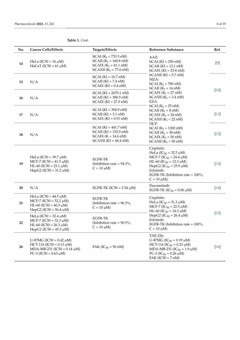
14 HeLa (IC50 = 16 µM)HaCaT (IC50 = 61 µM)
hCAI (KI = 733.3 nM)hCAII (KI = 160.8 nM)hCAIX (KI = 41.1 nM)hCAXII (KI = 77.6 nM)
AAZ:hCAI (KI = 250 nM)hCAII (KI = 12.1 nM)hCAIX (KI = 25.8 nM)hCAXII (KI = 5.7 nM)MZA:hCAI (KI = 780 nM)hCAII (KI = 14 nM)hCAIX (KI = 27 nM)hCAXII (KI = 3.4 nM)EZA:hCAI (KI = 25 nM)hCAII (KI = 8 nM)hCAIX (KI = 34 nM)hCAXII (KI = 22 nM)DCP:hCAI (KI = 1200 nM)hCAII (KI = 38 nM)hCAIX (KI = 50 nM)hCAXII (KI = 50 nM)
[9]
15 N/AhCAI (KI = 16.7 nM)hCAII (KI = 7.4 nM)hCAIX (KI = 0.4 nM)
[10]
16 N/AhCAI (KI = 2679.1 nM)hCAII (KI = 380.5 nM)hCAIX (KI = 27.0 nM)
17 N/AhCAI (KI = 394.9 nM)hCAII (KI = 3.1 nM)hCAIX (KI = 0.91 nM)
[11]
18 N/A
hCAI (KI = 441.7 nM)hCAII (KI = 152.9 nM)hCAIX (K = 14.6 nM)hCAXII (KI = 44.4 nM)
[12]
19
HeLa (IC50 = 39.7 µM)MCF-7 (IC50 = 41.5 µM)HL-60 (IC50 = 23.1 µM)HepG2 (IC50 = 31.2 µM)
EGFR-TK(Inhibition rate = 94.3%;C = 10 µM)
Cisplatin:HeLa (IC50 = 32.5 µM)MCF-7 (IC50 = 24.4 µM)HL-60 (IC50 = 12.3 µM)HepG2 (IC50 = 25.9 µM)Erlotinib:EGFR-TK (Inhibition rate = 100%;C = 10 µM);
[13]
20 N/A EGFR-TK (IC50 = 2.54 µM) Dacomitinib:EGFR-TK (IC50 = 0.06 µM) [14]
21
HeLa (IC50 = 44.5 µM)MCF-7 (IC50 = 52.2 µM)HL-60 (IC50 = 40.3 µM)HepG2 (IC50 = 56.4 µM)
EGFR-TK(Inhibition rate = 96.3%;C = 10 µM)
Cisplatin:HeLa (IC50 = 31.3 µM)MCF-7 (IC50 = 22.5 µM)HL-60 (IC50 = 14.3 µM)HepG2 (IC50 = 26.4 µM)Erlotinib:EGFR-TK (Inhibition rate = 100%;C = 10 µM)
[15]
22
HeLa (IC50 = 32.4 µM)MCF-7 (IC50 = 32.3 µM)HL-60 (IC50 = 26.3 µM)HepG2 (IC50 = 45.3 µM)
EGFR-TK(Inhibition rate = 90.5%;C = 10 µM)
26
U-87MG (IC50 = 0.42 µM)HCT-116 (IC50 = 0.13 µM)MDA-MB-231 (IC50 = 0.14 µM)PC-3 (IC50 = 0.63 µM)
FAK (IC50 = 50 nM)
TAE-226:U-87MG (IC50 = 0.19 µM)HCT-116 (IC50 = 0.23 µM)MDA-MB-231 (IC50 = 1.9 µM)PC-3 (IC50 = 0.26 µM)FAK (IC50 = 7 nM)
[16]
Pharmaceuticals 2022, 15, 221 4 of 19
Table 1. Cont.
No. Cancer Cells/Effects Targets/Effects Reference Substance Ref.
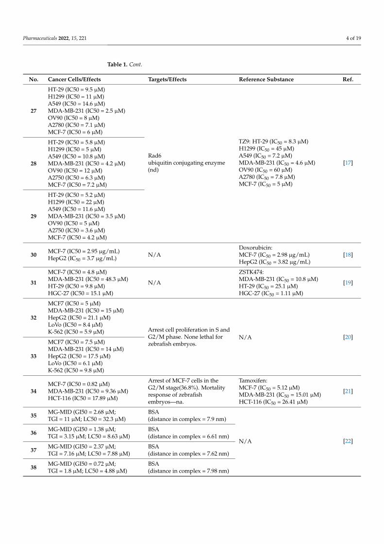
27
HT-29 (IC50 = 9.5 µM)H1299 (IC50 = 11 µM)A549 (IC50 = 14.6 µM)MDA-MB-231 (IC50 = 2.5 µM)OV90 (IC50 = 8 µM)A2780 (IC50 = 7.1 µM)MCF-7 (IC50 = 6 µM)
Rad6ubiquitin conjugating enzyme(nd)
TZ9: HT-29 (IC50 = 8.3 µM)H1299 (IC50 = 45 µM)A549 (IC50 = 7.2 µM)MDA-MB-231 (IC50 = 4.6 µM)OV90 (IC50 = 60 µM)A2780 (IC50 = 7.8 µM)MCF-7 (IC50 = 5 µM)
[17]28
HT-29 (IC50 = 5.8 µM)H1299 (IC50 = 5 µM)A549 (IC50 = 10.8 µM)MDA-MB-231 (IC50 = 4.2 µM)OV90 (IC50 = 12 µM)A2750 (IC50 = 6.3 µM)MCF-7 (IC50 = 7.2 µM)
29
HT-29 (IC50 = 5.2 µM)H1299 (IC50 = 22 µM)A549 (IC50 = 11.6 µM)MDA-MB-231 (IC50 = 3.5 µM)OV90 (IC50 = 5 µM)A2750 (IC50 = 3.6 µM)MCF-7 (IC50 = 4.2 µM)
30 MCF-7 (IC50 = 2.95 µg/mL)HepG2 (IC50 = 3.7 µg/mL) N/A
Doxorubicin:MCF-7 (IC50 = 2.98 µg/mL)HepG2 (IC50 = 3.82 µg/mL)
[18]
31
MCF-7 (IC50 = 4.8 µM)MDA-MB-231 (IC50 = 48.3 µM)HT-29 (IC50 = 9.8 µM)HGC-27 (IC50 = 15.1 µM)
N/A
ZSTK474:MDA-MB-231 (IC50 = 10.8 µM)HT-29 (IC50 = 25.1 µM)HGC-27 (IC50 = 1.11 µM)
[19]
32
MCF7 (IC50 = 5 µM)MDA-MB-231 (IC50 = 15 µM)HepG2 (IC50 = 21.1 µM)LoVo (IC50 = 8.4 µM)K-562 (IC50 = 5.9 µM) Arrest cell proliferation in S and
G2/M phase. None lethal forzebrafish embryos.
N/A [20]
33
MCF7 (IC50 = 7.5 µM)MDA-MB-231 (IC50 = 14 µM)HepG2 (IC50 = 17.5 µM)LoVo (IC50 = 6.1 µM)K-562 (IC50 = 9.8 µM)
34MCF-7 (IC50 = 0.82 µM)MDA-MB-231 (IC50 = 9.36 µM)HCT-116 (IC50 = 17.89 µM)
Arrest of MCF-7 cells in theG2/M stage(36.8%). Mortalityresponse of zebrafishembryos—na.
Tamoxifen:MCF-7 (IC50 = 5.12 µM)MDA-MB-231 (IC50 = 15.01 µM)HCT-116 (IC50 = 26.41 µM)
[21]
35 MG-MID (GI50 = 2.68 µM;TGI = 11 µM; LC50 = 32.3 µM)
BSA(distance in complex = 7.9 nm)
N/A [22]36 MG-MID (GI50 = 1.38 µM;
TGI = 3.15 µM; LC50 = 8.63 µM)BSA(distance in complex = 6.61 nm)
37 MG-MID (GI50 = 2.37 µM;TGI = 7.16 µM; LC50 = 7.88 µM)
BSA(distance in complex = 7.62 nm)
38 MG-MID (GI50 = 0.72 µM;TGI = 1.8 µM; LC50 = 4.88 µM)
BSA(distance in complex = 7.98 nm)
Pharmaceuticals 2022, 15, 221 5 of 19
Table 1. Cont.
No. Cancer Cells/Effects Targets/Effects Reference Substance Ref.
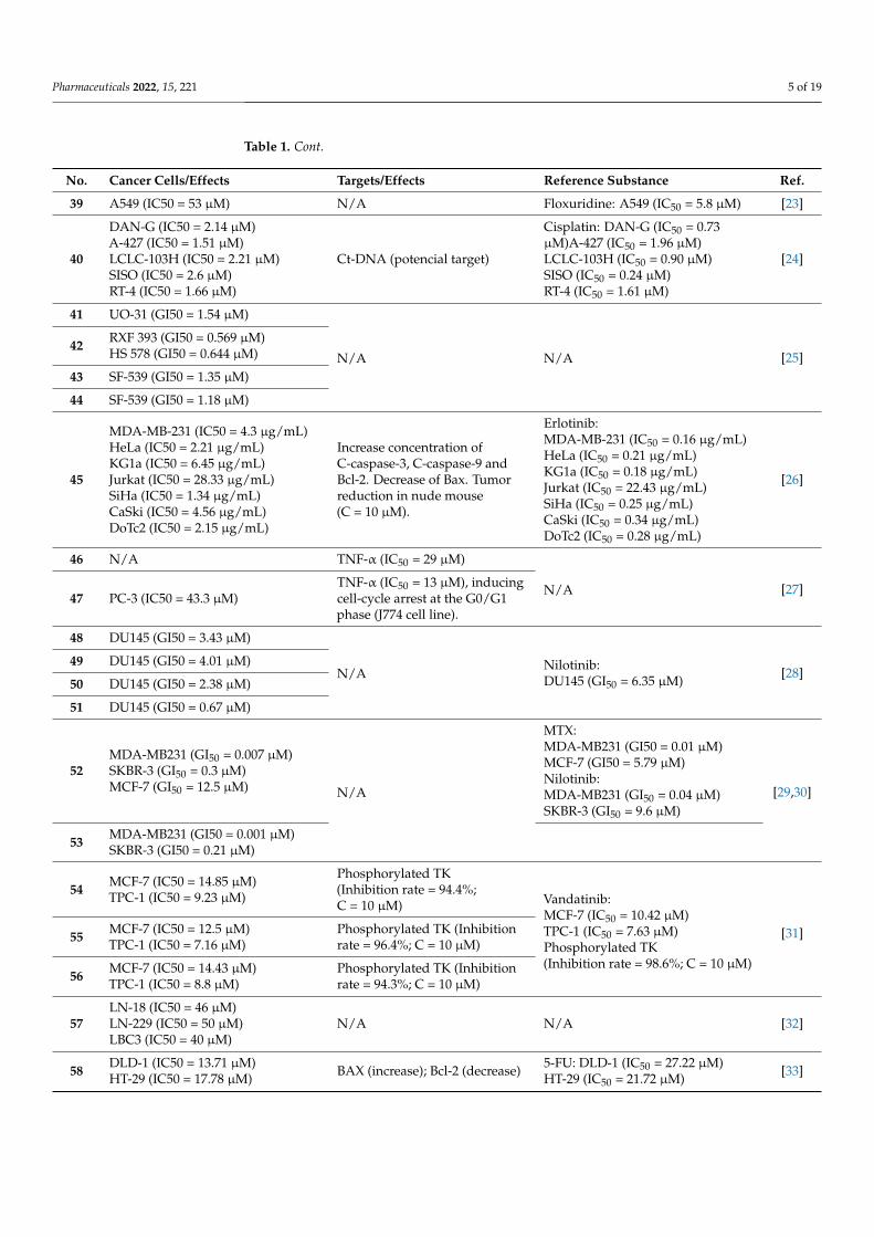
39 A549 (IC50 = 53 µM) N/A Floxuridine: A549 (IC50 = 5.8 µM) [23]
40
DAN-G (IC50 = 2.14 µM)A-427 (IC50 = 1.51 µM)LCLC-103H (IC50 = 2.21 µM)SISO (IC50 = 2.6 µM)RT-4 (IC50 = 1.66 µM)
Ct-DNA (potencial target)
Cisplatin: DAN-G (IC50 = 0.73µM)A-427 (IC50 = 1.96 µM)LCLC-103H (IC50 = 0.90 µM)SISO (IC50 = 0.24 µM)RT-4 (IC50 = 1.61 µM)
[24]
41 UO-31 (GI50 = 1.54 µM)
N/A N/A [25]42 RXF 393 (GI50 = 0.569 µM)
HS 578 (GI50 = 0.644 µM)
43 SF-539 (GI50 = 1.35 µM)
44 SF-539 (GI50 = 1.18 µM)
45
MDA-MB-231 (IC50 = 4.3 µg/mL)HeLa (IC50 = 2.21 µg/mL)KG1a (IC50 = 6.45 µg/mL)Jurkat (IC50 = 28.33 µg/mL)SiHa (IC50 = 1.34 µg/mL)CaSki (IC50 = 4.56 µg/mL)DoTc2 (IC50 = 2.15 µg/mL)
Increase concentration ofC-caspase-3, C-caspase-9 andBcl-2. Decrease of Bax. Tumorreduction in nude mouse(C = 10 µM).
Erlotinib:MDA-MB-231 (IC50 = 0.16 µg/mL)HeLa (IC50 = 0.21 µg/mL)KG1a (IC50 = 0.18 µg/mL)Jurkat (IC50 = 22.43 µg/mL)SiHa (IC50 = 0.25 µg/mL)CaSki (IC50 = 0.34 µg/mL)DoTc2 (IC50 = 0.28 µg/mL)
[26]
46 N/A TNF-α (IC50 = 29 µM)
N/A [27]47 PC-3 (IC50 = 43.3 µM)
TNF-α (IC50 = 13 µM), inducingcell-cycle arrest at the G0/G1phase (J774 cell line).
48 DU145 (GI50 = 3.43 µM)
N/ANilotinib:DU145 (GI50 = 6.35 µM) [28]
49 DU145 (GI50 = 4.01 µM)
50 DU145 (GI50 = 2.38 µM)
51 DU145 (GI50 = 0.67 µM)
52MDA-MB231 (GI50 = 0.007 µM)SKBR-3 (GI50 = 0.3 µM)MCF-7 (GI50 = 12.5 µM) N/A
MTX:MDA-MB231 (GI50 = 0.01 µM)MCF-7 (GI50 = 5.79 µM)Nilotinib:MDA-MB231 (GI50 = 0.04 µM)SKBR-3 (GI50 = 9.6 µM)
[29,30]
53 MDA-MB231 (GI50 = 0.001 µM)SKBR-3 (GI50 = 0.21 µM)
54 MCF-7 (IC50 = 14.85 µM)TPC-1 (IC50 = 9.23 µM)
Phosphorylated TK(Inhibition rate = 94.4%;C = 10 µM) Vandatinib:
MCF-7 (IC50 = 10.42 µM)TPC-1 (IC50 = 7.63 µM)Phosphorylated TK(Inhibition rate = 98.6%; C = 10 µM)
[31]55 MCF-7 (IC50 = 12.5 µM)TPC-1 (IC50 = 7.16 µM)
Phosphorylated TK (Inhibitionrate = 96.4%; C = 10 µM)
56 MCF-7 (IC50 = 14.43 µM)TPC-1 (IC50 = 8.8 µM)
Phosphorylated TK (Inhibitionrate = 94.3%; C = 10 µM)
57LN-18 (IC50 = 46 µM)LN-229 (IC50 = 50 µM)LBC3 (IC50 = 40 µM)
N/A N/A [32]
58 DLD-1 (IC50 = 13.71 µM)HT-29 (IC50 = 17.78 µM) BAX (increase); Bcl-2 (decrease) 5-FU: DLD-1 (IC50 = 27.22 µM)
HT-29 (IC50 = 21.72 µM) [33]

Pharmaceuticals 2022, 15, 221 6 of 19
Table 1. Cont.
No. Cancer Cells/Effects Targets/Effects Reference Substance Ref.
59
HCT-116 (Inhibition = 115.53%)SW-620 (Inhibition = 95.06%)SF-539 (Inhibition = 89.27%)OVCAR-4 (Inhibition = 94.39%)PC786-0 (Inhibition = 93.76%)ACHN (Inhibition = 86.27%)MCF-7 (Inhibition = 94.82%)
CDK2 (Inhibition rate = 82.38%;C = 10 µM; IC50 = 1.85 µM) Roscovitine:
CDK2 (Inhibition rate = 89.6%;C = 10 µM)
[34]
60ATCC (Inhibition = 90.02%)NCI-H460 (Inhibition = 83.66%)OVCAR-4 (Inhibition = 92.27%)
CDK2 (Inhibition rate = 81.96%;C = 10 µM; IC50 = 2.09 µM)
61 SKMEL-103 (IC50 = 25 µM) PI3K (decrease)AMPK (decrease) N/A [35]
62NCI-H460 (Growth Percent = −50%)MDA-MB468(Growth Percent = −20.7%)
N/A N/A [36]
63
HCC-2998(Growth Percent = −82.1%)RXF 393 (Growth Percent = −68%)NCI-H460(Growth Percent = −58.3%)ACHN (Growth Percent = −57%)MDA-MB-468(Growth Percent = −52.3%)
64
HCC-2998(Growth Percent = −69.3%)RXF 393 (Growth Percent = −66%)NCI-H460(Growth Percent = −64.8%)ACHN (Growth Percent = −45%)
65
HCC-2998 (Growth Percent = −77%)RXF 393 (Growth Percent = −74.4%)NCI-H460(Growth Percent = −49.4%)MDA-MB-468(Growth Percent = −47%)
66
HCC-2998(Growth Percent = −53.7%)RXF 393 (Growth Percent = −55%)NCI-H460(Growth Percent = −54.7%)ACHN (Growth Percent = −52.8%)NCI-H322M(Growth Percent = −50.5%)
67 A549 (IC50 = 144.1 µg/mL)Bel7402 (IC50 = 195.6 µg/mL) N/A N/A [37]
Pharmaceuticals 2022, 15, 221 7 of 19
Table 1. Cont.
No. Cancer Cells/Effects Targets/Effects Reference Substance Ref.
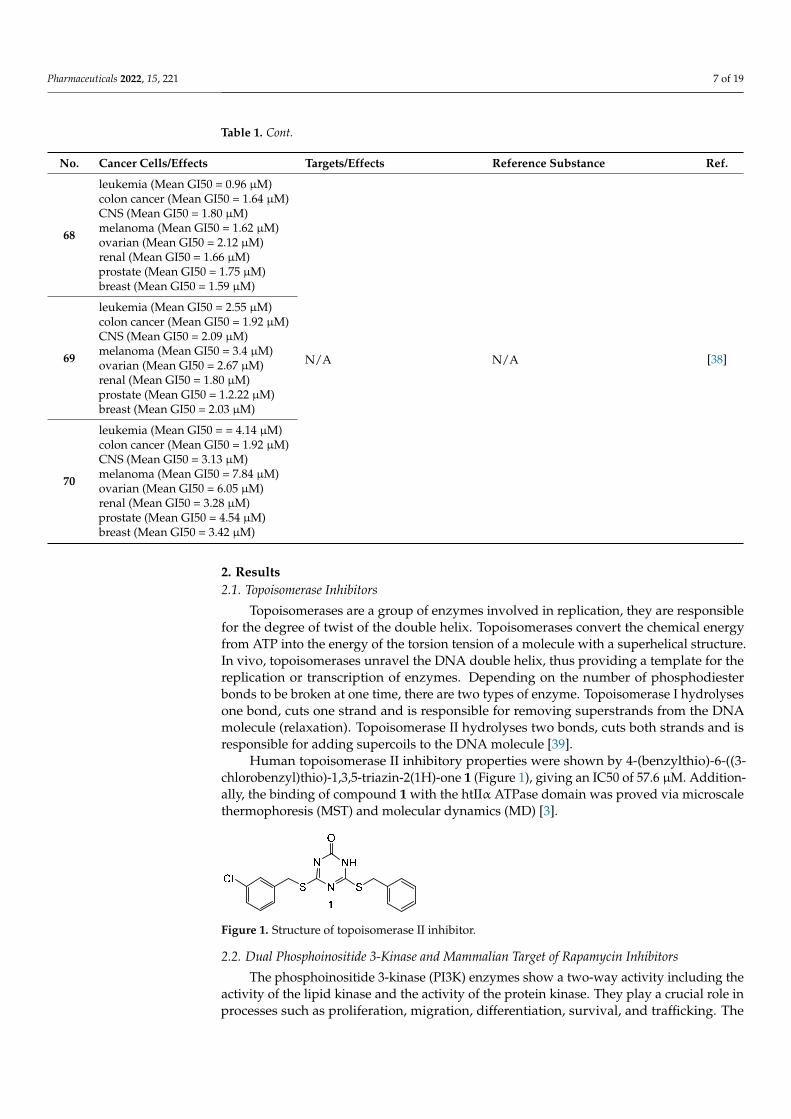
68
leukemia (Mean GI50 = 0.96 µM)colon cancer (Mean GI50 = 1.64 µM)CNS (Mean GI50 = 1.80 µM)melanoma (Mean GI50 = 1.62 µM)ovarian (Mean GI50 = 2.12 µM)renal (Mean GI50 = 1.66 µM)prostate (Mean GI50 = 1.75 µM)breast (Mean GI50 = 1.59 µM)
N/A N/A [38]69
leukemia (Mean GI50 = 2.55 µM)colon cancer (Mean GI50 = 1.92 µM)CNS (Mean GI50 = 2.09 µM)melanoma (Mean GI50 = 3.4 µM)ovarian (Mean GI50 = 2.67 µM)renal (Mean GI50 = 1.80 µM)prostate (Mean GI50 = 1.2.22 µM)breast (Mean GI50 = 2.03 µM)
70
leukemia (Mean GI50 = = 4.14 µM)colon cancer (Mean GI50 = 1.92 µM)CNS (Mean GI50 = 3.13 µM)melanoma (Mean GI50 = 7.84 µM)ovarian (Mean GI50 = 6.05 µM)renal (Mean GI50 = 3.28 µM)prostate (Mean GI50 = 4.54 µM)breast (Mean GI50 = 3.42 µM)
2. Results2.1. Topoisomerase Inhibitors
Topoisomerases are a group of enzymes involved in replication, they are responsiblefor the degree of twist of the double helix. Topoisomerases convert the chemical energyfrom ATP into the energy of the torsion tension of a molecule with a superhelical structure.In vivo, topoisomerases unravel the DNA double helix, thus providing a template for thereplication or transcription of enzymes. Depending on the number of phosphodiesterbonds to be broken at one time, there are two types of enzyme. Topoisomerase I hydrolysesone bond, cuts one strand and is responsible for removing superstrands from the DNAmolecule (relaxation). Topoisomerase II hydrolyses two bonds, cuts both strands and isresponsible for adding supercoils to the DNA molecule [39].
Human topoisomerase II inhibitory properties were shown by 4-(benzylthio)-6-((3-chlorobenzyl)thio)-1,3,5-triazin-2(1H)-one 1 (Figure 1), giving an IC50 of 57.6 µM. Addition-ally, the binding of compound 1 with the htIIα ATPase domain was proved via microscalethermophoresis (MST) and molecular dynamics (MD) [3].
Pharmaceuticals 2022, 15, x FOR PEER REVIEW 2 of 20
revealed a wide range of properties of s-triazine derivatives. In this review we will present currently investigated 1,3,5-triazine derivatives with anti-cancer activities.
This review presents the current state of knowledge on 1,3,5-triazine derivatives, their structures and anticancer activity, as well as their ability to inhibit different enzymes or their DNA-binding potential. This data could be helpful in the development of new drugs and therapeutic methods. By analysing the presented approach, a series of com-pounds with high potency and low toxicity can be designed, synthesized, characterized and evaluated for desired pharmacological activity. The collected data are presented in summary Table 1.
2. Results 2.1. Topoisomerase Inhibitors
Topoisomerases are a group of enzymes involved in replication, they are responsible for the degree of twist of the double helix. Topoisomerases convert the chemical energy from ATP into the energy of the torsion tension of a molecule with a superhelical structure. In vivo, topoisomerases unravel the DNA double helix, thus providing a template for the replication or transcription of enzymes. Depending on the number of phosphodiester bonds to be broken at one time, there are two types of enzyme. Topoisomerase I hydrolyses one bond, cuts one strand and is responsible for removing superstrands from the DNA molecule (relaxation). Topoisomerase II hydrolyses two bonds, cuts both strands and is responsible for adding supercoils to the DNA molecule [3].
Human topoisomerase II inhibitory properties were shown by 4-(benzylthio)-6-((3-chlorobenzyl)thio)-1,3,5-triazin-2(1H)-one 1 (Figure 1), giving an IC50 of 57.6 µM. Additionally, the binding of compound 1 with the htIIα ATPase domain was proved via microscale thermophoresis (MST) and molecular dynamics (MD) [4].
Figure 1. Structure of topoisomerase II inhibitor.
2.2. Dual Phosphoinositide 3-Kinase and Mammalian Target of Rapamycin Inhibitors The phosphoinositide 3-kinase (PI3K) enzymes show a two-way activity including
the activity of the lipid kinase and the activity of the protein kinase. They play a crucial role in processes such as proliferation, migration, differentiation, survival, and trafficking. The PI3K family contains eight isoforms divided into three distinct classes (I, II, and III) which may be different in terms of cellular responsibility [5].
The function of the mammalian target of rapamycin (mTor) is to regulate growth, proliferation and cell traffic, and the processes of translation and transcription. The mTOR catalyzes the phosphorylation ribosomal protein S6 kinase β-1 (S6K1), eukaryotic translation initiation factor 4E binding protein 1 (4E-BP1), Akt, protein kinase C (PKC), and type-I insulin-like growth factor receptor (IGF-IR), thereby regulating protein synthesis, nutrient metabolism, growth factor signaling, cell growth, and migration [6].
The construction of compounds with dual inhibitory effects contributes to obtaining a more selective effect. Potential anti-cancer drugs that inhibit PI3K and mTor at the same time showed greater efficiency and reduced the likelihood of inducing drug resistance [7].
Substituted 2-(thiophen-2-yl)-1,3,5-triazine derivative 2 (Figure 2) exhibited excellent anti-cancer potency for A549, MCF-7 (breast cancer) and Hela (cervical cancer) cell lines with IC50 values of 0.20 µM, 1.25 µM, and 1.03 µM, respectively. Western blot analysis proved drivative 2 could suppress the phosphorylation of AKT. The degree of inhibition
Figure 1. Structure of topoisomerase II inhibitor.
2.2. Dual Phosphoinositide 3-Kinase and Mammalian Target of Rapamycin Inhibitors
The phosphoinositide 3-kinase (PI3K) enzymes show a two-way activity including theactivity of the lipid kinase and the activity of the protein kinase. They play a crucial role inprocesses such as proliferation, migration, differentiation, survival, and trafficking. The
Pharmaceuticals 2022, 15, 221 8 of 19
PI3K family contains eight isoforms divided into three distinct classes (I, II, and III) whichmay be different in terms of cellular responsibility [40].
The function of the mammalian target of rapamycin (mTor) is to regulate growth,proliferation and cell traffic, and the processes of translation and transcription. The mTORcatalyzes the phosphorylation ribosomal protein S6 kinase β-1 (S6K1), eukaryotic transla-tion initiation factor 4E binding protein 1 (4E-BP1), Akt, protein kinase C (PKC), and type-Iinsulin-like growth factor receptor (IGF-IR), thereby regulating protein synthesis, nutrientmetabolism, growth factor signaling, cell growth, and migration [41].
The construction of compounds with dual inhibitory effects contributes to obtaining amore selective effect. Potential anti-cancer drugs that inhibit PI3K and mTor at the sametime showed greater efficiency and reduced the likelihood of inducing drug resistance [42].
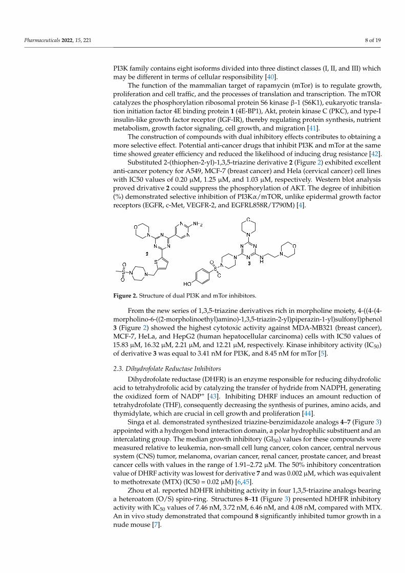
Substituted 2-(thiophen-2-yl)-1,3,5-triazine derivative 2 (Figure 2) exhibited excellentanti-cancer potency for A549, MCF-7 (breast cancer) and Hela (cervical cancer) cell lineswith IC50 values of 0.20 µM, 1.25 µM, and 1.03 µM, respectively. Western blot analysisproved drivative 2 could suppress the phosphorylation of AKT. The degree of inhibition(%) demonstrated selective inhibition of PI3Kα/mTOR, unlike epidermal growth factorreceptors (EGFR, c-Met, VEGFR-2, and EGFRL858R/T790M) [4].
Pharmaceuticals 2022, 15, x FOR PEER REVIEW 3 of 20
(%) demonstrated selective inhibition of PI3Kα/mTOR, unlike epidermal growth factor receptors (EGFR, c-Met, VEGFR-2, and EGFRL858R/T790M) [8].
From the new series of 1,3,5-triazine derivatives rich in morpholine moiety, 4-((4-(4-morpholino-6-((2-morpholinoethyl)amino)-1,3,5-triazin-2-yl)piperazin-1-yl)sulfonyl)phenol 3 (Figure 2) showed the highest cytotoxic activity against MDA-MB321 (breast cancer), MCF-7, HeLa, and HepG2 (human hepatocellular carcinoma) cells with IC50 values of 15.83 µM, 16.32 µM, 2.21 µM, and 12.21 µM, respectively. Kinase inhibitory activity (IC50) of derivative 3 was equal to 3.41 nM for PI3K, and 8.45 nM for mTor [9].
Figure 2. Structure of dual PI3K and mTor inhibitors.
2.3. Dihydrofolate Reductase Inhibitors Dihydrofolate reductase (DHFR) is an enzyme responsible for reducing dihydrofolic
acid to tetrahydrofolic acid by catalyzing the transfer of hydride from NADPH, generat-ing the oxidized form of NADP+ [10]. Inhibiting DHRF induces an amount reduction of tetrahydrofolate (THF), consequently decreasing the synthesis of purines, amino acids, and thymidylate, which are crucial in cell growth and proliferation [11].
Singa et al. demonstrated synthesized triazine-benzimidazole analogs 4–7 (Figure 3) appointed with a hydrogen bond interaction domain, a polar hydrophilic substituent and an intercalating group. The median growth inhibitory (GI50) values for these compounds were measured relative to leukemia, non-small cell lung cancer, colon cancer, central nervous system (CNS) tumor, melanoma, ovarian cancer, renal cancer, prostate cancer, and breast cancer cells with values in the range of 1.91–2.72 µM. The 50% inhibitory con-centration value of DHRF activity was lowest for derivative 7 and was 0.002 µM, which was equivalent to methotrexate (MTX) (IC50 = 0.02 µM) [12,13].
Zhou et al. reported hDHFR inhibiting activity in four 1,3,5-triazine analogs bearing a heteroatom (O/S) spiro-ring. Structures 8–11 (Figure 3) presented hDHFR inhibitory ac-tivity with IC50 values of 7.46 nM, 3.72 nM, 6.46 nM, and 4.08 nM, compared with MTX. An in vivo study demonstrated that compound 8 significantly inhibited tumor growth in a nude mouse [14].
A hybrid of 4,6-diamino-1,2-dihydro-1,3,5-triazine and chalcone led to the generation of 15 new compounds as potential DHFR and TrxR (thioredoxin reductase) inhibitors. The greatest results were exhibited by compounds 12 and 13 (Figure 3). Both acted cytotoxic against HCT116 (human colorectal carcinoma) (GI50 = 0.026 µM; GI50 = 0.116 µM) and MCF-7 (GI50 = 0.080 µM; GI50 = 0.127 µM) cancer cell lines. In addition, studies have shown strong in vitro inhibitory activities against recombinant human DHFR (IC50 = 0.0061 µM; IC50 = 0.0026 µM) and rat TrxR (IC50 = 4.6 µM; IC50 = 5.9 µM) enzymes [15].
Figure 2. Structure of dual PI3K and mTor inhibitors.
From the new series of 1,3,5-triazine derivatives rich in morpholine moiety, 4-((4-(4-morpholino-6-((2-morpholinoethyl)amino)-1,3,5-triazin-2-yl)piperazin-1-yl)sulfonyl)phenol3 (Figure 2) showed the highest cytotoxic activity against MDA-MB321 (breast cancer),MCF-7, HeLa, and HepG2 (human hepatocellular carcinoma) cells with IC50 values of15.83 µM, 16.32 µM, 2.21 µM, and 12.21 µM, respectively. Kinase inhibitory activity (IC50)of derivative 3 was equal to 3.41 nM for PI3K, and 8.45 nM for mTor [5].
2.3. Dihydrofolate Reductase Inhibitors
Dihydrofolate reductase (DHFR) is an enzyme responsible for reducing dihydrofolicacid to tetrahydrofolic acid by catalyzing the transfer of hydride from NADPH, generatingthe oxidized form of NADP+ [43]. Inhibiting DHRF induces an amount reduction oftetrahydrofolate (THF), consequently decreasing the synthesis of purines, amino acids, andthymidylate, which are crucial in cell growth and proliferation [44].
Singa et al. demonstrated synthesized triazine-benzimidazole analogs 4–7 (Figure 3)appointed with a hydrogen bond interaction domain, a polar hydrophilic substituent and anintercalating group. The median growth inhibitory (GI50) values for these compounds weremeasured relative to leukemia, non-small cell lung cancer, colon cancer, central nervoussystem (CNS) tumor, melanoma, ovarian cancer, renal cancer, prostate cancer, and breastcancer cells with values in the range of 1.91–2.72 µM. The 50% inhibitory concentrationvalue of DHRF activity was lowest for derivative 7 and was 0.002 µM, which was equivalentto methotrexate (MTX) (IC50 = 0.02 µM) [6,45].
Zhou et al. reported hDHFR inhibiting activity in four 1,3,5-triazine analogs bearinga heteroatom (O/S) spiro-ring. Structures 8–11 (Figure 3) presented hDHFR inhibitoryactivity with IC50 values of 7.46 nM, 3.72 nM, 6.46 nM, and 4.08 nM, compared with MTX.An in vivo study demonstrated that compound 8 significantly inhibited tumor growth in anude mouse [7].

Pharmaceuticals 2022, 15, 221 9 of 19Pharmaceuticals 2022, 15, x FOR PEER REVIEW 4 of 20
Figure 3. Structure of DHFR inhibitors.
2.4. Carbonic Anhydrase Inhibitors Carbonic anhydrases (CAs), metalloenzymes from the lyase group, are responsible
for pH homeostasis and catalyzing the reversible reaction of the formation of the bicar-bonate ion HCO from water and carbon dioxide [16].
Among the numerous isoforms we can distinguish the ubiquitous variants CA I and CA II in mammals. In a pathological condition such as hypoxia, increased expression of CA IX and CA XII is observed. These enzyme forms are involved in the regulation of pH homeostasis and intercellular communication and ion transport. 2-[4-Chloro-5-methyl-2-(naphthalen-1-ylmethylthio)-benzenesulfonyl]-1-[4-chloro-6-(4-sulfamoylphenylamino)-1,3,5-triazin-2-ylamino]guanidine 14 (Figure 4) acted with strongest selectivity toward hCA IX versus hCA I (hCA I/hCA IX = 18) and hCA II (hCA II/hCA IX = 4). Compound 14 showed prominent cytotoxicity towards HeLa cancer cells (IC50 = 17 µM) and did not ex-hibit toxicity to the non-cancerous HaCaT cells (IC50 = 61 µM) [17].
Research conducted by Havránková et al. considered the interaction of CA I, II and IX with 1,3,5-triazine derivatives incorporating piperazine, aminoalcohol and sulfona-mide. The results showed that 1,3,5-triazines with a 4-hydroxyaniline substituent achieved the highest ratio of selective inhibition (hCA IX/hCA II): compound 15 (18.50); compound 16 (14.09) (Figure 4) [18].
Based on the structure of SCL-0111, new 1,3,5-triazine derivatives 17 and 18 were synthesized (Figure 4) and their ability to inhibit CA I, II, IX, and XII was investigated. The most promising result was the selective inhibition of CA IX by compound 17 with a KI value = 0.91 nM [19], while compound 18 had a KI value of 14.6 nM [20].
Figure 3. Structure of DHFR inhibitors.
A hybrid of 4,6-diamino-1,2-dihydro-1,3,5-triazine and chalcone led to the generationof 15 new compounds as potential DHFR and TrxR (thioredoxin reductase) inhibitors. Thegreatest results were exhibited by compounds 12 and 13 (Figure 3). Both acted cytotoxicagainst HCT116 (human colorectal carcinoma) (GI50 = 0.026 µM; GI50 = 0.116 µM) andMCF-7 (GI50 = 0.080 µM; GI50 = 0.127 µM) cancer cell lines. In addition, studies have shownstrong in vitro inhibitory activities against recombinant human DHFR (IC50 = 0.0061 µM;IC50 = 0.0026 µM) and rat TrxR (IC50 = 4.6 µM; IC50 = 5.9 µM) enzymes [8].
2.4. Carbonic Anhydrase Inhibitors
Carbonic anhydrases (CAs), metalloenzymes from the lyase group, are responsible forpH homeostasis and catalyzing the reversible reaction of the formation of the bicarbonateion HCO−3 from water and carbon dioxide [46].
Among the numerous isoforms we can distinguish the ubiquitous variants CA I andCA II in mammals. In a pathological condition such as hypoxia, increased expression ofCA IX and CA XII is observed. These enzyme forms are involved in the regulation of pHhomeostasis and intercellular communication and ion transport. 2-[4-Chloro-5-methyl-2-(naphthalen-1-ylmethylthio)-benzenesulfonyl]-1-[4-chloro-6-(4-sulfamoylphenylamino)-1,3,5-triazin-2-ylamino]guanidine 14 (Figure 4) acted with strongest selectivity toward hCA IXversus hCA I (hCA I/hCA IX = 18) and hCA II (hCA II/hCA IX = 4). Compound 14showed prominent cytotoxicity towards HeLa cancer cells (IC50 = 17 µM) and did notexhibit toxicity to the non-cancerous HaCaT cells (IC50 = 61 µM) [9].
Research conducted by Havránková et al. considered the interaction of CA I, II and IXwith 1,3,5-triazine derivatives incorporating piperazine, aminoalcohol and sulfonamide.The results showed that 1,3,5-triazines with a 4-hydroxyaniline substituent achieved thehighest ratio of selective inhibition (hCA IX/hCA II): compound 15 (18.50); compound 16(14.09) (Figure 4) [10].
Based on the structure of SCL-0111, new 1,3,5-triazine derivatives 17 and 18 weresynthesized (Figure 4) and their ability to inhibit CA I, II, IX, and XII was investigated. Themost promising result was the selective inhibition of CA IX by compound 17 with a KIvalue = 0.91 nM [11], while compound 18 had a KI value of 14.6 nM [12].

Pharmaceuticals 2022, 15, 221 10 of 19Pharmaceuticals 2022, 15, x FOR PEER REVIEW 5 of 20
Figure 4. Structure of CA inhibitors.
2.5. Epidermal Growth Factor Receptor Inhibitors The role of the epidermal growth factor receptor (EGFR) in the pathogenesis process
is an important topic of scientific research. As a result, it was discovered that mutations leading to overexpression of EGFR genes (e.g., increased regulation or amplification) are significantly associated with many cancers: lung granuloma (40% of cases), rectal tumors, glioblastoma (50%), and epithelial carcinomas of the head and neck (80–100%) [21,22].
Through the “one pot” reaction, 15 novel monastrol-1,3,5-triazine derivatives were obtained and investigated for anti-cancer properties and cytotoxicity. Derivative 19 sub-stituted by 3-fluorphenylamino groups (Figure 5) presented highest IC50 against cancer cell lines [HeLa - 39.7 µM; MCF-7 - 41.5 µM; HL-60 (human pro-myelocytic leukemia cell) - 23.1 µM; HepG2 - 31.2]. This compound was nontoxic to normal epithelial cells MCF-12A while at a concentration of 10 nM the inhibition of EGFR-TK by 19 was equal 96.4% [23].
Analysis of molecular modelling and Lipinski’s rule of five allowed us to select four compounds that were tested for anti-breast cancer activity. The strongest action with re-spect to EGFR-TK was observed for 3-(4,6-bis((3-chlorophenyl)amino)-1,3,5-triazin-2-yl)thiazolidine-2,5-dione 20 (Figure 5) (IC50 = 2.54 µM). An in vitro study against MDA-MB-21, BT-474 (breast tumor) and MCF-7 showed an increase of apoptosis rates. In addi-tion, a significant decline expression of β-catenin was noticed in MDA-MB-21 cell lines [24].
Bhat et al. took a closer look at 4-aminoquinoline-1,3,5-triazine derivatives. Com-pounds 21 (Figure 5) presented IC50 values of 44.5 µM, 52.2 µM, 40.3 µM, and 56.4 µM against HeLa, MCF-7, HL-60, and HepG2. Derivative 22 (Figure 5) showed IC50 values of 32.4 µM, 32.3 µM, 26.3 µM, and 45.3 µM against HeLa, MCF-7, HL-60, and HepG2. Both molecules did not reveal cytotoxicity to MCF-12A cells. The activity of derivatives 21 and 22 inhibiting EGFR-TK was 96.3% and 90.5%, respectively [25].
Figure 4. Structure of CA inhibitors.
2.5. Epidermal Growth Factor Receptor Inhibitors
The role of the epidermal growth factor receptor (EGFR) in the pathogenesis processis an important topic of scientific research. As a result, it was discovered that mutationsleading to overexpression of EGFR genes (e.g., increased regulation or amplification) aresignificantly associated with many cancers: lung granuloma (40% of cases), rectal tumors,glioblastoma (50%), and epithelial carcinomas of the head and neck (80–100%) [47,48].
Through the “one pot” reaction, 15 novel monastrol-1,3,5-triazine derivatives wereobtained and investigated for anti-cancer properties and cytotoxicity. Derivative 19 sub-stituted by 3-fluorphenylamino groups (Figure 5) presented highest IC50 against cancercell lines [HeLa—39.7 µM; MCF-7—41.5 µM; HL-60 (human pro-myelocytic leukemiacell)—23.1 µM; HepG2—31.2]. This compound was nontoxic to normal epithelial cellsMCF-12A while at a concentration of 10 nM the inhibition of EGFR-TK by 19 was equal96.4% [13].
Pharmaceuticals 2022, 15, x FOR PEER REVIEW 6 of 20
Figure 5. Structure of EGFR inhibitors.
2.6. Vascular Endothelial Growth Factor Vascular endothelial growth factor (VEGF) production can be induced in a cell that
does not receive enough oxygen [26]. When a cell is deficient in oxygen, it produces hy-poxia induced factor (HIF). HIF stimulates the release of VEGF (including the modulation of erythropoiesis). Circulating VEGF then binds to VEGF receptors on endothelial cells, triggering a tyrosine kinase pathway leading to angiogenesis [27]. Expression of angio-poietin-2 in the absence of VEGF leads to endothelial cell death and vascular regression. VEGF acts as the central mediator of tumor angiogenesis, stimulating the growth of new blood vessels from nearby capillaries and allowing tumors to access the oxygen and nu-trients they need to grow [28].
Quinazoline-1,3,5-triazine derivatives 23, 24, and 25 (Figure 6) demonstrated anti-tumor activity against HeLa, MCF-7, HL-60, and HepG2 with IC50 values in range of 6–16 µM. In addition, they were non-toxic against the normal cell line of HFF (human foreskin fibroblasts). Molecular docking results demonstrated the high potency of derivatives 23, 24 and 25 to bind the hydrophobic pocket of the N-terminal chain in the ATP binding site of VEGFR [29].
Figure 6. Structure of VEGF inhibitors.
2.7. Focal Adhesion Kinase Inhibitors Focal Adhesion Kinase (FAK) is a 125-kDa cytoplasmic tyrosine kinase. Deregulation
of FAK-dependent processes such as cell adhesion, growth, survival, and mobility are a significant component of tumor progression. Overexpression of FAK leads to the inhibi-tion of apoptosis and an increase in the incidence of metastatic tumors [30].
Dao et al. showed that compound 26 (Figure 7) is the strongest FAK inhibitor (IC50 = 0.05 µM). Growth inhibitory activity on human glioblastoma (U-87MG), human colon car-cinoma (HCT-116), MDA-MB-231, and human prostate cancer (PC-3) by compound 26 obtained the following results 0.42 µM, 0.13 µM, 0.14 µM, and 0.63 µM compared to TAE-226 (0.19 µM, 0.23 µM, 1.9 µM, and 0.26 µM). Furthermore, compound 26 turned out to fit well into the ATP binding site of the FAK via molecular docking [31].
Figure 5. Structure of EGFR inhibitors.
Analysis of molecular modelling and Lipinski’s rule of five allowed us to select fourcompounds that were tested for anti-breast cancer activity. The strongest action withrespect to EGFR-TK was observed for 3-(4,6-bis((3-chlorophenyl)amino)-1,3,5-triazin-2-yl)thiazolidine-2,5-dione 20 (Figure 5) (IC50 = 2.54 µM). An in vitro study against MDA-MB-21, BT-474 (breast tumor) and MCF-7 showed an increase of apoptosis rates. In addition, asignificant decline expression of β-catenin was noticed in MDA-MB-21 cell lines [14].
Bhat et al. took a closer look at 4-aminoquinoline-1,3,5-triazine derivatives. Com-pounds 21 (Figure 5) presented IC50 values of 44.5 µM, 52.2 µM, 40.3 µM, and 56.4 µMagainst HeLa, MCF-7, HL-60, and HepG2. Derivative 22 (Figure 5) showed IC50 values of

Pharmaceuticals 2022, 15, 221 11 of 19
32.4 µM, 32.3 µM, 26.3 µM, and 45.3 µM against HeLa, MCF-7, HL-60, and HepG2. Bothmolecules did not reveal cytotoxicity to MCF-12A cells. The activity of derivatives 21 and22 inhibiting EGFR-TK was 96.3% and 90.5%, respectively [15].
2.6. Vascular Endothelial Growth Factor
Vascular endothelial growth factor (VEGF) production can be induced in a cell thatdoes not receive enough oxygen [49]. When a cell is deficient in oxygen, it produces hypoxiainduced factor (HIF). HIF stimulates the release of VEGF (including the modulation oferythropoiesis). Circulating VEGF then binds to VEGF receptors on endothelial cells, trig-gering a tyrosine kinase pathway leading to angiogenesis [50]. Expression of angiopoietin-2in the absence of VEGF leads to endothelial cell death and vascular regression. VEGF actsas the central mediator of tumor angiogenesis, stimulating the growth of new blood vesselsfrom nearby capillaries and allowing tumors to access the oxygen and nutrients they needto grow [51].
Quinazoline-1,3,5-triazine derivatives 23, 24, and 25 (Figure 6) demonstrated antitumoractivity against HeLa, MCF-7, HL-60, and HepG2 with IC50 values in range of 6–16 µM.In addition, they were non-toxic against the normal cell line of HFF (human foreskinfibroblasts). Molecular docking results demonstrated the high potency of derivatives 23, 24and 25 to bind the hydrophobic pocket of the N-terminal chain in the ATP binding site ofVEGFR [52].
Pharmaceuticals 2022, 15, x FOR PEER REVIEW 6 of 20
Figure 5. Structure of EGFR inhibitors.
2.6. Vascular Endothelial Growth Factor Vascular endothelial growth factor (VEGF) production can be induced in a cell that
does not receive enough oxygen [26]. When a cell is deficient in oxygen, it produces hy-poxia induced factor (HIF). HIF stimulates the release of VEGF (including the modulation of erythropoiesis). Circulating VEGF then binds to VEGF receptors on endothelial cells, triggering a tyrosine kinase pathway leading to angiogenesis [27]. Expression of angio-poietin-2 in the absence of VEGF leads to endothelial cell death and vascular regression. VEGF acts as the central mediator of tumor angiogenesis, stimulating the growth of new blood vessels from nearby capillaries and allowing tumors to access the oxygen and nu-trients they need to grow [28].
Quinazoline-1,3,5-triazine derivatives 23, 24, and 25 (Figure 6) demonstrated anti-tumor activity against HeLa, MCF-7, HL-60, and HepG2 with IC50 values in range of 6–16 µM. In addition, they were non-toxic against the normal cell line of HFF (human foreskin fibroblasts). Molecular docking results demonstrated the high potency of derivatives 23, 24 and 25 to bind the hydrophobic pocket of the N-terminal chain in the ATP binding site of VEGFR [29].
Figure 6. Structure of VEGF inhibitors.
2.7. Focal Adhesion Kinase Inhibitors Focal Adhesion Kinase (FAK) is a 125-kDa cytoplasmic tyrosine kinase. Deregulation
of FAK-dependent processes such as cell adhesion, growth, survival, and mobility are a significant component of tumor progression. Overexpression of FAK leads to the inhibi-tion of apoptosis and an increase in the incidence of metastatic tumors [30].
Dao et al. showed that compound 26 (Figure 7) is the strongest FAK inhibitor (IC50 = 0.05 µM). Growth inhibitory activity on human glioblastoma (U-87MG), human colon car-cinoma (HCT-116), MDA-MB-231, and human prostate cancer (PC-3) by compound 26 obtained the following results 0.42 µM, 0.13 µM, 0.14 µM, and 0.63 µM compared to TAE-226 (0.19 µM, 0.23 µM, 1.9 µM, and 0.26 µM). Furthermore, compound 26 turned out to fit well into the ATP binding site of the FAK via molecular docking [31].
Figure 6. Structure of VEGF inhibitors.
2.7. Focal Adhesion Kinase Inhibitors
Focal Adhesion Kinase (FAK) is a 125-kDa cytoplasmic tyrosine kinase. Deregulationof FAK-dependent processes such as cell adhesion, growth, survival, and mobility are asignificant component of tumor progression. Overexpression of FAK leads to the inhibitionof apoptosis and an increase in the incidence of metastatic tumors [53].
Dao et al. showed that compound 26 (Figure 7) is the strongest FAK inhibitor(IC50 = 0.05 µM). Growth inhibitory activity on human glioblastoma (U-87MG), humancolon carcinoma (HCT-116), MDA-MB-231, and human prostate cancer (PC-3) by com-pound 26 obtained the following results 0.42 µM, 0.13 µM, 0.14 µM, and 0.63 µM comparedto TAE-226 (0.19 µM, 0.23 µM, 1.9 µM, and 0.26 µM). Furthermore, compound 26 turnedout to fit well into the ATP binding site of the FAK via molecular docking [16].
Pharmaceuticals 2022, 15, x FOR PEER REVIEW 7 of 20
Figure 7. Structure of FAK inhibitor.
2.8. Ubiquitin Conjugating Enzyme Inhibitors RAD6, an E2 ubiquitin-conjugating enzyme, is overexpressed in many cancer cells
and is responsible for the positive regulation of β-catenin, its stabilization and activity. N’-phenyl-4,6-bis(arylamino)-1,3,5-triazine-2-carbohydrazides derivatives 27–29 (Figure 8) were evaluated for their ability to inhibit Rad6B ubiquitin conjugation in the human can-cer cell lines: OV90 (ovarian cancer), H1299 (human non-small cell lung carcinoma), A549, MCF-7, MDA-MB231, and HT-29 (colon cancer). For all of the examined compounds lower than for TZ9 IC50 values were obtained (3.3–22 µM) (Figure 8) [32].
Figure 8. Structure of ubiquitin conjugating enzyme inhibitors.
2.9. Primary Anticancer Studies Compound 30 (Figure 9) obtained via the click chemistry method showed higher po-
tency than doxorubicin. Derivative 30 exhibited an IC50 against MCF-7 and HepG2 cells of 2.95 µg/mL and 3.70 µg/mL, respectively, and showed no toxic activity against the growth of normal HFB4 cells [33].
Interesting results have emerged from the comparison of the antitumor properties of the two groups of 1,3,5-triazine derivatives. The groups differed only in one substituent, the first group contained chlorine and the second group contained morpholine. In the sec-ond case, a noticeable increase in cytotoxic activities was observed. According to cancer cell lines MCF-7, MDAMB-231, HT-29, HGC-27 the derivative 31 (Figure 9) proved to be most potent with IC50 values of 4.8 µM, 8.3 µM, 9.8 µM, and 15.1 µM [34].
Pyrazolyl-1,3,5-triazine derivatives were tested in vitro against MCF 7, MDA-MB-231, HepG2, LoVo (colorectal carcinoma) and K-562 (leukemia). Compounds 32 and 33 (Figure 9) demonstrated IC50 values within the range of 5 to 9 µM. An in vivo test on a zebrafish proved the non-toxicity of compounds 32 and 33 [35].
Trisubstituted s-triazine derivatives containing morpholine/piperidine, anilines, and dipeptides were evaluated for their anticancer activity against MCF-7 and MDA-MB-231. Among the 15 synthesized compounds, analog 34 (Figure 9) elicited the highest inhibitory properties against MCF-7 (IC50 = 0.82 µM). Moreover MCF-7 cells were significantly ar-rested in the G2/M stage. An in vivo studies of 34 in zebrafish presented non-toxic prop-erties [36].
Figure 7. Structure of FAK inhibitor.

Pharmaceuticals 2022, 15, 221 12 of 19
2.8. Ubiquitin Conjugating Enzyme Inhibitors
RAD6, an E2 ubiquitin-conjugating enzyme, is overexpressed in many cancer cellsand is responsible for the positive regulation of β-catenin, its stabilization and activity.N’-phenyl-4,6-bis(arylamino)-1,3,5-triazine-2-carbohydrazides derivatives 27–29 (Figure 8)were evaluated for their ability to inhibit Rad6B ubiquitin conjugation in the human cancercell lines: OV90 (ovarian cancer), H1299 (human non-small cell lung carcinoma), A549,MCF-7, MDA-MB231, and HT-29 (colon cancer). For all of the examined compounds lowerthan for TZ9 IC50 values were obtained (3.3–22 µM) (Figure 8) [17].
Pharmaceuticals 2022, 15, x FOR PEER REVIEW 7 of 20
Figure 7. Structure of FAK inhibitor.
2.8. Ubiquitin Conjugating Enzyme Inhibitors RAD6, an E2 ubiquitin-conjugating enzyme, is overexpressed in many cancer cells
and is responsible for the positive regulation of β-catenin, its stabilization and activity. N’-phenyl-4,6-bis(arylamino)-1,3,5-triazine-2-carbohydrazides derivatives 27–29 (Figure 8) were evaluated for their ability to inhibit Rad6B ubiquitin conjugation in the human can-cer cell lines: OV90 (ovarian cancer), H1299 (human non-small cell lung carcinoma), A549, MCF-7, MDA-MB231, and HT-29 (colon cancer). For all of the examined compounds lower than for TZ9 IC50 values were obtained (3.3–22 µM) (Figure 8) [32].
Figure 8. Structure of ubiquitin conjugating enzyme inhibitors.
2.9. Primary Anticancer Studies Compound 30 (Figure 9) obtained via the click chemistry method showed higher po-
tency than doxorubicin. Derivative 30 exhibited an IC50 against MCF-7 and HepG2 cells of 2.95 µg/mL and 3.70 µg/mL, respectively, and showed no toxic activity against the growth of normal HFB4 cells [33].
Interesting results have emerged from the comparison of the antitumor properties of the two groups of 1,3,5-triazine derivatives. The groups differed only in one substituent, the first group contained chlorine and the second group contained morpholine. In the sec-ond case, a noticeable increase in cytotoxic activities was observed. According to cancer cell lines MCF-7, MDAMB-231, HT-29, HGC-27 the derivative 31 (Figure 9) proved to be most potent with IC50 values of 4.8 µM, 8.3 µM, 9.8 µM, and 15.1 µM [34].
Pyrazolyl-1,3,5-triazine derivatives were tested in vitro against MCF 7, MDA-MB-231, HepG2, LoVo (colorectal carcinoma) and K-562 (leukemia). Compounds 32 and 33 (Figure 9) demonstrated IC50 values within the range of 5 to 9 µM. An in vivo test on a zebrafish proved the non-toxicity of compounds 32 and 33 [35].
Trisubstituted s-triazine derivatives containing morpholine/piperidine, anilines, and dipeptides were evaluated for their anticancer activity against MCF-7 and MDA-MB-231. Among the 15 synthesized compounds, analog 34 (Figure 9) elicited the highest inhibitory properties against MCF-7 (IC50 = 0.82 µM). Moreover MCF-7 cells were significantly ar-rested in the G2/M stage. An in vivo studies of 34 in zebrafish presented non-toxic prop-erties [36].
Figure 8. Structure of ubiquitin conjugating enzyme inhibitors.
2.9. Primary Anticancer Studies
Compound 30 (Figure 9) obtained via the click chemistry method showed higherpotency than doxorubicin. Derivative 30 exhibited an IC50 against MCF-7 and HepG2 cellsof 2.95 µg/mL and 3.70 µg/mL, respectively, and showed no toxic activity against thegrowth of normal HFB4 cells [18].
Pharmaceuticals 2022, 15, x FOR PEER REVIEW 8 of 20
A novel series of triazine-benzimidazole analogs were synthesized and their antipro-liferative activity against 60 human cancer cell lines was evaluated. Screening data re-vealed that triazine substituted with piperidine 35, phenyl 36, 4-fluorophenyl 37, and 4-chlorophenyl 38 (Figure 9) presented the highest inhibiting potency [37].
Figure 9. Structures of compounds 30–38.
4-Phenethylthio-2-phenylpyrazolo[1,5-a][1,3,5]triazin-7(6H)-one 39 (Figure 10) was designed and synthesized as a potential anticancer agent. An in vitro evaluation of its antiproliferative activity against A549 and MDA-MB231 confirmed the assumption. The test results were not good enough. On the other hand, modifications of the obtained struc-ture may contribute to the improvement of anti-cancer properties [38].
The series of novel hybrid molecules formed from 2,4-diamino-1,3,5-triazine and 2-iminocoumarin were tested toward the human pancreatic cancer cell line DAN-G, human A-427, human non-small cell lung cancer cell line LCLC-103H, human cervical cancer cell line SISO, and human urinary bladder cancer cell line RT-4. Compound 40 (Figure 10) presented the following values IC50: 2.14 µM, 1.51 µM, 2.21 µM, 2.60 µM, and 1.66 µM [39].
Moreno et al. designed and synthesized 28 1,3,5-triazine-based 2-pyrazolines. In vitro tests were conducted against 58 different human tumor cell lines. The first stage of re-search checked mean growth and growth inhibition, and identified four compounds 41–44 (Figure 10) with the lowest value (%). In the next step, the inhibitory activity of com-pounds 41–44 in terms of GI50 and LC50 was verified, determining the most susceptible carcinoma cell lines [40].
Figure 9. Structures of compounds 30–38.
Interesting results have emerged from the comparison of the antitumor properties ofthe two groups of 1,3,5-triazine derivatives. The groups differed only in one substituent,the first group contained chlorine and the second group contained morpholine. In thesecond case, a noticeable increase in cytotoxic activities was observed. According to cancer
Pharmaceuticals 2022, 15, 221 13 of 19
cell lines MCF-7, MDAMB-231, HT-29, HGC-27 the derivative 31 (Figure 9) proved to bemost potent with IC50 values of 4.8 µM, 8.3 µM, 9.8 µM, and 15.1 µM [19].
Pyrazolyl-1,3,5-triazine derivatives were tested in vitro against MCF 7, MDA-MB-231,HepG2, LoVo (colorectal carcinoma) and K-562 (leukemia). Compounds 32 and 33 (Figure 9)demonstrated IC50 values within the range of 5 to 9 µM. An in vivo test on a zebrafishproved the non-toxicity of compounds 32 and 33 [20].
Trisubstituted s-triazine derivatives containing morpholine/piperidine, anilines, anddipeptides were evaluated for their anticancer activity against MCF-7 and MDA-MB-231. Among the 15 synthesized compounds, analog 34 (Figure 9) elicited the highestinhibitory properties against MCF-7 (IC50 = 0.82 µM). Moreover MCF-7 cells were signifi-cantly arrested in the G2/M stage. An in vivo studies of 34 in zebrafish presented non-toxicproperties [21].
A novel series of triazine-benzimidazole analogs were synthesized and their an-tiproliferative activity against 60 human cancer cell lines was evaluated. Screening datarevealed that triazine substituted with piperidine 35, phenyl 36, 4-fluorophenyl 37, and4-chlorophenyl 38 (Figure 9) presented the highest inhibiting potency [22].
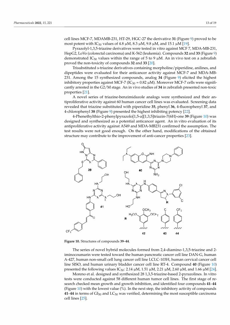
4-Phenethylthio-2-phenylpyrazolo[1,5-a][1,3,5]triazin-7(6H)-one 39 (Figure 10) wasdesigned and synthesized as a potential anticancer agent. An in vitro evaluation of itsantiproliferative activity against A549 and MDA-MB231 confirmed the assumption. Thetest results were not good enough. On the other hand, modifications of the obtainedstructure may contribute to the improvement of anti-cancer properties [23].
Pharmaceuticals 2022, 15, x FOR PEER REVIEW 9 of 20
Figure 10. Structures of compounds 39–44.
Wang et al. presented 16 compounds containing a phenylhydrazine and a thiazole moiety. Halogen-containing compound 45 (Figure 11) showed an uttermost inhibitory ef-fect against MDA-MB-231, HeLa, KG1a (acute myelogenous leukaemia), and Jurkat (T-cell leukaemia) cancer cells. Subsequently cervical cancer cells (SiHa, CaSki, DoTc2) were treated with compound 45, and the obtained IC50 values were in the range from 1.34 µg/mL to 4.56 µg/mL. An in vivo test on the nude mouse xenograft model revealed inhi-bition potency of compound 45 by the reduction of tumor volume [41].
The 2-(fluorophenylamino)-4,6-disubstituted 1,3,5-triazine induced inhibition of in-flammation and cancer growth. SAR studies underlined the important role of 3- and 4-fluorphenylamino moiety 46 and 47 (Figure 11). Compound 47 significantly reduced tu-mor tissue in several animal models and decreased PC-3 proliferation with an IC50 value of 20 µM. This analog also arrested PC-3 cells in stage G0/G1 [42].
Via three-components one spot condensation 110 new of 1,3,5-triazine derivatives were obtained. Antiproliferative activity of the most potent compounds 48–51 (Figure 11) identified in the screening against DU145 prostate-cancer cells had GI50 values of 3.43 µM, 4.01 µM, 2.38 µM and 0.67 µM, respectively [43]. Subsequent studies generated further derivatives that were tested for three breast tumors. Evaluation led to the determination that the most active structures are 52 and 53 (Figure 11) and indicated that the group of derivatives were more active against triple negative breast cancer MDA-MB231 [44,45].
Derivatives based on quinazoline combined with a 1,3,5-triazine ring via urea bridge presented antitumor activity against TPC-1 cells (thyroid cancer), MCF-7. Corresponding to the normal cell line (human foreskin fibroblasts), compounds 54–56 (Figure 11) were non-toxic. In addition, these structures showed the best IC50 values against carcinoma cells, and demonstrated tyrosine kinase inhibitory potency [46].
Figure 10. Structures of compounds 39–44.
The series of novel hybrid molecules formed from 2,4-diamino-1,3,5-triazine and 2-iminocoumarin were tested toward the human pancreatic cancer cell line DAN-G, humanA-427, human non-small cell lung cancer cell line LCLC-103H, human cervical cancer cellline SISO, and human urinary bladder cancer cell line RT-4. Compound 40 (Figure 10)presented the following values IC50: 2.14 µM, 1.51 µM, 2.21 µM, 2.60 µM, and 1.66 µM [24].
Moreno et al. designed and synthesized 28 1,3,5-triazine-based 2-pyrazolines. In vitrotests were conducted against 58 different human tumor cell lines. The first stage of re-search checked mean growth and growth inhibition, and identified four compounds 41–44(Figure 10) with the lowest value (%). In the next step, the inhibitory activity of compounds41–44 in terms of GI50 and LC50 was verified, determining the most susceptible carcinomacell lines [25].
Pharmaceuticals 2022, 15, 221 14 of 19
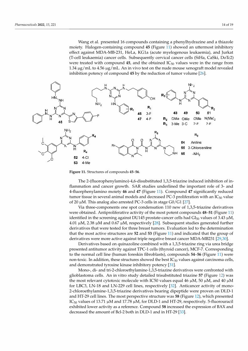
Wang et al. presented 16 compounds containing a phenylhydrazine and a thiazolemoiety. Halogen-containing compound 45 (Figure 11) showed an uttermost inhibitoryeffect against MDA-MB-231, HeLa, KG1a (acute myelogenous leukaemia), and Jurkat(T-cell leukaemia) cancer cells. Subsequently cervical cancer cells (SiHa, CaSki, DoTc2)were treated with compound 45, and the obtained IC50 values were in the range from1.34 µg/mL to 4.56 µg/mL. An in vivo test on the nude mouse xenograft model revealedinhibition potency of compound 45 by the reduction of tumor volume [26].
Pharmaceuticals 2022, 15, x FOR PEER REVIEW 10 of 20
Figure 11. Structures of compounds 45–56.
Mono-, di- and tri-2-chloroethylamine-1,3,5-triazine derivatives were confronted with glioblastoma cells. An in vitro study detailed trisubstituted triazine 57 (Figure 12) was the most relevant cytotoxic molecule with IC50 values equal 46 µM, 50 µM, and 40 µM for LBC3, LN-18 and LN-229 cell lines, respectively [47]. Anticancer activity of mono-2-chloroethylamine-1,3,5-triazine derivatives bearing dipeptide were proven on DLD-1 and HT-29 cell lines. The most perspective structure was 58 (Figure 12), which presented IC50 values of 13.71 µM and 17.78 µM, for DLD-1 and HT-29, respectively. 5-fluorouracil exhibited lower activity as a reference. Compound 58 increased the expression of BAX and decreased the amount of Bcl-2 both in DLD-1 and in HT-29 [48].
A total of thirty-four novel pyrazolo[1,5-a][1,3,5]triazine derivatives were screened against 60 cancer cell lines. Results suggested that the most antiproliferative compounds were 59 and 60 (Figure 12). Analog 59 exhibited% inhibition ranging from 40% to 115%, and 82.38% for CDK2, and derivative 60 exhibited% inhibition ranging from 43% to 92%, and 81.96% for CDK2 [49].
Hybrid molecule containing 1,4-naphthoquinone, 1,3,5-triazine and morpholine 61 (Figure 12) turned out to be strongly complexed with PI3Kγ and AMPK (5′ AMP-activated protein kinase) during docking studies. Analog 61 had an IC50 value of approximately 25 µM when exposed to the SKMEL-103 (N-RAS mutated) cell line. A Western blot deter-mined the decreased expression of both PI3Kγ and AMPK [50].
Figure 11. Structures of compounds 45–56.
The 2-(fluorophenylamino)-4,6-disubstituted 1,3,5-triazine induced inhibition of in-flammation and cancer growth. SAR studies underlined the important role of 3- and4-fluorphenylamino moiety 46 and 47 (Figure 11). Compound 47 significantly reducedtumor tissue in several animal models and decreased PC-3 proliferation with an IC50 valueof 20 µM. This analog also arrested PC-3 cells in stage G0/G1 [27].
Via three-components one spot condensation 110 new of 1,3,5-triazine derivativeswere obtained. Antiproliferative activity of the most potent compounds 48–51 (Figure 11)identified in the screening against DU145 prostate-cancer cells had GI50 values of 3.43 µM,4.01 µM, 2.38 µM and 0.67 µM, respectively [28]. Subsequent studies generated furtherderivatives that were tested for three breast tumors. Evaluation led to the determinationthat the most active structures are 52 and 53 (Figure 11) and indicated that the group ofderivatives were more active against triple negative breast cancer MDA-MB231 [29,30].
Derivatives based on quinazoline combined with a 1,3,5-triazine ring via urea bridgepresented antitumor activity against TPC-1 cells (thyroid cancer), MCF-7. Correspondingto the normal cell line (human foreskin fibroblasts), compounds 54–56 (Figure 11) werenon-toxic. In addition, these structures showed the best IC50 values against carcinoma cells,and demonstrated tyrosine kinase inhibitory potency [31].
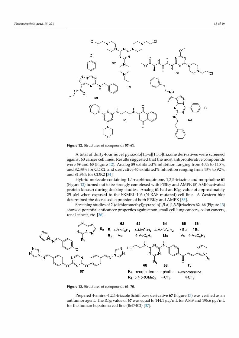
Mono-, di- and tri-2-chloroethylamine-1,3,5-triazine derivatives were confronted withglioblastoma cells. An in vitro study detailed trisubstituted triazine 57 (Figure 12) wasthe most relevant cytotoxic molecule with IC50 values equal 46 µM, 50 µM, and 40 µMfor LBC3, LN-18 and LN-229 cell lines, respectively [32]. Anticancer activity of mono-2-chloroethylamine-1,3,5-triazine derivatives bearing dipeptide were proven on DLD-1and HT-29 cell lines. The most perspective structure was 58 (Figure 12), which presentedIC50 values of 13.71 µM and 17.78 µM, for DLD-1 and HT-29, respectively. 5-fluorouracilexhibited lower activity as a reference. Compound 58 increased the expression of BAX anddecreased the amount of Bcl-2 both in DLD-1 and in HT-29 [33].
Pharmaceuticals 2022, 15, 221 15 of 19Pharmaceuticals 2022, 15, x FOR PEER REVIEW 11 of 20
Figure 12. Structures of compounds 57–61.
Screening studies of 2-(dichloromethyl)pyrazolo[1,5-a][1,3,5]triazines 62–66 (Figure 13) showed potential anticancer properties against non-small cell lung cancers, colon can-cers, renal cancer, etc. [51].
Prepared 4-amino-1,2,4-triazole Schiff base derivative 67 (Figure 13) was verified as an antitumor agent. The IC50 value of 67 was equal to 144.1 µg/mL for A549 and 195.6 µg/mL for the human hepatoma cell line (Bel7402) [52].
From the series of novel chalcone- and pyrazoline-based 1,3,5-triazines derivatives, compounds 68–70 (Figure 13) demonstrated the best potent in vitro anticancer activity with GI50 values significantly lower than reference drug 5-FU. Chalcone 68 showed GI50 values in the range of 0.422–3.05 µM, with the SR cell line (leukemia, GI50 = 0.422 µM) being the most sensitive strain. Compound 69 exhibited GI50 values in the range of 1.25–8.66 µM, with the MCF7 (GI50 = 1.25 µM) being the most sensitive strain, while compound 70 showed GI50 values in the range of 1.48–14.9 µM, being especially effective against HCT-116 with GI50 = 1.48 µM. The best cytotoxicity value was shown by compound 69 against UO-31 (renal cancer, LC50 = 5.08 µM) [53].
Figure 13. Structures of compounds 61–70.
Figure 12. Structures of compounds 57–61.
A total of thirty-four novel pyrazolo[1,5-a][1,3,5]triazine derivatives were screenedagainst 60 cancer cell lines. Results suggested that the most antiproliferative compoundswere 59 and 60 (Figure 12). Analog 59 exhibited% inhibition ranging from 40% to 115%,and 82.38% for CDK2, and derivative 60 exhibited% inhibition ranging from 43% to 92%,and 81.96% for CDK2 [34].
Hybrid molecule containing 1,4-naphthoquinone, 1,3,5-triazine and morpholine 61(Figure 12) turned out to be strongly complexed with PI3Kγ and AMPK (5′ AMP-activatedprotein kinase) during docking studies. Analog 61 had an IC50 value of approximately25 µM when exposed to the SKMEL-103 (N-RAS mutated) cell line. A Western blotdetermined the decreased expression of both PI3Kγ and AMPK [35].
Screening studies of 2-(dichloromethyl)pyrazolo[1,5-a][1,3,5]triazines 62–66 (Figure 13)showed potential anticancer properties against non-small cell lung cancers, colon cancers,renal cancer, etc. [36].
Pharmaceuticals 2022, 15, x FOR PEER REVIEW 11 of 20
Figure 12. Structures of compounds 57–61.
Screening studies of 2-(dichloromethyl)pyrazolo[1,5-a][1,3,5]triazines 62–66 (Figure 13) showed potential anticancer properties against non-small cell lung cancers, colon can-cers, renal cancer, etc. [51].
Prepared 4-amino-1,2,4-triazole Schiff base derivative 67 (Figure 13) was verified as an antitumor agent. The IC50 value of 67 was equal to 144.1 µg/mL for A549 and 195.6 µg/mL for the human hepatoma cell line (Bel7402) [52].
From the series of novel chalcone- and pyrazoline-based 1,3,5-triazines derivatives, compounds 68–70 (Figure 13) demonstrated the best potent in vitro anticancer activity with GI50 values significantly lower than reference drug 5-FU. Chalcone 68 showed GI50 values in the range of 0.422–3.05 µM, with the SR cell line (leukemia, GI50 = 0.422 µM) being the most sensitive strain. Compound 69 exhibited GI50 values in the range of 1.25–8.66 µM, with the MCF7 (GI50 = 1.25 µM) being the most sensitive strain, while compound 70 showed GI50 values in the range of 1.48–14.9 µM, being especially effective against HCT-116 with GI50 = 1.48 µM. The best cytotoxicity value was shown by compound 69 against UO-31 (renal cancer, LC50 = 5.08 µM) [53].
Figure 13. Structures of compounds 61–70.
Figure 13. Structures of compounds 61–70.
Prepared 4-amino-1,2,4-triazole Schiff base derivative 67 (Figure 13) was verified as anantitumor agent. The IC50 value of 67 was equal to 144.1 µg/mL for A549 and 195.6 µg/mLfor the human hepatoma cell line (Bel7402) [37].

Pharmaceuticals 2022, 15, 221 16 of 19
From the series of novel chalcone- and pyrazoline-based 1,3,5-triazines derivatives,compounds 68–70 (Figure 13) demonstrated the best potent in vitro anticancer activity withGI50 values significantly lower than reference drug 5-FU. Chalcone 68 showed GI50 valuesin the range of 0.422–3.05 µM, with the SR cell line (leukemia, GI50 = 0.422 µM) being themost sensitive strain. Compound 69 exhibited GI50 values in the range of 1.25–8.66 µM,with the MCF7 (GI50 = 1.25 µM) being the most sensitive strain, while compound 70showed GI50 values in the range of 1.48–14.9 µM, being especially effective against HCT-116 with GI50 = 1.48 µM. The best cytotoxicity value was shown by compound 69 againstUO-31 (renal cancer, LC50 = 5.08 µM) [38].
3. Search Strategy and Selection Criteria
The aim of this study was to collect knowledge and data on the synthesized novel1,3,5-triazine derivatives, their effects on cancer cells, and to identify enzymes as potentialtargets for these substances. To carry out the study, the following databases were searched:PubMed (NCBI), Web of Science, and Scopus, using the following key words: 1,3,5-triazine,s-triazine, anticancer, antitumor, and enzyme inhibitor. We examined original articlesand case studies published between 2015 and 2021. The results of the study include thecompounds from papers with the highest activity.
4. Conclusions
The “hybrid” approach incorporating a triazine framework ensures an improvedprofile against the target biological pathways pertaining to infectious parasites, microbes,and conditions such as cancer and neurodegeneration. The multi-targeting approach ofthe hybrid compounds ensures an effective overcoming of the key regulatory pathwayscontributing to complicacies such as drug resistance. This review presents a comprehensivediscussion on the candidature of the 1,3,5-triazine scaffold for a rational developmentof the hybrid molecules by conjugation with bioactive pharmacophoric moieties. Thebasis of superior efficacy of 1,3,5-triazine based hybrid molecules by considering theirinteractions with the cellular targets has also been discussed in a succinct manner. Theliterature revealed that s-triazine derivatives possess diverse anticancer potential, easysynthetic routes for synthesis, and have attracted researchers for development of newchemotherapeutic agents. Extensive research is required on the 1,3,5-triazine moiety to findnovel analogs suitable for clinical applications in cancer treatment.
Author Contributions: Conceptualization, D.M. and D.D.; writing—original draft preparation, D.M.;review and editing, D.M. and D.D.; supervision, D.D. All authors have read and agreed to thepublished version of the manuscript.
Funding: Publication was written during doctoral studies under the project No POWR.03.02.00-00-I051/16 co-funded from European Union funds, PO WER 2014–2020.
Institutional Review Board Statement: Not applicable.
Informed Consent Statement: Not applicable.
Data Availability Statement: Not applicable.
Conflicts of Interest: Authors declare no conflict of interest.
References1. WHO, Regional Office for Europe. World Cancer Report: Cancer Research for Cancer Development; IARC: Lyon, France, 2020.2. Pathak, A.; Tanwar, S.; Kumar, V.; Banarjee, B.D. Present and Future Prospect of Small Molecule & Related Targeted Therapy
Against Human Cancer. Vivechan Int. J. Res. 2018, 9, 36–49. [PubMed]3. Pogorelcnik, B.; Janežic, M.; Sosic, I.; Gobec, S.; Solmajer, T.; Perdih, A. 4,6-Substituted-1,3,5-Triazin-2(1H)-Ones as Monocyclic
Catalytic Inhibitors of Human DNA Topoisomerase IIα Targeting the ATP Binding Site. Bioorg. Med. Chem. 2015, 23, 4218–4229.[CrossRef] [PubMed]
Pharmaceuticals 2022, 15, 221 17 of 19
4. Zhang, B.; Zhang, Q.; Xiao, Z.; Sun, X.; Yang, Z.; Gu, Q.; Liu, Z.; Xie, T.; Jin, Q.; Zheng, P.; et al. Design, Synthesis and BiologicalEvaluation of Substituted 2-(Thiophen-2-Yl)-1,3,5-Triazine Derivatives as Potential Dual PI3Kα/MTOR Inhibitors. Bioorg. Chem.2020, 95, 103525. [CrossRef] [PubMed]
5. Hu, J.; Zhang, Y.; Tang, N.; Lu, Y.; Guo, P.; Huang, Z. Discovery of Novel 1,3,5-Triazine Derivatives as Potent Inhibitor of CervicalCancer via Dual Inhibition of PI3K/MTOR. Bioorg. Med. Chem. 2021, 32, 115997. [CrossRef] [PubMed]
6. Singla, P.; Luxami, V.; Paul, K. Synthesis, in Vitro Antitumor Activity, Dihydrofolate Reductase Inhibition, DNA Intercalationand Structure–Activity Relationship Studies of 1,3,5-Triazine Analogues. Bioorg. Med. Chem. Lett. 2016, 26, 518–523. [CrossRef][PubMed]
7. Zhou, X.; Lin, K.; Ma, X.; Chui, W.-K.; Zhou, W. Design, Synthesis, Docking Studies and Biological Evaluation of NovelDihydro-1,3,5-Triazines as Human DHFR Inhibitors. Eur. J. Med. Chem. 2017, 125, 1279–1288. [CrossRef] [PubMed]
8. Ng, H.-L.; Ma, X.; Chew, E.-H.; Chui, W.-K. Design, Synthesis, and Biological Evaluation of Coupled Bioactive Scaffolds asPotential Anticancer Agents for Dual Targeting of Dihydrofolate Reductase and Thioredoxin Reductase. J. Med. Chem. 2017, 60,1734–1745. [CrossRef] [PubMed]
9. Zołnowska, B.; Sławinski, J.; Szafranski, K.; Angeli, A.; Supuran, C.T.; Kawiak, A.; Wieczór, M.; Zielinska, J.; Baczek, T.; Bar-toszewska, S. Novel 2-(2-Arylmethylthio-4-Chloro-5-Methylbenzenesulfonyl)-1-(1,3,5-Triazin-2-Ylamino) Guanidine Derivatives:Inhibition of Human Carbonic Anhydrase Cytosolic Isozymes I and II and the Transmembrane Tumor-Associated Isozymes IXand XII, Anticancer Activity, and Molecular Modeling Studies. Eur. J. Med. Chem. 2018, 143, 1931–1941. [CrossRef] [PubMed]
10. Havránková, E.; Csöllei, J.; Vullo, D.; Garaj, V.; Pazdera, P.; Supuran, C.T. Novel Sulfonamide Incorporating Piperazine,Aminoalcohol and 1,3,5-Triazine Structural Motifs with Carbonic Anhydrase I, II and IX Inhibitory Action. Bioorg. Chem. 2018, 77,25–37. [CrossRef] [PubMed]
11. Lolak, N.; Akocak, S.; Bua, S.; Supuran, C.T. Design, Synthesis and Biological Evaluation of Novel Ureido BenzenesulfonamidesIncorporating 1,3,5-Triazine Moieties as Potent Carbonic Anhydrase IX Inhibitors. Bioorg. Chem. 2019, 82, 117–122. [CrossRef][PubMed]
12. Lolak, N.; Akocak, S.; Bua, S.; Sanku, R.K.K.; Supuran, C.T. Discovery of New Ureido Benzenesulfonamides Incorporating1,3,5-Triazine Moieties as Carbonic Anhydrase I, II, IX and XII Inhibitors. Bioorg. Med. Chem. 2019, 27, 1588–1594. [CrossRef][PubMed]
13. Srivastava, J.K.; Pillai, G.G.; Bhat, H.R.; Verma, A.; Singh, U.P. Design and Discovery of Novel Monastrol-1,3,5-Triazines as PotentAnti-Breast Cancer Agent via Attenuating Epidermal Growth Factor Receptor Tyrosine Kinase. Sci. Rep. 2017, 7, 5851. [CrossRef][PubMed]
14. Yan, W.; Zhao, Y.; He, J. Anti-breast Cancer Activity of Selected 1,3,5-triazines via Modulation of EGFR-TK. Mol. Med. Rep. 2018,18, 4175–4184. [CrossRef]
15. Bhat, H.R.; Masih, A.; Shakya, A.; Ghosh, S.K.; Singh, U.P. Design, Synthesis, Anticancer, Antibacterial, and Antifungal Evaluationof 4-aminoquinoline-1,3,5-triazine Derivatives. J. Heterocycl. Chem. 2020, 57, 390–399. [CrossRef]
16. Dao, P.; Smith, N.; Tomkiewicz-Raulet, C.; Yen-Pon, E.; Camacho-Artacho, M.; Lietha, D.; Herbeuval, J.-P.; Coumoul, X.; Garbay,C.; Chen, H. Design, Synthesis, and Evaluation of Novel Imidazo[1,2-a][1,3,5] Triazines and Their Derivatives as Focal AdhesionKinase Inhibitors with Antitumor Activity. J. Med. Chem. 2015, 58, 237–251. [CrossRef] [PubMed]
17. Kothayer, H.; Spencer, S.M.; Tripathi, K.; Westwell, A.D.; Palle, K. Synthesis and in Vitro Anticancer Evaluation of Some 4,6-Diamino-1,3,5-Triazine-2-Carbohydrazides as Rad6 Ubiquitin Conjugating Enzyme Inhibitors. Bioorg. Med. Chem. Lett. 2016, 26,2030–2034. [CrossRef] [PubMed]
18. El Malah, T.; Nour, H.F.; Nayl, A.A.; Elkhashab, R.A.; Abdel-Megeid, F.M.E.; Ali, M.M. Anticancer Evaluation of Tris (Tria-zolyl)Triazine Derivatives Generated via Click Chemistry. Aust. J. Chem. 2016, 69, 905. [CrossRef]
19. Kumar, G.J.; Kumar, S.N.; Thummuri, D.; Adari, L.B.S.; Naidu, V.G.M.; Srinivas, K.; Rao, V.J. Synthesis and Characterization ofNew S-Triazine Bearing Benzimidazole and Benzothiazole Derivatives as Anticancer Agents. Med. Chem. Res. 2015, 24, 3991–4001.[CrossRef]
20. Farooq, M.; Sharma, A.; Almarhoon, Z.; Al-Dhfyan, A.; El-Faham, A.; Taha, N.A.; Wadaan, M.A.M.; de la Torre, B.G.; Albericio, F.Design and Synthesis of Mono-and Di-Pyrazolyl-s-Triazine Derivatives, Their Anticancer Profile in Human Cancer Cell Lines,and in Vivo Toxicity in Zebrafish Embryos. Bioorg. Chem. 2019, 87, 457–464. [CrossRef]
21. Malebari, A.M.; Abd Alhameed, R.; Almarhoon, Z.; Farooq, M.; Wadaan, M.A.M.; Sharma, A.; de la Torre, B.G.; Albericio,F.; El-Faham, A. The Antiproliferative and Apoptotic Effect of a Novel Synthesized S-Triazine Dipeptide Series, and ToxicityScreening in Zebrafish Embryos. Molecules 2021, 26, 1170. [CrossRef]
22. Singla, P.; Luxami, V.; Paul, K. Synthesis and in Vitro Evaluation of Novel Triazine Analogues as Anticancer Agents and TheirInteraction Studies with Bovine Serum Albumin. Eur. J. Med. Chem. 2016, 117, 59–69. [CrossRef]
23. Smolnikov, S.; Gorgopina, E.; Lezhnyova, V.; Ong, G.; Chui, W.-K.; Dolzhenko, A. 4-Phenethylthio-2-Phenylpyrazolo[1,5-a][1,3,5]Triazin-7(6H)-One. Molbank 2017, 2017, M970. [CrossRef]
24. Makowska, A.; Saczewski, F.; Bednarski, P.; Saczewski, J.; Balewski, Ł. Hybrid Molecules Composed of 2,4-Diamino-1,3,5-Triazinesand 2-Imino-Coumarins and Coumarins. Synthesis and Cytotoxic Properties. Molecules 2018, 23, 1616. [CrossRef] [PubMed]
25. Moreno, L.; Quiroga, J.; Abonia, R.; Ramírez-Prada, J.; Insuasty, B. Synthesis of New 1,3,5-Triazine-Based 2-Pyrazolines asPotential Anticancer Agents. Molecules 2018, 23, 1956. [CrossRef]
Pharmaceuticals 2022, 15, 221 18 of 19
26. Wang, X.; Yi, Y.; Lv, Q.; Zhang, J.; Wu, K.; Wu, W.; Zhang, W. Novel 1,3,5-Triazine Derivatives Exert Potent Anti-Cervical CancerEffects by Modulating Bax, Bcl2 and Caspases Expression. Chem. Biol. Drug Des. 2018, 91, 728–734. [CrossRef]
27. Zacharie, B.; Abbott, S.D.; Duceppe, J.; Gagnon, L.; Grouix, B.; Geerts, L.; Gervais, L.; Sarra-Bournet, F.; Perron, V.; Wilb, N.; et al.Design and Synthesis of New 1,3,5-Trisubstituted Triazines for the Treatment of Cancer and Inflammation. ChemistryOpen 2018, 7,737–749. [CrossRef] [PubMed]
28. Junaid, A.; Lim, F.P.L.; Tiekink, E.R.T.; Dolzhenko, A.V. New One-Pot Synthesis of 1,3,5-Triazines: Three-Component Con-densation, Dimroth Rearrangement, and Dehydrogenative Aromatization. ACS Comb. Sci. 2019, 21, 548–555. [CrossRef][PubMed]
29. Junaid, A.; Lim, F.P.L.; Chuah, L.H.; Dolzhenko, A.V. 6, N 2 -Diaryl-1,3,5-Triazine-2,4-Diamines: Synthesis, AntiproliferativeActivity and 3D-QSAR Modeling. RSC Adv. 2020, 10, 12135–12144. [CrossRef]
30. Junaid, A.; Lim, F.P.L.; Tiekink, E.R.T.; Dolzhenko, A.V. Design, Synthesis, and Biological Evaluation of New 6, N 2 -Diaryl-1,3,5-Triazine-2,4-Diamines as Anticancer Agents Selectively Targeting Triple Negative Breast Cancer Cells. RSC Adv. 2020, 10,25517–25528. [CrossRef]
31. Pathak, P.; Naumovich, V.; Grishina, M.; Shukla, P.K.; Verma, A.; Potemkin, V. Quinazoline Based 1,3,5-triazine Derivatives asCancer Inhibitors by Impeding the Phosphorylated RET Tyrosine Kinase Pathway: Design, Synthesis, Docking, and QSAR Study.Arch. Pharm. 2019, 352, 1900053. [CrossRef]
32. Kretowski, R.; Drozdowska, D.; Kolesinska, B.; Kaminski, Z.; Fraczyk, J.; Cechowska-Pasko, M. The Cellular Effects of NovelTriazine Nitrogen Mustards in Glioblastoma LBC3, LN-18 and LN-229 Cell Lines. Investig. New Drugs 2019, 37, 984–993. [CrossRef][PubMed]
33. Wróbel, A.; Kolesinska, B.; Fraczyk, J.; Kaminski, Z.J.; Tankiewicz-Kwedlo, A.; Hermanowicz, J.; Czarnomysy, R.; Maliszewski, D.;Drozdowska, D. Synthesis and Cellular Effects of Novel 1,3,5-Triazine Derivatives in DLD and Ht-29 Human Colon Cancer CellLines. Investng. New Drugs 2020, 38, 990–1002. [CrossRef]
34. Oudah, K.H.; Najm, M.A.A.; Samir, N.; Serya, R.A.T.; Abouzid, K.A.M. Design, Synthesis and Molecular Docking of NovelPyrazolo[1,5-a][1,3,5]Triazine Derivatives as CDK2 Inhibitors. Bioorg. Chem. 2019, 92, 103239. [CrossRef]
35. Fiorot, R.; Westphal, R.; Lemos, B.; Romagna, R.; Gonçalves, P.; Fernandes, M.; Ferreira, C.; Taranto, A.; Greco, S. Synthesis,Molecular Modelling and Anticancer Activities of New Molecular Hybrids Containing 1,4-Naphthoquinone, 7-Chloroquinoline,1,3,5-Triazine and Morpholine Cores as PI3K and AMPK Inhibitors in the Metastatic Melanoma Cells. J. Braz. Chem. Soc. 2019, 30,1860–1873. [CrossRef]
36. Velihina, Y.S.; Pil’o, S.G.; Zyabrev, V.S.; Moskvina, V.S.; Shablykina, O.V.; Brovarets, V.S. 2-(Dichloromethyl)Pyrazolo[1,5-a][1,3,5]Triazines: Synthesis and Anticancer Activity. Biopolym. Cell 2020, 36, 60–73. [CrossRef]
37. Jiang, G.; Chang, Q.; Liang, D.; Zhang, Y.; Meng, Y.; Yi, Q. Preparation and Antitumor Effects of 4-Amino-1,2,4-Triazole SchiffBase Derivative. J. Int. Med. Res. 2020, 48, 030006052090387. [CrossRef] [PubMed]
38. Moreno, L.M.; Quiroga, J.; Abonia, R.; Lauria, A.; Martorana, A.; Insuasty, H.; Insuasty, B. Synthesis, Biological Evaluation, andin Silico Studies of Novel Chalcone- and Pyrazoline-Based 1,3,5-Triazines as Potential Anticancer Agents. RSC Adv. 2020, 10,34114–34129. [CrossRef]
39. Champoux, J.J. DNA Topoisomerases: Structure, Function, and Mechanism. Annu. Rev. Biochem. 2001, 70, 369–413. [CrossRef]40. Maheshwari, S.; Miller, M.S.; O’Meally, R.; Cole, R.N.; Amzel, L.M.; Gabelli, S.B. Kinetic and Structural Analyses Reveal Residues
in Phosphoinositide 3-Kinase α That Are Critical for Catalysis and Substrate Recognition. J. Biol. Chem. 2017, 292, 13541–13550.[CrossRef] [PubMed]
41. Hua, H.; Kong, Q.; Zhang, H.; Wang, J.; Luo, T.; Jiang, Y. Targeting MTOR for Cancer Therapy. J. Hematol. Oncol. 2019, 12, 71.[CrossRef] [PubMed]
42. Tarantelli, C.; Lupia, A.; Stathis, A.; Bertoni, F. Is There a Role for Dual PI3K/MTOR Inhibitors for Patients Affected withLymphoma? Int. J. Mol. Sci. 2020, 21, 1060. [CrossRef]
43. Osborne, M.J.; Schnell, J.; Benkovic, S.J.; Dyson, H.J.; Wright, P.E. Backbone Dynamics in Dihydrofolate Reductase Complexes:Role of Loop Flexibility in the Catalytic Mechanism. Biochemistry 2001, 40, 9846–9859. [CrossRef] [PubMed]
44. Wróbel, A.; Drozdowska, D. Recent Design and Structure-Activity Relationship Studies on the Modifications of DHFR Inhibitorsas Anticancer Agents. Curr. Med. Chem. 2021, 28, 910–939. [CrossRef]
45. Singla, P.; Luxami, V.; Paul, K. Triazine–Benzimidazole Hybrids: Anticancer Activity, DNA Interaction and DihydrofolateReductase Inhibitors. Bioorg. Med. Chem. 2015, 23, 1691–1700. [CrossRef] [PubMed]
46. Supuran, C.T.; Scozzafava, A.; Casini, A. Carbonic Anhydrase Inhibitors. Med. Res. Rev. 2003, 23, 146–189. [CrossRef]47. Walker, F.; Abramowitz, L.; Benabderrahmane, D.; Duval, X.; Descatoire, V.; Hénin, D.; Lehy, T.; Aparicio, T. Growth Factor
Receptor Expression in Anal Squamous Lesions: Modifications Associated with Oncogenic Human Papillomavirus and HumanImmunodeficiency Virus. Hum. Pathol. 2009, 40, 1517–1527. [CrossRef] [PubMed]
48. Kumar, V.; Abbas, A.K.; Aster, J.C.; Robbins, S.L. Robbins Basic Pathology, 9th ed.; Saunders: Philadelphia, PA, USA, 2013.49. Palmer, B.F.; Clegg, D.J. Oxygen Sensing and Metabolic Homeostasis. Mol. Cell. Endocrinol. 2014, 397, 51–58. [CrossRef]50. Karkkainen, M.J.; Petrova, T.V. Vascular Endothelial Growth Factor Receptors in the Regulation of Angiogenesis and Lymphan-
giogenesis. Oncogene 2000, 19, 5598–5605. [CrossRef] [PubMed]

Pharmaceuticals 2022, 15, 221 19 of 19
51. Muller, Y.A.; Li, B.; Christinger, H.W.; Wells, J.A.; Cunningham, B.C.; de Vos, A.M. Vascular Endothelial Growth Factor: CrystalStructure and Functional Mapping of the Kinase Domain Receptor Binding Site. Proc. Natl. Acad. Sci. USA 1997, 94, 7192–7197.[CrossRef] [PubMed]
52. Pathak, P.; Shukla, P.K.; Kumar, V.; Kumar, A.; Verma, A. Quinazoline Clubbed 1,3,5-Triazine Derivatives as VEGFR2 KinaseInhibitors: Design, Synthesis, Docking, in Vitro Cytotoxicity and in Ovo Antiangiogenic Activity. Inflammopharmacology 2018, 26,1441–1453. [CrossRef] [PubMed]
53. Mehlen, P.; Puisieux, A. Metastasis: A Question of Life or Death. Nat. Rev. Cancer 2006, 6, 449–458. [CrossRef] [PubMed]