RANIDAE - National University of Singapore
7
101 THE RAFFLES BULLETIN OF ZOOLOGY 2003 51(1): 101-107 © National University of Singapore MORPHOLOGY AND BUCCOPHARYNGEAL ANATOMY OF THE TADPOLE OF RANA (NASIRANA) ALTICOLA (ANURA: RANIDAE) Stéphane Grosjean, Mélanie Pérez and Annemarie Ohler Laboratoire de Zoologie des Reptiles et Amphibiens, Muséum national d’Histoire naturelle, 25 rue Cuvier, 75005 Paris, France Email: [email protected]; [email protected] ABSTRACT. - The external morphology and the buccopharyngeal features of the tadpole of Rana (Nasirana) alticola are described. Morphometric data are provided. This tadpole is easily distinguishable from other ranid species by its large size, high number of keratodont rows, coloured ocelli on tail, and large glands such as parotoid glands, supracaudal and infracaudal glands. A comparison with tadpoles from other populations is done. KEY WORDS. - Tadpole, Rana (Nasirana) alticola, external morphology, buccopharyngeal features, India, Thailand, Rana (Clinotarsus) curtipes. INTRODUCTION Rana alticola Boulenger, 1882, was described on the basis of four specimens from Shillong (Khasi Hills District, Meghalaya State, India) under the name Hylorana pipiens Jerdon, 1870. This is a poorly studied species distributed widely in latitude, longitude and elevation, from the Hills of Assam (India) to southern Thailand [the specimen mentioned as Hylorana alticola by Bourret (1939: 59) from Mao Son (Northern Vietnam) proved to be an Amolops nasicus (Boulenger, 1903) (Bourret, 1942: 350); it should therefore be removed from the species list of Vietnam (see Frost, 1985)]. Although adults are rare in herpetological collections, the tadpoles have been known for a long time (Boulenger, 1882; Annandale, 1912; Smith, 1924a, b; review by Bourret, 1942). These tadpoles bear conspicuous features such as large size, entirely black coloration with red ocelli, large parotoid glands, supracaudal gland, several other glands as well as secretory pores spread over ventral surface and caudal fin, and numerous keratodont rows. We redescribe the external morphology of Nasirana tadpoles from Thailand, and we give for the first time a description of the buccopharyngeal features of a specimen. MATERIALS AND METHODS A total of 69 Rana alticola tadpoles were collected from a quiet area of a stream bordering a primary forest, at the base of a waterfall in the Ramon Forest Park (8°27’ N, 98°32’ E, about 30 m above sea level), Phang Nga Province, Thailand. The tadpoles in developmental stages 25 to 41 (Gosner, 1960), preserved in a mixture of equal parts of 4% formaldehyde and 70% ethanol (Grillitsch, 1984), were deposited in the collections of the Muséum national d’Histoire naturelle of Paris (MNHN 2000.4594-4661). A specimen in stage 43 (MNHN 2000.8402) allowed us to ensure the identity of the species. Morphological terminology follows Altig & McDiarmid (1999), terminology of oral disc follows Altig (1970), keratodont formula (KF) follows Dubois (1995), developmental stages were determined according to Gosner (1960) and terminology of internal buccal features follows Wassersug (1976). Total length (TL) and distance from opening of vent to tip of tail from stage 26, distance from tip of snout to insertion of upper tail fin and snout-vent length from stage 27, and distance from tip of snout to opening of spiracle and distance from opening of vent to point of maximum height of tail from stage 28 were measured with a hand calipers. All other measurements were taken with a graduated ocular attached to a stereomicroscope. For exact location of measurement landmarks, see Grosjean (2001: Fig. 2). Drawings were made using a camera lucida. RESULTS Diagnosis of tadpole. – A large-sized tadpole (TL 16.4-92.7 mm, stages 25-38) beige-grey with black spots in tadpoles in stage 25 and uniformly black from stage 26, a large black ocellus with a red halo on caudal muscle at base of tail (followed by two smaller ones of decreasing size often poorly visible); a pair of large parotoid glands behind eyes, a pair of pectoral glands and a pair of ventroposterior ones, a
-
Upload
khangminh22 -
Category
Documents
-
view
6 -
download
0
Transcript of RANIDAE - National University of Singapore

101
THE RAFFLES BULLETIN OF ZOOLOGY 2003
THE RAFFLES BULLETIN OF ZOOLOGY 2003 51(1): 101-107© National University of Singapore
MORPHOLOGY AND BUCCOPHARYNGEAL ANATOMY OF THETADPOLE OF RANA (NASIRANA) ALTICOLA (ANURA: RANIDAE)
Stéphane Grosjean, Mélanie Pérez and Annemarie OhlerLaboratoire de Zoologie des Reptiles et Amphibiens, Muséum national d’Histoire naturelle,
25 rue Cuvier, 75005 Paris, FranceEmail: [email protected]; [email protected]
ABSTRACT. - The external morphology and the buccopharyngeal features of the tadpole of Rana (Nasirana)alticola are described. Morphometric data are provided. This tadpole is easily distinguishable from otherranid species by its large size, high number of keratodont rows, coloured ocelli on tail, and large glandssuch as parotoid glands, supracaudal and infracaudal glands. A comparison with tadpoles from otherpopulations is done.
KEY WORDS. - Tadpole, Rana (Nasirana) alticola, external morphology, buccopharyngeal features, India,Thailand, Rana (Clinotarsus) curtipes.
INTRODUCTION
Rana alticola Boulenger, 1882, was described on the basisof four specimens from Shillong (Khasi Hills District,Meghalaya State, India) under the name Hylorana pipiensJerdon, 1870. This is a poorly studied species distributedwidely in latitude, longitude and elevation, from the Hills ofAssam (India) to southern Thailand [the specimen mentionedas Hylorana alticola by Bourret (1939: 59) from Mao Son(Northern Vietnam) proved to be an Amolops nasicus(Boulenger, 1903) (Bourret, 1942: 350); it should thereforebe removed from the species list of Vietnam (see Frost,1985)]. Although adults are rare in herpetological collections,the tadpoles have been known for a long time (Boulenger,1882; Annandale, 1912; Smith, 1924a, b; review by Bourret,1942). These tadpoles bear conspicuous features such as largesize, entirely black coloration with red ocelli, large parotoidglands, supracaudal gland, several other glands as well assecretory pores spread over ventral surface and caudal fin,and numerous keratodont rows. We redescribe the externalmorphology of Nasirana tadpoles from Thailand, and wegive for the first time a description of the buccopharyngealfeatures of a specimen.
MATERIALS AND METHODS
A total of 69 Rana alticola tadpoles were collected from aquiet area of a stream bordering a primary forest, at the baseof a waterfall in the Ramon Forest Park (8°27’ N, 98°32’ E,about 30 m above sea level), Phang Nga Province, Thailand.The tadpoles in developmental stages 25 to 41 (Gosner,
1960), preserved in a mixture of equal parts of 4%formaldehyde and 70% ethanol (Grillitsch, 1984), weredeposited in the collections of the Muséum nationald’Histoire naturelle of Paris (MNHN 2000.4594-4661). Aspecimen in stage 43 (MNHN 2000.8402) allowed us toensure the identity of the species. Morphological terminologyfollows Altig & McDiarmid (1999), terminology of oral discfollows Altig (1970), keratodont formula (KF) followsDubois (1995), developmental stages were determinedaccording to Gosner (1960) and terminology of internalbuccal features follows Wassersug (1976). Total length (TL)and distance from opening of vent to tip of tail from stage26, distance from tip of snout to insertion of upper tail finand snout-vent length from stage 27, and distance from tipof snout to opening of spiracle and distance from openingof vent to point of maximum height of tail from stage 28were measured with a hand calipers. All other measurementswere taken with a graduated ocular attached to astereomicroscope. For exact location of measurementlandmarks, see Grosjean (2001: Fig. 2). Drawings were madeusing a camera lucida.
RESULTS
Diagnosis of tadpole. – A large-sized tadpole (TL 16.4-92.7mm, stages 25-38) beige-grey with black spots in tadpolesin stage 25 and uniformly black from stage 26, a large blackocellus with a red halo on caudal muscle at base of tail(followed by two smaller ones of decreasing size often poorlyvisible); a pair of large parotoid glands behind eyes, a pairof pectoral glands and a pair of ventroposterior ones, a

102
Grosjean et al.: Tadpole of Rana (Nasirana) alticola
supracaudal and an infracaudal gland; KF 1-2:(3+3)-(7+7)/1+1:4-8.
Larval morphology. – The following description is basedon a tadpole in stage 36 (MNHN 2000.4630). Morphometricdata are presented in Table 1. In dorsal view (Fig. 2A), bodyelliptical, widest at the posterior third; snout semi-circular.In profile (Fig. 2B), body depressed; body width 1.2 of bodyheight; snout rounded. Eyes slightly bulging, directed almostlaterally and positioned dorsolaterally, not visible in ventralview, diameter about 0.1 times snout-vent length. Nares oval,relatively small-sized, rimmed, with one anterolateralprojection, positioned almost dorsally and directed slightlyanterolaterally, distance between nares about 0.5 timesinterpupilar distance, closer to snout than to pupils. Spiraclesinistral, bulb-shaped, attached to body wall except at itsextremity, positioned ventrolaterally, oriented morehorizontally than posterodorsally; snout-spiracle distance 0.6times snout-vent length. Spiracular opening oval, situatedabove level of hind limb. Tail musculature robust, graduallytapering, almost reaching tail tip. Tail fins moderately high,not extending onto body, riddled with numerous white poreson whole surface with secreted white mucus. A gland on theventral fin, posterior to vent, the infracaudal gland; anotherat the beginning of dorsal fin, the supracaudal gland, slightlyextending on body. Upper fin convex, slightly higher thanlower; lower fin straight in proximal quarter, then slightly
convex. Maximum tail height located 20-30 % from anteriorend, tail tip rounded. Vent tube moderately large, roughlyconical, opening with bevelled edge hidden by large flap ofskin, directed posteriorly, linked to ventral tail fin, openingdextral. Presence of a pair of large parotoid glands behindeyes with pores visible without magnification, a pair of greypectoral glands ventrally and a pair of ventral glandsposteriorly situated at the level of hind limbs. Ventral surfacealso riddled with numerous white pores. All the pores secretewhite mucus (probably produced under the stress of capture),particularly the parotoid glands (Fig. 3) and the supracaudalgland but also pores of caudal fins and ventral surface.Neuromasts not observed.
Oral disk large (Fig. 4), anteroventral, slightly emarginated,directed ventrally. Papillae of moderate size, finger-shaped,some bifid. Single row of papillae on median part of lowerlabium and at extremities of large medial gap of upper labium(five papillae on each side); up to five rows of submarginalpapillae laterally; transverse row of submarginal papillaebetween accessory keratodont rows and lower keratodontrows; accessory keratodont rows present on submarginalpapillae and short ridges oriented at right angle to thekeratodont rows. KF 2:6+6/1+1:8; keratodonts about 80 µmlong with a curved spatulate apical portion bearing 26-29
Fig. 1. Natural habitat of the tadpoles of Rana alticola.
Fig. 2. Tadpole of Rana alticola in stage 36 (MNHN 2000.4630)showing its glands and three ocelli. A, dorsal view; B, profile. 1= parotoid gland; 2 = supracaudal gland; 3 = pectoral gland; 4 =posteroventral gland; 5 = infracaudal gland. Scale bar = 10 mm.
Fig. 3. Tadpole of Rana alticola in stage 35 (MNHN 2000.4660)showing venom expelled by parotoid and supracaudal glands. Totallength = 76.3 mm.

103
THE RAFFLES BULLETIN OF ZOOLOGY 2003
(counted from six keratodonts from row P6) marginaldenticles. Jaw sheaths slightly serrated, serrations black,distal half of sheaths dark brown, lower part white; uppersheath in wide flat arch slightly convex medially, lowersheath V-shaped.
In life, tadpole entirely brown-black, fins opaque, a blackocellus at the base of tail surrounded by a red halo. Inpreservative, back and flanks dark grey, ventral surface lightgrey with white pores; tail fins dark grey with white pores,caudal muscle dark brown with a black ocellus surroundedby a light orange halo.
Variation through ontogeny. – Coloration of tadpoles instage 25 differs greatly from that of tadpoles in olderdevelopmental stages. Colour in life of tadpoles in stage 25:anterior half of back light beige-grey with a darker blotchbetween eyes, posterior half of back dark brown-grey; irisall black, ventral part beige-grey transparent. Caudal musclesandy transparent with small black spots and an ocellus atthe base with a red halo. In preservative (Figs. 5A, B): backtransparent with internal organs visible through the skin, ablack dot behind each nare posteromedially, a large brownspot medial between eyes and nares, another behind eyes;posterolaterally a pair of parotoid glands with white pores.Background colour of back and upper flanks studded withmelanophores giving a grey colour; lower flanks and ventralsurface transparent. Caudal muscle off-white, with a blackocellus without a halo at its base and few black spots. Caudalfins opaque grey with white pores concentrated at the baseof upper fin and a few on the rest of lower and upper fins.From stage 26 tadpoles have the typical coloration describedfor the tadpole in stage 36. However slight differences incolour can occur between individuals: in preservative, twotadpoles in stage 34 (MNHN 2000.4658-4659) have backand upper flanks black brown, parotoids red brown; lowerflanks and belly light red brown with white pores posteriorly,branchial area yellow grey with white pores. Caudal musclered brown with a black ocellus surrounded by a yelloworange halo; tail fins black brown with white pores.
A great increase in size of tadpoles takes place between stages25 and 27 (Table 1). The number of keratodont rowsincreases during ontogeny. KF of tadpoles below stage 27(n = 14): 1-2:(3+3)-(5+6)/1+1:4-6 and KF of tadpoles in stage27 and above (n = 11): 1-2:(6+6)-(7+7)/1+1:7-8. The pairof pectoral glands and the pair of posteroventral glands arenot always evident.
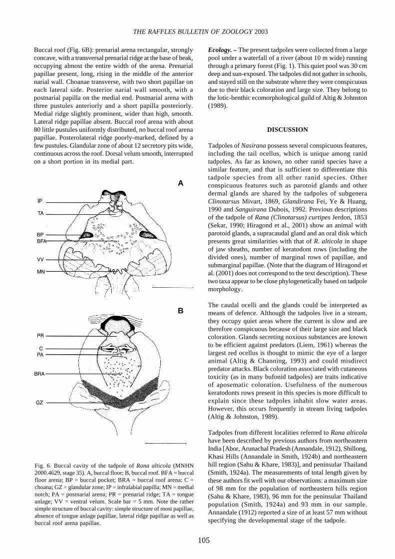
Description of the internal buccal features. – Thedescription is based on a tadpole in stage 35 (MNHN2000.4629).
Buccal floor (Fig. 6A): prelingual arena narrow with twopairs of large pustulose infralabial papillae, the first pairdirected transversely, the second one directed posteriorly.Tongue anlage rounded and truncate anteriorly, scarcelyprominent, without lingual papillae but with a pustule oneach side anterolaterally. Buccal floor arena wide, definedby 5-6 pustulose papillae on each side; interior of arenawithout papillae anteriorly, with about 30 little pustulosepapillae posteriorly. Buccal pockets wide, transverse, roughlyoval, closer to tongue anlage than to medial end of ventralvelum; about five prepocket papillae; no papillae posteriorlyto buccal pockets. Ventral velum continuous, with spicularsupport; margin with six projections on each side, the threemedial ones closer together, the two medial making up themedial notch, glottis not visible; medial projections withsmall-sized secretory pits. Branchial baskets oblique; threegill chambers on each side; filter ruffles with tertiary folds;lungs big (1.5 time length of buccal floor), fine and elongate,with their distal third inflated giving the lung a club shape.
Fig. 4. Oral disk of Rana alticola, MNHN 2000.4630, stage 36.Scale bar = 5 mm.
Fig. 5. Tadpole of Rana alticola in stage 25 (MNHN 2000.4656).A, dorsal view; B, profile. Total length = 20.0 mm. Note the strikingdifference of coloration and glands much less developed(supracaudal gland visible in dorsal view) in comparison with olderstages.
a
b

104
Grosjean et al.: Tadpole of Rana (Nasirana) alticola
Table 1. Morphometric variation among tadpoles of Rana alticola before and from stage 27. Mean value (mm) ± standard deviation, range(mm) in parentheses.
Variable Stage 25 and 26 Stage 27 and beyondn = 14* n = 11*
Distance from tip of snout to 4.8 ± 0.79 16.4 ± 1.84opening of spiracle (3.8-6.5) (12.6-19.7)
Distance from tip of snout to insertion 7.5 ± 1.51 24.7 ± 8.13of upper tail fin (5.4-11.7) (20.0-30.0)
Snout-vent length 9.0 ± 1.64 29.3 ± 9.60(7.3-13.7) (25.3-35.8)
Distance from opening of vent to 2.73 ± 1.09 9.4 ± 5.63point of maximum height of tail (1.8-5.9) (3.0-16.3)
n = 5Distance from opening of vent to 12.1 ± 6.34 42.7 ± 15.86
tip of tail (9.3-14.5) (36.6-59.6)n = 10
Total length 18.1 ± 1.87 71.0 ± 25.28(16.4-28.5) (61.8-92.7)
n = 10Maximum height of upper tail fin 1.3 ± 0.25 4.2 ± 0.67
(1.1-2.1) (2.5-4.9)n = 10
Maximum height of lower tail fin 1.2 ± 0.23 3.2 ± 0.47(1.0-1.9) (2.3-3.9)
n = 10Maximum tail height 3.7 ± 0.87 13.7 ± 1.33
(3.0-6.4) (11.5-15.8)n = 10
Maximum height of caudal muscle 1.3 ± 0.42 8.7 ± 2.37(1.0-2.6) (5.3-12.9)
n = 9Maximum body height 4.2 ± 0.84 13.9 ± 2.08
(3.3-6.7) (9.7-16.4)Maximum eye diameter 0.9 ± 0.17 2.9 ± 0.97
(0.7-1.3) (2.1-3.2)n = 9
Maximum body width 4.8 ± 1.00 15.2 ± 2.33(3.7-7.8) (11.6-18.4)
Interpupilar distance 3.26 ± 0.73 10.2 ± 1.14(2.6-5.4) (8.1-11.7)
n = 10Internarial distance 2.0 ± 0.29 4.5 ± 0.35
(1.7-2.8) (4.0-5.1)n = 13
Rostro-narial distance 0.1 ± 0.15 2.7 ± 0.48(0.8-1.3) (1.8-3.4)n = 13
Naro-pupilar distance 1.56 ± 0.53 4.8 ± 0.54(1.3-3.1) (3.8-5.6)n = 13
Oral disk width 2.5 ± 0.56 8.7 ± 0.89(2.1-4.1) (7.2-10.4)
n = 10Length in transverse plane of dorsal 1.6 ± 0.30 4.6 ± 0.38
papillae gap (1.3-2.4) (4.1-5.4)n = 10
*except when specified
105
THE RAFFLES BULLETIN OF ZOOLOGY 2003
Buccal roof (Fig. 6B): prenarial arena rectangular, stronglyconcave, with a transversal prenarial ridge at the base of beak,occupying almost the entire width of the arena. Prenarialpapillae present, long, rising in the middle of the anteriornarial wall. Choanae transverse, with two short papillae oneach lateral side. Posterior narial wall smooth, with apostnarial papilla on the medial end. Postnarial arena withthree pustules anteriorly and a short papilla posteriorly.Medial ridge slightly prominent, wider than high, smooth.Lateral ridge papillae absent. Buccal roof arena with about80 little pustules uniformly distributed, no buccal roof arenapapillae. Posterolateral ridge poorly-marked, defined by afew pustules. Glandular zone of about 12 secretory pits wide,continuous across the roof. Dorsal velum smooth, interruptedon a short portion in its medial part.
Ecology. – The present tadpoles were collected from a largepool under a waterfall of a river (about 10 m wide) runningthrough a primary forest (Fig. 1). This quiet pool was 30 cmdeep and sun-exposed. The tadpoles did not gather in schools,and stayed still on the substrate where they were conspicuousdue to their black coloration and large size. They belong tothe lotic-benthic ecomorphological guild of Altig & Johnston(1989).
DISCUSSION
Tadpoles of Nasirana possess several conspicuous features,including the tail ocellus, which is unique among ranidtadpoles. As far as known, no other ranid species have asimilar feature, and that is sufficient to differentiate thistadpole species from all other ranid species. Otherconspicuous features such as parotoid glands and otherdermal glands are shared by the tadpoles of subgeneraClinotarsus Mivart, 1869, Glandirana Fei, Ye & Huang,1990 and Sanguirana Dubois, 1992. Previous descriptionsof the tadpole of Rana (Clinotarsus) curtipes Jerdon, 1853(Sekar, 1990; Hiragond et al., 2001) show an animal withparotoid glands, a supracaudal gland and an oral disk whichpresents great similarities with that of R. alticola in shapeof jaw sheaths, number of keratodont rows (including thedivided ones), number of marginal rows of papillae, andsubmarginal papillae. (Note that the diagram of Hiragond etal. (2001) does not correspond to the text description). Thesetwo taxa appear to be close phylogenetically based on tadpolemorphology.
The caudal ocelli and the glands could be interpreted asmeans of defence. Although the tadpoles live in a stream,they occupy quiet areas where the current is slow and aretherefore conspicuous because of their large size and blackcoloration. Glands secreting noxious substances are knownto be efficient against predators (Liem, 1961) whereas thelargest red ocellus is thought to mimic the eye of a largeranimal (Altig & Channing, 1993) and could misdirectpredator attacks. Black coloration associated with cutaneoustoxicity (as in many bufonid tadpoles) are traits indicativeof aposematic coloration. Usefulness of the numerouskeratodonts rows present in this species is more difficult toexplain since these tadpoles inhabit slow water areas.However, this occurs frequently in stream living tadpoles(Altig & Johnston, 1989).
Tadpoles from different localities referred to Rana alticolahave been described by previous authors from northeasternIndia [Abor, Arunachal Pradesh (Annandale, 1912), Shillong,Khasi Hills (Annandale in Smith, 1924b) and northeasternhill region (Sahu & Khare, 1983)], and peninsular Thailand(Smith, 1924a). The measurements of total length given bythese authors fit well with our observations: a maximum sizeof 98 mm for the population of northeastern hills region(Sahu & Khare, 1983), 96 mm for the peninsular Thailandpopulation (Smith, 1924a) and 93 mm in our sample.Annandale (1912) reported a size of at least 57 mm withoutspecifying the developmental stage of the tadpole.
Fig. 6. Buccal cavity of the tadpole of Rana alticola (MNHN2000.4629, stage 35). A, buccal floor; B, buccal roof. BFA = buccalfloor arena; BP = buccal pocket; BRA = buccal roof arena; C =choana; GZ = glandular zone; IP = infralabial papilla; MN = medialnotch; PA = postnarial arena; PR = prenarial ridge; TA = tongueanlage; VV = ventral velum. Scale bar = 5 mm. Note the rathersimple structure of buccal cavity: simple structure of most papillae,absence of tongue anlage papillae, lateral ridge papillae as well asbuccal roof arena papillae.
A
B

106
Grosjean et al.: Tadpole of Rana (Nasirana) alticola
Differences in coloration and number of ocelli have beennoted by most of the previous authors. Although the centresof ocelli are invariably black, the coloration of the outer ringvaries from yellow (Annandale, 1912) to red (our sample)to orange (Smith, 1924a). Previous authors did not indicateif this coloration was from living or fixed tadpoles. It is thusnot possible to establish if these differences in colorationare due to interpopulational variation or, more likely, to afading in preservative. Boulenger (1882), Smith (1924a),Bourret (1942) and Sahu & Khare (1983) reported oneocellus on each side of the tadpole whereas Annandale (1912)reported the presence of several ocelli. Several ocelli are alsopresent on the tail of our specimens but they were not easilynoticeable because the small ocelli are darker and the outerring is very slightly marked or non-existent.
Slight differences occur in KF reported in the previousdescriptions. The highest number of rows in our sample is2:7+7/1+1:8 (stage 38), but it is reported to be 2:5+5/1+1:8(Annandale, 1912) and 2:5+5/1+1:6 (Sahu & Khare, 1983)in the northeastern Indian tadpoles. The tadpoles frompeninsular Thailand (Smith, 1924a) possess nine rows in theupper labium which is similar to the number observed in thetadpoles studied in this paper [Smith’s diagram (1924a) iswrong in lacking submarginal papillae and accessorykeratodont rows and with misinterpretation of KF]. Howeverontogenetic variation is common in species having highnumber of keratodonts rows such as North Americanpelobatids (Bresler & Bragg, 1954; Gosner & Black, 1954;Bragg & Hayes, 1963; Bragg et al., 1963; Hampton & Volpe,1963), tadpoles of Hoplobatrachus, a ranid genus (Lamotte& Zuber-Vogeli, 1954; Dutta & Mohanty-Hejmadi, 1984)and Leptobrachium (Vibrissaphora) echinatum , amegophryid species (Grosjean, 2001).
ACKNOWLEDGMENTS
This publication is a part of an active collaborative researchprogram between the Royal Forest Department of Thailand(Bangkok) and the Muséum national d’Histoire naturelle(Paris). Thanks to W. R. Heyer for his helpful criticism ofearly version of the manuscript. This is publication N°Publication N°03-74 see Keith (2003) of PPF “Faune et floredu sud-est asiatique”.
LITERATURE CITED
Altig, R., 1970. A key to the tadpoles of the continental UnitedStates and Canada. Herpetologica, 26: 180-207.
Altig, R. & A. Channing, 1993. Hypothesis: functional significance
of colour and pattern of anuran tadpoles. Herpetological
Journal, 3: 73-75.
Altig, R. & G. F. Johnston, 1989. Guilds of anuran larvae:relationships among developmental modes, morphologies, andhabitats. Herpetological Monographs, 3: 81-109.
Altig, R. & R. W. McDiarmid, 1999. Body plan. Developmentand morphology. In: McDiarmid, R. W. & R. Altig, (eds.),Tadpoles. The Biology of Anuran Larvae. The University ofChicago Press, Chicago and London. Pp. 24-51.
Annandale, N., 1912. Zoological results of the Abor Expedition1911-12, Batrachia. Records of the Indian Museum, 8:7-36.
Boulenger, G. A., 1882. Catalogue of the Batrachia Salientia s.Ecaudata in the collection of the British Museum. Taylor &Francis, London. 503 pp.
Bourret, R., 1939. Notes herpétologiques sur l’Indochine françaiseXX. Liste des Reptiles et Batraciens actuellement connus enIndochine française. Annexe du Bulletin de l’InstructionPublique Hanoi, 4: 49-60.
Bourret, R., 1942. Les batraciens de l’Indochine. Institutocéanographique de l’Indochine, Hanoi. 547 pp.
Bragg, A. N. & S. Hayes, 1963. A study of labial teeth rows intadpoles of Couch’s spadefoot. The Wasmann Journal ofBiology, 21: 149-154.
Bragg, A. N., R. Mathews & R. Jr. Kingsinger, 1963. The mouthparts of tadpoles of Hurter’s spadefoot. Herpetologica, 19: 284-285.
Bresler, J. & A. N. Bragg, 1954. Variations in the rows of labialteeth in tadpoles. Copeia, 1954: 255–257.
Dubois, A., 1995. Keratodont formulae in anuran tadpoles:proposals for a standardization. Journal of ZoologicalSystematics and Evolutionary Research, 33: 1-15.
Dutta, S. K. & P. Mohanty-Hejmadi, 1984. Ontogeny of teeth rowstructure in Rana tigerina tadpoles. Journal of the BombayNatural History Society, 80: 517-528.
Frost, D. R., 1985. Amphibian Species of the World. A taxonomicand geographical reference. Allen Press, Lawrence, KS. 732pp.
Gosner, K. L., 1960. A simplified table for staging anuran embryosand larvae, with notes on identification. Herpetologica, 16: 183-190.
Gosner, K. L. & I. H. Black, 1954. Larval development in Bufowoodhousei fowleri and Scaphiopus holbrooki holbrooki.Copeia, 1954: 251–255.
Grillitsch, B., 1984. Zur Eidonomie und Differentialdiagnose derLarven von Bufo b. bufo, B. calamita and B. v. viridis imVerlaufe ihrer Entwicklung von der Schlupfreife bis zumEinsetzen der Schwanzreduktion. Unpublished Ph.D.Dissertation, University of Vienna, Austria.
Grosjean, S., 2001. The tadpole of Leptobrachium (Vibrissaphora)echinatum (Amphibia, Anura, Megophryidae). Zoosystema, 23:143-156.
Hampton, S. H. & E. P. Volpe, 1963. Development andinterpopulation variability of the mouthparts of Scaphiopusholbrooki. American Midland Naturalist, 70: 319-328.
Hiragond, N. C., B. A. Shanbhag, & S. K. Saidapur, 2001.Description of the tadpole of a stream breeding frog, Ranacurtipes. Journal of Herpetology, 35: 166-168.
Keith, D., 2003. Le genre Brachyllus Brengke, 1896, en Chine.Nouvelles acquisitions pour la faune chinoise (Col.Melolonthidae). Bulletin de la Société entomologique deFrance (in press).
Lamotte, M. & M. Zuber-Vogeli, 1954. Contribution à l’étude desBatraciens de l’Ouest africain II. Le développement larvairede Bufo regularis Reuss, de Rana occipitalis Günther et deRana crassipes (Buch. et Peters). Bulletin de l’Institut françaisd’Afrique noire, 16: 940-954.

107
THE RAFFLES BULLETIN OF ZOOLOGY 2003
Liem, K. F., 1961. On the taxonomic status and the granular patchesof the Javanese frog Rana chalconota Schlegel. Herpetologica,17: 69-71.
Sahu, A. K. & M. K. Khare, 1983. Field key of Rana alticolaAnnandale (Anura: Ranidae) tadpoles. Journal of the BombayNatural History Society, 80: 144-148.
Sekar, A. G., 1990. Notes on morphometry, ecology, behaviourand food of tadpoles of Rana curtipes Jerdon, 1853. Journalof the Bombay Natural History Society, 87: 312-313.
Smith, M. A., 1924a. Description of Indian and Indo-Chinesetadpoles. Records of the Indian Museum, 24: 137-144.
Smith, M. A., 1924b. The tadpole of Tylototriton verrucosusAnderson. Records of the Indian Museum, 26: 309-310.
Wassersug, R. J., 1976. Oral morphology of anuran larvae:terminology and general description. Occasional Papers of theMuseum of Natural History, the University of Kansas, 48: 1-23.