danylo halytskyi lviv national medical university
78
DANYLO HALYTSKYI LVIV NATIONAL MEDICAL UNIVERSITY Department of Orthodontics Methodical Guides to Practical classes from Orthodontics 4th Year, VII Semester the second (master's) level of higher education (for students) Lviv-2020
-
Upload
khangminh22 -
Category
Documents
-
view
3 -
download
0
Transcript of danylo halytskyi lviv national medical university
DANYLO HALYTSKYI LVIV NATIONAL MEDICAL UNIVERSITY
Department of Orthodontics
Methodical Guides
to Practical classes from Orthodontics 4th Year, VII Semester
the second (master's) level of higher education (for students)
Lviv-2020

Methodical guides were developed by: Natalya L. Chuhkray, Professor, PhD (Med) Bezvushko E.V. Professor, PhD (Med) Dubetska-Hrabous I.S. Assos. Prof., Cand.Sci (Med) Sementsiv Kh.G. Assos. Prof., Cand.Sci (Med) Svitlana Ey. Leshchuk, Assist. Cand.Sci (Med) Danyluk D.V., Assist. Chief Editor: Chukhray N.L, Professor
Considered and approved by the Methodical Commission of the Orthodontics
Department (Head – Chukhray N.L., Professor), (protocol №1 from 29.08.2019)

THEMATIC PLAN OF THE PRACTICAL LESSONS
№ Theme of the lesson Hours
1. Classification of abnormalities and deformities by Engle, Kalvelis. The six keys of occlusion by Andrews.
2
2. Anomalies of teeth. Anomalies of number, size and eruption. 2
3. Anomalies of the position of individual teeth. Treatment of anomalies of the position of individual teeth. Types of diastem. Methods of treatment.
2
4. Anomalies of the dental arches. 2
5. Sagittal anomalies of occlusion. Distal occlusion. Etiology, pathogenesis, prophylaxis.
2
6. Clinic and diagnostics of the distal occlusion. 2
7. Complex treatment of the distal occlusion. 2
8. Mesial occlusion. Etiology, pathogenesis, prophylaxis. Clinic and diagnostics.
2
9. Complex treatment of the mesial occlusion. 2
10. Vertical anomalies of occlusion. Deep bite. Etiology, pathogenesis, prophylaxis. Clinic and diagnostics of deep bite.
2
11. Complex treatment of deep bite. 2
12. Open bite. Etiology, pathogenesis, prophylaxis. Clinic and diagnostics of open bite.
3
13. Complex treatment of open bite. 3
14. Transversal anomalies of occlusion. Crossbite. Etiology, pathogenesis, prophylaxis, clinic and diagnstics.
3
15. Complex treatment of crossbite. 3
16. The choice of orthodontic appliances depending on the period of bite formation. Indications and contraindications to the use of fixed orthodontic appliances. Preorthodontic trainers. Removable and non-removable retainers.
3
17 Summary lesson. Assessment the practical skills. 3
Total 40
THEMATIC PLAN OF THE SELF-WORK
№ Theme of the lesson Hours
1. Draw forms of bite according to the classification of Engle 2
2. Draw anomalies of position of teeth. Draw orthodontic appliances which are used during treatment of anomalies of position of teeth.
2
3. Draw the clinical types of diastem, treatment methods depending on the period of dentition.
2
4. Draw the typical forms of the dental arches constriction. 2
5. Draw orthodontic appliances (removable and fixed) for treatment of dental arches constriction and shortening.
2
6. Draw clinical forms of distal occlusion according to Engle’s classification.
2
7. Draw orthodontic appliances for distal occlusion treatment depending on the period of dentition and clinical form.
2
8. Draw mesial occlusion and anterior crossbite (false progenia). 2

9. Draw orthodontic appliances for mesial occlusion treatment depending on the period of dentition.
2
10. Draw deep bite and overbite. 2
11. Draw orthodontic appliances for deep bite treatment depending on the period of dentition.
2
12. Draw the clinical forms of open bite (in the frontal and posterior regions).
2
13. Draw orthodontic appliances for open bite treatment depending on the period of dentition (appliance with bite plane, Volodkina’s appliance, Herbst-Kozukaru’s appliance).
2
14. Draw the clinical forms of crossbite. 2
15. Draw orthodontic appliances for crossbite treatment depending on the period of dentition.
2
16. Draw removable and non-removable retainers. 5
Total
40
THEMATIC PLAN OF THE LECTURES
№ Topic of the lecture Lector
1. Anomalies of individual teeth. Etiology, pathogenesis, clinical characteristics, diagnostics, prevention and treatment.
Assoc. Prof. Musij-Sementsiv K.H.
2. Anomalies of teeth position. Etiology, pathogenesis, clinical characteristics, diagnostics, prevention and treatment.
Prof. Chuhkray N.L.
3. Malocclusions in the sagittal plane. Etiology, pathogenesis, clinical characteristics, diagnostics, prevention and treatment.
Prof. Chuhkray N.L.
4. Malocclusions in the vertical plane. Etiology, pathogenesis, clinical characteristics, diagnostics, prevention and treatment.
Assoc. Prof. Musij-Sementsiv K.H.
5. Malocclusions in the transversal plane. Etiology, pathogenesis, clinical characteristics, diagnostics, prevention and treatment.
Prof. Chuhkray N.L.

Practical Class 1 Classification of abnormalities and deformities by Engle, Kalvelis. The six keys of occlusion by Andrews.
Scientific objectives of the lesson: To acquaint the students with different classification of malocclusion.
The course of lesson
All the classifications of dentognati anomalies are mainly built on the registration of morphologic deviation, function disorders, etiologic factors or their combination.
One of the first classifications of relevance to the present day has been the classification of Angle (1889). The basis of his classifica- 29 tion Engle put the location of the first molar of the upper jaw. According to him, the first molars of the upper jaw is always in a fixed place, and all changes are due to the movable lower jaw. Key occlusion called Engle ratio of upper and lower first molars. Angle identified six types of anomalies of individual teeth and three classes contact first molars (Fig. 1).
At the first class, mesio-buccal cusp of the first molar of the upper jaw is in distal fissure of the first lower molars.
In the second class fissure of the first lower molar is located behind the mesial-buccal cusp of the upper first molars, and in the third grade fissure lies ahead mesial-buccal cusp of the first molar. The second class is divided into two subclasses:
1) The upper teeth are tilted in the direction of the labial (protrusion);
2) The upper incisors are inclined palatal direction (retrusion).
Figure 1. The classification of malocclusion by Angle The 3rd class is characterized by the mesial position of the lower 1st permanent
molar relative to the similar upper tooth. At such correlation the mesial buccal tubercle of the upper 1st permanent molar is behind the intertubercular sulcus of the lower 1st permanent molar. The lower frontal teeth cover the upper ones.
In addition, the kinds of anomalies identified Angle position of individual teeth: labial (buccal) occlusion, lingual occlusion, occlusion of the medial, distal occlusion, out occlusion, infra occlusion, supra occlusion. The main advantage is its classification Angle extreme simplicity with a clear definition of the basic link relationships of dentition (the so-called "key occlusion"). However, the inherent disadvantages of this classification can now use it only for preliminary orientation diagnosis. The main

disadvantages of this classification are: 1) First molar of the upper jaw is not always permanent (especially at an early removal of temporary teeth); 2) The upper jaw may take a different position in the skull, which will affect the position of the upper first molar; 3) The classification takes into account only the changes in the sagittal direction of bite; 4) The classification cannot be applied in the absence of first molars (in the period of temporary occlusion of the teeth, with their destruction and removal.)
D.A Kalvelis’ (1957) considers that classification should be based on morphologic changes of teeth, dental arches and occlusion on the whole taking into account the etiology and value of these derangements for functional and esthetics. Kalvelis classification have three groups: I. Anomalies of individual teeth. 1. Anomalies of the number of teeth: - adential – partial and full anodontia; - supplemental teeth (hyperdontia). 2. Anomalies of the size and form of teeth: - gigantic teeth; - acantoid teeth; - distorted teeth; - Hutchinson’s, Fournier’s teeth.
3. Anomalies of hard tooth tissues structure: - hypoplasia of the tooth crown. 4. Disorder of the process of eruption: - premature eruption of teeth; - delayed eruption of teeth. II. Anomalies of dental arches.
1. Derangement of dental arch formation: a) Anomalies position of individual teeth:
- labiobuccal eruption of teeth; - palatine-lingual eruption of teeth; - mesial eruption of teeth; - distal eruption of teeth; - infraocllusion; - supraocclusion; - torsiversion; - transposition; - dystopia of upper canines. b) Crowding.
c) Spacing between teeth. 2. Anomalies of the form of dental arches:
- narrowed dental arch; - saddle-shaped squeezed dental arch; - V-shaped dental arch; - quadrangular dental arch; - asymmetric dental arch. III. Anomalies of occlusion.
1. Saggital anomalies of occlusion: 1) prognathism; 2) progenia: false and true.
2. Transversal anomalies of occlusion: 1) narrowed dental arches; 2) inadequacy of the width of the upper and lower dental arches:

- disorder of the correlation of lateral teeth on one side (transversal or unilateral cross bite).
3. Vertical anomalies of occlusion: 1) deep overbite:
- covering occlusion; - combined occlusion with prognathism (roof-shaped);
2) open bite: - true occlusion (rachitic); - traumatic occlusion (caused by bad habits). The disadvantage of the classification is insufficient attention paid to the functional disorders of the dentoalveolar apparatus.
Classification Betelman All anomalies dentition divided them in position of individual teeth anomalies and
anomalies of articulation. Articulation anomalies are considered in three areas: the sagittal, vertical and transversal.
In the sagittal direction different anomalies of two types - distal and mesial bite, vertical - open and deep bite, and on transversal - unilateral and bilateral oblique bite. Distal occlusion is divided into the following four forms:
1) Lower micrognathia, 2) The upper macrognatia; 3) The upper macrognathia and lower micrognathia; 4) Maxillary prognatia with compression in the side panels.
Mesial bite is three forms: 1) Upper micrognathia; 2) The lower macrognatia; 3) The upper and lower macrognatia micrognathia.
WHO classification of the most complete includes a variety of dentofacial anomalies. It considers the size of the anomaly at the level of the jaw, dental arches and the relation of the teeth.
The World Health Organization recommends the following classification of dentofacial anomalies. I. ANOMALIES SIZE JAW
1) Macrognatia upper jaw. 2) Macrognatia lower jaw 3) Macrognatia both jaws. 4) Micrognathia upper jaw. 5) Micrognathia lower jaw. 6) Micrognathia both jaws.
II. ANOMALIES OF THE JAWS in the base of the scull.
1) Asymmetry. 2) Maxillary prognathia. 3) Mandibular prognatia. 4) Maxillary retrognathia. 5) Retro position of mandibula.
III. RATIO ANOMALIES arches 1) Distal occlusion. 2) Mesial occlusion. 3) Excessive overlap (horizontal
overlaps bite). 4) Excessive overlapping bites
(vertical overlapping bite).
5) Open bite. 6) Cross-bite posterior teeth. 7) Lingual occlusion posterior
mandible. 8) The shift of the midline.
IV. ANOMALIES OF TEETH 1) Overcrowding. 2) Move. 3) Rotation. 4) The gaps between the teeth. 5) Transposition. 6) Retention. 7) Other.
V. MAXILLA-FACIAL ANOMALIES FUNCTIONAL ORIGIN
1) Improper closing of the jaws. 2) Violation of swallowing. 3) Mouth breathing. 4) Sucking the tongue, lips and
fingers.

VI. DISEASES OF THE TEMPOROMANDIBULAR JOINT
1) Syndrome Bone.
2) Pain dysfunction syndrome joint.
3) Joint laxity. 4) Clicking the joint.
A definition of the optimal occlusion in the permanent dentition as defined by Lawrence F. Andrews in 1972.They have significant clinical implications for routine orthodontic therapy.
Andrews' six keys to occlusion: 1. Molar relationship. The distal surface of the distobuccal cusp of the upper first
permanent molar made contact and occluded with the mesial surface of the mesiobuccal cusp of the lower second molar. The mesiodistal cusp of the upper first permanent molar fell within the groove between the mesial and middle cusps of the lower first permanent molar. (The canines and premolars enjoyed a cusp-embrasure relationship buccally, and a cusp fossa relationship lingually.)
2. Crown angulation, the mesiodistal "tip" : the gingival part of the long axis of the crown is distal to the incisal part of the axis. The extent of angulation varies according to tooth type.
3. Crown inclination (labiolingual or buccolingual inclination). Crown inclination refers to the labiolingual or buccolingual inclination of the long axis of the crown, not to the inclination of the long axis of the entire tooth.
4. Rotations. Rotations are not present. 5. Spaces. There are no interdental spaces. 6. Occlusal plane. The plane of occlusion varied from generally flat to a slight curve of
Spee. Question to the control of the final knowledge level:
1. E. Angle’s classification. Advanteges and disadvantages. 2. A.I. Betelman’s classification. Advanteges and disadvantages. 3. D.A. Kalvelis’ classification. Advanteges and disadvantages. 4. WHO classification. Advanteges and disadvantages. 5. Six keys by Andrews.
Tests: 1. Parents of the 7-year-old child complain of aesthetic defect of the lower frontal teeth of their sun. Data of the intraoral examination: the lower incisors have the spikes shape form. What is the most common cause of this anomaly?
A. Congenital factor B. Improper feeding C. Disturbances of teeth eruption D. Pathology of the upper respiratory tract E. Narrowing of the upper dental arch
2. A 15-year-old girl complains of absence of the teeth 35, 45. Due to the anamnesis the child's father also doesn't have the teeth 35, 45. Data of X-ray: follicles of teeth 35, 45 are absent. Choose the correct diagnosis and etiology of this disease.
A. Secondary adentia, dental caries B. Hereditary adentia, hereditary factor C. Secondary adentia, trauma D. Secondary adentia, periodontal tissues disease E. Hereditary adentia, rickets
3. Parents of the 9-year-old child complain of unnatural color of upper anterior teeth. Data of the intraoral examination: white spots with indistinct contours on the vestibular

surface of the teeth 11 and 21. Determine to which pathology this anomaly is referred to:
A. Anomalies of the teeth position B. Anomalies of teeth size C. Anomalies of hard tissues of teeth D. Anomalies of teeth eruption terms E. Anomalies of number of teeth
4. A 13-year-old girl referred to the orthodontist with complains of aesthetic defect. Data of the intraoral examination: extra teeth are present in the frontal area of the upper dental arch has. Clarify the diagnosis:
A. Vestibular position of frontal teeth B. Oral position of all teeth C. Adentia of central incisors D. Supplemental teeth E. All answers are correct
5. Macrodontia refers to anomalies: A. Shape of teeth B. Position of teeth C. Structure of teeth D. All answers are correct E. Size of teeth
Answers: 1 – A; 2 – B; 3 – C; 4 – D; 5 – E.
Practical Class 2 Anomalies of teeth. Anomalies of number, size and eruption.
Scientific objectives of the lesson: To acquaint the students with anomalies of teeth, with etiology, pathogenesis, clinical features, diagnostics of аnomalies of number, size and eruption of teeth.
The course of lesson
Classificatin of anomalies of teeth:
I. ABNORMALITIES OF DENTAL PULP Tooth Resorption: Physiologic Idiopathic Pathologic Pulp Calcifications: Pulp Stones Secondary/Reparative Dentin Pulpal Obliteration
II. ALTERATIONS IN NUMBER OF TEETH Anodontia Supernumerary teeth Mesiodens
III. ALTERATIONS IN SIZE OF TEETH Macrodontia Microdontia
IV. ALTERATIONS IN SHAPE OF TEETH Fusion Gemination

Concrescence Dens in dente Dens evaginatus Talon cusp Taurodontism Dilaceration Hypercementosis Enamel Pearl Attrition Abrasion Erosion
V. ABNORMALITIES IN POSITION OF TEETH Submerged teeth Impacted teeth Transposed teeth Ankylosed teeth
VI. DEFECTS OF ENAMEL AND/OR DENTIN Hypoplasia Turner's Hypoplasia Amelogenesis Imperfecta Dentinogenesis Imperfecta Dentinal Dysplasia Odontodysplasia
I. ABNORMALITIES OF DENTAL PULP TOOTH RESORPTION
Any portion of a tooth may be resorbed as long as such surfaces are associated with other living tissues (for example, bone or pulp). Thus tooth resorption can occur from the internal surface of a tooth (pulpal surface) or from the external surface of a tooth (enamel of cementum surface). Resorption from the external enamel surface can occur only when the tooth is embedded, that is, surrounded by bone.
From the standpoint of etiology, tooth resorption is classified into three categories:
1. Physiologic root resorption 2. Idiopathic tooth resorption 3. Pathologic tooth resorption
Physiologic Root Resorption In physiologic root resorption, the roots of a deciduous tooth undergo resorption
before the tooth exfoliates. This is a normal physiologic phenomenon. Resorption can occur with or without the presence of a permanent successor tooth. However, if the permanent successor tooth is absent, the resorption of the deciduous tooth is delayed. Idiopathic Tooth Resorption
`Idiopathic tooth resorption is resorption that occurs either on the internal or external surface of a tooth from an obscure or unknown cause.
(i) Internal (central) idiopathic resorption results in localized increase in the size of the pulp due to idiopathic pulpal hyperplasia. The resorption may continue outwards from the pulpal surface of a crown or a root. This may result in a spontaneous fracture of the tooth. When the internal resorption occurs in a crown, the expanding pulp chamber perforates the dentin and involves the enamel, giving the enamel a pinkish discoloration. This clinical feature is used to describe it as a "pink tooth" of Mummery. Internal idiopathic resorption usually involves only one tooth in the dentition.

External (peripheral) idiopathic resorption can occur on any surface of a crown or root of a tooth. The crown of an erupted tooth cannot undergo external idiopathic resorption because its enamel surface is not surrounded by viable tissue (bone).However, external resorption can occur on the crown of an embedded tooth. If external resorption occurs on the root of a tooth, the resorptive process is followed by bone filling-in process of the excavated space. If the process of root resorption continues, it may result in the exfoliation of the crown or a spontaneous fracture of the root. Pathologic Tooth Resorption
(i) Pressure exerted by an impacted tooth produces a smooth resorbed surface on the adjacent tooth.
(ii) Apical infection produces an irregular resorbed root surface with estruction of the periodontal membrane and lamina dura.
(iii) Neoplasms of expansive nature tend to produce smooth tooth resorption (for example, odontomas, and slow growing ameloblastomas). Neoplasms of aggressive infiltrating nature tend to produce irregular external tooth resorption. Neoplasms of extremely aggressive nature have little time for tooth resorption to take place and, therefore, surround the teeth with little or no tooth resorption.
(iv) Trauma produces irregular tooth resorption. However, transient trauma or orthodontic treatment produces a smooth type of resorption. Replanted and transplanted teeth which are not able to reestablish their vascular supply, produce an irregular tooth resorption. Pulp Calcifications
Causes of pulp calcifications are advancing age, dental caries, orthodontic treatment, attrition, abrasion, erosion, dental restorations, trauma, dentinogenesis imperfecta, osteogenesis imperfecta, dentinal dysplasia, and osteopetrosis. Pulp calcification includes pulp stones (denticles), secondary or reparative dentin, and pulpal obliteration (calcific metamorphosis).
Pulp stones (denticles) radiographically appear as round or ovoid opacities within the pulp.
They may be free within the pulp or attached to the inner dentinal walls. They are not associated with any pain or discomfort. Little or no significance is attached to such stones except that they create a problem during endodontic therapy.
Secondary or reparative dentin develops as a calcified layer between normal pulp tissue and a large carious lesion. It is frequently associated with the successful use of calcium hydroxide as a pulp-capping material. Some clinicians differentiate between secondary and reparative dentin by using the term secondary dentin to denote deposition of dentin in the pulp chamber as a normal aging phenomenon or as a defense mechanism, and the term reparative dentin to denote deposition of dentin as a result of successful pulp capping treatment.
Pulpal obliteration (calcific metamorphosis of dental pulp) is the partial or complete calcification of a pulp chamber and canal. Even though the radiograph may give the illusion of complete obliteration, there is persistence of extremely fine root canal and remnants of the pulp material. Teeth that have pulpal obliteration create a difficult endodontic situation when such therapy becomes necessary. IV. ALTERATIONS IN SHAPE OF TEETH FUSION (Synodontia)
Fusion is a developmental union of two or more adjacent tooth germs. Although the exact cause is unknown, it could result from contact of two closely positioned tooth germs which fuse to varying degrees before calcification or from a physical force causing contact of adjacent tooth buds. The union between the teeth results in an abnormally large tooth, or union of the crowns, or union of the roots only, and must

involve the dentin. The root canals may be separate or fused. Clinically, a fusion results in one less tooth in the dental arch unless the fusion occurred with a supernumerary tooth. The involvement of a supernumerary tooth makes it impossible to differentiate fusion from gemination. GEMINATION
Gemination is the incomplete attempt of a tooth germ to divide into two. The resultant tooth has two crowns or a large crown partially separated, and sharing a single root and root canal. The pulp chamber may be partially divided or may be single and large. The etiology of this condition is unknown. Gemination results in one more tooth in the dental arch. It is not always possible to differentiate between gemination and a case in which there has been fusion between a normal tooth and a supernumerary tooth. CONCRESCENCE
Concrescence is a form of fusion occurring after root formation has been completed, resulting in teeth united by their cementum. It is developmental in origin. The involved teeth may erupt partially or may completely fail to erupt. Concrescence is most commonly seen in association with the maxillary second and third molars. It can also occur with a supernumerary tooth. On a radiograph, concrescence may be difficult to distinguish from superimposed images of closely positioned teeth unless additional radiographs are taken with changes in x-ray beam angulation. This condition is of no significance, unless one of the involved teeth requires extraction. DENS IN DENTE (Dens invaginatus, Dilated composite odontome)
Dens in dente, also known as dens invaginatus, is produced by an invagination of the calcified layers of a tooth into the body of the tooth. The invagination may be shallow and confined to the crown of the tooth or it may extend all the way to the apex. Therefore, it is sometimes called a tooth within a tooth. In the crown, the invagination often forms an enamel lined cavity projecting into the pulp. The cavity is usually connected to the outside of the tooth through a very narrow constriction which normally opens at the cingulum area. Consequently, the cavity offers conditions favorable for the development and spread of dental caries. The infection can spread to the pulp and later result in periapical infection. Therefore, these openings should be prophylactically restored as soon as possible after eruption. The maxillary lateral incisor is the most frequently affected tooth. Bilateral and symmetric cases are occasionally seen. Dens in dente can also occur in the root portion of a tooth from the invagination of Hertwig's epithelial root sheath. This anomaly is discovered incidentally on radiographic examination. DENS EVAGINATUS
Dens evaginatus is a developmental condition affecting predominantly premolar teeth. It exclusively occurs in individuals of the Mongoloid race (Asians, Eskimos, Native Americans). The anomalous tubercle or cusp is located in the center of the occlusal surface. The tubercle wears off relatively quickly causing early exposure of the accessory pulp horn that extends into the tubercle. This may result in periapical pathology. TALON CUSP
The talon cusp is an accessory cusp located on the lingual surface of maxillary or mandibular teeth. Any tooth may be affected but usually it is a maxillary central or lateral incisor. The cusp arises in the cingulum area and may produce occlusal disharmony. In combination with the normal incisal edge, the talon cusp forms a pattern resembling an eagle's talon. TAURODONTISM
Taurodont teeth have crowns of normal size and shape but have large rectangular bodiesand pulp chambers which are dramatically increased in their apico-occlusal heights. The apically displaced furcations result in extremely short roots and

pulp canals. This developmental anomaly almost always involves a molar tooth. In an individual, single or multiple teeth may be affected either unilaterally or bilaterally. Taurodontism is reported to be prevalent in Eskimos and in Middle Eastern populations. The condition has sometimes been seen in association with amelogenesis imperfecta, tricho-dento-osseous syndrome, and Klinefelter's syndrome. This anomaly is not recognizable clinically but on a radiograph, the rectangular pulp chamber is seen in an elongated tooth body with shortened roots and root canals. DILACERATION
Dilaceration is an abnormal bend in the root of a tooth. Though the exact cause is not known, it is believed to arise as a result of trauma to a developing tooth which alters the angle between the tooth germ and the portion of the tooth already developed. Dilaceration of roots may produce difficulties during extraction or root canal therapy. HYPERCEMENTOSIS
Hypercementosis is evident on a radiograph as an excessive build-up of cementum around all or part of a root of a tooth. Surrounding this bulbous enlargement of hypercementosis is a continuous periodontal membrane space and a normal lamina duva. In a large majority of instances, hypercementosis affects vital teeth. Generally no cause can be found, but occasionally contributing factors are detected such as periapical inflammation, tooth repair, and teeth that are not in occlusion (impacted, embedded, or without an antagonist). Generalized hypercementosis is sometimes associated with Paget's disease, acromegaly, and pituitary gigantism. No treatment is required. ENAMEL PEARL (Enameloma)
Enamel pearl, also known as enameloma, is an ectopic mass of enamel which can occur anywhere on the roots of teeth but is usually found at the furcation area of roots. The maxillary molars are more frequently affected than the mandibular molars. An enamel pearl does not produce any symptom, and when explored with a dental explorer it may be mistaken for calculus. On a radiograph, the enamel pearl appears as a well-defined round radiopacity. ATTRITION, ABRASION, EROSION
Attrition is the loss of tooth structure that results from physiologic wear on the incisal and occlusal surfaces of teeth. Chewing habits, bruxism, dental occlusion, and texture of food (tobacco chewing) influence the pattern and extent of attrition. Attrition is an age-related process. Pathologic conditions such as dentinogenesis imperfecta and amelogenesis imperfecta may result in increased attrition. The pulp is usually not exposed because the process of attrition proceeds slowly enough to allow for pulpal recession.
Abrasion is the loss of tooth structure that results from pathologic (mechanical) wear, that is, from friction of a foreign body on a tooth surface. The most common cause of abrasion is vigorous toothbrushing or the use of an abrasive dentifrice.
This results in notching of the facial root surfaces adjacent to the gingiva and is most severe on the side opposite to the dominant hand. Abrasion may also occur on the incisal or proximal surfaces from pipe smoking, improper use of toothpicks, misuse of dental floss, biting pencils, cutting thread with teeth, opening bottles or hair pins with teeth, and holding nails with teeth. The pulp is usually not exposed because the process of abrasion proceeds slowly enough to allow for pulpal recession.
Erosion is the loss of tooth structure that results from a chemical action not involving a bacterial process. It usually involves all surfaces of teeth but may sometimes involve only one type of surface. In most cases, the teeth are repeatedly in contact with acidic foods and beverages for short or prolonged periods of times to produce surface decalcification which ultimately results in erosion. Many fruit juices, and soft drinks have a pH low enough to decalcify enamel; and the habit of sucking lemons, grapefruits or

oranges results in continuous exposure to high acidity. Regurgitation of gastric contents as in chronic vomiting, anorexia nervosa, and bulimia syndrome produces generalized lingual erosion of teeth. Many cases of dental erosion are classified as idiopathic in origin because of a lack of definitive proof of chemical action.
Attrition, abrasion, and erosion should be diagnosed by clinical examination, history, and oral habits rather than by the use of radiographs. The radiographic appearance of attrition is that of a smooth wearing of the incisal and occlusal surfaces of teeth whereas that of abrasion depends on the etiology. Toothbrush abrasion is of well-defined semilunarshaped cervical radiolucencies. In both cases, the pulps are usually recessed. Erosions are usually not seen on radiographs, however, severe cases appear as radiolucent defects on tooth crowns. VI. DEFECTS OF ENAMEL AND/OR DENTIN HYPOPLASIA
Hypoplastic defects alter the shape of teeth. The most commonly observed changes are those resulting in a localized loss of enamel. This loss may take the form of a single pit defect or a series of pits encircling the tooth horizontally. The pits may coalesce to form a groove. The more severe forms of hypoplasia are enamel hypoplasia and enamel hypocalcification. Enamel hypoplasia occurs as a result of a disturbance in the formation of enamel matrix and subsequent deficient amount of enamel tissue. Enamel hypocalcification occurs when a normal amount of enamel matrix is formed but the matrix is not properly calcified.
Causes of hypoplasia: Local: 1. Trauma (Turner's hypoplasia)
2. Infection (Turner's hypoplasia) General: 1. Hereditary
a) Dentinogenesis imperfecta b) Amelogenesis imperfecta
2. Diseases of genetic or idiopathic origin a) Epidermolysis bullosa dystrophica b) Cleido-cranial dysostosis c) Osteogenesis imperfecta
3. Prenatal or congenital syphilis 4. Trophic disturbances
a) Gastro-intestinal disturbances b) Infantile tetany c) Vitamin D, calcium and phosphorus deficiency (rickets) d) Vitamin C deficiency (infantile scurvy) e) Exanthematous disease (measles, chicken pox, scarlet fever)
5. Endemic fluorosis Enamel hypoplasia and fluorosis Enamel hypoplasia Aetiology Tooth development can be disturbed by constitutional disturbances. Maternal illness during pregnancy can affect all primary teeth and first permanent molar teeth. Childhood febrile illness or gastroenteritis can affect the adult dentition. These disturbances produce a linear pattern of hypoplasia corresponding to the site of amelogenesis at the time ('chronological' hypoplasia). Infection or trauma to a primary tooth may cause hypoplasia of the underlying permanent successor. Clinical features Hypoplasia related to medical, dental and trauma history. Management Restoration of original morphology with appropriate materials. Fluorosis

Aetiology Amelogenesis can be disturbed by excessive chronic ingestion of fluoride either from naturally occuring sources in drinking water or from overdosage by fluoride supplements and toothpastes, or by a combination of the two. It can occur in the primary dentition but is largely confined to the permanent dentition. 20-24 months of age is a particularly vulnerable time for upper permanent central incisors. It commonly affects the outer enamel layers. Clinical features May vary from diffuse white opaque lines to scattered white flecking, or a more opaque and confluent dense white chalky mottling that may contain brown discoloration, or all the above with pitting hypoplasia. Differential diagnosis Other causes of intrinsic discoloration. Management Acid pumice microabrasion. Composite veneers. TURNER'S HYPOPLASIA
Turner's hypoplasia, also known as Turner's tooth, is a term used to describe a permanent tooth with a hypoplastic defect to its crown. Localized apical infection or trauma to a deciduous tooth is transmitted to the underlying permanent tooth. If the infection or trauma occurs while the crown of the permanent tooth is forming, the resulting enamel will be hypoplastic and/or hypomineralized. The mandibular bicuspids are most often affected by Turner's hypoplasia since the overlying deciduous molars are relatively more susceptible to infection. Frequently, the maxillary permanent central incisors are affected because of trauma to the overlying deciduous incisors. AMELOGENESIS IMPERFECTA
Amelogenesis imperfecta results from a disturbance in the ectodermal layers of developing teeth. It is a hereditary abnormality. There are two types of amelogenesis imperfecta: 1)enamel hypoplasia, in which there is defective formation of enamel matrix, and 2) enamel hypocalcification (hypomineralization) in which the correct amount of enamel is formed but the mineralization of the formed matrix is defective. Amelogenesis imperfecta is hereditary or idiopathic in origin and can affect either the primary or the permanent dentition. In generalized enamel hypoplasia, the surface of the enamel may be smooth or have pitted hypoplastic areas. The yellowish-brown color of dentin is seen through the thin enamel. The crowns of teeth do not have the usual bulbous contour, resulting in undersized crowns with lack of contact between adjacent teeth. The occlusal surfaces of posterior teeth show occlusal wear caused by abrasion of the thin enamel.
In generalized enamel hypocalcification (hypomineralization), the crowns of teeth are normal in size and shape when they erupt, however, with function, the soft enamel starts to fracture. The hypocalcified enamel and the softer dentin abrade rapidly, resulting in grossly worn down teeth. The increased permeability of the hypomineralized enamel gives it a dark brown color. The enamel has the same radiopacity as the dentin, and the two often cannot be differentiated on a radiograph.
The proximal surfaces of the crowns do not have the usual bulbous contour. The occlusal surfaces show wear caused by abrasion. DENTINOGENESIS IMPERFECTA (Hereditary opalescent dentin)
Dentinogenesis imperfecta is a hereditary abnormality in the formation of dentin. The clinical appearance of teeth varies from gray to brownish violet to yellowish brown color, but they exhibit a characteristic unusual translucent or opalescent hue. The crowns fracture easily because of abnormal dentinoenamel junction, and the exposed dentin undergoes rapid attrition. Radiographically, the teeth exhibit thin, short roots with constricted cervical portions of the teeth. The pulp chambers and root canals may be partially or completely obliterated. A condition called osteogenesis imperfecta has the same dental characteristics as those of dentinogenesis imperfecta. DENTINAL DYSPLASIA

Dentinal dysplasia is a hereditary abnormality. It is subdivided into type I or radicular type; and a more rare type II or coronal type.
Dentinal dysplasia type I (also known as rootless teeth) affects primarily the root portion of both the deciduous and permanent dentitions. The crowns are of normal color and shape. On a radiograph, the teeth are seen to have very short conical roots with a tendency towards pulpal obliteration. The teeth either exhibit no pulp chambers, or exhibit only residual small crescent-shaped pulp chambers.
An abnormality may not be suspected until radiographs reveal pulp and root changes. Frequently, periapical lesions (chronic abscesses, granulomas, or cysts) occur without any obvious cause that is, the lesions occur in the absence of caries or trauma to the teeth. Premature tooth loss may occur because of short roots or periapical inflammatory lesions.
Dentinal dysplasia type II (also known as coronal dysplasia) affects primarily the pulp chambers of the deciduous dentition. The crowns of the deciduous teeth are similar in color, shape and contour as those seen in hereditary opalescent dentin (dentinogenesis imperfecta) with premature closure of pulp chambers and canals. The crowns of the permanent teeth are normal but their pulp chambers are often extended and may resemble "thistle-tubes" which frequently contain pulp stones or may be totally obliterated. The roots of teeth with dentinal dysplasia type II are of normal shape and proportion. Periapical radiolucencies are not usually associated with Type II, but they are fairly common in Type I. ODONTODYSPLASIA (Odontogenesis imperfecta, ghost teeth)
Odontodysplasia or "ghost teeth" is a relatively rare developmental abnormality of unknown cause. It results in marked hypoplasia and hypocalcification of enamel and dentin. The cementum is much thinner than normal. The affected teeth are small and have short roots. They are brittle and fracture readily, resulting in pulpal infection. Both dentitions, deciduous and permanent, may be involved. A single tooth or several teeth in a localized area may exhibit the abnormality. The maxillary anterior teeth are affected more than the other teeth. Radiographic appearance shows thin and poorly mineralized enamel and dentin surrounding large pulp chambers and wide root canals. This thinness of enamel, dentin, and cementum gives the teeth the characteristic "egg shell" appearance and gives rise to the term "ghost teeth". Many of these teeth remain unerupted and may, therefore, be mistaken as teeth undergoing resorption. II. ALTERATIONS IN NUMBER OF TEETH Anomalies of number and form
Hyperdontia - additional teeth can either resemble the normal dentition (supplemental) or be a simple conical or tubercular shape (supernumerary). Midline supernumeraries are also called mesiodens and may be inverted. Unerupted supernumeraries often impede normal tooth eruption. Incidence and aetiology 1.5-3.5% of the population. Multifactorial genetic inheritance. The most common missing teeth are • third molars (25-35%) • upper lateral incisors (2%) • lower second premolars (3%) • lower incisors. Absent third molars Extraction of a second molar either to facilitate distal movement of the upper buccal segments or to relieve posterior crowding should not be considered in the absence of a third molar. These start to calcify any time between 8 and 14 years. Absent upper lateral incisors Management options for the space resultant upon absent upper lateral incisors are: • space opening

• space maintenance • space closure. The final decision depends on • the patient's attitude to orthodontic treatment • the anteroposterior and vertical skeletal relationships • the colour, size, shape and inclination of the canine and incisor teeth • whether the arches are spaced or crowded • the buccal segment occlusion. The possible plans are best assessed by carrying out a trial set-up of each using duplicate study models, followed by joint consultation with a restorative colleague Clinical features Associated with syndromes: cleidocranial dysplasia; Gardner's syndrome; Hallermann-Streiff syndrome; cleft lip and palate. Management Normal extraction or surgical removal.
Fig. 2 Supplemental upper primary incisor
Fig. 3 Radiograph a supernumerary permanent incisor is present between developing central incisors.
ANODONTIA
Anodontia denotes congenital absence of all the teeth because of failure of development of tooth germs. Total anodontia is a rare condition but partial anodontia (hypodontia) is more common.
Hypodontia (partial anodontia) denotes congenital absence of one or a few teeth. The affected teeth are usually the third molars and the maxillary lateral incisors. Oligodontia refers to the agenesis of numerous teeth.
Anodontia or hypodontia is often associated with a syndrome known as ectodermal dysplasia.
Pseudo anodontia is the clinical presentation of having no teeth when teeth have either been removed or obscured from view by hyperplastic gingiva.
Incidence and aetiology 3.5-6.5% of the population (not counting third molars). Clinical features Multifactorial genetic inheritance, cytotoxic drugs, radiotherapy. Hypodontia of genetic origin usually affects the last tooth in a series: lateral incisors; second premolars; third molars. Microdontia (small teeth) is an expression of hypodontia. Associated with syndromes: Albright's osteodystrophy; hypothyroidism; Down syndrome; ectodermal dysplasia; Goltz syndrome; Hallermann-Streiff syndrome; orofaciodigital syndrome; cleft lip and palate.

Fig. 4 Absent upper lateral incisors. Management Joint orthodontic, prosthodontic, oral surgery and paediatric dentistry treatment planning. SUPERNUMERARY TEETH
Supernumerary teeth (hyperdontia) are additional number of teeth, over and above the usual number for the dentition. Supernumerary teeth occur as isolated events but are also found in Gardner's syndrome, cleidocranial dysostosis syndrome, and in cases of cleft palate (or cleft lip).
Supernumerary teeth that occur in the molar area are called "paramolar teeth"; and, more specifically, those that erupt distally to the third molar are called "distodens" or "distomolar" teeth. Also, a supernumerary tooth that erupts ectopically either buccally or lingually to the normal arch is sometimes referred to as "peridens" (plural — "peridentes").
The order of frequency of supernumerary teeth is: the mesiodens, maxillary distomolar (4th molar), maxillary paramolar (buccal to first molar), mandibular premolar, and maxillary lateral incisors.
Some clinicians classify additional teeth according to their morphology: 1) supernumerary teeth and 2) supplemental teeth.
Supernumerary teeth are small, malformed extra teeth, for example mesiodens, distomolar and paramolar.
Supplemental teeth are extra teeth of normal morphology, for example extra premolars and lateral incisors. MESIODENS
Mesiodens (plural-mesiodentes) is a supernumerary tooth that occurs in the anterior maxillain the midline region near the maxillary central incisors. There may be one or more mesiodentes. The tooth crown may be cone-shaped with a short root or may resemble the adjacent teeth. It may be erupted or impacted, and occasionally inverted. Mesiodens is the most common supernumerary tooth. III. ALTERATIONS IN SIZE OF TEETH MACRODONTIA
Macrodontia (megadontia) refers to teeth that are larger than normal. The disorder may affect a single tooth or maybe generalized to all teeth as in pituitary gigantism. In a condition known as hemifacial hypertrophy, teeth on the affected side are abnormally large compared with the unaffected side. MICRODONTIA
Microdontia refers to teeth that are smaller than normal. Localized microdontia often involves the maxillary lateral incisors or maxillary third molars. The shape of the tooth may be altered as in the case of maxillary lateral incisors which appear as cone-

shaped or pegshaped; hence the term "peg laterals". Generalized microdontia may occur in a condition known as pituitary dwarfism. V. ABNORMALITIES IN POSITION OF TEETH SUBMERGED TEETH
A submerged tooth is a retained deciduous tooth (usually a molar) with its occlusal surface at a lower level than the adjoining permanent teeth. In the adjacent areas eruption and alveolar growth continue. The submerged deciduous tooth is usually ankylosed, and frequently has a congenitally missing subjacent permanent tooth. IMPACTED TEETH
An impacted tooth is a tooth which is prevented from erupting due to crowding of teeth or from some physical barrier or an abnormal eruption path. An embedded tooth is one which has no eruptive force. Any tooth can be impacted, however, it is very rare for the incisors and first molars to be impacted. Mandibular third molar is the most commonly impacted tooth; followed by the maxillary third molar, maxillary cuspid and premolar. Tooth impaction may be vertical, horizontal, mesioangular (crown tipped mesially) or distoangular (crown tipped distally). A retained impacted tooth has the potential to develop a dentigerous cyst or a neoplasm (ameloblastoma). TRANSPOSED TEETH
Transposed teeth are two teeth that have exchanged their positions in the dental arch. Abnormal pressures and/or crowding during tooth eruption deflects teeth along an abnormal eruptive path. The permanent canine is most often involved, its position interchanged with the first premolar more often than with the lateral incisor. Second premolars are infrequently found between the first and second molars. Transposition does not occur in primary dentitions. ANKYLOSED TEETH
An ankylosed tooth is a tooth in which there is fusion of the cementum to the surrounding bone. With the loss of the periodontal ligament, bone and cementum become inextricably mixed, causing union of tooth to alveolar bone. Ankylosed teeth are extremely difficult to extract and may sometimes require special skill. Anomalies of eruption and exfoliation Both eruption and exfoliation of primary and permanent teeth may be premature or delayed.
Natal teeth Natal teeth are usually lower incisors that are erupted at birth or appear soon after. Removal is indicated only if they interfere with suckling or if they are so mobile as to be at risk of inhalation. Eruption of teeth other than natal teeth, the following points should be borne in mind: • there is greater variation in the eruption sequence of primary teeth between races than there is in eruption times • poor diet and chronic ill health in child populations may alter eruption sequence • eruption times of permanent teeth tend to be later in Caucasians than in Mongoloids, who in turn tend to be later than Negroids • females tend to erupt their permanent teeth earlier than males, particularly second and third molars.
Factors causing premature exfoliation or delay in the eruption and exfoliation of primary or permanent teeth are given in Table. To ensure that any deviation in the normal eruption sequence is detected early, clinical vigilance is required during the developing dentition, supported by radiographic investigations where necessary. Particular attention should be given to the permanent maxillary incisors and canines, as early recognition of an anomaly in their eruption improves the prognosis.

Question to the control of the final knowledge level: 1. Describe classification of malocclusions by D.A. Kalvelis. 2. Describe three categories of tooth resorption. 3. What is idiopatic tooth resorption? 4. What is pathological tooth resorption? 5. Describe physiological root resorption? 6. What alterations in shape of teeth do you know? 7. Describe the stages of tooth formation. 8. What abnormalities of dental pulp do you know? 9. What alterations in shape of teeth do you know? 10. What alterations in shape do you know? 11. What anomalies of tooth position do you know? 12. What anomalies of tooth size do you know? 13. What anomalies of tooth form do you know? 14. What anomalies of tooth structure do you know?
Tests: 1. A 13-year-old patient complains of the wrong location of the 13 tooth. Objectively: all permanent teeth are present, the tooth 13 is above the occlusal plane. What anomaly does the tooth 13 have?
A. Infraocclusion B. Tortoanomaly C. Transposition D. Vestibular position E. Supraocclusion
2. What investigation method is used to confirm the diagnosis "dental retention"? A. Pont's method B. Korkhaus method C. Electromyography D. X-ray diagnostics E. Gnathodynamometry
3. The 16-year-old patient was referred to the orthodontist with complain of the cosmetic defect. The space between the teeth 21 and 23 was found. X-ray examination revealed the presence of the tooth 22 in the alveolar bone. To what type of teeth anomalies this case can be referred?

A. Teeth size anomalies B. Teeth form anomalies C. Teeth eruption anomalies D. Teeth structure anomalies E. Teeth number anomalies
4. The newborn's lower jaw is located distally from the upper jaw by: A. 7 mm B. 14 mm C. 20 mm D. 5 mm E. 19 mm
5. To what class of Angle's classification distal occlusion is referred to? A. The 2nd class B. The 3rd class C. The 1st class D. Teeth position anomalies E. There is no correct answer
Answers: 1 – E; 2 – D; 3 – C; 4 – B; 5 – A.
Practical Lesson 3 Anomalies of the position of individual teeth. Treatment of anomalies of the position of individual teeth. Types of diastem. Methods of treatment. Scientific objectives of the lesson: To teach the main principles of patients clinical and additional examination of the individual teeth position anomalies. To teach the main methods of prophylaxis and treatment of patients with individual teeth position anomalies. The course of lesson:
Accumulation of clinical data concerning the etiology and pathogenesis of dentognathic anomalies, determination of the variants of dentognathic apparatus structure allowed focusing attention on the correlations of not only frontal, but also lateral teeth at normal and pathological occlusion. Thus, Angle (1889), except for occlusion anomalies, singled out seven types of position anomalies of individual teeth: - labial or buccal occlusion; - lingual occlusion; - medial occlusion; - posterior occlusion; - torsion occlusion; - infraocclusion; - supraocclusion.
The diagnostics of anomalies, built on the principle of dentognathic apparatus development deviations from the development of facial skeleton bones in three mutually perpendicular planes — orbital, Frankfort, and sagittal — was offered by Simon in 1919. The classification is based on investigations and has been viewed in coordinate system. The usage of Simon's classification in practical work is very complicated because of the terminology and patients' examination complexity.
I. Zlotnik's classification (1952) was built on the basis of a couple of factors (etiological, morphological, and functional). This is actually one of the first attempts of proceeding to descriptive diagnosis. The author singles out, together with jaw development and occlusion anomalies, irregular position of individual teeth.
A. Betelman's classification (1956) became a further step to dentognathic anomalies systematization. The author viewed the anomalies of teeth position and occlusion in three directions: sagittal, vertical, and transversal. A. Betelman’s classification of individual teeth position anomalies consists of nine types:
- oral position; - vestibular position; - supraocclusion; - infraocclusion; - mesial position; - distal position; - tooth torsion; - diastemas; - crawding of teeth.
D. Kalvelis (1957) built his classification on the basis of morphological changes taking into account etiology and deviations meaning for functioning and esthetics.
Position anomalies of individual teeth are reflected in the chapter on dental arches anomalies and are referred to the deviations of dental arches formation:
1. Anomalous position of individual teeth: - labiobuccal position of teeth; - palatoglossal position of teeth; - medial position of teeth; - distal position of teeth; - low position of teeth (infraocclusion); - high position of teeth (supraocclusion); - tooth rotation around the longitudinal axis (torsion anomaly); - transposition (teeth change places); - diastems and diaereses between teeth; - compact teeth position (congestion).
2. Upper canine teeth allotopia. F. Khoroshilkina and Y.M. Malyhin differentiate such teeth position anomalies
relative to three mutually perpendicular planes: In the transversal direction: 1) medial or lateral position of frontal teeth; 2) vestibular or oral position of lateral teeth. In the vertical direction: 1) supraposition of upper teeth or infraposition of lower teeth; 2) supraposition of lower teeth or infraposition of upper teeth. In the sagittal direction: 1) protrusion or retrusion of frontal teeth; 2) medial or distal position of lateral teeth. Besides, the authors differentiate: 1) tooth rotation around its longitudinal axis; 2) transposition - neighboring teeth exchange places.
The WHO anomalies classification (Geneva, 1968) in its systematization recommends viewing teeth position anomalies in a separate chapter and subdivide them into: 1) congestion (including roof-shape location); 2) dislocation; 3) torsion; 4) spaces between teeth (including diastema); 5) transposition.
VESTIBULAR TEETH POSITION

Outside the dental arch, vestibularly, there may come out both individual teeth and groups of teeth. Canine teeth and central incisors are frequently located in vestibular position. The vestibular position of upper canine teeth is often combined with palatine displacement of lateral incisors. Etiologic agents may be: - carious and noncarious affecting of teeth; - irregular follicle anlage; - early extraction of milk teeth; - nasal breathing disturbance; - supplemental teeth, adentia; - inadequacy of teeth crowns to the width of jaws apical basis; - narrowing of dental arches; - retained teeth; - dental arch defects; - pernicious habits (sucking and biting of fingers, lower lip, tongue and different
objects). Treatment methods of vestibular position with insufficient space for a tooth
(teeth) in dental arch differentiate depending on the clinical presentation, condition of tooth (teeth), patient’s age. If space is absent in the dental arch for a tooth it is possible to correct the vestibular position of teeth with the help of orthodontic appliances; appliances in combination with surgical preparation - teeth extraction; and also separately by means of surgical and orthopedic methods.
Vestibular teeth position treatment at the presence of space in the dental arch is conducted with the help of removable appliances:
- Kaniura—Doroshenko's device;
- Schwarz' appliances with a vestibular arch;
- Osadchyi's device;
- Angle's sliding arch;
- bracket system. At 25 % space deficiency it is possible to create dental arch dilation with the help
of KofFin's loop, different types of screws, teeth transfer distally or medially, torsion anomalies treatment. Schwarz' appliances with a vestibular arch, Andresen-Haupl’s activator, bracket systems may be used.
To correct the vestibular position of canine teeth doctors more often resort to lsl premolars extraction with subsequent canine teeth transfer into the dental arch. Appliance choice depends on the position of the canine tooth root apex. There are three variants: root apex is deviated medially, distally, and along the middle of the crown part. Depending on this, the point of force application for tooth transfer is located along the middle of the crown part, close to the clinical neck along the middle of the alveolar crest. With this purpose it is possible to use removable and fixed orthodontic appliances:
- Angle's arch, apparatus of A.I. Pozdniakova, K.A. Kalamkarova;
- V.S. Kurilenko's device with movable activators. In cases when the question of extracting a lateral incisor for the purpose of cor-
recting the anomaly of canine tooth position is solved, one should take into account not only the crown position but also its anatomic form.
The lateral incisor is also extracted when the canine tooth root is declined forward. After the extraction the canine tooth transfers into its place.
Not infrequently tooth extraction is combined with the corticotomy of the osseous septum and compact layer of bone in the region of the extracted tooth, in that way canine tooth transfer into the dental arch may be accelerated.
It is also possible to accelerate the orthodontic treatment of vestibular tooth posi-tion by means of surgical preparation - transaction of the alveolar process with a thin fissure drill in the vestibular-lingual direction on both sides of the transferred tooth. The alveolar process is to be perforated parallel to the transferred tooth root, at the maximal distance from it, not damaging the alveolar walls of the neighbouring teeth.
The vestibular teeth position may be corrected with the help of the prosthetic method. With this purpose teeth are devitalized, pivot stumps are made and covered with crowns (plastic, porcelain or combined).
PALATINE TEETH POSITION If teeth or a group of teeth come out on the palatine side on the upper jaw, they have
the palatine position. It is the most characteristic of incisors and 2nd premolars. Etiologic agents of the palatine position may be: - carious and noncarious affecting of teeth; - interincisor bone underdevelopment; - upper jaw anterior part narrowing; - alveolar process growth disturbance; - supplemental teeth; - premature milk teeth extraction; - bad habits (sucking and biting of upper lip); - nasal breathing disturbance; - cleft lip and palate; - transitional dentition process violation; - irregular teeth germs anlage.
This anomaly may disturb lower jaw movement, distort speech. Depending on the patient's age, anomaly form, and its clinical presentation
different treatment methods are applied. The main clinical symptoms, playing an important role in the choice of treatment methods, are: - space presence (insufficient, absence, presence); - lower teeth location (crowding teeth, vestibular or oral position, diastema and tremas
between teeth); - the degree of upper teeth covering the lower ones (deep, medium, minimal or absent). A.I. Betelman and A.S. Chernomordyk (1952) differentiate five groups of incisors
palatine position: 1st group – deep covering of the upper teeth with the lower ones, palatine position of
the upper frontal teeth, normal development of the frontal part of the lower jaw and underdevelopment of its lateral parts. Treatment: Schwarz' gum shield, Bruckl's device;
The 2nd group – medium covering of the upper teeth with the lower ones, palatine position of the upper frontal teeth, normal development of the frontal and lateral parts of the lower jaw. Treatment: Bynin's gum shield, Bruckl's device;
The 3rd group – absence of covering or insignificant covering of the upper teeth with the lower ones, palatine position of the upper frontal teeth. Treatment: Bynin's gum shield
The 4th group – palatine position of incisors, caused by the tight standing of the upper frontal teeth. Treatment: Schwarz' appliance with a screw and springs for upper incisor, occlusive side plates, vestibular arch.
The 5th group – palatine incisors position, caused not only by growth delay in the region of upper frontal teeth, but also by the excessive development of the frontal part of the jaw with diastema and tremas.

Treatment: Schwarz' appliance with occlusive side palates, a screw along the sagittal line or with springs for upper incisors and vestibular arch (upper jaw); orthodontic apparatus with a vestibular arch. In the period of permanent occlusion (after 12 years) it is possible to use bracket systems.
LINGUAL TEETH POSITION The lingual position of individual teeth or groups of teeth may be met as separate
anomalies or in combination with other teeth and dental arches anomalies. Especially often this happens to the lateral incisors. This is explained by the fact that these teeth follicles are normally located somewhat orally from the roots of the similar milk teeth. Etiologic agents of lingual position:
- narrowing or flattening of anterior part of lower jaw;
- underdevelopement of lower jaw apical basis;
- incongruity of teeth crowns to the width of jaws apical basis;
- early extraction of temporary teeth;
- long period between the terms of eruption of lower incisors;
- retained incosors;
- supplemental tooth, adentia;
- dental arch defets;
- irregular position of germ. The lingual location of the lateral teeth declares itself during their coming out. In
this period it is possible to make a removable orthodontic appliance dilating the lower jaw, with a screw, a vestibular arch and elastic activators-pushers on an anomalously located tooth.
For the free transfer of the lateral incisors a milk canine tooth may be extracted. The space for permanent canine teeth later on will be created as a result of lower jaw growth at the expense of its future dilation or 1st premolars extraction, or using orthodontic appliances for its sizes increase.
It is possible to use appliances disjoining occlusion, with screws and sectoral saw cuts: Andresen-Hauple’s activator modifications, functionally acting Frankel's devices, device of P.S. Flis and G.P. Leonenko.
In older age in some cases at the lingual position of incisors with 50% and more space reduction and at the absence of space in the dental arch teeth extraction is resorted to with subsequent orthodontic intervention for their correct arrangement in the dental arch. From the esthetic point of view it is expedient to extract the 1st premolars. It is also possible to extract one lower incisor. At congestion, arising as a result of apical basis reduction, it is the best to dilate the dental arches.
In the period of permanent occlusion (after 12 years) it is possible to use bracket systems.
DIASTEMA Diastema is a space between the central incisors, more often met on the upper jaw. Diastema may be caused by:
- anomalies position of the upper lip frenulum;
- overdevelopment of apical basis of jaw;
- bad habits;
- late extraction of milk teeth;
- early extraction of milk teeth;
- anomalies of the lateral teeth form and size;
- supplemental tooth, partial adentia;
- inadequacy of teeth and jaws sizes (big jaws and small teeth);

- retained teeth;
- dental arch defects. There are differentiated two types of diastemas: true and false. False diastema arises in the period of transitional dentition and disappears after the eruption of the lateral incisors and canine. True diastema arises as a result of the penetration of the upper lip frenulum connective tissue fibers into the median suture. On the grounds of clinical examination, study of the roentgenogram of the incisors and alveolar process region, taking into account etiologic and pathogenetic agents F.Y. Khoroshilkina (1962) offered diastema types classification.
The first type of diastema is lateral deviation of the central incisors crowns at the correct location of their roots apices. This type of diastema is not infrequently caused by supplemental teeth, whose eruption was preceded by central incisors eruption, bad habits, fingers and tongue sucking.
The second type is corpus, lateral dislocation of incisors. It may be caused by lateral incisors adentia, bony tissue induration along the median suture, low attachment of the upper lip frenulum, distal position of an incisor, a canine tooth, or their allotopia. This type is not infrequently a hereditary trait. Kantorowich, Korkhaus call this diastema a true one, thus emphasizing its difference from the diastema arising under the influence of etiologic agents.
The third type is medial inclination of the central incisors crowns and lateral deviation of their roots. Such diastema takes place at the presence of supplemental teeth between the central incisors roots or of a supplemental tooth located across at odontoma, multiple cysts adentia. At diastema the central incisors crowns location may be different: 1) without axis rotation; 2) with rotation around the axis of the medial surface in the vestibular direction; 3) with rotation around the axis of the medial surface in the oral direction.
Such varieties of the central incisors position are met at all diastema types. In clinical practice there are sometimes observed asymmetrically located diastemas. In such cases the diastema is formed not because of the asymmetrical position of both similar teeth relative to the lip frenulum, but because of diastemas location on one side. Spaces between teeth not infrequently lead to speech violations (lisping), whistle appears at speaking loud and consonants pronunciation. Diastemas, violating dental arch continuity, reduce its endurance, and lead to periodontopathies development.
Diastemas treatment is to be begun after the roentgenography of the region of central incisors and alveolar processes, joining them, with the purpose of detecting the location of the incisors roots and crowns, interalveolar septum width and density, finding supplemental teeth to determine the reasons for diastema formation.
Diastema may be treated by orthodontic and complex (surgical-orthodontic, orthodontic-prosthetic, therapeutic) methods.
Orthodontic treatment may be conducted with the help of removable and fixed appliances.
Appliance choice depends on the diastema type. Action force will be at the different height from the scalprum: at the first type - closer to the cutting edge, at the second - near the clinical neck, at the third - along the middle of the alveolar process. At insignificant diastema it is possible to use thread ligature, which gives positive results.
Among fixed appliances Korkhaus' apparatus may be used: for this purpose orthodontic crowns or rings with vertical bars soldered to the medial edge for rubber rings attaching are put onto the teeth, subject to transfer. It is also possible to:
- glue orthodontic buttons onto the vestibular surfaces of central teeth with the following attachment of rubber rings;

- use Begg's apparatus, Babaskin's apparatus;
- use modern fixed orthodontic appliances - bracket system. Removable appliances: 1) Kalvelis' appliances with a hand-like elastic activator; 2) Schwarz' appliances with a vestibular arch and an elastic process; 3) Kurylenko's appliances with a movable activator and rubber recoil.
During diastema treatment surgical interventions are not infrequent - supplemental teeth extraction, upper lip frenulum plastic surgery, destruction of the bony septa between the central incisors sockets, checkerboarded compact osteotomy. These operations promote diastema self-regulation and facilitate orthodontic treatment.
If diastema appeared because of lateral incisors adentia, after central teeth approachment dental arch defect is compensated with fixed dentures with support on the upper canine teeth.
TEETH TORSIONS (TORSION ANOMALY) Teeth torsions are the most unfavorable anomalies of teeth position. Most often it
happens to the incisors, canine teeth, and premolars on both jaws. Turned round teeth may be located in the dental arch and outside it. Rotation degree may be different - from a couple of degrees to 90° and even 180°.
There is differentiated frontal axis torsion anomaly, when the crown is inclined labially or palatally, and sagittal axis torsion - the crown is inclined medially or distally. The anomaly may be caused by:
- irregular position of germ;
- premature milk tooth extraction with subsequent medial dislocation of distal teeth;
- supplemental teeth, adentia;
- retained tooth;
- dental arches narrowing and lack of space in the dental arch for individual teeth;
- macrodontia;
- long period between the terms of eruption of upper and lower teeth;
- anomalies of teeth form and size;
- underdevelopment of jaws apical basis;
- incongruity of teeth crowns to the width of jaws apical basic. Patients with this anomaly often complain of esthetics violation. When an orthodontic appliance is being chosen for torsion treatment the
following factors are taken into account: 1) degree of longitudinal axis rotation; 2) presence, lack or absence of space in the dental arch, necessary for the establishment of the rotated tooth in correct position; 3) the degree of root formation of the rotated tooth.
Torsions treatment is referred to complicated orthodontic interventions, as it is accompanied not only by mechanical tension of the periodontal tissues (interdental ligaments, periodontal fibers), but also by resorption of the alveolus bony tissue, root cement.
The treatment is usually conducted with mechanically acting appliances, removable or fixed, applying two counteractive forces. Fixed appliances: - a crown or a ring with a soldered bushing onto the rotated tooth in combination with an elastic or stationary Angle's arch (depending on the presence of space in the dental arch); - crowns with bushings, hooks, rubber recoil; - edgewise technique; - Z. Vasylenko's appliances (1967) with a removable elastic lever.

Removable appliance:
and appliance basis). Crowding of teeth
Crowding of teeth is close position of the teeth, when they rotated and overlap each other.
Crowding of teeth is diagnosed by clinical and additional (roentgenologic, antropometric, graphical) methods.
Degrees of teeth crowding:
- 0-1 mm – no crowding;
- 2-3 mm – mild crowding;
- 4-6 mm – medium degree;
- 7-10 mm – severe degree;
- > 10 mm – super-heavy degree. Treatment of teeth crowding is provided in different age period and in variety
ways. In case of jaw underdevelopment stimulation of its growth should be provided, in case of the displacement of individual teeth – they moves back, Hotz’ methods can be used. Treatment methods of adult patients depend on the degree of crowding. Also, it is possible to use the fixed and removable appliance.
Question to the control of the final knowledge level: 1. What classification of anomalies of individual teeth position do you know? 2. Enumerate the main etiological agents and their role in anomalies of the individual
teeth position appearance. 3. Enumerate the etiological factors of vestibular teeth position. What peculiarities of
treatment the vestibular location of individual teeth do you know? 4. Enumerate the etiological factors of palatine teeth position. 5. Enumerate factors, which influence on methods of treating incisors palatine position. 6. Enumerate the etiological factors of lingual teeth position. 7. Enumerate factors, which influence on methods of treating lingual teeth position. 8. Enumerate etiological factors of diastema. The peculiarities of treating of diastema. 9. What are the differences between true and false diastema? 10. What is torsion anomaly? Tests: 1. What etiological factors for anomalies of the individual teeth position formation do you know?
A. All correct answers B. Supplemental tooth, adentia C. Irregular position of germ D. Early extraction of temporary teeth E. Long period between the terms of eruption of upper and lower teeth
2. The third type of diastema by F. Khoroshilkina means: A. Lateral deviation of central incisors crowns at the correct location of their roots
apices B. Medial inclination of central incisors crowns and lateral deviation of their roots C. Corpus, lateral dislocation of incisors D. Asymmetric diastema E. Is no correct answer
3. Who described nine varietis of individual teeth position anomalies such as: oral and vestibular position, supraocclusion, infraocclusion, mesial and distal position, tooth torsion, diastemas and teeth crowding?

A. Angle B. S. Doroshenko C. Betelman D. Khoroshilkina and Y. Malyhin E. D. Kalvelis
4. What appliances are used to treat diastema in the period of permanent occlusion? A. Muelleman’s propulsor B. Bruckl’s appliance C. Frankel’s functional regulator D. Korkhaus’ appliances E. Schwarz’ gum shield
5. Indicate the primary method of teeth erruption anomalies investigation? A. Photometric methods B. Graphical methods C. Antropometric methods D. Electromyography E. X-ray (orthopantomography)
Answers: 1 – A; 2 – B; 3 – C; 4 – D; 5 – E.
Practical Class 4
Anomalies of the dental arches.
Scientific objectives of the lesson: To acquaint the students with etiology, pathogenesis, clinical features, diagnostics of anomalies of dental arches. The course of lesson: Anomalies in the form of dentition is most closely associated with abnormalities of the teeth. They should be viewed in three dimensions. In relation to the three mutually perpendicular planes, the following anomalous forms of the dentition are distinguished: • in the transversal direction – narrowing and widening of the dentition, • in the sagittal – elongation and shortening of the dentition, • in the vertical – dental alveolar shortening and dental alveolar elongation in individual segments of the dentition.
1) The anomalies form of dentition in transversal direction. Narrowing of dentition. The narrowed teeth rows are characterized by a change
in their shape due to a decrease in the distance between the median plane and laterally located teeth. The narrowing of the upper dentition is determined with respect to the midsagittal suture, the lower one with respect to the median plane of the face and jaw.
The main reasons for the restriction of the dental arches, and their apical bases are: difficulty nasal breathing, mouth breathing, bad habits, including a pacifier, thumb sucking, several fingers or another objects, a violation of swallowing, speech, facial parafunction, chewing muscles and the muscles of the tongue, tongue-tie, the correct position of the head during sleep, carious tooth decay, rickets dyspepsia, infectious and other diseases that affect the metabolism and weaken the body.
Narrowing dentition most commonly associated with a variety of malocclusion. The narrowing may be dental, alveolar and basal arches. The following types of forms: the restriction of the dental arches: flattened, elongated, saddle-shaped, V-shape, U-shaped, trapezoidal, general narrowed, or asymmetrical etc. Moreover, there is often overcrowded front teeth.

Fig. 5 Anomalies of the sixe and shape of dental arch/ Diagnosis is established on the basis of clinical and radiological examination, as
well as the study of control and diagnostic models of the jaws. Determine the width of the dentition in the premolar and molar region using the Pont method with the Linder-Hart correction, the Snagina method, and the width of the apical base (according to Howes). Comparison of the obtained data with the individual norm allows one to determine the severity of the dentition and choose a rational method of treatment. Treatment consists in the expansion of the dentition and their apical basis, the determination of possible options for setting individual teeth in the correct position, determining orthodontic indications for removing less valuable functional and aesthetically pleasing teeth to create a place in the dentition or to determine the extent of other surgical procedures (plastic frenulum ore tongue, compactosteotomy, etc.).
Expansion of the dentition is achieved with the help of various designs of removable and non-removable orthodontic devices. Removable plate expansive orthodontic devices.
One of the first orthodontic devices for expanding the dentition was proposed in 1882 Coffin – a plate-type apparatus with a wire spring located at the center of the base and sagittal cut. In 1886, Kingslej in the basis of the orthodontic apparatus put the screw. Schwartz improved the expanding plates, adding to them arrow-shaped clasps, retractive arcs, various springs and devices for moving teeth. Kalvelis D.A. Modified Coffin's expanding coil spring.
More commonly used removable plate machine with a screw for uniform expansion of the dentition, the basis of which is placed an expanding screw.
Asymmetric expansion of the dentition is achieved by means of plate-shaped apparatus with a screw and shaped sectoral cuts. In the region of the lateral teeth subject to vestibular displacement, the dentition is disunited.
The design of the expansive plate apparatus for the lower jaw with a significant narrowing of the dentition has some peculiarities. The lower edges of the basis of the apparatus must be thickened, since during pre-pressing, it is necessary to cut the plastic from the inner surface. In order to better fix the plate machine and prevent its

slipping towards the bottom of the oral cavity, it is recommended to wire the occlusal overlays on the lateral teeth.
Actively acting element of the expanding platelet apparatus for the lower jaw is an orthodontic screw or a spring with additional semicircular bends. Use also the standard springs of the Koller with a W-shaped loop.
To expand the dental rows except removable mechanically - and functionally operating orthodontic devices we can use non-removable devices.
The main objective in the treatment of this type of anomaly is an extension of the dental arches and the stimulation of the growth of the apical base. Depending on the age of the patient are different orthodontic appliances. During the period of temporary occlusion - is mostly positioners, a removable bite - extending plate with screw or spring, and in a constant bite - arc apparatus with a bracket system. With a significant narrowing of the dentition, are normally removed some teeth (most often - the first premolars). At older ages may use compact osteotomy, the disclosure of the palatal suture.
In the period of permanent bite, depending on the severity of the narrowed dentition and the apical base, one of the following methods is chosen: 1. When the dentition or dental alveolar arch is narrowed to 5 mm, in comparison with the individual norm, orthodontic treatment with the use of plate devices with screws or springs of Coffin, Kalvelis or non-removable arc devices is shown. 2. With a narrowing of the tooth or dental alveolar arc by more than 5 mm compared with the individual norm, combined treatment is indicated – preliminary surgical preparation in the form of compactosteotomy or tooth extraction (teeth) and subsequent orthodontic treatment. 3. When the apical basis of the maxilla is narrowed, equal to 37% or more in comparison with the individual norm (according to Howes), a combined treatment is performed - removal of individual teeth, compactosteotomy and subsequent orthodontic treatment. In severe forms of constriction, the median palatine suture is opened by means of non-removable dilating orthodontic devices. Expansion of dentition. Extended dentition is characterized by an increase in the distance between the median plane and laterally located teeth. The reasons for the expansion of dentition can be bad habits, wrong tab rudiments of teeth, delayed dentition, macrognathy, hereditary or acquired tumors, macroglossia, etc. The expansion of dentition is less common than their contraction. Treatment expansion of dentition is intended to delay their further growth in the transversal direction. During the period of temporary occlusion can be used vestibular plate, positioners, etc., during the period of replacement of bite recommended removable appliances with a screw, in the permanent dentition period - mostly non-removable devices arc. Depending on the cause of this disease, sometimes is necessary to spend surgical treatments.
2) The anomalies form of dentition in the sagittal direction. Elongation of dentition is stated on their overall length or the length of the anterior segment of the dental arch. The reasons for this anomaly are bad habits, macrodenty, supernumerary teeth, impaired swallowing, poor articulation of language. The causes of shortening of dentition are anomalies shapes, numbers and location of teeth, hypoplasia of the maxillary bone, bad habits, carious tooth decay, early loss of teeth, adentia, retention, incorrect position of the rudiments of teeth, and improper eruption. Wherein teeth are impacted some crowded state of partial or eruption. Treatment of dental anomalies in the form of series in the sagittal direction depends on the cause of disease which developed and subject to the same principles as in the treatment of dental anomalies in the form of series in transversal direction. Apply removable and non-

removable devices functional, mechanical and mixed action depending on the period of dentition.
3) Forms of dentition anomalies in the vertical direction (dentoalveolar shortening or lengthening) are usually the case in some segments of the dental arches. Typically, this is combined with vertical malocclusion (deep and open bite). Therefore, the principles of prevention and treatment will be provided in the relevant sections of which will be addressed in the vertical malocclusion.
Question to the control of the final knowledge level:
1. Enumerate the main etiological agents and their role in anomalies of the dental arch appearance. 2. Indicate types of dental arch anomalies. 3. Indicate anomalies of dental arches in transversal direction. 4. Indicate anomalies of dental arches in saggital direction. 5. Indicate anomalies of dental arches in vertical direction. 6. Indicate types of size and shape anomalies of dental arche. 7. What method of the dental arch anomalies do you know? 8. What are the treatment peculiarities in treatment of anomalies of the dental arches?
Tests: 1. Which classification presents a group of dental arches form anomalies:
A. Y. Katz B. I. Betelman C. L. V. Illinoi-Markosian D. L. P. Grigorieva E. D. A. Kalvelis
2. What changes in dentition occur in transversal direction:
A. Intrusion B. Extension C. Dento-alveolar lengthening D. Narrowing E. Dento-alveolar shortening
3. What reason leads to the expansion of the dental arch?
A. Sucking tongue B. Thumb-sucking C. Macroglossia D. The mouth type of breathing E. Microdontia
4. For the treatment of asymmetrically narrowed dentition in the mixed dentition will need:
A. Asymmetric screw position B. To deactivate the occlusion in the posterior teeth that are to be moved (from the
opposite side to make lateral bite plane C. To make the frontal bite plane using protractive springs D. To make lateral bite planes in lateral areas E. There is no correct answer

5. The patient is 8 years old. During examination of the oral cavity transition occlusion was revealed. The upper jaw is saddle-shaped form with narrowing in the region of premolars. Indicate active part of removable mechanical acting orthodontic appliances for the treatment;
A. Buccal shields and labial bandages B. Inclined planes C. Biting platforms D. Occlusive side plates E. Screws and Coffins loops
Answers: 1 – E; 2 – D; 3 – C; 4 – B; 5 – A.
Practical Class 5
Sagittal anomalies of occlusion. Distal occlusion. Etiology, pathogenesis, prophylaxis.
Scientific objectives of the lesson: To acquaint the students with sagittal anomalies of occlusion, with etiology, pathogenesis and prophylaxis of distal occlusion.
The course of lesson:
DISTAL OCCLUSION (posterior) occlusion is referred to sagittal occlusion anomalies and i; characterized by a great variety of clinical forms and presentations.The type of occlusion, at which the upper jaw is protruding excessively, was described for the first time in 1886 by V.V. Dementyev, and in 1889 Sternfeld applieences the term "prognathism", which has been widely used in orthodontics ever since. Term "posterior occlusion" was introduced by Licher in 1926, and now it has a lot оf synonims in literature: the 2nd class, anomaly of the 2nd group, posterior occlusion, distal bite, etc. Posterior occlusion is one of the most widespread anomalies and makes on average from 3.6 to 65 % of all the types of pathologic occlusions. The given data is rather variable, which is explained by the absence of a single technique of examination and criteria difference in the diagnostics of dentognathic anomalies. Posterior occlusion cases frequency varies in age aspect as well, but, according to many scholars, it takes place in the period of transitional dentition occlusion most often — from 15 to 20 %. This is explained, on the one hand, by unstable relative physiological equilibrium of the dentognathic apparatus in the period of transitional dentition, and on the other hand, by the fact that some anomalies in the period of mastication apparatus final formation disappear as a result of self-recovery.
Posterior occlusion may be caused by different etiological agents and be the consequence of many functional and morphological dentognathic apparatus damages. Heredity is of big importance in prognathism development. Studying twinsTsome scholars (Siemens, Praeger, Kantorowicz, Korkhaus, 1939) concluded that anomalies, including prognathism, may be injured. Not only face type, but also jaw size and occlusion form may be alike. Prognathism is also caused by constitutional peculiarities of organism, the state of the patient's reactivity to environmental factors influence.
Intrauterine factors stgnificant in posterior occlusion formation, first of all that is mechanical fetal injuries. In the womb the fetus is in the amniotic fluid protecting it from shakings and strokes. Fluid quantity of Fanges in different periods of intrauterine development (6 months of pregnancy) to 1 L (in the end of pregnancy). Increase in amniotic fluid quantity leads to amniotic pressure increase, which in its turn causes violations of fetal blood supply, and in case of its decrease may lead to pressure on different parts of fetus. Amniotic pressure on the fetus tight irrational clothes of the pregnant woman, wearing high heels create unfavorable environment for development of the embryo and fetus, which not infrequently causes occlusion anomalies.

At typical fetal position the upper and lower extremities are pressed to the face, and due to the high pressure of the amniotic fluid or mechanical pressure from outside there may appear a deformity or growth inhibition of the maxillofacial skeleton.
Schwarz considers that the ventral fetus position may lead to distal lower jaw position. Special attention must be paid to the mother's level of health during pregnancy. The influence of radioactive, pharmacological, and other external agents, hard working conditions, irrational unbalanced diet, metabolic disorder and endocrine glands dysfunction, injuries, early pregnancy toxemia may lead to dentognathic apparatus development deviations.
The parents' age, especially the mother's, at the time of child's birth, presence of hereditary, chronic, and specific diseases also mean a lot.
Posterior occlusion may appear at difficult prolonged labor. Obstetric intervention at abnormal labor (forceps or vacuum delivery) sometimes causes an injury of the dental germs or TMJ, which not infrequently leads to prognathism.
Etiological agents, arising as a result of extrauterine agents influence, are the main ones causing posterior occlusion.
The character of child's feeding considerably influences jaws growth and development. A child is known to be born with distal lower jaw location (physiological retrognathia).
The function of sucking is a big load on the infant's mastication muscles. Artificial feeding from a bottle violates favorable functional irritation and the mastication apparatus is inhibited in its development, sucking movements do not require lower jaw displacement, mixture from the bottle pours into the infant's mouth, and nipples can not substitute natural feeding. Feeding children with soft, grinded food, which does not require intensive mastication, develops mastication "laziness" in children. This leads to underdevelopment of the mastication muscles, causes underdevelopment of the lower jaw alveolar process.
Nasal breathing violation may be one of the reasons for prognathism. At mouth breathing the tongue changes its position it is adjacent not to the palatine surfaces of the upper teeth, but to the lower teeth. Without inner tongue support the upper dental arch narrows from the sides, becomes elongated and protrudes in the frontal part. Negative pressure forms in the nasal cavity at nasal duct obstruction. As a result of reinforced drawing in of the air and constant pressure of the air flow from the side of the oral cavity high palate forms and dental arches form changes.
Posterior occlusion formation is promoted by infantile diseases, especially rachitis. Vitamin D deficiency violates calcium and phosphorus metabolism. Bones become "soft" and are easily deformed. At that, growth zones activity is considerably disturbed.
Children's pernicious habits take significant place in prognathism etiopathogenesis. These habits include sucking fingers, tongue, lips, rubber nipple and other objects. All these habits exist in the period of occlusion formation and, acting for a long period of time, provoke bone deformation. Head position during sleep is very important. If the head is thrown back, the lower jaw acquires the distal position as this increases the recoil of the muscles dislocating the lower jaw backwards. The tongue position and functional state also influence dental arches and occlusion formation. Posterior occlusion most often arises because of the slow growth and development of the lower jaw after an injury, chronic inflammation, congenital absence or death of dental germs, premature extraction of milk teeth, permanent teeth retention on the lower jaw, supplemental teeth on the upper jaw.
Question to the control of the final knowledge level:
1. What is posterior occlusion according to the Angle's classification?

2. Enumerate the main etiological agents and their role in distal occlusion appearance. 3. The intrauterine factors role in posterior occlusion appearance. 4. The nasal breathing role in posterior occlusion appearance. 5. Indicate pernicious habits that can lead to distal occlusion development. 6. Indicate prophylactic measures for prevention of distal occlusion. 7. Enumerate the main forms of distal occlusion according to exiting classification. Test: 1. The main characterization of the sagittal plane is:
A. Sagittal gap, the correlation of the canines and first permanent molars (second temporary molars)
B. The depth of incisal covering, the size of the gap C. The coincidence of bases bridles upper and lower lip, the coincidence of the
median line D. Lateral teeth correlation E. Frontal teeth correlation
2. What diagnostic test used for diagnosis of distal occlusion?
A. 1st and 2nd Illyina- Markosian's tests B. Eschler- Bittnertes test C. 3rd and 4th Illyina- Markosian's tests D. 2nd Illyina- Markosian's tests E. there is no correct answer
3. Forming a sagittal gap may be due to:
A. Changes of inclination of the lower incisors vestibulary B. Dentoalveolar shortening of the upper frontal teeth C. Changes in the inclination of the upper incisors vestibulary D. Dentoalveolar lengthening the lower frontal teeth E. Dentoalveolar widening upper jaw
4. Child was born full-term with body weight at a rate of 3500 g and body length at a rate of 52 cm. It was the first physiological delivery. What position of child’s mandible is usually observed after birth?
A. Physiological progenia B. Direct relation C. Deep overbite D. Physiological retrogenia E. Open bite
5. Parents of 9 year old girl complain about a cosmetic defect, inability to bite off food. The child suffers from mouse breathing. Objectively: early mixed period of occlusion. The upper jaw is narrowed, there is gothic palate. Frontal teeth have fan-shaped position. Sagittal gap is 9 mm. What is the most probable cause of dentoalveolar deformity?
A. Missing of Tsilinski symptom B. Bad habits C. Poor sanitation of oral cavity D. Gestational toxicosis E. Pathology of upper airways
Answers: 1 – A; 2 – B; 3 – C; 4 – D; 5 – E.

Practical Class 6
Clinic and diagnostics of the distal occlusion.
Scientific objectives of the lesson: To acquaint the students with clinic and diagnostics of the distal occlusion.
The course of lesson:
The clinical symptom of posterior occlusion is the prognathic correlation between the dental arches, which may arise as a result of different interrelations between dentognathic apparatus elements, and also location of the apparatus in the cranium.
Thus, Angle in his anatomico-morphological classification refers the prognathic correlation between the dental arches to 2nd class anomalies, when the lower 1st molars are located distally relative to the upper ones. Angle considers this correlation to be caused by the lower jaw as the only movable bone of the facial skeleton. The author singles out two varieties of the 2nd class: 1) protrusion with frontal upper teeth diaereses and 2) frontal part retrusion.
A.Y. Katz recommends taking into account the functional state of mastication muscles during dentognathic anomalies diagnostics. At prognathic jaws correlation the functional insufficiency of the muscles protruding the lower jaw (m. pterygoideus lateralis) takes place. He refers such dental arches correlations to 2nd group anomalies.
L.V. Ilyina-Markosian, diagnosing prognathic occlusion forms, offers taking into account lower jaw displacement at usual location. She refers prognathic dental arches correlation to sagittal occlusion anomalies, applying the term "posterior occlusion" and singling out its three varieties:
- without lower jaw displacement; - with lower jaw displacement; - mixed form. D.A. Kalvelis takes account of etiological agents when diagnosing dentognathic
anomalies. The author refers prognathism to sagittal occlusion anomalies, emphasizing its hereditary nature.
On the basis of etiological signs A. Kantorowicz singles out posterior occlusion arising because of the distal position of the lower jaw or the 1st molars during their eruption.
According to V.Y. Kurliandskyi classification, dental arches correlation anomalies are to be evaluated by the signs of underdevelopment or excessive development of jaws and their combination with normal development. Therefore prognathic dental arches correlation may be caused by excessive development of the upper jaw or lower jaw underdevelopment.
A.l. Betelman refers posterior occlusion to sagittal anomalies and depending on the jaws development degree singles out four clinical forms:
lower micrognathia; upper macrognathia; upper macrognathia and lower micrognathia; upper prognathism with narrowing in the lateral parts. S.I. Kryshtab offered a pathogenetic classification of sagittal lower jaw
deformations. It is based on the degree of the articular process, as the centre of lower jaw longitudinal growth, being included into the pathologic process, and divided this deformation into two nosologic groups: condylar and extracondylar. The author considered typical of condylar ones to be characterised by jaws bodies underdevelopment, and of supracondylar-alveolar process reduction.

In recent years teleroentgenography is widely used in the diagnostics, prognosis, and choosing the method of treating an orthodontic pathology. Teleroentgenography is used as an additional investigation method, which allows characterizing facial skeleton growth peculiarities, localization, patient's individual profile.
El-Nofeli (1964), analyzing teleroentgenograms data, singles out two forms of posterior occlusion:
dental posterior occlusion with normal correlation of facial elements; skeletal posterior occlusion with pathologic correlation of facial elements.
A.M. Schwarz (1969) and F.Y. Khoroshilkina (1976) during studying lateral head teleroentgenograms detected three basic forms of posterior occlusion: dentognathic, gnathic, and combined.
A.S. Shcherbakov (1967) differentiates dentognathic and skeletal forms of posterior occlusion.
According to the WHO systematics (Geneva, 1968), posterior occlusion is represented in several chapters:
jaw size anomalies (upper jaw macrognathia, lower jaw micrognathia); anomalies of jaws position relative to the skull base (upper jaw prognathism,
lower jaw retrognathia); Posterior occlusion has characteristic facial and intraoral signs. The most significant
facial sign of prognathic occlusion is upper jaw protrusion and lower jaw distal position. As for other facial signs, e.g. the height of the lower part of face, it may change. Thus, if posterior occlusion combines with deep overbite, it has a tendency to reduction, and at combination with open bite — to increase. Nasolabial and mental folds expressiveness also depends on that.
At posterior occlusion combined with deep overbite the nasolabial folds and especially the mental one are more expressive. The lower lip looks valgus; the upper teeth are located on the red border of the lower lip, leaving imprints on it. The mandible angle is reduced.
If posterior occlusion is combined with open bite, facial expression is forced, the nasolabial folds are smoothed. Besides, the "thimble" symptom appears at swallowing saliva. The mandible angle is more than 123°. At upper frontal teeth protrusion the orbicular muscle of mouth is underdeveloped, the upper lip is shortened, the oral cavity is opened. These signs expressiveness depends on upper teeth protrusion degree.
Intraofafsigris of posterior occlusion are characterized by prognathic dental arches correlationinlhe frontal and lateral parts.. Violations in the frontal part declare themselves by forward upper dental arch displacement. Sometimes this displacement is conditioned by upper teeth protrusion, in other cases it takes place at the expense of the bone base. At that the upper frontal teeth may be located fanlike with spaces, without spaces, densely, and with opisthognathic inclination. The crown part of the frontal teeth seems enlarged, its cutting edge may have defects because of mechanical injuries.
If the upper frontal teeth protrude, there appears the so-called sagittal fissure between them and the lower frontal teeth. It may be different by length and size, sometimes being 10 mm and more. At upper frontal incisors protrusion the sagittal fissure is always bigger than at retrusion. Overbite depth in the frontal part at posterior occlusion may be different. If posterior occlusion is combined with deep overbite, the lower frontal teeth touch the mucous tunic of the hard palate, injuring it and leaving cutting edges' imprints on it. At that dental and alveolar arches deformation is most evident in the vertical direction. The alveolar processes are higher in the frontal parts of the upper and lower jaws than in the lateral parts. The closure line looks like a step. If posterior occlusion is combined with open bite, the lower incisors sculpri do not touch

the mucous tunic. D.A. Kalvelis notes that fanlike location of the upper frontal teeth may take place also at normal dental arches correlation in the lateral parts. A.V. Slaydyn names such dentognathic deformation "frontal prognathism", A.I. Betelman — "upper frontal teeth protrusion", V.Y. Kurliandskyi and L.V. Ilyina-Markosian — "frontal or false prognathism", and L.P. Hryhoryeva — "neutral posterior occlusion".
To detect the severity degree of this anomaly one should take into consideration the degree of mesiodistal displacement in the lateral parts. At posterior occlusion the lateral teeth are in distal correlation, at that the mesiobuccal tubercle of the upper 6 lh
tooth does not get into the fissure of the similar lower tooth, but is located in front. Mesiodistal displacement degree may be by 1/2 of the tubercle, 1 tubercle and more.
Besides, transversal lateral teeth correlation is very important. Posterior occlusion is often combined with jaw narrowing. The narrowed upper jaw may have different shapes (V-like, U-like). According to Kalvelis, upper jaw narrowing changes the form of both upper and lower dental arches, becoming trapeziform, which worsens the anomaly.
Functional violations are also important. Breathing function is violated in patients with posterior occlusion. As a result of chronic diseases of the upper air passages, and also adenoid tissue excrescence, nasal breathing is disturbed. According to statistics, in 50 % children with prognathism vital lung volume is reduced by more than 20 %. Insufficient oxygen supply and redox processes violation in the organism lead to the inhibition of somatic and psychic development of the child.
The orbicular muscle in patients with posterior occlusion is underdeveloped, the oral cavity is constantly opened. Because of mouth breathing and open lips mouth cavity impermeability is violated, tongue position changes, which leads to double chin formation. Besides, the function of swallowing is disturbed in 83.6 % of patients. When the oral cavity is closed, the face has a forced expression, and at swallowing the "thimble" symptom, i.e. dotted recesses on the chin skin, which indicates expression muscles hypertonus.
Patients with posterior occlusion very often have speech impairment. According to Z.P. Vasylevska, at prognathism tongue articulation zones form and location change on the palate at vowels pronunciation.
Mastication impairment arises at prognathism because of the decrease of the functioning mastication teeth surfaces area. The time of chewing food becomes longer, mastication efficacy and quality reduce.
At posterior occlusion carriage is often violated, bones ossification terms change. Posterior occlusion should be viewed as a disease of the whole organism with local
manifestations in the oral cavity, therefore it requires complex approach to differential diagnostics and treatment method choice.
In orthodontic practice, conducting differential diagnostics of posterior occlusion types, clinical tests, offered by Eschler—Bittner, are widely used. The tests consist in the following: the patient's face form in profile is remembered at usual occlusion, then the patient is offered to protrude the lower jaw till the neutral correlation of lateral incisors:
if face form improves at that, posterior occlusion is conditioned by lower jaw underdevelopment;
if face form becomes worse, there are no indications to growth stimulation of the lower jaw, and the reason for occlusion anomaly lies in sizes violation or upper jaw and dental arch position;
if face expression improves at first and then becomes worse, posterior occlu-sion is caused by the violation of both jaws growth and development. Here it should be found out to what degree lower jaw growth is to be stimulated.

Question to the control of the final knowledge level: 1. Enumerate the clinical forms of posterior occlusion by Betelman. 2. What are the facial signs of distal occlusion? 3. What are the intraoral signs of distal occlusion? 4. What is “thimble” symptom? 5. Indicate clinical functional test foe distal occlusion.
Tests: 1. A 13-year-old girl complains about an aesthetic defect. Objectively: the lower third of face is shortened, upper frontal teeth overbite the lower teeth more than 2/3 of height, oral inclination of frontal teeth, in lateral parts all along cusp-to-cusp relationship between the antagonists were revealed; By classification of Angle is II class of malocclusion. In what planes is observed malocclusion?
A. In transversal and vertical B. In transversal C. In sagittal and vertical D. In vertical E. In sagittal
2. Parents of an 8 year old boy complain about a cosmetic defect, inability to bite off food. The child has mouse breathing as a result of adenoids. Objectively: chin skewness, mental fold is most evident. The lower lip is everted, superior central incisor lies on it, nasolabial fold is flattened. In the oral cavity: occlusion period is early exfoliation period. The upper jaw is narrowed, there is gothic palate. Frontal teeth have fan-shaped position. Sagittal fissure is 6 mm. In the lateral parts contact of homonymous teeth is present. What is the most probable cause of dentoalveolar deformity?
A. Missing of Tselinski ledge B. Endocrinal diseases C. Pathology of upper airways D. Untimely sanitation of oral cavity E. Gestational toxicosis
3. With the help of what diagnostic test is it possible to detect distal occlusion?
A. Bimler’s tests B. Legan’s tests C. Majs tests D. Eschler-Bittner’s tests E. Frankel’s tests
4. The main facial features of the prognathic distal bite:
A. Smoothed nasolabial folds, a significant predominance of the upper lip over the lower, back chin position, increased angle of profile convexity B. Significant predominance of the upper lip over the lower
Back chin position C. Increased angle of profile convexity D. Smoothed nasolabial folds
5. The oral inclination of the lower incisors occurs as a result of:
A. The pressure of the lower lip B. The pressure of the tongue C. The pressure of the upper lip

D. The pressure of the cheeks E. The pressure of the maxillary sinus
Answers: 1 – A; 2 – C; 3 – D; 4 – A; 5 – A.
Practical Class 7 Complex treatment of the distal occlusion.
Scientific objectives of the lesson: To acquaint the students with complex treatment of the distal occlusion.
The course of lesson:
Posterior occlusion treatment is a complicated task, which depends not only on the clinical form and degree of its expressiveness, but also on the patient's age. The main efforts during posterior occlusion treatment are to be directed at: о avoiding the inhibiting influence of dysfunctional lips, cheeks, and tongue muscles on jaws growth and formation;
normalization of dentognathic apparatus functions — breathing, swallowing, speech, mastication;
correction of teeth position, dental arches form, occlusion; о stimulation of the growth of the dental arches apical bases in the parts of their growth inhibition;
hampering upper jaw growth and stimulating lower jaw growth.
Posterior occlusion treatment in the temporary period consists in prophylactic measures and comes to creating conditions promoting the normal development of the child's dentognathic apparatus. At that, the oral cavity and nasal part of the pharynx are subject to sanation. In this period great attention must be paid to myogymnastics by the technique of V.S. Kurylenko and Z.F. Vasylevska, aimed at strengthening the orbicular muscle of mouth and the muscles protruding the lower jaw. In some time skeletal muscles training may be included into complex treatment — using the well- known myogymnastics, worked out by Rodgers, and also Dass' apparatus. As the child adapts to treatment procedures, both prophylactic and treatment devices may be used.
Prophylactic devices: to prevent lower lip sucking and biting — a device on the lower jaw with
bandages on the vestibular arch; to prevent finger or tongue sucking — a plate on the upper jaw with a wire or
plastic protective shield. To treat children with posterior occlusion, complicated with deep overbite, such
treatment devices are used: Muelleman's propulsor or a plate on the upper jaw with an elongated inclined plane. The inclined plane is to be shaped from the palatine side from the frontal teeth necks at 45° angle for the advanced sliding of the lower incisors to the upper ones. It is expedient to use this device with a sling cap to keep the lower jaw in such position.
At prognathic occlusion, complicated with open bite, the normalization of the breathing and swallowing functions is emphasized. A vestibular mantel (Korbitz' plate) and Kraus' apparatus are used.
Frankel's devices of the 1st and 2nd type are used till the end of temporary period, till permanent teeth eruption. The 1st type — in patients with posterior occlusion combined with dental arches narrowing and frontal teeth protrusion. The 2nd type— in patients with posterior occlusion complicated with deep overbite.
Transitional dentition period is the most favorable for treatment, because it is the period of the most important development stages and dentognathic apparatus

establishment. The transitional dentition process is accompanied by increased jaw growth; biological potency to growth is realised to the biggest extent. All this should be taken into account when choosing a treatment method and an orthodontic appliance of efficient design.
In the period of transitional dentition together with myogymnastics and fight against pernicious habits different designs of orthodontic appliances are widely used: Schwarz' plates with an inclined or biting platform, Frankel's apparatus of the Is' and 2nd type, Kraus' apparatus, Muelleman's propulsor .
At the underdeveloped lower jaw, accompanied by compression in the lateral parts, there are applied removable devices for lower jaw dilation, which allow influencing teeth and dental arches, alveolar process and jaw selectively .
Dankov's appliance is used at lower jaw distal position. Orthodontic treatment at upper jaw overgrowth mainly consists in changing the axial inclination of the upper frontal teeth, dental arch form change, dental arch shortening, inhibiting upper jaw growth by means of extracting the lower premolars at canine teeth coming out.
Axial teeth inclination may be changed with the help of removable appliances with a retracting arch. In cases of considerable incisors protrusion plastic bandages or hooks are welded on the vestibular arches to prevent arch sliding. To transfer teeth (incisors and canine teeth) orally and reduce spaces between them Osadchyi's or Aisenberg's appliances are used at supraocclusion of the upper or lower frontal teeth biting platforms are used in the appliances.
To correct the form of the narrowed upper dental arch a screw or Koffin's omega loop is welded into removable devices. A device for treating posterior occlusion — a plate on the upper jaw, which combines the design peculiarities of Osadchyi's apparatus and Schwarz' plate with an inclined platform — is rather effective for these purposes. If it is necessary to dilate the dental arch, a screw or Koffin's loop is welded into it.
In most patients with posterior occlusion the united form prevails, i.e. lower jaw underdevelopment and upper jaw overgrowth, with the narrowing of one or both jaws. Such patients are treated taking into account morphological and functional peculiarities: jaws development degree (basal and alveolar arches), their location in the skull, jaws location relative to one another, dental arches narrowing
In the period of permanent occlusion instrument treatment is rather complicated and is accompanied by recurrences at older age, as stable articulation equilibrium has already appeared, constant myotatic reflexes have established, and the bones of jaws, articular, coronoid, and alveolar processes have lost the capacity to significant plastic transformations. The most expedient treatment methods in this period is the usage of mechanical action devices, brackets in particular. In such cases orthodontic treatment is combined with surgical preparation.
It is rather difficult to change the axial inclination of the upper frontal teeth at a big sagittal gap (more than 5 mm) and spaces absence without individual teeth extraction (more often premolars) with subsequent transfer of canine teeth and incisors. At macrognathia compact osteotomy is conducted for bone tissue plasticity increase and orthodontic treatment acceleration.
Surgical procedures are conducted in severe cases of posterior occlusion. To accelerate orthodontic treatment and shorten its terms physiotherapeutic
methods are widely used (vibration influence, vacuum therapy, electrophoresis, MRT), which give good results in combination with instrument treatment.
Thus, different forms of posterior occlusion are not treated by one method, but require individual approach in every single case. At that it is impossible to restrict influence to one jaw only, as in clinical practice there are almost no cases of an isolated anomaly of one jaw.
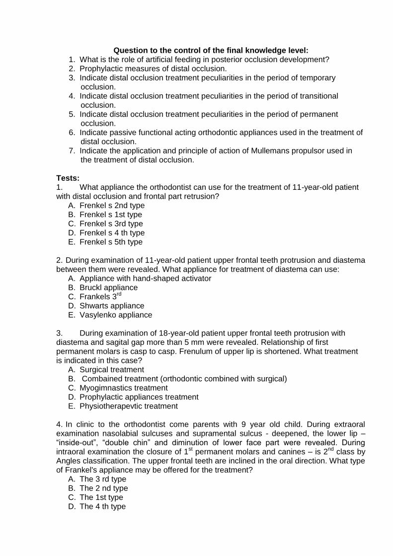
Question to the control of the final knowledge level:
1. What is the role of artificial feeding in posterior occlusion development? 2. Prophylactic measures of distal occlusion. 3. Indicate distal occlusion treatment peculiarities in the period of temporary
occlusion. 4. Indicate distal occlusion treatment peculiarities in the period of transitional
occlusion. 5. Indicate distal occlusion treatment peculiarities in the period of permanent
occlusion. 6. Indicate passive functional acting orthodontic appliances used in the treatment of
distal occlusion. 7. Indicate the application and principle of action of Mullemans propulsor used in
the treatment of distal occlusion. Tests: 1. What appliance the orthodontist can use for the treatment of 11-year-old patient with distal occlusion and frontal part retrusion?
A. Frenkel s 2nd type B. Frenkel s 1st type C. Frenkel s 3rd type D. Frenkel s 4 th type E. Frenkel s 5th type
2. During examination of 11-year-old patient upper frontal teeth protrusion and diastema between them were revealed. What appliance for treatment of diastema can use:
A. Appliance with hand-shaped activator B. Bruckl appliance C. Frankels 3rd D. Shwarts appliance E. Vasylenko appliance
3. During examination of 18-year-old patient upper frontal teeth protrusion with diastema and sagital gap more than 5 mm were revealed. Relationship of first permanent molars is casp to casp. Frenulum of upper lip is shortened. What treatment is indicated in this case?
A. Surgical treatment B. Combained treatment (orthodontic combined with surgical) C. Myogimnastics treatment D. Prophylactic appliances treatment E. Physiotherapevtic treatment
4. In clinic to the orthodontist come parents with 9 year old child. During extraoral examination nasolabial sulcuses and supramental sulcus - deepened, the lower lip – “inside-out”, “double chin” and diminution of lower face part were revealed. During intraoral examination the closure of 1st permanent molars and canines – is 2nd class by Angles classification. The upper frontal teeth are inclined in the oral direction. What type of Frankel's appliance may be offered for the treatment?
A. The 3 rd type B. The 2 nd type C. The 1st type D. The 4 th type

E. The 5 th type 5. Parents of 7-year-old patient come to the orthodontist for consultation. During clinical examination protrusion of frontal teeth in the upper jaw and distal position lower jaw (saggital gap – 6mm) were revealed. What method treatment is indicated in this case?
A. Complex treatment (myogymnastics and Frankel's 1st type appliance) B. Myogimnastics C. Osadchyis or Aisenberg appliances D. Muellemans propulsor E. Removable appliance with functionally directing action
Answers: 1 – A; 2 – A; 3 – B; 4 – B; 5 – A.
Practical Classes 8 Mesial occlusion. Etiology, pathogenesis, prophylaxis. Clinic and diagnostics. Scientific objectives of the lesson: To teach the students to understand the importance and to analyze the etiology, pathogenesis and prophylaxis of mesial occlusion. To teach the students the main principles of clinical and additional examination of patients with mesial occlusion.
The course of the lesson:
The term "mesial occlusion" was introduced into orthodontic practice by Lisher in 1926. In literature there are such terms for this pathology: progenia, forced occlusion, anterior type, inverse occlusion, articular progenia.
The progenia part among all dentognathic anomalies and deformations comprises from 1.9 to 18 %.
Parents apply to hospitals with their children concerning mesial occlusion already at young age more often than at other types of deformations. This happens because parents pay attention to the lower teeth covering the upper ones even when the pathology is not full-blown yet, and any other anomaly, for instance, posterior occlusion or deep overbite, is still imperceptible.
By Angle's classification (1889) mesial occlusion belongs to the 3rd class and is detected by lower jaw mesial dislocation. As a result 1st molars correlation violation is marked. The mesiobuccal tubercles of 6|6 teeth come upon the distal buccal tubercles of 6|6 teeth, and at more evident pathology the mesiobuccal tubercles of teeth come into the space between 76|67 teeth. Thus, the 2nd premolar articulates with the intertubercular space of the 1st molar, and at more evident mesial lower jaw dislocation even the lst premolar of the upper jaw articulates with it.
Lower jaw displacement may be both uni- and bilateral. At unilateral displacement median line correlation violation is observed. Unilateral displacement is mostly a consequence of the premature extraction of the 2nd temporary molar on the lower jaw on one side. Distally located teeth and teeth standing in front of the defect transfer to the side of the extracted tooth.
By Katz' classification (1940) mesial occlusion belongs to the 3rd group of anomalies and arises because of excessive functioning of the muscles protruding the lower jaw, or insufficient function of retractors group.
Functional pathology of this deformation consists in the following: articulate movements prevail in the child - opening and closing of the mouth, and gliding movements are absent, impossible because of the protruded lower jaw. This condition leads to rearrangement in the joint. Besides, a certain group of teeth does not participate in mastication - incisors are excluded from mastication. Their function is

taken by lateral teeth - premolars and molars, which consequently bear an increased load.
L.V. Ilyina-Markosian refers this pathology to sagittal occlusion anomalies, as plying the term "anterior occlusion", meaning lower jaw protrusion at usual location and singles out its three varieties:
- without lower jaw displacement; - general anterior occlusion; - frontal anterior occlusion; 1) with lower jaw displacement; 2) combined form. D.A. Kalvelis (1957), taking into account etiological factors and hereditary
character, refers progenia to sagittal occlusion anomalies, dividing it into true and false. V.Y. Kurliandskyi (1957) and A.I. Betelman (1956) based their classification оn the degree of jaw development. Thus, according to V.Y. Kurliandskyi, progenia referred to dental arches correlation anomalies and arises because of: - lower jaw overgrowth; - upper jaw underdevelopment.
According to A.I. Betelman's classification mesial occlusion belongs to sagittal plane anomalies and has such clinical forms:
upper jaw micrognathia; lower jaw macrognathia; lower jaw macrognathia and upper jaw micrognathia.
A.M. Schwarz (1969) and F.Y. Khoroshilkina (1976) studying lateral teleroengenograms of head detected dentoalveolar, gnathic, and combined forms of mesial occlusion.
A.S. Shcherbakov (1967) singles out dentoalveolar and skeletal forms. S.I. Doroshenko (1968) on the grounds of interpretation data of lateral head teleroentgenograms of patients with anterior dental arches correlation came to a correlation that it may be caused by the degree of jaws development, their form, location in the skull, lower jaw location in the TMJ, various correlations of bones in the skull. On the basis of these facts the author singles out the following forms of mesial occlusion: progenia as a consequence of lower jaw overgrowth:
its body and branch;
body;
branch;
frontal part of the jaw; progenia caused by upper jaw underdevelopment:
its body;
frontal part of the jaw; progenia caused by the anterior location of the lower jaw:
in the skull;
in the joint. In the WHO classification mesial occlusion is presented in the following chapter jaw size anomalies:
upper jaw micrognathia; lower jaw macrognathia;
anomalies of jaws location relative to the skull base: lower jaw prognathism; upper jaw retrognathia;
dental arches correlation anomalies: mesial occlusion. Mastication apparatus development is tightly connected with the development of
the whole organism. It begins from the fifth week of embryonal development (when the first teeth germs anlage takes place) and continues during many years after the child is

born until complete formation of permanent occlusion in mature age (18—20 years). If after child's birth the lower jaw alveolar process protrudes relative to the one of the upper jaw, this testifies to possible formation of mesial occlusion at temporary teeth eruption.
From the point of view of etiopathogenesis, all this period of time is expedient to be divided into three stages:
intrauterine, antenatal. During this time the fetus is under mother's organism protection, and its development disturbance considerably depends on the mother's state, and less - on environmental factors;
labor stage; postnatal, when the child is under the influence of environmental factors, adapts
to them at the expense of congenital and inherited properties. In the intrauterine period the fetus is under "mother's protectorate", in a peculiar
"microenvironment", but it can create a number of different conditions, negatively influencing the development of the embryo as a whole, and in particular — facial part development. This influence may be conditioned by different factors:
physical (fetus mechanical injury, radiation damage, thermal agents, vibration, etc.);
chemical (intake of different pharmacological, and especially hormonal preparations, alcoholic beverages, smoking during pregnancy);
biological (infectious diseases, genetic or hereditary diseases); social (conditions and way of life and work of the pregnant woman, diet in this
period). These agents, influencing the organism, leave a certain trace, the so-called
"phylogenetic background", which later declares itself in ontogenesis as congenital dento- gnathic anomalies.
Enumerated factors influence leads to specific and nonspecific diseases of the mother and father, which can be communicated from generation to generation by a dominant or a recessive character as a genetic disease of the whole organism or only of the dentognathic apparatus. Also progenia and upper jaw underdevelopment may be caused by the form, size and function of the tongue in the period of oral cavity embryonal development. The tongue, mainly pressing on the anterior part of the lower jaw, causes progenia, and its belated deepening onto the bottom of the oral cavity causes narrowing of the intermaxillary bone and upper jaw alveolar processes.
Besides, mesial occlusion may be caused by water-salt and vitamin metabolism disorders, early pregnancy toxemia. It has been established that pregnancy and labor pathologies frequency and progenia frequency are in direct proportion. Labor stage: progenia is more often (33 %) observed in children, who were born at transverse lie, breech presentation or footling presentation, after prolonged labor with a long anhydrous period, augmentation of labor, or operative delivery.
Postnatal period: the most critical for a child are the first months, later on - two first years of life. In the first period the child is under the influence of the environment, and it is the time when permanent occlusion foundation is laid.
Mesial occlusion may be caused by congenital peculiarities of the facial skeleton bone structure, especially of the lower jaw, intermaxillary bone underdevelopment, calcium metabolism disorder because of rickets or other diseases, partial or multiple adentia in the region of the upper jaw, multiple retention of the upper teeth or their premature loss, supplemental teeth on the lower jaw, late transitional dentition.
The shortened or irregularly attached tongue frenulum exerts constant pressure on the frontal part of the lower jaw, which leads to progenia. This is also promoted by macroglossia.

Palatopharyngeal tonsil hypertrophy leads to the reduction of the opening for air flow passage. Pressure is observed in the region of the epiglottis because of lingual tonsil enlargement, and the child, to ease breathing, instinctively protrudes the lower jaw, abducting the root of tongue together with the enlarged tonsil. Such mouth breathing causes mesial occlusion also because the habit of protruding the lower jaw appears.
Irregular swallowing (infantile type), irregular tongue location in the oral cavity during speaking and in quiescence, uneven wearing down of temporary teeth tubercles at the beginning of transitional dentition occlusion, especially of the lower canine teeth, uneven transitional dentition on both jaws - all these are etiological agents of progenia.
Individual lower incisors torsion leads to dental arches closure disorder, causes lower jaw protrusion. Upper jaw underdevelopment in the frontal part because of chronic inflammatory processes, new formations, surgical interventions on the jaws, endocrine disturbances, hypophysis hyperfunctioning leads to anterior occlusion formation.
Pernicious habits are very important: sucking of the upper lip, tongue, fingers, and different objects, sleeping on a high pillow, putting a palm or a fist under the chin in sitting position.
There are differentiated physiological and pathological types of progenia. Physiological progenia is characterised by multiple contacts between the dental
arches both in the front and lateral parts. It is viewed as an anatomic variant, which does not require any orthodontic treatment.
At pathologic progenia contacts between teeth are violated. There occur morphological, functional, and esthetic changes of the dentognathic apparatus, which require orthodontist's intervention.
Most authors differentiate two main progenia forms: true and false. L..V. Illyina-Markosian divides false progenia into two forms: anterior false progenia; forced occlusion. Their etiology, pathogenesis, functional and morphological disturbances, and
treatment differ. Some authors (L.V. Illyina-Markosian, D.A. Kalvelis) view false progenia as inverse overbite of individual upper frontal teeth at preserved correct correlation of both dental arches along the full length. A.I. Betelman, Y.M. Aleksan- drova, A.D. Mukhina refused from this term and classify false progenia as upper frontal teeth palatine position.
Forced occlusion is a kind of false progenia and develops as a result of the habit of protruding the lower jaw. This form of false progenia is also called articular.
Mesial occlusion has characteristic facial and intraoral features. The main facial feature is lower jaw protrusion. At external examination, in cases of lower jaw enlargement the disturbed harmony of face profile attracts attention: the chin and upper lip protrude considerably, at that the upper lip somewhat falls back relative to the lower one, the subnasal fold is deep, the lower lip red border is wide. At deep overbite the lower part of face is not infrequently shortened, as a result of which the lower lip is thickened. At increased lower jaw angles and open bite the lower part of face is elongated, the lips close tensely, the oral fissure not infrequently gapes. If mesial occlusion combines with forward lower jaw shift, facial signs of disturbances are fullblown.
Oral cavity examination shows that the lower jaw is located in front of the upper one, its dental arch is wider.
The closure of the 1SI permanent molars and canine teeth by 3rd Angle's type may be by 1/2 size of the 1st permanent molar tubercle, by one tubercle, by 1/2 of the Ist
permanent molar crown and more.

Frontal teeth correlation may vary: in some cases the labial surface of the upper incisors touches the lower incisors lingual surfaces, in other cases there is an inverse sagittal gap between the frontal teeth by 3 mm and more. Inverse overbite depth may be minimal, moderate, or deep.
Mesial occlusion is more often complicated with upper jaw narrowing, which causes lower lateral teeth prevailing over the upper ones. The upper jaw may be flattened in the frontal part. Uni- or bilateral crossed relation is observed in the lateral sarts.
The upper frontal teeth as a result of microgenia are located with torsions, transfer vestibularly, there is often observed frontal teeth congestion orally. The lower incisors sometimes deviate vestibularly, as a result of which diastemas and tremas form between them, or they press the upper incisors, increasing their palatine inclination.
At a most evident anomaly the lower jaw as though absorbs the upper one. The contact in the region of lateral and frontal teeth is violated, only the gliding of the over teeth lingual surface on the upper teeth buccal surfaces takes place.
Functional disorders are also very important at mesial occlusion. Face form is isolated. At the absence of occlusive contact between incisors food biting becomes impossible. Because of the forward shift of the whole lower jaw dental arch and molars correlation violation the general mastication area decreases. Tubercular closure, which forms between the masticatory teeth, hampers food grinding.
Sometimes, because of lower jaw protrusion, functional disorders, conditioned by articular heads location in the glenoid fossae, there appears pain in the joints, runch, clicking.
The speech of patients with mesial occlusion is violated, lisping appears. Dense location of the lower frontal teeth combines with dental tartar deposit, precervical caries, gingivitis. For the differential diagnostics of dentognathic and gnathic forms of mesial occlusion the clinical functional test by L. Ilyina-Markosian is used: the patient's face form is evaluated in profile at usual occlusion (the symptom of "capricious face") and if the patient can shift the lower jaw backwards to the maximum till the marginal incisors closure, and at that 1st permanent molars correlation becomes characteristic of the neutral occlussion, the dentoalveolar form of mesial occlusion with lower jaw protrusion is diagnosed. In another case mesial occlusion is caused by the difference in the dentognathic arches and/or jaws sizes. If jaw dislocation is present, face expression improves after the jaw is set in correct position.
Question to the control of the final knowledge level:
1. Enumerate the main forms of mesial occlusion according to existing classifications. 2. What is progenia frequency and spread? 3. Enumerate the main etiological agents and their role in mesial occlusion appearance. 4. Indicate pernicious habits that can lead to mesial occlusion development? 5. How does the dysfunction of breathing and swallowing cause mesial occlusion? 6. The prophylactic measures role in mesial occlusion prevention. 7. What are the facial signs of mesial occlusion? 8. What are the intraoral signs of mesial occlusion? 9. What dentoalveolar anomalies are accompaning mesial occlusion? 10. Indicate clinical functional test for mesial occlusion/ Tests: 1. Progenic bite in Betelman’ classification termed as:
A. Distal bite B. Transversal bite C. Sagittal bite

D. Mesial bite E. There is no correct answer
2. The main facial features of the mesial bite:
A. All answers are correct B. Deep nose-labial folds C. Upper lip falls back relative to the lower one D. Straight or protruding chin E. Increased mandible angle
3. For the differential diagnostic of dentoalveolar and gnathic forms of mesial bite is used:
A. Lateral cephalometry B. Direct cephalometry C. Occlusial radiography D. Contact intraoral radiography E. Orthopantomograms
4. Choose the main intraoral characteristics of mesial bite:
A. The presence of a sagittal space B. Retrusion of the upper frontal teeth C. Mesial correlation of the canines D. Mesial correlation of the first permanent molars or second temporary molars E. All answers are correct
5. During clinical examination of orthodontics patient the lower incisors cover the upper 1/2 of the crown height. The upper canine is projected on the first lower temporary molar. There are spaces between the lower frontal teeth. Identify possible etiological factor of this disease:
A. Biting the lower lip B. Thumb sucking C. Parafunction of the tongue D. Adenoidectomy E. Sleeping without pillow
Answers: 1 – D; 2 – A; 3 – A; 4 – E; 5 – C.
Practical Class 9
Complex treatment of the mesial occlusion Scientific objectives of the lesson: To acquaint the students with сomplex treatment of the mesial occlusion. The course of the lesson:
Mesial occlusion treatment largely depends on etiological agents and the possibility of their elimination, and also on the degree of morphological and functional disorders manifestation, the complicacy of their elimination in different age periods. In the period of temporary occlusion much attention is paid to the normalization of alveolar processes and jaws growth during frontal teeth coming out. If upper jaw underdevelopment and lower jaw overgrowth are observed in an infant, it is necessary to massage the alveolar process of the upper jaw in the anterior part for growth stimulation. At the shortened tongue frenulum a surgical intervention (frenulum plastic surgery) must be carried out. If the child is fed artificially, attention is to be paid to feeding correctness. One must watch upper incisors eruption and their position in

occlusion with the lower ones. In the period of temporary occlusion the measures of fighting against the pathology come to pernicious habits elimination and normalization of the breathing, swallowing, speech, and mastication functions. To break the child of the habit to breath orally, suck fingers, tongue, different objects, to normalize nasal breathing standard vestibular Hinz' and Schonher's plates may be used. To limit the following lower jaw growth and promote upper jaw physiological development removable or fixed gum shields, disjoining occlusion, may be offered. The upper jaw, brought out from the block at occlusion disconnection, has a possibility of normal development. To inhibit lower jaw growth in the sagittal direction a chin sling is used, fixed with a rubber recoil to a head cap. In the temporary occlusion period there often takes place lower jaw forced protrusive position, connected with the lower canine teeth, which bear against the opposing teeth with their cutting edges and fix the lower jaw in the forced position. Selective regrinding of the canine teeth tubercles, and sometimes of the incisors sculpri, creates conditions for free lower jaw movements in the sagittal plane and normal upper jaw growth. Remedial gymnastics is used to train the orbicular muscle of mouth and to attain lips closure and nasal breathing. With this purpose lip activators, and also H. Dass' activator-apparatus (1961) are used. In the initial period of transitional dentition, at the age of 5.5 years, mesial occlusion is treated with the same appliances as in the temporary occlusion period. The best time for treatment is the time before the eruption of the 1st permanent molars and canine teeth. The presence of permanent incisors with deep overbite allows using devices with inclined planes — Schwarz' gum shields, Katz' directing crowns. If overbite is insignificant, inclined planes are not indicated, because lateral teeth disconnection will promote the growth of their alveolar processes, which might lead to open bite development. In such cases, i.e. at insignificant overbite, it is expedient to use a removable plate with lateral teeth mastication surfaces covering, and at that permanent molars are to be covered. If there are diastemas in the region of lower frontal teeth, such plate must have an elastic wire arch, located from the labial side of the teeth. At deep or average reverse overbite and insignificant reverse sagittal gap B.N. Bynin's gum shield may be used. At evident upper jaw underdevelopment with insignificant overbite it is indicated to use an orthodontic appliance on the upper jaw with lateral teeth mastication surfaces covering, with elastic activators for the frontal group of teeth protrusion. At 6—9 years it is recommended to use Frankel's functionally acting device of the 3rd type, Baiters' bionator of the 3rd type, Bimmler's elastic occlusion former of the С type. Andresen—Haupl's activator for the treatment of mesial occlusion is effective at insignificant overbite (to 1.5 mm) or at the combination of mesial occlusion with open bite, which combines with the pernicious habit of tongue sucking or putting it between the dental arches in the region of the defect. The device consists of two basal plates for the lower and upper jaws, joined with plastic in interocclusal space. When detecting the constructive occlusion the frontal teeth are set in marginal closure, if it is possible, or their cutting edges are set at one horizontal level. The vestibular arch is located in the region of the lower frontal teeth. If there are diastemas between them, it is activated in the process of treatment, and the plastic, adjacent to the lingual surface of these teeth, is sawn down. To enhance upper incisors protrusion a screw is added to the plate, the saw cut is performed sector-wise.
In the period of second dentition, if there are diastemas in the region of frontal teeth, it is recommended to use gum shields to disconnect occlusion; hooks are welded into gum shields on the vestibular surface, the hooks are opened distally, and rubber recoil is put onto them.
Mesial occlusion treatment in the permanent occlusion period is long-term, requires complex treatment, not infrequently combining with surgical methods. Upper micrognathia treatment is aimed at upper jaw enlargement. This is achieved with the help of orthodontic appliances with activators and dilating devices (screws, wire
dilators), which promote upper frontal teeth protrusion, and lateral jaw parts dilation. Besides, this form of mesial occlusion also has clinical varieties, which depend on the degree of upper jaw underdevelopment, individual teeth quantity and position, overbite depth. The choice of treatment method and orthodontic appliances construction must be individual.
At the big lower and normal upper jaws the treatment must consist in lower jaw downsizing, which can be achieved by means of frontal teeth shifting backwards at the expense of spaces between them. If there are no enough spaces, the treatment is conducted with teeth extraction (more often — premolars), after what the frontal group of teeth is transferred with the help of removable orthodontic appliances. When a canine tooth is transferred distally the 6th and 7th teeth are usually taken as support, if it is possible, for the place of the extracted tooth not to be replaced by the lateral teeth, displaced forward. Fixed appliances are used — A.I. Pozdniakova's, Angle's with intermaxillary recoils. In case of teeth extraction it is better to use bracket system for the treatment. If the lower jaw is considerably big, orthodontic treatment alone will not produce the desired effect. The treatment is to be combined (surgical-orthodontic), for which purpose the alveolar process or the lower jaw body is cut partially, with subsequent bone fragments set in the position eliminating the deformation. At that, orthodontic preparation precedes the surgical procedure, and the orthodontist finishes the treatment.
The most widespread is the clinical case, when mesial occlusion arises as a result of upper jaw underdevelopment and lower jaw overgrowth. Treatment measures in this case are directed at upper jaw enlargement and lower jaw downsizing.
Orthodontic treatment of mesial occlusion may be combined with compact osteotomy on the upper jaw alveolar process. This intervention allows more corpus transfer of the upper frontal teeth in the vestibular direction and in such a way the change of the alveolar process profile form.
To improve orthodontic treatment results physiotherapeutic methods are used (vacuum therapy, vibration influence, MRT).
In the permanent period of occlusion preference should be given to fixed mechanically acting appliances in complex with facial masks, with the help of which it is possible to obtain constant results of treatment. Question to the control of the final knowledge level: 1. Mesial occlusion treatment peculiarities infancy. 2. Mesial occlusion treatment peculiarities the period of transitional dentition. 3. Mesial occlusion treatment peculiarities the period of permanent occlusion. 4. Indications to surgical procedures in complex treatment of mesial occlusion. 5. Indications to the application of Schwa and Bynin's gum shields. 6. Construction peculiarities of Frankls functional regulator of the 3rd type and indications to its application. 7. What is the essence of the functional method of treatment, offered by Frakel? Tests: 1. What preventive appliances are used for the training of circumoral muscle?
A. Dass’ activator B. Lips equilibrator C. Brukl’s appliance D. Vestibular shield E. All answers are correct
2. What changes in the size of the upper jaw orthodontist can find during clinical examination of patient with mesial bite:
A. Increasing of sagittal sizes B. Decreasing of the sagittal sizes C. Increasing of transversal sizes D. Decreasing of the transversal sizes E. There is no correct answer
3. For treatment of mesial occlusion orthodontist used appliance of Brukl’s. What of the following elements Brukl’s appliance includes:
A. Metal inclined bite plane B. Plastic lateral inclined bite plane C. Plastic frontal inclined bite plane D. Metal frontal bite plane E. There is no correct answer
4. What method can be used for treatment of gnathic forms of malocclusion?
A. Prosthetic B. Instrumental C. Biological D. Surgical and instrumental E. Biological and prosthetic
5. On visit to the orthodontist come parents with 8 year-old child. Objectively: the face is elongated by increasing the lower part, flattened upper lip, chin straight with smooth mental folds. Reverse incisor overlapping by 1/3 the upper incisor. Canines and first permanent molars are in mesial relation. The mother has a similar clinical situation. What design of the orthodontic appliance can use in this clinical case:
A. Bynin’s kappa B. Frenkel’s Regulator type III C. Katz plate D. Brukl’s appliance E. Plate with lateral flat bite planes and three-dimensional screw on upper jaw
Answers: 1 – A; 2 – B; 3 – C; 4 – D; 5 – E.
Practical Class 10 Vertical anomalies of occlusion. Deep bite. Etiology, pathogenesis, prophylaxis.
Clinic and diagnostics of deep bite.
Scientific objectives of the lesson: To acquaint the students with etiology, pathogenesis, clinical features, diagnostics of deep bite.
The course of the lesson:
Deep bite is referred to vertical occlusion anomalies. Its frequency comprises from 20 to 38%. Deep bite is characterized by different terms: "traumatic occlusion", "deep frontal or incisors overbite".
The term "descending occlusion" reflects a dynamic progressive process, at which the incisors of one jaw lose support on the dental tubercles of the teeth antagonists and glide to the gingival margin. The term "traumatic occlusion" testifies to the fact that the frontal teeth of one jaw at dental arches closure bear against the mucous tunic of the gums or alveolar process of the opposite side.

Deep bite is such a form of closure, at which the cutting edges of the lower frontal teeth are settled on the clivus of the dental tubercles of similar teeth. If the lower frontal teeth get over the tuberculum dentale of the upper teeth, deep bite forms.
Deep bite is the initial form of pathological occlusion formation. According to Angle's classification, deep bite is referred to the 1st class – "deviation from the norm in the frontal part while jaws location is in mesiodistal harmony".
According to Betelrnan's occlusions classification deep bite is a pathological occlusion belonging to vertical anomalies.
D.A. Kalvelis viewed deep bite as: - covering (opisthognathic); - combined with prognathism (roof-shaped). F.Y. Khoroshilkina divides deep bite into three degrees of deep overbite, which are detected by the height of the central incisors crowns: - the 1st – from 1/3 to 2/3 of their height; - the 2nd – from 2/3 to 3/3; - the 3rd – more than crowns height. Besides, the author evaluates the three degrees of deep overbite in millimeters: - the 1st – less than 5 mm; - the 2nd – from 6 to 9 mm; - the 3rd – more than 9 mm. L.V. Ilyina-Markosian singles out: A. deep bite without lower jaw displacement; B. deep bite with lower jaw displacement; С. combined type. In group A, in its turn, there are differentiated general deep bite and frontal deep overbite. General deep overbite is characterized by dento-gnathic lengthening in the anterior part of both jaws. There is dento-gnathic shortening in the region of lateral teeth. Frontal deep overbite differs from general deep overbite by the absence of changes in the region of lateral teeth.
According to the WHO classification deep bite is referred to dental arches correlation anomalies: А) excessive overbite; В) excessive covering occlusion.
Deep bite formation is conditioned by: - heredity; - different prenatal pathologies (toxicosis of pregnancy, injuries, infectious diseases during pregnancy, avitaminosis, etc.); - infancy diseases; - incorrect artificial feeding, which leads to the predominance of the muscles amplifying the lower jaw over the muscles protruding the lower jaw; - considerable break in the terms of upper and lower incisors coming out; - inter-maxillary bones overgrowth; - carious or no carious affection of the lateral teeth hard tissues, including their inhomogeneous wearing out; - premature loss of temporary molars, 1st permanent molars or 2nd lateral teeth; - pernicious habits – sucking and biting fingers, different objects provoke frontal teeth deviation, violation of proximal contacts and contacts with opposing teeth, which leads to the decrease of occlusion height, lst permanent molars establishment at irregular occlusion level and alveolar processes underdevelopment in the lateral parts. Violation of contacts between frontal teeth conditions dentognathic lengthening in this part; - changes of the frontal teeth location, their support loss, which causes dento-gnathic lengthening; - the same results may be caused by one dental arch enlargement at the presence of a supplemental tooth, diastems, temporary retained molars, at individual macrodontia or

dental arch decrease at retention or adentia of individual teeth (more often of the 2nd premolars), microdentia on one jaw, violation of the sequence of upper and lower teeth transitional dentition or the terms of permanent teeth eruption.
Vertical alveolar processes growth violation causes other pathologic occlusions (mesial, distal, cross), which complicate deep overbite and lead to a combined pathology.
Deep bite clinical presentation depends on its combination with or mesial occlusion, cross bite, dental arches anomalies, and individual teeth anomalies.
Facial signs may be in the form of supramental sulcus deepening, lower lip thickening and concomitant violations, characteristic of distal or mesial occlusion.
In the oral cavity there are observed changes in the from of the increase of the upper frontal teeth covering the lower ones by more than 1/3 at the absence of cutting-tubercular contact, dental arches flattening, the alveolar processes in the region of frontal teeth are high and overgrown, and in the lateral – low. At most evident violations (deep traumatic overbite) the lower frontal teeth bear with their cutting edges against the hard palate mucosa, repeating its form; sometimes the upper frontal teeth injure small interdental gingival papillae from the vestibular side of lower teeth and promote their desquamation.
At posterior occlusion, combined with upper frontal teeth protrusion, lower incisors not infrequently injure the mucous tunic of the palate, more rarely they do not touch it. At posterior occlusion, combined with upper frontal teeth retrusion, the dental arches are usually shortened; deep overbite at such a violation is called blocking, hampering lower jaw growth. Lower jaw protrusion becomes limited, which shows on the mastication muscles function.
At mesial occlusion and inverse overbite the dental arches form depends on the development degree of jaws basis and alveolar processes, teeth location, lower jaw displacement. Overbite depth also depends on the value of the basal and gonial angles.
Functional violations at deep bite declare themselves in the decrease of mastication efficacy, frontal teeth periodontal overload, and not infrequently - mucous tunic injuring, which promotes appearance and development of periodontal diseases Mouth breathing, irregular swallowing and speech disturbance promote dental arche narrowing, upper teeth location strengthening and overbite deepening. At deep occlusion there is observed high tongue position, which causes palatine vault enlargemen (roof-shaped deep overbite). Interocclusal space between the frontat-and lateral teetl at the lower jaw being in quiescence (especially at bruxism in adults) is absent; in some cases at most evident Spee's curve the distance between the dental arches in quiescence reaches 9 mm (average norm is 2 mm), which testifies to considerable violation of the mastication muscles function.
F.Y. Khoroshilkina and L.P. Zubkova noted: if at a narrow face the total width of upper incisors crowns makes more than 33 mm, it may be the reason for upper dental arch oval increase and overbite deepening.
N.H. Snahina and co-authors analyzed the data of studying jaw models of 100 patients with marginal periodontal diseases, most of whom had deep overbite. The authors found out that the dental arches width in the region of the 1st upper premolars was less by 3.99 mm on average, lower - by 3.85 mm, in the region of the 1st upper permanent molars - by 4.77 mm, lower - by 3.93 mm. At incisors retrusion the length of the anterior part of the upper dental arch was less by 2.36 mm on average, lower - by 2.94 mm. In all the patients there was found the narrowing of upper dental arch apical basis by 4.61 mm on average, lower - by 4.87 mm.
Lower dental arch and its apical basis narrowing in patients with deep overbite should be taken into account when planning treatment. It is not sufficient to find out the dental arches width and their anterior parts length at deep overbite. The data of Bjork,

Schwarz, and Van der Linden concerning mesial dislocation of the lateral teeth during life prove the importance of measuring their sagittal dimensions. This promotes dental arches length decrease, may hamper the obtaining of constant results of treating deep overbite, influence the location of frontal teeth and dental arches form.
F.Y. Khoroshilkina noted that the center of the incisor papilla practically does not change its position in the process of upper jaw growth; therefore it may be used for the study of frontal and lateral teeth position in the sagittal direction.There was not found such a stable reference point in the region of the lower dental arch. To diagnose deep bite varieties there is studied the crown width of the upper and lower incisors and their age location (correct position, protrusion, retrusion), the evidence of the upper incisors dental tubercles, contacts between the frontal teeth, bilateral correlation of the canine teeth and 1st permanent molars in the sagittal direction at the dental arches, closed in normal occlusion, early destruction or loss of temporary and permanent lateral teeth, diminution or mesial inclination of the upper and lower teeth to the place of destroyed or extracted ones, the evidence of morphological and functional disorders by the method of Zibert-Malyhin and complications of eliminating them with the help of Malyhin-Bilyi's method.
To diagnose deep bite one must measure and take into account: 1. the mesiodistal dimensions of the crowns of upper (SI) and lower (Si) incisors, their total; 2. the correspondence of the totals of the mesiodistal dimensions of the upper and lower incisors crowns by Tonn's index (1.35 mm); 3.the overbite depth; 4. the size of the sagittal fissure between the upper and lower central incisors; 5. the length of the anterior part of dental arches by Korkhaus; 6. dental arches width by Pont (with Lider and Harth's corrections).
The diagnose is put on the basis of clinical examination, the study of diagnostic models of jaws and measuring them, anthropometric study of face photographs of face and in profile, and also of lateral cephalometric of head (according to Schwarz, at deep overbite there is observed basal angle decrease, MT1 plane position is almost horizontal, the decrease of jaw height in the region of incisors and increase in the region of molars, vertical position of the upper incisors crowns, deep glenoid fossa of the TMJ), the evaluation of orthopantomographic jaws investigation data.
Question to the control of the final knowledge level: 1. Enumerate the main etiolgical agents and their role in deep bite appearance. 2. Indicate pernicious habits that can lead t deep bite development 3. Define of "deep forms of the bite". 4. What various forms of deep bite do you know? 5. What facial features of deep bite do you know? 6. What intraoral features of deep bite do you know? 7. What methds of deep bite diagnostics do you know? Tests: 1. According to WHO classification deep bite is referred to:
A. Anomalies of jaw size B. Anomalies in the jaws location relative to the plane of the skull base C. Anomalies of the teeth D. Anomalies of the teeth size E. Anomalies of the dental arches correlation

2. The main risk factors of deep bite development are: A. Early removal of temporary or permanent molars B. Reduction of the mandibular branch C. Increasing of temporal muscles tone D. Heredity E. Heredity, early removal of temporary or permanent molars, reduction of the mandibular branch, increasing of temporal muscles tone
3. What morphological features can cause a deep bite?
A. Dento-alveolar elongation in frontal areas or dento-alveolar shortening in lateral areas of jaws B. Dento-alveolar shortening in the frontal areas of the upper jaws C. Dento-alveolar elongation in the lateral areas of the jaws D. Dento-alveolar shortening in the frontal areas and dento-alveolar elongation in the lateral areas of the jaws
E. Dento-alveolar shortening in the frontal areas of the lower jaws 4. A deep bite can be caused by such morphological features:
A. Dento-alveolar elongation in frontal areas or dento-alveolar shortening in lateral areas of jaws
B. Dento-alveolar shortening in the frontal areas of the upper jaws C. Dento-alveolar elongation in the lateral areas of the jaws D. Dento-alveolar shortening in the frontal areas and dento-alveolar elongation in
the lateral areas of the jaws E. Dento-alveolar shortening in the frontal areas of the lower jaws
5. Analysis of 11-year-old girl's jaw models revealed that occlusal plane of the frontal maxillary teeth was of concave form, its lateral parts were convex. Form of the alveolar process also represents deformation of dental arches. The upper jaw is of saddle-like form with abrupt narrowing in the region of premolar teeth. What type of bite is it?
A. Distal B. Open C. Deep D. Mesial E. Cross
Answers: 1 – E; 2 – E; 3 – A; 4 – A; 5 – C.
Practical Classes 11 Complex treatment of the deep bite
Scientific objectives of the lesson: To acquaint the students with сomplex treatment of the deep bite.
The course of the lesson:
Deep bite treatment is the most effective in the period of coming out of temporary teeth, the 1st permanent molars, temporary incisors replacement with permanent ones, 2nd permanent molars eruption.
The treatment of malocclusions in vertical plane it is advisable in stages of physiological occlusion height elevation.
There are singled out 4 stages of the physiological elevation of occlusion height: - the 1st falls on 2-2.5 years, i.e. the moment of all temporary teeth emption completion; - the 2nd is marked at the age of 6 years, i.e. the time of 1st permanent molars emption;

- the 3rd – 12-13 years, after the complete replacement of temporary teeth with permanent, due to the vertical growth of the alveolar process, full value eruption and regular reciprocal arrangement of second permanent molars and canines; - the 4th –18-25 years, i.e. as a result of the emption and regular articulation of the 3rd molars; if they are absent, occlusion height elevation occurs at the expense of dento-alveolar lengthening. At all stages, as occlusion height increases dynamically the frontal overbite depth decreases, and dentitions correlation becomes orthognathic.
The main task of treatment: elimination of the reasons hampering dentoalveolar lengthening in the region of
lateral teeth, disjoining them; obstruction of dentoalveolar lengthening in the region of frontal teeth; correction of the dental arches from, individual teeth and their groups position; normalization of the lower jaw position and jaw growth. Disorders are eliminated in different ways, by means of different methods taking
into account their reasons, the period of occlusion formation, its correspondence to the patient's age and sex.
In the period of milk occlusion it is recommended to habituate children to hard food mastication (raw vegetables, fruit, hard bread, etc.) in order to stimulate the normal development of jaws, alveolar processes and dental arches. In case of carious destruction of the temporary molars crowns they should be restored, which is achieved with the help of fillings, restorative crowns.
If children have pernicious habits (sucking fingers, lips, different objects, pullin cheeks into the oral cavity and biting them with lateral teeth), it is difficult to brea them of the habits. Vestibular plates are used for the purpose.
If the tongue frenulum is attached irregularly, frenulum plastic surgery is con ducted.
Correct tongue functioning prevents the development of dental arches and jaw disorders, promotes overbite depth normalization.
Prematurely lost temporary molars are subject to replacement with removable dentures with the purpose of deep bite prophylaxis. Occlusion may be heightened o: artificial lateral teeth, having provided the contact of the lower frontal teeth cutting edges with the upper jaw prosthesis biting platform. At the formation and development of sagittal occlusion anomalies a vestibular plate with a biting platform for incisors, used during sleep, should be recommended, and curative gymnastics for the function normalization of the muscles surrounding the dental arches and carriage improvement. Preorthodontic trainers usage may be started.
In the final period of temporary occlusion and in the early period of transitional dentition, i.e. from 5.5 to 9 years, active orthodontic treatment is to be begun. Lateral teeth disjoining in this period at 1st permanent molars eruption promotes dento-alveolar lengthening till the contact with the opposing teeth, in which connection overbite depth decreases. At deep overbite a removable device for the upper jaw with an inclined or biting plane for lower frontal teeth rest, clasps or other fixing appliances may be used. The thickening in its anterior part, which is to disjoin the lateral teeth, is by 2 mm higher than the rest position. The device is fixed with clasps, vestibular arches, and other appliances. Functionally directing elements for deep bite correction 1. Flat anterior bite planet
A thickening of the acrylic base plate behind till the canine such that the lower anterior teeth extending usually till the canine such that the lower anterior may bite on it;
The extension should be flat and parallel to the occlusal plane.

The bite plane should high enough to disocclude the posterior teeth by about 2-3mm.
It reduces deep bite by separating the molars allowing them to over-erupt and so decreasing the over bite.
It can be used to free cuspal interferences to correct posterior crossbites of single teeth.
2. Inclined anterior bite plane It is modification of the anterior bite plane used in case where there is severe
retroclination of lower anterior teeth with increased overjet. The plane is inclined so that the lower anterior is proclined as it contacts the
slopping bite plane. The lower incisions engage the bite plane when the patient closes the mouth and mandible is guided to be held in the forward position;
It correct deep bite and increased overjet by allowing molars to over-erupt and proclining lower incisors.
For the elimination of lower jaw displacement forward or to the side, the inclined plane is made not smooth, but with imprints of the opposite incisors' sculpri and canine teeth tubercles — the biting platform.
Bimler's occlusion former and function regulators FR—I and FR-II are used.
For the treatment of distal deep overbite Andresen-Haupl's activator, Muelleman's propulsor, Baiters' bionator, open activator, etc. are administered. Orthodontic appliances are used for the purpose of using them both during sleep and during the day. They also include Katz' biting plate.
Propulsor of Moleman. Propulsor (pusher) Moleman (Muhleman) refers to a functionally active vestibular-oral. It is used to treat deep distal occlusion with protrusion of upper anterior teeth and the presence of between three and slight narrowing of the upper jaw. Propulsor Moleman holds the lower jaw in the extended position (to neutral ratio of the first permanent molars) and disjoint the occlusion in the area of lateral teeth. Valid and greater extension of the lower jaw position forward (over), that enhances the action of the muscles, which shift the jaw posteriorly. The pressure transmitted through the device on the lower jaw, facilitate its growth, and the effect on the upper front teeth – contributing to their retrusion. The vestibular shield pushes the cheeks, isolates the pressure on the lateral areas of the maxilla. Due to the separation of the lateral teeth observed elongation, which helps reduce the depth of incisal overlap. In case of early loss of deciduous molars, the dentition defects replace with a plastic, which prevents the displacement of the teeth in the direction of the defect. Propulsor prevents mouth breathing, helps to wean the child from bad habits sucking tongue, lips, fingers or other objects. Andresen–Haupl's activator. During deep overbite treatment attention is paid to the tongue position, because of space reduction in the oral cavity, usually takes a low, posterior position and lies flat between the lateral teeth. As a result of achieving lower jaw displacement with the help of removable dentures with the purpose of deep overbite prophylaxis, the tongue rises up to the palate, its form changes, as the agent causing its irregular position is eliminated. Lateral shields of function regulator hamper cheeks suction and drawing in. At deep overbite the frontward growth of the dental arch apical basis and vertical growth of the lateral teeth are hampered. Labial bamper stimulate lower jaw growth: lateral teeth disjoining create conditions for alveolar lengthening.
At upper and lower dental arches underdevelopment in the anterior part the function regulator FR-I is supplemented with labial bamper for drawing the upper and lower lips aside. In the final treatment phase the middle part of the lingual arch is pressed to the dental tubercles of the lower frontal teeth to prevent their dento-alveolar lengthening. In case of upper incisors retrusion there is conducted plastic surgery for

the upper jaw with a screw, resting against the palatine surface of vestibularly located teeth; sector saw cut, biting platform and numerous clasps or a plate with inhibitory springs, palatine arches.
For the treatment of mesial deep overbite orthodontic appliances are chosen depending on pathology varieties.
In the final period of transitional dentition and initial period of permanent occlusion, i.e. at the age of 9—12 years, there is used the physiological elevation of occlusion at the establishment of premolars, canine teeth and 2nd permanent molars. There are used the same orthodontic appliances as in the previous period, and also fixed orthodontic appliances, for example, Angle's device, bracket system.
Except for these appliances, it is possible to use Katz' directing crowns for the upper central or lateral incisors with a cast inclined plane, soldered to the palatine surface of crowns, and vestibularly lying tangent to the neighboring frontal teeth, and also (by indications) Huliayeva's apparatus.
In the period of permanent occlusion, at more than 12 years old, to eliminate the most evident dentognathic anomalies, combined with deep overbite, it is recommended to use intraoral vestibular arch orthodontic appliances with intermaxillary recoil (Angle's, Johnson's, Begg's devices, bracket systems, etc.). These appliances are used, as in the previous period, in combination with a removable plate for the upper jaw with a biting platform. For dentoalveolar lengthening rings with hooks, buttons or other devices for vertical interdental recoil applying are fixed on the transferred premolars and molars. With the рифове of dentoalveolar lengthening in the region of the upper lateral teeth it is possible to use an extracoronal vestibular arch in combination with oblique extraoral recoil. Low location of hooks on the hat in comparison with the hooks on the ends of the facial arch or the change of the inclination of the facial arch ends in comparison with the extracoronal arch ends increases vertical teeth transfer.
For dentoalveolar shortening in the anterior part of the upper dental arch and dentognathic lengthening in its lateral parts Luri's apparatus modifications are used. The device is made of orthodontic wire 1—1.2 mm in diameter. Rod-like bends are made behind the canine teeth by means of raising the middle part to the level of the incisors roots middle. Then a small vestibular U-arch is bent of steel wire 0.7—0.8 mm in diameter. Its middle part is adjacent to incisors, springs are made on ascending parts. Small arch ends are welded to the arch at the level of the lateral incisors distal surface. Rings with keyhole arrangements are fixed on the 1st premolars for better fixation of the extracoronal arch. The small arch is used either for dentoalveolar shortening or for the shortening in the region of incisors.
During the treatment of adults myotatic reflexes should be rebuilt, bruxism and other parafunctions are to be eliminated; one must watch the regular closure of dental arches at different types of occlusion, selectively regrind individual teeth tubercles. Such measures are resorted to as preliminary ones before orthodontic treatment and dentognathic prosthetics.
If some teeth are absent, especially lateral ones, dental prosthetics is obligatory after orthodontic treatment.
In the process of treating deep overbite one tries to obtain numerous contacts between the dental arches. If the treatment is begun in the period of transitional dentition or temporary occlusion, the child is to be observed by an orthodontist till the end of permanent occlusion formation. Unrepaired functional disorders promote anomaly recurrence.
Orthodontic treatment duration depends on the period of occlusion formation, degree of deep overbite manifestation and concomitant diseases, deformations of teeth, dental arches, occlusion, and general disturbances of organism development.

Treatment prognosis is favorable if the treatment is conducted in the initial period of transitional dentition or permanent occlusion, if in the process of treatment not only morphological, but also functional disorders are eliminated and deep overbite is not a family feature.
Retention duration after orthodontic treatment is finished depending on the period of occlusion formation, application of functional or mechanical treatment methods, presence of unrepaired functional disorders and obtained treatment results. After achieving numerous contacts between dental arches with the help of an activator, bionator, function regulator, and other functionally acting devices no retention period is needed.
If the treatment was conducted with mechanically acting devices and function disorders are not completely eliminated, retention devices are to be used, being chosen subject to possible teeth displacement. Devices application duration is individual, on average it equals the period of active orthodontic treatment. Question to the control of the final knowledge level: 1. Name the periods of the physiology height bite increasing. 2. What are the features of treatment of deep bite in the temporary dentition. 3. What are the features of treatment of deep bite in a mixed dentition. 4. Features of treatment of deep bite in a permanent bite. 5. The main elements of appliances which apply for treatment of different
forms of deep bite. Tests: 1. The goal of treatment of different forms of deep bite is?
A. Correction of shape and size of dental arches, bite height correction, degree of the overlap, normalization of the function of masticatory muscles and TMJ, correction of shape and size of dental arches
B. Bite height correction C. Degree of the overlap D. Normalization of the function of masticatory muscles and TMJ E. Correction of shape and size of dental arches
2. A 9 year-old boy complains of bad chewing of food. In anamnesis 3 years ago was extracted all milk molars in the mandible. Objectively: the lower third of the face is shortened, face is asymmetrical, chin is retruded. Intraorally: upper frontal teeth covered lower frontal teeth more than ½ height of crowns. Choose the correct appliance for the treatment of deep bite during the change of teeth?
A. Blue preorthodontic trainer B. Andresen-Haupl's activator C. Frenkel's appliance D. Hinz's plate E. Schwarz’ gum shields
3. During clinical examination of a 5 year-old boy the deep bite was revealed. Orthodontist recommended mio gymnastic exercises. What exercise will be train muscles- elevators of the lower jaw?
A. Click the tongue B. The lower jaw slowly move forward to the incisal overlap C. Count with the tongue teeth without opening the jaws D. There is no correct answer E. Play on the wind instruments, sing

4. What is the tactics in treating deep bite in period of temporary occlusion?
A. Oral cavity sanitation, hard food chewing, bad habits elimination, functional appliances
B. Bad habits elimination, mechanically acting devices C. Does not require treatment at this stage D. Oral cavity sanitation , soft food function devices E. Oral cavity sanitation, soft food, mechanically acting devices
5. Parents with 9 year-old child come to the orthodontist. During clinical examination the deep bite combined with mesial occlusion was revealed. What appliance for the treatment of mesial deep bite can be used?
A. Appliance with loops Rudolph B. Vestibular shield C. Physiotherapeutic treatment D. Miotrainer E. Brukle's appliance
Answers: 1 – A; 2 – B; 3 – B; 4 – A; 5 – E.
Practical Classes 12 Open bite. Etiology, pathogenesis, prophylaxis. Clinic and diagnostics of open
bite.
Scientific objectives of the lesson: To acquaint the students with etiology, pathogenesis, clinical features and diagnostics of open bite.
The course of the lesson: Open bite is referred to vertical anomalies. According to statistics, from 1.7 to 5.3
% patients have this anomaly. A.D. Mukhina and Z.F. Vasylevska believe that open bite is more often met in older age than in children with temporary occlusion. Open bite is characterized by the presents of the gap between the frontal or lateral teeth. The gap could be symmetric (between the homonymous teeth) or asymmetric, one-sided or two-side (between the lateral teeth). Open bite is met in many classifications. Thus, according to Angle's classification, open bite is referred to the 1st class, when "occlusion key" is preserved, jaws are located in "mesiodistal harmony", and the pathology is located in the frontal part, and also in individual teeth anomalies: infraocclusion of frontal teeth and supraocclusion of lateral teeth.
D.A. Kalvelis viewed open bite as a vertical anomaly and divided it into: - true open bite (rachitic): because of bony tissue disease muscles development advances the process of mineralization and resists self-regulation; - traumatic (caused by pernicious habits).
By Bateman’s classification this form of the bite is named the open bite with the underdevelopment of the elevator muscle of lower jaw and the orbicular muscle of the mouth.
F.Y. Khoroshilkina classifies open bite by the size of the vertical gap: • the 1st stage - less than 5 mm; • the 2nd stage - less than 9 mm; • the 3rd stage - more than 9 mm. The author also differentiates gnathic and dentoalveolar open bite by cephalometric image.
By WHO’ classification the open bite is considered to the anomalies of the dental arches.

Open bite might arise due to various etiologocal agents and be consequence of many functional and morphological violations of the dentognathic apparatus. The main of them are: 1. Heredity. 2. Children diseases with the impairment jaw-bone mineralization (rachitis). 3. The bad habits (finger sucking, suck the (baby's) dummy, lips, cheek and the outside things. 4. Oral breathing as a result of bad habit or adenoid vegetations. 5. Pathologic function of tongue (passing between the lips). 6. Congenital cleft of lip, alveolar process and palate, which caused the underdevelopment of the frontal part of the upper jaw. 7. Excess vertical growth of the lower jaw. 8. Atypical position of the dental germs of the second teeth 9. Macrooglossia. 10. Palatopharyngeal tonsils enlargement promotes forward tongue displacement, its irregular location. 11. Swallowing dysfunction (irregular swallowing with laying the tongue between the dental arches); infantile type of swallowing, when the child pushes the tongue tip off closed lips and draws the lips into the oral cavity; 12. Speech disturbance (irregular articulation of the tongue with surrounding tissues) promotes incomplete teeth eruption and open bite formation in the anterior part of the dental arches. 13. Irregular position during sleep (the head thrown back). 14. Individual teeth adentia, macrodontia on one of the jaws. 15. Violation of the sequence of upper and lower temporary teeth transitional dentition or of the terms of permanent teeth eruption. 16. Shortened tongue frenulum hampers its movements, leading to irregular position of the tongue apex, most often — between incisors. Not infrequently at such position children bite lateral tongue parts, which causes bilateral open bite development.
Pathogenesis of the open bite is caused by disturbance of jaw growth in unfavorable heredity, mothers’ diseases during pregnancy, calcium changing’s transgressions as a result of rachitis, endocrine diseases. Agapov, Iljina -Markosjan, Korkhaus devoted the much considerable deformated effect of the masticatory muscles on pathologically modified bone tissue. Consequently the lower jaw bends up in molar region under the action of jaw-erector muscles (masticator muscles, temporal and inner pterygoid muscles). In chin region the lower jaw bends down as a result of the traction of lower jaw- depressor muscles (digastric, mylohyoid and geniohyoid muscle). At that the upper jaw deforms inthe lateral part with lengthening of the frontal part. The deformation of the upper jaw is descended under the action of muscles which fasten to temporal bone, maxillary tuber, pterygoid process of sphenoid bone and pull down a zygomatic arch, alveolar process and surrounding tissues. The compression of the lateral part of the upper jaw evokes the dental arch modification – saddle-shaped and V-shaped forms. As a result of early rachitis trapezoid form with constriction in frontal part may be observed; as a result of late rachitis the lower molars bend forward to the tongue under the action of mylohyoid muscles.
Mostly in consequence of the bad habits the open bite develops with others anomalies of the bite in transversal and sagittal planes. The development of sucking mechanism begins from 18 week of pre-natal growth. When the temporary teeth are erupted, the sucking function fades away and the mastication function develops. But in special situation the sucking reflex is remained, the child sucks a finger, a lip and a tongue. As a result of such bad habits the open frontal bite develops. When the child sucks or bites the cheek, open lateral bite develops.

The dysfunction of the nasal breathing also stimulates the development of the open bite. The child respires with the open mouth and the deformation of the hard palate arises from the buccal muscle’ exertioning with forming the “Gothic palate”.
So as to make easier the breathing, the child displaces the lower jaw in back position, and this relation between the jaws contributes the development of the distal bite. Under the action of masticatory muscles the upper jaw narrows in the lateral part; the fore-part of the upper dental arch becomes longer as a result of the disturbed lips’ closing.
The mechanism of the swallowing also changes after a number of years. When the temporary bite forms, the physiological “infantile” type of the swallowing is replaced by the somatic type. The conserved “infantile” type of the swallowing is caused the development of the open bite.
Congenital cleft of the lip, the alveolar process and the palate are caused the underdevelopment of the frontal part of the upper jaw with forming of the asymmetric open bite.
In rare instances the open bite develops as a result of damage of the jaws and temporo-mandibular joint, the atypical position of the germs of the second teeth, excessive growth of the lower jaw.
Question to the control of the final knowledge level:
1. What are the facial signs of open bite? 2. What are the intraoral signs of open bite? 3. What methods of open bite diagnostics do you know? 4. What functional disorders take place at open bite? 5. What is the vertical fissure (gap), what manifestation degrees does it have? 6. What are the differences between frontal and lateral open bite?
Tests: 1. What facial signs characterize frontal open bite?
A. Increasing of a lower face part, deeping of nasolabial sulcusee B. Increasing of a lower face part, chin protrusion, deeping of nasolabial sulcus C. Diminution of middle face part, smoothed of supramental and nasolabial sulcuses D. There are not facial signs E. Increasing of a lower face part, smoothed of supramental and nasolabial
sulcuses 2. What intraoral signs characterize open bite?
A. Upper frontal teeth cover the lower ones by more than 1/3 B. The absence of cutting-tubercular contact of frontal teeth C. The absence of contacts between lower and upper teeth D. Dental arches flattening E. All correct answers
3. What vertical fissure(gap) characterizes the 2nd degree of open bite by F. Khoroshilkina?
A. Less than 2 mm B. Less than 5 mm C. More than 9 mm D. 5-9 mm E. More than 12 mm
4. According to WHO classification open bite is referred to:
A. Anomalies of jaws sizes B. Anomalies of jaws position relative to the cranium base C. Anomalies of teeth positions D. Anomalies of occlusion E. Anomalies of dental arches correlation
5. Who described two variants of open bite ( rachitic) and traumatic (caused by pernicious habits)?
A. D. Kalvelis B. Angle C. Khoroshilkina D. L. Ilyna-Markosian E. Betelman
Answers: 1 – E; 2 – C; 3 – D; 4 – E; 5 – A.
Practical Classes 13 Complex treatment of open bite.
Scientific objectives of the lesson: To acquaint the students with complex treatment of the open bite.
The course of the lesson: The treatment of the open bite plans taking into account the age of the patient,
the degree of the morphologic, aesthetic and functional disturbances with others anomalies of the bite in transversal and sagittal planes, anomalies of the allocation of the single teeth and dentitions. The treatment of the different forms of the open bite consist of:
The elimination of the factors, which leads to development of the open bite; The stimulation of the growth or the increasing of the dento-alveolar height in the
frontal part one or two jaws; The delay of the vertical growth or the dento-alveolar contraction in the lateral
parts of the jaws; The rebuilding of the muscles’ action and the function of the oral cavity; The decreasing of the tongue’ size and the increasing of the volume of the oral
cavity. In the period of temporary occlusion the main task of treatment are:
pernicious habits elimination; normalization of tongue position in the quiescence and during functioning; nasal breathing, lips closure, regular swallowing and speech sounds
pronunciation. In the period of temporary occlusion it is recommended to habituate children t hard
food mastication to stimulate the normal development of jaws, alveolar processes and dental arches.
In case of carious destruction of temporary teeth they should be restored. Prematurely lost temporary teeth are subject to replacement with removable or fixed dental prosthesis.
If the tongue frenulum is attached irregularly, frenulum plastic surgery is conducted.
If open bite represented with only the temporary molars close (the rest of teeth are disjoined), it is possible to make abrasive polishing of these teeth tubercles selectively.
It is recommended myogymnastics for the normalization of the muscles function.

Dento-gnathic functions are normalized by means of exercises with a logopedist and curative gymnastics. To break the child of the habit to suck fingers, lips, different objects, functional acting vestibular or vestibule-oral devices are used: individual Kraus' and standard Schonher's vestibular plates, and also domestic preorthodontic trainers. To break the child of the habit to suck tongue and swallow incorrectly, Kraus' vestibular plate is used. In the process of treatment it is important to attend to the correct position of the patient's head during sleep (it cannot be thrown back); the orbicular muscle of mouth is to be trained with the help of curative gymnastics. Exercises with support are beneficial, including exercises with the vestibular plate, Dass' activator.
In the period of transitional dentition varieties of two-jaw functionally acting devices are used to normalize jaw growth and open bite treatment. They are particularly indicated in the final period of temporary occlusion and initial period of transitional dentition, i.e. at the age of 5.5-9 years.
Klamt's open activator is made taking into account the main variety of occlusion anomaly (distal or mesial). The tongue is pulled aside from the teeth with the help of wire loops, located in the region of the vertical fissure between the incisors.
Baiters' bionator of the 2nd type is intended for the treatment of open bite. This bionator differs from the basic one: there is a plastic shield in its anterior part, which pulls the tongue apex aside from the dental arches and hampers the pernicious habit of pressing frontal teeth with the tongue.
Muelleman's propulsor and Andresen-Haupl's activator and other functionally acting devices with occlusive side plates in the region of contacting teeth and without them are used in the process of treating distal open bite. These devices' action is expected to change the tone of the mastication muscles, and also the muscles of the tongue, lips and cheeks, to normalize the position of the tongue and its functions.
For the treatment of open bite the function regulator FR-4 (with occlusive side plates) is used, for the treatment of open bite in combination with posterior occlusion – FR-I and FR-II, for the treatment of mesial occlusion – FR-III.
During the treatment of open bite, conditioned by dento-alveolar lengthening in the region of lateral teeth, vertical extra-oral pull bearing on the parietal part of the head and the lower part of the lower jaw body is used. With this purpose a hat and a chin sling are used.
Occlusion elevation on a device with occlusive side plates for the influence on the teeth and alveolar process in the lateral parts and extra-oral pull usage accelerate treatment.
In the initial period of transitional dentition the same treatment measures are resorted to as in the period of temporary occlusion. To eliminate the pernicious habit of laying the tongue between dental arches in the part of the defect and sucking it there is made a device for the upper jaw with wire supports for the tongue. Dento-alveolar lengthening is achieved with the help of removable devices with different springs, levers, vestibular or lingual arches for teeth transfer.
Open bite treatment, especially in teenagers and adults, must be complex including: 1) surgical procedures (plastic surgery of the shortened frenula of tongue, lips); 2) learning from a logopedist correct tongue articulation with surrounding tissues in the quiescence and during speaking; 3) resorting to curative gymnastics for the normalization of breathing and swallowing functions; 4) using intraoral orthodontic appliances with a rest for the tongue, devices for dento-gnathic lengthening in the region of open bite and by indications – dento-gnathic shortening in the region of lateral teeth, applying vertical extra-oral pulling for the vertical transfer of teeth; prophylactic measures – restoration of the teeth crowns at their

carious destruction, enamel hypoplasia and teeth replacement with prostheses after their.
In patients older than 18 years, in cases when with the help of orthodontic and complex methods of treatment there is no possibility to eliminate open bite at the most evident dental arches deformation, it is eliminated by means of a surgical method. The method of surgical treatment is chosen, taking into consideration the place and deformation degree of jaw parts.
The prognosis of treating the dento-alveolar form of open bite is more favorable than of the gnathic form. The result also depends on the age of treatment beginning. If functional disorders are not completely eliminated in the process of orthodontic and complex treatment, anomaly recurrence may appear. The prognosis of treating the gnathic form of open bite depends on the degree of its manifestation and jaws deformation. At considerable increase of the basal angle (40° and more) and macroglossia esthetic prognosis of treatment is unfavorable. The duration of retention period partially depends on the treatment method. After correcting occlusion with functionally acting devices (vestibular plate, Muelleman's propulsor, Andresen-Haupl's activator, Klamt's open activator, Baiters' bionator, Frankel's function regulator, etc.) there is no need in the elimination of retention apparatus functional disorders. After the usage of mechanically acting devices with one-jaw or inter-maxillary pull retention period equals the period of treatment or more than 6-8 months on average. The patient must gradually get out of the habit of using dento-alveolar traction and use the pull only during sleep.
The following errors might take place during open bite treatment: 1. During open bite elimination in the anterior part of dental arches attention is
paid to dento-alveolar lengthening in this part, without diagnosing dento-alveolar lengthening in the region of the upper lateral teeth. If no measures are taken concerning dento-alveolar shortening in the region of upper molars, after obtaining contacts between frontal teeth the face form remains disturbed. A smile exposes not only crowns of teeth, but also alveolar processes, which disharmonizes facial features.
2. Applying big force during teeth transfer may cause teeth painfulness, loosening, crowns color change as a result of hemorrhages in the pulp. Teeth traction is erroneous if they are densely located. Preliminary the dental arches must be dilated, or dense teeth location is to be eliminated by means of extracting some of them.
Question to the control of the final knowledge level: 1. What are of periods of the physiology getting up of height of bite? 2. What are the features of treatment of open bite in the milky bite? 3. What are the features of treatment of open bite in a mixed bite? 4. What are the features of treatment of open bite in a permanent bite? 5. Description of constructions of appliances which apply for treatment of different forms of open bite. 6. What for we use occlusal biting plane? 7. What surgical method we use for treatment of open bite?
Tests: 1. What is the tactic of treatment of open bite in the period of early temporary occlusion?
A. Oral cavity sanatation, soft food, fixed appliances of mechanical action B. Oral cavity sanatation, soft food, bracket system C. Oral cavity sanitation, hard food, pernicious habits elimination, myogymnastics
and passive functionally acting orthodontic appliances

D. Pernicious habits elimination, removable orthodontic appliances of mechanical action
E. Does not require treatment at this stage 2. What appliances are to be used frontal open bite treatment in the period of transitional occlusion?
A. Schwarz’ appliances on the upper jaw with biting platform in the region of frontal teeth
B. Katz’ crown C. Schwarz’ appliances on the upper jaw with occlusive side plates in the region f
lateral teeth D. Schwarz’ appliances on the upper jaw with inclined plane E. Schwarz’ gum shield
3. What appliances are to be used for open bite treatment in the period of permanent occlusion?
A. Frankel’s functional regulator of the 4th type B. Muelleman’s propulsor C. Hinz’ plates D. Bracket system E. Frankel’s functional regulator of the 3th type
4. A 12-year-old female patient was diagnosed with open bite and dentoalveolar elongation of lateral part of mandible. What construction of appliance is required?
A. Extraoral face bow B. Upper jaw appliance with occlusal biting plane C. Angle's sliding face bow D. Herbst appliance E. Upper jaw appliance with a face bow
5. Parents of a 3-year- old child come to the orthodontist with complain of vertical gap between the central incisors, during speaking a tongue is placed between the tooth rows, during swallowing is a symptom of "lemon peel". What preventive measures should be to avoid progression of disease?
A. Appointment of appliance treatment B. Plastic frenulum of the upper lip C. Plastic frenulum of the tongue D. Myogimnastic, sessions with a speech therapist E. Myogimnastic
Answers: 1 – C; 2 – D; 3 – D; 4 – B; 5 – C.
Practical Class 14
Transversal anomalies of occlusion. Cross bite. Etiology, pathogenesis, prophylaxis, clinic and diagnostics.
Scientific objectives of the lesson: To acquaint the students with etiology, pathogenesis, clinical features, diagnostics of cross bite.
The course of the lesson:
Cross bite is a form of malocclusion where a tooth (or teeth) has a more buccal or lingual position (that is, the tooth is either closer to the cheek or to the tongue) than

its corresponding antagonist tooth in the upper or lower dental arch. In other words, crossbite is a lateral misalignment of the dental arches.
Transversal plane is characterized by: - Midline matching ( face line, lip frenulum, interincisor lines); - Upper dental arch wider than lower on the size of buccal cusps.
According to Betelman's occlusion classification, cross bite is a pathological occlusion, belonging to transversal anomalies, which may be uni- and bilateral. Cross bite may be also viewed as labial (buccal) and lingual occlusion (according to Angle's classification of individual teeth position).
The WHO views cross bite as dental arches correlation anomaly: - lateral teeth cross bite; - lingual occlusion of the lower lateral teeth.
Kalvelis included cross bite to transversal anomalies, in particular – to the inadequacy of the lower and upper dental arches width: - violation of lateral teeth correlation on both sides (bilateral cross bite); - violation of lateral teeth correlation on one side (unilateral cross bite).
Y.I. Havrylov and I.I. Uzhumetskene, taking into account the diversity of clinical presentations, differentiate three forms of cross bite: The Ist form – buccal cross bite. 1. Without lateral lower jaw displacement: a) unilateral, conditioned by unilateral narrowing of the upper dental arch or jaw, dilation of the lower dental arch or jaw, these signs combination; b) bilateral, conditioned by bilateral symmetric or asymmetric narrowing of the upper dental arch or jaw, dilation of the lower dental arch or jaw, these signs combination. 2. With lateral jaw displacement: a) parallel to the median-sagittal plane; b) diagonally. 3. Combined buccal cross bite – combination of the 1st and 2nd varieties signs. The 2nd form – lingual cross bite. 1. Unilateral, conditioned by the unilaterally wide upper dental arch, the unilaterally narrowed lower jaw, or combination of these disorders. 2. Bilateral, conditioned by the wide upper dental arch or jaw, narrowed lower jaw, or these signs combination. The 3rd form – combined (buccal-lingual) cross bite. 1. Dento-alveolar — dilation or narrowing of the dento-alveolar arch of a jaw; combination of signs on both jaws. 2. Gnathic – dilation or narrowing of the jaw basis (underdevelopment, overgrowth). 3. Articular – lateral displacement of the lower jaw (parallel to the mediansagittal plane or diagonally).
L.V. Ilyina-Markosian singles out two forms of cross bite: - without lower jaw displacement; - with lateral lower jaw displacement.
On the basis of cephalometric methods of investigation Schwarz, Khoroshilkina, and Shcherbakov distinguish next forms of cross bite: 1. Dento-alveolar cross bite (dento-alveolar dilation or narrowing of the dental arch) – unilateral or bilateral. 2. Gnathic cross bite (narrowing or dilation of the jaws basis). 3. Articular cross bite – lower jaw displacement: - parallel to the median-sagittal plane; - diagonally.
Cross bite may be conditioned by numerous etiological factors, for example: - heredity;
- prenatal pathologies; - malposition; - amniotic fluid excess pressure; - gestational toxicosis, injuries, infectious diseases, avitaminosis, etc.; - birth injuries (torticollis); - fibrous dysplasia (McCune-Albright's syndrome); - shortening or lengthening of the lower jaw branch (Franceschetti-Tsvalen's syndrome of the lst branchial arch); - atypical position of teeth germs; - adentia; - violation of eruption process on one side (retention, violation of eruption sequence); - underdevelopment or overgrowth of one of the jaws; - functional insufficiency of mastication muscles on one side; - infancy diseases (osteomyelitis, staphylococcosis, etc.), leading to bone deformations; - calcium dysbolism; - nonunion (most often unilateral), residual defect of the chin after uranostaphyloplasty; - neoplasms; - irregular wearing out of the milk teeth tubercles; - bruxism; - caries, premature teeth extraction; - irrational prosthetics; - TMJ disease (ankylosis); - unilateral facial hemiatrophy (facial nerve paresis), neuralgias; - irregular position during sleep (on one side, putting a hand or a fist under the cheek); - pernicious habits: supporting a cheek with a hand, sucking fingers, biting different objects on one side.
Cross bite may develop in different periods of occlusion – temporary, transitional and permanent occlusion.
Cross bite has specially extraoral and intraoral signs. At cross bite face configuration is violated, lower jaw transversal movements are
hampered, which may lead to irregular distribution of mastication pressure, traumatic occlusion, and periodontal tissues disease. Some patients complain of buccal mucosa biting, irregular speech sounds pronunciation. TMJ function is not infrequently violated, especially at occlusion anomaly with lateral lower jaw displacement.
The clinical presentation of each cross bite variety has its peculiarities. 1. At the buccal cross bite without lower jaw displacement face asymmetry is possible without dislocation of the chin midpoint, which is detected by the relation to the median plane. Intraoral signs. The median line between the upper and lower central incisors usually coincide. But at dense position of the frontal teeth, their displacement, dental arches asymmetry it may be dislocated. In such cases the location of the basis of the lips and tongue frenula is found. The degree of dental arches correlation violation may be different. The upper lateral teeth buccal tubercles may be located in the longitudinal sulci on the mastication surface of the lower teeth or not touch them. 2. At the buccal cross bite with lower jaw displacement face asymmetry is observed, conditioned by the lateral displacement of the chin relative to the median-sagittal plane. The left and right profiles of such patients are differentiated by the form, which progresses with age. Intraoral signs. The median line between the upper and lower central incisors does not usually coincide because of lower jaw displacement, the change of the dental arches (and not infrequently jaws) form and size. The lower jaw may displace parallel and diagonally to the median-sagittal plane. Position of articular heads of the lower jaw in the joint changes at its lateral displacement, which shows on

the mesiodistal correlation of lateral teeth in occlusion. Distal correlation of the dental arches appears in the place of displacement, in the opposite place – neutral or mesial correlation. Palpation of the TMJ region during opening and closing of the mouth on the side of lower jaw displacement shows normal or feebly marked movement of the articular head, on the opposite side – marked stronger. At mouth opening the lower jaw in lateral position may shift to the central one, at closing – return to the initial position. In some patients there was noted the increase of mastication muscle proper tone and volume, which increases face asymmetry.
To characterize lateral lower jaw displacement Ilyina-Markosian's and clinical tests are used: the 1st test – examination of the face in physiological rest (disjoint in 2 mm). the 2nd test – examination of the face in habitual occlusion. the 3rd test – a patient is offered to open the mouth wide, facial signs of the pathology are studied. Face asymmetry increases, decreases or disappears depending on its reason (if face asymmetry increases and diagonal lower jaw displacement takes place, the pathology is of articular form); the 4h test – the lower jaw is set in usual occlusion, then facial harmony is evaluated from the esthetic point of view, there are detected the degree of lower jaw displacement, the size of inter-occlusal space in the region of lateral teeth, dental arches narrowing (or dilation) degree, facial skeleton bones asymmetry, etc.
The studying of an anterio-posterior head radiograph or cephalometrics not infrequently shows asymmetric development of the facial bones of left and right side, their irregular location in the vertical and transversal directions, diagonal lateral lower jaw displacement. The shortening of the lower jaw body or branch in the place of displacement or the thickening of this jaw body and of the chin on the opposite side are noted. 3. At the lingual cross bite on the basis of face examination of face and in profile lower jaw displacement and chin flattening are not infrequently detected. Intraoral signs. Sometimes mastication muscles hypotonia, mastication function disorder, lower jaw blocking, its lateral movements violation are detected. Occlusion and the form of dental arches change. At the excessively wide upper dental arch or the sharply narrowed lower one, the upper lateral teeth partially or completely slip by the lower ones on one or both sides. 4. At the combined buccal-lingual cross bite facial, dental, articular, muscle and other signs are characteristic of both buccal and lingual cross bite. The final diagnosis based on the clinical and laboratory research (anthropometric, biometric (Pont method), X-ray).
Question to the control of the final knowledge level: 1. Enumerate the main etiological agents and their role in cross bite appearance. 2. Indicate pernicious habits that lead to cross bite development. 3. Define the term buccal cross bite, lingual cross bite, combined (buccal-lingual)
cross bite. 4. Describe forms of cross bite by Y. Havrylov and I. Uzumetskene. 5. Describe forms of cross bite by L. Ilyina-Markosian. 6. What are the facial signs of buccal, lingual and combined cross bite? 7. What are the intraoral signs of buccal, lingual and combined cross bite? 8. What the methods of diagnostics do you know? 9. What functional disorders take place in case of cross bite?
Tests: 1. What etiological factors form cross bite?
A. All correct answers

B. Irrational prosthetics C. TMG diseases D. Carious and noncaries affection of teeth, premature loss of temporary teeth E. Violation of eruption process on one side (retention, violation of eruption
sequence) 2. Who classified the cross bite into buccal, lingual and combined cross bite?
A. Reeykhenbakh and Korkgaus B. Havrylov and Uzhumetskene C. Kalvelis D. Ilyina-Markosian E. Angle
3. According to WHO classification, the cross bite is referred to the following dental arches correlation anomalies:
A. Excessive overbite and excessive covering occlusion B. Lingual occlusion of lower lateral teeth C. Excessive covering occlusion D. Posterior occlusion E. Lateral teeth cross bite and lingual occlusion of lower lateral teeth
4. What facial signs characterize buccal cross bite with lower jaw displacement?
A. Face asymmetry is possible without the displacement of the chin midpoint, which is detected by the ration to the median plane
B. Diminution of lower face part, deepening of supramental sulcus, nasolabial sulcus may be smoothed
C. Diminution of a middle face part, smoothed f nasolabial and supramental sulcuses
D. Face asymmetry is observed, conditioned by the lateral displacement f the chin relative to the median-sagittal plane; the left and right profiles are differentiated by the form
E. There are no facial signs 5. Indicate pernicious habits that can lead to cross bite development:
A. Supporting a cheek with a hand B. Sucking and biting of fingers and different objects C. Sucking and biting of tongue on one side D. Sucking and biting of cheek on one side E. All correct answers
Answers: 1 – A; 2 – B; 3 – C; 4 – D; 5 – E.
Practical Classes 15 Complex treatment of cross bite.
Scientific objectives of the lesson: To acquaint the students with complex treatment of cross bite.
The course of the lesson: In the periods of temporary and early transitional dentition periods the treatment
consists in the elimination of etiological agents, which have caused the anomaly: - children are recommended to chew hard food on both sides of jaws; - myogymnastics is administered in cases of considerable lateral lower jaw isplacement; - fight against pernicious habits and oral breathing;

- extraction of retained milk teeth; - regrinding of the tubercles of milk molars and canine teeth, which have not worn out and hamper lower jaw transversal movements; - oral cavity sanation for regular mastication of food on both sides; - after premature loss of milk molars removable dentures are made to replace dental arches defects (artificial teeth should not have evident tubercles). - normalization of dental arches form. The main principles of cross bite treatment without displacement include: - disjoint the occlusion (bite plane); - normalization of dental arches form (screws, springs, wires).
To make appliances for the treatment of cross bite constructive occlusion is found: the dental arches are disjoined on the side of deformation to facilitate their dilation or narrowing, the lower jaw is set in correct position at its lateral displacement.
To treat the cross bite combined with lower jaw lateral displacement there is designed an inclined plane or a device: for the upper jaw – palatine, for the lower jaw – vestibular, on the side opposite to displacement. The inclined plane may also be made on the side of lower jaw displacement: on the upper plate – form the vestibular side. In most known devices orthodontic screws are used. At bilateral cross bite there is used an expansion appliance with occlusive bite planes on the lateral teeth without imprints of the opposing teeth, which facilitates dental arch expansion. At considerable narrowing of the upper dental arch, unilateral or bilateral, dilating devices with a screw or springs are recommended, and also devices with biting platforms in the lateral parts.
The most often used functionally directing device is activator. At the unilateral inadequacy of lateral teeth position (upper dental arch narrowing, lower dental arch dilation) Andresen-Haupl's activator is added with devices for lateral teeth transfer (springs, screws, levers, etc.). Occlusive side plates are preserved on the side of correctly formed occlusion. Occlusion normalizes as a result of teeth position correction, articular process and lower jaw branch growth, jaw dislocation replacement. An activator with a unilateral (on the side of correct dental arches closure) or bilateral sublingual bandage may be used. In the latter case it should not be adjacent to the teeth, which are to be inclined lingually with the help of a vestibular arch.
The most often used functionally acting device is Frankel's function regulator. Treatment with this device is the most effective in the final period of temporary occlusion and in the initial period of transitional dentition. At buccal cross bite the regulator is set in such a way that lateral shields are adjacent to the crowns and alveolar process of the lower jaw and do not touch them in the region of the upper jaw on one side at unilateral cross bite or on both sides at bilateral one; at lingual cross bite lateral shields and dento-alveolar parts correlation must be reverse.
In the final period of transitional dentition and initial period of permanent occlusion prophylactic and .treatment measures are the same as in the previous period. Different methods of orthodontic treatment stimulation (vibration, vacuum therapy, MRT) significantly improve the results and shorten the period of treatment.
In the period of permanent occlusion individual teeth position, dental arches form, and lower jaw displacement may be corrected. Mechanically acting devices are more often used, in combination with inter-maxillary pull, individual teeth extraction, methods of orthodontic treatment stimulation, and also different types of surgical procedures. To transfer upper and lower teeth in opposite directions after occlusion disjoining with the help of a removable device rings on lower and upper teeth with inter-maxillary pull are used. At the treatment of buccal cross bite rubber elastics are fastened by the hooks, soldered from the oral side of the rings, and by the hooks, located from the vestibular

side of the rings fixed on the lower lateral teeth. The dental arches are subject to disjoining in these parts.
In addition, during the permanent occlusion it is possible to use fixed orthodontic appliances by Nord, Derihsvayler, Bidenman, appliances with lingual arches and others.
In cases of the most evident cross bite, combined with face deformation, surgical treatment is used, whose method is chosen taking into account cross bite varieties, the degree of jaw parts sizes disorder and etiology. Surgical treatment is combined with preliminary or subsequent orthodontic treatment by indications. The obtained results are not infrequently fixed by means of tooth replacement, which in some cases may be the way of obtaining numerous contacts between the dental arches. During prosthetics attention must be paid to the position of the lower jaw relative to the median-sagittal plane of face. Lower jaw irregular position fixation increases face asymmetry, provokes uneasiness, symptoms of arthropathy (crunch, clicking, pain in TMJ). The main tasks of cross bite treatment are:
1. Normalization of upper and lower jaws growth and position. 2. Rearrangement of myotatic reflex, changing mastication muscles tone. 3. Normalization of TMJ function. 4. Correction of individual teeth and their groups position, dental arches form;
correlation between dental arches. 5. Disjoining lateral teeth, which promotes upper/lower dental arches widening or
narrowing (unilateral or bilateral). Question to the control of the final knowledge level:
1. Cross bite treatment peculiarities in the period of temporary occlusion. 2. Cross bite treatment peculiarities in the period of transitional occlusion. 3. Cross bite treatment peculiarities in the period of permanent occlusion. 4. What myogymnastics exercises are taken in treatment of cross bite? 5. Indicate the application and principle of action of appliances with screw and
occlusive side plates on lateral parts without the imprints of oppositing teeth mastication surface, used in the treatment of cross bite.
Tests: 1. What is the tactic of cross bite treatment in the period of early temporary occlusion?
A. Oral cavity sanation, soft food, pernicious habits elimination, myogymnastics, and fixed orthodontic appliances of mechanical action
B. Oral cavity sanation, soft food, pernicious habits elimination, myogymnastics, and bracket system
C. Pernicious habits elimination, myogymnastics, and fixed orthodontic appliances of mechanical action
D. Does not require treatment at this stage E. Oral cavity sanation, hard food, pernicious habits elimination, myogymnastics,
abrasive polishing of indelible tubercles of temporary canines and molars, cap with a head-chin strap and extraoral pull with the help of a stronger pull on the side opposite to the displacement
2. What appliances are used to treat cross bite in the period of permanent dentition?
A. Hinz’ appliances B. Bruckl’s appliances C. Frankel’s functional regulator D. Bracket system E. Muelleman’s propulsor

3. What appliances are used to treat unilateral buccal cross bite in the period of trasitional dentition?
A. Schonchner’s plates B. P.Flis-A.Tsyzh’s appliances C. S.Tril’s appliances D. Hinz’ appliances E. Appliances with screw and occlusive side plates on lateral parts without imprints
of opposing teeth mastication surface 4. A 5 year old girl with cross bite was referred to an orthodontist. Objectively: there are diastemas and tremas between frontal teeth, temporary canines and molars have no signs of physiological abrasion. Central line between incisors does not match. What is the doctor’s initial tactics?
A. Selective abrasive polishing of temporary canines and molars tubercles B. Does not require treatment at this stage C. Cap with a head-chin strap and extraoral pull, with the help of a stronger pull on
the side opposite to the displacement D. Myogymnastics E. Hard food mastication on both sides of jaws
5. The preventive examination of a 5-year-old boy revealed the carious affection of 54, 55 teeth, early loss of 84 tooth; pernicious habits (sucking and biting of tongue and cheek on one side). Boy sleeps on one side usually. What changes of bite are possible?
A. Open bite B. Cross bite C. Deep bite D. Distal occlusion E. Mesial occlusion
Answers: 1 – E; 2 – D; 3 – C; 4 – A; 5 – B.
Practical Class 16 The choice of orthodontic appliances depending on the period of bite formation. Indications and contraindications to the use of fixed orthodontic appliances. Preorthodontic trainers. Removable and non-removable retainers. Scientific objectives of the lesson: To acquaint the students with appliances which used for treatment of anomalies depending on the period of bite formation, preorthodontic trainers, removable and non-removable retainers.
The course of the lesson: During the period of temporary occlusion, the main task of the orthodontist is to
create optimal conditions for the growth and development of various parts of the dental system. This is achieved through preventive measures aimed at eliminating the causes of occlusion anomalies. Appropriate orthodontic appliances (instrumental method) are also used, according to the indications, which allow, first of all, stimulating the growth of underdeveloped areas of the alveolar processes of the jaws.
During the period of mixed occlusion, treatment is mainly carried out instrument method. Normalization of occlusion is achieved by moving the teeth, correcting the shape of the dental arches, stimulating the growth of underdeveloped and inhibiting the growth of overdeveloped parts of the jaws. During this period, orthodontic treatment is often combined with myogymnastics to accelerate treatment and consolidate its results.
During the period of permanent occlusion, the possibilities of orthodontic treatment are significantly limited in comparison with previous periods of its formation.

At this time, it is often impossible to influence the growth of the dental system, as it is mostly complete. Therefore, with the help of devices is carried out mainly by moving the teeth, correcting the shape and ratio of the dentition. In this period the complex method of treatment is more widely applied, for example, combine instrument treatment with surgical interventions, such as extraction of teeth, compactosteotomy, plastics of shortened frenulum of the upper and lower lips, etc. At sharply expressed forms of the open, distal and mesial bites formed at the expense of disturbances of growth and position of jaws, after completion of formation of a permanent bite apply a surgical method of treatment.
In the 1990s, the Australian orthodontist and inventor K. Farrell developed a series of devices for the correction of myofunctional problems in children at an early stage of orthodontic treatment - preorthodontic trainers.
Preorthodontic trainer is a standard removable functional device, which is designed for myofunctional training, correction of the ratio of the jaws, correction of the position of the teeth. They are made in factory conditions of silicone, universal in size, as designed using computer simulation.
The trainer program for children from 6 to 12 years includes two types of devices: soft (blue, colorless or green) and hard (pink or red). The soft model is the initial device which possesses the big elasticity that provides better adaptation in the oral cavity. The soft trainer has two openings in the front department that allows used it even at small complications of nasal breathing.
The device is used during sleep and at least 1 hour a day for 6-8 months, and then it is replaced by a hard trainer. During this time, breathing and swallowing are normalized, hyperactivity of the chin muscles is eliminated, and the condition of the teeth is improved. The hard trainer has the same design as the soft trainer (except for the holes for breathing), but is made of a more hard material. In the second stage, the trainer - the program make the correction of the position of the teeth, the shape of the dentition and occlusion. The duration of treatment is 6-12 months.
LM activators are standard functional removable devices that are designed to provide effective correction of occlusion, normalization of muscle function and elimination of bad habits, creation of optimal conditions for growth and development of jaws, normalization of position of permanent teeth at eruption during the period of mixed occlusion.
LM activator is made of biocompatible silicone. The design of the device consists of high walls from recesses for teeth, lingual edges, additional holes that facilitate breathing and make it possible wearing the device in pathology of the ENT organs. There are two modifications of LM activators - low model, for the correction of deep occlusion, and high, for the treatment of open occlusion. There are 13 sizes of low model and 11 - high. It allows accurately select a model for each patient. A special ruler (LM-OrthoSizer) is used for easy sizing.
The device of the Myobrace system is a standard functional removable device for correction of a bite, the form of dentitions, and also position of teeth at patients with dental maxillary anomalies arising as a result of myoffunctional disturbances.
The device is a two-layer structure that combines the possibilities of functional correction due to external silicone guides with active mechanical action of the elastic frame, forming an inner layer. The elongated distal ends of the device provide good resistance for the second molars. The effect of alignment of dentitions is achieved thanks to the built-in framework operating on the principle of an orthodontic arch, and also existence of individual cells for teeth of front group.
In addition, the devices of the system "Myobrace" have all the features characteristic of myofunctional trainers: "tongue" for training the correct position of the tongue, lip bumpers, tongue limiter, special thickenings in the area of the molars that
provide the expression of the TMJ. The device "Myobrace", as well as other devices of the system of myofunctional trainers, has openings for gradual adjustment of type of breath.
Unlike previous models of pre-orthodontic trainers, Myobrace devices are available in seven sizes and are designed for correction during mixed and permanent occlusion. The size of the device is selected individually for each patient by measuring the mesiodistal dimensions of the four upper incisors or a disposable paper ruler.
The retention device (retainer) is a special orthodontic construction which serves for fixing of results of correction of a tooth bite by braces. It is the retainers that prevent the teeth from returning to their former, incorrect position, and fix the correct bite.
Modern orthodontics offers two main types of retention devices - removable and non-removable retainers. Each species has a special design and its advantages and disadvantages.
Removable retainers also come in several types - it can be a metal arc or a plastic plate or cap. Advantages of removable retainers:
- ease of orthodontic care construction; - ease of personal hygiene; - many patients experience stress from of permanent orthodontic construction
(braces) and after treatment want to rest from them. Removable retainers provide such an opportunity. Disadvantages of removable retainers:
- they often look very unaesthetic; - possible irritation of the gums with a plastic base; - sometimes speech is disturbed due to fixed retainers; - sometimes saliva begins to be actively secreted through a foreign body. Caring for them is quite simple - you should clean them with a toothbrush and
soap and store in a special container. The use of toothpaste is not recommended, as it can damage the plastic parts of the structure. Removable retainers can be removed only during meals - they should be worn even in sleep.
Fixed retainers essentially most resemble lingual braces, ie arcs located on the inner surface teeth. Non-removable retainers are a wire arc that attaches to the entire dentition. Advantages of non-removable retainers:
- effects on teeth around the clock; - quick addiction to the device; - no discomfort during wearing; - high aesthetics and invisibility for others.
Disadvantages of non-removable retainers: - the need for constant medical supervision; - some difficulties with hygienic procedures; - the non-removable retainers may peel off. In children and adolescents, retainers are usually worn twice as long as
conventional braces. However, this is very individual and depends on how complex the case of malocclusion has been corrected. Most adults, after correcting the bite, are generally recommended to constantly wear retainers to prevent recurrence of the disease. Removable retainers should be worn constantly for the first few months, even during sleep, removing them only when eating. Then the mode becomes much more gentle, and they can be removed for about half an hour. And in a year in general it will be enough to put on retainers only for the night.

Question to the control of the final knowledge level: 1. Classification of orthodontic appliances. 2. The main structural elements of orthodontic appliances. 3. What elements of orthodontic appliances are fixative. 4. What elements of orthodontic appliances are active. 5. What elements of orthodontic appliances are auxiliary. 6. Preorthodontic trainer. mechanism of action, structure, appliances. 7. Retention devices. Functions, types, advantages, disadvantages, term and
method of operation.
Tests: 1. 3.5-year-old child has symmetrical face, the middle part is predominant in proportions, swallowing is infantile, breathing is nasal. In the oral cavity the dentition corresponds with the age norms, the sagittal fissure is 3 mm, every tooth in the lateral part has its antagonist, the lower teeth touch the hard palate. Miogymnastics with Dassa orbicularis oris activator is recommended. What function is normalized by this apparatus in the given case?
A. Chewing B. Lips closure C. Breathing D. Speech E. Swallowing
2. During examination of the 11-year-old child`s oral cavity the 23rd tooth vestibular position was detected. Correlation of the 16th and 46th is Angle class I, and 26th and 36th is Angle class II. The width of the 23rd crown is 8 mm. The dentition lacks 4 mm to place the 23rd properly. Front teeth occlusion is normal. Choose the optimal treatment approach.
A. Extract the canine; move the 24th and 25th teeth to replace the 23rd B. Move the upper lateral teeth on the right side in distal direction, than move the canine into the correct place C. Widen the upper and lower dentition, and move the canine into the correct place D. Extract the first premolar, and move the canine into the correct place E. Widen the upper dentition, and move the canine into the correct place
3. A 9-year-old child has a symmetrical maxillary diastema with crown divergence. The relationship of the lateral teeth is neutral. In the anterior segment the depth of incisal overbite is 1/3 of the crown height. What appliance can be used for simultaneous treatment of diastema and crown divergence?
A. Maxillary plate with spring arms B. Maxillary plate with protraction springs C. Bracket system with Andrew`s straight-wire D. Maxillary plate with vestibular arch E. Standard edgewise technique
4. Preventive examination a 6-year-old child revealed that the child had deciduous dentition, direct incisor contact in the frontal segment, no gaps between teeth; contact of homonymous canines and molars; abrasion of masticatory tubercles of the molars. The child`s condition corresponds with the following period:
A. Stable deciduous dentition B. Physiological wear of deciduous dentition C. Mixed dentition

D. Exfoliation E. Development of deciduous dentition
5. A 13–year-old child has been taken to an orthodontist. Objectively: the child has permanent dentition. The 13 tooth with mesio-distal dimensions of 8.9 mm erupted vestibularly outside the dental arch, the interval between the 14 and 12 tooth is 9.2 mm. Select a rational plan of treatment:
A. Extraction of the 14 tooth, moving the 13 tooth in the distal direction into the dental arch B. Expansion of the dental arch and moving the 13 tooth C. Finger massage of the 13 tooth D. Extraction of the 13 tooth E. Moving the 13 tooth into its proper place in the dental arch
Answers: 1 – B; 2 – B; 3 – A; 4 – B; 5 – B.
Practical Class 17 Summary lesson. Assessment the practical skills.
List of practical skills
1. Clinical examination of an orthodontic patient. 2. Selection of a spoon for taking an impression. 3. Determination of signs of orthognathic occlusion in three planes on models. 4. Determination of Andrews occlusion keys on the model. 5. Imprinting with alginate impression masses. 6. Casting of gypsum model and formation of model base. 7. Evaluation of orthopantomograms 8. Carrying out biometric measurements on models (according to Pon, Korkhaus,
Gerlach). 9. Determination of dental anomalies on models (on patients). 10. Determining the type of construction of the orthodontic appliance according to
the pathology on the diagnostic model. 11. Activation of the orthodontic appliance (screw, vestibular arch, palatal activator). 12. Determination of Ilyina-Markosyan functional tests. 13. Determination of Ashler-Bitner functional tests 14. Carrying out of a complex of myogymnastic exercises at various types of
malocclusion (distal, medial, open bite). 15. Determination of the function of breathing, chewing, swallowing, speech.
REFERENCES Main: 1. Flis P.S., Leonenko G.P.etc. Ortodontics Dentognatic anomalies and deformation – Kyiv, AUS Medicine Publishing, 2015. – 176p.

2. Flis P.S. "Orthodontics". – Kyiv, MEDICINE, 2008, – 333p. 3. Golovko N.V. et al. Orthodontics. Occlusion development, diagnostic of malocclusion, orthodontical diagnosis. Poltava. – 2008. – 95p. Additional: 1. Ravindra Nanda, Sunil Kapila. Current therapy in orthodontics. – Mosby, 2010. – 396p. 2. RavindaNanda, Flavio Andres Ubire. Temporary Anhorage Devises in orthodontis. – Mosby, 2008. – 432p. 3. Alexander R.G. The 20 principles of the ALEXANDER DISIPLINE. - Quintessence Publishing Co., 2008. – 236p. 4. Wheeler’s Dental anatomy, physiology and occlusion, Major M.Ash, 7th edition, W.B. Saunders Company, 1992, 478p. 5. William R. Proffit etc. Contemporal Orthodontics, 3rd edition, Hardcover – September 8, 1999. – 674p. 6. Carranza’s Clinical Periodontology, 10th edition, Saunders, An imprint of Elsiver, 2006 – 2400p. 7. Alan B. Carr, Daviid T. Brown, McCracken’s Removable Partial Prosthodontics, Elsiveir, 2011. – 385p. 8. Tomas M. Garber, Robert L. Vanarsdal. Orthodontics Current Principles and Techniques, 3rd edition, Mosby-Year Book, 1994, 944p. 9. Pubmed. – Режим доступу: http://www.ncbi.nlm.nih.gov/pubmed/ 10.Google Scholar – Режим доступу: https://scholar.google.com.ua/ 11.European Journal of Orthodontics. – Mode of access: https://academic.oup.com/ejo